Nonrandom Rearrangement of Chromosome 14 at Band q32.33 in Human Lymphoid Malignancies with Mature...
8
(CANCER RESEARCH 49, 1275-1281, March 1. 1989] Nonrandom Rearrangement of Chromosome 14 at Band q32.33 in Human Lymphoid Malignancies with Mature B-Cell Phenotype1 Kazuhiro Nishida,2 Masafumi Taniwaki, Shinichi Misawa, and Tatsuo Abe Third Department of Medicine (K. M., M. T., S. M] and Department of Hygiene [T. A.], Kyoto Prefectural University of Medicine, Kawaramachi Hirokoji, Kamigyo-ku, Kyoto 602, Japan ABSTRACT Chromosomes were studied in 61 patients with differentiated B-cell malignancies including 21 with non-Hodgkin's lymphoma (NHL), three with hairy cell leukemia (HCL), eight with Waldenstrom's macroglobu- linemia (YVM),and 29 with plasma cell disorder. Chromosomally abnor mal clones were identified in 35 of 61 patients studied: all with NHL, all with HCL, three of eight with WM, and eight of 29 with plasma cell disorder. The most recurrent chromosomal abnormality, observed in 26 of the 35 patients whose chromosomes were abnormal, was a re arrangement involving chromosome 14, in which an additional segment was attached at band 32 in the long arm to form a 14q+ marker chromosome. This rearrangement was seen in 17 patients with NHL, three with HCL, one with WM, and five with plasma cell disorder. In NHL, the rearrangement correlates with histológica!subclasses: t(14;18) in all four patients with malignant lymphoma (MI Hollimlar. mixed small cleaved and large cell; t(8;14) or its variant form, t(8;22), in all six with Ml .-small noncleaved cell; and t(ll;14) in two of three with Ml - diffuse, mixed small and large cell. A t(14;18) was also found in each patient with Ml -diffusi-, large cell, WM, and multiple myeloma, and a variant three-way translocation, t(5;18;14) (ql3;q21;q32), in one with ML-diffuse, small cleaved cell. The donor sites for these I4q+ were assigned to oncogene loci: c-mir (8q24), bcl-l (1 Iql3), and hcl-2 (18q21). Moreover, the donor sites were also located near ¡mmunoglobulinlight chain gene loci in each patient with leukemic ML-diffuse, mixed small and large cell, 1(2:1-1) (pl3;q32.3), and HCL, t(14;22Xq32.3;qll.2). These findings suggest that chimeric DNA formation, not only between an immunoglobulin gene and a certain oncogene, but also between the IgH gene and one of the IgL genes may be potentially relevant in malignant B-cell proliferation. INTRODUCTION Cytogenetic studies have disclosed a close association of specific chromosomal translocations with histológica! classifi cation in B-cell lymphoma: t(8;14) in Burkitt's lymphoma, t(14;18) in follicular lymphoma, and t(ll;14) in diffuse lym phoma (1, 2). However, these translocations have also been observed in other B-cell malignancies including ALL,3 CLL, and MM (3-5). Apart from morphological classifications of lymphoma and leukemia, immunological studies of tumor cells using mono clonal antibodies make it possible to classify B-cell neoplasms to a certain stage of differentiation. However, the morphologi cal types and immunological findings are not concordant in many instances. The morphological, immunological, and cyto- Received 8/18/88: accepted 12/6/88. The costs of publication of this article were defrayed in part by the payment of page charges. This article must therefore be hereby marked advertisement in accordance with 18 U.S.C. Section 1734 solely to indicate this fact. 1This work was supported in part by Grants-in-Aid from the Ministry of Health and Welfare, and the Ministry of Education, Science and Culture of Japan. 2 To whom requests for reprints should be addressed. 3The abbreviations used are: ALL, acute lymphoblastic leukemia: NHL, non- Hodgkin's lymphoma; ML. malignant lymphoma; SL, small lymphocytic; FM, follicular, mixed small cleaved and large cell; FL, follicular, predominantly large cell; DSC. diffuse, small cleaved cell; DM, diffuse, mixed small and large cell; DL, diffuse, large cell; IBL, large cell, immunoblastic; SNC, small noncleaved cell; HCL, hairy cell leukemia; WM, Waldenstrom's macroglobulinemia; PCD, plasma cell disorder; MM, multiple myeloma; PCL, plasma cell leukemia; PCM, plasmacytoma; IgH, immunoglobulin heavy chain; IgL, immunoglobulin light chain; CLL, chronic lymphocytic leukemia. genetic observations for ALL have been uniformly summarized to provide clinicians with a series of guidelines to identify these subtypes and to manage these diseases (6). In the present study, the chromosomes were examined in 61 patients with B-cell malignancies. Among the clonal karyotypic changes observed in 35 patients, a rearrangement of chromo some 14 at band q32 was detected in 26. The 14q+ marker chromosome resulted from a variety of reciprocal translocations and the donor sites for the 14q+ chromosome specified the histológica! types of B-cell malignancies. Therefore, chromo some rearrangements may have a significant role in diagnosing B-cell malignancies, and may also be used to predict prognosis. In addition, molecular genetic studies on these specific re arrangements may reveal the activation of cellular oncogenes that were found in some B-cell malignancies. MATERIALS AND METHODS Patients. Chromosome studies of neoplastic B-cells were performed in 21 patients with NHL (10 males and 11 females), three with HCL (two males and one female), eight with WM (five males and three females), and 29 with PCD (16 males and 13 females) between January 1982 and December 1987. All 61 patients were diagnosed histologically, or hematologically, and immunologically. A histological diagnosis was based on the findings of biopsied lymph nodes or tumor tissues (21 patients with NHL and one each with WM and PCM) according to the classification proposed by the Non-Hodgkin's Lymphoma Pathologic Classification Project (7). Immunologically, all patients were studied for serum M-protein and urine Bence Jones protein by an electrophoretic study. In all patients with NHL (Cases 1-21), all with HCL (Cases 22-24), one with WM (Case 30), and three with PCD (Cases 53, 60, and 61), immunological phenotyping was done on single cell suspensions using a panel of monoclonal antibodies and monospecific antisera against heavy and light chains. Cytogenetic Studies. Chromosome analyses were performed as de scribed previously (8,9). Briefly, a minced cell suspension, bone marrow aspirate, blood, or peritoneal or pleural fluid was dispersed in 10 ml of RPMI 1640 medium supplemented with 15% fetal calf serum at a final concentration of 1 x IO6cells/ml. The cells were cultured at 37°Cfor 24 or 48 h with 5% CO2 in humidified air, and then exposed to Colcemid (0.05 Mg/ml) for 60 min. For high resolution banding analysis, the cultured cells were exposed to 10 ^g/ml of ethidium bromide (Sigma) for the last 2 h of culture, and then treated with 0.05 Mg/ml of Colcemid for 15 min. The cells were processed in 0.075 M potassium chloride for 20 min, and fixed with methanol:glacial acetic acid (3:1) and chromo some preparations were made by an ordinary air drying method. In two patients with HCL (Cases 23 and 24), the cultures of neoplastic cells were exposed to the B-cell mitogen, protein A, of Staphylococcus aureus strain Cowan 1 (STA) (10) (final concentration, 0.01%), and harvested by the standard procedure after 24 h of incubation. G-banding with the trypsin-Giemsa method was done in all instances. Special staining techniques, i.e., R, C, and Q banding, were done with base specific antibiotics and fluorochromes of chromomycin A3, distamycin A, 4'-6- diaminido-2-phenylindole, and actinomycin D for qualified identifica tion of marker chromosomes (9). A minimum of 25 well-spread meta- phases were selected for photography and the chromosomes were arranged according to the International System for Human Cytogenetic Nomenclature (ISCN, 1985) (11). 1275 Research. on February 4, 2015. © 1989 American Association for Cancer cancerres.aacrjournals.org Downloaded from
Transcript of Nonrandom Rearrangement of Chromosome 14 at Band q32.33 in Human Lymphoid Malignancies with Mature...

(CANCER RESEARCH 49, 1275-1281, March 1. 1989]
Nonrandom Rearrangement of Chromosome 14 at Band q32.33 in HumanLymphoid Malignancies with Mature B-Cell Phenotype1
Kazuhiro Nishida,2 Masafumi Taniwaki, Shinichi Misawa, and Tatsuo Abe
Third Department of Medicine (K. M., M. T., S. M] and Department of Hygiene [T. A.], Kyoto Prefectural University of Medicine, Kawaramachi Hirokoji, Kamigyo-ku,Kyoto 602, Japan
ABSTRACT
Chromosomes were studied in 61 patients with differentiated B-cellmalignancies including 21 with non-Hodgkin's lymphoma (NHL), threewith hairy cell leukemia (HCL), eight with Waldenstrom's macroglobu-
linemia (YVM),and 29 with plasma cell disorder. Chromosomally abnormal clones were identified in 35 of 61 patients studied: all with NHL, allwith HCL, three of eight with WM, and eight of 29 with plasma celldisorder. The most recurrent chromosomal abnormality, observed in 26of the 35 patients whose chromosomes were abnormal, was a rearrangement involving chromosome 14, in which an additional segmentwas attached at band 32 in the long arm to form a 14q+ markerchromosome. This rearrangement was seen in 17 patients with NHL,three with HCL, one with WM, and five with plasma cell disorder. InNHL, the rearrangement correlates with histológica!subclasses: t(14;18)in all four patients with malignant lymphoma (MI Hollimlar. mixedsmall cleaved and large cell; t(8;14) or its variant form, t(8;22), in all sixwith Ml .-small noncleaved cell; and t(ll;14) in two of three with Ml -diffuse, mixed small and large cell. A t(14;18) was also found in eachpatient with Ml -diffusi-, large cell, WM, and multiple myeloma, and avariant three-way translocation, t(5;18;14) (ql3;q21;q32), in one withML-diffuse, small cleaved cell. The donor sites for these I4q+ wereassigned to oncogene loci: c-mir (8q24), bcl-l (1 Iql3), and hcl-2 (18q21).Moreover, the donor sites were also located near ¡mmunoglobulinlightchain gene loci in each patient with leukemic ML-diffuse, mixed smalland large cell, 1(2:1-1) (pl3;q32.3), and HCL, t(14;22Xq32.3;qll.2).These findings suggest that chimeric DNA formation, not only betweenan immunoglobulin gene and a certain oncogene, but also between theIgH gene and one of the IgL genes may be potentially relevant inmalignant B-cell proliferation.
INTRODUCTION
Cytogenetic studies have disclosed a close association ofspecific chromosomal translocations with histológica! classification in B-cell lymphoma: t(8;14) in Burkitt's lymphoma,
t(14;18) in follicular lymphoma, and t(ll;14) in diffuse lymphoma (1, 2). However, these translocations have also beenobserved in other B-cell malignancies including ALL,3 CLL,and MM (3-5).
Apart from morphological classifications of lymphoma andleukemia, immunological studies of tumor cells using monoclonal antibodies make it possible to classify B-cell neoplasmsto a certain stage of differentiation. However, the morphological types and immunological findings are not concordant inmany instances. The morphological, immunological, and cyto-
Received 8/18/88: accepted 12/6/88.The costs of publication of this article were defrayed in part by the payment
of page charges. This article must therefore be hereby marked advertisement inaccordance with 18 U.S.C. Section 1734 solely to indicate this fact.
1This work was supported in part by Grants-in-Aid from the Ministry of
Health and Welfare, and the Ministry of Education, Science and Culture of Japan.2To whom requests for reprints should be addressed.3The abbreviations used are: ALL, acute lymphoblastic leukemia: NHL, non-
Hodgkin's lymphoma; ML. malignant lymphoma; SL, small lymphocytic; FM,
follicular, mixed small cleaved and large cell; FL, follicular, predominantly largecell; DSC. diffuse, small cleaved cell; DM, diffuse, mixed small and large cell;DL, diffuse, large cell; IBL, large cell, immunoblastic; SNC, small noncleavedcell; HCL, hairy cell leukemia; WM, Waldenstrom's macroglobulinemia; PCD,
plasma cell disorder; MM, multiple myeloma; PCL, plasma cell leukemia; PCM,plasmacytoma; IgH, immunoglobulin heavy chain; IgL, immunoglobulin lightchain; CLL, chronic lymphocytic leukemia.
genetic observations for ALL have been uniformly summarizedto provide clinicians with a series of guidelines to identify thesesubtypes and to manage these diseases (6).
In the present study, the chromosomes were examined in 61patients with B-cell malignancies. Among the clonal karyotypicchanges observed in 35 patients, a rearrangement of chromosome 14 at band q32 was detected in 26. The 14q+ markerchromosome resulted from a variety of reciprocal translocationsand the donor sites for the 14q+ chromosome specified thehistológica! types of B-cell malignancies. Therefore, chromosome rearrangements may have a significant role in diagnosingB-cell malignancies, and may also be used to predict prognosis.In addition, molecular genetic studies on these specific rearrangements may reveal the activation of cellular oncogenesthat were found in some B-cell malignancies.
MATERIALS AND METHODS
Patients. Chromosome studies of neoplastic B-cells were performedin 21 patients with NHL (10 males and 11 females), three with HCL(two males and one female), eight with WM (five males and threefemales), and 29 with PCD (16 males and 13 females) between January1982 and December 1987. All 61 patients were diagnosed histologically,or hematologically, and immunologically. A histological diagnosis wasbased on the findings of biopsied lymph nodes or tumor tissues (21patients with NHL and one each with WM and PCM) according to theclassification proposed by the Non-Hodgkin's Lymphoma Pathologic
Classification Project (7).Immunologically, all patients were studied for serum M-protein and
urine Bence Jones protein by an electrophoretic study. In all patientswith NHL (Cases 1-21), all with HCL (Cases 22-24), one with WM(Case 30), and three with PCD (Cases 53, 60, and 61), immunologicalphenotyping was done on single cell suspensions using a panel ofmonoclonal antibodies and monospecific antisera against heavy andlight chains.
Cytogenetic Studies. Chromosome analyses were performed as described previously (8,9). Briefly, a minced cell suspension, bone marrowaspirate, blood, or peritoneal or pleural fluid was dispersed in 10 ml ofRPMI 1640 medium supplemented with 15% fetal calf serum at a finalconcentration of 1 x IO6cells/ml. The cells were cultured at 37°Cfor
24 or 48 h with 5% CO2 in humidified air, and then exposed to Colcemid(0.05 Mg/ml) for 60 min. For high resolution banding analysis, thecultured cells were exposed to 10 ^g/ml of ethidium bromide (Sigma)for the last 2 h of culture, and then treated with 0.05 Mg/ml of Colcemidfor 15 min. The cells were processed in 0.075 Mpotassium chloride for20 min, and fixed with methanol:glacial acetic acid (3:1) and chromosome preparations were made by an ordinary air drying method. In twopatients with HCL (Cases 23 and 24), the cultures of neoplastic cellswere exposed to the B-cell mitogen, protein A, of Staphylococcus aureusstrain Cowan 1 (STA) (10) (final concentration, 0.01%), and harvestedby the standard procedure after 24 h of incubation. G-banding with thetrypsin-Giemsa method was done in all instances. Special stainingtechniques, i.e., R, C, and Q banding, were done with base specificantibiotics and fluorochromes of chromomycin A3, distamycin A, 4'-6-
diaminido-2-phenylindole, and actinomycin D for qualified identification of marker chromosomes (9). A minimum of 25 well-spread meta-phases were selected for photography and the chromosomes werearranged according to the International System for Human CytogeneticNomenclature (ISCN, 1985) (11).
1275
Research. on February 4, 2015. © 1989 American Association for Cancercancerres.aacrjournals.org Downloaded from

14q+ MARKER CHROMOSOME IN B-CELL NEOPLASMS
RESULTS
Non-Hodgkin's Lymphoma. The histológica! classification ofthe 21 patients was ML-FM in four, ML-FL in one, ML-DSCin three, ML-DM in three, ML-DL in three, ML-IBL in one,and ML-SNC in six. All these lymphomas were of B-cell origin.
The specific features of the surface membrane immunoglobulinare shown in Table 1.
All 21 patients had clonal karyotypic abnormalities. Themodal chromosome number varied from hypodiploid to hyper-
iripio id; one patient showed hypodiploidy, eight diploidy, 10hyperdiploidy, and two hypertriploidy. Five patients (Cases 15,16, and 18-20) had a t(8;14) as a sole abnormality; five (Cases1, 3,4, 9, and 17) had three to five abnormalities; and 10 (Cases2, 5-8, 10, 12-14, and 19) had complicated alterations. Thesekaryotypes had many kinds of chromosomal abnormalities including trisomy, monosomy, partial duplication, deletion, reciprocal translocation, dicentric, or isochromosome.
Rearrangement of chromosome 14 at band q32 was the mostcommon chromosomal abnormality and was detected in 17 of
the 21 patients. Among six different donor sites, three werecorrelated with histopathological subtypes (Table 2). Firstly, atranslocation between chromosomes 14 and 18, t(14;18)(q32.3;q21.3), was detected in four of five patients with follic-ular lymphoma (Cases 1-5) (Fig. la). This translocation wasalso found in a patient with ML-DL (Case 12), in whom anothertranslocation, t(8;22)(q24;ql 1), was observed in all metaphasesexamined. In addition, a three-way translocation involving thesame break points as a standard t(14;18) was found in a patientwith ML-DSC (Case 6) (Fig. 2). Secondly, a t(8;14)(q24.1;q32.3) was found in five of six patients with ML-SNC(Cases 16-21) and in a patient with ML-IBL (Case 15) (Fig.le). In the remaining patient with ML-SNC (Case 21),t(8;22)(q24.1;qll.2) was seen (Fig. Id). Thirdly, of three patients with ML-DM, two had a t(ll;14)(ql3.3;q32) (Cases 9and 11) (Fig. \e).
Other 14q+ marker chromosomes were found as a translocation, t(2;14)(q35;q32), in a patient with ML-DSC (Case 7), at(2;14)(pl3;q32) in a patient with leukemic ML-DM (Case 10)(Fig. 3fl), and a t(l;14)(p22;q32) in a patient with ML-DL(Case 13) (Table 2).
Table 1 Summary of cytogenetic results in 21 patients with non-Hodgkin's lymphoma
Patients are grouped according to the histológica!types based on N-HLPC Project (1982) (7).
Case no.° Age/sexSamplesource*
Surfacemarker0 Karyotype
FMl*
2
3
4FL
5
DSC6*
7»
8*
DM9
10
11DL
12*
13
14
IBL15*
SNC161718192021*
59/F
68/M
51/F
49/F
61/M
75/F
33/F
71/M
57/F
59/F
77/F
53/M
65/F
59/F
48/M
40/M9/M
27/F69/M71/M10/M
LN
LN
LN
LN
LN
LN
LN
BM
LN
PB
LN
Tumor
LN
LN
BM
BMBMPBBMBMBM
M-K
M-K
M-L
G-L
M-L
M-L
M
M-K
M-L
M-L
G-K
G
M-K
G-K
M-LB1+M-KM-LM-KG-L
49, XX, +X, -1.+12, +21, +der(l)t(l;X) (p36.3;qll),I(14;18)(q32.3;q21.3)
75, XXYY, +2, +5, +5, +9, +10, +12, +12, +13, +15,+16, +16, +20, t(l;5) (p22;q33), +der(3)t(3;7) (q21;?),+del (6) (ql3q25), +del(6),+der(7)t(7;?)(p21;?), +der(7),+del(l I)(ql3q23), del(12)(q21q24), t(14;18) (q32;q21),+i(17q),+i(17q), +der(19)t(19;?)(ql3;?), +marl, +mar2
47, XX, -4, +8, +der(4)t(l;4)(ql 2;q35),dup( 11)(q 13q23),t( 14;18)(q32;q21 )
48,XX,+2,+12,t( 14;18) (q32;q21 )
73, XXY, -Y, +l,+2, +7,+8, +10.+ 13, -14, -15, +16,
47, XX, -6,-15, +19, del(l)(q42.1), t(5;18;14)(ql3;q21;q32), +dic(6;15)(ql I;pl3), +del(13)(q22.1)
46, XX, +2, -9, -10, -22, t(2;14)(q35;q32),+del(6)(q22),+der(22)t(9;22)(q 12;p11)
52, XY, -1, -6, +11, +18, +19, +22,t( 1;3)(p21 ;p25),+der( 1)t( 1;1)(p34;q21 ), t(5;9)(q31 ;q34),+der(9)t(9;?)(q34;?), t(ll;13)(pl3;ql2), t(14;?)(pl3;?),+der(21)t(21;?)(q22;?), +mar
47, X, -X, +3, -4, +7, -14, +der(4)t(4;?)(q31 ;?),+der( 14)t( 11;14)(ql 3.3;q32.3)
49, X,+3, +13, +13, t(X; 3; 18)(q21.2;p24.2;pl 1.3),t(2; 14)(p 13;q32), t(8; 11)(p23.1 ;p 13)
46,XX,t(l I;14)(ql3.3;q32.3)
49, XY, +7, +8,del(3)(q25), del(6)(q21), t(8;22)(q24.1;qll), t(14;18)(q32;q21),+der(18)t(14;18)(q32;q21)
46, XX, -9, -17, -18, -20, t(l;9;16) (q21;ql3;pl3),t(l;14) (p22;q32), +del(3)(p21), +del(5)(pl3),del(6)(p21 ),+der( 18)t( 18;?)(p 11;?),+mar
51, X,-X, +5,+8, +9,-21, t(3;l I)(q29;pl3),t(6; 11Xq23;p 15), +der(7)t(7; 11Xp15;p 15), +mar
46,XY,t(8;14)(q24.1;q32)
46,XY,t(8;14)(q24.1;q32)48, XY.+l, +19, t(l;7)(q21.3;q22.3), t(8;14)(q24.1;q32)46,XX,t(8;14)(q24.1;q32)46,XY,t(8;14)(q24.1;q32)46,XY,t(8;14)(q24.1;q32)45, -Y,-4, -21, t(X;5;?)(q28;ql5pl5;?),del(l)(q22q32),
t(3;?)(pl2;?),t(4;20)(q21;pl3),t(8;22)(q24.1;qll),del(17)(pll),+der(21)t(21;?)(pl3;?), +der(22)t(8;22)(q24.1;ql 1)
" Patient (*) was previously treated (all others untreated).b LN, lymph node; BM, bone marrow; PB, peripheral blood; Tumor, tumor in the peritoneum.' M, IgM; G, IgG; K, kappa; L, lambda; B,*, positive for B,.
1276
Research. on February 4, 2015. © 1989 American Association for Cancercancerres.aacrjournals.org Downloaded from

14q+ MARKER CHROMOSOME IN B-CELL NEOPLASMS
Table 2 Association of the donor chromosome sites for the 14q+ markers withknown oncogene! and with subtypes ofB-cell malignancies in 26 patients showing
14q+ markers
I4q+markers1(14:18)
t(8;14)K
1:14)t(2;14)t(2;14)t(3;14)t(6;14)K14;14)t(14;22)t(14;?)TotalDonor
chromosomeBreak
site18q21.38q24.1
Ilql3.3Ip222pl32q353pll6p2114q32.122qll.29Mapped
gene"bcl-2myc
bcl-1—IgK——lipini—IgL—Type
of No. of casesdiseaseobserved*FM
tDSCDLWMMMSNCIBLDM;DLDMDSCMMPCLHCLHCLHCL1*MM
226
°Human Gene Mapping 9 (36).* Three-way translocation (*), t(5;18;14).
a
?-|4. i I-1««14 18 14 18
INu8 14 22
¿à a*6 14
Fig. 1. Partial karyotypes from patients with a B coll neoplasm, a and b, at(14;18)(q32.3;q2l,3) in a patient with ML-FM (Case 1) and a patient with WM(Case 30); e, a t(8;14)(q24.1;q32.3) in a patient with ML-SNC (Burkitt's type)(Case 18); d, a variant t(8;22)(q24;qll) in a patient with ML-SNC (Burkitt'stype) (Case 21);/, a 1(11;14)(ql3.3;q32.3) in a patient with ML-DM (Case 11);g, a t(6;14)(p21.1;q32.3) in a patient with plasma cell leukemia (Case 60); arrows,break points involved in reciprocal translocations.
Hairy Cell Leukemia. Diagnosis of HCL was confirmed bydemonstrating the presence of tartrate-resistant acid phospha-tase-positive cells in all three patients. The leukemic cells ofthese patients were shown to be of B-cell origin. One patientshowed a serum M-component, IgG-K, and the same type ofsurface membrane immunoglobulin (Case 22).
These three patients had clonal chromosomal abnormalitiesincluding a 14q+ marker chromosome (Table 3); t(14;22)(q32.3;ql 1.2) (Case 22), t(14;14)(q32.11;q32.33) (Case 23), and+der(14)t(14;?)(q32.3;?) (Case 24). In 11 of 100 metaphase
cells from case 22, a translocation occurred between chromosomes 14 and 22 resulting in the formation of both a 14q+ anda Ph'-like chromosome (Fig. 3ft).
Waldenstrom's Macroglobulinemia. 8 patients were diagnosed
by monoclonal increase of the serum IgM level and the presenceof neoplastic lymphoplasmacytoid cells in the marrow. In Case30, neoplastic invasion was also found in the blood, lymphnodes, and pleural fluid. A cervical lymph node was histologi-cally classified as ML-SL.
Chromosome studies were carried out on marrow cells. InCase 30, the chromosomes were examined in cells obtainedfrom blood, lymph node, and pleural effusion. Three patientshad clonal chromosomal abnormalities (Table 3) and fiveshowed a normal karyotype. In Case 30, a t(14;18) was shownin 11 of 25 lymph node cells. In this patient, another+der(18)t(14;18) was seen (Fig. Ib). Blood and pleural fluidshowed a normal karyotype.
Multiple Myeloma and Related Disorders. 29 patients withPCD were examined in this series. The patients were classifiedon the basis of their diagnoses and clinical status at the time ofchromosome analysis as follows: 25 patients with MM, onewith leukemic MM, two with PCL, and one with PCM.
Chromosome analyses were done on bone marrow aspiratesfrom 24 patients and five (21%) showed abnormal clones.Peripheral blood was successfully studied in three patients andtwo (one leukemic MM and one PCL, Cases 58 and 60) hadchromosomally abnormal clones. A complex karyotype wasfound in specimens of peritoneal fluid from a patient with MM(Case 54) and needle-biopsied tumor tissue from a patient withsolitary nonproducing PCM (Case 61) (Table 3). 21 patientshad neither numerical nor structural aberrations. Among eightpatients who showed chromosomal abnormalities, five had oneor two 14q+ marker chromosomes (Cases 53, 54, 55, 58,and 60), i.e., t(14;18)(32.3;q21.3), t(3;14)(pll;q32), t(6;14)(p21.1;q32) (Fig. I/), or 14q+ markers whose donor chromosomes were not identified.
Chromosomal Abnormalities other than I4q+ RearrangementDetected in Patients with B-Cell Malignancies. Other recurrentchromosomal abnormalities detected are summarized in Table4. Abnormalities of 6q were seen in 10 patients, Iq in 10, Ip insix, lip in six, 7p in four, Xq in four, monosomy for X inseven, and trisomy for chromosome 12 in five and for chromosome 3 in four. Among them, trisomy for chromosome 12was observed as an additional chromosomal change in four ofeight patients with t(14;18) (Cases 1, 2, 4, and 53).
DISCUSSION
Clonal chromosomal abnormalities were identified in 35 of61 patients with B-cell malignancies; all 21 patients with NHL,all three with HCL, three of eight with WM, and eight of 29with PCD. Chromosome studies have been successfully performed only in a very restricted number of patients with WMor MM (12). This is because of the very slow proliferation rateof the neoplastic B-cells. In the present study, rearrangementsinvolving chromosome 14 at band q32 was observed in 26patients (Fig. 4, Table 2). Among them, the donor chromosomeand its break point could be identified in 23 patients. Thus, itwas found that certain types of 14q+ rearrangement are associated with certain types of B-cell malignancies.
A t(14;18)(q32.3;q21.3) was found in eight patients: four offive patients with follicular lymphoma, and one patient eachwith ML-DSC, ML-DL, WM, and nonsecretory MM. Thepresent study confirmed that this translocation is highly specific
1277
Research. on February 4, 2015. © 1989 American Association for Cancercancerres.aacrjournals.org Downloaded from
]4q+ MARKER CHROMOSOME IN B-CELL NEOPLASMS
(CV
It ir ir v «Iv
V10 11 12II §tvt «fis
13 14 15 16 17
-19- 20
»421 22
X Xmar
NO5673-80337
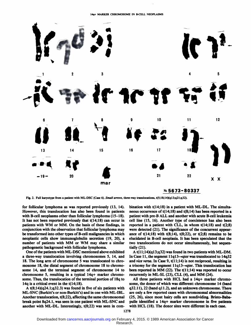
Fig. 2. Full karyotype from a patient with ML-DSC (Case 6). Small arrows, three-way translocations, t(5;18;14Xql3;q21;q32).
for follicular lymphoma as was reported previously (13, 14).However, this translocation has also been found in patientswith B-cell neoplasms other than follicular lymphoma (15-18).It has not been reported previously that t(14;18) can occur inpatients with WM or MM. On the basis of these findings, inconjunction with the observation that follicular lymphoma maybe transformed into other types of B-cell malignancies in whichneoplastic cells show immunoglobulin secretion (19, 20), anumber of patients with MM or WM may share a similarpathogenetic background with follicular lymphoma.
One of the patients with ML-DSC mentioned above exhibiteda three-way translocation involving chromosomes 5, 14, and18. The long arm of chromosome 5 was translocated to chromosome 18, the distal segment of chromosome 18 to chromosome 14, and the terminal segment of chromosome 14 tochromosome 5, resulting in a typical 14q+ marker chromosome. Thus, the translocation of the terminal portion of 18q to14q is a critical event in the t( 14:18).
A t(8;14)(q24.1;q32.3) was found in five of six patients withML-SNC (Burkitt's or non-Burkitt's) and in one with ML-IBL.
Another translocation, t(8;22), affecting the same chromosomalbreak point 8q24.1, was seen in one patient with ML-SNC andanother with ML-DL. Interestingly, t(8;22) was seen in com
bination with t(14;18) in a patient with ML-DL. The simultaneous occurrence of t( 14;18) and t(8;14) has been reported in apatient with pre-B ALL and another with acute B-cell leukemiacell line (15, 16). Another type of coexistence has also beenreported in a patient with CLL, in whom t(14;18) and t(2;8)were detected (21 ). The significance of the concurrent appearance of t(14;18) with t(8;14), t(8;22), or t(2;8) remains to beelucidated in B-cell neoplasia. It has been speculated that thetwo translocations do not occur simultaneously, but sequentially (21).
A t(l I;14)(ql3;q32) was found in two patients with ML-DM.In Case 11, the segment 11q 13—»qterwas translocated to 14q32and vice versa. In Case 9, t(l 1;14) is not reciprocal, resulting ina trisomy for the segment 1Iql3—»qter.This translocation hasbeen reported in MM (22). The t(l 1;14) was reported to occurrecurrently in ML-SL (23), CLL (4), and MM (24).
All three patients with HCL had a 14q+ marker chromosome, the donor of which was different: chromosome 14 (bandq32.11), 22 (band ql 1.2), and an unknown chromosome. Thereare only a few reported cases with chromosomal abnormalities(25, 26), since most hairy cells are nondividing. Bristo-Baba-pulle identified a 14q+ marker chromosome in five patientswith HCL (18). The donor sites were different in each case.
1278
Research. on February 4, 2015. © 1989 American Association for Cancercancerres.aacrjournals.org Downloaded from

14q+ MARKER CHROMOSOME IN B-CELL NEOPLASMS
Table 3 Summary ofcytogenetic results in patients with hairy cell leukemia, macroglobulinemia, and plasma cell disordersPatients are grouped according to types of B-cell malignancy.
Caseno.°HCL22
23Age/sex60/M60/FSample
source*PB
PB"Surface
marker'G-K
G-LM-proteinIgG-K(-)Abnormal
cells(%f11100Karyotype46,XY,del(l)(q32.lq42.3),t(14;22)(q32.3;qll.2)
45,X-,-X,+der(5)t(5; 17)(p 15;q2 1),del(6)(q 15q25),
24
WM25-29303132»
MM33-5253*
54«55»
5657»58«
PCL5960
40/M
40/F70/M70/M
49/F
60/F67/F
69/M79/M62/F
57/M59/F
PB«
LNBMBM
Pleuraleffusion
BMAscitesBM
BMBMPB.BM
PBPB
G-K
M-KNDND
IgM-KIgMIgM
(nonsecretory)
NDND
NDNDND
BJ-LBJ-L
IgG-KIgA-KIgG-K
ND IgG-LNonproducing
I(14;14)(q32.11;q32.33)100 46,XY,-14,+der( 14)t( 14;?)(q32.3;?)
Normal karyotypes44 48,XX,+X,t( 14;18)(q32;q21 ),+der( 18)t( 14;18)40 46,XY,del(15)(q24)40 48, XY, -4, -19, +21, +22, t(7;17)(pl5;pl3),
+der( 19)t(4; 19)(q23;ql 3.4), +mar 1
Normal karyotypes100 82, XX, -X, -2,+4, -7,+8, +9,+9, -10,-11,+12,
+13, +14, +15, -17, +18, -20, -20, -20.+22,+der(X) t(X;?)(q28;?), +der(2)t(2;7)(q35;ql 1),+der(2), +der(7)t(l;7)(q32;p22),+der( 11)t( 11;?)(q23;?), t( 14;18)(q32.3;q21.3),t(14;18), del(16)(ql 1), del(16), +i(16q), +i(16q),+der(20)t(ll;20)(ql3.3;ql3.1), +der(20), +der(20),+der(20)
SCA 46,XX,t(I4;18Xq32.3;q21.3)100 73.XX : 1p-, 3p-, oq-, 9q+, 11p+, i(17q), 14q+100 53, XX : +l,+3, +7, +9, +12, +15, +17, +18, +19,
-22, +der(2)t(2;6)(p25;ql3), +dic(6)(ql 1), +der(8),+der(l 1), t(l I;?)(pl4;?), +der(14)t(14;?)(q32;?)
15 47,XY,+14/46,XY,-2,+marSCA 47,XY,+20,del( 11)(p 11.2)100 41 ,XX: t( 1;6Kq44;q 16), t(3; 14)(p 11;q32),
del( 11)(q14),der(22)t( 12;22)(22qter-22p 12::12q 11-12q24::12ql3-l2q24::12ql3-12qter)
Normal karyotype100 46,XX,del( 1)(p 13),t(6; 14)(p21.1 ;q32)/45,X-
-X,del( 1)(p 13),t(6; 14)(p21.1 ;q32)
PCM6175/FTumorNonproducing10083,XX:7q+,llq+,20q+
°Patients (*) previously treated (all other untreated).* Stimulated with STA (**) (10).' ND, not done.d SCA, single cell abnormality.
14 22
14Fig. 3. Partial karyotypes of reciprocal translocations between the immunn
globulin heavy chain gene locus and one of the light chain gene loci, a, at(2;14)(pl3;q32.3) in a patient with leukemic ML-DM (Case 10); at(14;22)(q32.3;ql 1.2) resulting in 14q+ marker and I'll' chromosome ina patient
with HCL (Case 22).
We identified a novel translocation, t(6;14)(p21.1;q32) in apatient with PCL (Case 60). Recently, the human homologue,lipini, of the murine pim-l gene, has been assigned to band6p21 (27). The hpim gene is expressed in various hematopoieticcell lines. A case of childhood T-cell ALL was reported, inwhich the chromosomal segment, 6p21-pter, was translocatedto the 7q36 band where the T-cell receptor-/3-chain gene hasbeen assigned (28). It is possible that hpim is also implicated in
the pathogenesis of B cell malignancy.Chromosomal translocation results in the conjunction of an
oncogene with the immunoglobulin gene in B-cell neoplasm(29, 30). In our series, 16 patients showed this type of rearrangement among 26 with the 14q+ marker. However, chromosomal translocations occurred between the IgH gene locusand one of the IgL gene loci in two patients (Cases 10 and 22).One is t(2;14)(pl3;q32.3) found in a patient with leukemic ML-DM. The neoplastic cells showed a surface IgM immunoglobulin of X-light chain. The other is t(14;22)(q32.3;qll.2) inanother patient with HCL. The leukemic cells bore a surfaceIgG of the «-lightchain type. Each chromosomal abnormalityhas been reported previously: t(2;14) in a child with CLL (31),whose leukemic cells expressed IgM and IgD-/c membraneimmun*»globulins;and t(14;22) in 3 patients with ALL (32, 33)and CLL (4). Both of our cases revealed the mismatch betweenthe IgL phenotype of the neoplastic cells and the gene lociinvolved in these translocations. Similar findings were reportedin a few cell lines (34, 35), in which t(8;22) was found in cellsproducing the K-light chain. The role of translocations betweenthe loci for IgH and K-or X-light chains, t(2;14) or t(14;22), inthe pathogenesis of B-cell malignancies remains to be eluci
dated.In conclusion, we found a variety of 14q+ marker chromo
some in 26 of 35 patients with clonal chromosomal anomaliesin differentiated B-cell malignancy. In NHL, three 14q+ marker
1279
Research. on February 4, 2015. © 1989 American Association for Cancercancerres.aacrjournals.org Downloaded from

14q+ MARKER CHROMOSOME IN B-CELL NEOPLASMS
Table 4 Association of recurrent chromosome abnormalities with types ofB-cell malignancies
Chromosome abnor
mality14q32.36qlq18q21.38424.1-XIPlipIlql3.3+12+37pXqFMFL(n
= 4)(n=l)411
1421311DSC(«
=3)2221»11NHL°DM(n= 3)(n3
I212*«*213L
IBLSNC!
1512**1
611HCL
WM(n= 3) (n =3)3
111111MM432111322111PCDPCL(n=l)111PCM
Total(n = 1) (35)2610105444
*, complex translocation (5;18;14); ", combined with t(8;22); **', t(l I;14)(ql3.3;q32.3).
REFERENCES
9 10 11 12
13 U 2H 15 16 17 W 20 21 22 X
Fig. 4. Human chromosome map (Human Gene Mapping 9) (36) of onco-genes, immunoglobulin genes, and chromosomal break points (•and O) in B-cellneoplasia. O, donor sites of 14q+ marker chromosome. The karyotype representsGiemsa bands at the approximately 550 bands stage, according to the ISCN 1985(11).
chromosomes were associated with the morphological classification, i.e., t(14;18) in ML-FM, t(8;14) in ML-SNC, andt(l 1;14) in ML-DM. The donor for the 14q+ marker chromosome originated from the bands of oncogene loci; bcl-2, c-myc,or bcl-l. However, the t(14;18) was also observed in patientswith ML-DSC, ML-DL, WM, and MM (a single case of each).A new chromosomal translocation, (6;14)(p21.1;q32.3), associated with oncogene c-pim, was found in a patient with PCL.Thus, the 14q+ chromosome was reconstructed between animmunoglobulin gene and an oncogene, with rare exceptions,between the IgH gene and one of the IgL genes.
ACKNOWLEDGMENTS
The authors are particularly indebted to Professor Tatsuro Takino,Director of the Third Department of Medicine, Kyoto Prefectura!University of Medicine, for kindly reviewing the manuscript, and arealso grateful to Dr. James R. Miller for constructive comments on themanuscript. We thank our colleagues, Dr. Taira Maekawa who performed the study of immunologie cell markers, and Yohko Vanumewho provided expert technical assistance.
We are also grateful to the following for offering the opportunity tostudy their patients: Professor Atsushi Sawada, Kyoto Prefectura! University of Medicine; Professor Teruo Kitani, Associate Professor Takeshi Yonezawa, and Dr. Osamu Sugiyama, Osaka University; Dr.Masanori Shimoyama, National Cancer Center; and Dr. Hiroshi Fujii,Kyoto First Red Cross Hospital.
10.
12.
13.
14.
15.
16.
17.
18.
19.
20.
21.
Yunis, J. J. The chromosomal basis of human neoplasia. Science (Wash.DC), 221: 227-236, 1983.Rowley, J. D. Biological implications of consistent chromosome rearrangements in leukemia and lymphoma. Cancer Res., 44:3159-3168,1984.Kaneko, Y., Rowley, J. D., Variakojis, D., Chilcote, R. R., Check, I., andSakurai, M. Correlation of karyotype with clinical features in acute lympho-blastic leukemia. Cancer Res., 42: 2918-2929, 1982.Nowell, P. C, Shankey, T. V., Finan, J., Guerry, D., and Besa, E. Proliferation, differentiation, and cytogenetics of chronic leukemic B lymphocytescultured with mitomycin treated normal cells. Blood, 57:444-451, 1981.Nishida, K., Taniwaki, M., Misawa, S., Abe, T., Takino, T., and Fujii, H.Translocation (14; 18) found in a patient with multiple myeloma. CancerGenet. Cytogenet., 25:375-377, 1987.First MIC Cooperative Study Group. Morphologic, Immunologie, and Cytogenetic (MIC) Working Classification of Acute Lymphoblastic Leuke-mias:report of the workshop held in Leuven, Belgium, September 15-17,1986. Cancer Genet. Cytogenet., 23: 189-197, 1986.The Non-Hodgkin's Lymphoma Pathologic Classification Project. NationalCancer Institute sponsored study of classifications of non-Hodgkin's lym-
phomas: summary and description of a working formulation for clinicalusage. Cancer (Phila.), 49: 2112-2135, 1982.Misawa, S., Horiike, S., Taniwaki, M., Abe, T., and Takino, T. Prefixationtreatment with ethidium bromide for high resolution banding analysis ofchromosomes from cultured human bone marrow cells. Cancer Genet. Cytogenet., 22: 319-329, 1986.Taniwaki, M. New chromosome banding techniques with base specific antibiotics and fluorochromes for qualified identification of marker chromosomesin leukemia and related disorders. Acta Haematol. Jpn., 48: 1423-1439,1985.Tamaki, T., Yonezawa, T., Kanayama, Y., Inoue, R., Kanakura, l '.. Katagiri,
S., Machii, T., Kitani, T., and Tarili, S. Mitogenic response of neoplastic"hairy cells" to Staphylococcus aureus Cowan 1. Acta Haematol. Jpn., 48:1042-1052, 1985.An International System for Human Cytogenetic Nomenclature (ISCN1985): report of the standing committee on human cytogenetic nomenclature.Cytogenet. Cell Genet., 1-118, 1985.Mitelman, F. Catalog of Chromosome Aberrations in Cancer, Ed. 2. NewYork: Alan R. Liss, Inc., 1985.Yunis, J. J., Oken, M. M., Kaplan, M. E., Ensrud, K. M., Howe, R. R., andTheologides, A. Distinctive chromosomal abnormalities in histologie subtypes of non-Hodgkin's lymphoma. N. Engl. J. Med., 507:1231-1236,1982.
Bloomfield, C. D., Arther, D. C., Frizzerà , G., Levine, E. G., Peterson, B.A., and Gajl-Peczalska, K. J. Nonrandom chromosome abnormalities inlymphoma. Cancer Res., 43: 2975-2984,1983.Mufti, G. J., Hamblin, T. J., Oscier, D. G., and Johnson, S. Common ALLwith pre-B-cell features showing (8;14) and (14; 18) chromosome translocations. Blood, 62: 1142-1146, 1983.Tsujimoto, Y., Finger, L. R., Yunis, J. J., Nowell, P. C., and Croce, C. M.Cloning of the human chromosome breakpoint of neoplastic B-cells with thet(14;18) chromosome translocation. Science (Wash. DC), 226: 1097-1099,1985.Fukuhara, S., Nasu, K., Kita, K., Ueshima, Y., Oguma, S., Yamabe, H.,Nishigori, M., and Uchino, H. Cytogenetic approaches to the clarification ofpathogenesis in lymphoid malignancies: clinicopathologic characterizationof 14q+ marker-positive non T cell malignancies. Jpn. J. Clin. Oncol., 13:461-476, 1983.Brito-Babapulle, V., Pittman, S., Melo, J. V., Parreira, L., and Catovsky, D.The I4q+ marker in hairy cell leukemia: a cytogenetic study of 15 cases.Leukemia Res., 10: 131-138, 1986.Bain, G. D., and Belch, A. Nodular mixed cell lymphoma with monoclonalgammopathy. Am. J. Clin. Pathol., 76: 832-837, 1981.Alexanian, R. Monoclonal gammopathy in lymphoma. Arch. Intern. Med.,135:62-66, 1975.Geisler, C., Philip, P., Plesner, P., Andersson, P., Zeuthen, J., Guldhammer,
1280
Research. on February 4, 2015. © 1989 American Association for Cancercancerres.aacrjournals.org Downloaded from
14q+ MARKER CHROMOSOME IN B-CELL NEOPLASMS
I!., and Hansen, M. M. Simultaneous presence of translocations t(14;18) andt(2;8) in a case of chronic lymphocytic leukemia. Cancer Genet. Cytogenet.,22: 35-44, 1986.
22. Venti, G., Mecucci, C., Donti, E., and Tabilio, A. Translocation t(l 1;14) andtrisomy Ilql3-qter in multiple myeloma. Ann. Genet. (Paris), 27: 53-54,1984.
23. Yunis, J. J., Oken, M. M., Theologides, A., Howe, R. R., and Kaplan, M. E.Recurrent chromosomal defects are found in most patients with non-Hodg-kin's-lymphoma. Cancer Genet. Cytogenet., 13:17-28, 1984.
24. Dewald, G. W., Kyle, R. A., Hicks, G. A., and Greipp, P. R. The clinicalsignificance of cytogenetic studies in 100 patients with multiple myeloma,plasma cell leukemia or amyloidosis. Blood. 66: 380-390, 1985.
25. Khalid, D., Li, Y-S., Flements, R. J., and Hayhoe, F. G. Chromosomalabnormalities in a case of hairy cell leukemia. Leukemia Res., 5: 431-435,1981.
26. Sadamori, V. and Sandberg, A. A. 14q+ and 6q- anomalies in a case withhairy cell leukemia. Cancer Genet. Cytogenet., 8:89-90, 1983.
27. Nagarajan, I... Louie, E., Tsujimoto, Y.. ar-Rushdi, A., Huebner. K., andCroce, C. M. Localization of the human pirn oncogene (PIM) to a region ofchromosome 6 involved in translocations in acute leukemia. Proc. Nati.Acad. Sci. USA, 83: 2556-2560, 1986.
28. Raimondi, S. C., Ching-Hon, P., Behm, F. G., and Williams, D. L. 7q32-q36 translocation in childhood T cell leukemia: cytogenetic evidence for
involvement of the T cell receptor /j-chain gene. Blood, 69:131 -134, 1987.29. Klein, G. Specific chromosomal translocations and the genesis of It-cell
derived tumors in mice and men. Cell, 32: 311-315, 1983.30. Croce, C. M., and Nowell, P. C. Molecular basis of human B cell neoplasia.
Blood, 65: 1-7. 1985.31. Sonnier, J. A., Buchanan, G. R., Howard-Peebles, P. N., Rutledge, J., and
Smith, R. G. Chromosomal translocation involving the immunoglobulinkappa-chain and heavy-chain loci in a child with chronic lymphocytic leukemia. N. Engl. J. Med., 309: 590-594, 1983.
32. Ayraud, N., Dujardin, P., and Audoly, P. Leucémieaiguëlymphoblastiqueavec chromosome Philadelphie: rôleprobable d'une translocation 14-22.
Nouv. Presse. Méd.,4: 3013, 1975.33. Sandberg, A. A., Morgan, R., Kipps, T. J., Hecht, B. K., and Hecht, F. The
Philadelphia (Ph) chromosome in leukemia. II. variant l'h translocations inacute lymphoblastic leukemia. Cancer Genet. Cytogenet., 14: 11-21, 1985.
34. Magrath, I., Erikson, J., Whang Peng, J., Sieverts, H., Armstrong, G.,Benjamin, D., Tricke, T., Alabaster, O., and Croce, C. M. Synthesis of kappalight chains by cell lines containing an 8;22 chromosomal translocationderived from a male homosexual with Burkitt's lymphoma. Science (Wash.DC), 222: 1094-1098, 1983.
35. Hecht, F., Morgan, R., and Hecht, B. K. Unexpected lambda chain expressionin lymphocytic malignancy. Int. J. Radiât.Oncol., //: 267-270, 1985.
36. Human Gene Mapping 9. Paris Conference (1987): ninth internationalworkshop on human gene mapping. Cytogenet. Cell Genet., 46:1-762,1987.
1281
Research. on February 4, 2015. © 1989 American Association for Cancercancerres.aacrjournals.org Downloaded from

1989;49:1275-1281. Cancer Res Kazuhiro Nishida, Masafumi Taniwaki, Shinichi Misawa, et al.
Phenotypein Human Lymphoid Malignancies with Mature B-Cell Nonrandom Rearrangement of Chromosome 14 at Band q32.33
Updated version
http://cancerres.aacrjournals.org/content/49/5/1275
Access the most recent version of this article at:
E-mail alerts related to this article or journal.Sign up to receive free email-alerts
Subscriptions
Reprints and
To order reprints of this article or to subscribe to the journal, contact the AACR Publications
Permissions
To request permission to re-use all or part of this article, contact the AACR Publications
Research. on February 4, 2015. © 1989 American Association for Cancercancerres.aacrjournals.org Downloaded from