Non-viral gene delivery nanoparticles based on Poly(β-amino esters) for treatment of glioblastoma
19
Non-Viral Gene Delivery Nanoparticles Based on Poly(β-amino esters) for Treatment of Glioblastoma Stephany T. Tzeng 1 , Hugo Guerrero-Cázares 2 , Elliott E. Martinez 1 , Joel C. Sunshine 1 , Alfredo Quiñones-Hinojosa 2,* , and Jordan J. Green 1,* 1 Department of Biomedical Engineering and the Institute for NanoBioTechnology, Johns Hopkins University School of Medicine, Baltimore, MD 21231, USA 2 Departments of Neurosurgery and Oncology, Johns Hopkins University School of Medicine, Baltimore, MD 21231, USA Abstract Glioblastoma (GB) is currently characterized by low survival rates and therapies with insufficient efficacy. Here, we describe biodegradable polymers that can deliver genes to primary GB cells as well as GB tumor stem cells in vitro with low non-specific toxicity and transfection efficiencies of up to 60.6±5 % in normal (10%) serum conditions. We developed polymer-DNA nanoparticles that remained more stable in normal serum and could also be stored for at least 3 months in ready- to-use form with no measurable decrease in efficacy, expanding their potential in a practical or clinical setting. A subset of polymers was identified that shows a high degree of specificity to tumor cells compared with healthy astrocytes and human neural stem cells when cultured (separately or in co-culture), yielding higher transfection in GB cells while having little to no apparent effect on healthy cells. Keywords Drug delivery; gene therapy; nanoparticle; stem cell; DNA; cancer 1. Introduction Approximately 184,300 new central nervous system (CNS) tumors are diagnosed annually, with 50,000 of those originating in the CNS and the rest being metastases from other tumors [1–5]. Malignant gliomas are the most common primary brain tumor and are refractory to combinatorial surgical resection, radiation, and chemotherapy [6–8]. These tumors possess the ability to extensively invade surrounding tissue, making curative resection impossible [9–11]. As a result, experimental treatments like gene therapy are attractive for GB patients. Despite the vast potential of gene therapy, viral methods are plagued with safety concerns [12], while non-viral delivery vehicles often lack efficiency [13]. A high-throughput screening approach based on a synthetic library of cationic poly(β-amino esters) (PBAEs) © 2011 Elsevier Ltd. All rights reserved. * Address for correspondence: Jordan J. Green, 400 North Broadway, Smith Building, Room 5017, Baltimore, MD 21231, Tel.: +1 410 614 9113, [email protected]; Alfredo Quinones-Hinojosa, 1550 Orleans Street, CRB II, Room 247, Baltimore, MD 21231, Tel.: +1 410 502 2869, [email protected]. Publisher's Disclaimer: This is a PDF file of an unedited manuscript that has been accepted for publication. As a service to our customers we are providing this early version of the manuscript. The manuscript will undergo copyediting, typesetting, and review of the resulting proof before it is published in its final citable form. Please note that during the production process errors may be discovered which could affect the content, and all legal disclaimers that apply to the journal pertain. NIH Public Access Author Manuscript Biomaterials. Author manuscript; available in PMC 2012 August 1. Published in final edited form as: Biomaterials. 2011 August ; 32(23): 5402–5410. doi:10.1016/j.biomaterials.2011.04.016. NIH-PA Author Manuscript NIH-PA Author Manuscript NIH-PA Author Manuscript
-
Upload
johnshopkins -
Category
Documents
-
view
3 -
download
0
Transcript of Non-viral gene delivery nanoparticles based on Poly(β-amino esters) for treatment of glioblastoma
Non-Viral Gene Delivery Nanoparticles Based on Poly(β-aminoesters) for Treatment of Glioblastoma
Stephany T. Tzeng1, Hugo Guerrero-Cázares2, Elliott E. Martinez1, Joel C. Sunshine1,Alfredo Quiñones-Hinojosa2,*, and Jordan J. Green1,*
1 Department of Biomedical Engineering and the Institute for NanoBioTechnology, Johns HopkinsUniversity School of Medicine, Baltimore, MD 21231, USA2 Departments of Neurosurgery and Oncology, Johns Hopkins University School of Medicine,Baltimore, MD 21231, USA
AbstractGlioblastoma (GB) is currently characterized by low survival rates and therapies with insufficientefficacy. Here, we describe biodegradable polymers that can deliver genes to primary GB cells aswell as GB tumor stem cells in vitro with low non-specific toxicity and transfection efficiencies ofup to 60.6±5 % in normal (10%) serum conditions. We developed polymer-DNA nanoparticlesthat remained more stable in normal serum and could also be stored for at least 3 months in ready-to-use form with no measurable decrease in efficacy, expanding their potential in a practical orclinical setting. A subset of polymers was identified that shows a high degree of specificity totumor cells compared with healthy astrocytes and human neural stem cells when cultured(separately or in co-culture), yielding higher transfection in GB cells while having little to noapparent effect on healthy cells.
KeywordsDrug delivery; gene therapy; nanoparticle; stem cell; DNA; cancer
1. IntroductionApproximately 184,300 new central nervous system (CNS) tumors are diagnosed annually,with 50,000 of those originating in the CNS and the rest being metastases from other tumors[1–5]. Malignant gliomas are the most common primary brain tumor and are refractory tocombinatorial surgical resection, radiation, and chemotherapy [6–8]. These tumors possessthe ability to extensively invade surrounding tissue, making curative resection impossible[9–11]. As a result, experimental treatments like gene therapy are attractive for GB patients.Despite the vast potential of gene therapy, viral methods are plagued with safety concerns[12], while non-viral delivery vehicles often lack efficiency [13]. A high-throughputscreening approach based on a synthetic library of cationic poly(β-amino esters) (PBAEs)
© 2011 Elsevier Ltd. All rights reserved.*Address for correspondence: Jordan J. Green, 400 North Broadway, Smith Building, Room 5017, Baltimore, MD 21231, Tel.: +1 410614 9113, [email protected]; Alfredo Quinones-Hinojosa, 1550 Orleans Street, CRB II, Room 247, Baltimore, MD 21231, Tel.: +1 410502 2869, [email protected]'s Disclaimer: This is a PDF file of an unedited manuscript that has been accepted for publication. As a service to ourcustomers we are providing this early version of the manuscript. The manuscript will undergo copyediting, typesetting, and review ofthe resulting proof before it is published in its final citable form. Please note that during the production process errors may bediscovered which could affect the content, and all legal disclaimers that apply to the journal pertain.
NIH Public AccessAuthor ManuscriptBiomaterials. Author manuscript; available in PMC 2012 August 1.
Published in final edited form as:Biomaterials. 2011 August ; 32(23): 5402–5410. doi:10.1016/j.biomaterials.2011.04.016.
NIH
-PA Author Manuscript
NIH
-PA Author Manuscript
NIH
-PA Author Manuscript
has led to high transfection efficiency and low toxicity in many cell types [14, 15], thoughthey have not previously been tested in brain tumors. Hundreds of these polymers can besynthesized by combinatorial chemistry, and many cause superior transfection compared toleading commercial agents like Lipofectamine 2000 (Invitrogen), while also being less toxicto cells [16].
Also important for eventual clinical use is the stability of the polymer-DNA nanoparticles,as well as their ease of preparation. Because PBAEs are electrostatically complexed withDNA in slightly acidic, aqueous suspension, the particles have rapid release properties dueto hydrolytic degradation of the polymers. While these release properties are advantageousfor intracellular gene delivery, they are not conducive for practical use by clinicians or large-scale production. It is therefore necessary to develop a way of preparing a dried form of thepolymer-DNA nanoparticles that can be stored for long periods of time and easilyreconstituted just before use. However, increased aggregation is a complication for colloidalsuspensions that are dried without a lyoprotectant [17], and an effective way of lyophilizingand storing these particles is essential for eventual translation of this technology.
We sought to apply and improve upon previously validated methods of non-viral genedelivery for the treatment of primary human brain tumor cells, including the identification ofefficacious reagents and the development of more practical and easily-used formulations.Aside from the malignant astrocytes that comprise the bulk of a GB tumor, we are alsointerested in brain tumor stem cells (BTSCs). BTSCs are hypothesized to be the source ofGB tumors and to play a role in the inevitable tumor recurrence following surgery,chemotherapy, and radiotherapy [18–21]. Aside from being resistant to more types oftherapy than astrocytes [22], BTSCs can cause regrowth of an entire tumor at low cellconcentrations [23], which the malignant astrocytes alone cannot do [20, 21]. Thepersistence of BTSCs despite therapy, as well as their tumorigenic capacity, make them aparticularly good target of experimental therapies like gene delivery. A comparison of thetransfection efficiencies in GB astrocytes, BTSCs, and healthy (non-cancerous) versions ofboth is of interest, since we have recently found that cell-type specificity can be determinedsimply by the polymer structure itself [15].
2. Materials and methods2.1. Materials
Lipofectamine 2000 (Invitrogen, Carlsbad, CA), FuGENE® HD (Roche), Opti-MEM I(Invitrogen), pEGFP-N1 DNA (Elim Biopharmaceuticals, Hayward, CA), pDsRed-Max-N1[24] and FUGW [25] (Addgene DNA plasmids 21718 and 14883, respectively, Cambridge,MA), and cell culture media components were used as received. Label IT® Tracker Cy3 Kitwas purchased from Mirus Bio. 4′,6-diamidino-2-phenylindole dihydrochloride (DAPI) waspurchased from Sigma (Saint Louis, MO) and used as a 750 nM solution in PBS.
2.2. Cell CultureAll intraoperative samples were obtained as described previously under institutionallyapproved protocols. Human BTSCs (BTSC line 551) was isolated from a GB tumor sampleof an adult patient (69 years old) diagnosed with GB, as previously described [26]. Briefly,necrotic tissue and blood vessels were removed from the tumor, which was dissociatedmechanically and with trypsin/EDTA. After inhibition of trypsin with 1:1 DMEM/F-12(Invitrogen) with 10% heat-inactivated FBS and 1% antibiotic-antimycotic (anti-anti, finalconcentration 1X, Invitrogen), the cells were collected by centrifugation, and the serum-containing medium was replaced with BTSC neurosphere medium [1:1 DMEM/F-12, 2%B-27 serum-free supplement (B-27, final concentration 1X, Gibco, Bethesda, MD), 1% anti-
Tzeng et al. Page 2
Biomaterials. Author manuscript; available in PMC 2012 August 1.
NIH
-PA Author Manuscript
NIH
-PA Author Manuscript
NIH
-PA Author Manuscript
anti, 20 ng/mL basic fibroblast growth factor (bFGF, Invitrogen), and 20 ng/mL epidermalgrowth factor (EGF, Sigma, Saint Louis, MO)]. A GB astrocyte cell line (GB 319) wasderived from BTSCs isolated as above (from a 79-year-old adult GB patient). Afterisolation, they were maintained in astrocyte medium with serum (1:1 DMEM/F-12, 10%FBS, 1% anti-anti) as adherent cultures.
Human fetal neural stem cells (NSCs) (F34) were derived after 17 weeks of gestation,obtained from elective abortion. Brain cortical tissue was mechanically dissociated and cellswere maintained in neurosphere medium consisting of 2:1 high-glucose DMEM with l-glutamine (Invitrogen)/Ham’s F-12 (Cellgro), 1X B-27, 1% anti-anti, 20 ng/mL bFGF, 20ng/mL EGF, 20 ng/mL leukemia inhibitory factor (LIF, Millipore, Billerica, MA), and 5 μg/mL heparin (Sigma)]. BTSC 551 was stably transduced with EGFP using the lentiviralvector FUGW. Cells were incubated with the virus overnight in the presence of 8 μg/mLpolybrene, then sorted by FACS. The GFP+ 551 BTSCs were maintained in neurospheremedium in nonadherent flasks and passaged approximately every 10 days by mechanicaldissociation. For transfection experiments, 551 BTSCs were plated on laminin-coated plates,either as neurospheres or dissociated into single cells, and maintained in neurospheremedium following the protocol described by Pollard et al. [27]. F34 fetal neurospheres weremaintained either as NSCs or in astrocyte medium with serum for differentiation and wereused as non-cancerous controls for BTSC 551 BTSCs and GB 319 astrocytes. All cells wereincubated at 37°C in a humid atmosphere of 5% CO2.
2.3. Synthesis of Poly(β-amino esters)PBAEs were synthesized in a two-step reaction as described previously [14] using smallcommercially-available molecules (Figure 1). These small molecules were purchased fromAcros Organics (E8), Alfa Aesar (B4, B6, S3, S4, S5, E7), Fluka (E6), Monomer-Polymerand Dajac Labs (B5), Sigma-Aldrich (E9), and TCI America (E3, E4, E5), and used asreceived. Briefly, for each polymer, one backbone monomer (labeled as B4, B5, or B6) wasmixed with a sidechain monomer (S3, S4, or S5), either neat or as a 500 mg/mL solution inDMSO, at a 1.1:1 or 1.2:1 ratio of B:S. The mixture was stirred at 90°C for 24 hr to yieldacrylate-terminated base polymers by Michael addition. Base polymers were endcappedwith a small molecule (E3, E4, E5, E6, E7, E8, or E9) by dissolving endcapping moleculesin DMSO (167 mg/mL) and adding 480 μL endcapping solution to 320 μL of 0.5 M basepolymer in DMSO, then shaking the mixture at room temperature overnight. Final polymerswere stored with dessicant at 4°C as 100 mg/mL solutions in DMSO. Polymers used foroptimization experiments were stored in small aliquots at −20°C to minimize freeze-thaw ofsamples.
2.4. DNA Delivery to GB 319 Astrocytes2.4.1. Nanoparticle Uptake—Plasmid DNA encoding EGFP was labeled with Cy3 usinga Mirus Bio kit according to manufacturer instructions. A solution of 150 μg/mL DNA and100 μL/mL Label IT Tracker reagent was prepared in buffer, then incubated at 37°C for 3 hron an orbital shaker. The labeled DNA was precipitated with ethanol in 300 mM sodiumacetate buffer and cooled at −20°C for 30 min. The DNA was pelleted by centrifugation at15,000 g at 4°C for 15 min, then washed with 70% ethanol and centrifuged again. The driedpellet was reconstituted in water at a concentration of 0.5 mg/mL, and concentration wasverified using by absorbance at 260 nm using a Biotek Synergy 2 Gen5 Multiplate reader.The absence of protein from the solution was measured by absorbance at 280 nm (A260/A280= 1.78).
2.4.2. Initial Screenings of Polymer Formulations—GB 319 astrocytes were platedin 24-well plates at a density of 75,000 cells/well 24 hr before transfection. Just before
Tzeng et al. Page 3
Biomaterials. Author manuscript; available in PMC 2012 August 1.
NIH
-PA Author Manuscript
NIH
-PA Author Manuscript
NIH
-PA Author Manuscript
transfection, all culture media were replaced with serum-free DMEM/F-12 (1:1). EGFPplasmid DNA was delivered to GB astrocytes as previously described [28]. Briefly, DNAand polymer were each diluted in 25 mM sodium acetate (pH 5), then combined at a 30:1,60:1, 90:1, or 120:1 weight/weight ratio (w/w) of polymer to DNA (volume ratio was 1:1 ineach case). The mixture was incubated for 10 min to allow complexation to occur, thenadded to the cell medium at a volumetric ratio of 1:5 complexes to culture medium.Lipofectamine 2000 and FuGENE HD were prepared according to the manufacturers’instructions and used as positive controls, with FuGENE HD used at a ratio of 4 μL reagentto 1 μg DNA and Lipofectamine at 2.5 μL reagent to 1 μg DNA. The dose used was 1.5 μgDNA per well unless stated otherwise. After 4 hr incubation time at 37°C, the particles andtransfection medium were aspirated and replaced with fresh complete astrocyte medium.
After 48 hr, cells were fixed with 10% buffered formalin for 20 min, stained with DAPI for5 min, and stored in PBS at 4°C for up to 1 week. Using a Zeiss Observer, fluorescentimages were taken (total of 6 images per experimental group). The number of DAPI+ cellswere counted in ImageJ to calculate viability (expressed as a percentage of untreatedcontrols, mean±standard error). The number of GFP+ cells were counted in ImageJ andnormalized to the number of cells in the same image to calculate transfection efficiency(expressed as a percentage, mean±standard error).
Top formulations, along with FuGENE HD and Lipofectamine 2000, were used in repeatexperiments with complete culture medium as the transfection medium to assess the efficacyof the reagents in 10% FBS. Doses between 0.15 μg to 1.5 μg DNA per well were tested tofind a dose that would allow at least 80% viability. Viability and transfection efficiencywere quantified after 48 hr as described above.
2.4.3. Optimization of Stable Nanoparticle Formulations—The top polymer fromthe initial screenings was used to form nanoparticles with DNA that could be stored in dryform and used at later time points with little or no loss of efficacy. Nanoparticles wereformed as above, at twice the normal concentration in 25 mM sodium acetate. Followingcomplexation, the particles were mixed in a 1:1 (v/v) ratio with a solution of D-sucrose in 25mM sodium acetate at varying sugar concentrations (0, 30, 60, and 90 mg/mL sucrose insodium acetate added to particles, for a final concentration of 0–45 mg/mL sucrose). Thesuspension of particles in sucrose and sodium acetate was frozen at −80°C for 2 hr, thenlyophilized for 48 hr and stored at room temperature, 4°C, or −20°C with dessicant.Particles were tested immediately after lyophilization (t=0) and after 1, 2, and 3 months ofstorage.
Before use, particles were reconstituted in the same starting volume of distilled water.Transfection experiments were carried out as described above. For quantification, the totalfluorescence from each well was also measured using the plate reader before fixation andstaining with DAPI. Transfection efficiency and viability were quantified as above. Thenumber of cells in each well was also used to normalize fluorescent signal from the platereader (RFU/cell) and normalized to untreated controls (mean± standard error). The directnumber-averaged size of the particles and effective concentration was also tested at the sametime points using nanoparticle tracking analysis (NTA) with a NanoSight LM10-HS(NanoSight Ltd., Wiltshire, UK). Each sample was diluted in PBS 1:90 from the standardtransfection concentration for a total of 3 measurements. The mean diameter, modediameter, and standard deviation of the size distribution were recorded. Results were pooledand expressed as mean±standard error for each parameter.
Tzeng et al. Page 4
Biomaterials. Author manuscript; available in PMC 2012 August 1.
NIH
-PA Author Manuscript
NIH
-PA Author Manuscript
NIH
-PA Author Manuscript
2.5. DNA Delivery to GB 551 Brain Tumor Stem Cells2.5.1. DNA Delivery to Brain Tumor Stem Cells in Monolayer—Beforetransfections, 96-well plates were coated with 20 μg/mL laminin (Sigma) overnight at 4°Cand washed once with PBS. BTSC neurospheres were collected by centrifugation and thesupernatant discarded. Neurospheres were mechanically dissociated by trituration with a 200μL micropipette and plated on laminin at 15,000 cells/well in complete medium. Cells wereincubated 24 hr to allow them to adhere. Transfections were carried out as above usingplasmid DsRed-Max instead of EGFP, scaled down by a factor of 5 from 24- to 96-wellplates. Complete BTSC medium was used, with all growth factors but no serum added.Quantification was carried out with ImageJ using the area of red fluorescence as a marker ofsuccessful transfection and the area of GFP signal as a marker of all cells in the image.
2.5.2. DNA Delivery to Brain Tumor Stem Cells as 3-D Neurospheres—Beforetransfections, 96-well plates were coated with laminin and neurospheres were collected.Using a 5-mL glass pipette, neurospheres were gently resuspended in fresh medium with allgrowth factors and supplements. Separately, 100 mL of the suspension were triturated with a200 μL micropipette to mechanically dissociate neurospheres for counting with ahemacytometer. All intact neurospheres were then diluted to a final seeding density of15,000 cells/well in the laminin-coated plates. Neurospheres were incubated for 24 hr toallow them to adhere before transfections. Transfections were carried out as above, scaleddown by a factor of 5 from 24- to 96-well plates.
To quantify 3-D cultures, neurospheres were removed from the wells with Accutase®.Complete medium was added and neurospheres were mechanically dissociated. The cellswere collected by centrifugation, Accutase and media were removed, and the pellet wasgently triturated to a single-cell suspension in FACS buffer (2% FBS in PBS) with orwithout 5000 ppm 7-aminoactinomycin D (7-AAD). Cells were kept on ice for no more than1 hr before analysis using a C6 Flow Cytometer® (Accuri Cytometers, Inc., Ann Arbor,MI). Events with high FL3 and low FL1 signal were counted as non-viable (7AAD+) cells.Events with high FL1 and high FL2 signal were counted as healthy, successfully transfected(DsRed+) cells.
2.6. DNA Delivery to Human Fetal Cells2.6. Fetal Neural Stem Cells—Before transfections, 96-well plates were coated withlaminin and F34 neurospheres were collected, dissociated, and plated as an adherentmonolayer following the same 96-well plate procedure described above. Transfections werecarried out as using DsRed-Max as was done for BTSCs. Transfection and viability werequantified using DsRed signal and DAPI signal, respectively.
2.6. Fetal Astrocytes—F34 neurospheres were plated in complete astrocyte medium with10% FBS in a culture flask. After one passage, cells were trypsinized and seeded in 96-wellplates for transfections with DsRed-Max. Transfection and quantification were carried outthe same as with the other cell types.
2.7. StatisticsAll experiments were performed with n=4 replicates unless otherwise noted. Bar graphsindicate mean±standard error of the mean. GraphPad Prism was used for all statisticalanalysis. One-way ANOVA with a post hoc Dunnett test was used to assess statisticalsignificance.
Tzeng et al. Page 5
Biomaterials. Author manuscript; available in PMC 2012 August 1.
NIH
-PA Author Manuscript
NIH
-PA Author Manuscript
NIH
-PA Author Manuscript
3. Results and DiscussionUsing fluorescent protein-coding plasmid vectors as an indicator of successful genedelivery, we sought to compare the efficacy and nonspecific cytotoxicity of PBAEs inprimary GB astrocytes and BTSCs, as well as in healthy primary astrocytes and NSCs. Wealso explored methods of expanding the practical usage of PBAE-DNA nanoparticles vialyophilization and long-term storage. We identified several PBAE structures that could beused as efficient gene delivery agents to brain cancer cells. Although gene therapy isemerging as an attractive option with great potential to treat GB, most groups have thus farinvestigated viral or cellular vectors, which may carry additional safety risks. Furthermore,most synthetic gene vectors must be prepared shortly before use and are not stable for longperiods of time. Here, we show that synthetic PBAEs can transfect GB astrocytes andBTSCs with high efficiency superior to their efficiency in non-cancerous astrocytes andNSCs, and we show the development of a stable form of the PBAE-DNA nanoparticles thatcan be easily prepared, stored, and reconstituted with little to no loss of function over threemonths.
3.1 DNA Nanoparticle Uptake by Glioblastoma AstrocytesThe ability of PBAEs to facilitate DNA entry into the cell was assessed by fluorescenceimaging. Using peak absorbance values, one Cy3 molecule was calculated to have beenconjugated approximately once per 83 nucleotides. DNA enters GB cells efficiently using awide range of polymers with varying structures (Figure 2). Within hours after transfection,most if not all cells have taken up nanoparticles. Of note, however, is that the Cy3 signalindicates only the presence of DNA in the cell and does not provide information on theplasmid’s integrity or exact location. It is known that many downstream barriers to genedelivery exist following entry into the cell, including escape from the acidic endosome,trafficking to the nucleus, and transcription and translation of the gene [29–31]. Althoughmany studies of nucleic acid delivery use uptake as a measure of successful transfection,efficient uptake is likely necessary but not sufficient for high transgene expression. Inparticular, although polymers 455 and 447 were among the most successful polymers tocause transgene expression, most of the other polymers tested also cause uptake in as manycells as do 455 and 447 (Figure 2).
3.2. Gene Delivery to Glioblastoma AstrocytesPBAEs similar to some of those studied here have been found to be effective in overcomingintracellular barriers to gene delivery [13, 29, 32] in addition to simply facilitating uptake.To identify the most effective of those polymers, they were used to transfect GB 319astrocytes with EGFP plasmid DNA at polymer-to-DNA weight-to-weight (w/w) ratios from30:1 to 120:1. Formulations of polymeric nanoparticles administered in the absence ofserum are show variable transfection efficiency and viability in 319 GB astrocytesdepending on polymer structure (Supplemental Figure S1). All were used at a dose of 1.5 μgDNA per condition unless otherwise stated. Because a simple combinatorial synthesisscheme was used to design the library of PBAEs used in this study, many distinct moleculescould be tested. Although some trends were observed, it was also found that changes assmall as one or two atoms in the base monomers could alter the polymers’ effects on cells.For instance, all polymers tested with the E7 end cap showed transfection efficiency above15%; however, both 447 and 457 showed significantly higher transfection at the same w/wratios than 657 (for 30 w/w, 38.4±3% and 45.1±5% for 447 and 457, respectively, comparedto 18.4±1% for 657). The same is true of 454, which was more effective than 554, and of455, which was more effective than both 545 and 655.
Tzeng et al. Page 6
Biomaterials. Author manuscript; available in PMC 2012 August 1.
NIH
-PA Author Manuscript
NIH
-PA Author Manuscript
NIH
-PA Author Manuscript
To further narrow the range of PBAEs most effective in GB gene delivery, top formulationswere tested again, using a wider range of dosages (0.15 to 3.0 μg DNA per condition) and inthe presence of 10% FBS. In agreement with studies by other groups on lipofection [33–35],the lipid-DNA complex formed by Lipofectamine 2000 was found here to be much lesseffective at gene transfer in the presence of serum, though its intrinsic toxicity was alsolessened at lower doses (Figure 3B and C).
Many PBAEs, on the other hand, remained effective in the presence of serum. A subset,compared favorably to results from FuGENE HD and Lipofectamine 2000 transfection,yielding higher transfection efficiency at the optimal dose and still retaining 80% or greaterviability (Figure 3B). For instance, one of the leading polymers found in this study was 447,used at a 30:1 w/w ratio to DNA, which transfected up to 60.6±5 % of cells in the absence ofserum or, at a lower dose and in serum-containing medium, 40.0±2 % of cells whilemaintaining 82.5±4% viability. At doses low enough for Lipofectamine 2000 to maintain80.1±2% viability, its transfection efficiency was decreased to 13.6±2%.
3.3. Duration of Transgene ExpressionPolymeric methods of gene delivery are thought to be safer than virus-mediated deliverypartly because of the reduced likelihood of inflammatory response and insertionalmutagenesis. Because polymer-delivered plasmids integrate only rarely into the host cell’sgenome, however, a potential concern is that expression of the transgene will be tootransient. When GB 319 astrocytes were transfected with EGFP plasmids in the presence of10% serum, expression Lipofectamine 2000 expression became visible first of all testedconditions (within 5 hr). However, it also declined the most quickly, decreasing to between0–5 cells per microscope field (10x magnification) at the same time that FuGENE- andPBAE-mediated expression was still increasing. Of the reagents tested, PBAE 456 peakedlatest in the number of cells per well (between 7–8 days), while the other PBAEs andFuGENE peaked slightly earlier, at 4–6 days (Figure 4). It should be noted that this numberwas not normalized to the number of total cells per field; however, the consistentlyincreasing number of EGFP+ cells beyond the first few days suggests that GFP plasmidsmay be retained through the cell divisions during that time and are not completely silencedeven after two weeks.
Forty days later, all groups except Lipofectamine 2000 contained some GFP+ cells,including one of the most promising polymers from initial screens, PBAE 447 (Figure 4).Interestingly, in the experimental group transfected with PBAE 453, the number of GFP+
cells steadily increased up to the 40th day. Because of higher toxicity, 453 had not beenincluded in optimization experiments; however, as a consequence of this toxicity, theincreasing number of stably transfected GFP+ cells was visually clear, as shown in Figure 4.In each of the other PBAE formulations, a small group of cells (0.5–1.1%) appeared to bestably GFP+ after 70 days.
This raises the possibility that delivered transgenes could be continually expressed in a smallnumber of cells that can act as long-term reservoirs for human brain cancer therapeutics.Instead of transfection lasting only a few days or weeks, as is typical of non-viral genedelivery, long-term protein expression could be achieved. Long-term expression may beimportant for diseases like brain cancer, where treatment will likely need to persist tocompletely eradicate all tumor cells or to continually slow tumor growth. At the same time,the low frequency (<1%) of chromosomal insertion greatly reduces the probability that atumor suppressor gene or other essential gene will be disrupted and therefore cause de novomutations.
Tzeng et al. Page 7
Biomaterials. Author manuscript; available in PMC 2012 August 1.
NIH
-PA Author Manuscript
NIH
-PA Author Manuscript
NIH
-PA Author Manuscript
3.4. Optimization and Characterization of Stable Nanoparticle FormulationsTo optimize nanoparticle formulation for storage in dry form while maintaining stability, weused one of our top polymers as a test case, 447 at a 30:1 weight ratio to DNA. There wasvery little transfection and few particles remained when particles were lyophilized withoutmodification; by adding 30–90 mg/mL sucrose as a lyoprotectant before freezing (finalconcentration 15–45 mg/mL sucrose), full functionality of the particles was retained afterimmediate reconstitution in water, measured by transfection efficiency, viability, and sizing(Supplemental Figure S2) in both the presence and the absence of serum. Moreover, freshlyprepared particles usually begin to aggregate in suspension over time, particularly in mediacontaining salts or serum, which can be measured by an increase in effective size and awider size distribution (Figure 5). Lyoprotectants are known to help nanoparticles retainmorphology and functionality after freezing and drying processes [17]. In this study,particles lyophilized with sucrose remained more stable in serum compared with freshlyprepared particles over a period of four hours, a critical window of time for nanoparticleuptake during transfections.
After 3 months, particles lyophilized with 60 mg/mL sucrose and stored at 4°C were notsignificantly different in transfection efficiency or toxicity compared to freshly preparedparticles made with polymer stored at 4°C and fresh DNA stored at −20°C (p>0.05).Though the measured percent transfection efficiency of lyophilized particles was lower, thetotal fluorescence measured in each well after transfection did not show a significantdecrease after 3 months compared to the starting value (Figure 6). Combined with theincrease in measured viability over time, this may suggest that, while the proportion ofGFP+ cells decreases up to 25% after 3 months, the absolute number of GFP+ cells or thetotal amount of GFP produced in each well remains approximately the same. Even after 6months, approximately 50% of the original transfection efficiency and total fluorescence percell is retained (Figure 6).
3.5. Gene Delivery to Adherent 551 GB BTSCsUsing a smaller range of polymers shown to be effective in gene delivery to GB astrocytes,the same transfection screening experiments were carried out on stably transduced GFP+
BTSCs using plasmid DsRed-Max. Dissociated BTSCs plated in monolayer on laminincould also be transfected by PBAEs with high efficacy. As with the GB astrocytes, topPBAEs showed significantly less non-specific toxicity than Lipofectamine 2000 (ANOVA,post hoc Dunnett test, p<0.05) and caused up to 43.0±7% transfection efficiency whilemaintaining 61.4±5% viability, or 37.6±4% transfection with 85.7±6% viability (Figure 7).
When BTSCs transfected with PBAE 447 at a 30:1 w/w ratio were maintained in culturewithout passaging, the cells reformed adherent 3-D neurospheres over time, with DsRed-Max transgene expression still visible throughout the reformed neurosphere after 45 days(Figure 7B). Even after passaging the transfected BTSCs, mechanically dissociating allneurospheres, and re-plating on laminin, a subset of the cells remained stably DsRed+ atleast 90 days after transfection.
Significantly, when BTSCs were plated on laminin as a mixture of single cells andneurospheres as a co-culture, PBAEs 454 and 456 were relatively effective in penetratinginto the 3-D structure and transfecting cells throughout the neurosphere (up to 24.3±7% and30.8±2% transfection, respectively), while DsRed was expressed in up to 40–45% of cellswhen 447 or 457 was used (Figure 7C).
Tzeng et al. Page 8
Biomaterials. Author manuscript; available in PMC 2012 August 1.
NIH
-PA Author Manuscript
NIH
-PA Author Manuscript
NIH
-PA Author Manuscript
3.6. Comparison of PBAE-mediated Gene Delivery to GB Cells and Healthy CellsAn eventual goal of this technology will be to deliver genes intracranially, where both GBcells and healthy brain cells could potentially be exposed to DNA-containing nanoparticles.Ideally, the polymer used for transfection should preferentially affect GB cells while havingminimal or no effect on healthy cells. However, another possible therapeutic strategy is todeliver genes that are expressed only by GB cells using cancer-specific promoters or thathave an effect only on cancer cells, such as through the induction of apoptosis. In the latterstrategy, gene transfer of an exogenous paracrine factor to healthy cells would not beharmful and could even be advantageous, as these healthy cells could serve as a “factory”for secreted factors that would kill surrounding tumor cells.
In this work, we identified PBAEs that could be effective for both potential strategies. Mostof the top PBAE formulations for GB astrocytes or 551 BTSCs were significantly lesseffective (p<0.05) on F34 fetal cortical cells, especially when the healthy cells weremaintained as undifferentiated NSCs rather than cultured as astrocytes (Figure 8). Inparticular, polymer 457 transfected nearly 20% of BTSCs but only 4.8% of F34 astrocytes,and it had no apparent effect on F34 NSCs. Polymer 456 had 13.4% efficacy on BTSCs withno quantifiable toxicity, but it had little to no effect on F34 cells (0.3% transfected withgreater than 85% viability). On the other hand, 447’s efficacy in GB astrocytes and BTSCs,along with its moderate efficacy in healthy astrocytes and healthy NSCs (19.1±2%transfection), suggests that it may be particularly useful in delivery of genes whose productsare secreted proteins that are specifically cytotoxic to cancer cells. Lipofectamine 2000 wasalmost completely ineffective on BTSCs at a standard dose (0.3 μg per well); by doublingthe dose, transfection efficiency increased drastically to 41.2±6%, but viability plummetedto 42.9±4%. This effect was even more pronounced in fetal astrocytes, in whichLipofectamine 2000 treatment killed nearly all cells (Figure 8).
ConclusionsEnd-modified versions of synthetic poly(β-amino esters) can be used for high levels of non-viral gene delivery to GB astrocytes and BTSCs. In this study, we identified polymers thatare not only efficient in delivery to brain tumor cells but also in healthy cells (447), as wellas polymers that affect only GB-derived cells and BTSCs with little or no apparent effect onhuman healthy cells and human neural stem cells (457 and 456). This shows potential formultiple strategies of treating GB through gene therapy, either using targeted deliverymediated by polymer structure or using delivered plasmids with differential expression indifferent cell types. We also identified formulations where PBAEs, once complexed withDNA, can be kept in a lyophilized form at 4°C and maintain function and size for at least 3months, allowing for long-term storage and ease of use, both of which may be useful in aclinical setting. This work describes a promising biomaterials-mediated platform for thetreatment of glioblastoma, a deadly disease in critical need of new therapeutic strategies.Future studies will examine the transfection efficiency and biological effects of PBAEs in anin vivo system to elucidate the potential role of PBAE-mediated gene delivery in thetreatment of brain cancer.
Supplementary MaterialRefer to Web version on PubMed Central for supplementary material.
AcknowledgmentsThe 551 BTSCs were transduced and provided by Dr. Tomás Garzón-Muvdi. This work was supported in part bythe Institute for NanoBioTechnology at Johns Hopkins University and TEDCO MSCRF (2009-MSCRFE-0098-00).
Tzeng et al. Page 9
Biomaterials. Author manuscript; available in PMC 2012 August 1.
NIH
-PA Author Manuscript
NIH
-PA Author Manuscript
NIH
-PA Author Manuscript
HG and AQ were funded by the National Institutes of Health (R01NS070024), the Howard Hughes MedicalInstitute, and the Robert Wood Johnson Foundation.
References1. DeAngelis LM. Brain tumors. N Engl J Med. 2001; 344(2):114–23. [PubMed: 11150363]2. Nguyen TD, DeAngelis LM. Brain metastases. Neurol Clin. 2007; 25(4):1173–92. x–xi. [PubMed:
17964030]3. American Cancer Society. Cancer Facts & Figures 2010. Atlanta: American Cancer Society; 2010.
Contract No.: March 164. CBTRUS. CBTRUS Statistical Report: Primary Brain Tumors in the United States, 2004–2007.
Central Brain Tumor Registry of the United States. 2011. Available from: www.cbtrus.org5. Klos KJ, O’Neill BP. Brain metastases. Neurologist. 2004; 10(1):31–46. [PubMed: 14720313]6. McGirt MJ, Chaichana KL, Gathinji M, Attenello FJ, Than K, Olivi A, et al. Independent
association of extent of resection with survival in patients with malignant brain astrocytoma. JNeurosurg. 2009; 110(1):156–62. [PubMed: 18847342]
7. McGirt MJ, Mukherjee D, Chaichana KL, Than KD, Weingart JD, Quinones-Hinojosa A.Association of surgically acquired motor and language deficits on overall survival after resection ofglioblastoma multiforme. Neurosurgery. 2009; 65(3):463–9. discussion 9–70. [PubMed: 19687690]
8. McGirt MJ, Than KD, Weingart JD, Chaichana KL, Attenello FJ, Olivi A, et al. Gliadel (BCNU)wafer plus concomitant temozolomide therapy after primary resection of glioblastoma multiforme. JNeurosurg. 2009; 110(3):583–8. [PubMed: 19046047]
9. Jemal A, Thomas A, Murray T, Thun M. Cancer statistics, 2002. CA Cancer J Clin. 2002; 52(1):23–47. [PubMed: 11814064]
10. NCI. Cancer Advances in Focus: Brain and Other Central Nervous System Cancers. 2009.11. Stupp R, Mason WP, van den Bent MJ, Weller M, Fisher B, Taphoorn MJ, et al. Radiotherapy plus
concomitant and adjuvant temozolomide for glioblastoma. N Engl J Med. 2005; 352(10):987–96.[PubMed: 15758009]
12. Thomas CE, Ehrhardt A, Kay MA. Progress and problems with the use of viral vectors for genetherapy. Nat Rev Genet. 2003; 4(5):346–58. [PubMed: 12728277]
13. Putnam D. Polymers for gene delivery across length scales. Nat Mater. 2006; 5(6):439–51.[PubMed: 16738681]
14. Bhise NS, Gray RS, Sunshine JC, Htet S, Ewald AJ, Green JJ. The relationship between terminalfunctionalization and molecular weight of a gene delivery polymer and transfection efficacy inmammary epithelial 2-D cultures and 3-D organotypic cultures. Biomaterials. 2010; 31(31):8088–96. [PubMed: 20674001]
15. Sunshine J, Green JJ, Mahon K, Yang F, Eltoukhy A, Nguyen DN, et al. Small molecule end groupof linear polymer determine cell-type gene delivery efficacy. Adv Mater. 2009; 21(48):4947–51.
16. Green JJ, Langer R, Anderson DG. A combinatorial polymer library approach yields insight intononviral gene delivery. Acc Chem Res. 2008; 41(6):749–59.
17. Abdelwahed W, Degobert G, Stainmesse S, Fessi H. Freeze-drying of nanoparticles: formulation,process and storage considerations. Adv Drug Deliv Rev. 2006; 58(15):1688–713. [PubMed:17118485]
18. Bao S, Wu Q, McLendon RE, Hao Y, Shi Q, Hjelmeland AB, et al. Glioma stem cells promoteradioresistance by preferential activation of the DNA damage response. Nature. 2006; 444(7120):756–60. [PubMed: 17051156]
19. Fine HA. Glioma stem cells: not all created equal. Cancer Cell. 2009; 15(4):247–9. [PubMed:19345322]
20. Mohyeldin A, Garzon-Muvdi T, Quinones-Hinojosa A. Oxygen in stem cell biology: a criticalcomponent of the stem cell niche. Cell Stem Cell. 2010; 7(2):150–61. [PubMed: 20682444]
21. Zaidi HA, Kosztowski T, DiMeco F, Quinones-Hinojosa A. Origins and clinical implications ofthe brain tumor stem cell hypothesis. J Neurooncol. 2009; 93(1):49–60. [PubMed: 19430882]
Tzeng et al. Page 10
Biomaterials. Author manuscript; available in PMC 2012 August 1.
NIH
-PA Author Manuscript
NIH
-PA Author Manuscript
NIH
-PA Author Manuscript
22. Salmaggi A, Boiardi A, Gelati M, Russo A, Calatozzolo C, Ciusani E, et al. Glioblastoma-derivedtumorospheres identify a population of tumor stem-like cells with angiogenic potential andenhanced multidrug resistance phenotype. Glia. 2006; 54(8):850–60. [PubMed: 16981197]
23. Singh SK, Clarke ID, Terasaki M, Bonn VE, Hawkins C, Squire J, et al. Identification of a cancerstem cell in human brain tumors. Cancer Res. 2003; 63(18):5821–8. [PubMed: 14522905]
24. Strack RL, Strongin DE, Bhattacharyya D, Tao W, Berman A, Broxmeyer HE, et al. Anoncytotoxic DsRed variant for whole-cell labeling. Nat Methods. 2008; 5(11):955–7. [PubMed:18953349]
25. Lois C, Hong EJ, Pease S, Brown EJ, Baltimore D. Germline transmission and tissue-specificexpression of transgenes delivered by lentiviral vectors. Science. 2002; 295(5556):868–72.[PubMed: 11786607]
26. Guerrero-Cazares H, Chaichana KL, Quinones-Hinojosa A. Neurosphere culture and humanorganotypic model to evaluate brain tumor stem cells. Methods Mol Biol. 2009; 568:73–83.[PubMed: 19582422]
27. Pollard SM, Yoshikawa K, Clarke ID, Danovi D, Stricker S, Russell R, et al. Glioma stem celllines expanded in adherent culture have tumor-specific phenotypes and are suitable for chemicaland genetic screens. Cell Stem Cell. 2009; 4(6):568–80. [PubMed: 19497285]
28. Green JJ, Shi J, Chiu E, Leshchiner ES, Langer R, Anderson DG. Biodegradable polymeric vectorsfor gene delivery to human endothelial cells. Bioconjug Chem. 2006; 17(5):1162–9. [PubMed:16984124]
29. Akinc A, Langer R. Measuring the pH environment of DNA delivered using nonviral vectors:implications for lysosomal trafficking. Biotechnol Bioeng. 2002; 78(5):503–8. [PubMed:12115119]
30. Boussif O, Lezoualc’h F, Zanta MA, Mergny MD, Scherman D, Demeneix B, et al. A versatilevector for gene and oligonucleotide transfer into cells in culture and in vivo: polyethylenimine.Proc Natl Acad Sci U S A. 1995; 92(16):7297–301. [PubMed: 7638184]
31. Varga CM, Tedford NC, Thomas M, Klibanov AM, Griffith LG, Lauffenburger DA. Quantitativecomparison of polyethylenimine formulations and adenoviral vectors in terms of intracellular genedelivery processes. Gene Ther. 2005; 12(13):1023–32. [PubMed: 15815703]
32. Green JJ, Zugates GT, Tedford NC, Huang Y, Griffith LG, Lauffenburger DA, et al. Combinatorialmodification of degradable polymers enables transfection of human cells comparable toadenovirus. Adv Mater. 2007; 19(19):2836–42.
33. Dodds E, Dunckley MG, Naujoks K, Michaelis U, Dickson G. Lipofection of cultured mousemuscle cells: a direct comparison of Lipofectamine and DOSPER. Gene Ther. 1998; 5(4):542–51.[PubMed: 9614580]
34. Felgner PL, Gadek TR, Holm M, Roman R, Chan HW, Wenz M, et al. Lipofection: a highlyefficient, lipid-mediated DNA-transfection procedure. Proc Natl Acad Sci U S A. 1987; 84(21):7413–7. [PubMed: 2823261]
35. Hofland HE, Shephard L, Sullivan SM. Formation of stable cationic lipid/DNA complexes forgene transfer. Proc Natl Acad Sci U S A. 1996; 93(14):7305–9. [PubMed: 8692988]
Tzeng et al. Page 11
Biomaterials. Author manuscript; available in PMC 2012 August 1.
NIH
-PA Author Manuscript
NIH
-PA Author Manuscript
NIH
-PA Author Manuscript
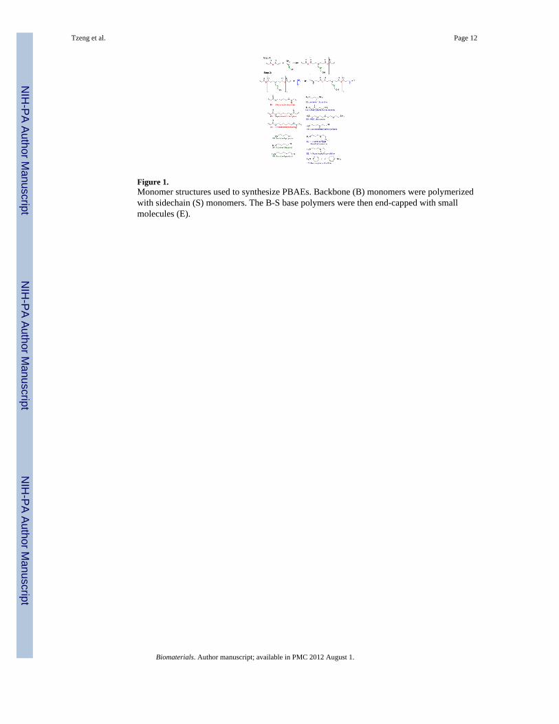
Figure 1.Monomer structures used to synthesize PBAEs. Backbone (B) monomers were polymerizedwith sidechain (S) monomers. The B-S base polymers were then end-capped with smallmolecules (E).
Tzeng et al. Page 12
Biomaterials. Author manuscript; available in PMC 2012 August 1.
NIH
-PA Author Manuscript
NIH
-PA Author Manuscript
NIH
-PA Author Manuscript
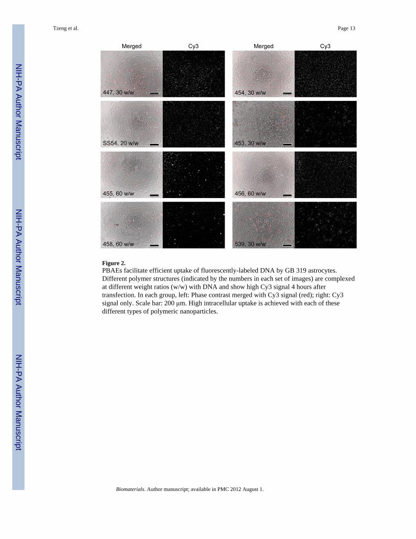
Figure 2.PBAEs facilitate efficient uptake of fluorescently-labeled DNA by GB 319 astrocytes.Different polymer structures (indicated by the numbers in each set of images) are complexedat different weight ratios (w/w) with DNA and show high Cy3 signal 4 hours aftertransfection. In each group, left: Phase contrast merged with Cy3 signal (red); right: Cy3signal only. Scale bar: 200 μm. High intracellular uptake is achieved with each of thesedifferent types of polymeric nanoparticles.
Tzeng et al. Page 13
Biomaterials. Author manuscript; available in PMC 2012 August 1.
NIH
-PA Author Manuscript
NIH
-PA Author Manuscript
NIH
-PA Author Manuscript
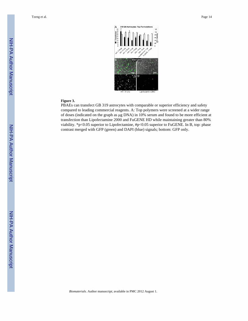
Figure 3.PBAEs can transfect GB 319 astrocytes with comparable or superior efficiency and safetycompared to leading commercial reagents. A: Top polymers were screened at a wider rangeof doses (indicated on the graph as μg DNA) in 10% serum and found to be more efficient attransfection than Lipofectamine 2000 and FuGENE HD while maintaining greater than 80%viability. *p<0.05 superior to Lipofectamine, #p<0.05 superior to FuGENE. In B, top: phasecontrast merged with GFP (green) and DAPI (blue) signals; bottom: GFP only.
Tzeng et al. Page 14
Biomaterials. Author manuscript; available in PMC 2012 August 1.
NIH
-PA Author Manuscript
NIH
-PA Author Manuscript
NIH
-PA Author Manuscript
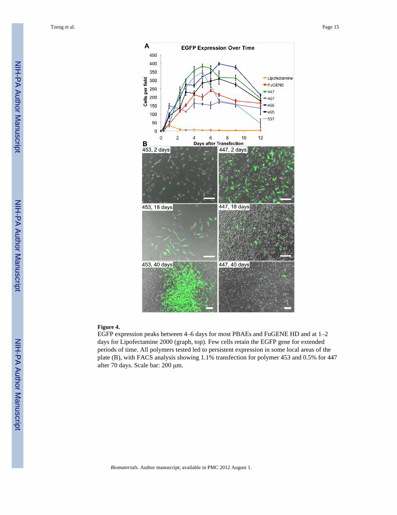
Figure 4.EGFP expression peaks between 4–6 days for most PBAEs and FuGENE HD and at 1–2days for Lipofectamine 2000 (graph, top). Few cells retain the EGFP gene for extendedperiods of time. All polymers tested led to persistent expression in some local areas of theplate (B), with FACS analysis showing 1.1% transfection for polymer 453 and 0.5% for 447after 70 days. Scale bar: 200 μm.
Tzeng et al. Page 15
Biomaterials. Author manuscript; available in PMC 2012 August 1.
NIH
-PA Author Manuscript
NIH
-PA Author Manuscript
NIH
-PA Author Manuscript
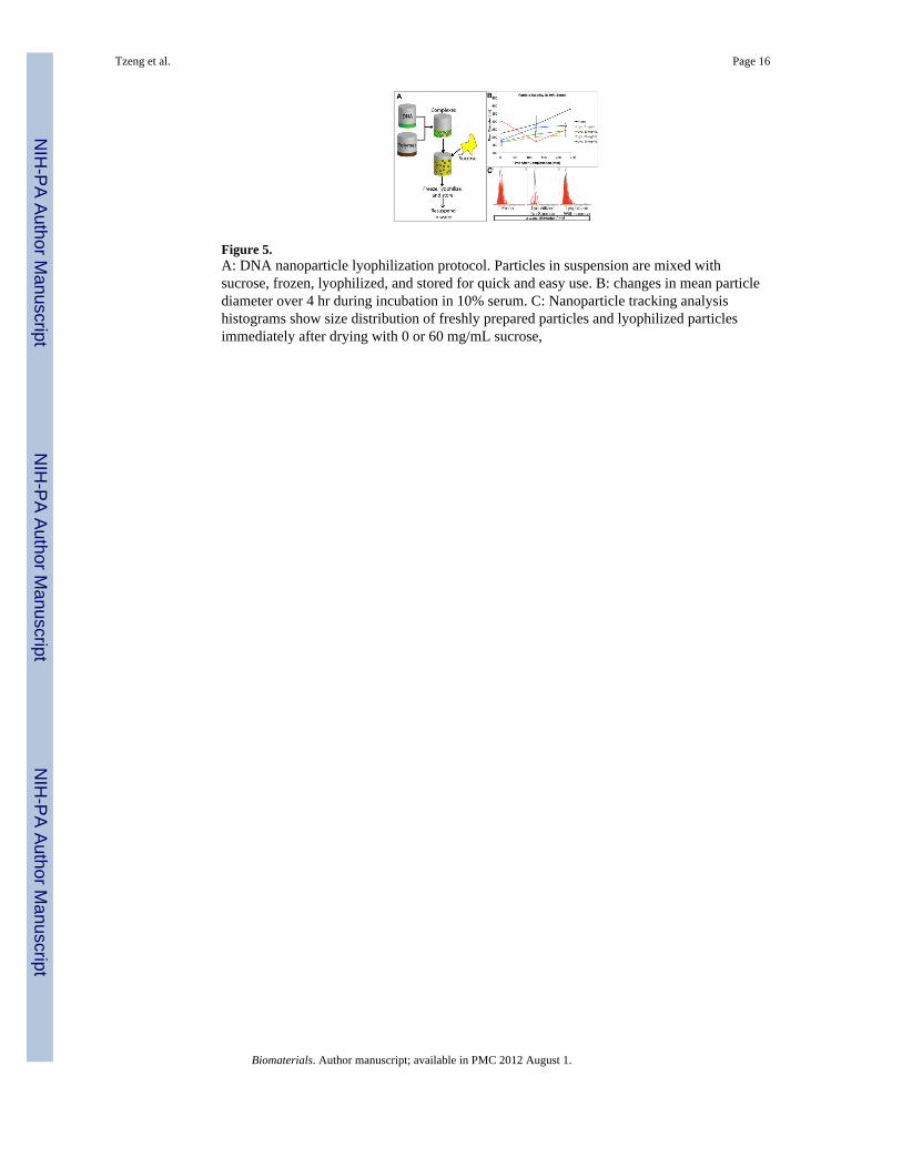
Figure 5.A: DNA nanoparticle lyophilization protocol. Particles in suspension are mixed withsucrose, frozen, lyophilized, and stored for quick and easy use. B: changes in mean particlediameter over 4 hr during incubation in 10% serum. C: Nanoparticle tracking analysishistograms show size distribution of freshly prepared particles and lyophilized particlesimmediately after drying with 0 or 60 mg/mL sucrose,
Tzeng et al. Page 16
Biomaterials. Author manuscript; available in PMC 2012 August 1.
NIH
-PA Author Manuscript
NIH
-PA Author Manuscript
NIH
-PA Author Manuscript
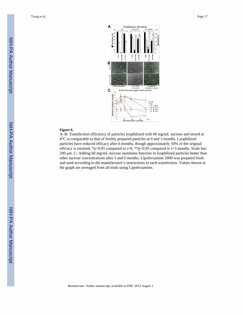
Figure 6.A–B: Transfection efficiency of particles lyophilized with 60 mg/mL sucrose and stored at4°C is comparable to that of freshly prepared particles at 0 and 3 months. Lyophilizedparticles have reduced efficacy after 6 months, though approximately 50% of the originalefficacy is retained. *p<0.05 compared to t=0; **p<0.05 compared to t=3 months. Scale bar:200 μm. C: Adding 60 mg/mL sucrose maintains function in lyophilized particles better thanother sucrose concentrations after 3 and 6 months. Lipofectamine 2000 was prepared freshand used according to the manufacturer’s instructions in each transfection. Values shown inthe graph are averaged from all trials using Lipofectamine.
Tzeng et al. Page 17
Biomaterials. Author manuscript; available in PMC 2012 August 1.
NIH
-PA Author Manuscript
NIH
-PA Author Manuscript
NIH
-PA Author Manuscript
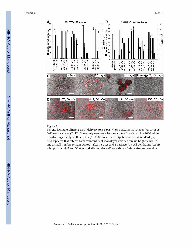
Figure 7.PBAEs facilitate efficient DNA delivery to BTSCs when plated in monolayer (A, C) or as3–D neurospheres (B, D). Some polymers were less toxic than Lipofectamine 2000 whiletransfecting equally well or better (*p<0.05 superior to Lipofectamine). After 45 days,neurospheres that reform from overconfluent monolayer cultures remain brightly DsRed+,and a small number remain DsRed+ after 75 days and 1 passage (C). All conditions (C) arewith polymer 447 and 30 w/w and all conditions (D) are shown 3 days after transfection.
Tzeng et al. Page 18
Biomaterials. Author manuscript; available in PMC 2012 August 1.
NIH
-PA Author Manuscript
NIH
-PA Author Manuscript
NIH
-PA Author Manuscript
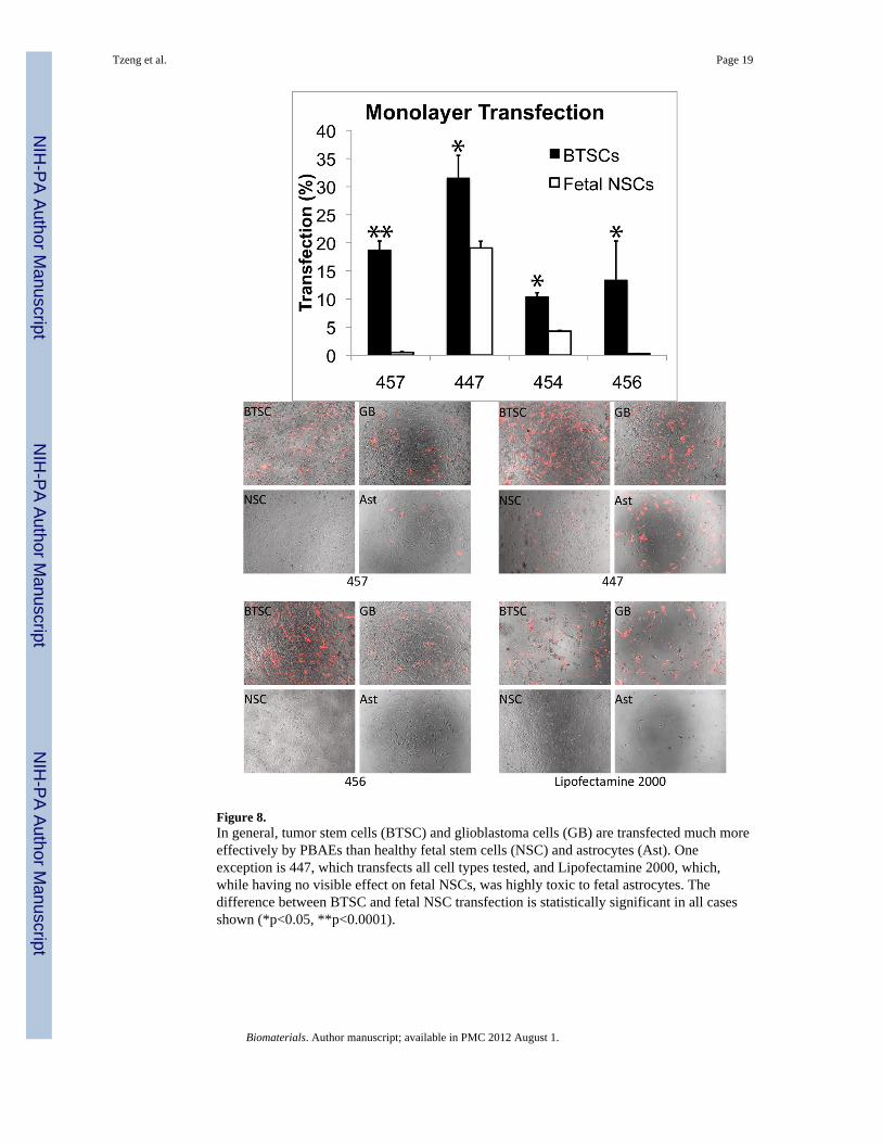
Figure 8.In general, tumor stem cells (BTSC) and glioblastoma cells (GB) are transfected much moreeffectively by PBAEs than healthy fetal stem cells (NSC) and astrocytes (Ast). Oneexception is 447, which transfects all cell types tested, and Lipofectamine 2000, which,while having no visible effect on fetal NSCs, was highly toxic to fetal astrocytes. Thedifference between BTSC and fetal NSC transfection is statistically significant in all casesshown (*p<0.05, **p<0.0001).
Tzeng et al. Page 19
Biomaterials. Author manuscript; available in PMC 2012 August 1.
NIH
-PA Author Manuscript
NIH
-PA Author Manuscript
NIH
-PA Author Manuscript