Specific Dopaminergic Neurons for the Formation of Labile Aversive Memory
Upload
independentCategory
view
0download
0
NMDA Receptors on Non-Dopaminergic Neurons in theVTA Support Cocaine SensitizationYu Luo1., Cameron H. Good1., Oscar Diaz-Ruiz1, YaJun Zhang1, Alexander F. Hoffman1, Lufei Shan1,
Serena Y. Kuang1, Nasir Malik1, Vladimir I. Chefer2, Andreas C. Tomac1¤, Carl R. Lupica1, Cristina M.
Backman1*
1 Cellular Neurobiology Research Branch, National Institute on Drug Abuse, National Institutes of Health, Baltimore, Maryland, United States of America, 2 Behavioral
Neuroscience Branch, National Institute on Drug Abuse, National Institutes of Health, Baltimore, Maryland, United States of America
Abstract
Background: The initiation of behavioral sensitization to cocaine and other psychomotor stimulants is thought to reflect N-methyl-D-aspartate receptor (NMDAR)-mediated synaptic plasticity in the mesolimbic dopamine (DA) circuitry. Theimportance of drug induced NMDAR mediated adaptations in ventral tegmental area (VTA) DA neurons, and its associationwith drug seeking behaviors, has recently been evaluated in Cre-loxp mice lacking functional NMDARs in DA neuronsexpressing Cre recombinase under the control of the endogenous dopamine transporter gene (NR1DATCre mice).
Methodology and Principal Findings: Using an additional NR1DATCre mouse transgenic model, we demonstrate that whilethe selective inactivation of NMDARs in DA neurons eliminates the induction of molecular changes leading to synapticstrengthening, behavioral measures such as cocaine induced locomotor sensitization and conditioned place preferenceremain intact in NR1DATCre mice. Since VTA DA neurons projecting to the prefrontal cortex and amygdala express little or nodetectable levels of the dopamine transporter, it has been speculated that NMDA receptors in DA neurons projecting tothese brain areas may have been spared in NR1DATCre mice. Here we demonstrate that the NMDA receptor gene is ablated inthe majority of VTA DA neurons, including those exhibiting undetectable DAT expression levels in our NR1DATCre transgenicmodel, and that application of an NMDAR antagonist within the VTA of NR1DATCre animals still blocks sensitization tococaine.
Conclusions/Significance: These results eliminate the possibility of NMDAR mediated neuroplasticity in the different DAneuronal subpopulations in our NR1DATCre mouse model and therefore suggest that NMDARs on non-DA neurons within theVTA must play a major role in cocaine-related addictive behavior.
Citation: Luo Y, Good CH, Diaz-Ruiz O, Zhang Y, Hoffman AF, et al. (2010) NMDA Receptors on Non-Dopaminergic Neurons in the VTA Support CocaineSensitization. PLoS ONE 5(8): e12141. doi:10.1371/journal.pone.0012141
Editor: Georges Chapouthier, L’universite Pierre et Marie Curie, France
Received July 6, 2010; Accepted July 19, 2010; Published August 16, 2010
Copyright: � 2010 Luo et al. This is an open-access article distributed under the terms of the Creative Commons Attribution License, which permits unrestricteduse, distribution, and reproduction in any medium, provided the original author and source are credited.
Funding: This research was supported by the Intramural Research Program of the National Institute on Drug Abuse, National Institutes of Health. The fundershad no role in study design, data collection and analysis, decision to publish, or preparation of the manuscript.
Competing Interests: The authors have declared that no competing interests exist.
* E-mail: [email protected]
. These authors contributed equally to this work.
¤ Current address: Department of Neurological Surgery, Case Western Reserve University, Cleveland, Ohio, United States of America
Introduction
Midbrain dopamine (DA) neurons in the ventral tegmental area
(VTA) represent a common substrate for drugs of abuse and
mediate the engagement of addictive behaviors. Glutamatergic
transmission within the VTA has been shown to be particularly
important since local injection of glutamate antagonists during
repeated drug administration blocks behavioral sensitization and
conditional place preference (CPP) [1,2,3,4,5,6]. Previous work
has also shown that abused drugs evoke N-methyl-D-aspartate
receptor (NMDAR)-dependent, long-term potentiation (LTP) of
glutamatergic transmission in DA neurons [7,8,9]. Therefore,
NMDAR-dependent LTP might represent an essential component
of the neural basis of sensitization, and the development of
compulsive drug-seeking behavior. The role of VTA NMDARs in
addictive behavior was recently investigated using DA cell-specific
ablation of the gene coding for subunit 1 of the NMDA receptor
(NR1), by expressing Cre recombinase using the DAT promoter
[10,11]. This causes a selective absence of functional NMDARs on
DA neurons expressing DAT, and represents a significant advance
because it permits an evaluation of NMDAR function in these
cells, compared to the non-selective effects of antagonists on
multiple neuron subtypes. Surprisingly, these prior studies
demonstrated that despite the elimination of NMDAR-dependent
LTP in DA neurons in these NR1 knockout (KO) mice, cocaine
sensitization developed normally [10,11], suggesting that locomo-
tor sensitization does not require NMDAR-dependent neuronal
plasticity.
The role of NR1 on VTA DA neurons in the reinforcing effects
of cocaine also remains uncertain since previous studies reported
conflicting results of this KO on CPP. Zweifel et al., [11] reported
that the loss of NR1 in DA neurons blocked cocaine CPP, whereas
PLoS ONE | www.plosone.org 1 August 2010 | Volume 5 | Issue 8 | e12141

Engblom et al., [10] found that CPP was unaltered in similar KO
mice. However, despite these discordant observations, both groups
reported that DA neuron NR1 deletion altered long-term
behavioral plasticity determined using cocaine challenge injections
in previously sensitized mice [11], and CPP reinstatement
following extinction [10].
The absence of effects of DA neuron NR1 KO on the
development of cocaine sensitization and CPP has led to the
proposal that NMDARs remaining on mesocortical and mesoa-
mygdala projecting DA neurons may be those that are critical for
these cocaine-associated phenomena. This is because these
populations of DA neurons express little or no detectable levels
of DAT [12], and since both studies used a strategy that relied
upon DAT expression to target the NR1 KOs, these DA neurons
may exhibit relatively normal levels of NMDAR function [10,11].
To further examine the role played by NMDARs in the
development of addiction, and specifically its association to the
mesocorticolimbic DA system, we developed an independent
transgenic line in which the gene for NR1 was deleted in DA
neurons by expressing Cre recombinase after the stop codon of the
DAT gene locus. We find that the NMDAR coding region is
deleted, even in DA neurons with low or undetectable DAT
protein levels, that the development of sensitization and CPP
progressed normally, despite the absence of NMDAR-dependent
LTP in these NR1DATCre mice, and that local intra-VTA
application of an NMDAR antagonist blocked behavioral
sensitization in the NR1 KO mice.
Materials and Methods
MiceAll animal protocols were conducted under National Institutes
Health (NIH) Guidelines using the NIH handbook Animals in
Research and were approved by the Institutional Animal Care and
Use Committee (National Institute on Drug Abuse, Intramural
Research Program, Baltimore, MD), approval ID 09-CNRB-57.
Grin1loxP mice were obtained from Jackson laboratories [13]. For
the conditional inactivation of Grin1, a transgenic line, Slc6a3Cre,
was used in which Cre recombinase is driven by the DA
transporter promoter [6]. To minimize interference with gene
function by preservation of both alleles, Cre recombinase
expression was driven from the 39-untranslated region (39UTR)
of the endogenous DAT gene by means of an IRES sequence. The
Grin1loxP line [13] was mixed with the Slc6a3Cre to obtain regional
knockout (Slc6a3Cre/wtGrin1loxP/loxP, or NR1DATCre) and control
mice (Slc6a3Cre/wtGrin1 wt/wt, or WT). Animals were genotyped
using Grin1loxP and Slc6a3Cre primers described elsewhere [6,13].
To determine if Grin1 deletion was specific to DAT expressing
regions, primers specific to the recombination (DloxP) were
utilized. Dissections of the olfactory bulb, ventral mesencephalon,
motor cortex, dorsal striatum, lung, heart, kidney, liver and tail
were performed and DNA was extracted for PCR analysis.
Experiments in this study were conducted in NR1DATCre animals
backcrossed in c57bl/6 for at least 6 generations. However,
behavioral sensitization (excluding those animals injected with
AP5), and CPP experiments were conducted on NR1DATCre and
WT animals in congenic c57bl/6 background. All animals used in
behavioral studies and voltammetric recordings were between 2–4
months of age; electrophysiological studies utilized mice between
21–30d.
Immunohistochemistry and confocal microscopyProteins were detected with primary antibodies to TH and
DAT (Chemicon, CA). Primary antibodies were detected using
Alexa Flour 549 and Alexa Flour 488-conjugated, anti-rabbit and
anti- rat antibodies (Invitrogene, Carlsbad, CA). Images were
analyzed on a Nikon (Melville, NY) Diaphot inverted confocal
microscope using the Volocity confocal imaging system (Perkin
Elmer).
UV-laser microdisection, DNA extraction and PCR analysisCoronal cryosections (5 mm) of fresh frozen NR1DATCre mouse
midbrains were cut (Leica cryostat CM3050S), mounted on
polyethylene naphthalate (PEN) membrane slides (Leica Micro-
systems), fixed with 75% ethanol for 10 min, acetone for 2 min,
and air-dried. Sections were incubated overnight at 4uC with
antibodies against DAT (1:30,Chemicon, CA) and TH (1:40
Chemicon, CA) in blocking media. Sections were washed with PB
and incubated for 3 hours at RT in a mixture of secondary
antibodies (Invitrogene, Carlsbad, CA), Alexa Flour 549 anti-
rabbit IgG, and Alexa Flour 488 anti-rat IgG, made up in blocking
media. Sections were washed with distilled H2O, and air-dried.
Fluorescent-labeled neurons were collected under epifluorescent
optics using an LMD6000 system. The following types of neurons
and tissue within the VTA were collected for PCR analyses: 1) TH
and DAT double immunoreactive neurons (n = 15 samples, with
2–5 cells per sample), 2) TH immunoreactive neurons with
undetectable DAT expression levels (n = 36 samples, with 2–5 cells
per sample), and 3) tissue surrounding TH and/or DAT positive
neurons (n = 5 samples). Isolated neurons and tissue were collected
by gravity directly into a cap of a 0.5 ml thin-walled PCR-tube.
Cell lysis buffer (5 ml per sample) was added directly into the lid,
and lysis was performed at 65uC for 2 hours, followed by 10 min
at 95uC to inactivate proteinase K. A multiplex reaction for the
recombination band, and the control gene GAPDH was performed
for each sample. The recombination band (DloxP) was genotyped
by PCR primers forward, 59- agatacaagaccctgact- 39 flanking the
59 loxP site, and reverse, 59-cacttgagtagcgccaagtgc-39 in the Pgk
promoter of the Pgk-Neomycin cassette, and was sequenced
verified. The GAPDH gene was amplified with primers forward,
59- atggtgaaggtcggtgtga -39 and reverse, 59- aatctccactttgcca-
ctgc -39.
Recordings in brain slicesSlice preparation. Animals were rapidly decapitated using a
guillotine and their brains were removed and transferred to a
beaker containing oxygenated (95% O2/5% CO2), ice-cold
artificial cerebral spinal fluid (aCSF) (in mM: sucrose, 194;
NaCl, 30; KCl, 4.5; MgCl2, 1; NaH2PO4,1.2; glucose, 10;
NaHCO3, 26). Coronal hemisections (280 mm) containing the
nucleus accumbens (NAc) were prepared as described previously
[14]. NAc slices were were transferred to an oxygenated holding
chamber (35uC) filled with normal aCSF (in mM: NaCl, 126; KCl,
3; MgCl2, 1.5; CaCl2, 2.4; NaH2PO4, 1.2; glucose, 11; NaHCO3,
26) for 25–30 min, then gradually cooled to room temperature for
$30 min prior to recording. During voltammetric recordings,
slices were continuously superfused with aCSF (2 ml/min) and
maintained at 31–33uC.
For VTA slices, the brain was then blocked to isolate the VTA
before being glued onto the cutting stage of a vibrating tissue slicer
(Leica, VT1000) and submersed in ice-cold, oxygenated aCSF.
Four sagittal slices (280 mm) containing the VTA were obtained
from each mouse. The slices were transferred to an oxygenated
holding chamber (31uC) filled with normal aCSF and allowed to
recover for at least 1 hour before recording.
Electrophysiology. One brain slice was transferred to a
heated chamber (31–33uC) and superfused (2 ml/min) with aCSF
+100 mM picrotoxin. VTA neurons were visualized with an
NMDA, Sensitization, and VTA
PLoS ONE | www.plosone.org 2 August 2010 | Volume 5 | Issue 8 | e12141
upright microscope (Zeiss Axioscope, Germany), modified to
provide a gradient contrast image utilizing infrared illumination.
Putative DA neurons were initially selected for recording by their
large cell bodies and the presence of Ih, as originally described by
Johnson and North [15]. Each cell was labeled with biocytin
(0.125%) and processed for tyrosine hydroxylase (TH)
immunohistochemistry to confirm its identity as a DA neuron.
Recording electrodes (3–5 MV) were filled with (in mM) CsCl,
130; NaCl, 4; MgCl2, 2; EGTA, 1.1; HEPES, 5; Na2-ATP, 2; Na2-
creatine-phosphate, 5; Na3-GTP, 0.6; biocytin, 0.125%, pH 7.2
with KOH. Intracellular recordings were performed using an
Axopatch 200B (Axon Instruments, Burlingame, CA). Voltage
steps and stimulation protocols were delivered using the
Strathclyde electrophysiology software package (WCP, courtesy
of Dr. John Dempster, Strathclyde University, Glasgow, UK) and
an A/D board (ITC-18, Instrutech Corp., Bellmore, NY) residing
in a personal computer.
Synaptic currents were evoked by 0.2 ms, constant current
pulses delivered every 30s via a bipolar tungsten electrode (FHC,
Bowdoinham, ME; 300 mm tip separation), placed anterior to the
VTA. Stimulus intensity was adjusted to achieve 30–50% of
maximum response amplitude. I–V curves were obtained by
varying the holding potential between 290 and +50 mV. NMDA
current measurements were made by averaging the current, with
respect to baseline, between 30 to 35 ms after stimulation. AP5
was applied in a subset of cells to test for NMDA currents.
For LTP experiments, intracellular solution contained (in mM):
K-gluconate, 140; KCl, 5; HEPES, 10; EGTA, 0.2; MgCl2, 2; Mg-
ATP, 4; Na2-GTP, 0.3; Na2-phosphocreatine, 10; bioctytin,
0.125%; pH 7.2 with KOH. Cells were initially patched in
voltage-clamp to test for the presence of Ih. Following this
confirmation, the recording was switched into current-clamp for
the duration of the experiment. To prevent action potentials from
occluding stimulus-evoked EPSPs, the cell was manually hyper-
polarized to 265mV by injecting negative direct current (,0.05–
0.2 nA) through the pipette.
Following at least 10 minutes of stable baseline EPSP recording,
LTP was initiated using a spike-timing protocol [8]. Immediately
preceding the initiation of the LTP protocol, the holding current
was removed to allow the cell to freely fire action potentials. The
LTP protocol consisted of 20 bursts of 5 pairings of stimulation
and postsynaptic depolarization, with the presynaptic stimulation
preceding the depolarization by 5 ms. Each postsynaptic depolar-
ization was initiated with current injection (1.5–2 nA) lasting 3 ms.
The 5 pairings within a burst were separated by 100 ms (10 Hz),
and each of the 20 bursts was separated by 5 sec. Following the
last burst, the negative current was again applied to drive the cell
back to 265 mV for post-LTP recordings of stimulus-evoked
EPSPs.
Voltammetry. Carbon fibers (7 mm diameter) were prepared
as described previously [14,16]. Voltammetric scan and
stimulation timing protocols were performed using a
voltammetric amplifier (EVA-8, HEKA Electronic) and PCI-
based A/D boards (National Instruments) and custom software
(courtesy of Dr. Mark Wightman, Univ. of North Carolina). Scans
consisted of sweeps from 20.4 to 1.0 V and back to 20.4 V, at a
rate of 400 V/s, and were obtained every 100 ms. A 5 sec. (50
scan) control period preceded each electrically-evoked response,
and was used to obtain a background current that was digitally
subtracted from the current obtained during the peak of the
response. Currents were converted to concentration by in vitro
calibration of each electrode against a 1 mM DA standard. All
signals used in analyses matched the expected voltammetric profile
for DA. Under stereoscopic magnification, carbon fibers were
lowered to a depth of ,100 mm in the nucleus accumbens core,
generally medial to the anterior commissure. A bipolar stimulating
electrode was positioned ,75–100 from the carbon fiber.
Constant current pulses (10–1000 mA, 1 ms duration) were
delivered between voltammetric scans to elicit dopamine release.
10 pulse trains (400–500 mA, 1 ms, 5–60 Hz) were used to assess
the frequency-dependence of DA release. For cocaine and AP5
experiments, responses were obtained every 2 minutes using single
pulse stimulation. After obtaining a 10-minute stable baseline
response, cocaine (500 nM) or AP5 (40 mM) was superfused into
the recording chamber for 10–20 minutes, until a stable drug
response was observed. Comparisons were performed by
averaging 3–4 responses during the control (pre-drug) and post-
drug period. DA uptake was assessed by fitting the decay portion
of each signal to a single exponential function. The obtained tau
values have previously been demonstrated to be related to the
efficiency of DAT-mediated uptake of DA (Vmax/Km), and are
sensitive to DAT inhibitors [16].
Behavioral proceduresNR1DATCre and WT mice were maintained on a 12 h light-dark
schedule (lights on 6:00am).
Sensitization in activity chamber. Locomotor activity was
measured in activity chambers (Omnitech Electronic Inc,
Columbus, OH). The monitor contained 16 horizontal and 8
vertical infrared sensors spaced 2.5 cm apart. Mice were placed in
the locomotor chambers and their activity was measured for
60 min. Then the animals were injected with either saline
(habituation) or cocaine (sensitization), and the measurements
continued for another 60 min. The treatment schedule consisted
of injections with saline (habituation) daily for 2 days, followed by
cocaine (15 mg/kg, i.p.) daily for 4 days. After the last treatment,
mice were kept in the home cage. On day 22 animals were
challenged with cocaine (15 mg/kg, i.p.).
Homecage sensitization. Mice for each genotype were
divided into two groups: The first group received saline
injections (i.p.) in the homecage for 6 days, while the second
group received 15 mg/kg cocaine i.p. for one day, followed by
20 mg/kg i.p. for 5 consecutive days in the home cage. On day 7,
both groups were habituated to the locomotor activity chambers
(Omnitech Electronic Inc, Columbus, OH) for 60 min prior to
receiving a 15 mg/kg cocaine injection. Locomotor activity was
measured during habituation and for 60 min after cocaine
injection.
Infusion of AP5 in VTA. Mice were implanted with bilateral
chronic indwelling guide cannulae aimed 1 mm above the VTA
(AP 23.0, ML 60.4, DV 23.5 according to Franklin and Paxinos,
[17] that was secured by dental cement. Animals were given at
least 2 weeks to recover from surgery before receiving infusions
(0.5 ml over 2 min into VTA by pump). The tips of the inner
injection cannulae extended 1 mm below the guide cannulae into
the injection sites. For the sensitization experiment WT, and
NR1DATCre mice were administered four pairs of injections (one
intracranial and one i.p.), with one pair given everyday. On each
injection day, mice were first habituated to the locomotor chamber
for 60 min, before receiving their respective intra cranial injection
of AP5 (0.04 mM, 0.5 ml each side; Tocris, Ballwin, MO) or saline
(0.5 ml each side), followed by a cocaine i.p. injection (15 mg/kg).
Immediately following the i.p, injections, mice were returned to
the activity boxes and locomotor activity was recorded for an
additional 30 min.
Conditioned place preference. The conditioned place
preference (CPP) chamber consists of two Plexiglas
compartments (23.2612.7612.7 cm) with black walls (Med
NMDA, Sensitization, and VTA
PLoS ONE | www.plosone.org 3 August 2010 | Volume 5 | Issue 8 | e12141
Associates Inc, St. Albans, VT, USA). The CPP procedure
consisted of four phases: habituation/pre-conditioning,
conditioning, preference test, and extinction/reinstatement. CPP
was measured as amount of time spent in cocaine-paired
compartment. On day one (habituation), both compartments of
the CPP chamber were equipped with the same floor type (smooth
plastic surface) and animals were allowed to explore both
compartments during one session of 15 min. Next day, for pre-
conditioning (pre-test), one compartment was equipped with a
mesh floor and the other compartment with grid-mesh floor. Mice
were allowed to explore both compartments during one session of
15 minutes. Animals that spent more than 65% of the time in any
one compartment were eliminated from the experiment. Overall
CPP conditions were unbiased for both phenotypes. The next
eight days were dedicated to the conditioning phase. During the
conditioning phase subjects had access to the entire apparatus and
pairing of saline or cocaine with a particular floor type was
randomly assigned in each group. Animals received one
conditioning trial each day after an alternative injection of saline
or cocaine (10 mg/kg i.p.). Immediately after the injection, mice
were placed in the conditioning apparatus containing the
corresponding stimulus floor type and were allowed to explore
both compartments during 30 min. The presentation order of
saline and cocaine and floor- pairing was counterbalanced within
and between genotypes. The preference test took place on day 9,
or 24 hr after the last conditioning trial. To conduct this test, the
chambers were equipped with half mesh and half mesh-grid floor,
and mice were placed in the chamber without any previous
injection. The preference test duration was 15 min, and the time
spent on the floor paired with cocaine and saline was measured.
The primary index for deciding whether place conditioning has
occurred was significant difference between times spent on the
drug-paired floor during the pre-test and the preference test.
Extinction consisted of 8 daily sessions in which both compart-
ments were equipped with the same floor distribution as during the
preference test (half mesh and half mesh-grid floor). Mice were
allowed to explore both compartments during one daily session of
30 min. On day 18, for the extinction test, compartments were
equipped with the same floor as during the preference test, and
mice were allowed to explore both compartments during one
session of 15 min. On day 19, for the reinstatement test, the same
floor order was used. Animals received a single cocaine injection
(10 mg/kg, ip) and were allowed to explore both compartments
during one session of 15 min.
Results
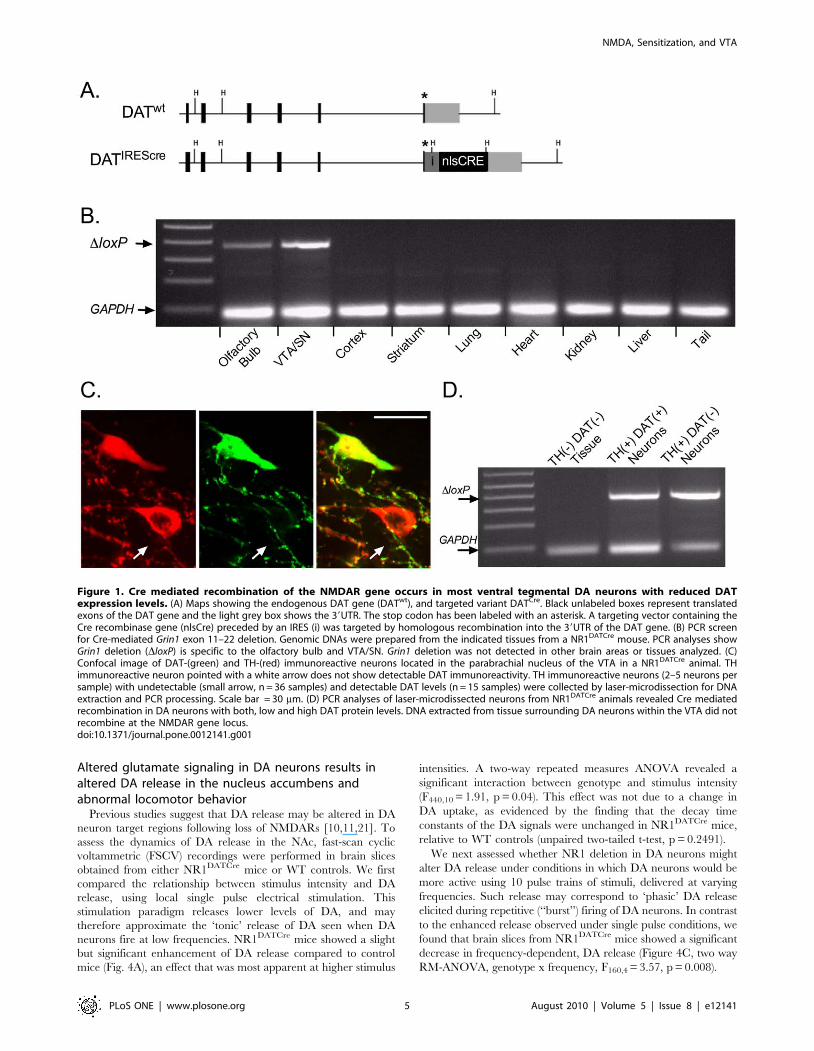
Cre mediated recombination is restricted to the ventralmesencephalon and olfactory bulb in NR1DATCre mice
To assess the spatial specificity of Cre mediated recombination
in NR1DATCre animals, we examined recombination at the Grin1
gene locus in different organs of the mouse using PCR primers
specific to the recombined DNA (Fig. 1B). PCR analyses showed
Cre mediated recombination in the VTA, substantia nigra (SN),
and olfactory bulb. All other areas analyzed did not show the
recombination band (Fig. 1B).
VTA DA neurons with low or undetectable DAT proteinlevels express Cre mediated recombination
It has recently been demonstrated that the adult dopaminergic
system is composed of two functionally and molecularly distinct
types of DA neurons with anatomical segregation in the midbrain,
and with non-overlaping axonal targets [12]. Mesocorticolimbic
midbrain DA neurons projecting to the medial prefrontal cortex
and basolateral amygdala fire action potentials at significantly
higher frequencies and posses significantly lower DAT mRNA
expression levels, compared to other DA subtypes [12]. It has been
suggested that mesocorticolimbic DA neurons with low DA
reuptake capacity may mediate sustained forms of behaviorally
relevant DA release in vivo [18,19,20]. Using antibodies against
TH and DAT we identified many VTA DA neurons with low or
undetectable DAT expression levels (Fig. 1C), mostly clustered in
the medial posterior VTA, and predominantly located in the
medial aspect of the paranigral and parabrachial nucleus, as
previously described [12].
To address the question of whether DA neurons with
undetectable DAT levels express Cre mediated recombination in
NR1DATCre animals, we collected populations of TH immunore-
active neurons in the VTA that lacked DAT immunoreactivity
using UV-laser microdissection, and analyzed them using PCR
(Fig. 1C). For comparison, neurons that were labeled by both TH
and DAT antibodies, and non-immunoreactive tissue within the
VTA, were also collected and analyzed by PCR. Selected pools of
2–5 labeled neurons each, or non-immunoreactive VTA tissues,
were collected from coronal fixed cryosections. Primers specific for
the recombination event in the Grin1 gene locus were utilized to
ascertain if VTA DA neurons express Cre mediated recombina-
tion. Thirty-four of thirty-six samples containing TH immunore-
active/DAT negative neurons expressed a robust recombination
band, indicating Cre mediated recombination (Fig. 1D). All
samples with TH/DAT double stained neurons showed the
recombination band (n = 15; Fig. 1D). Non-immunoreactive tissue
surrounding VTA DA cells did not show the recombination band
(n = 5; Fig. 1D). Thus, contrary to previous studies suggesting the
absence of recombination in low-DAT expressing DA neuron
subpopulations, our PCR analyses suggests that DNA coding for
the NR1 subunit had recombined in nearly all of these neurons.
Inactivation of NMDARs in DA neurons eliminates theinduction of synaptic strengthening
To test whether or not the selective deletion of the NR1 subunit
resulted in functional inactivation of the NMDA channel in VTA
DA neurons, we performed intracellular recordings of evoked
EPSCs in brain slices containing the VTA. At a holding potential
of +50mV, neurons from WT animals displayed large NMDA
currents (,100 pA), whereas no NMDA currents were seen in DA
neurons from NR1DATCre mice (Fig. 2A–B). We also observed
large NMDA currents in non-DA VTA neurons in the NR1DATCre
animals, confirming the selectivity of the KO (n = 4, data not
shown). To further verify the absence of an NMDA component in
the synaptic responses in KO animals, we applied the selective
NMDAR antagonist AP5. This antagonist did not alter EPSCs
evoked in NR1DATCre DA cells, but it did reduce the amplitudes of
EPSCs evoked in control cells (Fig. 2C–D). Furthermore, the
remaining current following AP5 application was completely
blocked by the AMPA receptor antagonist, DNQX in both groups
of cells (data not shown).
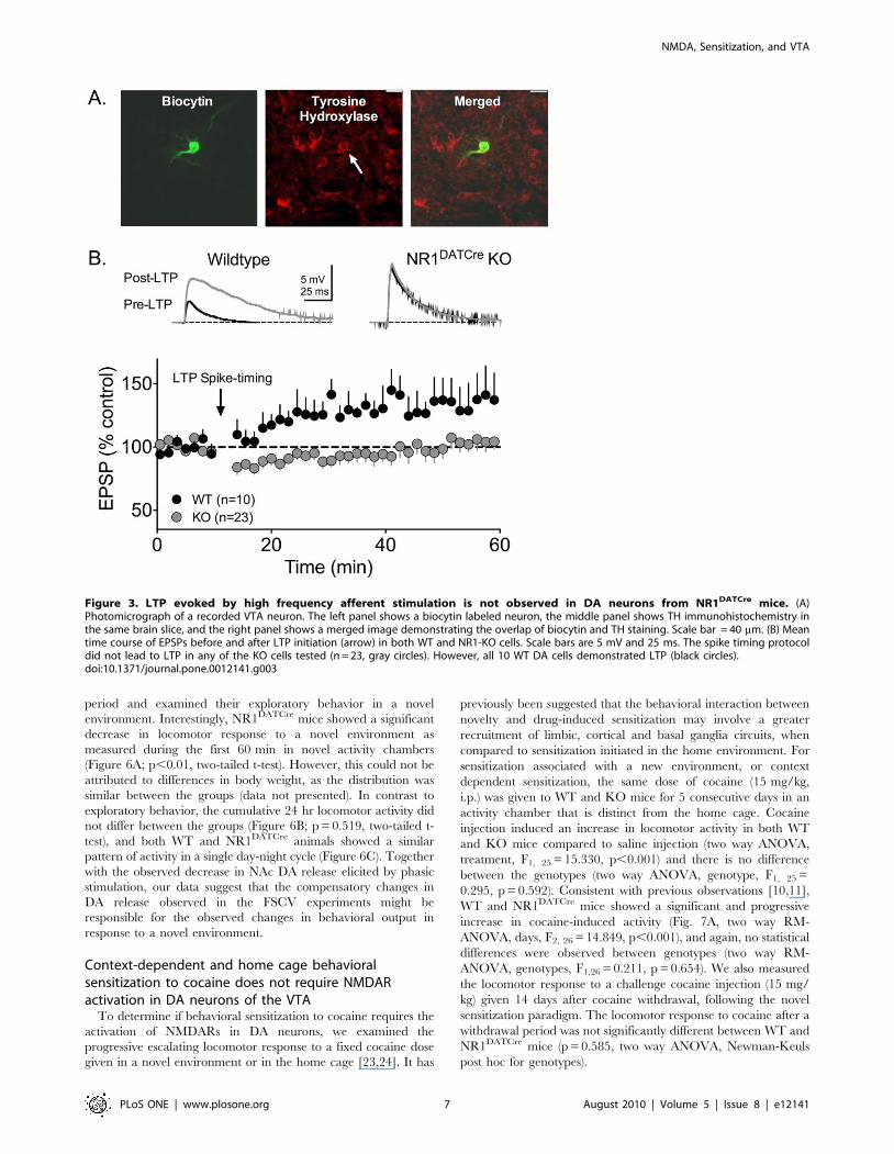
The activation of NMDARs on VTA DA neurons by
endogenous glutamate has previously been established as neces-
sary for the expression of LTP in these cells [9]. In current clamp
recordings, a spike-timing dependent LTP protocol [8] reliably
generated LTP in cells from WT animals, but was never observed
in VTA DA neurons from NR1DATCre animals (Fig. 3B), thereby
confirming that functional NMDARs are required for VTA DA
cells to express this form of LTP. During recordings, all cells were
filled with biocytin, and subsequently processed for the immuno-
histochemical presence of TH (Fig. 3A).
NMDA, Sensitization, and VTA
PLoS ONE | www.plosone.org 4 August 2010 | Volume 5 | Issue 8 | e12141
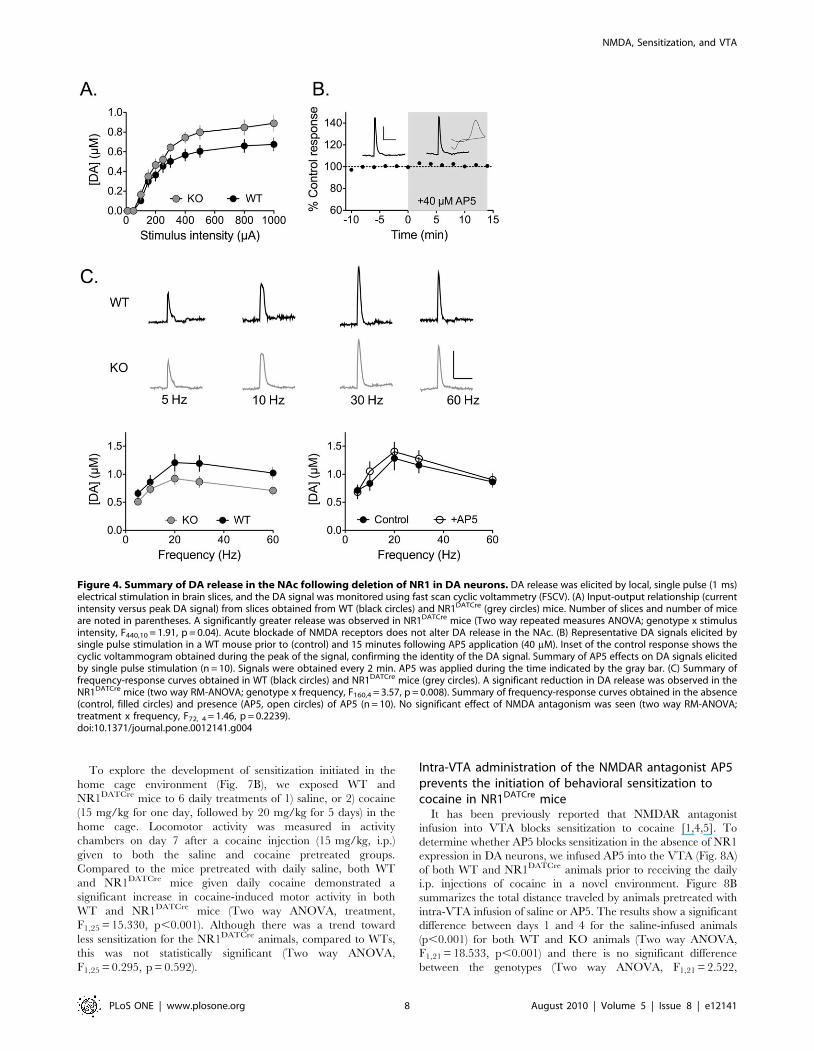
Altered glutamate signaling in DA neurons results inaltered DA release in the nucleus accumbens andabnormal locomotor behavior
Previous studies suggest that DA release may be altered in DA
neuron target regions following loss of NMDARs [10,11,21]. To
assess the dynamics of DA release in the NAc, fast-scan cyclic
voltammetric (FSCV) recordings were performed in brain slices
obtained from either NR1DATCre mice or WT controls. We first
compared the relationship between stimulus intensity and DA
release, using local single pulse electrical stimulation. This
stimulation paradigm releases lower levels of DA, and may
therefore approximate the ‘tonic’ release of DA seen when DA
neurons fire at low frequencies. NR1DATCre mice showed a slight
but significant enhancement of DA release compared to control
mice (Fig. 4A), an effect that was most apparent at higher stimulus
intensities. A two-way repeated measures ANOVA revealed a
significant interaction between genotype and stimulus intensity
(F440,10 = 1.91, p = 0.04). This effect was not due to a change in
DA uptake, as evidenced by the finding that the decay time
constants of the DA signals were unchanged in NR1DATCre mice,
relative to WT controls (unpaired two-tailed t-test, p = 0.2491).
We next assessed whether NR1 deletion in DA neurons might
alter DA release under conditions in which DA neurons would be
more active using 10 pulse trains of stimuli, delivered at varying
frequencies. Such release may correspond to ‘phasic’ DA release
elicited during repetitive (‘‘burst’’) firing of DA neurons. In contrast
to the enhanced release observed under single pulse conditions, we
found that brain slices from NR1DATCre mice showed a significant
decrease in frequency-dependent, DA release (Figure 4C, two way
RM-ANOVA, genotype x frequency, F160,4 = 3.57, p = 0.008).
Figure 1. Cre mediated recombination of the NMDAR gene occurs in most ventral tegmental DA neurons with reduced DATexpression levels. (A) Maps showing the endogenous DAT gene (DATwt), and targeted variant DATCre. Black unlabeled boxes represent translatedexons of the DAT gene and the light grey box shows the 39UTR. The stop codon has been labeled with an asterisk. A targeting vector containing theCre recombinase gene (nlsCre) preceded by an IRES (i) was targeted by homologous recombination into the 39UTR of the DAT gene. (B) PCR screenfor Cre-mediated Grin1 exon 11–22 deletion. Genomic DNAs were prepared from the indicated tissues from a NR1DATCre mouse. PCR analyses showGrin1 deletion (DloxP) is specific to the olfactory bulb and VTA/SN. Grin1 deletion was not detected in other brain areas or tissues analyzed. (C)Confocal image of DAT-(green) and TH-(red) immunoreactive neurons located in the parabrachial nucleus of the VTA in a NR1DATCre animal. THimmunoreactive neuron pointed with a white arrow does not show detectable DAT immunoreactivity. TH immunoreactive neurons (2–5 neurons persample) with undetectable (small arrow, n = 36 samples) and detectable DAT levels (n = 15 samples) were collected by laser-microdissection for DNAextraction and PCR processing. Scale bar = 30 mm. (D) PCR analyses of laser-microdissected neurons from NR1DATCre animals revealed Cre mediatedrecombination in DA neurons with both, low and high DAT protein levels. DNA extracted from tissue surrounding DA neurons within the VTA did notrecombine at the NMDAR gene locus.doi:10.1371/journal.pone.0012141.g001
NMDA, Sensitization, and VTA
PLoS ONE | www.plosone.org 5 August 2010 | Volume 5 | Issue 8 | e12141

The differences in DA release under single pulse and pulse train
conditions between WT and NR1DATCre mice suggest that
deletion of NMDARs has effects on the dynamic output of DA
neurons, and it has been suggested that NMDARs located on DA
terminals in the NAc may directly regulate DA release [22]. To
evaluate this, we examined the effect of acute NMDA receptor
blockade on DA release in NAc slices from WT mice. In contrast
to the results obtained with the KO mice, the NMDAR antagonist
AP5 (40 mM) had no effect on DA release elicited by single pulse
stimulation (Fig. 4B). Furthermore, frequency-response curves of
DA release, generated prior to and during AP5 application
(Fig. 4C), were not significantly different (two way RM-ANOVA;
treatment x frequency, F72, 4 = 1.46, p = 0.2239). Therefore, these
data suggest that the differences in DA release observed in the
NAc following NR1 deletion reflect compensatory changes in DA
release dynamics, rather than the loss of an acute modulation of
DA release by NMDARs.
Our final FSCV experiments determined whether the loss of
NMDAR function in DA neurons can influence the direct effects
of cocaine on DA uptake via the DAT in the NAc. Application of
cocaine (500 nM) to NAc brain slices significantly enhanced the
amplitude of the DA signal and prolonged its time course
(increased tau) to the same degree in both WT and NR1DATCre
KO slices (Fig. 5). Thus, the direct pharmacological effect of
cocaine at the DAT in the NAc was unaltered in NR1DATCre mice.
The observed differences in DA release in NR1DATCre KO
slices might cause changes in locomotor activity in these mice.
Therefore, we monitored their activity levels during a 24-hour
Figure 2. Absence of functional NMDAR in NR1DATCre mice. (A) Example waveforms recorded at +50 mV from both wildtype (WT) andNR1DATCre (KO) cells before (black trace) and after AP5 application (grey trace). In WT cells, the NMDA component could be obtained by subtractingthe AMPA current remaining after AP5 application from the control current, as indicated. In KO cells, only the AMPA mediated EPSC was observed.The gray block represents the time over which the NMDA current was measured. Scale bars are 20 pA and 20 ms. (B) Average NMDA currentmeasured as described in A. Responses were evoked at holding potentials, beginning at 290 mV in 10 mV increments to +50mV. (C) Decay timeconstant (tau) of EPSCs measured before and after AP5 in control and KO cells. Following AP5 superfusion, the remaining current in the WT cellsexhibited similar decay constants as the KO animals. (D) Mean time course of NMDA currents before and after AP5 application. Following AP5 the WTresponse amplitude was reduced to the same level observed in the KO cells, thus confirming the lack of active NMDA channels in DA NR1DATCre
neurons.doi:10.1371/journal.pone.0012141.g002
NMDA, Sensitization, and VTA
PLoS ONE | www.plosone.org 6 August 2010 | Volume 5 | Issue 8 | e12141
period and examined their exploratory behavior in a novel
environment. Interestingly, NR1DATCre mice showed a significant
decrease in locomotor response to a novel environment as
measured during the first 60 min in novel activity chambers
(Figure 6A; p,0.01, two-tailed t-test). However, this could not be
attributed to differences in body weight, as the distribution was
similar between the groups (data not presented). In contrast to
exploratory behavior, the cumulative 24 hr locomotor activity did
not differ between the groups (Figure 6B; p = 0.519, two-tailed t-
test), and both WT and NR1DATCre animals showed a similar
pattern of activity in a single day-night cycle (Figure 6C). Together
with the observed decrease in NAc DA release elicited by phasic
stimulation, our data suggest that the compensatory changes in
DA release observed in the FSCV experiments might be
responsible for the observed changes in behavioral output in
response to a novel environment.
Context-dependent and home cage behavioralsensitization to cocaine does not require NMDARactivation in DA neurons of the VTA
To determine if behavioral sensitization to cocaine requires the
activation of NMDARs in DA neurons, we examined the
progressive escalating locomotor response to a fixed cocaine dose
given in a novel environment or in the home cage [23,24]. It has
previously been suggested that the behavioral interaction between
novelty and drug-induced sensitization may involve a greater
recruitment of limbic, cortical and basal ganglia circuits, when
compared to sensitization initiated in the home environment. For
sensitization associated with a new environment, or context
dependent sensitization, the same dose of cocaine (15 mg/kg,
i.p.) was given to WT and KO mice for 5 consecutive days in an
activity chamber that is distinct from the home cage. Cocaine
injection induced an increase in locomotor activity in both WT
and KO mice compared to saline injection (two way ANOVA,
treatment, F1, 25 = 15.330, p,0.001) and there is no difference
between the genotypes (two way ANOVA, genotype, F1, 25 =
0.295, p = 0.592). Consistent with previous observations [10,11],
WT and NR1DATCre mice showed a significant and progressive
increase in cocaine-induced activity (Fig. 7A, two way RM-
ANOVA, days, F2, 26 = 14.849, p,0.001), and again, no statistical
differences were observed between genotypes (two way RM-
ANOVA, genotypes, F1,26 = 0.211, p = 0.654). We also measured
the locomotor response to a challenge cocaine injection (15 mg/
kg) given 14 days after cocaine withdrawal, following the novel
sensitization paradigm. The locomotor response to cocaine after a
withdrawal period was not significantly different between WT and
NR1DATCre mice (p = 0.585, two way ANOVA, Newman-Keuls
post hoc for genotypes).
Figure 3. LTP evoked by high frequency afferent stimulation is not observed in DA neurons from NR1DATCre mice. (A)Photomicrograph of a recorded VTA neuron. The left panel shows a biocytin labeled neuron, the middle panel shows TH immunohistochemistry inthe same brain slice, and the right panel shows a merged image demonstrating the overlap of biocytin and TH staining. Scale bar = 40 mm. (B) Meantime course of EPSPs before and after LTP initiation (arrow) in both WT and NR1-KO cells. Scale bars are 5 mV and 25 ms. The spike timing protocoldid not lead to LTP in any of the KO cells tested (n = 23, gray circles). However, all 10 WT DA cells demonstrated LTP (black circles).doi:10.1371/journal.pone.0012141.g003
NMDA, Sensitization, and VTA
PLoS ONE | www.plosone.org 7 August 2010 | Volume 5 | Issue 8 | e12141
To explore the development of sensitization initiated in the
home cage environment (Fig. 7B), we exposed WT and
NR1DATCre mice to 6 daily treatments of 1) saline, or 2) cocaine
(15 mg/kg for one day, followed by 20 mg/kg for 5 days) in the
home cage. Locomotor activity was measured in activity
chambers on day 7 after a cocaine injection (15 mg/kg, i.p.)
given to both the saline and cocaine pretreated groups.
Compared to the mice pretreated with daily saline, both WT
and NR1DATCre mice given daily cocaine demonstrated a
significant increase in cocaine-induced motor activity in both
WT and NR1DATCre mice (Two way ANOVA, treatment,
F1,25 = 15.330, p,0.001). Although there was a trend toward
less sensitization for the NR1DATCre animals, compared to WTs,
this was not statistically significant (Two way ANOVA,
F1,25 = 0.295, p = 0.592).
Intra-VTA administration of the NMDAR antagonist AP5prevents the initiation of behavioral sensitization tococaine in NR1DATCre mice
It has been previously reported that NMDAR antagonist
infusion into VTA blocks sensitization to cocaine [1,4,5]. To
determine whether AP5 blocks sensitization in the absence of NR1
expression in DA neurons, we infused AP5 into the VTA (Fig. 8A)
of both WT and NR1DATCre animals prior to receiving the daily
i.p. injections of cocaine in a novel environment. Figure 8B
summarizes the total distance traveled by animals pretreated with
intra-VTA infusion of saline or AP5. The results show a significant
difference between days 1 and 4 for the saline-infused animals
(p,0.001) for both WT and KO animals (Two way ANOVA,
F1,21 = 18.533, p,0.001) and there is no significant difference
between the genotypes (Two way ANOVA, F1,21 = 2.522,
Figure 4. Summary of DA release in the NAc following deletion of NR1 in DA neurons. DA release was elicited by local, single pulse (1 ms)electrical stimulation in brain slices, and the DA signal was monitored using fast scan cyclic voltammetry (FSCV). (A) Input-output relationship (currentintensity versus peak DA signal) from slices obtained from WT (black circles) and NR1DATCre (grey circles) mice. Number of slices and number of miceare noted in parentheses. A significantly greater release was observed in NR1DATCre mice (Two way repeated measures ANOVA; genotype x stimulusintensity, F440,10 = 1.91, p = 0.04). Acute blockade of NMDA receptors does not alter DA release in the NAc. (B) Representative DA signals elicited bysingle pulse stimulation in a WT mouse prior to (control) and 15 minutes following AP5 application (40 mM). Inset of the control response shows thecyclic voltammogram obtained during the peak of the signal, confirming the identity of the DA signal. Summary of AP5 effects on DA signals elicitedby single pulse stimulation (n = 10). Signals were obtained every 2 min. AP5 was applied during the time indicated by the gray bar. (C) Summary offrequency-response curves obtained in WT (black circles) and NR1DATCre mice (grey circles). A significant reduction in DA release was observed in theNR1DATCre mice (two way RM-ANOVA; genotype x frequency, F160,4 = 3.57, p = 0.008). Summary of frequency-response curves obtained in the absence(control, filled circles) and presence (AP5, open circles) of AP5 (n = 10). No significant effect of NMDA antagonism was seen (two way RM-ANOVA;treatment x frequency, F72, 4 = 1.46, p = 0.2239).doi:10.1371/journal.pone.0012141.g004
NMDA, Sensitization, and VTA
PLoS ONE | www.plosone.org 8 August 2010 | Volume 5 | Issue 8 | e12141

p = 0.130). In the animals that received AP5 infusion into the
VTA, there is no difference in total distance traveled between days
1 and 4 of cocaine injection (Two way ANOVA, F1,22 = 0.510,
p = 0.484). In summary, behavioral sensitization to cocaine was
blocked by intra-VTA infusion of the NMDAR antagonist AP5 in
both the WT and the NR1DATCre animals, suggesting that
NMDARs localized on non-DA neurons in the VTA are
responsible for cocaine sensitization.
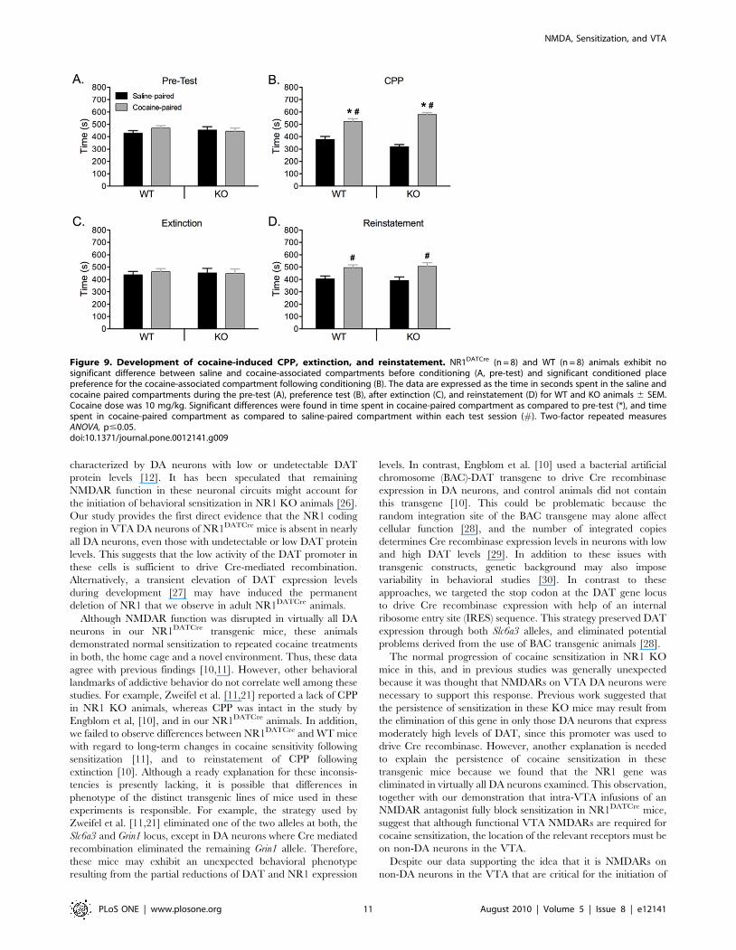
NMDARs in DA neurons are not required for thedevelopment of conditioned place preference (CPP) tococaine in NR1DATCre animals
To evaluate the rewarding properties of cocaine in NR1DATCre
mice, we employed a CPP paradigm (Fig. 9). In addition, to assess
the persistence and relapse of cocaine-seeking behavior in
NR1DATCre animals, we studied the extinction and reinstatement
of the CPP response. There was no difference between time spent
in each of the conditioning compartments during pre-test
(genotype x compartment interaction: F(1,31) = 1.3; p = 0.26),
ensuring an unbiased baseline (Fig. 9A). Two-factor repeated
measures ANOVA (genotype: WT vs. NR1DATCre animals; drug:
saline vs. cocaine pairing; conditioning: pre-test vs. post-test)
revealed a significant main effect of the drug (F(1,28) = 32.8; p,
0. 001) as well as significant drug x conditioning (F(1,23) = 78.5;
p,0.0001) and genotype x drug x conditioning (F(1,23) = 15.7;
p,0.0001) interactions. Analysis of the interaction revealed a
significant conditioning effect for the cocaine-paired compartment
in both WT (F(1,7) = 6.2; p = 0.041) an NR1DATCre (F(1,7) = 39.7;
p,0.0001) mice, indicating that animals from both genotypes
demonstrated significant preference for the cocaine paired-
compartment (Fig. 9B). CPP was then extinguished by repeated
saline injections (8 days) in both the previously cocaine-paired
floor and the saline-paired floor (Fig. 9C). There was no significant
preference for cocaine-paired compartment in NR1DATCre
animals (F(1,7) = 0.02; p = 0. 889) and WT (F(1,7) = 0.14; p = 0. 71),
indicating that both genotypes showed robust extinction after 8
sessions. As relapse to cocaine-seeking behavior can be triggered
by drug exposure, we tested for reinstatement of CPP. Following
the extinction phase, the reinstatement of CPP was investigated
with a challenge cocaine injection (10 mg/kg). Following this
priming dose of cocaine there was no significant effect of
conditioning in WT (F(1,7) = 1.56; p = 0. 27) and NR1DATCre
(F(1,7) = 0.82; p = 0. 39) animals (Fig. 9D), indicating that there was
Figure 5. Acute effects of cocaine in NAc slices. Voltammetrictraces of DA signals taken prior to (black) and following bathapplication of 500 nM cocaine (gray). Cocaine significantly increasedboth the amplitude and the decay time constant (tau) in slices fromboth WT (10 slices from 3 subjects) and NR1DATCre (7 slices from 4subjects) mice. **p,0.001; ***p,0.0001, paired t-test.doi:10.1371/journal.pone.0012141.g005
Figure 6. Basal locomotor activity in NR1DATCre mice. (A) Locomotor response to novel environment. Total distance traveled in the first hour inthe locomotor chamber in WT (n = 8) and NR1DATCre KO (n = 8) animals. KO animals were significantly less active during the first hour presented to theactivity chambers, p,0.05, two-tailed t-test. (B) Total distance traveled in 24 hours in the locomotor chamber. There were not significant differencesbetween the genotypes, p = 0.519, two-tailed t-test. (C) Day-night locomotion in a 24 hr circadian cycle at 1 hr intervals in WT and NR1DATCre animals.doi:10.1371/journal.pone.0012141.g006
NMDA, Sensitization, and VTA
PLoS ONE | www.plosone.org 9 August 2010 | Volume 5 | Issue 8 | e12141

no significant difference between times spent on the drug-paired
floor during pre-test and reinstatement-test. However, there was
significant difference between saline- and cocaine-paired com-
partment for each genotype (F(1,15) = 7.47; p = 0. 016 and
F(1,15) = 4.85; p = 0. 045, for WT and NR1DATCre animals,
respectively).
Discussion
The involvement of local glutamatergic synapses within the
VTA in learning and the development of adaptive processes in
animal models of drug abuse has been widely documented [25].
The fact that both, single or repeated administration of an
addictive drug directly into the VTA initiates sensitization to
subsequent systemic drug challenge, together with the observation
that NMDAR antagonists delivered directly to the VTA block the
development of sensitization [1,4,5], suggests a role for glutama-
tergic transmission within the VTA for the initiation of addictive
behaviors. However, the precise substrates through which altered
glutamatergic transmission results in drug modified synaptic
plasticity and the development of addictive behaviors remain to
be elucidated. Our results extend previous findings by using a
transgenic mouse lacking the NR1 receptor subunit in DA neurons
(NR1DATCre), and by confirming the absence of NMDARs in
virtually all VTA DA neurons. Since our approach, like previous
studies [6,10,11,21], relied upon DAT expression for the selective
elimination of NMDAR function in DA neurons, we considered it
important to determine whether NR1 gene deletion occurred in
mesostriatal and mesocortical circuits, because the latter is
Figure 7. Cocaine induced sensitization in NR1DATCre mice. (A)Context dependent sensitization. Cumulative 30 min locomotor re-sponse to cocaine (15 mg/kg, i.p.) in WT (n = 7) and NR1DATCre (n = 8)animals at different days after injection. S2, day 2 of saline injection. C4,day 4 of cocaine injection. C20 represents a challenge cocaine injectionafter withdrawal. Cocaine exposure in association with a new environ-ment induced significant sensitization in both WT and NR1DATCre mice(two way ANOVA, Newman-Keuls post hoc for all genotypes: *p,0.05 forWT compared to saline injection, ** p,0.01 for KO compared to salineinjection; #p,0.05 for WT compared to C1, ## p,0.01 for KOcompared to C1). No statistical differences were observed betweengenotypes neither at the initial development of sensitization (C1 versusC4) nor at the challenge injection after withdrawal (C20, two way RM-ANOVA, genotypes, F1, 65 = 0.00703, p = 0.934). (B) Cocaine-inducedsensitization in the home cage. Cumulative 30 min locomotor responseto cocaine in WT and NR1DATCre animals pretreated with saline (WT, n = 8and KO, n = 6) or 20 mg/kg cocaine, i.p. (WT, n = 8 and KO, n = 7) in thehome cage, prior to cocaine challenge (15 mg/kg, i.p.) in activitychambers. Cocaine exposure in the home cage induced significantsensitization in both, WT and NR1DATCre mice (Two way ANOVA,F1,28 = 3.57, p,0.001; Newman-Keuls post hoc for all genotypes: *p,0.05 for KO, ** p,0.01 for WT). Data presented as mean 6 SEM.doi:10.1371/journal.pone.0012141.g007
Figure 8. Effect of pretreatment with AP5 in the VTA on thedevelopment of behavioral sensitization to cocaine inNR1DATCre mice. (A) Location of the guide cannula and the injectioncannula in the ventral tegmental area (VTA). Left panel is the illustrationfrom the mouse brain atlas [17], indicating millimeters from bregma.The right half shows the image with dyes indicating the position ofcannula tips. (B) Preceding cocaine injections with intra-VTA infusions ofAP5 blocks the induction of locomotor sensitization by cocaine in bothWT (n = 8) and NR1DATCre (n = 5) mice, an effect that was not observedby intra-VTA saline injections in WT (n = 5) and NR1DATCre (n = 6). Datawere collected on days 1 and 4 of cocaine treatment. The results show asignificant difference between days 1 and 4 for the saline-infusedanimals (* p,0.001, two way ANOVA, day, F1,21 = 18.533), but not forthose treated with intra-VTA AP5 (p = 0.679, two way ANOVA).doi:10.1371/journal.pone.0012141.g008
NMDA, Sensitization, and VTA
PLoS ONE | www.plosone.org 10 August 2010 | Volume 5 | Issue 8 | e12141
characterized by DA neurons with low or undetectable DAT
protein levels [12]. It has been speculated that remaining
NMDAR function in these neuronal circuits might account for
the initiation of behavioral sensitization in NR1 KO animals [26].
Our study provides the first direct evidence that the NR1 coding
region in VTA DA neurons of NR1DATCre mice is absent in nearly
all DA neurons, even those with undetectable or low DAT protein
levels. This suggests that the low activity of the DAT promoter in
these cells is sufficient to drive Cre-mediated recombination.
Alternatively, a transient elevation of DAT expression levels
during development [27] may have induced the permanent
deletion of NR1 that we observe in adult NR1DATCre animals.
Although NMDAR function was disrupted in virtually all DA
neurons in our NR1DATCre transgenic mice, these animals
demonstrated normal sensitization to repeated cocaine treatments
in both, the home cage and a novel environment. Thus, these data
agree with previous findings [10,11]. However, other behavioral
landmarks of addictive behavior do not correlate well among these
studies. For example, Zweifel et al. [11,21] reported a lack of CPP
in NR1 KO animals, whereas CPP was intact in the study by
Engblom et al, [10], and in our NR1DATCre animals. In addition,
we failed to observe differences between NR1DATCre and WT mice
with regard to long-term changes in cocaine sensitivity following
sensitization [11], and to reinstatement of CPP following
extinction [10]. Although a ready explanation for these inconsis-
tencies is presently lacking, it is possible that differences in
phenotype of the distinct transgenic lines of mice used in these
experiments is responsible. For example, the strategy used by
Zweifel et al. [11,21] eliminated one of the two alleles at both, the
Slc6a3 and Grin1 locus, except in DA neurons where Cre mediated
recombination eliminated the remaining Grin1 allele. Therefore,
these mice may exhibit an unexpected behavioral phenotype
resulting from the partial reductions of DAT and NR1 expression
levels. In contrast, Engblom et al. [10] used a bacterial artificial
chromosome (BAC)-DAT transgene to drive Cre recombinase
expression in DA neurons, and control animals did not contain
this transgene [10]. This could be problematic because the
random integration site of the BAC transgene may alone affect
cellular function [28], and the number of integrated copies
determines Cre recombinase expression levels in neurons with low
and high DAT levels [29]. In addition to these issues with
transgenic constructs, genetic background may also impose
variability in behavioral studies [30]. In contrast to these
approaches, we targeted the stop codon at the DAT gene locus
to drive Cre recombinase expression with help of an internal
ribosome entry site (IRES) sequence. This strategy preserved DAT
expression through both Slc6a3 alleles, and eliminated potential
problems derived from the use of BAC transgenic animals [28].
The normal progression of cocaine sensitization in NR1 KO
mice in this, and in previous studies was generally unexpected
because it was thought that NMDARs on VTA DA neurons were
necessary to support this response. Previous work suggested that
the persistence of sensitization in these KO mice may result from
the elimination of this gene in only those DA neurons that express
moderately high levels of DAT, since this promoter was used to
drive Cre recombinase. However, another explanation is needed
to explain the persistence of cocaine sensitization in these
transgenic mice because we found that the NR1 gene was
eliminated in virtually all DA neurons examined. This observation,
together with our demonstration that intra-VTA infusions of an
NMDAR antagonist fully block sensitization in NR1DATCre mice,
suggest that although functional VTA NMDARs are required for
cocaine sensitization, the location of the relevant receptors must be
on non-DA neurons in the VTA.
Despite our data supporting the idea that it is NMDARs on
non-DA neurons in the VTA that are critical for the initiation of
Figure 9. Development of cocaine-induced CPP, extinction, and reinstatement. NR1DATCre (n = 8) and WT (n = 8) animals exhibit nosignificant difference between saline and cocaine-associated compartments before conditioning (A, pre-test) and significant conditioned placepreference for the cocaine-associated compartment following conditioning (B). The data are expressed as the time in seconds spent in the saline andcocaine paired compartments during the pre-test (A), preference test (B), after extinction (C), and reinstatement (D) for WT and KO animals 6 SEM.Cocaine dose was 10 mg/kg. Significant differences were found in time spent in cocaine-paired compartment as compared to pre-test (*), and timespent in cocaine-paired compartment as compared to saline-paired compartment within each test session (#). Two-factor repeated measuresANOVA, p#0.05.doi:10.1371/journal.pone.0012141.g009
NMDA, Sensitization, and VTA
PLoS ONE | www.plosone.org 11 August 2010 | Volume 5 | Issue 8 | e12141
sensitization, our results do not eliminate roles for NMDARs in
other functions of these DA neurons. Indeed, we also demonstrate
a loss of NMDAR-mediated synaptic plasticity in these DA
neurons, as well as changes in DA neurotransmission in the NAc of
NR1DATCre animals. It has been proposed that the acquisition of
conditioned behavioral responses is governed by the phasic firing
of DA neurons initiated by NMDAR activity [21,31,32,33,34].
The increased DA release elicited by single-pulse electrical
stimulation that we observed is consistent with the modest
elevation in basal DA levels in similar NR1-lacking mice [10].
However, DA release at higher burst-like frequencies of axon
terminal activation was reduced in the NAc of mice lacking NR1
in DA neurons. This is consistent with a previous report of reduced
burst-induced DA release in other NMDAR KO mice [21].
Previous studies using in vivo electrochemical techniques have also
shown that higher frequency DA transients are associated with DA
neuron bursting, and occur in the NAc when animals explore a
novel environment [35]. Therefore, the diminished burst-stimu-
lation-induced DA release in the NAc that we observed may
explain the significantly reduced locomotor responses of
NR1DATCre mice when exposed to a novel environment. Thus,
we speculate that the blunted response to novel environment may
be mediated by a deficit in burst-related DA release. Although this,
and the other changes we observed in NAc DA release in the NR1
KO animals appear to directly implicate NMDARs, we also found
that these changes did not occur during acute NMDAR antagonist
application in WT mice. This implies that there are alterations in
DAergic circuits that are secondary to the absence of NR1, and
that these compensatory changes should be considered when
evaluating both behavioral and physiological data from transgenic
mice.
Taken together, the results presented in this study demonstrate
that while the elimination of functional NMDA receptors in DA
neurons prevents LTP of glutamate synapses onto VTA DA cells,
the development of CPP and behavioral sensitization remained
intact. However, infusion of an NMDAR antagonist directly into
the VTA of these NR1DATCre mice blocked behavioral sensitiza-
tion to cocaine. Therefore, our study suggests that initiation of
cocaine sensitization and CPP in the VTA occurs through the
activation of NMDARs on non-dopaminergic neuronal substrates.
These results narrow down the relevant neuronal candidates that
support these phenomena, and indirectly implicate the remaining
predominant cellular phenotypes in the VTA; either c-aminobu-
tyric acid (GABA) or local circuit glutamatergic neurons [36].
Acknowledgments
The authors would like to thank Dr. Barry J. Hoffer and Dr. Bruce T.
Hope for helpful discussions and comments on the manuscript, and Dr.
Toni S. Shippenberg and Dr. Silvia Pontis for their technical assistance
with confocal microscopy.
Author Contributions
Conceived and designed the experiments: YL CHG ODR YZ AFH NM
VIC ACT CRL CMB. Performed the experiments: YL CHG ODR YZ
AFH LS SYK NM CMB. Analyzed the data: YL CHG ODR YZ AFH LS
SYK NM VIC CRL CMB. Contributed reagents/materials/analysis tools:
YL CHG ODR YZ AFH NM VIC ACT CRL CMB. Wrote the paper: YL
CHG ODR AFH VIC CRL CMB.
References
1. Dunn JM, Inderwies BR, Licata SC, Pierce RC (2005) Repeated administration
of AMPA or a metabotropic glutamate receptor agonist into the rat ventral
tegmental area augments the subsequent behavioral hyperactivity induced by
cocaine. Psychopharmacology (Berl) 179: 172–180.
2. Harris GC, Aston-Jones G (2003) Critical role for ventral tegmental glutamate in
preference for a cocaine-conditioned environment. Neuropsychopharmacology
28: 73–76.
3. Harris GC, Wimmer M, Byrne R, Aston-Jones G (2004) Glutamate-associated
plasticity in the ventral tegmental area is necessary for conditioning
environmental stimuli with morphine. Neuroscience 129: 841–847.
4. Kalivas PW, Alesdatter JE (1993) Involvement of N-methyl-D-aspartate receptor
stimulation in the ventral tegmental area and amygdala in behavioral
sensitization to cocaine. J Pharmacol Exp Ther 267: 486–495.
5. Vezina P, Queen AL (2000) Induction of locomotor sensitization by
amphetamine requires the activation of NMDA receptors in the rat ventral
tegmental area. Psychopharmacology (Berl) 151: 184–191.
6. Backman CM, Malik N, Zhang Y, Shan L, Grinberg A, et al. (2006)
Characterization of a mouse strain expressing Cre recombinase from the 39
untranslated region of the dopamine transporter locus. Genesis 44: 383–390.
7. Borgland SL, Malenka RC, Bonci A (2004) Acute and chronic cocaine-induced
potentiation of synaptic strength in the ventral tegmental area: electrophysio-
logical and behavioral correlates in individual rats. J Neurosci 24: 7482–7490.
8. Liu QS, Pu L, Poo MM (2005) Repeated cocaine exposure in vivo facilitates
LTP induction in midbrain dopamine neurons. Nature 437: 1027–1031.
9. Ungless MA, Whistler JL, Malenka RC, Bonci A (2001) Single cocaine exposure
in vivo induces long-term potentiation in dopamine neurons. Nature 411:
583–587.
10. Engblom D, Bilbao A, Sanchis-Segura C, Dahan L, Perreau-Lenz S, et al. (2008)
Glutamate receptors on dopamine neurons control the persistence of cocaine
seeking. Neuron 59: 497–508.
11. Zweifel LS, Argilli E, Bonci A, Palmiter RD (2008) Role of NMDA receptors in
dopamine neurons for plasticity and addictive behaviors. Neuron 59: 486–496.
12. Lammel S, Hetzel A, Hackel O, Jones I, Liss B, et al. (2008) Unique properties of
mesoprefrontal neurons within a dual mesocorticolimbic dopamine system.
Neuron 57: 760–773.
13. Tsien JZ, Huerta PT, Tonegawa S (1996) The essential role of hippocampal
CA1 NMDA receptor-dependent synaptic plasticity in spatial memory. Cell 87:
1327–1338.
14. Li X, Hoffman AF, Peng XQ, Lupica CR, Gardner EL, et al. (2009) Attenuation
of basal and cocaine-enhanced locomotion and nucleus accumbens dopamine in
cannabinoid CB1-receptor-knockout mice. Psychopharmacology (Berl) 204:
1–11.
15. Johnson SW, North RA (1992) Two types of neurone in the rat ventral
tegmental area and their synaptic inputs. J Physiol 450: 455–468.
16. Chen YH, Harvey BK, Hoffman AF, Wang Y, Chiang YH, et al. (2008) MPTP-
induced deficits in striatal synaptic plasticity are prevented by glial cell line-
derived neurotrophic factor expressed via an adeno-associated viral vector.
FASEB J 22: 261–275.
17. Franklin KBJ, Paxinos G (1997) The Mouse Brain in Stereotaxic Coordinates:
New York: Academic Press.
18. Bassareo V, De Luca MA, Di Chiara G (2002) Differential Expression of
Motivational Stimulus Properties by Dopamine in Nucleus Accumbens Shell
versus Core and Prefrontal Cortex. J Neurosci 22: 4709–4719.
19. Stefani MR, Moghaddam B (2006) Rule learning and reward contingency are
associated with dissociable patterns of dopamine activation in the rat
prefrontal cortex, nucleus accumbens, and dorsal striatum. J Neurosci 26:
8810–8818.
20. Floresco SB, West AR, Ash B, Moore H, Grace AA (2003) Afferent modulation
of dopamine neuron firing differentially regulates tonic and phasic dopamine
transmission. Nat Neurosci 6: 968–973.
21. Zweifel LS, Parker JG, Lobb CJ, Rainwater A, Wall VZ, et al. (2009) Disruption
of NMDAR-dependent burst firing by dopamine neurons provides selective
assessment of phasic dopamine-dependent behavior. Proc Natl Acad Sci U S A
106: 7281–7288.
22. Gracy KN, Pickel VM (1996) Ultrastructural immunocytochemical localization
of the N-methyl-D-aspartate receptor and tyrosine hydroxylase in the shell of the
rat nucleus accumbens. Brain Res 739: 169–181.
23. Badiani A, Robinson TE (2004) Drug-induced neurobehavioral plasticity: the
role of environmental context. Behav Pharmacol 15: 327–339.
24. Szumlinski KK, Kalivas PW (2004) Novel ideas about novelty. Commentary on
Badiani and Robinson drug-induced neurobehavioral plasticity: the role of
environmental context. Behav Pharmacol 15: 373–376.
25. Kauer JA, Malenka RC (2007) Synaptic plasticity and addiction. Nat Rev
Neurosci 8: 844–858.
26. Carr DB, Kalivas PW (2008) Confused about NMDA and addiction? Targeted
knockouts provide answers and new questions. Neuron 59: 353–355.
27. Coulter CL, Happe HK, Murrin LC (1996) Postnatal development of the
dopamine transporter: a quantitative autoradiographic study. Brain Res Dev
Brain Res 92: 172–181.
28. Heintz N (2001) BAC to the future: the use of bac transgenic mice for
neuroscience research. Nat Rev Neurosci 2: 861–870.
29. Parlato R, Rieker C, Turiault M, Tronche F, Schutz G (2006) Survival of DA
neurons is independent of CREM upregulation in absence of CREB. Genesis 44:
454–464.
NMDA, Sensitization, and VTA
PLoS ONE | www.plosone.org 12 August 2010 | Volume 5 | Issue 8 | e12141

30. Doetschman T (2009) Influence of genetic background on genetically engineered
mouse phenotypes. Methods Mol Biol 530: 423–433.
31. Fiorillo CD, Tobler PN, Schultz W (2003) Discrete coding of reward probability
and uncertainty by dopamine neurons. Science 299: 1898–1902.
32. Schultz W (2007) Behavioral dopamine signals. Trends Neurosci 30: 203–210.
33. Tobler PN, Fiorillo CD, Schultz W (2005) Adaptive coding of reward value by
dopamine neurons. Science 307: 1642–1645.
34. Tsai HC, Zhang F, Adamantidis A, Stuber GD, Bonci A, et al. (2009) Phasic
firing in dopaminergic neurons is sufficient for behavioral conditioning. Science324: 1080–1084.
35. Sombers LA, Beyene M, Carelli RM, Wightman RM (2009) Synaptic overflow
of dopamine in the nucleus accumbens arises from neuronal activity in theventral tegmental area. J Neurosci 29: 1735–1742.
36. Yamaguchi T, Sheen W, Morales M (2007) Glutamatergic neurons are presentin the rat ventral tegmental area. Eur J Neurosci 25: 106–118.
NMDA, Sensitization, and VTA
PLoS ONE | www.plosone.org 13 August 2010 | Volume 5 | Issue 8 | e12141
Copyright © 2022 FDOKUMEN