Nitte University Journal of Health Science
115
-
Upload
khangminh22 -
Category
Documents
-
view
0 -
download
0
Transcript of Nitte University Journal of Health Science


1
NUJHS Vol. 5, No.1, 2015, ISSN 2249-7110March
Nitte University Journal of Health Science
Nitte University Journal of Health Science
Editorial BoardEditor-in-Chief: K. R. Shetty, Editor: Arunachalam Kumar,
Associate Editor: M. S. Ravi
Board of AdvisorsG M. Shantharam Shetty G S. Ramananda Shetty Rajendra Prasad G C. S. Shastri
G Fatima D'Silva G Dhanesh Kumar K.U. G C. Vaman Rao G M.S. MoodithayaG Satheesh Kumar Bhandary G Vasudeva Kamath, USA G B. Sundar, Bengaluru,
G Avinash Shetty, USA G Kapadia, Bahrain, G Anupam Agarwal, USA
G Christopher Sudhakar G M. Rajshekar
Editorial Office: Email: [email protected] / web: nitte.edu.in/journalPhone: 0824-2204300 Fax: 824-2204305
Address: Editor, Nitte University Journal of Health ScienceUniversity Enclave, Health Science Complex,
Deralakatte, Mangalore 575018, India
The opinions and observations contained the journal are those of
the authors/s and not of the NUJHS Editorial Board
Subscription: Annual (4 issues) Rs. 1000 / Single copy: Rs. 300:
For subscriptions or copies contact: [email protected]
G
The Nitte University Journal of Health Science (NUJHS) is a peer-reviewed indexed, open access, quarterly
research publication. The annual subscription for NUJHS is Rs 1,000/- (4 issues). DDs / Checks payable to
Nitte University Journal of Health Sciences, Syndicate Bank, ABSMIDS Branch, Deralakatte can be mailed to
Dr. Arunachalam Kumar, Editor, NUJHS Journal Office, K. S. Hegde Medical Academy, Mangalore 585018,
India. Single copies are available on payment of Rs. 300 each, by cash or check at the Journal Office.
INDEXED / CITED
in Index Copernicus, Google Scholar, GFMER, Nursesmeet, HINARI, Mosbys, getCITED, EBSCOhost,Summon by Serial Solutions, SCOPUS, Genamics JournalSeek, EMBASE / Excerpta Medica, ProQuest, ProQuest
Pharma Collection, WAME, ResearchGate, SciVerse, Biobase-CABS, DOAJ, Journal Rate, Urlich's International Periodical Directory, Biblioteca Informa,
SCIRUS & Health Science Research Network.
Research Bib, World Cat, Universal Impact Factor, CIRRIE, CiteFactor,

2
NUJHS Vol. 5, No.1, 2015, ISSN 2249-7110March
Nitte University Journal of Health Science
ContentsPage No
Contents 2
Original Articles
Case Reports
Low Prevalence of Chlamydia Trachomatis Infection In Women From Southern Nigeria - Adesiji Y O, Iyere S I & Ogah I J 4
A Pilot Trail: Effectiveness of Bibliotherapy on Quality of Life, Psychological Distress and Depression Among Patient With Chronic Leg and Foot Ulcer in Mangalore
- Kirupa P, Preetham Rai B & Srinivasa Bhat U 9
Fingerprint Pattern Characteristics of Intellectually Disabled Children - An Original Study- Meril Ann Soman, Ramakrishna Avadhani, Rani Nallathamby, Meera Jacob 14
A Morphological Study of Placenta in Children With and Without Hypospadias- Bindhu S, R K Avadhani & Meera Jacob 17
A Study on Occurrence of Selected Risk Factors of Pregnancy Among Antenatal Women With A Viewto Develop an Information Booklet
- Neethu Varghese & Philomena Fernandes 21
Pain, Anxiety & Functional Status of Patients With Lower Limb Fracture and Dislocation After Open Reduction - Ambili Alphonse Thomas& Fatima D'silva 26
Stress and Burnout Assessment Among Post Graduate Dental Students - Aditya Shetty, Amrith Shetty,Mithra N Hegde, Dhanya Narasimhan & Shishir Shetty 31
Attitude of College Students Towards Alcohol Consumption in Mangalore- Deepak Daniel, Devishri Shetty, Greeshma Jilson Jose, Haritha J, Jeevan Ravi
Lakshmi S Pillai, Anupa Neghandi, Santhosh & Rashmi Kundapur 37
Comparative Study of Superdisintegrants Using Antiemetic Drug as A Model - D S Sandeep, R Narayana Charyulu & Prashant Nayak 40
Prevalence of Neck and Back Pain Among Paediatric Dentists- Siddharth M Shetty, Shreema Shetty, Anoop Hegde & Nirmal Babu 45
A Research Critique on the Lived-in Experiences of Patients Subjected to Chemotherapy In SelectedHospitals at Chennai
- Malarvizhi M & Bhavani M 48
A Study to Determine the Effectiveness of An Awareness Programme on Knowledge on Substance Abuseand Its Consequences Among the Students of A Selected Pre University College of Udupi District, Karnataka
- Charis Theou I,Asha K Nayak & Tessy Treesa Jose 53
Stress Among Early Adolescents and Maternal and Teachers Role Perception in AddressingAdolescents' Stress in Selected Schools of Thiruvananthapuram
- Aswathy K L , Kasturi R A & Maxie A 58
Correlation Between BMI and Pregnancy Outcome Among Postnatal Mothers With PregnancyInduced Hypertension in Selected Hospitals Bangalore
- Prathima P & S Anuchitra 62
Rare Variation in the Origin of Left Testicular Artery From Left External Iliac Artery: A Case Report- Huban Thomas R, Prasanna L C, Vivek Kumar, Antony Sylvan D'souza 68
A Multidisciplinary Approach for Functional Rehabilitation of A Patient With Skeletal Class IiiMalocclusion and Amelogenesis Imperfecta
- Archana S Shetty, Krishna Prasad D, M S Ravi & B Rajendra Prasad 71

3
NUJHS Vol. 5, No.1, 2015, ISSN 2249-7110March
Nitte University Journal of Health Science
Double Superiorvenacava and its Associated Clinical Implications - A Case Report and Literature Review- Sushma R Kotian, Antony Sylvan D Souza, Praveena Ravichandran, Pallavi Bhat & Mamatha Hosapatna 75
Management of Soft Tissue Injuries – Case Series- Muralee Mohan, B Rajendra Prasad, S M Sharma, Tripthi Shetty & Priyadharsana P S 79
Recurrent Pyogenic Grauloma - A Case ReportAmitha Ramesh & Agumbe Priyanka Prakash 83
Variation in the Structure of Levator Glandulae Thyroidea – A Case Report- Raghavendra A Y, Vishal Kumar, Vinay Kumar V & Harsha C R 86
Autommune Haemolytic Anemia : A Report of Two Cases1 - Chandrika Rao & Jayaprakash Shetty 88
A Case Report : Ectopic Pregnancy Due to Failure of Emergency Contraceptive - Arkierupaia Shadap 91
A Case of Disseminated Tuberculosis with Ocular Involvement- Md Fekarul Islam, Devdeep Mukherjee, Ritabrata Kundu, Prabal Chandra Niyogi & Joydeep Das 94
Review Articles
Periodontal Prosthesis - Review- Vinaya S Bhat, Krishna Prasad D & Prakyath Malli 97
Oncology Nurse Navigator Programme - A Narrative Review- Shejila C H, Mamatha S Pai & Donald J Fernandes 103
Body Donation as Gift to Medical Science for Better Tomorrow - Literature Review- Meera Jacob, R K Avadhani, Rani Nallathamby, Meril Ann Soman & Bindhu S 108
Page No
Instructions to authors 111
Nujhs Declaration and Right Transfer Form 114

Introduction :
Chlamydia trachomatis is the most prevalent sexually
transmitted bacterial infection worldwide, with an
estimated 4-5 million new cases each year. Chlamydia 1trachomatis is the most implicated organism in infertility.
Up to 40% of women with untreated Chlamydia develop
symptomatic Pelvic Inflammatory Disease and are at high
risk of severe complications including chronic pain, ectopic
pregnancy and infertility. Chlamydia is also the cause of
Trachoma blindness, affecting over 90% of the population 2in some developing countries. Untreated cases of
chlamydia can spread to the uterus causing pelvic 3inflammatory disease. In the developing world, laboratory
services for sexual ly
transmitted infections
(STIs) are either not
available, or where limited
services are available,
patients may not be able
to pay for or physically 4access those services.
LOW PREVALENCE OF CHLAMYDIA TRACHOMATIS INFECTION IN WOMEN FROM SOUTHERN NIGERIA
1 2 3Adesiji Y O , Iyere S I & Ogah I J1 2 Senior Lecturer,Department of Medical Microbiology & Parasitology, ResearchScholar, Department of Biomedical
3 Science, College of Health Sciences, Ladoke Akintola University of Technology, Osogbo, ResearchScholar, Infectious Diseases and Environmental Health Research Group. Department of Microbiology, Faculty of Life Sciences,
University of Ilorin, Nigeria
Correspondence :Adesiji Y O
Senior Lecturer,Department of Medical Microbiology & Parasitology,College of Health Sciences, Ladoke Akintola University of Technology, Osogbo, Nigeria.
Mobile : 234+8032948270 E-mail : [email protected], [email protected] :
Chlamydia trachomatis infections are the most common bacterial cause of sexually transmitted diseases (STDs) in the world. However,
most Nigeria health care facilities do not screen for Chlamydia antigen in gynaecological and general out-patient clinics. This study was
to document the prevalence of Chlamydia trachomatis infection in patients attending Family Planning Clinics and Gynaecology clinics in
Southern Nigeria. Endocervical swabs were collected from a hundred and forty patients and were screened using Chlamydia Rapid Test
Device –Swab / Urine (Interchemical Ltd. China). Out of 140 patients screened, 1 (0.7%) was positive for Chlamydia trachomatis antigen.
There seem to be an association between Chlamydia infection and abortion thus screening for chlamydia trachomatis infection in
asymptomatic patients to prevent the adverse consequences. This study presents an update in Chlamydia trachomatis in the Southern
part of Nigeria.
Keywords: Chlamydia trachomatis, prevalence, women, southern, Nigeria
Original Article
Access this article online
Quick Response Code
When tests are performed in many areas, diagnosis of C.
trachomatis genital infection is only performed in selected
populations and is often based on the presence of clinical
symptoms. Considering the high rate of asymptomatic
chlamydial infection, particularly in women, a substantial
“silent” or undetected epidemic of C. trachomatis
infections could put this population at significant risk for 5HIV infection.
In women with previous or invasive Chlamydia infection as
indicated by the presence of 1gM antibody against C.
trachomatis, increased rates of preterm delivery,
premature rupture of membranes, low birth weight, and
still birth have been observed. Infection with C.
trachomatis is also implicated in post abortal, post 6Caesarean section, and post partum maternal infections.
Commonly unrecognized and often poorly or inadequately
treated, Chlamydia infections can ascend the reproductive
tract resulting in pelvic inflammatory disease (PID) and,
consequently, lead to chronic pelvic pain, ectopic 7pregnancy, and infertility. Premarital sexual intercourse
4
NUJHS Vol. 5, No.1, 2015, ISSN 2249-7110March
Nitte University Journal of Health Science
Keywords : -
Chlamydia trachomatis, prevalence, women,southern, Nigeria Adesiji Y O

5
NUJHS Vol. 5, No.1, 2015, ISSN 2249-7110March
Nitte University Journal of Health Science
and intercourse with multiple partners have been shown to
be significant risk factors for C. trachomatis as well as HIV
infection and it is also associated with an increased risk of 8cervical cancer.
In many developed countries, screening programmes for
Chlamydia have been set up to reduce transmission and
reproductive tract morbidity. In most parts of Nigeria, C.
trachomatis are not routinely screened for, hence relative
information about frequencies of the infection are gotten
from individual laboratory reports and research projects of 9limited study areas. A study of prevalence of Chlamydia
infection in women attending family planning clinic and
obstetrics and gynaecological clinic will provide valuable
information on evidence for the need to include its
screening as a routine antenatal care in our health care
facilities. The aim of this study was to determine the
prevalence of C. trachomatis in patients attending
gynaecological and family planning clinics in Southern part
of Nigeria.
Materials and Mathod :
Study population: The study population were patients
attending Family Planning and Obstetrics and Gynaecology
clinics from selected Hospitals in Southern part of Nigeria.
They were patients who volunteered to participate in the
study. A total of 140 endocervical swabs (ECS) samples
were collected from Osogbo in Osun State, 62 samples
were collected from the Family Planning clinics, 22 samples
from Ladoke Akintola University of Technology Teaching
Hospital (L.T.H) and 40 samples from Asubiaro state
Hospital, 38 samples were collected from Obstetrics and
Gynaecology clinic of Adeoyo Maternity Hospital (A.M.H)
and 40 samples were collected from the Family Planning
clinic of University of Benin Teaching Hospital (U.B.T.H).
Sampling technique: C o n v e n i e n c e s a m p l i n g
techniques was used in which women who were willing,
and met the inclusion criteria were recruited consecutively
during the period of the study; a structured questionnaire
was applied after which an informed consent was
obtained.
Sample collection: Endocervical swabs were collected with
the assistance of the medical personnel (The Nurses).
Cusco Vaginal Speculum was inserted into the vagina for
the visualization of the cervix. A swab stick was inserted
through the speculum into the endocervical canal and
rotated. This permitted acquisition of columnar or cuboidal
epithelial cells which are the main reservoir of Chlamydia
organism. It was withdrawn without contamination from
exocervical or virginal cells. The swabs were transported
promptly to the laboratory and processed within 30
minutes of collection. Structured questionnaire was used
to obtained demographic details and other relevant
information such as number of sex partner, use of
contraceptives, past STDs, educational status, knowledge
about the C. trachomatis infection, etc from the
participants.
Sample analysis: Collected samples were analysed using
Chlamydia Rapid Test Device -Swab/Urine (Interchemical
Ltd. China). The Chlamydia Rapid Test Device (Swab/Urine)
is a qualitative, lateral flow immunoassay for the detection
of Chlamydia antigen from female cervical swab, male
urethral swab and male urine specimens. In this test,
antibody specific to the Chlamydia antigen is coated on the
test line region of the test. During testing, the extracted
antigen solution reacts with an antibody to Chlamydia that
is coated onto particles. The mixture migrates up to react
with the antibody to Chlamydia on the membrane and
generates a coloured line in the test line region. The
presence of this coloured line in the test line region
indicates a positive result, while its absence indicates a
negative result. To serve as a procedural control, a coloured
line will always appear in the control line region indicating
that proper volume of specimen has been added and
membrane wicking has occurred (Chlamydia Antigen Rapid
test). The test procedure was conducted according to the
manufacturer's instruction manual described by Sanders et 10al.
Results :
Of the one hundred and forty samples screened for
Chlamydia trachomatis antigen only one from U.B.T.H was
Keywords : -
Chlamydia trachomatis, prevalence, women,southern, Nigeria Adesiji Y O

6
NUJHS Vol. 5, No.1, 2015, ISSN 2249-7110March
Nitte University Journal of Health Science
positive (Table 1). Results as regards subject's sexual
partner in Table 2 revealed that in L.T.H 22 subject had one
sexual partner, in Asubiaro; 10 had no sexual partner while
30 had one sexual partner, in A.M.H 38 had one sexual
partner and in UBTH 4 had no sexual partner while 36 had
one sexual partner. In totality, the majority (127) subjects
had one sexual partners while few (13) subjects had no
sexual partners. Table 2 also showed the use of
contraceptives among subjects. In L.T.H the entire 22
subjects were on contraceptives (18 IUCD and 4 Injectable),
amongst subjects in Asubiaro, 30 were on IUCD and 10 with
no use of contraceptive, A.M.H 38 of them were not on
contraceptives and for U.B.T.H 30 were on IUCD while 10
use no contraceptive. Of all 140 patients, 82 were on
contraceptive while 58 did not use. Table 2 further revealed
that all (140) patients had no past incidence of STDs.
In term of educational status and knowledge about
Chlamydia trachomatis, table 3 showed that 18 subjects
had tertiary education, 1 with secondary school education,
1 with primary school education and 1 had none in L.T.H.
Subjects from Asubiaro, 27 had tertiary education, 10 had
secondary education and 2 had primary education. In
A.M.H, 22 subjects had tertiary education, 12 had primary
education, and 7 had primary education. Among U.B.T.H
subjects, 36 had tertiary education and 3 had secondary
education. Table 3 also revealed subjects knowledge about
chlamydia trachomatis, few (10 subject in total) 3 LTH, 4
ASUBIARO and 3 U.B.T.H had knowledge about the
infection.
From the data obtained, the age range with the highest
frequency was 30 – 39years having 69 subjects followed by
35 – 39years having 41 subjects and then 25 – 29years
having 16 subjects. The least value was obtained from age
45 years and above having 2 subjects, 1 from L.T.H and 1
from U.B.T.H (Table 4). Results of data collected on events in
the study sites showed that 15 in totality subjects had
dysuria, no cases of pre-matured birth, 6 had miscarriage,
10 were for abortion, 14 were experiencing change in
menstrual cycle, and 25 subjects were also for abnormal
vaginal discharge treatment. The highest number, subjects
(37) were for lower abdominal pain treatment (Table 5).
Keywords : -
Chlamydia trachomatis, prevalence, women,southern, Nigeria Adesiji Y O
Table 1 : Shows the number of sample collected from each sites, number of positive results and the incidence.
STUDY SITES SAMPLE
COLLECTION RESULTS
L.T.H 22 0 0
ASUBIARO 40 0 0
A.M.H 38 0 0
U.B.T.H 40 1 2.5
TOTAL 140 1 0.7
POSITIVE PERCENTAGE (%)
Note: 0 = negative (negative results absence of coloured line)1= positive (positive results presence of coloured line)
Table 2 : Frequency distribution of risk factors among female subject in the study sites
RISK FACTORS L.T.H Asubiaro A.M.H U.B.T.H Total
Number of sex partner
No sexual partner - 9 - 4
one sexual partner 22 31 38 36 13
one and above - - - - 127
Use of Contraceptives
IUCD 18 30 - 30 78
Injectable 4 - - - 4
No IUCD - 10 38 10 58
Past STDs
Yes - - - - -
No 22 40 38 40 140
L.T.H Asubiaro A.M.H U.B.T.H Total
Educational status
Tertiary 18 27 19 34 98
Secondary 1 11 12 4 28
Primary 2 2 7 2 13
None 1 - - - 1
Knowledge
Yes 3 4 - 3 10
No 19 37 38 38 130
AGE RANGE (YEARS)
20 - 24 - 2 4 - 6
25 – 29 10 3 10 3 16
30 - 34 8 19 20 20 69
35 - 39 3 15 4 14 41
40 - 44 1 1 - 2 6
Over 45 1 - - 1 2
L.T.H Asubiaro A.M.H U.B.T.H Total
Table 3 : Frequency distribution of Educational status and knowledge of Chlamydia trachomatis amongst female subjects
Table 4 : Frequency distribution of age female subjects

7
NUJHS Vol. 5, No.1, 2015, ISSN 2249-7110March
Nitte University Journal of Health Science
Keywords : -
Chlamydia trachomatis, prevalence, women,southern, Nigeria Adesiji Y O
Table 5 : Frequency distribution of various events in the study sites of female subjects
Events
Dysuria 3 5 1 9 18
Miscarriage 2 1 1 2 6
Premature Birth - - - - -
Abortion 4 4 1 1 10
Change in menstrual cycle 2 8 - 4 14
Abnormal vaginal discharge 8 9 5 3 25
Lower abdominal pain 2 9 14 12 37
Others 1 4 16 9 30
L.T.H Asubiaro A.M.H U.B.T.H Total
Discussion :
This study reports a low prevalence of 0.1% (1/140) in the
population sampled across three Western States of
Nigeria. Previous report has shown a high prevalence of the 11chlamydia infection in most parts of Africa. In most parts
of Nigeria, C. trachomatis are not routinely screened for,
hence relative information about frequencies of the
infection are based on laboratory reports and research
based findings. Despite the fact that women are at a high 12risk of infection, earlier report by Harry et al. stated that
there is a sociocultural inhibition that prevents women
from reporting sexual symptoms, non-availability of facility
to detect the organism in many health units and the largely
asymptomatic nature of the disease. The positive result
was from University of Benin Teaching Hospital (U.B.T.H),
where earlier reports of similar studies in the same location 9were higher. In north east Nigeria, the report of Amin et
13al. on the outcome of opportunistic screening for
Chlamydia trachomatis in women seen in the antenatal
and gynaecology clinics revealed 9% prevalence In Eastern 14part of Nigeria, a report by Ikeme et al in a study to
determine seroprevalence of C. trachomatis among
population comprised of 136 female undergraduate
students and 150 non-student women, reported an overall 15prevalence of 29.4%. In Lagos Nigeria, Oloyede et al
reported that Chlamydia screening was positive in 14
(18.2%) among 77 women undergoing infertility. In Port 16Harcourt, Kennedy et al reported 11% rate of prevalence
of Chlamydia trachomatis infection among female
undergraduate of University of Port- Harcourt, Mawak et 17al reported 56.1% of total of 164 total samples from
women tested positive for C. trachomatis in Jos (North
.
18Central, Nigeria). Only Brabin and colleagues reported a
comparable prevalence of 0.5% in 204 girls aged 12±17
years and 8.2% in 206 girls aged 17±19 years in a rural
population in South-eastern Nigeria, using cervical 18specimens. Possible explanation for lower prevalence
obtained from this study could be attributed to several
factors such as the lower sample size enrolled in the study, 19and the detection technique employed, with haven stated
that molecular detection methods are often more reliable
that other methods. Several studies have shown that the
major risk factor for chlamydial infection is sexual activities
and it is the commonest sexually transmitted organism (20,21)throughout the world. In this study, low rate of
Chlamydia trachomatis among subjects may be due to the
fact that majority (127) of 140 screened had on one sexual
partner (Table 2). This means that subjects in this category
are probably married and no subjects with more than one
sexual partner. Also, majority (69) of the subjects were
within the range of 30 – 34 years of age. This is in
agreement with the previously reported association of C.
trachomatis infection that it is common in women with a 22higher number of sexual partners or a new sexual partner
that age and marital status were considered as factors for
variation of incidence of Chlamydia trachomatis and 14Ikeme, et al indicated that age <30 years were
independently significant risk factors for cervical antigen
positivity. Other factor observed for low prevalence was 23high use of IUCD among subjects. The results indicate low
sexual activities and high use of contraceptives, no subject
indicated any past experience of STDs. However, from
personal observation and evidence from literature, women
in this part of the world may not disclose information that
relates to previous sexual habits and infections out of fear
of stigmatization and cultural inhibitions, hence so,
observation in this study might not indicate the true
occurrence of Chlamydia infection.
The positive result from this study was obtained from 32
years woman who has had a previous history of abortion.
Thus, there seems to be an association between Chlamydia
infection and abortion. Although, it is not usually
scientifically valid to conclude based on one individual

8Keywords : -
Chlamydia trachomatis, prevalence, women,southern, Nigeria Adesiji Y O
NUJHS Vol. 5, No.1, 2015, ISSN 2249-7110March
Nitte University Journal of Health Science
data, it was also observed from the study that the
knowledge about Chlamydial infection is poor among the
women attending Family Planning and Obstetrics and
Gynaecology clinic in the study sites despite the fact that
majority had attained their tertiary educational status
(Table 3). This may be because infections are asymptomatic 24and among the symptomatic cases, it is seldom severe. It
1 Ogiogwa IO, Motayo BO, Okerentugba PO, Innocent-Adiele HC, Tafeng Y, Onoh CC, Nwanze JC, Okonko IO. Detection of Chlamydia TrachomatisAntigen among Attendees of a Fertility Clinic in Abeokuta, Ogun State, Nigeria. Researcher. 2012; 4(4): 96-100
2 WHO, World Health Organization. VISION 2020 Action Plan for 2006–2011. Planning Meeting. Geneva, 11–13 July 2006
3 CDC, 2014. www.m.cdc.gov/en/HealthsafetyTopics/Diseasesconditions/STDs/chlamydiaFS
4 Peeling RW, Holmes KK, Mabey D, Ronald A. Rapid diagnostic tests for Sexually transmitted infections Rapid tests for sexually transmitted infections (STIs): the way forward. Sex Transm Infect; 2006; 82: 5 1-6
5 Sturm-Ramirez K, Brumblay H, Diop K, Guèye-Ndiaye A, Sankalé J, Thior I, N'Doye I, Thior I, N'Doye, I, Hsieh C, Mboup S, Kanki PJ. Molecular Epidemiology of Genital Chlamydia trachomatis Infection in High-Risk Women in Senegal. West Africa J Clin Microbiol. 2000; 38(1): 138–145.
6 McGregor JA, French JI. Chlamydia trachomatis infection during pregnancy. Am J Obstet Gynecol. 1991; 164:1782-9.
7 Chernesky MA. The laboratory diagnosis of Chlamydia trachomatis infection. Can J Infect Dis Med Microbiol. 2005; 16:39-44.
8 Anttila T, Saikku P, Koskela P, Bloigu A, Dillner J, Ikäheimo I. Serotypes of Chlamydia trachomatis and risk development of cervical squamous cell carcinoma'. JAMA. 2001;1(285):47-51.
9 Okoror LE, Agbonlahor DE, Esumeh FI, Umolu PI. Prevalence of chlamydia in patients attending gynaecological clinics in south eastern Nigeria'. African Health Sciences; 2007; 7(1): 18-24
10 Sanders JW, Hook EW, Welsh LE, Shepherd ME, Quinn TC. Evaluation of an enzyme immunoassay for detection of Chlamydia trachomatis in urine of asymptomatic men. J Clin Microbiol. 1994; 32: 24-27.
11 Okonofua FE. Infertility in Sub Saharan Africa' In: Okonofua F and Odunsi K (2003). (eds). Contemporary Obstetrics and Gyneacology for Developing Countries. Ed 1, Woman's Health and Action Research Center. Benin City, Edo State, Nigeria. 1991; pp 129 -156.
12 Harry TC, Saravanamuttu KM, Rasid S, Shrestha TL. 'Audit evaluating the value of routine screening of Chlamydia trachomatis urethra infection in men'. Int J STD AIDS. 1994; 5:374 – 375.
13 Amin JD, Zaria LT, El-Nafaty AU, Mai AM. Genital Chlamydia trachomatis infection in women in a Nigerian hospital'. Genitourin
was observed that the low level of knowledge about the
infection among women could be a contributing factor for
acceleration of the spread of Chlamydia infection in other
parts of Nigeria.
This study present an update in Chlamydia trachomatis in
the Southern part of Nigeria.
References :
Med. 1997; 73: 146-147.14 Ikeme AC, Ezegwui HU, Ikeako LC, Agbata I, Agbata E. Seroprevalence
of Chlamydia trachomatis in Enugu, Nigeria. Niger J Clin Pract 2011;14:176-80.
15 Oloyede OA, Fakoya TA, Oloyede AA, Alayo AM. Prevalence and Awareness about Chlamydial Infection in Women Undergoing Infertility Evaluation in Lagos, Nigeria' Int J Health Res. 2009; 2(2): 157-162.
16 Warison, KT, Odigie J, Eyaru S. Prevalence of Chlamydia trachomatis Infection among Female Undergraduates of the University of Port Harcourt Using Strand Displacement and Amplification [SDA] Technique. The Nigerian Health Journal. 2012; 12:2
17 Mawak JD, Dashe N, Agabi YA, Panshak BW. Prevalence of Genital Chlamydia Trachomatis Infection among Gynaecologic Clinic Attendees in Jos, Nigeria. Shiraz E Medical Journal. 2011; 12:2
18 Brabin L, Kemp J, Orikomaba K. Reproductive tract infections and abortion among adolescent girls in rural Nigeria. Lancet. 1995; 345:300-304.
19 Vidwan NK, Regi A, Steinhoff M, Huppert JS, Staat MA, Dodd C, Nongrum R, Anandan S, Verghese V. Low prevalence of Chlamydia trachomatis infection in non-urban pregnant women in Vellore, S. India. PLoS One. 2012; 7:e34794.
20 MacLean AB. Pelvic Infection. In: Edmonds KD(ed). Dewhurst's Textbook of Obstetrics and Gyneacology for post graduates, ed 6, Blackwell Sciences Ltd, London. 1999; pp: 393 - 409.
21 Jones GE, Low JC, Machell J, Amstrong K. Comparison of five tests for the detection of antibodies against chlamydia (enzootic) abortion of ewes. Vet Rec. 1997; 141(7):164-8.
22 Van Verkoyeen RP, Peeter MF, Van Rijsoort-Vos JH, van der Meijden WI, Marton JW. Sensitivity and specificity of three new commercially available Chlamydia trachomatis tests. Int J STD AIDS. 2002; 2:23-5.
23 Alarape AI, Olapegba PO, Chovwen CO. Condom use among students: The influence condom self-efficacy, social norms and affective attitude toward condom. J Soc Sci. 2008; 17:237-41.
24 Opaneye AA. Pelvic Infections. In: Okonofua F, Odunsi K. (Eds). Contemporary Obstetrics and Gyneacology for Developing Countries, ed 1, Woman's Health and Action Research Center. Benin City, Edo State, Nigeria. 2003; pp.54–65.

9
NUJHS Vol. 5, No.1, 2015, ISSN 2249-7110March
Nitte University Journal of Health Science
Keywords : leg and foot ulcer, bibliotherapy, distress, qualityof life, Depression - Kirupa P
Introduction
Epidemiology and economic burden of the chronic wound
was serious endemic in the developed and developing
countries in the world.
A recent study in the UK shows that a prevalence of
patients with a chronic wound was 3.55 per 1000
population. In the chronic wounds, leg and foot ulcer
accounts for the 28%
wounds. Prevalence of
chronic wounds among
hospital inpatients was 130.7% . In India it is
estimated a prevalence
rate of chronic wounds at
4.5 per 1000 population.
A PILOT TRAIL: EFFECTIVENESS OF BIBLIOTHERAPY ON QUALITY OF LIFE, PSYCHOLOGICAL DISTRESS AND DEPRESSION AMONG
PATIENT WITH CHRONIC LEG AND FOOT ULCER IN MANGALORE2 3Kirupa P , Preetham Rai B & Srinivasa Bhat U
1 2 3Research Scholar, Nitte University, Professor, Department of Surgery, Associate Professor,Department of Psychiatry, K.S. Hegde Medical Academy,
Nitte University, Mangalore, Karnataka, India.
Correspondence :Kirupa P
Research Scholar, Nitte University, Mangalore - 575 018.Mobile : +91 98449 58743 E-mail : [email protected]
Abstract :
Introduction : Many clients living with chronic leg and foot ulcers experience diminished quality of life, pain, psychosocial
maladjustment, limited work capacity, and physical disabilities. Bibliotherapy helps the individual to cope up with illness.
Objectives : Assess the pretest level of psychological distress, quality of life and depression in both Interventional and control group.
Evaluate the effectiveness of bibliotherapy on quality of life, psychological distress and depression
Methods : Pilot study was conducted among the twenty patients with chronic leg and foot ulcer .Randomized clinical trial comparison
with pair group method was used to evaluate the effectiveness of bibliotherapy .Data were assessed by using chronic wound impact
schedule, Kessler's psychological distress scale and beck depression inventory were used to assess the quality of life, psychological
distress and depression respectively. Simple random sampling by lottery method was used to collect the data.
Results: Findings show that there is mean of the quality of life (56.3), distress (28.3) and depression (22.5) were falls on the moderate
level among the patient suffering with chronic leg and foot ulcer. Two way analysis of variance proves that bibliotherapy highly
significant in increasing quality of life (F= 20.3,P<0.001) ,decreasing the psychological distress (F=25.2,P<0.01) and decreasing
depression (F=5.18 ,P<0.05).
Conclusion. Need based bibliotherapy is effective to meet the psychological aspects of chronic leg and foot ulcer
Key words: leg and foot ulcer, bibliotherapy, distress, quality of life, Depression
1
Access this article online
Quick Response Code
The etiology of chronic wounds varies from diabetes,
atherosclerosis, tuberculosis, leprosy, venous ulcers, 2pressure ulcers, vasculitis and trauma .
In Bangalore Victoria Hospital a clinical study of the ulcer of
leg among 200 clients reported that diabetic ulcer
accounting for 68 cases (34%) followed by venous ulcer
(24%), traumatic ulcer (16%), arterial ulcer (12%)
malignant ulcer (5%), tropic ulcer (3%) and others 12
(6%).Ulcers are breaks in the layers of the skin that fail to 3heal. They may be accompanied by inflammation .
Chronic leg and foot ulcers are often painful and recurrent,
and they can have a negative Physical, physiological, social
and psychological impact on clients and families, thus
decreasing their quality of life. Leg and foot ulcers are often
recalcitrant to healing, tend to recur, and become long-
Original Article

10
NUJHS Vol. 5, No.1, 2015, ISSN 2249-7110March
Nitte University Journal of Health Science
Keywords : leg and foot ulcer, bibliotherapy, distress, qualityof life, Depression - Kirupa P
4 term chronic healthcare problems . Research reported
that chronic physical illness one of the increased risk of 5depression .
Many clients living with chronic leg and foot ulcers
experience diminished quality of life, pain, psychosocial
maladjustment, limited work capacity, and physical 4disabilities .
Need for the Study
In India, L H Hiranandani Hospital, world mental health day
celebration 2011 reported that patients suffering from a
physical illness, especially chronic, tend to develop not just
minor mental problems like distress and anxiety, but also
major ones like depression, phobias and even sexual
dysfunction. About 14-20% of chronically ill patients have
psychological problems, apart from minor distress and 5anxiety .
Pain and stress may slow wound healing through various
intricate mechanisms. Psychological stressor that triggers
the hypothalamic-pituitary-adrenal axis promoting the
production of vasopressin and glucocorticoid (cortisol).
Cortisol reduces the immune- inflammatory response,
suppresses cellular differentiation and proliferation, and
inhibits the regeneration of endothelial cells and delays 6collagen synthesis .
The psychological impact of chronic physical illness can be
prevented by bibliotherapy. Bibliotherapy as defined by
the American Library Association is the use of selected
reading materials as therapeutic adjuncts in medicine and
psychiatry; also, guidance in the solution of personal 7problems through directed reading .
Bibliotherapeutic intervention may be undertaken for
many reasons such as to develop an individual's self-
concept; to increase an individual's understanding of
human behavior or motivations to foster an individual's
honest self-appraisal; to provide a way for a person to find
interests outside of self; to relieve emotional or mental
pressure; to show an individual that he or she is not the
first or only person to encounter such a problem; to show
an individual that there is more than one solution to a
problem and to help an individual plan a constructive 8course of action to solve a problem .
In Trinity College, Ireland the study was conducted on
bibliotherapy is a form of self-administered treatment in
which structured materials provide a means to alleviate
distress. Thematic analyses revealed that bibliotherapy 9schemes are effective in alleviating the distress .
In Indian Scenario need to focus the psychological
perspective of patient with chronic leg and foot ulcer. The
above literature indicated the therapeutic effect of
bibliotherapy on the psychological distress and depression
.Researcher interested to know the novel effect of
bibliotherapy on psychological distress, quality of life and
depression among the patients suffering with chronic leg
and foot Ulcer.
Objectives :
1. Assess the pre test level of psychological distress, quality
of life and depression in both Interventional and control
group
2. Evaluate the effectiveness of bibliotherapy on quality of
life, psychological distress and depression.
Materials and Methods
This study is the pilot trail for the main study to evaluate the
effectiveness of bibliotherapy on selected psychological
variables such as quality of life, distress and depression.
This randomized clinical trial was conducted between
March 2013 to June 2013. In the evaluative approach,
randomized control trial comparison with parallel group
was chosen for this pilot trial. In this trail interventional
group were received usual care along with bibliotherapy
and control group were received only usual care. Pretest stand baseline admission was done 1 day ,Post test was
th thadministered on 7 day ,I follow up on 14 day and II follow stup was conducted on 21 day for both interventional and
control group.
Sample & sample size
Patients those who are fulfilling the sampling criteria.

11
NUJHS Vol. 5, No.1, 2015, ISSN 2249-7110March
Nitte University Journal of Health Science
Keywords : leg and foot ulcer, bibliotherapy, distress, qualityof life, Depression - Kirupa P
Sample size comprises of 20 patients with chronic leg and
foot ulcer.
Patients suffering with chronic leg and foot ulcer were
randomized into interventional and control group by
simple random sampling by lottery method.
Setting
The study was conducted in K.S Hegde medical college
hospital, Mangalore.
Drawing a protocol
Bibliotherapy is a use of books in therapeutic context by the
researcher based on the Problem solving technique,
Coping with the condition, therapeutic regimen and follow
up in the form of storytelling, activity exercise, protocol and
poetry presentation specific to the chronic leg and foot
ulcer caused by the venous ulcer and diabetic foot.
Researcher was started to prepare the intervention after
the consultation with the experts and guides .Researcher
was conducted a small qualitative study on experiences of
patient living with chronic leg and foot ulcer in order to
understand need of the person. Based on the findings and
after collection of adequate literature the researcher was
prepared book for patient suffering with chronic leg and
foot ulcer.
This book is a contemporary approach to help the patient
deal the problem both mentally and physically. Core
concept of the book implies that accepting the illness,
create knowledge and motivation of self care, caution
about the complication, managing of day to day battle with
chronic leg wounds.
Content validity of the book obtained from the experts in
the psychiatric, surgical and nursing field .After the
validation, the book was translated in both Kannada and thMalayalam version. Book was prepared on 8 grade level of
reading.
Sampling criteria
Inclusion criteria : Patient those who are
1. Diagnosed as diabetic foot ulcer or venous ulcer.
2. Age between 30years -65years
3. Able to understand and speak either Kannada,
Malayalam or English
4. Both male and female.
5. Had the site of ulcer below knee.
6. Visited the K.S Hegde hospital for the treatment.
7. In all the stages of diabetic foot ulcer or venous ulcer.
8. Underwent all the type of surgical procedure for the
diabetic foot .
9. Patient in the stage of before and after the surgical
procedure.th10.Educated above 10 standard.
Exclusion criteria
1. Chronic leg and foot ulcer patient suffering with any
other serious co existing illness.
2. Patient those who are uncooperative.
3. Patient those who are unconscious, drowsy and
disoriented at the time of study.
4. Diagnosed with chronic alcoholism and alcohol
dependant syndrome.
Data collection procedure
Data were collected from the participant by using
sociodemographic proforma; Chronic wound impact
schedule, Kessler's psychological distress scale and Beck
depression inventory. The researcher recruited the
participant based on the predetermined sampling criteria.
Three instruments were translated into Kannada and
Malayalam version and language validation done.
Reliability of the three instruments was fall on above 0.7 by
split half method and found to be reliable in the translated
version of Kannada and Malayalam. Pre test was conducted
to before the administration of the intervention.
Bibliotherapy was administered for one week in the
intervention along with usual care
Scientific adequacy of the research
The study strictly followed the privacy, confidentiality of
the ethical clearance procedure. This study conducted by
the researcher as a part of PhD program.
and venous ulcer

12
NUJHS Vol. 5, No.1, 2015, ISSN 2249-7110March
Nitte University Journal of Health Science
Keywords : leg and foot ulcer, bibliotherapy, distress, qualityof life, Depression - Kirupa P
Tables with caption separately
Table 1- Independent t test analysis between the interventional and control group
Baseline data Interventional
Group(10) Group(10) Inference
Mean SD Mean SD T value
Quality of life 56.2 56.3 6 7.7 0.15, p>0.05 NS
Psychological Distress 28.3 6.3 27.1 3 0.5, p>0.05 NS
Depression 22.5 7.2 22 4.5 0.18 ,P>0.05 NS
Control N=20
S, S*, S** –significant (P< 0.05, 0.01, 0.001) NS- Non Significant df =18
Table 2 : shows Mean, SD, Mean difference on quality of life among the Interventional and control group N=20
Data colle-
ction Point Mean SD Mean SD Difference
Post test 65.21 9.3 53.06 5.8 12.15
I follow Up 67.8 9.18 54.64 5.3 13.2
II Follow Up 70.74 7.8 52.24 5.4 18.5
Interventional Control Mean
Data colle-
ction Point Mean SD Mean SD Difference
Post test 20.6 5.91 28.1 3.87 7.5
I Follow up 17.1 3.38 26 3.23 8.9
II follow up 16.8 4.63 23.6 3.5 6.8
Interventional Control Mean
Data colle-
ction Point Mean SD Mean SD Difference
Post test 17.2 7.036 21.4 4.248 4.2
I follow up 14.4 6.132 20.3 3.529 5.9
II Follow up 13.1 5.486 18.1 4.095 5
Interventional Control Mean
Table 3 : shows the Mean, SD and mean difference on the psychological distress in the Interventional and control group patients. N=20
Table 4 : shows the Mean, SD and mean difference of the Depression in the Interventional and control group patients. N=20.
Table 5. Two way analysis of variance on quality of life, Distress and depression between the group and within group. N=20
Variables F value P value
Quality of life 20.3 0.00 S**
Distress 25.2 0.00 S*
Depression 5.18 0.03 S
S, S*, S** - significant (P< 0.05, 0.01, 0.001)NS- Non Significant df =18
Results :
Results presented as follows
Table 1 reveals baseline mean of quality of life is 56.3 SD 6
in the interventional group and 66.2 SD 7.7 in control
group .Distress mean is 28.3 SD 6.3 in the interventional
group and 27.1 SD 3 in the control group. Depression mean
is 22.5 SD 7.2 in the interventional group and 22 and 4.5 in
the control group Independent t test value obtained were
0.15 P>0.05, 0.5 P>0.05, 0.18 P>0.05 for quality of life,
psychological distress and depression respectively. It
shows that there is no significant difference between
baseline values of quality of life distress and depression
between the Interventional and control group.
Table 2 shows that in the Interventional group, mean of
quality of life was 56.3, 65.2, 67.8 and 70.4 in the pretest,
posttest, I follow up and II follow up respectively. In the
control group, mean of quality of life was 56.28, 53.06, 54.6
and 52.2 in the pretest, posttest, I follow up and II follow up
respectively. Quality of life is significantly increased in the
quality of life after the bibliotherapy in the interventional
group than the control group.
Table 3 shows that , In the Interventional group, mean of
psychological distress was 28.3,20.6,17.1 and 16.8 in the
pretest, posttest ,I follow up and II follow up respectively. In
the control group, mean of psychological distress was 27.1,
28.1, 26 and 23.6 in the pretest, posttest, I follow up and II
follow up respectively. After the bibliotherapy there is a
significant decrease in the psychological distress in the
interventional group than the control group.
Table 4 shows that in the Interventional group, mean of
depression was 22.5, 17.2, 14.4 and 13.1 in the pretest,
posttest, I follow up and II follow up respectively. In the
control group ,mean of depression was 22,21.4,20.3 and
18.1 in the pretest, posttest ,I follow up and II follow up
respectively .After the bibliotherapy a significant decrease
in depression in the interventional group in the I follow up
and II follow up than the control group.

13
NUJHS Vol. 5, No.1, 2015, ISSN 2249-7110March
Nitte University Journal of Health Science
Keywords : leg and foot ulcer, bibliotherapy, distress, qualityof life, Depression - Kirupa P
Table 5 shows the two way analysis (ANOVA) of variance of
quality of life, psychological distress and depression in
various points of time between the Interventional and
control group. F value obtained between groups indicated
that statistically, there is significant difference between the
intervention and control group. Interventional group,
quality of life is significantly increased than the control
group. Psychological distress and Depression significantly
decreased in the intervention group than the control
group. Bibliotherapy was effective improving the quality of
life and decreasing the distress and depression.
Discussion
Pilot trail findings were discussed in to two aspects
a) Baseline level of quality of life, distress and depression
Baseline level of quality of life, distress and depression
implies that there is no significant difference in the level of
quality of life, distress and depression in both
interventional and control group (p>0.05). Findings show
that there is mean of the quality of life (56.3), distress
(28.3) and depression (22.5) were produced moderate
impact among the patient suffering with chronic leg and
foot ulcer.
Above findings was supported by the study conducted
Jones JE, Robinson J, Barr W, Carlisle C in UK on Impact of 9exudates and odour from chronic venous leg ulceration .
b) Effectiveness of Bibliotherapy
Two way analysis of variance proves that bibliotherapy
highly significant in increasing quality of life (F=
20.3,P<0.001) ,decreasing the psychological distress
(F=25.2,P<0.01) and decreasing depression (F=5.18
,P<0.05).Comparatively psychological distress, quality of
life were shows highly significant difference in the
interventional group than the depression. Bibliotherapy to
the mild and moderate depression is less effective in the
course of administration than the psychological distress.
Effectiveness of bibliotherapy was supported by the study of Songprakun W and McCann TV. On Evaluation of a
bibliotherapy manual for reducing psychological distress
in people with depression: a randomized controlled trial in 10Thailand .
Conclusion
Chronic leg and foot ulcers are disabling and constitute a
significant burden on clients and the health-care system
and it have a negative psychological impact on clients and
families. Bibliotherapy is helps the patient to cope with the
illness, reduce the psychological impact. Pilot trail proves
that bibliotherapy is significantly effective in improving the
quality of life and reducing distress and depression in
chronic leg and foot ulcer. Pilot trail gave root to conduct
main study for the researcher.
Acknowledgement :
Our heartfelt thanks to the ethical and research committee
of the Nitte university and K.S Hedge medical college
hospital. Our special thanks to the valuators of the book,
Traslators and Language valuators of the Instruments
1. Vowden K, Vowden P, Posnett J. The resource costs of wound care in Bradford and Airedale primary care trust in the UK. Journal of Wound Care. 2009;18(3):93–98.
2. Gupta N, Gupta SK, Shukla VK, Singh SP.An Indian community-based epidemiological study of wounds. Journal of Wound Care . 2004 Sep;13(8):323-5.
3. Dr.T Prabakhar clinical study of the ulcer of the leg 2006 dissertation submitted to RGUHS.
4. K. Solowiej, BSc (Hons), V. Mason, PhD and D. Upton, PhD, FBPsS. psychological stress and pain in wound care part 2 Review of pain and assessment of tools. The journal of wound care vol 19, no 3, March 2010 ; page no :109-115.
5. A world federation of mental health report .Nightingale nursing Times.vol6,no.7.october 2010;page no:3-4.
6. Dr Ganesh Kumar, Physical illness may affect mental health.TNN, Oct 11, 2010, 05.35am IST.www.google.com
References :
7. Kevin Y Woo .wound related pain: anxiety stress and wound healing a clinical review. Wounds UK, 2010, Vol 6, No 4 www.google.com
8. Pehrsson, D. E., & McMillen, P. Bibliotherapy: Overview and implications for counselors (ACAPCD-02). Alexandria, VA: American Counseling Association ;2007.
9. Naylor EV et al Bibliotherapy as a treatment for depression in primary care. Journal of Clinical Psychology in Medical Settings. 2010 September;17(3):258-71.
10. Jones JE, Robinson J, Barr W, Carlisle C. Impact of exudates and odour from chronic venous leg ulceration. Nursing Standard. 2008 Jul 16-22;22(45):53-4, 56, 58 .
11. Songprakun W and McCann TV. Evaluation of a bibliotherapy manual for reducing psychological distress in people with depression: a randomized controlled trial. Journal of Advanced Nursing. 2012 Dec; 68(12):2674-84.

14
NUJHS Vol. 5, No.1, 2015, ISSN 2249-7110March
Nitte University Journal of Health Science
Keywords : Fingerprint, Intellectually disabled, Loop,Dermatoglyphics - Charly Chacko Joseph
Introduction :
Fingerprint patterns are impressions made by the minute
ridge formations or furrows present on the fingertips. The rddermal ridges are formed during 3 month of intrauterine
life. The fingerprint patterns are genetically determined
and the pattern once formed doesn't alter with
developmental or environmental changes. Human
fingerprints are unique and they offer an infallible means of (1), (2)personal identification.
Intellectual disability, also known as mental retardation is
characterized by below average intellectual functioning
level and s ignif icant
limitations in daily living (3)skills. Symptoms may
appear at birth or in early
childhood. Common signs
and symptoms include
developmental delay,
difficulties with problem
FINGERPRINT PATTERN CHARACTERISTICS OF INTELLECTUALLY DISABLED CHILDREN - AN ORIGINAL STUDY
1 2 3 4Meril Ann Soman , Ramakrishna Avadhani , Rani Nallathamby , Meera Jacob ,5Charly Chacko Joseph
1,3,5 2 4Post Graduates , Professor & HOD , Assistant Professor , Department of Anatomy, Yenepoya Medical College, 5Mangalore, Department of Anaesthesia, Mahatma Gandhi Medical College and Research Institute, Pondicherry, India.
Correspondence :Charly Chacko Joseph
Post Graduate, Department of Anaesthesia, Mahatma Gandhi Institute of Medical Science and Research Centre,Pondicherry 607 402, India.
E-mail : [email protected]
Abstract :
Fingerprint patterns are unique patterns made by friction ridges and furrows present on the pads of finger tips. Uniqueness and
persistence are the two underlying features of fingerprint patterns. Aim of this present study was to determine the differences in the
incidence of fingerprint patterns in intellectually disabled children compared to normal healthy children. Intellectual disability is a
generalized disorder appearing before adulthood and is characterized by limitations in both intellectual functioning and in adaptive
behavior. The present study comprising of 120 students (60 intellectually disabled and 60 controls) was carried out in Pediatrics
outpatient department, Yenepoya Medical College and Hospital, Mangalore. The incidence of the four fingerprint patterns (Ulnar loop,
Radial loop, Whorls and Arches) were determined in both the groups. Ulnar loop pattern had the highest incidence in both the groups
and the least incidence was shown by arch pattern. There exists difference in the frequency of the fingerprint patterns in males and
females of both the groups. The study was conducted to observe for any difference in the incidence of fingerprint patterns between
intellectually disabled and normal children.
Keywords : Fingerprint, Intellectually disabled, Loop, Dermatoglyphics
Access this article online
Quick Response Code
solving skills and in learning social rules. Mostly, it persists
throughout adulthood. The causes for intellectual
disability include genetic factors, prenatal maternal
infections, childhood illnesses, injuries and environmental (4)factors such as malnutrition.
Fingerprint patterns are of multifactorial polygenic
inheritance. Many chromosomal disorders have
characteristic dermatoglyphic patterns which may aid in
the diagnosis of those disorders. In this study, the
dermatoglyphic patterns of the intellectually disabled
children were compared with the controls. An attempt has
been made to study whether there exist any difference in
the incidence of the fingerprint patterns in both the
groups.
Materials and Methods
This prospective study was done among subjects who had
visited the Pediatrics outpatient department of Yenepoya
Medical College and Hospital. The study included a total of
Original Article

15
NUJHS Vol. 5, No.1, 2015, ISSN 2249-7110March
Nitte University Journal of Health Science
Keywords : Fingerprint, Intellectually disabled, Loop,Dermatoglyphics - Charly Chacko Joseph
120 subjects (60 intellectually disabled and 60 controls) in
the age group of 8-14 years. Each group consisted of 30
males and 30 females. The subjects were requested to
wash their hands with soap and water and dry it using a
clean hand towel. The ink pad used was of Faber Castell
Company of size 110mm* 69 mm. A white sheet of paper
was provided which had 10 different blocks for all fingers of
both the hands. The subject was asked to press his/her
fingertips on the stamp pad and thereafter to the white
paper to transfer the fingerprint impression. Specific
number was given to each digit. Right thumb was marked
as 1 and number 10 was given to left little finger. Other
details such as age and sex of each subject were noted.
Subjects with any hand deformity or permanent scars on
the fingertips were excluded from the study.
Results :
The study was carried out in 120 children of age group 8-14
Table 1 : Distribution of fingerprint patterns in intellectually disabled and controls
Fingerprint Intellectually Disabled Controls
Patterns Number % Number %
ULNAR LOOP 324 54% 386 64.3%
RADIAL LOOP 47 7.8% 20 3.4%
WHORLS 205 34.2% 156 26%
ARCHES 24 4% 38 6.3%
TOTAL 600 100% 600 100%
Table-1 shows distribution of fingerprint patterns in
intellectually disabled and controls. Ulnar loop pattern
shows the highest incidence in both the groups followed by
whorls. Arches show the least frequency in intellectually
disabled and radial loop pattern is found to be the least
among controls.
years of which 60 subjects were intellectually disabled and
60 were controls. 30 males and 30 females were present in
each group.
TABLE 2 : Distribution of fingerprint patterns according to gender in intellectually disabled and controls
Fingerprint Intellectually Disabled Control
Patterns Males Females Males Females
Number % Number % Number % Number %
Ulnar loops 162 54% 162 54% 184 61.3% 202 67.4%
Radial loops 18 6% 29 9.7% 16 5.3% 04 1.3%
Whorls 114 38% 91 30.3% 86 28.7% 70 23.3%
Arches 06 2% 18 06% 14 4.7% 24 08%
Total 300 100% 300 100% 300 100% 300 100%
Table 2 shows the distribution of fingerprint patterns according to gender in intellectually disabled and control
groups. Ulnar loop shows the highest frequency in males and females of both the groups followed by whorls.
Arches show the least frequency in males of both the groups and also in females of intellectually disabled group.
SEX SIDE ULNAR LOOPS WHORLS ARCHES RADIAL LOOPS
Number % Number % Number % Number %
Intellectually MALE RIGHT 84 56% 53 35.3% 03 02% 10 6.7%
disabled LEFT 78 52% 61 40.7% 03 02% 08 5.3%
FEMALE RIGHT 85 56.7% 47 31.3% 05 3.3% 13 8.7%
LEFT 77 51.3% 44 29.3% 13 8.7% 16 10.7%
Controls MALE RIGHT 91 60.7% 47 31.3% 08 5.3% 04 2.7%
LEFT 93 62% 39 26% 06 04% 12 08%
FEMALE RIGHT 102 68% 33 22% 13 8.7% 02 1.3%
LEFT 100 66.7% 37 24.7% 11 7.3% 02 1.3%
TABLE 3 : Side wise distribution of fingerprint patterns in males and females of intellectually disabled subjects and controls
Table 3 shows sidewise distribution of fingerprint patterns in males and females of intellectually disabled
subjects and controls. Ulnar loops followed by whorls showed the highest predominance on both sides in males
and females of both the groups.

16
NUJHS Vol. 5, No.1, 2015, ISSN 2249-7110March
Nitte University Journal of Health Science
Keywords : Fingerprint, Intellectually disabled, Loop,Dermatoglyphics - Charly Chacko Joseph
Discussion :
Dermatoglyphics is the scientific study of fingerprints. The
term 'dermatoglyphics' was coined from the Greek word
'derma' which means 'skin' and the term 'glyphic' which
means carving'. Fingerprint patterns are easily deposited
on suitable surfaces by sweat secretions from the eccrine (5), (6) glands present in the friction ridges of finger tips. Less
variation is seen in fingerprint patterns among subjects
with genetic syndromes than between control subjects.
Dermatoglyphics is an emerging field which acts as a non
invasive and early predictor of mental retardation in
children. The study of dermatoglyphics is a simple, yet
complicated tool to diagnose the chromosomal (7)abnormalities.
In the present study, the incidence of fingerprint patterns
in intellectually disabled was compared with control
subjects. The study was done sex wise and side wise. The
four fingerprint patterns taken into consideration were
ulnar loop, radial loop, whorls and arches. Ulnar loop had
the highest incidence in both the groups followed by
whorls. Arch pattern was seen the least in intellectually
disabled whereas radial loops had the least frequency
among controls. In sex wise distribution of fingerprint
patterns among intellectually disabled, ulnar loop pattern
showed equal frequency in both the sexes, whorls were
seen to be more in males compared to females whereas
radial loops and arches were seen more in females. In
sidewise distribution of patterns among mentally retarded,
right side showed higher frequency of ulnar loops among
males and females.
A study was conducted in 2013 by Dr. Bhagwat V B on
palmar dermatoglyphics in mentally retarded children
which also showed the predominance of ulnar loop
pattern. According to Dr. Bhagwat V B, among mentally
retarded children, the percentage of ulnar loop was higher (8)in males compared to females. Whereas in the present
study, both males and females shared equal frequencies of
ulnar loops, whorls were seen more in males and radial
loops and arches were seen more in females.
Conclusion :
Dermatoglyphics, as a non invasive approach can definitely
aid in the early detection of mental retardation in children.
Since the dermatoglyphic patterns are established by birth,
it can be considered as a diagnostic tool in identifying
various genetic abnormalities in children. Therefore
dermatoglyphics can be considered as an inexpensive and
noninvasive screening method in certain genetic disorders.
Acknowledgement :
Authors sincerely thank all the participants of this study
and also Dr. Bhagya B Sharma, Lecturer, Department of
Anatomy, Yenepoya Medical College, Mangalore, for her
kind co-operation and help during this study.
References :1. Dr. Rastogi P, Ms. Pillai K.R. A study of Fingerprints in Relation to
Gender and Blood Group. J Indian Acad Forensic Med. 2010; 32(1): 11-14.
2. Bhavana D, Ruchi J, Prakash T, L.K.J. Study of Fingerprint Patterns In Relationship with Blood Group and Gender- a Stastical Review. Res.J.Forensic Sci. 2013 March; 1(1): 15-17.
3. Sharma M K, Jhawar P, Sharma H, Sharma S, Kalavatia I. Dermatoglyphics an attempt to predict downs syndrome. Int J Biol Med Res. 2012; 3(2):1631-1635
4. Alter M, Bruhel H. Dermatoglyphics in idiopathic mental retardation. American Journal of Diseases in children. 1967; 113: 702-706.
5. http://en.wikipedia.org/wiki/Fingerprint6. Pillay V.V. 2009. Textbook of Forensic Medicine and Toxicology. 15th
ed. Hyderabad. Paras Medical Publishers. 53-94.7. Clare Davison. Dermatoglyphic in patients with idiopathic severe
subnormality and Genetic studies in mental subnormality. British Journal of Psychiatry special publication. 1973; 8: 21-24.
8. Bhagwat V B, Meshram M M. Study of Palmar Dermatoglyphics in Mentally Retarded Children. IOSR-JDMS. 2013; 8(1): 23-27

17
NUJHS Vol. 5, No.1, 2015, ISSN 2249-7110March
Nitte University Journal of Health Science
Keywords : Hypospadias, Feto-placental ratio, Placenta. - Bindhu S
Original Article
A MORPHOLOGICAL STUDY OF PLACENTA IN CHILDREN WITH AND WITHOUT HYPOSPADIAS
1 2 3Bindhu S , R K Avadhani & Meera Jacob1 2 3Associate Professor, Professor & HOD, Assistant Professor, Department of Anatomy, Yenepoya Medical College,
Yenepoya University, Mangalore, Karnataka, India.
Correspondence :Bindhu S
Associate Professor, Department of Anatomy, Yenepoya Medical College, Yenepoya University, Mangalore 575018, Karnataka, India.Mobile : +91 99456 66156 E-mail : [email protected]
Abstract :
Introduction : Hypospadias can be defined as an abnormal urethral orifice under surface of the penis with or without chordee and with
or without dorsal hood. At a critical time in sexual differentiation of the male fetus, HCG enters fetal plasma from syncytio trophoblast;
acts as an LH surrogate and stimulate replication of testicular Ley dig cells and testosterone synthesis to promote male sexual
differentiation. The placental insufficiency may disrupt the supply of nutrients and hcG to the fetus leading to growth retardation and
hypospadias.
Aim : The aim of this study was to observe and document morphological changes of placenta in children with hypospadias and compare
with controls.
Materials &Methods : The present study was a case control study from July 2008 to July 2011 The data base of the labor registries of the
hospital indicated that there were total 3243 male births during this period. All examined for presence /absence of hypospadias by
attending pediatrician. Hypospadias was detected in 17 male newborns. Control cases comprised of 68 male newborns without
hypospadias of similar gestational age and birth weight collected by cluster sampling.
Result :
Total number of male birth during the study period was 3243, in that17 children born with hypospadias. The incidence of hypospadias in
our hospital was 0.52%. Gestational age, Birth weight, Placental weight, Placental thickness, Placental volume, volume of infarcts, F.P
Ratio, Cord length, were similar in children with hypospadias when compared with controls. CONCLUSION: Fetal factors like gestational
age, birth weight, placental weight, Feto-placental ratio were not significantly associated with hypospadias. This study shows no role of
placenta in the etiology of hypospadias in children with normal birth weight.
Keywords : Hypospadias, Feto-placental ratio, Placenta.
Introduction :
Hypospadias, in boys, defined as an association of three
anomalies of the penis:(1) an abnormal ventral opening of
the urethral meatus that may be located anywhere from
the ventral aspect of the glans penis to the perineum, (2) an
abnormal ventral curvature of the penis (chordee), and (3)
an abnormal distribution of foreskin with a “hood” present
dorsally and deficient 1f o r e s k i n v e n t r a l l y.
Hypospadias is typically
diagnosed at new born
physical examination .This
is not always the case
for boys with milder
forms of hypospadias or a
non-retractile prepuce or for those with the megameatus 2,3,4intact prepuce(MIP) variant . These boys may escape
diagnosis until the foreskin is fully retracted or circumcision
is performed. Although uncommon, simple apparently
isolated hypospadias may be the only visible indication of a
significant underlying abnormality. The only treatment for
this condition is surgery. Thus prevention is imperative. To
accomplish this, it is necessary to determine the etiology of
hypospadias. The association of growth retardation and
hypospadias is well established. Fetal testosterone
secretion is under the influence of placental hcG during
first 14 weeks of gestation. Chorionic gonadotropin
stimulates fetal testicular testosterone secretion that is
maximum at approximately the same time that maximal
level of HCG is attained. Thus, at a critical time in sexual
Access this article online
Quick Response Code

18
NUJHS Vol. 5, No.1, 2015, ISSN 2249-7110March
Nitte University Journal of Health Science
Keywords : Hypospadias, Feto-placental ratio, Placenta. - Bindhu S
differentiation of the male fetus, HCG enters fetal plasma
from syncytio trophoblast; acts as an LH surrogate and
stimulate replication of testicular Ley dig cells and
testosterone synthesis to promote male sexual 5differentiation .
The placental insufficiency may disrupt the supply of
nutrients and hcG to the fetus leading to growth
retardation and hypospadias. To validate this hypothesis,
we analyzed all the male infants born at our hospital with
hypospadias for fetal growth parameters, and collected
placentae for detailed evaluation .And assessed maternal
risk factors associated with hypospadias by questionnaire
proforma.
Materials & methods:
The present study was a case control study from July 2008
to July 2011 .The data base of the labour registries
indicated that there were total 3243 male births during this
period. All examined for presence /absence of hypospadias
by attending pediatrician. Hypospadias was detected in 17
male newborns. Control cases comprised of 68 male
newborns without hypospadias of similar gestational age
and birth weight collected by cluster sampling.
Once hypospadias was identified, the neonate was
examined in detail to identify other anomalies, weight at
birth, and gestational age. The placenta was collected and
examined for placental weight, thickness, placental volume
and cord information. Fetus to placental weight ratio was
measured as a reference for placental function and
intrauterine fetal growth. The placenta of these controls
was also subjected to detailed evaluation and examination.
Data was compiled and analyzed by descriptive analysis;
comparison of risk factors was done using student t test. P
value <0.05 was considered as significant.
Result :
Total number of male birth during the study period was
3243, in that17 children born with hypospadias. The
incidence of hypospadias in our hospital was 0.52%. The
characteristics of child at birth are considered as fetal
demographic factors associated with hypospadias (Table1).
Characteristics of child at birth--Gestational age, Birth
weight, Placental weight, Placental thickness, Placental
volume, volume of infarcts, F.P Ratio, Cord length, were
similar in children with hypospadias when compared with
controls (Table 1).Gestational age was similar in
hypospadias (38.64±0.99weeks) when compared with
controls (38.37±1.14weeks). Birth weight in children with
hypospadias was (2.96±0.19kg), when compared with
controls (3.01±0.17kg). There was no significant difference
in placental weight in children with hypospadias
(462.31±8.56gm) when compared with controls
(461.92±8.04gm). Placental thickness was similar in
children with hypospadias (2.08±0.27cm) when compared
with controls (2.00±0.00cm). There was no significant
difference in placental volume in children with
hypospadias (362.65±14.14cc) when compared with
controls (364.22±17.17cc). Feto-placental ratio was not
higher in children with hypospadias (6.53±0.40) when
compared to controls (6.74±0.42). There was no significant
difference in length of umbilical cord in children with
hypospadias vs controls (58.31±2.52vs56.85±2.91, P-
0.18).Number of blood vessels in the umbilical cord were
normal in children with hypospadias.
Gestat iona l age was s imi lar in hypospadias
(38.64±0.99weeks) when compared with controls
(38.37±1.14weeks). Birth weight in children with
hypospadias was (2.96±0.19kg), when compared with
controls (3.01±0.17kg). There was no significant difference
in placental weight in children with hypospadias
(462.31±8.56gm) when compared with controls
(461.92±8.04gm). Placental thickness was similar in
children with hypospadias (2.08±0.27cm) when compared
with controls (2.00±0.00cm). There was no significant
difference in placental volume in children with
hypospadias (362.65±14.14cc) when compared with
controls (364.22±17.17cc). Percentage of infarct in the
total volume of placenta was similar in both the groups
(3.45±0.23 vs3.48±0.30,P-0.81). Feto-placental ratio was
not higher in children with hypospadias (6.53±0.40) when
compared to controls (6.74±0.42). There was no significant
difference in length of umbilical cord in children with

19
NUJHS Vol. 5, No.1, 2015, ISSN 2249-7110March
Nitte University Journal of Health Science
Keywords : Hypospadias, Feto-placental ratio, Placenta. - Bindhu S
hypospadias vs controls (58.31±2.52vs56.85±2.91(P-0.18).
Number of blood vessels in the umbilical cord was normal
in children with hypospadias.
1. Paulozzi J, Erickson J D, Jackson R J. Hypospadias trends in two U S surveillance systems.Pediatrics.1997;100(5):831-834.
2. Boisen K, Chellakooty M, Schmidt I: Hypospadias in a cohort of 1072 Danish newborn boys: Prevalence and relationship to placental weight, anthropometrical measurements at birth, and reproductive hormone levels at 3 months of age. J Clin Endocrinol Metab 2005; 90(7):4041-4046.
3. Duckett JW, Keating MA.Technical challenge of the megameatus intact prepuce hypospadias variant: the pyramid procedure. J Urol 1989; 141(6):1407-1409.
4. Hatch DA, Maizels M, Zaontz MR. Hypospadias hidden by a complete
References :
prepuce. Surg Gynecol Obstet 1989; 169(3):233-2345. Stephens FD, Smith ED, Hutson JM: Embryogenesis of hypospadias. In:
Stephens FD, ed. Congenital anomalies of the urinary and genital tracts, Oxford, UK: Isis Medical Media Ltd.; 1996:80-90
6. Hussain N1, Chaghtai A, Herndon CD, Herson VC, Rosenkrantz TS, McKenna PH.Hypospadias and early gestation growth restriction in infants. Pediatrics 2002; 109(3):473-478.
7. Weidner I S, Moller H, Jensen TK, Skakkebaek NE. Risk factors of cryptorchidism and hypospadias .J Urol.1999;161:1606-1609.
8. Aschim EL, Haugen TB, Tretli S, Daltveit AK, Grotmol T. Risk factors for
Discussion :
Many authors have suggested that disturbance of placental
function early in pregnancy is the key mechanism
underlying both preterm birth/low birth weight and the
improper closure of the urethra, because the placenta is
the main producer of pregnancy hormones in early
pregnancy and is thus instrumental in the differentiation 6,7,8and development of the fetal organs . This study could
not find an association between hypospadias risk and
preterm birth (< 37 weeks gestation) and/or being small for thgestational age (< 10 percentile) because in this study all
the children born with hypospadias were normal birth
weight (>2.8 kg) and all of them were term birth (>39
weeks). It is well known that in normal, preterm and term
infants there is a direct relation between birth weight and 9weight of placenta . In this study, since all the children born
with a normal birth weight, there was no significant
association between birth weight and placental weight and
all placentae were of normal weight. At term, the placenta
is approximately 3cm thick and measures 15-25cm
Characteristics Hypospadias Controls P-value
(n-17) (n-68)
Gestational age(week) 38.64±0.99 38.37±1.14 0.96
Birth weight(kg) 2.96±0.19 3.01±0.17 0.11
Placental weight(gm) 462.31±8.56 461.92±8.04 0.90
Placental thickness(cm) 2.08±0.27 2.00±0.00 0.32
Placental volume(cc) 362.65±14.14 364.22±17.77 0.78
Feto-placental ratio 6.53±0.40 6.74±0.42 0.16
Cord length(cm) 58.31±2.52 56.85±2.91 0.18
P-value <0.05 is considered as significant
Table 1 : Comparison of fetal demographic factors associated with hypospadias
10diameter . Placental thickness is closely related to fetal
well-being and may be a key factor in perinatal outcome. In
this study, all the placentae were of normal thickness
because none of the children in the study group were low
birth weight. Ultra sonographic study of placental volume
found that, place
. In this study all the
children born with normal birth weight and placentae were
in normal weight hence volume of placenta in children with
hypospadias was similar with the control on comparison.
The ratio of placental weight to birth weight is described as
a marker of fetal growth. The correlation of birth weight
and placental size is to be expected as both placental
weight and size are known to increase as birth weight 13increases . In this study the feto-placental ratio in
hypospadias children was not found to be increased in
comparison with the controls.
Conclusion :
Several studies have found reduced placental function as
underlying etiology for low birth weight and hypospadias.
In the present study all the children born with hypospadias
were of normal birth weight. Fetal factors like gestational
age, birth weight, placental weight, Feto-placental ratio
were not significantly associated with hypospadias. This
study shows no role of placenta in the etiology of
hypospadias in normal birth weight children.
Acknowledgement :
I thank Dr. R.B. Nerli ,Professor & HOD of Urology, KLE
Hospital, KLE University ,Belgaum, for his valuable advice
and guidance to complete this study.
ntal volume was directly proportional to 11the birth weight of the babies . However, there is limited
information on the relationship between intrauterine 12placental volume and birth weight

20
NUJHS Vol. 5, No.1, 2015, ISSN 2249-7110March
Nitte University Journal of Health Science
Keywords : Hypospadias, Feto-placental ratio, Placenta. - Bindhu S
hypospadias in Norwegian boys—association with testicular dysgenesis syndrome? Int J Androl. 2004;27(4):213–221.
9. Thame M, Wilks RJ, McFarlane-Anderson N, Bennett FI & Forrester TE. Fetal growth is directly related to maternal anthropometry and placental volume. European Journal of Clinical Nutrition. 2004 ;58: 894–900.
' th10. Sadler T.W. Langmann Medical embryology,9 edition. Baltimore, MD: Lippincott Williams and Wilkins; 2004;p.117-48.
11. Wolf H, Oosting H, Treffers P E.A longitudinal study of the relationship between placental fetal growth measured by ultrasonography. Am J Obstet.Gynecol. 1989;161: 1140-45.
12. Clappe JH, Rizk KH. Appleby –Wineberg SK, Grass JR. Second trimester placental volume predicts birth weight at term. J Soc Gynecol Investig 1995;2:19-22.
13. Fugimoto, Tomohide Suwa ,Kazuhiko Kabe, Tomoko Adachi, Massao Nakabayashi and Teruke Amamiya. Placental insufficiency in early gestation is associated with hypospadias. J Paediatric Surg 2008:43;358-361.
21
NUJHS Vol. 5, No.1, 2015, ISSN 2249-7110March
Nitte University Journal of Health Science
Keywords : risk factors, antenatal women, information booklet- Philomena Fernandes
Introduction:
Pregnancy is a unique, exciting and often joyous time in a
woman's life, as it highlights the woman's amazing creative
and nurturing powers while providing a bridge to the
future. However, a pregnant woman needs to be a
responsible woman so as to best support the health of her
future child, as the growing fetus depends entirely on its
mother's healthy body for all needs1. Pregnancy is a
natural physiological process but there are certain high risk
factors that may come across and complicate the
pregnancy and childbirth and often pregnant women are
not aware of the seriousness of these problems.
According to WHO, worldwide maternal mortality ratio is
estimated to be 400 per 1,
00,000 live births with 5,
36,000 maternal deaths
occurring annually and
global lifetime risk of
death from pregnancy of 1
in 92(2009). In India
currently (2010) MMR is
Original Article
Access this article online
Quick Response Code
A STUDY ON OCCURRENCE OF SELECTED RISK FACTORS OF PREGNANCY AMONG ANTENATAL WOMEN WITH A VIEW TO
DEVELOP AN INFORMATION BOOKLET1 2
Neethu Varghese & Philomena Fernandes1Lecturer, Department of Obstetrics & Gynaecological Nursing, Anjarakandy Medical College, Kannur, Kerala,
2Associate Professor & HOD, Department of Obstetrics & Gynaecological Nursing,Nitte Usha Institute of Nursing Sciences, Nitte University, Mangalore, Karnataka, India.
Correspondence:Philomena Fernandes
Associate Professor & HOD, Department of Obstetrics & Gynaecological Nursing, Nitte Usha Institute of Nursing Sciences, Nitte University, Mangalore - 575 018, Karnataka, India.
Mobile: +91 9449 207845 E-mail : [email protected]
Abstract :
The descriptive study was conducted to determine the occurrence of selected risk factors of pregnancy among antenatal women.
Structured interview schedule was used to assess the risk factors of pregnancy among antenatal women. 150 samples were selected by
purposive sampling technique. The study findings revealed that increased maternal age, short stature, increased blood pressure,
abortion, decreased hemoglobin and GDM were the most prevalent risk factors in the study sample. There was significant association
between gravid state and abortion (cal=26.78, p<0.05), gravid state and age (cal=9.79, p<0.05), education and hemoglobin level
(cal=6.02, p<0.05) at 0.05 level of significance.
Keywords:risk factors, antenatal women, information booklet
found to be 290 per 1, 00,000 live births. Identification of
the risk factors together with appropriate and timely
interventions during the perinatal period can prevent
morbidity and mortality among mothers and infants to a
great extent. For health professionals maternal mortality is
not statistics, not rates or ratios but it is women whose
faces are seen in the throes of agony, distress and despair,
and this is not simply because these women die in the
prime of their lives, at a time of great expectation and joy or
not because maternal death is one of the most terrible
ways to die, be it bleeding to death, the convulsions of
toxaemia of pregnancy, the unbearable pangs of
obstructed labour or the agony of puerperal sepsis. It is
because in almost each and every case, it is a 5 event that
could have been prevented. So the investigator feels that
nurses are responsible and accountable for early
identification of risk factors and also imparting health
education to gravid mothers for prevention of this
enduring epidemic.
Objectives of the Study
· To determine the occurrence of selected risk factors of

RISK STATUS RISK FACTORS FREQUENCY PERCENTAGE
BIOPHYSICAL RISK FACTORS
Age
No Risk 1. 19-29 yrs 116 77.3
At Risk 2. 30 -35 yrs 28 18.7
3. <19 or >35 yrs 6 4
Height
No Risk 1. >150 cm 126 84
At Risk 2. 145-150 cm 20 13.3
3. <145 cm 4 2.7
22
NUJHS Vol. 5, No.1, 2015, ISSN 2249-7110March
Nitte University Journal of Health Science
Keywords : risk factors, antenatal women, information booklet- Philomena Fernandes
pregnancy among antenatal women.
· To develop an information booklet on selected risk
factors of pregnancy
· To find the association of selected risk factors of
pregnancy among
· antenatal women with selected demographic variables
Materials and Methods:
The study adopted a survey approach with a descriptive
design and was conducted in Justice K.S Hegde Charitable
Hospital Mangalore from 16/8/11 to 25/9/11. For the study
150 antenatal women were selected through purposive
sampling technique. An informed consent was taken from
all subjects and data were collected by structured
interview schedule on selected risk factors of pregnancy.
The selected risk factors were categorised into biophysical
factors, psychosocialfactors and environmental factors.
Biophysical factors includes age, height, various medical
disorders and obstetrical factors. Psychosocial factors
includes history of psychiatric disorders, smoking or
alchohol, access to health care facilities, support from
family or spouse. Environmental factors includes exposure
to radiation, pesticides or industrial pollutants. The data
was analysed using descriptive and inferential statistics.
Results :
Major findings are discussed under the following headings
Section 1: Description of sample characteristics n=150
SL. DEMOGRAPHIC FREQUENCY PERCENTAGE
NO VARIABLE (f) (%)
1 Age of the mother
a 18-23yrs 40 26.7
b 24-29yrs 76 50.6
c 30-35 yrs 28 18.7
d 36-41 yrs 6 4
2 Educational status
a No formal education 24 16
b Primary education(1-7) 56 37.3
c High school(8-10) 45 30
d PUC 13 8.7
e Graduate level and above 12 8
3 Religion
a Hindu 87 58
b Muslim 46 30.7
c Christian 17 11.3
4 Average monthly income
a <5000/- 74 49.3
b 5000-10000/- 45 30
c >10000/- 31 27
5 Occupation
a Working woman 52 34.7
b Non-working woman 98 65.3
6 Type of the family
A Joint family 77 51.4
B Nuclear family 73 48.6
7 Gravida
A Primigravida 72 48
B Multigravida 70 46.7
C Grandmultipara 8 5.3
8 Mode of delivery
A Vaginal delivery 52 71.24
B Caesarean delivery 21 28.76
9 Gestational weeks
A <12 weeks 19 12.6
B 13-28 50 32.7
C 29-40 81 54.7
Section 2: Occurrence of selected risk factors of pregnancyFrequency and percentage distribution of risk factors

23
NUJHS Vol. 5, No.1, 2015, ISSN 2249-7110March
Nitte University Journal of Health Science
Keywords : risk factors, antenatal women, information booklet- Philomena Fernandes
RISK STATUS RISK FACTORS FREQUENCY PERCENTAGE
Present pregnancy gestation
No Risk 1. Single 146 96.7
At Risk 2. Twins 4 2.7
3. Triplets and above 0 0
Pulmonary disease
No risk 1. No history of pulmonary disease 143 95.4
At risk 2. Previous history of pulmonary disease but treated 5 3.3
3. Present history of pulmonary disease and on treatment 2 1.4
Cardiac disease
No risk 1. No history of cardiac disease 146 97.3
At risk 2. Cardiac disease present and treated 3 2
3. cardiac disease present and not treated 1 .7
Blood pressure
No risk 1. 120 /80 mm of Hg 131 87.3
At risk 2. >120 /80 but< 140/90 mm of Hg 9 6
3. >140/90 mm of Hg 10 6.7
Family history of blood pressure
No risk 1. No family history of hypertension or pregnancy induced hypertension 110 73.3
At risk 2. Family history of hypertension 35 23.3
3. Family history of pregnancy induced hypertension 5 3.4
Random blood sugar level
No risk 1. 80-119 mg/dl 130 86.7
At risk 2. 120-200 mg/dl 17 11.3
3. >200 mg/dl 3 2
Diabetes
No risk 1. No history of diabetes 137 91.3
At risk 2. Diabetes under control 11 7.3
3. Diabetes not under control 2 1.3
Family history of diabetes mellitus or previous overweight baby
No risk 1. No family history of diabetes mellitus or previous overweight baby 127 84.7
At risk 2. Family history of diabetes mellitus 20 13.3
3. Both family history of diabetes mellitus and overweight baby 3 2
Renal disease
No risk 1. No history of any renal disease 145 96.7
At risk 2 .Previous history of renal disease but treated 5 3.3
3. Present history of renal disease and on treatment 0 0
Fibroids
No risk 1. No history of uterine fibroids 144 96
At risk 2. History of asymptomatic fibroids 4 2.7
3. History of symptomatic fibroids 2 1.3
Hemoglobin concentration
No risk 1. >10 gm/dl 129 86
At risk 2. 7-10gm/dl 18 12
3. <7 gm/dl 3 2
Abortions
No risk 1. No abortions 120 80
At risk 2. 1-2 abortions 27 18
3. >2 abortions 3 2
Vaginal bleeding
No risk 1. No vaginal bleeding 142 94.7
At risk 2. Mild bleeding but controlled 6 4
3. Severe bleeding treated with blood transfusion 2 1.3

RISK STATUS RISK FACTORS FREQUENCY PERCENTAGE
Infectious disease
No risk 1. No history of any infectious disease 148 98.7
At risk 2. Previous History of infectious disease and treated 2 1.3
3. Present history of infectious disease on treatment 0 0
Seizures
No risk 1. No history of seizures 146 97.3
At risk 2. Seizures but controlled 4 2.7
3. Seizures but not controlled 0 0
Hypothyroidism or hyperthyroidism
No risk 1. No history of hypothyroidism or hyperthyroidism 146 97.3
At risk 2. hypothyroidism or hyperthyroidism but under control 4 2.7
3. Not under control 0 0
Pregnancy related complications or inherited disorders
No risk 1. No family history of inherited disorders or pregnancy related complications 143 95.3
At risk 2. Family history of inherited disorders or pregnancy related complications 6 4
3. Family history of both inherited disorders and pregnancy related complications 1 .7
Antenatal visit
No risk 1. Regular antenatal visit 143 95.3
At risk 2. Minimum one antenatal visit per trimester 7 4.7
3. No antenatal visit 0 O
PSYCHOSOCIAL FACTORS
Psychiatric disorders
No risk 1. No history of psychiatric disorders 145 96.7
At risk 2. History of psychiatric disorders but treated 4 2.7
3. History of psychiatric disorders but not treated 1 .7
Smoking or alcohol
No risk 1. No smoking or alcohol 0 0
At risk 2. Smoking or alcohol
3. Smoking and alcohol 0 0
Access to health care facilities
No risk 1. Adequate access to health care facilities(within 1 km) 140 96.3
At risk 2. Limited access to health care facilities (less than or equal to 5km) 7 4.7
3. Very limited access to health care facilities(more than 5 km) 3 2
Family support
No risk 1. Good family support 145 96.7
At risk 2. Support from spouse or family only 5 3.3
3. No support from family and spouse 0 0
ENVIRONMENTAL FACTORS
Radiation or pesticides
No risk 1. No exposure to radiation or pesticides 146 97.3
At risk 2. Exposure to radiation or pesticides 4 2.7
3. Exposure to radiation and pesticides 0 0
Industrial pollutants
No risk 1. No exposure to any industrial pollutants 147 98
At risk 2. Limited Exposure to industrial pollutants 3 2
3. Exposure to industrial pollutants 0 0
24
NUJHS Vol. 5, No.1, 2015, ISSN 2249-7110March
Nitte University Journal of Health Science
Keywords : risk factors, antenatal women, information booklet- Philomena Fernandes


26
NUJHS Vol. 5, No.1, 2015, ISSN 2249-7110March
Nitte University Journal of Health Science
Keywords : Pain, anxiety, functional status, lower limb fracture.- Fatima D'silva
Introduction :
Bones form the skeleton of the body and allow the body to
be supported against gravity and to move and function in
the world. Bone is not a stagnant organ. It is the body's
reservoir of calcium and is always undergoing change
under the influence of hormones. Parathyroid hormone
increases blood calcium levels by leeching calcium from
bone, while calcitonin has the opposite effect, allowing 1bone to accept calcium from the blood.
A bone fracture is a medical condition in which there is a
break in the continuity of
the bone. A bone fracture
can be the result of a direct
blow, repeated blows, a
twisting force, disease that
affects the strength of the
bone, high force impact or
stress, or trivial injury as a
Original Article
Access this article online
Quick Response Code
PAIN, ANXIETY & FUNCTIONAL STATUS OF PATIENTS WITH LOWER LIMB FRACTURE AND DISLOCATION AFTER OPEN
REDUCTION1 2
Ambili Alphonse Thomas & Fatima D'silva1 2 Post Graduate, Principal & Professor, Department of Medical Surgical Nursing,
Nitte Usha Institute of Nursing Sciences, Nitte University, Paneer, Mangalore, Karnataka, India.
Correspondence :Fatima D'silva
Principal & Professor, Department of Medical Surgical Nursing, Nitte Usha Institute of Nursing Sciences,Nitte University, Mangalore - 575 018, Karnataka, India.
E-mail : [email protected]
Abstract :
A descriptive-co relational study was conducted to assess severity of pain, level of anxiety and functional status of patients with lower th rdlimb fracture and dislocation after open reduction from 17 September to 3 Nov 2012. Purposive sampling technique was used to
select the subjects for the study. Data was collected by using demographic proforma, Numerical Pain Rating Scale, Beck's Anxiety
Inventory and Functional status rating scale. The findings of the study showed that out of sixty samples, majority were males 49(82%).
Majority of the subjects 53(88%) were married. Nature of job revealed that 21 (35%) were laborers. 27 (45%) of the subjects had
monthly income between 5001-8000 rupees. 32 (53.3%) had no habits of smoking, alcoholism or tobacco chewing. The findings of the
study revealed that on the first post-operative day the mean value of pain (8.70), anxiety (22.85) and functional status scores (49.20) was
greater than the pain (1.70), anxiety (11.90) and functional status scores (3.453) of the tenth post-operative day. A significant
association was found between functional status and age (p=0.043, 0.05 level of significance).No significant correlation was found
between severity of pain, level of anxiety and functional status of these patients.
Keywords : Pain, anxiety, functional status, lower limb fracture.
result of certain medical conditions that weaken the bones,
such as osteoporosis, bone cancer, or osteogenesis
imperfecta, where the fracture is then properly termed as a
pathologic fracture. The main causes of fracture are
traumatic or pathological. 80% of patients admitted in the
orthopedic ward have injuries due to trauma. Most of
these are due to RTA, others are due to domestic or work
related injuries. India accounts for as high as 6 per cent of
the world's RTAs, although it has 1 per cent of the world's 2vehicles.
Fractures of the lower limb are common. They are often
associated with considerable morbidity and a lengthy
hospital stay. People with lower limb injuries may have
difficulty, if working involves prolonged standing or
walking, squatting, crouching, lifting heavy objects or work
that involves weight bearing and twisting through the 3lower limbs .
Patients with any orthopedic conditions often require

27
NUJHS Vol. 5, No.1, 2015, ISSN 2249-7110March
Nitte University Journal of Health Science
Keywords : Pain, anxiety, functional status, lower limb fracture.- Fatima D'silva
more prolonged treatment than any other patients.
Fixation in bed with inability to leave the bed for any
purpose makes them rely on others even for their basic
needs. Problems of musculoskeletal system have
significant impact on persons, their family, society and also
the country because it decreases the productivity of the 4individual .
Research suggests there is a high prevalence of anxiety and
depression amongst patients with chronic musculoskeletal
pain, which can influence the effectiveness of 5rehabilitation programs. It is therefore important for
clinicians including nurses involved in musculoskeletal
rehabilitation programs to consider screening patients for
elevated levels of anxiety and depression. Keeping this in
mind the investigator was interested to assess the level of
pain, anxiety and functional status of patient with lower
limb fracture after open and closed reduction.
Materials and Methods :
Descriptive co relational research design was adopted in
this study. Population comprised of patients with lower
limb fracture and dislocation after open reduction
admitted in the orthopedic wards of selected hospitals.
Purposive sampling technique was used for the selection of
60 samples. Severity of pain was assessed by Numerical
Pain Rating Scale; Level of anxiety by Beck's anxiety
inventory (BAI) and Functional status by a rating scale. The
reliability of the tools was assessed using Cronbachs alpha
and it was found to be 0.75 for Becks anxiety inventory and
0.81 for the Functional status rating scale. The Numerical
Pain Rating Scale was scored as follows:
1-3---- mild pain4-6----- moderate pain 7-10 ----Severe pain
Becks Anxiety Inventory had 21 items and was scored as 0,
1, 2 and 3. Higher scores depicted severe anxiety. The
Functional status rating scale had15 items and the levels of
disability were scored as follows:
0-15 ----No disability16-30----Mild 31-45---Moderate46-60----Severe
The data collection period extended from 17.09.2012 to
03.11.2012. Formal written permission was obtained from
Hospital authority prior to data collection. An informed
consent was taken from the patients admitted in
orthopedic wards. Data was collected at two point of time
i.e. on the first post-operative day and the tenth post-
operative day using the same assessment tools.
Results :
Major findings of the study are discussed and presented
below:
Section 1: Part A - Description of sample characteristics
Table-1: Percentage distribution of subjects based on demographic characteristics n=60
Demographic Variables Frequency (f) Percentage (%)
Age (in years)
21-30 9 15
31-40 21 35
41-50 21 35
51-60 9 15
Gender
Male 49 82
Female 11 18
Marital status
Married 53 88
Unmarried 7 12
Widow / Widower 1 -
Divorced 1 -
Nature of job
Government employee 6 10
Agriculture 18 30
Laborers 21 35
House hold worker 9 15
Others….. 6 10
Monthly Income (in rupees)
<5000 10 17
5001-8000 27 45
8001-11000 12 20
>11000 11 18
Recreational activities (in hospital)
Watching TV 15 25
Reading books 3 5
Listening music 6 10
Talking with neighbor patients 27 45
More than one option 9 15
Habits
Smoking 13 22
Alcoholism 10 17
Tobacco chewing 1 2
No habits mentioned above 32 53
More than one habits 4 7

28
NUJHS Vol. 5, No.1, 2015, ISSN 2249-7110March
Nitte University Journal of Health Science
Keywords : Pain, anxiety, functional status, lower limb fracture.- Fatima D'silva
Section 1 : Part B - Clinical proforma on fracture assessmentTable - 2 : Percentage distribution of samples based on fracture assessment n=60
Demographic Variables Frequency (f) Percentage (%)
Type of fracture
Road traffic accident 48 81
Fall 12 19
Extremity Involved
Right lower limb 17 22
Left lower limb 43 78
Bone involved
Patella 17 28
Tibia 39 65
Calcaneum 4 7
Section 2: Level of pain, anxiety and functional status assessed by using mean and standard deviation
Table: 3 Mean and standard deviation of pain, anxiety and functional status of patients with lower limb fracture or dislocation after open reduction
VARIABLES FIRST POST
OPERATIVE DAY OPERATIVE DAY
Mean + S.D Mean + S.D
Pain 8.70 + 0.70 1.70 +1.14
Anxiety 22.85 + 4.52 11.90 + 29.10
Functional status 49.20 + 4.57 3.45 + 5.02
TENTH POST
Section 3 : Association between severity of pain, level of
anxiety and functional status of patients with selected
demographic variables.
The study shows that the calculated p value of Fisher's
exact test for functional status and the variable age
(p=0.043) is lesser than the p value at p< 0.05, hence the
research hypothesis was accepted and it is concluded that
there is a significant association between functional status
and age.
Section 4 : Relationship between severity of pain, level of
anxiety and functional status of patients with open
reduction
No significant correlation was found between pain, level of
anxiety and functional status of patients with lower limb
fracture and dislocation. (p>.05 level of significance)
Discussion :
Section 1: Part a - Description of sample characteristics
Majority 42 (70%) were within the age group of 31-50 years
and were males 49(82%). Similar findings were reported in
a study conducted in Switzerland to assess the
psychological distress and quality of life after orthopedic
trauma. The study revealed that majority were men (59%) 6and the mean age was found to be 44.5 . Contrary to the
study few reviews have revealed higher incidence of lower 7, 8limb fracture among females.
Part B - Clinical proforma on fracture assessment
Majority 48 (81%) of fracture were due to Road Traffic
Accident. Similar findings were reported in a study
conducted in Baltimore, U.S. to assess patient-oriented
functional outcome after unilateral lower extremity
fracture. The study revealed that 73% of the fractures were 9due to road traffic accident.
Section 2: Severity of pain, Level of anxiety and Functional
status of patients with open reduction.
On the first post-operative day the mean score of pain
(8.70+ 0.70), anxiety (22.85+ 4.52), and functional status of
fracture patients was comparatively greater than the mean
scores of the tenth post-operative day for pain, anxiety and
functional status respectively. (1.70+ 1.14, 11.90+ 29.10,
3.45+ 5.02).Similar findings were reported in a study to
determine the comparison of pain and its treatment in
advanced dementia and cognitively intact patients with hip
fracture. The study revealed that 40% of patients were
having severe pain preoperatively which continued even in 10 the post-operative period. Few studies have highlighted
the intense severity of pain experienced by patients with
lower limb fracture and recommended the need for early
p a i n m a n a ge m e nt t h ro u g h psyc h o l o g i ca l o r 11,12pharmacological intervention.
In the present study 52(87%) of the patients with lower
limb fracture had moderate anxiety. Several studies have
reported the prevalence of anxiety and depression among
patients with lower limb fracture. A study conducted in
Jordan to assess the prevalence of anxiety and depression
after lower limb fracture among 56 patients revealed the
presence of anxiety and depressive symptoms in 37% and 1320%, respectively. The above findings suggest that health

29
NUJHS Vol. 5, No.1, 2015, ISSN 2249-7110March
Nitte University Journal of Health Science
Keywords : Pain, anxiety, functional status, lower limb fracture.- Fatima D'silva
professionals need to implement interventions to decrease
post-traumatic stress disorder, depression and anxiety to
increase the quality of life following lower limb injuries.
The present study findings revealed 45(75%) of the
subjects had severe functional disability on the first post-thoperative day .On the 10 post-operative day none had
severe disability but only 20(33%) had moderate disability.
The findings are supported by another prospective study
conducted on 367 patients with hip fracture revealed
(44%) had developed some functional dependency most 14commonly to bathing (42%) and dressing (21%).
Section 5: Association between severity of pain, level of
anxiety and functional status of patients with open
reduction with selected demographic variables.
The present study revealed that on the first post-operative
day there was a significant association between functional
status and age (p=0.043) at 0.05 level of significance. The
above findings indicated that age has an influence on
functional status.
Section 6: Relationship between severity of pain, level of
anxiety and functional status of patients with open
reduction.
The study findings revealed that there is no relation
between severity of pain, level of anxiety and functional
status of patients with open reduction. Contrary to the
present study a study conducted in Taiwan revealed a
positive correlation between post-traumatic stress
disorder and depression (r=0.70, P<0.001) and between
post-traumatic stress disorder and anxiety (r=0.57,
P<0.001), and a negative correlation between post-
traumatic stress disorder and quality of life (r=-0.47, 15P<0.001).
Conclusion :
The study calls for strengthening supportive and educative
care for reducing pain and anxiety and thus improving the
functional status of patients with lower limb fracture.
Better management makes a difference in the lives of those
in need and will be a fulfilling role for many nurses as a care
giver.
References :
1. Shiel C. Fracture. [Serial on the Internet]. Medicine Net Inc.; 2007 [cited 2012 Feb 7]. Available from:MedicineNet.com.http:// www.medicinenet.com/fracture/article.htm.
2. Indian driving school. [Serial on the Internet].2010Dec [cited 2012 Sep 12]. Available on URL :http://www.indiandrivingschools.com/ accidents-on-indian-roads.html.
3. Wikipedia the free encyclopedia [serial on the Internet]. Bone fracture, [cited 2012 Feb 12]. Available from: http://en.wikipedia. org/wiki/Bone_fracture
4. Pallant F J, Baily M C. Assessment of the structure of the hospital anxiety and depression scale in musculoskeletal patients. Health and quality of life outcomes. [Serial on the Internet]. 2005 Dec [cited 2012 Sep 12]; 82(3):82. Available on: http://www.hqlo.com/content /3/1/82.
5. Epidemiology of Road Traffic Accidents in India-a Review of Literature. [Internet]. 2010 May [cited 2012 Dec 5]. [Serial on the Internet].2012 Nov [cited 2012 Dec 12]. Available on: http://www.srtt.org/ road_traffic_accidents.htm.
6. Mohit Bhandari, Jason W. Busse, Beate P. Hanson, Pamela Leece, . Olufemi R. Ayeni,Emil H. Schemitsch Psychological distress and quality
of life after orthopedic trauma: an observational study. Pubmed. [Internet]. 2008 Feb.[cited 2012 sep 12]. 51(1): 15–22. Available on: http://www.ncbi.nlm.nih.gov/pmc/articles/PMC2386305. \
7. Kaye J A, Jick H. Epidemiology of lower limb fractures in general practice in the United Kingdom. [Internet]. 2004 Sep [cited 2013 Feb 11]. 10:368-374. Available on: http://injuryprevention.bmj.com /content/10/6/368.short
8. Ismail AA 1, Pye SR, Cockerill WC, Lunt M, Silman AJ, Reeve J, Banzer D, Benevolenskaya LI, Bhalla A, Bruges Armas J, Cannata JB, Cooper C, Delmas PD,Dequeker J, Dilsen G, Falch JA, Felsch B, Felsenberg D, Finn
JD, Gennari C, Hoszowski K, Jajic I, Janott J, Johnell O, Kanis JA, Kragl G, Lopez Vaz A, Lorenc R, Lyritis G, Marchand F, Masaryk P, Matthis C, Miazgowski T, Naves-Diaz M, Pols HA, Poor G, Rapado A, Raspe HH, Reid DM, Reisinger W, Scheidt-Nave C,Stepan J, Todd C, Weber K, Woolf AD, O'Neill TW.. Incidence of Limb Fracture across Europe: Results from the European Prospective Osteoporosis Study. [Internet]. 2002 Jun [cited 2013 Feb 11]. 13(7): 565-571. Available on: http://link.springer.com/article/10.1007%2Fs001980200074?LI=true
9. MacKenzie EJ1, Burgess AR, McAndrew MP, Swiontkowski MF, Cushing BM, deLateur BJ, Jurkovich GJ, Morris JA Jr.. Patient-oriented functional outcome after unilateral lower extremity fracture. Pubmed. [Internet]. 1993 May.[cited 2012 sep 12]. 7(5):393-401. Available on : http://www.ncbi.nlm.nih.gov/pubmed/8229375
10. Morrison RS, Siu AL. A comparison of pain and its treatment in advanced dementia and cognitively intact patients with hip fracture. Pubmed. [Internet] 2000 Apr. [cited 2012 Sep 12]. 19(4):240-8. Available on: http://www.ncbi.nlm.nih.gov/pubmed/10799790.
11. Castillo RC, MacKenzie EJ, Wegener ST, Bosse MJ. Prevalence of chronic pain following limb threatening lower extremity trauma. Pubmed. [Internet] 2006 Jun [cited 2012 Sep 12]. 124(3):321-9.Available on: http://www.ncbi.nlm.nih.gov/pubmed/16781066.
12. Wong EM, Chan SW. The pain experience and beliefs of Chinese patients who have sustained a traumatic limb fracture. Pubmed. [Internet]. 2008 Apr. [cited 2012 sep 12]. 16(2):80-7. Available on : http://www.ncbi.nlm.nih.gov/pubmed/18519058.
13. Ziad M Hawamdeh MZ, Othman S Y, and IbrahimAI.Assessment of anxiety and depression after lower limb fracture in Jordanian patients. Pubmed[Internet]. 2008 Jun [cited 2013 Feb 12]. 4(3): 627–633. Available on: http://www.ncbi.nlm.nih.gov/pmc/articles /PMC2526369/

30
NUJHS Vol. 5, No.1, 2015, ISSN 2249-7110March
Nitte University Journal of Health Science
Keywords : Pain, anxiety, functional status, lower limb fracture.- Fatima D'silva
14. Cree M, Carriere KC, Soskolne CL, Suarez-Almazor M. Functional dependence after hip fracture. Pubmed. [Internet]. 2001 Oct [cited 2012 Sep 12]. 80(10):736-43. Available on : http://www.ncbi. nlm.nih.gov/pubmed/11562555
15. Wang CH, Tsay SL, Bond AE. Post-traumatic stress disorder, depression, anxiety and quality of life in patients with traffic-related injuries. Pubmed. [Internet]. 2005 Oct. [cited 2012 Sep 12]. 52(1):22-30. Available on: http://www.ncbi.nlm.nih.gov/pubmed/16149977.

31
NUJHS Vol. 5, No.1, 2015, ISSN 2249-7110March
Nitte University Journal of Health Science
Keywords : Stress, burnout, dental postgraduates, education- Dhanya Narasimhan
Introduction :
Dental education is a highly demanding and challenging
course placing heavy demands on the mental resources of
students making them vulnerable to high levels of stress.
Stress describes external demands (physical and mental)
on an individual's physical
and psychological well-1being. Some dose of
stress is beneficial and acts
as a driving force for
improved performance,
but the persistence of long
t e r m s t r e s s c a n b e
debilitating.
Original Article
Access this article online
Quick Response Code
STRESS AND BURNOUT ASSESSMENT AMONG POST GRADUATE DENTAL STUDENTS
1 2 3 4 5Aditya Shetty , Amrith Shetty Mithra N Hegde , Dhanya Narasimhan & Shishir Shetty
1 3 4 5Reader, Professor & HOD, Post Graduate, Professor, Department of Conservative Dentistry & Endodontics, A B 2Shetty Memorial Institute of Dental Sciences, Nitte University, Mangalore, Karnataka, India, Consultant Psychiatrist,
Cadwaladr University, Local Health Board, Swn-y-coed,Grove road,Wrexham,Zip code LL11 1DD ,UK Betsi
Correspondence :Dhanya Narasimhan
Post Graduate, Department of Conservative Dentistry & Endodontics, A B Shetty Memorial Institute of Dental Sciences, Nitte University, Mangalore - 575 018, Karnataka, India.
Mobile : + 91 72042 24365 E-mail : [email protected]
Abstract :
Context : High levels of stress and burnout affect the academic, and clinical learning process and long term health of the individual.
Aims : The aim of the study was to assess stress and burnout among post graduate dental students in a dental college in India
Settings and Design : 82 post graduate dental students enrolled in the MDS programme in a dental school in India completed the
Graduate Dental Environment Stress (GDES) questionnaire and Maslach Burnout Inventory. Burnout was assessed in three domains
Emotional Exhaution, Depersonalization and Personal Accomplishment
Statistical analysis used : Summary statistics (proportions, mean and standard deviation) were used to summarize the responses to the
Graduate Dental Environment Stress (GDES) questionnaire and Maslach Burnout Inventory
Results : The statistical analysis revealed that the mean overall score on GDES 30 stress questionnaire was 2.28. Top three stressors
among the Postgraduate students was lack of leisure time, examinations and assessments, and insecurity regarding professional future .
Females had significantly higher stress rates . 21% of respondents were “cases of burnout” in the Emotional Exhaustion (EE) component,
29 % were “burnout” cases of Depersonalization (DP) while 54% were “burnout” cases in the Personal Accomplishment (PA) domain
Conclusions : Moderate to high levels of stress and burnout were detected among this study sample. There is a need to come up with
effective strategies in the postgraduate curriculum to tackle stress and burnout.
Keywords : Stress, burnout, dental postgraduates, education
Key Message : Postgraduate dental program is a specialized course which can be quite challenging physically and mentally. Stress and
burnout are detrimental to the learning process. So it is important to identify the domains of maximum stress and burnout and come up
with efficient strategies to tackle it.
,
Studies have shown that in comparison to the general
population, dental students have been reported to
experience increased levels of anxiety and depres¬sion at
times approaching levels seen in general medical patients 2judged psychiatrically ill. A study conducted comparing
chronic stress between medical and dental students
concluded that chronic stress was more pronounced 3 among dental students.
Post graduate training in dental science is a complex
educational experience. The residents of all specialties are
expected to train clinically, complete research assignments
and participate in teaching programme. Apart from this,
long working hours, additional strain of financial issues,

32
NUJHS Vol. 5, No.1, 2015, ISSN 2249-7110March
Nitte University Journal of Health Science
Keywords : Stress, burnout, dental postgraduates, education- Dhanya Narasimhan
family obligations and future employment uncertainty add 4to substantial stress circumstances. As many studies show
that stress and anxiety are predictors of academic and
clinical performance it becomes all the more important to 5,6 assess the stress levels.
This study aimed to evaluate the mental health of
postgraduate students in a dental college in India
specifically with respect to stress and burnout. This adds to
the knowledge base from the findings from similar studies 7 8 9in other countries - Switzerland , United Kingdom , Spain
10and Greece . We are not aware of any similar studies
among postgraduate dental students in the Indian
population.
Subjects and Methods :
All postgraduate dental students enrolled in the MDS
program of a dental college in India in the year 2011-2013
comprised the target population and were requested to
participate in the study ( n= 126) . The study sample
included residents in six departments: Conservative
dentistry and Endodontics, Orthodontics, Oral and
Maxillofacial surgery, Pedodontics, Periodontics,
Prosthodontics , Oral medicine and radiology and Oral
pathology. The MDS program is a 36 month programme
which when successfully completed leads to a Masters
degree in Dental Surgery (MDS) specific to each
department. Stress and burnout were separately
evaluated. To measure perceived stress, a modified version
of the Graduate Dental Environment Stress (GDES30) scale 11 was used. The participants were administered a
questionnaire in English language. Postgraduate students
were invited to complete the questionnaires with pen or
pencil and completion required approximately 20 minutes.
The questionnaire was anonymous and participation was
voluntary.
The questionnaire basically consisted of 30 statements
pertaining to various sources of stress perceived by the
dental postgraduates on a daily basis. The statements were
to be rated on a 4 point likert scale as 1: “not stressful at all”,
2: “somewhat stressful”,3 “quite stressful”,4: “very
stressful” .
To quantify burnout the 22-item Maslach Burnout
Inventory (MBI) (Maslach et al., 1996) was employed, as 7,8,11used in previous investigations to measure burnout.
This questionnaire is a modification of the list of stressor 12items listed by Garbee and colleagues in 1980. The three
dimensions of burnout that were measured by this 22
point scale were Emotional Exhaustion (EE; nine items),
Depersonalization (DP; five items) and Personal
Accomplishment (PA; eight items). Participants were asked
to rank these items on a seven-point Likert scale where 0
means “never”, 1: “a few times a year”,2: “monthly” ,3: “a
few times a month” ,4: “weekly” , 5: “a few times a week”
and 6 : “everyday”. The subscale score thresholds that were
recommended by the MBI manual were used to identify
individuals who met the criteria of burnout “cases”– EE: >
26, DP: > 12, and PA: < 32. 13
Summary statistics (proportions, mean and standard
deviation) were used to summarize the responses to the
thirty stress items, as well as the participants' demographic
information. Subsequently, overall mean GDES30 and MBI
subscale scores were computed .Cronbach's alpha was
computed for the GDES as a measure of its internal
consistency , proportions of burnout “cases” were
calculated among the entire sample of residents and
stratified according to year of study (first, second, and
third) , age and gender
Results :
Out of the 126 postgraduates contacted, 82 completed
the stress survey questionnaire and 72 completed the
burnout questionnaire .The Cronbach's alpha was 0.876 for
the stress questionnaire. Respondents had a mean age of
27 and were balanced in terms of age and gender. The
specialty distribution of the respondents and the year of
study are depicted in Table I.
The mean overall score was GDES30 was 2.28(SD=0.09),
the vast majority of answers were in range between 2
(somewhat stressful) and 3 (quite stressful). Distribution of
individual responses to the thirty statements , as well as
mean scores and ranking of the stress factors are given in
Table II .Top three stressors overall among the Postgraduate
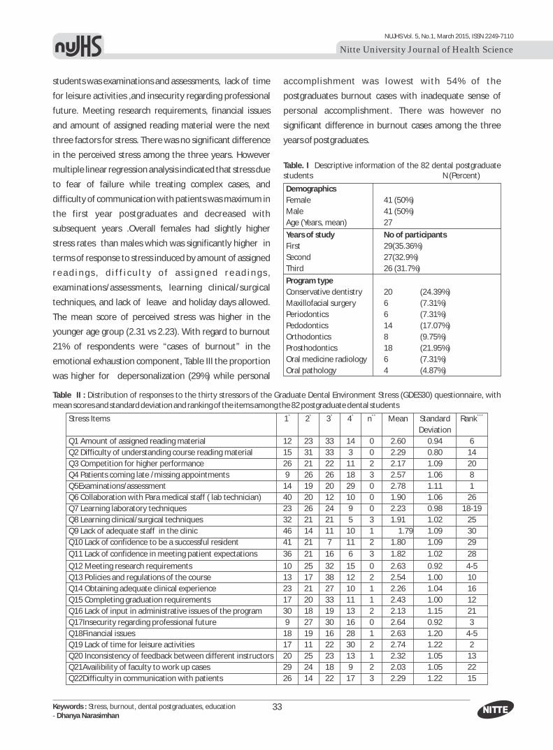
33
NUJHS Vol. 5, No.1, 2015, ISSN 2249-7110March
Nitte University Journal of Health Science
Keywords : Stress, burnout, dental postgraduates, education- Dhanya Narasimhan
students was examinations and assessments, lack of time
for leisure activities ,and insecurity regarding professional
future. Meeting research requirements, financial issues
and amount of assigned reading material were the next
three factors for stress. There was no significant difference
in the perceived stress among the three years. However
multiple linear regression analysis indicated that stress due
to fear of failure while treating complex cases, and
difficulty of communication with patients was maximum in
the first year postgraduates and decreased with
subsequent years .Overall females had slightly higher
stress rates than males which was significantly higher in
terms of response to stress induced by amount of assigned
r e a d i n g s , d i f f i c u l t y o f a s s i g n e d r e a d i n g s ,
examinations/assessments, learning clinical/surgical
techniques, and lack of leave and holiday days allowed.
The mean score of perceived stress was higher in the
younger age group (2.31 vs 2.23). With regard to burnout
21% of respondents were “cases of burnout” in the
emotional exhaustion component, Table III the proportion
was higher for depersonalization (29%) while personal
accomplishment was lowest with 54% of the
postgraduates burnout cases with inadequate sense of
personal accomplishment. There was however no
significant difference in burnout cases among the three
years of postgraduates.
Demographics
Female
Male 41 (50%)
Age (Years, mean) 27
Years of study No of participants
First 29(35.36%)
Second 27(32.9%)
Third 26 (31.7%)
Program type
Conservative dentistry 20 (24.39%)
Maxillofacial surgery 6 (7.31%)
Periodontics 6 (7.31%)
Pedodontics 14 (17.07%)
Orthodontics 8 (9.75%)
Prosthodontics 18 (21.95%)
Oral medicine radiology 6 (7.31%)
Oral pathology 4 (4.87%)
41 (50%)
Table. I Descriptive information of the 82 dental postgraduate students N (Percent)
Table II : Distribution of responses to the thirty stressors of the Graduate Dental Environment Stress (GDES30) questionnaire, with mean scores and standard deviation and ranking of the items among the 82 postgraduate dental students
* * * * ** ***Stress Items 1 2 3 4 n Mean Standard Rank
Deviation
Q1 Amount of assigned reading material 12 23 33 14 0 2.60 0.94 6
Q2 Difficulty of understanding course reading material 15 31 33 3 0 2.29 0.80 14
Q3 Competition for higher performance 26 21 22 11 2 2.17 1.09 20
Q4 Patients coming late /missing appointments 9 26 26 18 3 2.57 1.06 8
Q5Examinations/assessment 14 19 20 29 0 2.78 1.11 1
Q6 Collaboration with Para medical staff ( lab technician) 40 20 12 10 0 1.90 1.06 26
Q7 Learning laboratory techniques 23 26 24 9 0 2.23 0.98 18-19
Q8 Learning clinical/surgical techniques 32 21 21 5 3 1.91 1.02 25
Q9 Lack of adequate staff in the clinic 46 14 11 10 1 1.79 1.09 30
Q10 Lack of confidence to be a successful resident 41 21 7 11 2 1.80 1.09 29
Q11 Lack of confidence in meeting patient expectations 36 21 16 6 3 1.82 1.02 28
Q12 Meeting research requirements 10 25 32 15 0 2.63 0.92 4-5
Q13 Policies and regulations of the course 13 17 38 12 2 2.54 1.00 10
Q14 Obtaining adequate clinical experience 23 21 27 10 1 2.26 1.04 16
Q15 Completing graduation requirements 17 20 33 11 1 2.43 1.00 12
Q16 Lack of input in administrative issues of the program 30 18 19 13 2 2.13 1.15 21
Q17Insecurity regarding professional future 9 27 30 16 0 2.64 0.92 3
Q18Financial issues 18 19 16 28 1 2.63 1.20 4-5
Q19 Lack of time for leisure activities 17 11 22 30 2 2.74 1.22 2
Q20 Inconsistency of feedback between different instructors 20 25 23 13 1 2.32 1.05 13
Q21Availibility of faculty to work up cases 29 24 18 9 2 2.03 1.05 22
Q22Difficulty in communication with patients 26 14 22 17 3 2.29 1.22 15

34
NUJHS Vol. 5, No.1, 2015, ISSN 2249-7110March
Nitte University Journal of Health Science
Keywords : Stress, burnout, dental postgraduates, education- Dhanya Narasimhan
* * * * ** ***Stress Items 1 2 3 4 n Mean Standard Rank
Deviation
Q23 Lack of leave and holidays 15 25 22 18 2 2.47 1.10 11
Q24 Fear of failure when treating complex cases 15 33 24 8 2 2.25 0.95 17
Q25 Neglect for personal cases 14 24 20 22 2 2.56 1.18 9
Q26 Awareness of own competences and limitations 19 28 28 6 1 2.23 0.93 18-19
Q27 Collaboration with part time faculty 32 26 15 7 2 1.91 1.00 24
Q28 Doing case presentations to patients 28 25 19 7 3 1.98 1.03 23
Q29 Doing presentations in seminar activities 16 19 22 23 2 2.58 1.16 7
Q30 Collaboration with other specialties282918431.900.9527
* Percent calculated among non-missing responses, where 1: not stressful at all, 2: somewhat stressful, 3: quite stressful, 4: very stressful
** Number of missing responses/ not aplicable
*** Items ranked from highest to lowest mean score
Table III : Distribution of perceived stress (GDES30 mean score and standard deviation [SD]) and burnout (MBI subscale mean scores and SD, and percent of “high” or “low scorers”, or “cases”) among the entire sample, and stratified by age, gender and year of study among 72 postgraduate students
Perceived stress
GDES mean exhaustion Mean ( SD) accomplishment
Mean (SD) Mean (SD)
Overall 2.28(0.09) 17.88(11.25) 21% 8.54(8.67) 29% 29.37(10.14) 54%
Age <27 2.31(0.10) 18.14(11.11) 21% 9.31(9.30) 31% 30.55(10.73) 46%
>27 2.23(0.11) 17.40(11.71) 21% 7.08(7.29) 25% 27.16(8.69) 71%
Gender Male 2.20(0.11) 17.57(11.34) 25% 9.8(7.12) 31% 30.57(6.45) 51%
Female 2.36(0.11) 18.18(11.34) 13% 7.3(3.42) 27% 28.54(8.95) 70%
Year of study First 2.31(0.13) 18.61(13.73) 25% 10.69(10.31) 30% 29.30 (12.26) 48%
Second 2.22(0.12) 17.08(8.38) 8% 6.86(8.58) 30% 30.95(8.29) 56%
Third 2.30(0.11) 17.86(11.07) 27% 7.78(6.35) 27% 27.86(9.33) 59%
* Proportions of burnout “cases” were calculated using the thresholds used in the Maslach Burnout Inventory – high emotional exhaustion score: > 26
** High depersonalization score: > 12
*** Reduced personal accomplishment score: < 32
* ** ***Emotional % high Depersonalization % high Personal % reduced
Discussion :
The study found moderate to high levels of stress and
burnout faced by post graduates students in their three
years of training. The overall stress score being 2.28 was
higher among this study group of dental postgraduates
when compared to a similar study on Greek dental 7residents (2.1)
“Examinations and assessment” “lack of time for leisure
activities” and “insecurity regarding professional future”
were top stressors among the study group. This is not
surprising considering the meticulous and strict nature of
the course. The stress of examination and assessment can
be attributed to the competitive nature of the course, and
the fear of failing the course requirement. Previous
findings of similar studies rank “lack of leisure time” and
“completing graduation requirements” the highest 14,15,16stressors. Our data did not reveal stress and burnout
to be linked to the study year, unlike studies conducted in 9,17European dental residents. This could be explained on
two grounds. First, the timing of the survey corresponded
to almost ten months from the beginning of the academic
year. Hence, the students in the first year would have had
significant exposure to the professional environment and
its attendant mental demands including facing the taxing
clinical workload. There is a possibility that although the
course burden increases with the year, the coping skills
may also be improve with time. This could also be the
reason that burnout is consistently higher in the lower age
group (< 26years) .There is also the limitation of limited
sample size. In any case, it must be acknowledged that the
typically small size of all studies in the field, limits their

9between-subgroups inferential potential. Increased stress
of communication with patients among first year
postgraduates can be justified, because India has a multi
linguistic population .The setting of the study is a dental
college situated at the border of two states with the
population speaking various dialects. Gender has been
considered to be an important factor in influencing the
extent of psychological symptoms. The female students in
our study had a higher level of stress relative to their male
counterparts. There is higher female stress in many studies
c o n d u c t e d a m o n g d e n t a l a n d m e d i c a l 2,18,19,20,21professionals. This could be attributed to socio
cultural differences, different psychological attributes 5,22,23related to coping skills and vulnerability to life events.
Our study revealed high levels of burnout among the
postgraduates with more than 54% of the postgraduates
feeling low Personal Accomplishment and 29% feeling
burnout with Depersonalization , and 21 % Emotionally
Exhausted. This burnout is much higher than burnout
values obtained from postgraduates in European dental 9schools. The current study set up is a dental college
offering completely free dental care to all the patients
irrespective of the dental care required. This is similar to
most government dental hospitals where patient dental
care is almost always free or at minimal cost. Such a
35
NUJHS Vol. 5, No.1, 2015, ISSN 2249-7110March
Nitte University Journal of Health Science
Keywords : Stress, burnout, dental postgraduates, education- Dhanya Narasimhan
scenario leads to an immense case load which is handled by
the postgraduate dental students. A disproportion
between the numbers of patients to the post graduate
students can lead to quicker burnout, and more stress.
Limitations of the study
The limitations of our study are limitations inherent in the
methodology i.e. a chance of misinterpretation of
questions and lack of response could not be controlled. The
cross-sectional approach to interpretation of stress across
three years , the difference in the number of postgraduates
from different subjects participating in the study are
limiting factors to an accurate assessment.
Conclusion :
Our study reveals high levels of stress and burnout among
post graduate dental students, which must not be
overlooked. There is a need to come up with an effective
strategy in the postgraduate curriculum to tackle stress and
burnout .Regulating bodies for dental education should
take initiatives to provide financial security and future job
prospects for the students, policies to update the
examination systems and make it relatively less stressful
should be looked into. Addressing issues that may arise in
the educational setting will help improve the academic
environment and lead to superior professional training.
1. Atkinson TM, Millar K, Kay EJ, Blinkhorn AS. Stress in dental practice. Dent Update 1991;18:60-4
2. Lloyd C, Musser LA. Psychiatric symptoms in dental students. J Nerv Ment Dis 1989 ;177(2):61–9
3. Schmitter M, Liedl M, Beck J, Rammelsberg P. Chronic stress in medical and dental education. Med Teach 2008;30:97–99
4. Thomas N K: Resident burnout. JAMA 2004; 292: 2880–9 5. Sanders A E, Lushington K: Sources of stress for Australian dental
students. J Dent Educ 1999;63: 688–697 6. Tedesco LA.A Psychosocial perspective on the dental education
experience and student performance .JDentEdu 1986;50:601-6067. Divaris K, Lai CS, Polychronopoulou A, Eliades T, Katsaros C. Stress and
burnout among Swiss dental residents. Schweiz Monatsschr Zahnmed. 2012; 122(7-8):610-5
8. Gorter R, Freeman R, Hammen S, Murtomaa H, Blinkhorn A, Humphris G.Psychological stress and health in undergraduate dental students:fifth year outcomes compared with first year baseline results from five European dental schools. Eur J Dent Educ 2008;12:61–8
9. Humphris G, Lilley J, Kaney S, Broomfield D:Burnout and stress-related factors among junior staff of three dental hospital specialties. Br Dent J 1997;183:15–21
10. Alemany Martínez A, Berini Aytés L, Gay Escoda C. The burnout syndrome and associated personality disturbances. The study in three
Reference :
graduate programs in Dentistry at the University of Barcelona. Med Oral Patol Oral Cir Bucal. 2008;13:E444-50
11. Divaris K, Polychronopoulou A, Taoufik A, Katsaros C, Eliades T.Stress and burnout in postgraduate dental education. Eur J Dent Educ 2012;16:35–42
12. Garbee WH, Zucker SB, Selby GR. Perceived sources of stress among dental students. J Am Dent Assoc 1980;100:853-7.
13. Maslach C, Jackson S E, Leiter M P. Maslach Burnout Inventory Manual, Palo Alto, CA 3rd ed. Consulting Psychologists Press, Inc; 1996
14. Sofola OO, Jeboda SO. Perceived sources of stress in Nigerian dental students. Eur J Dent Educ 2006;10:20–3
15. Polychronopoulou A, Divaris K.Dental students'perceived sources of stress: a multi-countrystudy. J Dent Educ 2009;73:631–9
16. Alzahem AM, van der Molen HT, Alaujan AH, Schmidt H G, Zamakhshary MH. Stress amongst dental students: a systematic review. Eur J Dent Educ 2011;15:8–18
17. Polychronopoulou A, Divaris K: A longitudinal study of Greek dental students' perceived sources of stress. J Dent Educ2010;74:524–30
18. Madhan B, Rajpurohit A, Gayathri H. Mental health of postgraduate orthodontic students in India: A multi-institution survey. J Dent Educ 2012; 76(2): 200-9.
19. Naidu RS, Adams JS, Simeon D, Persad S. Sources of stress and psychological disturbance among dental stu¬dents in the West Indies.

36
NUJHS Vol. 5, No.1, 2015, ISSN 2249-7110March
Nitte University Journal of Health Science
Keywords : Stress, burnout, dental postgraduates, education- Dhanya Narasimhan
J Dent Educ 2002;66(9):1021–3020. Newbury-Birch D, Lowry RJ, Kamali F. The changing patterns of
drinking, illicit drug use, stress, anxiety, and depression in dental students in a UK dental school: a longitudinal study. Br Dent J 2002;192(11):646–9.
21. Pau A, Rowland ML, Naidoo S, AbdulKadir R, Makrynika E, Moraru R, Huang B, Croucher R.. Emotional intelligence and perceived stress in
dental undergraduates: a multinational survey. J Dent Educ 2007;71(2):197–99
22. Schéle I, Hedman L, Hammarström A.Shared ambiguity but different experiences and demands among dental students – a gender perspective. Qualitative Research in Psychology 2011;8(1):1–25
23. Polychronopoulou A, Divaris K: Perceived sources of stress among Greek dental students. J Dent Educ 2005; 69: 687–92

37
NUJHS Vol. 5, No.1, 2015, ISSN 2249-7110March
Nitte University Journal of Health Science
Keywords : Attitude, alcohol consumption, youngsters,harmful effects - Rashmi Kundapur
Introduction :
According to various studies conducted the response of the
adolescent towards alcohol is different. Attitudes ranged
from acceptance that is to "easy to get" and is "just going to
happen, "to personal responsibilities, it's your choice not
to drink peer pressure; you end up drinking to be part of
group".
Generally they spoke of alcohol as a fact of life and part of
g r o w i n g u p . I t i s
glamorized and made to
"look exciting" and "cool"
in advertising and is
ub iqu i tous at soc ia l
events. More than half of
the students around
56.7% expect that alcohol
Original Article
Access this article online
Quick Response Code
ATTITUDE OF COLLEGE STUDENTS TOWARDS ALCOHOL CONSUMPTION IN MANGALORE
1 1 1 1 1Deepak Daniel , Devishri Shetty , Greeshma Jilson Jose , Haritha J , Jeevan Ravi ,1 1 2 3
Lakshmi S Pillai , Anupa Neghandi , Santhosh & Rashmi Kundapur1 2 3Students, Medico Social Worker, Associate Professor, Department of Community Medicine, K. S Hegde Medical
Academy, Nitte university, Mangalore, Karnataka, India.
Correspondence:Rashmi Kundapur,
Department of Community Medicine,K S Hegde Medical Academy, Nitte University, Mangalore - 575 018, Karnataka, India.
Mobile : +91 98804 96567 E-mail : [email protected]
Abstract :
Introduction : According to various studies conducted, the response of the adolescent towards alcohol is different. Attitudes ranged
from acceptance that is to "easy to get" and is "just going to happen, "to personal responsibilities, it's your choice not to drink peer
pressure; you end up drinking to be part of group”.
Objectives : To assess the attitude of youngsters towards alcohol consumption.
Materials and methods : A survey was performed in colleges under NITTE University. Students present at the time of visit were covered.
A questionnaire was prepared comprising of a set of 14 questions covering the 3 main objectives of the survey.
Results : As per our survey of the 1150 students, 26.4% (304) students consume alcohol. Of these who consume alcohol, 188 (61.84%)
students felt it was safe to drink. Also a majority of 219 students (72.03%) did not want to stop their drinking habits and 225 (74.01%)
students haven't tried quitting. Surprisingly, a good 1050 (91.30%) out of 1150 students responded that they were aware of the harmful
effects of alcohol consumption.
Conclusion : We have concluded that the students are well informed and they know the ill effects of alcohol consumption even though,
most of them consider it as a part of life style.
Keywords : Attitude, alcohol consumption, youngsters, harmful effects
will relax them, 54.5% think that they will be more open
and friendly.46.7% think that they will target problems if 1they drink. Many youngsters gave 3 most common reasons
provided for why they started drinking were:- That friends
did it and look like fun, A desire to experiment and see what
it was like, Following the examples of family members and
relatives
Many youngsters consume alcohol on a trail and error
basis. Early teens and junior high were characterized as
they have experimented on drinking. They consume
alcohol from friends and peer who would often buy alcohol
for them.
The studies have also shown that exposure to alcohol
advertising effects, youth's attitudes about alcohols role in
society.

38
NUJHS Vol. 5, No.1, 2015, ISSN 2249-7110March
Nitte University Journal of Health Science
Keywords : Attitude, alcohol consumption, youngsters,harmful effects - Rashmi Kundapur
Among elementary school children, exposure to beer
commercials is related to beliefs about beer drinking and
expectation to drink as adult. Most adolescents respond
positively to television advertisements for alcohol and do
not think the advertisements are boring. Many like adds
that, Spotlight attractive model, Make drinking look like fun
Are in attractive, exotic settings, Make the product look 2good, Are humorous
The effect of religion on the attitude of youth towards
alcohol is also seen. It showed that students with no
religious affiliation reported significantly high level of
drinking frequency and quantity than those who are.
Regular attendance at religious services is linked to healthy,
stable family life and well behaved children.
Very few youngsters also showed following attitude
towards alcohol. Alcohol influences traffic and results in
road accidents, family problem and relationship violence 3and crime, health and financial problems Nearly half of
them think that youth can be diverted to sports and leisure
activities. They also think that they need more health
information
Many think of joining various programmes against alcohol
in youth.
Objectives:
To assess the attitude of youngsters towards alcohol
consumption.
Material and Methods:
A survey was performed in colleges under NITTE University.
Students present at the time of visit were covered. A
questionnaire was prepared comprising of a set of 14
questions covering the 3 main objectives of the survey. The
students were asked to fill the questionnaire irrespective of
their gender or whether they consume alcohol or not. The
sample size used was 1150 students and the sampling
method used is universal sampling. The responses were
calculated & tabulated. Also statistical analysis was done
using proportions to know the prevalence, gender
comparison, effects & attitude with regard to alcohol
consumption.
Operational definitions:
1. ABUSE: It is called abuse when the consumer fails to
fulfill role obligations at work, school or home.
Physically hazardous situations to the consumers.
They might have legal problems. Continued use
alcohol despite serious social and interpersonal
problems.
2. DEPENDENCE: Consumers have tolerance,
withdrawal symptoms, persistent desire to cut down
drinking. Great amount of time of consumers is spent
with activity related to alcohol. Social, occupational or
recreational activities are given up by dependents.
Continued use despite of knowledge of serious social,
psychological and physical problems.
3. Social Drinkers: This type of consumers drink slowly,
know when to stop, does not drink to get drunk, never
drives after drinking, they respect non-drinkers also
knows and obeys laws related to drinking.
4. Binge Drinking: Consumption of five or more drinks at
a single sitting for a man and four drinks at a single sitting for a woman.
5. Ever user: The respondent, who accepts having taken one or more mentioned substances ever in life.
6. Regular user: The respondent, who accepts having
used one or more mentioned substances during past
one year and has been taking it at least once a week or
several times in the previous month.
Result :
As per our survey of the 1150 students, 26.4% (304)
students consume alcohol. Of these who consume alcohol,
188 (61.84%) students felt it was safe to drink. Also a
majority of 219 students (72.03%) did not want to stop
their drinking habits and 225 (74.01%) students haven't
tried quitting. Out of 304 students, only 82 (26.97%)
students agreed to drive after drinking. Surprisingly, a good
1050 (91.30%) out of 1150 students responded that they
were aware of the harmful effects of alcohol consumption.
Around 13 (1.13%) students did not respond to the
question.(Table-1)

When asked about their attitude towards alcohol
consumption, out of the 1150 students, most of the
students believed that alcohol is a 'part of the lifestyle' (895
students; 294.4%) followed by 'social evil' (586 students;
192.7%) and a 'party element' (491students; 161.5%). Also
242 students (79.6%) believed that alcohol is used for
pleasure and 241 students (79.27%) felt that it is a stress
buster. A minority of just 211 students (69.4%) felt that
alcohol is used as an emotional companion.(Fig-1)
39
NUJHS Vol. 5, No.1, 2015, ISSN 2249-7110March
Nitte University Journal of Health Science
Keywords : Attitude, alcohol consumption, youngsters,harmful effects - Rashmi Kundapur
Discussion :
More than quarter of those consuming alcohol admit that
they drive after drinking and most of them are involved in
Reference :1. Attitude on alcohol and drinking pattern on youth by Killibarda B.et.al .
page 67,68. 2. Child and youth drinking novascotia health promotion and protection
by T.schrans,DLT Schellnek,ZonYi page (iii)3. Positive future by Simon Talbot and Prof.Tim Crabbe, Substance page2 4. Awareness and practices of road safety measures among
undergraduate medical students in a South Indian state Awareness and practices of road safety measures among undergraduate medical students in a South Indian state V Kulkarni et al. J Forensic Leg Med 2013 may
5. J Forensic Leg Med 2013 may
ATTITUDE TOWARDS
ALCOHOL RESPONSE
CONSUMPTION
Drive after drinking 82(26.97%) 218(71.7%) 4(1.3%)
Considers safe to drink 188(61.84%) 116(38.1%) 0(0%)
Wants to stop drinking 83(27.3%) 219(72.03%) 2(0.65%)
Tried quitting 74(24.34%) 225(74.01%) 5(1.64%)
Aware of harmful
effects 1050(91.30%) 87(7.56%) 13(1.13%)
YES NO NO
Table-1 Showing the attitude of college students about alcoholconsumption
Figure -1 Showing attitude of youngsters towards alcoholconsumption
77.82%
21.04%
50.95%
20.95%
18.34%
42.69% part of life style
pleasure
social evil
stress buster
emotional companion
party element
RTAs or other legal issues. Study conducted by V Kulkarni
et.al. Provides similar results of drunken driving among 4undergraduate students .Vast majority of people
consuming alcohol believe that it is a safe practice. More
than quarter of this group states that they want to stop
drinking and most of them have tried quitting. More than
90% of the total population studies are well aware of the
harmful effects of alcohol. A small proportion of those who
consume alcohol are also aware of its ill effects. The level of
awareness among developed nations, according to their
studies, is higher than that of our population. Studies have
shown that students who see their friends drinking on
various public portals also get influenced into drinking and
smoking. Hence portals like face book and orkut are also
becoming a new recent for the increasing alcohol trends in
youngsters. It proves itself to be an indirect form of peer
pressure where the students indulge themselves in alcohol
habits.
Conclusion :
After assessing the result we can conclude that the
students are well informed and they know the ill effects of
alcohol consumption even though, most of them consider
it as a part of life style.
Acknowledgments :
We sincerely thank the management of all colleges under
NITTE University to grant us permission to carry out this
survey. We thank Dr. Uday Kiran, Head of Department,
Community Medicine, KSHEMA and his staff for their
indispensable support.

40
NUJHS Vol. 5, No.1, 2015, ISSN 2249-7110March
Nitte University Journal of Health Science
Keywords : Superdisintegrants, promethazine HCl, sodium starchglycolate, croscarmellose, crospovidone - D S Sandeep
Introduction :
The tablet is the most widely used dosage form because of
its convenience in terms of self administration,
compactness and ease in manufacturing. However,
geriatric and pediatric patients experience difficulty in
swallowing conventional tablets, which leads to poor
patient compliance. To overcome this problem, scientists
have developed innovative drug delivery systems known as
Orally Disintegrating Tablets (ODT). These are novel types 1of tablets that disintegrate/disperse/dissolve in saliva .
In order to formulate ODT we need special agents called as
superdisintegrants. A disintegrant is a substance in a tablet
formulation that enables
the tablet to break up into
smaller fragments upon
c o n t a c t w i t h
gastrointestinal fluids.
Superdisintegrants are
used at a low level in the
s o l i d d o s a g e f o r m ,
Original Article
Access this article online
Quick Response Code
COMPARATIVE STUDY OF SUPERDISINTEGRANTS USING ANTIEMETIC DRUG AS A MODEL
1 2 3 D S Sandeep , R Narayana Charyulu & Prashant Nayak
1,3 2 Assistant Professors, Professor & HOD, Department of Pharmaceutics, Nitte Gulabhi Shetty Memorial Institute of Pharmaceutical Sciences, Nitte University, Paneer, Mangalore, Karnataka, India.
Correspondence:D S Sandeep
Assistant Professor, Department of Pharmaceutics, Nitte Gulabhi Shetty Memorial Institute of Pharmaceutical Sciences,Nitte University Mangalore - 575 018, Karnataka, India.
Mobile : +91 90367 17475 E-mail : sandypharama@gmail Abstract :
In the present investigation comparison of three different superdisintegrants was carried out by formulating orally disintegrating
tablets. Promethazine HCl was used as model drug which is an antiemetic drug. Sodium starch glycolate, croscarmellose and
crospovidone were selected as superdisintegrants and each one was used in three different concentrations (2%, 3.5% and 5%). The
drug-polymer compatibility was ruled out by FTIR studies. A total of nine formulations (PF1-PF9) were made by direct compression. All
prepared formulations were evaluated for weight variation, hardness, friability, drug content, disintegration time, wetting time and in
vitro drug release parameters. The results of the evaluation parameters for all the nine formulations of promethazine HCl were within
the standard limits. The in vitro drug release for promethazine HCl tablets of all the formulations (PF1-PF9) was carried out using
phosphate buffer pH 6.8 as dissolution medium. Among all the formulations the tablets formulated with crospovidone (PF7-PF9) have
shown 91.43 - 98.43% (maximum) drug release at the end of 10 min than sodium starch glycolate and croscarmellose, hence from the
present work, it concluded that among three superdisintegrants crospovidone is the ideal superdisintegrant for formulating oral
disintegrating tablets for promethazine HCl.
Keywords : Superdisintegrants, promethazine HCl, sodium starch glycolate, croscarmellose, crospovidone
typically 1–10% by weight relative to the total weight of the
dosage unit. Examples of some superdisintegrants are
croscarmellose, crospovidone and sodium starch glycolate.
Microcrystall ine cellulose and low substituted
hydroxypropylcellulose were used as disintegrating agents
in the range of 8:2 – 9:1 to prepare fast dissolving tablet.
Agar powder is used as disintegrant for the development of
rapidly disintegration tablets by enhancing the porosity of
agar by water treatment. Sodium starch gycolate,
crospovidone and croscarmellose are some of the popular
superdisintegrants. Superdisintegrants can act by 4
mechanisms namely swelling, wicking, repulsive force and 2deformation .
Ideal properties of superdisintegrants
Good Compressibility and Flow Properties
If the powders have 12-16% compressibility, they are said
to have good flow powders. Crospovidones are
s i g n i f i ca nt l y m o re co m p re s s i b l e t h a n o t h e r
superdisintegrants.

41
NUJHS Vol. 5, No.1, 2015, ISSN 2249-7110March
Nitte University Journal of Health Science
Keywords : Superdisintegrants, promethazine HCl, sodium starchglycolate, croscarmellose, crospovidone - D S Sandeep
Poor Solubility
The solubility of the major component in a tablet
formulation can affect both the rate and the mechanism of
tablet disintegration. Water soluble materials tend to
dissolve rather than disintegrate, while insoluble materials
generally produce rapidly disintegrating tablets.
Poor Gel Formation Capacity
Gels can delay dissolution as the drug must first diffuse
through the gel layer before being released into the body.
Sodium starch glycolate is used as superdisintegrant in
tablet formulation at a concentration of 4-6%.
Good Hydration Capacity
Drugs or other excipients, which are hydrophobic and
could be adsorbed on disintegrant surfaces, advertently
influence the extent of hydration and the effectiveness of
these disintegrants. Addition of fast disintegrants of high
hydration capacity is reported to minimize this problem, 3and therefore, enhance dissolution .
Complexation
Anionic disintegrants like croscarmellose sodium and
sodium starch glycolate may complex with cationic drug
actives and slow dissolution. Crospovidone a non-ionic
polymer does not interact with cationic drug actives to
retard drug release. The effects of superdisintegrants like
croscarmellose sodium, sodium starch glycolate and
polyplasdone XL on the dissolution behavior of several
cationic drugs with varying water solubility reports that
polyplasdone XL had a more rapid dissolution rate for the
model cationic drugs, irrespective of their aqueous 4solubilities .
Materials and Methods
Promethazine HCl was obtained as gift sample from Mayer
Healthcare Pharmaceuticals, Bangalore. Microcrystalline
cellulose was obtained from SD fine chemicals. Sodium
starch glycolate, croscarmellose and crospovidone were
obtained from Shreeji chemicals, Mumbai. Talc,
magnesium stearate was obtained from SD fine chemicals.
Aspartame, raspberry flavor were obtained from SD fine
chemicals.
Methods
In the present investigation direct compression method
was employed for the formulation of orally disintegrating
tablets of promethazine HCl with three different
superdisintegrants in different concentrations (2%, 3.5% &
5%) for their comparative study. promethazine HCl tablets
are available in 25 mg and 50 mg doses in the market. Dose
of 25 mg is selected for the present study. Microcrystalline
cellulose was used as diluent, talc was used as glidant,
magnesium stearate as lubricant, aspartame was used as
sweetening agent and raspberry flavor was added to
improve taste of tablets. The drug and the excipients were
passed through #60-sieve. Weighed amount of drug and
excipients except magnesium stearate were mixed in a
mortar-pestle by geometric addition method for 20 min.
The blend was then lubricated by further mixing with
magnesium stearate. The mixture blend was subjected for
drying to remove the moisture content at 40 to 45 ºC, the
mixture was blended with flavor and the powder blend was
then compressed on 10 station rotary punching machine
(Rimek RSB-4) using 8 mm round shaped punches. A total
of nine formulations were prepared by direct compression
method, which is shown in Table 1. The tablets of all the
nine formulations were subjected for evaluation.
Evaluation of tablets
Thickness: The thickness of tablets was determined by
using digital caliper (Coolant proof IP 65). The tablet is
placed in between the two jaws of caliper scale and the
reading was noted down. Three trials for each formulation 5were carried out .
Hardness: The tablet hardness, which is the force required
to break a tablet was measured by using Pfizer hardness
tester (S 14). Tablet is squeezed by two jaws. The first
machines continually applied force with a spring and screw
thread until the tablet started to break. When the tablet
fractured, the hardness was read with a sliding scale. Three trials for each formulation were performed. The limit of
2hardness is 3-6 kg/cm .
Friability: Friability is the loss of weight of tablet in
container/package due to removal of fine particles from

42
NUJHS Vol. 5, No.1, 2015, ISSN 2249-7110March
Nitte University Journal of Health Science
Keywords : Superdisintegrants, promethazine HCl, sodium starchglycolate, croscarmellose, crospovidone - D S Sandeep
surface. This test is performed to ensure the ability of
tablets to withstand shocks during processing, handling,
transportation and shipment. The friability of tablets was
determined using Roche friabilator (EF-2 USP). Ten tablets
were initially weighed and transferred into the friabilator.
The friabilator was operated at 25 rpm for four minutes.
After four minutes the tablets were weighed again. The
percentage friability of tablets was calculated using the
following formula. The standard limit of friability is not 6more than 1 % .
% Friability X 100initial weight -
initial weight
final weight
% deviation X 100individual average weight
Average weight
weight -
Weight variation: Twenty tablets were weighed
individually and all together. Average weight was
calculated from the total weight of all tablets. The
individual weights were compared with the average
weight. The percentage difference in the weight variation
should be within the permissible limits (±7.5%). The
percentage deviation can be calculated using following
formula.
Drug content: The drug content was estimated to know the
percentage of drug present in the tablet. Twenty tablets
were weighed and powdered. An amount of powder
equivalent to 150 mg of promethazine HCl was dissolved in
100 ml of pH 6.8 phosphate buffer, filtered, diluted suitably
and analyzed for drug content at 249.60 nm using UV-7Visible spectrophotometer (Shimadzu UV-1700) .
In vitro dispersion time: This test was performed for the
ability of tablets to breakdown into small fragments in fluid.
In vitro dispersion time was carried out by dropping a tablet
in a Petri dish containing 10 ml of saliva fluid (pH 6.8). The
time required for the tablet to disintegrate completely in
the fluid was noted down. Three trials for each formulation 8were carried out .
Wetting time: Wetting time of tablet is related to contact
angle. Wetting time is another important parameter, which
needs to be assessed to give an insight into the
disintegration properties of tablets. Lower wetting time
implies quicker disintegration of tablets. Wetting time of
tablet can be carried out using a piece of tissue paper
folded twice was placed in a small Petri dish (ID=6.5cm)
containing 6 ml of simulated saliva pH 6.8, a tablet was put
on the paper, and the time for complete wetting was
measured which gives wetting time of tablet. Three trials 8for each formulation were performed .
In vitro drug release: In vitro drug release of the samples
was carried out using USP – type II dissolution apparatus
(TDT 08L paddle type). The dissolution medium, 900 ml of
phosphate buffer (pH 6.8) solution, was placed into the
dissolution flask maintaining the temperature of 37±0.5 ºC
at 50 rpm. One tablet was placed in each flask of dissolution
apparatus. The apparatus was allowed to run for 10 min.
Samples measuring 5 ml were withdrawn at an interval of
2, 4, 6, 8 and 10 min. Samples were filtered through 10 µm
filter. Same volume of the fresh dissolution medium was
replaced every time. The collected samples were suitable
diluted and analyzed at 249.60 nm by UV-Visible
spectrophotometer (Shimadzu UV-1700) using dissolution
medium as blank. The cumulative percentage drug release 9was calculated .
Results and Discussion :
The results of evaluation parameters for the nine
formulations are shown in Table 2 & 3. The results of in vitro
drug release of tablets for all the nine formulations are
shown in Table 4, 5 and 6.
The thickness of the tablet indicates that die fill was
uniform. The thickness depends upon the size of the punch
(8 mm) and the weight of the tablet (150 mg). The thickness
of tablets from batch PF1-PF9 was found to be 2.50 - 2.86 2mm and hardness was found to be 3.1 - 4.2 kg/cm . The
friability of all the formulated tablets of promethazine HCl
was found to be between 0.45 - 0.72 % and all the
formulated tablets of promethazine HCl were shown the
friability within the official limits. The weight variation for
the tablets of all the (PF1-PF9) formulations was within the
standard limits (±7.5%). All the formulated tablets (PF1-
PF9) have shown in vitro dispersion time of less than 60 sec.

SR.
No. Pf1 Pf2 PF3
1 0 0 0 0
2 2 31.23±0.65 35.87±0.21 38.67±0.96
3 4 37.98±1.12 42.87±1.12 43.12±0.54
4 6 46.80±1.23 48.40±0.76 57.54±0.43
5 8 52.54±0.76 56.65±0.76 65.78±0.54
6 10 58.85±0.98 63.87±0.75 72.78±1.10
Time (min) % Cumulative drug release
SR.
No. Pf4 Pf5 PF6
1 0 0 0 0
2 2 32.67±0.23 36.19±0.34 35.28±0.87
3 4 43.12±0.67 44.78±1.45 47.32±1.15
4 6 57.54±0.65 59.63±1.00 60.22±0.37
5 8 65.78±0.78 68.20±1.32 73.73±0.62
6 10 72.78±1.15 74.87±0.78 82.65±0.47
Time (min) % Cumulative drug release
SR.
No. Pf7 Pf8 PF8
1 0 0 0 0
2 2 42.76±0.76 45.72±0.73 47.98±1.00
3 4 55.87±0.43 59.98±0.54 62.12±0.62
4 6 72.50±1.32 75.65±0.32 79.43±0.91
5 8 84.92±1.12 89.64±0.65 88.90±0.47
6 10 91.43±0.63 94.34±1.34 98.43±1.27
Time (min) % Cumulative drug release
43
NUJHS Vol. 5, No.1, 2015, ISSN 2249-7110March
Nitte University Journal of Health Science
Keywords : Superdisintegrants, promethazine HCl, sodium starchglycolate, croscarmellose, crospovidone - D S Sandeep
Among all the formulations, tablets prepared with
crospovidone were shown less than 40 sec of dispersion
time. The wetting time of all the formulations (PF1-PF9) are
found to be within 39.30-68.33 sec which complies with
the official limits. The drug content of all the nine
formulations of promethazine HCl tablets was found to be
within the range of 96.78-99.71% which was within the
limits of IP specifications. The formulations PF1-PF3 were
formulated with the help of sodium starch glycolate in
concentration 2%, 3.5% and 5% respectively. The
formulations PF4-PF6 were formulated with the help of
croscarmellose in concentration 2%, 3.5% and 5% and the
formulations PF7-PF9 were formulated with the help of
crospovidone in concentrations 2%, 3.5% and 5%
respectively. The formulations PF7-PF9 containing
crospovidone shown 91.43-98.43% drug release which was
the highest drug release compared to all the other
formulations. The drug release profile for all the nine
formulations are shown in Figure 1, 2 and 3.
Table 1: Formulation design of promethazine HCl orally disintegrating tablets
Ingredients(mg) Pf1 Pf2 Pf3 Pf4 Pf5 Pf6 Pf7 Pf8 PF9
Promethazine HCl 25 25 25 25 25 25 25 25 25
SSG 3 5.25 7.5 - - - - - -
Croscarmellose - - - 3 5.25 7.5 - - -
Crospovidone - - - - - - 3 5.25 7.5
Aspartame 5 5 5 5 5 5 5 5 5
Raspberry flavour 3 3 3 3 3 3 3 3 3
Talc 10 10 10 10 10 10 10 10 10
Magnesium state 3 3 3 3 3 3 3 3 3
MCC(q.s) 150 150 150 150 150 150 150 150 150
Table 2 : Results of thickness, hardness, friability and weight variation of promethazine HCl tablets
Formulation *Thickness *Hardness Friability Weight 2code (mm) (kg/cm ) (%) variation
Pf1 2.62±0.01 3.7±0.38 0.51 149.10±0.20
PF2 2.63±0.07 3.4±0.33 0.48 151.09±0.33
PF3 2.66±0.02 3.4±0.65 0.45 150.19±0.21
PF4 2.63±0.05 4.2±0.25 0.72 150.33±1.76
PF5 2.52±0.01 3.8±0.31 0.70 148.80±1.03
PF6 2.53±0.05 3.8±0.72 0.67 150.33±2.12
PF7 2.51±0.05 3.2±0.22 0.64 149.60±1.28
PF8 2.50 ±0.05 3.1±0.30 0.60 150.43±1.71
Pf9 2.65 ±0.03 3.8±0.38 0.54 151.67±1.27
*Value expressed as mean ±SD, n=3
Table 3 : Results of in vitro dispersion time, wetting time and water absorption ratio of promethazine HCl tablets
Formulation *In vitro *Wetting % Drug
code dispersion time (sec) content
time (sec)
PF1 58.00±4.13 68.33±3.51 98.21±0.66
PF2 55.33±4.16 64.13±3.81 98.21±0.66
PF3 40.33±3.18 48.12±3.21 96.78±0.86
PF4 54.21±3.10 60.65±3.00 98.55±0.76
PF5 48.11±4.10 54.00±4.00 98.73±1.10
PF6 45.33±3.11 50.33±3.11 98.22±0.38
PF7 38.00±1.98 50.21±3.05 99.28±0.28
PF8 27.66±2.51 44.00±2.31 98.91±0.81
PF9 18.34±1.15 39.30±1.54 99.71±0.16
*Value expressed as mean ±SD, n=3
Table 4 : In vitro drug release data of promethazine HCl tablets formulated with sodium starch glycolate
Table 5 : In vitro drug release data of promethazine HCl tablets formulated with croscarmellose
Table 6 : In vitro drug release data of promethazine HCl tablets formulated with crospovidone

44
NUJHS Vol. 5, No.1, 2015, ISSN 2249-7110March
Nitte University Journal of Health Science
Keywords : Superdisintegrants, promethazine HCl, sodium starchglycolate, croscarmellose, crospovidone - D S Sandeep
Figure 1: In vitro drug release profile of promethazine HCl tablets formulated with sodium starch glycolate
Figure 2 : In vitro drug release profile of promethazine HCl tablets formulated with crosscaremelose
Figure 3 : In vitro drug release profile of promethazine HCl tablets formulated with crospovidone
Conclusion :
A comparative study of three superdisintegrants (sodium
starch glycolate, croscarmellose and crospovidone) was
carried out using promethazine HCl as model drug by
formulating nine batches (PF1-PF9) by direct compression
method. The tablets were evaluated for parameters like
thickness, hardness, friability, in vitro dispersion time,
wetting time and percentage drug content. All the
evaluation parameters of nine formulations were found to
be within the IP limits. All the formulated tablets were
examined for in vitro drug release studies. Among the
three superdisintegrants, crospovidone showed maximum
percentage drug release and hence it was found to be the
ideal superdisintegrant for the formulation of
promethazine HCl orally disingrating tablets.
References :1. Biradar SS, Bhagavati ST, Kuppasad IJ. Fast dissolving drug delivery
systems: A brief overview. Int J Pharm 2006; 4(2):62-68.2. Gordon MS, Rudraraju VS, Dani K. Effect of the mode of
superdisintegrants incorporation on dissolution in wet granulated tablets. J Pharm Sci. 1993; 82: 220-226.
3. Priyanka S, Vandana S. A review article on: superdisintegrants. Int J Drug Res. Tech. 2013, 3 (4):76-87.
4. John C, Carter H, Benson TR. The role of disintegrants in solid oral dosages. Int J Pharm 2006; 1(3):231-236.
5. Quadir A, Kolter K, Robin SD. A comparative study of current superdisintegrants. Int J Pharm Sci 2006; 1(4):45-50.
6. Takao M, Mizumato R, Joseph L. Formulation design of a novel fast disintegrating tablet. Int J Pharma 2005; 3:83-90.
7. Sumiya K, Baba Y, Inomata S. Preparation and evaluation of orally disintegrating clonidine hydrochloride tablets Eur J Pharm Sci 2000; 1(7):652-666.
8. Augsburger LL, Kornblum S. Functionality comparison of 3 classes of superdisintegrants in promoting aspirin tablets disintegration and dissolution. AAPS Pharm Sci Tech 2005; 6(4):63-69.
9. Chowdary KP, Hemavathi R, Jaiswal PK. Formulation and dissolution rate studies on dispersible tablets of ibuprofen. Ind J Pharm Sci 2000; 62(3):213-216.

45
NUJHS Vol. 5, No.1, 2015, ISSN 2249-7110March
Nitte University Journal of Health Science
Keywords : Low Back Pain, Neck Pain, Dentistry, Paedodontist- Anoop Hegde
Introduction :
Locomotor system disorders are frequently seen in (1)dentistry . It is known that the most painful regions arethe
(2,3)cervical and lumbar spine . Factors associated
with professional work may predispose to back and
neckpain. On account of the narrow visual field of the
oralcavity, having to work with a limited scope of
movementconstitutes high risks for lowback and neck pain (4,). It has
been demonstrated that tensely mainta ined
asymmetricbody posture is a risk for lowback pain (LBP);
andprolonged static neck position and repeated
movements
are work-related risk (5,6)factors for neck pain .In
the light of these findings,
the aim of this study, isto
investigate the risk factors
associated with lowback
and neck pain in dentistry.
Original Article
Access this article online
Quick Response Code
PREVALENCE OF NECK AND BACK PAIN AMONG PAEDIATRIC DENTISTS
1 2 3 4Siddharth M Shetty , Shreema Shetty , Anoop Hegde &Nirmal Babu
1 3 4Associate Professor, Post Graduate, Senior Resident, Department of Orthopaedics, K.S. Hegde Medical Academy, 2Assistant Professor, Department of Conservative Dentistry & Endodontics, A.B. Shetty Memorial Institute of Dental
Sciences, Nitte University Mangalore, Karnataka, India.
Correspondence :Anoop Hegde
Post Graduate, Department of Orthopaedics, K.S. Hegde Medical Academy, Nitte University Mangalore - 575 018, Karnataka, India.Mobile : +91 99641 43600 E-mail : [email protected]
Abstract :
Occupational diseases are present worldwide. Dentists believe that they are at a higher risk for development of musculoskeletal
disorders due to the postures attained at work. Hence, we conducted a study for understanding the prevalence of such ailments
amongst the paedodontist population. We employed a cross-sectional study of 270 paedodontists who were selected at random and
were asked to complete a self-administered questionnaire. The questions were about personal characteristics, job history, specific work
habits and mostly pertaining to clinical dentistry with details of any recent occurrence of neck or back pain. Results tabulated showed a
79.6 % of the paedodontists reported having experienced at least one episode of neck or back pain in the immediate past 12 months.
This value is way above the occurrence of similar complaints seen among the general population which is estimated to be around 55%.
The study concluded indicating that the incidence of neck and back pain among dentists is higher than general population. This may be
attributed to extreme postures that may be attained during the clinical work and which may be extreme in paediatric dentists.
Keywords: Low back pain, neck pain, dentistry, Paedodontist
Materials and Methods :
Using a simple random sampling method, 270 dentists
were selected and asked to complete a self-administered
questionnaire, 240 dentists completed and returned the
questionnaire. The questions were about age, gender, job
history, work characteristics mostly pertaining to dentistry
including physical risk factors at work plus any report about
the occurrence of low back pain (LBP) and neck pain, place
and duration of employment, number of patients visited
per month, time and duration of work per day and the
posture of body while working,
Part of the questionnaire was allotted to lower back pain
and/or neck pain and included questions about the same.
These included duration of musculoskeletal complaint;
complaints in the upper or lower limb (e.g., feeling pain,
paresthesia, and numbness). The participants were also
asked if they received any treatments. Their responses
were categorized as either “no treatment,” “drug,”
“exercise,” or “physiotherapy.”
Data were anonymously coded and entered into

46
NUJHS Vol. 5, No.1, 2015, ISSN 2249-7110March
Nitte University Journal of Health Science
Keywords : Low Back Pain, Neck Pain, Dentistry, Paedodontist- Anoop Hegde
aspreadsheet programme before being analysed using
theSPSS software.
12 Basicstatistics were calculated, including prevalence
rates.Differences Prevalence of neck and back pain
werecalculated usingthe chi-square test for categorical
variables and by thestudent t-test for continuous variables.
p-values below0.05 were considered statistically
significant throughout.
Results :
Profile of respondents
Of the 270 dentists who answered the questionnaire:
Two hundred and forty questionnaires (88.8 per cent) were
returned, fully or partiallycompleted.Missing data were
excluded from the analysis. It was noted that:
1) 44.6%were male and 55.4% were female (Table 1).
2) The age group under the study ranged from 21-62 years.
Mean age was 28.4 years (SD = 5.94years) (Table 2).
3) The years of work under the study ranged from 2-30
years. Mean being 3.440 (SD=4.8453) (Table 2)
4) The hours of work in a day ranged from 1-12 hours.Mean
being 4.765(SD=1.7247) (Table 2)
5) Most dentists (80 per cent) reported having at least one
MSD symptom in the past 12 months (Fig 1).
6) Pain in the spine was significantly more likely to be
reported by younger dentists (p<0.001) and dentists
with less years of experience.
7) Lower back pain, which interfered with daily activity,
was significantly more likely to be reported by dentists
who worked shorter hours (p<0.05).
8) 19% of dentists with pain underwent physiotherapy to
get rid of pain
Over one-third of all dentists (36.4 per cent) had sought
medical advice ortreatment of MSD during the previous 12
months.
Discussion :
So far, many factors for development of musculoskeletal
pain have been studied.
Table 1. Distribution Of Age Groups By Gender For Dentists Surveyed
Age Group Gender
Male Female
20-29 33.75% (81) 46.66% (112)
30-39 8.33% (20) 6.25% (15)
40-49 1.66% (4) 1.25% (3)
50-59 0.83% (2) 0.83% (2)
60-69 0.00 (0) 0.41 (1)
Total 44.6% (107) 55.4% (133)
Table 2. Mean (±Sd) Age, Sex, Experience, Working Hours Per Day,
Age Sex Years of Hours of Work
Work In A Day
N Valid 240 240 240 240
Missing 0 0 0 0
Mean 28.40 1.45 3.440 4.765
Std. Deviation 5.940 .498 4.8453 1.7247
Minimum 21 1 .2 1.0
Maximum 62 2 30.0 12.0
Figure 1
However, we studied additional variables that may cause
musculoskeletal disorders. To the best of our knowledge,
there is scarce information about the epidemiology of
musculoskeletaldisorders.
Health care work is recognised as a high riskjob for MSD;
however most of the studieshave been carried out in
specific groups ofhealthcare professionals such as dentists
a n d d e n t a l h y g i e n i s t s , n u r s e s , ra d i o l o g i s t s ,
ophthalmologists, and physiotherapists.The dental
profession howeverhas one of the highest prevalence 16, -20forMSD.
It has been proven that postures which may exert a higher
pressure on intervertebral disk as well as prolonged spinal

47
NUJHS Vol. 5, No.1, 2015, ISSN 2249-7110March
Nitte University Journal of Health Science
Keywords : Low Back Pain, Neck Pain, Dentistry, Paedodontist- Anoop Hegde
hypomobility are among important factors leading to
degenerative changes in the lumbar spine and subsequent
LBP. Since such postures are not uncommon in daily
practice of a dentist, some authors believe that they are at
a higher risk of developing musculoskeletal disorders than 7,8,14other job groups Nonetheless, our results showed that
the prevalence of LBP and neck pain in dentists is very
much higher than other study groups.
Al Wazzan, et al, in their study, reported that only 37% of
those suffering back and neck pain sought medical
treatment and concluded that these symptoms among
dental personnel are not severe enough to ask for medica-15tions.
Alice lai ,et al in their study reported that the prevalence of
self-reported MSD among dental personnel is high.
Severalwork-related factors have been identified to be
associated with musculoskeletal symptoms in varyingbody
. 21regions
One limitation of this study is the lack of objective
measurement methods.
It is also thought that individual psychologicalproperties,
such as stress intolerance, which would beexpected to
contribute to incidence and intensity of
locomotor pain needs to be included in future studies.
Conclusion:
It is understood that work duration and working postures
are root cause of back and neck pain among the
paedodontists. This study also highlights, the fact the
incidence among dentistry to be higher than general
population. We opine that the practice of dentistry is not
per se an ignition for development of neck and low back
pain, rather accelerates the process and increases the
severity of symptoms due to the working posture.
1 Szymanska J. Disorders of the musculoskeletal systemamong dentists from the aspect of ergonomics andprophylaxis. Ann Agric Environ Med 2002;9: 169-73.
2. Armstrong TJ, Lifshitz Y. Evaluation and design of jobs forcontrol of cumulative trauma disorders. In: Industrialhygenie science series: Egonomic interventions to prevent musculoskeletal injuries in industry. Cheksea: Lewis, 1987:73-87.
3. Diakow PRP, Cassidiy JD. Back pain in dentists. JManipulative Physiol Ther 1984; 7: 85-8.
4. Al Wassan KA, Almas K, Al Shethri SE, Qahtani Al. Backand neck problems among dentists and dental auxillaries.JContemp Dent Pract 2001; 2: 17-30.
5. Alexopoulos EC, Stathi IC, Charizani F. Prevalance ofmusculoskeletal disorders in dentists. BMC MusculoskeletalDisorders 2004; 5: 16.
6. Yee T, Crawford L, Harber P. Work environment of dentalhygienists. J Occup Environ Med 2005; 47: 633-9.
7. Chams H, Mohammadi SF, Moayyeri AR. Frequency and assortment of sel f -reported occupat ional complaints among Iranian ophthalmologists. Med Gen Med 2004;6(4):1.
8. Davatchi F, TehraniBanihashemi A, Gholami J, et al. The prevalence of musculoskeletal complaints in a rural area in Iran: a WHO-ILAR COPCORD study (stage 1, rural study) in Iran. ClinRheumatol2009; 28:1267-74.
9. Szymanska J. Disorders of the musculoskeletal system among dentists from the aspect of ergonomics and prophylaxis. Ann Agric Environ Med 2002;9:169-73.
10. Ratzon NZ. Musculoskeletal symptoms among dentists in relation to work posture. Work 2000;15(3):153-8.
11. Lehto TU, Helenius HY, Alaranta HT. Musculoskeletal symptoms of dentists assessed by a multidisciplinary approach. Commun Dent Oral
References :
Epidemiol1991;19(1):38-44. 12. Fish DR, Morris-Allen DM. Musculoskeletal disorders in dentists. NY
State Dent J 1998;64:44-8. 13. Rundcrantz BL. Pain and discomfort in the musculoskeletal system
among dentists. Swed Dent J Suppl1991;76:1-102. 14. Alipour A. Neck and shoulder pain: prevalence, incidence and risk
factors, the IKCO cohort study. Stockholm, Sweden. Department of Clinical Neuroscience, Section of Personal Prevention, Karolinska Institute. 2008. Available at http://diss. kib.ki.se/2008/978-91-7409-126-7/thesis.pdf (Accessed January 21, 2010).
15. Al Wazzan KA, Almas K, Al Shethri SE, Al-Qahtani MQ. Back and neck problems among dentists and dental auxillaries. J Contemp Dent Pract
16. Smith DR, Nihashi M, Adachi Y, Koga H, IshtakeT. A detailed analysis of musculoskeletal disorderrisk factors among Japanese nurses. J Safety Res2006; 37:195-200.
17. Hayes MJ, Cockrell D, Smith DR. A Systematicreview of musculoskeletal disorders among dentalprofessionals. Int J Dental Hygiene 2009; 159-65.
18. Campo M, Weiser S, Koenig KL, Nordin M. Work-related musculoskeletal disorders in physical therapists:a prospective cohort study with 1-year followup.PhysTher 2008; 88:608-19.
19. Dhimitri KC, McGwin G Jr, McNeal SF. Symptomsof musculoskeletal disorders in ophthalmologists.Am J Opthalmol 2005; 139:179-81.
20. Boiselle PM, Levine D, Horwich PJ. Repetitivestress symptoms in radiology: prevalence and responseto ergonomic interventions. J Am CollRadiol2008; 5:919-23
21. Alice laI 1, KyiOo yin 1, Shivanthiballala 2, Lay Waikhin 3, Nayakebp ballala1, Linnaing Prevalence of musculoskeletal disorders in the dental professionin Brunei Darussalam.

48
NUJHS Vol. 5, No.1, 2015, ISSN 2249-7110March
Nitte University Journal of Health Science
Keywords : Phenomenology, lived experiences, Chemotherapy, colaizzi's - Malarvizhi M
Introduction :
Cancer is a term that is used to refer malignant neoplasms.
It is a disease of the cell in which the normal mechanisms of
the control of growth and proliferation have been altered.
It is invasive, spreading directly to surrounding tissues as
well as to new sites in the body. A major concern with the
diagnosis and treatment of cancer is the multifocality and
multicentrality. The level of distress varies from women to
women and within an individual in different situation and
role.
Cancer is a disease of cells characterized by a loss of the
normal control mechanisms that maintain tissue
o r g a n i z a t i o n .
Chemotherapy is the use
of one or more chemical
agents to eradicate the
cancer cells or to stop
tumour progress (Durr and
Huff 1994)
C h e m o t h e r a p y i s a
Original Article
Access this article online
Quick Response Code
A RESEARCH CRITIQUE ON THE LIVED-IN EXPERIENCES OF PATIENTS SUBJECTED TO CHEMOTHERAPY IN SELECTED
HOSPITALS AT CHENNAI1 2
Malarvizhi M & Bhavani M1 2 Lecturer, Faculty of Nursing Research Scholar, Department of Human Genetics,
Sri Ramachandra University, Chennai, Tamilnadu, India.
Correspondence : Malarvizhi M
Lecturer, Faculty of Nursing, Department of Human Genetics, Sri Ramachandra University, Chennai 600 116, Tamilnadu, India.E-mail : [email protected]
Abstract :
This qualitative study explored the experiences of patients subjected to chemotherapy. The research approach used in this study was
phenomenology. Interviews were conducted with fifteen patients subjected to chemotherapy. The study was conducted at selected
hospitals at Chennai. From the colaizzi's analysis of the data seven dimensions-eight themes and fifty sub themes were emerged. These
findings show that patients subjected to chemotherapy has lot of physical and psychological impacts. There was an association between
the socio demographic variables and the lived in experiences of patients subjected to chemotherapy. The association was found
between the educational status, type of family and the number of chemotherapy cycle. So, the patients need adequate education on the
side effects from the Nurses during each cycle of chemotherapy. Therefore this study is important for nurses working in the cancer unit
to make the patient to realize and aware the effects faced by the patients subjected to chemotherapy.
Keywords: Phenomenology, lived experiences, Chemotherapy, colaizzi's
systemic intervention and is appropriate when the disease
is widespread, the risk of undetectable disease is high, and
the tumor cannot be resected and is resistant to radiation
therapy. The objective of chemotherapy is to destroy
malignant tumor cells without excessive destruction of
normal cells. Several types of cancers are now considered
curable with chemotherapy, even in advanced stages. The
most valuable intervention that can be offered to a patient
with cancer is presence of nurse as a caring person. Its
dimension consists of verbal expressions of empathy,
positive regard, and availability of practical support.
Chemotherapy for malignancies, too advanced for surgery,
which accounts for 80% of all cancers, is a scientific
wasteland (Dr. Ulrich Abel, 1990). Amazingly, most
patients cope well and face the difficulties in their lives with
courage. For many patients the experience brings about a
re-appraisal of goals and values, making life richer and
more meaningful. Griffin (1998) identified and ranked the
symptoms experienced among 155 cancer patients who
received chemotherapy. The findings revealed that the

49
NUJHS Vol. 5, No.1, 2015, ISSN 2249-7110March
Nitte University Journal of Health Science
Keywords : Phenomenology, lived experiences, Chemotherapy, colaizzi's - Malarvizhi M
patients reported on the experience of physical symptom
13 and 7 psychological. Nausea was reported as the most
severe symptoms followed by tiredness and loss of hair.
The study suggested that a reduction in the severity of
some symptoms experienced whi le receiv ing
chemotherapy and a shift from concerns about physical to
psychological issues.
The spiritual and existential matters often marks this phase
of illness from diagnosis and treatment because women
focus their legacy to their families, children and engaged in
planning for their future.(Butler et al 2003)
The emotional side-effects of a person diagnosed as cancer
and person who receives chemotherapy are different for
everyone. They may even change during the treatment.
Before starting chemotherapy, many patients feel anxious
about not knowing what to expect and their upcoming
treatments. Many patients said that they experienced
depression and or emotional stress before and during
chemotherapy. Emotional side-effects affects the cancer
patient's day-to-day activities. Mitchell (2007) described
the social and emotional effects of chemotherapy among
19 patients. The major themes that emerged from the data
were, striving for normality, feeling-up, feeling-down,
flagging, being sociable and anxiety, about the
chemotherapy treatment.
Chemotherapy may bring about another cycle of
emotional change, such as anger, denial, guilt, depression
and acceptance, as well as impact on other aspects of
psychosocial functioning. (Holland and Zitton 1991)
The severity of side-effects varies between individuals, the
type of drug administered, and the type of care delivered,
including information and other psychosocial
interventions. (Coates et al. 1987), Elizabeth
L.Mcgarvey(2001) explored the psychological sequel
among women with cancer. The findings revealed that they
experienced lower self-esteem, poor body image, and
lower quality of life, and affects on sexuality.
In Sri Ramachandra Hospital, an average of 15 patients per
month are admitted to the inpatient department for
chemotherapy treatment in the general wards namely E6
and E7. The investigator felt that it is very important to elicit
their lived-in experiences with regard to physical,
psychological, social, spiritual, financial and sexual
dimensions in order to support and help them to continue
with their remaining chemotherapy cycle.
Subjective experiences of individual undergoing
chemotherapy allow the practitioner to understand and
gain insight on the patient current experience and support
him/her with the use of their constructive coping
strategies. This helps the nurses to develop strategies for
the coping abilities of cancer patient.
So the investigator strongly believes that the feelings which
the patients undergo must be explored. This realization,
strongly motivated the investigator to do this research
critique on lived-in experiences of patients subjected to
chemotherapy. Through eliciting the lived-in experiences,
it is possible to identify the patients felt needs in all
dimensions which in turn will inspire the nursing care.
Nursing is a profession focused on assisting individuals,
families and communities in attaining, maintaining, and
recovering optimal health and functioning. Modern
definitions of nursing define it as a science and an art that
focuses on promoting quality of life as defined by persons
and families, throughout their life experiences from birth
to care at the end of life. So, the nurses play an important
role as a supporter, collaborator, and motivator in order to
help the patient to have an optimal health and functioning.
Materials and Methods :
Qualitative study design and phenomenological approach
was selected. An in depth interview was conducted. The
patients who had been admitted within the study period,
and who fulfilled the inclusion criteria were selected with
the sample size of 15. Inclusion Criteria were the patients
subjected to chemotherapy, Patients who were admitted
on the previous day of chemotherapy and staying in the sthospital on 1 day of chemotherapy.
Prior to data collection, the necessary permission was
obtained from the concerned authorities. Before

50
NUJHS Vol. 5, No.1, 2015, ISSN 2249-7110March
Nitte University Journal of Health Science
Keywords : Phenomenology, lived experiences, Chemotherapy, colaizzi's - Malarvizhi M
commencing the data collection the investigator got verbal
consent from the patients. 15 samples who were admitted
for chemotherapy were chosen for the study. The
procedure was explained to each patient individually,
ensuring comfort and privacy. The study was explained to
each participant and informed them that their identity will
not be revealed and confidentiality will be maintained and
will be utilized for the research purpose only. Informed oral
consent was obtained from the patients, and the
interviews were audio-taped. An in depth interview was
done on a one-to-one basis.
The total time taken for collecting the data from each
participant was 30- 45 minutes. After the completion of the
interview, each patient was given an opportunity to clarify
the doubts.
Results :
Colaizzi's data analysis framework was used to analyze the
transcripts in this study. From the analysis of the data,
seven dimensions-eight themes and fifty sub themes
emerged from the experiences of patients subjected to
chemotherapy and they were as follows: The dimensions
were physical, psychological, social, economic, spiritual
and sexual dimension and various suggestions given by the
patients and the level of satisfaction of the Nursing care as
expressed by the patients.
Discussion :
From the analysis of the data, seven dimensions-eight
themes and fifty sub themes emerged from the patients
subjected to chemotherapy.
A. Physical dimension
In physical dimension, the major themes identified were
mouth ulcer, anorexia, loss of the sense of taste , hair loss,
insomnia, tiredness and unable to perform household
activities. All the 15 patients (100%) have experienced hair
loss, mouth ulcer, insomnia and unable to perform
household activities, 14 patients (93%) verbalized that they
had anorexia and loss of the sense of taste.
“My hair is falling like a leaf that falls from the tree
during winter”
“My mouth ulcer is troubling me like anything because
of that I am not able to eat well
and to discriminate the taste of different delicious
foods”.
“At night , I am unable to sleep properly because of the
generalized weakness”.
“I am not able to carry out the household activities like
before this makes me stressful”.
The above findings were consistent with the study of
Warren (1999) who used a qualitative design on reducing
the discomfort following chemotherapy. The procedure
itself can be associated with severe anxiety and discomfort
for the patient. The author reports that the patients'
experience of discomfort during and after the procedure
can be reduced by proper explanation of the whole
ORIENTATION IDENTIFICATION EXPLOITATION RESOLUTION
DI
MENSIONS
Developing thestrategies in the form
of nursing module
Figure 1 : Conceptual Framework - Modified Peplau’s Nurse - Patient Interaction Theory
+ Introducing self+ Familiarizing self+ Developing Trust+ Establishing nearness+ Winning to have high
degree of confidenceand co operation
3 Patient is an uniqueindividual
3 Has strain at feeling of experience before, during after chemotherapy
3 Non masking of hiddenconcepts through eliciting lived - in experiences.
Physical
Psychological
Psychological
Spiritual
Financial
Sexual

51
NUJHS Vol. 5, No.1, 2015, ISSN 2249-7110March
Nitte University Journal of Health Science
Keywords : Phenomenology, lived experiences, Chemotherapy, colaizzi's - Malarvizhi M
treatment process before starting the procedure.
B. Psychological dimension
The identified themes were anger, fear, feeling ashamed,
disturbed body image and feeling of happiness.15 patients
(100%) felt ashamed of themselves because of their
altered body image. 10 patients (67%) were anxious and
worried about their illness and they got irritated often.13
patients(87%) felt happy about the treatment.
“I get angry and irritated often”.
“Everyday I am worried about my children”.
“I am much troubled, that there is nobody to whom I
can express my grief”.
“I have a shameful feeling that my hair is lost”.
“I feel that I have lost my self-esteem and body image
when I look at others in the society”.
“I am so sad because I am ill”.
“I am very happy about the chemotherapy treatment”.
Marrs (2000) conducted a study by using qualitative
techniques of grounded theory in identifying the patients
perception of recovery after chemotherapy. 10 men and 15
women were interviewed one month after the first
chemotherapy cycle. Verbatim transcript were analyzed for
major themes. The results of this study highlights the need
for patients concern and support with greater emphasis on
their psychosocial needs. Nurses must also consider
providing support to patients in the pre admission and
recovery phase.
C. Social dimension
The identified themes were low self esteem, neighbours
thoughts of misperception about the illness, problems with
communication.15 patients (100%) experienced problem
in communicating with the neighbours, and had low self
esteem. 10 patients(67%) felt that they had adequate
family support.
“I know I can't hide things but some how I will manage
this problem and I will take care of myself and my
family”.
“My friends are helping me to be happy in all the
situations”.
“I 'm highly emotional and I'm also worried about my
prognosis”.
The above study findings coincided with the study of
Elizabeth (2001) studied how the patients and the
significant others felt about their social support services.
The findings revealed that higher level of social support
received from their peer group than from their family
members.
D. Economical dimension
The following theme were based on the income. 12
patients(80%) belong to low socioeconomic background
and treatment through military.
“If I hadn't had ECHS scheme, I wouldn't have taken
the treatment and I would have died long before ”.
E. Spiritual dimension
The identified themes were bargaining and thanking God.
14 patients (93%) expressed that they would visit the
temple and pleaded with God to cure their illness and
thanked God for helping them live for a while.
“Of course, I do believe in God, so I pleaded to him to
help me to feel better everyday”.
“Even my enemy should not get this type of illness”.
“I should thank God for the life given back to me”.
“I believe him to make me live for a long time”.
The above findings were consistent with the study of
Deegan (2007) analyzed the spiritual needs for the
hospitalized terminally ill patients. The findings revealed
that the spiritual needs should be met by the hospital staff
and help the patient to be satisfied in all the other
dimensions.
F. Sexual dimension
The identified theme was satisfaction of the spouse. Six
patients (40%) expressed that they were unable to satisfy
their spouse.
“I am not able to satisfy my husband”.

52
NUJHS Vol. 5, No.1, 2015, ISSN 2249-7110March
Nitte University Journal of Health Science
Keywords : Phenomenology, lived experiences, Chemotherapy, colaizzi's - Malarvizhi M
G. Satisfaction Of Nursing Care
13 patients (87%) expressed that they were satisfied with
the level of care given by the nurses in the ward.
“Nurses are like god and they take care of me well
during my illness”
H. Suggestion by The Patients
13 patients (87%) suggested to have a separate ward and
separate nurse for treating the cancer patients.
“I feel that it would be better to have a separate nurse
for each patient, during chemotherapy”.
Four (27 %) patients were illiterate and they were able to
recognize the illness early and came for the treatment as
soon as they diagnosed to have the illness. Four (27 %)
were graduate and all these patients were diagnosed at the
later stage and they have come for their treatment.
12 patients (80%) were from nuclear family and these
patients experienced neglected family and they
experienced courageousness to face the illness. Three
patients(20%) were from the joint family and they felt that
they were neglected from the family and now in hospital
they could not accept the feeling of being alone.
Three (53%) patients had third cycle of chemotherapy who
were able to cope well with their illness and they could face
the illness with courage. Five patients (54%) had more than
three cycle but they were not able to face their illness and
they were anxious about their prognosis.
Conclusion :
Therefore the findings of this study is important for nurses
working with patients subjected to chemotherapy. The
investigator was able to find out that each patient is
unique. Their experience and perception varied from
person to person during the process of illness . The
essential step in the health care system is to elicit the
patient's experiences and to counsel them to strengthens
their self esteem by enhancing their compliance and
abilities in order to meet their health needs.
Nurses need to expand their time for the patients to meet
the other needs than the physical needs. Nurses need to
improve their communication, knowledge through staff
development programme. Nurses can be the supporter,
counselor to the patients and strengthen their self reliance
by the quality care.
Acknowledgement:
Its my pleasure and privelage to record my deep sense of
gratitude to Prof.P.V.Ramachandran, Chairman, Nursing
Education, College of Nursing, SRU and my guide Dr.Eilean
Victoria, Assistant Professor, Sultan Qaboos University, for
his inspiring guidance, valuable suggestions, constant
encouragement for the completion of the study. I am
deeply indebted to my beloved parents. I owe my success
to them who made this task possible through their support.
1. Amy, J., Hoffmann., . Relationship among pain, fatigue, Insomnia, and gender in persons with lung cancer. Oncology Nursing Forum. 2007, 34(4), 785-790.
2. Arantzamendi. Psychological needs of patients receiving chemotherapy: An exploration of patient perceptions. European Journal of Cancer Care. 2004. 13(1): 23-31.
3. Arqneis, Ambrissi, P., Beatlie, S., &Greden,E. Patient's perception of acute pain following therapeutic procedure. Dimens Critical Care Nursing.2000. 9(3): 150-155.
4. Brien, B.J., Rusthoven, J.R. Impact of chemotherapy associated Nausea and vomiting on patients functional status and on costs. Jounal of Supportive Oncology. 2000.149(5), 296-302.
5. David, S., Fischer, Tish Knobt Henry,T. The cancer chemotherapy thHandbook. (5 ed.). Boston: Mosby publication. 2002. 436-442.
6. Deegan, P.E. The lived experience of cancer patient in the recovery process and a shared decision making program to support it. Journal of psychiatric rehabilitation. 2007.31(1), 62-69.
7. Elizabeth, L., Mcgarvey.Psychological sequelae and alopecia among women with cancer. Cancer Practice. 2001.9(6), 283-289.
8. Guray, Y.Perception of recovery from patient's point of view. Journal of Advanced Nursing. 2000.22(9), 241-249.
9. Heli vaartio., Kirsi kiviemi. Men's experience and their resources from cancer diagnosis to recovery. Journal of urology. 2003.23(6), 43-46.
10. Manoj Pandey.Stress, Anxiety and depression in Cancer patients undergoing chemotherapy. World Journal surgery Oncology. 2006.45(2), 34-38.
th11. Polit DF, Beck CT, Nursing Research: Principles and Methods. 7 edition, Philadelphia: Williams and wilkins ; 2004.
12. Redeker, N.S.Insomnia, Fatigue, Anxiety, Depression of cancer patients undergoing Chemotherapy. Scholar International Nursing Practice.2000. 14(4), 275-290.
13. Sonja Mcilfatrick. Patients experiences of having chemotherapy in a day hospital setting. Journal of Advanced Nursing. 2007.59(2), 264-273.
References :

53
NUJHS Vol. 5, No.1, 2015, ISSN 2249-7110March
Nitte University Journal of Health Science
Keywords : awareness programme, knowledge, substanceabuse. - Charis Theou I
Introduction:
Adolescence is a fluctuating period wherein there is total
confusion regarding everything that is supposed to be
performed. It is a period wherein the adolescents love to be
popular among all their peers and ready to do things as
they wish and something
that gives them a thrill.
They have their own social
grouping, new values in
selection of friends and
social acceptance etc.
Presently we have lot of
addicts in our country who
Original Article
Access this article online
Quick Response Code
A STUDY TO DETERMINE THE EFFECTIVENESS OF AN AWARENESS PROGRAMME ON KNOWLEDGE ON SUBSTANCE ABUSE AND ITS
CONSEQUENCES AMONG THE STUDENTS OF A SELECTED PRE UNIVERSITY COLLEGE OF UDUPI DISTRICT, KARNATAKA
1 2 3Charis Theou I ,Asha K Nayak & Tessy Treesa Jose
1 2 3Post Graduate, Assistant Professor, Professor & HOD, Department of Psychiatric Nursing,Manipal College of Nursing Manipal, Manipal University, Manipal, Karnataka.
Correspondence :Charis Theou I
Post Graduate, Department of Psychiatric Nursing Manipal College of Nursing Manipal, Manipal University,Manipal - 576 014, Karnataka, India.
Mobile : +91 88675 96464 E-mail : [email protected]
Abstract :
Background : Adolescence is a fluctuating period wherein they love to do things as they wish and something that gives them a thrill
without reasoning and hence they need to be guided.
Objective : Determine the knowledge and find the effectiveness of an awareness program on substance abuse and its consequences
among the PU College students.
Find the association between the pretest knowledge scores and the selected variables like age, gender, monthly income of parents,
and education of parents, birth order and history of substance abuse in the family.
Method : A Pre-test post-test design was used. Fifty three students from Udupi district was selected by convenient sampling. The tool
used was knowledge questionnaire on substance abuse and its consequences. Demographic proforma was used to collect the
background information. SPSS software version 16 was used for data analysis.
Results : Pretest knowledge shows that 91% of the students had average knowledge and about 2% of the students had poor knowledge
whereas only 7% had good knowledge. The post-test result shows that 28(52.8%) students had good knowledge on substance abuse
and its consequences. The indices show a steady increase in knowledge from 7.5 % during the pre-test to 52.8% during the posttest with
a mean difference of 4.23 between pre-test and post-test at 0.05 level of significance(p>0.001)
Conclusion : Awareness programme helps students to gain knowledge and helps in enlightening their future.
Keywords : awareness programme, knowledge, substance abuse.
use them as a daily routine and are spreading rapidly like
fire. Drug addiction is spreading like a contagious disease in
the world and India cannot escape from its clutches and
people are subjected to greater stress and strains as a
result of the rapid change in the life style. The drug
situation in India has acquired a serious dimension. India is
no longer merely a country for the transit of Illicit drugs
from the Golden Triangle or Golden Crescent, but it is fast 1becoming a potential market for its consumption.
Various studies on substance abuse are being carried out
because of the various reasons. A study carried out by the
ministry of social justice and empowerment revealed that

54
NUJHS Vol. 5, No.1, 2015, ISSN 2249-7110March
Nitte University Journal of Health Science
Keywords : awareness programme, knowledge, substanceabuse. - Charis Theou I
the rate of prevalence for various substance among the age
group 12-18 was highest for which alcohol was 21.4% and
the next prevalent was Cannabis with 3%, drugs of Opiate
origin 0.7% and other illicit drugs were 3.6%. Apart from
this, the study also surveyed the people who are at high risk
and they were workers at transport, sex workers and 2children in the streets.
A survey was conducted by the ministry of health in 14
cities of India on drug abuse. A total of 4648 members
were surveyed among which 371 women abused drugs
which totals up to 8 % of them. Both men and women
abused cannabis i.e. 40%, among the rest 33% used
alcohol, 6% used sedatives and tranquillizers, 5% heroin, 35% painkillers, 5% opioids and 1% inhalants.
The report given by Ministry of Health and Family Welfare
revealed that in Delhi alone, 20 times increase in the sale
and production was noted on Indian Made Foreign Liquor
between 1982 and 1988. As a result, drinking members in
the family increased leaving nearly 5 million addicts. The
revenue from liquor was Rs.50 crore at the time of
Independence and presently Rs.12,000 crore per year,
every year Rs.60,000 crore goes for illicit liquor alone.
Kerala stands first today in the consumption of liquor (8.3
liters) followed by Punjab (7.9 liters) against the National
average of 5.7 liters. A survey conducted jointly by WHO
and Alcohol and Drug information Center among the
college students in Kerala found that the age at which they
consumed liquor for first time is coming down from 19 in 41986, 17 in 1990 and 14 in 1994.
A study conducted by Gincy in Mangalore University
Colleges among 15,000 students showed that 0.4% of
females and 7.04% of males have used substances varying
from Ganja to Heroin. Among them, 0.4 % of the females
and 6.6 % of the males were drug addicts. It also revealed
that among 15,000 students under study 1050 students
were addicts.
Purpose of the study
Thus keeping into consideration the various different
consequences that can be caused to the students who
knowingly or unknowingly get trapped into the clutches of
substance abuse the study is undertaken among the PU
College students in order to guide them to choose the right
path and make our nation a better place to live in.
Objectives :
1. Determine the knowledge and find the effectiveness of
an awareness program on substance abuse and its
consequences among the PU College students.
2. Find the association between the pretest knowledge
scores and the selected variables like age, gender,
monthly income of parents, and education of parents,
birth order and history of substance abuse in the family.
Hypothesis :
1. H : There will be a significant difference between the 1
pre-test and post-test knowledge scores on substance
use and its consequences among PU College students.
2. H : There will be a significant association between the 2
pretest knowledge scores of PU College students on
substance abuse and the selected variables.
Materials and methods :
The present study aimed at determining the effectiveness
of an awareness programme on knowledge of substance
abuse and its consequences among the students of
selected PU College. Therefore an evaluative approach was adopted. The design selected for the study was one group
pretest posttest design. The study was conducted in English
medium PU College in Udupi Taluk, Karnataka. The sample
size was calculated based on the pilot study findings and
estimated sample size was 51. Total samples present in the
study were 53. The samples were conveniently selected ndfrom 2 PUC, B section. Ethical clearance was taken from
the Dean of Manipal College of Nursing Manipal,
Institutional ethical committee of Kasturba Hospital,
Principal of the selected College and informed consent
from the participants.
The instruments used for the study were Demographic
proforma which had 10 items designed to gather the
background information of the subjects like age in years,
gender, monthly income of parents in rupees, education of

55
NUJHS Vol. 5, No.1, 2015, ISSN 2249-7110March
Nitte University Journal of Health Science
Keywords : awareness programme, knowledge, substanceabuse. - Charis Theou I
father, education of mother, birth order, history of
substance abuse in the family, religion, place of residence
and the type of family. A tool on knowledge questionnaire
on substance abuse and its consequences was used. The
items were constructed after reviewing the research and
non-research literature. A blue print was made and based
on blue print, items were constructed. Content areas
included were meaning of substance abuse, causes of
substance abuse, and consequences of selected
substances, treatment and rehabilitation of the substance
abusers. The knowledge score were arbitrarily categorized
into poor = 0-9, moderate =10-18, good =19-26. The
maximum possible score was 26.
To establish the content validity of the tool, the constructed
tools were submitted to seven experts. The experts were
selected on the basis of their clinical expertise, experience
and interest in the problem being studied. Two experts
were from the field of psychiatry, two from psychiatry
social work, one from psychiatric nursing, and two from
community health nursing. The experts evaluated each
item in terms of its relevancy, adequacy and
appropriateness. All the items in the demographic
proforma had 100% agreement and were retained as they
were. A tool on knowledge on substance abuse and its
consequences having 40 questions were given for
validation and depending on the suggestions and opinion
of the experts, some items were modified and a total of 26
questions were included for the study. The split half
method using spearman brown prophecy formula was
used to calculate the reliability of the knowledge
questionnaire among 20 students. The reliability co-
efficient of the tool was(r=0.805). The tool was found to be
reliable.
The content of the awareness programme on substance
abuse and its consequences was developed based on the
literature review and opinion from the experts. The
content validity was established. The areas included in the
awareness programme were meaning of substance abuse,
causes of substance abuse, and consequences of selected
substances, treatment and rehabilitation of the substance
abuser. The awareness programme was conducted to the
group of students which had lecture, discussion and an
video on substance abuse and its consequences. The
session was conducted by using Power Point slides.
The pilot study was conducted among twelve PU College
students. On day one, the demographic proforma and a
knowledge questionnaire on substance abuse and its
consequences were administered. An awareness
programme on substance abuse and its consequences was
given on the same day. On eight day, the post test was
conducted by administering the same questionnaire on
substance abuse and its consequences.
The SPSS statistical package (16 version) was used for
analysis of the data. Descriptive statistics (frequency and
percentage) were used to describe the sample
characteristics and inferential statistics (paired sample't'
test) were used to determine the effectiveness of the
awareness programme. The significance between the pre-
test and post-test knowledge score is computed with the
paired sample't' test which requires normal distribution.
The Shapiro wilk test was done to test the normality.
Results :
The table 1 describes the sample characteristics in terms of
frequency and percentage of 53 students. Out of 53
students, females were 35 (66%). Majority of them were
17years old. Majority of samples i.e. 23(43.4%) students
family income was > 20,001 Rupees. The fathers' education
was graduation for 28 (52.8%) students and mothers
education was higher secondary for 21 (39.6%) samples.
Knowledge and effectiveness of an awareness
programme on substance abuse and its consequences
among the PU College students.
Figure 1 indicates that majority of the students i.e. 90.6% of
the students had average knowledge, 1.9% of the students
had poor knowledge whereas only 7.5% had good
knowledge on substance abuse and its consequences.
Figure 2 indicates that among 53 samples, 4 (7.5 %) had
good knowledge during the pre-test. Twenty eight (52.8%)

56
NUJHS Vol. 5, No.1, 2015, ISSN 2249-7110March
Nitte University Journal of Health Science
Keywords : awareness programme, knowledge, substanceabuse. - Charis Theou I
of the samples had good knowledge after the intervention.
The indices show a steady increase in the knowledge from
7.5 % during the pre-test to 52.8% during the post test.
The significance between the pre-test and post-test
knowledge score was computed with the paired sample't'
test which requires normal distribution. The Shapiro wilks
test was done to test the normality. This test shows that the
samples are normally distributed.
The data presented in the table 2 show that the obtained't
'value is 11.36 which is significant at 0.05 level. Hence the
awareness programme was an effective method to
increase the level of knowledge among the PU College
students.
Association between the pre-test knowledge scores on
knowledge on substance abuse and its consequences and
the selected variables.
The findings of the study revealed that the computed chi 2square values for each variables were age( ? =5.379 p (4) ,
2<0.252), gender( ? =0.703 p<0.599), monthly income (2) ,
2 2(? =1.982 p<0.675), education of father (? =15.01,(4) , (4)
2p<0.301), education of mother(? =9.351 p<0.283 ), birth (6) ,
2order (? =5.141 p<0.135 ), history of substance abuse in (4) ,
2 2the family(? =4.866 p<0.085), religion(? =2.385 (2) , (4) ,
2p<0.677), place of residence (? =3.053 p<0.531) and type (4) ,
2of family was (? =0.981 p<0 .422).All the above variables (2) ,
were not found to be statistically significant at 0.05 level of
significance. Hence it was inferred that the present
knowledge was independent of all the selected variables.
1. Karnool R. A study to assess the effectiveness of
structured teaching programme (STP) on knowledge of
engineering college students regarding drug addiction
in selected engineering colleges at Bangalore. 2013
Table 1: Frequency and percentage distribution of sample characteristics. n= 53
Sample characteristics Frequency(f) Percentage(%)
Birth order
First 28 52.8
Second 22 41.5
Third 03 05.7
Sample characteristics Frequency(f) Percentage(%)
History of substance abuse in family
Yes 11 20.8
No 42 79.2
Religion
Christian 04 07.5
Hindu 44 83.0
Muslim 05 09.4
Place of residence
Rural 17 32.1
Urban 24 45.3
Semi urban 12 22.6
Type of family
Nuclear 45 84.9
Joint 08 15.1
Table 2: Mean and 'p' value of the pre-test and post-test knowledge scores of PU College on substance abuse and its consequences. (n=53)
Mean SD Mean SD 't' p value
difference difference value
Pre-test 14.24 2.41 4.23 0.45 11.36 <0.001Post-test 18.47 2.86
p<0.05
Figure 1: Pie diagram showing Knowledge of PU college students on substance abuse and its consequences. n=53
1.9%
90.6%
7.5%
Poor
Ave...
Figure 2 : Bar diagram on comparison between pre-test and post knowledge score of PU college students on substance abuse and its consequences Effectiveness of awareness programme on substance abuse and its consequences n=53
p
1.9
90.6
7.50
47.2 52.8
0
50
100
Poor Average Good
Pre-...
e
r
c


58
NUJHS Vol. 5, No.1, 2015, ISSN 2249-7110March
Nitte University Journal of Health Science
Keywords : Stress, Early adolescents, level of stress, source of stress,maternal role perception, Teachers' role perception. - Aswathy K L
Introduction :
Adolescence can be a challenging time for children,
parents and teachers alike. Most often parents feel
unprepared and they may view the years from 10 through 114 as a time just “to get through.” Growing
up—negotiating a path between independence and
reliance on others—is a tough business. It creates stress,
and it can affect the mental health status of young people 2ill-equipped to cope, communicate and solve problems.
Adolescence, which literally means, 'to grow in to
maturity', is generally regarded as the psychological, social
and maturational process
by the pubertal changes. It
involves three distinct sub
phases: early adolescence,
middle adolescence and 3late adolescence.
Children are forced to
Original Article
Access this article online
Quick Response Code
STRESS AMONG EARLY ADOLESCENTS AND MATERNAL AND TEACHERS ROLE PERCEPTION IN ADDRESSING ADOLESCENTS' STRESS IN SELECTED SCHOOLS OF THIRUVANANTHAPURAM
1 2 3Aswathy K L , Kasturi R A & Maxie A
1Assistant Professor, Government College of Nursing, Thiruvananthapuram,2 3Principal, Manipal School of Nursing, Assistant Professor, Department of Community Health Nursing, Manipal College
of Nursing, Manipal University, Manipal, Karnataka, India.Correspondence :
Aswathy K LAssistant Professor, Government College of Nursing, Thiruvananthapuram.
Mobile : +91 98468 85280 E-mail : [email protected]
Abstract :
Objective: The objectives of the study were to assess the stress of early adolescents and to describe the role perceptions of mothers and
teachers in addressing adolescent stress.
Materials and Methods: This quantitative descriptive survey was conducted in two phases in Thiruvananthapuram district, Kerala. Data
were collected from early adolescent children (959), their teachers (61) and mothers (136). In the first phase, level of stress and source
of stress among early adolescent children and teachers' role perception in addressing adolescent stress were measured. In the second
phase, role perception in addressing their adolescent's stress was assessed among mothers of adolescents with high stress. Result: A
total of 97 (10.1%) adolescents had severe stress. Majority of adolescents have cited mothers' and fathers' parenting role as their major
stressor (56.5% and 53.5% respectively). Mothers reported that over watching of television is a major cause of lack of interest in studies
and that is the main reason for stress at home. 37.7% of the teachers felt that school is giving a lot of academic stress to the child.
Conclusion: The study finding calls for collective actions of student-teacher-parent associations to reduce stress of adolescents.
Key words: Stress, Early adolescents, level of stress, source of stress, maternal role perception, Teachers' role perception.
listen messages regarding their safety and security.
Moreover, fears and worries are prevalent during this 4period of development. Early adolescent children spend
much of their waking hours in school-related activities.
Thus teachers also contribute to the contextual 5environment of school-age children. Recent reports in the
news-papers and media bring horrible pictures of the
increasing adolescent suicide in India, especially in Kerala.
In the year 2010, National Crime Bureau's study disclosed 61640 boys and 1490 girls committed suicide in India. Over
expectation of the parents about their adolescent children
in academic and extra -curricular activities and incapability
of their adolescent boys and girls leads to adjustment
problems in the students both at academic and societal
level. This signifies the importance of assessing the level
and source of stress among adolescents in Kerala. The
objective of the study is to assess the stress of early
adolescents and to describe the role perceptions of
mothers and teachers in addressing this problem. This will

59
NUJHS Vol. 5, No.1, 2015, ISSN 2249-7110March
Nitte University Journal of Health Science
Keywords : Stress, Early adolescents, level of stress, source of stress,maternal role perception, Teachers' role perception. - Aswathy K L
help to design effective measures at family and community
level to alleviate stress of adolescents.
Materials and methods:
The study was a quantitative descriptive survey conducted
in two phases in three randomly selected schools of
Thiruvananthapuram district. In the first phase, level of
stress and source of stress among early adolescent children
and teachers' role perception in addressing adolescent
stress were measured. In the second phase, role
perception in addressing their adolescent's stress was
assessed among mothers of adolescents with high stress.
Population for the present study was early adolescent
children in the age group of 10 to 14 years studying in 5th-
9th standard, their mothers and teachers. Estimated
sample size was 900. Total of 959 early adolescent children
were included in the study. Samples of teachers in the
present study were all teachers who were taking classes for
the selected children and who were present during data
collection. In the present study 61 teachers have
participated in the study. In phase II of the study mothers of
early adolescent children were the samples for the study.
All mothers of selected early adolescent children with high
stress were selected as the sample. In the study 76 mothers
were included in the study.
Multi stage Cluster sampling was used to select the early
adolescent children. Self- report was used as the technique
to collect data from early adolescent children, mothers and
teachers. There were 6 tools used for data collection.
· Tool 1,2 and 3: Socio-demographic proforma of early
adolescent children, mothers and teachers respectively.
· Tool 4: Adolescent stress assessment scale- has two
sections.
Section 1: Assessment of level of stress: This section is a
three point rating scale that are grouped under three
domains of stress- intra-personal stress, inter-personal
stress and extra-personal stress. Level of stress was
measured as mild, moderate and severe based on mean
and standard deviation.
Section 2: Assessment of source of stress: This is a three
point rating scale that consists of 25 items in which the
children has to rate either mild stressor, moderate stressor
or severe stressor. The stressor items are grouped under
three domains- intra-personal, inter-personal and extra-
personal stressors.
· Tool 5: Semi structured questionnaire to assess
maternal role perception in addressing adolescents'
stress: Consists of three domains- maternal role
perception in prevention, detection and reduction of
adolescent stress.
· Tool 6: Semi structured questionnaire to assess
teacher's role perception in addressing adolescents'
stress: Consists of three domains- teachers' role
perception in prevention, detection and reduction of
adolescent.
Reliability coefficient 'r' for adolescent level of stress scale
was 0.8 and source of stress was 0.74. Prior administrative
permission was obtained. On the scheduled date
questionnaire for collecting socio-personal details and
stress assessment scale was given to the students. An
average of 30 minutes was taken by each class to fill the
tool. On the same day role perception of teachers in
addressing adolescent stress was collected using semi-
structured questionnaire. In the second phase after
analyzing the stress of early adolescent children, children
with high stress were selected. Semi-structured
questionnaire for assessing maternal role perception in
addressing adolescent stress was send to mothers through
children. Filled questionnaires were collected back.
Statistical Methods :
The obtained data were analyzed based on the objectives
by using descriptive and inferential statistics. Frequency
and Percentages were used to describe the demographic
characteristics of sample, source of stress and role
perceptions of mothers and teachers. Mean and SD was
used to describe the level of stress.
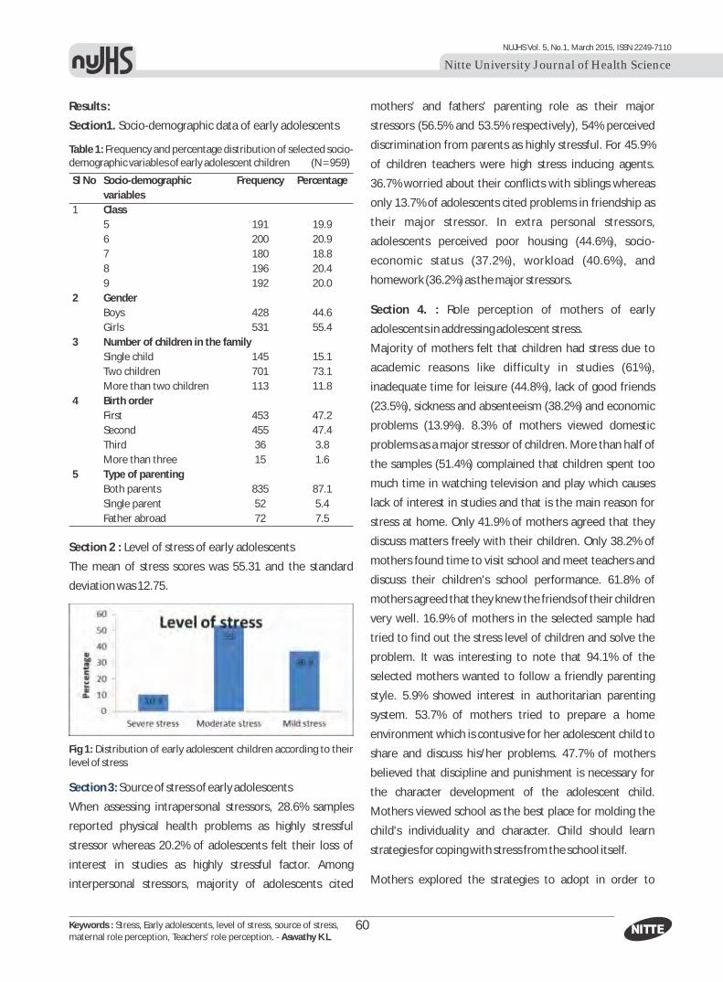
Results :
Section1. Socio-demographic data of early adolescents
60
NUJHS Vol. 5, No.1, 2015, ISSN 2249-7110March
Nitte University Journal of Health Science
Keywords : Stress, Early adolescents, level of stress, source of stress,maternal role perception, Teachers' role perception. - Aswathy K L
Table 1: Frequency and percentage distribution of selected socio-demographic variables of early adolescent children (N = 959)
Sl No Socio-demographic Frequency Percentage
variables
1 Class
5 191 19.9
6 200 20.9
7 180 18.8
8 196 20.4
9 192 20.0
2 Gender
Boys 428 44.6
Girls 531 55.4
3 Number of children in the family
Single child 145 15.1
Two children 701 73.1
More than two children 113 11.8
4 Birth order
First 453 47.2
Second 455 47.4
Third 36 3.8
More than three 15 1.6
5 Type of parenting
Both parents 835 87.1
Single parent 52 5.4
Father abroad 72 7.5
Section 2 : Level of stress of early adolescents
The mean of stress scores was 55.31 and the standard
deviation was 12.75.
Fig 1: Distribution of early adolescent children according to their level of stress
Section 3: Source of stress of early adolescents
When assessing intrapersonal stressors, 28.6% samples
reported physical health problems as highly stressful
stressor whereas 20.2% of adolescents felt their loss of
interest in studies as highly stressful factor. Among
interpersonal stressors, majority of adolescents cited
mothers' and fathers' parenting role as their major
stressors (56.5% and 53.5% respectively), 54% perceived
discrimination from parents as highly stressful. For 45.9%
of children teachers were high stress inducing agents.
36.7% worried about their conflicts with siblings whereas
only 13.7% of adolescents cited problems in friendship as
their major stressor. In extra personal stressors,
adolescents perceived poor housing (44.6%), socio-
economic status (37.2%), workload (40.6%), and
homework (36.2%) as the major stressors.
Section 4. : Role perception of mothers of early
adolescents in addressing adolescent stress.
Majority of mothers felt that children had stress due to
academic reasons like difficulty in studies (61%),
inadequate time for leisure (44.8%), lack of good friends
(23.5%), sickness and absenteeism (38.2%) and economic
problems (13.9%). 8.3% of mothers viewed domestic
problems as a major stressor of children. More than half of
the samples (51.4%) complained that children spent too
much time in watching television and play which causes
lack of interest in studies and that is the main reason for
stress at home. Only 41.9% of mothers agreed that they
discuss matters freely with their children. Only 38.2% of
mothers found time to visit school and meet teachers and
discuss their children's school performance. 61.8% of
mothers agreed that they knew the friends of their children
very well. 16.9% of mothers in the selected sample had
tried to find out the stress level of children and solve the
problem. It was interesting to note that 94.1% of the
selected mothers wanted to follow a friendly parenting
style. 5.9% showed interest in authoritarian parenting
system. 53.7% of mothers tried to prepare a home
environment which is contusive for her adolescent child to
share and discuss his/her problems. 47.7% of mothers
believed that discipline and punishment is necessary for
the character development of the adolescent child.
Mothers viewed school as the best place for molding the
child's individuality and character. Child should learn
strategies for coping with stress from the school itself.
Mothers explored the strategies to adopt in order to

61
NUJHS Vol. 5, No.1, 2015, ISSN 2249-7110March
Nitte University Journal of Health Science
Keywords : Stress, Early adolescents, level of stress, source of stress,maternal role perception, Teachers' role perception. - Aswathy K L
prevent, reduce and manage stress among early
adolescent children.
Section 5: Role perception of teachers of early adolescent
children in addressing adolescent stress.
96.7% of the teachers agreed that their adolescent
students had stress and there are genuine reasons for their
stress: school is giving a lot of academic stress to the child
(37.7%); teachers find time to talk with their students other
than the academic matters (96.7%); able to recognize the
features of stress in their students (85.2%); allowed the
students to discuss their personal problems with them
(91.8%). Teachers expressed strategies for preventing,
reducing and managing stress in early adolescent children.
Discussion:
The present study found that 10.1% of adolescents had
severe stress, 53 % had moderate stress and 36.9% had
mild stress. Mean stress score was 55 and standard
deviation was 12. Majority of adolescents experienced
moderate stress.
A study in Kerala among children in the age group from 4 to 8 17 years where severe stress was 1.9 % A study held in
Brazil by C. R. Sbaraini and L. B. Schermann revealed a
higher level of stress (27.2%) among adolescents over 10 9years.
It was baffling to note that early adolescents (56.5% and
53.5%) felt that parents and teachers were the stress
inducers. Being impulsive and closer to peers rather than
parents and teachers is a part of adolescence. Being
controlled by teachers and parents like coming home in
time, not spending too much time on outdoors, television
and games, being punctual and complying with home-
work etc may be construed as stressors by adolescents.
Eventhough mothers agree that their child is stressed, they
do not perceive that their parenting style had an influence
on their stress. The finding of the present study signifies
the importance of exploring the parenting styles and
attitude of mothers in caring their adolescent children.
Conclusion :
Study revealed that in the current scenario of societal and
educational setup, early adolescents are experiencing
stress. Major stressors of early adolescent children were
identified from the home and school environment.
Stressors that were identified as highly stressful were
parenting role of parents, teachers, work load, home-work,
high expectation of parents, parental conflicts, economic
factors etc. Study also identified that mothers and teachers
perception in addressing adolescent stress was not related
either to their parenting or teaching behaviours. Inspite of
mothers and teachers recommendations of suitable
strategies to prevent stress among adolescents at school or
home, early adolescent children had identified them as
their stressful factors. This indicates that there guidance is
required for mothers or teachers in identification of stress
of adolescents. School administration should plan
strategies to prevent and reduce stress of academics. I n
the present study there is uncertainty of data as the
investigator could not directly meet the mothers for data
collection.
1. Helping your child through early adolescence. U.S. Department of Education Office of Communications and Outreach. Washington DC. 2005.
2. Walker J. Teen in distress series Adolescent Stress and Depression.[Internet]. 2005. Available from http://www.extension. umn.edu/ distribution/ youth development/ da3083.html
th3. Wong DL. Whaley and Wong's Nursing care of infants and children. 6 ed. Mosby. 1996. P 448-450.
4. Brown SL, Teufel JA, Birch DA, Kancherla V. Gender, age, and behavior differences in early adolescent worry. J Sch Health. 2006 Oct; 76(8):430-7.
5. Carolina M. Teaching with affection: characteristics and Determinant factors of quality in teacher student relationships [Internet]. Available
References:
at revistaiaf.abacolombia.org.co/en/pdf/v1n1/v1n1_2_en.pdf6. National Crime record bureau. India. 2010[Internet].Available from
http://ncrb.nic.in/7. Varni JW. PedsQL™ (Pediatric Quality of Life Inventory).MAPI research
trust available at www.pedsql.org8. Sapna D. Prevalence of stress among school children in Kerala.
Available at http://www.articlesbase.com/mental-health-articles/prevalence-of-stress-among-school-children-in-kerala-1868702.htmlPosted: Feb 17, 2010.
9. Sbaraini CR, Schermann LB. Prevalence of childhood stress and associated factors: a study of schoolchildren in a city in Rio Grande doSul State, Brazil.CadSaudePublica. 2008 May;24(5):1082-8.

62
NUJHS Vol. 5, No.1, 2015, ISSN 2249-7110March
Nitte University Journal of Health Science
Keywords : BMI, Pregnancy outcome, Pregnancy InducedHypertension, Low birth weight. - Prathima P
Original Article
CORRELATION BETWEEN BMI AND PREGNANCY OUTCOME AMONG POSTNATAL MOTHERS WITH PREGNANCY INDUCED
HYPERTENSION IN SELECTED HOSPITALS BANGALORE1 2
Prathima P & S Anuchitra1HOD, Department of OBG, Universal College of Nursing, IIMB post Arekere Micolayout Bannerghatta Main Road,
2Bangalore, Vice & Principal, HOD, Department of OBG, P.D. Bharatesh College of Nursing, Belgaum.
Correspondence :Prathima P
HOD, Department of OBG, Universal College of Nursing, IIMB post Arekere Micolayout , Bannerghatta Main road, Bangalore – 76Mobile : +91 99867 78622 E-mail : [email protected]
Abstract :
Title : Correlation between BMI and pregnancy Outcome among postnatal mothers with pregnancy Induced hypertension in selected
hospital Bangalore.
Objectives : To identify and correlate BMI and pregnancy Outcome among postnatal mothers with pregnancy Induced hypertension.
Method : A non experimental correlation design was utilized among 80 postnatal mothers who were diagnosed as Pregnancy induced
hypertension during their antenatal period selected as samples by using purposive sampling technique. Demographic data were
collected by interview method, their BMI was calculated, pregnancy outcomes were identified from records by using an outcome
checklist.
Results : Underweight mothers had low birth weight babies and received NICU care. Among normal weight mothers 17.5 % delivered by
LSCS, 15% babies were low birth weight babies 12.5 were preterm babies among them 10% received NICU care. In the overweight group
18.75% undergone LSCS, 18.75% were LBW and 1.25% VLBW, 8.75% babies were preterm, 12.5% newborn received NICU care. Among
Obese mothers 8.75% delivered by LSCS, 6.25% of LBW babies, 5% were preterm and all of them received NICU care. There is a positive
correlation between BMI and diagnosis and type of delivery. Significant at .01 and .05 Level (p value .008 and .019 respectively).
Negative correlation between birth weight and diagnosis and gestational age. r = -.499 significant at .01 level (p value .000)
Conclusion : Obesity and under weight is a leading, preventable cause of mortality worldwide. Preeclampsia increases maternal and
perinatal morbidity and mortality rates. All women who are in reproductive age group and under risk to develop pregnancy induced
hypertension need to be educated about to maintenance of normal weight before pregnancy. Nurses have more responsibility on
creating awareness among women how to maintain normal weight to avoid development of complications to the mother and newborn.
Keywords : BMI, Pregnancy outcome, Pregnancy Induced Hypertension, Low birth weight.
Access this article online
Quick Response Code

63
NUJHS Vol. 5, No.1, 2015, ISSN 2249-7110March
Nitte University Journal of Health Science
Keywords : BMI, Pregnancy outcome, Pregnancy InducedHypertension, Low birth weight. - Prathima P

64
NUJHS Vol. 5, No.1, 2015, ISSN 2249-7110March
Nitte University Journal of Health Science
Keywords : BMI, Pregnancy outcome, Pregnancy InducedHypertension, Low birth weight. - Prathima P

65
NUJHS Vol. 5, No.1, 2015, ISSN 2249-7110March
Nitte University Journal of Health Science
Keywords : BMI, Pregnancy outcome, Pregnancy InducedHypertension, Low birth weight. - Prathima P
Table: 1 Demographic Variable
Variables Under weight Normal weight Over weight Obese
N % N % N % N %
AGE <20 0 0 2 2.5 2 2.5 1 1.25
21-25 2 2.5 27 33.75 12 15 5 6.25
26-30 0 0 7 8.75 14 17.5 2 2.5
31-35 0 0 2 2.5 2 2.5 1 1.25
36-40 0 0 1 1.25 0 0 0 0
Education primary School 0 0 1 1.25 2 2.5 0 0
Middle school 0 0 0 0 2 2.5 1 1.25
Sec.education 0 0 15 18.75 8 10 2 2.5
PUC 1 1.25 15 18.75 11 13.75 2 2.5
Diploma 0 0 2 2.5 1 1.25 0 0
Graduate 1 1.25 6 7.5 6 7.5 4 5
Religion Hindu 2 2.5 30 37.5 23 28.75 7 8.75
Christian 0 0 1 1.25 2 2.5 1 1.25
Muslim 0 0 8 10 5 6.25 1 1.25
Income in Rs <5000 0 0 7 8.75 3 3.75 0 0
5001-10000 0 0 15 18.75 9 11.25 5 6.25
10001-15000 2 2.5 13 16.25 13 16.25 2 2.5
15001-20000 0 0 1 1.25 3 3.75 0 0
>20001 0 0 3 3.75 2 2.5 2 2.5
Occupation Home maker 1 1.25 36 45 27 33.75 8 10
Skilled worker 0 0 2 2.5 2 2.5 1 1.25
Unskilled worker 1 1.25 1 1.25 1 1.25 0 0
Parity First pregnancy 2 2.5 19 23.75 13 16.25 5 6.25
Second pregnancy 0 0 13 16.25 10 12.5 4 5
Third pregnancy 0 0 4 5 5 6.25 0 0
Fourth pregnancy 0 0 3 3.75 2 2.5 0 0
Physical activity Sedentary worker 2 2.5 24 30 14 17.5 5 6.25
Moderate worker 0 0 13 16.25 14 17.5 3 3.75
Heavy worker 0 0 2 2.5 2 2.5 1 1.25
Diagnosis Gestational HT 0 0 28 35 10 12.5 1 1.25
Mild.PIH 2 2.5 8 10 16 20 5 6.25
Severe.PIH 0 0 3 3.75 4 5 3 3.75

66
NUJHS Vol. 5, No.1, 2015, ISSN 2249-7110March
Nitte University Journal of Health Science
Keywords : BMI, Pregnancy outcome, Pregnancy InducedHypertension, Low birth weight. - Prathima P
Figure 1 Distribution of postnatal mothers according to their BMI.
Table : 2 Percentage distribution of Pregnancy Outcome
BMI Underweight Normal weight Overweight Obese
Outcome N % N % N % N %
Delivery NVD 1 1.25 25 31.25 15 18.75 2 2.5
LSCS 1 1.25 14 17.5 15 18.75 7 8.75
Birth weight Normal 0 0 24 30 14 17.5 4 5
LBW 2 2.5 12 15 15 18.75 5 6.25
VLBW 0 0 3 3.75 1 1.25 0 0
Gestational age Full term 1 1.25 29 36.25 23 28.75 5 6.25
Preterm 1 1.25 10 12.5 7 8.75 4 5
NICU care No 0 0 31 38.75 20 25 5 6.25
Yes 2 2.5 8 10 10 12.5 4 5
Table: 3 BMI and correlation
BMI Birth weight Diagnosis
r value P value r value P value r value P value** **Diagnosis .293 .008 -.499 .000 - -* **Type of delivery .261 .019 - - .449 .000
**Gestational age - - -.494 .000
**. Correlation is significant at the 0.01 level (2-tailed).*. Correlation is significant at the 0.05 level (2-tailed).
Discussion :
The present study indicated that maternal pre pregnancy
weight related with greater risks of pregnancy-induced
hypertension, caesarean delivery, preterm delivery and
prematurity, birth weight at birth. Also reveals that there is
a correlation between BMI and pregnancy outcome and
association between demographic variable and pregnancy
outcome.
A comparative study conducted to investigate the impact
of maternal obesity on pregnancy outcomes. Rates of
pregnancy complications and neonatal outcomes were
collected from perinatal data list and compared between
women with normal pre pregnancy body mass index and
those with an obese pre pregnancy body mass index. Rates
of pregnancy complications and neonatal outcomes were
also evaluated by the level of obesity, severe obesity, and
morbid obesity (BMI>30, BMI=35-39.9 and, BMI>40).
Rates of gestational diabetes and gestational hypertension
were higher for obese 62 % versus normal weight only 10 %
in gravid. Women with morbid or severe obesity had a
greater incidence of gestational diabetes and gestational 9hypertension. This is similar to present study.
Another study which was similar to present study
prospective population-based cohort study was done to
evaluate morbidly obese (BMI>40) women have an
increased risk of pregnancy complications and adverse
perinatal outcomes. The result of the study was an
increased risk of the following outcomes: preeclampsia 20
%, ante partum stillbirth 15%, caesarean delivery 35 %,
instrumental delivery12 %, shoulder dystocia 10 %,
meconium aspiration 40 %, foetal distress 30 %, early
neonatal death 7 %, and large-for-gestational age 10 % was
found women with BMIs between 35.1 and 40 but to a
lesser degree compared to BMIs above 40.33 In this study
8.75 % of obese mother were delivered by LSCS and 6.25 % 10of newborn babies were LBW.
Conclusion :
Obesity and under weight is a leading, preventable cause of
mortality worldwide and authorities view it as one of the

67
NUJHS Vol. 5, No.1, 2015, ISSN 2249-7110March
Nitte University Journal of Health Science
Keywords : BMI, Pregnancy outcome, Pregnancy InducedHypertension, Low birth weight. - Prathima P
most serious public health problems of the 21st century. In
developing countries the obesity and underweight were
still as a risk factor for maternal and neonatal mortality and
morbidity. Preeclampsia increases maternal and perinatal
morbidity and mortality rates. This study revealed that
there is a correlation between BMI and pregnancy
outcome. Result of this present study and other studies
cited in this article are stating that there is association
between BMI and Pregnancy outcome which is modifiable.
All women who are in reproductive age group and under
risk to develop pregnancy induced hypertension need to be
educated about to maintenance of normal weight before
pregnancy. Nurses have more responsibility on creating
awareness among women how to maintain normal weight
to avoid development of complications to the mother and
newborn.
1. Hendler I, Goldenberg RL, Mercer BM, Iams JD, Meis PJ, Moawad AH, MacPherson CA, Caritis SN, Miodovnik M, Menard KM, Thurnau GR, Sorokin Y Association between maternal body mass index and spontaneous and indicated preterm birth. American journal of obstetrics and gynaecology.2005 Jan; 19(2):P. 882-6.
2. Bell A W, Ehrhardt R A. Regulation of placental nutrient transport and implications for foetal growth. Journal of Nutrition.2002Feb; 15(1):P. 215-35.
3. Nan Li, Enqing Liu, Jia Guo, Lei Pan, Baojuan Li, Ping Wang, Jin Liu, Yue Wang, Gongshu Liu, Andrea A. Baccarelli, Lifang Hou, Gang Hu (2013) Maternal Prepregnancy Body Mass Index and Gestational Weight Gain on Pregnancy Outcomes. PLOS ONE; 8:12, 2-7
4. PerinatoI J, Joy S, Istwan N, Rhea D, Desch C, Stanziano G. Assess the effect of underweight on maternal and neonatal outcome. Wake Forest University, Winston-Salem, North Carolina 2008May; 26(5):P. 345-9.
5. Mrozikiewicz A, Drews K, Nowocien G, Kaluba-Skotarczak A. Obesity in pregnant women as a problem in obstetrics. Journal of Geburtshilfe
neonatal complication.2007 Mar; 78(3):P.234-8. 6. Nilsen R M, Lise A, Monsen B, Midttun O, Nygard O, Pedersen E R.
Maternal tryptophan and kynurenine pathway metabolites and risk of preeclampsia. American journal of Obstetrics and gynecology. 2009 Aug; 119(6):P.1243-50.
7. Raatikainen K, Heiskanen N, Heinonen S. Transition from overweight to obesity worsens pregnancy outcome in a body mass index dependent manner. Department of Obstetrics and Gynecology, Kuopio University Hospital, Kuopio, Finland. 2006 Jan; 14(1):P.165-71.
8. Watkins M L, Rasmussen S A, Honein M A, Botto L D, Moore C A. Division of Birth Defects and Developmental Disabilities. American journal of paediatrics. 2003 May; 111(5):P.1152-8.
9. Alanis M C, Goodnight W H, Hill E G, Robinson C J, Villers M S. Impact of maternal obesity on pregnancy outcomes. Acta Obstetrical and Gynecological Scand. Charleston, South Carolina. 2010 Jul; 89(7):P.924-30.
10. Blomberg M. Maternal Obesity and Risk of Postpartum Hemorrhage. Obstetrics and Gynecology .2011Mar; 11(8):P. 561-8.
References:

68
NUJHS Vol. 5, No.1, 2015, ISSN 2249-7110March
Nitte University Journal of Health Science
Keywords : Rare variation, Testicular artery, External iliac artery- Huban Thomas R
Introduction :
The testicular arteries are paired vessels that usually arise
from the abdominal aorta at the second lumbar vertebral
level. Each artery passes obliquely downwards and
posterior to the peritoneum. Descending on the posterior
abdominal wall, it reaches the deep inguinal ring where it
enters the spermatic cord [1, 2]. There are reports about
the variant origin of testicular artery arising from the renal
artery, accessory renal artery, suprarenal artery, one of the
lumbar arteries, common or internal iliac artery, and the
inferior phrenic arteries. Variations of these arteries have
been extensively studied due to their importance in
testicular physiology. Moreover, this knowledge has a
practical implication during testicular surgery. [3].To the
best of our knowledge this kind of variation in the origin of
left testicular artery from left external iliac artery has not
been reported in the
literature before.
Case presentation :
During routine dissection
for undergraduate medical
students at Department of
Human Anatomy, Kasturba
RARE VARIATION IN THE ORIGIN OF LEFT TESTICULAR ARTERY FROM LEFT EXTERNAL ILIAC ARTERY : A CASE REPORT
1 2 3 4Huban Thomas R , Prasanna L C , Vivek Kumar , Antony Sylvan D'souza1 2 3 4Senior Grade Lecturer, Associate Professor, Post Graduate, Professor & HOD,
Department of Anatomy, Kasturba Medical College, Manipal University, Manipal, Karnataka, India.
Correspondence :Huban Thomas R
Senior Grade Lecturer, Department of Anatomy, Kasturba Medical College, Manipal University, Manipal 576 104, Karnataka, India.Mobile : +91 98443 43546 E-mail : [email protected]
Abstract :
Keywords: Rare variation, Testicular artery, External iliac artery
Testicular artery usually arises from the antero-lateral part of the abdominal aorta below the origin of the renal arteries. Very rarely
variations in the origin of the testicular arteries were observed. During routine dissection for undergraduate medical students, an
abnormal origin and course of the left side testicular artery was detected in a 55-year-old male cadaver. On the left side, testicular artery
arose from the external iliac artery half way before its entry into front of the thigh. Later it runs in the inguinal canal to reach the testis. In
contrast, right side testicular artery has normal origin and course. Such variations in the origin and course of the testicular artery are
important in surgical and diagnostic interventions to avoid diagnostic and surgical errors to prevent hazardous complications like
testicular hypoperfusion and atrophy.
Access this article online
Quick Response Code
Case Report
Medical College, Manipal University, Manipal, we observed
an abnormal origin and course of the left side testicular
artery in a 55-year-old male cadaver. On the left side,
testicular artery arose from the external iliac artery half
way before its entry into front of the thigh. Later it runs in
the inguinal canal to reach the testis. In contrast, right side
testicular artery and the testicular vein have normal origin
and course. This anomalous left testicular artery had a
diameter of 10 mm and a length of about 32 mm from its
origin to its entry into the deep inguinal ring.
Discussion :
Anatomy of the gonadal arteries has assumed importance
because of the development of new operative techniques
within the abdominal cavity for operations such as
varicocele and undescended testes [4]. During
laparoscopic surgery of the male abdomen and pelvis many
complications occurred due to unfamiliar anatomy in the
operative field [5]. Awareness of variations in the testicular
arteries, such as those presented in this case report,
becomes important during such surgical procedures.
Variant anatomy of gonadal arteries has been reported in
number of cases. Anomalies in the origin, course, and

69
NUJHS Vol. 5, No.1, 2015, ISSN 2249-7110March
Nitte University Journal of Health Science
Keywords : Rare variation, Testicular artery, External iliac artery- Huban Thomas R
number of Testicular Arteries were observed in 4.7 present
of cases in a study of 150 cadavers [6]. There are few
reports of a high Testicular Artery origin in the literature.
Shinohara et al. found a Testicular Artery originating 1 cm
superior to the origin of the inferior phrenic artery [7].
Brohi et al. described the case of a high origin of the left
Testicular Artery which originated from the left renal artery
[8]. Xue et al. found a right TA artery arising from the
anterior surface of the abdominal aorta at the level of the
left renal artery [9]. Shoja et al reported that the gonadal
artery originated from the main or accessory renal artery
[10]. Deepthinath et al reported a double left testicular
artery, in which one originated from an accessory renal
artery and the other from the main renal artery [11].
The first attempt at classification of Testicular Artery
variations was made by Machnicki et al. [12]. Their study
included TAs from both foetuses and adults grouped
according to their origin from the aorta or renal artery. Four
major types were observed: Type A - a single Testicular
Artery originating from the aorta; Type B - a single
Testicular Artery originating from the renal artery; Type C -
two Testicular Arteries originating from the aorta that
supplied the same gonad; Type D - two Testicular Arteries
supplying the same gonad, one arising from the aorta and
the other from the renal artery [12]. Some years later,
Çiçekcibasi et al. classified the variations into four
alternative types: Type I - Testicular Artery arising from the
suprarenal artery; Type II - Testicular Artery originating
from the renal artery; Type III - Testicular Artery of high-
positional origin from the abdominal aorta, close to the
renal artery lineage; Type IV - Testicular Artery duplication
originating from the aorta or from various vessels [13]. Our
case report is not matching with any of the above said
classification and not been reported in the literature
before.
Regarding the embryologic basis, explanation for individual
or combined variations of renal and gonadal arteries has
been related to the embryological development of both
vessels from the lateral mesonephric branches of the
dorsal aorta. The embryologic explanation of these
variations has been presented and discussed by Felix. The
developing mesonephros, metanephros, suprarenal
glands and gonads are supplied by nine pairs of lateral
mesonephric arteries arising from the dorsal aorta. Felix
divided these arteries into three groups as follows: the 1st
and 2nd arteries as the cranial; the 3rd to 5th arteries as the
middle, and the 6th to 9th arteries as the caudal group. The
middle group gives rise to the renal arteries. Persistence of
more than one arteries of the middle group results as
multiple renal arteries. Felix also stated that although
anyone of these nine arteries may become the gonadal
artery, it usually arises from the caudal group [14].
Figure - Anterior view of the male AbdomenIVC – inferior vena cava, REIA – right external iliac artery, LEIA – left external iliac artery, RTA – right testicular artery, LTA- left testicular artery, RTV- right testicular vein, LTV- left testicular vein
Conclusion :
Anatomical knowledge of Morphological anomalies of the
gonadal arteries may be important not only for the clinical
point of view but they also explain the embryological basis
and some pathological conditions. The origin and course of
the TA must be carefully identified and demarcated in order
to preserve and prevent testicular atrophy. A deeper
understanding of these variations and their special
relations to adjacent vessels is especially significant in
avoiding the complications in
Furthermore, radiologists should be familiar
with TA variants in order to provide an accurate diagnosis
during pre-clinical studies.
surgical and diagnostic
interventions.

70
NUJHS Vol. 5, No.1, 2015, ISSN 2249-7110March
Nitte University Journal of Health Science
Keywords : Rare variation, Testicular artery, External iliac artery- Huban Thomas R
1.
th2. Moore KL, Dalley AF: Clinically oriented anatomy. 4 edition. Philadelphia: Lippincott Williams and Wilkins; 1999:92.
3.
4. Pushpa Dhar, Kumud Lal. Main and accessory renal arteries – A morphological study. Journal of Anaomy and Embryoogyl. 2005;110: 101–10.
5. Brohi RA, Sargon MF, Yener N. High origin and unusual suprarenal branch of a testicular artery. Surg Radiol Anat 2001; 23:207–8.
6. Asala S, Chaudhary SC, Masumbuko-Kahamba N, Bidmos M: Anatomical variations in the human testicular blood vessels. Ann Anat 2001, 183:545-49.
7. Shinohara H, Nakatani T, Fukuo Y, Morisawa S, Matsuda T: Case with high positioned origin of the testicular artery. Anat Rec 1990, 226:264-65.
Hollinshead WH: Anatomy for Surgeons. Volume New York: Harper and Row;1971:579-80.
Paraskevas GK, Ioannidis O, Raikos A, Papaziogas B, K Natsis, Spyridakis L,et al. High origin of a testicular artery: a case report and review of the literature. Journal of Medical Case Reports 2011, 5:75
References :
8. Brohi RA, Sargon MF, Yener N: High origin and unusual suprarenal branch of a testicular artery. Surg Radiol Anat 2001, 23:207-08.
9. Xue HG, Yang CY, Ishida S, Ishizaka K, Ishihara A, Ishida A, Tanuma K: Duplicate testicular veins accompanied by anomalies of the testicular arteries. Ann Anat 2005, 187:393-98.
10. Shoja MM, Tubbs RS, Shakeri AB, Oakes WJ. Origins of the gonadal artery: embryologic implications. Clin Anat 2007; 20:428-32.
11. Deepthinath R, Satheesha Nayak B, Mehta RB, Bhat S, Rodrigues V, Samuel VP, Venkataramana V, Prasad AM.. Multiple variations in the paired arteries of the abdominal aorta. Clin Anat 2006; 19:566-8.
12. Machnicki A, Grzybiak M: Variations in testicular arteries in fetuses and adults. Folia Morphol (Warsz) 1997, 56:277-85.
13. Çiçekcibasi AE, Salbacak A, Seker M, Ziylan T, Büyükmumcu M, Uysal II: The origin of gonadal arteries in human fetuses: anatomical variations. Ann Anat 2002, 184:275-79.
14. Kiebel F, Mall FP. Manual of human embryology. Vol 2. Philadelphia: Lippincott; 1912:820–25.

Introduction:
A biologic and functional approach to restorative dentistry
is essential for the satisfactory performance and fulfillment
of those requisites basic to Prosthodontics. Accordingly,
the masticatory organ must be considered as a functional,
consolidated unit, with proper attention being directed to
all the elements that comprise this unit. All functional
factors are interrelated, and proper regard for each aspect
is essential, if the restoration and maintenance of the
health of the entire functioning mechanism is to be a
realization.
Amelogenesis imperfecta (AI) has been defined as a
complex group of hereditary
e n a m e l d e f e c t s n o t
associated with evidence of systemic disease affecting
b o t h p r i m a r y a n d 1,2permanent dentitions. It is
a r a r e e n a m e l
m i n e ra l i za t i o n d efe c t
71
NUJHS Vol. 5, No.1, 2015, ISSN 2249-7110March
Nitte University Journal of Health Science
Keywords : Amelogenesis imperfecta, orthognathic surgery,orthodontic therapy, full mouth rehabilitation. - Krishna Prasad D
A MULTIDISCIPLINARY APPROACH FOR FUNCTIONAL REHABILITATION OF A PATIENT WITH SKELETAL CLASS III
MALOCCLUSION AND AMELOGENESIS IMPERFECTA1 2 3 4Archana S Shetty , Krishna Prasad D , M S Ravi & B Rajendra Prasad
1 2Senior Lecturer, Department of Prosthodontics, V.S. Dental College & Hospital, Bangalore, Professor & HOD, 3Department of Prosthodontics and Crown & Bridge, Professor, Department of Orthodontics & Dentofacial
4Orthopaedics, Principal & Dean, A.B. Shetty Memorial Institute of Dental Sciences, Nitte University, Mangalore, Karnataka, India.
Correspondence :Krishna Prasad D
Professor & HOD, Department of Prosthodontics, A.B. Shetty Memorial Institute of Dental Sciences,Nitte University, Mangalore - 575 018, Karnataka, India.
E-mail : [email protected]
Abstract :
Amelogenesis Imperfecta has been defined as a group of hereditary enamel defects not associated with evidence of systemic disease.
Restoration for patients with this condition should be oriented toward the functional and esthetic rehabilitation and the protection of
these teeth. The specific objectives of the treatment were to enhance esthetics, eliminate tooth sensitivity and restore masticatory
function. Management of a patient with skeletal class III malocclusion and Amleogenesis Imperfecta is a challenge for the clinician.
Surgical correction of the skeletal class III malocclusion, orthodontic therapy followed by full mouth rehabilitation as a multidisciplinary
approach is vital for the functional rehabilitation of the patient.
Keywords: Amelogenesis imperfecta, orthognathic surgery, orthodontic therapy, full mouth rehabilitation.
Access this article online
Quick Response Code
Case Report
described by Spokes in 1890 as “hereditary brown teeth” 3,4 with a reported incidence of 1:14,000. Other associated
findings in patients with AI include delayed eruption of
teeth, taurodontism, congenitally missing teeth, crown 5and root resorption, and pulp calcification.
Clinical report:
An 18 year old male patient presented with poor esthetics,
gap between upper and lower teeth; and poor masticatory
efficiency. Patient exhibited a concave facial profile with a
skeletal class III malocclusion, mandibular prognathism
with increased anterior facial height. On intra oral
examination, dark yellowish discoloration of all teeth, open
proximal contacts and esthetic disharmonies with open
bite from first molar to first molar with occlusal contacts
only at second molar was revealed (Figure 1). A panoramic
radiographic examination of the teeth revealed
generalized defective enamel on all the teeth. A lateral
cephlogram revealed a class III skeletal malocclusion with
skeletal open bite with prognathic mandible, increased
mandibular plane and increased lower anterior face height

72
NUJHS Vol. 5, No.1, 2015, ISSN 2249-7110March
Nitte University Journal of Health Science
Keywords : Amelogenesis imperfecta, orthognathic surgery,orthodontic therapy, full mouth rehabilitation. - Krishna Prasad D
(Figure 2). A methodical multidisciplinary approach was
planned for the oral rehabilitation of the patient starting
with orthodontic alignment, surgical correction of jaw
relationship and finally prosthodontic rehabilitation to
restore both function and esthetics. The detailed
treatment plan and the possible outcome were explained
to the patient and informed consent was obtained before
the treatment procedures.
Orthognathic surgery was performed as bilateral sagittal
split osteotomy [BSSO] with mandibular set back and
upward rotation for correction of open bite as well as
prognathic mandible. Presurgical orthodontics with 0.022”
preadjusted edgewise appliance [Roth system] for pre
surgical alignment and space closure were carried out. A
month after the orthognathic surgery, post-surgical
orthodontics was begun for minor occlusal corrections
(Figure 3,4). Perfect occlusal relationship was not
established as the case was required to undergo full mouth
rehabilitation.
The patient was now ready for prosthodontic
rehabilitation. Maxillary and mandibular complete-arch
impressions were made using irreversible hydrocolloid
(Zelgan, Dentsply, Gurgaon, India) impression material.
Diagnostic casts were fabricated from Type-III dental stone
(Kalstone, Kalabhai private limited, Mumbai, India). The
opening axis of the mandible was determined by face bow
transfer and mounted on a Whipmix semiadjustable
articulator [Whip Mix Articulator; Whip MixCorp;
Louisville, Ky]. Bite registration using Type II modeling wax
(Hyderabad Dental Products, Hyderabad, India) was made
at the orthodont ica l ly establ ished maximum
intercuspation.
One of the most common method for establishing an
acceptable plane of occlusion is indirect analysis using the
Pankey-Mann-Schuyler (PMS) method with the Broderick 6occlusal plane analyzer (BOPA). When it has been
determined that restoration of all or most of the posterior
teeth is necessary, the PMS technique using BOPA provides
a simple and practical method to determining the
preliminary occlusal plane on diagnostic casts. Hence BOPA
was used as a guide for determining an acceptable plane of
occlusion and the reduction required for each tooth 6,7,8preparation. Diagnostic preparations were made on the
stone casts, and a diagnostic waxup was completed with a
canine guided concept of occlusion design (Figure 5). Tooth
reduction guides and heat processed acrylic resin
provisional restorations were fabricated from the
diagnostic wax-up.
Tooth preparation and restoration with porcelain fused to
metal crowns was done in a sequential manner, starting
with the mandibular anteriors, followed by the maxillary
anteriors, mandibular posteriors and finally the maxillary
posteriors following the PMS philosophy of occlusal
rehabilitation. The anterior guidance was established
based on esthetics, phonetics and development of a canine
guided occlusion. Soft tissue was retracted using knitted
cord [UltraPak, Ultradent products, south Jordan, Utah]
with aluminium sulfate hemostatic agent[Gelcord, Pascal
Co Inc, Bellevue, Wa], and complete arch impressions were
made with the addition polymerization silicone impression TM material[Express 3M ESPE].Wax patterns were
completed to the contour of the final restorations, and the
wax patterns for the teeth receiving porcelain-fused-to-
metal restorations were cut back approximately 1 mm to
allow for the porcelain addition. All occlusal surfaces were
planned to be fabricated in metal. The wax patterns were
invested in a phosphate bonded investment, and cast.
Porcelain shade Vita B2 was applied to the metal-ceramic
castings. All the restorations were characterized, glazed,
and polished, and luted with glass ionomer cement (Figure
6). The esthetics and functional efficiency has improved
drastically after the rehabilitation (Figure 7,8).
Complete mouth rehabilitation is a dynamic functional
problem, and embodies the correlation and integration of
all component parts into one functioning unit. The aim and
endeavor, therefore, must be reconstruction and
rehabilitation of the whole, satisfying all the related
factors. The science of complete mouth rehabilitation rests
upon three proved and accepted fundamentals: namely,
the existence of a physiologic rest position of the mandible,

73
NUJHS Vol. 5, No.1, 2015, ISSN 2249-7110March
Nitte University Journal of Health Science
Keywords : Amelogenesis imperfecta, orthognathic surgery,orthodontic therapy, full mouth rehabilitation. - Krishna Prasad D
which is a constant; the recognition of a vertical dimension;
and, finally, the acceptance of a dynamic, functional centric
occlusion
The clinical features of amelogenesis imperfecta
compounded with a skeletal class III malocclusion
presented special challenges to the treatment team.
Though the treatment planning was complicated, the
diagnostic wax-up demonstrated that full-arch restorative
Fig 1 : Pre Treatment intra oral view
Fig 2 : Pre treatment Lateral cephalogram
Fig 3 : Intra oral view- One month afterthe orthognathic surgery
Fig 4 : Lateral cephalogram - Onemonth after the orthognathic surgery
Fig 5 : Diagnostic wax up in the semiadjustable articulator
Fig 6 : Post treatment intra oral view
Fig 7 : Pre treatment facial view Fig 8 : Post treatment facial view
treatment could be accomplished, and without
unfavorable stresses caused by the arch malrelations.
Careful technique and very close follow-up are required to
maintain occlusal stability when restoring arch
discrepancies. The planned occlusal relations were tested
in the mouth with the acrylic resin provisional restorations,
and were found satisfactory by the patient as to esthetics,
phonetics, and function.
Discussion:
Complete mouth rehabilitation is a dynamic functional
problem, and embodies the correlation and integration of
all component parts into one functioning unit. The aim and
endeavor, therefore, must be reconstruction and
rehabilitation of the whole, satisfying all the related
factors. The science of complete mouth rehabilitation rests
upon three proved and accepted fundamentals: namely,
the existence of a physiologic rest position of the mandible,
which is a constant; the recognition of a vertical dimension;
and, finally, the acceptance of a dynamic, functional centric
occlusion

74
NUJHS Vol. 5, No.1, 2015, ISSN 2249-7110March
Nitte University Journal of Health Science
Keywords : Amelogenesis imperfecta, orthognathic surgery,orthodontic therapy, full mouth rehabilitation. - Krishna Prasad D
The clinical features of amelogenesis imperfecta
compounded with a skeletal class III malocclusion
presented special challenges to the treatment team.
Though the treatment planning was complicated, the
diagnostic wax-up demonstrated that full-arch restorative
treatment could be accomplished, and without
unfavorable stresses caused by the arch malrelations.
Careful technique and very close follow-up are required to
maintain occlusal stability when restoring arch
discrepancies. The planned occlusal relations were tested
in the mouth with the acrylic resin provisional restorations,
and were found satisfactory by the patient as to esthetics,
phonetics, and function.
Conclusion :
The procedure explained in this clinical report is an
organized way to rehabilitate the lost occlusion. An
advanced restorative treatment plan involving the re-
organization of the patient's occlusion is a major challenge
for the restorative team. Successful completion will
depend upon:
1. An accurate record of the patient's pre-treatment
occlusion.
2. A clear idea of the occlusion of the definitive restoration,
including the jaw relationship at which it is to occur.
3. A detailed sequential plan and the execution of
treatment from the pre-treatment phase until the final
rehabilitation.
A multidisciplinary sequential approach with pre surgical
orthodontic alignment followed by planned orthognathic
surgery and subsequent post surgical orthodontic
alignment and occlusal corrections and further
Prosthodontic treatment approach ensured a successful
esthetic and functional rehabilitation.
References:1. Robinson FG, Haubenreich JE. Oral rehabilitation of a young adult with
hypoplastic amelogenesis imperfecta: a clinical report. J Prosthet Dent 2006 Jan;95(1):10-3.
2. Sari T, Usumez A. Restoring function and esthetics in a patient with amelogenesis imperfecta: a clinical report. J Prosthet Dent 2003 Dec; 90(6):522-5.
3. Lindunger A, Smedberg JI. A retrospective study of the prosthodontic management of patients with amelogenesis imperfecta. Int J Prosthodon, 2005 May-June; 18(3):189-94.
4. Bouvier D, Duprez JP, Pirel C, Vincent B. Amelogenesis imperfecta – a prosthetic rehabilitation: A clinical report J Prosthet Dent 1999 Aug; 82(2):130-1.
5. Yip HK, Smales RJ. Oral Rehabilitation of young adults with amelogenesis imperfecta. Int J Prosthodont 2003 July-Aug 16(4):345-9.
6. Dawson PE. Evaluation, diagnosis and treatment of occlusal problems. 2nd ed. StLouis: Elsevier; 1989. p. 85, 373-81.
7. Small BW. Occlusal plane analysis using the Broadrick flag. Gen Dent 2005;53:250-2.
8. Sumit Bedia, Shankar Dange, Arun N. Determination of the occlusal plane using a custom-made occlusal plane analyzer: A clinical report. J Prosthet Dent 2007;98:348-352.

75
NUJHS Vol. 5, No.1, 2015, ISSN 2249-7110March
Nitte University Journal of Health Science
Keywords : persistent left superior vena cava, coronary sinus,superior vena cava, right atrium - Mamatha H
Introduction :
Abnormalities in the cardio-vascular system are of extreme
interest because of their significance in different
developmental problems and their effect on the organ of
circulation.
Normal anatomy describes the formation of a single
superior venacava by the union of right and left
brachiocephalic veins which are in turn is formed by the
union of corresponding internal jugular and subclavian
veins, draining the head and neck as well as the superior
extremity.
Double superior vena cava
(SVC) with the persistent
left superior vena cava
(PLSVC) is a rare venous
malformation. Patients
with PLSVC may have
other associated cardiac
Case Report
Access this article online
Quick Response Code
DOUBLE SUPERIORVENACAVA AND ITS ASSOCIATED CLINICAL IMPLICATIONS - A CASE REPORT AND LITERATURE REVIEW
1 2 3 4Sushma R Kotian , Antony Sylvan D Souza , Praveena Ravichandran , Pallavi Bhat
4& Mamatha Hosapatna
1 2 3 4Lecturer , Professor & HOD , Postgraduate , Associate Professor , Department of Anatomy, Kasturba Medical College, Manipal University, Manipal, Karnataka, India.
Correspondence :Mamatha H
Associate Professor, Department of Anatomy, Kasturba Medical CollageManipal University, Manipal 576104, Karnataka, India
Mobile : +91 820 2922327 E-mail : [email protected]
Abstract :
Abnormalities of the vascular system are always of extreme interest due to its importance in circulation. Normally the superior vena
cava is a single vascular structure formed by the union of right and left brachiocephalic veins which are in turn formed by the union of
corresponding internal jugular and subclavian veins, draining the head and neck as well as the superior extremity. However during
routine dissection in the Department of Anatomy, Kasturba Medical College, Manipal, we came across a case of double superior vena
cava with persistent left superior vena cava in a 58-year-old male cadaver. Both the vena cavae were formed as continuations of
brachiocephalic veins of the corresponding side. The persistent left superior vena cava opened into the enlarged coronary sinus that
drained into the right atrium between the opening of inferior venacava and right atrioventricular orifice. No communication was
observed between the two vena cavae. A persistent left superior vena cava does not by itself produce any physiological derangement.
But it has important clinical implications in certain clinical interventions. It may complicate placement of cardiac catheters or pacemaker
leads. Awareness of this anomaly may therefore reduce confusions and thus would help to avoid further complications.
Keywords : persistent left superior vena cava, coronary sinus, superior vena cava, right atrium
malformations such as atrial septal defect, ventricular 1,2septal defect or endocardial cushion defect. Presence of
PLSVC may also interfere and cause problems during
various invasive procedures such as pacemaker
implantation, central venous catheterisation, retrograde
delivery of cardioplegia and retrograde left ventricular 3-5pacing. The present case reports the existence of one
such anomalous PLSVC in an adult cadaver.
Case Report :
During routine dissection in the department of Anatomy,
Kasturba Medical College, Manipal, we came across a case
of double superior vena cava with persistent left superior
vena cava (PLSVC) in a 65-year-old male cadaver. Both the
vena cavae were formed as continuations of
brachiocephalic veins of the corresponding sides. The
PLSVChad the same length and caliber compared to the
superior vena cava (Figure 1). When traced, it opened into
the enlarged coronary sinus that further drained into the
right atrium between the opening of inferior venacava and

76
NUJHS Vol. 5, No.1, 2015, ISSN 2249-7110March
Nitte University Journal of Health Science
Keywords : persistent left superior vena cava, coronary sinus,superior vena cava, right atrium - Mamatha H
right atrioventricular orifice (Figure 2).There was no
communication between the two vena cavae (Figure 1).
The hemiazygos and accessory hemiazygos veins drained
normally into the azygos vein which in turn drained into the
right superior vena cava. No other associated variations
were observed.
Figure 1: Showing persistent left superior vena cava (PLSVC). Both the vena cavae (SVC & PLSVC) were formed as continuations of brachiocephalic veins of the corresponding side. No communication was observed between the two veins. SVC: Superior vena cava, RA: Right auricle.
Figure 2: Showing the persistent left superior vena cava (PLSVC) opening into the enlarged coronary sinus that drains into the right atrium between the opening of inferior vena cava and right atrioventricular orifice.
Discussion:
Double superior vena cava with a PLSVC is a rare anomaly.
It is estimated to exist in 0.3- 0.5% of the general
population and 3-10% of patients with other forms of
congenital heart disease. It is a remnant of a vessel which
persists as an embryological counterpart of the normal 6right-sided superior vena cava.
During the fifth week of intrauterine life, in the human
fetus, three pairs of major veins can be distinguished: the
vitelline veins, carrying blood from the yolk sac to the sinus
venosus; the umbilical veins, originating in the chorionic
villi and carrying oxygenated blood to the embryo; and the
cardinal veins, draining the body of the embryo proper. The
cardinal veins form the main venous drainage system of the
embryo. This system consists of the anterior cardinal veins,
which drain the cephalic part of the embryo, and the
posterior cardinal veins, draining the remaining part of the
body of the embryo. The anterior and posterior veins join
to form common cardinal veins and enter the right and left
horns of the sinus venosus. Formation of the vena cava
system is characterized by the appearance of anastomoses
between the left and right sides in such a manner that the
blood from the left side is directed to the right side. The
anastomosis between the anterior cardinal veins develops
into the left brachiocephalic vein. Most of the blood from
the left side of the head and the left upper extremity is thus
directed to the right. The terminal portion of the left
anterior cardinal vein entering into the left brachiocephalic
vein is retained as the left superior intercostal vein. The
superior vena cava is thus formed by the right common
cardinal vein and the proximal portion of the right anterior
cardinal vein. On the other hand, the left common cardinal
vein and the distal part of the left horn become atretic and
forms the ligament of Marshall or ligament of the left
superior vena cava. If this normal regression of the left 7cardinal vein fails to occur, it results in a PLSVC.
Reports have stated the variations and abnormalities 1-4,8related to double superior vena cava. The most
common thoracic venous abnormality is the LPSVC
draining into the coronary sinus in the presence of both left
and right superior vena cavae. This anomaly is usually
asymptomatic and does not require treatment unless 9-11accompanied by other cardiac anomalies. A bridging
8innominate vein is usually observed in these cases.

However, in the present case, a double SVC with a PLSVC
was observed and there was no communication between
the two superior vena cavae unlike as reported previously.
Rarely, the left SVC may also drain into the left atrium as a 11result of arterial desaturation.
This anomaly was observed in approximately 7.5% of cases
of LPSVC, and it results in a small right to left shunt. This has
a minor haemodynamic effect, mainly a variable degree of 1,8systemic cyanosisand may lead to clinical symptoms.
The left SVC has also been reported to be identifiable in the
fetus and be accompanied by coarctation and arch 12hypoplasia.
PLSVC may also give rise to rhythm disturbances such as
sinus node dysfunction and atrioventricular block. These
rhythm problems may be related to the stretching of the
conduction tissue caused by the enlargement of the 1,4,5coronary sinus. It may also be associated with other
7malformations such as situsinversus or tetralogy of Fallot.
Detailed and accurate echocardiographic studies is
therefore useful in identifying this rare congenital defect,
thus avoiding further complications during invasive
procedures such as cardiac pacemaker implantation,
resynchronization therapy, radiofrequency catheter
ablation, internal jugular or subclavian vein catheter 1-4insertion.
During cardiac surgery, the presence of PLSVC would be a
relative contraindication to the administration of
retrograde cardioplegia. It may be possible to clamp the
PLSVC to avoid the cardioplegia solution from perfusing
retrograde up the PLSVC and its tributaries. However, there
is a possibility that there may be some steal of cardioplegia
solution through an accessory vein. Further, the coronary
sinus catheter balloon may not be able to occlude the
dilated coronary sinus. This may result in the failure of flow
of cardioplegia solution to the myocardium. Thus, the
cardioplegia solution administered would largely be
distributed to the left internal jugular and left subclavian 8veins, rather than the myocardium.
If the right superior vena cava is absent, all venous return
from the upper body will drain through the LPSVC. Hence,
this may affect the use of the retrograde cardioplegia. In
such a case, occlusion or ligation of the LPSVC would be 6fatal as this may result in cerebral congestion.
During heart transplantation in a patient with PLSVC, the
coronary sinus must be dissected carefully to permit re-5,6anastomosis of PLSVC to right atrium. Therefore, both
surgeons and perfusionists should be aware of the
anatomy of PLSVC and its associated intraoperative
complications.
CT or MRI is an important non-invasive diagnostic tool for
accurate diagnosis of this rare congenital venous 6malformation. The left SVC opening to the right atrium
through the coronary sinus can be clearly shown by MPR
and 3D VR obtained through MDCT as indicated by Onbas 13et al.
Double SVC is a rare congenital anomaly and is sparsely
available in the medical literature. Therefore both
clinicians and sonographers should be alerted about the
possible existence of this venous anomaly, other cardiac
abnormalities associated with it and their clinical
consequences so as to prevent possible complications in
routine clinical practice and during cardiopulmonary
bypass. The present case report adds to the existing
knowledge of these congenital abnormalities and stresses
on the use of different diagnostic techniques for its
accurate diagnosis thereby avoiding further complications
while planning different interventions.
Acknowledgements :
We are grateful to the cadaver donors who have voluntarily
donated the same for the purpose of medical research.
77
NUJHS Vol. 5, No.1, 2015, ISSN 2249-7110March
Nitte University Journal of Health Science
Keywords : persistent left superior vena cava, coronary sinus,superior vena cava, right atrium - Mamatha H

78
NUJHS Vol. 5, No.1, 2015, ISSN 2249-7110March
Nitte University Journal of Health Science
Keywords : persistent left superior vena cava, coronary sinus,superior vena cava, right atrium - Mamatha H
1. Erdogan M, Karakas P, Uygur F. Persistent left superior vena cava: the anatomical and surgical importance. West Indian Med J. 2007; 56: 72-76.
2. Ying ZQ, Ma J, Xu G, Chen MY. Double superior vena cava with a persistent left superior vena cava. Intern Med. 2008; 47: 679-680.
3. Biffi M, Boriani G, Frabetti L, Bronzetti G, BranziA . Left superior vena cava persistence in patients undergoing pacemaker or cardioverter-defibrillator implantation: a 10-year experience. Chest. 2001; 120: 139-144.
4. Chandra A, Reul GJ Jr. Persistent left superior vena cava. Discovered during placement of central venous catheter. Tex Heart Inst J. 1998; 25: 90.
5. Goyal SK, Punnam SR, Verma G, Ruberg FL. Persistent left superior vena cava: a case report and review of literature. Cardiovasc Ultrasound. 2008; 6: 50.
6. Yurtdas M, Sahin M.Double superior vena cava (persistent left superior vena cava draining into the coronary sinus) – case report.Eastern Journal of Medicine. 2013; 18: 23-25.
References:
7. Sadler TW.Langman's Medical Embryology. 7th ed. Baltimore: Williams and Wilkins. 1995; 221-3.
8. Paval J, Nayak S. A persistent left superior vena cava. Singapore Med J. 2007;48: 90-93.
9. Gerber TC, Kuzo RS. Images in cardiovascular medicine. Persistent left superior vena cava demonstrated with multislice spiral computed tomography. Circulation. 2002; 105:79.
10. Minniti S, Visentini S, Procacci C. Congenital anomalies of the venae cavae: embryological origin, imaging features and report of three new variants. EurRadiol. 2002; 12:2040–2055.
11. Park MK. Pediatric cardiology for practitioners. 4th ed. St. Louis: Mosby. 2002; 141– 263.
12. Pasquini L, Fichera A, Tan T, Ho SY, Gardiner H. Left superior caval vein: a powerful indicator of fetal coarctation. Heart. 2005; 91:539–540.
13. Onbas O, Kantarci M, Koplay M, Olgun H, Alper F, Aydinli B, Zirek H, Ceviz N. Congenital anomalies of the aorta and vena cava: 16-detector-row CT imaging findings. DiagnIntervRadiol. 2008; 13:163-171

NUJHS Vol. 5, No.1, 2015, ISSN 2249-7110March
Nitte University Journal of Health Science
Keywords : Soft tissue injury, Trauma, Management - Priyadharsana P S 79
Introduction :
Injuries to the head and neck are more common, accounts
for more than 70% of road traffic accidents. Of the 613
maxillofacial injury patients treated in the casualty of
KSHEMA hospital, Mangalore (2010-2011), 542 patients (1)sustained soft-tissue injuries. Facial soft tissue injuries
vary in severity based on the impact force and type of injury
into minor superficial wounds to massive avulsions.
Classification of soft tissue injuries:
Ø Contusion
Ø Abrasion
Ø Laceration
Simple laceration
Stellate laceration
Flap like laceration
Ø Avulsion injuries
Ø Bites
Ø Burns
Case Report
Access this article online
Quick Response Code
MANAGEMENT OF SOFT TISSUE INJURIES – CASE SERIES1 2 3 4 5
Muralee Mohan , B Rajendra Prasad , S M Sharma , Tripthi Shetty & Priyadharsana P S1 2 3 4 5Professor, Professor & Principal, Professor & HOD, Assistant Professor, Post Graduate A.B. Shetty Memorial Institute
of Dental Sciences, Nitte University, Mangalore - 575018.
Correspondence :Priyadharsana P S
Post Graduate, Department of Oral & Maxillofacial Surgery, A.B Shetty Memorial Institute of Dental Sciences,Nitte University, Mangalore - 575 018, Karnataka, India.
E-mail : [email protected]
Abstract :
Soft tissue injuries, whether isolated or in combination with other injuries, are amongst the most common traumatic craniofacial
injuries encountered in our day to day practice.
Soft tissue injuries may involve the skin, subcutaneous tissue, underlying muscle or a combination of any of these elements. Although
rarely life-threatening, the treatment of these injuries can be complex and may have significant impact on the patients' facial aesthetics
and function. Hence one should know the “do's and don'ts”.
Disfigurement following trauma, has a detrimental effect on the victim's personality and future. Therefore such cases should be most
appropriately managed with thorough knowledge of applied anatomy, an aesthetic sense and meticulous tissue handling, along with
surgical skills to repair composite structures.
Keywords : Soft tissue injury, Trauma, Management
Classification of Wound (2)Centers for Disease Control & Prevention (CDC) 1999
Ø CLEAN – 75% of surgical wound
Ø CLEAN CONTAMINATED
Ø CONTAMINATED
Ø DIRTY/ INFECTED
The aim of managing such complex injury is to achieve
functional and aesthetic recovery in the shortest time
period. The operating surgeon should understand the
biomechanics and molecular biology of wound healing and
the art of soft tissue repair. Management of complex soft
tissue injuries are always a challenge to the surgeons.
Since there are a very few literatures on principles of
managing soft tissue injuries we have attempted to
highlight the same in this article.
Case Series :
Here we present a series of 3 road traffic accident cases
who reported to our department with facial soft tissue
injury.

80
NUJHS Vol. 5, No.1, 2015, ISSN 2249-7110March
Nitte University Journal of Health Science
Keywords : Soft tissue injury, Trauma, Management - Priyadharsana P S
Case 1:
A 21 year old male patient reported with severe laceration
soft tissue injury of upper and lower lip following a bike
accident.
Figure 1 : Pre Operative
Wound debridement done with hydrogen peroxide and
povidone – iodine followed by thorough irrigation with
normal saline. Suturing was done in layers. Subcutaneous
layer, orbicularis oris muscle layer closed with 4-0 vicryl and
skin with 4-0 prolene.
Figure 3 : Post Op Day 4
Figure 2 : Post Operative
Post operative day 4: patient reported with a complaint of
discoloration of lower lip. Patient was placed under
observation. On day 7 sutures were removed and the
avulsed necrosed portion of soft tissue was removed.
Figure 4 : Post Op Day 7
On alternative days chlorhexidine acetate dressing was
changed and the area was kept moist. Wound was allowed
to heal by secondary intention.
Figure 5 : Post Op Day 10
Case 2 :
A 50 year old male patient reported with lacerated nasal
soft tissue injury caused due to a cut by a glass piece.
Patient reported to the department after 9 hours of injury.
Blackish discolouration of the soft tissue noted and patient
was explained the complications.
Figure 1 : Pre Operative
Suturing was done with 5-0 prolene and patient was on
regular follow up.

81
NUJHS Vol. 5, No.1, 2015, ISSN 2249-7110March
Nitte University Journal of Health Science
Keywords : Soft tissue injury, Trauma, Management - Priyadharsana P S
Figure 2 : Post Operative
On post operative day 2 blackish discoloration of soft tissue
noticed. Till post operative day 5 lignocaine without
adrenaline was given along the suture line to cause
vasodilation. Noticed colour change from blackish
discolouration to reddish pink on post operative day 10.
Later on patient was prescribed contractubex (R) for better
results.
Figure 3 : Post Op Day 2 Figure 4 : Post Op Day 10
Figure 5 : Post Op Day 12 Figure 6 : Post Op Day 15
Case 3 :
A 45 year old woman reported with severe soft
tissue injury of mid maxillary region following a
fall. Wound was debrided and suturing done in
layers with 4-0 ethilon.
Figure 1 : Pre Operative Figure 2 : Post Operative
Post operative day 2 necrosis of the soft tissue noticed.
Daily change of bactigras (chlorhexidine acetate) dressing
was done as we planned for a wait and watch. On post
operative day 3 dehiscence of the wound noted in the both
right and left commisure of the lip. Barrel bandage placed
and patient was adviced to restrict the mouth opening. On
day 4 collagen membrane suturing was done to act as a
scaffold for the wound. Patient is currently on regular
follow up.
Figure 3 : Post Op Day 4
Discussion :
Facial soft tissue is more common since the incidence of
road traffic accidents is very high. Facial soft tissue injury is
given maximum attention because the management is
based on both aesthetic and functional aspect. Necrosis of
the soft tissue is one of the major complications of deep or
massive soft tissue injury. Since orofacial region has
numerous blood supplies from branches of facial artery,
the end result of treatment is most often positive.

82
NUJHS Vol. 5, No.1, 2015, ISSN 2249-7110March
Nitte University Journal of Health Science
Keywords : Soft tissue injury, Trauma, Management - Priyadharsana P S
Clinical evaluation should be carried out under adequate
light source, copious irrigation and hemostasis. Horizontal
injury across the facial region is less likely to damage the
facial nerve than the vertical injuries. Ideally facial wounds
without additional injuries should be repaired as soon as
possible. In major trauma requiring the resuscitative (3) measures, the wound can be managed after 4 – 6 hours.
Local anaesthetics without adrenaline are preferred in
such injuries to avoid vasoconstriction which compromises
the blood supply to injured area. According to the literature
hydrogen peroxide and povidone – iodine should be
avoided in fresh wounds since they impede with healing
process. The author has quoted that non-ionic detergent (4) minimises inflammatory response. Irrigation removes
enough bacteria if used with 7 pounds of pressure per
square inch. This pressure is generated by forcefully expressing saline from 35 ml syringe with 18 gauge needle.
(4)
Regeneration of cells occurs from stratum germinatum or
basal layer in the epidermis. Regeneration of cells on the
face results from both basal layer and epidermal pegs.
Epidermal pegs are numerous in the face and hence
significant portion of epidermal layer can be removed
without scarring.
Wounds in the face should be closed in layers to attain
anatomic alignment and to avoid dead space. The most
common reasons for suture scar or suture mark is closing
the wounds under tension and delayed removal. Ideally
facial sutures should be removed between post operative
days 4 to 6. Pressure dressing should be avoided in (5) devitalised tissues to prevent anaerobic infection.
Topical antibiotic ointment for post operative use should (6) be discontinued after 7 days to prevent tissue reaction.
Following are the timings for removal of sutures based on
different areas of head and neck,
Face / ear – 4 to 6 days
Scalp – 6 to 8 days
Eyelid – 3 to 5 days
Neck – 5 to 7 days.
Conclusion: Successful treatment of the patient with
orofacial soft tissue injury requires regular follow up to
ensure proper healing of the wound, in order to prevent
functional & esthetic facial derangement. The surgeon
should be familiar with the anatomy of the facial
structures, various treatment modalities and should
closely monitor the patient until optimal healing of the soft
tissue had occurred in order to prevent scar formation.
References :1. Sharma S.M, Gautam, Hardik shah; Characteristics of maxillofacial
soft tissue injuries – 2 years clinical review; Journal of Indian dentist research & review, dec 2012. 26-32
2. web source; http://www.cdc.gov/nchs/icd9.htm3. Bhattacharya; management of soft tissue wounds of the face; Indian
journal of plastic surgery. 2012 sep – dec; 45(3): 436 – 443.nd4. Raymond J Fonseca; oral and maxillofacial trauma; 2 edition.
5. Thomas; The Role of the Epidermis and the Mechanism of Action of Occlusive Dressings in Scarring; wound repair regen 2011 sep (1) s16-s21.
6. Nicholas J.V Hogg, Bruce B. Horswell; soft tissue pediatric facial trauma: a review; journal of Canadian Dental Association;july/august 2006, vol. 72(6): 549-552
83
NUJHS Vol. 5, No.1, 2015, ISSN 2249-7110March
Nitte University Journal of Health Science
Keywords : pyogenic granuloma, recurrent, incisional biopsy.- Agumbe Priyanka Prakash
Case report :
A 35 year old male patient reported to the department of
periodontics, A B Shetty Memorial Institute of Dental,
Sciences Mangalore with a chief complaint of swelling and
discomfort in the inner side of lower anterior teeth since
one month. The swelling which was gradual in progression,
associated with pain and bleeding on trauma while eating
or brushing. He gave a history of recurrence three times in
the last one month at the same site. He gave history of a
similar lesion 4 days back at the same site for which the
patient had undergone surgical excision.
On clinical examination, a localised gingival swelling pink to
red in colour measuring 1 cm X 0.75 cm was present with
respect to lingual aspect of left mandibular central incisor
which was bleeding on probing that area. The growth was
firm on palpation, non - tender with absence of discharge.
P h y s i c a l e x a m i n a t i o n r e v e a l e d n o c e r v i c a l
lymphadenopathy or any other abnormality. On hard
tissue examination there
was no trauma form
occlusion no mobility
detected. There was
moderate supragingival
and sub gingival calculus
present in that tooth.
Patient presented no
Case Report
Access this article online
Quick Response Code
RECURRENT PYOGENIC GRAULOMA - A CASE REPORT1 2
Amitha Ramesh & Agumbe Priyanka Prakash1 2Professor, Post Graduate, Department of Periodontics, A.B. Shetty Memorial Institute of Dental Sciences, Nitte
University, Mangalore, Karnataka, India.
Correspondence :Agumbe Priyanka Prakash
Post Graduate, Department of Periodontics. A B Shetty Memorial Institute of Dental SciencesNitte University, Mangalore -575 018, Karnataka, India.
Mobile : +91 94497 52055 E-mail : [email protected]
Abstract :
A non-specific, conditioned gingival enlargement, pyogenic granuloma is a reactive lesion that can occur anywhere on mucosa or
skin. In the oral cavity, the gingiva is the most frequent site. The recurrence rate is up to 15%. The most common cause of pyogenic
granuloma is local irritating factor. This case report describes a recurrent pyogenic granuloma in a 35 year old patient and its
successful surgical management.
Keywords : pyogenic granuloma, recurrent, incisional biopsy.
relevant medical history. The periapical radiograph showed
no detectable bony defect. Based on the history, clinical
and radiographic features a provisional diagnosis of
pyogenic granuloma was made and the lesion was planned
for excisional biopsy under local anesthesia.
First a thorough conventional non - surgical full mouth
scaling and root planning was performed with curettes.
There was moderate bleeding which could be controlled
within a few minutes on applying pressure with gauze.
After local anesthesia, with the help of a 15 no. BP blade
the lesion was excised completely by trimming the
remnants of soft tissue to prevent recurrence of the lesion.
Antibiotics and analgesics were prescribed for 1 week. The
excised tissue was then sent in formalin for
histopathological examination.
Histopathological examination of H and E stained sections
show epithelium and connective tissue. Epithelium is
parakeratinized stratified squamous type. The connective
tissue shows numerous blood vessels, chronic
inflammatory cells like lymphocytes and plasma cells.
Extravasated RBCs were also seen. The histopathological
examination confirmed the clinical diagnosis of pyogenic
granuloma. The patient was recalled after a week and the
excised area was evaluated. Healing was satisfactory.

84
NUJHS Vol. 5, No.1, 2015, ISSN 2249-7110March
Nitte University Journal of Health Science
Keywords : pyogenic granuloma, recurrent, incisional biopsy.- Agumbe Priyanka Prakash
Discussion :
Pyogenic granuloma, a non-specific conditioned
enlargement is a tumour like gingival enlargement that is
considered as an exaggerated conditioned response to
minor trauma. The lesion varies from a discrete spherical,
tumor like mass with a pedunculated attachment to a
flattened, keloidlike enlargement with a broad base. It is
bright red or purple and either friable to firm, depending
on its duration. In majority of cases it presents with
ulceration and purulent exudation. The lesion tends to
involute spontaneously to become a fibroepithelial 1papilloma, or may persist relatively unchanged for years.
In 1897, Poncet and Dor first brought the matter to the
attention of the surgeons in France under the name of 2“Botryomycoma hominis”. Hartzell in 1904 gave the term
“pyogenic granuloma” or “granuloma pyogenicum”.
A n ge l o p o u l o s h i s to l o g i ca l l y d e s c r i b e d i t a s
“hemangiomatous granuloma” due to the presence of
numerous blood vessels seen in histological section. Two
forms of pyogenic granulomas were described, the lobular
capillary hemangioma (LCH) and the non-lobular capillary 3hemangioma (non-LCH).
Pyogenic granuloma is a commonly occurring 3inflammatory hyperplasia of the skin and oral mucosa.
Pyogenic granuloma is the most common lesion
constituting upto 57% of all cases of focal reactive gingival 4 lesions. The female: male ratio is 1.7:1. Females are more
susceptible than males because of the hormonal changes
that occur in women during puberty, pregnancy and 5menopause. Most of the lesions, upto 51.6% occurred in
4the incisor/canine region. Pyogenic granuloma occurs in nd rd 6all ages with a peak incidence of 2 and 3 decades of life.
It is not associated with pus as its name suggests and
histologically it resembles an angiomatous lesion rather
than a granulomatous lesion. It is known by a variety of
names such as Crocker and Hartzell's disease, granuloma
pyogenicum, granuloma pediculatum benignum, benign
vascular tumour and during pregnancy as granuloma
gravidarum. This tumour like growth is considered to be
non-neoplastic in nature and it presents itself in the oral 3cavity in various clinical and histological forms.
Various causes like chronic low grade trauma, physical
trauma, hormonal factors, bacteria, viruses and certain
drugs have been implicated as causative factors in the
Pre-operative Excised pyogenic granuloma
Post-operative Histological section

85
NUJHS Vol. 5, No.1, 2015, ISSN 2249-7110March
Nitte University Journal of Health Science
Keywords : pyogenic granuloma, recurrent, incisional biopsy.- Agumbe Priyanka Prakash
development of pyogenic granulomas. Oral pyogenic
granulomas show a predilection for the gingiva, accounting
for 75% of the cases. Local irritants such as calculus, foreign
material in the gingiva and poor oral hygiene are the 3 precipitating factors. Studies have shown that pyogenic
granuloma as a hyperplastic, neovascular response to an
angiogenic stimulus with imbalance of promotors and
inhibitors. Angiogenic growth factors such as vascular
endothelial growth factors (VEGF) and decorin,
transcription factors (PATF 2 and Pstat3) and signal
transduction pathways (MAPK) are overexpressed in 6pyogenic granulomas.
The result is the exuberant production of granulation tissue
producing a dark red or purplish polypoid mass extending
from the gingival crevice. The surface is usually smooth but,
because of its location, there is normally some surface 7ulceration with a fibrinous covering.
Radiographic findings are usually absent. However,
Angelopoulos concluded that in some cases long standing
gingival pyogenic granulomas caused localized alveolar 3bone resorption. Excision and biopsy of the lesion is the
recommended line of treatment unless it would produce a
marked deformity and in such a case incisional biopsy is
recommended. Conservative surgical excision of the lesion
with removal of irritants such as plaque, calculus and
foreign materials is recommended for small painless non-
bleeding lesions. Excision of the gingival lesions up to the
periosteum with thorough scaling and root planning of
adjacent teeth to remove all visible sources of irritation is
recommended. Various other treatment modalities such
as use of Nd: YAG laser, carbon dioxide laser, flash lamp
pulse dye laser, cryosurgery, electrodessication, sodium
tetradecyl sulfate sclerotherapy and use of intra lesional
steroids have been used by various clinicians. The
treatment of pyogenic granuloma consists of removal of 3the lesion with the elimination of irritating local factors.
8The recurrence rate is about 15%. Taira et al., have shown a
recurrence rate of 16% in excised lesions and also
described a case of multiple deep satellite lesions
surrounding the original excised lesion in a case of Warner 4Wilson James syndrome.
Bachymeyer et al and Lee et al reported four cases of oral
pyogenic granuloma in chronic graft-verses host disease in
patients who were under cyclospoprin. Fowler et al first
reported a case in which pyogenic granuloma was found
associated with GTR. Differential diagnosis includes
peripheral giant cell granuloma, peripheral ossifying
fibroma, metastatic cancer, hemangioma, pregnancy
tumour, conventional granulation tissue, hyperplastic
gingival inflammation, Kaposi's sarcoma, bacillary 9angiomatosis and Non-Hodgkins lymphoma.
In the case presented here, the patient gave a previous
history of recurrence of similar lesion within 4 days of
surgical removal. On clinical examination, moderate
supragingival and subgingival calculus was detected.
Hence the presence of the local factors could be the reason
for the lesion to recur. So a thorough scaling and root
planning was performed before the surgical excision of the
lesion. Satisfactory healing was seen after 1 week of
surgery without any sign of recurrence and patient
discomfort. There was no recurrence seen after 1 month of
follow up.
th1. Carranza;s Clinical Periodontology 10 edition page 383 2. Douglass W Montgomery and George D Culver :Granuloma
pyogenicum; Arch Derm Syphilol 1932;26(1):131-138.33. Sheiba R. Gomes, Quaid Johar Shakir, Prarthana V. Thaker, and
Jamshed K. Tavadia: Pyogenic granuloma of the gingiva: A misnomer? – A case report and review of literature ;Journal of Indian Socirty of periodontology 2013 Jul-Aug; 17(4): 514–519.
4. O. A.Effiom, W.L. Adeyemo and O.O.Soyele:focal reactive lesions of the gingiva : an analysis of 314 cases at a tertiary health institution in Nigeria;Niger Med J.2011 Jan-March:52(1):35-40.
References :
5. Dr. Sanjay Venugopal, Pyogenic granuloma- A case report J. Dental Research 1:1: pages 80-85.
6. Pyogenic granuloma ( lobular capillary hemangioma) Leslie P Lawley.7. William H Binnie ;periodontal cysts and epulides ; periodontology
2000, vol 21,1999 16-32)th8. Carranza;s Clinical Periodontology 11 edition page 126
9. Hamid Jafarzadeh, Majid Sanatkhani, Nooshin Mohtasham; Oral pyogenic granuloma:a review , Journal of oral science, vol no. 4 .167-175,2006.

86
NUJHS Vol. 5, No.1, 2015, ISSN 2249-7110March
Nitte University Journal of Health Science
Keywords : Isthmus, Thyroid gland, Levator glandulae thyroidea,Morphology. - Raghavendra A Y
Background :
The thyroid gland is the largest endocrine gland in the
body. It is an important and easily approachable endocrine 1gland, situated in the lower part of anterior aspect of neck .
It is a horseshoe-shaped mass clasping the upper part of
the trachea. The thyroid gland consists of two symmetrical
lobes united by an isthmus, lies in front of the second, third
and fourth tracheal ring. A pyramidal lobe of variable size
may be present extending from the isthmus or from the
junction of the isthmus and one of the lateral lobes (usually
the left) and connected to the thyroid cartilage and hyoid 1, 2bone .
There may be in addition to the pyramidal lobe, a
fibromuscular band known as the levator glandulae
thyroideae (LGT) which
usually replace the upper
part of the pyramidal lobe.
The LGT is a fibro-musculo-
glandular band. It is
usually present on the left
s ide connect ing the
pyramidal lobe of thyroid
Access this article online
Quick Response Code
VARIATION IN THE STRUCTURE OF LEVATOR GLANDULAE THYROIDEA – A CASE REPORT
1 2 3 4Raghavendra A Y , Vishal Kumar , Vinay Kumar V & Harsha C R
1 2,3 4Assistant Professor, Associate Professors, Post graduate, Department of Anatomy,K.S. Hegde Medical Academy, Nitte University, Mangalore, Karnataka, India.
Correspondence: Raghavendra A Y
Department of Anatomy, K.S. Hegde Medical Academy, Nitte University, Mangalore - 575 018, Kanrataka, India.
Mobile : +91 99721 34242 E-mail : [email protected]
Abstract :
The thyroid gland is an important and easily approachable endocrine gland, situated in the lower part of anterior aspect of neck. The
Levator glandulae thyroidea (LGT) is a fibro-musculo-glandular band. It is usually present on the left side connecting the pyramidal lobe
of thyroid gland to the hyoid bone. During the routine dissection of neck it was observed that the LGT was present on the right side of
midline of neck extending from pyramidal lobe of the right side of isthmus of thyroid gland to the inferior border of hyoid bone. It was
muscular throughout with 6.5cm in length, 1.5cm breadth and 1.75mm in its thickness. This is a rare variation in the morphology and
situation of LGT observed for the first time. The presence of LGT and its anatomical variations gain importance in the pathologies related
to thyroid gland and their treatment modalities.
Keywords: Isthmus, Thyroid gland, Levator glandulae thyroidea, Morphology.
gland to the hyoid bone. The presence of LGT and its
anatomical variations gain importance in the pathologies 1, 3, 4related to thyroid gland and their treatment modalities .
This case has been presented here to report one of such
variations which has got a good clinical significance.
Case Report :
During the routine dissection of neck in an elderly male
cadaver, it was observed LGT on the right side of the
midline of neck extending from isthmus of thyroid gland to
inferior border of hyoid bone. It was muscular throughout
with 6.5cm in length, 1.5cm breadth and 1.75mm in its
thickness. Initially the skin, superficial fascia and investing
layer of deep fascia were carefully reflected and the ndisthmus was identified lying at the level of 2 tracheal ring.
The pyramidal lobe was situated on the right side of the
midline along the upper border of the isthmus of thyroid
gland. The sternohyoid muscle was identified and reflected
above to its proximal attachment to hyoid bone on both
right and left side and LGT was situated on right side. The
course of the LGT was carefully dissected. The connective
tissue septum was found separating it from overlying
sternohyoid and superior belly of omohyoid muscles.
Case Report

87
NUJHS Vol. 5, No.1, 2015, ISSN 2249-7110March
Nitte University Journal of Health Science
Keywords : Isthmus, Thyroid gland, Levator glandulae thyroidea,Morphology. - Raghavendra A Y
Sternothyroid was found separately on the lateral side. A
small branch from nerve to omohyoid was found to be
supplying the LGT. On the left side neither pyramidal lobe
nor the LGT was found. The anastomosis between the
branches of right and left superior thyroid arteries along
the superior border of isthmus was noted.
Figure 1 : Muscular levator glandulae thyroidea (LGT) situated on the right side of midline of the neck extending from pyramidal lobe of thyroid gland to lower border of hyoid bone.
Discussion :
According to Standring, the LGT extends from the
pyramidal lobe or the upper border of the isthmus usually 2on the left side, to the body of hyoid bone above .
According to S.D. Joshi et al, the LGT was present in 27
(30%) cases. The LGT was attached to hyoid bone in 18
(66.66%) instances. It was attached to the upper border of
thyroid cartilage in 14 (14.81%) and to the lower border of 5the thyroid cartilage in 5 (18.51%) cases . Harjeet et al.
described it in 94 (22.9%) cases in males and 17 (10.6%)
cases in females. They described it as extending caudally
from the body of the hyoid in 53.2% of males and in 52.9%
of females, in 10.8% from the median thyroid ligament, and
from the lower border of the lamina of the thyroid in 634.04% . Marshall found LGT attached to the hyoid bone in
17 (28.3%) cases, and in 9 cases it merged with the fascia 7covering the thyroid cartilage . Faysal et al. observed an
unusual case in which LGT extended from the apex of the 8mastoid process . Enayetullah found LGT in 32% cases and
its association with pyramidal lobe in 22% cases. In most
cases LGT were associated with pyramidal lobe and most of 9the pyramidal lobes were situated on the left side .
Gunapriya et al., reported a case of presence of LGT with
absence of pyramidal lobe on the right side, which
stretched from the upper border of isthmus of thyroid
gland, to the lower border of the lamina of thyroid
cartilage, which measured 1 cm in length and 0.6 cm in 10breadth . Sreekanth Tallapaneni et al., observed that the
LGT was arising from the upper part of anterior border of
the thyroid cartilage and got inserted into the substance of rdthe right lobe along the lower 2/3 of its anterior border
11with the agenesis of the isthmus .
Conclusion :
Though previously many authors have mentioned about
the presence of LGT and its variations, the present case is a
rare one. This study signifies the need for thorough
understanding and the knowledge of anatomy of thyroid
gland and its associated variations.
1. Hollinshead WH (1974). Text book of anatomy, 3rd edition, Oxford & IBH publishing co, New Delhi: 773-76.
2. Standring S (2006) In: Gray's Anatomy, 39th Edition, London: Elsevier Chruchill Livingstone. 561.
3. Wood JF (1953) Buchanan's manual of anatomy. 8th Ed. Billiere Tendall and Cox, London.
4. Hollinshead WH, Rosse C (1985) Textbook of anatomy. 4th Ed. Harper & Row Publishers, Philadelphia, New York, London.
5. S.D. Joshi et al. (2010) The thyroid gland and its variations: a cadaveric study. Folia Morphol.Vol. 69, No. 1: 47–50.
6. Harjeet A, Sahni D, Indar J, Aggarwal AK (2004) Shape, measurement and weight of the thyroied gland in northwest Indians. Surg Radiol Anat, 26: 91–95.
7. Marshall CF (1895) Variations in the form of the thyroid gland in man. J
References :
Anat, 29: 234–339.8. Faysal SA, Sami KH, Fuad HA, Jihad HS (1996). An unusual levator
glandulae thyroidea: a case report and literature review. J Anat Soc India, 45: 125–128.
9. Enayetullah M. (1996) Gross and histomorphological study of thyroid and parathyroid glands in Bangladeshi people, University of Dhaka; 1-146.
10. Gunapriya R, Varsha S, Senthilk Kumar B.(2010) Levator Glandulae Thyroideae with the absence of Pyramidal Lobe –A case report.; Internation Journal of Anatomical Sciences, 1: 45-47.
11. Sreekanth Tallapaneni, Simmi Soni, Syed Shakir Noman, Mohammad Irfan Ali, Faraz Adil Hashmi.(2013) Agenesis of the isthmus of the thyroid gland with right lateral thyreo glandularis. Journal of Evolution of Medical and Dental Sciences, vol 2, issue 9: 1377-84.

88
NUJHS Vol. 5, No.1, 2015, ISSN 2249-7110March
Nitte University Journal of Health Science
Keywords : AIHA, Blood transfusion, direct antiglobulin test,autoantibodies - Chandrika Rao
Introduction :
Immune hemolytic anaemia (IHA) is the clinical condition
in which antibodies of immunoglobulin G (IgG) and / or
immunoglobulin M (IgM) bind to red cell surface antigens
and initiate red cell destruction via complement system
and reticuloendothelial system. [1] Immune hemolysis is a
shortening of red blood cell survival due directly or
indirectly to antibodies. [2] It is necessary to identify these
irregular antibodies in patient serum in order to select
appropriate blood for transfusion. Even in the most vexing
situation encountered, where no compatible blood units
are available for patient with severe anemia, transfusion
should not be denied.
Case 1 :
A 15 years old female presented with complaints of cough
of one month duration associated with fever and vomiting
of two weeks duration. On examination there was
tachycardia, severe pallor with icterus. Systemic
examinat ion showed f i rm hepatomegaly and
splenomegaly. Complete
h e m o g r a m r e v e a l e d
m a r k e d l y r e d u c e d
haemoglobin (2.9gm/dl),
E S R w a s r a i s e d
(160mm/hr), low platelet
count and high red cell
indices. Ret iculocyte
Access this article online
Quick Response Code
AUTOMMUNE HAEMOLYTIC ANEMIA : A REPORT OF TWO CASES1 2
Chandrika Rao & Jayaprakash Shetty1 2Assistant Professor & Blood Bank Officer, Professor & HOD, Department of Pathology
K.S. Hegde Medical Academy, Nitte University, Mangalore, Karnataka, India.
Correspondence :Chandrika Rao
Assistant Professor & Blood Bank Officer Department of Pathology, K.S. Hegde Medical Academy,Nitte University, Mangalore - 575 018, Karnataka, India.
E-mail : [email protected] :
Autoimmune hemolytic anaemia (AIHA) is rare clinical disorder and requires efficient immunohematological and transfusion
support. We report two cases of Immune hemolytic anaemia. A case of AIHA in congestive cardiac failure and second a known case
of Giant cell arteritis with cold antibodies. Both the cases we encountered problems during cross matching.
Keywords : AIHA, Blood transfusion, direct antiglobulin test, autoantibodies
count was 6% and negative for malarial parasite. Blood
s m e a r s h o w e d h a e m o l y t i c a n a e m i a w i t h
thrombocytopenia. Bone marrow examination revealed
erythroid hyperplasia. Liver function test showed high
bilirubin levels. ANA and dsDNA were within normal range.
A Vitamin B12 level was reduced. Direct coombs test was
strongly positive with polyspecific AHG plus monoclonal
C3d. Diagnosis of autoimmune haemolytic anaemia with
congestive cardiac failure was made. Transfusion was
requested but cross match was incompatible. Two units of
least incompatible (best matched) packed red cells were
transfused. Her haemoglobin was raised to 8.8gm/dl
during her hospital stay. Her clinical condition improved
and she was discharged with an advice to follow up.
Case 2 :
A 56years old female was referred to our hospital with
complaints of giddiness of one month duration and history
of transient loss of vision mainly in the right eye. She had
similar complaint one year back and was diagnosed as a
case of Giant cell arteritis and was treated with steroids. On
general physical examination there was pallor and mild
pedal oedema. Systemic examination within normal limits.
Hematological examination revealed moderate anaemia
(Hemoglobin - 6.3gm/dl), markedly elevated ESR
(135mm/hr) and reticulocyte count of 10%. Peripheral
blood smear showed agglutination of red cells,
polychromasia, and anemia. Erythroid hyperplasia was
Case Report

89
NUJHS Vol. 5, No.1, 2015, ISSN 2249-7110March
Nitte University Journal of Health Science
Keywords : AIHA, Blood transfusion, direct antiglobulin test,autoantibodies - Chandrika Rao
noted in bonemarrow. Blood urea, creatinine, and liver
function tests were within normal limits. ANA and anti-ds
DNA were within normal reference range. The patient's
sample was received in the blood bank for cross matching.
Cell and serum grouping showed discrepancy. Patient was
typed as O Rh positive. Direct antiglobulin test with
polyspecific AHG plus monoclonal anti C3d was strongly
positive. Blood transfusion was requested and cross match
was incompatible. One unit of O positive least
incompatible packed red cell was transfused without any
adverse reactions. Biopsy of superficial temporal artery
showed features consistent with Arteritis. Her clinical
condition improved and she was discharged from hospital.
Discussion :
Autoimmune haemolytic anemia (AIHA) is rare. Correct
diagnosis is dependent on proper comprehension of the
pathophysiology and laboratory tests performed by the
transfusion laboratory. [3]
Warm autoantibodies are responsible for 48 -70% of AIHA
cases. [1, 2, 4, 5] Positive direct antiglobulin test may be the
first serological evidence. Anemia is of variable severity
and some patients present with fulminant hemolysis,
j a u n d i c e , p a l l o r , h e m o g l o b i n u r i a a n d
hepatosplenomegaly. [3, 6]
In the first case, patient presented with severe anaemia,
jaundice, mild hepatosplenomegaly and evidence of
hemolysis. The direct coombs test was positive. Our
patient had received “least incompatible” transfusion, due
to severe anaemia with imminent clinical deterioration.
When decision to transfuse mismatched blood is taken,
transfusion of small aliquots to provide relief of symptoms
and avoid fluid overload has been recommended. [2, 7]
Leukoreduced blood products and premedication with
antihistamines and antipyretics to prevent febrile and
allergic reactions are recommended in patients with
multiple antibodies. [2, 8]
The second patient was a rare case of Giant cell arteritis,
with cold antibodies presented with history of giddiness
and transient loss of vision, high ESR, low haemoglobin,
ev idence of hemolys i s in b lood smear and
autoagglutination. The occurrence of cold antibodies in
arteritis suggest disordered plasmaprotein metabolism
and a relationship to certain condition in which
dysgloblinaemia occurs: polyarterits nodosa, rheumatoid
arthritis, rheumatic fever and lupus erythematosus.[9]
Instances of Giant cell arterits have shown disorders of
plasma protein metabolism. Small and Gavrilesen (1963)
found elevated alpha 2 glycoprotein with marked elevation
of alpha 2 globulins and slight elevation of gamma
globulins. [9] These abnormalities were corrected by
adrenal steroid therapy. Whitfield, Meynell, Fessy and
Hudson (1962) described a patient with Giant cell arteritis
in who there was circulating factor VIII inhibitor which
disappeared on steroid therapy. [9] Further, elucidation of
the relationship of various forms of vasculitis to the
dysglobulinaemias may appear with more complete
plasma protein analysis in patients with these disorders. [9]
In context to Indian scenario Das et al opined that decision
to transfuse in AIHA should be based on the clinical
condition of the patient.[1,10] No critical patient should be
d e n i e d b l o o d t ra n s f u s i o n d u e to s e ro l o g i c
incompatibility.[1]Transfusion must be performed under
control of vital parameters, such as cardiac function (ECG),
renal function and diuresis. If there is no vital indication for
transfusion it is prudent to wait for the results of
immunohematological tests and ensuing transfusion
advice based on this. [3]
Corticosteroid therapy is the mainstay of treatment in
AIHA. Transfusion is of transient benefit but may be
required initially because of severity of anaemia.
Transfusion of red cells in AIHA can be complicated because
of cross matching problems and rapid in vivo destruction of
transfused cells due to the presence of autoantibodies.
[11-14] immunosuppressive agents including monoclonal
anti-CD20 (Rituximab) may prove useful in AIHA.
Splenectomy is of benefit in refractory cases. [11]
In conclusion, a good communication must be established
between the clinician and the transfusion specialist to
assess the clinical urgency and the complexity of the

90
NUJHS Vol. 5, No.1, 2015, ISSN 2249-7110March
Nitte University Journal of Health Science
Keywords : AIHA, Blood transfusion, direct antiglobulin test,autoantibodies - Chandrika Rao
serological studies. The final decision to transfuse should
depend on the evaluation of the patient's clinical status
and the benefits must be weighed to the potential risks of
transfusion.
1. Chaudhary RK, Das SS. Autoimmune haemolytic anemia: From lab to bedside. Asian J Transfusion Science 2014; 8:5-11.
2. Kaur P, Basu S, Kaur R, Kaur G. Immune haemolytic anemia: A report of two cases. J Laboratory Physicians 2009;1:22-4.
3. Zeerleder S. Autoimmune haemolytic anaemia – A practical guide to cope with a diagnostic and therapeutic challenge. The Netherlands J Med 2011;69:177-84.
4. Sokol RJ, Hewitt S, Stamps BK. Autoimmune hemolysis: an 18 year study of 865 cases referred to a regional transfusion centre. Br Med J 1981;282:2023-7.
5. Petz LD, Garraty G. Acquired immune haemolytic anemias. New York: Churchill Livingstone;1980
6. Gehrs BC, Friedberg RC. Autoimmune haemolytic anaemia. Am J Hematol 2002;69:258-71.
7. Ness PM. How do I encourage clinicians to transfuse mismatched blood in patients with autoimmune haemolytic anaemia in urgent situations? Transfusion 2006;46:1859-62.
8. Meny G. Review: Transfusing incompatible RBC's – clinical aspects.
References:
Immunohemat 2004;20:161-6.9. Flanagan P, McCracken AW, Jones FR, Cross RM. Necrotizing arteritis
with giant cells associated with haemolytic anaemia. J Clin Path 1965;18:588-92.
10. Das SS, Chaudhary R. Transfusion support in autoimmune haemolytic anemia. Indian J Hematol Blood Transfus 2006;1:9-13.
11. Reddy VRS, Samayam P, Ravichander B, Bai U. Autoimmune haemolytic anemia: Mixed type – A case report. Indian J Hematol Blood Transfus 2011:27;107-10.
12. Shetty SK, Joshi SS, Shenoy VM, Shetty SB. Autoimmune haemolytic anemia: an interesting presentation and review of literature. Int J Biomed Res 2013;4:355-56.
13. Park D, Yang C, Kim K. Autoimmune haemolytic anemia in children. Yonsei Med J 1987;28:313-21.
14. Sutaone B, Jain N, Mathur NB, Khalil A. Blood transfusion in autoimmune haemolytic anemia: a practical problem. Indian Pediatr 1993;30:264-66.

91
NUJHS Vol. 5, No.1, 2015, ISSN 2249-7110March
Nitte University Journal of Health Science
Keywords : Ectopic, emergency contraceptive,human Chorionic gonadotrophin (HCG). - Arkierupaia Shadap
Introduction :
An ectopic pregnancy is one in which the fertilized ovum is
implanted and develops outside the normal endometrial
cavity. It contributes significantly to the cause of maternal
mortality and morbidity. It has been seen that with
increased incidence over the couples of decades, there has
been a decline in the mortality rate. The recent
improvement in the management of ectopic pregnancy is
due to the recognition of high risk cases, early
diagnosis(even before rupture) through the use of advent
technology i.e; transvaginal ultrasonography(TVS), serum [1]beta hCG and laparoscopy surgery.
Extra-uterine and intra- uterine are the sites of ectopic
implantation and of which the most commonly seen is tubal
ectopic pregnancy which the extra-uterine site. The main
causes of tubal ectopic pregnancy are due to the damage and
dysfunction of the fallopian
tubes, eg; tubal adhesion,
pelvic inflammatory disease,
salpingitis, previous tubal
surgeries, and alteration in
tubal motility. The alteration
in the tubal motility is mainly
due to the use of certain
Access this article online
Quick Response Code
A CASE REPORT: ECTOPIC PREGNANCY DUE TO FAILURE OF EMERGENCY CONTRACEPTIVE
Arkierupaia ShadapLecturer, Department of Obstetrics & Gynecological Nursing, Sikkim Manipal College of Nursing,
Sikkim Manipal University, Gangtok, Sikkim, India.
Correspondence :Arkierupaia Shadap
Lecturer, Department of Obstetrics & Gynecological Nursing, Sikkim Manipal College of Nursing,Sikkim Manipal University, Gangotk, Sikkim - 737 102, India.
Mobile : +91 81162 13637 E-mail : [email protected]
Abstract :
An ectopic pregnancy is a pregnancy that develops outside the mother's womb when the fertilized egg from the ovary does not implant
itself normally in the uterus. It is usually found in the first 5-10 weeks of pregnancy. To prevent unwanted pregnancy, emergency
contraceptive used has been popular since the last two decades. It is seen to have 99% successful rate if taken within 72 hours after
unprotected coitus and of which 1% is seen to have normal pregnancy or ectopic pregnancy. Early diagnosis through thorough and detail
history collection/physical examination with early management has saved many maternal lives from unwanted risks, shocks, death etc.
Keywords: Ectopic, emergency contraceptive, human Chorionic gonadotrophin (HCG).
[2]contraceptive methods.
To avoid unwanted pregnancy after unprotected sexual
intercourse, the new era (since two decades) has come up
with the development of emergency contraceptives
methods and the commonly used are;
- combined hormonal regime (Yuzpe method) in which
two tablets of Ovral (0.25mg levonorgestrel and
50microgram ethinyl oestrdiol) to be taken within 72hrs
after coitus and two more tablets to be taken 12hrs later.
- Levonorgestrel 0.75mg can be taken exclusively for 2
doses separated by 12-hours interval.
World Health Organization, Thaler shows that
levonorgestrel efficacy was best when exclusively use than
that of other emergency contraceptive methods [3][4] available.
The case report below occurred after the use of
emergency contraceptive pills containing two tablets
(1.50mg) levonorgestrel as single dose after 24 hours but
within 72hours of unprotected sexual intercourse by
woman who does not have risk factors for ectopic
pregnancy.
Case Report

92
NUJHS Vol. 5, No.1, 2015, ISSN 2249-7110March
Nitte University Journal of Health Science
Keywords : Ectopic, emergency contraceptive,human Chorionic gonadotrophin (HCG). - Arkierupaia Shadap
A Case report :st A patient came to the OPD (1 visit) on the 15.01.2013 with
the complaint of missed period and on urine test at home
(using preg- kit) it was found to be positive. Her last
menstruation period was on the 12.12.2012. Through
history collection it came to know that the patient took an
emergency contraceptive after 48 hours of contact with
her husband but within 72hours. She has been married for
more than six years and has a 5 years daughter.
On examination the patient's height is 152cm and weight
47kgs, vital signs were normal, no sign of pale and pallor, no
history of any abortion or previous ectopic pregnancy and
no complaint of abdominal pain or tenderness. According
to her last menstrual period, her gestational age was
almost 5 weeks (i.e; 4weeks 6days). Per vaginal
examination was not done. On vaginal ultrasonography, it
was found that the uterine endometrium lining was thick
and cavity was empty. It was not clear so the doctor's
advised her to come after 1week. A week after the patient ndagain went for check-up (2 visit) and this time she came
with the complaints of brownish discharge from the vagina.
On examination through vaginal ultrasonography it was
again found that the endometrium lining was thick, no
reports of any gestational sac in the cavity and the doctor
told that probably the brownish discharge must have been
the implantation bleeding. It was also advised for serum
beta hCG investigation and report shows it to be 3000IU/L.
Still with these reports and findings the doctor advised the
patient to come again after one more week because of the
emptiness in the uterine cavity. The patient started
showing stressful face and anxious looks, the concerned
doctor counselled the patient not to worry and fixed date
for the next week appointment. By the end of that week,
the client gestation age has reached 7 weeks. On that third
visit, still with the complaints of slight brownish thick
discharge, vaginal ultrasonography was done. This time the
uterine cavity still remains empty but a clear gestational sac
with cardiac activity was seen on the left side of the
fallopian tube. After a thorough vaginal ultrasonography it
was diagnosed to be a left sided ectopic pregnancy with a
positive cardiac activity.
The doctor's then explained the condition to the patient
and that immediate intervention should be taken. The
positive fetal cardiac activity in the fallopian tube has the
risk of tubal rupture which may lead to many
complications. So surgical removal of the ectopic was
advised through laparoscopy. Pre-operative investigations
was done and report shows serum beta hCG =3900IU/L,
haemoglobin = 12.4mg/dl, A+ blood group. On the same
day of diagnosed the ectopic pregnancy, emergency
laparoscopic of left sided salpingectomy was done. The
surgery was successful and condition of the patient
improved day by day. The client then got discharge on the th7 post-operative day with no complaints and stable vital
signs.
Discussion :
Emergency contraceptive may be considered to be safe
from unwanted pregnancies after unprotected intercourse
as showed by many studies. Used of emergency
contraceptives account for 75% reduction in the unwanted [9]pregnancies. . After the unprotected intercourse, it is seen
that only 5.2% is the pregnancy rate, when emergency
contraceptive is used correctly i.e; with 2 doses of 0.75 mg
levonorgestrel at a 12-hour interval, a maximum of 72 [10]hours.
Many studies reported the effectiveness of emergency
contraceptive pills in lowering the risk of pregnancy and
their use reduces the chance of ectopic pregnancy. But still,
few women faced the danger of being diagnosed with [5]ectopic pregnancy .In fact, statistics supported the fact
that women who took emergency contraceptives were less
likely to have ectopic pregnancy simply because they were
less likely to become pregnant.
The mechanisms of levonorgestrel is said to be
multifactorial. It is seen that the progesterone drug level
prevent the egg to move through the isthmus because of
the relax tubal myoelectrical activity. As discussed above,
changes in the motility of the tubes contribute to a delayed
transportation of the egg from the tube to the endometrial
cavity, which is a fact leading to the occurrence of ectopic [6][7]pregnancy.

93
NUJHS Vol. 5, No.1, 2015, ISSN 2249-7110March
Nitte University Journal of Health Science
Keywords : Ectopic, emergency contraceptive,human Chorionic gonadotrophin (HCG). - Arkierupaia Shadap
It should be accentuated that the effectiveness of the
emergency contraceptive pills/ treatment is said to be best
and almost 100% when the drug is taken up to 24 hours
after unprotected intercourse. It is seen that with the
delayed and the increase in the time duration between the
period of coitus and the beginning of treatment the
efficacy is little low. It is because the mechanism of
contraceptive action occurs mainly during fertilization and [8]not during the period of blastocyst implantation.
Report from literatures among women who does not have
any identifiable risk factor for ectopic pregnancy believe
the cause of ectopic pregnancy was due to unfertilized and
fertilized ova which remains in the ampulla for almost [11]3days.
Referencesth1. Dutta's. D.C. Textbook of obstetrics. 7 edition.
2. Cunningham FG, Gant NF, Leveno KJ, Gilstrap LC, Hauth JC, Wenstrom KD. Ectopic Pregnancy. In: Williams Obstetrics. 21st edition. McGraw-Hill; 2001.
3. Thaler G. Collaboration with academia in the development of post ovulatory methods. International Journal of Gynaecology & Obstetrics. 1999;67(Suppl 2):S77-83.
4. Task force on postovulatory methods of fertility regulation: World Health Organization. Randomised controlled trial of levonorgestrel versus the Yuzpe regimen of combined oral contraceptives for emergency contraception. Lancet. 1998;352:429-33.
5. Cleland K1, Raymond E, Trussell J, Cheng L, Zhu H. Ectopic pregnancy and emergency contraceptive pills: a systematic review.
Obstet Gynecol. 2010 Jun;115(6):1263-6.
6. Liukko P, Erkkola R, Laakso L. Ectopic pregnancies during use of low-dose progestógenos for oral contraception. Contraception. 1977;16:575-80.
7. Shoupe D, Mishell DR, Bradford L. The significance of bleeding patterns in Norplant users. Obstet Gynecol. 1991;77:256-62.
8. Faúndes A, Brache V, Alvarez F. Emergency contraception–clinical and ethical aspects. Int J Gynaecol Obstet. 2003,82:297-305.
9. Trussell J, Ellertson C, von Hertzen H. Estimating the effectiveness of emergency contraceptive pills. Contraception. 2003;67:259-65.
10. Wellbery C. Emergency contraception. Arch Fam Med 2000; 9: 642-6. 11. Pereira, Cabar, Raiza, Roncaglia, Marcelo Zugaib. Emergency
contraception and ectopic pregnancy: report of 2 cases. Clinics vol.60 no.6 São Paulo Dec. 2005
abstractabstractabstract202020abstract1false1
Conclusion :
Ectopic pregnancies are dangerous and it is still one of the
leading causes of death among the reproductive age
pregnancy, because of tubal rupture which may be due to
late diagnosis and intervention. Ectopic pregnancies occur
when the baby starts growing outside the uterus and this
risk both the baby and the mother. Emergency
contraceptives are believed to stop the pregnancy before it
starts, which may be one cause of the pregnancy to be
ectopic. So, early diagnosis for early management is very
important so as to prevent rupture of the ectopic
pregnancy and prevent death from shock and to reduce the
maternal mortality rate in India.

94
NUJHS Vol. 5, No.1, 2015, ISSN 2249-7110March
Nitte University Journal of Health Science
Keywords : Disseminated Tuberculosis (DTB),posterior synechiae, squint, cataract - Devdeep Mukherjee
Case Report :
A 5 years female child was admitted with the history of
evening rise of temperature (100°F-101°F) for last 10-12
days which was associated with cough for same duration.
She was also having nausea, vomiting, generalized
headache and decreased appetite for last 5-6 days. She also
complained of visual disturbances and photophobia for last
15 days.
There was no significant past history. There was no history
of contact with PTB. She had received all her vaccines as per
National Immunization Schedule. Her developmental
milestones were also normal as per age. Her
ophthalmological examination had also been done 5
months back as she was complaining of headache and was
recorded to be normal.
On physical examination,
the child was irritable and
disoriented. Her vitals
were stable. There were
obvious bilateral squint
and corneal opacity on the
right eye. There were few
A CASE OF DISSEMINATED TUBERCULOSIS WITH OCULAR INVOLVEMENT
Md Fekarul Islam , Devdeep Mukherjee , Ritabrata Kundu ,4 5
Prabal Chandra Niyogi & Joydeep Das1 2 3 4 5Post Graduate, Senior Resident, Professor, Deputy Director, Assistant Professor, Institute of Child Health, Department of Pediatric Medicine, Institute of Child Health, Biresh Guha Street, Kolkata, West Bengal, India.
Correspondence :Devdeep Mukherjee
Flat 6F, Uttara Co-operative Housing Society, 13, Broad Street, Kolkata - 700 019, West Bengal, India.E-mail : [email protected]
Abstract :
Disseminated Tuberculosis (DTB) refers to tubercular involvement of two or more non-contiguous sites and is commonly associated
with immunocompromised state. It is an unusual presentation of Tuberculosis (TB), especially in the absence of immunodeficiency.
1.4% of patients with Pulmonary Tuberculosis (PTB) develop ocular manifestations but many patients with ocular TB have no evidence
of PTB. Tuberculosis can cause a wide variety of ophthalmic findings, ranging from the ocular surface through the optic nerve and to the
central nervous system. In this article, we report a case of Disseminated Tuberculosis with ocular involvement in a 5years old female. Our
case is unique for the presence of bilateral squint, unilateral nebular type of corneal opacity, bilateral iritis with posterior synechiae and
cataract at the same time. It lays emphasis on the fact that a patient with tuberculosis should be screened for multiple foci.
Keywords : Disseminated Tuberculosis (DTB), posterior synechiae, squint, cataract
1 2 3
scattered crepitations on chest auscultation. There was no
hepato-splenomegaly or meningeal sign. She had no
lymphadenopathy. No other focal neurological signs were
evident.
Following admission, her blood count showed Hemoglobin
10.2gm/dl, Total Leucocyte Count 10,100/cumm with
Neutrophil 66% and Lymphocyte 30%, Platelet count 5
Lakh/cumm, Reticulocyte count 1.2%, Erythrocyte
sedimentation rate 16 mm in first 1hour. C-reactive protein
was 20.6 mg/dl.HIV serology was non-reactive. Chest X-Ray
revealed bilateral diffuse miliary mottling suggestive of
Miliary Tuberculosis [Figure 1].
Case Report
Figure 1: Mantoux test was 10 mm in transverse dimension. Zeihl- Neelsen stain for acid fast bacilli. Early morning gastric aspirate was positive for 2 consecutive days. Cerebro-spinal fluid study showed cells of 50/cumm with all lymphocytes, protein 64.6gm/dl, sugar 55gm/dl (capillary blood glucose-90 m g / d l ) , A d e n o s i n e Deaminase8.9 U/L (normal upto 10 U/L).
Access this article online
Quick Response Code
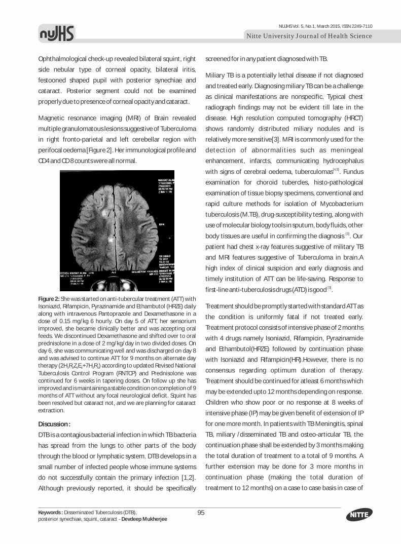
95
NUJHS Vol. 5, No.1, 2015, ISSN 2249-7110March
Nitte University Journal of Health Science
Keywords : Disseminated Tuberculosis (DTB),posterior synechiae, squint, cataract - Devdeep Mukherjee
Ophthalmological check-up revealed bilateral squint, right
side nebular type of corneal opacity, bilateral iritis,
festooned shaped pupil with posterior synechiae and
cataract. Posterior segment could not be examined
properly due to presence of corneal opacity and cataract.
Magnetic resonance imaging (MRI) of Brain revealed
multiple granulomatous lesions suggestive of Tuberculoma
in right fronto-parietal and left cerebellar region with
perifocal oedema [Figure 2]. Her immunological profile and
CD4 and CD 8 counts were all normal.
Figure 2: She was started on anti-tubercular treatment (ATT) with Isoniazid, Rifampicin, Pyrazinamide and Ethambutol (HRZE) daily along with intravenous Pantoprazole and Dexamethasone in a dose of 0.15 mg/kg 6 hourly. On day 5 of ATT, her sensorium improved, she became clinically better and was accepting oral feeds. We discontinued Dexamethasone and shifted over to oral prednisolone in a dose of 2 mg/kg/day in two divided doses. On day 6, she was communicating well and was discharged on day 8 and was advised to continue ATT for 9 months on alternate day therapy (2H R Z E +7H R ) according to updated Revised National 3 3 3 3 3 3
Tuberculosis Control Program (RNTCP) and Prednisolone was continued for 6 weeks in tapering doses. On follow up she has improved and is maintaining a stable condition on completion of 9 months of ATT without any focal neurological deficit. Squint has been resolved but cataract not, and we are planning for cataract extraction.
Discussion :
DTB is a contagious bacterial infection in which TB bacteria
has spread from the lungs to other parts of the body
through the blood or lymphatic system. DTB develops in a
small number of infected people whose immune systems
do not successfully contain the primary infection [1,2].
Although previously reported, it should be specifically
screened for in any patient diagnosed with TB.
Miliary TB is a potentially lethal disease if not diagnosed
and treated early. Diagnosing miliary TB can be a challenge
as clinical manifestations are nonspecific. Typical chest
radiograph findings may not be evident till late in the
disease. High resolution computed tomography (HRCT)
shows randomly distributed miliary nodules and is
relatively more sensitive[3]. MRI is commonly used for the
detection of abnormalities such as meningeal
enhancement, infarcts, communicating hydrocephalus [4,5]with signs of cerebral oedema, tuberculomas . Fundus
examination for choroid tubercles, histo-pathological
examination of tissue biopsy specimens, conventional and
rapid culture methods for isolation of Mycobacterium
tuberculosis (M.TB), drug-susceptibility testing, along with
use of molecular biology tools in sputum, body fluids, other [3]body tissues are useful in confirming the diagnosis . Our
patient had chest x-ray features suggestive of military TB
and MRI features suggestive of Tuberculoma in brain.A
high index of clinical suspicion and early diagnosis and
timely institution of ATT can be life-saving. Response to [3]first-line anti-tuberculosis drugs (ATD) is good .
Treatment should be promptly started with standard ATT as
the condition is uniformly fatal if not treated early.
Treatment protocol consists of intensive phase of 2 months
with 4 drugs namely Isoniazid, Rifampicin, Pyrazinamide
and Ethambutol(HRZE) followed by continuation phase
with Isoniazid and Rifampicin(HR).However, there is no
consensus regarding optimum duration of therapy.
Treatment should be continued for atleast 6 months which
may be extended upto 12 months depending on response.
Children who show poor or no response at 8 weeks of
intensive phase (IP) may be given benefit of extension of IP
for one more month. In patients with TB Meningitis, spinal
TB, miliary /disseminated TB and osteo-articular TB, the
continuation phase shall be extended by 3 months making
the total duration of treatment to a total of 9 months. A
further extension may be done for 3 more months in
continuation phase (making the total duration of
treatment to 12 months) on a case to case basis in case of

96
NUJHS Vol. 5, No.1, 2015, ISSN 2249-7110March
Nitte University Journal of Health Science
Keywords : Disseminated Tuberculosis (DTB),posterior synechiae, squint, cataract - Devdeep Mukherjee
1. Ellner JJ. Tuberculosis. In: Goldman L, Schafer AI, eds. Cecil Medicine. 24th ed.Philadelphia, PA: Elsevier Saunders; 2011:chap 332.
2. Fitzgerald DW, Sterling TR, Haas DW. Mycobacterium tuberculosis. In: Mandell GL, Bennett JE, Dolan R, eds.Mandell, Douglas, and Bennett's Principles and Practice of Infectious Diseases. 7th ed. Philadelphia, PA: Elsevier Churchill-Livingstone; 2009:chap 250.
3. Surendra K. Sharma,Alladi Mohan &AbhishekSharma: Challenges in the diagnosis & treatment of miliary tuberculosis, Indian J Med Res 135, May 2012, pp 703-730
4. Kleigman, Stanton, St. Geme, Schor& Behrman: Nelson Textbook of thPediatrics, 19 Edition, Volume I, Saunders, Philadelphia, Chapter 207,
1006(2012)5. A Parthasarathy, PSN Menon, Piyush Gupta & MKC Nair: IAP Textbook
thof Pediatrics, 5 Edition, Volume 1 & 2, 272 & 363(2013)
delayed response and as per the discretion of the treating
physician. Under RNTCP, all patients shall be covered under [6]directly observed intermittent (thrice weekly) therapy .
Similar directly observed intermittent (thrice weekly)
therapy was employed in our patient for 9 months, to
which she has responded.
The current incidence of ocular Tuberculosis is
uncertain.1.4% of patients with PTB develop ocular
manifestations but many patients with ocular TB have no
evidence of PTB. Ocular TB is most often a result of
hematogenous spread during PTB or extra-pulmonary
tuberculosis. Infection may also occur via local spread from
an active sinus or meningeal infection. It is often unilateral
and asymmetric. Tuberculosis infection of the eyelid can
start discretely as a minute nodule and later become lupus [7]vulgaris, often accompanied by lymphadenopathy .The
most common manifestation of ocular involvement is
uveitis, usually presenting as a chronic anterior uveitis,
panuveitis or as a choroiditis. Broad posterior synechiae in
patients with latent TB have been highly suggestive of
tuberculous uveitis in India and Singapore. The most
frequent complications related to TB-uveitis included [8]cystoid macular edema (40%) and cataract (38.9%) Other
anterior segment presentations include conjunctival
granulomas, phlyctenulosis, sclerokeratitis, interstitial [7]keratitis, episcleritis .
The most common ocular sign in posterior segments is
choroidal mass followed by choroiditis [7].The presence of
choroidal lesions, with or without inflammation, is strongly
correlated with systemic disease. The majority of the
choroid tubercles are unilateral, and can range in size from
1-4mm to several disk sizes in diameter [9].Other posterior
segment findings include optic neuropathy and cranial [9]nerve palsies .
In our case ocular findings were bilateral squint, right side
nebular type of corneal opacity, bilateraliritis, festooned
shaped pupil with posterior synechiae and cataract.
Posterior segments could not be examined properly due to
presence of corneal opacity and cataract. We are reporting
this case as Disseminated Tuberculosis is an uncommon
presentation of Tuberculosis and furthermore she had
distinct ocular findings.
We would specifically like to emphasise that whenever TB
is found in any organ, evidence of TB in other locations
should be looked for, as early diagnosis and prompt
institution of proper therapy is utmost importance for TB
especially for DTB.
6. Ashok Kumar, Devesh Gupta, SharathBuruginaNagaraja, Varinder Singh, GR Sethi and Jagadish Prasad:Updated National Guidelines for Pediatric Tuberculosis in India, 2012,Indian Pediatrics,P-305,Volume 50__March 16, 2013
7. Carolyn T. Bramante, BA Elizabeth A. Talbot, Sivakumar R. Rathinam, Rosalind Stevens, Michael E. Zegans:Diagnosis of Ocular Tuberculosis : A Role for New Testing Modalities? Aravind Eye Care System
8. Reema Bansal, Aman Sharma, Amod Gupta: Intraocular Tuberculosis,Expert Rev Ophthalmol. 2012;7(4):341-349.
9. C. Michael Samson: Ocular Tuberculosi.www.uveitis.org/docs/dm/ ocular_tuberculosis.pdf?
10. Sharma A,Thapa B, Lavaju P:Ocular tuberculosis: an update, Nepal J Ophthalmol; 3 ( 5 ): 52-67(2011)
References:

97
NUJHS Vol. 5, No.1, 2015, ISSN 2249-7110March
Nitte University Journal of Health Science
Keywords : Periodontally involved teeth, Multidisciplinary approach,periodontal prosthesis - Prakyath Malli
Introduction :
Periodontal diseases are a complex condition occurring as
a result of etiological factors combined with the systemic
condition of the individual. Any individual whose
periodontium has been affected will require rehabilitation
of both periodontium and affected tooth structure. The
incidence of prosthetic treatment in patients with reduced
periodontal support is constantly on the rise.
Periodontal prosthetic patient is best managed with joint
consultation as a team consisting of Prosthodontist,
Periodontist and is frequently to include Orthodontist,
Endodontist and Oral Surgeon. Throughout the course of
the therapy the team should have a co ordination with
each other and patient as well so as each mode of
treatment is planned carefully to attain highest success in 1treatment.
Case characteristics of periodontal prosthetic patient /
p a t i e n t s r e q u i r i n g
periodontal prosthesis
Pat ients in n eed o f
periodontal prosthesis
clinically present following 2, 3situations (Fig 1)
· Periodontitis varying
Access this article online
Quick Response Code
PERIODONTAL PROSTHESIS - REVIEW1 2 3
Vinaya S Bhat , Krishna Prasad D & Prakyath Malli1 2 3Professor, Professor & HOD, Post Graduate, Department of Prosthodontics and Crown & Bridge, A B Shetty
Memorial Institute of Dental Sciences, Nitte University, Mangalore, Karnataka, India.
Correspondence :Prakyath Malli
Post Graduate, Department of Prosthodontics and Crown & Bridge, A.B. Shetty Memorial Institute of Dental Sciences, Nitte University, Mangalore - 575 018, Karnataka, India.
Mobile : +91 80957 27027 E-mail : [email protected]
Abstract :
Planning a course of treatment for periodontially compromised patients is a multidisciplinary approach involving sequential therapy by
a Periodontist and a Prosthodontist. Specific problems have to be recognized and a treatment plan has to be formulated from both the
disciplines to deliver best professional and clinical skills in management of a periodontially compromised individual. Hence this review
article addresses case characteristics, objectives, sequence of therapy and treatment planning in patients receiving periodontal
prosthesis.
Keywords : Periodontally involved teeth, Multidisciplinary approach, periodontal prosthesis
from moderate to advanced stage
· Mobility of the tooth with migration
· Posterior bite collapse
· Mal positioned teeth
· Improperly done restorations contributing to
periodontal problems
· Furcation invasions
· Plaque accumulation, root sensitivity, root caries and
gingival inflammation due to increased exposure of
proximal root concavities and flutings.
· Poor esthetics
· Difficulty in mastication due to absence of stable
occlusion
· Para functional habits leading to incisal and occlusal
wear
· Radiographicaly – Deep angular infra bony defects,
osseous cratering, furcation involvement, horizontal 4 bone loss and loss of lamina dura (Fig 2)
Objectives of therapy in periodontal prosthetic patients
The best approach to manage a periodontal prosthetic
patient is as a team which consist mainly a Prosthodontist
and a Periodontist and sometimes also includes the
Orthodontist, Endodontist and Oral surgeon. The team
must have co ordination during the course of treatment
Review Article

and also communication with the patient is necessary for
better treatment outcome. The main purpose of
periodontal aspect of the program is to establish a sound
foundation in which the final prosthesis will be placed.
5, 6Its objectives are .
· Removal of local and environmental etiologic factors
· Plaque control and oral hygiene maintenance during the
course of therapy
· Removal of pockets
· Restoring osseous and gingival contours
· Removal of furcal invasions by combined periodontal,
endodontic and prosthetic procedures.
· Periodic recall and maintenance program.
The goal of the treatment program should establish the
physiologic form and function of all the teeth and should
also control tooth mobility by mechanical stabilization
through splinting. The prosthesis must have the following 5, 6designs.
· It should establish physiologic occlusion
· It should stabilize the mobile teeth
· Development of embrasure form and proximal contact
relationships, marginal fit and coronal contours
· Replacing in adequate restorations
· Establishing esthetic and phonetic features.
Following are different types of periodontal prosthesis
1. Hawley Bite Plane Therapy.
2. Provisional Restoration and Stabilization.
3. Periodontal Splints.
4. Restoration Treatment Procedures Using Pins in
Periodontal Prosthesis.
5. Telescopic Crown Prosthesis.
6. Semi Precision and Precision Attachment.
7. Gingival Prosthesis.
1) Hawley bite plane therapy
The maxillary Hawley appliance with an anterior bite plane 7is useful in periodontal prosthetic cases in numerous ways.
98
NUJHS Vol. 5, No.1, 2015, ISSN 2249-7110March
Nitte University Journal of Health Science
Keywords : Periodontally involved teeth, Multidisciplinary approach,periodontal prosthesis - Prakyath Malli
Whenever there is occlusal trauma this appliance serves
the following purpose
· Provides rest for neuro musculature and periodontal
attachment apparatus
· Posterior occlusal disarticulation
· Control of local etiologic factors causing inflammation8, 9· Enhance healing of occluso traumatic lesions.
2) Provisional restoration and stabilization (fig 3 )
Provisional restorations help in management of
periodontal prosthetic patient and are placed during the
surgical phase of therapy. One of the most important
functions of provisional restoration is stabilization of
mobile teeth. However, during the early phases of initial
therapy will result in decrease in tooth mobility through
the control of inflammation and initial occlusal
adjustments, lesions of irreversible and progressive tooth 10, 11mobility must be treated.
3) Periodontal splints
Splinting is mechanical joining to teeth to enhance their
ability to withstand forces placed on them. Splinting of
teeth whether temporary or permanent, is adjunctive
therapy; it will not cure periodontal disease. However
dental splinting can prevent
a) Pathologic migration
b) Retain teeth in position
c) Facilitate treatment during scaling, curettage, and
periodontal surgery
d) Support teeth so that the effect of occlusal interferences
can be more readily located and removed by occlusal
equilibration
e) Stabilize teeth so that occlusal forces are distributed to a
combination of teeth rather than to individual teeth.
Indications and contraindications of splinting
Indications
a) Usually, FPD's and splints are preferable to RPD's with
advanced loss of periodontal support. They provide
rigidity and a more favorable force distribution to the 12remaining periodontium.

99
NUJHS Vol. 5, No.1, 2015, ISSN 2249-7110March
Nitte University Journal of Health Science
Keywords : Periodontally involved teeth, Multidisciplinary approach,periodontal prosthesis - Prakyath Malli
b) There is an agreement that splinting of mobile teeth and
FPD's, after periodontal and initial occlusal therapy is
indicated when mobility is increasing and interfering
with chewing ability and comfort. Lindhe indicates
splinting for
· Progressive mobility of the tooth as a result of
gradual increasing width of the periodontal ligament
in teeth with reduced height of the alveolar bone.
· Increased bridge mobility despite splinting
c) Progressive mobility can be controlled by uni lateral
splints even though resistance to bucco or labiolingual
mobility is less than mesiodistal mobility.
d) Increased bridge mobility requires cross arch splinting.
The main objective of splinting is to produce an
environment where total mobility of the splint is normal 13 or at least no longer increasing.
e) Nyman et al. have demonstrated long term splint
stability despite minimal periodontal support and hyper 14mobility of isolated abutment teeth .
Contraindications
a) Most patients with moderate periodontitis, having
slight or no detectable mobility after periodontal
treatment, do not require fixed splints
b) Lindhe described two situations
1. Increased mobility of the tooth with increased width
of the periodontal ligament, but normal height of the
alveolar bone
2. Increased mobility of a tooth with increased width of
the periodontal ligament and reduced height of the 12alveolar bone .
c) Splinting is contraindicated in patients with gingivitis.
Resolution of inflammation by root planning and
reduction of occlusal prematurities will usually 12,15,16significantly reduce detectable mobility.
d) Patients with advanced periodontitis have varying
numbers of teeth that exhibit severe bone loss,
advanced mobility, and edentulous areas.
Classification of stabilization by Splinting
Teeth can be splinting by several methods.
A) Temporary stabilization
I. Removable extra coronal splints
II. Fixed extra coronal splints
III. Intracoronal splints
IV. Etched metal resin bonded splints.
B) Provisional stabilization
I. Acrylic splints
II. Metal bond and acrylic splints.
C) Long term stabilization
I. Removable splints
II. Fixed splints
III. Combination removable and fixed splints
4) Restorative treatment procedures using pins in
periodontal prosthesis (Fig 4 )
Parallel and Non parallel pin techniques are used in
dentistry for teeth that exhibit mobility. It is temporarily
splinted for cases exhibiting marked mobility to re evaluate
prognosis, if the teeth is firm then prognosis is more
favorable after which permanent stabilization is instituted.
Advantages of pin retained restoration
1. Retention of restoration
2. Conservation of tooth structure
3. Operators chair time is less
4. Good esthetics of natural labial and buccal enamel
5. Hygiene better than complete veneer crowns
6. Reduced laboratory procedures
7. Less cost than complete veneer crowns
8. Good stabilization of mobile teeth can be achieved by
parallel pin technique
Disadvantages of parallel pin technique
1. Possibility of encroachment upon dental pulp
2. More attention to detail needed by the operator
3. Technicians un familiar with the technique
4. Abutment restoration might become loose if depth of
the pins into sound tooth structure is not sufficient
5. If buccal and lingual walls are thin and un supported
enamel fracture might occur
Advantages of non parallel pin technique
These advantages are in addition to those of parallel pin
technique

100
NUJHS Vol. 5, No.1, 2015, ISSN 2249-7110March
Nitte University Journal of Health Science
Keywords : Periodontally involved teeth, Multidisciplinary approach,periodontal prosthesis - Prakyath Malli
1. May be used with crowded or maltose teeth 172. No necessity of paralleling device.
5) Telescopic crown prosthesis in severe periodontal
destruction (Fig 5 )
Telescopic crowns prosthesis consists of covering the
prepared teeth with thin gold copings, or thimbles, that in
corporate shoulder on the gingival margins to which super 18 structure is abutted with temporary luting agents. The
surface of the copings are un polished because cement will
not adhere to a polished surface. The copings may be
soldered or removed as individual units with the
superstructure that provides splinting. Often these two
approaches are integrated in a single case. If complete arch
splinting is necessary, cross linkage of abutments across
soldered copings allows a bilateral splint to be divided into 19 manageable units.
Advantages :
1. It is difficult to prepare the walls of the short teeth to a
minimal taper for resistance form. Copings are
contoured to nearly parallel axis to overcome this
problem.
2. Paralleling of severely tipped abutment teeth is possible
without orthodontics.
3. Full arch periodontal splinting is accomplished in
multiple smaller segments.
4. Abutment teeth are protected if superstructure is
dislodged.
5. Super structures temporarily cemented can be removed
for treatment of recurrent periodontal disease.
6. Teeth with minimal prognosis can be intentionally
included in a splint, which can be eventually extracted
with the abutment crown converted into poetic
Disadvantages :
1. Retention between the coping and tooth must be
greater than between super structure and coping
2. Fit is difficult between the super structure margins and
coping finish lines
3. Esthetic limitations in the anterior areas due to the bulk
of the double castings with narrow embrasure spaces
4. In short abutment teeth with narrow embrasure spaces
telescoping is contra indicated.
6) Semi precision attachment and precision attachment in
terminal borderline cases (Fig 6 )
Whenever there is a periodontally involved arch principles
of stabilization and intra arch support can be achieved with
intra coronal semi precision or precision attachment. As a
general rule removable partial dentures with extra coronal
clasps should not be used as a splinting vehicle as they
create forces too great for periodontally involved teeth to
withstand. The intra coronal retainers on the other hand,
allow for better control of bucco lingual, mesio distal and
vertical forces.
Advantages of intra coronal retainers over extra coronal
clasping
1. It prevents lateral stresses in the periodontium of the
retainers during insertion or removal, and stabilizes the
abutment teeth against lateral forces. With intra
coronal retainer's attachments, the forces of function
are directed vertically.
2. The intra coronal attachments use parallel walls rather
than undercuts to resist the forces of displacement.
3. It can be used for short clinical crowns, and it eliminates 20the display of clasps.
Disadvantages
1. Intra coronal attachments require prepared abutments
and castings; but in the case of periodontally involved
and mobile teeth, splinting is usually recommended.
2. They also require complicated clinical and laboratory
procedures.
3. Eventually wear, with resultant loss of frictional
resistance to denture removal
4. They are difficult to repair or replace
5. Intra coronal attachments are effective in proportion to
length, thus they are least effective in short teeth. It is
true that at least two third of the length of the
manufactured precision attachment must be used for
satisfactory results, but now with the milled in semi
precision cases and other developed techniques, this is
no longer a factor of concern.

101
NUJHS Vol. 5, No.1, 2015, ISSN 2249-7110March
Nitte University Journal of Health Science
Keywords : Periodontally involved teeth, Multidisciplinary approach,periodontal prosthesis - Prakyath Malli
Fig1. : Example of periodontal prosthetic patient
Fig2. : Radiographic assessment
Fig3. : Provisional restoration used as a splintto stabilize mobile teeth
Fig4. Periodontal splint positioned on the teeth
Fig 5. : Telescopic crown prosthesis Fig6. : Semi Precision attachment
Fig7. : Gingival prosthesis Fig8. : Gingival prosthesis used to cover ridge defects
7) Gingival prosthesis (Fig 7 and 8 )
Gingival replacement prostheses have historically been
used to replace lost tissue when other methods of
regenerative procedures are considered un predictable or
impossible, with this method, large tissue volumes are
easily replaced.
Tissue replacement prostheses are used to replace tissue
lost through surgical gingival procedures, trauma, ridge
resorption or traumatic tooth extraction. Materials used
for gingival prostheses include pink auto cure and heat-
cured acrylics, porcelains, composite resins and
thermoplastic acrylics, as well as silicone-based soft
materials.
Gingival defects are treated with surgical or prosthetic
approaches. Minor procedures to rebuild papillae and

102
NUJHS Vol. 5, No.1, 2015, ISSN 2249-7110March
Nitte University Journal of Health Science
Keywords : Periodontally involved teeth, Multidisciplinary approach,periodontal prosthesis - Prakyath Malli
grafting procedure s that may involve not only soft-tissue
manipulation but also bone augmentation to support the
soft tissue. With the success of these treatments the result
mimics the original soft tissue contours. It is possible to
create esthetically pleasing and anatomically correct tissue
contours when small volumes of tissue are being
reconstructed, but this method is unpredictable when a
large volume of tissue is missing. The surgical costs, healing
time, discomfort and unpredictability make this choice
unpopular.
Prosthetic replacement, with acrylics, composite resins,
porcelains and silicones, is a more predictable approach to
replacing lost tissue architecture. It is especially useful
when a larger amount of tissue needs replacement. Ideal
tissue contours can be waxed, processed and then colored
to match the surrounding tissue. The patient need not
undergo any additional surgical procedures and receives an
esthetically pleasing, functional restoration. It is possible to
show the patient a waxed-up result or even take a try in
prosthesis directly to the mouth for evaluation before 21significant treatment is initiated.
Discussion :
Prosthodontic treatment of a patient with periodontally
involved teeth is a multidisciplinary approach. It includes
fabrication of a simple fixed prosthesis to complicated
procedures like full mouth rehabilitation. Each patients
presenting with different clinical situations has challenging
treatment to be formulated by the operators. Sequence of
therapy, combination of different aspects of treatment and
operator skill play an extremely critical role in the success
of managing such patients. The objectives of treating these
patients have been arrived at removal of all etiologic
factors, treating the cause of periodontal breakdown,
prosthetic stabilization and maintaining oral health with
recall program. The array of prosthesis includes bite plane
appliances, splints to precision attachment and telescopic
crowns. Gingival prosthesis is another form of periodontal
prosthesis given for replacing lost soft tissues. Accurate
diagnosis holds a key for the longevity of treatment success
in these cases.
Patient cooperation is yet another aspect for good
management of patients. Since it is a multidisciplinary
approach, several appointments may be required to
complete the course of treatment and patient compliance
in maintaining also plays a major role in long term success.
This review article has compiled all aspects of treatment
with periodontal prosthesis.
1. Meyers H.E, Baraff L.S. A restorative approach to periodontally treated mouth. Dent clin north am 1965; 9: 13.
2. Amsterdam M. Periodontal prosthesis. In: Goldman H, Cohen DE (eds). Periodontal therapy, ed 5. St louis: mosby, 1973:990.
3. Stahl S.S: Marginal lesion. In Goldman H.M, Cohen D.W (eds): Periodontal therapy, Ed 4. St. Louis: mosby 1968; 120-121.
4. Rosenberg M. Periodontal and prosthetic management for advanced cases: quintessence publication co Inc, 1988.
5. Meyers H.E, Baraff L.S. A restorative approach to periodontally treated mouth. Dent clin north am 1965; 9: 13.
6. Rosenberg M.M. Management of osseous defects, furcation involvement and periodontal pulpal lesions. In clark jw (ed) : Clinical dentistry, vol 3, chap 10. Hagerstown, md: Harper and row, 1979.
7. Heckert L. Prerestorative therapy using a modified hawley splint. J prosthet dent 1980; 43:126.
8. Amsterdam M. Periodontal prosthesis. Twenty five years in retrospect. Alpha omegan, 1974; 67: 29-31.
9. Miller G.M, Kreuzer D.W. The modified hawley appliance. part II. Int j periodont rest dent 1982; 2(1): 29-45.
10. Amsterdam M, Fox L: Provisonal splinting: Principles and techniques. Dent clin north am1959; 3: 73-99.
11. Lindhe J, Nyman S. The role of occlusion in periodontal disease and the biological rationale for splinting in treatment of periodontitis. Oral sci rev1977; 10:11.
References :12. Lindhe J. Textbook of clinical periodontology. Copenhagen. 1983,
Munksgaard; 466-479.13. Lindhe J, Nyman S. The role of occlusion in periodontal disease and the
biological rationale for splinting in treatment of periodontitis. Oral sci rev1977; 10:11.
14. Nyman S, Lind he J, Lundgren D. The role of occlusion for stability of fixed bridges in patients with reduced periodontal support. J Clan Periodontol1979; 50: 163-169.
15. Keel W, Sisisky H, Phillips C. The effect of splinting on tooth mobility. I. During initial therapy. J Clan Periodontal 1979; 6:45-58.
16. Polson A.M , Meitner S.W , Sander H.A. Reversibility of bone loss due to trauma alone and trauma super imposed upon periodontitis. J Periodont 1976; 11:290.
17. Gredier A, William R. Periodontal prosthesis . The c.v. Mosby company st Louis; 1968.
18. Pichard J.F, fedrer,m. A modern adaptation of the telescopic principle in periodontal prosthesis. J periodontal 1962; 33:360-364.
19. Pugh C.E, Smerke J.W. Rationale for fixed prosthesis in the management of advanced periodontal disease. Dent Clin North Am 1969; 13(1): 243-262.
20. Goodman J, Goodman H.W: Balance of force in precision free end restorations. J Pros Dent 1963;.13:302.
21. Barizlay I, and Tamblyn I: Gingival prosthesis- review. J Can Dent Assoc 2003; 69(2):74–8.

103
NUJHS Vol. 5, No.1, 2015, ISSN 2249-7110March
Nitte University Journal of Health Science
Keywords : patient navigation, nurse navigator, cancer care,patient outcomes - Shejila C H
Introduction :
Cancer is a huge global health burden; touching all region 1and people from different socioeconomic level.
Continuum of cancer care spans a lengthy trajectory from 2initial diagnosis through treatment. Cancer care is
complex and usually requires a team based approach from
multiple health professionals to ensure quality and
continuity in care. Coordination of cancer care has gained
increased attention recently because it can critically and
positively affects patient
safety as well as care
quality across different 3services and settings.
Nurses have a major role in
cancer patients' care and
many specific nursing
roles in cancer services are
ONCOLOGY NURSE NAVIGATOR PROGRAMME - A NARRATIVE REVIEW
1 2 3Shejila C H , Mamatha S Pai & Donald J Fernandes 1 2Research Scholar, Professor & HOD, Department of Child Health Nursing, Manipal College of Nursing Manipal,
3Professor & HOD Department of Radiotherapy & Oncology, Kasturba Medical College,Manipal University, Manipal, Karnataka, India.
Correspondence :Shejila C H
Research Scholar, Manipal College of Nursing Manipal,Maniapl University, Manipal - 576 104, Karnataka, India.
Mobile : +91 80959 97753 E-mail : [email protected]
Abstract: Introduction : Cancer Care is complex and often requires multiple health care professionals to work in a coordinated and integrated
fashion to deliver most effective care available. A team based approach from surgeons, medical oncologists, radiologists, nurse
specialists and social worker is required to ensure quality and continuity in care. Specific nursing roles in cancer services are expanding
and evolving.
Objective : To explore the role of nurse as navigator in oncology care.
Methodology : Literature survey was conducted from published journals, text books and online databases (CINAHL, Pubmed, Proquest,
Ovid, Medline and Science direct) from January 2000 to January 2014 by using terms pivot nurse ,oncology nurse, case manager, nurse
navigator ,patient navigator, oncology nurse navigator along with terms oncology or cancer care.
Results : Literature review on nurse navigator programme reveals positive outcomes in various aspects of cancer care, but lack of
consensus in study population, intervention settings, outcome measures and methodologies have been noted.
Conclusion : Patient navigation using nurses is viewed as an effective strategy to improve standard of oncology care delivered. This
review provides evidence that nurse navigator programme can improve specific patient outcomes in cancer care.
Keywords : patient navigation, nurse navigator, cancer care, patient outcomes
Access this article online
Quick Response Code
expanding and evolving. Certain specific nursing roles and
its application in cancer care have been tested in a few
countries. A primary literature search reveals lack of
published literature/studies from India. Hence the
objective of this review is to explore the emerging role of
nurse as navigator in oncology care.
Methodology :
Literature survey was conducted from published journals,
text books and online databases (CINAHL, Pubmed,
Proquest, Ovid, Medline, Science Direct) from January
2000 to January 2014 .Since nurse navigation is a new
term the key words used were oncology nurse, pivot nurse,
case manager, patient navigator, nurse navigator, oncology
nurse navigator along with terms oncology or cancer care.
Literature review
Literature review is organized under following headings
Review Article

104
NUJHS Vol. 5, No.1, 2015, ISSN 2249-7110March
Nitte University Journal of Health Science
Keywords : patient navigation, nurse navigator, cancer care,patient outcomes - Shejila C H
o Concept of Patient Navigation
o Role of oncology nurse
o Oncology nurse navigator
o Effectiveness of patient/nurse navigation
programme
Concept of patient navigation
The word navigate is derived from two Latin words - Navis
(ship) and agree (to drive).
The meaning of word navigate is to travel over or through 4 safely. Navigation is a process whereby a patient is given
personalized care and support across the continuum of
cancer care The first patient navigation programme was
created by Harold P. Freeman at Harlem Hospital, New York
in 1990 and thereafter the model continues to evolve and 6 expand. Patient navigator programmes share
characteristics with case management and it is focused on 7 a range of health care needs and issues. The model focuses
on meeting the needs of patients such as providing disease
and treatment related information and support and linking
with other health care professionals. It is noted that
services provided include detection of cases, identification
of barriers to care, development and implementation of
care plan and tracking throughout treatment and its 8 completion.
Role of oncology nurse
Nurse's role as a care provider, manager, principal educator
and advocate has developed over many years. Oncology
nurses are remarkably involved in education of patients,
their families, peers and public. They provide patients with
information about disease, management of side effects,
nutritional care, emotional coping and other skills that can
be developed and nurtured. Oncology nurses are suitable
for this task because of their greater knowledge and 9 understanding of various aspects of cancer care. Patients
need a qualified nurse to provide information and
education about their care, manage their needs and
problems from diagnosis through treatment and
survivorship and help them to develop coping 10mechanisms.
5 .
Oncology nurse navigator
The term nurse navigator has introduced to the oncology
health care setting in recent years but seems to continue to 11fall under the broad heading of patient navigator. The
national comprehensive cancer network (2011) stated that
the patient navigator is most often a nurse and used the
term patient navigation interchangeably with case 12manager. The Oncology Nurse Navigator is a professional
whose clinical nursing expertise guides patients, families
and their caregivers in informed decision-making;
collaborating with a multi-disciplinary team , allow for
timely cancer screening, diagnosis, treatment, and
supportive care across the cancer continuum.
(The National Coalition of Oncology Nurse Navigators, 13 NCONN). The role of a patient navigator includes some
aspects of case management, advocacy, patient education 14 and social work . But the Navigator role goes beyond
minimal function of a case manager or a patient advocate.Navigators oversee the treatment process ,provide
information and support to the patient, link with other
professionals in treatment process and act as a single,
constant contact. It is a more widely used term in
addressing problems related to integration, coordination
and continuity of cancer care, and is fit in with the concept
of a holistic approach that centres on the quality of life of
the person with cancer. Nurse navigator role has been
implemented in different health care settings and is helpful
to the multidisciplinary team for continuum of patient care 15 from diagnosis to survivorship. Specific roles of nurse
navigator in cancer care is illustrated in Figure 1
Effectiveness of patient/nurse navigation programme
A randomized controlled trial conducted by Myriam
Skrutowski et. al revealed the impact of a pivot nurse in
decreasing Symptom distress, fatigue and improving QOL 16in patient with lung and breast cancer.
A systematic review conducted by Robinson et al on patient
navigation in breast cancer reported that adherence to 17cancer care enhanced with patient navigation.
A descriptive study conducted by Swanson J on oncology

105
NUJHS Vol. 5, No.1, 2015, ISSN 2249-7110March
Nitte University Journal of Health Science
Keywords : patient navigation, nurse navigator, cancer care,patient outcomes - Shejila C H
nurse navigator's role in management of distress revealed
that visits of navigator have a statistically significant effect 18on inpatients' distress scores (p=0.044).
Carrol JK in a qualitative study with 35 Breast and colorectal
cancer patients tried to explore Patients experiences with
navigation. The results of study reveal that Navigated
patient received emotional support and assistance with
informational needs, problem solving and coordination of 19cancer care.
Case B conducted a systematic review to explore impact of
oncology nurse as navigator on specific patient outcomes
revealed that nurse navigator programme showed positive
outcomes related to time of diagnosis, different mood
states, support and continuity of care ,patient satisfaction 20and cost outcomes.
In a randomized controlled trial Kevin Fiscella et al revealed
that patient navigation has improved satisfaction with care 21in breast and colorectal patients.
Few studies have been illustrated in the form of a
methodological matrix in Table 1
Table 1 : Studies on Patient Navigation/Nurse Navigation in Cancer Care
Author & Year
Ann Hook 2012
Carrol JK 2010
Robinson et al
2010
Case B 2010
Swanson J
2010
Fiscella k 2008
Skrutowski M,
2008
Aim
Explore patient
satisfaction in newly
diagnosed breast
cancer patients
Explore Patients
experiences with
navigation
Evaluate outcome of
navigation in breast
cancer care
Explore impact of
oncology nurse as
navigator on specific
patient outcomes
Determine role of ONN
in distress
management
To assess Time of
completion of
treatment,
satisfaction with care,
psychological distress
Examine presence of
pivot nurse in relieving
symptom distress,
fatigue, improve QOL
and use of health
resources
Study Variables
Patient satisfaction
Navigation care
Screening,
diagnosis,
treatment
Time of diagnosis,
support and
continuity of care,
mood states,
satisfaction and
cost outcomes
Distress scores
Time of
completion of
treatment,
satisfaction with
care, psychological
distress
fatigue, symptom
distress, QOL and
use of health
resources
Design
Non experimental,
descriptive study
Qualitative
Systematic review
Systematic review
Descriptive study
RCT
RCT
Sample and
Sample size
103 patients in
rural community
setting
35 Breast and
colorectal cancer
patients
Studies from 1990-
2009
18 studies
55 in patients with
cancer
438 breast and
colorectal cancer
patients
113 patients with
breast and lung
cancer
Major Findings
72% satisfied with NN
Navigated patient received
assistance with informational
needs, problem solving,
emotional support and
cancer care coordination
Evidence supports patient
navigation has improved
many aspects of breast
cancer care
Positive outcomes in time of
diagnosis, support and
continuity of care, mood
states, satisfaction and cost
outcomes
Patients seen by ONN
Shows lower distress scores
No statistical difference in
completion of treatment,
high satis faction, less
psychological distress
No significant difference in
Symptom distress, fatigue,
QOL and use of health
resources

106
NUJHS Vol. 5, No.1, 2015, ISSN 2249-7110March
Nitte University Journal of Health Science
Keywords : patient navigation, nurse navigator, cancer care,patient outcomes - Shejila C H
Results
The literature reviewing nurse navigator programmes has
proved positive outcomes in various aspects of cancer care
like diagnosis, timeliness in completion of treatment,
support and continuity of care, patient satisfaction and use 16-21 of health resources. But divergency in studies has been
noted in study population, intervention settings, outcome
measures and methodologies. Most of the research studies
are conducted in breast cancer, lung cancer, head and neck
cancers and colorectal cancers.
Discussion
Patient and their families need a constant support nurse,
advocate or a navigator to guide them through the
increased perplexity of cancer care. Since the introduction
of patient navigation into oncologic care, nurse researchers
have clearly identified specific care outcomes that result
from presence of oncologic nurse navigator. The nurse's
role in navigated care for cancer patients has been defined
22and implemented diversely . Lack of consensus in areas
like qualification of navigator, scope of practice, health care
setting, disease or area of navigation focus contributes to 23diverse use of this concept in patient care. Lack of
standardization in navigator programmes challenges
researchers in this field of study.
Future Directions and Implication for Nursing Practice
Nurse navigator programme is a promising resolution for
patient care inadequacies, effective means for reducing
barrier in oncology care and increase patient satisfaction
and quality of care. This model has been already
implemented in most of the developed countries.
Interventions need to be developed in developing
countries like India to enhance treatment adherence, since
non compliance with treatment due to increasing cost is a
major problem. Research based evidences are lacking on
use of an appropriate conceptual framework for
implementation of a nurse navigation programme. Further
Figure 1. : Schematic Representation of Nursing navigator’s role in cancer care
CANCER CARE CONTINUUM
Multidisciplinarycollaboration,care planning,patienteducation,dischargeplanning and education
Follow upSupportivecareReferral tocommunityorganization -
NURSES NAVIGATION(clinical knowledge, communication skills, problem solving skills, cancer care system Knowledge)
PREVENTIONSURVIVALORPALLIATION
TREATMENTDIAGNOSIS
EarlyDetection
Emotionalsupportcoordination andeducation referral torelevant department

107
NUJHS Vol. 5, No.1, 2015, ISSN 2249-7110March
Nitte University Journal of Health Science
Keywords : patient navigation, nurse navigator, cancer care,patient outcomes - Shejila C H
Conclusion :
Navigation is a process by which encompasses assessment
of patient needs, development of a plan for education,
c o o r d i n a t i o n , c o m m u n i c a t i o n , s u p p o r t a n d
implementation of same for effective transition through
the illness and evaluation of its effect on patient , family
and care givers . Patient navigation using nurses is viewed
as an effective strategy to improve standard of oncology
care as well as to achieve organizational outcomes.
studies are warranted in evaluating patient satisfaction
using nurse navigation in various types of cancers as well as
in different populations of patients. Specific tools to test
and validate patient navigation programmes are lacking, so
identification of key components and its relevant
evaluation tools to test effectiveness of navigation
programme is essential. Replication of studies exploring
various patient outcomes and nurse navigation will offer
the necessary evidence base to trend in oncologic nursing.
References :
1. America Cancer Society. Cancer Facts & Figures 2013. 2013 [cited 2013 27 February]; Available from: http://www.cancer.org/acs /groups /content/@epidemiologysurveilance/documents/document/acspc-036845.pdf. Retrieved on
2. Doll R, Barroetavena MC, Ellwood AL, Fillion L, Habra M, Linden W. The cancer care navigator. Toward a conceptual framework for a new role in oncology. Oncology Exchange. 2007;6(4):28-33.
4. Hopkins J, Mumber MP. Patient navigation through the cancer care continuum: An overview. Journal of Oncology Practice. 2009;5(4):150-2.
6. Freeman HP, Muth B, Kerner J. Expanding access to cancer screening and clinical follow-up among the medically underserved. Cancer practice. 1995;3(1):19.
July 2, 2014.
3. Lee T, Ko I, Lee I, Kim E, Shin M, Roh S, et al. Effect of nurse navigators on health outcomes of cancer patients. Cancer Nursing 2011; 34(5): 376-384.
5. American Academy of Oncology Nurse Navigators (2010) Home page. Retrieved on June 2014 from http:/www.aonnonline.org
7. Lantz, P. M., Keeton, K., Romano, L., & DeGroff, A. Case management in public health screening programs: the experience of the national breast and cervical cancer early detection program. Journal of Public Health Management and Practice. 2004; 10(6), 545-555.
8. Battaglia, T. A., Roloff, K., Posner, M. A., & Freund, K. M. Improving follow-up to abnormal breast cancer screening in an urban population. Cancer. 2007; 109(S2), 359-367.
9. Seek, A. J., & Hogle, W. P. Modeling a better way: navigating the healthcare system for patients with lung cancer. Clinical journal of oncology nursing.2007; 11(1), 81-85.
10. DeSanto-Madeya, S., Bauer-Wu, S., & Gross, A. Activities of daily living in women with advanced breast cancer. In Oncology nursing forum. 2007 ;34(4), 841-846
11. Curran C. R. Navigation the chaotic health care system. Nursing Economics. 2003. 21 (6), 261.
12. The national comprehensive cancer network (2011). The case manager or patient navigator: providing support for cancer patient dur ing treatment and beyond. Retr ieved fromhttp:// www.nccn.org/patients/living-with-cancer/understanding-
treatment/152-case-managers-for-cancer-patients.html13. The National Coalition of Oncology Nurse Navigators. Home page
.Retrieved on July 2014 from http://www.nconn.org/14. Fisher, S.M, Sauaia,A and Kutner , J.S. Patient navigation: A culturally
competent stratergy to address disparities in palliative care. Journal of palliative Medicine 2007;.10,1023-1028
17. Robinson –White S, Conroy B, Slavish KH, Rosenzwieng M .Patient navigation in breast cancer a systematic review. Cancer Nurse 2010;33(2):127-140
18. Swanson J, Koch L.The role of oncologic nurse navigator in distress management of adult patients. A retrospective study. Oncology Nursing Forum. 2010; 37(1):69-76
20. Case, M. A. B. Oncology nurse navigator. Clinical journal of oncology nursing. 2011;15(1): 33-40.
21. Fiscella, K., Whitley, E., Hendren, S., Raich, P., Humiston , S., Winters, P, et.al Patient navigation for breast and colorectal cancer treatment: a randomized tr ia l . Cancer Epidemiology Biomarkers & Prevention.2012; 21(10): 1673-1681.
22. Freund ,K.M, Battaglia, T, Calhoun E, Dudley D.J, Fiscella K, Paskett E, et.al. National cancer institute patient navigation research programme: methods, protocol, and measures.Cancer. 2008;113:3391-9
23. Darnell ,J.S. Patient navigation a call to action. Social work. 2007;52 (1):81-84.
15. Fillion L, De Serres M, Lapointe-Goupil R, Bairati I, Gagnon P, Deschamps M, et al. Implementing the role of patient-navigator nurse at a university hospital centre. Canadian Oncology Nursing Journal. 2006;16(1):11.
16. Skrutkowski M, Saucier A, Eades M, Swidzinski M, Ritchie J, Marchionni C, et al., editors. Impact of a pivot nurse in oncology on patients with lung or breast cancer: symptom distress, fatigue, quality of life, and use of healthcare resources. Oncology nursing forum; 2008: Onc Nurs Society.
19. Carroll JK, Humiston SG, Meldrum SC, Salamone CM, Jean-Pierre P, Epstein RM, et al. Patients' experiences with navigation for cancer care. Patient education and counseling. 2010;80(2):241-7.

108
NUJHS Vol. 5, No.1, 2015, ISSN 2249-7110March
Nitte University Journal of Health Science
Keywords : Anatomy, dissection, body donation, medical education- Bindhu S
Introduction :
Anatomy is the study of structure of human body and forms
the basic subject studied by undergraduate and
postgraduate students. Dissection of human cadavers
forms an integral part of anatomy learning and research.
Only source for the cadavers in the medical institutions will
be unclaimed bodies and few donated bodies. As the
number of medical colleges are increasing in the present
scenario, most of the medical schools experience 1difficulties in obtaining adequate number of cadavers . At
present, unclaimed bodies are the main source of cadavers
that is coming from the authorised government
institutions. So the body donation programmes should be
encouraged by the medical schools and regular campaigns
must be held to create awareness among the public
regarding Body donation.
Significance of Body Donation :
Anatomical donation or
body bequest is defined as
act of giving one's body
after death for medical
education and research
and thus a person can give
back to society a chance to
learn something that can
Access this article online
Quick Response Code
BODY DONATION AS GIFT TO MEDICAL SCIENCE FOR BETTER TOMORROW - LITERATURE REVIEW
1 2 3 4 5Meera Jacob , R K Avadhani , Rani Nallathamby , Meril Ann Soman & Bindhu S
1 2 3,4 5Assistant Professor, Professor & HOD, Post Graduates, Associate Professor, Department of Anatomy,Yenepoya Medical College, Mangalore, Karnataka, India.
Correspondence :Bindhu S
Associate Professor, Department of Anatomy, Yenepoya Medical College, Mangalore - 575 018, Karnataka, India.Mobile : +91 99456 66156 E-mail : [email protected]
Abstract :
Anatomy is one of the basic and very important subject studied by medical students at the beginning of their medical career. Best
method for learning anatomy is by dissection of human cadavers and this forms an indispensable part of training health care
professionals. Since the numbers of medical institutions are increasing, there is an increased demand of cadavers for anatomic
dissection. Body donation is an act of donating one's body after death for medical research and education. In this article an attempt has
been made to collect literature related to body donation, its importance and the ethical issues related to body donation.
Keywords : Anatomy, dissection, body donation, medical education
influence generations to come. Delmas suggested that the
donation is a clear will made by persons free and 2informed .Body donation is considered as an ultimate gift
which should be appreciated by the educators. Availability
of textbooks and internet information cannot replace the
cadaveric dissection in learning anatomy .So hands on
experience provided by the cadaveric dissection is superior 3to other artificial sustitutes . Apart from learning anatomy
3it is also used for developing new surgical techniques .
Body donation is the preferred source of availability of 4cadavers' worldwide .
History :
In ancient India human body was dissected by shushruta by 5about 500 B.C . It was his belief that for one to be a skilful
surgeon, one must first be an Anatomist. Even though the
issue of using humans for dissection was in opposition to
religious law, it was an essential tool for true understanding 6of human Anatomy .The concept of acquiring knowledge
by dissection of human body initially started in the
fifteenth century and it was initially done by Andreas
Vesalius (1514-1564). Slowly and steadily the importance
was realised. Once autopsy was accepted to establish the
cause of death, importance of dissection was more
enlightened. By establishment of Anatomy act 1832,
Review Article

109
NUJHS Vol. 5, No.1, 2015, ISSN 2249-7110March
Nitte University Journal of Health Science
Keywords : Anatomy, dissection, body donation, medical education- Bindhu S
7unclaimed bodies were used for anatomic dissection .By th th18 and 19 century, the anatomical dissection became
very popular in Unite States medical education. When the
demand for cadaveric supply increased, the thefts by grave
robbers became more common and sold the bodies to 7medical schools for dissection . Murder act established in
1752, permitted the use of corpses of executed criminals
for dissection. Donation of body to science was unheard till
Jeremy Bentham, philosopher donated his body to science 5in 1832 .
Body Donation and Anatomy Act :
Body donation is regulated by various acts according to
each country and is considered as the expression of
solidarity .In U.K, the Anatomy act was passed in 1832
which permitted the donation of the body of the deceased
by his kin. This permitted doctors, teachers of Anatomy and
bonafide medical students to dissect donated bodies. This
act was replaced by Anatomy act 1924, which was in turn 5replaced by Human Tissue Act 2004 . In India, Anatomy act
was enacted in 1949 and is adopted by all states. It calls for
the supply of unclaimed bodies to medical and teaching
institutes for the purpose of anatomical dissection and for
research purposes. It can be donated by relatives of 1deceased according to dead person's wishes . Anatomy act
is a state act published in State Government Gazette
.According to this almost anyone can donate for medical
research and education. It provides for the collection of a
dead body for teaching purpose, only if death occurs in a
state hospital or in public place within prescribed zone of
medical institution ,provided that police have declared a
lapse of 48 hours that there are no claimants for the body 8 and it could be used for medical purpose .Patnaik
suggested that a draft act should be made for all the states
to use as model for amendment of anatomy acts in order to
avoid the discrepancies existing between any two or more 9acts .
Factors Affecting Body Donation :
There are few researches that are conducted to know the
factors that influence the willingness of body donation.
Boulware et al showed that the important factors that can
influence body donation will be race, ethinicity,
demographic factors, awareness about body donation,
age, sex, education, occupation, income, religious aspects
and so on. He conducted a study among the households of
Maryland and observed that the older age is negatively 10associated with willingness to donate cadaveric organs .
He also showed that demographic and attitudinal factors
were also involved in the decision to donate one's own
body. Armstrong in his study noted that younger age group
were more willing to donate when compared to older one 11in Australia .Similar finding was also recorded by Alashek
et al, noted that lack of adequate knowledge , unease
about body manipulation and religious implications were 12the barriers for cadaveric donation .Golchet et al reported
that many factors l ike age,culture,personality
characteristics ,views on death and mortality and
humanitarian concerns influence people's opinion towards 13body donation .Bolt et al revealed that there are three
principal factors motivating Dutch people for body
donation like desire to be useful after death, negative
att i tude towards funera l and express ion of 14gratitude .Rokad and Gaikawad noted that the reasons for
Indian donors to donate their bodies was due to the fact
that the body should be utilised for mankind's benefit than 5being burnt to death . Negative factors that can affect
body donation were also studied. Most important factor
for less number of body donation in Indians is due to lack of
awareness. Few of the general population are aware of 14body donation . The attitude of anatomists towards body
donation is not well known. Organ donation is more
preferred when compared to body donation not only by 5general population but also by the medical personnals .
Role of Social Networks :
Society should play a major role in motivating the people to
donate their bodies for training of medical and other health
care professionals. According to Cantorovitch, main
reasons for “No body donation” are due to lack of
awareness, religious uncertainities, distrust of medicine
and hostility of new ideas. In the present scenario, mass 15media can play a major role in motivating the society .
Conesa et al studied the influence of television, press and

110
NUJHS Vol. 5, No.1, 2015, ISSN 2249-7110March
Nitte University Journal of Health Science
Keywords : Anatomy, dissection, body donation, medical education- Bindhu S
magazines, hoarding and posters, campaign about
donation, information given by health professionals
regarding body donation. It was observed that media with
greater impact was television, second was press and radio,
the third being magazines and talks with health 16professionals .
Body Donation Programme :
Body donation is a generous and unselfish act for those
who wish to be useful to living after death. There is no
substitute for cadaveric dissection in learning anatomy and
new surgical techniques. Body donation units should be
established in all medical schools and awareness
programmes should be designed. That factors that can
cause repulsion for body donation should be taken care.
Awareness programmes should be started with the help of
mass media Alashek et al suggested that public educational
campaigns should be conducted with religious
12leadership . Donors should be assured that their bodies
will be treated with dignity and respect. Practise of
honouring the cadavers at the commencement of medical 17course by students and teachers as followed in Korea
should be followed in all medical schools.
Conclusion :
Dissection of human cadavers is compulsory for medical
education. So one cadaver dissection can teach many
doctors who can treat thousands of patients and can be a
relief to their loved ones. So the decision of an individual to
donate his/her body is gift for advancement of medical
sciences. Government should encourage and educate the
public to create awareness about organ and body
donation. Body donation is the act of giving one's body
after death without any conditions for education and
research in medicine.
1. Ajita R. Singh YI. Body Donation and Its Relevance in Anatomy Learning. J Anat Soc India 2007; 56(1): 2-7
3. Agthong S,and Wiwanitkit V.Cadaver donation:a retrospective review at the king chulalongkorn memorial Hospital,Bangkok.The southeast Asian journal of tropical medicine and public health 2002; Suppl:166-7
2. Delmas V. Donation of bodies to science. Bull Acad Natl Med: 2001; 185 (5): 849-856. Lemmp H K. Perceptions of dissection by students in one medical school: beyond learning about anatomy. A qualitative study. Med Educ: 2005; 39: 318-325
4. Sehirli US, Saka E, Sarikaya O. Attitude of Turkish anatomists toward cadaver donation. Clin Ana. 2004; 17(8): 677-681
5. Rokade SA,Bahetee BH.Body donation ,A Review.Medical Journal of Western India.2013;41(1):36-40
6. Marios Loukas,Alexis Lanteri,Julie Ferraciola,R.Shane Tubbs.Anatomy in Ancient India:A focus on the Susruta Samhita.J.Anat(2010)217, pp646-650.
7. Rath G,Garg K.Inception of cadaver dissection and its relevance in present day scenario of medical education.J Indian Med Assoc.2006 Jun;104(6):331-3.
8. Subramanium B.V.Law in relation to medical men In:Modi's medical Jurisprudence and Toxicology .22 edition;Butterworths .New Delhi.pp.724-727.
9. Patnaik,V.V.G.(2002):Editorial .JAnatSocIndia;50(2):143-44.
References:
10. Boulware LE, Ratner LE, Cooper LA, LaVeist TA, Powe NR. Whole body donation for medical science: a population –based study. Clin Anat 2004;17(7);570-7.
11. Armstrong GT. Age: an indicator of willingness to donate. J Transplant Coord 1996;6(4):171-3.
12. Alashek W, Ehtuish E, Elhabashi A, Emberish W, Mishra A. Reasons for unwillingness of Libyans to donate organs after death. Lybian J Med 2009;4(3):110-3
13. Golchet ,G.,Carr,J.and Harris,M.G:Why do'nt we have enough cornea donors?.A literature review and Survey.Optometry;71(5):318-28.
14. Bolt S, Venbrux E, Eisinga R, Kuks JB, Veening JG, Gerrits PO. Motivation for body donation to science: more than an altruistic act. Ann Anat 2010; 192(2):70-4.
15. Cantarovich F. Public opinion and organ donation suggestions for overcoming barriers. AnnTransplant: 2005; 10(1): 22-25
16. Conesa C, Rios Zambudio A, Ramirez P, Canteras M, Rodriguez MM, Parrilla P. Influence of different sources of information on attitude toward organ donation: a factor analysis. Transplant Proc:2004; 36(5): 1245-1248.
17. Park JT, Jang Y, Park MS, Pae C, Park J, Hu KS, Park J S, Seung-Ho Han, Ki-Seok Koh, Hee-Jin Kim. The trend of body donation for education based on Korean social and religious culture. Anat Sci Educ 2011;4:33-8.

111
NUJHS Vol. 5, No.1, 2015, ISSN 2249-7110March
Nitte University Journal of Health Science
INSTRUCTIONS TO AUTHORS
INSTRUCTIONS TO AUTHORS
The Nitte University Journal of Health Science (ISSN 2249-7110) is
a peer-reviewed, indexed research publication printed every
three months. The NUJHS invites ethically-cleared, original
research articles in any area related to advancement of
knowledge in health sciences. The complete contents of each
issue of Nitte University Journal of Health Science are also
available for open access on the university website link.
. E-mail manuscript/s as
attachment/s to [email protected]. Send one hard copy
of submissions to Dr. Arunachalam Kumar, Editor NUJHS, Journal
Office, K.S. Hegde Medical Academy, Mangalore 575018, India
Submission format
(A) Review Articles:
• Reviews are written by researchers of considerable
experience in the field concerned. The authors should
review the recent trends or advances in that field in the
light of their own work.
• The major portion of the above articles should deal with
the up-to-date developments in the field in the 3 – 5 years.
Authors are advised to search Medline and other databases
on the internet, apartment from collecting information
using conventional methods.
• These articles besides should contain a covering letter, title
page, summary and keywords. The articles should be
written under appropriate sub-headings. The authors are
encouraged to use flow charts, boxes, cartoons, tables,
photographs of good resolution and figures for better
presentation. Some of the other details are given below:
(B) Original Research Articles:
These may either be a full length research article or a short
communication. These papers should be arranged into the
following sections:
1. Title page with authors name and affiliations
2. Abstract and key words
3. Introduction
4. Materials and Methods
5. Results
6. Discussion
7. Conclusion
8. Acknowledgement
9. References
10. Tables with captions separately
11. Figures with legends separately
Title page: It should be paginated as page 1 of the paper. It should
include the title, authors names and affiliations, running title,
address for correspondence including e-mail address and also the
http://nitte.edu.in/journal/
total number of pages, figures and tables. Title: Must be
informative, specific, unambiguous and short. It should not
exceed 150 characters.
Authors and affiliations: The names of authors and their
affiliations should be given. It should be made clear which address
relates to which author.
Address for correspondence: The corresponding authors address
should be given on the title page. The e-mail ID of the
corresponding author or the contact e-mail ID must also be
provided.
Abstract and key words: It must start on a new page carrying the
following information: (a) Title (without authors names or
affiliations), (b) Abstract, (c) Key words, (d) Running title. It should
not exceed 250 words excluding the title and the key words. The
abstract must be concise, clear and informative rather than
indicative.
The abstract must be in a structured form and explain briefly what
was intended, done, observed and concluded. The conclusions
and recommendations not found in the text of the article should
not be given in the abstract.
Keywords: Provides 3-5 keywords which will help readers or
indexing agencies in cross-indexing the study. The words found in
title need not be given as key words. Use terms from the latest
Medical Subject Headings (MeSH) list of Index Medicus. A more
general term may be used if a suitable MeSH term is not available.
Introduction: It should start on a new page. Essentially this
section must introduce the subject and briefly say how the idea
for this research topic originated. Give a concise background of
the study. Do not review literature extensively but provide the
most recent work that has a direct bearing if any on the subject.
Justification for research aims and objectives must be clearly
mentioned without any ambiguity. The purpose of the study
should be stated at the end.
Materials and Methods: This section should deal with the
materials used and the methodology (how the work was carried
out). The procedure adopted should be described in sufficient
detail to allow the experiment to be interpreted and repeated by
the readers, if desired. The number of subjects, the number of
groups, the study design, sources of drugs with dosage regimen or
instruments used, statistical methods and ethical aspects must be
mentioned under the section. The data collection procedure must
be described. If a procedure is a commonly used, giving a
previously published reference would suffice. If a method is not
well known (though previously published) it is better to describe it
briefly with due acknowledgement. Give explicit descriptions of
modifications or new methods so that the readers can judge their

112
NUJHS Vol. 5, No.1, 2015, ISSN 2249-7110March
Nitte University Journal of Health Science
INSTRUCTIONS TO AUTHORS
accuracy, reproducibility and reliability.
The nomenclature, the source of material and equipment used,
with details of the manufacturer in parentheses, should be clearly
mentioned. Drugs and chemicals should be precisely identified
using their non-proprietary names or generic names. If necessary,
the proprietary or commercial name may be inserted once in
parentheses. The first letter of the drug name should be small for
generic name (e.g., dipyridamole, propranolol) but capitalized for
proprietary names (e.g., Persantin, Inderal). New or uncommon
drug should be identified by the chemical name and structural
formula.
The does of drugs should be given as unit weight per kilogram
body weight e.g., mg/kg and the concentrations should be given
in terms of molarity e.g., nm or mM. The routes of administration
m a y b e a b b r e v i a t e d , e . g . , i n t r a - a r t e r i a l ( i . a ) ,
intracerebroventricular (i.c.v.), intra-gastric gavage (i.g.),
intramuscular (i.m.), intraperitoneal (i.p.), intravenous (i.v.), per
os (p.o.), subcutaneous (s.c.), transdermal (t.d.)etc.
Statistical Methods: The variation of data should be expressed in
terms of the standard error of mean (SEM) or the standard
deviation (SD), along with the number of observations (n). The
details of statistical tests used and the level of significance should
be stated. If more than one test is used it is important to indicate
which groups and parameters have been subjected to which test
and why.
Results: The results should be stated concisely without
comments. They should be presented in logical sequence in the
text with appropriate reference to tables and / or figures. The data
given in tables or figures should not be repeated in the text. The
same data should not be presented in both tabular and graphic
forms. Simple data may be given in the text itself instead of figures
or tables. Avoid discussions and conclusions in the results section.
Discussion: This section should deal with the interpretation,
rather than recapitulation of results. It is important to discuss the
new and significant observations in the light of previous work.
Discuss also the weaknesses or pitfalls in the study. New
hypotheses or recommendations can be put forth. Avoid
unqualified statement and conclusions not completely supported
by the data. Repetition of information given under Introduction
and Results should be avoided.
Conclusions: It must be drawn considering the strengths and
weaknesses of the study. Make sure conclusions drawn should
agree with the objectives stated under Introduction.
Acknowledgements: These should be typed on a new page.
Acknowledge only those who have contributed to the scientific
content or provided technical support. Sources of financial
support may be mentioned.
References: It should begin on a new page. The number of
references should normally be restricted to a maximum of 25 for a
full paper. Majority of them should preferably be of articles
published in the last 5 years.
Papers which have been submitted and accepted but not yet
published may be included in the list of references with the name
of the journal and indicated as “In press”. Avoid using abstracts as
references. The “unpublished observations” and “personal
communications” should not be used as references.
References are to be cited in the text by superscribed number and
should be in the order in which they appear. References cited only
in tables or in legends to figures should be numbered in
accordance with a sequence established by the first identification
in the text of the particular table or illustration. As far as possible
mentioning names of author(s) for reference should be avoided in
the text.
Please note the following examples for a journal article, a chapter
in multi-authored book, and a single-authored book respectively.
Vega KJ, Pina I, Krevsky B. Heart transplantation is associated with
an increased risk for pancreatobiliary disease. Ann Intern Med
1996; 124: 980-3.
More than six authors: Parkin DM, Clayton D, Black RJ, Masuyer E,
Friedl HP, Ivanov E, et al. Childhood leukaemia in Europe after
Chernobyl: 5 year follow-up. Br J Cancer 1996;73:1006-12.
Books: Entwistle N. In, Excellence in higher education. (De Corte
E., ed), 2003; pp. 83-99, Portland Press, London.
Bowden A fundamentals of enzyme kinetics, 3rd edn. 2004;
Portland Press, London.
Web references: As a minimum, the full URL should be given and
the date when the reference was last accessed. Any further
information, if known (author names, dates, reference to a source
publication, etc.), should also be given. Web references can be
listed separately (e.g., after the reference list) under a different
heading if desired or can be included in the reference list.
Check list for Tables
• Serially numbered in Arabic numerals?
• Short self explanatory heading given?
• Columns have headings?
• Units of data given?
• “n” mentioned?
• Mean ± SD or Mean ± SEM given?
• Statistical significance of groups indicated by asterisks or
other markers?
• P values given?
• Rows and columns properly aligned?
• Appropriate position in the text indicated?
Figures: Each figure must be numbered and a short descriptive
caption must be provided. A computer drawn figure with good

113
NUJHS Vol. 5, No.1, 2015, ISSN 2249-7110March
Nitte University Journal of Health Science
contrast is acceptable. Sometimes, raw data for graphs may be
required in Excel sheet when the article is accepted for
publication. Graphic files for diagrams and figures may be
converted to *.pcx, *.tiff,*.jpg format. These files should not
exceed 2 MB in size.
Check list for Figures
• Serially numbered? Self explanatory caption given?
• X and Y axes graduated?
• X and Y axes titled (legend)?
• Units mentioned (if necessary)?
• Different symbols/markers for different groups given?
• SD or SEM represented (graphically)?
• Statistical significance indicated?
• Approximate position in the text marked?
(D) Short/Brief communications: While other things remain the
same as described above, these papers should be considerably
briefer than Original Articles.
(E) Letter to Editor / Correspondence: This may either be a small
research communication or a commentary on a contemporary
issue or remarks/queries on a recently published article in NUJHS
(F) Case Reports: Interesting clinical cases (with pharmacologic
significance) may be considered for publication. Those with
photographs stand a better chance. The case reports should have
an unstructured abstract, introduction, case history and a brief
discussion.
INSTRUCTIONS TO AUTHORS

114
NUJHS Vol. 5, No.1, 2015, ISSN 2249-7110March
Nitte University Journal of Health Science
COPYRIGHT TRANSFER FORM
NUJHS DECLARATION AND RIGHT TRANSFER FORM (To be signed by all authors)
I/We, the undersigned author(s) of the manuscript
entitled____________________________________________________________
hereby declare that the above manuscript which is submitted for publication in NUJHS is NOT under
consideration elsewhere.
The manuscript is not published already in part or whole except in the form of abstract in any journal or
magazine for private or public circulation. We have read instructions to authors No part of this manuscript
referenced or otherwise has been copied verbatim any source.
I/We give consent for publication in the NUJHS in any media (print, electronic or any other) and transfer
copyright to the NUJHS in the event of its publication in the NUJHS.
I/we do not have any conflict of interest (financial or other) other than those declared.
I/We have read the final version of the manuscript and am/are responsible for the contents.
The work described in the manuscript is my/our own and my/our individual contribution to this work is
significant enough to qualify for authorship.
No one who has contributed significantly to the work has been denied authorship and those who helped have
been duly acknowledged.
I / we also agree to the authorship of the article in the following sequence:
Authors Name(s) Signatures
1
2
3
4
5
Ethical Committee & Animal ethical Committee clearances have been obtained in required cases. ‘Informed
Consent' has been obtained where relevant.
‘Informed Consent' has been obtained where relevant.