Transcriptional Regulation of Chemokine Expression in Ovarian Cancer
Upload
independentCategory
view
2download
0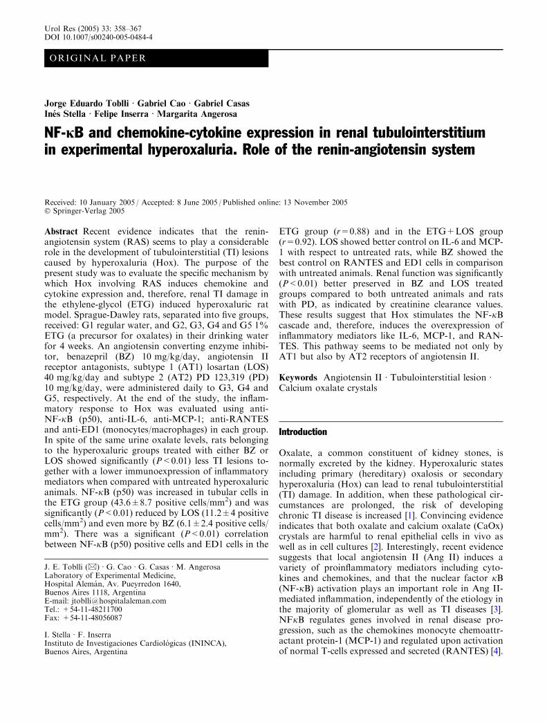
ORIGINAL PAPER
Jorge Eduardo Toblli Æ Gabriel Cao Æ Gabriel Casas
Ines Stella Æ Felipe Inserra Æ Margarita Angerosa
NF-jB and chemokine-cytokine expression in renal tubulointerstitiumin experimental hyperoxaluria. Role of the renin-angiotensin system
Received: 10 January 2005 / Accepted: 8 June 2005 / Published online: 13 November 2005� Springer-Verlag 2005
Abstract Recent evidence indicates that the renin-angiotensin system (RAS) seems to play a considerablerole in the development of tubulointerstitial (TI) lesionscaused by hyperoxaluria (Hox). The purpose of thepresent study was to evaluate the specific mechanism bywhich Hox involving RAS induces chemokine andcytokine expression and, therefore, renal TI damage inthe ethylene-glycol (ETG) induced hyperoxaluric ratmodel. Sprague-Dawley rats, separated into five groups,received: G1 regular water, and G2, G3, G4 and G5 1%ETG (a precursor for oxalates) in their drinking waterfor 4 weeks. An angiotensin converting enzyme inhibi-tor, benazepril (BZ) 10 mg/kg/day, angiotensin IIreceptor antagonists, subtype 1 (AT1) losartan (LOS)40 mg/kg/day and subtype 2 (AT2) PD 123,319 (PD)10 mg/kg/day, were administered daily to G3, G4 andG5, respectively. At the end of the study, the inflam-matory response to Hox was evaluated using anti-NF-jB (p50), anti-IL-6, anti-MCP-1; anti-RANTESand anti-ED1 (monocytes/macrophages) in each group.In spite of the same urine oxalate levels, rats belongingto the hyperoxaluric groups treated with either BZ orLOS showed significantly (P<0.01) less TI lesions to-gether with a lower immunoexpression of inflammatorymediators when compared with untreated hyperoxaluricanimals. NF-jB (p50) was increased in tubular cells inthe ETG group (43.6±8.7 positive cells/mm2) and wassignificantly (P<0.01) reduced by LOS (11.2±4 positivecells/mm2) and even more by BZ (6.1±2.4 positive cells/mm2). There was a significant (P<0.01) correlationbetween NF-jB (p50) positive cells and ED1 cells in the
ETG group (r=0.88) and in the ETG+LOS group(r=0.92). LOS showed better control on IL-6 and MCP-1 with respect to untreated rats, while BZ showed thebest control on RANTES and ED1 cells in comparisonwith untreated animals. Renal function was significantly(P<0.01) better preserved in BZ and LOS treatedgroups compared to both untreated animals and ratswith PD, as indicated by creatinine clearance values.These results suggest that Hox stimulates the NF-jBcascade and, therefore, induces the overexpression ofinflammatory mediators like IL-6, MCP-1, and RAN-TES. This pathway seems to be mediated not only byAT1 but also by AT2 receptors of angiotensin II.
Keywords Angiotensin II Æ Tubulointerstitial lesion ÆCalcium oxalate crystals
Introduction
Oxalate, a common constituent of kidney stones, isnormally excreted by the kidney. Hyperoxaluric statesincluding primary (hereditary) oxalosis or secondaryhyperoxaluria (Hox) can lead to renal tubulointerstitial(TI) damage. In addition, when these pathological cir-cumstances are prolonged, the risk of developingchronic TI disease is increased [1]. Convincing evidenceindicates that both oxalate and calcium oxalate (CaOx)crystals are harmful to renal epithelial cells in vivo aswell as in cell cultures [2]. Interestingly, recent evidencesuggests that local angiotensin II (Ang II) induces avariety of proinflammatory mediators including cyto-kines and chemokines, and that the nuclear factor jB(NF-jB) activation plays an important role in Ang II-mediated inflammation, independently of the etiology inthe majority of glomerular as well as TI diseases [3].NFjB regulates genes involved in renal disease pro-gression, such as the chemokines monocyte chemoattr-actant protein-1 (MCP-1) and regulated upon activationof normal T-cells expressed and secreted (RANTES) [4].
J. E. Toblli (&) Æ G. Cao Æ G. Casas Æ M. AngerosaLaboratory of Experimental Medicine,Hospital Aleman, Av. Pueyrredon 1640,Buenos Aires 1118, ArgentinaE-mail: [email protected].: +54-11-48211700Fax: +54-11-48056087
I. Stella Æ F. InserraInstituto de Investigaciones Cardiologicas (ININCA),Buenos Aires, Argentina
Urol Res (2005) 33: 358–367DOI 10.1007/s00240-005-0484-4
Additionally, both MCP-1 and RANTES, which aredeeply involved in the inflammatory process, are stim-ulated by Ang II, and at least the former is overex-pressed following contact with CaOx crystals or Hox intubular epithelial cells [5–7]. CaOx crystal endocytosisand the subsequent tubular epithelial cell responsespromote an increase in reactive oxygen species (ROS)production [8]. ROS interacts with interstitial cellsleading to an inflammatory reaction and, eventually,tissue injury [9]. Previous studies in our laboratory haveshown that interaction against the renin-angiotensinsystem (RAS) provides substantial benefit involvingprotection against renal TI lesions [10–17]. In accor-dance with this information, the purpose of thepresent study was to evaluate the specific mechanismby which RAS induces chemokine and cytokineexpression in CaOx crystal TI injury in a hyperoxaluricrat model.
Methods and materials
Two-month-old male Sprague-Dawley rats initiallyweighing 250–270 g were housed in metabolic cages, anddivided into five groups each of eight animals: a controlgroup (G1), the ethylene glycol (ETG) group (G2), theETG+benazepril (BZ) group (G3), the ETG+losartan(LOS) group (G4), and the ETG+PD 123,319 (PD)group (G5). All of the animals were allowed to drink tapwater, and were fed standard rat chow ad libitum. Over4 weeks, ETG 1% (as a precursor for oxalates) wascontinuously administered to G2, G3, G4 and G5 intheir drinking water. Every day, BZ 10 mg/kg and LOS40 mg/kg were administered to G3 and G4, respectively,by gavage. A total of 10 mg/kg/day PD 123,319, aselective Ang II type 2 receptor (AT2) antagonist, wascontinuously administered to G5 in drinking water.
Biochemical determination of 24 h urine collected atbaseline and after 4 weeks was carried out. At thesetimes, blood samples were also obtained for serumdeterminations. The kidneys were harvested for lightmicroscopy and immunohistochemical studies after4 weeks. At baseline and at the end of the experiment,systolic blood pressure (SBP) was measured by tail cuffplethysmography, as previously described [12, 16].
Biochemical procedures
Oxalate was determined by enzymatic methods (SigmaDiagnostics, St. Louis, Mo.), while calcium was deter-mined by standard methods using atomic absorption.Creatinine clearances were calculated using the standardformula. Calcium oxalate crystals were identified usingbrightfield phase contrast and an adapted polarized lightmicroscope, and quantified on ten microscopic fields persample examined at a magnification of ·400. Crystallu-ria was graded as: 0=no crystals per field, 1=<10crystals per field, 2=10–25 crystals per field, 3=26–50
crystals per field, and 4=>50 crystals per field, as pre-viously described [17].
Kidney processing and examination
Kidneys were perfused with saline solution through theabdominal aorta until they were free of blood. Decap-sulated kidneys were cut longitudinally and fixed inphosphate-buffered 10% formaldehyde (pH 7.2) andembedded in paraffin. Sections 4 lm thick were cut andstained with hematoxylin-eosin (H-E) and Masson’strichrome.
Immunolabelling and light microscopy
Immunolabelling of specimens was carried out with amodified avidin-biotin-peroxidase complex techniqueVectastain ABC kit (Universal Elite, Vector Laborato-ries, Calif.) and the specimens handled using immuno-histochemical standard techniques. With the aim ofdetecting chemokines, MCP-1 and RANTES, rabbitpolyclonal IgG anti-MCP-1 (ab-7202 Abcam, Cam-bridge, UK), and goat polyclonal IgG anti-RANTES(sc-1410 Santa Cruz Biotechnology, Santa Cruz,Calif.), at a dilution 1:100 were used, respectively. Fi-nally, a goat polyclonal IgG anti-IL-6 (sc-1265 SantaCruz Biotechnology), and a goat polyclonal IgG anti-NF-jB p50 (sc-114 Santa Cruz Biotechnology), wereused.
Morphological analysis
Histological sections were taken from the kidneys ofeach animal and studied using a light microscope NikonE400 (Nikon, Melville, N. Y.). For a better identifica-tion of calcium oxalate crystals, polarized light micros-copy was used. All tissue samples were evaluatedindependently by two investigators without priorknowledge of the group to which the rats belonged.Interstitium was defined as renal tissue excludingglomeruli, tubules or blood vessels. In order to estimatethe TI damage in each group, semi-quantitative scores ofcrystal deposits and TI lesions were determined on tenmicroscopic fields per section, examined at a magnifi-cation of ·100. The scores were graded each accordingto the following scale: 0=absent, 1=mild (involving£ 25% of each microscopic field), 2=moderate (>26%and £ 50%), 3=severe (>51% and £ 75%), or 4=verysevere (>76%), as previously described [17]. Whencomparing various groups, the same areas of the kidneywere analyzed.
Immunohistochemical evaluation was carried outaccording to the following schedule: transcription factorNF-jB (p50) and ED1(monocyte/macrophage) wereexpressed as positive cells/mm2, assessed on 20 consec-utive microscopic fields at ·400 magnification; IL-6,
359
MCP-1 and RANTES were expressed as percentage ofpositive immunostaining area/mm2. Data were averagedand the results were expressed as mean±SD. All mea-surements were carried out using an image analyzerImage-Pro Plus version 4.5 for Windows (MediaCybernetics, Silver Spring, Md.).
Statistical methods
All of the statistical analyses were performed usingabsolute values and processed through GraphPad Prism,version 2.0 (GraphPad Software, San Diego, Calif.). Thetest to determine whether the data fit a normal distri-bution were performed by the Kolmogorov and Smirnovmethod. For parameters with a normal distribution, allcomparisons among groups were carried out usingANOVA. Values were expressed as mean±SD. Thedifference in mean values between groups was assessedby the Tukey-Kramer multiple comparisons test. Sta-tistical analysis for parameters such as histological datawith a non-normal distribution was performed by theKruskal-Wallis test (nonparametric ANOVA) andDunn’s multiple comparison test. Finally, a Spearmanrank correlation was performed when appropriate. Avalue of P<0.05 was considered significant.
Results
At the end of the experiment, there were no significantdifferences in body weight and SBP among the fivegroups (Table 1). As expected, a marked and significantincrease in urinary excretion of oxalate was observed inrats from groups that received ETG (G2, G3, G4 andG5) (Table 1). Urinary calcium was significantly de-creased (P<0.01) in rats from G2 (ETG) and G5(ETG+PD) in comparison with the other groups. Dueto the fact that oxalate was determined in acidified urinesamples, which dissolves crystals and consequentlyindicates the total amount of oxalate, including oxalateprecipitated with calcium, and taking into account thatcalcium was measured in urine that was not acidified,such calcium represents the amount of free calcium ions.The reduction in urinary calcium excretion observed in
rats from G2 and G5 may be interpreted as formation ofCaOx crystals and its subsequent deposition in the renalinterstitium. In accordance with this, animals from G2and G5 presented a high level of crystalluria and lowercreatinine clearance when compared with the othergroups (P<0.01). On the other hand, hyperoxaluric ratstreated with BZ (G3) and with LOS (G4) had a signifi-cantly lower amount of CaOx crystals in their urine, anda higher creatinine clearance relative to untreated hy-peroxaluric rats (G2) and hyperoxaluric animals withPD (G5) (Table 1).
Histological and immunohistochemical findings
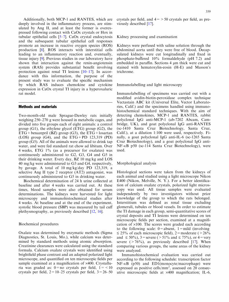
Polarized light microscopy showed that untreated hy-peroxaluric animals (G2) as well as those from G5(ETG+PD) had diffuse CaOx crystals in the tubularlumens and in the renal interstitium, mainly in the cortexbut also in the medulla (Fig. 1). In addition, TI lesionscharacterized by tubular atrophy and dilatation,inflammatory cell infiltrates mostly involving monocytesand macrophages (ED1 positive cells), in the cortex andin the medulla, were also present in this group (Table 2,Figs. 2A, 3). Interestingly, whereas tubular atrophy anddilatation were significantly diminished in rats from G3(ETG+BZ) and G4 (ETG+LOS) (Table 2, Figs. 1, 2A,3), animals from G5 (ETG+PD), despite showing ansignificant degree of TI lesions, presented a similarreduction in ED1 positive cells in the renal interstitiumcompared to the control and BZ groups.
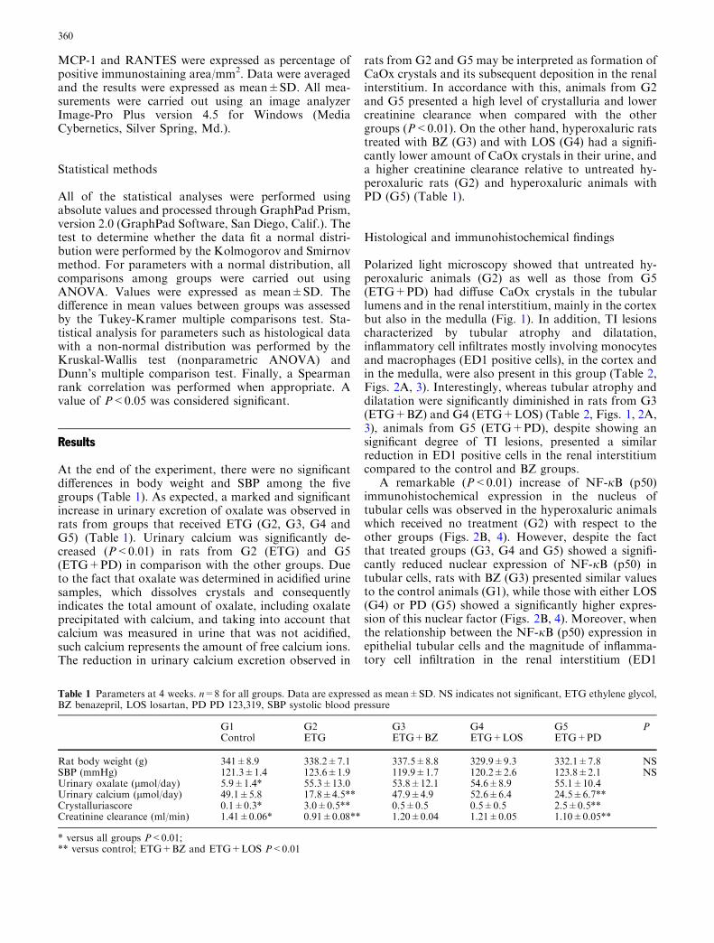
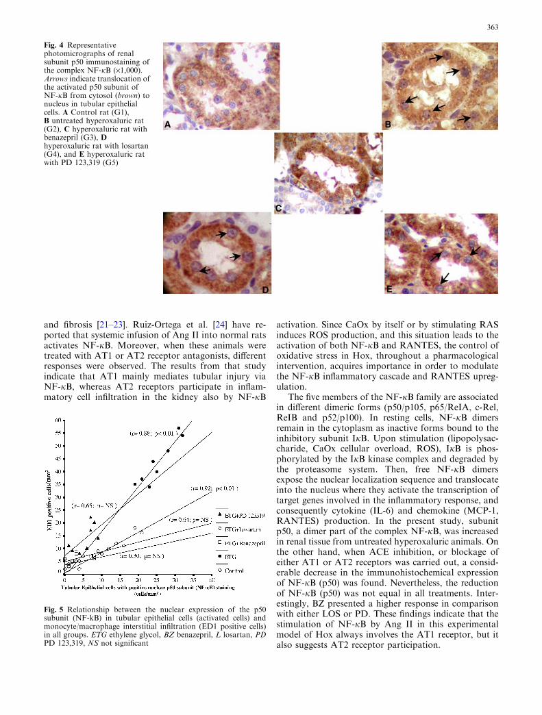
A remarkable (P<0.01) increase of NF-jB (p50)immunohistochemical expression in the nucleus oftubular cells was observed in the hyperoxaluric animalswhich received no treatment (G2) with respect to theother groups (Figs. 2B, 4). However, despite the factthat treated groups (G3, G4 and G5) showed a signifi-cantly reduced nuclear expression of NF-jB (p50) intubular cells, rats with BZ (G3) presented similar valuesto the control animals (G1), while those with either LOS(G4) or PD (G5) showed a significantly higher expres-sion of this nuclear factor (Figs. 2B, 4). Moreover, whenthe relationship between the NF-jB (p50) expression inepithelial tubular cells and the magnitude of inflamma-tory cell infiltration in the renal interstitium (ED1
Table 1 Parameters at 4 weeks. n=8 for all groups. Data are expressed as mean±SD. NS indicates not significant, ETG ethylene glycol,BZ benazepril, LOS losartan, PD PD 123,319, SBP systolic blood pressure
G1 G2 G3 G4 G5 PControl ETG ETG+BZ ETG+LOS ETG+PD
Rat body weight (g) 341±8.9 338.2±7.1 337.5±8.8 329.9±9.3 332.1±7.8 NSSBP (mmHg) 121.3±1.4 123.6±1.9 119.9±1.7 120.2±2.6 123.8±2.1 NSUrinary oxalate (lmol/day) 5.9±1.4* 55.3±13.0 53.8±12.1 54.6±8.9 55.1±10.4Urinary calcium (lmol/day) 49.1±5.8 17.8±4.5** 47.9±4.9 52.6±6.4 24.5±6.7**Crystalluriascore 0.1±0.3* 3.0±0.5** 0.5±0.5 0.5±0.5 2.5±0.5**Creatinine clearance (ml/min) 1.41±0.06* 0.91±0.08** 1.20±0.04 1.21±0.05 1.10±0.05**
* versus all groups P<0.01;** versus control; ETG+BZ and ETG+LOS P<0.01
360
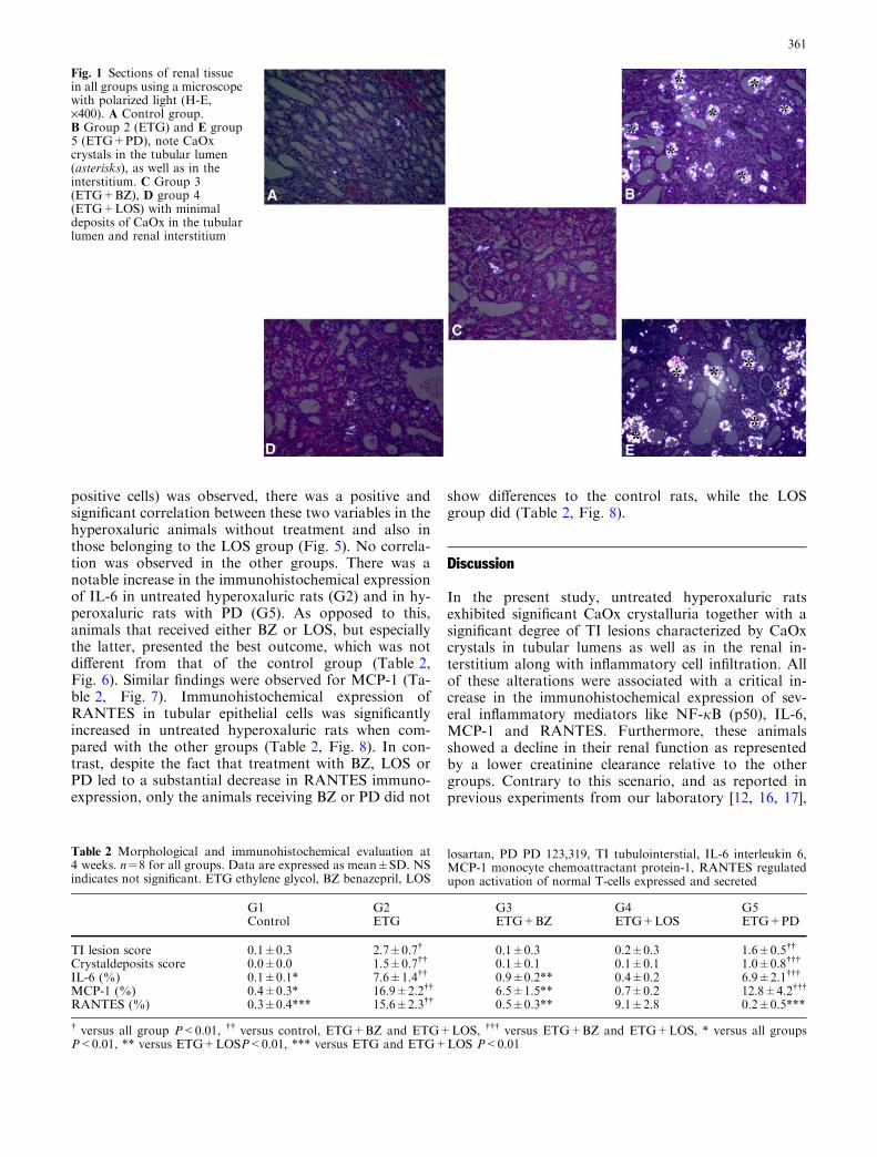
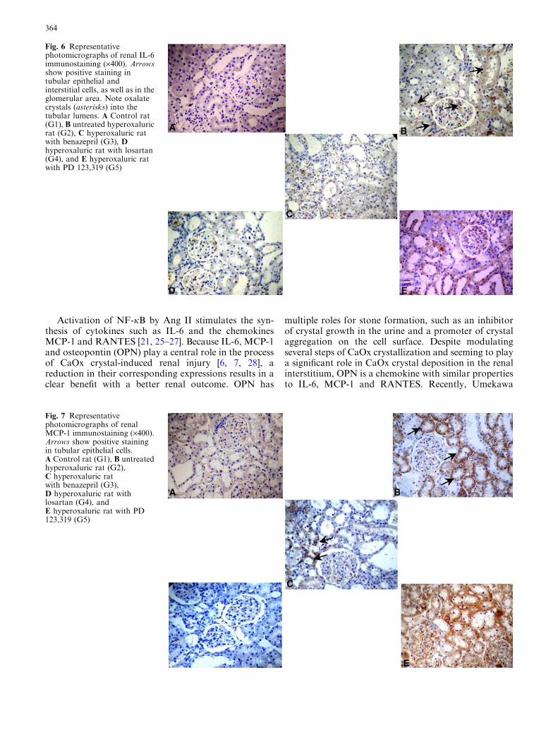
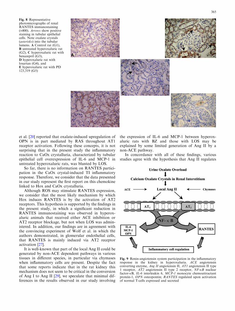
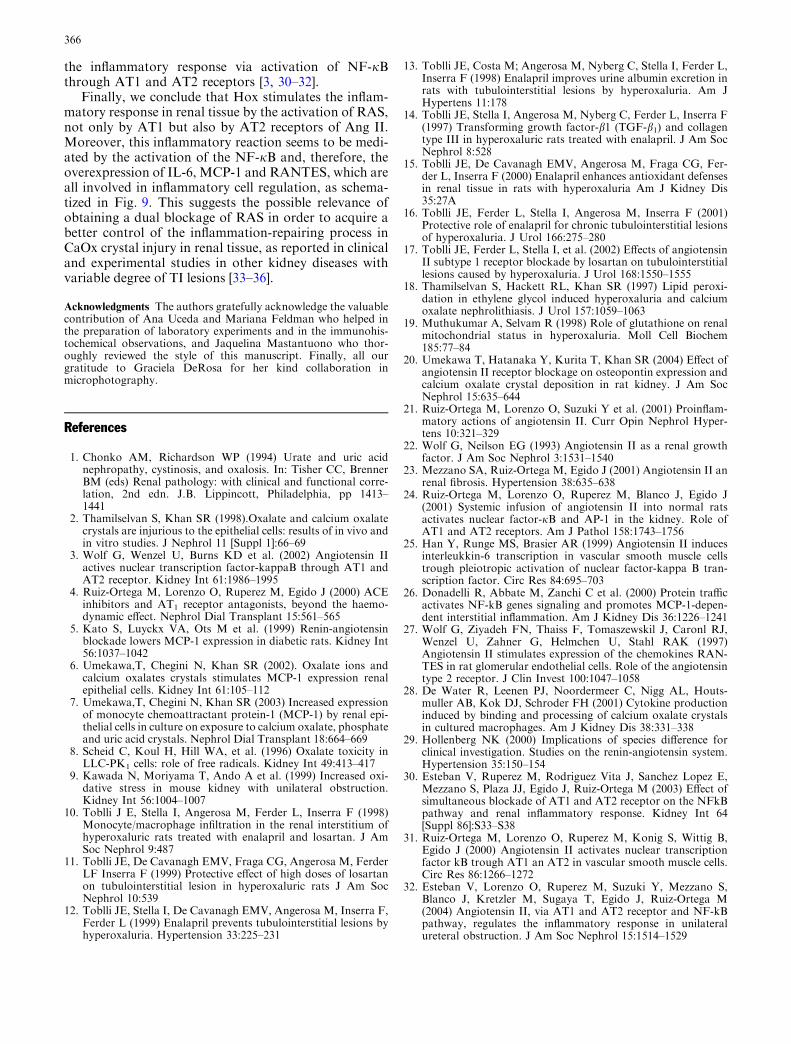
positive cells) was observed, there was a positive andsignificant correlation between these two variables in thehyperoxaluric animals without treatment and also inthose belonging to the LOS group (Fig. 5). No correla-tion was observed in the other groups. There was anotable increase in the immunohistochemical expressionof IL-6 in untreated hyperoxaluric rats (G2) and in hy-peroxaluric rats with PD (G5). As opposed to this,animals that received either BZ or LOS, but especiallythe latter, presented the best outcome, which was notdifferent from that of the control group (Table 2,Fig. 6). Similar findings were observed for MCP-1 (Ta-ble 2, Fig. 7). Immunohistochemical expression ofRANTES in tubular epithelial cells was significantlyincreased in untreated hyperoxaluric rats when com-pared with the other groups (Table 2, Fig. 8). In con-trast, despite the fact that treatment with BZ, LOS orPD led to a substantial decrease in RANTES immuno-expression, only the animals receiving BZ or PD did not
show differences to the control rats, while the LOSgroup did (Table 2, Fig. 8).
Discussion
In the present study, untreated hyperoxaluric ratsexhibited significant CaOx crystalluria together with asignificant degree of TI lesions characterized by CaOxcrystals in tubular lumens as well as in the renal in-terstitium along with inflammatory cell infiltration. Allof these alterations were associated with a critical in-crease in the immunohistochemical expression of sev-eral inflammatory mediators like NF-jB (p50), IL-6,MCP-1 and RANTES. Furthermore, these animalsshowed a decline in their renal function as representedby a lower creatinine clearance relative to the othergroups. Contrary to this scenario, and as reported inprevious experiments from our laboratory [12, 16, 17],
Table 2 Morphological and immunohistochemical evaluation at4 weeks. n=8 for all groups. Data are expressed as mean±SD. NSindicates not significant. ETG ethylene glycol, BZ benazepril, LOS
losartan, PD PD 123,319, TI tubulointerstial, IL-6 interleukin 6,MCP-1 monocyte chemoattractant protein-1, RANTES regulatedupon activation of normal T-cells expressed and secreted
Fig. 1 Sections of renal tissuein all groups using a microscopewith polarized light (H-E,·400). A Control group.B Group 2 (ETG) and E group5 (ETG+PD), note CaOxcrystals in the tubular lumen(asterisks), as well as in theinterstitium. C Group 3(ETG+BZ), D group 4(ETG+LOS) with minimaldeposits of CaOx in the tubularlumen and renal interstitium
G1 G2 G3 G4 G5Control ETG ETG+BZ ETG+LOS ETG+PD
TI lesion score 0.1±0.3 2.7±0.7� 0.1±0.3 0.2±0.3 1.6±0.5��
Crystaldeposits score 0.0±0.0 1.5±0.7�� 0.1±0.1 0.1±0.1 1.0±0.8���
IL-6 (%) 0.1±0.1* 7.6±1.4�� 0.9±0.2** 0.4±0.2 6.9±2.1���
MCP-1 (%) 0.4±0.3* 16.9±2.2�� 6.5±1.5** 0.7±0.2 12.8±4.2���
RANTES (%) 0.3±0.4*** 15.6±2.3�� 0.5±0.3** 9.1±2.8 0.2±0.5***
� versus all group P<0.01, �� versus control, ETG+BZ and ETG+LOS, ��� versus ETG+BZ and ETG+LOS, * versus all groupsP<0.01, ** versus ETG+LOSP<0.01, *** versus ETG and ETG+LOS P<0.01
361

intervention against RAS seems to be favorable inmodulating the inflammatory response to Hox andCaOx crystalluria in the renal interstitium, since bothangiotensin converting enzyme (ACE) inhibition, byBZ, and Ang II antagonism by blockage of AT1receptors with LOS led to a remarkable reduction inthese lesions with an improvement in renal function.
Urine oxalate overload evokes CaOx crystal forma-tion. Subsequently, the interrelationship between CaOx
crystals with tubular epithelial cells generates oxidativestress by stimulating ROS, and promotes local RASactivation, as previously reported ]8, 17–20]. Ang II, themain effector peptide of RAS, plays a central role in thepathophysiology of a broad range of renal diseases byinducing inflammatory responses, including ROS over-production. Currently, it is widely accepted that Ang IIis a proinflammatory molecule which participates as agrowth factor that regulates cell proliferation, apoptosis
Fig. 2 A Quantitative analysisof the ED1 positive cells(monocyte/macrophage) inrenal interstitium among thefive groups. Values areexpressed as mean±SD. ETGethylene glycol, BZ benazepril,L losartan. PD PD 123,319.(* versus ETG, ETG+LOS andETG+PD P< 0.01. ** versusETG+BZ, ETG+LOS andETG+PD P< 0.01. *** versusETG+LOS P<0.05 . X versusETG+PD P<0.05).B Quantitative analysis of theimmunohistochemical nuclearexpression of p50 subunit(NF-kB) in tubular epithelialcell (activated cells). (* versusETG, ETG+LOS andETG+PD P<0.01. ** versusETG+BZ, ETG+LOS andETG+PD P< 0.01. *** versusETG+LOS and ETG+PDP<0.05)
Fig. 3 Renal tissue with ED1immunostaining (·400).A Control group, B group 2(ETG) arrows indicate positivecells corresponding tomonocytes/macrophagesinfiltrates, C group 3(ETG+BZ) and E (ETG+PD)with an important reduction inED1 positive cells in bothgroups. D Group 4(ETG+LOS) partial reductionof inflammatory infiltration(arrows)
362
and fibrosis [21–23]. Ruiz-Ortega et al. [24] have re-ported that systemic infusion of Ang II into normal ratsactivates NF-jB. Moreover, when these animals weretreated with AT1 or AT2 receptor antagonists, differentresponses were observed. The results from that studyindicate that AT1 mainly mediates tubular injury viaNF-jB, whereas AT2 receptors participate in inflam-matory cell infiltration in the kidney also by NF-jB
activation. Since CaOx by itself or by stimulating RASinduces ROS production, and this situation leads to theactivation of both NF-jB and RANTES, the control ofoxidative stress in Hox, throughout a pharmacologicalintervention, acquires importance in order to modulatethe NF-jB inflammatory cascade and RANTES upreg-ulation.
The five members of the NF-jB family are associatedin different dimeric forms (p50/p105, p65/ReIA, c-Rel,ReIB and p52/p100). In resting cells, NF-jB dimersremain in the cytoplasm as inactive forms bound to theinhibitory subunit IjB. Upon stimulation (lipopolysac-charide, CaOx cellular overload, ROS), IjB is phos-phorylated by the IjB kinase complex and degraded bythe proteasome system. Then, free NF-jB dimersexpose the nuclear localization sequence and translocateinto the nucleus where they activate the transcription oftarget genes involved in the inflammatory response, andconsequently cytokine (IL-6) and chemokine (MCP-1,RANTES) production. In the present study, subunitp50, a dimer part of the complex NF-jB, was increasedin renal tissue from untreated hyperoxaluric animals. Onthe other hand, when ACE inhibition, or blockage ofeither AT1 or AT2 receptors was carried out, a consid-erable decrease in the immunohistochemical expressionof NF-jB (p50) was found. Nevertheless, the reductionof NF-jB (p50) was not equal in all treatments. Inter-estingly, BZ presented a higher response in comparisonwith either LOS or PD. These findings indicate that thestimulation of NF-jB by Ang II in this experimentalmodel of Hox always involves the AT1 receptor, but italso suggests AT2 receptor participation.
Fig. 4 Representativephotomicrographs of renalsubunit p50 immunostaining ofthe complex NF-jB (·1,000).Arrows indicate translocation ofthe activated p50 subunit ofNF-jB from cytosol (brown) tonucleus in tubular epithelialcells. A Control rat (G1),B untreated hyperoxaluric rat(G2), C hyperoxaluric rat withbenazepril (G3), Dhyperoxaluric rat with losartan(G4), and E hyperoxaluric ratwith PD 123,319 (G5)
Fig. 5 Relationship between the nuclear expression of the p50subunit (NF-kB) in tubular epithelial cells (activated cells) andmonocyte/macrophage interstitial infiltration (ED1 positive cells)in all groups. ETG ethylene glycol, BZ benazepril, L losartan, PDPD 123,319, NS not significant
363
Activation of NF-jB by Ang II stimulates the syn-thesis of cytokines such as IL-6 and the chemokinesMCP-1 and RANTES [21, 25–27]. Because IL-6, MCP-1and osteopontin (OPN) play a central role in the processof CaOx crystal-induced renal injury [6, 7, 28], areduction in their corresponding expressions results in aclear benefit with a better renal outcome. OPN has
multiple roles for stone formation, such as an inhibitorof crystal growth in the urine and a promoter of crystalaggregation on the cell surface. Despite modulatingseveral steps of CaOx crystallization and seeming to playa significant role in CaOx crystal deposition in the renalinterstitium, OPN is a chemokine with similar propertiesto IL-6, MCP-1 and RANTES. Recently, Umekawa
Fig. 6 Representativephotomicrographs of renal IL-6immunostaining (·400). Arrowsshow positive staining intubular epithelial andinterstitial cells, as well as in theglomerular area. Note oxalatecrystals (asterisks) into thetubular lumens. A Control rat(G1), B untreated hyperoxaluricrat (G2), C hyperoxaluric ratwith benazepril (G3), Dhyperoxaluric rat with losartan(G4), and E hyperoxaluric ratwith PD 123,319 (G5)
Fig. 7 Representativephotomicrographs of renalMCP-1 immunostaining (·400).Arrows show positive stainingin tubular epithelial cells.A Control rat (G1), B untreatedhyperoxaluric rat (G2),C hyperoxaluric ratwith benazepril (G3),D hyperoxaluric rat withlosartan (G4), andE hyperoxaluric rat with PD123,319 (G5)
364
et al. [20] reported that oxalate-induced upregulation ofOPN is in part mediated by RAS throughout AT1receptor activation. Following these concepts, it is notsurprising that in the present study the inflammatoryreaction to CaOx crystalluria, characterized by tubularepithelial cell overexpression of IL-6 and MCP-1 inuntreated hyperoxaluric rats, was blunted by LOS.
So far, there is no information on RANTES partici-pation in the CaOx crystal-induced TI inflammatoryresponse. Therefore, we consider that the data presentedin our study represent the first report on this chemokinelinked to Hox and CaOx crystalluria.
Although ROS may stimulate RANTES expression,we consider that the most likely mechanism by whichHox induces RANTES is by the activation of AT2receptors. This hypothesis is supported by the findings inthe present study, in which a significant reduction inRANTES immunostaining was observed in hyperox-aluric animals that received either ACE inhibition orAT2 receptor blockage, but not when LOS was admin-istered. In addition, our findings are in agreement withthe convincing experiment of Wolf et al. in which theauthors demonstrated, in glomerular endothelial cells,that RANTES is mainly induced via AT2 receptoractivation [27].
It is well-known that part of the local Ang II could begenerated by non-ACE dependent pathways in varioustissues in different species, in particular via chymaseswhen inflammatory cells are present. Despite the factthat some reports indicate that in the rat kidney thismechanism does not seem to be critical in the conversionof Ang I to Ang II [29], we speculate that minimal dif-ferences in the results observed in our study involving
the expression of IL-6 and MCP-1 between hyperox-aluric rats with BZ and those with LOS may beexplained by some limited generation of Ang II by anon-ACE pathway.
In concordance with all of these findings, variousstudies agree with the hypothesis that Ang II regulates
Fig. 9 Renin-angiotensin system participation in the inflammatoryresponse in the kidney in hyperoxaluria. ACE angiotensinconverting enzyme, Ang II angiotensin II, AT1 angiotensin II type1 receptor, AT2 angiotensin II type 2 receptor, NF-jB nuclearfactor-jB, IL-6 interleukin 6, MCP-1 monocyte chemoattractantprotein-1, OPN osteopontin; RANTES regulated upon activationof normal T-cells expressed and secreted
Fig. 8 Representativephotomicrographs of renalRANTES immunostaining(·400). Arrows show positivestaining in tubular epithelialcells. Note oxalate crystals(asterisks) into the tubularlumens. A Control rat (G1),B untreated hyperoxaluric rat(G2), C hyperoxaluric rat withbenazepril (G3),D hyperoxaluric rat withlosartan (G4), andE hyperoxaluric rat with PD123,319 (G5)
365
the inflammatory response via activation of NF-jBthrough AT1 and AT2 receptors [3, 30–32].
Finally, we conclude that Hox stimulates the inflam-matory response in renal tissue by the activation of RAS,not only by AT1 but also by AT2 receptors of Ang II.Moreover, this inflammatory reaction seems to be medi-ated by the activation of the NF-jB and, therefore, theoverexpression of IL-6, MCP-1 and RANTES, which areall involved in inflammatory cell regulation, as schema-tized in Fig. 9. This suggests the possible relevance ofobtaining a dual blockage of RAS in order to acquire abetter control of the inflammation-repairing process inCaOx crystal injury in renal tissue, as reported in clinicaland experimental studies in other kidney diseases withvariable degree of TI lesions [33–36].
Acknowledgments The authors gratefully acknowledge the valuablecontribution of Ana Uceda and Mariana Feldman who helped inthe preparation of laboratory experiments and in the immunohis-tochemical observations, and Jaquelina Mastantuono who thor-oughly reviewed the style of this manuscript. Finally, all ourgratitude to Graciela DeRosa for her kind collaboration inmicrophotography.
References
1. Chonko AM, Richardson WP (1994) Urate and uric acidnephropathy, cystinosis, and oxalosis. In: Tisher CC, BrennerBM (eds) Renal pathology: with clinical and functional corre-lation, 2nd edn. J.B. Lippincott, Philadelphia, pp 1413–1441
2. Thamilselvan S, Khan SR (1998).Oxalate and calcium oxalatecrystals are injurious to the epithelial cells: results of in vivo andin vitro studies. J Nephrol 11 [Suppl 1]:66–69
3. Wolf G, Wenzel U, Burns KD et al. (2002) Angiotensin IIactives nuclear transcription factor-kappaB through AT1 andAT2 receptor. Kidney Int 61:1986–1995
4. Ruiz-Ortega M, Lorenzo O, Ruperez M, Egido J (2000) ACEinhibitors and AT1 receptor antagonists, beyond the haemo-dynamic effect. Nephrol Dial Transplant 15:561–565
5. Kato S, Luyckx VA, Ots M et al. (1999) Renin-angiotensinblockade lowers MCP-1 expression in diabetic rats. Kidney Int56:1037–1042
6. Umekawa,T, Chegini N, Khan SR (2002). Oxalate ions andcalcium oxalates crystals stimulates MCP-1 expression renalepithelial cells. Kidney Int 61:105–112
7. Umekawa,T, Chegini N, Khan SR (2003) Increased expressionof monocyte chemoattractant protein-1 (MCP-1) by renal epi-thelial cells in culture on exposure to calcium oxalate, phosphateand uric acid crystals. Nephrol Dial Transplant 18:664–669
8. Scheid C, Koul H, Hill WA, et al. (1996) Oxalate toxicity inLLC-PK1 cells: role of free radicals. Kidney Int 49:413–417
9. Kawada N, Moriyama T, Ando A et al. (1999) Increased oxi-dative stress in mouse kidney with unilateral obstruction.Kidney Int 56:1004–1007
10. Toblli J E, Stella I, Angerosa M, Ferder L, Inserra F (1998)Monocyte/macrophage infiltration in the renal interstitium ofhyperoxaluric rats treated with enalapril and losartan. J AmSoc Nephrol 9:487
11. Toblli JE, De Cavanagh EMV, Fraga CG, Angerosa M, FerderLF Inserra F (1999) Protective effect of high doses of losartanon tubulointerstitial lesion in hyperoxaluric rats J Am SocNephrol 10:539
12. Toblli JE, Stella I, De Cavanagh EMV, Angerosa M, Inserra F,Ferder L (1999) Enalapril prevents tubulointerstitial lesions byhyperoxaluria. Hypertension 33:225–231
13. Toblli JE, Costa M; Angerosa M, Nyberg C, Stella I, Ferder L,Inserra F (1998) Enalapril improves urine albumin excretion inrats with tubulointerstitial lesions by hyperoxaluria. Am JHypertens 11:178
14. Toblli JE, Stella I, Angerosa M, Nyberg C, Ferder L, Inserra F(1997) Transforming growth factor-b1 (TGF-b1) and collagentype III in hyperoxaluric rats treated with enalapril. J Am SocNephrol 8:528
15. Toblli JE, De Cavanagh EMV, Angerosa M, Fraga CG, Fer-der L, Inserra F (2000) Enalapril enhances antioxidant defensesin renal tissue in rats with hyperoxaluria Am J Kidney Dis35:27A
16. Toblli JE, Ferder L, Stella I, Angerosa M, Inserra F (2001)Protective role of enalapril for chronic tubulointerstitial lesionsof hyperoxaluria. J Urol 166:275–280
17. Toblli JE, Ferder L, Stella I, et al. (2002) Effects of angiotensinII subtype 1 receptor blockade by losartan on tubulointerstitiallesions caused by hyperoxaluria. J Urol 168:1550–1555
18. Thamilselvan S, Hackett RL, Khan SR (1997) Lipid peroxi-dation in ethylene glycol induced hyperoxaluria and calciumoxalate nephrolithiasis. J Urol 157:1059–1063
19. Muthukumar A, Selvam R (1998) Role of glutathione on renalmitochondrial status in hyperoxaluria. Moll Cell Biochem185:77–84
20. Umekawa T, Hatanaka Y, Kurita T, Khan SR (2004) Effect ofangiotensin II receptor blockage on osteopontin expression andcalcium oxalate crystal deposition in rat kidney. J Am SocNephrol 15:635–644
21. Ruiz-Ortega M, Lorenzo O, Suzuki Y et al. (2001) Proinflam-matory actions of angiotensin II. Curr Opin Nephrol Hyper-tens 10:321–329
22. Wolf G, Neilson EG (1993) Angiotensin II as a renal growthfactor. J Am Soc Nephrol 3:1531–1540
23. Mezzano SA, Ruiz-Ortega M, Egido J (2001) Angiotensin II anrenal fibrosis. Hypertension 38:635–638
24. Ruiz-Ortega M, Lorenzo O, Ruperez M, Blanco J, Egido J(2001) Systemic infusion of angiotensin II into normal ratsactivates nuclear factor-jB and AP-1 in the kidney. Role ofAT1 and AT2 receptors. Am J Pathol 158:1743–1756
25. Han Y, Runge MS, Brasier AR (1999) Angiotensin II inducesinterleukkin-6 transcription in vascular smooth muscle cellstrough pleiotropic activation of nuclear factor-kappa B tran-scription factor. Circ Res 84:695–703
26. Donadelli R, Abbate M, Zanchi C et al. (2000) Protein trafficactivates NF-kB genes signaling and promotes MCP-1-depen-dent interstitial inflammation. Am J Kidney Dis 36:1226–1241
27. Wolf G, Ziyadeh FN, Thaiss F, Tomaszewskil J, Caronl RJ,Wenzel U, Zahner G, Helmchen U, Stahl RAK (1997)Angiotensin II stimulates expression of the chemokines RAN-TES in rat glomerular endothelial cells. Role of the angiotensintype 2 receptor. J Clin Invest 100:1047–1058
28. De Water R, Leenen PJ, Noordermeer C, Nigg AL, Houts-muller AB, Kok DJ, Schroder FH (2001) Cytokine productioninduced by binding and processing of calcium oxalate crystalsin cultured macrophages. Am J Kidney Dis 38:331–338
29. Hollenberg NK (2000) Implications of species difference forclinical investigation. Studies on the renin-angiotensin system.Hypertension 35:150–154
30. Esteban V, Ruperez M, Rodriguez Vita J, Sanchez Lopez E,Mezzano S, Plaza JJ, Egido J, Ruiz-Ortega M (2003) Effect ofsimultaneous blockade of AT1 and AT2 receptor on the NFkBpathway and renal inflammatory response. Kidney Int 64[Suppl 86]:S33–S38
31. Ruiz-Ortega M, Lorenzo O, Ruperez M, Konig S, Wittig B,Egido J (2000) Angiotensin II activates nuclear transcriptionfactor kB trough AT1 an AT2 in vascular smooth muscle cells.Circ Res 86:1266–1272
32. Esteban V, Lorenzo O, Ruperez M, Suzuki Y, Mezzano S,Blanco J, Kretzler M, Sugaya T, Egido J, Ruiz-Ortega M(2004) Angiotensin II, via AT1 and AT2 receptor and NF-kBpathway, regulates the inflammatory response in unilateralureteral obstruction. J Am Soc Nephrol 15:1514–1529
366
33. Toblli JE, DeRosa G, Cao G, Piorno P, Pagano P (2004) ACEinhibitor and angiotensin type I receptor antagonist in combi-nation reduce renal damage in obese Zucker rats. Kidney Int65:2343–2359
34. Campbell R, Sangalli F, Perticucci E, Aros C, Viscarra C,Perna A, Remuzzi A, Bertocchi F, Fagiani L, Remuzzi G,Ruggenenti P (2003) Effects of combined ACE inhibitor andangiotensin II antagonist treatment in human chronic nephr-opathies. Kidney Int 63:1094–1103
35. Mogensen CE, Neldam S, Tikkanen I, Oren S, Viskoper R,Watts RW, Cooper ME (2000) Randomized controlled trial of
dual blockade of renin-angiotensin system in patients withhypertension, microalbuminuria, and non-insulin dependentdiabetes: the candesartan and lisinopril microalbuminuria(CALM) study. BMJ 321:1440–1444
36. Luno J, Barrio V, Goicoechea MA, Gonzalez C, De VinuesaSG, Gomez F, Bernis C, Espinosa M, Ahijado F, Gomez J,Escalada P (2002) Effects of dual blockade of the renin-angiotensin system in primary proteinuric nephropathies.Kidney Int 62 [Suppl 82]:S47–S52
367
Copyright © 2022 FDOKUMEN