New World Clade B Arenaviruses Can Use Transferrin Receptor 1 (TfR1)Dependent and Independent Entry...
12
Published Ahead of Print 14 November 2007. 2008, 82(2):938. DOI: 10.1128/JVI.01397-07. J. Virol. M. Cannon Holt, Genevieve A. Hamilton, Vanessa K. Martin and Paula Meg L. Flanagan, Jill Oldenburg, Therese Reignier, Nathalia Strains Are Associated with the Use of TfR1 Glycoproteins from Human Pathogenic and -Independent Entry Pathways, and Transferrin Receptor 1 (TfR1)-Dependent New World Clade B Arenaviruses Can Use http://jvi.asm.org/content/82/2/938 Updated information and services can be found at: These include: REFERENCES http://jvi.asm.org/content/82/2/938#ref-list-1 at: This article cites 34 articles, 15 of which can be accessed free CONTENT ALERTS more» articles cite this article), Receive: RSS Feeds, eTOCs, free email alerts (when new http://journals.asm.org/site/misc/reprints.xhtml Information about commercial reprint orders: http://journals.asm.org/site/subscriptions/ To subscribe to to another ASM Journal go to: on January 7, 2014 by guest http://jvi.asm.org/ Downloaded from on January 7, 2014 by guest http://jvi.asm.org/ Downloaded from
-
Upload
independent -
Category
Documents
-
view
1 -
download
0
Transcript of New World Clade B Arenaviruses Can Use Transferrin Receptor 1 (TfR1)Dependent and Independent Entry...

Published Ahead of Print 14 November 2007. 2008, 82(2):938. DOI: 10.1128/JVI.01397-07. J. Virol.
M. CannonHolt, Genevieve A. Hamilton, Vanessa K. Martin and Paula Meg L. Flanagan, Jill Oldenburg, Therese Reignier, Nathalia Strains Are Associated with the Use of TfR1Glycoproteins from Human Pathogenic and -Independent Entry Pathways, andTransferrin Receptor 1 (TfR1)-Dependent New World Clade B Arenaviruses Can Use
http://jvi.asm.org/content/82/2/938Updated information and services can be found at:
These include:
REFERENCEShttp://jvi.asm.org/content/82/2/938#ref-list-1at:
This article cites 34 articles, 15 of which can be accessed free
CONTENT ALERTS more»articles cite this article),
Receive: RSS Feeds, eTOCs, free email alerts (when new
http://journals.asm.org/site/misc/reprints.xhtmlInformation about commercial reprint orders: http://journals.asm.org/site/subscriptions/To subscribe to to another ASM Journal go to:
on January 7, 2014 by guesthttp://jvi.asm
.org/D
ownloaded from
on January 7, 2014 by guest
http://jvi.asm.org/
Dow
nloaded from
JOURNAL OF VIROLOGY, Jan. 2008, p. 938–948 Vol. 82, No. 20022-538X/08/$08.00�0 doi:10.1128/JVI.01397-07Copyright © 2008, American Society for Microbiology. All Rights Reserved.
New World Clade B Arenaviruses Can Use Transferrin Receptor 1(TfR1)-Dependent and -Independent Entry Pathways, and
Glycoproteins from Human Pathogenic Strains AreAssociated with the Use of TfR1�
Meg L. Flanagan,1 Jill Oldenburg,1 Therese Reignier,1 Nathalia Holt,1 Genevieve A. Hamilton,1Vanessa K. Martin,1,2 and Paula M. Cannon1,2*
Saban Research Institute of Childrens Hospital Los Angeles, Los Angeles, California 90027,1 and University ofSouthern California Keck School of Medicine, Los Angeles, California 900892
Received 26 June 2007/Accepted 26 October 2007
Arenaviruses are rodent-borne viruses, with five members of the family capable of causing severe hemor-rhagic fevers if transmitted to humans. To date, two distinct cellular receptors have been identified that areused by different pathogenic viruses, �-dystroglycan by Lassa fever virus and transferrin receptor 1 (TfR1) bycertain New World clade B viruses. Our previous studies have suggested that other, as-yet-unknown receptorsare involved in arenavirus entry. In the present study, we examined the use of TfR1 by the glycoproteins (GPs)from a panel of New World clade B arenaviruses comprising three pathogenic and two nonpathogenic strains.Interestingly, we found that TfR1 was only used by the GPs from the pathogenic viruses, with entry of thenonpathogenic strains being TfR1 independent. The pathogenic GPs could also direct entry into cells byTfR1-independent pathways, albeit less efficiently. A comparison of the abilities of TfR1 orthologs fromdifferent species to support arenavirus entry found that the human and feline receptors were able to enhanceentry of the pathogenic strains, but that neither the murine or canine forms were functional. Since the abilityto use TfR1 is a characteristic feature of the human pathogens, this interaction may represent an importanttarget in the treatment of New World hemorrhagic fevers. In addition, the ability to use TfR1 may be a usefultool to predict the likelihood that any existing or newly discovered viruses in this family could infect humans.
Arenaviruses are enveloped RNA viruses that enter hostcells through the action of a fusion glycoprotein (GP) dis-played on the surface of the viral particle (3). While the naturalhosts of the viruses are rodents, several members of the familycan also be transmitted to humans, causing severe hemorrhagicfevers. The combination of high morbidity/mortality rates, air-borne transmission capability, and person-to-person spreadhas resulted in their classification by the Centers for DiseaseControl and Prevention as category A bioterrorism agents.
Phylogenetic analyses have divided the arenaviruses into OldWorld and New World strains. Lymphocytic choriomeningitisvirus (LCMV) and Lassa fever virus (LASV) comprise thehuman pathogens in the Old World group, but only LASVcauses a hemorrhagic fever. The New World arenaviruses arefurther subdivided into clades A, B, and C (2), with all of thehuman pathogens being found in clade B. These include Juninvirus (JUNV), Machupo virus (MACV), and Guanarito virus(GTOV), which are carried by New World rodents and causesimilar pathologies and mortality rates in humans exposed viacontact with contaminated rodent waste (12, 15–18, 33). CladeB also includes Sabia virus (SABV), which has been reportedto have caused only a single human infection outside of labo-
ratory-acquired infections (24). It also contains three virusesthat are not associated with any known natural human infec-tions, Cupixi virus (CPXV), Tacaribe virus (TCRV), andAmapari virus (AMAV), although TCRV has been reported tohave caused a single case of laboratory-acquired febrile illnesswith mild central nervous system symptoms (24).
The arenavirus GP is a typical class I fusion protein, com-prising two noncovalently associated subunits, GP1 and GP2,which are cleaved from a precursor protein, GPC. Whenpresent on the surface of retroviral vector particles, arenavirusGPs are able to direct entry into a broad range of cell types invitro (21, 26–28). The GP1 subunit contains the receptor bind-ing site (22) and GP1-Fc immunoadhesin fusion proteins bindto cells in a manner that recapitulates the tropism of GP-pseudotyped retroviral vectors (21, 25, 28).
The arenaviruses display a complex pattern of receptor use.To date, two different cellular receptors have been identified,and it is likely that others exist (21, 26–28). LASV, certainstrains of LCMV (5), and New World clade C viruses (32) use�-dystroglycan (�-DG) to enter cells, although some strains ofLCMV can infect cells independently of �-DG (14, 27). Morerecently, transferrin receptor 1 (TfR1; also called TFRC) wasreported as a receptor for the New World clade B arenavirusesthat cause hemorrhagic fevers in humans (25).
We are interested in understanding the determinants ofpathogenicity in the New World clade B arenaviruses and, inparticular, the differences between the pathogenic and non-pathogenic members of this group. Previously, we reportedthat pseudotyped retroviral vectors displaying GPs from the
* Corresponding author. Mailing address: Department of ResearchImmunology and Bone Marrow Transplantation, Childrens HospitalLos Angeles, 4650 Sunset Boulevard, Mailstop 62, Los Angeles, CA90027. Phone: (323) 361-5916. Fax: (323) 361-3566. E-mail: [email protected].
� Published ahead of print on 14 November 2007.
938
on January 7, 2014 by guesthttp://jvi.asm
.org/D
ownloaded from
pathogenic clade B1 viruses, JUNV and MACV, exhibitedmarkedly different properties from vectors carrying the GPfrom the related, but nonpathogenic, virus TCRV (21). Thesefindings prompted us to consider whether the ability to usealternate host receptors and/or entry pathways could underliethe differences in the ability of the clade B viruses to causehuman disease.
In the present study, we have expanded the analysis ofpathogenic and nonpathogenic clade B arenaviruses to includemembers of the B2 lineage, GTOV and AMAV. We examineddependence on TfR1 for entry into both human and rodentcells and observed a correlation between the ability to usehuman TfR1 (hTfR1) and known human pathogenicity. Theseobservations also held true for the feline ortholog of TfR1, butthe murine and canine receptors were unable to enhance entryby any GP. Finally, we discovered that the nonpathogenic cladeB members could infect cells efficiently in a completely TfR1-independent manner, suggesting that the clade B viruses canuse additional, unknown receptors.
MATERIALS AND METHODS
Cell lines. 293A, 293T, NIH 3T3, CHO-K1, and T1B27 cells were maintainedin Dulbecco’s modified Eagle’s medium (DMEM) (Mediatech, Herndon, VA)supplemented with 10% fetal bovine serum (FBS) (HyClone, Logan, UT) and 2mM glutamine (Gemini Bio-Products, West Sacramento, CA). CEM and BHK21cells were maintained in RPMI (Mediatech, Herndon, VA) supplemented with10% FBS and 2 mM glutamine. �-DG knockout (�/�) and heterozygous control(�/�) R1 murine embryonic stem (mES) cells (35) were generously provided byKevin Campbell (University of Iowa) and cultured in DMEM supplemented with20% FBS, 2 mM glutamine, 1 mM nonessential amino acids (Chemicon, Te-mecula, CA), 0.001% �-mercaptoethanol (Sigma, St. Louis, MO) and 103 U/mlof murine leukemia inhibiting factor (Millipore, Billerica, MA). All cells weremaintained in 5% CO2 atmosphere except T1B27 and BHK21 cells, which weremaintained at 10% CO2.
GP-pseudotyped retroviral vectors. Expression plasmids for the GPs fromJUNV (Parodi strain), MACV (Carvallo strain), TCRV (TRVL 11598 strain),LCMV (Armstrong 53b strain), LASV (Josiah strain), and vesicular stomatitisvirus (VSV) (Indiana strain) have been described previously (21, 27). An expres-sion plasmid for the GTOV GP (INH-95551 strain) was kindly provided byStefan Kunz (The Scripps Research Institute) (28). A human codon-optimizedform of AMAV GP (BeAn 70563 strain) was chemically synthesized and clonedinto the pCAGGS expression vector (20). Pseudotyped retroviral vectors dis-playing GPs were generated by cotransfection of 293T cells with expressionplasmids for the specific GP, together with plasmids expressing murine leukemiavirus Gag-Pol and an enhanced green fluorescent protein (EGFP)-expressingretroviral vector genome, pMND-eGFP, as described previously (21, 27).
The efficiency of entry of the GP-pseudotyped retroviral vectors into cells wasmeasured as previously described (21, 27). Briefly, vectors were incubated withtarget cells for 4 h and then replaced with fresh medium. After 48 h, the cellswere trypsinized, washed in phosphate-buffered saline, and analyzed for EGFPexpression using a FACScan flow cytometer (BD, Franklin Lakes, NJ). Theefficiency of entry (titer) was determined by multiplying the percentage of EGFP-positive cells by the number of cells initially incubated with the vectors. Titerswere expressed as transducing units (TU) per ml of vector-containing superna-tant. In experiments with control and experimental arms (e.g., small interferingRNA [siRNA] treatment and antibody blocking), the titers were made relative tothe control (mock treatment) cells and were expressed as a percentage of thecontrol titer.
Antibody pretreatment assay. 293A cells were seeded in 12-well plates 1 dayprior to transduction with GP-pseudotyped vectors. The cells were pretreatedwith 5 nM or 50 nM of mouse anti-human TfR1 antibody (clone M-A712; BDBiosciences, San Jose, CA) or medium alone for 30 min at 37°C, followed bytransduction with GP-pseudotyped vectors for 4 h in the presence of the anti-hTfR1 antibody. The antibody-vector mixtures were then replaced with freshmedia, and the cells were allowed to recover for 48 to 72 h before fluorescence-activated cell sorter (FACS) analysis for EGFP expression. Titers were deter-mined by measurement of the percentage of EGFP-positive cells, as describedabove.
TfR1 knockdown by siRNA. hTfR1 and murine TfR1 (mTfR1)-specificsiRNAs were purchased as ON-TARGET plus SMART pools (Dharmacon,Lafayette, CO; catalog no. L-003941-00 and L-055550-01). A nontargeted siRNA(5�-AACACAGCAACCUCUACUUGG-3�) was used as a control. 293A or NIH3T3 cells were seeded at 3 � 106 cells per 10-cm plate for 12 to 14 h beforetransfection. Forty microliters of Lipofectamine 2000 (Invitrogen) was mixedwith 110 �l of Opti-MEM I (Invitrogen), incubated for 10 min at room temper-ature, and then added to 825 �l of Opti-MEM I plus 25 �l of 20 �M siRNA,followed by incubation for 20 min at room temperature. Opti-MEM I was addedto the siRNA mixture to give a final volume of 5 ml and then added to cellsprerinsed with Opti-MEM I. The cells were incubated for 4 h, when the siRNAmixture was replaced with fresh DMEM plus 20% FBS, for overnight incubation.The following day, the cells were trypsinized and seeded into six-well plates at adensity of 1 � 105 cells per well. Twenty-four hours later, aliquots of cells fromboth control- and TfR1-siRNA-treated plates were harvested using sodium do-decyl sulfate-lysis buffer, in order to assay the efficiency of knockdown of TfR1expression by Western blotting. The remaining cells were transduced with GP-pseudotyped vectors, and entry efficiency was determined by FACS analysis forEGFP expression, as described previously.
TfR1 expression was detected by Western blotting of cell lysates using a 1:500dilution of mouse anti-TfR1 antibody, clone H68.4 (Invitrogen). This antibodyrecognizes a conserved epitope in chicken, mouse, rat, Chinese hamster, andhuman TfR1. Specific bands were detected using horseradish peroxidase-conju-gated goat anti-mouse immunoglobulin G (IgG) (Pierce, Rockford, IL), diluted1:10,000, followed by incubation with the ECL enhanced chemiluminescencedetection reagent (Amersham Biosciences, Piscataway, NJ) and exposure toKodak BIOMAX XAR film (Sigma). To ensure equal protein loading, mem-branes were also probed with mouse anti-actin monoclonal antibody (Sigma),diluted 1:10,000.
Transient transfection of CHO-K1 cells with TfR1. Expression plasmids formurine, human, canine, and feline TfR1 orthologs, as well as a series of chimerichuman/murine receptors (34), were generously provided by Susan Ross (Uni-versity of Pennsylvania). CHO-K1 cells were transiently transfected with theexpression plasmids using Lipofectamine 2000 (Invitrogen). Briefly, CHO-K1cells were plated to be 95 to 100% confluent in 10-cm plates at the time oftransfection and were transfected with 24 �g of TfR1 expression plasmids. Cellswere incubated in transfection mixture for 4 h and then cultured overnight inDMEM plus 10% FBS. The following day, cells were trypsinized, plated intosix-well plates at 40% confluence and incubated overnight. Cells were thentransduced with GP-pseudotyped vectors for 4 h and the titers determined 48 hlater by FACS analysis for EGFP expression, as described above. Cell sampleswere also collected at the time of transduction and analyzed by Western blottingto determine levels of TfR1 expression, as described above.
Immunoadhesin production and binding assay. An immunoadhesin constructcomprising the MACV GP1 sequence fused to a rabbit IgG heavy-chain Fcsequence has previously been described (21). Immunoadhesin-containing culturesupernatants were generated by transfection of 293T cells, followed by concen-tration of culture supernatants using centrifugal filter units (21). The ability ofthe immunoadhesin to bind to CHO-K1 cells (control) or cells previously trans-fected with hTfR1 or mTFR1 expression plasmids was assessed by incubating 30�l of the MACV immunoadhesin stock with 5 � 105 cells on ice for 30 min,followed by washing and incubation with 50 �l of a 1:50 dilution of fluoresceinisothiocyanate (FITC)-conjugated anti-rabbit IgG (BD Pharmingen, San Jose,CA) for 20 min on ice. Cells were analyzed for binding on a FACScan flowcytometer, as described previously (21). As controls, aliquots of all cell popula-tions were also stained with only the secondary antibody.
RESULTS
Consistent tropism differences are observed between patho-genic and nonpathogenic clade B GPs. The New World cladeB arenaviruses have been divided into subgroups B1, B2, andB3, based on phylogenetic relationships (Fig. 1A) (8). Previ-ously, we reported differences in the characteristics of the entrypathways used by GPs from the pathogenic clade B1 viruses,JUNV and MACV, versus the related but nonpathogenic B1virus, TCRV (21). Specifically, JUNV and MACV GP-pseudotyped retroviral vectors could transduce human (CEM)but not murine (TIB27) T-lymphocyte cell lines, while TCRVGP vectors demonstrated the opposite pattern. In addition, we
VOL. 82, 2008 TfR1 USE BY NEW WORLD CLADE B ARENAVIRUSES 939
on January 7, 2014 by guesthttp://jvi.asm
.org/D
ownloaded from

also found that JUNV and MACV GP vectors were consis-tently less efficient at transducing cell lines derived from bothmice (NIH 3T3) and Chinese hamsters (CHO-K1) comparedto TCRV vectors. These observations led us to speculate thatdifferent cellular receptors and entry pathways were being usedby the pathogenic versus nonpathogenic clade B1 viruses andthat this characteristic might reflect an essential determinant ofhuman pathogenicity.
To examine whether these patterns were more generally trueacross the clade B viruses, we expanded the panel to includeGPs from pathogenic (GTOV) and nonpathogenic (AMAV)clade B2 viruses. GP-pseudotyped vectors were generated andstandardized on human kidney epithelial 293A cells, where weobserved that all five of the clade B vectors produced similartiters (approximately 104 TU/ml of unconcentrated culture su-pernatant) (Fig. 1B). As a control, we included vectorspseudotyped with the VSV glycoprotein (VSV-G). This glyco-protein has very broad tropism, and the resulting vectors effi-ciently transduce diverse cell lines, thereby providing a controlfor the level of transduction directed by the retroviral vectormachinery in each cell line. The vectors were then examined onthe panel of indicator cell lines (CEM, TIB27, NIH 3T3, andCHO-K1 cells) that have previously revealed differences be-tween the pathogenic and nonpathogenic B1 viruses (21). Inaddition, we also included a Syrian hamster cell line, BHK21.
The results of these studies revealed that GTOV GP had atropism pattern that was most similar to those of the patho-genic B1 viruses, JUNV and MACV, while the AMAV GPvectors were more similar to the nonpathogenic TCRV vectors(Fig. 1B). Specifically, we observed that GTOV but not AMAVGP vectors could transduce CEM cells, while the oppositepattern was true on T1B27 cells. Furthermore, we found that
the JUNV, MACV, and GTOV GP vectors transduced thethree rodent cell lines (NIH 3T3, CHO-K1, and BHK21 cells)with significantly less efficiency than either TCRV or AMAVGP vectors. In agreement with our previous studies (21), wefound that JUNV vectors gave higher titers on these threerodent cell lines than the MACV vectors and we also notedthat the GTOV vectors behaved more like the MACV vectorsthan the JUNV vectors. Taken together, these data indicatethat the characteristics of the entry pathways directed by theclade B GPs are more closely correlated with human pathoge-nicity than with close phylogenetic relationships.
hTfR1 use by clade B viruses. Recently, Radoshitzky et al.(25) demonstrated that hTfR1 plays a role in the entry ofJUNV, MACV, GTOV, and SABV into human cell lines, sincepretreatment of 293T cells with anti-TfR1 antibody was able toreduce infectivity by all four viruses and pretreatment of 293Tcells with either soluble hTfR1 or anti-hTFR1 antibody re-duced the titer of MACV and JUNV GP-pseudotyped retro-viral vectors. We performed similar antibody blocking experi-ments using 293A cells and the expanded panel of GP vectors,with VSV-G-pseudotyped vectors serving as a control. Weobserved that while incubation with 50 nM of anti-hTfR1 an-tibody caused significant reductions in titers for the JUNV,MACV, and GTOV GP vectors (78, 93, and 70% reductions,respectively), there was no significant inhibition of entry foreither TCRV or AMAV GP vectors beyond the level that weobserved with the control VSV-G-pseudotyped vectors (Fig.2A). These data therefore suggest that although the patho-genic clade B viruses require hTfR1 for entry into 293A cells,the nonpathogenic viruses TCRV and AMAV do not.
To confirm these results, we treated 293A cells with hTfR1-targeted siRNA prior to incubation with pseudotyped vectors. As
FIG. 1. Tropism of clade B GPs for different cell lines. (A) Phylogenetic relationships among the New World clade B arenaviruses based onsequence analysis of GP. Viruses that exhibit pathogenicity in humans (boxed) are distributed throughout all three sublineages (adapted fromreference 8). (B) Titers of GP-pseudotyped retroviral vectors on various cell lines. The values shown are means � standard errors from two to eightindependent experiments. All vector-cell combinations that gave no titer (�) were confirmed using 10� concentrated stocks of vectors.
940 FLANAGAN ET AL. J. VIROL.
on January 7, 2014 by guesthttp://jvi.asm
.org/D
ownloaded from
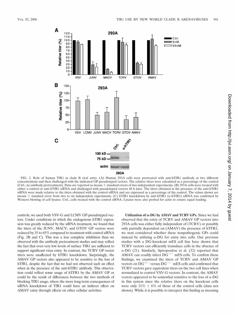
controls, we used both VSV-G and LCMV GP-pseudotyped vec-tors. Under conditions in which the endogenous hTfR1 expres-sion was greatly reduced by the siRNA treatment, we found thatthe titers of the JUNV, MACV, and GTOV GP vectors werereduced by 35 to 65% compared to treatment with control siRNA(Fig. 2B and C). This was a less complete inhibition than weobserved with the antibody pretreatment studies and may reflectthe fact that even very low levels of surface TfR1 are sufficient tosupport significant virus entry. In contrast, the TCRV GP vectortiters were unaffected by hTfR1 knockdown. Surprisingly, theAMAV GP vectors also appeared to be sensitive to the loss ofhTfR1, despite the fact that we had not observed such an effectwhen in the presence of the anti-hTfR1 antibody. This observa-tion could reflect some usage of hTFR1 by the AMAV GP orcould be the result of differences between the two methods ofblocking TfR1 usage, where the more long-term consequences ofsiRNA knockdown of TfR1 could have an indirect effect onAMAV entry through effects on other cellular activities.
Utilization of �-DG by AMAV and TCRV GPs. Since we hadobserved that the entry of TCRV and AMAV GP vectors into293A cells was either fully independent of (TCRV) or possiblyonly partially dependent on (AMAV) the presence of hTFR1,we next considered whether these nonpathogenic GPs couldinstead be utilizing �-DG for entry into cells. Our previousstudies with a DG-knockout mES cell line have shown thatTCRV vectors can efficiently transduce cells in the absence of�-DG (21). Similarly, Spiropoulou et al. (32) reported thatAMAV can readily infect DG�/� mES cells. To confirm thesefindings, we examined the titers of TCRV and AMAV GPvectors on DG�/� versus DG�/� mES cells and confirmed thatTCRV vectors gave equivalent titers on the two cell lines whennormalized to control VSV-G vectors. In contrast, the AMAVvectors appeared to be somewhat sensitive to the loss of �-DGin this system since the relative titers on the knockout cellswere only 31% � 6% of those of the control cells (data notshown). While it is possible to interpret this finding as meaning
FIG. 2. Role of human TfR1 in clade B viral entry. (A) Human 293A cells were pretreated with anti-hTfR1 antibody at two differentconcentrations and then challenged with the indicated GP-pseudotyped vectors. The relative titers were calculated as a percentage of the control(Ctrl.; no antibody pretreatment). Data are reported as means � standard errors of two independent experiments. (B) 293A cells were treated witheither a control or anti-hTfR1 siRNA and challenged with pseudotyped vectors 48 h later. The titers obtained in the presence of the anti-hTfR1siRNA were made relative to the titers obtained with the control siRNA and are expressed as a percentage of the control. The values shown aremeans � standard error from two to six independent experiments. (C) hTfR1 knockdown by anti-hTfR1 (�-hTfR1) siRNA was confirmed byWestern blotting of cell lysates. Ctrl., cells treated with the control siRNA. Lysates were also probed for actin to ensure equal loading.
VOL. 82, 2008 TfR1 USE BY NEW WORLD CLADE B ARENAVIRUSES 941
on January 7, 2014 by guesthttp://jvi.asm
.org/D
ownloaded from

that AMAV entry is enhanced by the presence of �-DG, wehesitate to draw this conclusion since the mES cells are clonalin origin. An alternative explanation is that variations in thelevels of another unknown factor(s) that AMAV requires forentry underlie these differences. However, when combinedwith the data from the hTfR1 siRNA studies, these observa-tions do suggest that some differences may exist between theentry pathways used by the two nonpathogenic viruses, TCRVand AMAV.
All clade B viruses enter murine cells independently ofTfR1. Our observations so far support a role for hTfR1 in theentry of the pathogenic clade B arenaviruses into human cells.However, these viruses are found predominantly in rodenthosts in nature. We therefore asked whether TfR1 was alsobeing used for entry into rodent cells, by looking at viral entryinto murine NIH 3T3 cells. As shown in Fig. 3, siRNA knock-down of mTfR1 in NIH 3T3 cells resulted in no reduction intiter relative to the control siRNA treatment, suggesting thatmTfR1 plays no role in entry into these cells, even by thepathogenic viruses. A caveat of these findings is that OldWorld mice are not natural hosts for the New World arenavi-ruses, which are carried by New World rodents in the Criceti-dae family.
Addition of hTfR1, but not mTfR1, enhances entry intoCHO-K1 cells. Radoshitzky et al. (25) demonstrated that thesusceptibility of CHO-K1 cells to transduction by JUNV andMACV GP vectors could be increased four- to eightfold by thetransient expression of hTfR1. This suggested that entry intoCHO-K1 cells via the endogenous receptor(s) was limiting. Wetherefore asked whether entry could be increased into
CHO-K1 cells by the transient expression of either hTfR1 ormTfR1 prior to challenge with pseudotyped vectors. As shownin Fig. 4, we found that expression of hTfR1 in CHO-K1 cellshad no effect on the titers of VSV-G- or LCMV GP-pseudotyped vectors but caused significant increases in thetiter of vectors pseudotyped with the GPs from JUNV (1/2 log;P 0.01), MACV (2 logs; P 0.01), and GTOV (1 log; P 0.01). In contrast, the titers of TCRV and AMAV vectors wereunaffected by the expression of hTfR1. In agreement with thefindings from the siRNA knockdown experiments in NIH 3T3cells, we found that addition of the mTfR1 had no effect on thetiters of any of the pseudotyped vectors tested. Taken together,these findings suggest that although the entry of the pathogenicviruses is enhanced by TfR1, this is only true for the humanreceptor, and not for the murine ortholog.
Species specificity in the ability of TfR1 to function as aclade B receptor. We expanded our studies to examine the useof the canine and feline TfR1 orthologs (23) by expressingthese receptors in CHO-K1 cells. These analyses revealed fur-ther specificities in the GP-TfR1 interaction, since the felinereceptor was able to enhance entry, while the canine formcould not. Similar to our findings with hTfR1, significant en-hancement by the feline receptor was restricted to GPs fromthe three pathogenic strains (Fig. 5). The same results werealso observed when NIH 3T3 cells were transfected (data notshown).
We also noted differences in the effectiveness of the recep-tors from different species that were GP specific. For example,MACV GP-directed entry, which was the most strongly en-hanced by hTfR1 expression in both CHO-K1 and NIH 3T3cells, showed a preference for the human receptor over thefeline receptor, while JUNV and GTOV, which exhibitedlower overall levels of enhancement by hTfR1, displayed aslight preference for the feline receptor. JUNV GP vectorsalone displayed a small enhancement of entry in the presenceof the canine TfR1. Finally, we observed that AMAV GPvector entry was slightly enhanced by the feline TfR1, againsuggesting that AMAV behaves differently from TCRV andmay have an intermediate phenotype in regards to TfR1 usage.
hTfR1 enhances entry at the level of binding. While our datashow that expression of hTfR1 can enhance entry intoCHO-K1 cells for pathogenic GP vectors, it is unclear at whatstage this enhancement is occurring. The arenavirus GP is aclass I viral fusion protein, comprising noncovalently linkedsubunits designated GP1 and GP2. GP1 is responsible for hostreceptor binding (22), while GP2 is responsible for fusion ofviral and cell membranes (4). Immunoadhesins, which are fu-sions between the receptor binding subunit of a viral fusionprotein and an immunoglobulin Fc domain, are useful tools tomeasure the binding of a viral fusion protein to a cell. Previ-ously we have shown that immunoadhesins containing the GP1sequences of MACV, TCRV, or LASV bind only to those cellsthat are able to support entry (21). We therefore comparedMACV GP1 immunoadhesin binding to CHO-K1, CHO-K1(mTfR1), and CHO-K1(hTfR1) cells. These data (Fig. 6)revealed that the expression of hTFR1 in CHO-K1 cells re-sulted in a significant increase in the ability of the MACVimmunoadhesin to bind to these cells, while the expression ofmTfR1 did not. Since it is likely that the increase in binding weobserved can fully account for the increase in titers when
FIG. 3. Vector titer is unaffected by mTfR1 knockdown. (A) NIH3T3 cells were treated with either a control or anti-mTfR1 siRNA andchallenged with pseudotyped vectors 48 h later. The titers obtained inthe presence of the anti-mTfR1 siRNA were made relative to the titersobtained with the control siRNA and are expressed as a percentage ofthe control. The values shown are means � standard errors for two toseven independent experiments. (B) siRNA knockdown of mTfR1 wasconfirmed by Western blotting of cell lysates. �-mTfR1, anti-mTfR1;Ctrl., cells treated with the control siRNA. Lysates were also probedfor actin to ensure equal loading.
942 FLANAGAN ET AL. J. VIROL.
on January 7, 2014 by guesthttp://jvi.asm
.org/D
ownloaded from

hTfR1 is present, our data are most consistent with the hTfR1molecule functioning as a primary attachment molecule forMACV.
hTfR1/mTfR1 chimeras reveal complex interaction withGPs. It has previously been reported that mouse mammarytumor virus (MMTV) uses TfR1 for entry and, in an oppositepattern from what we observe for the arenaviruses, is able touse the murine but not the human version of this molecule(34). Through the use of human/murine chimeras, the residuesresponsible for this species difference in MMTV utilizationhave been mapped to two regions that are spatially close on thesurface of TfR1, at the outer edge of the TfR1 homodimer, ina region that is distinct from the binding sites for either of itsnatural ligands, transferrin or the hereditary hematochroma-tosis protein, HFE (34). Since it has also been demonstratedthat soluble transferrin does not compete with MACV GPvectors for entry into human cells (25), we asked if the clade B
GPs could be binding to a region of hTFR1 similar to the oneMMTV uses on mTfR1.
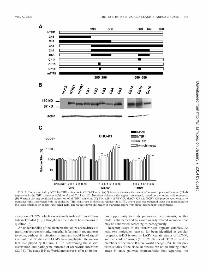
To address this question, we used MACV GP-pseudotypedvectors, since these vectors displayed the greatest increase(fold) in titer (2 orders of magnitude) when hTfR1 was trans-fected into CHO-K1 cells (Fig. 4). We analyzed the ability ofthe vectors to enter CHO-K1 cells transfected with a panel ofreciprocal chimeras between hTfR1 and mTfR1 (Fig. 7A), withchimeras 3 to 5 and 16 containing substitutions in hTfR1 of themurine sequences necessary to confer full MMTV entry (34).As controls, we used VSV-G and TCRV GP vectors, neither ofwhich is affected by the expression of either hTfR1 or mTfR1in CHO-K1 cells (Fig. 4).
We first demonstrated that all of the chimeric proteins usedwere expressed at similar levels to the wild-type hTfR1 andmTfR1 molecules (Fig. 7B). We then confirmed that expres-sion of the hTfR1 and mTfR1 parental molecules, as well as
FIG. 4. hTfR1, but not mTfR1, increases the titer of pathogenic GP vectors on CHO-K1 cells. (A) Titers (TU/ml) of pseudotyped retroviralvectors were obtained on mock-transfected CHO-K1 cells or cells transiently transfected with expression plasmids for hTfR1 or mTfR1. The valuesshown are means � standard errors from three independent experiments. (B) Vector titers are displayed as normalized relative titers (%), whereeach experimental value was first made relative to the value obtained on the mock-transfected cells and then normalized to the ratios obtained withthe VSV-G vectors. This normalizes the data for any toxic effects of the siRNAs. (C) Western blotting confirmed expression of both hTfR1 andmTfR1.
VOL. 82, 2008 TfR1 USE BY NEW WORLD CLADE B ARENAVIRUSES 943
on January 7, 2014 by guesthttp://jvi.asm
.org/D
ownloaded from

the chimeras, had no significant effect on the titers of eitherVSV-G or TCRV GP vectors. In contrast, the MACV GPvectors exhibited a range of responses to the chimeric proteins,with a general increase in vector titer being observed as theextent of human sequence in the molecules was increased (Fig.7C). However, the pattern obtained did not highlight any par-ticular region as being essential for hTfR1 receptor function.For example, since neither of the reciprocal chimeras 1 and 3displayed significant MACV receptor activity, it is likely thatthe MACV GP binding site on hTfR1 is not a simple linearepitope. Interestingly, chimera 16, which is an hTFR1 receptorcontaining murine sequence substitutions that are sufficient tosupport full MMTV entry, produced the second-highest titer of
all of the chimeras tested. It is therefore probable that MACVGP does not use the same binding site on hTfR1 as MMTVuses on mTfR1 or, if it does, the species specificity that weobserved is determined by residues outside of this region.
DISCUSSION
Arenaviruses are found predominantly in rodent reservoirs,with humans acting as incidental hosts. The host range of theviruses is determined largely by the habitat of the rodent vec-tor. For the New World clade B arenaviruses, among which arefour known human pathogens, the rodent hosts are New Worldrats and mice from the Sigmodontinae subfamily. A possible
FIG. 5. Ability of TfR1 orthologs from different species to enhance entry into CHO-K1 cells. (A) CHO-K1 cells were transfected with the TfR1orthologs from humans, mice, dogs (cTfR1), and cats (fTFR1), and expression was confirmed by Western blotting. (B) Mock- and TfR1-transfectedCHO-K1 cells were challenged with the indicated pseudotyped vectors. Vector titers were made relative to the value obtained for the mock-transfected cells and are expressed as a percentage of that control. The values shown are means � standard errors from two to four independentexperiments.
FIG. 6. Binding of MACV GP1 immunoadhesin to CHO-K1 cells transfected with hTFR1 or mTFR1. CHO-K1 cells were mock transfectedor transfected with expression plasmids for hTfR1 or mTfR1. The binding of MACV GP1 immunoadhesin to each cell line was detected byincubation with the immunoadhesin, followed by an FITC-labeled goat anti-rabbit secondary antibody and FACS analysis (bottom panel). As acontrol, cells were also treated with just the secondary antibody (top panel). The percentage of cells staining positive in each sample is indicated.
944 FLANAGAN ET AL. J. VIROL.
on January 7, 2014 by guesthttp://jvi.asm
.org/D
ownloaded from
exception is TCRV, which was originally isolated from Artibeusbats in Trinidad (10), although the true natural host remains inquestion (3).
An understanding of the elements that allow arenaviruses totransition between chronic, nonlethal infections in rodent hoststo acute, pathogenic infections in humans would be of signif-icant interest. Studies with LCMV have highlighted the impor-tant role played by the viral GP in determining the in vivodistribution and pathogenic outcome of arenavirus infections(29, 31). The clade B New World arenaviruses offer an impor-
tant opportunity to study pathogenic determinants, as thisclade is characterized by evolutionarily related members thatmay be subdivided according to pathogenicity.
Receptor usage in the arenaviruses appears complex. Atleast two molecules have so far been identified as cellularreceptors: �-DG is used by LASV, certain strains of LCMV,and two clade C viruses (8, 13, 27, 32), while TfR1 is used bymembers of the clade B New World lineage (25). In our pre-vious studies of the clade B1 viruses, we noted striking differ-ences in entry pathway characteristics that separated the
FIG. 7. Entry directed by hTfR1/mTfR1 chimeras in CHO-K1 cells. (A) Schematic showing the extent of human (open) and mouse (filled)sequences in the TfR1 chimeras (Ch1 to -5 and Ch14 to -16). Numbers delineate the regions exchanged, based on the amino acid sequence.(B) Western blotting confirmed expression of all TfR1 chimeras. (C) The ability of VSV-G, MACV GP and TCRV GP-pseudotyped vectors totransduce cells transfected with the indicated TfR1 constructs is shown as relative titers (%), where each experimental value was normalized tothe value obtained on mock-transfected cells. The values shown are means � standard errors from three independent experiments.
VOL. 82, 2008 TfR1 USE BY NEW WORLD CLADE B ARENAVIRUSES 945
on January 7, 2014 by guesthttp://jvi.asm
.org/D
ownloaded from

pathogenic and nonpathogenic members of the group. In thepresent study, we have extended those observations to includethe B2 lineage. Together, our data reveal that the ability of aGP to use hTfR1 correlates with being from a strain designatedas pathogenic for humans.
Despite the evidence supporting the use of hTfR1 as anarenavirus receptor, it is also clear that the clade B viruses canuse TfR1-independent entry pathways. Together with our pre-vious findings (21, 27), we ruled out �-DG as being involved inthe entry of these viruses, suggesting that other, as-yet-uniden-tified receptors exist. We found three distinct situations inwhich TfR1-independent pathways were being used. First, theentry of the nonpathogenic viruses, TCRV and AMAV, intoboth human and rodent cell lines was both TfR1 and �-DGindependent. Second, we observed that the entry of the patho-genic viruses, JUNV, MACV, and GTOV, into murine NIH3T3 cells did not depend on TfR1, since siRNA knockdown ofmTfR1 had no effect on vector titer, while the addition ofmTfR1 to CHO-K1 cells was unable to increase the entryof MACV GP-pseudotyped vectors. In addition, the fact thatMACV entry into CHO-K1 cells could be stimulated by almost2 orders of magnitude when hTfR1 was present implies thatentry into CHO-K1 cells is suboptimal for MACV, which inturn suggests that the endogenous Chinese hamster TfR1 can-not facilitate clade B entry. The third situation we observedwas the entry of pathogenic viruses into human cells that weredepleted for hTfR1, either by siRNA knockdown or antibodyblockage, although it is difficult to rule out that entry wasoccurring due to incomplete removal of hTfR1. Overall, we donot yet understand the relationship between these differentTfR-1-independent pathways and, specifically, whether thenon-TfR1 receptor that is used by TCRV and AMAV is thesame molecule that is used by the pathogenic viruses underconditions in which hTfR1 is unavailable.
It is now appreciated that several different roles can beplayed by the cellular factors that are recruited by viral fusionproteins to mediate the entry of enveloped viruses into cells. Inthe simplest case, the fusion protein binds to a single receptormolecule and this interaction triggers conformational changesin the fusion protein that expose the fusion peptide and initiatevirus-cell fusion (9). Alternatively, as is the case with the in-fluenza virus hemagglutinin protein, binding to the receptorcan allow the virus to be endocytosed into a low-pH environ-ment that provides the trigger for the necessary conforma-tional change. Other viruses, such as avian leukosis virus, ap-pear to use a combination of these features (19). Viruses alsovary in the number of receptor molecules with which they caninteract. Some viruses have evolved the ability to bind to morethan one cellular receptor (alternate primary receptors), suchas the 10A1 variant of the murine leukemia virus Env protein(11) that shows expanded tropism. Another variation is the useof both a primary and secondary (or co-) receptor. The proto-type example of this is human immunodeficiency virus type 1(HIV-1) entry, where binding to CD4 triggers initial confor-mational changes in the viral Env protein that expose thecoreceptor (e.g., CCR5) binding site (36), with engagement ofthe coreceptor necessary for virus-cell fusion. Finally, someviruses clearly make use of less-specific interactions with at-tachment factors to facilitate binding to the cell surface beforeinteraction with the true receptor. An example of this type of
interaction is the binding of HIV-1 to syndecans, which en-hances virus entry in macrophages (30).
At present, it is not clear which of these roles is being playedby hTfR1 in arenavirus entry. It could be acting as a classicviral receptor that is itself sufficient to orchestrate the entry ofMACV, JUNV, and GTOV into cells. Alternatively, the factthat entry can occur in certain situations in the absence ofhTfR1 leads us to consider that an interaction with hTfR1could reflect an initial virus-cell interaction that occurs beforehanding the GP onto a second receptor molecule that actuallydirects virus-cell fusion. Certainly, our immunoadhesin bindingstudies demonstrated a strong interaction between MACVGP1 and hTfR1. Such a scenario could therefore resembleeither the receptor/coreceptor interactions of the HIV Envwith CD4 and CCR5 or the less-specific interaction of HIV-1Env with syndecans, but with hTfR1 binding representing anadditional step, superimposed at the beginning of an existingentry pathway.
TfR1 normally transitions between the cell surface and en-dosomes. At extracellular pH 7.4, TfR1 binds iron-laden trans-ferrin and the resulting complex is endocytosed via clathrin-coated pits. As the endosome acidifies to pH 5.5, the iron isreleased, and the apo-transferrin/TfR1 complex is recycled tothe cell surface, where the transferrin is released (1). Ourprevious studies with NH4Cl, an inhibitor of endosomal acid-ification, have revealed that clade B entry requires endosomalacidification, although we identified at least one cell line, K562,in which JUNV and MACV entry did not (21). Interestingly, ithas been reported that the pH required to trigger cell-cellfusion directed by the JUNV GP is between 5.0 and 5.5 (6, 37).Although TfR1 typically recycles through early endosomes thatonly acidify to pH 6.5, it is possible that engagement of thereceptor with the arenavirus GP could divert TfR1 from therecycling pathway to deeper in the endosomal pathway, whereit would encounter a lower-pH environment.
Our analysis of the functionality of TfR1 from differentspecies revealed that both the human and feline receptorscould be effectively used, while the murine and canine formswere not. mTfR1 and hTfR1 are 76.8% identical, and thehuman and canine receptors are 78.6% identical. However,despite this homology, no clade B arenaviruses could usemTfR1, and only JUNV GP appeared able to use the canineTfR1, albeit with significantly less efficiency than either thehuman or feline forms. Similar species-specific restrictions areseen with other viruses that use TfR1 for entry; MMTV canuse murine but not human, cat, dog, or hamster TfR1 (34),while canine parvovirus can use human, canine, and felineTfR1, but not quail or hamster (23). Studies of the interactionof MMTV with mTfR1 have shown that this virus binds to theouter edge of the TfR1 dimer, distant from the binding site foreither transferrin or HFE. Our preliminary studies with hu-man/mouse chimeric receptors did not identify any particularregion of hTfR1 as being sufficient to allow high-level entry ofMACV vectors, but we noted that substitution for the hTfR1with the murine segments that confer susceptibility to MMTVinfection did not abolish MACV entry.
A consistent observation that we made with the pseudotypedvectors carrying the hTfR1-using GPs (MACV, JUNV, andGTOV) was that they resulted in lower titers on all of thenonlymphocytic rodent cell lines that we examined (NIH 3T3,
946 FLANAGAN ET AL. J. VIROL.
on January 7, 2014 by guesthttp://jvi.asm
.org/D
ownloaded from
CHO-K1, and BHK21 cells) compared to the titers obtainedwith the TCRV and AMAV vectors. Such differences were notseen on nonlymphocytic cell lines of human origin (21). Thissuggests that, similar to the situation in murine cells, neitherthe Chinese hamster nor Syrian hamster TfR1 receptor canpromote clade B entry. This is an intriguing result as hamstersare more closely related to the New World rodents that are thenatural hosts for these viruses. However, a definitive analysis ofwhether TfR1 is ever used in a rodent host will require studiesusing the specific species’ TfR1.
We also noted that while the addition of hTfR1 to CHO-K1cells could increase the titer of MACV vectors, it merelyserved to increase titers to the same levels that AMAV andTCRV vectors achieved in the absence of this molecule (Fig.4A). This suggests that, in the absence of a functional TfR1,entry of pathogenic viruses into rodent cells is limited, whilethis is not the case for the non-hTfR1-using viruses. Thesefindings are consistent with a model whereby acquisition of theability to use TfR1 comes at a price of reduced ability to accessany TfR1-independent pathways (at least in the rodent celllines we have examined). We further speculate that the ances-tral clade B virus may have used a receptor other than TfR1,but that certain viruses in this lineage subsequently evolved theability to use TfR1. These viruses are now also able to jumpspecies and infect humans, but whether the acquisition of hu-man TfR1 binding activity is the root cause of the ability tojump species, or an accident of the similarity with a receptorspecies used in the natural host, awaits further studies.
In the opposite scenario, it is also possible that the ancestralclade B virus used TfR1 as a receptor but that certain membersof the lineage evolved to use an alternate receptor(s) andthereby lost the ability to infect humans as accidental hosts.However, any such model must account for the fact that bothhTfR1-dependent and -independent viruses are foundthroughout the different clade B sublineages (Fig. 1A), sug-gesting that any such changes could have occurred more thanonce. Indeed, it will be interesting to determine if any otherarenaviruses that can infect humans are also using TfR1. Ourpreliminary investigations with the putative human pathogenWhitewater Arroyo virus (7) have shown that this New Worldclade A virus does not use hTfR1 (26).
In summary, the finding that TfR1 use by clade B arenavi-ruses is correlated with the ability to cause severe hemorrhagicfevers in humans has clear implications for therapies aimed atblocking this interaction, as discussed by Radoshitzky et al.(25). However, it also has wider implications for characterizingthese viruses, as it suggests that a good predictor of humanpathogenicity would be the ability to use hTfR1. If this hypoth-esis holds true, hTfR1 receptor use could serve as a diagnostictest applied to both existing and newly identified members ofthis family.
ACKNOWLEDGMENTS
We thank En xiu Wang, Susan Ross, and Stefan Kunz for reagentsand helpful discussions.
This work was supported by PHS grant 1U54 AI065359 to the PacificSouthwest Regional Center of Excellence for Biodefense and Emerg-ing Infectious Diseases (P.M.C.) and a Saban Research Institute Ca-reer Development Award (T.R.).
REFERENCES
1. Aisen, P. 2007. Transferrin receptor 1. Int. J. Biochem. Cell Biol. 36:2137–2143.
2. Bowen, M. D., C. J. Peters, and S. T. Nichol. 1996. The phylogeny of NewWorld (Tacaribe complex) arenaviruses. Virology 219:285–290.
3. Buchmeier, M. J., J.-C. de la Torre, and C. J. Peters. 2006. Arenaviridae: theviruses and their replication, p. 1–37. In D. M. Knipe and P. M. Howley (ed.),Fields virology, 5th ed. Lippincott Williams & Wilkins, Philadelphia, PA.
4. Burns, J. W., and M. J. Buchmeier. 1991. Protein-protein interactions inlymphocytic choriomeningitis virus. Virology 183:620–629.
5. Cao, W., M. D. Henry, P. Borrow, H. Yamada, J. H. Elder, E. V. Ravkov, S. T.Nichol, R. W. Compans, K. P. Campbell, and M. B. A. Oldstone. 1998.Identification of alpha-dystroglycan as a receptor for lymphocytic chorio-meningitis virus and Lassa fever virus. Science 282:2079–2081.
6. Castilla, V., and S. E. Mersich. 1996. Low-pH-induced fusion of Vero cellsinfected with Junin virus. Arch. Virol. 141:1307–1317.
7. Centers for Disease Control and Prevention. 2000. Fatal illnesses associatedwith a New World arenavirus—California, 1999–2000. Morb. Mort. Wkly.Rep. 49:709–711.
8. Charrel, R. N., H. Feldmann, C. F. Fulhorst, R. Khelifa, R. de Chesse, andX. K. de Lamballerie. 2002. Phylogeny of New World arenaviruses based onthe complete coding sequences of the small genomic segment identified anevolutionary lineage produced by intrasegmental recombination. Biochem.Biophys. Res. Commun. 296:1118–1124.
9. Colman, P. M., and M. C. Lawrence. 2003. The structural biology of type Iviral membrane fusion. Nat. Rev. Mol. Cell Biol. 4:309–319.
10. Downs, W. G., C. R. Anderson, L. Spence, T. H. G. Aitken, and A. M.Greenhall. 1963. Tacaribe virus, a new agent isolated from Artibeus bats andmosquitoes in Trinidad, West Indies. Am. J. Trop. Med. Hyg. 12:640–646.
11. Han, J.-Y., P. M. Cannon, K.-M. Lai, Y. Zhao, M. V. Eiden, and W. F.Anderson. 1997. Identification of envelope protein residues required for theexpanded host range of 10A1 murine leukemia virus. J. Virol. 71:8103–8108.
12. Kunz, S., and J. C. de la Torre. 2005. Novel antiviral strategies to combathuman arenavirus infections. Curr. Mol. Med. 5:735–751.
13. Kunz, S., J. M. Rojek, M. Kanagawa, C. F. Spiropoulou, R. Barresi, K. P.Campbell, and M. B. A. Oldstone. 2005. Posttranslational modification of�-dystroglycan, the cellular receptor for arenaviruses, by the glycosyltrans-ferase LARGE is critical for virus binding. J. Virol. 79:14282–14296.
14. Kunz, S., N. Sevilla, J. M. Rojek, and M. B. A. Oldstone. 2004. Use ofalternative receptors different than �-dystroglycan by selected isolates oflymphocytic choriomeningitis virus. Virology 325:432–445.
15. Mackenzie, R. B. 1965. Epidemiology of Machupo virus infection. I. Patternof human infection, San Joaquin, Bolivia, 1962–1964. Am. J. Trop. Med.Hyg. 14:808–813.
16. Maiztegui, J., M. Feuillade, and A. Briggiler. 1986. Progressive extension ofthe endemic area and changing incidence of Argentine hemorrhagic fever.Med. Microbiol. Immunol. 175:149–152.
17. Mills, J. N., B. A. Ellis, J. E. Childs, K. T. McKee, Jr., J. I. Maiztegui, C. J.Peters, T. G. Ksiazek, and P. B. Jahrling. 1994. Prevalence of infection withJunin virus in rodent populations in the epidemic area of Argentine hem-orrhagic fever. Am. J. Trop. Med. Hyg. 51:554–562.
18. Mills, J. N., B. A. Ellis, K. T. McKee, Jr., G. E. Calderon, J. I. Maiztegui,G. O. Nelson, T. G. Ksiazek, C. J. Peters, and J. E. Childs. 1992. A longi-tudinal study of Junin virus activity in the rodent reservoir of Argentinehemorrhagic fever. Am. J. Trop. Med. Hyg. 47:749–763.
19. Mothes, W., A. L. Boerger, S. Narayan, J. M. Cunningham, and J. A. Young.2000. Retroviral entry mediated by receptor priming and low pH triggeringof an envelope glycoprotein. Cell 103:679–689.
20. Niwa, H., K. Yamamura, and J. Miyazaki. 1991. Efficient selection forhigh-expression transfectants with a novel eukaryotic vector. Gene 108:193–199.
21. Oldenburg, J., T. Reignier, M. L. Flanagan, G. A. Hamilton, and P. M.Cannon. 2007. Differences in tropism and pH dependence for glycoproteinsfrom the clade B1 arenaviruses: implications for receptor usage and patho-genicity. Virology 364:132–139.
22. Parekh, B. S., and M. J. Buchmeier. 1986. Proteins of lymphocytic chorio-meningitis virus: antigenic topography of the viral glycoproteins. Virology153:168–178.
23. Parker, J. S., W. J. Murphy, D. Wang, S. J. O’Brien, and C. R. Parrish. 2001.Canine and feline parvoviruses can use human or feline transferrin receptorsto bind, enter, and infect cells. J. Virol. 75:3896–3902.
24. Peters, C. J. 1995. Arenavirus diseases, p. 227–246. In J. Portfield (ed.),Exotic viral infections. Chapman & Hall Medical, London, United Kingdom.
25. Radoshitzky, S. R., J. Abraham, C. F. Spiropoulou, J. H. Kuhn, D. Nguyen,W. Li, J. Nagel, P. J. Schmidt, J. H. Nunberg, N. C. Andrews, M. Farzan, andH. Choe. 2007. Transferrin receptor 1 is a cellular receptor for New Worldhaemorrhagic fever arenaviruses. Nature 446:92–96.
26. Reignier, T., J. Oldenburg, M. Flanagan, G. A. Hamilton, V. K. Martin, andP. M. Cannon. 9 November 2007. Receptor use by the Whitewater Arroyovirus glycoprotein. Virology. [Epub ahead of print.]
27. Reignier, T., J. Oldenburg, B. Noble, E. Lamb, V. Romanowski, M. J. Buch-
VOL. 82, 2008 TfR1 USE BY NEW WORLD CLADE B ARENAVIRUSES 947
on January 7, 2014 by guesthttp://jvi.asm
.org/D
ownloaded from

meier, and P. M. Cannon. 2006. Receptor use by pathogenic arenaviruses.Virology 353:111–120.
28. Rojek, J. M., C. F. Spiropoulou, and S. Kunz. 2006. Characterization of thecellular receptors for the South American hemorrhagic fever viruses Junin,Guanarito, and Machupo. Virology 349:476–491.
29. Salvato, M., P. Borrow, E. Shimomaye, and M. B. A. Oldstone. 1991. Mo-lecular basis of viral persistence: a single amino acid change in the glyco-protein of lymphocytic choriomeningitis virus is associated with suppressionof the antiviral cytotoxic T-lymphocyte response and establishment of per-sistence. J. Virol. 65:1863–1869.
30. Saphire, A. C. S., M. D. Bobardt, Z. Zhang, G. David, and P. A. Gallay. 2001.Syndecans serve as attachment receptors for human immunodeficiency virustype 1 on macrophages. J. Virol. 75:9187–9200.
31. Smelt, S. C., P. Borrow, S. Kunz, W. Cao, A. Tishon, H. Lewicki, K. P.Campbell, and M. B. A. Oldstone. 2001. Differences in affinity of binding oflymphocytic choriomeningitis virus strains to the cellular receptor �-dystro-glycan correlate with viral tropism and disease kinetics. J. Virol. 75:448–457.
32. Spiropoulou, C. F., S. Kunz, P. E. Rollin, K. P. Campbell, and M. B. A.
Oldstone. 2002. New World arenavirus clade C, but not clade A and Bviruses, utilizes �-dystroglycan as its major receptor. J. Virol. 76:5140–5146.
33. Vainrub, B., and R. Salas. 1994. Latin American hemorrhagic fever. Infect.Dis. Clin. N. Am. 8:47–59.
34. Wang, E., L. Albritton, and S. R. Ross. 2006. Identification of the segmentsof the mouse transferrin receptor 1 required for mouse mammary tumorvirus infection. J. Biol. Chem. 281:10243–10249.
35. Williamson, R. A., M. D. Henry, K. J. Daniels, R. F. Hrstka, J. C. Lee, Y.Sunada, O. Ibraghimov-Beskrovnaya, and K. P. Campbell. 1997. Dystrogly-can is essential for early embryonic development: disruption of Reichert’smembrane in Dag1-null mice. Hum. Mol. Genet. 6:831–841.
36. Wu, L., N. P. Gerard, R. Wyatt, H. Choe, C. Parolin, N. Ruffing, A. Borsetti,A. A. Cardoso, E. Desjardin, W. Newman, C. Gerard, and J. Sodroski. 1996.CD4-induced interaction of primary HIV-1 gp120 glycoproteins with thechemokine receptor CCR-5. Nature 384:179–183.
37. York, J., and J. H. Nunberg. 2006. Role of the stable signal peptide of Juninarenavirus envelope glycoprotein in pH-dependent membrane fusion. J. Vi-rol. 80:7775–7780.
948 FLANAGAN ET AL. J. VIROL.
on January 7, 2014 by guesthttp://jvi.asm
.org/D
ownloaded from