New Protein Clustering of Breast Cancer Tissue Proteomics Using Actin Content as a Cellularity...
7
New Protein Clustering of Breast Cancer Tissue Proteomics Using Actin Content as a Cellularity Indicator Ida Pucci-Minafra,* ,†,‡ Patrizia Cancemi, † Nadia Ninfa Albanese, † Gianluca Di Cara, † Maria Rita Marabeti, † Antonio Marrazzo, †,§ and Salvatore Minafra †,‡ Dipartimento di Oncologia Sperimentale e Applicazioni Cliniche (DOSAC), Università di Palermo, Patermo, Italy, Centro di Oncobiologia Sperimentale (COBS), Palermo, Italy, and Ospedale “La Maddalena” D.O. III livello, Palermo, Italy Received November 15, 2007 In the present study, we report the comparative proteome profiles of proteins solubilized from 37 breast cancer surgical tissues, normalized for the actin content. Blood-derived proteins were excluded from the analysis. Among the tumor-derived protein spots, a large proportion (39%) was found present in all patients. These included several glycolytic enzymes, detox and heat shock proteins, members of annexin and S100 protein families, cathepsin D, and two “rare” proteins, DDAH2 involved in the angiogenesis control, and the oncogene PARK7. Other proteins, such as psoriasin, galectin1, cofilin, peroredoxins, SH3L1, and others, showed sporadic presence and high expression level, which suggests their possible role for patient stratification. Keywords: proteomic • two-dimensional electrophoresis • peptide mass fingerprint • breast cancer • surgical tissues • protein cluster • cellularity indicator Introduction The exponentially increasing volume of information ex- tracted from genomic and proteomic applications on cancer, while providing new insights into molecular composition of cancer cells and tissues, imposes new challenges on data rationalization as a tool for clinically relevant biomarker discovery. In recent years, the major agencies for cancer research have committed extensive resources to identify and make available in clinical practice biomarkers that can help to demonstrate the presence of a tumor and its malignant potential or to predict tumor behavior, prognosis, or response to treatments. 1 The increasing number of detected proteins in the different laboratories conducting proteomic research has generated many candidate biomarkers with potential clinical value. However, translation from bench to bedside of the patient has proven to be more difficult than previously expected. A major reason is the complexity of the cancer as a biological system under its different hierarchical levels: genes, proteins, cells, and tissues. Breast cancer represents the most frequent and potentially aggressive type of cancer and the leading cause of cancer deaths in women in the Western countries. Moreover, breast cancer is one of the most enigmatic and poorly predictable cancers in its evolution, likely because it includes several different forms that behave differently among patients. This biological hetero- geneity is consistent with observed varied responses to thera- pies across patient populations. Current clinical parameters for breast cancer diagnosis and cure are tumor size, axillary lymph node status, histological grading, and presence or absence of metastases. 2 Prognostic/ predictive properties, such as estrogen and progesterone recep- tor status, and human epidermal growth factor receptor (HER- 2/neu) status, are currently used for therapeutic decision. 3,4 Conversely, it is now emerging that the number of genetic mutations 5 and epigenetic deregulations 6 in cancer is far more higher than previously thought. Therefore, proteomic screening for differential protein ex- pression in subsets of tumor samples is an essential tool to generate databases, to contribute to the knowledge of biological pathways in a given cancer tissue, and to allow a molecular classification of cancer for patient stratification and biomarker discovery. Eligibility of clinically relevant biomarkers for therapeutic use implies at least two conditions: easy accessibility from the bloodstream or tissues, and elevated expression levels with respect to the normal tissues. In our laboratories, we have recently developed adequate extraction procedures for proteomic profiling of breast cancer tissues derived from surgery. 7 A limitation of this kind of approach is the mentioned heterogeneity of breast cancer tissues, for which proteomic profiling may be influenced by extension and amount of neoplastic cells within the host stroma. To minimize this limitation and to introduce a objective criterion to compare proteomes of different tissues, the expres- sion levels of individual proteins were normalized for actin content in each tissue extract. * To whom correspondence should be addressed. Prof. Ida Pucci-Minafra, Dipartimento di Oncologia Sperimentale e Applicazioni Cliniche; Via San Lorenzo Colli 312, 90146 Palermo, Italy. E-mail: [email protected]. Tel/Fax: 39 91 6806420. † Dipartimento di Oncologia Sperimentale e Applicazioni Cliniche (DOSAC), Università di Palermo. ‡ Centro di Oncobiologia Sperimentale (COBS). § Ospedale “La Maddalena” D.O. III livello. 1412 Journal of Proteome Research 2008, 7, 1412–1418 10.1021/pr700748m CCC: $40.75 2008 American Chemical Society Published on Web 02/20/2008
-
Upload
independent -
Category
Documents
-
view
0 -
download
0
Transcript of New Protein Clustering of Breast Cancer Tissue Proteomics Using Actin Content as a Cellularity...

New Protein Clustering of Breast Cancer Tissue Proteomics Using
Actin Content as a Cellularity Indicator
Ida Pucci-Minafra,*,†,‡ Patrizia Cancemi,† Nadia Ninfa Albanese,† Gianluca Di Cara,†
Maria Rita Marabeti,† Antonio Marrazzo,†,§ and Salvatore Minafra†,‡
Dipartimento di Oncologia Sperimentale e Applicazioni Cliniche (DOSAC), Università di Palermo,Patermo, Italy, Centro di Oncobiologia Sperimentale (COBS), Palermo, Italy, and Ospedale “La Maddalena”
D.O. III livello, Palermo, Italy
Received November 15, 2007
In the present study, we report the comparative proteome profiles of proteins solubilized from 37 breastcancer surgical tissues, normalized for the actin content. Blood-derived proteins were excluded fromthe analysis. Among the tumor-derived protein spots, a large proportion (39%) was found present inall patients. These included several glycolytic enzymes, detox and heat shock proteins, members ofannexin and S100 protein families, cathepsin D, and two “rare” proteins, DDAH2 involved in theangiogenesis control, and the oncogene PARK7. Other proteins, such as psoriasin, galectin1, cofilin,peroredoxins, SH3L1, and others, showed sporadic presence and high expression level, which suggeststheir possible role for patient stratification.
Keywords: proteomic • two-dimensional electrophoresis • peptide mass fingerprint • breast cancer •surgical tissues • protein cluster • cellularity indicator
Introduction
The exponentially increasing volume of information ex-tracted from genomic and proteomic applications on cancer,while providing new insights into molecular composition ofcancer cells and tissues, imposes new challenges on datarationalization as a tool for clinically relevant biomarkerdiscovery. In recent years, the major agencies for cancerresearch have committed extensive resources to identify andmake available in clinical practice biomarkers that can help todemonstrate the presence of a tumor and its malignantpotential or to predict tumor behavior, prognosis, or responseto treatments.1 The increasing number of detected proteins inthe different laboratories conducting proteomic research hasgenerated many candidate biomarkers with potential clinicalvalue. However, translation from bench to bedside of thepatient has proven to be more difficult than previouslyexpected. A major reason is the complexity of the cancer as abiological system under its different hierarchical levels: genes,proteins, cells, and tissues.
Breast cancer represents the most frequent and potentiallyaggressive type of cancer and the leading cause of cancer deathsin women in the Western countries. Moreover, breast canceris one of the most enigmatic and poorly predictable cancersin its evolution, likely because it includes several different forms
that behave differently among patients. This biological hetero-geneity is consistent with observed varied responses to thera-pies across patient populations.
Current clinical parameters for breast cancer diagnosis andcure are tumor size, axillary lymph node status, histologicalgrading, and presence or absence of metastases.2 Prognostic/predictive properties, such as estrogen and progesterone recep-tor status, and human epidermal growth factor receptor (HER-2/neu) status, are currently used for therapeutic decision.3,4
Conversely, it is now emerging that the number of geneticmutations5 and epigenetic deregulations6 in cancer is far morehigher than previously thought.
Therefore, proteomic screening for differential protein ex-pression in subsets of tumor samples is an essential tool togenerate databases, to contribute to the knowledge of biologicalpathways in a given cancer tissue, and to allow a molecularclassification of cancer for patient stratification and biomarkerdiscovery.
Eligibility of clinically relevant biomarkers for therapeutic useimplies at least two conditions: easy accessibility from thebloodstream or tissues, and elevated expression levels withrespect to the normal tissues.
In our laboratories, we have recently developed adequateextraction procedures for proteomic profiling of breast cancertissues derived from surgery.7 A limitation of this kind ofapproach is the mentioned heterogeneity of breast cancertissues, for which proteomic profiling may be influenced byextension and amount of neoplastic cells within the hoststroma. To minimize this limitation and to introduce a objectivecriterion to compare proteomes of different tissues, the expres-sion levels of individual proteins were normalized for actincontent in each tissue extract.
* To whom correspondence should be addressed. Prof. Ida Pucci-Minafra,Dipartimento di Oncologia Sperimentale e Applicazioni Cliniche; Via SanLorenzo Colli 312, 90146 Palermo, Italy. E-mail: [email protected]. Tel/Fax:39 91 6806420.
† Dipartimento di Oncologia Sperimentale e Applicazioni Cliniche (DOSAC),Università di Palermo.
‡ Centro di Oncobiologia Sperimentale (COBS).§ Ospedale “La Maddalena” D.O. III livello.
1412 Journal of Proteome Research 2008, 7, 1412–1418 10.1021/pr700748m CCC: $40.75 2008 American Chemical SocietyPublished on Web 02/20/2008
In the present study, we analyzed the proteomic profiles ofsurgical tissues of 37 patients diagnosed for ductal infiltratingbreast cancer.
Collectively, we produced a catalogue of 160 proteins. Thisnumber includes different isoforms of the same protein, as wellas proteins derived from blood. The latter were filtered awaybefore generating protein clusters according to an arbitraryscale of expression levels calculated as relative aboundance(%Vol) of each protein spot normalized for actin content. Bythis criterion, we produced six protein clusters, namely, veryhighly expressed, highly expressed, medium/highly expressed,medium/lowly expressed, lowly expressed, and very lowlyexpressed proteins.
Interestingly, a large proportion of the tumor-derived pro-teins (39%) was found present in all patients. These includedseveral glycolytic enzymes, detox and heat shock proteins,
members of annexin and S100 protein families, cathepsin D,and two proteins not described before in breast cancer tissueproteomics, namely, DDAH2, involved in the angiogenesiscontrol, and the oncogene PARK7. Other proteins, includingpsoriasin, galectin1, cofilin, peroredoxins, and SH3L1, showedsporadic presence and high expression level, which suggeststheir possible role for patient stratification.
We believe that this information may substantially contributeto the progress of protein profiling of breast cancer and helpto organize subclasses for clinical applications. The statisticalcorrelations with clinical parameters were not included in thisstudy, because a larger population of patients is needed togeneralize the results. Therefore, a contribution of efforts inthis direction from several laboratories is to be encouraged.
Materials and Methods
Clinical Specimens. Sample management was performedaccording to the bioethical recommendations. Aliquots ofbreast cancer and its adjacent nontumoral tissues were ob-tained during surgical intervention and immediately frozen in
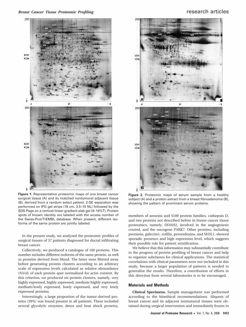
Figure 1. Representative proteomic maps of one breast cancersurgical tissue (A) and its matched nontumoral adjacent tissue(B), derived from a random select patient. 2-DE separation wasperformed on IPG gel strips (18 cm, 3.5–10 NL) followed by theSDS-Page on a vertical linear-gradient slab gel (9–16%T). Proteinspots of known identity are labeled with the access number ofthe Swiss-Prot/TrEMBL database. When present, different iso-forms of the same protein are jointly labeled.
Figure 2. Proteomic maps of serum sample from a healthysubject (A) and a protein extract from a breast fibroadenoma (B),showing the pattern of prominent serum proteins.
Breast Cancer Tissue Proteomic Profiling research articles
Journal of Proteome Research • Vol. 7, No. 4, 2008 1413

liquid nitrogen and stored at -80 °C until use. The patientsdid not receive any cytotoxic/endocrine treatment prior tosurgery. In each cases, nontumoral tissue was located at least5 cm away from the primary tumor. Diagnosis of ductal breastcancer (G2/G3) was confirmed histopathologically.
Sample Preparations. The frozen breast tissue samples werewashed several times with phosphate-buffered saline andhomogenized in RIPA buffer (50 mM Tris, pH 7.5, 0.1% NonidetP-40, 0.1% deoxycholate, 150 mM NaCl, and 4 mM EDTA) anda mixture of protease inhibitors (0.01% aprotinin, 10 mMsodium pyrophosphate, 2 mM sodium orthovanadate, and 1mM PMSF). The extraction was carried out overnight at 4 °Cwith the same buffer. The total cellular lysate was centrifugedat 15 000 rpm for 20 min to clear debris, and the supernatantwas dialysed against ultrapure distilled water, lyophilised, andstored at -80 °C until analysis. Protein concentration in thecellular extracts was determined using the Bradford method.8
Two-Dimensional Gel Electrophoresis. The proteins ex-tracted from breast cancer tissue and normal adjacent tissuewere solubilized in a buffer containing 4% CHAPS, 40 mM Tris,and 65 mM DTE in 8 M urea. Aliquots of 45 µg (analytical gels)or 1.5 mg (preparative gels) of total proteins were separatelymixed with 350 µL of rehydration solution containing 8 M urea,2% CHAPS, 10 mM DTE, and 0.5% carrier anpholytes (Resolyte3.5–10) and applied for IEF using commercial sigmoidal IPGstrips, 18 cm long with pH range 3.5–10. The second dimensionwas carried out on 9–16% linear gradient polyacrylamide gels(SDS-PAGE), and the separated proteins were visualized byammoniacal silver staining. Two or three replicas were per-formed for each patient.
Image Acquisition and Data Analysis. Silver-stained gelswere digitized using a computing densitometer and analyzedwith ImageMaster 2D Platinum software (Amersham Bio-
sciences, Sweden). Gel calibration was carried out using aninternal standard and the support of the ExPaSy molecularbiology server, as described.9 Quantitative variations in proteinexpression levels were calculated as the volume of the spots(i.e., integration of optical density over the spot area). To correctfor differences in gel staining, spot volumes relative to the sumof the volume of all spots on each gel (%Vol) were calculatedby the software. Furthermore, for comparative profiling, indi-vidual spot %Vol was normalized for the actin %Vol value. Theresulting figure was considered an indicator of protein expres-sion level and utilized for protein clusterization.
N-terminal Protein Sequencing. N-terminal microsequenc-ing was performed by automated Edman degradation in aProtein Sequencer (Procise 491, Applied Biosystems) on proteinsamples electrotransferred onto PVDF membranes at 50 V for30 min at 4 °C.10 The Swiss-Prot/TrEMBL/BLAST databases wereused for online sequence similarity search at http://www.expasy.ch/tools/#similarity.
Matrix-Assisted Laser Desorption Ionization-Time ofFlight (MALDI-TOF). Mass spectrometric sequencing wascarried out after in-gel digestion of protein spots, usingsequencing-grade trypsin (20 µg/vial), according to the methodof Shevchenko et al. 11 with some modification.10 The trypticpeptide extracts were dried in a vacuum centrifuge andredissolved in 10 µL of 0.1% trifluoroacetic acid (TFA). Thematrix, R-cyano-4-hydroxycinnamic acid (HCCA), was pur-chased from Sigma-Aldrich. A saturated solution of HCCA (1µL) at 2 mg/200 µL in CH3CN/H2O (50:50 (v/v)) containing 0.1%TFA was mixed with 1 µL of peptide solution on the MALDIplate and left to dry. MALDI-TOF mass spectra were recordedon a Voyager DE-PRO (Applied-Biosystems) mass spectrometer,in the 500–5000 Da mass range, using a minimum of 100 shotsof laser per spectrum. Delayed extraction source and reflector
Figure 3. Panel showing an experimental window from ImageMaster 2D Platinum software containing the sections of actin spots (whitecrosses) from the matched two-dimensional gels of the 37 breast cancer surgical tissues, indicated from P01 to P37.
research articles Pucci-Minafra et al.
1414 Journal of Proteome Research • Vol. 7, No. 4, 2008

equipment allowed sufficient resolution to consider MH+ ofmonoisotopic peptide masses. Internal calibration was doneusing trypsin autolysis fragments at m/z 842.5100, 1045.5642,and 2211.1046 Da. Peptide mass fingerprinting was comparedto the theoretical masses from the Swiss-Prot or NCBI sequencedatabases using Mascot (http://www.matrixscience.com/). Typi-cal search parameters were as follows: (50 ppm of masstolerance, carbamidomethylation of cysteine residues, onemissed enzymatic cleavage for trypsin, a minimum of fourpeptide mass hits was required for a match, methionineresidues could be considered in oxidized form, no restrictionwas placed on the isoelectric point of the protein, and a proteinmass range from 5 to 100 kDa was allowed.
Western Blotting. For immune detection, the gels (9–16%linear gradient SDS-PAGE) were electrotransferred onto nitro-cellulose membrane (HyBond ECL, Amersham) at 50 V for 1 hat 4 °C. After electrotransfer, the membranes were blocked with5% of dry milk in T-TBS for 1 h and probed with one of thefollowing antibodies (Santa Cruz): monoclonal anti-PARK7,anti-S10A7, anti-GDIR, and goat polyclonal anti-DDAH1 andanti-DDAH2, at 1:1000 diluted with 1% milk in T-TBS. Followingincubation with anti-mouse or anti-goat peroxidase-linkedantibody (Amersham), the reaction was revealed using en-hanced chemioluminiscence (Pierce), according the manufac-turer’s instructions.
Figure 4. Diagram illustrating the distribution of the identified proteins among the 37 patients, designated from P01 to P37. Alphabeticalletters following protein abbreviated names indicate the different isoforms, starting from the more acidic one. The black boxes indicateabsence of proteins in the map of the corresponding patient, and the crosses in each box indicate the patient expressing the givenprotein at its highest level.
Breast Cancer Tissue Proteomic Profiling research articles
Journal of Proteome Research • Vol. 7, No. 4, 2008 1415

Results and Discussion
The methodology described to isolate proteins from thesurgical specimens allows the releasing of salt-soluble proteinsfrom cellular and extracellular compartments. No efforts weremade to extract ECM components, because they not pertinentto the present research.
We have recently reported the comparative proteomicprofiles of 13 pairs of surgical samples obtained from patientswith ductal infiltrating breast cancer.7 As expected, the con-tribution of cellular proteins to the proteomic complexity, wasmuch higher in the tumor extracts than in their pairednontumoral conterparts. As an example, panels A and B ofFigure 1 show two proteomic maps, respectively, generatedfrom a breast cancer tissue extract and its paired nonaffectedtissue. The labels on the map indicate the access number(Swiss-Prot/TrEMBL database) of the identified proteins. Aremarkable difference between the two proteomic maps forprotein spot composition is clearly observed. We suggest thatthis different pattern is due to the reduced amount of parenchimain the mammary gland of healthy adult women, opposed tothe abundant cell population present in the tumor core. Indeed,the proteomic profile of nontumoral tissue extracts reveals thealmost exclusive presence of serum and blood proteins,consisting mainly of albumin, immunoglubulins, and otherglobulins. Figure 2A shows the serum proteome of a healthysubject compared with the normal tissue extract. Similarly, thefibroadenoma proteome, a benign breast lesion, dispalys a verysmall amount of cellular proteins with respect to blood-derivedproteins (Figure 2B).
The collection of proteins so far identified in our tumorsamples contains 160 proteins, including isoelectric variantsand bood-derived proteins (supplemental Table 1 in SupportingInformation). When present, the different isoforms of the sameprotein are indicated by alphabetical letters, starting from themore acidic one.
Since the cellularity of the tumor biopsy, that is, cell densitieswithin an area of the surgical sample, may be very variableamong the different subjects, measurements of relative expres-sion levels (%Vol) of individual protein spots were normalizedin each map for actin content.
Figure 3 shows an experimental window containing thesections of actin spots from the 37 two-dimensional gels,testifying the variability of actin amount among the proteomicmaps of our study, likely as an effect of different degree ofcellularity in the samples examined, under the same quantityof protein loading in the 2D-IPG.
Protein Presence-Absence. As shown in Figure 1A, thetumor samples, besides the proteins of cellular origin, containvariable amount of blood-derived proteins. The latter werefiltered out from our catalogue. The remaining amount, likelyrepresenting tumor-related proteins, accounted for 87, 34 ofwhich, that is 39%, were expressed in all samples. Figure 4 is adiagram illustrating the distribution of the identified proteinsamong the 37 patients, indicated from P01 to P37. The blackboxes indicate absence of proteins in the map of the corre-sponding patient, and the crosses in each box indicate thepatient expressing the given protein at its highest level amongthe 37 chosen ones.
Figure 5. Protein clusters were generated by sorting proteins with the highest expression level among the 37 patients. The proteinspot expression level was calculated as the %Vol normalized for actin content, of each patient map, after subtraction of blood-derivedproteins. The range of values are expressed on an arbitrary scale, from 0.05 to >1, and indicated as: VHEP, very highly expressedproteins (range 0.8-1); HEP, highly expressed proteins (0.6–0.79); M/HEP, medium/highly expressed proteins (0.4–0.59); M/LEP, medium/lowly expressed (0.2–0.39); LEP, lowly expressed (0.1–0.19); VLEP, very lowly expressed (0.05–0.09).
research articles Pucci-Minafra et al.
1416 Journal of Proteome Research • Vol. 7, No. 4, 2008

Interestingly, and in agreement with current knowledge,9,12
a major portion of ubiquitous proteins belong to the glycolyticmetabolism (12/34, i.e., 35%, including isoforms); seven proteinforms include 2 protein foldings (PDA1, and 3 isoforms of PPIA),a calcium-binding chaperone (CALR) and 2 heat shock proteins(HSP74 and GRP94); four proteins are represented by 3members of redox-defense machinery (SOD, THIO, GSTP1),and one recently identified oncogene PARK7, a master regulatorof antioxidant transcriptional responses.13 Other interstingcomponents of this proteomic profiling are 2 regulators of thetransduction machinery (GDIR and GDIS), the Dimethylargini-nase 2 (DDAH2), a relevant enzyme involved in the angiogen-esis control,14 and a subgroup of proteins like ANXA2, S10A6calcium-binding protein, and vinculin which individually and/or in synergy are important players of events related to invasionand metastasis.15–19
Finally, another ubiquitously expressed protein was thecathepsin D, a controversial prognostic marker for breastcancer progression.20 This was detected under two isoelectricvariants with pI of 5.29 and 5.69, respectively, the latter beingusually the more aboundant.
It is worth to mention that several ubiquitous proteins aremultifunctional, therefore, may be involved in more metabolicpathways, besides their major role in the cell. Indeed, HSP70protein family, GRP94, and CALR may also exert a role in thetumor immune-responses.21 Similarly, G3P, ENOA, PGAM1,and TPIS, besides their role in the glycolytic pathway, are
known to exploit several diversified functions; that is, G3P2, isknown to facilitate membrane fusion, microtubule bundling,and it is capable of nucleic acid binding;22,23 alpha-enolasefunctions as a plasminogen receptor on some cell types,24 whilePGAM1 has been shown to substain indefinite proliferation inembryonic fibroblasts.25 It has been also reported that triose-phosphate isomerase activity, which play a key role in theinterconnection between the pentose phosphate pathway andthe lipid metabolism, tends to be higher expressed in immaturemyeloid cells.26,27 Recently, it has also been demonstrated thatcalreticulin has a role in regulating p53 function by affectingits rate of degradation and nuclear localization.28
Protein Clustering According to the Expression Level. Aftersubtraction of proteins derived from blood, and normalizationfor actin contents, we sorted the proteins by the highestexpression level among the 37 analyzed patients. These proteinswere then grouped according to an arbitrary scale, with values(%Vol normalized for actin) ranging from 0.05 to >1. By thiscriterion, we generated six protein clusters (Figure 5): (1) veryhighly expressed proteins (VHEP, 14.94%), (2) highly expressedproteins (HEP, 16.09%), (3) medium/highly expressed proteins(M/HEP, 18.39%), (4) medium/lowly expressed (M/LEP, 39.08%),(5) lowly expressed (LEP, 6.89%), and (6) very lowly expressed(VLEP, 4.59%).
Remarkably, among the VHEP and HEP clusters are someglycolytic enzymes, proteins of cellular defenses, proteinsbelonging to the S100 family, and the 2 isoforms of CATD.These two groups also include proteins with a very peculiarproperty: their appearance is sporadic, but when present, theirexpression is never low. This is the case, among others, ofgalectin 1 (LEG1), several proteins of the S100-calcium bindingfamily, and SH3L1, a gene product belonging to the SH3BGR(SH3 domain-binding glutamic acid-rich-like protein family)and structurally related to thioredoxin (Trx) super family.29
Psoriasin (S10A7) is one of the most variable proteins amongthe 37 patients. It is highly expressed in 15/37 patients, and itspresence may be predictive of invasive and metastaic progres-sion of the tumor.30–32
The clusters of M/LEP (39.08%) and M/HEP (18.39%) alltogether represent the major part of the proteins (57.47%). Itis worth to notice also the presence of “rare” proteins likeTYPH/ PD-ECGF known to be angiogenic growth factors invitro and in vivo33 and the cited PARK7.
A general observation concerns the differential expressionof isoelectric variants of the same proteins: very often the levelof intensity of these forms is not the same in different patients,suggesting the existence of epigenetic factors, differentiallyoperating in the various tumor environments.
Western Blot Validation of Selected Proteins. Finally, im-munological assays were performed to confirm the resultsobtained by peptide mass fingerprinting in relation to a set ofnew proteins, namely, PARK7, S10A7, GDIR, DDAH1, andDDAH2 (Figure 6). The validation was performed on couplesof patients, chosen among the ones indicated by P01-P37 inthe diagram in Figure 4, respectively, showing high and lowlevel (or absence) of the cited proteins.
Concluding Remarks
A good performance of proteomic profiling and proteindetection by sequencing methodologies needs remarkableefforts, both as human and economical resources. Thoroughand courageous selection of results is necessary to ensure highquality and reliable data. Therefore, to achieve the goal for
Figure 6. Panel showing a Western blot assay to validate a groupof new proteins detected in the breast cancer proteomics bypeptide mass fingerprint. Namely, PARK7, S10A7, GDIR, DDAH1,and DDAH2. The validation was performed on couples ofpatients, chosen among the ones indicated by P01-P37 in thediagram in Figure 4, respectively, showing high and low level(or absence) of the cited proteins. On the left: molecular markers.
Breast Cancer Tissue Proteomic Profiling research articles
Journal of Proteome Research • Vol. 7, No. 4, 2008 1417

translational proteomics, that is, differential diagnosis, prog-nosis, prediction, and monitor course, collective efforts are tobe encouraged. This study represents a contribution in thisdirection.
Acknowledgment. The present research is part of theproject DIAMOL (Por Sicilia misura 3.4). The authors thankthe numerous persons who have given their support to thiswork and, in particular, Dr. Pietra Taormina, Dr. Elena Roz,Dr. Carmelo Lupo, Dr. Maria Vera Lo Presti, Dr SilvanaCaricato, Dr. Lara Fricano, Miss Gilda Barbera, Mr. VitoBalsamo, and all the Breast Unit Staff directed by Dr. A.Marrazzo, at the Maddalena Hospital.
Note Added after ASAP Publication. Cathepsin D wasmisspelled in the Abstract and Introduction on the versionpublished ASAP on 2/20/2008. The correct version was pub-lished 3/7/2008.
Supporting Information Available: Table listing thecollection of proteins so far identified in our tumor samplescontaining 160 proteins, including isoelectric variants andbood-derived proteins. This material is available free of chargevia the Internet at http://pubs.acs.org.
References(1) Hinestrosa, M. C.; Dickersin, K.; Klein, P.; Mayer, M.; Noss, K.;
Slamon, D.; Sledge, G.; Visco, F. M. Shaping the future ofbiomarker research in breast cancer to ensure clinical relevance.Nat. Rev. Cancer 2007, 7 (4), 309–315.
(2) Donegan, W. L. Tumor-related prognostic factors for breast cancer.CA Cancer J. Clin. 1997, 47 (1), 28–51.
(3) Goldhirsch, A.; Wood, W. C.; Gelber, R. D.; Coates, A. S.; Thürli-mann, B.; Senn, H. J. Meeting highlights: updated internationalexpert consensus on the primary therapy of early breast cancer.J.Clin.Oncol. 2003, 21 (17), 3357–3365.
(4) Hussain, S. A.; Palmer, D. H.; Spooner, D.; Rea, D. W. Molecularlytargeted therapeutics for breast cancer. BioDrugs 2007, 21 (4), 215–224.
(5) Greenman, G.; Stephens, P.; Smith, R.; Gillian, L.; Dalgliesh, G. L.;et al. Patterns of somatic mutation in human cancer genomes.Nature 2007, 446 (7132), 153–158.
(6) Malik, K.; Brown, W. Epigenetic gene deregulation in cancer. Br. J.Cancer 2000, 83 (12), 1583–1588.
(7) Pucci-Minafra, I.; Cancemi, P.; Marabeti, M. R.; Albanese, N. N.;Di Cara, G.; Taormina, P.; Marrazzo, A. Proteomic profiling of 13paired ductal infiltrating breast carcinomas and non-tumoraladjacent counterparts. Proteomics Clin. Appl. 2007, 1, 118–129.
(8) Bradford, M. M. A rapid and sensitive method for the quantitationof microgram quantities of protein utilizing the principle ofprotein-dye binding. Anal. Biochem. 1976, 7 (72), 248–254.
(9) Pucci-Minafra, I.; Fontana, S.; Cancemi, P.; Alaimo, G.; Minafra,S. Proteomic patterns of cultured breast cancer cells and epithelialmammary cells. Ann. N.Y. Acad. Sci. 2002, 963, 122–139.
(10) Pucci-Minafra, I.; Cancemi, P.; Fontana, S.; Minafra, L.; Feo, S.;Becchi, M.; Freyria, A. M.; Minafra, S. Expanding the proteincatalogue in the proteome reference map of human breast cancercells. Proteomics 2006, 6 (8), 2609–2625.
(11) Shevchenko, A.; Wilm, M.; Vorm, O.; Mann, M. Mass spectrometricsequencing of proteins silver-stained polyacrylamide gels. Anal.Chem. 1996, 68 (5), 850–858.
(12) Altenberg, B.; Greulich, K. O. Genes of glycolysis are ubiquitouslyoverexpressed in 24 cancer classes. Genomics 2004, 84 (6), 1014–1020.
(13) Clements, C. M.; McNally, R. S.; Conti, B. J.; Mak, T. W.; Ting, J. P.DJ-1, a cancer- and Parkinson’s disease-associated protein, sta-bilizes the antioxidant transcriptional master regulator Nrf2. Proc.Natl. Acad. Sci. U.S.A. 2006, 103 (41), 15091–15096.
(14) Hasegawa, K.; Wakino, S.; Tanaka, T.; Kimoto, M.; Tatematsu, S.;Kanda, T.; Yoshioka, K.; Homma, K.; Sugano, N.; Kurabayashi, M.;Saruta, T.; Hayashi, K. Dimethylarginine dimethylaminohydrolase2 increases vascular endothelial growth factor expression through
Sp1 transcription factor in endothelial cells. Arterioscler. Thromb.Vasc. Biol. 2006, 26 (7), 1488–1494.
(15) Díaz, V. M.; Hurtado, M.; Thomson, T. M.; Reventos, J.; Paciucci,R. Specific interaction of tissue-type plasminogen activator (t-PA)with annexin II on the membrane of pancreatic cancer cellsactivates plasminogen and promotes invasion in vitro. Gut 2004,53 (7), 993–1000.
(16) Ortiz-Zapater, E.; Peiró, S.; Roda, O.; Corominas, J. M.; Aguilar, S.;Ampurdanés, C.; Real, F. X.; Navarro, P. Tissue plasminogenactivator induces pancreatic cancer cell proliferation by a non-catalytic mechanism that requires extracellular signal-regulatedkinase 1/2 activation through epidermal growth factor receptorand annexin A2. Am. J. Pathol. 2007, 170 (5), 1573–1584.
(17) Rehman, I.; Cross, S. S.; Azzouzi, A. R.; Catto, J. W.; Deloulme, J. C.;Larre, S.; Champigneuille, J.; Fromont, G.; Cussenot, O.; Hamdy,F. C. S100A6 (Calcyclin) is a prostate basal cell marker absent inprostate cancer and its precursors. Br. J. Cancer 2004, 91 (4), 739–744.
(18) Vimalachandran, D.; Greenhalf, W.; Thompson, C.; Lüttges, J.;Prime, W.; Campbell, F.; Dodson, A.; Watson, R.; Crnogorac-Jurcevic, T.; Lemoine, N.; Neoptolemos, J.; Costello, E. High nuclearS100A6 (Calcyclin) is significantly associated with poor survivalin pancreatic cancer patients. Cancer Res. 2005, 65 (8), 3218–3225.
(19) Liu, M.; Oberg, K.; Zhou, Y. Expression and function of vinculinin neuroendocrine tumors. Tumor Biol. 2007, 28 (4), 96–204.
(20) Zhang, Y. G.; Du, J.; Tian, X. X.; Zhong, Y. F.; Fang, W. G. Expressionof E-cadherin, beta-catenin, cathepsin D, gelatinases and theirinhibitors in invasive ductal breast carcinomas. Chin. Med. J. 2007,120 (18), 1597–1605.
(21) Obeid, M.; Tesniere, A.; Ghiringhelli, F.; Fimia, G. M.; Apetoh, L.;Perfettini, J. L.; Castedo, M.; Mignot, G.; Panaretakis, T.; Casares,N.; Métivier, D.; Larochette, N.; van Endert, P.; Ciccosanti, F.;Piacentini, M.; Zitvogel, L.; Kroemer, G. Calreticulin exposuredictates the immunogenicity of cancer cell death. Nat. Med. 2007,13 (1), 54–61.
(22) Sirover, M. A. New insights into an old protein: the functionaldiversity of mammalian glyceraldehyde-3-phosphate dehydroge-nase. Biochim. Biophys. Acta 1999, 1432 (2), 159–184.
(23) Savagner, P. Leaving the neighborhood: molecular mechanismsinvolved during epithelial-mesenchymal transition. BioEssays 2001,23 (10), 912–923.
(24) Miles, L. A.; Dahlberg, C. M.; Plescia, J.; Felez, J.; Kato, K.; Plow,E. F. Role of cell-surface lysines in plasminogen binding to cells:identification of alpha-enolase as a candidate plasminogen recep-tor. Biochemistry 1991, 30 (6), 1682–1691.
(25) Kondoh, H.; Lleonart, M. E.; Gil, J.; Wang, J.; Degan, P.; Peters, G.;Martinez, D.; Carnero, A.; Beach, D. Glycolytic enzymes canmodulate cellular life span. Cancer Res. 2005, 65 (1), 177–185.
(26) Yin, M. Y.; Gao, X. Z.; Wang, Z. Q.; Preisler, H. D. Studies of theproliferation and differentiation of immature myeloid cells in vitro:4: Preculture proto-oncogene expression and the behaviour ofmyeloid leukemia cells in vitro. Cell Biochem. Funct. 1991, 9 (1),39–47.
(27) Orosz, F.; Oláh, J.; Ovádi, J. Triosephosphate isomerase deficiency:facts and doubts. IUBMB Life 2006, 58 (12), 703–715.
(28) Mesaeli, N.; Phillipson, C. Impaired p53 expression, function, andnuclear localization in calreticulin-deficient cells. Mol. Biol. Cell2004, 15 (4), 1862–1870.
(29) Xu, C.; Zheng, P.; Shen, S.; Xu, Y.; Wei, L.; Gao, H.; Wang, S.; Zhu,C.; Tang, Y.; Wu, J.; Zhang, Q.; Shi, Y. NMR structure and regulatedexpression in APL cell of human SH3BGRL3. FEBS Lett. 2005, 579(13), 2788–2794.
(30) Al Haddad, S.; Zhang, Z.; Leygue, E.; Snell, L.; Huang, A.; Niu, Y.;Hiller-Hitchcock, T.; Hole, K.; Murphy, L. C.; Watson, P. H.Psoriasin (S100A7) expression and invasive breast cancer. Am. J.Pathol. 1999, 155 (6), 2057–2066.
(31) Emberley, E. D.; Murphy, L. C.; Watson, P. H. S100A7 and theprogression of breast cancer. Breast Cancer Res. 2004, 6 (4), 153–159.
(32) Krop, I.; März, A.; Carlsson, H.; Li, X.; Bloushtain-Qimron, N.; Hu,M.; Gelman, R.; Sabel, M. S.; Schnitt, S.; Ramaswamy, S.; Kleer,C. G.; Enerbäck, C.; Polyak, K. A putative role for psoriasin in breasttumor progression. Cancer Res. 2005, 65 (24), 11326–11334.
(33) Toi, M.; Inada, K.; Hoshina, S.; Suzuki, H.; Kondo, S.; Tominaga,T. Vascular endothelial growth factor and platelet-derived endot-helial cell growth factor are frequently coexpressed in highlyvascularized human breast cancer. Clin. Cancer Res. 1995, 1 (9),961–964.
PR700748M
research articles Pucci-Minafra et al.
1418 Journal of Proteome Research • Vol. 7, No. 4, 2008