New Insights in Biochemistry, Genetics, and Physiology - MDPI
17
Citation: Parrotta, L.; Tanwar, U.K.; Aloisi, I.; Sobieszczuk-Nowicka, E.; Arasimowicz-Jelonek, M.; Del Duca, S. Plant Transglutaminases: New Insights in Biochemistry, Genetics, and Physiology. Cells 2022, 11, 1529. https://doi.org/10.3390/ cells11091529 Academic Editors: Elisabetta Verderio Edwards, Mari T. Kaartinen and Anne-Marie van Dam Received: 3 March 2022 Accepted: 29 April 2022 Published: 3 May 2022 Publisher’s Note: MDPI stays neutral with regard to jurisdictional claims in published maps and institutional affil- iations. Copyright: © 2022 by the authors. Licensee MDPI, Basel, Switzerland. This article is an open access article distributed under the terms and conditions of the Creative Commons Attribution (CC BY) license (https:// creativecommons.org/licenses/by/ 4.0/). cells Review Plant Transglutaminases: New Insights in Biochemistry, Genetics, and Physiology Luigi Parrotta 1,2,† , Umesh Kumar Tanwar 3,† , Iris Aloisi 1 , Ewa Sobieszczuk-Nowicka 3 , Magdalena Arasimowicz-Jelonek 4 and Stefano Del Duca 1,2, * 1 Department of Biological, Geological and Environmental Sciences, University of Bologna, Via Irnerio 42, 40126 Bologna, Italy; [email protected] (L.P.); [email protected] (I.A.) 2 Interdepartmental Centre for Agri-Food Industrial Research, University of Bologna, Via Quinto Bucci 336, 47521 Cesena, Italy 3 Department of Plant Physiology, Faculty of Biology, Adam Mickiewicz University in Pozna´ n, Uniwersytetu Pozna ´ nskiego 6, 61-614 Pozna ´ n, Poland; [email protected] (U.K.T.); [email protected] (E.S.-N.) 4 Department of Plant Ecophysiology, Faculty of Biology, Adam Mickiewicz University in Pozna´ n, Uniwersytetu Pozna ´ nskiego 6, 61-614 Pozna ´ n, Poland; [email protected] * Correspondence: [email protected] † These authors contributed equally to this work. Abstract: Transglutaminases (TGases) are calcium-dependent enzymes that catalyse an acyl-transfer reaction between primary amino groups and protein-bound Gln residues. They are widely distributed in nature, being found in vertebrates, invertebrates, microorganisms, and plants. TGases and their functionality have been less studied in plants than humans and animals. TGases are distributed in all plant organs, such as leaves, tubers, roots, flowers, buds, pollen, and various cell compartments, including chloroplasts, the cytoplasm, and the cell wall. Recent molecular, physiological, and bio- chemical evidence pointing to the role of TGases in plant biology and the mechanisms in which they are involved allows us to consider their role in processes such as photosynthesis, plant fertilisa- tion, responses to biotic and abiotic stresses, and leaf senescence. In the present paper, an in-depth description of the biochemical characteristics and a bioinformatics comparison of plant TGases is provided. We also present the phylogenetic relationship, gene structure, and sequence alignment of TGase proteins in various plant species, not described elsewhere. Currently, our knowledge of these proteins in plants is still insufficient. Further research with the aim of identifying and describing the regulatory components of these enzymes and the processes regulated by them is needed. Keywords: plant transglutaminase; bioinformatics; biochemical features; physiological roles 1. Introduction Transglutaminases (TGases) (EC 2.3.2.13) are protein–glutamine γ-glutamyl trans- ferases that are calcium (Ca 2+ )-dependent in all organisms, except bacteria. The term TGase was first introduced in 1959 [1], when an enzyme showing transamidating properties in a guinea-pig liver was first discovered [2]. TGases catalyse the acyl-transfer reaction between one or two primary amino groups, such as those of polyamines (PAs), to the γ-carboxyamide group of protein endo-glutamine residues [3]. TGases are known to be widely distributed in nature, being found in vertebrates, invertebrates, molluscs, plants, and microorganisms [2,4]. Among plants, TGase activity has been reported in angiosperms [5,6] and studied in several cellular processes. It is distributed in different organs, such as leaves, tubers, roots, flowers, buds, and pollen, as well as in various cell compartments, including chloroplasts, the cytoplasm, and the cell wall [7,8]. TGases have been reported as being associated with growth (e.g., cell cycle, apical growth, seedling growth, and root growth), pollen–pistil interactions, differentiation, programmed Cells 2022, 11, 1529. https://doi.org/10.3390/cells11091529 https://www.mdpi.com/journal/cells
-
Upload
khangminh22 -
Category
Documents
-
view
3 -
download
0
Transcript of New Insights in Biochemistry, Genetics, and Physiology - MDPI

Citation: Parrotta, L.; Tanwar, U.K.;
Aloisi, I.; Sobieszczuk-Nowicka, E.;
Arasimowicz-Jelonek, M.; Del Duca,
S. Plant Transglutaminases: New
Insights in Biochemistry, Genetics,
and Physiology. Cells 2022, 11, 1529.
https://doi.org/10.3390/
cells11091529
Academic Editors: Elisabetta
Verderio Edwards, Mari T. Kaartinen
and Anne-Marie van Dam
Received: 3 March 2022
Accepted: 29 April 2022
Published: 3 May 2022
Publisher’s Note: MDPI stays neutral
with regard to jurisdictional claims in
published maps and institutional affil-
iations.
Copyright: © 2022 by the authors.
Licensee MDPI, Basel, Switzerland.
This article is an open access article
distributed under the terms and
conditions of the Creative Commons
Attribution (CC BY) license (https://
creativecommons.org/licenses/by/
4.0/).
cells
Review
Plant Transglutaminases: New Insights in Biochemistry,Genetics, and PhysiologyLuigi Parrotta 1,2,† , Umesh Kumar Tanwar 3,†, Iris Aloisi 1, Ewa Sobieszczuk-Nowicka 3,Magdalena Arasimowicz-Jelonek 4 and Stefano Del Duca 1,2,*
1 Department of Biological, Geological and Environmental Sciences, University of Bologna, Via Irnerio 42,40126 Bologna, Italy; [email protected] (L.P.); [email protected] (I.A.)
2 Interdepartmental Centre for Agri-Food Industrial Research, University of Bologna, Via Quinto Bucci 336,47521 Cesena, Italy
3 Department of Plant Physiology, Faculty of Biology, Adam Mickiewicz University in Poznan,Uniwersytetu Poznanskiego 6, 61-614 Poznan, Poland; [email protected] (U.K.T.);[email protected] (E.S.-N.)
4 Department of Plant Ecophysiology, Faculty of Biology, Adam Mickiewicz University in Poznan,Uniwersytetu Poznanskiego 6, 61-614 Poznan, Poland; [email protected]
* Correspondence: [email protected]† These authors contributed equally to this work.
Abstract: Transglutaminases (TGases) are calcium-dependent enzymes that catalyse an acyl-transferreaction between primary amino groups and protein-bound Gln residues. They are widely distributedin nature, being found in vertebrates, invertebrates, microorganisms, and plants. TGases and theirfunctionality have been less studied in plants than humans and animals. TGases are distributed inall plant organs, such as leaves, tubers, roots, flowers, buds, pollen, and various cell compartments,including chloroplasts, the cytoplasm, and the cell wall. Recent molecular, physiological, and bio-chemical evidence pointing to the role of TGases in plant biology and the mechanisms in whichthey are involved allows us to consider their role in processes such as photosynthesis, plant fertilisa-tion, responses to biotic and abiotic stresses, and leaf senescence. In the present paper, an in-depthdescription of the biochemical characteristics and a bioinformatics comparison of plant TGases isprovided. We also present the phylogenetic relationship, gene structure, and sequence alignment ofTGase proteins in various plant species, not described elsewhere. Currently, our knowledge of theseproteins in plants is still insufficient. Further research with the aim of identifying and describing theregulatory components of these enzymes and the processes regulated by them is needed.
Keywords: plant transglutaminase; bioinformatics; biochemical features; physiological roles
1. Introduction
Transglutaminases (TGases) (EC 2.3.2.13) are protein–glutamine γ-glutamyl trans-ferases that are calcium (Ca2+)-dependent in all organisms, except bacteria. The term TGasewas first introduced in 1959 [1], when an enzyme showing transamidating propertiesin a guinea-pig liver was first discovered [2]. TGases catalyse the acyl-transfer reactionbetween one or two primary amino groups, such as those of polyamines (PAs), to theγ-carboxyamide group of protein endo-glutamine residues [3].
TGases are known to be widely distributed in nature, being found in vertebrates,invertebrates, molluscs, plants, and microorganisms [2,4]. Among plants, TGase activity hasbeen reported in angiosperms [5,6] and studied in several cellular processes. It is distributedin different organs, such as leaves, tubers, roots, flowers, buds, and pollen, as well as invarious cell compartments, including chloroplasts, the cytoplasm, and the cell wall [7,8].TGases have been reported as being associated with growth (e.g., cell cycle, apical growth,seedling growth, and root growth), pollen–pistil interactions, differentiation, programmed
Cells 2022, 11, 1529. https://doi.org/10.3390/cells11091529 https://www.mdpi.com/journal/cells
Cells 2022, 11, 1529 2 of 17
cell death, and stress responses [9–13], as well as being involved in maintaining the stabilityof photosynthetic membrane proteins via the covalent binding of PAs to membrane proteincomplexes [14].
In this review, we describe the biochemical characteristics and provide a bioinformaticscomparison of plant TGases and data on the physiological roles proposed for these enzymesby referring to the literature of the last 10 years. Specifically, the review is focused on theinvolvement of TGases in photosynthesis, fertilisation, biotic and abiotic stress responses,and leaf senescence.
2. Biochemical Features of TGases
TGases are able to catalyse three reactions: (i) deamidating glutamine (Gln) residues,(ii) transamidating activity with amine incorporation into protein endo-Gln residues, and(iii) crosslinking between Gln and Lys protein residues [15]. Concerning the last reaction,TGases catalyse crosslink reactions between two proteins or two residues of the sameprotein, always involving Lys and Gln residues [5]. The inter- and intra-protein X-linkscatalysed by TGases lead to structural and chemical modifications that ultimately affect thefunctional properties of these proteins or allow for further structural forms and the creationof polymers (in the case of X-links between more proteins) of high molecular weight.
TGases and their functions have been less studied in plants than in humans andanimals, mostly mammals [16]. Present knowledge about the biochemical features of plantTGases is fragmentary, as different studies have focused on different aspects of TGasebiochemistry. To better clarify the biochemical properties of plant TGases, we summarisethe main characteristics of these enzymes in this paper.
The first evidence for the presence of TGases in plants was reported in 1987 [6,17].More than 20 years later, Suzuki and co-workers found that the core catalytic domain of apeptide, N-glycanase (PNGase), contains the Cys, His, and Asp catalytic triads, which arehighly conserved in eukaryotic TGases [18]. Based on the homology of the TGase domain,the first plant TGase was identified in Arabidopsis thaliana (AtPNG1). In Arabidopsis, asingle gene, encoding a putative N-glycanase and containing the Cys–His–Asp catalytictriad, was identified. AtPNG1 consists of 721 amino acids and has a molecular weight of86 kDa. In particular, the triad domain in AtPNG1 is Cys251–His278–Asp295 [19]. Theseobservations were confirmed in Oryza sativa, where OsPNG1, a protein with 447 aminoacids, has shown 62% homology and 75% similarity with AtPNG1, which is covered by427 amino acids in this case [20]. In Cucumber sativum, the CsPNG1 gene consists of a1836-bp open reading frame encoding a protein of 611 amino acids. In soybean (Glycinemax), GmPNG1 was predicted to encode for 720 amino acids with 54% similarity to theamino acid sequence of CsPNG1. Furthermore, the conserved domain near the catalytictriad has shown a similarity of 72% with AtPNG1 [21].
Initially, research on TGases mainly focused on their biochemical inhibition or activa-tion. Early studies on plant TGases confirmed their Ca2+-dependency and inhibition bydifferent chemicals, such as dithiothreitol (DTT), copper (Cu2+), ethylenediaminetetraaceticacid (EDTA), ethyleneglycoltetraacetic acid (EGTA), and o-phenanthroline (a metallopro-tease inhibitor of TGase activity). All of these inhibited and/or decreased the activityof plant TGases in vitro [6,17]. The Ca2+-dependency of plant TGases was confirmed innumerous papers, but the first clear evidence was reported by Bonner’s group [22]. DelDuca and co-authors reported that the plastidial TGase was not only Ca2+-dependent butalso light-stimulated and that it catalysed the incorporation of PAs into thylakoid andstromal proteins in Helianthus tuberosus [23]. TGase Ca2+-dependency was also confirmedin the roots and leaves of Pisum sativum, Phaseolus vulgaris, wheat, and barley, where theenzyme’s activity was inhibited up to 80% in pea roots and leaves and 100% in all othercases analysed with 1 mM EDTA and/or EGTA [22]. TGase activity decreased by 34% inpea roots and 24% in pea leaves following the addition of 10 mM iodoacetamide. Thisinhibition was not reported with 1 mM guanosine triphosphate (GTP) and at a low Ca2+
concentration (1 µm free Ca2+), indicating that, in this respect, plant TGases may be dif-

Cells 2022, 11, 1529 3 of 17
ferent from mammalian ones and suggesting that the active sites of plant TGases may besimilar but not identical to the active sites of mammalian tissue enzymes. Furthermore, theactivity of mammalian tissue TGases is regulated by GTP at low concentrations of Ca2+ [22].In Arabidopsis, AtPNG1p was demonstrated to efficiently polymerise bovine serum albumin(BSA) in a Ca2+- and DTT-dependent manner. Both GTP and EGTA inhibited enzymeactivity, thought it was not affected by magnesium, sodium, or potassium [19]. The TGaseactivity in chloroplasts isolated from the leaves of Helianthus tuberosus was inhibited bySH reagents, such as dithiobis-ethylamine (DTEA), N-ethylmaleimide (NEM), and DTT.In particular, DTT showed an inhibitory effect that reached its maximum at 10 mM (98%inhibition at 10 mM and 34% at 1 mM), whereas 1 mM DTEA caused 86% inhibition and10 mM NEM caused 43% inhibition [24]. In lupine seedlings, DTT slowed down the rate ofcasein polymerisation induced by TGase activity [25]. Del Duca and co-workers reportedthat the in vitro TGase activity of chloroplasts was enhanced by 1 mM Ca2+ and severelyinhibited by 1 mM EGTA in a dose-dependent manner [24]. Additionally in Oryza sativa,TGase activity was shown to be increased by exogenous Ca2+, and inhibited by EGTA.The presence of specific compounds, such as GTP, monodansyl cadaverine (MDC), andDTT, completely inhibited TGase activity [26]. In the green alga Chlamydomonas reinhardtii,TGase activity was not impaired by of 1 mM Zn2+ but was completely blocked by NEMand p-chloromercuribenzoate [27].
The TGase of maize leaf, when expressed in bacteria, was found to be inhibited by thecompetitive substrate MDC, GTP, and in the absence of exogenous Ca2+. TGase activity inchloroplasts (thylakoids and grana) was inhibited by MDC, GTP, diethyldithiocarbamicacid (DIECA), and 3-(3,4-dichlorophenyl)-1,1-dimethylurea (Diuron) [28]. In Rosmarinusofficinalis, TGase activity was increasingly stimulated by 2–6 mM CaCl2 (from 5 to 20%), andwas not inhibited by 2–14% NaCl [29]. In addition, TGase activity is affected by differentions; in particular, it was reported that Mg2+ had a slightly inhibitory effect [24]. Moderateinhibition was reported for Fe, Cu, and Mn; differently, in rosemary, the TGase was notinhibited by Mg, Ba, and Zn [29]. Shu and co-authors [30] reported an upregulated activityin the presence of NaCl, but in the presence of o-phenanthroline, the gene expressionlevel of TGase declined and the application of exogenous o-phenanthroline significantlydecreased endogenous PAs content in cucumber leaves.
The inhibitory roles and precise functions of proteases are still unclear. What isknown is that proteases can cleave TGase substrates, thereby favouring accessibility tothe binding sites. Furthermore, in mammalian cells and microorganisms, TGases can bedirectly activated by protease-induced processing [31,32], and this activation mechanismcannot be excluded in plants. It has been hypothesised that the direct action of proteaseinhibitors may inactivate the Cys thiol group in the active site of plant TGases [24].
The influence of biogenic diamines on enzyme activity has also been exhaustivelystudied. When TGase activity was checked by testing the incorporation of 3[H]putrescine(Put) into N-N′-dimethyl casein, cadaverine showed a higher apparent inhibition of TGasesthan diaminopropane [6]. Spermidine (Spd) and spermine (Spm) showed a better incorpo-ration than Put in sprout apices of H. tuberosus and in apical meristematic tissue of etiolatedpea seedlings. Additionally, 5 mM histamine, a competitive substrate of TGases, causeda 64% inhibition of the activity, as measured by the incorporation of labelled PAs intoprotein substrates [17]. Several authors have reported that TGase activity was affected bydifferent substrates and amine concentrations; in particular, the large subunit of RuBisCOwas shown to be a protein substrate in Medicago sativa [33,34].
TGase activity in plants impacts the photosynthetic machinery. The enzyme wasshown to be light-inducible in Quercus ilex [35]. Likewise in rice, TGase activity was shownto be light-sensitive and completely inhibited by darkness [36]. A recent study indicatedthat the overexpression of TGases could promote the CO2 assimilation rate by activatingCalvin cycle enzymes [37].
Only few researchers have attempted to highlight the enzymatic kinetics of plantTGases. Michaelis–Menten kinetics were calculated by testing the incorporation of 3[H]Put

Cells 2022, 11, 1529 4 of 17
into N-N′-dimethyl casein catalysed by a pea seedling TGase. The Lineweaver–Burk plotof the data showed an apparent VMax of 41 nmol/mg protein h and an apparent KM of9.63 mM of Put [6]. A purified recombinant maize TGase had a KM of 3.98 µmol L−1 and aVMax of 2711 µmol L−1 min−1, as calculated with a fluorometric method [38].
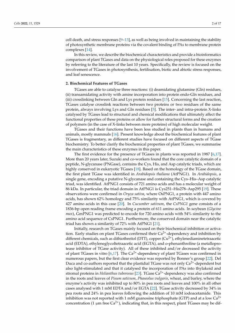
The presence and distribution of TGases in some angiosperms and algae with theirmain biochemical features is presented in Table 1. Plant TGases have been identifiedin different cell compartments. Molecular weight varies significantly, from the 30 kDathylakoid-localised TGase isolated from Cucumber sativus cotyledons [39] to the 150 kDaTGases found in Zea mays thylakoids and grana extracts [28] and the 160–180 kDa bandfound in mature maize pollen [40]. However, the most frequently found form, based onlyon molecular weight, is the 58 kDa one. Most reports have indicated that the optimum pHfor TGase activity assays falls within the range of 7.5–8.5.
Table 1. Biochemical features of TGases in higher plants and algae. Plant species, source, molec-ular weight, optimum pH for activity assay, and localisation in cell compartments are reported;n.d.: not determined.
Plant Species Source Molecular Weight (kDa) Optimum pH Assay Localisation References
Arabidopsis thalianaRecombinant enzyme 86 7.5–8.5 Microsomal fraction [19]
Entire plant n.d. 8.4 Entire cell [20]
Beta vulgaris Leaf n.d. 7.8 Entire cell [41]
Capsicum annuum Entire plant 70, 60, 5664, 34 n.d. Root [42]
Chlamydomonas reinhardtii Cell 72 7.4 Cell wall [27]
Cucumber sativus Cotyledon 77, 58, 50, 30 8.5 Thylakoid [39]
Dunaliella salina Cell and chloroplast 70, 50, 25 8.5 Chloroplast [43,44]
Glycine max Leaf and seedling 80 7.6 Entire cell [45,46]
Helianthus tuberosus
Leaf 58, 22, 19 8.0–9.5 Chloroplast [24]
Chloroplast 58, 24, 150 n.d. Thylakoid and stroma [47]
Chloroplast 58 8.5 Thylakoid and stroma [23]
Etiolated apex 85, 75, 58 8.5 Immature cell [48]
Hordeum vulgare Leaf 33, 58, 75 8.5 Thylakoid [3]
Malus domestica Pollen 70, 75 6.5 Chloroplast, microsomal,and cell wall [49]
Nicotiana tabacumFlower corolla 38, 58 7.5–8.5 Microsome, plastid, and
cell wall [50,51]
Flower corolla n.d. 6.5 Epidermis and cell wall [52]
Oryza sativa Entire plant 40 6.5 Thylakoid [26]
Entire plant 72, 39 n.d. Chloroplast [36]
Rosmarinus officinalis Entire plant n.d. 7.0 n.d. [29]
Zea mays
Pollen 58–50; 160–180 n.d. Pollen [40]
Chloroplast 58, 61–67, 77, 150 n.d. Thylakoid and grana [28]
Chloroplast 58 n.d. Thylakoid [7]
Recombinant enzyme 58 8.0 Chloroplast [14]
Recombinant enzyme 47 8.0 n.d. [53]
Meristematic callus 58, 34 n.d. Chloroplast and adult leaf [54]
Chloroplast 39 8.5 Thylakoid [19,55]
Recombinant enzyme 55 8.5 Chloroplast [56]
3. Bioinformatics Analyses
Bioinformatics analyses, such as comparisons of gene sequences, can support biochem-ical data and add new knowledge regarding the phylogenetic relationships and genomicorganisation of TGases in plants. Here, we present a comparison by sequence alignments,phylogenetic relationships, and data on the genomic organisation of different TGases invarious plant species for the first time.

Cells 2022, 11, 1529 5 of 17
To date, TGases have been identified in an increasing number of plant species, buta comparative analysis of their characteristics has not been performed. We selected theTGase family members from model plants [57] available in the PLAZA_5.0 database (https://bioinformatics.psb.ugent.be/plaza/, accessed on 17 March 2022): a total of 41 TGasegenes were found to be distributed in 30 plant species (Figure 1). Among angiosperms,Glycine max, Nicotiana tabacum, and Miscanthus sinensis have two duplicated genes each andEucalyptus grandis has three duplicated genes. Notably, Selaginella moellendorffii (Lycophyta)has five duplicated genes. Thus, gene duplication seems to have played a dominant role inthe expansion of the TGase family in plants. Gene duplication, expansion, and subsequentdiversification are features of the evolutionary process. The abundance of duplicate genes inplant genomes originated from ancient duplication events and a high rate of the retention ofextant pairs of duplicate genes. These duplicates have contributed to the evolution of novelfunctions, such as in growth and development, disease resistance, and stress tolerance [58].
The phylogenetic analysis (Figure 1) classified the plant TGases into taxonomic groups,i.e., monocots, dicots, bryophytes, lycophytes, marchantiophytes, and chlorophytes. Themonocots and dicots (angiosperms) form a separate clade, suggesting that they are moreevolutionarily divergent than the other species. This analysis is consistent with plantevolutionary history. The gene structure analysis (Figure 1) showed that angiospermshave different intron/exon arrangements compared to other plant taxa, though no majordifferences were observed between monocots and dicots. TGase gene size varied from2 to 28 kbp in most of the examined plants; however, it was significantly larger in Vitisvinifera (54 kbp) and barley (60 kbp). Though V. vinifera was found to have genome sizeof only ~500 Mb, its TGase gene was large with long introns. This might be because ofthe repetitive/transposable elements (TEs) abundance (41%) in the grapevine genome [59];moreover, introns are quite rich in repeats and TEs. The large size of the barley TGasemight also have been due to the specific characteristics of its genome, which is rich inpseudogenes and small gene fragments mainly located towards chromosome tips or astandemly repeated units [60]. These repetitive regions are present in introns and/orintergenic spaces [61,62]. Most plant TGases include one conserved large exon, whichmight be associated with the enzyme’s active site. The level of conservation of plantTGases was compared to animal and microbial ones via ConSurf analysis using PF01841(Transglut_core, https://pfam.xfam.org/family/PF01841 accessed on 17 March 2022).
The multiple sequence alignments (Figure 2) showed that TGases had a highly con-served domain typical of these enzymes, which consists of the Cys–His–Asp catalytic triad.The protein sequence of Misin04G291400 (Miscanthus sinensis) was found to differ in thecatalytic residues; this might have been due to the partial sequence availability. Plant TGaseprotein sequences are less conserved than those of animals and microbes at the N- and/orC-terminus; however, this does not affect the catalytic activity. Plant TGases withoutconserved catalytic domains might have arisen due to a unique deletion/substitution inthe genome. For example, despite high similarity at the nucleotide level, the unique dele-tion of guanine (G) in the maize TGZ15/TGZ21 cDNA sequences resulted in a frameshiftin their amino acid sequences and, consequently, a lack of homology at their C-termini,where the TGase catalytic triad was found by Villalobos et al. [28]. A rice homolog ofthe TGZ15/TGZ21 proteins, named TGO, also possesses a unique G deletion [26] that isnot observed in other available rice sequences, including the genomic sequence of ricechromosome 4. The maize TGZ15/TGZ21 and rice TGO proteins share a high sequencesimilarity (70%), though only upstream of the G deletion positions, while their C-terminidiffer in length, sequence, and catalytic triad localization. However, overexpression experi-ments have provided evidence that both proteins possess TGase activity [7,14,26,28,63–65],although their catalytic domains are not conserved. Overall, the bioinformatics analysissuggests that most plant TGase sequences are conserved in nature. Although a few exhibitquite different features, they still exert a similar function.

Cells 2022, 11, 1529 6 of 17
Cells 2022, 11, x FOR PEER REVIEW 6 of 18
(https://bioinformatics.psb.ugent.be/plaza/, accessed on 17 March 2022): a total of 41 TGase genes were found to be distributed in 30 plant species (Figure 1). Among angio-sperms, Glycine max, Nicotiana tabacum, and Miscanthus sinensis have two duplicated genes each and Eucalyptus grandis has three duplicated genes. Notably, Selaginella moellendorffii (Lycophyta) has five duplicated genes. Thus, gene duplication seems to have played a dominant role in the expansion of the TGase family in plants. Gene duplication, expan-sion, and subsequent diversification are features of the evolutionary process. The abun-dance of duplicate genes in plant genomes originated from ancient duplication events and a high rate of the retention of extant pairs of duplicate genes. These duplicates have con-tributed to the evolution of novel functions, such as in growth and development, disease resistance, and stress tolerance [58].
The phylogenetic analysis (Figure 1) classified the plant TGases into taxonomic groups, i.e., monocots, dicots, bryophytes, lycophytes, marchantiophytes, and chloro-phytes. The monocots and dicots (angiosperms) form a separate clade, suggesting that they are more evolutionarily divergent than the other species. This analysis is consistent with plant evolutionary history. The gene structure analysis (Figure 1) showed that angi-osperms have different intron/exon arrangements compared to other plant taxa, though no major differences were observed between monocots and dicots. TGase gene size varied from 2 to 28 kbp in most of the examined plants; however, it was significantly larger in Vitis vinifera (54 kbp) and barley (60 kbp). Though V. vinifera was found to have genome size of only ~500 Mb, its TGase gene was large with long introns. This might be because of the repetitive/transposable elements (TEs) abundance (41%) in the grapevine genome [59]; moreover, introns are quite rich in repeats and TEs. The large size of the barley TGase might also have been due to the specific characteristics of its genome, which is rich in pseudogenes and small gene fragments mainly located towards chromosome tips or as tandemly repeated units [60]. These repetitive regions are present in introns and/or inter-genic spaces [61,62]. Most plant TGases include one conserved large exon, which might be associated with the enzyme’s active site. The level of conservation of plant TGases was compared to animal and microbial ones via ConSurf analysis using PF01841 (Transglut_core, https://pfam.xfam.org/family/PF01841 accessed on 17 March 2022 ).
Figure 1. Phylogenetic relationship and gene structure of transglutaminases in various plant species.The phylogenetic analyses were conducted via amino acid sequence alignment using ClustalWand the neighbour-joining method in MEGA-11, and evolutionary distances were computed us-ing the JTT matrix-based method. The proportions of replicate trees in which the associated taxaclustered together in the bootstrap test (1000 replicates) are shown next to the branches. The genestructure, showing the intron/exon pattern, was constructed by The Gene Structure Display Server(GSDS 2.0, (Hu, Jin et al., 2015)) tool (http://gsds.cbi.pku.edu.cn/, accessed on 17 March 2022).Black shaded triangles at the end of gene names represent block/tendem duplication. The trans-glutaminase genes from various plant species used here were as follows: EL10Ac4g09118 (Betavulgaris); Glyma.03G246500 and Glyma.19G243900 (Glycine max); AT5G49570 (Arabidopsis thaliana);Nitab4.5_0004469g0060 and Nitab4.5_0005582g0050 (Nicotiana tabacum); CsaV3_2G004220 (Cucumissativus); MD12G1238800 (Malus domestica); CAN.G134.108 (Capsicum annuum); AL8G23370 (Ara-bidopsis lyrata); GSVIVG01021825001 (Vitis vinifera); Solyc01g097440.3 (Solanum lycopersicum); Eu-cgr.H04377, Eucgr.H04378, and Eucgr.H04379 (Eucalyptus grandis); Medtr7g114940 (Medicago truncat-ula); Lj1g0002084 (Lotus japonicas); Prupe.6G341700, Prupe.6G342000 (Prunus persica); CARHR287990(Cardamine hirsute); Thhalv10003714m.g (Eutrema salsugineum); FvH4_6g03130 (Fragaria vesca);Os07g0497400 (Oryza sativa ssp. Japonica); Zm00001eb105420 (Zea mays); Sobic.002G308500(Sorghum bicolor); Bradi1g26930 (Brachypodium distachyon); Misin03G268800 and Misin04G291400(Miscanthus sinensis); Sevir.2G332100 and Sevir.7G286400 (Setaria viridis); Spipo14G0050100 (Spirodelapolyrhiza); Ot_Chr7_g13391 (Oropetium thomaeum); Horvu_MOREX_2H01G320400 (Hordeum vulgare);Pp3c4_6120 (Physcomitrium patens); SMO343G0028, SMO367G0055, SMO367G0213, SMO367G0934,and SMO367G1212 (Selaginella moellendorffii); Mapoly0021s0092 (Marchantia polymorpha); andCre01.g053900 (Chlamydomonas reinhardtii).

Cells 2022, 11, 1529 7 of 17Cells 2022, 11, x FOR PEER REVIEW 8 of 18
Figure 2. Multiple sequences alignments of transglutaminases and peptide N-glycanases. The pro-tein sequences were aligned using ClustalW, and the ConSurf analysis was performed on the web-server (https://consurf.tau.ac.il/, accessed on 17 March 2022). The sequences in the black boxes show Pfam-PF01841 (Transglut_core). The arrows indicate the Cys–His–Asp amino acids of the catalytic triad. Despite having an amino acid sequence different from those of known animal TGases (Factor XIIIa, hTGaseII, and human TGaseIII), the core domains of the mouse PNGase and that of human TG2 have the same fold, as also reported in the SCOP database (family: transglu-taminase core; http://scop.mrc-lmb.cam.ac.uk/scop/data/scop.b.e.d.b.f.html, accessed on 14 March 2022). The protein sequences used here, other than the plants, were: C. elegans (Cepng-1, NP_492913.1), S. cerevisiae (ScPNGase, NP_015229.1), mouse (mPNGase, NP_067479), human (hPNGase, AF250924.2) and mouse (mTG2, NP_033399.1) peptide N-glycanases, human Factor XIIIa (hF.XIIIa, NP_000120), human TGase II (hTGaseII, NP_945189), and human TGase III (hTGaseIII, Q08188) transglutaminases.
4. Physiological Role of TGases in Plants In plants, TGases have been primarily studied by focusing on the molecular mecha-
nisms linking PAs to proteins by inter- and intramolecular bonds. These findings have been correlated to several aspects of growth and differentiation, as well as to stress re-sponses [5,15]. Research on plant TGases has been hampered by difficulties in the purifi-cation of the enzyme and by the limited/scarce sequence identities between animal TGases and those reported in the available plant databases [66]. In general, studies on plant TGases have mainly dealt with its distribution and function [48].
It is known that TGases are present in most plant organs and organelles. Here, we report the most recent evidence for their involvement in various processes, since the last extensive review on this topic did not account for the last decade of results [5,66]. A sche-matic model of the main physiological roles of plant TGases is shown in Figure 3.
Figure 2. Multiple sequences alignments of transglutaminases and peptide N-glycanases. The proteinsequences were aligned using ClustalW, and the ConSurf analysis was performed on the webserver(https://consurf.tau.ac.il/, accessed on 17 March 2022). The sequences in the black boxes showPfam-PF01841 (Transglut_core). The arrows indicate the Cys–His–Asp amino acids of the catalytictriad. Despite having an amino acid sequence different from those of known animal TGases (FactorXIIIa, hTGaseII, and human TGaseIII), the core domains of the mouse PNGase and that of humanTG2 have the same fold, as also reported in the SCOP database (family: transglutaminase core; http://scop.mrc-lmb.cam.ac.uk/scop/data/scop.b.e.d.b.f.html, accessed on 14 March 2022). The proteinsequences used here, other than the plants, were: C. elegans (Cepng-1, NP_492913.1), S. cerevisiae(ScPNGase, NP_015229.1), mouse (mPNGase, NP_067479), human (hPNGase, AF250924.2) and mouse(mTG2, NP_033399.1) peptide N-glycanases, human Factor XIIIa (hF.XIIIa, NP_000120), human TGaseII (hTGaseII, NP_945189), and human TGase III (hTGaseIII, Q08188) transglutaminases.
4. Physiological Role of TGases in Plants
In plants, TGases have been primarily studied by focusing on the molecular mech-anisms linking PAs to proteins by inter- and intramolecular bonds. These findings havebeen correlated to several aspects of growth and differentiation, as well as to stress re-sponses [5,15]. Research on plant TGases has been hampered by difficulties in the purifica-tion of the enzyme and by the limited/scarce sequence identities between animal TGasesand those reported in the available plant databases [66]. In general, studies on plant TGaseshave mainly dealt with its distribution and function [48].
It is known that TGases are present in most plant organs and organelles. Here, wereport the most recent evidence for their involvement in various processes, since the lastextensive review on this topic did not account for the last decade of results [5,66]. Aschematic model of the main physiological roles of plant TGases is shown in Figure 3.

Cells 2022, 11, 1529 8 of 17
Cells 2022, 11, x FOR PEER REVIEW 9 of 18
Figure 3. A model summarizing the main physiological roles of plant TGase. TGases have been found in several cellular compartments of different plants. In chloroplasts, TGases contribute to photosynthetic efficiency by increasing the level of bound PAs, ROS scavenging, and CO2 assimi-lation. These aspects are related to the action of TGases on different substrates, e.g., RuBisCO PSII proteins, ATPase, and PMF proteins (A). The enzyme is also present in the cytosol, where it can post-translationally modify the cytoskeletal proteins directly or do so via the binding of PAs, con-tributing to its organisation. This happens in the elongation of the pollen tube (B). In plant–patho-gen interactions, TGases behave similarly to a PAMP, e.g., the PEP-13 motif of Phytophthora in-festans. Otherwise, specific plant TGase isoforms might contribute to plant defence mechanisms (C). Different abiotic stresses (temperature, wounding, light, and salt stress) stimulate TGase activ-ity, improving plant resilience through the activation of several signalling pathways and the stim-ulation of several physiological processes, e.g., photosynthetic efficiency, HSP response, antioxi-dant system, and PA-based cell signalling. By specific inhibitor, TGase inhibition reduces re-sistance to abiotic stresses and causes decreases in the bound PA contents, decreases in the photo-chemical efficiency of PSII, and growth reduction (D). In the leaf senescence process, TGases in-crease the accumulation of HSPs and bound PAs. TGases are also involved in the modification of chloroplast proteins and the modulation of anti-senescence enzymes and ATP synthases, finally increasing the photosynthetic efficiency (E).
4.1. TGases and Photosynthesis Villalobos et al. first reported that a TGase was mainly present in the grana-appressed
thylakoids of light-exposed maize chloroplasts [54]. The activity of maize TGase was found to be inhibited by GTP, DTT, and other compounds but significantly increased when the enzymatic assay was performed in the presence of light [49]. The gene sequence analysis showed that maize TGase possesses a chloroplast import peptide composed of 47 amino acids and B-type repeats that are located in a non-catalytic domain of the enzyme. The overexpression of the maize plastidial TGase increased the activity of TGases in thylakoids of Arabidopsis thaliana [67]. In chloroplasts, TGases appear to stabilise both the photosynthetic complexes and Rubisco. Being regulated by light and other external fac-tors, TGases might exert a photoprotective effect on photosynthesis [7]. The overexpres-sion of TGases has been shown to increase the CO2 assimilation rate through the activation of Calvin cycle enzymes in tomato leaves [37]. Changes in cellular redox homeostasis have been proposed to be involved in the activation of Calvin cycle enzymes [37]. The enhanced
Figure 3. A model summarizing the main physiological roles of plant TGase. TGases have beenfound in several cellular compartments of different plants. In chloroplasts, TGases contribute tophotosynthetic efficiency by increasing the level of bound PAs, ROS scavenging, and CO2 assimi-lation. These aspects are related to the action of TGases on different substrates, e.g., RuBisCO PSIIproteins, ATPase, and PMF proteins (A). The enzyme is also present in the cytosol, where it canpost-translationally modify the cytoskeletal proteins directly or do so via the binding of PAs, con-tributing to its organisation. This happens in the elongation of the pollen tube (B). In plant–pathogeninteractions, TGases behave similarly to a PAMP, e.g., the PEP-13 motif of Phytophthora infestans.Otherwise, specific plant TGase isoforms might contribute to plant defence mechanisms (C). Differentabiotic stresses (temperature, wounding, light, and salt stress) stimulate TGase activity, improvingplant resilience through the activation of several signalling pathways and the stimulation of severalphysiological processes, e.g., photosynthetic efficiency, HSP response, antioxidant system, and PA-based cell signalling. By specific inhibitor, TGase inhibition reduces resistance to abiotic stresses andcauses decreases in the bound PA contents, decreases in the photochemical efficiency of PSII, andgrowth reduction (D). In the leaf senescence process, TGases increase the accumulation of HSPs andbound PAs. TGases are also involved in the modification of chloroplast proteins and the modulationof anti-senescence enzymes and ATP synthases, finally increasing the photosynthetic efficiency (E).
4.1. TGases and Photosynthesis
Villalobos et al. first reported that a TGase was mainly present in the grana-appressedthylakoids of light-exposed maize chloroplasts [54]. The activity of maize TGase was foundto be inhibited by GTP, DTT, and other compounds but significantly increased when theenzymatic assay was performed in the presence of light [49]. The gene sequence analysisshowed that maize TGase possesses a chloroplast import peptide composed of 47 aminoacids and B-type repeats that are located in a non-catalytic domain of the enzyme. Theoverexpression of the maize plastidial TGase increased the activity of TGases in thylakoidsof Arabidopsis thaliana [67]. In chloroplasts, TGases appear to stabilise both the photo-synthetic complexes and Rubisco. Being regulated by light and other external factors,

Cells 2022, 11, 1529 9 of 17
TGases might exert a photoprotective effect on photosynthesis [7]. The overexpressionof TGases has been shown to increase the CO2 assimilation rate through the activationof Calvin cycle enzymes in tomato leaves [37]. Changes in cellular redox homeostasishave been proposed to be involved in the activation of Calvin cycle enzymes [37]. Theenhanced TGase-mediated binding of PAs to thylakoid membranes is surely involved inthe aggregation of the light-harvesting complex (LHCII), which exerts a key regulatoryrole in dissipating excess excitation energy, thus improving photochemical efficiency undersalt stress [68]. TGase activity increases salt stress tolerance in cucumber plants due toan increased endogenous PAs content and ROS scavenging capacity, as well as the pro-motion of carbon assimilation and photosynthetic products. However, the mechanism bywhich TGase regulates the photochemical efficiency of plants under salt stress remainsunclear [30]. Plastidial proteins involved in photoprotection and in promoting the thy-lakoid electrochemical gradient are TGase substrates. Consequently, TGase interconnectsmore PSII proteins with other photoprotective and proton motive force (PMF) proteins(e.g., LHCII and ATPase); moreover, TGase changes the balance of pmf, thereby increasingthe PA-linked protein pool [67]. A recent study confirmed that a PNG1 gene containing atypical TGase catalytic triad domain, like that of AtPNG1, plays a positive role in improvingplant salt tolerance in cucumber plants [21].
4.2. TGases and Plant Fertilisation
Several studies have highlighted the involvement of TGases in the fertilization processof angiosperms. In particular, the enzyme plays a role in pollen–pistil recognition and pollenrejection; it is a crucial factor for pollen tube growth, being involved in the organisation ofcytoskeleton proteins [49,69]. Moreover, several plant models suggest that TGase activity isinvolved in the self-incompatibility response [9,12,13,70].
TGases have been reported to not only be localised inside the pollen tube but also existextracellularly. In the pollen tube cytosol, TGases modify cytoskeletal proteins, therebyregulating apical growth. Some reports also suggest an extracellular localisation of theenzyme and its involvement in pollen tube cell-wall construction and organisation [5,49,71].During pistil fertilisation, the pollen tube grows through the stigma and style followinga precise set of extracellular signals including PAs, which can regulate the growth of thepollen tube [72,73]. In fact, in in vitro pollen germination experiments, PAs were releasedinto the germination medium together with other factors (RNAs and proteins). It has beenreported that an extracellular TGase is required for apple pollen tube growth, suggesting itspossible involvement in pollen tube and style adhesion, thus favouring cross-talk betweenmale and female counterparts [74]. In addition to a cytosolic form of TGase, data suggestthe existence of TGase forms associated with the internal membranes and the cell wall ofpollen tubes. This different localization extends the functional range of pollen TGases thatcan be precisely redistributed in different cellular compartments [75]. The presence of anextracellular TGase raises a question regarding the function locally exerted by this enzyme.
4.3. TGases and Biotic Stress Responses
TGases may play an important role in plant–pathogen interactions and the resultingdefence responses. Thus, TGases are involved in the hypersensitive reaction (HR), whichconsists of programmed cell death at the site of pathogen entry and it is associated withrestriction of pathogen multiplication and spread [76,77]. The HR is accompanied by anincrease in TGase activity and its products that are distributed in different fractions, thoughmainly in those containing proteins released from membranes and cell walls by high ionicstrength and detergents [11]. The synthesis of mono-(γ-glutamyl)-Put and bis-(γ-glutamyl)-Spd, which represent solid evidence of TGase catalysis, revealed that both transamidatingand cross-linking activity were enhanced in leaves undergoing the HR but not in mockcontrols [78]. In a recent article, healthy susceptible and resistant to Phytophthora capsicipepper (Capsicum annuum) plants showed a very similar pattern of TGase accumulation,thought it was distinct when inoculated with the pathogen. Such differently expressed

Cells 2022, 11, 1529 10 of 17
post-infection patterns of TGases indicate that the defence mechanism of resistant plantsmight be based on the activation of specific plant TGase isoforms. These data support thehypothesis that TGases play role sin defence responses against some pathogens [42], suchas Phytophthora spp., one of the most dangerous plant pathogens.
TGase activity has also been detected in different Phytophthora species. A 42-kDacell wall-associated TGase (GP42) of Phytophthora sojae was found to contain a surface-exposed fragment called PEP-13 that acts as an elicitor of defence responses in parsleyand potato [79,80]. The PEP-13 motif was reported to be highly conserved in severalPhytophthora species. TGases activate defence responses, thus suggesting their function asgenus-specific recognition determinants in host and non-host plants [15]. In addition, TGasestructural sequences with eliciting activity, associated with plant defence mechanisms, wereisolated and characterised in Phytophthora cinnamomi. The fragments were found to encodea 533 deduced amino acid protein that includes an ORF with high identity similarity toPhytophthora sojae (70%), Phytophthora megasperma (70%), and Phytophthora infestans (61%)TGases. The alignment of a TGase gene with several TGase proteins revealed that theprotein contains the conserved catalytic domain [81]. In addition, a recent study on thebiochemical characterization of an acyltransferase enzyme, responsible for the pathogenicityof Phytophthora melonis, indicated that this protein possesses two domains, A (ranging fromresidues 260 to 620) and B (ranging from 141 to 219). The A domain possesses TGase-elicitorproperties [82].
4.4. TGases and Abiotic Stress Responses
TGases regulate the posttranslational modification of proteins involved in a widerange of plant responses to environmental stresses. In general, the stress-related functionof TGases could be ascribed to a positive relationship between enzyme activity, PA biosyn-thesis, and the photosynthetic efficiency maintained by the activation states of the Calvincycle [37]. In addition, TGase-improved photosynthetic capacity seems to be supported bychanges in the cellular redox status and activation of antioxidant enzymes [37]. This wasconfirmed by studies showing that TGase-deficient mutants (tgase-1 and tgase-2) of tomatoesexhibited a decreased activity of antioxidant enzymes engaged in the ascorbate (AsA)–GSHcycle, while TGase-overexpressing (TGaseOE) plants showed enhanced activities of ascor-bate peroxidase (APX), dehydroascorbate reductase (DHAR), and glutathione reductase(GR). High TGase activity in TGaseOE plants also correlated with significantly increasedratios of both GSH/GSSG and AsA/DHA [37]. This upregulated antioxidant machinerycan prevent redox homeostasis misbalance provoked by the over-accumulation of reactiveoxygen and nitrogen species under stress conditions. More recently, Jahan et al. [83] re-ported that the overexpression of TGases in tomatoes enhanced tolerance to heat stress. Acomparative transcriptomic study between wild-type (WT) and TGaseOE tomato plantsrevealed that in TGase-induced heat tolerance, a crucial role is played by genes associatedwith pathways responsible for protein processing in the endoplasmic reticulum, as well ascarbon fixation [83]. Moreover, the specific high-temperature response of TGaseOE plantswas associated with increased expression of heat-induced heat shock factors compared toWT plants, which was consistent with the expression patterns of heat shock proteins, thusindicating that heat shock factors might perform a pivotal role in the thermotolerance ofTGaseOE plants.
An enhanced salt tolerance was observed in tobacco plants overexpressing cucumberCsTGase [84]. The transgenic plants showed vigorous growth and higher net photosyn-thetic rate, as well as stomatal conductance. In turn, the CsTGase-induced salt tolerancewas associated with increased levels of chloroplast PAs, enhanced transcript levels ofphotosynthesis-related genes, and the accumulation of thylakoid membrane proteins suchas D1 and D2 [84]. It is noteworthy that significantly higher TGase activity was observed ina salt-tolerant cultivar of cucumber in comparison to a salt-sensitive one [30,85]. The TGase-mediated tolerance to NaCl was proven by spraying leaves with 1 mM o-phenanthroline,

Cells 2022, 11, 1529 11 of 17
which resulted in decreased bound PA levels, the decreased photochemical efficiency ofPSII, and the growth reduction of both cucumber cultivars [30].
Interestingly, TGases appear to play functional roles during acclimation to high salinitylevels. As indicated in the green halophilic microalga Dunaliella salina, acute hyper-salinestress under light caused an immediate change in the concentration of chloroplast TGaseswith concomitant variations in enzymatic activity [44]. Moreover, a PA-deficient variantof Dunaliella exhibiting low TGase activity was found to be more severely affected by saltstress; however, put application visibly recovered TGase activity and led to considerableenhancements of chlorophyll a and b content [44]. In the marine macroalga Grateloupiadoryphora (Mont.) Howe exposed to moderate hyposaline conditions, diminished TGaseactivity correlated with an increased pool of free PAs [86]. The positive relationship betweenTGases and free PAs could constitute a simple metabolic adjustment during acclimation tohyposaline conditions, since free PAs were found to be able to increase photosynthetic ratein the macroalga.
According to Pinto-Marijuan et al. [35], TGases are involved in adaptations to differentlight conditions. Holm oak leaves exposed to darkness until midday and then subjectedto abrupt high light intensity showed enhanced TGase activity, resulting in the maximumaccumulation of bound Put. The photoprotective role of TGases was hypothesised to bedue to their enhanced activity during increasing light intensity, as previously observed inthe systematically distant PA-deficient strain of Dunaliella [44]. Although TGase activityin the microalga was induced by salt stress, it was always higher in the light than in thedark [5].
Finally, TGases could also mediate the response to wounding and the wound-healingprocess. As documented by Serafini-Fracassini et al. [87], TGase activity was enhancedin tuber explants of Helianthus tuberosus as a result of wounding, in which the enzymetriggers the resumption of the cell cycle. This highlights the role of TGases in linking abioticand biotic stimuli since insect/herbivore feeding and pathogen attack is often related toplant tissue injury. Recently, the involvement of TGases in wounding was also reportedin Arabidopsis thaliana [88]. In this experimental model, an Atpng1 knockout (KO) linewas analysed during plant development and under heat and wounding stress. WT andKO lines were compared in terms of response to wounding and recovery from wounding(e.g., the formation of a scarring tissue that covered the entire wound). TGases accumulateddifferently in the two lines: in the stem of the WT line, TGases were mainly localised in the2–3 cell layers underneath dead cells on the stem surface, thus suggesting their involvementin the wound healing process (probably by exerting a gluing function and by strengtheningcell walls), as previously observed during senescence in Nicotiana tabacum petals [51]. In theKO line, the lack of TGase activity in the cell walls may have been related to the observedweaker anatomical structure, characterised by parenchyma with large spherical cells andwide intercellular spaces. These features were probably due to the reduced stiffness of theKO cell walls, failing to counteract the internal turgor pressure [89]. In WT leaves, a rapidincrease in TGase activity was observed; within 15 min, it was about three-fold higher thanthe basal activity observed at time 0 and then decreased. On the contrary, in KO leaves,the wound was effective, as activity remained at a constantly low level for 24 h. In theWT line, wounding-induced AtPNG1 transcript accumulation was observed within thefirst 5 min and reached minimum levels after 15–30 min. The potential involvement ofTGases in the healing processes is still poorly understood for plants, but it has already beendemonstrated for animal tissues [90,91]. These preliminary results suggest that the enzymeplays a role in plant wounding responses.
4.5. TGases and Leaf Senescence
In plants, senescence is a highly controlled and active process requiring global metabolicreprogramming, aimed at the organised disintegration and remobilization of valuable re-sources. It is a fundamental aspect of plant development that is necessary to optimiseresource allocation and promote phenotypic plasticity in order to acclimate to adverse

Cells 2022, 11, 1529 12 of 17
environmental conditions [92]. Structural changes of the chloroplast, eventually resultingin chloroplast degradation, mark the first phase of a sequential process that leads to leafsenescence, both developmental and stress-induced [93].
Physiological and structural changes in chloroplasts during senescence are associ-ated with PA conjugation, modifications of chloroplast proteins, and the modulation ofchloroplast-localised TGases (ChlTGases). The barley ChlTGase was found to be activatedduring dark-induced leaf senescence, which is associated with enhanced local TGase ac-cumulation and activity, as well as the increased expression of the barley HvPng1-likegene [3]. Results with barley leaves also showed that TGase activity was lower when thesamples were incubated with cytokinin, a phytohormone known for its anti-senescenceproperties [3]. The ChlTGase localization within chloroplast structures, as well as the iden-tification of the post-translational modification of plastid proteins (PA-conjugated proteins),suggested a notable contribution of ChlTGases to the dark-induced senescence-associatedprocess, including stress response, photosynthesis inhibition, and cell death manifestedby the chloroplast-to-gerontoplast conversion and subsequent degradation [94]. In situlocalization and changes in ChlTGase activity during dark-induced senescence were shownto mirror the increase in the level of plastid membrane-bound Put and Spd [3,94]. In fact,ChlTGase was shown to catalyse the binding of 3[H]Put and 3[H]Spd to photosystem pro-teins [94]. Substrates of ChlTGases in senescing and non-senescing leaves include apopro-teins of the chlorophyll a/b antenna complex, LHCII, ATP synthase, and pSbS (photosystemII 22kDa protein)—proteins that are essential in energy-dependent quenching and increasedthermal dissipation of excessively absorbed light energy in photosystems [7,23,47,55,94].Several stress-responsive proteins detected in the PA-bound fraction only after inducedsenescence include the antioxidant enzyme peroxiredoxin, a heat shock protein, ent-copalyldiphosphate synthase, and IAA-amino acid hydrolase [94–98]. The senescence-associatedchanges in the amount of mono- and bis-(γ-glutamyl)-Put in senescent leaves also cor-roborated earlier studies on the tobacco corolla. In the latter experimental system, theamount of bis-(γ-glutamyl)-Put and bis-(γ-glutamyl)-Spd decreased and the amount ofmono-(γ-glutamyl)-Put increased during petal senescence [50]. In this experimental model,it was also shown that TGase activity was involved in the PCD that takes place followingsenescence [51]. The fact that PAs, in concert with TGases, are functionally involved ininduced leaf senescence is supported by proteomic analyses and TGase activity/transcriptmodulation [3,94]. The most studied plant gene coding for a protein with TGase activity,AtPNG1, is constitutively expressed at low levels in all plant organs during various stagesof development and under various light conditions [55]. A similar expression patternwas found for the HvPNG1-like homolog in barley. However, HvPNG1-like transcriptionincreased as soon as senescence was induced in the dark, concomitant with the start of cellstructure disintegration [94].
5. Conclusions
In this review, we summarise information about plant TGases from studies carriedout mainly during the last decade. These enzymes are involved in numerous cellularprocesses and are present in most plant organs of the species investigated so far. Deeperknowledge would allow us to better understand whether plant TGases are involved in thesame basic cellular functions as those of animal TGases. Some features of plant TGases areshared with animal ones, such as their involvement in the wounding response and PCD,as well as in some cellular processes such as cell-to-cell adhesion, which, in plants, occursin the pollen-style interaction during fertilisation. We also highlight some characteristicsthat, at least for now, seem to be specific for plant TGases, such as light dependence andapical growth.
In addition to the main biochemical characteristics of plant TGases, we present abioinformatics analysis of TGases reported from different plant species for the first time.Gene structure results highlight that angiosperms have different intron/exon arrangementsthan other plant taxa; no substantial differences were observed between monocots and

Cells 2022, 11, 1529 13 of 17
dicots. Furthermore, the bioinformatics analysis allowed us to demonstrate that there aredifferent types of plant TGases, thus supporting the biochemical evidence and the idea thatplant TGases are conserved in nature from an evolutionary point of view.
Author Contributions: L.P. and I.A., Topic idea; L.P. and U.K.T., Data analysis; U.K.T., M.A.-J., L.P.and I.A., Writing—original draft preparation; E.S.-N. and S.D.D., Writing—review and editing. Allauthors have read and agreed to the published version of the manuscript.
Funding: This work was supported by funds from the Poland–Italy Bilateral exchange programmeCanaletto project: PO19MO03.
Institutional Review Board Statement: Not applicable.
Informed Consent Statement: Not applicable.
Data Availability Statement: Publicly available datasets were analysed in this study. These data canbe found here: https://plants.ensembl.org/index.html (accessed on 14 March 2022).
Acknowledgments: The authors are grateful to Stefania Biondi (University of Bologna) for comments,criticism, and constructive suggestions that greatly improved the entire manuscript.
Conflicts of Interest: The authors declare no conflict of interest.
Abbreviations
TGase: transglutaminase; PA: polyamine; LHCII: light-harvesting complex; Put: putrescine; Spd: spermidine.
References1. Clarke, D.; Mycek, M.; Neidle, A.; Waelsch, H. The incorporation of amines into protein. Arch. Biochem. Biophys. 1959, 79, 338–354.
[CrossRef]2. Duarte, L.; Matte, C.R.; Bizarro, C.V.; Ayub, M.A.N.Z. Transglutaminases: Part I—Origins, sources, and biotechnological
characteristics. World J. Microb. Biot. 2020, 36, 15. [CrossRef] [PubMed]3. Sobieszczuk-Nowicka, E.; Wieczorek, P.; Legocka, J. Kinetin affects the level of chloroplast polyamines and transglutaminase
activity during senescence of barley leaves. Acta Biochim. Pol. 2009, 56, 255–259. [CrossRef] [PubMed]4. Del Duca, S.; Serafini-Fracassini, D. Transglutaminases of higher, lower plants and fungi. Prog. Exp. Tumor Res. 2005, 38, 223–247.5. Serafini-Fracassini, D.; Del Duca, S. Transglutaminases: Widespread cross-linking enzymes in plants. Ann. Bot. Lond. 2008, 102,
145–152. [CrossRef]6. Icekson, I.; Apelbaum, A. Evidence for Transglutaminase Activity in Plant-Tissue. Plant Physiol. 1987, 84, 972–974. [CrossRef]7. Campos, A.; Carvajal-Vallejos, P.K.; Villalobos, E.; Franco, C.F.; Almeida, A.M.; Coelho, A.V.; Torne, J.M.; Santos, M. Characterisa-
tion of Zea mays L. plastidial transglutaminase: Interactions with thylakoid membrane proteins. Plant Biol. 2010, 12, 708–716.[CrossRef]
8. Sobieszczuk-Nowicka, E.; Legocka, J. Plastid-associated polyamines: Their role in differentiation, structure, functioning, stressresponse and senescence. Plant Biol. 2014, 16, 297–305. [CrossRef]
9. Del Duca, S.; Cai, G.; Di Sandro, A.; Serafini-Fracassini, D. Compatible and self-incompatible pollination in Pyrus communisdisplays different polyamine levels and transglutaminase activity. Amino Acids 2010, 38, 659–667. [CrossRef]
10. Del Duca, S.; Faleri, C.; Iorio, R.A.; Cresti, M.; Serafini-Fracassini, D.; Cai, G. Distribution of Transglutaminase in Pear PollenTubes in Relation to Cytoskeleton and Membrane Dynamics. Plant Physiol. 2013, 161, 1706–1721. [CrossRef]
11. Del Duca, S.; Serafini-Fracassini, D.; Cai, G. Senescence and programmed cell death in plants: Polyamine action mediated bytransglutaminase. Front. Plant Sci. 2014, 5, 120. [CrossRef] [PubMed]
12. Del Duca, S.; Aloisi, I.; Parrotta, L.; Cai, G. Cytoskeleton, Transglutaminase and Gametophytic Self-Incompatibility in the Malinae(Rosaceae). Int. J. Mol. Sci. 2019, 20, 209. [CrossRef] [PubMed]
13. Mandrone, M.; Antognoni, F.; Aloisi, I.; Potente, G.; Poli, F.; Cai, G.; Faleri, C.; Parrotta, L.; Del Duca, S. Compatible andIncompatible Pollen-Styles Interaction in Pyrus communis L. Show Different Transglutaminase Features, Polyamine Pattern andMetabolomics Profiles. Front. Plant Sci. 2019, 10, 741. [CrossRef] [PubMed]
14. Ioannidis, N.E.; Ortigosa, S.M.; Veramendi, J.; Pinto-Marijuan, M.; Fleck, I.; Carvajal, P.; Kotzabasis, K.; Santos, M.; Torne, J.M.Remodeling of tobacco thylakoids by over-expression of maize plastidial transglutaminase. Biochim. Biophys. Acta-Bioenerg. 2009,1787, 1215–1222. [CrossRef]
15. Lorand, L.; Iismaa, S.E. Transglutaminase diseases: From biochemistry to the bedside. Faseb J. 2019, 33, 3–12. [CrossRef]16. Brunner, F.; Rosahl, S.; Lee, J.; Rudd, J.J.; Geiler, C.; Kauppinen, S.; Rasmussen, G.; Scheel, D.; Nurnberger, T. Pep-13, a plant
defense-inducing pathogen-associated pattern from Phytophthora transglutaminases. Embo J. 2002, 21, 6681–6688. [CrossRef]

Cells 2022, 11, 1529 14 of 17
17. Serafini-Fracassini, D.; Del Duca, S.; D’Orazi, D. First evidence for polyamine conjugation mediated by an enzymic activity inplants. Plant Physiol. 1988, 87, 757–761. [CrossRef]
18. Suzuki, T.; Park, H.; Hollingsworth, N.M.; Sternglanz, R.; Lennarz, W.J. PNG1, a yeast gene encoding a highly conserved peptide:N-glycanase. J. Cell Biol. 2000, 149, 1039–1051. [CrossRef]
19. Della Mea, M.; Caparros-Ruiz, D.; Claparols, I.; Serafini-Fracassini, D.; Rigau, J. AtPng1p. The first plant transglutaminase. PlantPhysiol. 2004, 135, 2046–2054. [CrossRef]
20. Diepold, A.; Li, G.; Lennarz, W.J.; Nurnberger, T.; Brunner, F. The Arabidopsis AtPNG1 gene encodes a peptide: N-glycanase.Plant J. 2007, 52, 94–104. [CrossRef]
21. Hou, K.; Wang, Y.; Tao, M.Q.; Jahan, M.S.; Shu, S.; Sun, J.; Guo, S.R. Characterization of the CsPNG1 gene from cucumber and itsfunction in response to salinity stress. Plant Physiol. Bioch. 2020, 150, 140–150. [CrossRef] [PubMed]
22. Lilley, G.R.; Skill, J.; Griffin, M.; Bonner, P.L.R. Detection of Ca2+-dependent transglutaminase activity in root and leaf tissue ofmonocotyledonous and dicotyledonous plants. Plant Physiol. 1998, 117, 1115–1123. [CrossRef] [PubMed]
23. Del Duca, S.; Tidu, V.; Bassi, R.; Serafini-Fracassini, D.; Esposito, C. Identification of transglutaminase activity and its substrates inisolated chloroplast of Helianthus tuberosus. Planta 1994, 193, 283–289. [CrossRef]
24. Del Duca, S.; Dondini, L.; Della Mea, M.; Munoz de Rueda, P.; Serafini-Fracassini, D. Factors affecting transglutaminase activitycatalysing polyamine conjugation to endogenous substrates in the entire chloroplast. Plant Physiol. Bioch. 2000, 38, 429–439.[CrossRef]
25. Siepaio, M.P.; Meunier, J.C.F. Diamine Oxidase and Transglutaminase Activities in White Lupine Seedlings with Respect toCross-Linking of Proteins. J. Agr. Food Chem. 1995, 43, 1151–1156. [CrossRef]
26. Campos, N.; Castanon, S.; Urreta, I.; Santos, M.; Torne, J.M. Rice transglutaminase gene: Identification, protein expression,functionality, light dependence and specific cell location. Plant Sci. 2013, 205, 97–110. [CrossRef]
27. Waffenschmidt, S.; Kusch, T.; Woessner, J.P. A transglutaminase immunologically related to tissue transglutaminase catalyzescross-linking of cell wall proteins in Chlamydomonas reinhardtii. Plant Physiol. 1999, 121, 1003–1015. [CrossRef]
28. Villalobos, E.; Santos, M.; Talavera, D.; Rodriguez-Falcon, M.; Torne, J.M. Molecular cloning and characterization of a maizetransglutaminase complementary DNA. Gene 2004, 336, 93–104. [CrossRef]
29. El-Hofi, M.; Ismail, A.; Nour, M.; Ibrahim, O. Isolation, harac cation and haracterization of transglutaminase from rosemary(Rosmarinus officinalis L.) leaves. Acta Sci. Pol. Technol. Aliment. 2014, 13, 267–278. [CrossRef]
30. Shu, S.; Tang, Y.Y.; Zhou, X.P.; Jahan, M.S.; Sun, J.; Wang, Y.; Guo, S.R. Physiological mechanism of transglutaminase-mediatedimprovement in salt tolerance of cucumber seedlings. Plant Physiol. Bioch. 2020, 156, 333–344. [CrossRef]
31. Kim, H.C.; Lewis, M.S.; Gorman, J.J.; Park, S.C.; Girard, J.E.; Folk, J.E.; Chung, S.I. Protransglutaminase-E from Guinea-PigSkin—Isolation and Partial Characterization. J. Biol. Chem. 1990, 265, 21971–21978. [CrossRef]
32. Pasternack, R.; Dorsch, S.; Otterbach, J.T.; Robenek, I.R.; Wolf, S.; Fuchsbauer, H.L. Bacterial pro-transglutaminase fromStreptoverticillium mobaraense—Purification, characterisation and sequence of the zymogen. Eur. J. Biochem. 1998, 257, 570–576.[CrossRef] [PubMed]
33. Margosiak, S.A.; Dharma, A.; Brucecarver, M.R.; Gonzales, A.P.; Louie, D.; Kuehn, G.D. Identification of the Large Subunitof Ribulose 1,5-Bisphosphate Carboxylase Oxygenase as a Substrate for Transglutaminase in Medicago-sativa L (Alfalfa). PlantPhysiol. 1990, 92, 88–96. [CrossRef] [PubMed]
34. Kuehn, G.D.; Sotelo, M.; Morales, T.; Brucecarver, M.R.; Guzman, E.; Margosiak, S.A. Purification and Properties of Transglutami-nase from Medicago-sativa L (Alfalfa). Faseb J. 1991, 5, A1510.
35. Pinto-Marijuan, M.; de Agazio, M.; Zacchini, M.; Santos, M.A.; Torne, J.M.; Fleck, I. Response of transglutaminase activity andbound putrescine to changes in light intensity under natural or controlled conditions in Quercus ilex leaves. Physiol. Plant. 2007,131, 159–169. [CrossRef]
36. Campos, N.; Torne, J.M.; Bleda, M.J.; Manich, A.; Urreta, I.; Montalban, I.A.; Castanon, S.; Moncalean, P.; Santos, M. Proteomicand transcriptomic analysis of rice tranglutaminase and chloroplast-related proteins. Plant Sci. 2014, 229, 142–153. [CrossRef]
37. Zhong, M.; Wang, Y.; Hou, K.; Shu, S.; Sun, J.; Guo, S.R. TGase positively regulates photosynthesis via activation of Calvin cycleenzymes in tomato. Hortic. Res.-Engl. 2019, 6. [CrossRef]
38. Li, H.B.; Zhang, L.W.; Cui, Y.H.; Luo, X.; Xue, C.H.; Wang, S.M.; Jiao, Y.H.; Zhang, S.; Liu, W.L.; Fan, R.B.; et al. Characterizationof recombinant Zea mays transglutaminase expressed in Pichia pastoris and its impact on full and non-fat yoghurts. J. Sci. FoodAgr. 2014, 94, 1225–1230. [CrossRef]
39. Sobieszczuk-Nowicka, E.; Di Sandro, A.; Del Duca, S.; Serafini-Fracassini, D.; Legocka, J. Plastid-membrane-associated polyaminesand thylakoid transglutaminases during etioplast-to-chloroplast transformation stimulated by kinetin. Physiol. Plant 2007, 130,590–600. [CrossRef]
40. Santos, M.; Alché Ramírez, J.d.D.; Rodríguez García, M.I.; Torné, J.M. Transglutaminase activity and localization duringmicrospore induction in maize. Appl. Biol. Med. 2007, 1, 280–286.
41. Signorini, M.; Beninati, S.; Bergamini, C.M. Identification of Transglutaminase Activity in the Leaves of Silver Beet (Beta vulgaris L.).J. Plant Physiol. 1991, 137, 547–552. [CrossRef]
42. Piccini, C.; Parrotta, L.; Faleri, C.; Romi, M.; Del Duca, S.; Cai, G. Histomolecular responses in susceptible and resistant phenotypesof Capsicum annuum L. infected with Phytophthora capsici. Sci. Hortic. Amst. 2019, 244, 122–133. [CrossRef]

Cells 2022, 11, 1529 15 of 17
43. Dondini, L.; Bonazzi, S.; Serafini-Fracassini, D. Recovery of growth capacity and of chloroplast transglutaminase activity inducedby polyamines in a polyamine-deficient variant strain of Dunaliella salina. J. Plant Physiol. 2000, 157, 473–480. [CrossRef]
44. Dondini, L.; Bonazzi, S.; Del Duca, S.; Bregoli, A.M.; Serafini-Fracassini, D. Acclimation of chloroplast transglutaminase to highNaCl concentration in a polyamine-deficient variant strain of Dunaliella salina and in its wild type. J. Plant Physiol. 2001, 158,185–197. [CrossRef]
45. Kang, H.; Cho, Y.D. Purification and properties of transglutaminase from soybean (Glycine max) leaves. Biochem. Biophys. Res.Commun. 1996, 223, 288–292. [CrossRef] [PubMed]
46. Kang, H.; Lee, S.G.; Cho, Y.D. Identification of glycinin in vivo as a polyamine-conjugated protein via a gamma-glutamyl linkage.Biochem. J. 1998, 332, 467–473. [CrossRef]
47. Dondini, L.; Del Duca, S.; Dall’Agata, L.; Bassi, R.; Gastaldelli, M.; Della Mea, M.; Di Sandro, A.; Claparols, I.; Serafini-Fracassini,D. Suborganellar localisation and effect of light on Helianthus tuberosus chloroplast transglutaminases and their substrates. Planta2003, 217, 84–95. [CrossRef]
48. Beninati, S.; Iorio, R.A.; Tasco, G.; Serafini-Fracassini, D.; Casadio, R.; Del Duca, S. Expression of different forms of transglutami-nases by immature cells of Helianthus tuberosus sprout apices. Amino Acids 2013, 44, 271–283. [CrossRef]
49. Di Sandro, A.; Del Duca, S.; Verderio, E.; Hargreaves, A.J.; Scarpellini, A.; Cai, G.; Cresti, M.; Faleri, C.; Iorio, R.A.; Hirose, S.; et al.An extracellular transglutaminase is required for apple pollen tube growth. Biochem. J. 2010, 429, 261–271. [CrossRef]
50. Serafini-Fracassini, D.; Del Duca, S. Biochemistry and function of plant transglutaminases. Minerva Biotecnol. 2002, 14, 135–141.51. Della Mea, M.; De Filippis, F.; Genovesi, V.; Serafini-Fracassini, D.; Del Duca, S. The acropetal wave of developmental cell death of
tobacco corolla is preceded by activation of transglutaminase in different cell compartments. Plant Physiol. 2007, 144, 1211–1222.[CrossRef] [PubMed]
52. Cai, G.; Della Mea, M.; Faleri, C.; Fattorini, L.; Aloisi, I.; Serafini-Fracassini, D.; Del Duca, S. Spermine either delays or promotes celldeath in Nicotiana tabacum L. corolla depending on the floral developmental stage and affects the distribution of transglutaminase.Plant Sci. 2015, 241, 11–22. [CrossRef] [PubMed]
53. Li, H.B.; Zhang, L.W.; Cui, Y.H.; Luo, X.; Xue, C.H.; Wang, S.M. Expression of soluble recombinant transglutaminase from Zeamays in Pichia pastoris. World J. Microb. Biot. 2013, 29, 939–947. [CrossRef] [PubMed]
54. Villalobos, E.; Torne, J.M.; Rigau, J.; Olles, I.; Claparols, I.; Santos, M. Immunogold localization of a transglutaminase related tograna development in different maize cell types. Protoplasma 2001, 216, 155–163. [CrossRef] [PubMed]
55. Della Mea, M.; Di Sandro, A.; Dondini, L.; Del Duca, S.; Vantini, F.; Bergamini, C.; Bassi, R.; Serafini-Fracassini, D. A Zea mays39-kDa thylakoid transglutaminase catalyses the modification by polyamines of light-harvesting complex II in a light-dependentway. Planta 2004, 219, 754–764. [CrossRef]
56. Carvajal-Vallejos, P.K.; Campos, A.; Fuentes-Prior, P.; Villalobos, E.; Almeida, A.M.; Barbera, E.; Torne, J.M.; Santos, M. Purificationand in vitro refolding of maize chloroplast transglutaminase over-expressed in Escherichia coli. Biotechnol. Lett. 2007, 29, 1255–1262.[CrossRef]
57. Chang, C.R.; Bowman, J.L.; Meyerowitz, E.M. Field Guide to Plant Model Systems. Cell 2016, 167, 325–339. [CrossRef]58. Panchy, N.; Lehti-Shiu, M.; Shiu, S.H. Evolution of Gene Duplication in Plants. Plant Physiol. 2016, 171, 2294–2316. [CrossRef]59. Jaillon, O.; Aury, J.M.; Noel, B.; Policriti, A.; Clepet, C.; Casagrande, A.; Choisne, N.; Aubourg, S.; Vitulo, N.; Jubin, C.; et al.
The grapevine genome sequence suggests ancestral hexaploidization in major angiosperm phyla. Nature 2007, 449, U463–U465.[CrossRef]
60. Prade, V.M.; Gundlach, H.; Twardziok, S.; Chapman, B.; Tan, C.; Langridge, P.; Schulman, A.H.; Stein, N.; Waugh, R.; Zhang, G.The pseudogenes of barley. Plant J. 2018, 93, 502–514. [CrossRef]
61. Bureau, T.E.; Wessler, S.R. Mobile Inverted-Repeat Elements of the Tourist Family Are Associated with the Genes of Many CerealGrasses. Proc. Natl. Acad. Sci. USA 1994, 91, 1411–1415. [CrossRef] [PubMed]
62. Mascher, M.; Wicker, T.; Jenkins, J.; Plott, C.; Lux, T.; Koh, C.S.; Ens, J.; Gundlach, H.; Boston, L.B.; Tulpova, Z.; et al. Long-readsequence assembly: A technical evaluation in barley. Plant Cell 2021, 33, 1888–1906. [CrossRef] [PubMed]
63. Ortigosa, S.M.; Díaz-Vivancos, P.; Clemente-Moreno, M.J.; Pintó-Marijuan, M.; Fleck, I.; Veramendi, J.; Santos, M.; Hernandez,J.A.; Torné, J.M. Oxidative stress induced in tobacco leaves by chloroplast over-expression of maize plastidial transglutaminase.Planta 2010, 232, 593–605. [CrossRef] [PubMed]
64. Campos, N.; Villalobos, E.; Fontanet, P.; Torné, J.M.; Santos, M. A peptide of 17 aminoacids from the N-terminal region of maizeplastidial transglutaminase is essential for chloroplast targeting. Am. J. Mol. Biol. 2012, 2, 245–257. [CrossRef]
65. Ioannidis, N.E.; Lopera, O.; Santos, M.; Torne, J.M.; Kotzabasis, K. Role of Plastid Transglutaminase in LHCII Polyamination andThylakoid Electron and Proton Flow. PLoS ONE 2012, 7, e419791. [CrossRef]
66. Serafini-Fracassini, D.; Della Mea, M.; Tasco, G.; Casadio, R.; Del Duca, S. Plant and animal transglutaminases: Do similarfunctions imply similar structures? Amino Acids 2009, 36, 643–657. [CrossRef]
67. Ioannidis, N.E.; Malliarakis, D.; Torne, J.M.; Santos, M.; Kotzabasis, K. The Over-expression of the Plastidial Transglutaminasefrom Maize in Arabidopsis Increases the Activation Threshold of Photoprotection. Front. Plant Sci. 2016, 7, 635. [CrossRef]
68. Shu, S.; Yuan, R.N.; Shen, J.L.; Chen, J.; Wang, L.J.; Wu, J.Q.; Sun, J.; Wang, Y.; Guo, S.R. The positive regulation of putrescine onlight-harvesting complex II and excitation energy dissipation in salt-stressed cucumber seedlings. Envion. Exp. Bot. 2019, 162,283–294. [CrossRef]

Cells 2022, 11, 1529 16 of 17
69. Aloisi, I.; Cai, G.; Serafini-Fracassini, D.; Del Duca, S. Polyamines in Pollen: From Microsporogenesis to Fertilization. Front. PlantSci. 2016, 7, 155. [CrossRef]
70. Gentile, A.; Antognoni, F.; Iorio, R.A.; Distefano, G.; Las Casas, G.; La Malfa, S.; Serafini-Fracassini, D.; Del Duca, S. Polyaminesand transglutaminase activity are involved in compatible and self-incompatible pollination of Citrus grandis. Amino Acids 2012,42, 1025–1035. [CrossRef]
71. Del Duca, S.; Serafini-Fracassini, D.; Bonner, P.; Cresti, M.; Cai, G. Effects of post-translational modifications catalysed by pollentransglutaminase on the functional properties of microtubules and actin filaments. Biochem. J. 2009, 418, 651–664. [CrossRef][PubMed]
72. Aloisi, I.; Cai, G.; Tumiatti, V.; Minarini, A.; Del Duca, S. Natural polyamines and synthetic analogs modify the growth and themorphology of Pyrus communis pollen tubes affecting ROS levels and causing cell death. Plant Sci. 2015, 239, 92–105. [CrossRef]
73. Aloisi, I.; Cai, G.; Faleri, C.; Navazio, L.; Serafini-Fracassini, D.; Del Duca, S. Spermine Regulates Pollen Tube Growth byModulating Ca2+-Dependent Actin Organization and Cell Wall Structure. Front. Plant Sci. 2017, 8, 1701. [CrossRef] [PubMed]
74. Guan, Y.F.; Guo, J.Z.; Li, H.; Yang, Z.B. Signaling in Pollen Tube Growth: Crosstalk, Feedback, and Missing Links. Mol. Plant.2013, 6, 1053–1064. [CrossRef] [PubMed]
75. Del Duca, S.; Serafini-Fracassini, D.; Cai, G. An unconventional road for the secretion of transglutaminase in pollen tubes? PlantSignal. Behav. 2013, 8, e24446. [CrossRef]
76. Greenberg, J.T.; Yao, N. The role and regulation of programmed cell death in plant-pathogen interactions. Cell Microbiol. 2004, 6,201–211. [CrossRef] [PubMed]
77. Lam, E.; Kato, N.; Lawton, M. Programmed cell death, mitochondria and the plant hypersensitive response. Nature 2001, 411,848–853. [CrossRef]
78. Del Duca, S.; Betti, L.; Trebbi, G.; Serafini-Fracassini, D.; Torrigiani, P. Transglutaminase activity changes during the hypersensitivereaction, a typical defense response of tobacco NN plants to TMV. Physiol. Plant. 2007, 131, 241–250. [CrossRef]
79. Halim, V.A.; Hunger, A.; Macioszek, V.; Landgraf, P.; Nürnberger, T.; Scheel, D.; Rosahl, S. The oligopeptide elicitor Pep-13induces salicylic acid-dependent and-independent defense reactions in potato. Physiol. Mol. Plant Pathol. 2004, 64, 311–318.[CrossRef]
80. Parker, J.E.; Schulte, W.; Hahlbrock, K.; Scheel, D. An extracellular glycoprotein from Phytophthora megasperma f. sp. glycineaelicits phytoalexin synthesis in cultured parsley cells and protoplasts. Mol. Plant-Microbe Interact. 1991, 4, 19–27. [CrossRef]
81. Martins, I.M.; Matos, M.; Costa, R.; Silva, F.; Pascoal, A.; Estevinho, L.M.; Choupina, A.B. Transglutaminases: Recent achievementsand new sources. Appl. Microbiol. Biot. 2014, 98, 6957–6964. [CrossRef] [PubMed]
82. Ahmad, A.; Akram, W.; Bashir, Z.; Shahzadi, I.; Wang, R.; Abbas, H.M.K.; Hu, D.; Ahmed, S.; Xu, X.M.; Li, G.H.; et al. Functionaland Structural Analysis of a Novel Acyltransferase from Pathogenic Phytophthora melonis. ACS Omega 2021, 6, 1797–1808.[CrossRef] [PubMed]
83. Jahan, M.S.; Shi, Z.R.; Zhong, M.; Zhang, Y.M.; Zhou, R.R.; El-Mogy, M.M.; Sun, J.; Shu, S.; Guo, S.R.; Wang, Y. Comparativetranscriptome analysis reveals gene network regulation of TGase-induced thermotolerance in tomato. Not. Bot. Horti Agrobo.2021, 49, 12208. [CrossRef]
84. Zhong, M.; Wang, Y.; Zhang, Y.M.; Shu, S.; Sun, J.; Guo, S.R. Overexpression of Transglutaminase from Cucumber in TobaccoIncreases Salt Tolerance through Regulation of Photosynthesis. Int. J. Mol. Sci. 2019, 20, 894. [CrossRef]
85. Tang, Y.-Y.; Yuan, Y.-H.; Shu, S.; Guo, S.-R. Regulatory mechanism of NaCl stress on photosynthesis and antioxidant capacitymediated by transglutaminase in cucumber (Cucumis sativus L.) seedlings. Sci. Hortic. Amst. 2018, 235, 294–306. [CrossRef]
86. García-Jiménez, P.; Just, P.M.; Delgado, A.M.; Robaina, R.R. Transglutaminase activity decrease during acclimation to hyposalineconditions in marine seaweed Grateloupia doryphora (Rhodophyta, Halymeniaceae). J. Plant Physiol. 2007, 164, 367–370. [CrossRef]
87. Serafini-Fracassini, D.; Del Duca, S.; Torrigiani, P. Polyamine conjugation during the cell cycle of Helianthus tuberosus: Nonenzymatic and transglutaminase-like binding activity. Plant Physiol. Biochem. 1989, 27, 659–668.
88. Serafini-Fracassini, D.; Della Mea, M.; Parrotta, L.; Faleri, C.; Cai, G.; Del Duca, S.; Aloisi, I. AtPng1 knockout mutant ofArabidopsis thaliana shows a juvenile phenotype, morpho-functional changes, altered stress response and cell wall modifications.Plant Physiol. Bioch. 2021, 167, 11–21. [CrossRef]
89. Carter, R.; Woolfenden, H.; Baillie, A.; Amsbury, S.; Carroll, S.; Healicon, E.; Sovatzoglou, S.; Braybrook, S.; Gray, J.E.; Hobbs, J.Stomatal opening involves polar, not radial, stiffening of guard cells. Curr. Biol. 2017, 27, 2974–2983.e2972. [CrossRef]
90. Telci, D.; Griffin, M. Tissue transglutaminase (TG2)—A wound response enzyme. Front. Biosci. Landmrk 2006, 11, 867–882.[CrossRef]
91. Qin, Y.; Guo, X.W.; Li, L.; Wang, H.W.; Kim, W. The Antioxidant Property of Chitosan Green Tea Polyphenols Complex InducesTransglutaminase Activation in Wound Healing. J. Med. Food 2013, 16, 487–498. [CrossRef] [PubMed]
92. Paluch-Lubawa, E.; Stolarska, E.; Sobieszczuk-Nowicka, E. Dark-Induced Barley Leaf Senescence–A Crop System for StudyingSenescence and Autophagy Mechanisms. Front. Plant Sci. 2021, 12, 425. [CrossRef]
93. Sarwat, M.; Naqvi, A.R.; Ahmad, P.; Ashraf, M.; Akram, N.A. Phytohormones and microRNAs as sensors and regulators of leafsenescence: Assigning macro roles to small molecules. Biotechnol. Adv. 2013, 31, 1153–1171. [CrossRef] [PubMed]
94. Sobieszczuk-Nowicka, E.; Zmienko, A.; Samelak-Czajka, A.; Łuczak, M.; Pietrowska-Borek, M.; Iorio, R.; Del Duca, S.; Figlerowicz,M.; Legocka, J. Dark-induced senescence of barley leaves involves activation of plastid transglutaminases. Amino Acids 2015, 47,825–838. [CrossRef] [PubMed]

Cells 2022, 11, 1529 17 of 17
95. van der Graaff, E.; Schwacke, R.; Schneider, A.; Desimone, M.; Flügge, U.-I.; Kunze, R. Transcription analysis of Arabidopsismembrane transporters and hormone pathways during developmental and induced leaf senescence. Plant Physiol. 2006, 141,776–792. [CrossRef]
96. Iqbal, N.; Nazar, R.; Khan, M.I.R.; Masood, A.; Khan, N.A. Role of gibberellins in regulation of source–sink relations underoptimal and limiting environmental conditions. Curr. Sci. 2011, 100, 998–1007.
97. Cejudo, F.J.; Ferrández, J.; Cano, B.; Puerto-Galán, L.; Guinea, M. The function of the NADPH thioredoxin reductase C-2-Cysperoxiredoxin system in plastid redox regulation and signalling. FEBS Lett. 2012, 586, 2974–2980. [CrossRef]
98. Wang, W.; Vinocur, B.; Shoseyov, O.; Altman, A. Role of plant heat-shock proteins and molecular chaperones in the abiotic stressresponse. Trends Plant Sci. 2004, 9, 244–252. [CrossRef]