New cyathealean tree ferns from the Cretaceous of South Africa: Natalipteris wildei gen. et sp. nov....
14
New cyathealean tree ferns from the Cretaceous of South Africa: Natalipteris wildei gen. et sp. nov. and Kwazulupteris schaarschmidtii gen. et sp. nov. Ezequiel Ignacio Vera a,b,⇑ , Rafael Herbst c a CONICET-Museo Argentino de Ciencias Naturales ‘‘Bernardino Rivadavia’’, Av. Angel Gallardo 470, C1405DJR Ciudad de Buenos Aires, Argentina b Área de Paleontología, Departamento de Ciencias Geológicas, Facultad de Ciencias Exactas y Naturales, Universidad de Buenos Aires, Pabellón II, Ciudad Universitaria, C1428EGA Buenos Aires, Argentina c Instituto Superior de Correlación Geológica-CONICET, Las Piedras 201 7°/B, 4000 S.M. de Tucumán, Argentina article info Article history: Received 26 June 2013 Received in revised form 26 August 2014 Accepted 27 August 2014 Available online 6 September 2014 Keywords: Cyatheales Tree ferns Cretaceous South Africa Fossil abstract Two new genera and species of permineralized tree ferns, Natalipteris wildei and Kwazulupteris schaarsch- midtii, are defined and described in detail. Natalipteris wildei is a solenostelic stem without well-devel- oped sclerenchyma sheaths in their vascular strands, and has a single vascular strand in the petiole bases, which are somewhat similar to the one present in Cibotium and Nishidacaulis. On the other hand, K. schaarschmidtii is a dictyostelic fern, with petiole bases with fused adaxial arcs and a single isolated meristele in the petiolar pith. Both taxa present features that preclude their placement in the recognized families of Cyatheales, but may be referred to the ‘‘core tree ferns’’ clade. Fossil specimens were found in the Senckenberg Forschungsinstitut Palaeobotanical Collection and, although they lack precise strati- graphic provenance, it is suggested that they were collected from the Mzinene Formation (Albian– Turonian). Ó 2014 Elsevier Ltd. All rights reserved. 1. Introduction The Order Cyatheales is a clade of ferns that, together with poly- pods and heterosporous water ferns, comprises the ‘‘core leptosp- orangiate ferns’’ group (Pryer et al., 2004). One of the most conspicuous features of this group of plants is the development of an arborescent habit, present in many (but not all) representa- tives of the order. Cyathealean ferns are solenostelic or dictyostelic, with some taxa having accessory medullary and cortical bundles, and have pinnate fronds bearing indusiate or exindusiate sori. Hairs or scales cover the stem and petioles of most tree ferns (Korall et al., 2007; Kramer and Green, 1990). Living Cyatheales are found in tropical lowland to submontane environments, as well as subtropical and Southern Hemisphere temperate forests, reach- ing cool latitudes in southern South America, New Zealand and Tasmania (Large and Braggins, 2004). Several schemes for the sys- tematic classification of the cyathealean tree ferns have been published (e.g., Holttum and Sen, 1961; Korall et al., 2006; Lantz et al., 1999; Smith et al., 2006; Tryon, 1970). Most recent proposals for classification of extant ferns, summarized in Christenhusz et al. (2011), recognize eight families among Cyatheales: Thyrsopterida- ceae, Loxomataceae, Culcitaceae, Plagogyriaceae, Cibotiaceae, Dick- soniaceae, Metaxyaceae and Cyatheaceae. The fossil record of the cyathealean tree ferns extends back into the Jurassic, and comprises permineralized stems, impression/ compression fertile and sterile fronds, and isolated palynomorphs (Tidwell and Ash, 1994). In particular, more than 20 taxa based on permineralized stems have been described worldwide (Lantz et al., 1999; Tidwell and Ash, 1994; Vera, 2009, 2013). Among them, some specimens can be conclusively referred to recognized families of the Cyatheales. For example, Dendropteridium cyatheo- ides Bancroft, 1932 and Alsophilocaulis calveloi Menéndez emend. Vera, 2010 clearly present affinities with the Cyatheaceae s.s., whereas others, described as representatives of the genus Cibotium (e.g., C. oregonense Barrington, 1983, C. iwatense Ogura, 1933), may be attributed to the Cibotiaceae. In addition, many taxa present a mixture of features that make them impossible to place in a partic- ular family, suggesting that the diversity of tree ferns was notably greater than nowadays (Lantz et al., 1999; Stockey and Rothwell, 2004; Vera, 2013). http://dx.doi.org/10.1016/j.jafrearsci.2014.08.017 1464-343X/Ó 2014 Elsevier Ltd. All rights reserved. ⇑ Corresponding author at: CONICET-Museo Argentino de Ciencias Naturales ‘‘Bernardino Rivadavia’’, Av. Angel Gallardo 470, C1405DJR Ciudad de Buenos Aires, Argentina. Tel./fax: +54 (11) 4982 6595. E-mail addresses: [email protected] (E.I. Vera), [email protected] (R. Herbst). Journal of African Earth Sciences 101 (2015) 56–69 Contents lists available at ScienceDirect Journal of African Earth Sciences journal homepage: www.elsevier.com/locate/jafrearsci
Transcript of New cyathealean tree ferns from the Cretaceous of South Africa: Natalipteris wildei gen. et sp. nov....
Journal of African Earth Sciences 101 (2015) 56–69
Contents lists available at ScienceDirect
Journal of African Earth Sciences
journal homepage: www.elsevier .com/locate / ja f rearsc i
New cyathealean tree ferns from the Cretaceous of South Africa:Natalipteris wildei gen. et sp. nov. and Kwazulupteris schaarschmidtii gen.et sp. nov.
http://dx.doi.org/10.1016/j.jafrearsci.2014.08.0171464-343X/� 2014 Elsevier Ltd. All rights reserved.
⇑ Corresponding author at: CONICET-Museo Argentino de Ciencias Naturales‘‘Bernardino Rivadavia’’, Av. Angel Gallardo 470, C1405DJR Ciudad de Buenos Aires,Argentina. Tel./fax: +54 (11) 4982 6595.
E-mail addresses: [email protected] (E.I. Vera), [email protected](R. Herbst).
Ezequiel Ignacio Vera a,b,⇑, Rafael Herbst c
a CONICET-Museo Argentino de Ciencias Naturales ‘‘Bernardino Rivadavia’’, Av. Angel Gallardo 470, C1405DJR Ciudad de Buenos Aires, Argentinab Área de Paleontología, Departamento de Ciencias Geológicas, Facultad de Ciencias Exactas y Naturales, Universidad de Buenos Aires, Pabellón II, Ciudad Universitaria,C1428EGA Buenos Aires, Argentinac Instituto Superior de Correlación Geológica-CONICET, Las Piedras 201 7�/B, 4000 S.M. de Tucumán, Argentina
a r t i c l e i n f o a b s t r a c t
Article history:Received 26 June 2013Received in revised form 26 August 2014Accepted 27 August 2014Available online 6 September 2014
Keywords:CyathealesTree fernsCretaceousSouth AfricaFossil
Two new genera and species of permineralized tree ferns, Natalipteris wildei and Kwazulupteris schaarsch-midtii, are defined and described in detail. Natalipteris wildei is a solenostelic stem without well-devel-oped sclerenchyma sheaths in their vascular strands, and has a single vascular strand in the petiolebases, which are somewhat similar to the one present in Cibotium and Nishidacaulis. On the other hand,K. schaarschmidtii is a dictyostelic fern, with petiole bases with fused adaxial arcs and a single isolatedmeristele in the petiolar pith. Both taxa present features that preclude their placement in the recognizedfamilies of Cyatheales, but may be referred to the ‘‘core tree ferns’’ clade. Fossil specimens were found inthe Senckenberg Forschungsinstitut Palaeobotanical Collection and, although they lack precise strati-graphic provenance, it is suggested that they were collected from the Mzinene Formation (Albian–Turonian).
� 2014 Elsevier Ltd. All rights reserved.
1. Introduction
The Order Cyatheales is a clade of ferns that, together with poly-pods and heterosporous water ferns, comprises the ‘‘core leptosp-orangiate ferns’’ group (Pryer et al., 2004). One of the mostconspicuous features of this group of plants is the developmentof an arborescent habit, present in many (but not all) representa-tives of the order. Cyathealean ferns are solenostelic or dictyostelic,with some taxa having accessory medullary and cortical bundles,and have pinnate fronds bearing indusiate or exindusiate sori.Hairs or scales cover the stem and petioles of most tree ferns(Korall et al., 2007; Kramer and Green, 1990). Living Cyathealesare found in tropical lowland to submontane environments, as wellas subtropical and Southern Hemisphere temperate forests, reach-ing cool latitudes in southern South America, New Zealand andTasmania (Large and Braggins, 2004). Several schemes for the sys-tematic classification of the cyathealean tree ferns have been
published (e.g., Holttum and Sen, 1961; Korall et al., 2006; Lantzet al., 1999; Smith et al., 2006; Tryon, 1970). Most recent proposalsfor classification of extant ferns, summarized in Christenhusz et al.(2011), recognize eight families among Cyatheales: Thyrsopterida-ceae, Loxomataceae, Culcitaceae, Plagogyriaceae, Cibotiaceae, Dick-soniaceae, Metaxyaceae and Cyatheaceae.
The fossil record of the cyathealean tree ferns extends back intothe Jurassic, and comprises permineralized stems, impression/compression fertile and sterile fronds, and isolated palynomorphs(Tidwell and Ash, 1994). In particular, more than 20 taxa basedon permineralized stems have been described worldwide (Lantzet al., 1999; Tidwell and Ash, 1994; Vera, 2009, 2013). Amongthem, some specimens can be conclusively referred to recognizedfamilies of the Cyatheales. For example, Dendropteridium cyatheo-ides Bancroft, 1932 and Alsophilocaulis calveloi Menéndez emend.Vera, 2010 clearly present affinities with the Cyatheaceae s.s.,whereas others, described as representatives of the genus Cibotium(e.g., C. oregonense Barrington, 1983, C. iwatense Ogura, 1933), maybe attributed to the Cibotiaceae. In addition, many taxa present amixture of features that make them impossible to place in a partic-ular family, suggesting that the diversity of tree ferns was notablygreater than nowadays (Lantz et al., 1999; Stockey and Rothwell,2004; Vera, 2013).
E.I. Vera, R. Herbst / Journal of African Earth Sciences 101 (2015) 56–69 57
In Africa, permineralized tree ferns referable to the Cyathealesare scarce. In particular, only a single species, Dendropteridium cya-theoides (Bancroft, 1932) from the Neogene of Mount Elgon, atpresent in Kenya, has been described. As was above noted, thistaxon is a derived representative of the Order Cyatheales, and pres-ent characteristics which clearly place it in the Family Cyatheaceae.
In this work, we describe two new taxa, referable to the OrderCyatheales, with a combination of anatomical features not presentin any of the cyathealean families recently recognized by Smithet al. (2006). The specimens on which the new taxa are based wereidentified in the Senckenberg Forschungsinstitut PalaeobotanicalCollection, and lack precise information of their provenance, buta tentative age and stratigraphic provenance is also given.
2. Material and methods
2.1. Provenance of the studied materials
Among the rich paleobotanical collection of the SenckenbergForschungsinstitut (Frankfurt a/Main, Germany) several specimensof Cyathealean ferns were found. They appear to have been col-lected by Mr. G. Stumke and brought to Senckenberg by Mr. TeunsErasmus, who in the seventies was studying Osmundaceous fernson which he wrote his Ph. D. dissertation (Erasmus, 1978). Partof the specimens, in the form of some thick slices, were alreadymounted in some synthetic cement but not polished. The labelsaccompanying this material only read: ‘‘Hluhluwe District, Natal,Südafrika’’ and the indicated age only: ‘‘Kreide’’. With this meagerinformation it is somewhat difficult to establish the exact source ororigin of these specimens.
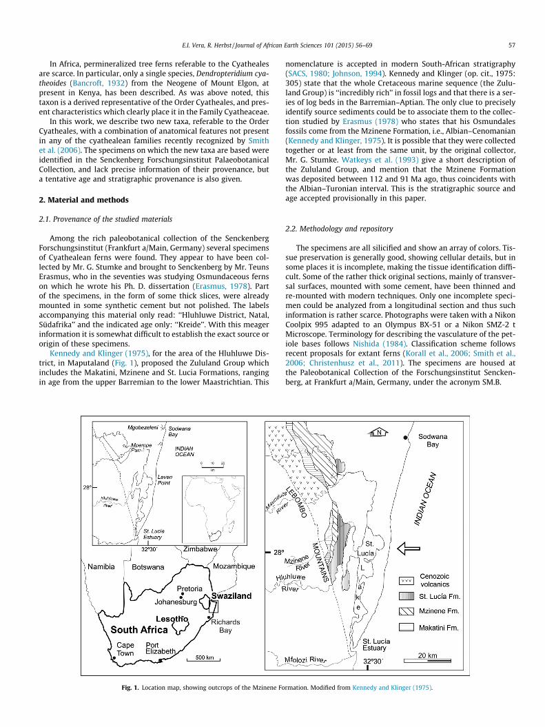
Kennedy and Klinger (1975), for the area of the Hluhluwe Dis-trict, in Maputaland (Fig. 1), proposed the Zululand Group whichincludes the Makatini, Mzinene and St. Lucia Formations, rangingin age from the upper Barremian to the lower Maastrichtian. This
Fig. 1. Location map, showing outcrops of the Mzinene Fo
nomenclature is accepted in modern South-African stratigraphy(SACS, 1980; Johnson, 1994). Kennedy and Klinger (op. cit., 1975:305) state that the whole Cretaceous marine sequence (the Zulu-land Group) is ‘‘incredibly rich’’ in fossil logs and that there is a ser-ies of log beds in the Barremian–Aptian. The only clue to preciselyidentify source sediments could be to associate them to the collec-tion studied by Erasmus (1978) who states that his Osmundalesfossils come from the Mzinene Formation, i.e., Albian–Cenomanian(Kennedy and Klinger, 1975). It is possible that they were collectedtogether or at least from the same unit, by the original collector,Mr. G. Stumke. Watkeys et al. (1993) give a short description ofthe Zululand Group, and mention that the Mzinene Formationwas deposited between 112 and 91 Ma ago, thus coincidents withthe Albian–Turonian interval. This is the stratigraphic source andage accepted provisionally in this paper.
2.2. Methodology and repository
The specimens are all silicified and show an array of colors. Tis-sue preservation is generally good, showing cellular details, but insome places it is incomplete, making the tissue identification diffi-cult. Some of the rather thick original sections, mainly of transver-sal surfaces, mounted with some cement, have been thinned andre-mounted with modern techniques. Only one incomplete speci-men could be analyzed from a longitudinal section and thus suchinformation is rather scarce. Photographs were taken with a NikonCoolpix 995 adapted to an Olympus BX-51 or a Nikon SMZ-2 tMicroscope. Terminology for describing the vasculature of the pet-iole bases follows Nishida (1984). Classification scheme followsrecent proposals for extant ferns (Korall et al., 2006; Smith et al.,2006; Christenhusz et al., 2011). The specimens are housed atthe Paleobotanical Collection of the Forschungsinstitut Sencken-berg, at Frankfurt a/Main, Germany, under the acronym SM.B.
rmation. Modified from Kennedy and Klinger (1975).

Fig. 2. Anatomy of Natalipteris wildei gen. et sp. nov. (A) general view of the stem with attached petiole bases and adventitious roots (SM.B 20635); (B) detail of the caulinevascular strand, showing metaxylem tracheids (SM.B 20633); (C) close-up of the vascular strand, showing xylem (Xi), phloem (Ph), tangential cells (Tg), pericycle (Pe) andendodermis (En); external side up (SM.B 20633); (D) sclerotic nest (SM.B 20633); (E) detail of a sclerotic nest (SM.B 20633); (F) and (G), mucilage cells (SM.B 20633); (H)departing leaf trace. Notice the sclerenchyma strands (arrows) located adaxially to the vascular strand. Scale bar represents 10 mm in A, 5 mm in H, 500 lm in B and D,200 lm in C and F, and 100 lm in E and G.
58 E.I. Vera, R. Herbst / Journal of African Earth Sciences 101 (2015) 56–69
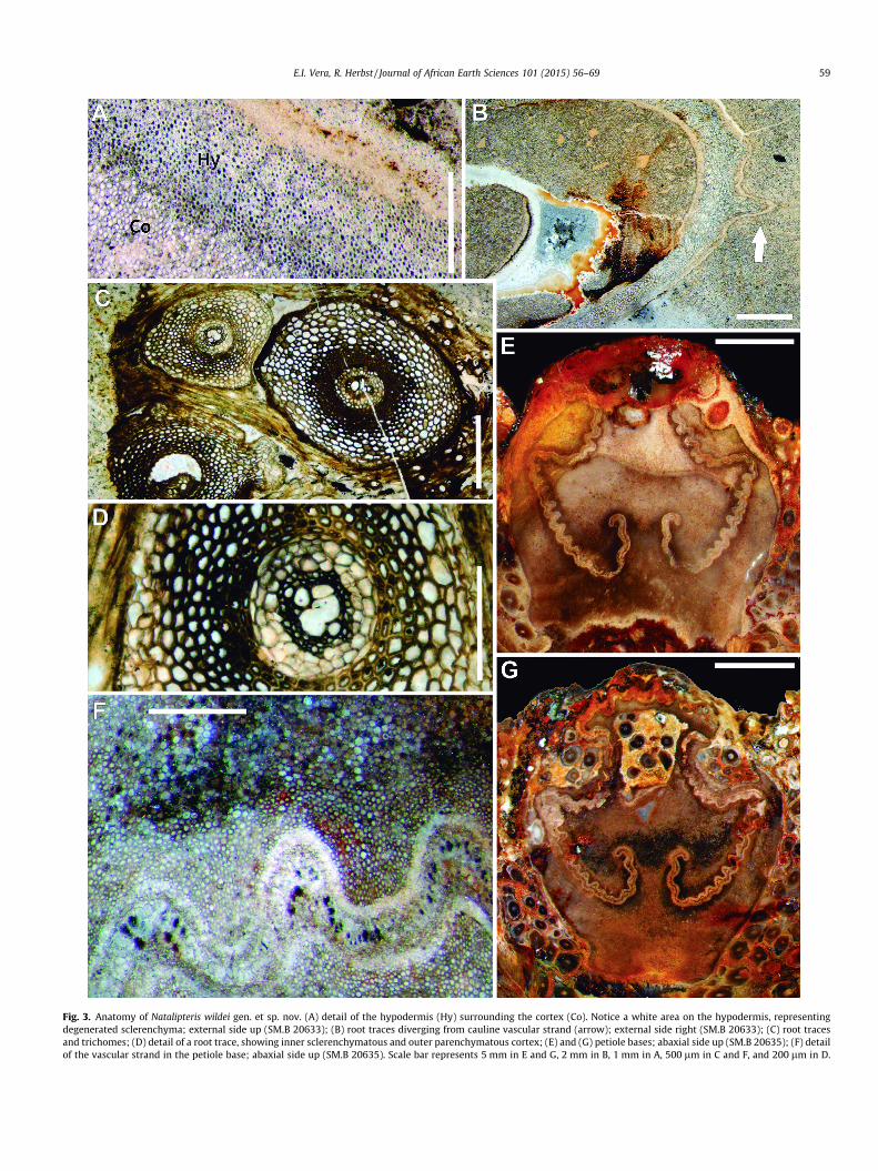
Fig. 3. Anatomy of Natalipteris wildei gen. et sp. nov. (A) detail of the hypodermis (Hy) surrounding the cortex (Co). Notice a white area on the hypodermis, representingdegenerated sclerenchyma; external side up (SM.B 20633); (B) root traces diverging from cauline vascular strand (arrow); external side right (SM.B 20633); (C) root tracesand trichomes; (D) detail of a root trace, showing inner sclerenchymatous and outer parenchymatous cortex; (E) and (G) petiole bases; abaxial side up (SM.B 20635); (F) detailof the vascular strand in the petiole base; abaxial side up (SM.B 20635). Scale bar represents 5 mm in E and G, 2 mm in B, 1 mm in A, 500 lm in C and F, and 200 lm in D.
E.I. Vera, R. Herbst / Journal of African Earth Sciences 101 (2015) 56–69 59
60 E.I. Vera, R. Herbst / Journal of African Earth Sciences 101 (2015) 56–69
3. Systematic paleontology
Class Polypodiopsida Cronquist, Takhtajan and Zimmerman1966
Order Cyatheales A.B. Frank in J. Leunis 1877Family uncertainNatalipteris R. Herbst et Vera gen. nov. (Figs. 2, 3, 6A and C)Type species: Natalipteris wildei R. Herbst et Vera sp. nov.Etimology: After the Natal Province, one of the older names of
the KwaZulu-Natal Province of South Africa, and pteris, fern.Referred species: Type species only.Diagnosis: Same as for species, by monotipy.Natalipteris wildei R. Herbst et Vera sp. nov.Etimology: Dedicated to Dr. Volker Wilde, (Curator of the Senc-
kenberg Paleobotanical Collection) for his many contributions toEuropean paleobotany.
Type locality: ‘‘Hluhluwe District, Natal, South Africa’’ (fromaccompanying labels).
Stratigraphic horizon: Most probably, Mzinene Formation(Albian–Turonian).
Holotype: SM.B 20635.Referred specimen: SM.B 20633Diagnosis: Fossil tree fern stem with attached petiole bases and
adventitious roots. Stem solenostelic, without medullary bundlesand sclerenchyma sheath; with margins of leaf gap pointing cen-trifugally. Cortex narrow, surrounded by hypodermis and epider-mis, from where an indument of multicellular hairs emerge. Pithand cortex parenchymatous, with interspersed sclerotic nests andmucilague cells. Petiole bases circular to oval; vascular strand sin-gle, with a modified omega-shaped morphology, presenting a wavymorphology due to repetition of V-shaped units. Abaxial arc smal-ler than adaxial arc. Lateral folds in the abaxial half of the strand,with the superior series approximately five times longer than infe-rior series. Adventitious roots diarch, presenting inner sclerenchy-matous and outer parenchymatous cortex.
Description: The description is based on a thin fragment of thestem (SM.B 20635) and a mounted microscope slide (SM.B20633), which most certainly belonged to the same stem. The spec-imen is ca. 10 cm wide, and contains a stem (5–9 cm in diameter)surrounded by a mantle of persistent petiole bases and adventi-tious roots (Fig. 2A).
Internally, the stem consists of a pith 25–50 mm in diameter,which is surrounded by an undulating solenostele. At the leafgap margin, the meristele slightly curves abaxially. Surroundingthe stele is a narrow (0.7–2 cm) cortex, which is completelyenclosed by a one-layered hypodermis and the epidermis(Fig. 2A, 3A and 6A). Externally, a mantle of multicellular hairs ispresent (Fig. 3C).
The pith is mainly composed of parenchyma cells, 68–120 lmin diameter, with thin walls. Interspersed within the pith are scle-rotic nests, ranging from 668 to 3600 lm in diameter, with circularto irregular shapes in cross sections. Sclerenchyma cells of this tis-sue are 60–130 lm in diameter, and have tick (16–32 lm) wallsand narrow luminae (Fig. 2D and E). Although the distribution ofthese sclerenchyma nests seems to be random, these clusters ofsclerenchymatous cells are often grouped near the vascularstrands, in particular during the development of leaf traces(Fig. 2H). Mucilague cells are also scattered in the pith, singly orin groups of 2–3 (Fig. 2F and G).
The maximum thickness (2100–2768 lm) of the vascular bun-dle is reached in the middle of the consecutive orthostichies, butthey gradually become narrow to both margins. The vascularstrand is amphicribral and composed of primary xylem (with trac-heids 25–95 lm in diameter), with small interspersed parenchymacells (Fig. 2B), phloem, 30–200 lm in thickness, including a layer of
tangential cells, and the pericycle, consisting of parenchyma-likecells 16–35 lm in diameter (Fig. 2C). Finally, the endodermisencloses the vascular bundle (Fig. 2C). Externally, no sclerenchymasheath was identified.
Stem cortex consists of several layers of tissue. The most inter-nal zone is parenchymatous, and presents the same histologicalcharacteristics as of the pith, with sclerotic nests and mucilaguecells interspersed among thick-walled parenchyma cells. This layeris sheathed by a single-layered sclerenchymatous hypodermis,consisting of cells 15–48 lm in diameter, with thick walls(Fig. 3A and C), which appears partially degenerated in someregions of the stem (Fig. 3A). The degree of preservation of thespecimen precludes the identification of other cell types betweenthis layer and the external trichomes (which most certainlyemerged from the epidermal layer, Fig. 3C).
Root traces are scarce, and were difficult to identify in the stud-ied specimens. They diverge from the cauline cylinder (Fig. 3B), atthe region where the leaf trace is starting to develop. After separa-tion from the central stele, they traverse the stem cortex until theyvascularize the adventitious roots.
Leaf trace formation initiates with an outward expansion of thestem vascular bundle, where the xylem becomes thinner than inthe adjacent sectors of the stele (Fig. 2H). Distally, the strandexpansion continues, exhibiting marginal undulation of the bun-dle. This progression continues, and two lateral constrictionsappear in the structure. At the same time, the leaf trace separatesfrom the cauline vascular cylinder, developing a leaf gap. Finally,the adaxial ends of the produced leaf trace curve inwards (Fig. 6C).
Petiole bases are circular to oval in cross section (Fig. 3E and G),ranging from 1.5 to 2.3 cm in diameter. They have a narrow (1–2tracheids thick; Fig. 3F), bilaterally symmetrical, vascular strand,which presents a modified omega-shaped morphology, with theadaxial arc larger than the abaxial arc, and with its adaxial marginspointing to the center of the petiole forming the median pair, alsodeveloping two small ‘‘hooks’’ (Fig. 3E and G). Along all the lengthof the meristele, the vascular strand presents a wavy morphology,appearing with a typical corrugated aspect due to repetition of V-shaped units (Fig. 3F). Two lateral folds are present in the leaf trace,with the superior series approximately five times longer than theinferior series. In the studied materials, these constrictions arealways present in the abaxial half of the strand (Fig. 3E and G).
Surrounding the petiole vascular trace, several layers of smallsclerenchyma cells are present (Fig. 3F), derived from the scleroticnests present in the stem ground tissue. Petiole pith and cortex areof the same tissue composition as the stem ground tissues.
Adventitious roots are circular to oval in outline, 1000–1780 lm in diameter, and are generally present running verticallyparallel to the stem, forming a dense indument. The roots arediarch, with a central xylem surrounded by phloem. This tissue isenclosed by a well-defined pericycle and the endodermis, the latterdefined by tangentially elongated cells. The cortex consists of aninternal sclerenchymatous layer, with cells 68–232 lm diameter,and a successive external parenchymatous layer, with thin-walledcells (24–65 lm in diameter) with narrow walls. The outermosttissue is the epidermis, consisting of cells 15–35 lm in diameter(Fig. 3C and D).
Comparisons with extant taxa and affinities: A solenostelic stem,surrounded by a mantle of adventitious roots and petiole bases,and petiole bases with wavy contour are characters that link Nat-alipteris wildei with the tree ferns of the Order Cyatheales.
Thyrsopteridaceae, represented by Thyrsopteris elegans, havesolenostelic stems with medullary bundles, absent in Natalipteris.Petiole bases of Thyrsopteris are vascularized by a three-partedstrand, whereas Natalipteris petiole bases have an undivided bun-dle (Table 1).
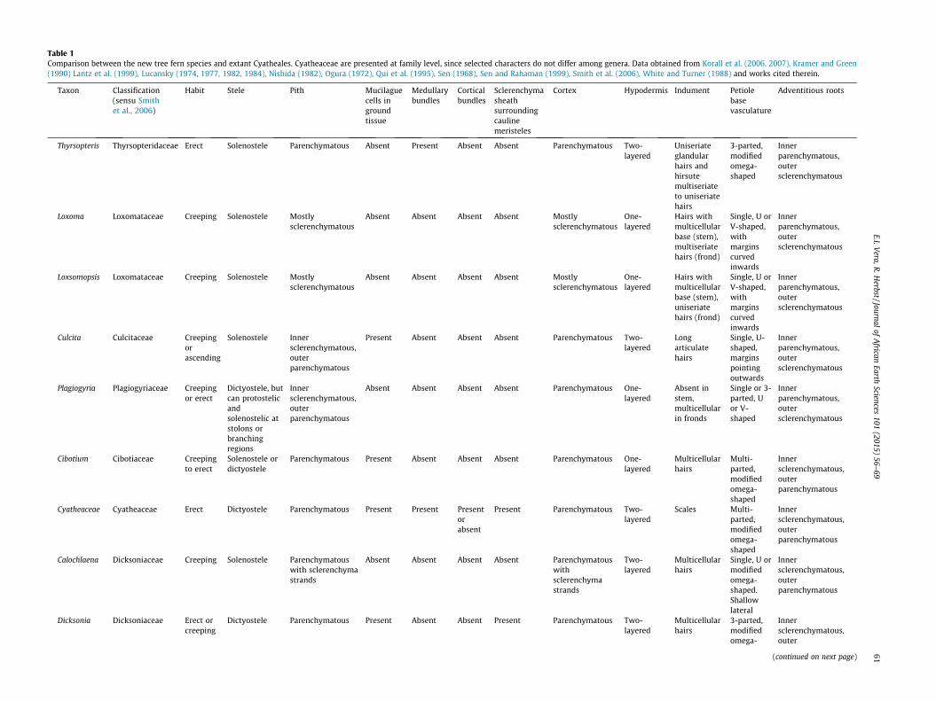
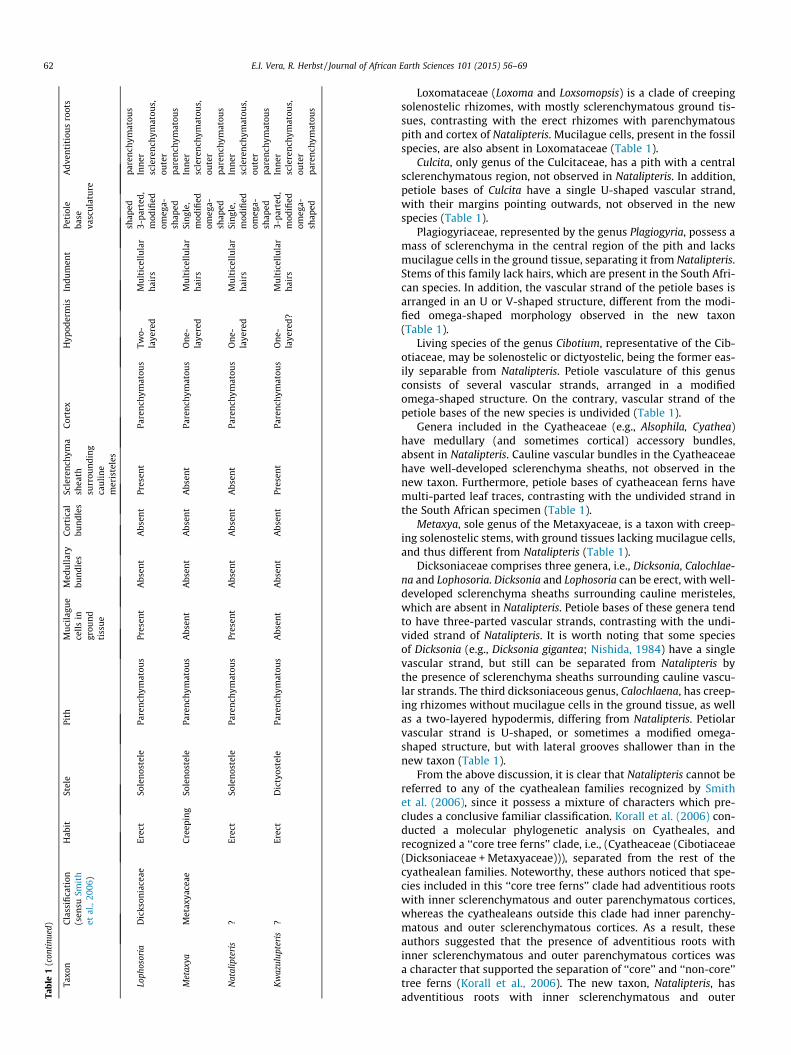
Table 1Comparison between the new tree fern species and extant Cyatheales. Cyatheaceae are presented at family level, since selected characters do not differ among genera. Data obtained from Korall et al. (2006, 2007), Kramer and Green(1990) Lantz et al. (1999), Lucansky (1974, 1977, 1982, 1984), Nishida (1982), Ogura (1972), Qui et al. (1995), Sen (1968), Sen and Rahaman (1999), Smith et al. (2006), White and Turner (1988) and works cited therein.
Taxon Classification(sensu Smithet al., 2006)
Habit Stele Pith Mucilaguecells ingroundtissue
Medullarybundles
Corticalbundles
Sclerenchymasheathsurroundingcaulinemeristeles
Cortex Hypodermis Indument Petiolebasevasculature
Adventitious roots
Thyrsopteris Thyrsopteridaceae Erect Solenostele Parenchymatous Absent Present Absent Absent Parenchymatous Two-layered
Uniseriateglandularhairs andhirsutemultiseriateto uniseriatehairs
3-parted,modifiedomega-shaped
Innerparenchymatous,outersclerenchymatous
Loxoma Loxomataceae Creeping Solenostele Mostlysclerenchymatous
Absent Absent Absent Absent Mostlysclerenchymatous
One-layered
Hairs withmulticellularbase (stem),multiseriatehairs (frond)
Single, U orV-shaped,withmarginscurvedinwards
Innerparenchymatous,outersclerenchymatous
Loxsomopsis Loxomataceae Creeping Solenostele Mostlysclerenchymatous
Absent Absent Absent Absent Mostlysclerenchymatous
One-layered
Hairs withmulticellularbase (stem),uniseriatehairs (frond)
Single, U orV-shaped,withmarginscurvedinwards
Innerparenchymatous,outersclerenchymatous
Culcita Culcitaceae Creepingorascending
Solenostele Innersclerenchymatous,outerparenchymatous
Present Absent Absent Absent Parenchymatous Two-layered
Longarticulatehairs
Single, U-shaped,marginspointingoutwards
Innerparenchymatous,outersclerenchymatous
Plagiogyria Plagiogyriaceae Creepingor erect
Dictyostele, butcan protostelicandsolenostelic atstolons orbranchingregions
Innersclerenchymatous,outerparenchymatous
Absent Absent Absent Absent Parenchymatous One-layered
Absent instem,multicellularin fronds
Single or 3-parted, Uor V-shaped
Innerparenchymatous,outersclerenchymatous
Cibotium Cibotiaceae Creepingto erect
Solenostele ordictyostele
Parenchymatous Present Absent Absent Absent Parenchymatous One-layered
Multicellularhairs
Multi-parted,modifiedomega-shaped
Innersclerenchymatous,outerparenchymatous
Cyatheaceae Cyatheaceae Erect Dictyostele Parenchymatous Present Present Presentorabsent
Present Parenchymatous Two-layered
Scales Multi-parted,modifiedomega-shaped
Innersclerenchymatous,outerparenchymatous
Calochlaena Dicksoniaceae Creeping Solenostele Parenchymatouswith sclerenchymastrands
Absent Absent Absent Absent Parenchymatouswithsclerenchymastrands
Two-layered
Multicellularhairs
Single, U ormodifiedomega-shaped.Shallowlateral
Innersclerenchymatous,outerparenchymatous
Dicksonia Dicksoniaceae Erect orcreeping
Dictyostele Parenchymatous Present Absent Absent Present Parenchymatous Two-layered
Multicellularhairs
3-parted,modifiedomega-
Innersclerenchymatous,outer
(continued on next page)
E.I.Vera,R
.Herbst/Journal
ofA
fricanEarth
Sciences101
(2015)56–
6961
Tabl
e1
(con
tinu
ed)
Taxo
nC
lass
ifica
tion
(sen
suSm
ith
etal
.,20
06)
Hab
itSt
ele
Pith
Mu
cila
gue
cell
sin
grou
nd
tiss
ue
Med
ull
ary
bun
dles
Cor
tica
lbu
ndl
esSc
lere
nch
yma
shea
thsu
rrou
ndi
ng
cau
lin
em
eris
tele
s
Cor
tex
Hyp
oder
mis
Indu
men
tPe
tiol
eba
seva
scu
latu
re
Adv
enti
tiou
sro
ots
shap
edpa
ren
chym
atou
sLo
phos
oria
Dic
kson
iace
aeEr
ect
Sole
nos
tele
Pare
nch
ymat
ous
Pres
ent
Abs
ent
Abs
ent
Pres
ent
Pare
nch
ymat
ous
Two-
laye
red
Mu
ltic
ellu
lar
hai
rs3-
part
ed,
mod
ified
omeg
a-sh
aped
Inn
ersc
lere
nch
ymat
ous,
oute
rpa
ren
chym
atou
sM
etax
yaM
etax
yace
aeC
reep
ing
Sole
nos
tele
Pare
nch
ymat
ous
Abs
ent
Abs
ent
Abs
ent
Abs
ent
Pare
nch
ymat
ous
On
e-la
yere
dM
ult
icel
lula
rh
airs
Sin
gle,
mod
ified
omeg
a-sh
aped
Inn
ersc
lere
nch
ymat
ous,
oute
rpa
ren
chym
atou
sN
atal
ipte
ris
?Er
ect
Sole
nos
tele
Pare
nch
ymat
ous
Pres
ent
Abs
ent
Abs
ent
Abs
ent
Pare
nch
ymat
ous
On
e-la
yere
dM
ult
icel
lula
rh
airs
Sin
gle,
mod
ified
omeg
a-sh
aped
Inn
ersc
lere
nch
ymat
ous,
oute
rpa
ren
chym
atou
sK
waz
ulup
teri
s?
Erec
tD
icty
oste
lePa
ren
chym
atou
sA
bsen
tA
bsen
tA
bsen
tPr
esen
tPa
ren
chym
atou
sO
ne-
laye
red?
Mu
ltic
ellu
lar
hai
rs3-
part
ed,
mod
ified
omeg
a-sh
aped
Inn
ersc
lere
nch
ymat
ous,
oute
rpa
ren
chym
atou
s
62 E.I. Vera, R. Herbst / Journal of African Earth Sciences 101 (2015) 56–69
Loxomataceae (Loxoma and Loxsomopsis) is a clade of creepingsolenostelic rhizomes, with mostly sclerenchymatous ground tis-sues, contrasting with the erect rhizomes with parenchymatouspith and cortex of Natalipteris. Mucilague cells, present in the fossilspecies, are also absent in Loxomataceae (Table 1).
Culcita, only genus of the Culcitaceae, has a pith with a centralsclerenchymatous region, not observed in Natalipteris. In addition,petiole bases of Culcita have a single U-shaped vascular strand,with their margins pointing outwards, not observed in the newspecies (Table 1).
Plagiogyriaceae, represented by the genus Plagiogyria, possess amass of sclerenchyma in the central region of the pith and lacksmucilague cells in the ground tissue, separating it from Natalipteris.Stems of this family lack hairs, which are present in the South Afri-can species. In addition, the vascular strand of the petiole bases isarranged in an U or V-shaped structure, different from the modi-fied omega-shaped morphology observed in the new taxon(Table 1).
Living species of the genus Cibotium, representative of the Cib-otiaceae, may be solenostelic or dictyostelic, being the former eas-ily separable from Natalipteris. Petiole vasculature of this genusconsists of several vascular strands, arranged in a modifiedomega-shaped structure. On the contrary, vascular strand of thepetiole bases of the new species is undivided (Table 1).
Genera included in the Cyatheaceae (e.g., Alsophila, Cyathea)have medullary (and sometimes cortical) accessory bundles,absent in Natalipteris. Cauline vascular bundles in the Cyatheaceaehave well-developed sclerenchyma sheaths, not observed in thenew taxon. Furthermore, petiole bases of cyatheacean ferns havemulti-parted leaf traces, contrasting with the undivided strand inthe South African specimen (Table 1).
Metaxya, sole genus of the Metaxyaceae, is a taxon with creep-ing solenostelic stems, with ground tissues lacking mucilague cells,and thus different from Natalipteris (Table 1).
Dicksoniaceae comprises three genera, i.e., Dicksonia, Calochlae-na and Lophosoria. Dicksonia and Lophosoria can be erect, with well-developed sclerenchyma sheaths surrounding cauline meristeles,which are absent in Natalipteris. Petiole bases of these genera tendto have three-parted vascular strands, contrasting with the undi-vided strand of Natalipteris. It is worth noting that some speciesof Dicksonia (e.g., Dicksonia gigantea; Nishida, 1984) have a singlevascular strand, but still can be separated from Natalipteris bythe presence of sclerenchyma sheaths surrounding cauline vascu-lar strands. The third dicksoniaceous genus, Calochlaena, has creep-ing rhizomes without mucilague cells in the ground tissue, as wellas a two-layered hypodermis, differing from Natalipteris. Petiolarvascular strand is U-shaped, or sometimes a modified omega-shaped structure, but with lateral grooves shallower than in thenew taxon (Table 1).
From the above discussion, it is clear that Natalipteris cannot bereferred to any of the cyathealean families recognized by Smithet al. (2006), since it possess a mixture of characters which pre-cludes a conclusive familiar classification. Korall et al. (2006) con-ducted a molecular phylogenetic analysis on Cyatheales, andrecognized a ‘‘core tree ferns’’ clade, i.e., (Cyatheaceae (Cibotiaceae(Dicksoniaceae + Metaxyaceae))), separated from the rest of thecyathealean families. Noteworthy, these authors noticed that spe-cies included in this ‘‘core tree ferns’’ clade had adventitious rootswith inner sclerenchymatous and outer parenchymatous cortices,whereas the cyathealeans outside this clade had inner parenchy-matous and outer sclerenchymatous cortices. As a result, theseauthors suggested that the presence of adventitious roots withinner sclerenchymatous and outer parenchymatous cortices wasa character that supported the separation of ‘‘core’’ and ‘‘non-core’’tree ferns (Korall et al., 2006). The new taxon, Natalipteris, hasadventitious roots with inner sclerenchymatous and outer
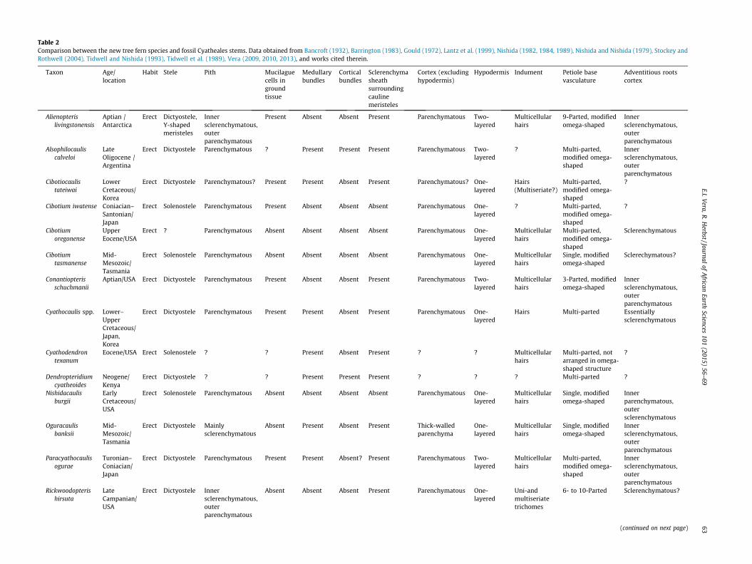
Table 2Comparison between the new tree fern species and fossil Cyatheales stems. Data obtained from Bancroft (1932), Barrington (1983), Gould (1972), Lantz et al. (1999), Nishida (1982, 1984, 1989), Nishida and Nishida (1979), Stockey andRothwell (2004), Tidwell and Nishida (1993), Tidwell et al. (1989), Vera (2009, 2010, 2013), and works cited therein.
Taxon Age/location
Habit Stele Pith Mucilaguecells ingroundtissue
Medullarybundles
Corticalbundles
Sclerenchymasheathsurroundingcaulinemeristeles
Cortex (excludinghypodermis)
Hypodermis Indument Petiole basevasculature
Adventitious rootscortex
Alienopterislivingstonensis
Aptian /Antarctica
Erect Dictyostele,Y-shapedmeristeles
Innersclerenchymatous,outerparenchymatous
Present Absent Absent Present Parenchymatous Two-layered
Multicellularhairs
9-Parted, modifiedomega-shaped
Innersclerenchymatous,outerparenchymatous
Alsophilocauliscalveloi
LateOligocene /Argentina
Erect Dictyostele Parenchymatous ? Present Present Present Parenchymatous Two-layered
? Multi-parted,modified omega-shaped
Innersclerenchymatous,outerparenchymatous
Cibotiocaulistateiwai
LowerCretaceous/Korea
Erect Dictyostele Parenchymatous? Present Present Absent Present Parenchymatous? One-layered
Hairs(Multiseriate?)
Multi-parted,modified omega-shaped
?
Cibotium iwatense Coniacian–Santonian/Japan
Erect Solenostele Parenchymatous Present Absent Absent Absent Parenchymatous One-layered
? Multi-parted,modified omega-shaped
?
Cibotiumoregonense
UpperEocene/USA
Erect ? Parenchymatous Absent Absent Absent Absent Parenchymatous One-layered
Multicellularhairs
Multi-parted,modified omega-shaped
Sclerenchymatous
Cibotiumtasmanense
Mid-Mesozoic/Tasmania
Erect Solenostele Parenchymatous Absent Absent Absent Absent Parenchymatous One-layered
Multicellularhairs
Single, modifiedomega-shaped
Sclerechymatous?
Conantiopterisschuchmanii
Aptian/USA Erect Dictyostele Parenchymatous Present Absent Absent Present Parenchymatous Two-layered
Multicellularhairs
3-Parted, modifiedomega-shaped
Innersclerenchymatous,outerparenchymatous
Cyathocaulis spp. Lower–UpperCretaceous/Japan,Korea
Erect Dictyostele Parenchymatous Present Present Absent Present Parenchymatous One-layered
Hairs Multi-parted Essentiallysclerenchymatous
Cyathodendrontexanum
Eocene/USA Erect Solenostele ? ? Present Absent Present ? ? Multicellularhairs
Multi-parted, notarranged in omega-shaped structure
?
Dendropteridiumcyatheoides
Neogene/Kenya
Erect Dictyostele ? ? Present Present Present ? ? ? Multi-parted ?
Nishidacaulisburgii
EarlyCretaceous/USA
Erect Solenostele Parenchymatous Absent Absent Absent Absent Parenchymatous One-layered
Multicellularhairs
Single, modifiedomega-shaped
Innerparenchymatous,outersclerenchymatous
Oguracaulisbanksii
Mid-Mesozoic/Tasmania
Erect Dictyostele Mainlysclerenchymatous
Absent Present Absent Present Thick-walledparenchyma
One-layered
Multicellularhairs
Single, modifiedomega-shaped
Innersclerenchymatous,outerparenchymatous
Paracyathocaulisogurae
Turonian–Coniacian/Japan
Erect Dictyostele Parenchymatous Present Present Absent? Present Parenchymatous Two-layered
Multicellularhairs
Multi-parted,modified omega-shaped
Innersclerenchymatous,outerparenchymatous
Rickwoodopterishirsuta
LateCampanian/USA
Erect Dictyostele Innersclerenchymatous,outerparenchymatous
Absent Absent Absent Present Parenchymatous One-layered
Uni-andmultiseriatetrichomes
6- to 10-Parted Sclerenchymatous?
(continued on next page)
E.I.Vera,R
.Herbst/Journal
ofA
fricanEarth
Sciences101
(2015)56–
6963
Tabl
e2
(con
tinu
ed)
Taxo
nA
ge/
loca
tion
Hab
itSt
ele
Pith
Mu
cila
gue
cell
sin
grou
ndti
ssu
e
Med
ull
ary
bun
dles
Cor
tica
lbu
ndl
esSc
lere
nch
yma
shea
thsu
rrou
ndi
ng
cau
lin
em
eris
tele
s
Cor
tex
(exc
ludi
ng
hyp
oder
mis
)H
ypod
erm
isIn
dum
ent
Peti
ole
base
vasc
ula
ture
Adv
enti
tiou
sro
ots
cort
ex
Yava
nna
chim
aeri
caA
ptia
n/
An
tarc
tica
Erec
tSo
len
oste
lePa
ren
chym
atou
sPr
esen
tPr
esen
tA
bsen
tPr
esen
tPa
ren
chym
atou
sO
ne-
laye
red?
Mu
ltic
ellu
lar
hai
rs?
Sin
gle
(pro
xim
ally
),th
ree
part
ed(d
ista
lly)
,mod
ified
omeg
a-sh
aped
Inn
erpa
ren
chym
atou
s,ou
ter
scle
ren
chym
atou
sN
atal
ipte
ris
wild
eiA
lbia
n–
Turo
nia
n?/
Sou
thA
fric
a
Erec
tSo
len
oste
lePa
ren
chym
atou
sPr
esen
tA
bsen
tA
bsen
tA
bsen
tPa
ren
chym
atou
sO
ne-
laye
red
Mu
ltic
ellu
lar
hai
rsSi
ngl
e,m
odifi
edom
ega-
shap
edIn
ner
scle
ren
chym
atou
s,ou
ter
pare
nch
ymat
ous
Kw
azul
upte
ris
scha
arsc
hmid
tii
Alb
ian
-Tu
ron
ian
?/So
uth
Afr
ica
Erec
tD
icty
oste
lePa
ren
chym
atou
sA
bsen
tA
bsen
tA
bsen
tPr
esen
tPa
ren
chym
atou
sO
ne-
laye
red?
Mu
ltic
ellu
lar
hai
rs3-
Part
ed,m
odifi
edom
ega-
shap
edIn
ner
scle
ren
chym
atou
s,ou
ter
pare
nch
ymat
ous
64 E.I. Vera, R. Herbst / Journal of African Earth Sciences 101 (2015) 56–69
parenchymatous cortex, and thus may be referred to the ‘‘core treeferns’’.
Comparisons with fossil Cyatheales: Natalipteris wildei can be sep-arated from fossil taxa referable to the Cyatheaceae (AlsophilocaulisMenéndez, 1961, Dendropteridium Bancroft, 1932, CyathocaulisOgura emend. Nishida, 1989, Cibotiocaulis Ogura emend. Nishida,1989, and Paracyathocaulis Nishida, 1989) by the presence of dict-yostelic stems with sclerenchyma sheaths surrounding caulinemeristeles, and petiole bases with multi-parted vasculature inthe latter taxa. A stem with accessory medullary bundles, andmulti-parted vascular strands in the petiole bases, are alsorecorded in Cyathodendron texanum Arnold, 1945 (Table 2).
Some fossil species included in the genus Cibotium (C. iwatenseOgura, 1933 and C. oregonense Barrington, 1983) have petiole baseswith multi-parted vascular strands, contrasting with Natalipteris.In addition, Cibotium oregonense and Cibotium tasmanense Gould,1972 lack mucilague cells in ground tissues, whereas these struc-tures are present in Natalipteris (Table 2).
The Antarctic Alienopteris livingstonensis Vera, 2009 has Y-shapedcauline meristeles, surrounded by sclerenchyma sheaths. In addition,petiole bases have nine meristeles, the features absent in Natalipteris.Another species from Antarctica, Yavanna chimaerica Vera, 2013, alsohas sclerenchyma sheaths in its cauline meristeles. Furthermore, thistaxon has medullary bundles, absent in the new species (Table 2).
Oguracaulis banksii (Tidwell et al., 1989) and Rickwoodopterishirsuta Stockey et Rothwell 2004 share the presence of abundantsclerenchymatous tissue in the pith, absent in Natalipteris. The for-mer species also have sclerenchyma sheaths surrounding caulinevascula strands, not observed in the new species. In addition, Rick-woodopteris hirsuta possess six to ten meristeles in the petiolebases (Table 2).
Conantiopteris schuchmanii Lantz, Rothwell et Stockey 1999 isdictyostelic, and possess cauline sclerenchyma sheaths, absent inNatalipteris. Petioles of this taxon have three-parted vasculature,contrasting with the undivided vasculature observed in the newspecies (Table 2).
Natalipteris wildei and Nishidacaulis burgii Tidwell et Nishida,1993 share the presence of a solenostelic stem without sclerenchy-matous sheath surrounding the vascular trace, and an undividedvascular trace of the petiole (Table 2). However, Natalipteris canbe separated from N. burgii by the presence of mucilague cellsand sclerotic nests in the ground tissue, and petiole bases with dee-per lateral folds and a smaller abaxial arc.
Lophosoriorhachis Nishida, 1982, a genus erected for isolatedrachides, has shallow lateral grooves in the petioles, which arelocated in the middle region of the vascular strand (Nishida,1982), whereas in Natalipteris the lateral grooves are deeper, andlocated in the abaxial region of the strand. Another genus of iso-lated rachides, Thyrsopterorachis Nishida et Nishida 1979 possessdeeper and abaxially oriented lateral grooves, as well as a propor-tionally larger abaxial arc (Nishida and Nishida, 1979).
In summary, the combination of features observed in Natalipteriswildei are not present in any of the already described fossilcyathealean stems, supporting its recognition as a new taxon.
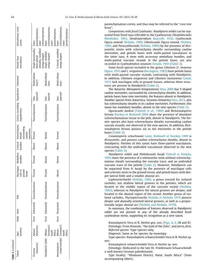
Kwazulupteris Vera et R. Herbst gen. nov. (Figs. 4, 5, 6B and D)Etimology: From Kwazulu, ‘‘the land of the Zulu’’, and pteris, fern.Referred species: Type species only.Diagnosis: Same as for species, by monotipy.Type species: Kwazulupteris schaarschmidtii Vera et R. Herbst sp.
nov.Kwazulupteris schaarschmidtii Vera et Herbst sp. nov.Etimology: Dedicated to the late Dr. Friedemann Schaarschmidt
a well-known German paleobotanist.Type locality: ‘‘Hluhluwe District, Natal, South Africa’’ (from
accompanying labels).
Fig. 4. Anatomy of Kwazulupteris schaarschmidtii gen. et sp. nov., SM.B 20628. (A) general view of the stem with attached petiole bases and adventitious roots; (B) detail of acauline meristele, showing xylem and probable limit of the endodermis (arrow); external side up; (C) U-shaped meristele; external side up; (D) cauline cylinder, showingseparating root traces (arrows); external side up and (E) root traces; external side right. Scale bar represents 3 cm in A, 3 mm in C and D, 1 mm in E and 50 lm in B.
E.I. Vera, R. Herbst / Journal of African Earth Sciences 101 (2015) 56–69 65
Stratigraphic horizon: Most probably, Mzinene Formation(Albian–Turonian).
Holotype: SM.B 20628, consisting of a stem fragment and amicroscopic slide.
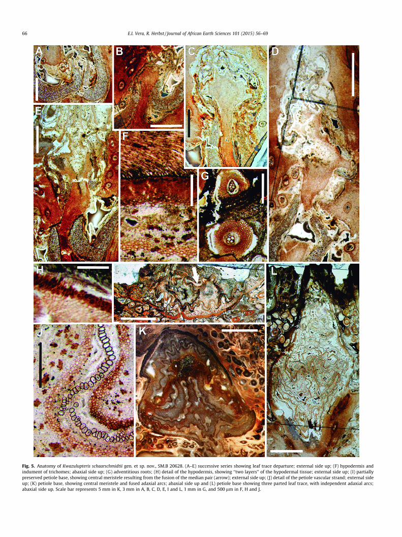
Fig. 5. Anatomy of Kwazulupteris schaarschmidtii gen. et sp. nov., SM.B 20628. (A–E) successive series showing leaf trace departure; external side up; (F) hypodermis andindument of trichomes; abaxial side up; (G) adventitious roots; (H) detail of the hypodermis, showing ‘‘two layers’’ of the hypodermal tissue; external side up; (I) partiallypreserved petiole base, showing central meristele resulting from the fusion of the median pair (arrow); external side up; (J) detail of the petiole vascular strand; external sideup; (K) petiole base, showing central meristele and fused adaxial arcs; abaxial side up and (L) petiole base showing three parted leaf trace, with independent adaxial arcs;abaxial side up. Scale bar represents 5 mm in K, 3 mm in A, B, C, D, E, I and L, 1 mm in G, and 500 lm in F, H and J.
66 E.I. Vera, R. Herbst / Journal of African Earth Sciences 101 (2015) 56–69
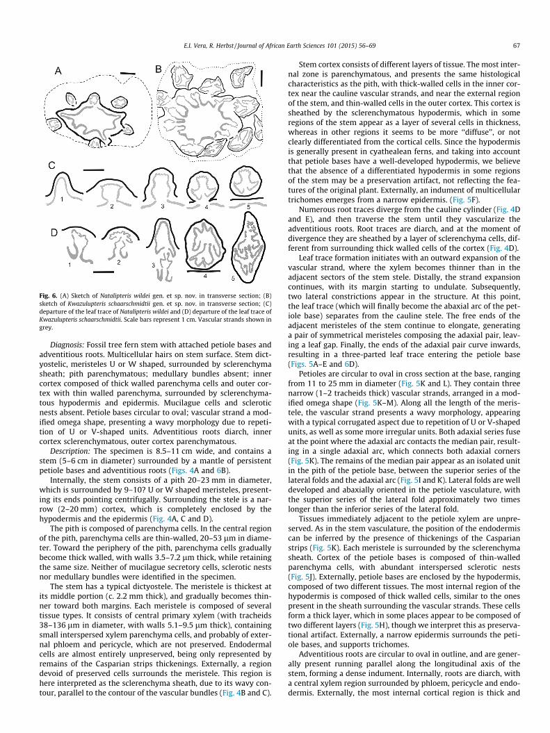
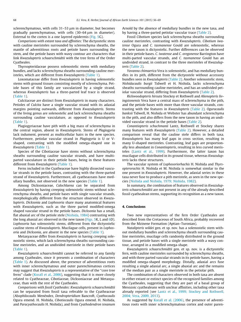
Fig. 6. (A) Sketch of Natalipteris wildei gen. et sp. nov. in transverse section; (B)sketch of Kwazulupteris schaarschmidtii gen. et sp. nov. in transverse section; (C)departure of the leaf trace of Natalipteris wildei and (D) departure of the leaf trace ofKwazulupteris schaarschmidtii. Scale bars represent 1 cm. Vascular strands shown ingrey.
E.I. Vera, R. Herbst / Journal of African Earth Sciences 101 (2015) 56–69 67
Diagnosis: Fossil tree fern stem with attached petiole bases andadventitious roots. Multicellular hairs on stem surface. Stem dict-yostelic, meristeles U or W shaped, surrounded by sclerenchymasheath; pith parenchymatous; medullary bundles absent; innercortex composed of thick walled parenchyma cells and outer cor-tex with thin walled parenchyma, surrounded by sclerenchyma-tous hypodermis and epidermis. Mucilague cells and scleroticnests absent. Petiole bases circular to oval; vascular strand a mod-ified omega shape, presenting a wavy morphology due to repeti-tion of U or V-shaped units. Adventitious roots diarch, innercortex sclerenchymatous, outer cortex parenchymatous.
Description: The specimen is 8.5–11 cm wide, and contains astem (5–6 cm in diameter) surrounded by a mantle of persistentpetiole bases and adventitious roots (Figs. 4A and 6B).
Internally, the stem consists of a pith 20–23 mm in diameter,which is surrounded by 9–10? U or W shaped meristeles, present-ing its ends pointing centrifugally. Surrounding the stele is a nar-row (2–20 mm) cortex, which is completely enclosed by thehypodermis and the epidermis (Fig. 4A, C and D).
The pith is composed of parenchyma cells. In the central regionof the pith, parenchyma cells are thin-walled, 20–53 lm in diame-ter. Toward the periphery of the pith, parenchyma cells graduallybecome thick walled, with walls 3.5–7.2 lm thick, while retainingthe same size. Neither of mucilague secretory cells, sclerotic nestsnor medullary bundles were identified in the specimen.
The stem has a typical dictyostele. The meristele is thickest atits middle portion (c. 2.2 mm thick), and gradually becomes thin-ner toward both margins. Each meristele is composed of severaltissue types. It consists of central primary xylem (with tracheids38–136 lm in diameter, with walls 5.1–9.5 lm thick), containingsmall interspersed xylem parenchyma cells, and probably of exter-nal phloem and pericycle, which are not preserved. Endodermalcells are almost entirely unpreserved, being only represented byremains of the Casparian strips thickenings. Externally, a regiondevoid of preserved cells surrounds the meristele. This region ishere interpreted as the sclerenchyma sheath, due to its wavy con-tour, parallel to the contour of the vascular bundles (Fig. 4B and C).
Stem cortex consists of different layers of tissue. The most inter-nal zone is parenchymatous, and presents the same histologicalcharacteristics as the pith, with thick-walled cells in the inner cor-tex near the cauline vascular strands, and near the external regionof the stem, and thin-walled cells in the outer cortex. This cortex issheathed by the sclerenchymatous hypodermis, which in someregions of the stem appear as a layer of several cells in thickness,whereas in other regions it seems to be more ‘‘diffuse’’, or notclearly differentiated from the cortical cells. Since the hypodermisis generally present in cyathealean ferns, and taking into accountthat petiole bases have a well-developed hypodermis, we believethat the absence of a differentiated hypodermis in some regionsof the stem may be a preservation artifact, not reflecting the fea-tures of the original plant. Externally, an indument of multicellulartrichomes emerges from a narrow epidermis. (Fig. 5F).
Numerous root traces diverge from the cauline cylinder (Fig. 4Dand E), and then traverse the stem until they vascularize theadventitious roots. Root traces are diarch, and at the moment ofdivergence they are sheathed by a layer of sclerenchyma cells, dif-ferent from surrounding thick walled cells of the cortex (Fig. 4D).
Leaf trace formation initiates with an outward expansion of thevascular strand, where the xylem becomes thinner than in theadjacent sectors of the stem stele. Distally, the strand expansioncontinues, with its margin starting to undulate. Subsequently,two lateral constrictions appear in the structure. At this point,the leaf trace (which will finally become the abaxial arc of the pet-iole base) separates from the cauline stele. The free ends of theadjacent meristeles of the stem continue to elongate, generatinga pair of symmetrical meristeles composing the adaxial pair, leav-ing a leaf gap. Finally, the ends of the adaxial pair curve inwards,resulting in a three-parted leaf trace entering the petiole base(Figs. 5A–E and 6D).
Petioles are circular to oval in cross section at the base, rangingfrom 11 to 25 mm in diameter (Fig. 5K and L). They contain threenarrow (1–2 tracheids thick) vascular strands, arranged in a mod-ified omega shape (Fig. 5K–M). Along all the length of the meris-tele, the vascular strand presents a wavy morphology, appearingwith a typical corrugated aspect due to repetition of U or V-shapedunits, as well as some more irregular units. Both adaxial series fuseat the point where the adaxial arc contacts the median pair, result-ing in a single adaxial arc, which connects both adaxial corners(Fig. 5K). The remains of the median pair appear as an isolated unitin the pith of the petiole base, between the superior series of thelateral folds and the adaxial arc (Fig. 5I and K). Lateral folds are welldeveloped and abaxially oriented in the petiole vasculature, withthe superior series of the lateral fold approximately two timeslonger than the inferior series of the lateral fold.
Tissues immediately adjacent to the petiole xylem are unpre-served. As in the stem vasculature, the position of the endodermiscan be inferred by the presence of thickenings of the Casparianstrips (Fig. 5K). Each meristele is surrounded by the sclerenchymasheath. Cortex of the petiole bases is composed of thin-walledparenchyma cells, with abundant interspersed sclerotic nests(Fig. 5J). Externally, petiole bases are enclosed by the hypodermis,composed of two different tissues. The most internal region of thehypodermis is composed of thick walled cells, similar to the onespresent in the sheath surrounding the vascular strands. These cellsform a thick layer, which in some places appear to be composed oftwo different layers (Fig. 5H), though we interpret this as preserva-tional artifact. Externally, a narrow epidermis surrounds the peti-ole bases, and supports trichomes.
Adventitious roots are circular to oval in outline, and are gener-ally present running parallel along the longitudinal axis of thestem, forming a dense indument. Internally, roots are diarch, witha central xylem region surrounded by phloem, pericycle and endo-dermis. Externally, the most internal cortical region is thick and
68 E.I. Vera, R. Herbst / Journal of African Earth Sciences 101 (2015) 56–69
sclerenchymatous, with cells 31–53 lm in diameter, but becomesgradually parenchymatous, with cells (30–64 lm in diameter).External to the cortex is a one layered epidermis (Fig. 5G).
Comparisons with extant taxa and affinities: The dictyostelic stemwith cauline meristeles surrounded by sclerenchyma sheaths, themantle of adventitious roots and petiole bases surrounding thestem, and the petiole bases with wavy contour are characters thatlink Kwazulupteris schaarschmidtii with the tree ferns of the OrderCyatheales.
Thyrsopteridaceae possess solenostelic stems with medullarybundles, and lacks sclerenchyma sheaths surrounding cauline mer-isteles, which are different from Kwazulupteris (Table 1).
Loxomataceae differ from Kwazulupteris in having solenostelicstems with ground tissues consisting mostly of sclerenchyma. Pet-iole bases of this family are vascularized by a single strand,whereas Kwazulupteris has a three-parted leaf trace is observed(Table 1).
Culcitaceae are distinct from Kwazulupteris in many characters.Petioles of Culcita have a single vascular strand with its adaxialmargins pointing outwards, different from Kwazulupteris. Stemsof the living genus are solenostelic and lack sclerenchyma sheathssurrounding cauline vasculature, as opposed to Kwazulupteris(Table 1).
Plagiogyriaceae have pith with a big mass of sclerenchyma inthe central region, absent in Kwazulupteris. Stems of Plagiogyrialack indument, present as multicellular hairs in the new species.Furthermore, petiolar vascular strand in Plagiogyria is U or V-shaped, contrasting with the modified omega-shaped one inKwazulupteris (Table 1).
Species of the Cibotiaceae have stems without sclerenchymasheaths surrounding cauline vascular strands, and have multi-parted vasculature in their petiole bases, being in these featuresdifferent from Kwazulupteris (Table 1).
Ferns included in the Cyatheaceae have highly dissected vascu-lar strands in the petiole bases, contrasting with the three-partedstrand of Kwazulupteris. Furthermore, all cyatheaceans have med-ullary bundles, not observed in the new species (Table 1).
Among Dicksoniaceae, Calochlaena can be separated fromKwazulupteris by having creeping solenostelic stems without scle-renchyma sheaths, and petiole bases with single vascular strands,morphologically different from the structure observed in Kwazu-lupteris. Dicksonia and Lophosoria share many anatomical featureswith Kwazulupteris, such as the three parted modified-omegashaped vascular strand in the petiole bases. Dicksonia has a small,flat abaxial arc of the petiole stele (Nishida, 1984) contrasting withthe long abaxial arc observed in the new taxon (Figs. 5K, L and 6D).Lophosoria has solenostelic stems, different from the dictyosteliccauline stems of Kwazulupteris. Mucilague cells, present in Lophos-oria and Dicksonia, are absent in the new species (Table 1).
Metaxyaceae differ from Kwazulupteris in having creeping sole-nostelic stems, which lack sclerenchyma sheaths surrounding cau-line meristeles, and an undivided meristele in their petiole bases(Table 1).
Kwazulupteris schaarschmidtii cannot be referred to any familyamong Cyatheales, since it presents a combination of characters(Table 1). As discussed above, the presence of adventitious rootswith inner sclerenchymatous and outer parenchimatous corticesmay suggest that Kwazulupteris is a representative of the ‘‘core treeferns’’ clade (Korall et al., 2006), suggesting that it is more closelyrelated to Cyatheaceae, Cibotiaceae, Dicksoniaceae and Metaxya-ceae, than with the rest of the Cyatheales.
Comparisons with fossil Cyatheales: Kwazulupteris schaarschmidtiican be separated from fossil taxa referable to the Cyatheaceae(Alsophilocaulis Menéndez, Dendropteridium Bancroft, CyathocaulisOgura emend. H. Nishida, Cibotiocaulis Ogura emend. H. Nishida,and Paracyathocaulis H. Nishida), and from Cyathodendron texanum
Arnold by the absence of medullary bundles in the new taxa, andby having a three-parted petiolar vascular trace (Table 2).
Fossil Cibotium species lack sclerenchyma sheaths surroundingcauline meristeles, contrasting with Kwazulupteris. Cibotium iwa-tense Ogura and C. tasmanense Gould are solenostelic, whereasthe new taxon is dictyostelic. Further differences can be observedin their petiole bases. C. iwatense and C. oregonense Barrington havemulti-parted vascular strands, and C. tasmanense Gould has anundivided strand, in contrast to the three meristeles of Kwazulup-teris (Table 2).
Yavanna chimaerica Vera is solenostelic, and has medullary bun-dles in its pith, different from the dictyostele without accessorybundles seen in Kwazulupteris (Table 2). Another solenostelic stem,Nishidacaulis burgii Tidwell et H. Nishida, lacks sclerenchymasheaths surrounding cauline meristeles, and has an undivided pet-iolar vascular strand, differing from Kwazulupteris (Table 2).
Rickwoodopteris hirsuta Stockey et Rothwell and Alienopteris liv-ingstonensis Vera have a central mass of sclerenchyma in the pith,and the petiole bases with more than three vascular strands, con-trasting with the features in Kwazulupteris (Table 2). Oguracaulisbanksii Tidwell, H. Nishida et Webster has abundant sclerenchymain the pith, and also differs from the new taxon in having an undi-vided vascular strand in the petiole bases (Table 2).
Conantiopteris schuchmanii Lantz, Rothwell et Stockey sharesmany features with Kwazulupteris (Table 2). However, a detailedcomparison reveal that the cauline stele differs in both taxa.Kwazulupteris has many leaf gaps in cross section, resulting inmany U-shaped meristeles. Contrasting, leaf gaps are proportion-ally less abundant in Conantiopteris, resulting in less curved meris-teles (Lantz et al., 1999). Furthermore, the latter taxon hasmucilague cells distributed in its ground tissue, whereas Kwazulup-teris lacks these structures.
The vascular system of Lophosoriorhachis H. Nishida and Thyrs-opterorachis H. Nishida et M. Nishida is somewhat similar to theone present in Kwazulupteris. However, the adaxial series in thesetaxa never fuse to produce a pith meristele, as seen in the new spe-cies (Nishida and Nishida, 1979; Nishida, 1982).
In summary, the combination of features observed in Kwazulup-teris schaarschmidtii are not present in any of the already describedfossil cyathealean stems, supporting its recognition as a new taxon.
4. Conclusions
Two new representatives of the fern Order Cyatheales aredescribed from the Cretaceous of South Africa, probably recoveredfrom the Mzinene Formation (Aptian–Turonian).
Natalipteris wildei gen. et sp. nov. has a solenostelic stem with-out medullary bundles and sclerenchyma sheath surrounding cau-line meristeles, mucilage cells randomly distributed in the groundtissue, and petiole bases with a single meristele with a wavy con-tour, arranged in a modified omega-shape.
Kwazulupteris schaarschmidtii gen. et sp. nov. is a dictyostelicfern, with cauline meristeles surrounded by sclerenchyma sheaths,and with three parted vascular strands in its petiole bases, having amodified omega-shaped morphology. Distally, adaxial arcs fuseresulting a single adaxial arc, a single abaxial arc and the remainsof the median pair as a single meristele in the petiolar pith.
The combination of characters observed in both taxa are absentin either extant or extinct species of the recognized families amongthe Cyatheales, suggesting that they are part of a basal group ofMesozoic cyathealeans with unclear affinities, including other taxaalready reported (see Lantz et al., 1999; Stockey and Rothwell,2004; Vera, 2009, 2013).
As suggested by Korall et al. (2006), the presence of adventi-tious roots with inner sclerenchymatous cortex and outer paren-
E.I. Vera, R. Herbst / Journal of African Earth Sciences 101 (2015) 56–69 69
chymatous cortex support the placement of Natalipteris andKwazulupteris in the ‘‘core tree ferns’’ clade, suggesting they aremore closely related with the Cyatheaceae, Dicksoniaceae, Cibotia-ceae and Metaxyaceae, than with the rest of the Cyatheales.
Acknowledgements
The authors thanks the careful and detailed revision of the edi-tor and an anonymous reviewer, which greatly improved the finalversion of the manuscript. R. Herbst wishes to thank to Dr. VolkerWilde and Mrs. Karin Schmidt for very kind assistance and helpfuldiscussions and the loan of specimens, during his several visits atthe Senckenberg Forschungsinstitute (Frankfurt, Germany).
References
Arnold, C.A., 1945. Silicified plant remains from the Mesozoic and Tertiary ofwestern North America. I. Ferns. Papers of the Michigan Academy of Sciences.Arts Lett. 30, 3–34.
Bancroft, H., 1932. A fossil cyatheoid stem from Mount Elgon, East Africa. NewPhytol. 31, 241–253.
Barrington, D.S., 1983. Cibotium oregonense: an Eocene tree-fern stem and petioleswith internal structure. Am. J. Bot. 70, 1118–1124.
Christenhusz, M.J.M., Zhang, X.-C., Schneider, H., 2011. A linear sequence of extantfamilies and genera of lycophytes and ferns. Phytotaxa 19, 7–54.
Erasmus, T., 1978. The anatomy and evolution of Osmundacaulis Miller emend.with notes on the geometry of the xylem framework of the Osmundaceousstele. University of Pretoria, unpublished thesis, 21 plates, 155 pp.
Gould, R.E., 1972. Cibotium tasmanense sp. nov., a fossil tree-fern from the Tertiaryof Tasmania. Aust. J. Bot. 20, 119–126.
Holttum, R.E., Sen, U., 1961. Morphology and classification of tree ferns.Phytomorphology 11, 406–420.
Johnson, M.R., 1994. Lexicon of South African Stratigraphy. Part 1: Phanerozoicunits. South African Committee for Stratigraphy, Council for Geoscience,Pretoria. 56pp.
Kennedy, W.J., Klinger, H.C., 1975. Cretaceous faunas from Zululand and Natal,South Africa. Introduction. Stratigraphy. Bulletin British Museum (NaturalHistory). Geology 25 (4), 265–315.
Korall, P., Pryer, K.M., Metzgar, J.S., Schneider, H., Conant, D.S., 2006. Tree ferns:monophyletic groups and their relationships as revealed by four protein-codingplastid loci. Mol. Phylogenet. Evol. 39, 830–845.
Korall, P., Conant, D.S., Metzgar, J.S., Schneider, H., Pryer, K.M., 2007. A molecularphylogeny of scaly tree ferns (Cyatheaceae). Am. J. Bot. 94, 873–886.
Kramer, K.U., Green, P.S., 1990. The Families and Genera of Vascular Plants.Pteridophytes and Gymnosperms, vol. 1. Springer-Verlag, Berlin, 404pp.
Lantz, T.C., Rothwell, G.W., Stockey, R.A., 1999. Conantiopteris schuchmanii, gen. etsp. nov., and the role of fossils in resolving the phylogeny of Cyatheaceae s.l. J.Plant. Res. 112, 361–381.
Large, M.F., Braggins, J.E., 2004. Tree Ferns. Timber Press, Portland, Oregon, 359pp.Lucansky, T.W., 1974. Comparative studies of the nodal and vascular anatomy of
neotropical Cyatheaceae. I. Metaxya and Lophosoria. Am. J. Bot. 61, 461–474.Lucansky, T.W., 1977. Anatomical studies of Cyathea and Trichipteris (Cyatheaceae).
Am. J. Bot. 64, 253–259.Lucansky, T.W., 1982. Anatomical studies of the neotropical Cyatheaceae. II.
Metaxya and Lophosoria. Am. Fern J. 72, 19–28.
Menéndez, C.A., 1961. Estípite petrificado de una nueva Cyatheaceae del Terciariode Neuquén. Boletín de la Sociedad Argentina de Botánica 9, 331–358.
Nishida, H., 1982. Lophosoriorhachis japonica n. gen. et sp., from the LowerCretaceous of Choshi, Chiba Prefecture, Japan. Palaeontogr. Abteilung B 181,118–122.
Nishida, H., 1984. Anatomical studies of the frond axis of the Cyatheaceae, s.l. with arevision of permineralized frond axes from the Cretaceous of Japan. In: Nishida,M. (Ed.), Contributions to the Botany in the Andes I. Academia Scientific BookInc., Tokio, pp. 5–80.
Nishida, H., 1989. Structure and affinities of the petrified plants from the Cretaceousof Japan and Saghalien. V. Tree fern stems from Hokkaido, Paracyathocaulisogurae gen. et comb. nov. and Cyathocaulis yezopteroides sp. nov. Bot. Mag. 102,255–282.
Nishida, H., Nishida, M., 1979. Thyrsopterorachis, gen. nov., a tree fern rachis fromthe Upper Cretaceous of Hokkaido, Japan. Bot. Mag., Tokyo 92, 187–195.
Ogura, Y., 1933. On the structure of a fossil fern stem of Cibotium type from theUpper Cretaceous of Iwate. Bot. Mag., Tokyo 47, 748–754.
Ogura, Y., 1972. Comparative anatomy of vegetative organs of the Pteridophytes.Handbuch der Pflanzenanatomie, vol 7. Borntraeger, Berlin, 502pp.
Pryer, K.M., Schuettpelz, E., Wolf, P.G., Schneider, H., Smith, A.R., Cranfill, R., 2004.Phylogeny and evolution of ferns (Monilophytes) with a focus on the earlyleptosporangiate divergences. Am. J. Bot. 91, 1582–1598.
Qui, Y.-J., White, R.A., Turner, M.D., 1995. The developmental anatomy of Metaxyarostrata (Filicales: Metaxyaceae). Am. J. Bot. 82, 969–981.
SACS – South African Committee for Stratigraphy, 1980. Stratigraphy of SouthAfrica. Part 1: Lithostratigraphy of the Republic of South Africa, South WestAfrica/ Namibia, and the Republics of Bophuthatswana, Transkei and Venda.Handbook of the Geological Survey of South Africa 8, 690pp.
Sen, U., 1968. Anatomy of Culcita macrocarpa. Can. J. Bot. 46, 43–46.Sen, R., Rahaman, S., 1999. Anatomy of Thyrsopteris elegans Kunze. Indian Fern J. 16,
123–129.Smith, A.R., Pryer, K.M., Schuettpelz, E., Korall, P., Schneider, H., Wolf, P.G., 2006. A
classification for extant ferns. Taxon 55, 705–731.Stockey, R.A., Rothwell, G.W., 2004. Cretaceous tree ferns of western North America:
Rickwoodopteris hirsuta (Cyatheaceae s.l.). Rev. Palaeobot. Palynol. 132 (1–2),103–114.
Tidwell, W.D., Ash, S.R., 1994. A review of selected Triassic to Early Cretaceous ferns.J. Plant. Res. 107, 417–442.
Tidwell, W.D., Nishida, H., 1993. A new fossilized tree fern stem, Nishidacaulis burgiigen. et sp. nov., from Nebraska-South Dakota, USA. Rev. Palaeobot. Palynol. 78,55–67.
Tidwell, W., Nishida, H., Webster, N., 1989. Oguracaulis banksii gen. et sp. nov., amid-Mesozoic tree-fern from Tasmania, Australia. Pap. Proc. R. Soc. Tas. 123,15–25.
Tryon, R., 1970. The classification of the Cyatheaceae. Contributions from the GrayHerbarium of the Harvard University 200, 1–53.
Vera, E.I., 2009. Alienopteris livingstonensis gen. et sp. nov., enigmatic petrified treefern stem (Cyatheales) from the Aptian Cerro Negro Formation, Antarctica.Cretaceous Res. 30, 401–410.
Vera, E.I., 2010. Oligocene ferns from the Rancahué Formation (Aluminé, Neuquén,Argentina): Cuyenopteris patagoniensis gen. et sp. nov. (Polypodiales:Blechnaceae/Dryopteridaceae) and Alsophilocaulis calveloi Menéndez emend.Vera (Cyatheales: Cyatheaceae). Geobios 43 (4), 465–478.
Vera, E.I., 2013. New cyathealean tree fern, Yavanna chimaerica gen. et sp. nov., fromthe Early Cretaceous of Livingston Island, Antarctica. Cretaceous Res. 44, 214–222.
Watkeys, M.K., Mason, T.R., Goodman, P.S., 1993. The role of geology in thedevelopment of Maputaland, South Africa. J. Afr. Earth Sc. 16 (1–2), 205–221.
White, R.A., Turner, M.D., 1988. Calochlaena, a new genus of Dicksonioid ferns. Am.Fern J. 3, 86–95.