PHYLOGENETIC ANALYSES OF THE RED ALGAL ORDER RHODYMENIALES SUPPORTS RECOGNITION OF THE...
16
PHYLOGENETIC ANALYSES OF THE RED ALGAL ORDER RHODYMENIALES SUPPORTS RECOGNITION OF THE HYMENOCLADIACEAE FAM. NOV., FRYEELLACEAE FAM. NOV., AND NEOGASTROCLONIUM GEN. NOV. 1 Line Le Gall 2 Centre for Environmental and Molecular Algal Research, Department of Biology, University of New Brunswick, Fredericton, New Brunswick, Canada, E3B 5A3 Jennifer L. Dalen Museum of New Zealand Te Papa Tongarewa, PO Box 467, Wellington, New Zealand and Gary W. Saunders Centre for Environmental and Molecular Algal Research, Department of Biology, University of New Brunswick, Fredericton, New Brunswick, Canada, E3B 5A3 Systematics of the red algal order Rhodymeniales was investigated using combined large-subunit nuclear ribosomal DNA (LSU) and elongation fac- tor 2 (EF2) analyses. These data were subjected to distance, parsimony, and Bayesian analyses, and the resulting phylogenies were largely congruent with previously published SSU results in that the four currently recognized rhodymenialean families (Champiaceae, Faucheaceae, Lomentariaceae, and Rhodymeniaceae) were resolved as monophyletic lineages (with the exception of Coelothrix, which is here transferred to the Champiaceae from the Rhodymeniaceae). In addition, taxa presently con- sidered as incertae sedis consisted of two lineages (Fryeella lineage and Hymenocladia lineage). Based on these results, two new families are proposed: (i) the Fryeellaceae fam. nov. to accommodate the genera Fryeella, Hymenocladiopsis, and a new taxon from Tasmania, Australia; and (ii) the Hymenocla- diaceae fam. nov., to accommodate Asteromenia, Hymenocladia, and Erythrymenia. In addition to resolving familial relationships, these analyses resolved some novel interspecific affinities, and we propose a new genus, Neogastroclonium gen. nov., for Gastroclonium subarticulatum, a species that differs significantly in both morphology and molec- ular data from genuine species of Gastroclonium. Relationships among additional faucheacean and lomentariacean taxa were investigated using LSU data only, and these results are discussed. The familial classification of the Rhodymeniales pro- posed herein is discussed in light of vegetative and reproductive anatomy, most notably the ontogeny of the tetrasporangia. Key index words: Ceratodictyon; Coelothrix ; Floride- ophyceae; Fryeellaceae; Gelidiopsis ; Hymenocladi- aceae; Neogastroclonium; Rhodymeniales; systematics In the current taxonomic framework, the Rhody- meniales is the representative order of the Rho- dymeniophycidae, one of five florideophycean subclasses (Saunders and Hommersand 2004, Le Gall and Saunders 2007). Within Rhodymenio- phycidae, the Rhodymeniales has strong affinities with the Halymeniales and Sebdeniales (Withall and Saunders 2006). The taxonomic history of the Rhodymeniales has been convoluted, and here we discuss only the major changes that have occurred within this order; the reader is referred to Saunders et al. (1999) for a more comprehensive history. The Rhodymeniales was established by Schmitz (1889) to accommodate florideophycean taxa that were procarpic (i.e., the carpogonium is positioned in close proximity to the auxiliary cell from which zygote development is ultimately initiated) and had carposporophyte development toward the thallus surface. At the time, the order was established, it included the six families (Bonnemaisoniaceae, Ceramiaceae, Delesseriaceae, Rhodomelaceae, Rho- dymeniaceae, and Sphaerococcaceae). In the early 20th century, Oltmanns (1904) and Sjo ¨stedt (1926) respectively removed the Bonnemaisoniaceae, Ceramiaceae, Delesseriaceae, Rhodomelaceae, and Sphaerococcaceae from the Rhodymeniales, leaving the order with the single family Rhodymeniaceae. This taxon, defined by procarpy, auxiliary cells formed prior to fertilization, and outward carposp- orophyte development, was relatively homogenous, but failed to reflect subordinal diversity. Bliding 1 Received 17 January 2008. Accepted 25 April 2008. 2 Author for correspondence: e-mail [email protected]. J. Phycol. 44, 1556–1571 (2008) ȑ 2008 Phycological Society of America DOI: 10.1111/j.1529-8817.2008.00599.x 1556
Transcript of PHYLOGENETIC ANALYSES OF THE RED ALGAL ORDER RHODYMENIALES SUPPORTS RECOGNITION OF THE...

PHYLOGENETIC ANALYSES OF THE RED ALGAL ORDER RHODYMENIALESSUPPORTS RECOGNITION OF THE HYMENOCLADIACEAE FAM. NOV.,
FRYEELLACEAE FAM. NOV., AND NEOGASTROCLONIUM GEN. NOV.1
Line Le Gall 2
Centre for Environmental and Molecular Algal Research, Department of Biology, University of New Brunswick, Fredericton,
New Brunswick, Canada, E3B 5A3
Jennifer L. Dalen
Museum of New Zealand Te Papa Tongarewa, PO Box 467, Wellington, New Zealand
and Gary W. Saunders
Centre for Environmental and Molecular Algal Research, Department of Biology, University of New Brunswick, Fredericton,
New Brunswick, Canada, E3B 5A3
Systematics of the red algal order Rhodymenialeswas investigated using combined large-subunitnuclear ribosomal DNA (LSU) and elongation fac-tor 2 (EF2) analyses. These data were subjected todistance, parsimony, and Bayesian analyses, and theresulting phylogenies were largely congruent withpreviously published SSU results in that the fourcurrently recognized rhodymenialean families(Champiaceae, Faucheaceae, Lomentariaceae, andRhodymeniaceae) were resolved as monophyleticlineages (with the exception of Coelothrix, which ishere transferred to the Champiaceae from theRhodymeniaceae). In addition, taxa presently con-sidered as incertae sedis consisted of two lineages(Fryeella lineage and Hymenocladia lineage). Basedon these results, two new families are proposed:(i) the Fryeellaceae fam. nov. to accommodate thegenera Fryeella, Hymenocladiopsis, and a new taxonfrom Tasmania, Australia; and (ii) the Hymenocla-diaceae fam. nov., to accommodate Asteromenia,Hymenocladia, and Erythrymenia. In addition toresolving familial relationships, these analysesresolved some novel interspecific affinities, and wepropose a new genus, Neogastroclonium gen. nov.,for Gastroclonium subarticulatum, a species thatdiffers significantly in both morphology and molec-ular data from genuine species of Gastroclonium.Relationships among additional faucheacean andlomentariacean taxa were investigated using LSUdata only, and these results are discussed. Thefamilial classification of the Rhodymeniales pro-posed herein is discussed in light of vegetative andreproductive anatomy, most notably the ontogenyof the tetrasporangia.
Key index words: Ceratodictyon; Coelothrix; Floride-ophyceae; Fryeellaceae; Gelidiopsis; Hymenocladi-aceae; Neogastroclonium; Rhodymeniales;systematics
In the current taxonomic framework, the Rhody-meniales is the representative order of the Rho-dymeniophycidae, one of five florideophyceansubclasses (Saunders and Hommersand 2004, LeGall and Saunders 2007). Within Rhodymenio-phycidae, the Rhodymeniales has strong affinitieswith the Halymeniales and Sebdeniales (Withalland Saunders 2006). The taxonomic history of theRhodymeniales has been convoluted, and here wediscuss only the major changes that have occurredwithin this order; the reader is referred toSaunders et al. (1999) for a more comprehensivehistory.
The Rhodymeniales was established by Schmitz(1889) to accommodate florideophycean taxa thatwere procarpic (i.e., the carpogonium is positionedin close proximity to the auxiliary cell from whichzygote development is ultimately initiated) and hadcarposporophyte development toward the thallussurface. At the time, the order was established, itincluded the six families (Bonnemaisoniaceae,Ceramiaceae, Delesseriaceae, Rhodomelaceae, Rho-dymeniaceae, and Sphaerococcaceae). In the early20th century, Oltmanns (1904) and Sjostedt (1926)respectively removed the Bonnemaisoniaceae,Ceramiaceae, Delesseriaceae, Rhodomelaceae, andSphaerococcaceae from the Rhodymeniales, leavingthe order with the single family Rhodymeniaceae.This taxon, defined by procarpy, auxiliary cellsformed prior to fertilization, and outward carposp-orophyte development, was relatively homogenous,but failed to reflect subordinal diversity. Bliding
1Received 17 January 2008. Accepted 25 April 2008.2Author for correspondence: e-mail [email protected].
J. Phycol. 44, 1556–1571 (2008)� 2008 Phycological Society of AmericaDOI: 10.1111/j.1529-8817.2008.00599.x
1556

(1928) subsequently split the Rhodymeniales intotwo families, Rhodymeniaceae and Champiaceae.Species of the Rhodymeniaceae were characterizedby solid vegetative construction or, in the case ofhollow thalli, lacking longitudinal filaments liningthe cavity, cruciately divided tetrasporangia, three-celled carpogonial branches, and carposporophytesconsisting almost entirely of carposporangia. Mem-bers of the Champiaceae were characterized by hol-low thallus portions lined with longitudinalmedullary filaments, tetrahedral tetrasporangialdivision, four-celled carpogonial branches, and gen-erally only the terminal cells of carposporophytesdifferentiating into carposporangia. Three yearslater, Kylin (1931) designated three subfamilies forthe Rhodymeniaceae (Faucheae, Hymenocladieae,and Rhodymenieae) and two for the Champiaceae(Champieae and Lomentarieae). Kylin (1956)abandoned his subfamily designations in favor ofeight informal groups, but the only substantialchange was the splitting of the Rhodymenieae intothree groups. Sparling (1957) accepted division ofthe Rhodymeniales into the two families Champia-ceae and Rhodymeniaceae but rejected the charac-ters used to differentiate and subdivide them,emphasizing only the presence (Champiaceae) orabsence (Rhodymeniaceae) of longitudinal fila-ments bordering the hollow parts of thalli. Later,Guiry and Irvine (1981) segregated the generaPalmaria, Halosaccion, and Leptosora from the Rhody-meniaceae.
Saunders et al. (1999) provided the first compre-hensive molecular phylogeny for the Rhodymenialesusing small-subunit ribosomal DNA sequences(SSU) from �60% of the recognized genera in theorder. They confirmed monophyly for the order butchallenged its infraordinal classification. On thebasis of their molecular results, they considered theRhodymeniales as consisting of two major lineages:one consisting of a more restricted Rhodymenia-ceae; and a second comprising the Champiaceae(expanded by the inclusion of Dictyothamnion),Lomentariaceae (expanded by the inclusion of Gel-idiopsis), and the new family Faucheaceae corre-sponding roughly to Kylin’s (1931) Faucheae. Inaddition, the four genera, Erythrymenia, Fryeella,Hymenocladia, and Hymenocladiopsis, previouslyregarded as members of the Rhodymeniaceae wereof unresolved affinities in their analyses and leftincertae sedis. Contrary to Sparling (1957),Saunders et al. (1999) again emphasized reproduc-tive features to differentiate among the families: theChampiaceae was considered as including taxa withintercalary positioning of the tetrahedral tetraspo-rangia and four-celled carpogonial branches; theFaucheaceae included taxa with terminal cruciatetetrasporangia and three-celled carpogonialbranches; the Lomentariaceae was defined as taxawith terminal positioning of tetrahedral (occasion-ally cruciate) tetrasporangia and three-celled
carpogonial branches; and the restricted Rhodymen-iaceae included species with exclusively cruciate tet-rasporangia (terminal or intercalary) and mainlyfour-celled carpogonial branches.
Despite these taxonomic advances, relationshipsamong the major lineages in the Rhodymenialesremain largely unresolved. This lack of resolutionmay rest in part with gene choice. Saunders et al.’s(1999) phylogenies were inferred from SSU data,which are relatively conserved, but display surpris-ingly variable rates of evolution within the Rhody-meniales that possibly lead to branch-attractionartifacts. Recent simulation studies have establishedthat the accuracy of phylogenetic trees determinedfrom molecular data can be improved by addingmore taxa and ⁄ or more genes (Graybeal 1998,Rokas and Carroll 2005). In light of the previousfindings, we have selected two novel nuclear mark-ers to assess rhodymenialean systematics, LSU andEF2, both known to have potential for phylogeneticanalyses in florideophycean systematics (Harper andSaunders 2001, Le Gall and Saunders 2007). Inaddition, we have expanded taxonomic representa-tion to include 48 species belonging to 32 genera(�70% of the currently recognized genera in theRhodymeniales) in an effort to improve phylo-genetic inference within the order. As part ofimproving phylogenetic resolution at and above thefamilial level, this study has several specific goals:(i) to assess the phylogenetic affinities of severalgenera (Asteromenia, Erythrymenia, Fryeella, Hymeno-cladia, and Hymenocladiopsis) currently considered asincertae sedis in the Rhodymeniales; (ii) to establishthe affinity of the genus Coelothrix, heretoforeregarded as a member of the Rhodymeniaceaedespite the presence of anatomical features (longi-tudinal filaments) characteristic of the Champia-ceae; and (iii) to clarify the inter- and intragenericaffinities of representative taxa within the Champia-ceae, Lomentariaceae, and Faucheaceae. Taxonomicproposals resulting from our molecular analyses arediscussed in light of anatomical observations, mostnotably those on developmental patterns of tetra-sporangial nemathecia ⁄ sori. Recent monographs onthe genera Asteromenia (Saunders et al. 2006) andLeptofauchea (Dalen and Saunders 2007) indicatethat the traditional taxonomic weight given to tetra-sporangial positioning as terminal versus intercalarypresents an overly simplistic and artificial distinctionfor what are likely a number of divergent anddistinct developmental patterns.
MATERIALS AND METHODS
Sample collection, morphological and anatomical analyses. Spec-imens (Table S1 in the supplementary material) were collectedby SCUBA and as drift or attached individuals in the intertidaland preserved by mounting on herbarium paper with asubsample placed in silica gel for subsequent molecularanalyses. Tissue for anatomical study was excised from pressedspecimens and rehydrated in a 5% detergent (in freshwater)
PHYLOGENY OF THE RHODYMENIALES 1557

solution for 1–3 min. Sections were obtained by hand or withthe aid of a freezing microtome (Leica CM1850; LeicaMicrosystems, Wetzlar, Germany). Section thickness was typi-cally between 15–30 lm for vegetative and tetrasporangialregions and 30–50 lm for cystocarps. Sections were stained inan acidified aniline blue solution (10 parts 1% aniline bluesolution added to 90 parts 7% acetic acid solution [Kraft1988]), then rinsed and permanently mounted in a 50%aqueous Karo� (ACH Food Companies Inc., Memphis, TN,USA) solution (with 4% formaldehyde to prevent microbialgrowth). Photomicrographs were obtained using a Leica digitalcamera (DFC480) mounted to a Leica microscope (CTR5000).All images were imported into Adobe�PhotoShop�5.5 (AdobeSystems Inc., San Jose, CA, USA) for plate assembly.
Molecular analyses. Samples were processed and genomicDNA extracted as described previously (Saunders 1993). PartialLSU and EF2 genes were PCR amplified, cleaned, andsequenced following protocols of Harper and Saunders(2001) and Le Gall and Saunders (2007), respectively. Formembers of the genus Rhodymenia, the EF2 was amplified as twooverlapping fragments using primers EF2L1 and EF2R3rhod(5¢GTVCGRCGTCCCATCATGAG 3¢); EF2L4rhod (5¢GCTGG-TCTKCAYGGWTGGGC3¢) and EF2R2 under the publishedPCR parameters. The two fragments were sequenced with thePCR primers, as well as the internal primers EF2R4ter andEF2L3, respectively [for additional primer sequences see LeGall and Saunders (2007)].
The BigDye Terminator v3.1 Cycle Sequencing Kit (PEApplied Biosystems [ABI], Foster City, CA, USA) was used forthe sequencing of PCR products, and reactions were analyzedon an ABI Prism 3100 genetic analyzer. Complete sequences(excluding the 5¢ and 3¢ PCR regions) were determined in bothdirections for all taxa and assembled using Sequencher TM 4.2(Gene Codes Corporation, Ann Arbor, MI, USA).
Sequence alignments and tree rooting. The LSU and EF2sequences were aligned manually with the assistance ofMacClade version 4.06 (Maddison and Maddison 2003), andambiguous regions of the ribosomal alignments were removedprior to analyses. Phylogenetic analyses were performed onfour alignments. The first three correspond to the partial LSU,EF2, and combined LSU and EF2 data, respectively, from 49taxa. Forty-six of these taxa represented 32 of the 46 currentlyrecognized genera of the Rhodymeniales. The remaining threetaxa were chosen from among the Sebdeniales, a sister groupto the Rhodymeniales (Withall and Saunders 2006), to root thetree. The fourth alignment included partial LSU sequencesfrom seven members of the Lomentariaceae and 14 membersof the Faucheaceae.
Phylogenetic analyses. Parsimony and distance analyses wereperformed using PAUP*version 4.0b10 (Swofford 2003) on thefour alignments. Parsimony used a heuristic search with 100random additions, gaps treated as missing data, and with treebisection-reconnection branch swapping in effect. The pro-gram Modeltest version 3.7 (Posada and Crandall 1998) wasused to determine parameters for distance analyses, which werecompleted with neighbor joining. Distance and parsimonyanalyses were subjected to bootstrap resampling (2,000 repli-cates, 2,000 replicates with 20 random additions, respectively)to estimate robustness (Felsenstein 1985). MrBayes (MPIversion 3.1.2 for Unix ⁄ Linux clusters; Huelsenbeck andRonquist 2001) was used to complete Bayesian inference forthe single gene data sets, as well as the combined alignmentunder a general-time-reversible model with the covarion optioninvoked (Huelsenbeck 2002). Parameters were unlinked whendata were partitioned (Tratio, Revmat, Statefreq, Pinvar, Shape,Switchrates, and brlens), and the overall rate was allowed to bedifferent across partitions. Sampling was performed every 1,000generations. Analyses were run twice for 5 million generations,and appropriate burn-in for each run was determined by
plotting the overall likelihood against generations prior toestimating the posterior probability distribution. Final resultswere based on the pooled samples from the stationary phase ofthe two independent runs.
RESULTS
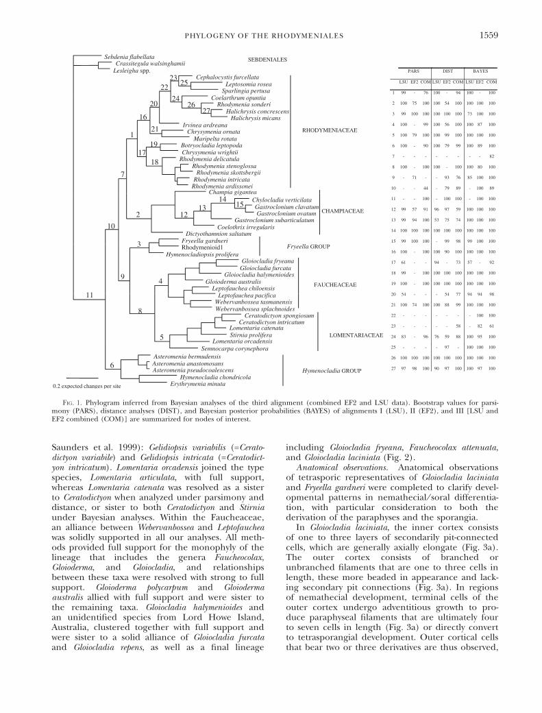
Phylogenetic analyses. Phylogenetic analyses ofalignments 1, 2, and 3 were completed to resolveinfraordinal relationships among the Rhodymeni-ales. The phylogram inferred from Bayesian analysesof alignment 3 is presented ()LnL = 47,747.61)with posterior probabilities, as well as bootstrapresults for distance and parsimony analyses, of align-ments 1, 2, and 3 appended (Fig. 1). All analysesrecovered, with moderate to full support, mono-phyly of the Lomentariaceae (node 5). Monophylyof the Faucheaceae (node 4) and its sister relation-ships with the Lomentariaceae (node 8) were sup-ported by all analyses except parsimony analysis ofalignment 2 (EF2), which failed to resolve the rela-tionships among Fryeella, Hymenocladiopsis, and a newtaxon, labeled Rhodymenioid1, relative to membersof the Faucheaceae. The Champiaceae (node 2) wassupported by all analyses but differs from its tradi-tional composition by inclusion of Coelothrix (node12). In all analyses, Gastroclonium clavatum andGastroclonium ovatum (node 15) allied with Chylocl-adia verticillata (node 14) to the exclusion of Gastroc-lonium subarticulatum (see Table S1 for taxonomicauthors). The Rhodymeniaceae (node 1) was mod-erately to fully supported in LSU and combinedanalyses but differed from its traditional composi-tion by the exclusion of three species of Asteromenia,which solidly joined Hymenocladia and Erythrymenia(Hymenocladia Group, node 6) in almost all analyses(except parsimony of alignment 2). Within the Rho-dymeniaceae, Rhodymenia sonderi allied with the genusHalichrysis (node 26) rather than joining the otherspecies of Rhodymenia. Fryeella, Rhodymenioid1 andHymenocladiopsis formed a separate lineage (FryeellaGroup) supported in all of our analyses (node 3) andvariably allied to the Faucheaceae and Lomentaria-ceae (node 9; not supported under parsimony).Distance and Bayesian analyses of alignments 2 and 3moderately supported positioning of the Hymeno-cladia Group as a sister to all of the other includedRhodymeniales with moderate support (node10).
Alignment 4 was generated to explore relation-ships among additional species of the Faucheaceaeand Lomentariaceae. The consensus Bayesian tree ispresented ()LnL = 8699.06), and posterior proba-bilities, as well as bootstrap results for distance andparsimony analyses, are reported (Fig. 2). Withinthe Lomentariaceae, Semnocarpa was positioned asan early divergence relative to a fully supported line-age containing species of Ceratodictyon, Lomentaria,and Stirnia. The monophyletic genus Ceratodictyonincluded species assigned to Gelidiopsis in sometaxonomic treatments (e.g., Price and Kraft 1991,
1558 LINE LE GALL ET AL.
Saunders et al. 1999): Gelidiopsis variabilis (=Cerato-dictyon variabile) and Gelidiopsis intricata (=Ceratodict-yon intricatum). Lomentaria orcadensis joined the typespecies, Lomentaria articulata, with full support,whereas Lomentaria catenata was resolved as a sisterto Ceratodictyon when analyzed under parsimony anddistance, or sister to both Ceratodictyon and Stirniaunder Bayesian analyses. Within the Faucheaceae,an alliance between Webervanbossea and Leptofaucheawas solidly supported in all our analyses. All meth-ods provided full support for the monophyly of thelineage that includes the genera Faucheocolax,Gloioderma, and Gloiocladia, and relationshipsbetween these taxa were resolved with strong to fullsupport. Gloioderma polycarpum and Gloiodermaaustralis allied with full support and were sister tothe remaining taxa. Gloiocladia halymenioides andan unidentified species from Lord Howe Island,Australia, clustered together with full support andwere sister to a solid alliance of Gloiocladia furcataand Gloiocladia repens, as well as a final lineage
including Gloiocladia fryeana, Faucheocolax attenuata,and Gloiocladia laciniata (Fig. 2).
Anatomical observations. Anatomical observationsof tetrasporic representatives of Gloiocladia laciniataand Fryeella gardneri were completed to clarify devel-opmental patterns in nemathecial ⁄ soral differentia-tion, with particular consideration to both thederivation of the paraphyses and the sporangia.
In Gloiocladia laciniata, the inner cortex consistsof one to three layers of secondarily pit-connectedcells, which are generally axially elongate (Fig. 3a).The outer cortex consists of branched orunbranched filaments that are one to three cells inlength, these more beaded in appearance and lack-ing secondary pit connections (Fig. 3a). In regionsof nemathecial development, terminal cells of theouter cortex undergo adventitious growth to pro-duce paraphyseal filaments that are ultimately fourto seven cells in length (Fig. 3a) or directly convertto tetrasporangial development. Outer cortical cellsthat bear two or three derivatives are thus observed,
Fig. 1. Phylogram inferred from Bayesian analyses of the third alignment (combined EF2 and LSU data). Bootstrap values for parsi-mony (PARS), distance analyses (DIST), and Bayesian posterior probabilities (BAYES) of alignments I (LSU), II (EF2), and III [LSU andEF2 combined (COM)] are summarized for nodes of interest.
PHYLOGENY OF THE RHODYMENIALES 1559

these being some combination of paraphyseal fila-ments and tetrasporangia (Fig. 3, b and c). Whereouter cortical filaments consisting of a single cellconvert to tetrasporangial development, initials(Fig. 3d) and mature sporangia were observeddirectly on inner cortical cells. Unlike Leptofauchea,in which terminal vegetative cells initiate nemathe-cial development and any terminal cell in that sys-tem retains the ability to convert to sporangialdevelopment (Dalen and Saunders 2007), in Gloiocl-adia laciniata terminal vegetative cells appear to haveone of two developmental fates, either converting toparaphyseal filaments or tetrasporangia, but termi-nal cells of the former apparently lack the ability toconvert into tetrasporangia. Mature tetrasporangiaare cruciately or decussately divided, 32–36 · 40–43 mm in dimension (Fig. 3e). An additional obser-vation is that tetrasporangia appear to be confinedto a single surface, even in plants that lack thedecumbent morphology characteristic of some pop-ulations of this species.
Tetrasporangial development in Fryeella gardneri issimilar to that described for Gloiocladia laciniata withthe exception that differentiation of outer corticalcells into tetrasporangial versus paraphyseal initialsoccurs slightly later. The outer cortex in Fryeella isinitially composed of a single (rarely two) incom-plete cell layer (Fig. 4a). Vegetative cells initiallyswell in size in the regions of nemathecial develop-ment with some of these cells experiencing adventi-tious development to produce two- to three-celledfilaments resulting in three- to four-celled paraphy-ses consisting of smaller cells positioned on highlyelongate and inflated basal cells (Fig. 4b). The basalcells (modified outer cortical cells) of paraphysesinitially remain highly inflated (Fig. 4c) but eventu-ally become elongate (Fig. 4d) as other modifiedcortical cells, which did not produce paraphyseal fil-aments, enlarge to produce tetrasporangia (Fig. 4e).Pit plugs were difficult to discern in our rehydratedmaterial; however, our interpretation is supportedby the fact that tetrasporangia were always deeply
Fig. 2. Phylogram inferred from Bayesian analyses of the fourth alignment (LSU data for eight Lomentariaceae and 14 Faucheaceae).Values above and below nodes indicate Bayesian posterior probabilities and bootstrap values for parsimony and distance analyses, respec-tively. Asterisks denote nodes that are fully supported in all our analyses. Taxa in bold represent type species.
1560 LINE LE GALL ET AL.

embedded in nemathecia (Fig. 4e), generallydirectly positioned on inner cortical cells, and werenever in an intercalary position (i.e., the smaller ter-minal cells of the paraphyses were never subtendedby differentiating tetrasporangia). Thus, differentia-tion of outer cortical cells into paraphyseal versustetrasporangial initials appears to occur during orfollowing the swelling of the outer cortical layer asit modifies during nemathecial development.
Character state analyses. To assess putative phylo-genesis of key reproductive structures, three attri-butes were coded as unordered binary charactersfor the taxa included in alignment 3: (i) the num-ber of cells in the carpogonial branch (three vs.four); (ii) the pattern of tetrasporangial division(cruciate vs. tetrahedral); and (iii) the mode of
nemathecial ⁄ soral development (i.e., ‘paraphyses’[when present] and tetrasporangia derived largelyfrom the modification of existing cortical cells vs.adventitious growth leading to the development ofparaphyses and ⁄ or tetrasporangia). Hypotheses onthe phylogenesis of these reproductive structureswere generated under parsimony by mapping char-acter states on the Bayesian tree displayed in Fig-ure 1. ACCTRAN and DELTRAN strategies wereemployed, but the same evolutionary hypotheseswere generated (Fig. 5), which are discussed below.
DISCUSSION
Improving phylogenetic inference for the Rhodymeniales.The first extensive molecular phylogenetic analyses
Fig. 3. Nemathecial development in Gloiocladia laciniata (GWS001447; UNB). (a) Section of thallus at the transition between normalvegetative cortical filaments of uniform lengths and the progressive more elongate filaments of a tetrasporangial nemathecium. An innercortical cell (arrow) bears a single-celled and a branched outer-cortical lateral. Scale bar = 15 lm. (b) Outer cortical cell (arrow) bearinga paraphyseal filament and two tetrasporangial initials (arrowheads). Scale bar = 12 lm. (c) Outer cortical cell (arrow) clearly pit con-nected (arrowheads) to two paraphyseal filaments and bearing a sessile tetrasporangial initial. Scale bar = 12 lm. (d) Inner cortical cell(arrow) directly bearing a tetrasporangial initial (arrowhead). Scale bar = 12 lm. (e) A decussately divided mature tetrasporangium slightlydisplaced among paraphyses from its point of attachment. Scale bar = 12 lm.
PHYLOGENY OF THE RHODYMENIALES 1561

of the Rhodymeniales completed by Saunders et al.(1999) were significant in challenging the existingfamilial system of classification, both by resolving sixmonophyletic groups of taxa and for providing afoundation for the eventual construction of a morenatural system of classification. That study, however,largely failed to resolve relationships among the sixlineages and was based solely on SSU data, a markerdisplaying an unexpectedly variable rate of evolu-tion among rhodymenialean taxa that can result inbranch-attraction artifacts. The need to add moredata (taxa and ⁄ or characters) to improve phyloge-netic resolution is evident. To our knowledge, onlyfour EF2 and 14 LSU sequences had been
characterized for rhodymenialean taxa prior to ourwork, rendering this study the most comprehensiveto consider systematics in the Rhodymeniales usingthese markers. Our results are largely congruentwith those published by Saunders et al. (1999) inthat the four rhodymenialean families were resolvedunder all combined analyses as monophyleticlineages [with the exception of Coelothrix, which isincluded here for the first time in molecularanalyses and which joined the Champiaceae ratherthan the Rhodymeniaceae, and Halichrysis, whichallied to the Rhodymeniaceae rather than theFaucheaceae (see Saunders et al. 2006)]. Moreover,taxa previously considered incertae sedis in the
Fig. 4. Nemathecial development in Fryeella gardneri (GWS004232; UNB). (a) Vegetative section showing cortex at region adjacent tonemathecial development. Inner cortical cells bearing a single (occasionally two) sporadically interrupted surface cortical layer. Scalebar = 30 lm. (b) Swelling of outer cortical cells (arrow) during nemathecial initiation, some dividing into three- or four-celled paraphysisconsisting of smaller cells (double arrows) on highly elongate and inflated basal cells. Scale bar = 15 lm. (c) Basal cells of paraphyses thatremain highly inflated (arrow) during early stages of nemathecial development. Scale bar = 15 lm. (d) Narrowing of basal paraphysis cells(double arrow) during nemathecial development and maturation of tetrasporangia. Scale bar = 15 lm. (e) Cruciate and decussatelydivided mature tetrasporangia borne directly on inner cortical cells and deeply embedded among the paraphyseal filaments. Scalebar = 12 lm.
1562 LINE LE GALL ET AL.

Rhodymeniaceae were consistently resolved as twodistinct lineages in this study.
Hymenocladia Group. In this study, the generaAsteromenia, Erythrymenia, and Hymenocladia are sol-idly allied. In Saunders et al. (1999) the generaErythrymenia and Hymenocladia were weakly resolvedas sister to a lineage containing the Champiaceae,Lomentariaceae, and Faucheaceae, whereas thecurrent analyses variably resolved the Hymenocladialineage as sister to the remaining Rhodymeniales.Evolutionary hypothesis concerning reproductivestructures (Fig. 5) indicates that in the rhodymenia-lean ancestor: (i) the number of cells in the carpo-gonial branch was four; (ii) tetrasporangia werecruciate; and (iii) the tetrasporangia differentiateddirectly from existing cortical cells. The number ofcells in the carpogonial branch is unknown inErythrymenia, but, if our evolutionary speculations
hold true, is likely to be four as reported for bothAsteromenia and Hymenocladia. Hymenocladia differsfrom Asteromenia and Erythrymenia by the presence oftetrahedral tetrasporangia, a character state derivedindependently in at least four lineages of the Rhody-meniales (Fig. 5). In a monograph of the genusAsteromenia, Saunders et al. (2006) reported that tet-rasporangia differentiate directly from intercalarycells of the inner cortex without modification ofcontiguous cells. Observations on Erythrymenia andHymenocladia by Kylin (1931, 1956), as well as draw-ings of the latter by Sparling (1957), similarly sug-gest that tetrasporangia differentiate frompreformed cortical cells rather than from adventi-tious cortical growth. Corresponding observations ofthe early stages in tetrasporangial differentiation arenecessary to confirm the similarity in developmentto Asteromenia. These three genera also share solid
Fig. 5. The phylogenic tree for the Rhodymeniales based on the Bayesian results (Fig. 1), with hypothesis on the evolution of (i) thenumber of cells in the carpogonial branch (three vs. four); (ii) the pattern of tetrasporangial division (cruciate vs. tetrahedral); and (iii)the mode of nemathecial ⁄ soral development [i.e., ‘paraphyses’ (when present) and tetrasporangia derive largely from the modification ofexisting cortical cells vs. adventitious growth contributes to development of paraphyses and ⁄ or tetrasporangia]. aIndicates that reinvestiga-tion of the tetrasporangial development is needed to assess direct versus adventitious origin of the nemathecium. Hypotheses were gener-ated under parsimony by mapping character states (changes are indicated along internal branches). Thallus construction (solid vs.hollow) and occurrence of the tela arachnoidea are also indicated for the taxa included in the tree, but the evolution of these characters isnot predicted on the tree.
PHYLOGENY OF THE RHODYMENIALES 1563
thallus construction and the absence of a tela arach-noidea in the cystocarpic cavity.
The subfamily Hymenocladiae was proposed byKylin (1931) to accommodate the single genusHymenocladia, whereas in 1956, he abandoned thissubfamilial designation in favor of referring it infor-mally to the ‘‘Hymenocladia Gruppe.’’ Sparling(1957) retained the subfamily Hymenocladiae forthis genus and further noted a distinctive anatomi-cal similarity between Erythrymenia and Hymenocladia(both of which produce a mixture of small andlarge cells within the medulla). On the basis of ana-tomical similarities and our molecular data, we for-mally propose removing the genera Asteromenia,Erythrymenia, and Hymenocladia from the Rhodymeni-aceae to a new family, the Hymenocladiaceae.
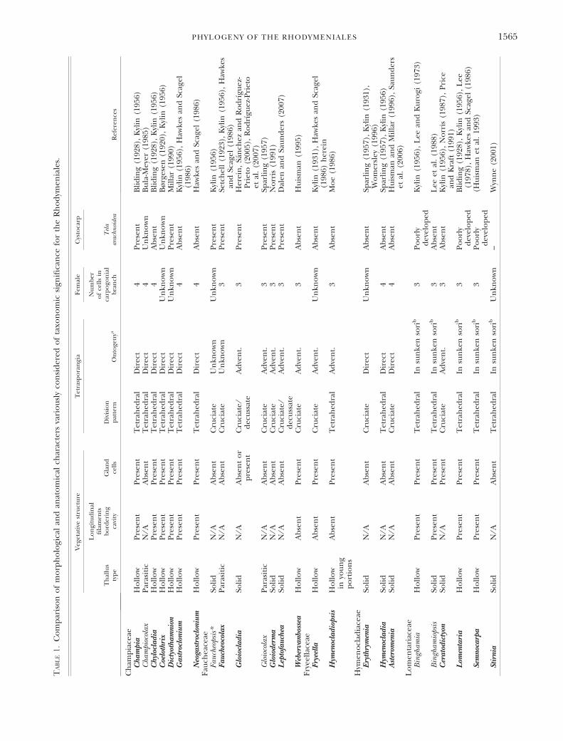
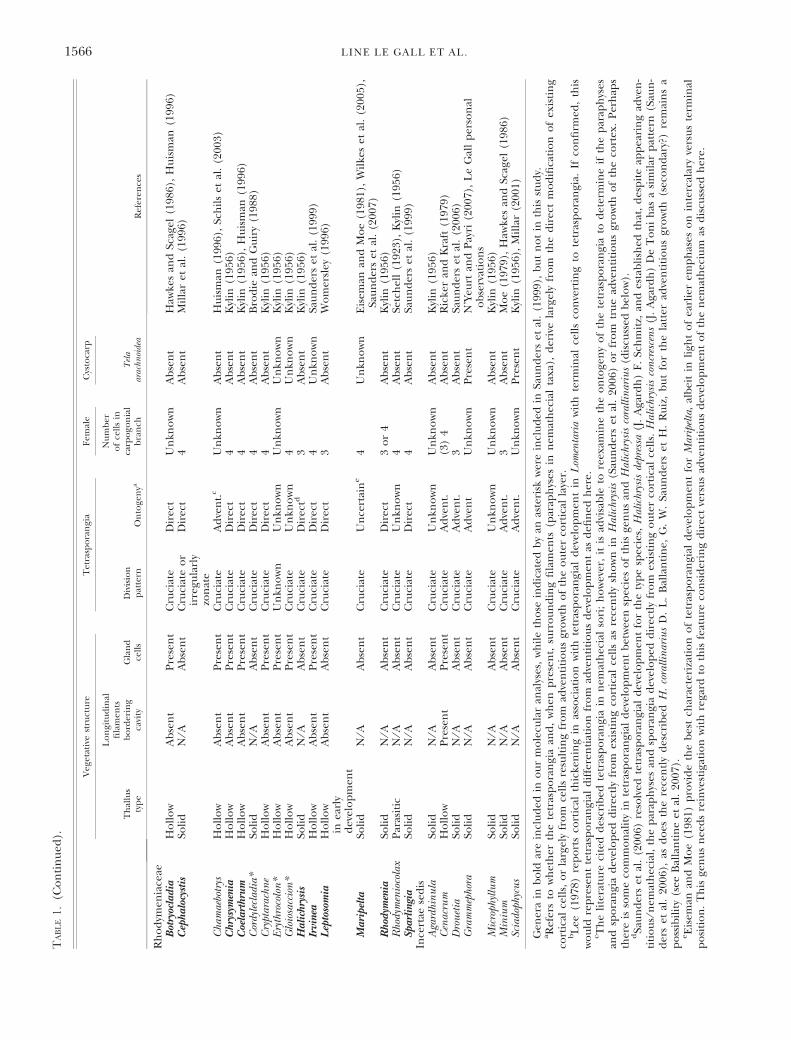
Rhodymeniaceae. In this study, the Rhodymenia-ceae was resolved as a monophyletic lineage corre-sponding to the Rhodymeniaceae sensu stricto asdelimited by Saunders et al. (1999) differing only bythe addition of the genus Halichrysis (see Saunderset al. 2006). The Rhodymeniaceae incorporates taxawith a wide range of vegetative anatomies thatinclude species with either solid or hollow thalli.Tetrasporangia are typically cruciately divided(although in Cephalocystis the basically cruciate divi-sion pattern can become highly irregular), a puta-tively ancestral character state for the order (Fig. 5).Tetrasporangia in the Rhodymeniaceae werereported to be terminal by Kylin (1931) andSparling (1957), but subsequently, the family hascome to be defined largely by taxa with intercalarytetrasporangia (see Saunders et al. 1999). Morerecently (Saunders et al. 2006, Dalen and Saunders2007), however, tetrasporangial development hasbeen receiving more attention, the earlier distinc-tion of terminal versus intercalary positioning beingconsidered too simplistic to distinguish adequatelythe variations in tetrasporangial development in theorder. These latter studies are emphasizing the over-all development of the nemathecia ⁄ sori (paraphysesand tetrasporangia) and their derivation largelyfrom existing versus adventitious cortical cells. Thisdistinction has prompted our reexamination of thepublished literature on rhodymenialean tetrasporan-gial development (Table 1). Tetrasporangia differ-entiate directly from preexisting cortical cells(either terminal and ⁄ or intercalary in position) inmost rhodymeniacean taxa, and preliminary obser-vations suggest that records to the contrary requirecareful reinvestigation. For example, Womersley(1996) depicted adventitious growth of the outercortex to form paraphyseal nemathecia in Rhodyme-nia verrucosa and Rhodymenia sonderi (as Rhodymeniaaustralis). However, observations (Saunders unpub-lished data) on tetrasporangia from isolates geneti-cally matched to cystocarpic plants of Rhodymeniaverrucosa indicate that development is typical to thatof other Rhodymeniaceae and not as reported byWomersley, his descriptions possibly having been
based on specimens conforming in general habit tothe distinctive female gametophytes but actuallyrepresenting superficially similar members of otherrhodymenialean families. Further unifying featuresof genuine species of Rhodymeniaceae are a lackof tela arachnoidea and for the most part four-celledcarpogonial branches (Fig. 5).
Champiaceae. The Champiaceae traditionally hasincluded taxa with hollow thalli lined by longitudi-nal filaments and traversed by single-layered dia-phragms (Lee 1978). More recently, the emphasison vegetative features was removed and the familydefined on the basis of four-celled carpogonialbranches and intercalary, tetrahedrally divided tet-rasporangia (Saunders et al. 1999). In this study,the Champiaceae allied weakly with the Rhodymeni-aceae and shared with them the putatively ancestralstates of four-celled carpogonial branches and dif-ferentiation of tetrasporangia directly from preexist-ing rather than adventitious cortical cells (Table 1;Fig. 5). Tetrasporangial cleavage in the Champia-ceae is tetrahedral, suggesting that this derived stateevolved in the ancestor to this lineage and serves(although not uniquely) to define the family(Fig. 5).
Our molecular data solidly position Coelothrixirregularis within the Champiaceae (Fig. 1). The taxo-nomic affiliations of Coelothrix within the Rhodymen-iales have been an ongoing conundrum. Børgesen(1920) proposed this genus to accommodate aCaribbean taxon identified by Harvey as Cordylecladiairregularis. Børgesen described vegetative organiza-tion from resoaked dried material noting the hollowcentral medulla and inwardly projecting ‘‘gland’’cells, and hypothesized affinities to the somewhatsimilarly hollow and ‘‘gland’’-cell bearing generaChrysymenia and Chylocladia, both of which were sub-sequently considered to be only distantly related byBliding (1928) when he split the Rhodymenialesinto two families (Chrysymenia being placed in theRhodymeniaceae, and Chylocladia in the Champia-ceae). Kylin (1931) discussed the taxonomic affini-ties of Coelothrix relative to Chrysymenia andChylocladia, as well as Lomentaria (then in the Cham-piaceae) and Hymenocladia (Rhodymeniaceae) butconcluded that the systematic position of Coelothrixwas still uncertain. Kylin (1956) included this genuswithin his Chrysymenia group (Rhodymeniaceae),despite Coelothrix having tetrahedral rather than cru-ciate tetrasporangia. Sparling (1957) emphasizedthallus construction as having family-level signifi-cance, arguing that in members of the Rhodymenia-ceae thalli could be either solid or hollow, but inthe event of the latter, lacking longitudinal fila-ments around the cavity border, whereas in theChampiaceae longitudinal filaments always line thecavities. In this regard Coelothrix, with its longitudi-nal filaments, conforms to the Champiaceae asnoted by Taylor (1960) who did not, however, effecta formal transfer. Subsequently, Irvine and Guiry
1564 LINE LE GALL ET AL.
Table
1.
Co
mp
aris
on
of
mo
rph
olo
gica
lan
dan
ato
mic
alch
arac
ters
vari
ou
sly
con
sid
ered
of
taxo
no
mic
sign
ifica
nce
for
the
Rh
od
ymen
iale
s.
Veg
etat
ive
stru
ctu
reT
etra
spo
ran
gia
Fem
ale
Cys
toca
rp
Ref
eren
ces
Th
allu
sty
pe
Lo
ngi
tud
inal
fila
men
tsb
ord
erin
gca
vity
Gla
nd
cell
sD
ivis
ion
pat
tern
On
toge
nya
Nu
mb
ero
fce
lls
inca
rpo
gon
ial
bra
nch
Tel
aar
achn
oide
a
Ch
amp
iace
aeC
ha
mp
iaH
oll
ow
Pre
sen
tP
rese
nt
Tet
rah
edra
lD
irec
t4
Pre
sen
tB
lid
ing
(192
8),
Kyl
in(1
956)
Cha
mpi
ocol
axP
aras
itic
N⁄A
Ab
sen
tT
etra
hed
ral
Dir
ect
4U
nkn
ow
nB
ula
-Mey
er(1
985)
Ch
yloc
lad
iaH
oll
ow
Pre
sen
tP
rese
nt
Tet
rah
edra
lD
irec
t4
Ab
sen
tB
lid
ing
(192
8),
Kyl
in(1
956)
Coe
loth
rix
Ho
llo
wP
rese
nt
Pre
sen
tT
etra
hed
ral
Dir
ect
Un
kno
wn
Un
kno
wn
Bø
rges
en(1
920)
,K
ylin
(195
6)D
icty
oth
am
nion
Ho
llo
wP
rese
nt
Pre
sen
tT
etra
hed
ral
Dir
ect
Un
kno
wn
Pre
sen
tM
illa
r(1
990)
Ga
stro
clon
ium
Ho
llo
wP
rese
nt
Pre
sen
tT
etra
hed
ral
Dir
ect
4A
bse
nt
Kyl
in(1
956)
,H
awke
san
dSc
agel
(198
6)N
eoga
stro
clon
ium
Ho
llo
wP
rese
nt
Pre
sen
tT
etra
hed
ral
Dir
ect
4A
bse
nt
Haw
kes
and
Scag
el(1
986)
Fau
chea
ceae
Fau
cheo
psis
*So
lid
N⁄A
Ab
sen
tC
ruci
ate
Un
kno
wn
Un
kno
wn
Pre
sen
tK
ylin
(195
6)F
au
cheo
cola
xP
aras
itic
N⁄A
Ab
sen
tC
ruci
ate
Un
kno
wn
3P
rese
nt
Setc
hel
l(1
923)
,K
ylin
(195
6),
Haw
kes
and
Scag
el(1
986)
Glo
iocl
ad
iaSo
lid
N⁄A
Ab
sen
to
rp
rese
nt
Cru
ciat
e⁄
dec
uss
ate
Ad
ven
t.3
Pre
sen
tH
erei
n,
San
chez
and
Ro
drı
guez
-P
riet
o(2
005)
,R
od
rıgu
ez-P
riet
oet
al.
(200
7)G
loio
cola
xP
aras
itic
N⁄A
Ab
sen
tC
ruci
ate
Ad
ven
t.3
Pre
sen
tSp
arli
ng
(195
7)G
loio
der
ma
Soli
dN
⁄AA
bse
nt
Cru
ciat
eA
dve
nt.
3P
rese
nt
No
rris
(199
1)L
epto
fau
chea
Soli
dN
⁄AA
bse
nt
Cru
ciat
e⁄
dec
uss
ate
Ad
ven
t.3
Pre
sen
tD
alen
and
Sau
nd
ers
(200
7)
Web
erva
nbos
sea
Ho
llo
wA
bse
nt
Pre
sen
tC
ruci
ate
Ad
ven
t.3
Ab
sen
tH
uis
man
(199
5)F
ryee
llac
eae
Fry
eell
aH
oll
ow
Ab
sen
tP
rese
nt
Cru
ciat
eA
dve
nt.
Un
kno
wn
Ab
sen
tK
ylin
(193
1),
Haw
kes
and
Scag
el(1
986)
her
ein
Hym
enoc
lad
iop
sis
Ho
llo
win
you
ng
po
rtio
ns
Ab
sen
tP
rese
nt
Tet
rah
edra
lA
dve
nt.
3A
bse
nt
Mo
e(1
986)
Hym
eno
clad
iace
aeE
ryth
rym
enia
Soli
dN
⁄AA
bse
nt
Cru
ciat
eD
irec
tU
nkn
ow
nA
bse
nt
Spar
lin
g(1
957)
,K
ylin
(193
1),
Wo
mer
sley
(199
6)H
ymen
ocla
dia
Soli
dN
⁄AA
bse
nt
Tet
rah
edra
lD
irec
t4
Ab
sen
tSp
arli
ng
(195
7),
Kyl
in(1
956)
Ast
erom
enia
Soli
dN
⁄AA
bse
nt
Cru
ciat
eD
irec
t4
Ab
sen
tH
uis
man
and
Mil
lar
(199
6),
Sau
nd
ers
etal
.(2
006)
Lo
men
tari
acea
eB
ingh
amia
Ho
llo
wP
rese
nt
Pre
sen
tT
etra
hed
ral
Insu
nke
nso
rib
3P
oo
rly
dev
elo
ped
Kyl
in(1
956)
,L
eean
dK
uro
gi(1
973)
Bin
gham
iops
isSo
lid
Pre
sen
tP
rese
nt
Tet
rah
edra
lIn
sun
ken
sori
b3
Ab
sen
tL
eeet
al.
(198
8)C
era
tod
icty
onSo
lid
N⁄A
Pre
sen
tC
ruci
ate
Ad
ven
t.3
Ab
sen
tK
ylin
(195
6),
No
rris
(198
7),
Pri
cean
dK
raft
(199
1)L
omen
tari
aH
oll
ow
Pre
sen
tP
rese
nt
Tet
rah
edra
lIn
sun
ken
sori
b3
Po
orl
yd
evel
op
edB
lid
ing
(192
8),
Kyl
in(1
956)
,L
ee(1
978)
,H
awke
san
dSc
agel
(198
6)Se
mno
carp
aH
oll
ow
Pre
sen
tP
rese
nt
Tet
rah
edra
lIn
sun
ken
sori
b3
Po
orl
yd
evel
op
ed(H
uis
man
etal
.19
93)
Stir
nia
Soli
dN
⁄AA
bse
nt
Tet
rah
edra
lIn
sun
ken
sori
bU
nkn
ow
n–
Wyn
ne
(200
1)
PHYLOGENY OF THE RHODYMENIALES 1565
Table
1.
(Co
nti
nu
ed).
Veg
etat
ive
stru
ctu
reT
etra
spo
ran
gia
Fem
ale
Cys
toca
rp
Ref
eren
ces
Th
allu
sty
pe
Lo
ngi
tud
inal
fila
men
tsb
ord
erin
gca
vity
Gla
nd
cell
sD
ivis
ion
pat
tern
On
toge
nya
Nu
mb
ero
fce
lls
inca
rpo
gon
ial
bra
nch
Tel
aar
achn
oide
a
Rh
od
ymen
iace
aeB
otry
ocla
dia
Ho
llo
wA
bse
nt
Pre
sen
tC
ruci
ate
Dir
ect
Un
kno
wn
Ab
sen
tH
awke
san
dSc
agel
(198
6),
Hu
ism
an(1
996)
Cep
ha
locy
stis
Soli
dN
⁄AA
bse
nt
Cru
ciat
eo
rir
regu
larl
yzo
nat
e
Dir
ect
4A
bse
nt
Mil
lar
etal
.(1
996)
Cha
mae
botr
ysH
oll
ow
Ab
sen
tP
rese
nt
Cru
ciat
eA
dve
nt.
cU
nkn
ow
nA
bse
nt
Hu
ism
an(1
996)
,Sc
hil
set
al.
(200
3)C
hry
sym
enia
Ho
llo
wA
bse
nt
Pre
sen
tC
ruci
ate
Dir
ect
4A
bse
nt
Kyl
in(1
956)
Coe
lart
hru
mH
oll
ow
Ab
sen
tP
rese
nt
Cru
ciat
eD
irec
t4
Ab
sen
tK
ylin
(195
6),
Hu
ism
an(1
996)
Cor
dyle
clad
ia*
Soli
dN
⁄AA
bse
nt
Cru
ciat
eD
irec
t4
Ab
sen
tB
rod
iean
dG
uir
y(1
988)
Cry
ptar
achn
eH
oll
ow
Ab
sen
tP
rese
nt
Cru
ciat
eD
irec
t4
Ab
sen
tK
ylin
(195
6)E
ryth
roco
lon
*H
oll
ow
Ab
sen
tP
rese
nt
Un
kno
wn
Un
kno
wn
Un
kno
wn
Un
kno
wn
Kyl
in(1
956)
Glo
iosa
ccio
n*
Ho
llo
wA
bse
nt
Pre
sen
tC
ruci
ate
Un
kno
wn
4U
nkn
ow
nK
ylin
(195
6)H
ali
chry
sis
Soli
dN
⁄AA
bse
nt
Cru
ciat
eD
irec
td3
Ab
sen
tK
ylin
(195
6)Ir
vine
aH
oll
ow
Ab
sen
tP
rese
nt
Cru
ciat
eD
irec
t4
Un
kno
wn
Sau
nd
ers
etal
.(1
999)
Lep
toso
mia
Ho
llo
win
earl
yd
evel
op
men
t
Ab
sen
tA
bse
nt
Cru
ciat
eD
irec
t3
Ab
sen
tW
om
ersl
ey(1
996)
Ma
rip
elta
Soli
dN
⁄AA
bse
nt
Cru
ciat
eU
nce
rtai
ne
4U
nkn
ow
nE
isem
anan
dM
oe
(198
1),
Wil
kes
etal
.(2
005)
,Sa
un
der
set
al.
(200
7)R
hod
ymen
iaSo
lid
N⁄A
Ab
sen
tC
ruci
ate
Dir
ect
3o
r4
Ab
sen
tK
ylin
(195
6)R
hody
men
ioco
lax
Par
asit
icN
⁄AA
bse
nt
Cru
ciat
eU
nkn
ow
n4
Ab
sen
tSe
tch
ell
(192
3),
Kyl
in(1
956)
Spa
rlin
gia
Soli
dN
⁄AA
bse
nt
Cru
ciat
eD
irec
t4
Ab
sen
tSa
un
der
set
al.
(199
9)In
cert
aese
dis
Aga
rdhi
nu
laSo
lid
N⁄A
Ab
sen
tC
ruci
ate
Un
kno
wn
Un
kno
wn
Ab
sen
tK
ylin
(195
6)C
enac
rum
Ho
llo
wP
rese
nt
Pre
sen
tC
ruci
ate
Ad
ven
t.(3
)4
Ab
sen
tR
icke
ran
dK
raft
(197
9)D
rou
etia
Soli
dN
⁄AA
bse
nt
Cru
ciat
eA
dve
nt.
3A
bse
nt
Sau
nd
ers
etal
.(2
006)
Gra
mm
epho
raSo
lid
N⁄A
Ab
sen
tC
ruci
ate
Ad
ven
tU
nkn
ow
nP
rese
nt
N’Y
eurt
and
Pay
ri(2
007)
,L
eG
all
per
son
alo
bse
rvat
ion
sM
icro
phyl
lum
Soli
dN
⁄AA
bse
nt
Cru
ciat
eU
nkn
ow
nU
nkn
ow
nA
bse
nt
Kyl
in(1
956)
Min
ium
Soli
dN
⁄AA
bse
nt
Cru
ciat
eA
dve
nt.
3A
bse
nt
Mo
e(1
979)
,H
awke
san
dSc
agel
(198
6)Sc
iado
phyc
us
Soli
dN
⁄AA
bse
nt
Cru
ciat
eA
dve
nt.
Un
kno
wn
Pre
sen
tK
ylin
(195
6),
Mil
lar
(200
1)
Gen
era
inb
old
are
incl
ud
edin
ou
rm
ole
cula
ran
alys
es,
wh
ile
tho
sein
dic
ated
by
anas
teri
skw
ere
incl
ud
edin
Sau
nd
ers
etal
.(1
999)
,b
ut
no
tin
this
stu
dy.
aR
efer
sto
wh
eth
erth
ete
tras
po
ran
gia
and
,w
hen
pre
sen
t,su
rro
un
din
gfi
lam
ents
(par
aph
yses
inn
emat
hec
ial
taxa
),d
eriv
ela
rgel
yfr
om
the
dir
ect
mo
difi
cati
on
of
exis
tin
gco
rtic
alce
lls,
or
larg
ely
fro
mce
lls
resu
ltin
gfr
om
adve
nti
tio
us
gro
wth
of
the
ou
ter
cort
ical
laye
r.bL
ee(1
978)
rep
ort
sco
rtic
alth
icke
nin
gin
asso
ciat
ion
wit
hte
tras
po
ran
gial
dev
elo
pm
ent
inL
omen
tari
aw
ith
term
inal
cell
sco
nve
rtin
gto
tetr
asp
ora
ngi
a.If
con
firm
ed,
this
wo
uld
rep
rese
nt
tetr
asp
ora
ngi
ald
iffe
ren
tiat
ion
fro
mad
ven
titi
ou
sd
evel
op
men
tas
defi
ned
her
e.c T
he
lite
ratu
reci
ted
des
crib
edte
tras
po
ran
gia
inn
emat
hec
ial
sori
;h
ow
ever
,it
isad
visa
ble
tore
exam
ine
the
on
toge
ny
of
the
tetr
asp
ora
ngi
ato
det
erm
ine
ifth
ep
arap
hys
esan
dsp
ora
ngi
ad
evel
op
edd
irec
tly
fro
mex
isti
ng
cort
ical
cell
sas
rece
ntl
ysh
ow
nin
Hal
ichr
ysis
(Sau
nd
ers
etal
.20
06)
or
fro
mtr
ue
adve
nti
tio
us
gro
wth
of
the
cort
ex.
Per
hap
sth
ere
isso
me
com
mo
nal
ity
inte
tras
po
ran
gial
dev
elo
pm
ent
bet
wee
nsp
ecie
so
fth
isge
nu
san
dH
alic
hrys
isco
rall
inar
ius
(dis
cuss
edb
elo
w).
dSa
un
der
set
al.
(200
6)re
solv
edte
tras
po
ran
gial
dev
elo
pm
ent
for
the
typ
esp
ecie
s,H
alic
hrys
isde
pres
sa(J
.A
gard
h)
F.
Sch
mit
z,an
des
tab
lish
edth
at,
des
pit
eap
pea
rin
gad
ven
-ti
tio
us
⁄nem
ath
ecia
l,th
ep
arap
hys
esan
dsp
ora
ngi
ad
evel
op
edd
irec
tly
fro
mex
isti
ng
ou
ter
cort
ical
cell
s.H
alic
hrys
isco
ncr
esce
ns
(J.
Aga
rdh
)D
eT
on
ih
asa
sim
ilar
pat
tern
(Sau
n-
der
set
al.
2006
),as
do
esth
ere
cen
tly
des
crib
edH
.co
rall
inar
ius
D.
L.
Bal
lan
tin
e,G
.W
.Sa
un
der
set
H.
Ru
iz,
bu
tfo
rth
ela
tter
adve
nti
tio
us
gro
wth
(sec
on
dar
y?)
rem
ain
sa
po
ssib
ilit
y(s
eeB
alla
nti
ne
etal
.20
07).
eE
isem
anan
dM
oe
(198
1)p
rovi
de
the
bes
tch
arac
teri
zati
on
of
tetr
asp
ora
ngi
ald
evel
op
men
tfo
rM
arip
elta
,al
bei
tin
ligh
to
fea
rlie
rem
ph
ases
on
inte
rcal
ary
vers
us
term
inal
po
siti
on
.T
his
gen
us
nee
ds
rein
vest
igat
ion
wit
hre
gard
toth
isfe
atu
reco
nsi
der
ing
dir
ect
vers
us
adve
nti
tio
us
dev
elo
pm
ent
of
the
nem
ath
eciu
mas
dis
cuss
edh
ere.
1566 LINE LE GALL ET AL.

(1980) and Guiry and Irvine (1981) considered theposition of secretory cells in genera with hollowthalli as a means by which the Champiaceae (secre-tory cells borne on longitudinal filaments lining thehollow cavity) can be reliably separated from theRhodymeniaceae (secretory cells borne on largemedullary cells lining the hollow cavities). Theseauthors pointed out that Coelothrix possesses secre-tory cells on peripheral filaments, but they empha-sized the irregular pattern of these filaments asjustification for retaining this genus in the Rhody-meniaceae. Molecular results here support the long-held, but often-rejected positioning of Coelothrix inthe Champiaceae and we now formally propose itstransfer.
Our molecular data consistently recovered twoseparate lines for species presently included in thegenus Gastroclonium: Gastroclonium clavatum andGastroclonium ovatum as a monophyletic lineageallied to Chylocladia; and Gastroclonium subarticulatumas a sister to the previous lineage. The generaGastroclonium and Chylocladia are similar in structureand reproduction, the only distinguishing featurebeing the occurrence of hollow parts in the stalks ofChylocladia, whereas those of Gastroclonium are solid.In contrast to Chylocladia and the other genuine spe-cies of Gastroclonium, G. subarticulatum exhibits ostio-late cystocarps (Hawkes and Scagel 1986). Kylin(1956) considered the occurrence of an ostiole as akey feature distinguishing Champia from the threeremaining genera included in the Champiaceae atthat time, Gastroclonium, Chylocladia, and Coeloseira.The last-mentioned genus was proposed by Hollen-berg (1940) to accommodate a new species fromSouthern California characterized by polysporangiarather than tetrasporangia. Chang and Xia (1978),however, argued that the occasional occurrence ofpolysporangia in several species of Gastroclonium,including the type species G. ovatum, as well as theobservation that polysporangia were derived fromtetrasporangia, fatally compromised this feature as ageneric distinction with the result that they reducedCoeloseira to synonymy with Gastroclonium. Gastrocloni-um subarticulatum differs from the genera Gastrocloni-um (sensu stricto including Coeloseira) andChylocladia in both molecular and anatomical fea-tures, and we therefore propose the new genusNeogastroclonium to accommodate this species.Gastroclonium iyengarii also has ostiolate cystocarps(Balakrishnan 1975) and should be reassessed inlight of our results.
Fryeella Group. Our molecular results positionedFryeella as a close ally to a new taxon (Rhodymeni-oid1) from Tasmania, Australia, and Hymenocladiop-sis resolving this lineage as sister to the relatedFaucheaceae and Lomentariaceae. All of these lin-eages share two putatively derived character states:three-celled carpogonial branches and adventitiouscortical growth as part of tetrasporangial differenti-ation (Fig. 5). Fryeella has cruciate tetrasporangia,
whereas cleavage is tetrahedral in Hymenocladiopsis.Characterization of the new entity is yet to becompleted and is part of a larger independentstudy investigating cryptic rhodymenialean taxafrom Australia (Saunders and McDonald, unpub-lished data). On the basis of our molecular resultsand unifying anatomical features (Table 1), wepropose the new family Fryeellaceae to accommo-date these taxa.
Lomentariaceae. Traditionally, the Lomentariaceaehas included taxa with hollow thalli with chambersseparated by multilayered diaphragms (Lee 1978).Saunders et al. (1999) challenged this distinction,noting that some of the genera included in the fam-ily at that time lacked this feature, as did a numberof other predominantly solid taxa that they weretransferring into it from the Rhodymeniaceae onthe basis of their molecular results. Saunders et al.(1999) argued that the family was best defined onthe basis of three-celled carpogonial branches, theoccurrence of terminal and generally tetrahedraltetrasporangia, and in most cases, a characteristiclomentariaceous fusion cell in carposporophytedevelopment in which the outlines of the compo-nent cells disappeared or were obscured. The recentinclusion of the solid genus Stirnia (Wynne 2001) inthe Lomentariaceae reinforced this change in char-acter emphasis, one that is here confirmed by ourmolecular results (Fig. 1).
The initially monospecific genus Ceratodictyongrows symbiotically with a sponge and was character-ized by its unusual habit. Nevertheless, Norris(1987) noted that the only tangible distinctionbetween Ceratodictyon and Gelidiopsis was the reticula-tion of thalli in the former as a result of the bond-ing of closely adjacent terete axes by the associatedsponge tissue, a feature that Price et al. (1984) haddemonstrated in culture not to be obligate. Norris(1987), therefore, formally subsumed Gelidiopsiswithin Ceratodictyon, a judgment not accepted byPrice and Kraft (1991) who argued that Gelidiopsisand Ceratodictyon should be maintained as distincttaxa despite their vegetative and reproductive simi-larities, emphasizing the consistency of the unusualhabit of the latter under natural conditions. Ourmolecular data (Fig. 2), however, corroborate Nor-ris’s hypothesis, and we therefore support his mer-ger of these genera and the combinationsCeratodictyon intricatum and Ceratodictyon variabile thathe effected.
Our molecular results (Fig. 2) resolved the genusLomentaria as two distinct lineages: Lomentariacatenata as sister to Ceratodictyon and Stirnia; and alineage for Lomentaria articulata, type of the genus,and L. orcadensis. Guiry (in Irvine and Guiry 1983)observed that Lomentaria articulata has multilayeredplugs constricting the medullary cavity at regularintervals as well as terete thalli, whereas other spe-cies, such as Lomentaria clavellosa and Lomentariaorcadensis, have plugs occurring only at the bases of
PHYLOGENY OF THE RHODYMENIALES 1567

the branches, which give the fronds an unconstrict-ed appearance, as well as compressed or flattenedthalli. He suggested moving these two latter taxa toa separate genus, Chondrothamnion Kutzing(1843:438). Further anatomical and phylogeneticstudies are needed to clarify taxonomic relation-ships among species of Lomentaria, but our molecu-lar results strongly support an alliance betweenL. articulata and L. orcadensis, in contrast to Guiry’sproposal, and indicate that L. catenata should beconsidered in any revisionist attempts to definemonophyletic groups among the many species cur-rently included in this genus.
Faucheaceae. The Faucheaceae is a well-circumscribedfamily characterized by three-celled carpogonialbranches and cruciately divided tetrasporangia. In arecent monograph of the genus Leptofauchea, Dalenand Saunders (2007) provided a detailed descrip-tion of the adventitious growth of the cortex toform nemathecial filaments (paraphyses) andtetrasporangia, and here we provide similar observa-tions from Gloiocladia laciniata. This mode of tetrasp-orangial development is highly characteristic of thisfamily and its allies. Within the family, two lineagesare strongly supported in our molecular trees: thefirst consisting of Webervanbossea and Leptofauchea;and the second including the genera Faucheocolax,Gloioderma, and Gloiocladia. For the latter assem-blage, only the genus Gloioderma as restricted byDalen and Saunders (2007) is presently mono-phyletic. Diagnostic features for the genera Fauchea,Gloiocladia, and Gloioderma are poorly defined (Irvineand Guiry 1980, Sanchez and Rodrıguez-Prieto2005), and recently Rodrıguez-Prieto et al. (2007)have formally merged the genera Fauchea andGloiocladia based on thorough observation of thetwo generitypes [Fauchea repens (=Gloiocladia repens)and Gloiocladia furcata] as well as molecular data(SSU sequences). Our data fully support a closealliance between Gloiocladia repens and Gloiocladiafurcata, although we consider that the transfer of allFauchea species to the genus Gloiocladia was prema-ture pending further anatomical and phylogeneticstudies that are needed to clarify taxonomic rela-tionships among species of this lineage. As an exam-ple, the cluster of Pacific taxa included in our trees(Gloiocladia laciniata, Gloiocladia fryeana, andFaucheocolax attenuata) form a monophyletic sisterlineage to the European representatives and differin some details of vegetative anatomy (Hawkes andScagel 1986), indicating that a distinct genus maybe warranted. An unfortunate consequence of thecurrent adherence to the ICBN principle of prioritywould be the generic name Faucheocolax beingassigned to what would be a taxon of largely non-parasitic species. For now, we can confidently statethat our results strongly support four species groupsin this part of the tree: (i) the Pacific cluster ofGloiocladia fryeana, Gloiocladia laciniata, and Faucheo-colax attenuata; (ii) Gloiocladia repens and Gloiocladia
furcata; (iii) Gloioderma sp. and Gloiocladia halymenio-ides; and (iv) Gloioderma australis and Gloiodermapolycarpum.
CONCLUSIONS
Throughout the history of rhodymenialean sys-tematics, several vegetative (thallus construction,occurrence of longitudinal medullary filaments,positioning of secretory cells) and reproductive fea-tures (tetrasporangial position and cleavage pattern,number of cells in the carpogonial branches, occur-rence or lack of tela arachnoidea) have been used toclassify members of this order. The Faucheaceae,Fryeellaceae, Lomentariaceae, and Rhodymeniaceaeare now known to encompass taxa with either solidor hollow thalli, making this character taxonomi-cally unreliable for infraordinal classification of theorder. Nevertheless, it is noteworthy that hollowmembers of the Champiaceae and Lomentariaceaedo consistently have cavities bordered by longitudi-nal filaments, whereas hollow members of the Frye-ellaceae and Rhodymeniaceae lack this feature.Based on our evolutionary hypotheses (Fig. 5)regarding tetrasporangial cleavage, transition fromcruciate to the derived tetrahedral state hasoccurred independently four times, whereas rever-sion to the ancestral character state has happenedonce. This character is thus relatively variable andonly unequivocally characteristic at the family levelfor the Champiaceae. If our hypotheses on the evo-lutionary patterns reflected by the number of cellsin the carpogonial branches hold true, then this is auseful feature for designating families. Derivation ofthe three-celled from the ancestral four-celled car-pogonial branch state has happened at least twice inthe Rhodymeniaceae, but, more interestingly, itappears to be the state in the common ancestorleading to the Fryeellaceae, Faucheaceae, andLomentariaceae. In this study, we placed emphasison whether tetrasporangial nemathecia (tetraspo-rangia and paraphyses) differentiate directly frompreexisting cortical cells or from adventitious devel-opment of the outer cortical layer as documentedhere for Gloiocladia laciniata and Fryeella gardneri [seealso Saunders et al. (2006) and Dalen and Saunders(2007)]. If our indications are correct (Fig. 5), thenthis feature also evolved only once and in the com-mon ancestor leading to the Fryeellaceae, Fauchea-ceae, and Lomentariaceae. To further test thishypothesis, taxa in the Rhodymeniaceae reported tohave adventitious nemathecial development [e.g.,Rhodymenia spp. (Womersley 1996) and Chamaebotrysspp. (Huisman 1996, Schils et al. 2003)] will needto be studied in greater detail.
Still to be addressed by molecular analyses arethe genera Agardhinula, Binghamia, Binghamiopsis,Cenacrum, Cryptarachne, Drouetia, Grammephora, Micro-phyllum, Minium, and Sciadophycus. Based on theindications of reproductive features, it is likely that
1568 LINE LE GALL ET AL.
Binghamia (Lee and Kurogi 1973) and Binghamiop-sis (Lee et al. 1988) will join the Lomentariaceae.The genus Cryptarachne was proposed by Kylin(1931) for compressed or flat species of Chrysyme-nia with ‘‘internal rhizoids’’ in the medullary cavi-ties. Most authors [see Abbott and Littler (1969)for references] reject generic status for Cryptarachnebecause some species of Chrysymenia (including thetype, Chrysymenia ventricosa) also have internal rhi-zoids. Nevertheless, Ben Maız et al. (1987)regarded Cryptarachne as a valid genus despiteunclear diagnostic characteristics and discussedputative inclusion of Chrysymenia wrightii in thegenus. In our molecular analyses, the genus Chrysy-menia was resolved as two separate lineages: C.wrightii allies with Botryocladia leptopoda, whereasChrysymenia ornata joins Maripelta rotata. Delineationof the genera Chrysymenia and Cryptarachne are inneed of further clarification, but regardless ofwhether Cryptarachne ultimately warrants genericrecognition, it has anatomical features (Table 1)characteristic of the Rhodymeniaceae. The generaCenacrum (Ricker and Kraft 1979), Drouetia (Saun-ders et al. 2006), Grammephora (N’Yeurt and Payri2007), Minium (Moe 1979), and Sciadophycus (Kylin1956) all display cruciate tetrasporangia in nemath-ecia reportedly arising from adventitious growth ofcortical cells to form paraphyses. On the basis ofthese anatomical indications, we predict that thesegenera will join either the Fryeellaceae or the Fau-cheaceae. In the absence of new anatomicaland ⁄ or molecular data, we advocate that the mono-typic genera Agardhinula and Microphyllum, beregarded as incertae sedis until their taxonomicaffinities are clarified.
TAXONOMIC TREATMENT
Hymenocladiaceae L. Le Gall, Dalen et G. W.Saunders, fam. nov.
Diagnosis: Thalli pseudoparenchymati, pleni,aliquot generis partibus parvas cellas secundariasdetinentibus, quae interponuntur inter majoresmedullatas cellulas. Cum carpogoniales rami notisunt, quattuor cellis componuntur. Quae nascuntura tetrasporangialibus cellis, mutando pristinascorticis cellas, minime tamen mutatis corticis celliscontiguis, atque divisis sporangialibus tetraedricis velcruciatis. Cum efficiuntur analyseis de compositiselementis ex ordinibus ADN inter se sequentibus inregionibus E2F LSUque, docent monophyleticamcomplexionem esse, conjunctam cum precocisecessione a ceteris Rhodymenialibus generibus.
Solid, pseudoparenchymatous thalli, some mem-bers with secondary cells intercalary among largermedullary cells. Where known, carpogonial branchesfour-celled; tetrasporangial initials arising from trans-formation of existing cells of the cortex with littlemodification of contiguous cortical cells, tetrahedralor cruciate sporangial cleavage. Phylogenetic analyses
of combined EF2 and LSU data indicate a monophy-letic assemblage with early divergence relative toother families of Rhodymeniales.
Type genus: Hymenocladia J. Agardh 1852:772. SeeTable 1 for included genera.
Fryeellaceae L. Le Gall, Dalen et G. W. Saunders,fam. nov.
Diagnosis: Vegetalis constructio varia pseudopa-renchymatus, cortex gracilis, plerumque composituscellis semel vel bis stratis. Ubi carpogoniales raminoti sunt, tribus cellis componuntur. Tetrasporangi-alis nemathecia, iis composita corticialibus cellisquae in latitudinem creverunt quaeque, in fine pos-tremo, tetrasporangia genuerunt vel paraphyseiscompositas proceris basales cellis, brevia fila ferenti-bus (una vel duas cellas) adventiciarum cellarum.Cruciata vel tetraedrica tetrasporangia.
Vegetative construction variable, pseudoparenchy-matous, cortex thin, generally one or two cell layers.Where known, carpogonial branches three-celled.Tetrasporangial nemathecia originating throughenlargement of outer cortical cells that ultimatelydevelop as tetrasporangia, or as paraphyses withelongate basal cells subtending short filaments(one- to two-celled) of adventitious cells; tetra-sporangia cruciate or tetrahedral.
Type genus: Fryeella Kylin 1931:15. See Table 1 forincluded genera.
Neogastroclonium L. Le Gall, Dalen et G.W. Saun-ders, gen. nov.
Diagnosis: Generis Champiacei algae, erectis axi-bus compositae, plenis (id est sine vacuitate) cylin-dratisque, ad apices ferentibus cavos saeptatosqueramos. Cum cognitum est cystocarpium, ostioloinstrui videtur.
Champiacean algae with cylindrical solid erectaxes bearing hollow, septate, branches towards api-ces. Cystocarps ostiolate where known.
Type species: Neogastroclonium subarticulatum (Tur-ner) L. Le Gall, Dalen et G. W. Saunders, comb.nov.
Neogastroclonium subarticulatum (Turner) L. LeGall. Dalen et G. W. Saunders, comb. nov.
Basionym: Fucus ovalis var. subarticulatus Turner1809 (Fuci sive plantarum fucorum generi a botani-cis ascriptarum icones descriptiones et historia. Fuci,or colored figures and descriptions of the plantsreferred by botanists to the genus Fucus. Vol. 2, Lon-don, p. 23).
Lectotype: BM [see Hawkes (1986) for more infor-mation].
Synonyms: Gastroclonium subarticulatum (Turner)Kutzing 1843:441. Lomentaria ovalis var. subarticulata(Turner) Harvey 1853:73. Lomentaria ovalis var. coul-teri Harvey 1853:78. Lomentaria ovalis var. robustior J.Agardh 1876:634. Chylocladia ovalis var. coulteri(Harvey) Farlow 1876:695. Chylocladia ovata var. sub-articulata (Turner) Batters 1902:74. Chylocladia ovalisvar. subarticulata (Turner) Kylin 1925:42. Gastrocloni-um coulteri (Harvey) Kylin 1931:30.
PHYLOGENY OF THE RHODYMENIALES 1569
We are grateful to many colleagues listed in Table S1 for pro-viding key samples that made this study possible. We alsothank Marguerite Czarnecki and Jean Lavoux for kindly pro-viding the Latin translation of the diagnoses. We would liketo thank John M. Huisman, Gerry T. Kraft, and an anony-mous reviewer for fruitful comments on the manuscript. Thisresearch was funded by the Natural Sciences and EngineeringResearch Council of Canada, Canada Research Chair Pro-gram, Canada Foundation for Innovation, and New Bruns-wick Innovation Fund.
Abbott, I. A. & Littler, M. M. 1969. Some Rhodymeniales fromHawaii. Phycologia 8:165–9.
Agardh, J. G. 1852. Species Genera et Ordines Algarum, seu DescriptionesSuccinctae Specierum, Generum et Ordinum, Quibus Algarum Reg-num Constituitur. Volumen Secundum: Algas Florideas Complectens.C.W.K. Gleerup, Lund, Sweden, 720 pp.
Agardh, J. G. 1876. Species Genera et Ordines Algarum, seu DescriptionesSuccinctae Specierum, Generum et Ordinum, Quibus Algarum Reg-num Constituitur. Volumen Tertium: De Florideis Curae Posteriores.Part 1. C.W.K. Gleerup, Leipzig, Germany, 724 pp.
Balakrishnan, M. S. 1975. On Gastroclonium iyengarii Srinivasan.Curr. Sci. 44:24.
Ballantine, D. L., Saunders, G. W. & Ruiz, H. 2007. Halichrysis cor-allinarius sp. nov. (Rhodymeniaceae, Rhodophyta) fromPuerto Rico, Caribbean Sea. Phycol. Res. 55:240–8.
Batters, E. A. L. 1902. A catalogue of the British marine algae. J. Bot.Br. Foreign 40:1–107.
Ben Maız, N., Boudouresque, C.-F., Riouall, R. & Lauret, M. 1987.The algal flora of the Etang de Thau (Mediterranean coast ofFrance): on the occurrence of a Japanese member of theRhodymeniales, Chrysymenia wrightii (Rhodophyta). Bot. Mar.30:357–64.
Bliding, C. 1928. Studien uber die Florideenordnung Rhodymeni-ales. Universitets Arsskrift Ny Foljd Lunds 24:1–74.
Børgesen, F. 1920. The marine algae of the Danish West Indies.Part 3. Rhodophyceae. Dan. Bot. Arkiv 3:369–498.
Brodie, J. & Guiry, M. D. 1988. Life-history and reproduction ofCordylecladia erecta (Rhodymeniaceae, Rhodophyta). Br. Phycol.J. 23:347–64.
Bula-Meyer, G. 1985. Champiocolax sarae gen. et sp. nov., an adel-phohemiparasite of the Champiaceae (Rhodymeniales, Rho-dophyta). Phycologia 24:429–35.
Chang, C. F. & Xia, B. M. 1978. A new species of Gastroclonium fromthe Xisha Islands, Guangdong Province, China. Oceanol. Lim-nol. Sin. 9:209–14.
Dalen, J. L. & Saunders, G. W. 2007. A review of the red algal genusLeptofauchea (Faucheaceae, Rhodymeniales) including adescription of L. chiloensis sp. nov. Phycologia 46:198–213.
Eiseman, N. J. & Moe, R. L. 1981. Maripelta atlantica sp. nov.(Rhodophyta, Rhodymeniales) a new deep-water alga fromFlorida. J. Phycol. 17:299–308.
Farlow, W. G. 1876. List of the marine algae of the United States.Rep. U. S. Fish Comm. 1873 ⁄ 1875:691–718.
Felsenstein, J. 1985. Confidence limits on phylogenies: an approachusing the bootstrap. Evolution 39:783–91.
Graybeal, A. 1998. Is it better to add taxa or characters to a difficultphylogenetic problem? Syst. Biol. 47:9–17.
Guiry, M. D. & Irvine, D. E. G. 1981. A critical reassessment ofinfraordinal classification in the Rhodymeniales. Proc. Int.Seaweed Symp. 8:106–11.
Harper, J. T. & Saunders, G. W. 2001. The application of sequencesof the ribosomal cistron to the systematics and classification ofthe florideophyte red algae (Florideophyceae, Rhodophyta).Cah. Biol. Mar. 42:25–38.
Harvey, W. H. 1853. Nereis boreali-americana…Part II. Rhodosper-meae. Smithson. Contrib. Knowledge 5:1–243.
Hawkes, M. W. 1986. Lectotypification of species names of Rhody-meniales (Rhodophyta) for the red algal flora of BritishColumbia and northern Washington. Taxon 35:329–33.
Hawkes, M. W. & Scagel, R. F. 1986. The marine algae of BritishColumbia and northern Washington: division Rhodophyta
(red algae), class Rhodophyceae, order Rhodymeniales. Can. J.Bot. 64:1549–80.
Hollenberg, G. J. 1940. New marine algae from southern Califor-nia. I. Am. J. Bot. 27:868–77.
Huelsenbeck, J. P. 2002. Testing a covariotide model of DNA sub-stitution. Mol. Biol. Evol. 19:698–707.
Huelsenbeck, J. P. & Ronquist, F. 2001. MrBayes: Bayesian infer-ence of phylogenetic trees. Bioinformatics 17:754–5.
Huisman, J. M. 1995. The morphology and taxonomy of Weber-vanbossea De Toni f. (Rhodymeniales, Rhodophyta). Cryptogam.Bot. 5:367–74.
Huisman, J. M. 1996. The red algal genus Coelarthrum Børgesen(Rhodymeniaceae, Rhodymeniales) in Australian seas,including the description of Chamaebotrys gen. nov. Phycologia35:95–112.
Huisman, J. M., Foard, H. J. & Kraft, G. T. 1993. Semnocarpa gen.nov. (Rhodophyta, Rhodymeniales) from southern and wes-tern Australia. Eur. J. Phycol. 28:145–55.
Huisman, J. M. & Millar, A. J. K. 1996. Asteromenia (Rhodymenia-ceae, Rhodymeniales), a new red algal genus based on Faucheapeltata. J. Phycol. 32:138–45.
Irvine, D. E. G. & Guiry, M. D. 1980. Taxonomy of the Rhody-meniales. In Desikachary, T. V. & Raja Rao, V. N. [Eds.] Tax-onomy of Algae. Papers Presented at the International Symposium onTaxonomy of Algae, Centre of Advanced Study in Botany, Universityof Madras. University of Madras, Madras, India, pp. 287–303.
Irvine, L. M. & Guiry, M. D. 1983. Rhodymeniales. In Irvine, L. M.[Ed.] Seaweeds of the British Isles. Vol. 1. Rhodophyta. Part 2A.Cryptonemiales (Sensu Stricto), Palmariales, Rhodymeniales. BritishMuseum (Natural History), London, pp. 73–98.
Kraft, G. T. 1988. Dotyophycus abbottiae (Nemaliales), a new red algalspecies from western Australia. Phycologia 27:131–41.
Kutzing, F. T. 1843. Phycologia Generalis. Brockhaus, Leipzig,Germany, 459 pp.
Kylin, H. 1925. The marine red algae in the vicinity of thebiological station at Friday Harbor, Wash. Acta Univ. Lund-ensis 21:1–87.
Kylin, H. 1931. Die Florideenordung Rhodymeniales. Acta Univ.Lundensis 27:1–48.
Kylin, H. 1956. Die Gattungen der Rhodophyceen. C.W.K. GleerupsForlag, Lund, Sweden, 673 pp.
Le Gall, L. & Saunders, G. W. 2007. A nuclear phylogeny of theFlorideophyceae (Rhodophyta) inferred from combined EF2,small subunit and large subunit ribosomal DNA: establishingthe Corallinophycidae subclassis nov. Mol. Phylogenet. Evol.43:1118–30.
Lee, I. K. 1978. Studies on Rhodymeniales from Hokkaido. J. Fac.Sci. Hokkaido Univ. Ser. V (Bot.) 11:1–203.
Lee, I. K. & Kurogi, M. 1973. The development and structure ofvegetative and reproductive organs of Binghamia californica(Rhodophyta). Bot. Mag. Tokyo 86:253–66.
Lee, I. K., West, J. A. & Hommersand, M. H. 1988. Binghamiopsiscaespitosa gen. et sp. nov. (Lomentariaceae, Rhodophyceae)from the eastern Pacific. Korean J. Phycol. 3:1–13.
Maddison, W. & Maddison, D. 2003. MacClade, v. 4.06. SinauerAssociates, Sunderland, Massachusetts.
Millar, A. J. K. 1990. Marine red algae of the Coffs Harbour region,northern New South Wales. Aust. Syst. Bot. 3:293–593.
Millar, A. J. K. 2001. The genus Sciadophycus (Rhodymeniaceae,Rhodophyta) from the Indo-Pacific. Phycologia 40:168–71.
Millar, A. J. K., Saunders, G. W., Strachan, I. M. & Kraft, G. T. 1996.The morphology, reproduction and small-subunit rRNA genesequence of Cephalocystis (Rhodymeniaceae, Rhodophyta), anew genus based on Cordylecladia furcellata J. Agardh fromAustralia. Phycologia 35:48–60.
Moe, R. L. 1979. Minium parvum gen. et sp. nov. a crustose memberof the Rhodymeniales (Rhodophyta). Phycologia 18:38–46.
Moe, R. L. 1986. Hymenocladiopsis crustigena (Rhodymeniaceae), anew genus and species of marine Rhodophyceae from theAntarctic peninsula. Phycologia 25:1–9.
N’Yeurt, A. D. R. & Payri, C. E. 2007. Grammephora peyssonnelioidesgen. et sp. nov. (Rhodophyta, Rhodymeniaceae) from theSolomon Islands, South Pacific. Phycol. Res. 55:286–94.
1570 LINE LE GALL ET AL.

Norris, R. E. 1987. The systematic position of Gelidiopsis andCeratodictyon (Gigartinales, Rhodophyceae), genera new toSouth Africa. S. Afr. J. Bot. 53:239–46.
Norris, R. E. 1991. Some unusual marine red algae (Rhodophyta)from South-Africa. Phycologia 30:582–96.
Oltmanns, F. 1904. Morphologie und Biologie der Algen. Gustav Fisher,Jena, Germany, 733 pp.
Posada, D. & Crandall, K. A. 1998. Modeltest: testing the model ofDNA substitution. Bioinformatics 14:817–8.
Price, I. R., Fricker, R. L. & Wilkinson, C. R. 1984. Ceratodictyonspongiosum (Rhodophyta), the macroalgal partner in an alga-sponge symbiosis, grown in unialgal culture. J. Phycol. 20:156–8.
Price, I. R. & Kraft, G. T. 1991. Reproductive development andclassification of the red algal genus Ceratodictyon (Rhodymen-iales, Rhodophyta). Phycologia 30:106–16.
Ricker, R. W. & Kraft, G. T. 1979. Morphology of the sub-antarcticred alga Cenacrum subsutum gen. et sp. nov. (Rhodymeniales)from Macquarie Island. J. Phycol. 15:434–44.
Rodrıguez-Prieto, C., Freshwater, D. W. & Sanchez, N. 2007.Vegetative and reproductive morphology of Gloiocladia repens(C. Agardh) Sanchez et Rodrıguez-Prieto comb. nov. (Rho-dymeniales, Rhodophyta), with a taxonomic re-assessment ofthe genera Fauchea and Gloiocladia. Eur. J. Phycol. 42:145–62.
Rokas, A. & Carroll, S. B. 2005. More genes or more taxa? Therelative contribution of gene number and taxon number tophylogenetic accuracy. Mol. Biol. Evol. 22:1337–44.
Sanchez, N. & Rodrıguez-Prieto, C. 2005. Vegetative and repro-ductive morphology of the type species of Gloiocladia, G. furcata(Faucheaceae, Rhodophyta). Phycologia 44:222–33.
Saunders, G. W. 1993. Gel purification of red algal genomic DNA –an inexpensive and rapid method for the isolation of poly-merase chain reaction-friendly DNA. J. Phycol. 29:251–4.
Saunders, G. W. & Hommersand, M. H. 2004. Assessing red algalsupraordinal diversity and taxonomy in the context of con-temporary systematic data. Am. J. Bot. 91:1494–507.
Saunders, G. W., Lane, C. E., Schneider, C. W. & Kraft, G. 2006.Unraveling the Asteromenia peltata species complex with clari-fication of the genera Halichrysis and Drouetia (Rhodymenia-ceae, Rhodophyta). Can. J. Bot. 84:1581–607.
Saunders, G. W., Strachan, I. M. & Kraft, G. T. 1999. The families ofthe order Rhodymeniales (Rhodophyta): a molecular-system-atic investigation with a description of Faucheaceae fam. nov.Phycologia 38:23–40.
Saunders, G. W., Wilkes, R. J. & Guiry, M. D. 2007. A nuclear small-subunit ribosomal DNA perspective on the taxonomic affini-ties of Maripelta (Rhodymeniales, Rhodophyta). Cryptogam.Algol. 28:191–4.
Schils, T., Huisman, J. M. & Coppejans, E. 2003. Chamaebotrys erectussp. nov. (Rhodymeniales, Rhodophyta) from the SocotraArchipelago, Yemen. Bot. Mar. 46:2–8.
Schmitz, F. 1889. Systematische ubersicht der bisher bekanntengattungen der Florideen. Flora 72:435–56.
Setchell, W. A. 1923. Parasitic Florideae, II. Univ. Calif. Publ. Bot.10:393–6.
Sjostedt, L. G. 1926. Floridean studies. Acta Univ. Lundensis 22:1–95.
Sparling, S. R. 1957. The structure and the reproduction of somemembers of the Rhodymeniaceae. Univ. Calif. Publ. Bot.29:319–96.
Swofford, D. L. 2003. PAUP*. Phylogenetic Analyses Using Parsimony(*and Other Methods). Sinauer Associates, Sunderland, Massa-chusetts.
Taylor, W. R. 1960. Marine Algae of the Eastern Tropical and SubtropicalCoasts of the Americas. The University of Michigan Press, AnnArbor, 870 pp.
Turner, D. 1809. Fuci Sive Plantarum Fucorum Generi a BotanicisAscriptarum Icones Descriptiones et Historia [Fuci, or colouredfigures and descriptions of the plants referred by botanists tothe genus Fucus]. Vol. 2. Arch, London, 162 pp.
Wilkes, R. J., McIvor, L. M. & Guiry, M. D. 2005. The taxonomicposition of Maripelta rotata (Rhodymeniaceae, Rhodophyta).Cryptogam. Algol. 26:67–75.
Withall, R. D. & Saunders, G. W. 2006. Combining small and largesubunit ribosomal DNA genes to resolve relationships amongorders of Rhodymeniophycidae (Rhodophyta): recognition ofthe Acrosymphytales ord. nov. Eur. J. Phycol. 41:379–94.
Womersley, H. B. S. 1996. The Marine Benthic Flora of Southern Aus-tralia – Part IIIB – Gracilariales, Rhodymeniales, Corallinales andBonnemaisoniales. Australian Biological Resources Study & theState Herbarium of South Australia, Canberra & Adelaide,Australia, 393 pp.
Wynne, M. J. 2001. Stirnia prolifera gen. et sp. nov. (Rhodymen-iales, Rhodophyta) from the Sultanate of Oman. Bot. Mar.44:163–9.
Supplementary Material
The following supplementary material is avail-able for this article:
Table S1. List of species used in this study andGenBank accession numbers for EF2 and LSUdata. Accession numbers marked in bold indicatesequences determined in this study. Collectiondetails are available from the authors uponrequest.
This material is available as part of the onlinearticle.
Please note: Wiley-Blackwell are not responsi-ble for the content or functionality of any supple-mentary materials supplied by the authors. Anyqueries (other than missing material) should bedirected to the corresponding author for thearticle.
PHYLOGENY OF THE RHODYMENIALES 1571