Notice seeking clearance for EROAD to acquire the shares of ...
G
A
Nh
OD
a
ARR1AA
KCShSC
1
(ma[mteysan[tasttbti
o
0h
ARTICLE IN PRESS Model
PSUSC-26943; No. of Pages 7
Applied Surface Science xxx (2014) xxx– xxx
Contents lists available at ScienceDirect
Applied Surface Science
j ourna l ho me page: www.elsev ier .com/ locate /apsusc
ew approach of long-term modification of Topas® to acquire surfaceydrophilicity for chromosome spreading
. Mednova ∗, D. Kwasny, N. Rozlosnik, W.E. Svendsen, K. Almdalepartment of Micro and Nanotechnology, Technical University of Denmark, Oesteds Plads, Building 345 East, DK-2800 Kgs. Lyngby, Denmark
r t i c l e i n f o
rticle history:eceived 16 October 2013eceived in revised form7 December 2013ccepted 20 December 2013vailable online xxx
a b s t r a c t
A modified and improved photografting procedure of Topas® surface hydrophilization is investigated inorder to obtain stable modification of the polymer for long term storage. The achieved hydrophilicity andmonitoring of the wettability during one month of storage are presented as well as a description of theoptimal cleaning procedure and storage conditions to maintain the modified surface. Three minutes ofoxygen plasma activation followed by 4 min of acrylic acid UV-photografting at 50 ◦C leads to the moststable hydrophilicity that was characterized by an initial water contact angle of 53.5◦ ± 1.2◦. Storage of
eywords:yclic olefin copolymersurface modificationydrophilicityurface aging
the modified material in cold water at 4 ◦C and refraining from ultrasonic cleaning limit water contactangle increase to 5◦ over 30 days. In comparison with pristine hydrophobic Topas, the proposed treatmentimproves chromosome spreading ability significantly.
© 2013 Elsevier B.V. All rights reserved.
hromosome spreading
. Introduction
Developed in the early 1980s, Fluorescent in situ HybridizationFISH) is a versatile and popular technique for effective chromoso-
al abnormalities and genetic mutations detection in cytogeneticnalysis, genetic counseling, medicine, species identification, etc.1]. This method enables detecting DNA specific features on chro-
osomes with high degree of sequence complementarity. In ordero assign the abnormalities in the genome, it is beneficial for thentire karyotype to be visualized simultaneously [2]. Proper anal-sis requires that metaphase chromosome spreading is performeduch that no overlapping and well distinguishable chromosomesre obtained. The key factor in obtaining reliable spreads is a combi-ation of engineering proficiency coupled with external conditions3–5]. Furthermore, the spreading efficiency is also dependent onhe surface hydrophilicity. Recently our research group presentedn all-polymer semi-closed device for metaphase chromosomepreading in order to diminish the effect of environmental factors,o simplify the spreading procedure, and to reduce probe consump-ion [6]. Consideration of device optimization and especially better
Please cite this article in press as: O. Mednova, et al., New approach of lonchromosome spreading, Appl. Surf. Sci. (2014), http://dx.doi.org/10.1016/j
onding of constitutive parts led to the suggestion of the substitu-ion of glass with a polymer chip. Cyclic olefin copolymer (COC) is anncreasingly popular, chemically resistant replacement material for
∗ Corresponding author. Tel.: +45 50208970.E-mail addresses: [email protected],
[email protected] (O. Mednova).
169-4332/$ – see front matter © 2013 Elsevier B.V. All rights reserved.ttp://dx.doi.org/10.1016/j.apsusc.2013.12.118
glass microfluidic fabrication [7]. The copolymer Topas-5013 hasbeen selected for the present investigation. Despite the benefits,the measured water stable contact angle of Topas® is 101.3◦ ± 0.5◦.Thus, the water incompatibility of Topas® is unacceptably high forchromosome spreading. Therefore, stable Topas® surface modifi-cation prior to polymer device fabrication is required.
The generation of specific functional groups on a surface canbe accomplished through a broad variety of modification tech-niques based either on physical adsorption or covalent binding. Themost popular and easy adjustable modification technique amongthem is gas plasma treatment. High energy of plasma irradiationrenders surface hydrophilic due to induced C H and C C bondsbreakage/radical generation followed by polymer chain trunca-tion, recombination, cross-linking reactions, reaction with smallmolecules and etching [8,9] and [10]. Moreover, C C bond scissionincreases the surface micro-roughness and thereby contributes tothe material wettability through enhancement of mechanical inter-locking and enlargement of the surface area for both chemical andphysical interactions [11]. The critical drawback of plasma treat-ment is the temporary nature of the effect of the modification due torapid surface reorganization driven by surface energy minimizationand the release of trapped charges. Furthermore, the mechanismof the surface recovery is not well understood and has only beenstudied for a few gases such as argon and oxygen [8,12].
g-term modification of Topas® to acquire surface hydrophilicity for.apsusc.2013.12.118
UV initiated photografting is suggested as an alternative toplasma etching. This technique enables introduction of specific sur-face functional groups while preserving the original characteristicsof the bulk material. Besides, photografting is also a reliable bonding

ING Model
A
2 urface
tibpteaRtow
caobm
2
2
pptc
2
(goratwtoscautttswDpw
2
wadmr
2
(
ARTICLEPSUSC-26943; No. of Pages 7
O. Mednova et al. / Applied S
echnique that preserves channels shape and dimensions after seal-ng [13]. UV-photoinitiated grafting experiments on Topas® haveeen successfully carried out by several research groups. For exam-le, a water contact angle reduction of approximately 65◦ comparedo the original Topas® substrate was achieved by Jena and cowork-rs [13]. Another investigation of the photografting on Topas® ofcrylic acid was published by Ranby et al. [14] and lately refined byohr’s group [15]. Unfortunately, in view of the surface reorganiza-ion, the longest monitoring period did not exceed 14 days and nother parameters except monomer choice and activation settingsere examined.
In this investigation Topas® modification by plasma treatmentoupled with immediate UV-grafting is studied with the aim ofmplifying the attractive features from both modification meth-ds. In particular, post-plasma hydrophobic recovery is preventedy coating of the glassy polymeric substrate with hydrophiliconomers.
. Material and methods
.1. Slides preparation
Prototypes of all-plastic lab-on-chip devices have been lab-rocessed from Topas®-5013 (ChipShop, Germany). A standardrocedure was used for 1 mm × 26 mm × 76 mm slides prepara-ion on ENGEL Victory 80/45 Tech injection molding machine withustom optimized molding parameters.
.2. Plasma treatment and photo-grafting procedures
All slides were cleaned ultrasonically in isopropanol/acetone1:1 by volume) bath at 40 ◦C for 30 min and dried under nitro-en stream. Plasma treatment was provided with 99.99% of purityxygen and carbon dioxide (AGA A/S) at a Plasma Cleaner Attoeactor (Diener Electronic) equipped with 13.56 MHz/50 W gener-tor at working pressure of 0.5 mbar. The polymer chips that wereo be modified by both plasma and photo-grafting methodologyere immediately placed in UV-photoreactor with a total irradia-
ion intensity of 7 mW/cm2 and 360 nm wave length. Concentrationf treatment solutions equals to 3 mol/L for each tested monomeruch as acrylic acid (AA), 2-hydroxyethyl acrylate (2-HEA) or 2-arboxyethyl acrylate (2-CEA) and 0.2 mol/L of benzophenone incetone. All chemicals has been purchased from Sigma–Aldrich andsed without prior purification. Topas® samples were immersed inhe solution and exposed to UV light. After the specified period ofime, the slides were removed from the solution, cleaned with ace-one/isopropanol and immersed in deionized water for 24 h undertirring. Storage has been tested at three various conditions whichere cold water at 4 ◦C, a desiccator and at ambient conditions.ata in the paper are reported for sample stored at ambient tem-erature and humidity. The slides stored in the absence of waterere additionally baked for 24 h at 50 ◦C after the last treatment.
.3. Contact angle measurement
Contact Angle System (OCA-15, DataPhysics) was used for staticater contact angle observation. Topas® plates were thermostated
t 25 ◦C and at least five deionized water drops of 1 � L have beeneposited in the sessile regime. After 30 s the contact angle waseasured and average angles with standard deviation have been
ecorded.
Please cite this article in press as: O. Mednova, et al., New approach of lonchromosome spreading, Appl. Surf. Sci. (2014), http://dx.doi.org/10.1016/j
.4. Chromosome spreading
Lymphocyte cell samples were acquired from Panum InstituteCopenhagen, Denmark) and were stored in cold fixative solution of
PRESS Science xxx (2014) xxx– xxx
methanol and acetic acid (3:1) which was exchanged with a freshsolution before chromosome spreading. Before cell solution wasapplied to modified slides, an ice cold water drop was placed. Afterdrying in open air for 5 min samples were investigated under aninverted optical microscope.
2.5. Atomic force microscopy (AFM) surface characterization
AFM images were recorded on XE-150 system (Park Systems,Korea) in tapping mode. Standard non contact high frequency can-tilevers (TAP300Al, Budget Sensors) with a force constant of 40 N/mwere used. AFM examinations were carried out in air at room tem-perature. The roughness was calculated for 3 �m × 3 �m area.
3. Results and discussion
3.1. Surfaces hydrophilicity and aging control
The hydrophilicity of all samples was estimated by stable watercontact angle measurements. The measured average stable watercontact angle of unmodified Topas® used here is 101.3◦ ± 0.5◦
which is higher than reported earlier [16,17]. Therefore, in thepresent study, estimates of the decrease of the water contact angleare relative to the originally measured wettability.
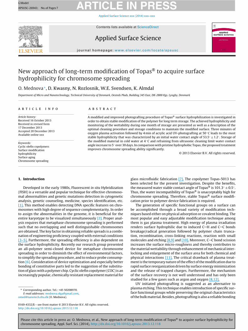
Prior to grafting modification, a batch of Topas® chips has beenplasma treated with gases in order to control the modified sur-faces during aging better. Plasma treatment times exceeding 180 swere avoided since this leads to substantial pore formation in thematerial, which makes it unsuitable for microfluidic fabrication.The monitoring of a stable water contact angle for samples treatedwith oxygen, carbon dioxide, and oxygen carbon dioxide mixtureare presented in Fig. 1.
Depending on the plasma gas and treatment time varioushydrophilicities of Topas® were obtained. As compared to the orig-inal material, 3 min of any plasma etching caused a significantdecrease of the initial water contact angle for all test samples. Thelowest contact angle of 9.3◦ ± 1.9◦ was obtained with carbon diox-ide plasma. However, that particular treatment also provokes themost rapid recovery towards the unmodified Topas® level. All sam-ples treated with CO2 show stable water contact angle growth ofmore than 60◦ in one month. Moreover, roughness developmentis induced most likely due to polymer chain scission and renderscarbon dioxide unsuitable for further application in the currentinvestigation.
Interestingly, samples treated with both oxygen and the mix-ture plasma for 3 min possess similar post treatment wettabilitywith contact angles above 20◦ as well as identical trends with con-tact angle growth of up to 60◦ within one month of recovery. Basedon these observations, both gases can be employed for the devel-opment of the combined treatment method. However, oxygen ischosen for further surface modification studies essentially for sim-plicity reasons.
Similar to the plasma investigation described above, the threemonomers acrylic acid (AA), 2-hydroxyethyl acrylate, (2-HEA), and2-carboxyethyl acrylate (2-CEA) have been examined for pho-tografting. All selected monomers differ by type of hydrophilicgroup and carbon chain length. Depending on molecular con-formation and bonding efficiency final wettability might vary.Furthermore, the process temperature has a great impact on thegrafting ability as higher energy provides stronger bonding [18].
g-term modification of Topas® to acquire surface hydrophilicity for.apsusc.2013.12.118
Therefore, 4 min of UV irradiation at 50 ◦C were applied on Topas®
chips as an optimal regime for photografting. Results of the experi-ment are presented in Fig. 2 and show a clear difference dependingon the applied conditions.
ARTICLE IN PRESSG Model
APSUSC-26943; No. of Pages 7
O. Mednova et al. / Applied Surface Science xxx (2014) xxx– xxx 3
0 5 10 15 20 25 30
20
30
40
50
60
70
80
90
wat
er s
tabl
e co
ntac
t ang
le
days after trea tment
(a)
0 5 10 15 20 25 30
0
20
40
60
80
100
wat
er s
tabl
e co
ntac
t ang
le
days after t rea tmen t(b)
0 5 10 15 20 25 3010
20
30
40
50
60
70
wat
er s
tabl
e co
ntac
t ang
le
days after t rea tmen t(c)
Fo
t2gGtt
0 5 10 15 20 25 3030
40
50
60
70
wat
erst
able
cont
acta
ngle
days after treatment
only the combined modification technique shows a unique long-term stable wettability with water stable contact angle of around60◦ over one month of surface monitoring, signaling insignificantreorganization of the surface.
0 5 10 15 20 25 30
30
40
50
60
70
80
90
100
110
wat
erst
able
cont
acta
ngle
ig. 1. Topas® slides plasma treated with (a) – oxygen, (b) – carbon dioxide, (c) –xygen and carbon dioxide mixture (1/1 by volume) over the specified time.
The lowest contact angle of 35.1◦ ± 2.6◦ is obtained for pho-ografting with 2-HEA. During the first week after treatment the-HEA sample possesses even better wettability than a standard
Please cite this article in press as: O. Mednova, et al., New approach of lonchromosome spreading, Appl. Surf. Sci. (2014), http://dx.doi.org/10.1016/j
lass slide for which the stable water contact angle is around 40◦.rafting with 2-CEA leads to results similar as for 2-HEA. Even
hough the carboxyl group is more polar than the hydroxyl group,he 2-CEA treated surface results in higher hydrophobicity – the
Fig. 2. Topas® slides treated with combined method: 3 min of oxygen plasma fol-lowed with grafting of AA (-�-), 2-HEA (-•-) and 2-CEA (-�-) over 4 min at 50 ◦C.
contact angle a 2-CEA photografted slide is 7◦ higher than for 2-HEA grafting – presumably due to monomer reorganization withintramolecular hydrogen bond formation on the surface. The mostinteresting and practically applicable result is the response of a sur-face grafted with AA providing the most stable hydrophilicity overthe whole period of monitoring. While initially at 53.5◦ ± 1.5◦, theincreasing trend of the AA-grafted surface contact angle deceler-ates after 5 days and fluctuates between approximately 57◦ and62◦ over the next weeks. Even though the contact angle is quitehigh, this modification promises steady properties even for timeslonger than the experimental timeframe.
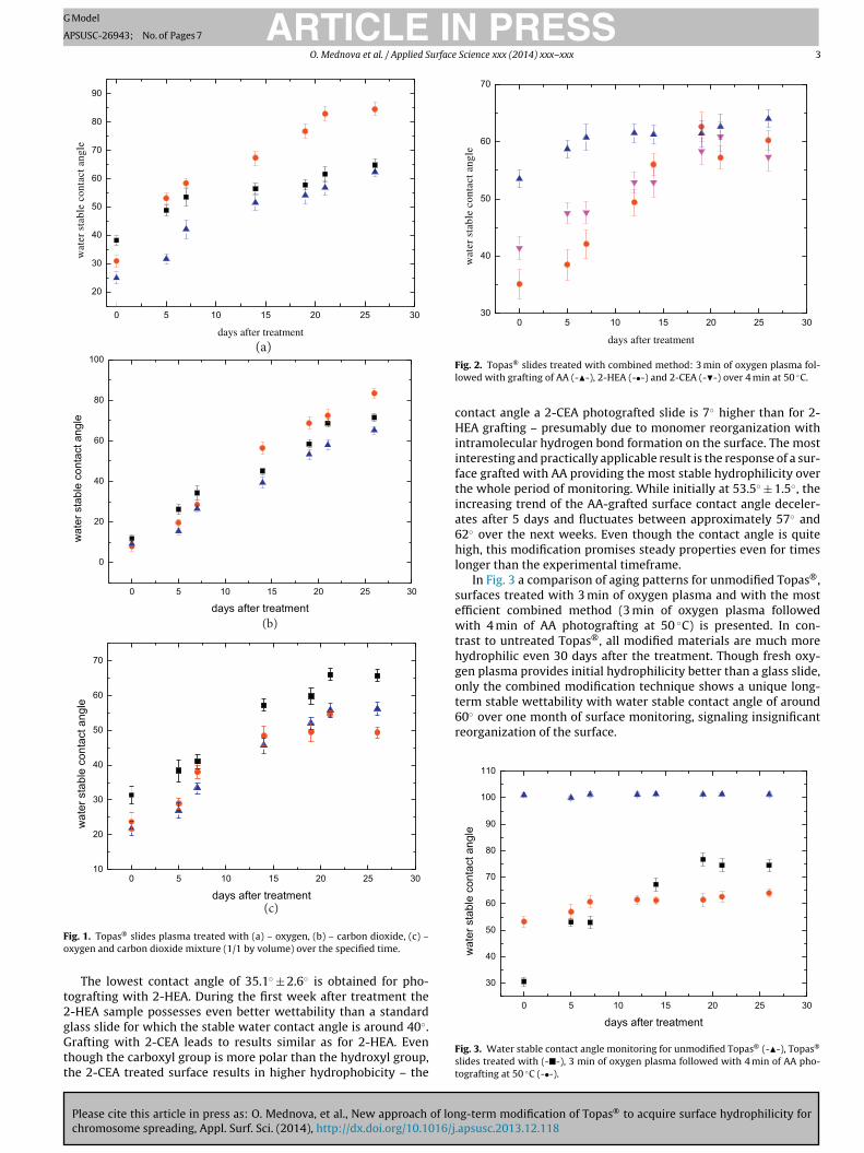
In Fig. 3 a comparison of aging patterns for unmodified Topas®,surfaces treated with 3 min of oxygen plasma and with the mostefficient combined method (3 min of oxygen plasma followedwith 4 min of AA photografting at 50 ◦C) is presented. In con-trast to untreated Topas®, all modified materials are much morehydrophilic even 30 days after the treatment. Though fresh oxy-gen plasma provides initial hydrophilicity better than a glass slide,
g-term modification of Topas® to acquire surface hydrophilicity for.apsusc.2013.12.118
days after treatment
Fig. 3. Water stable contact angle monitoring for unmodified Topas® (-�-), Topas®
slides treated with (-�-), 3 min of oxygen plasma followed with 4 min of AA pho-tografting at 50 ◦C (-•-).
ARTICLE IN PRESSG Model
APSUSC-26943; No. of Pages 7
4 O. Mednova et al. / Applied Surface Science xxx (2014) xxx– xxx
0 5 10 15 20 2534
36
38
40
42
44
stored in cold waterstored in a desiccatorstored on a shelve
wat
erst
able
cont
acta
ngle
F
3
recrcRtoin
F
aa6rgcmt
i
0 5 10 15 20 25 3050
55
60
65
70
75
stor
edin
ice-
cold
wat
er
days after treatment
Fig. 6. Wettability degradation depending on storage conditions: ice-cold water (-
Fo
days of storage
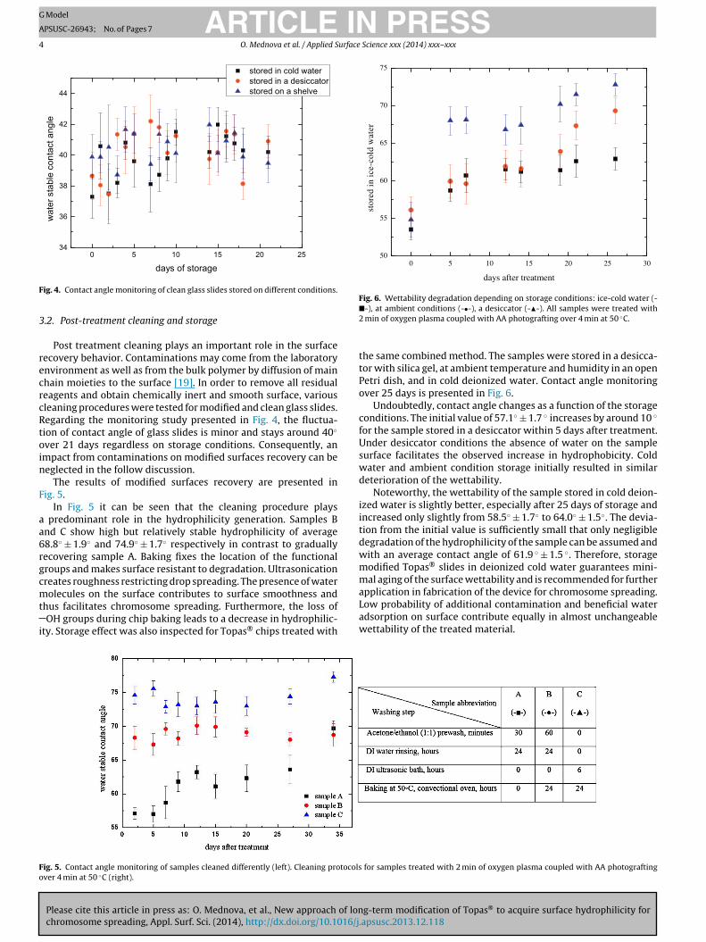
ig. 4. Contact angle monitoring of clean glass slides stored on different conditions.
.2. Post-treatment cleaning and storage
Post treatment cleaning plays an important role in the surfaceecovery behavior. Contaminations may come from the laboratorynvironment as well as from the bulk polymer by diffusion of mainhain moieties to the surface [19]. In order to remove all residualeagents and obtain chemically inert and smooth surface, variousleaning procedures were tested for modified and clean glass slides.egarding the monitoring study presented in Fig. 4, the fluctua-ion of contact angle of glass slides is minor and stays around 40◦
ver 21 days regardless on storage conditions. Consequently, anmpact from contaminations on modified surfaces recovery can beeglected in the follow discussion.
The results of modified surfaces recovery are presented inig. 5.
In Fig. 5 it can be seen that the cleaning procedure plays predominant role in the hydrophilicity generation. Samples Bnd C show high but relatively stable hydrophilicity of average8.8◦ ± 1.9◦ and 74.9◦ ± 1.7◦ respectively in contrast to graduallyecovering sample A. Baking fixes the location of the functionalroups and makes surface resistant to degradation. Ultrasonicationreates roughness restricting drop spreading. The presence of water
Please cite this article in press as: O. Mednova, et al., New approach of lonchromosome spreading, Appl. Surf. Sci. (2014), http://dx.doi.org/10.1016/j
olecules on the surface contributes to surface smoothness andhus facilitates chromosome spreading. Furthermore, the loss ofOH groups during chip baking leads to a decrease in hydrophilic-
ty. Storage effect was also inspected for Topas® chips treated with
ig. 5. Contact angle monitoring of samples cleaned differently (left). Cleaning protocolsver 4 min at 50 ◦C (right).
�-), at ambient conditions (-•-), a desiccator (-�-). All samples were treated with2 min of oxygen plasma coupled with AA photografting over 4 min at 50 ◦C.
the same combined method. The samples were stored in a desicca-tor with silica gel, at ambient temperature and humidity in an openPetri dish, and in cold deionized water. Contact angle monitoringover 25 days is presented in Fig. 6.
Undoubtedly, contact angle changes as a function of the storageconditions. The initial value of 57.1◦ ± 1.7 ◦ increases by around 10 ◦
for the sample stored in a desiccator within 5 days after treatment.Under desiccator conditions the absence of water on the samplesurface facilitates the observed increase in hydrophobicity. Coldwater and ambient condition storage initially resulted in similardeterioration of the wettability.
Noteworthy, the wettability of the sample stored in cold deion-ized water is slightly better, especially after 25 days of storage andincreased only slightly from 58.5◦ ± 1.7◦ to 64.0◦ ± 1.5◦. The devia-tion from the initial value is sufficiently small that only negligibledegradation of the hydrophilicity of the sample can be assumed andwith an average contact angle of 61.9 ◦ ± 1.5 ◦. Therefore, storagemodified Topas® slides in deionized cold water guarantees mini-mal aging of the surface wettability and is recommended for furtherapplication in fabrication of the device for chromosome spreading.
g-term modification of Topas® to acquire surface hydrophilicity for.apsusc.2013.12.118
Low probability of additional contamination and beneficial wateradsorption on surface contribute equally in almost unchangeablewettability of the treated material.
for samples treated with 2 min of oxygen plasma coupled with AA photografting

ARTICLE IN PRESSG Model
APSUSC-26943; No. of Pages 7
O. Mednova et al. / Applied Surface Science xxx (2014) xxx– xxx 5
left) a
3
oa
esfotstdpu
mo
T
rspii
w[
TA
Fig. 7. 3 �m × 3 �m AFM surface images of unmodified Topas® (
.3. Surface characterization
Surface chemistry of both original and modified chips was thor-ughly analyzed to estimate the effect of the treatment by revealingny distinctive surface features and active group presence.
Visual inspection under an optical microscope reveals a pres-nce of post-production lines on the surface of unmodified Topaslides that probably arise from the injection-molding. These arti-acts can have an influence on the drop spreading, which mayccur along these lines rather than spreading uniformly. To avoidhese lines the optimization of the injection molding parametersuch as temperatures which affect the pressure distribution insidehe mold is necessary. After post-photografting cleaning additionalefects such scratches and streaks were noticed on the surface sincehysical interactions between samples and magnetic bars werenavoidable.
The possible effect of surface roughness introduced by the treat-ents of slides is investigated with AFM. Fig. 7 shows AFM images
f untreated and modified surfaces.The resulting average roughness of treated slides is given in
able 1.As can be seen, oxygen plasma does not influence the surface
oughness. The other two treatment methods induce the oppo-ite effect and leave the surface rougher especially in case of thehotografted samples. For example, the combined method of mod-
fication with 2-HEA results in 2.475 ± 0.15 nm of roughness whichs twice as high as the unmodified slide.
Please cite this article in press as: O. Mednova, et al., New approach of lonchromosome spreading, Appl. Surf. Sci. (2014), http://dx.doi.org/10.1016/j
Surface characterization of both original and aged Topas slidesith XPS and ATR-FTIR verified results previously published by Roy
8].
able 1verage surface roughness for samples treated differently.
Sample description Averagesurfaceroughness
Unmodified 1.305 ± 0.16After oxygen plasma (2 min) 1.202 ± 0.12After carbon dioxide plasma (2 min) 1.707 ± 0.16After oxygen plasma (2 min) + 2-HEA grafting (4 min at 50 ◦C) 2.475 ± 0.15After oxygen plasma (2 min) + AA grafting (4 min at 50 ◦C) 2.372 ± 0.15
nd modified with oxygen plasma followed by AA grafting (right).
3.4. Chromosome spreading investigation
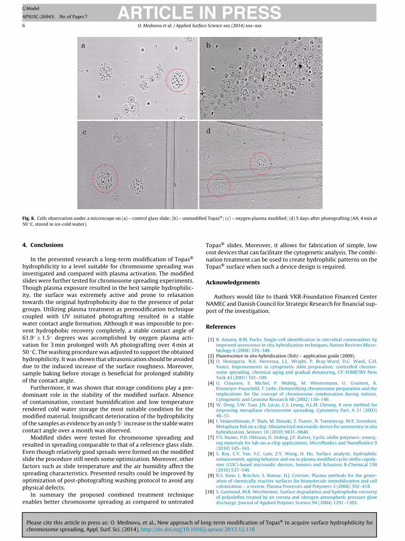
All modified surfaces have been tested for chromosome spread-ing to examine their applicability for cytogenetic analysis. Aglass slide was used as a reference. Fig. 8 shows pictures of thechromosome spreads on a glass slide (a) with noticeable spread-ing enhancement in changing from untreated Topas® slide (b)towards semi-hydrophilic oxygen plasma and AA-grafted surfaces(c, d).
Valid and reliable chromosome spreads is considered to be thesteady stretch of nuclei characterized by the absence of significantcell agglomeration, well distinguishable and elongated chromo-somes without overlaps. Such spreads allows a complete stretchedchromosome to be easily observed and analyzed with cytogenetictechniques. The obvious wettability deficiency of Topas® is provedby Fig. 8(b). The hydrophobic surface prevents the chromosomespreading as the cell suspension drop does not spread uniformlyover the surface which prolongs the fixative evaporation as com-pared to glass slide. Apparently, sample cells form compressedclusters preferably at the edges of the drop due to surface tension.
Once slides were treated, their hydrophilicity was increased andtherefore chromosome spreading was improved regardless of thetreatment method. The majority of the chromosomes are separatedand distinguishable, without any overlaps. Moreover, no signifi-cant differences were recorded between chromosome spreads onsimilarly treated but differently stored slides. Subsequently, thepresence of surface bound water influences only the stability ofthe water contact angle and cell spreading and does not affect thechromosome spreading.
Although all treated surfaces were applicable for chromosomespreading, better quality spreads were obtained on freshly plasmatreated chips. It can be seen in Fig. 8(c) that the spreads are bet-ter resolved than those in Fig. 8(d). This is most likely due to thesurface roughness induced by the combined treatment method.During post-photografting washing procedure surface artifacts andimperfections were formed due to the physical contact with mag-netic stirrer bar and other samples. Furthermore, injection moldinginduced lines of the original Topas® slides have a great influence on
g-term modification of Topas® to acquire surface hydrophilicity for.apsusc.2013.12.118
drop spreading. The scratches and the monomer residues presenton the surface of the chips impede proper chromosome spreading.The cells in the solution were accumulating on those structures anddue to limited space the chromosome spreading was affected.
ARTICLE IN PRESSG Model
APSUSC-26943; No. of Pages 7
6 O. Mednova et al. / Applied Surface Science xxx (2014) xxx– xxx
F dified5
4
hisTitgcwv6v5hdso
dormoc
rEsfsop
e
ig. 8. Cells observation under a microscope on (a) – control glass slide; (b) – unmo0 ◦C, stored in ice-cold water).
. Conclusions
In the presented research a long-term modification of Topas®
ydrophilicity to a level suitable for chromosome spreading wasnvestigated and compared with plasma activation. The modifiedlides were further tested for chromosome spreading experiments.hough plasma exposure resulted in the best sample hydrophilic-ty, the surface was extremely active and prone to relaxationowards the original hydrophobicity due to the presence of polarroups. Utilizing plasma treatment as premodification techniqueoupled with UV initiated photografting resulted in a stableater contact angle formation. Although it was impossible to pre-
ent hydrophobic recovery completely, a stable contact angle of1.9◦ ± 1.5◦ degrees was accomplished by oxygen plasma acti-ation for 3 min prolonged with AA photografting over 4 min at0 ◦C. The washing procedure was adjusted to support the obtainedydrophilicity. It was shown that ultrasonication should be avoidedue to the induced increase of the surface roughness. Moreover,ample baking before storage is beneficial for prolonged stabilityf the contact angle.
Furthermore, it was shown that storage conditions play a pre-ominant role in the stability of the modified surface. Absencef contamination, constant humidification and low temperatureendered cold water storage the most suitable condition for theodified material. Insignificant deterioration of the hydrophilicity
f the samples as evidence by an only 5◦ increase in the stable waterontact angle over a month was observed.
Modified slides were tested for chromosome spreading andesulted in spreading comparable to that of a reference glass slide.ven though relatively good spreads were formed on the modifiedlide the procedure still needs some optimization. Moreover, otheractors such as slide temperature and the air humidity affect thepreading characteristics. Presented results could be improved by
Please cite this article in press as: O. Mednova, et al., New approach of lonchromosome spreading, Appl. Surf. Sci. (2014), http://dx.doi.org/10.1016/j
ptimization of post-photografting washing protocol to avoid anyhysical defects.
In summary the proposed combined treatment techniquenables better chromosome spreading as compared to untreated
[
Topas®; (c) – oxygen plasma modified; (d) 5 days after photografting (AA, 4 min at
Topas® slides. Moreover, it allows for fabrication of simple, lowcost devices that can facilitate the cytogenetic analysis. The combi-nation treatment can be used to create hydrophilic patterns on theTopas® surface when such a device design is required.
Acknowledgements
Authors would like to thank VKR-Foundation Financed CenterNAMEC and Danish Council for Strategic Research for financial sup-port of the investigation.
References
[1] R. Amann, B.M. Fuchs, Single-cell identification in microbial communities byimproved uorescence in situ hybridization techniques, Nature Reviews Micro-biology 6 (2008) 339–348.
[2] Fluorescence in situ hybridization (fish) – application guide (2009).[3] O. Henegariu, N.A. Heerema, L.L. Wright, P. Bray-Ward, D.C. Ward, G.H.
Vance, Improvements in cytogenetic slide preparation: controlled chromo-some spreading, chemical aging and gradual denaturing, CY-TOMETRY-NewYork 43 (2001) 101–109.
[4] U. Claussen, S. Michel, P. Muhlig, M. Westermann, U. Grummt, K.Kromeyer-Hauschild, T. Liehr, Demystifying chromosome preparation and theimplications for the concept of chromosome condensation during mitosis,Cytogenetic and Genome Research 98 (2002) 136–146.
[5] W. Deng, S.W. Tsao, J.N. Lucas, C.S. Leung, A.L.M. Cheung, A new method forimproving metaphase chromosome spreading, Cytometry Part, A 51 (2003)46–51.
[6] I. Vedarethinam, P. Shah, M. Dimaki, Z. Tumer, N. Tommerup, W.E. Svendsen,Metaphase fish on a chip: Miniaturized microuidic device for uorescence in situhybridization, Sensors 10 (2010) 9831–9846.
[7] P.S. Nunes, P.D. Ohlsson, O. Ordeig, J.P. Kutter, Cyclic olefin polymers: emerg-ing materials for lab-on-a-chip applications, Microfluidics and Nanofluidics 9(2010) 145–161.
[8] S. Roy, C.Y. Yue, Y.C. Lam, Z.Y. Wang, H. Hu, Surface analysis, hydrophilicenhancement, ageing behavior and ow in plasma modified cyclic olefin copoly-mer (COC)-based microuidic devices, Sensors and Actuators B-Chemical 150(2010) 537–549.
[9] K.S. Siow, L. Britcher, S. Kumar, H.J. Griesser, Plasma methods for the gener-
g-term modification of Topas® to acquire surface hydrophilicity for.apsusc.2013.12.118
ation of chemically reactive surfaces for biomolecule immobilization and cellcolonization – a review, Plasma Processes and Polymers 3 (2006) 392–418.
10] S. Guimond, M.R. Wertheimer, Surface degradation and hydrophobic recoveryof polyolefins treated by air corona and nitrogen atmospheric pressure glowdischarge, Journal of Applied Polymer Science 94 (2004) 1291–1303.

ING Model
A
urface
[
[
[
[
[
[
[
[
ARTICLEPSUSC-26943; No. of Pages 7
O. Mednova et al. / Applied S
11] D. Nikolova, E. Dayss, G. Leps, A. Wutzler, Surface modification of cycloolefiniccopolymers for optimization of the adhesion to metals, Surface and InterfaceAnalysis 36 (2004) 689–693, 10th European Conference on Applications ofSurface and Interface Analysis (ECASIA 03), Berlin, Germany, OCT 05–10, 2003.
12] C.W. Tsao, L. Hromada, J. Liu, P. Kumar, D.L. DeVoe, Low temperature bonding ofPMMA and COC microuidic substrates using UV/ozone surface treatment, Labon a Chip 7 (2007) 499–505.
13] R.K. Jena, C.Y. Yue, L. Anand, Improvement of thermal bond strength and sur-face properties of Cyclic Olefin Copolymer (COC) based microuidic device usingthe photo-grafting technique, Sensors and Actuators B-Chemical 157 (2011)518–526.
Please cite this article in press as: O. Mednova, et al., New approach of lonchromosome spreading, Appl. Surf. Sci. (2014), http://dx.doi.org/10.1016/j
14] B. Ranby, W. Yang, O. Tretinnikov, Surface photografting of polymer fibers,films and sheets, Nuclear instruments & methods in physics research sectionB-beam interactions with materials and atoms 151 (1999) 301–305, 3rd Inter-national Symposium on Ionizing Radiation and Polymers (IRaP 98), Weinbohla,Germany, September 19–24, 1998.
[
PRESS Science xxx (2014) xxx– xxx 7
15] T. Rohr, D. Ogletree, F. Svec, J. Frechet, Surface functionalization of thermoplas-tic polymers for the fabrication of microuidic devices by photoinitiated grafting,Advanced Functional Materials 13 (2003) 264–270.
16] R.K. Jena, C.Y. Yue, Cyclic olefin copolymer based microfluidicdevices for biochip applications: Ultraviolet surface grafting using2-methacryloyloxyethyl phosphorylcholine, Biomicrofluidics 6 (2012).
17] T.B. Stachowiak, D.A. Mair, T.G. Holden, L.J. Lee, F. Svec, J.M.J. Frechet,Hydrophilic surface modification of cyclic olefin copolymer microfluidicchips using sequential photografting, Journal of separation science 30 (2007)1088–1093.
18] N. De Smet, M. Rymarczyk-Machal, E. Schacht, Modification of poly-
g-term modification of Topas® to acquire surface hydrophilicity for.apsusc.2013.12.118
dimethylsiloxane surfaces using benzophenone, Journal of BiomaterialsScience, Polymer Edition 20 (2009) 2039–2053.
19] D. Bodas, C. Khan-Malek, Hydrophilization and hydrophobic recovery of PDMSby oxygen plasma and chemical treatment – an SEM investigation, Sensors andActuators B-Chemical 123 (2007) 368–373.
Copyright © 2022 FDOKUMEN