Airways changes related to air pollution exposure in wheezing children
Upload
khangminh22Category
view
2download
0
Basal cells of the human airways acquire mesenchymaltraits in idiopathic pulmonary fibrosis and in cultureHulda R Jonsdottir1,2,3,8, Ari J Arason1,2,3,8, Ragnar Palsson1,2,4, Sigridur R Franzdottir1,2, Tomas Gudbjartsson3,5,Helgi J Isaksson4, Gunnar Gudmundsson6,7, Thorarinn Gudjonsson1,2 and Magnus K Magnusson1,2,6
Idiopathic pulmonary fibrosis (IPF) is a progressive interstitial lung disease with high morbidity and mortality. The cellularsource of the fibrotic process is currently under debate with one suggested mechanism being epithelial-to-mesenchymaltransition (EMT) in the alveolar region. In this study, we show that airway epithelium overlying fibroblastic foci in IPFcontains a layer of p63-positive basal cells while lacking ciliated and goblet cells. This basal epithelium shows increasedexpression of CK14, Vimentin and N-cadherin while retaining E-cadherin. The underlying fibroblastic foci shows bothE- and N-cadherin-positive cells. To determine if p63-positive basal cells were able to undergo EMT in culture, we treatedVA10, a p63-positive basal cell line, with the serum replacement UltroserG. A sub-population of treated cells acquireda mesenchymal phenotype, including an E- to N-cadherin switch. After isolation, these cells portrayed a phenotypepresenting major hallmarks of EMT (loss of epithelial markers, gain of mesenchymal markers, increased migration andanchorage-independent growth). This phenotypic switch was prevented in p63 knockdown (KD) cells. In conclusion, weshow that airway epithelium overlying fibroblastic foci in IPF lacks its characteristic functional identity, shows increasedreactivity of basal cells and acquisition of a partial EMT phenotype. This study suggests that some p63-positive basal cellsare prone to phenotypic changes and could act as EMT progenitors in IPF.Laboratory Investigation (2015) 95, 1418–1428; doi:10.1038/labinvest.2015.114; published online 21 September 2015
Idiopathic pulmonary fibrosis (IPF) is a severe lung diseasecharacterized by progressive diminution in lung function basedon an underlying fibrotic process in the lung parenchyma.1
The characteristic fibrosis contains the so-called fibroblastic focifound in close proximity to the lung epithelium. Many studieshave indicated the importance of the pulmonary epithelial cells asthe initial cellular target of injury.2–4 Although critical steps in theprogressive fibrosis have been delineated, there are still majorquestions unanswered, especially regarding the interactionbetween epithelium and fibroblastic foci and cellular origin ofthe myofibroblasts that dominate in fibrotic foci of IPF.
Different cellular sources have been suggested to contributeto the fibroblastic foci in IPF. These include residentfibroblasts/myofibroblasts from the lung, mesenchymal stemcells derived from blood that have taken residence within thelung parenchyma and lung epithelial cells that have under-gone epithelial-to-mesenchymal transition (EMT).5
EMT is a process well known in normal development,wound healing and cancer metastasis.6–8 The initiation ofEMT is commonly marked by repression of the epithelialcell adhesion molecule E-cadherin and the dissociation ofepithelial cells. The epithelial cells lose their distinct markerexpression and gain a mesenchymal expression profile andmorphology.9 This expressional switch includes an increase inthe expression of various mesenchymal markers, such asVimentin, N-cadherin and fibronectin.10
Recently, studies have shown that various microRNAs(miRNAs), predominantly the miR-200 family, have animportant role in both EMT and lung fibrosis.11,12 ThemiR-200 family member miR-200c is believed to be pivotalwhen it comes to the downregulation of E-cadherin, one ofthe hallmarks of EMT.13 When this miRNA is downregulated,its target, the transcription factor ZEB1, is free to down-regulate E-cadherin expression.14
1Stem Cell Research Unit, Biomedical Center, Faculty of Medicine, University of Iceland, Reykjavik, Iceland; 2Department of Laboratory Hematology, Landspitali UniversityHospital, Reykjavik, Iceland; 3Faculty of Medicine, University of Iceland, Reykjavik, Iceland; 4Department of Pathology, Landspitali University Hospital, Reykjavik, Iceland;5Department of Cardiothoracic Surgery, Landspitali University Hospital, Reykjavik, Iceland; 6Department of Respiratory Medicine and Sleep, Landspitali University Hospital,Reykjavik, Iceland and 7Department of Pharmacology and Toxicology, Faculty of Medicine, University of Iceland, Reykjavik, IcelandCorrespondence: Professor MK Magnusson, MD, Department of Pharmacology and Toxicology, Faculty of Medicine, University of Iceland, Vatnsmyrarvegur 16, Reykjavik101, Iceland.E-mail: [email protected] authors contributed equally to this work.
Received 2 December 2014; revised 17 July 2015; accepted 29 July 2015
1418 Laboratory Investigation | Volume 95 December 2015 | www.laboratoryinvestigation.org
Laboratory Investigation (2015) 95, 1418–1428© 2015 USCAP, Inc All rights reserved 0023-6837/15

To argue against EMT as a driver of fibrosis in IPF, it hasbeen shown that in the bleomycin-induced mouse modelof pulmonary fibrosis multiple stromal cell populationscontributed to the pulmonary fibrosis without any evidencefor EMT.15 A more recent study suggested pathophysiologicaldifferences between human IPF and the bleomycin-inducedmouse model and furthermore, suggested that the transcrip-tion factor GRHL2 may have a crucial role in epithelialactivation in lung fibrosis and perhaps also in epithelialplasticity.16
Basal cells reside in the airway epithelium from the tracheaand, in diminishing numbers, down to the respiratorybronchioles.17 They are candidate stem cells in the conductingairways, responsible for normal cell replacement andepithelial remodeling upon lung injury.18 Upon damage inairway epithelium, the basal cells show increased reactivitydemonstrated by increased expression of p63, a basal cellrestricted transcription factor and its downstream targetcytokeratin 14 (CK14).19,20 Recent studies have implicatedp63 with EMT. Das et al21 showed that fibrotic lesions in oralsubmucosa showed marked upregulation of p63 and Ohet al22 showed that primary human keratinocytes transfectedwith ΔNp63 underwent EMT when they had depleted theirnormal proliferative capacity.
Alveolar type II cells have been suspected as the cellularsource of IPF through EMT.2,4 However, the contribution ofbasal cells has also been implicated. Chilosi et al23 showedhighly increased numbers of p63-positive cells in bronchiolesof IPF lungs, at areas showing both epithelial hyperplasia andsquamous metaplasia. Seibold et al24 found bronchial basal-,ciliated- and goblet cells to populate the characteristichoneycomb cysts in IPF lungs. However, the possibleconnection between basal cells and the EMT process in IPFstill remains an open question. One reason for the lack ofcurrent understanding of the cellular origin of IPF is the lackof representing cell culture models to study diseasesprogression.
We have recently generated a bronchial epithelial cell linereferred to as VA10. VA10 has basal cell properties evidencedby p63 and CK14 expression and its ability to generatepseudostratified epithelium in air–liquid interface (ALI)culture.25 In addition, this cell line forms bronchio-alveolar-like structures in three-dimensional co-culture with endothe-lial cells.25,26 In this study, we focus on the p63-positive lungepithelial basal cell as one potential progenitor cell for themyofibroblast in IPF.
We present data demonstrating that the epithelial cellsin airways adjacent to fibroblastic foci show evidence ofpartial mesenchymal differentiation. Furthermore, using theVA10 in vitro basal cell model, we show that basal cells areable to undergo EMT, evidenced by both expression pattern,as well as cellular phenotypic characterization and thatthis transition is dependent upon p63 being present inthe cells.
MATERIALS AND METHODSEthics StatementLung tissue samples were provided by written informedconsent from patients who underwent open lung surgicalprocedures. This study was approved by the LandspitaliHospital Ethics Committee. Reference number 88374–96345.
Samples of Lung Tissue from Patients with IPFSamples were analyzed from eight patients who underwentopen lung biopsy at the Department of CardiothoracicSurgery at Landspitali University Hospital. In all cases, thesurgical biopsy was obtained with video-assisted thoraco-scopic surgical approach that has been described elsewhere.27
These patients fulfilled criteria for having IPF as judged by amultidisciplinary clinical team. Sections that showed clearfibroblastic foci were selected. Control samples were obtainedfrom patients who had undergone lobectomies and were freeof pulmonary fibrosis.
Cell CultureThe bronchial epithelial cell line VA10 was previouslyestablished at the laboratory.25 VA10 cells were cultured inLHC-9 medium (Invitrogen, NY, USA) supplemented with50 IU/ml penicillin and 50 mg/ml streptomycin (Invitrogen).UltroserG-treated VA10 cells were cultured in DMEM/F12(Invitrogen) supplemented with 2% UltroserG (Pall Biosepra,France) and 50 IU/ml penicillin and 50 mg/ml streptomycin(Invitrogen). 3D cultures were performed as previouslydescribed.26
Production of Lentivirus and Cell TransductionLentiviral transduction was performed as previouslydescribed.28 In short, plasmids containing scrambled hairpin(pLKO.1 shSCR; Addgene plasmid 17920)29 or shRNA againstp63 (shp63alpha pLKO.1 puro; Addgene plasmid 19120)30
were used with packaging plasmids (psPAX2 and pMD2.G)(Addgene plasmids 12260 and 12259, respectively) to generateviral titer in HEK-293 T cells using Arrest-in (OpenBiosystems). Transduction of VA10 cells was performedusing low MOI volume and 8 mg/ml polybrene and positivecells selected with 0.7 μg/ml puromycin.
Cell SortingVA10 cells were sorted using Magnetic Cell Sorting (MACS,Miltenyi Biotec, Germany). Cells were suspended in MACSbuffer (Miltenyi Biotec) and incubated, either with primarymicrobeads for EpCAM (CD326, Miltenyi Biotec) orunconjugated primary antibody for Thy-1 (clone AS02) for30 min at 4 °C. Unbound beads or antibody were removedby three consecutive washes with MACS buffer. For primaryantibodies, a secondary incubation with anti-mouse IgGmicrobeads (Miltenyi Biotec) was conducted for 30 min at4 °C. After incubation, cells were washed three times withMACS buffer. Cells were then suspended in 1 ml of MACSbuffer and either loaded onto manual MACS separation
Role of p63-positive basal cells in EMT in IPFHR Jonsdottir et al
www.laboratoryinvestigation.org | Laboratory Investigation | Volume 95 December 2015 1419
columns (MS or LD columns, Miltenyi Biotec) or runthrough an autoMACS cells sorter (Miltenyi Biotec).
Migration AssayA total of 20 000 starved VA10 cells (DMEM only, 24 h) wereseeded onto migration filters (8 μm pores, BD Bioscience) inDMEM (Invitrogen) supplemented with 50 IU/ml penicillinand 50 mg/ml streptomycin (Invitrogen). Endothelial growthmedium (LONZA) supplemented with 5% FBS (Invitrogen)was then added to the lower well. Cells were allowed tomigrate for 24 h. Then fixed for 5 min in 3.5% formaldehyde(Sigma Aldrich) and stained with 0.1% crystal violet for15 min. Filters were rinsed with PBS and cells on the apicallayer of the filter wiped off with a Q-tip. Density of migratedcells was evaluated by dissolving migrated cells in 0.5 ml 10%acetic acid (Sigma Aldrich) and measuring optical density at590 nm using a spectrometer.
Soft Agar AssayPlates were coated with 1% soft agar (Sigma Aldrich) dilutedin DMEM (Invitrogen) and kept at 4 °C for 20 min to allowsolidification. A total of 30 000 cells were seeded into 0.5%soft agar preheated to 40 °C and placed onto the solidified 1%surface. The plate was incubated at 4 °C for 10 min to allowsolidification of the upper layer. Cells were then cultured for20 days. After 20 days, cells were stained with 0.05% crystalviolet for 1 h and colonies over 30 μm in size were countedin each well using a phase contrast microscope (Leica).Three representative areas were counted in each well. Eachexperiment was conducted in triplicate.
RNA Isolation, cDNA Synthesis and qRT-PCRRNA was isolated from VA10 cells using TriReagent solution(Sigma Aldrich). Reverse transcription was carried out usingthe RevertAid First Strand Synthesis Kit (Fermentas) accord-ing to the manufacturer’s instructions. For quantification ofmiR-200c, hsa-miR-200c-3p LNA™ PCR primer set (Exicon,204482) was used with an 3’ non-fluorescent MGB quencherand FAM 5’ reporter dye. GAPDH was used as theendogenous reference gene and amplified using commerciallyavailable primers (Applied Biosystems, 4326317E). Data wereanalyzed using 7500 Software v2.0 (Applied Biosystems).
ImmunochemistryParaffin-embedded tissue samples of control and IPF lungbiopsies were obtained from the Department of Pathology,Landspitali University Hospital. The samples were deparaffi-nized, antigen retrieved by boiling in TE buffer for 20 min(unless stated otherwise) and stained with EnVisionH+System-HRP kit (Dako) according to the manufacturer’sinstructions. Primary antibodies (see below) incubated at RTfor 30 min. Cell cultures were fixed with 3.5% fomaldehydefor 10 min at RT or with methanol at –20 °C. Cultures werethen washed two times with PBS and then blocked with 10%FBS in IF buffer (0.2% Triton X-100; 0.1% BSA and 0.05%
Tween-20 in PBS). Primary antibodies incubated for 30 minat RT followed by three 10-min washes with PBS. Secondaryisotype-specific Alexa Fluor antibody conjugates (Invitrogen)were incubated for 30 min at RT. Nuclei stained for 15 min atRT using TO-PRO-3 (Invitrogen). Samples were embedded inFluoromount-G (Southern Biotech, Birmingham, AL, USA)and analyzed with confocal microscopy (Zeiss LSM5 Pasqal).Representative confocal images were quantified usingCellProfiler.31
Western BlotIn all, 5 μg of protein lysate was mixed with 4X NuPage LDSsample buffer (Invitrogen), reduced with mercaptoethanol(Sigma Aldrich) and heated to 75 °C for 10 min. Then loadedon 10% SDS Bis-Tris gels (Invitrogen) and run on 180 V for45 min in 1X NuPage MES running buffer (Invitrogen).Proteins were transferred at 30 V for 1.5 h in 1X NuPagetransfer buffer (Invitrogen) containing 10% methanol to amethanol-activated (Sigma Aldrich) PVDF membrane(Millipore). After transfer, membrane was washed in PBS atroom temperature for 5 min and then blocked with 5% BSA(Applichem) in TBS+0.1% Tween (Sigma Aldrich) for 1 h.Primary antibodies were incubated overnight at 4 °C in 5%BSA in 0.1% TBS+Tween. The next day, the membrane waswashed for 3 × 10 min in PBS. Secondary infrared (IR)antibodies (LiCOR) were incubated in 0.1% TBS-Tween+0.02% SDS (Sigma Aldrich) for 1 h at 1:20 000 dilution.Subsequently, the membrane was washed for 3 × 10 min inPBS. Signal detection and analysis were performed using anOddysey Infrared Image Scanner and the correspondingImage Studio software (LiCOR).
List of antibodies
Antibody Species Clone # Manufacturer Dilution Application
E-cadherin Mouse 34 610182 BD 1:100/1000 Tissue/WB
N-cadherin Mouse 32 610921 BD 1:100/1000 Tissue/WB
EpCAM Mouse VU-1D9 ncl-ESA Leica 1:100/1000 IF/WB
Vimentin Mouse V9 M0725 Dako 1:100 /100/
1000
Tissue/IF/WB
CK14 Mouse LLL02 ncl-l-ll002 Leica 1:100 Tissue/IF
Thy-1 Mouse AS02 CP28 Calbiochem 1:100/1000/
100
IF/WB/CS
p63 Mouse 7JUL ncl-p63 Leica 1:25/500 Tissue/WB
Actin Mouse C4 3280 Abcam 1:2000 WB
Fibronectin Mouse N/A Prof. Deane
Mosher lab
1:200 IF
Statistical AnalysisData are presented as mean and s.d. (error bars) fromnumber of independent experiments. Graphs and calculations
Role of p63-positive basal cells in EMT in IPFHR Jonsdottir et al
1420 Laboratory Investigation | Volume 95 December 2015 | www.laboratoryinvestigation.org

were done using GraphPad Prism. All experimental proce-dures were repeated at least three times. All samples weretested for equal variance.
RESULTSAirway Epithelium Overlying Fibroblastic Foci in IPFHas Lost Pseudostratified Epithelial Architecture andAcquired a Partial Mesenchymal PhenotypeTo analyze the phenotype of the airway epithelium in IPF,we stained lung tissue from IPF patients and controls withmarkers of various epithelial and mesenchymal differentiationstages. We found that in IPF patients, CK14, a marker knownto be upregulated during lung tissue repair,32 is stronglyexpressed in the bronchial epithelium adjacent to fibroblasticfoci (Figure 1). Another basal cell marker, the transcriptionfactor p63, a known regulator of CK14 expression showedincreased expression that follows a similar pattern as CK14.No expression of CK14 and p63 was found within thefibroblastic foci. Interestingly, the epithelium shows a morestratified squamous phenotype around the foci with CK14and p63 expression also found in suprabasal cells (Figure 1and Supplementary Figure S1). We found it to be a generalrule of thumb that airway epithelium adjacent to a focusshowed this phenotype. E-cadherin was strongly expressed inall epithelial cells, but interestingly, cells within the fibro-blastic foci also stained positive, whereas E-cadherin expres-sion in control samples is limited to the epithelium (Figure 1).To further analyze whether the epithelial cells had acquireda mesenchymal phenotype, we stained for N-cadherin, aclassical marker for EMT. N-cadherin is highly expressedduring human lung development but in the normaladult human lung N-cadherin is primarily expressed inmesothelial cells and scattered basal cells in bronchioles.33
Interestingly, basal cells close to fibroblastic foci showedincreased N-cadherin expression compared with controlsamples. When we stained for the mesenchymal markerVimentin, strong expression was seen in the stroma, includingthe sub-epithelial fibroblastic foci. Expression of Vimentinwas also highly upregulated in epithelium adjacent tofibroblastic foci.
Phenotypic Plasticity of Bronchial-Derived BasalEpithelial Cells in CultureGiven the mixed mesenchymal and epithelial differentiationpattern in the bronchial epithelium adjacent to fibroblasticfoci and the presence of E-cadherin-positive cells within thefoci, we wanted to evaluate whether bronchial-derived basalcells could undergo phenotypic changes toward EMT, thus
Figure 1 Basal cell reactivity, hyperplasia and upregulation of EMTmarkers adjacent to fibroblastic foci. Epithelium adjacent to fibroblasticfoci shows increased reactivity and mesenchymal phenotype. Sparsestaining of CK14 is seen in control samples, whereas CK14 is stronglyexpressed in epithelium overlying fibroblastic foci. P63, a restrictedtranscription factor for basal cells is expressed in epithelialcells located both basally and in the top layer indicating formation ofmetaplasia in IPF samples. E-cadherin is abundant in epithelium both incontrol and IPF samples. In addition, E-cadherin is present in cells withinthe foci. N-cadherin is expressed in lower basal epithelial cells in IPFsamples whereas control samples are negative. Vimentin is expressed inboth epithelium and mesenchyme in IPF samples. Bar 100 μm.
Role of p63-positive basal cells in EMT in IPFHR Jonsdottir et al
www.laboratoryinvestigation.org | Laboratory Investigation | Volume 95 December 2015 1421

contributing to the fibrotic process of pulmonary fibrosis. Wehave previously generated a human bronchial-derived basalepithelial cell line (VA10) that is capable of generating apseudostratified epithelial layer in ALI cultures.25 In addition,VA10 forms bronchioalveolar-like structures in 3D co-culturewith endothelial cells26 demonstrating the stem cell-likeproperties of this cell line. To analyze the plasticity of cellswith basal cell properties, we tested if VA10 cells could be
induced to undergo EMT. In vivo the bronchial basal cell issteadily maintained and, furthermore, gives rise to daughtercells capable of undergoing mucociliary differentiation. Thisin vivo-like behavior can be mimicked in culture using one ofseveral culture media and the right in vitro conditions. Weselected the serum substitute UltroserG (UG) commonly usedfor ALI culture to facilitate differentiation of basal cells34,35
and a known inducer of EMT.36,37 In contrast to the cuboidalepithelial phenotype seen in monolayer when cultured inbronchial-defined medium (serum-free LHC-9 medium),a sub-population of VA10 cells in UG supplemented media(2%) showed distinct changes toward a mesenchymalphenotype (Figure 2, top panel). In UG media, the VA10cells arrange into two distinct sub-populations, islands ofcuboidal epithelial-like cells separated by a zone of elongatedfibroblast-like cells (Figure 2, top right: arrows). Immuno-phenotypic characterization revealed that the epithelial-likeislands displayed an epithelial phenotype, expressing EpCAM,CK14 and E-cadherin. The elongated fibroblast-like cells,however, showed a mesenchymal phenotype as evidenced byreduced expression of EpCAM, CK14 and E-cadherin andacquisition of N-cadherin expression (Figure 2, lower twopanels: arrows and outline and Supplementary Figure S2).Interestingly, no phenotypic change is observed when VA10cells are treated with transforming growth factor beta 1(TGF-β1), a well-known EMT inducer (data not shown).EpCAM is ubiquitously expressed in bronchial epithelium butits high expression has been linked to stem cell properties innumber of epithelial tissues.38,39 Furthermore, downregula-tion of EpCAM is frequently observed in EMT.40,41 Therefore,we next decided to separate EpCAM-positive and EpCAM-negative cellular sub-populations and evaluate if these cellsretained their epithelial and mesenchymal traits, respectively,and analyze the phenotypic plasticity of each population.
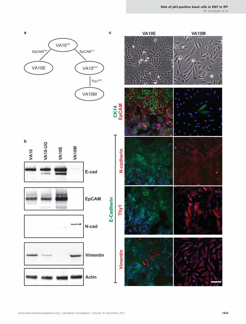
Purification and Characterization of Mesenchymal CellsDerived from VA10Confluent VA10 cells cultured in UG media were magneti-cally separated (MACS) based on their EpCAM status.Further purification of EpCAMneg cells was done by selectingfor Thy-1-positive cells (flow-diagram, Figure 3a). VA10-EpCAMpos (VA10E) demonstrated a cuboidal epithelialphenotype, whereas VA10-EpCAMneg/Thy-1pos (VA10M)showed elongated spindle-like phenotype (Figure 3c,
Figure 2 UltroserG induces epithelial-to-mesenchymal transition in asubset of VA10 cells. Sub-population of cells cultured in UltroserGmedium (UG-treated) show elongated phenotype (top panel, arrows).EpCAM and CK14 immunostaining reveals an EpCam/CK14-negativemesenchymal sub-population in treated cells (middle panel, arrows).Double staining for E- and N-cadherin also shows two distinct cellularsub-populations in treated cells indicating a cadherin switch (bottompanel, outline). Cells were counterstained with TOPRO-3 nuclear stain(middle and bottom panel). Bar 200 μm.
Figure 3 Purification and characterization of VA10-derived epithelial and mesenchymal sub-populations. (a) Purification of epithelial and mesenchymalsub-populations from VA10. Using immunomagnetic cell sorting against EpCAM VA10 was split into EpCAMhigh (left) and EpCAMlow (right). TheEpCAMlow sub-population was further sorted according to Thy-1 expression. The resulting sublines will hereafter be termed VA10E and VA10M,respectively. (b) Surface marker expression of VA10M (Thy-1+) indicates a true mesenchymal phenotype. Immunoblot analysis reveals a difference inepithelial and mesenchymal marker expression between the different cell lines tested. E-cadherin expression has been abolished in VA10M, whereasN-cadherin and Vimentin are upregulated indicating a mesenchymal expression pattern. (c) Characterization of VA10-derived epithelial- andmesenchymal sub-populations. VA10E has a cobblestone-like morphology (top panel, left), whereas VA10M is elongated and fibroblast like (top panel,right). Immunostaining reveals a strong mesenchymal expression pattern in VA10M, whereas VA10E predominantly exhibits an epithelial phenotype(bottom panel).
Role of p63-positive basal cells in EMT in IPFHR Jonsdottir et al
1422 Laboratory Investigation | Volume 95 December 2015 | www.laboratoryinvestigation.org
Role of p63-positive basal cells in EMT in IPFHR Jonsdottir et al
www.laboratoryinvestigation.org | Laboratory Investigation | Volume 95 December 2015 1423
top panel). The epithelial and mesenchymal phenotype inVA10E and VA10M, respectively, were further evaluated withimmunocharacterization (Figures 3b and c). Western blotting(Figure 3b) showed that untreated VA10 cells (in bronchialmedium) and treated VA10 cells (in UG medium) showedstrong expression of the epithelial markers E-cadherin andEpCAM. Expression of Vimentin was observed in both cellpopulation but no expression of N-cadherin was seen inVA10E. VA10E cells showed a strict epithelial phenotype,whereas VA10M cells were negative for both E-cadherin andEpCAM but positive for mesenchymal markers (Figure 3b).These phenotypic differences were further characterized andconfirmed by immunofluorescence staining. VA10E cells werestrongly positive for CK14, EpCAM and E-cadherin butnegative for N-cadherin, Thy-1. Vimentin expression can stillbe observed in a few cells (Figure 3c, left). VA10M cells on theother hand had lost their epithelial phenotype except foroccasional positive CK14 cells. VA10M were uniformlypositive for the mesenchymal markers Thy-1, Vimentin andN-cadherin (Figure 3b, right). Collectively, we have generateda mesenchymal sub-line from VA10 by exposing the cells tothe serum substitute UG. The mesenchymal-like cells referredto as VA10M have undergone phenotypic changes suggestiveof EMT.
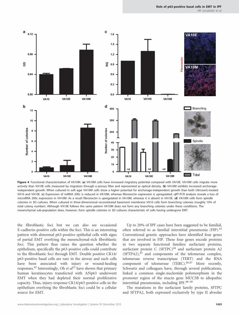
VA10M Has Acquired a Functional MesenchymalPhenotypeTo further analyze the mesenchymal properties of VA10M,we conducted various functional assays that have beenshown to associate with EMT. Migration, a phenotypeassociated with mesenchymal cells, was tested using atranswell migration assay. Although the VA10 and VA10Ecell lines showed similar migratory capabilities, VA10M cellsshowed almost twofold more migration than the VA10E cells(Figure 4a). Anchorage-independent growth has also beenlinked with a mesenchymal phenotype and while bothVA10 and VA10E cells show limited anchorage-dependentgrowth, the VA10M cells had three- to fourfold increasednumber of colonies compared with VA10E in a soft agar assay(Figure 4b).
A number of miRNAs, including the mir-200 family, hasbeen shown to be important in the preservation of epithelialintegrity. In particular, loss of miR-200c expression has beenassociated with loss of epithelial integrity and EMT.14
Although both VA10 and VA10E cells retain their miR-200cexpression, its expression is markedly downregulated inVA10M. Also, in accordance to the miR-200c expression,upregulation of the mesenchymal EMT marker fibronectinwas clearly detected in VA10M (Figure 4c). When cultured ina three-dimensional matrigel-based co-culture system withendothelial cells, both treated VA10 and VA10E cells formbranching bronchioalveolar-like structures like the originalVA10 cell line.26 Under these conditions, approximately 10%of colonies form large branching structures (Figure 4d, whitebars). However, when VA10M cells were cultured in the same
three-dimensional co-culture system they did not exhibit anybranching but instead formed large spindle-like coloniessimilar to fibroblasts (Figure 4d, black bars). In all co-cultureconditions, approximately 90% of colonies were small withno distinct morphology (Figure 4d, gray bars). These variousfunctional assays show that although the VA10E sub-lineretains the phenotype exhibited by its mother cell line(VA10), the VA10M cells have gained a markedly differentphenotype, with clear mesenchymal properties. Interestingly,VA10M can be reverted back to the original phenotypeby culturing the cells on LHC-9 medium (SupplementaryFigure S3). In summary, we have shown that the p63-positivebasal cell line VA10 is able, under specific conditions, toundergo EMT, both with regard to protein expression andcellular phenotype.
KD of p63 Abrogates UG-Stimulated EMT Capacity ofVA10To evaluate whether the expression of p63 is necessary for themesenchymal transformation of VA10 cells, we performed alentiviral-based stable KD with p63 and control scrambledshRNA in VA10 (VA10p63KD and VA10SCR, respectively).This resulted in a significant decrease in p63 protein levels(Supplementary Figure S4). When cultured in UG media,no spindle-like phenotype was observed in VA10p63KD,compared with VA10SCR (Figure 5). Immunostaining revealedthat the VA10p63KD cells showed no upregulation ofN-cadherin or Thy-1, whereas retaining E-cadherin expres-sion, in contrast to VA10SCR (Figure 5). VA10SCR showedmore Vimentin expression in E-cadherin-negative cells, whereasVimentin was exclusively co-expressed with E-cadherin in KDcells, indicating no sub-population distinction.
DISCUSSIONIn this study, we have shown that basal epithelial cells over-lying fibroblastic foci in IPF patient samples acquire increasedreactivity as measured by p63 and CK14 expression andacquisition of a partial mesenchymal phenotype as measured byN-cadherin and Vimentin expression. Although we cannotclaim that basal cells are the cellular source of fibroblasts withinthe foci, there is no doubt that the overlying epithelium, inparticular the basal cells, acquire mesenchymal traits that maybe either the consequence of signals derived from the foci orsignify that the epithelium is directly contributing to theirformation by undergoing EMT. Furthermore, our in vitrostudies show that the p63-positive basal cell line VA10 is able toundergo a mesenchymal transition as evidenced by inductionof EMT upon culture in UG containing medium.
In the IPF samples, we see that the epithelium overlying thefibroblastic foci is rich in p63-positive cells that have not onlya basal but also a suprabasal pattern and show a mixed E- andN-cadherin expression along with both CK14 and Vimentinexpression (Figure 1). This pattern is indicative of a partialEMT in the epithelial cells. Underlying this abnormalepithelium, there are abundant mesenchymal cells forming
Role of p63-positive basal cells in EMT in IPFHR Jonsdottir et al
1424 Laboratory Investigation | Volume 95 December 2015 | www.laboratoryinvestigation.org
the fibroblastic foci but we can also see occasionalE-cadherin-positive cells within the foci. This is an interestingpattern with abnormal p63-positive epithelial cells with signsof partial EMT overlying the mesenchymal-rich fibroblasticfoci. This pattern thus raises the question whether theepithelium, specifically the p63-positive cells could contributeto the fibroblastic foci through EMT. Double positive CK14/p63-positive basal cells are rare in the airway and such cellshave been associated with injury or wound-healingresponses.42 Interestingly, Oh et al22 have shown that primaryhuman keratinocytes transfected with ΔNp63 underwentEMT when they had depleted their normal proliferativecapacity. Thus, injury-response CK14/p63-positive cells in theepithelium overlying the fibroblastic foci could be a cellularsource for EMT.
Up to 20% of IPF cases have been suggested to be familial,often referred to as familial interstitial pneumonia (FIP).43
Conventional genetic approaches have identified four genesthat are involved in FIP. These four genes encode proteinsin two separate functional families: surfactant proteins,surfactant protein C (SFTPC)44 and surfactant protein A2(SFTPA2),45 and components of the telomerase complex,telomerase reverse transcriptase (TERT) and the RNAcomponent of telomerase (TERC).46,47 More recently,Schwartz and colleagues have, through several publications,linked a common single-nucleotide polymorphism in thepromoter region of the mucin gene MUC5B to idiopathicinterstitial pneumonias, including IPF.48–50
The mutations in the surfactant family proteins, SFTPCand SFTPA2, both expressed exclusively by type II alveolar
Figure 4 Functional characterization of VA10M. (a) VA10M cells have increased migratory potential compared with VA10E. VA10M cells migrate moreactively than VA10E cells measured by migration through a porous filter and represented as optical density. (b) VA10M exhibits increased anchorage-independent growth. When cultured in soft agar VA10M cells show a higher potential for anchorage-independent growth than both UltroserG-treatedVA10 and VA10E. (c) Expression of miRNA 200c is reduced in VA10M, whereas fibronectin expression is upregulated. qRT-PCR analysis reveals a loss ofmicroRNA 200c expression in VA10M. As a result fibronectin is upregulated in VA10M, whereas it is absent in VA10E. (d) VA10M cells form spindlecolonies in 3D cultures. When cultured in three-dimensional reconstituted basement membrane VA10 cells form branching colonies (roughly 10% oftotal colony number). Although VA10E follows the same pattern VA10M does not form any branching colonies under these conditions. Themesenchymal sub-population does, however, form spindle colonies in 3D cultures characteristic of cells having undergone EMT.
Role of p63-positive basal cells in EMT in IPFHR Jonsdottir et al
www.laboratoryinvestigation.org | Laboratory Investigation | Volume 95 December 2015 1425

epithelial cells (AECs) in the lungs, suggest that AECdysfunction is a prominent feature of IPF. Furthermore,accumulating evidence supports endoplasmic reticulum (ER)stress with consequent apoptosis as a prominent feature in IPF.Compared with COPD and normal lungs, protein levels of ERstress mediators, such as ATF-6, ATF-4 and the apoptosis-inductor C/EBPzeta, were elevated in AECs of IPF lungs.51 HowER stress with subsequent epithelial injury leads to a fibroticresponse in the adjacent mesenchymal compartment is stillintensely debated. The two major hypotheses are that epithelialcells adjacent to the injured area undergo EMT or that theinjury leads to proliferation of the local mesenchymal cells, suchas fibroblasts or myofibroblasts.43 Although the surfactant typemutations have a described mechanism of epithelial injurythrough ER stress, the mechanism by which telomerasedysfunction (TERT and TERC mutations) leads to IPF is lessunderstood. It is thought that epithelial cell dysfunction may bemediated by epithelial cell senescence and impaired response toepithelial injury, as a consequence of telomere dysfunction.52
Although most investigators agree that the epithelialcompartment is where the initial injury occurs in IPF, thereis much less known about the following events. As statedabove, the main debate focuses on the origin of the cellularcomponents of the fibrotic response. The progressive nature ofthe fibrotic scarring is what drives the destructive nature of IPF.The data collected with mouse models of surfactant mutantssuggest that fibrosis in the commonly used bleomycin mousemodel worsens in the background of ER stress.53 In the samebleomycin model with surfactant (SFTPC) mutated mice, thedata also support the notion that ER stress can contribute toEMT in vivo.54 Overexpression of mutant SFTPC in lungepithelial cell lines largely recapitulated the ER stress-inducedEMT.55 To argue against EMT as a driver of fibrosis, it hasbeen shown as previously discussed that in the bleomcyin-induced model multiple stromal cell populations contributedto the pulmonary fibrosis without any evidence for EMT.15
Another important unsolved issue is the epithelial cell oforigin contributing to epithelial injury and EMT. Thesurfactant mutants would strongly suggest the AEC-type IIcells, given their almost exclusive expression of surfactantproteins. This is also supported by the fact that the initial andmain fibrotic changes in IPF are located distally in theairways. The possible contribution of the MUC5B promoterregion polymorphism to IPF pathogenesis raises the questionwhether epithelia in upper airways may also contribute to IPF.In the normal lung, MUC5B is expressed in sub-mucosal cellsof the upper airways, whereas goblet cells express MUC5Bunder stress conditions.56 In addition, TGF-β1-induced EMTis a well-documented process in A549, an alveolar type II cellline.57 Interestingly, TGF-β1 does not induce EMT in VA10nor another bronchial-derived epithelial cell line, Calu-3.57
This indicates that the mechanism of EMT could be differentin bronchial-derived cells compared with alveolar cells.
We have previously shown that VA10 cells can capturephenotypic architecture of upper and lower airways
Figure 5 Knockdown (KD) of p63 abrogates UG-induced EMT of VA10cells. p63 KD cells are unable to undergo UG-induced EMT. Theexpression of the mesenchymal markers N-cadherin and Thy-1 isabrogated in treated KD cells, whereas scrambled control cells show adivision into two cellular sub-populations much like the mother cell lineVA10. The expression of Vimentin can still be detected in treated KDcells, although to a lesser extent than in treated scrambled cells. Bars100 μm.
Role of p63-positive basal cells in EMT in IPFHR Jonsdottir et al
1426 Laboratory Investigation | Volume 95 December 2015 | www.laboratoryinvestigation.org
depending cell culture conditions. When cultured inALI-conditions, VA10 cells can generate pseudostratifiedepithelium mimicking trachea and larger bronchi andbronchioles. In this model, VA10 generates differentiatedciliated cells as well as goblet cells upon induction.28 Inaddition, we showed when VA10 is embedded intothree-dimensional basement membrane matrix in co-culturewith endothelial cells, we see increase in branching structurescapturing bronchioalveolar phenotype.26 Given this plasticityof the basal cell line VA10 and the expression pattern found inIPF lungs, it can be hypothesized that the basal cell couldcontribute to the fibrotic process in IPF through EMT.
Supplementary Information accompanies the paper on the LaboratoryInvestigation website (http://www.laboratoryinvestigation.org)
ACKNOWLEDGMENTSWe thank Professor Deane Mosher for kindly providing us with a fibronectinantibody. Funding was provided by the Icelandic Research Council project(grant number 120423021), the University of Iceland Research- and DoctoralStudies Fund and Landspitali University Hospital Scientific Fund.
DISCLOSURE/CONFLICT OF INTERESTThe authors declare no conflict of interest.
1. Bradley B, Branley HM, Egan JJ et al. Interstitial lung disease guideline:the British Thoracic Society in collaboration with the Thoracic Societyof Australia and New Zealand and the Irish Thoracic Society. Thorax2008;63:v1–v58.
2. Kim KK, Kugler MC, Wolters PJ et al. Alveolar epithelial cell mesen-chymal transition develops in vivo during pulmonary fibrosis and isregulated by the extracellular matrix. Proc Natl Acad Sci USA 2006;103:13180–13185.
3. Selman M, Pardo A. Role of epithelial cells in idiopathic pulmonaryfibrosis: from innocent targets to serial killers. Proc Am Thorac Soc2006;3:364–372.
4. Willis BC, Liebler JM, Luby-Phelps K et al. Induction of epithelial-mesenchymal transition in alveolar epithelial cells by transforminggrowth factor-beta1: potential role in idiopathic pulmonary fibrosis.Am J Pathol 2005;166:1321–1332.
5. Willis BC, duBois RM, Borok Z. Epithelial origin of myofibroblasts duringfibrosis in the lung. Proc Am Thorac Soc 2006;3:377–382.
6. Savagner P. The epithelial-mesenchymal transition (EMT) phenom-enon. Ann Oncol 2010;21:vii89–vii92.
7. Kerosuo L, Bronner-Fraser M. What is bad in cancer is good in theembryo: importance of EMT in neural crest development. Semin CellDev Biol 2012;23:320–332.
8. Nakamura M, Tokura Y. Epithelial-mesenchymal transition in the skin. JDermatol Sci 2011;61:7–13.
9. Kalluri R, Neilson EG. Epithelial-mesenchymal transition and itsimplications for fibrosis. J Clin Invest 2003;112:1776–1784.
10. Mongroo PS, Rustgi AK. The role of the miR-200 family in epithelial-mesenchymal transition. Cancer Biol Ther 2010;10:219–222.
11. Yang S, Banerjee S, de Freitas A et al. Participation of miR-200 inpulmonary fibrosis. Am J Pathol 2012;180:484–493.
12. Eades G, Yao Y, Yang M et al. miR-200a regulates Sirt1 expression andepithelial to mesenchymal transition (EMT)-like transformation inmammary epithelial cells. J Biol Chem 2011;286:25992–26002.
13. Hilmarsdottir B, Briem E, Bergthorsson JT et al. Functional role of themicroRNA-200 family in breast morphogenesis and neoplasia. Genes(Basel) 2014;5:804–820.
14. Korpal M, Lee ES, Hu G et al. The miR-200 family inhibits epithelial-mesenchymal transition and cancer cell migration by direct targetingof E-cadherin transcriptional repressors Zeb1 and Zeb2. J Biol Chem2008;283:14910–14914.
15. Rock JR, Barkauskas CE, Cronce MJ et al. Multiple stromal populationscontribute to pulmonary fibrosis without evidence for epithelialto mesenchymal transition. Proc Natl Acad Sci USA 2011;108:E1475–E1483.
16. Varma S, Mahavadi P, Sasikumar S et al. Grainyhead-like 2 (Grhl2)distribution reveals novel pathophysiological differences betweenhuman idiopathic pulmonary fibrosis and mouse models of pulmonaryfibrosis. Am J Physiol Lung Cell Mol Physiol 2014;306:L405–L419.
17. Boers JE, Ambergen AW, Thunnissen FB. Number and proliferationof basal and parabasal cells in normal human airway epithelium. Am JRespir Crit Care Med 1998;157:2000–2006.
18. Rawlins EL, Hogan BL. Epithelial stem cells of the lung: privileged fewor opportunities for many? Development 2006;133:2455–2465.
19. Sheikh HA, Fuhrer K, Cieply K et al. P63 expression in assessment ofbronchioloalveolar proliferations of the lung. Mod Pathol 2004;17:1134–1140.
20. Hinata N, Takemura T, Ikushima S et al. Phenotype of regenerativeepithelium in idiopathic interstitial pneumonias. J Med Dent Sci2003;50:213–224.
21. Das RK, Anura A, Pal M et al. Epithelio-mesenchymal transitional attri-butes in oral sub-mucous fibrosis. Exp Mol Pathol 2013;95:259–269.
22. Oh JE, Kim RH, Shin KH et al. Deltanp63alpha protein triggersepithelial-mesenchymal transition and confers stem cell properties innormal human keratinocytes. J Biol Chem 2011;286:38757–38767.
23. Chilosi M, Poletti V, Murer B et al. Abnormal re-epithelialization andlung remodeling in idiopathic pulmonary fibrosis: the role of deltan-P63. Lab Invest 2002;82:1335–1345.
24. Seibold MA, Smith RW, Urbanek C et al. The idiopathic pulmonaryfibrosis honeycomb cyst contains a mucocilary pseudostratifiedepithelium. PLoS One 2013;8:e58658.
25. Halldorsson S, Asgrimsson V, Axelsson I et al. Differentiation potentialof a basal epithelial cell line established from human bronchial explant.In Vitro Cell Dev Biol Anim 2007;43:283–289.
26. Franzdottir SR, Axelsson IT, Arason AJ et al. Airway branchingmorphogenesis in three dimensional culture. Respir Res 2010;11:162.
27. Sigurdsson MI, Isaksson HJ, Gudmundsson G et al. Diagnostic surgicallung biopsies for suspected interstitial lung diseases: a retrospectivestudy. Ann Thorac Surg 2009;88:227–232.
28. Arason AJ, Jonsdottir HR, Halldorsson S et al. Deltanp63 has a role inmaintaining epithelial integrity in airway epithelium. PLoS One 2014;9:e88683.
29. Saharia A, Guittat L, Crocker S et al. Flap endonuclease 1 contributes totelomere stability. Curr Biol 2008;18:496–500.
30. Godar S, Ince TA, Bell GW et al. Growth-inhibitory and tumor-suppressive functions of p53 depend on its repression of Cd44expression. Cell 2008;134:62–73.
31. Carpenter AE, Jones TR, Lamprecht MR et al. Cellprofiler: image analysissoftware for identifying and quantifying cell phenotypes. Genome Biol2006;7:R100.
32. Cole BB, Smith RW, Jenkins KM et al. Tracheal basal cells: a facultativeprogenitor cell pool. Am J Pathol 2010;177:362–376.
33. Kaarteenaho R, Lappi-Blanco E, Lehtonen S. Epithelial N-cadherin andnuclear beta-catenin are up-regulated during early development ofhuman lung. BMC Dev Biol 2010;10:113.
34. Yamaya M, Finkbeiner WE, Chun SY et al. Differentiated structure andfunction of cultures from human tracheal epithelium. Am J Physiol1992;262:L713–L724.
35. Sachs LA, Finkbeiner WE, Widdicombe JH. Effects of media ondifferentiation of cultured human tracheal Epithelium. In Vitro CellDev Biol Anim 2003;39:56–62.
36. Boyer B, Tucker GC, Valles AM et al. Reversible transition towards afibroblastic phenotype in a rat carcinoma cell line. Int J Cancer Suppl1989;4:69–75.
37. Hunter I, Lindh M, Obrink B. Differential regulation of C-cam isoformsin epithelial cells. J Cell Sci 1994;107(Pt 5): 1205–1216.
38. Turner R, Lozoya O, Wang Y et al. Human hepatic stem cell andmaturational liver lineage biology. Hepatology 2011;53:1035–1045.
39. Munz M, Baeuerle PA, Gires O. The emerging role of EpCAM in cancerand stem cell signaling. Cancer Res 2009;69:5627–5629.
40. Santisteban M, Reiman JM, Asiedu MK et al. Immune-induced epithelialto mesenchymal transition in vivo generates breast cancer stem cells.Cancer Res 2009;69:2887–2895.
Role of p63-positive basal cells in EMT in IPFHR Jonsdottir et al
www.laboratoryinvestigation.org | Laboratory Investigation | Volume 95 December 2015 1427
41. Frederick BA, Helfrich BA, Coldren CD et al. Epithelial to mesenchymaltransition predicts gefitinib resistance in cell lines of head and necksquamous cell carcinoma and non-small cell lung carcinoma. MolCancer Ther 2007;6:1683–1691.
42. Crespin S, Bacchetta M, Bou Saab J et al. Cx26 regulates proliferation ofrepairing basal airway epithelial cells. Int J Biochem Cell Biol 2014;52:152–160.
43. Kropski JA, Lawson WE, Young LR et al. Genetic studies provide clueson the pathogenesis of idiopathic pulmonary fibrosis. Dis ModelsMech 2013;6:9–17.
44. Nogee LM, Dunbar 3rd AE , Wert SE et al. A mutation in the surfactantprotein C gene associated with familial interstitial lung disease. N EnglJ Med 2001;344:573–579.
45. Wang Y, Kuan PJ, Xing C et al. Genetic defects in surfactant protein A2are associated with pulmonary fibrosis and lung cancer. Am J HumGenet 2009;84:52–59.
46. Armanios MY, Chen JJ, Cogan JD et al. Telomerase mutations in familieswith idiopathic pulmonary fibrosis. N Engl J Med 2007;356:1317–1326.
47. Tsakiri KD, Cronkhite JT, Kuan PJ et al. Adult-onset pulmonary fibrosis causedby mutations in telomerase. Proc Natl Acad Sci USA 2007;104:7552–7557.
48. Seibold MA, Wise AL, Speer MC et al. A common Muc5b promoterpolymorphism and pulmonary fibrosis. N Engl J Med 2011;364:1503–1512.
49. Fingerlin TE, Murphy E, Zhang W et al. Genome-wide association studyidentifies multiple susceptibility loci for pulmonary fibrosis. Nat Genet2013;45:613–620.
50. Hunninghake GM, Hatabu H, Okajima Y et al. Muc5b promoterpolymorphism and interstitial lung abnormalities. N Engl J Med2013;45:613–620.
51. Korfei M, Ruppert C, Mahavadi P et al. Epithelial endoplasmic reticulumstress and apoptosis in sporadic idiopathic pulmonary fibrosis. Am JRespir Crit Care Med 2008;178:838–846.
52. Armanios M, Blackburn EH. The telomere syndromes. Nat Rev Genet2012;13:693–704.
53. Lawson WE, Cheng DS, Degryse AL et al. Endoplasmic reticulum stressenhances fibrotic remodeling in the lungs. Proc Natl Acad Sci USA2011;108:10562–10567.
54. Tanjore H, Blackwell TS, Lawson WE. Emerging evidence forendoplasmic reticulum stress in the pathogenesis of idiopathicpulmonary fibrosis. Am J Physiol Lung Cell Mol Physiol 2012;302:L721–L729.
55. Zhong Q, Zhou B, Ann DK et al. Role of endoplasmic reticulum stress inepithelial-mesenchymal transition of alveolar epithelial cells: effects ofmisfolded surfactant protein. Am J Respir Cell Mol Biol 2011;45:498–509.
56. Thai P, Loukoianov A, Wachi S et al. Regulation of airway mucin geneexpression. Annu Rev Physiol 2008;70:405–429.
57. Buckley ST, Medina C, Ehrhardt C. Differential susceptibility toepithelial-mesenchymal transition (EMT) of alveolar, bronchial andintestinal epithelial cells in vitro and the effect of angiotensin IIreceptor inhibition. Cell Tissue Res 2010;342:39–51.
Role of p63-positive basal cells in EMT in IPFHR Jonsdottir et al
1428 Laboratory Investigation | Volume 95 December 2015 | www.laboratoryinvestigation.org
Copyright © 2022 FDOKUMEN