Neutron Spectrometry With a Passive Bonner Sphere System Around a Medical LINAC and Evaluation of...
11
IEEE TRANSACTIONS ON NUCLEAR SCIENCE, VOL. 56, NO. 5, OCTOBER 2009 2885 Neutron Spectrometry With a Passive Bonner Sphere System Around a Medical LINAC and Evaluation of the Associated Unfolding Uncertainties Khalil Amgarou, Véronique Lacoste, Alain Martin, Bruno Asselineau, and Laurent Donadille Abstract—It is generally known that the use of high-energy elec- tron linear accelerators (LINACs) in radiotherapy medical treat- ments may generate secondary neutrons, mainly via photonuclear giant dipole resonance reactions of incident photons with all the heavy materials present inside the gantry and along the beam line. A detailed knowledge (i.e., fluence energy distribution) of this parasite radiation, which is approximately isotropic and not con- fined within the primary LINAC beam field, would be of great in- terest to estimate the associated radiological risk for the patient and the working staff. It has been shown, in this study, that our recently developed passive Bonner sphere system, using pure gold activation foils as central detectors, is well adapted to measure neu- tron spectra at pulsed and intense mixed n- fields with high-en- ergy photon component. This system was used to characterize the neutron field around a new generation medical electron LINAC. Two measurement positions (isocenter and maze entrance) inside the treatment room of this facility, with the machine operating in Bremsstrahlung photon mode, were chosen. The obtained specific Au saturation activities were processed by means of the NUBAY unfolding code, which performs a Bayesian estimation of a param- eterized spectrum, to derive the final neutron spectra. Another un- folding method (MAXED), based on the maximum entropy prin- ciple and which may depend to some extent on considered the ini- tial guess or default spectrum, was also applied to check the ro- bustness of the NUBAY solutions as well as to carry out a sensi- tivity analysis to confirm their stability and to corroborate the as- sociated uncertainties on their output results. Also presented are the obtained integral quantities, in terms of neutron fluence and ambient dose equivalent rates normalized to the primary LINAC photon dose, together with an estimation of their associated uncer- tainties due to the unfolding code used. Index Terms—Bonner sphere system, electron medical LINACs, neutron spectrometry, unfolding procedures. I. INTRODUCTION C ANCER is the major human epidemic in modern society and its effect in the public health is very considerable. Fortunately and thanks to the incessant progress in medicine, Manuscript received June 13, 2008; revised December 09, 2008, April 08, 2009, and June 10, 2009. Current version published October 07, 2009. K. Amgarou was with the Institute for Radiological Protection and Nuclear Safety (IRSN), F-13115 Saint Paul-Lez-Durance, France. He is now with the Grup de Física de les Radiacions, Facultat de Ciencies, Universitat Autònoma de Barcelona, E-08193 Bellaterra, Spain (e-mail: [email protected]). V. Lacoste, A. Martin, and B. Asselineau are with the Institute for Radi- ological Protection and Nuclear Safety (IRSN), F-13115 Saint Paul-Lez-Du- rance, France (e-mail: [email protected]; [email protected]; bruno. [email protected]). L. Donadille is with the Institute for Radiological Protection and Nu- clear Safety (IRSN), F-92262 Fontenay aux Roses, France (e-mail: lau- [email protected]). Color versions of one or more of the figures in this paper are available online at http://ieeexplore.ieee.org. Digital Object Identifier 10.1109/TNS.2009.2026416 more than half the cases in France can now be cured, according to the European Cancer Registry Study (EUROCARE-4) of Survival and Care of Cancer Patients [1], [2]. Indeed, ra- diotherapy, using, for instance, high-energy electron linear accelerators (LINACs) to irradiate tumor tissues with mono-en- ergetic electrons or Bremsstrahlung photon spectra, is one of the most effective medical treatments for this malignant illness [3]. The present status of the national radiotherapy services is rep- resented by more than 350 LINAC instruments and their ap- plication is in continuous evolution. New sophisticated treat- ment protocols such as the intensity-modulated radiotherapy (IMRT), image-guided radiotherapy (IGRT) and 3-dimensional conformal radiotherapy (3DCRT) techniques are increasingly used [4]. Essentially, the main role of these techniques is to im- prove local irradiation of the well-defined melanoma volume, normally with complex geometries, without compromising the neighboring healthy organs. Most of these LINACs operate at acceleration potentials above 10 MV; so they are able to generate secondary neutrons via photonuclear and electronuclear giant dipole resonance (GDR) reactions of incident photons and electrons, respectively, with all the heavy materials present inside the gantry and along the beam line [5], [6]. Nevertheless, the electronuclear reaction is mostly neglected since its cross-sec- tions are at least two orders of magnitude smaller than the photonuclear ones [5]. The neutron parasite radiation, which is approximately isotropic and not confined within the primary LINAC beam field, can be transmitted in all directions, through the massive shielding and collimation systems of the accelerator head, un- dergoing further interactions with the concrete walls, floor and ceiling as well as with other large structures, furniture and the patient under treatment. Consequently, whole-body exposure to both neutrons and their subsequent induced radioactivity may suppose a potential hazard to the patient itself and to the working staff. The neutron source term information provided by the accel- erator manufacturer is practically inexistent and the scarce data available in the literature are usually valid only for the partic- ular cases of the considered LINAC device and the own specifi- cations of the corresponding treatment room. For this purpose, the International Electrotechnical Commission [7], in its report 60601-2-1, stressed the need to determine the unwanted neutron production of every high-energy LINAC used in radiotherapy medical treatments. 0018-9499/$26.00 © 2009 IEEE Authorized licensed use limited to: Universitat Autonoma De Barcelona. Downloaded on October 8, 2009 at 06:47 from IEEE Xplore. Restrictions apply.
Transcript of Neutron Spectrometry With a Passive Bonner Sphere System Around a Medical LINAC and Evaluation of...
IEEE TRANSACTIONS ON NUCLEAR SCIENCE, VOL. 56, NO. 5, OCTOBER 2009 2885
Neutron Spectrometry With a Passive Bonner SphereSystem Around a Medical LINAC and Evaluation of
the Associated Unfolding UncertaintiesKhalil Amgarou, Véronique Lacoste, Alain Martin, Bruno Asselineau, and Laurent Donadille
Abstract—It is generally known that the use of high-energy elec-tron linear accelerators (LINACs) in radiotherapy medical treat-ments may generate secondary neutrons, mainly via photonuclear� � giant dipole resonance reactions of incident photons with allthe heavy materials present inside the gantry and along the beamline. A detailed knowledge (i.e., fluence energy distribution) of thisparasite radiation, which is approximately isotropic and not con-fined within the primary LINAC beam field, would be of great in-terest to estimate the associated radiological risk for the patientand the working staff. It has been shown, in this study, that ourrecently developed passive Bonner sphere system, using pure goldactivation foils as central detectors, is well adapted to measure neu-tron spectra at pulsed and intense mixed n- fields with high-en-ergy photon component. This system was used to characterize theneutron field around a new generation medical electron LINAC.Two measurement positions (isocenter and maze entrance) insidethe treatment room of this facility, with the machine operating inBremsstrahlung photon mode, were chosen. The obtained specific���Au saturation activities were processed by means of the NUBAYunfolding code, which performs a Bayesian estimation of a param-eterized spectrum, to derive the final neutron spectra. Another un-folding method (MAXED), based on the maximum entropy prin-ciple and which may depend to some extent on considered the ini-tial guess or default spectrum, was also applied to check the ro-bustness of the NUBAY solutions as well as to carry out a sensi-tivity analysis to confirm their stability and to corroborate the as-sociated uncertainties on their output results. Also presented arethe obtained integral quantities, in terms of neutron fluence andambient dose equivalent rates normalized to the primary LINACphoton dose, together with an estimation of their associated uncer-tainties due to the unfolding code used.
Index Terms—Bonner sphere system, electron medical LINACs,neutron spectrometry, unfolding procedures.
I. INTRODUCTION
C ANCER is the major human epidemic in modern societyand its effect in the public health is very considerable.
Fortunately and thanks to the incessant progress in medicine,
Manuscript received June 13, 2008; revised December 09, 2008, April 08,2009, and June 10, 2009. Current version published October 07, 2009.
K. Amgarou was with the Institute for Radiological Protection and NuclearSafety (IRSN), F-13115 Saint Paul-Lez-Durance, France. He is now with theGrup de Física de les Radiacions, Facultat de Ciencies, Universitat Autònomade Barcelona, E-08193 Bellaterra, Spain (e-mail: [email protected]).
V. Lacoste, A. Martin, and B. Asselineau are with the Institute for Radi-ological Protection and Nuclear Safety (IRSN), F-13115 Saint Paul-Lez-Du-rance, France (e-mail: [email protected]; [email protected]; [email protected]).
L. Donadille is with the Institute for Radiological Protection and Nu-clear Safety (IRSN), F-92262 Fontenay aux Roses, France (e-mail: [email protected]).
Color versions of one or more of the figures in this paper are available onlineat http://ieeexplore.ieee.org.
Digital Object Identifier 10.1109/TNS.2009.2026416
more than half the cases in France can now be cured, accordingto the European Cancer Registry Study (EUROCARE-4) ofSurvival and Care of Cancer Patients [1], [2]. Indeed, ra-diotherapy, using, for instance, high-energy electron linearaccelerators (LINACs) to irradiate tumor tissues with mono-en-ergetic electrons or Bremsstrahlung photon spectra, is one ofthe most effective medical treatments for this malignant illness[3].
The present status of the national radiotherapy services is rep-resented by more than 350 LINAC instruments and their ap-plication is in continuous evolution. New sophisticated treat-ment protocols such as the intensity-modulated radiotherapy(IMRT), image-guided radiotherapy (IGRT) and 3-dimensionalconformal radiotherapy (3DCRT) techniques are increasinglyused [4]. Essentially, the main role of these techniques is to im-prove local irradiation of the well-defined melanoma volume,normally with complex geometries, without compromising theneighboring healthy organs.
Most of these LINACs operate at acceleration potentialsabove 10 MV; so they are able to generate secondary neutronsvia photonuclear and electronuclear giant dipoleresonance (GDR) reactions of incident photons and electrons,respectively, with all the heavy materials present inside thegantry and along the beam line [5], [6]. Nevertheless, theelectronuclear reaction is mostly neglected since its cross-sec-tions are at least two orders of magnitude smaller than thephotonuclear ones [5].
The neutron parasite radiation, which is approximatelyisotropic and not confined within the primary LINAC beamfield, can be transmitted in all directions, through the massiveshielding and collimation systems of the accelerator head, un-dergoing further interactions with the concrete walls, floor andceiling as well as with other large structures, furniture and thepatient under treatment. Consequently, whole-body exposureto both neutrons and their subsequent induced radioactivitymay suppose a potential hazard to the patient itself and to theworking staff.
The neutron source term information provided by the accel-erator manufacturer is practically inexistent and the scarce dataavailable in the literature are usually valid only for the partic-ular cases of the considered LINAC device and the own specifi-cations of the corresponding treatment room. For this purpose,the International Electrotechnical Commission [7], in its report60601-2-1, stressed the need to determine the unwanted neutronproduction of every high-energy LINAC used in radiotherapymedical treatments.
0018-9499/$26.00 © 2009 IEEE
Authorized licensed use limited to: Universitat Autonoma De Barcelona. Downloaded on October 8, 2009 at 06:47 from IEEE Xplore. Restrictions apply.

2886 IEEE TRANSACTIONS ON NUCLEAR SCIENCE, VOL. 56, NO. 5, OCTOBER 2009
In this aspect, the Laboratory of Neutron Metrology andDosimetry (LMDN) of the Institute for Radiological Protectionand Nuclear Safety (IRSN), mandated by the French Authorityof Nuclear Safety (ASN), has performed a large scale study toevaluate occupational exposure associated with photons andsecondary neutrons in six medical electron LINACs for severalcommonly encountered configurations [8].
Within this study, two measurement campaigns were carriedout in a private hospital (Sainte Catherine, Avignon) to eval-uate the neutron spectra around a new generation of a medicalelectron LINAC machine. In the first campaign, various activemeasuring systems, such as He proportional counter Bonnermulti-sphere spectrometer (BSS) [9] and ROSPEC [10], wereemployed inside the treatment room of this facility giving riseto unsatisfactory results.
The possible reasons for the failure of these active systemsto measure the generated neutron spectra in such severe irradia-tion environments (i.e., pulsed and intense mixed n- fields withhigh-energy photon component) are:
a) The high-energy and intensity of direct or room-scatteredBremsstrahlung photon spectra, which probably producedpulse pile-up effect and a small amount of undesirableHe and/or He photonuclear reactions
[11], [12] on the He proportional counter. These phe-nomena may affect, in some extent, the detector reading(i.e., the subsequent pulse-height distribution).
b) The presence of excessive radiofrequency (RF) mi-crowaves (typically between MHz and MHz)that may perturb the correct working of the associatedelectronics.
To overcome all the above difficulties, a passive BSS usinggold foils as central thermal neutron detectors was used in thesecond campaign. This system was developed recently in ourlaboratory [13]. The present paper reports on the experimentalcharacterization, by means of the IRSN passive BSS, of theneutron spectra at two positions inside the treatment room ofthe Avignon LINAC facility, with the machine operating inBremsstrahlung photon mode. A special emphasis is devotedto the unfolding procedures applied in this study. The inte-gral quantities, in terms of neutron fluence and ambient doseequivalent rates [14] normalized to the primary LINAC photondose, together with an attempt to estimate their associateduncertainties, due to the unfolding code used, are also given.
II. MATERIALS AND METHODS
A. LINAC Facility
The Avignon LINAC device is a new generation of VarianClinac 2100C (Varian Medical Systems, Inc., Palo Alto, Cal-ifornia) with an on-board imager accessory. This integrates 2robotically-controlled opposite arms, which can be freely posi-tioned along the 3 axes of motion. The right arm consists of a kVX-ray tube emitter and the left one has a large flat-panel digitalmonitor. Both perform high precision automated image-guidedof cancer targeting and tracking. The accelerator can operate intwo modes: mono-energetic electron or Bremsstrahlung photonbeams. Their respective nominal acceleration potentials are: a)6, 9, 12, 16 and 20 MV for electrons; and b) 6 and 18 MV for
Fig. 1. Plan view of the Avignon LINAC treatment room. The locations of thetwo measurement positions are also indicated.
photons. The Bremsstrahlung photon spectra are usually pro-duced by impinging electrons on a tungsten/copper target [15].Inside the LINAC head, movable tungsten jaws offer the pos-sibility of selecting different planar field areas of the incidentbeam, from cm to cm at the isocenter. Thelatter is a reference position located along the beam axis at 1 mfrom the target and around which the LINAC gantry can rotatecompletely. The additional use of the latest inner multi-leaf col-limators (MLCs) allows adjustment of the above field areas tothe irregular shapes of tumor tissues.
The layout of the Avignon LINAC treatment room, which isbuilt on the clinic first floor at the ground level, is shown inFig. 1. The main space volume is approximately 150 m , whichis accessed by a m m m maze entranceand a motor-driven door, shielded with 25 cm thick paraffin and7.5 cm thick Pb. The critical lateral walls (with labels B andF in Fig. 1) and ceiling surrounding the accelerator, able to bedirectly irradiated with the LINAC primary beam, are made ofheavier concrete (3.90 g/cm density and 145–150 cm thick)whereas the remaining vault (with walls A, C, D, E, G, H, I and Jin Fig. 1) is housed by ordinary concrete (2.35 g/cm density and120–185 cm thick). On the right side of the treatment room, sev-eral plastic supports, made of extruded polystyrene foam of verylow density and which are necessary to fix the patient during thetumor irradiation, are stored.
B. Irradiation Conditions
The neutron spectra measurements were performed inBremsstrahlung photon mode at two positions inside the treat-ment room: one at the isocenter (position 1) and the other at themaze entrance (position 2). In the second position, the effectivecentre of the inner gold foils was positioned at the same heightas in the isocenter. The LINAC nominal acceleration potentialand beam field area were 18 MV and cm , respectively.The MLCs were parked in their retracted position (out ofincident photon field). Spheres were irradiated sequentially foreach of the selected positions, with the LINAC primary beamdirected towards the floor (gantry positioned at 0 rotationangle), at photon dose rates of 1 Gy/min for position 1 and
Authorized licensed use limited to: Universitat Autonoma De Barcelona. Downloaded on October 8, 2009 at 06:47 from IEEE Xplore. Restrictions apply.

AMGAROU et al.: NEUTRON SPECTROMETRY WITH A PASSIVE BONNER SPHERE SYSTEM 2887
Fig. 2. MCNPX calculated passive BSS neutron energy response functions ofthe passive BSS after data smoothing process.
6 Gy/min for position 2. Due to the limited availability of thisfacility, irradiation time was 5 minutes per sphere in both cases.This irradiation time was long enough to have quite satisfactorystatistics on the obtained results (the standard deviations of netcount rates measured under the considered photo-peaks of theirradiated gold foils were of the order of 1.7% in position 1 andof 4.5% in position 2).
C. Experimental Set-Up
The IRSN passive Bonner sphere system consists of tenpolyethylene spheres of 0.95 g/cm density. The chosen di-ameters for these spheres are 7.62 cm, 8.89 cm, 10.16 cm,11.43 cm, 12.70 cm, 15.24 cm, 17.78 cm, 20.32 cm, 25.40 cmand 30.48 cm. However, for convenience, they were labeledin inch units (i.e., 3 , 3.5 , 4 , 4.5 , 5 , 6 , 7 , 8 , 10 and12 , respectively). Gold Au disc foils (15 mm diameter,0.25 mm thick, 19.3 g cm density and 99.99% chemical pu-rity) are placed horizontally in the centre of the above spheres.Each sphere has its own height adapted Al support to assure thesame vertical position during the whole BSS irradiation run.
The response matrix (see Fig. 2) of the IRSN passive BSSwas calculated for an extended neutron energy range (from
MeV to 1 GeV) by means of MCNPX 2.5.0 simulations[16]. In these simulations, the latest evaluated nuclear datalibrary, ENDF/B-VI release 8 [17], was selected for incidentneutron energies below 150 MeV whereas physical modelswere used above this value. Additionally, a free-gas treat-ment option was chosen to account for carbon and hydrogenbinding of thermal neutrons in polyethylene. Statistical relativeuncertainties on the simulation results were below 1%. Theobtained response matrix was validated experimentally at theVan Gogh irradiator and the AMANDE Tandetron accelerator,both available in our facilities [18]. This experimental charac-terization was performed with a standard source [13] aswell as with the following mono-energetic reference neutronspectra: 144 keV, 320 keV, 565 keV, 1.2 MeV, 2.5 MeV, 5 MeV,14.8 MeV and 19 MeV [19].
D. Data Acquisition
When Au nuclei are irradiated to a neutron flux, Auradionuclides are formed as a result of Au Au radia-tive capture reaction and they decay days through
cascade with the predominant emission of 412 keV -rays.In our case, after neutron irradiation, the gold foils wereremoved from the spheres and their induced activities weremeasured using a portable NaI detector. The latter is acylindrical scintillation Tl doped crystal, model Bicron, withan integrated photomultiplier tube. It has a nominal energyresolution of 50 keV at the Cs 662 keV -ray photo-peak.All the associated electronic components (HV power supply,preamplifier, shaping amplifier, gain stabilizer, analog-to-dig-ital converter and multi-channel analyzer) are incorporated intoa compact Canberra Unispec module that operates from a PCUSB port. The complete unit is placed vertically upon a metalsupport and it is covered with a 5 cm thick portable cylindricallead (Pb) shield with an outer diameter of 19.5 cm. The Unispecmodule is controlled by Genie-2000 spectroscopic software,which permits also the analysis of the recorded pulse-height dis-tributions. The NaI detector efficiency was evaluated using theMCNPX 2.5.0 code after being benchmarked with five ( Cs,
Co, Na, Ba and Eu) certified standard sources [13].As the induced Au activity is continuously decaying
during the neutron irradiation phase, after a delay (waiting)period and along the NaI measurement, the specific saturationactivity of each sphere can be estimated as
(1)
being the Au decay constant, the goldfoil mass, the photo-peak net area count rate after backgroundcontinuum subtraction and dead time correction, (0.9550) thebranching ratio of the considered emitted -rays (412 keV) and
the corresponding detection efficiency. , and are re-spectively the irradiation, waiting and measurement times. Thethird multiplier term on the right hand of this equation is a nor-malization factor usually applied to account for any time vari-ation of the incident beam intensity during the BSS measure-ments at the considered irradiation position. For this reason, anygiven reference monitor, able to record the eventual temporalevolution of this intensity, must be used simultaneously duringthe measurement stage of each sphere . Therefore, we can asso-ciate to this sphere the corresponding monitor reading valuewhereas an average value could be estimated for all the spheresused at the same irradiation run. In this work, the monitor nor-malization factor, which was controlled by two inner ionizationchambers of the LINAC device, was very negligible ( 0.1%).
It should be noticed that, in the presence of photons with en-ergies higher than 8 MeV, Au Au photonuclear re-action might take also place on the gold foils. The resulting
Au radionuclides days decays overwhelm-ingly by electron capture (with a 7.2% branch) and emits
-rays (branches) of approximately 66 keV (59.30%), 76 keV(16.31%), 333 keV (22.86%), 356 keV (86.90%) and 426 keV
Authorized licensed use limited to: Universitat Autonoma De Barcelona. Downloaded on October 8, 2009 at 06:47 from IEEE Xplore. Restrictions apply.

2888 IEEE TRANSACTIONS ON NUCLEAR SCIENCE, VOL. 56, NO. 5, OCTOBER 2009
Fig. 3. Example of the Au (412 keV) photo-peak contamination due to thepresence of Au radionuclides during the NaI measurements.
Fig. 4. Multi-Gaussian fit of the NaI measured pulse-height distribution (seeFig. 3) performed by the Genie-2000 software after background continuum sub-traction.
(7.21%) [17]. Hence, the pulse-height distributions of these pho-tons, in particular those with 426 keV energy, may to some ex-tend overlap the NaI photo-peak of the 412 keV -rays emittedby the neutron induced Au radionuclides. Fig. 3 gives an ex-ample of the Au (412 keV) photo-peak contamination due tothe presence of Au radionuclides when measured with ourportable NaI detector.
Generally, the Genie-2000 spectroscopic software is fullyadapted to separate between the two main peaks, andas shown in Fig. 4, by performing multi-Gaussian fit of themeasured pulse-height distribution after background continuumsubtraction. As shown in both Fig. 3 and Fig. 4, the second peak
, which corresponds to Au, includes a small contributionof Au 426 keV -rays. The amount of this contribution isrelated to the net area of the first peak (see the Appendix).
III. UNFOLDING PROCEDURES
From the measured specific Au saturation activity dataof the passive BSS and knowledge of its response matrix, the
sought neutron spectrum may be obtained by unfolding the fol-lowing set of equations with positive variables
(2)
where is the number of spheres used, is the responsefunction of the sphere and is the energydistribution (spectrum) of the neutron fluence rate .
Mathematically, these equations are formally known as linearFredholm integrals of the first kind [20] and constitute a case ex-ample of an ill-posed problem [21]. As it is difficult to evaluatecontinuous functions from a finite number of measurements, thefollowing approximation is applied:
(3)
where stands for the total number of the considered energybins, is the integrated neutron fluence rate at the th energybin of width and corresponds to themean response of the sphere over the th energy bin. Thesuch equations above can be written in matrix form
(4)
with representing the component vector of the BSS mea-surements, in terms of specific saturation activities, thecomponent vector of the neutron fluence rate and theresponse matrix of the system.
The chosen number of energy bins should be large enoughto approximate (2). However, even using this discrete formu-lation, the problem remains under-determined (i.e., )and an infinite number of solutions could be obtained. Com-plexity also arises from the fact that the BSS individual mea-surements cannot be considered as truly independent since, asshown in Fig. 2, the corresponding response functions of thespheres used are broad smoothed curves, which overlap eachother along extended neutron energy intervals. In fact, a numberof six non-vanishing eigenvalues of the structure matrix
[22], being the matrix transpose of , wasobtained by applying the so-called singular value decomposi-tion (SVD) method [23]. This number indicates, on average, theamount of spectral information that can be extracted from theBSS measurements. Another difficulty also arises in the esti-mation of the uncertainties associated to the unfolding outputresults because of the non-trivial relation that typically existsbetween the final neutron spectrum and the BSS measurements.
To date, the problem of the neutron spectrum unfoldingfrom the BSS measurements has been extensively studied,giving rise to a number of independent methods, using namelyleast-square, Monte Carlo, maximum entropy and Bayesianapproach procedures as well as genetic and neutral networkalgorithms [24]–[31].
To obtain a physically acceptable result, it is crucial to re-strict the space of solutions by including a priori informationto the above unfolding methods. This information is usually re-lated to the laws governing the production of neutrons and their
Authorized licensed use limited to: Universitat Autonoma De Barcelona. Downloaded on October 8, 2009 at 06:47 from IEEE Xplore. Restrictions apply.

AMGAROU et al.: NEUTRON SPECTROMETRY WITH A PASSIVE BONNER SPHERE SYSTEM 2889
subsequent interactions with the surrounding materials. In gen-eral, there are two ways of implementing the a priori informa-tion about the measured neutron spectrum for almost all the un-folding codes:
a) By taking an initial guess or default spectrum, with non-negative values at each of the chosen neutronenergy bins , to start the iteration process. The de-fault spectrum can be obtained, in most cases, from MonteCarlo simulations of the given problem or from previousmeasurements usually performed in limited energy rangeswith complementary neutron spectrometry systems.
b) By representing the unknown spectrum with a parame-terized function based on physical meaning. The chosenparameters should be quite sufficient to better describethe main features of the neutron spectrum but they mustnot exceed the number of the BSS individual measure-ments.
In the present work, two unfolding codes (NUBAY [26] andMAXED [27]) were used. These codes were extensively em-ployed by our group to characterize several neutron fields usingthe active BSS (cited in Section I and which has a more or lessthe same response behavior as the passive BSS) as well as otherspectrometry systems giving rise to quite satisfactory results[32]–[35].
A. NUBAY Code
The NUBAY (Neutron spectrum Unfolding using BAYesianparameter estimation) code was developed at the Physikalisch-Technische Bundesanstalt (PTB, Germany) [26]. It is supportedby the software package WinBUGS (version 1.4) [36], whichperforms Bayesian approach analysis [37] of complex statisticalproblems using the Markov chain Monte Carlo technique andGibbs sampling [38], [39].
In this code, the measured neutron spectrum is assumed tobe a linear superposition of three non-negative components: athermal-Maxwellian distribution, an intermediate region fittedwith a straight line in lethargy representation (i.e., as be-havior in term of neutron fluence rate), which drops smoothlyto zero at its two (left and right) boundary sides, and a high-en-ergy (fast) fission-Maxwellian peak. Their mathematical formu-lations are as follows [40]:
(5a)
(5b)
(5c)
where is the magnitude of the thermal peak, is the magni-tude of the intermediate region, is the magnitude of the fastpeak, is the slope of the intermediate region ,
is the mean energy in MeV of the fast peak, is the “tem-
perature” of the thermal peak (set to MeV), isthe lowest energy of intermediate region (set to MeV) and
is its associated high energy . andare, respectively, the shape at low and high ends of intermediateregion (both set to 5.0).
Initial prior uniform probability distributions, with con-venient lower and upper limits, must be chosen for the freeparameters. Their posterior probability distributions are ob-tained by applying the rules of Bayesian probability theoryand taking into account the BSS measurements and their as-sociated overall uncertainties. Some parameters may be set toappropriate fixed values, instead of being free variables, whenadditional information is available, when the experimental dataare not strong enough or when they do not play an importantrole in the final neutron spectrum.
WinBUGS also provides several tools (Geweke [41] andGelman-Rubin [42] convergence diagnostics as well asdeviance information criterion [43]) and indicators (autocorre-lation analysis, plots of the posterior probability distributionsand a graphical representation against the iteration number ofthe running mean values with their 95% confidence intervals)to test and check the results obtained.
A large number of iterations was selected to ensure con-vergence. The original response matrix, calculated for 121 log-arithmic equidistant discrete neutron energies (10 per decades)between MeV and 1 GeV [16], was limited to an upperenergy of 25 MeV and re-binned to 5 values per decade to re-duce unnecessarily long running times. Once the NUBAY cal-culation is finished, the output medians should be chosen as theoptimal values of the free parameters [26], especially for thosewith asymmetric posterior probability distributions, to derivethe resulting neutron spectrum using a MS Excel template.
B. MAXED Code
MAXED (MAXimum Entropy Deconvolution) is a Fortranunfolding code developed at the Environmental MeasurementsLaboratory (EML, DOE, USA) [27]. It is based on the maximumentropy principle [44], which is considered one of the most pow-erful and robust technique for the inverse reconstitution of pos-itive distributions in different kinds of mathematical inferenceproblems. The approach used in MAXED has the following as-pects:
a) It permits the inclusion of an initial default spectrum in awell-defined and mathematically consistent way.
b) It employs an annealing optimization algorithm [45], ableto distinguish between different local optima by means ofuphill and downhill moves, to correct the default spectrumfeatures that are not in agreement with the measurementdata.
c) The solution neutron spectrum is the one that maximizesthe relative entropy of the system.
d) The output neutron fluence rate at each energy binis related to its initial value given by the default spec-trum, the response matrix and a set of additional param-eters (determined during the unfolding process from theBSS measurements and their associated uncertainties) al-lowing further calculations of the corresponding covari-ance matrix [46].
Authorized licensed use limited to: Universitat Autonoma De Barcelona. Downloaded on October 8, 2009 at 06:47 from IEEE Xplore. Restrictions apply.

2890 IEEE TRANSACTIONS ON NUCLEAR SCIENCE, VOL. 56, NO. 5, OCTOBER 2009
Fig. 5. Corrected specific Au saturation activities at the measurement posi-tions 1 and 2 (details are given in the text). The uncertainty bars correspond tothe combined standard deviations of the measurements. For a good visualizationthe data of the position 2 were multiplied by a factor 5.
Unlike NUBAY, MAXED provides a “free-form” solution(i.e., the output neutron spectrum does not have to fit any basicparametric function) but it may depend, to some extent, on theinitial default spectrum used. In the present work, MAXED wasused in two different ways:
a) To check whether the parameterization used in NUBAYwas adequate by examining and modifying the featuresof its output neutron spectrum that are not in agreementwith the BSS measurements.
b) To carry out a sensitivity study based on the variations ofthe parameters obtained with NUBAY for the fast neutroncomponent. This study serves to confirm the stability ofthe NUBAY solution and to corroborate the associateduncertainties on its output results.
The programs used are the MAXED few-channelMXD_FC33 and the associated subroutine IQU_FC33, both ofwhich are provided in the program package UMG version 3.3[47]. The subroutine IQU_FC33 requires an additional file toobtain the fractions (thermal, intermediate, fast and total) of theoutput neutron spectrum in terms of fluence and ambient doseequivalent rates.
IV. RESULTS AND DISCUSSION
A. Measurement Data
For the passive BSS measurements at the LINAC positions1 and 2, the obtained specific Au saturation activities, afterbeing corrected for any possible Au 426 keV contribution(as explained in the Appendix), are shown in Fig. 5. The asso-ciated overall uncertainties were evaluated as a square-rootof the quadratic sum of the relative standard deviations of mea-sured net count rates due to 412 keV -rays emitted by theneutron-induced Au radionuclides, gold foil thicknesses andNaI efficiencies. The quality of the measured data is indicatedby their global smoothness behavior. This behavior, which wasdiscussed by Alevra and Thomas [22], suggests that no seriousproblem was observed with any of the passive BSS measure-ments for the two irradiation positions.
Fig. 6. Measured neutron fluence rate energy distributions at the two irradiationpositions 1 and 2.
B. Data Analysis
1) NUBAY Unfolding: Different initial prior uniform proba-bility distributions were chosen for each of the free parametersused by NUBAY to model the measured neutron spectra at thetwo irradiation positions. The slope in lethargy representation ofthe intermediate region was fixed at 0.0 since it has been found,after many tests, that it is the only way to attain convergence.Even varying the initial values for the other free parameters, theresults obtained by this code were always the same. The pos-terior probability distributions were plotted, during the runningprocesses, for the three neutron spectral components (thermal,intermediate and fast) magnitudes , and as well as forthe mean energy, , of the fast peak. Additional posterior prob-ability distributions were also drawn for the neutron fluence rate,
, and the ambient dose equivalent rate, .At the end of each running process, the mean and median
values of all these parameters were listed together with theirstandard deviations. Except for the thermal peak magnitude forposition 1 and the mean energy of the fast neutron peak for posi-tion 2, both of which with asymmetric probability distributions,the mean and median values of the other parameters were ingood agreement. The high value found for the magnitude inthe case of position 1 indicates that the solution spectrum hasa predominant fast neutron component. In turn, the high valuecomputed for confirms the almost fully thermalization of theneutron spectrum at position 2 after performing multiple scat-tering events before reaching the maze entrance zone.
Final results: The measured neutron spectra were thendetermined using the optimal (median) values of the NUBAYoutput parameters and the result obtained are shown in Fig. 6.Table I provides the total neutron fluences and the ambientdose equivalents (after being normalized to the LINAC primaryphoton dose) of these spectra as well as the correspondingvalues of their fluence-averaged energies. The global relativeuncertainties were evaluated as a square-root of the quadraticsum of the relative standard deviations given by NUBAY andthe systematic one of the response functions (of about 4.5%)[19].
Authorized licensed use limited to: Universitat Autonoma De Barcelona. Downloaded on October 8, 2009 at 06:47 from IEEE Xplore. Restrictions apply.

AMGAROU et al.: NEUTRON SPECTROMETRY WITH A PASSIVE BONNER SPHERE SYSTEM 2891
TABLE INORMALIZED VALUES TO THE PRIMARY PHOTON DOSE FOR THE NEUTRON
FLUENCE AND AMBIENT DOSE EQUIVALENT OF THE AVIGNON LINAC AS
WELL AS THE FLUENCE AVERAGED ENERGIES OF THE MEASURED SPECTRA
In both cases, the mean energies found for the fast neutronpeaks, (0.905 0.084) MeV for the position 1 and (1.040.84) MeV for position 2, are in accordance with the GDRmodel that predicts a main high-energy neutron peak centeredaround 1 MeV, due to a nuclear evaporation process, which is, inprinciple, very similar to that of fission [5]. Indeed, the energydistributions for the neutron emitted by the two processes havethe same Maxwellian exponential factors: inthe case of evaporation and in case of fission,being the nuclear temperature. As statedin Section I, the thermal neutron component may be a conse-quence of the scattering effects of the originally high-energyphotoneutrons that emerge from the accelerator gantry with thesurrounding concrete walls, floor and ceiling as well as withother large structures and furniture present in the LINAC treat-ment room. These results are very comparable to those reportedby [48]–[56] for similar medical LINAC facilities but withdifferent treatment room configurations, nominal accelerationpotential and primary beam field areas.
2) MAXED Unfolding: As explained in Section III-B, theMAXED code needs a priori information about the sought neu-tron spectrum in the form of initial fluence rate value at each en-ergy bin . In general, any features in the default spectrumthat are contradicted by the passive BSS measured data will bemodified by MAXED, while those that are consistent with thesemeasurements will be kept unchanged. In a first step, MAXEDwas used to determine the neutron fluence energy distribution.Two different default spectra were chosen as prior information:
— A distribution, which is equivalent to a simple hori-zontal (flat) line in lethargy representation. This choice as-sumes a minimum of prior information on the shape of theneutron spectrum under study. Although this choice of de-fault spectrum is not the most favorable for MAXED andphysically unacceptable, it is a useful step since it permits,in a first stage of exploratory, a qualitative evaluation ofthe measured neutron spectrum.
— The output neutron spectrum derived from NUBAY. Thischoice is expected to provide MAXED with the maximumamount of a priori information available leading, there-fore, to the determination of a reliable solution spectrum.
The MAXED solutions, for the two measurement positions andusing the above default spectra, are shown in Fig. 7 and Fig. 8.The energy distribution of the fluence-to-ambient dose equiva-lent conversion factors [14] is also displayed in these figures toshow the role played by neutrons at each energy region. It hasbeen found that:
— The shape of the MAXED output neutron spectra usingthe NUBAY solution as default spectrum is very consistentwith the passive BSS measurements (i.e., the deviations
Fig. 7. Solution neutron spectra of MAXED at position 1 using flat (in lethargy)and NUBAY solution as default spectra together with the energy distribution ofthe neutron fluence-to-ambient dose equivalent conversion factors.
between the specific saturation activities of the spheresused and those calculated by folding the output neutronspectrum with their response functions are within the as-sociated overall uncertainties of the BSS measurements).Consequently, there is an excellent agreement, in termsof neutron fluence and ambient dose equivalent rates,between the NUBAY results and those obtained withMAXED using the NUBAY solution as default spectrum.
— The shapes of the intermediate and high-energy regions ofthe MAXED solution for the position 1 using the flat de-fault spectrum approach those of the NUBAY output neu-tron spectrum. This indicates that the resolution of our pas-sive BSS is well-adapted for the MAXED code in theseenergy domains. However, when using a flat distributionfor thermal neutrons (from 0.5 eV down to eV),MAXED did not correctly estimate their fluence rate. Thisbehavior can be explained by the fact that the low weightedtails of the passive BSS response functions in this energydomain (see Fig. 2) are inappropriate for the MAXED codeto generate a thermal-Maxwellian peak from the initial flatdefault spectrum. In this instance, the MAXED solutionspectrum remains closer in shape to the flat default spec-trum than to the physically more realistic Maxwellian-likeenergy distribution. For the same reason, MAXED un-folding using a flat default spectrum failed completely todetermine the actual energy distribution of thermal neu-trons at position 2, which constitute the predominant com-ponents of the corresponding spectra at the maze entrancezone.
By considering three energy intervals to define the thermal( to MeV), intermediate ( to
MeV) and fast ( to 20 MeV) neutronregions, the values of the fluence and ambient dose equivalentrates obtained with MAXED using the flat default spectrumat position 2 are, respectively, about 42% and 20% lower thanthose obtained using the NUBAY solution. These differencesaccentuate the limitation of the MAXED code to derive thermalspectra from the IRSN passive BSS measurements when usingonly a flat default spectrum as a priori information. In the case
Authorized licensed use limited to: Universitat Autonoma De Barcelona. Downloaded on October 8, 2009 at 06:47 from IEEE Xplore. Restrictions apply.
2892 IEEE TRANSACTIONS ON NUCLEAR SCIENCE, VOL. 56, NO. 5, OCTOBER 2009
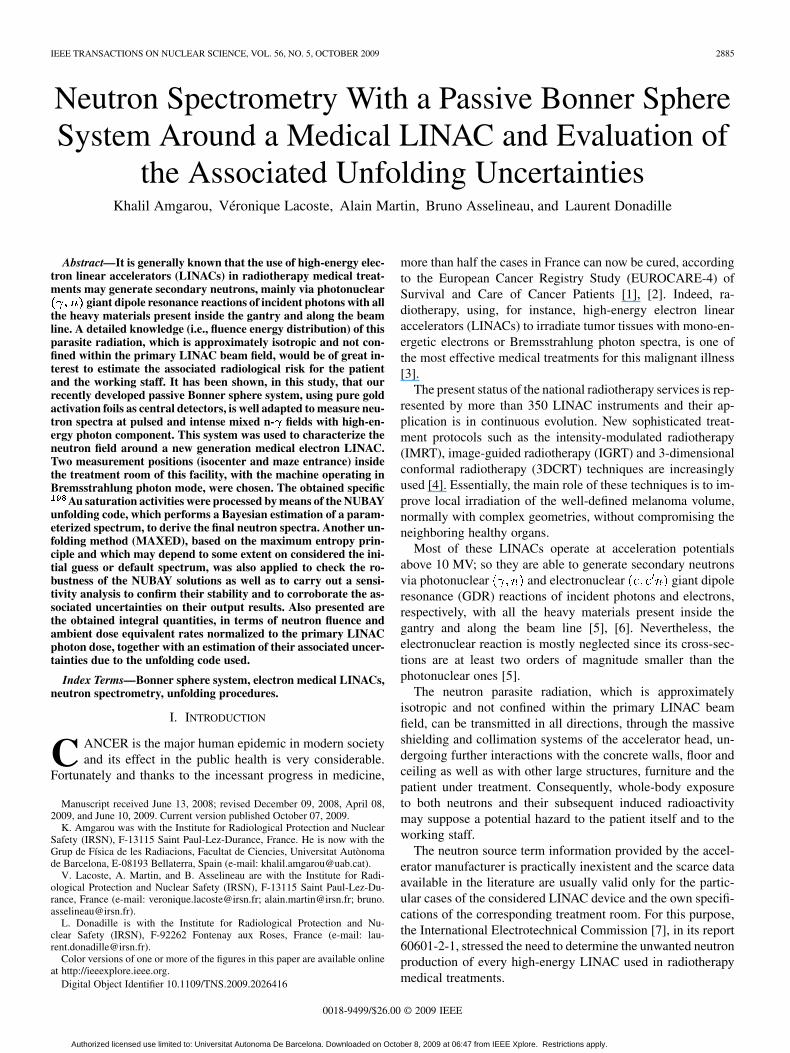
Fig. 8. Solution neutron spectra of MAXED at position 2 using flat (in lethargy)and NUBAY solution as default spectra together with the energy distribution ofthe neutron fluence-to-ambient dose equivalent conversion factors.
of position 1, the fluence rate obtained with MAXED usingthe flat default spectrum is about 7% higher than that obtainedusing the NUBAY solution, but the corresponding values ofthe ambient dose equivalent rates are quite comparable (within3%) in both cases. The 7% excess shown for the fluence rate inthis position is mainly due to thermal neutrons, which are notwell resolved with the MAXED code when using a flat defaultspectrum. However, because of the limited role played bythermal and intermediate neutrons in the dose calculation, thereis a general agreement between the values of the ambient doseequivalent rates evaluated with MAXED using the flat defaultspectrum and those resulting from the NUBAY solution.
Sensitivity analysis: In this step, MAXED was used todetermine the sensitivity of its solution spectrum by inferringsome modifications on the NUBAY default spectrum. One of themain reasons for doing neutron spectrometry with the Bonnersphere system is to assess the ambient dose equivalent rate. Asthis operational quantity depends strongly on the fluence rateof fast neutrons, the parameters defining the high-energy re-gion of the default spectrum ( and ) were varied and thesubsequent consequences were studied. For each measurementposition, six different modified default spectra, all originatingfrom the NUBAY solution spectrum, were considered. For fourof these default spectra, the positions of the high-energy peakswere shifted from their nominal values and successively set to0.1 MeV, 0.5 MeV, 1.5 MeV and 5 MeV. For the other two de-fault spectra, the nominal magnitudes of the high-energy peaksof the NUBAY solution spectrum were first divided and thenmultiplied by a factor of 2.
In all cases, the MAXED solution spectra, using the abovemodified default spectra, have presented almost similar shapesin the high-energy domain with peaks usually located aroundthe same values as those obtained with NUBAY. This demon-strates that when reasonable prior information was given toMAXED unfolding code, its output neutron spectra were ratherunwavering and they were, in general, close to those given byNUBAY. The maximum spectral variations (with respect tothe MAXED solutions that result from the NUBAY nominaldefault spectra) were observed only for the extreme cases of the
modified default spectra with the high-energy peaks shifted to0.1 MeV and 5 MeV. In the case of position 1, the relative dif-ferences found for the integral quantities (fluence and ambientdose equivalent rates) were below 3% and 7%, respectively,whereas maximums of these relative differences (up to 4.5%and 17.5%, respectively) were observed for position 2.
All these results prove the robustness of the NUBAY outputneutron spectra at the two LINAC positions. In addition,NUBAY gives a good estimation of the associated uncertain-ties to the integral quantities since they were, in all cases,very consistent with the spread of the MAXED results usingthe NUBAY solutions as default spectra but after modifyingthe magnitudes and the positions of their fast neutron peaks.These uncertainties may also account for any deviation of thefast-Maxwellian peak from that of the actual generated pho-toneutron spectrum according to the GDR model predictions. Infact, for photonuclear interactions of the incident high-energyphotons with the heavy nuclei present inside the gantry andalong the beam line, the GDR model really predicts a main peak(with a Maxwellian energy distribution) due to nuclear evapo-ration or decay processes of the subsequent excited daughtersplus a small tail (with a relative proportion below 14%) on itsright side as a result of local direct extraction or knock-outmechanism [5]. In this mechanism, the energies and directionsof emitted photoneutrons are highly dependent on those of theincident photons; so that, it is not easy to formulate an accurateanalytical expression, to be implemented by the NUBAY code,of its subsequent contribution to the final neutron spectrum.
V. CONCLUSION
This work highlights the convenience of using the IRSN pas-sive BSS to measure the neutron spectra at pulsed and intensemixed n- fields with a high-energy photon component. Thissystem was used to characterize the neutron field around a newgeneration medical electron LINAC, with the machine operatingin Bremsstrahlung photon mode. The measured specific Ausaturation activities were processed by means of the NUBAYunfolding code, which performs a Bayesian estimation of a pa-rameterized spectrum, to derive the final neutron spectra. An-other unfolding method (MAXED), based on the maximum en-tropy principle and which may depend to some extent on theconsidered initial guess or default spectrum, was also appliedto check the robustness of the NUBAY solutions as well as tocarry out a sensitivity analysis to confirm their stability and tocorroborate the associated uncertainties on their output results.
In general, there is a good agreement between the NUBAYresults and those obtained with MAXED using the NUBAY so-lution as a default spectrum. This means that maximum priorinformation about the measured spectrum was given in this wayto the MAXED code. As NUBAY is based on the Bayesianapproach analysis, it provides a good estimation of the asso-ciated uncertainties on its output results. From the sensitivityanalysis carried out with MAXED, by inferring some modifica-tions on the respective fast neutron peaks of the NUBAY solu-tion spectra, we have observed that the spread of the MAXEDresults of the integral quantities, in terms of fluence and ambientdose equivalent rates, are very consistent with their associateduncertainties as estimated by the NUBAY code. The NUBAY
Authorized licensed use limited to: Universitat Autonoma De Barcelona. Downloaded on October 8, 2009 at 06:47 from IEEE Xplore. Restrictions apply.

AMGAROU et al.: NEUTRON SPECTROMETRY WITH A PASSIVE BONNER SPHERE SYSTEM 2893
TABLE IIPHOTONS EMITTED BY Au AND Au TOGETHER WITH THIER BRANCHING
RATIOS AND MCNPX CALCULATED DETECTION EFFICIENCIES. THE
UNCERTAINTIES OF THESE EFFICIENCIES ARE PURELY STATISTICAL
output relative standard deviations were added as a square-rootof the quadratic sum with the systematic one of the responsefunctions to evaluate the global relative uncertainties of the in-tegral quantities.
On the other hand, the MAXED unfolding code has shownsome limitations when a flat default spectrum or when the meanenergies of the fast neutron peaks for the NUBAY solutionspectra were shifted to their extreme values. However, whenreasonable prior information was given to this code, its outputneutron spectra were rather unwavering and they were, ingeneral, close to those given by NUBAY.
The neutron spectrum at position 1 (isocenter) is quite hard,with a small thermal component whereas that at position 2(maze entrance) is almost fully thermalized proving the effec-tiveness of the structural configuration used for the treatmentroom of this facility. In both cases, the optimal values obtainedfor the mean energy of the fast neutrons are in accordance withthe GDR model that predicts a Maxwellian high-energy peakcentered around 1 MeV, due to nuclear evaporation process,which is, in principle, very similar to that of fission.
In order to assess accurately the neutron effective dose deliv-ered to the patients and the working staff, it would be of great in-terest to extend this study by drawing global maps of a represen-tative sample of the current medical LINAC facilities. For thispurpose, further measurements of the neutron spectra should becarried out at different points inside and outside the consideredtreatment rooms, using for instance our passive and active BSSs,for all the possible configurations of the LINAC apparatus or,at least, for the most common ones used in radiotherapy treat-ments.
APPENDIX
A. Determination of the 426 keV -Rays’ Contributionon the Photo-Peak Measured With NaI Detector
With the help of an identical expression to (1), the specificsaturation activity of Au isotope, which may be formed asa results of the photonuclear Au Au reaction of in-cident high-energy photons (above 8 MeV) with the gold foils,can be estimated from each of its emitted -rays, particularlythose listed in Table II, which are able to affect, to some extent,the Au photo-peak when measured with the portable NaI de-tector described in Section II-D. Also given in this table are thecorresponding branching ratios and the NaI detection efficien-
cies. These efficiencies were calculated using the same MCNPXmodel as in [13].
Then we can write the following equations:
(A1)
(A2)
(A3)
From (A1) and (A2), we obtain
(A4)
The same from (A2) and (A3)
(A5)
Generally, the Genie-2000 spectroscopic software is fullyadapted to separate between two main peaks, andas shown in Fig. 4, by performing multi-Gaussian fit of themeasured pulse-height distribution after background con-tinuum subtraction. If we assume that and
, (A4) becomes
(A6)
By replacing this expression in (A4), we establish the fol-lowing expression:
(A7)
Which means that the contribution of Au 426 keV -raysto the Au photo-peak when measured with the portable NaIdetector is related to the net area of the first peak (see Fig.3) and represents 5.63% of this peak. Therefore, to obtain thenet count rate due to 412 keV -rays, emitted by the neutroninduced Au radionuclides, the following correction must beapplied to the second peak of Fig. 4
(A8)
ACKNOWLEDGMENT
The authors would like to thank the staff at Sainte CatherineHospital of Avignon (France), particularly Dr. R. Garcia, fortheir valuable help in setting up and performing the LINAC ir-radiations of our passive BSS.
REFERENCES
[1] F. Berrino, R. De Angelis, M. Sant, S. Rosso, M. B. Lasota, J. W.Coebergh, and M. Santaquilani, EUROCARE Working group, “Sur-vival for eight major cancers and all cancers combined for Europeanadults diagnosed in 1995–99: Results of the EUROCARE-4 study,” TheLancet Oncology, vol. 8, pp. 773–783, 2007.
[2] A. Verdecchia, S. Francisci, H. Brenner, G. Gatta, A. Micheli, L. Man-gone, and I. Kunkler, EUROCARE Working group, “Recent cancersurvival in Europe: A 2000–02 period analysis of EUROCARE-4 data,”The Lancet Oncology, vol. 8, pp. 784–796, 2007.
Authorized licensed use limited to: Universitat Autonoma De Barcelona. Downloaded on October 8, 2009 at 06:47 from IEEE Xplore. Restrictions apply.

2894 IEEE TRANSACTIONS ON NUCLEAR SCIENCE, VOL. 56, NO. 5, OCTOBER 2009
[3] F. d’Errico, “Dosimetric issues in radiation protection radiotherapy pa-tients,” Radiat. Prot. Dosim., vol. 118, pp. 207–212, 2006.
[4] C. Huntzinger, P. Munro, S. Johnson, M. Miettinen, C. Zankowski, G.Ahlstrom, R. Glettig, R. Filliberti, W. Kaissl, M. Kamber, M. Amstutz,L. Bouchet, D. Klebanov, H. Mostafavi, and R. Stark, “Dynamic tar-geting image-guided radiotherapy,” Med. Dosim., vol. 31, pp. 113–125,2006.
[5] Neutron Contamination From Medical Electron Accelerators, NationalCouncil on Radiation Protection and Measurements (NCRP) Rep. No.79, 1984.
[6] Radiation Protection for Particle Accelerator Facilities, NationalCouncil on Radiation Protection and Measurements (NCRP) Rep. No.144, 2003.
[7] Medical Electrical Equipment—Part 2-1: Particular Requirements forthe Safety of Electron Accelerators in the Range 1 MeV to 50 MeV,International Electrotechnical Commission (IEC) standard report60601-2-1, 1998.
[8] L. Donadille, F. Trompier, I. Robbes, S. Derreumaux, J. Mantione,B. Asselineau, K. Amgarou, A. Martin, J. F. Bottollier-Depois, F.Queinnec, B. Aubert, and I. Clairand, “Radiation protection ofworkersassociated with secondary neutrons produced by medical linear accel-erators,” Radiat. Meas., vol. 43, pp. 939–943, 2008.
[9] V. Lacoste, V. Gressier, J.-L. Pochat, F. Fernández, M. Bakali, andT. Bouassoule, “Characterization of Bonner sphere systems at mono-energetic and thermal neutron fields,” Radiat. Prot. Dosim., vol. 110,pp. 529–532, 2004.
[10] P. Crovisier, B. Asselineau, G. Pelcot, L. Van-Ryckeghem, A. Cadiou,H. Truffert, J. E. Groetz, and M. Benmosbah, “French comparison exer-cise with the rotating neutron spectrometer, ‘ROSPEC’,” Radiat. Prot.Dosim., vol. 115, pp. 324–328, 2005.
[11] T. Shima, S. Naito, Y. Nagai, T. Baba, K. Tamura, H. Ohgaki, T. Kii,and H. Toyokawa, “Photodesintegration cross sections of He and Heat low energies,” Nucl. Phys. A, vol. 718, pp. 23c–26c, 2003.
[12] S. Naito, Y. Nagai, T. Shima, H. Makii, K. Mishima, K. Tamura, H.Toyokawa, H. Ohgaki, J. Golak, R. Skibinski, H. Witala, W. Glöckle,A. Nogga, and H. Kamada, “New data for total He��� ��� andHe��� ���� cross sections compared to current theory,” Phys. Rev.
C, vol. 73, p. 034003, 2006.[13] K. Amgarou, V. Lacoste, H. Muller, and F. Fernández, “Set-up of a
passive Bonner sphere system for neutron spectrometry at mixed fieldswith predominant photon component based on activation detector,” Ra-diat. Prot. Dosim., vol. 126, pp. 337–341, 2007.
[14] Conversion Coefficients for Use in Radiological Protection Against Ex-ternal Radiation, International Commission on Radiological Protrec-tion (ICRP) publication 75, Pergamon, 1997.
[15] M. L. Williams and E. Sajo, “Deterministic calculations of photonspectra for clinical accelerator targets,” Med. Phys., vol. 29, pp.1019–1028, 2002.
[16] K. Amgarou, MCNPX Response Matrix Calculations of the IRSN Pas-sive Bonner Sphere System IRSN Report DRPH/SDE no. 2007-24.
[17] D. E. Cullen, A Temperature Dependent ENDF/B-VI, Release8 Cross Section Library, Lawrence Livermore Nat. Lab., Rep.UCRL-TR-202284, 2004.
[18] V. Gresssier and J. L. Pochat, “Les installations de l’IRSN dédiées à lamétrologie des neutrons,” Radioprotection, vol. 41, pp. 11–32, 2006.
[19] K. Amgarou, Experimental Characterisation of the IRSN PassiveBonner Sphere System at AMANDE Facility, IRSN Rep. DRPH/SDEno. 2007-13.
[20] A. D. Polyanin and A. V. Manzhirov, Handbook of Integral Equa-tions. Boca Raton, FL: CRC, 1998.
[21] J. Hadamard, “Sur les problèmes aux dérivées partielles et leur signifi-cation physique,” Princeton Univ. Bull., pp. 49–52, 1902.
[22] A. V. Alevra and D. J. Thomas, “Neutron spectrometry in mixed fields:Multisphere spectrometers,” Radiat. Prot. Dosim., vol. 107, pp. 37–72,2003.
[23] A. Höcker and V. Kartvelishvili, “SVD approach to data unfolding,”Nucl. Instrum. Meth. A, vol. 372, pp. 469–481, 2002.
[24] M. Matzke, Unfolding of Pulse Height Spectra: The HEPRO ProgramSystem Physikalisch-Technische Bundesanstalt, Braunschweig, Rep.PTB-N-19, 1994.
[25] M. Mattzke, “Unfolding procedures,” Radiat. Prot. Dosim., vol. 107,pp. 155–174, 2003.
[26] M. Reginatto, “Bayesian approach for quantifying the uncertainty ofneutron doses derived from spectrometric measurements,” Radiat.Prot. Dosim., vol. 121, pp. 64–69, 2006.
[27] M. Reginatto and P. Goldhagen, “MAXED, a computer code formaximum entropy deconvolution of multisphere neutron spectrometerdata,” Health Phys., vol. 77, pp. 583–1999, 1999.
[28] E. Cordes, G. Fehrenbacher, R. Schutz, M. Sprunck, K. Hahn, R. Hof-mann, J. P. Biersack, and W. Wahl, “An approach to unfold the re-sponse of a multi-element system using an artificial neural network,”IEEE Trans. Nucl. Sci., vol. 45, pp. 1464–1469, 1998.
[29] M. Tomás, F. Fernández, M. Bakali, and H. Muller, “MITOM: A newunfolding code based on a spectra model method applied to neutronspectrometry,” Radiat. Prot. Dosim., vol. 110, pp. 545–548, 2004.
[30] R. Bedogni, C. Domingo, A. Esposito, and F. Fernández, “FRUIT: Anoperational tool for multisphere neutron spectrometry in workplaces,”Nucl. Instrum. Meth. A, vol. 476, pp. 242–246, 2002.
[31] D. W. Freeman, D. R. Edwards, and A. E. Bolon, “Genetic algo-rithms—A new technique for solving the neutron spectrum unfoldingproblem,” Nucl. Instrum. Meth. A, vol. 425, pp. 549–576, 1999.
[32] V. Lacoste, M. Reginatto, B. Asselineau, and H. Muller, “Bonnersphere neutron spectrometry at nuclear workplaces in the frameworkof the EVIDOS project,” Radiat. Prot. Dosim., vol. 125, pp. 304–308,2007.
[33] V. Gressier, V. Lacoste, L. Lebreton, H. Muller, G. Pelcot, M. Bakali,F. Fernández, M. Tómas, N. J. Roberts, D. J. Thomas, M. Reginatto,B. Wiegel, and J. Wittstock, “Characterisation of the IRSN CANEL/T400 facility producing realistic neutron fields for calibration and testpurposes,” Radiat. Prot. Dosim., vol. 110, pp. 523–527, 2004.
[34] M. Luszik-Bhadra, D. Bartlett, T. Bolognese-Milsztajn, M. Boschung,M. Coeck, G. Curzio, F. d’Errico, A. Fiechtner, V. Lacoste, L. Lind-borg, M. Reginatto, H. Schuhmacher, R. Tanner, and F. Vanhavere,“Characterisation of mixed neutron-photon workplace fields at nuclearfacilities by spectrometry (energy and direction) within the EVIDOSproject,” Radiat. Prot. Dosim., vol. 124, pp. 219–229, 2007.
[35] L. Lebreton, A. Zimbal, and D. Thomas, “Experimental comparisonof ����� neutron fluence energy distributions,” Radiat. Prot.Dosim., vol. 126, pp. 3–7, 2007.
[36] D. Spiegelhalter, A. Thomas, N. Best, and D. Lunn, WinBUGS UserManual, 2003 [Online]. Available: http://www.mrc-bsu.cam.ac.uk/bugs
[37] D. S. Sivia, Data Analysis—A Bayesian Tutorial. Oxford, U.K.:Clarendon, 1996.
[38] S. P. Brooks, “Markov chain Monte Carlo method and its application,”The Statistician, vol. 47, pp. 69–100, 1998.
[39] D. J. C. MacKay’s, Information Theory, Inference, and Learning Algo-rithms. Cambridge, U.K.: Cambridge Univ. Press, 2003.
[40] Neutron Fluence Measurements International Atomic Energy Agency(IAEA) reports series No. 107, 1970.
[41] J. Geweke, “Evaluating the accuracy of sampling-based approachesto the calculation of posterior moments,” in Bayesian Statiist., J. M.Bernardo, J. O. Berger, A. P. Dawid, and A. F. M. Smith, Eds. Ox-ford, U.K.: Oxford Univ. Press, 1992, vol. 4, pp. 169–193.
[42] S. P. Brooks and A. Gelman, “Alternative methods for monitoring con-vergence of iterative simulations,” J. Comput. Graph. Statist., vol. 7,pp. 434–455, 1998.
[43] D. J. Spiegelhalter, N. G. Best, B. P. Carlin, and A. Van Der Linde,“Bayesian measures of model complexity and fit,” J. Roy. Statist. Soc.B, vol. 64, pp. 583–640, 2002.
[44] E. T. Jaynes, “Information theory and statistical mechanics,” Phys.Rev., vol. 106, pp. 620–630, 1957.
[45] W. L. Goffe, G. D. Ferrier, and J. Rogers, “Global optimization of sta-tistical functions with simulated annealing,” J. Econometrics, vol. 60,pp. 65–99, 1994.
[46] M. Reginatto, P. Goldhagen, and S. Neumann, “Spectrum unfolding,sensitivity analysis and propagation of uncertainties with the maximumentropy deconvolution code MAXED,” Nucl. Instrum. Meth. A, vol.476, pp. 242–246, 2002.
[47] UMG, 2004, Unfolding With MAXED and GRAVEL, Version 3.3,This package is available under request from RSICC (Radiation SafetyInformation Computational Center) and from the OECD/NEA (Or-ganisation for Economic Co-operation’s Development Nuclear EnergyAgency) data bank.
[48] R. Barquero, R. Mendez, H. R. Vega-Carrillo, M. P. Iñiguez, and T. M.Edwards, “Neutron spectra and dosimetric features around an 18 MVLINAC accelerator,” Health Phys., vol. 88, pp. 48–58, 2005.
[49] P. H. McGinley, Shielding Techniques for Radiation Oncology Facili-ties. Madison, WI: Medical Physics Publishing, 1998.
[50] W. L. Huang, Q. F. Li, Y. Z. Lin, Q. Su, and Y. S. Luo, “Measurementsof photoneutrons produced by 15 MeV electron linac for radiographyapplications,” Nucl. Instrum. Meth. B, vol. 251, pp. 361–366, 2006.
[51] D. J. Thomas, A. G. Bardell, and E. M. Macaulay, “Characterisationof a gold foil-based Bonner sphere set and measurements of neutronspectra at a medical accelerator,” Nucl. Instrum. Meth. A, vol. 476, pp.31–35, 2002.
Authorized licensed use limited to: Universitat Autonoma De Barcelona. Downloaded on October 8, 2009 at 06:47 from IEEE Xplore. Restrictions apply.

AMGAROU et al.: NEUTRON SPECTROMETRY WITH A PASSIVE BONNER SPHERE SYSTEM 2895
[52] D. J. Thomas, N. P. Hawkes, L. N. Jones, P. Kolkowski, and N. J.Roberts, “Caharacterization and utilization of a Bonner sphere setbased on gold activation foils,” Radiat. Prot. Dosim., vol. 126, pp.229–233, 2007.
[53] R. M. Howell, M. S. Ferenci, N. E. Hertel, and G. D. Fullerton, “Inves-tigation of secondary neutron dose for 18 MV dynamic MLC IMRTdelivery,” Med. Phys., vol. 32, pp. 786–793, 2005.
[54] R. M. Howell, M. S. Ferenci, N. E. Hertel, G. D. Fullerton, T. Fox, andL. W. Davis, “Measurements of secondary neutron dose from 15 MVand 18 MV IMRT,” Radiat. Prot. Dosim., vol. 115, pp. 508–512, 2005.
[55] O. Chibani and C. M. C. Ma, “Photonuclear dose calculations for high-energy photon beams from Siemens and Varian linacs,” Med. Phys.,vol. 30, pp. 1990–2000, 2003.
[56] A. Zanini, E. Durisi, F. Fasolo, C. Ongaro, L. Visca, U. Nastasi, K.W. Burn, G. Scielzo, J. O. Adler, J. R. M. Annand, and G. Rosner,“Monte Carlo simulation of the photoneutron field in linac radiotherapytreatments with different collimation systems,” Phys. Med. Biol., vol.49, pp. 571–582, 2004.
Authorized licensed use limited to: Universitat Autonoma De Barcelona. Downloaded on October 8, 2009 at 06:47 from IEEE Xplore. Restrictions apply.