neurocognition of patients on whole brain radiotherapy: a ...
144
NEUROCOGNITION OF PATIENTS ON WHOLE BRAIN RADIOTHERAPY: A PROSPECTIVE STUDY Dissertation submitted for partial fulfillment of the rules and regulations DOCTOR OF MEDICINE BRANCH - XVIII (PSYCHIATRY) THE TAMILNADU DR.MGR MEDICAL UNIVERSITY, CHENNAI, TAMIL NADU MAY 2018
-
Upload
khangminh22 -
Category
Documents
-
view
1 -
download
0
Transcript of neurocognition of patients on whole brain radiotherapy: a ...
NEUROCOGNITION OF PATIENTS ON
WHOLE BRAIN RADIOTHERAPY:
A PROSPECTIVE STUDY
Dissertation submitted for partial fulfillment
of the rules and regulations
DOCTOR OF MEDICINE
BRANCH - XVIII (PSYCHIATRY)
THE TAMILNADU DR.MGR MEDICAL UNIVERSITY,
CHENNAI,
TAMIL NADU
MAY 2018
CERTIFICATE
This is to certify that the dissertation titled, “NEUROCOGNITION
OF PATIENTS ON WHOLE BRAIN RADIOTHERAPY:
A PROSPECTIVE STUDY” is the bonafide work of Dr. Rajesh M, in
part fulfillment of the requirements for the M.D. Branch – XVIII (Psychiatry)
examination of The Tamilnadu Dr. M. G. R. Medical University, to be held in
May 2018. The period of study was from March 2017 – Sep 2017.
The Director, The Dean, Institute of Mental Health Madras Medical College Chennai – 600 010. Chennai – 600 003

CERTIFICATE OF GUIDE
This is to certify that the dissertation titled, “NEUROCOGNITION OF
PATIENTS ON WHOLE BRAIN RADIOTHERAPY: A PROSPECTIVE
STUDY” is the original work of Dr. Rajesh M, done under my guidance
submitted in partial fulfillment of the requirements for M.D. Branch – XVIII
[Psychiatry] examination of The Tamilnadu Dr. M. G. R. Medical University,
to be held in May 2018.
Dr Poornachandrika, MD, DCh Associate Professor,
Institute of Mental Health, Chennai.
DECLARATION
I, Dr.RAJESH M, solemnly declare that the dissertation titled,
“Neurocognition of patients on Whole Brain Radiotherapy:
A prospective study’’ is a bonafide work done by myself at the Madras
Medical College, Chennai, during the period from March 2017 – September
2017 under the guidance and supervision of Prof. Dr. Shanthi Nambi MD,
FIPS, Professor of Psychiatry, Madras Medical College.
The dissertation is submitted to The Tamilnadu Dr. M.G.R. Medical
University towards part fulfillment for M.D. Branch XVIII (Psychiatry)
examination.
Place: Dr. RAJESH. M
Date :
ACKNOWLEDGEMENTS
With gratitude, I sincerely thank Professor Dr. Shanthi Nambi, MD,
FIPS, Director, Institute of Mental Health, Chennai, for her constant guidance
and support.
I acknowledge and thank Professor Dr. R. Narayana Babu, MD, DCh,
Dean, Madras Medical College, Chennai, for permitting me to utilize the
institutional and academic resources.
I sincerely thank Professor Dr. N.V. Kalayarasi, MDRT, DCh Head
of Department of Radiation Oncology, Barnard Institute of Radiology, Madras
Medical College for her guidance and permitting me to utilize the departmental
resources.
I also profoundly thank Professor Dr, R.Ravi. MD,DMRD, Director
Barnard institute of Radiology, Madras Medical College, Chennai for his
support.
With deep gratitude I thank my guide Dr Poornachandrika, MD, DCh,
associate professor and co-guide Dr. Mythili, MD, DMRT, assistant professor,
for their guidance, encouragement and support. I sincerely thank all my Senior
Professors, Associate Professors and Assistant Professors of Institute of Mental
health, for their constant cooperation and enthusiastic encouragement.
I profoundly thank the statistician Mr.Srinivasan.R, M.Sc,M.P.S,Ph.D,
ICMR, TB Research Institute, Chetpet for analysis and support.
I acknowledge the help and support rendered by my batch colleagues,
and also my seniors and juniors towards smooth conduct and coordination of

the study. I specially my juniors Suba sree, Rose Monica and Sivaji for their
help in compiling the data.
I also thank all my family members for blessings and cooperation.
Above all I thank all the participants of the study – the patients of
Barnard Institute of Radiology and their family members for their consent and
cooperation.

Urkund Analysis Result Analysed Document: pliagarism 3.docx (D31316503)Submitted: 10/14/2017 3:00:00 PM Submitted By: [email protected] Significance: 1 %
Sources included in the report:
Thèse Faty-MENIAI 20-03-17.pdf (D26588718)
Instances where selected sources appear:
1
U R K N DU
ABBREVIATIONS
ADL – Activities of Daily Living
AMPA – α-amino-3-hydroxy-5-methyl-4-isoxazolepropionic
acid
ARC – Activity-regulated cytoskeleton-associated protein
ASTRO – American Society for Radiation Oncology
BKT – Binet Kamath Test.
EORTC - European Organization for Research and Treatment of
Cancer
FACT-BR – Functional Assessment of Cancer Therapy - Brain
HVLT – Hopkins Verbal Learning Test
IMRT – Intensity modulated Radiotherapy
KPS – Karnofsky Performance Status
LBADL – Lawton Body Activities of Daily Living
LTP – Long term potentiation
MGd – Motexafin gadolinium
MMSE – Mini Mental Status Examination
MoCA – Montreal Cognitive Assessment Scale
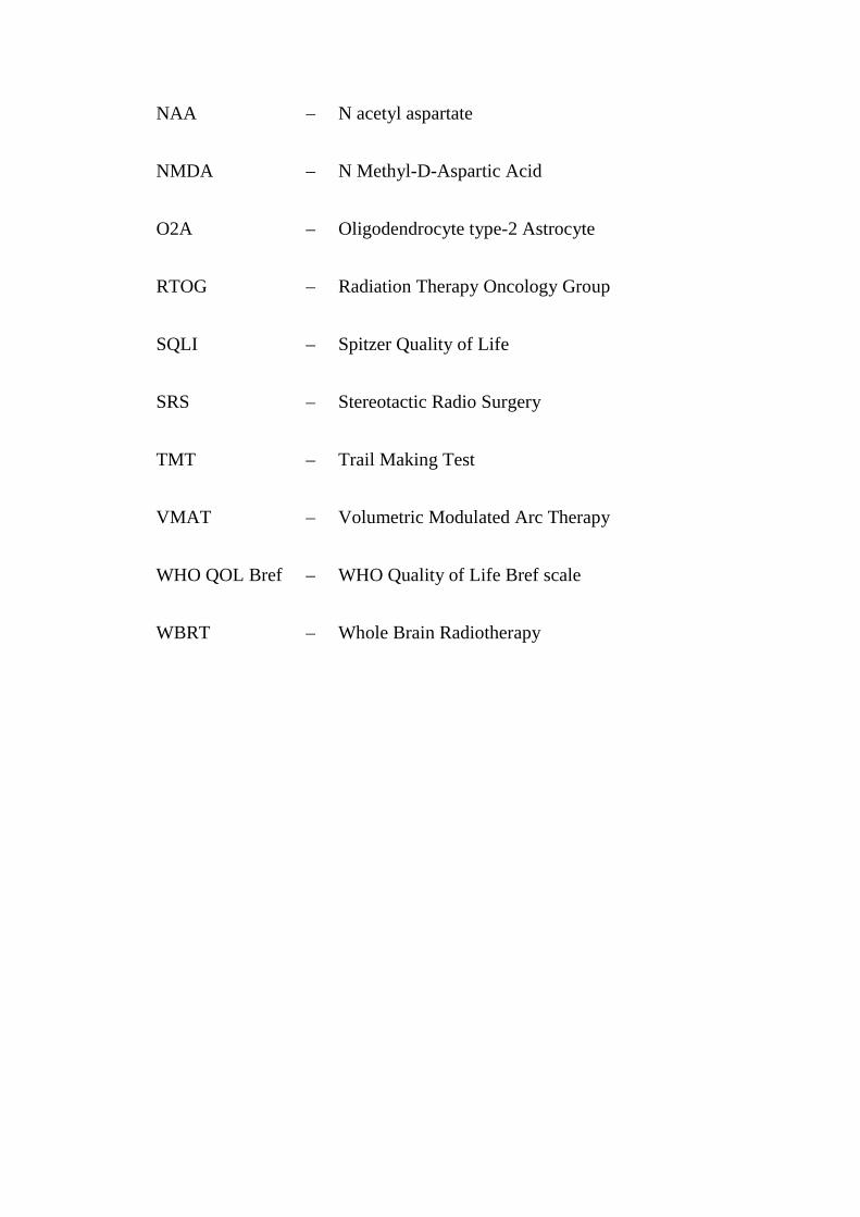
NAA – N acetyl aspartate
NMDA – N Methyl-D-Aspartic Acid
O2A – Oligodendrocyte type-2 Astrocyte
RTOG – Radiation Therapy Oncology Group
SQLI – Spitzer Quality of Life
SRS – Stereotactic Radio Surgery
TMT – Trail Making Test
VMAT – Volumetric Modulated Arc Therapy
WHO QOL Bref – WHO Quality of Life Bref scale
WBRT – Whole Brain Radiotherapy
TABLE OF CONTENTS
NO
TOPIC
Page No
1. INTRODUCTION 1
2. REVIEW OF LITERATURE 4
3. AIMS AND OBJECTIVES 41
4. METHODOLOGY 42
5. RESULTS 52
6. DISCUSSION 68
7. CONCLUSION 79
8. LIMITATIONS 80
9. FUTURE DIRECTIONS 81
10. BIBLIOGRAPHY 82
11. APPENDIX
1
INTRODUCTION
Brain metastasis is a condition with high morbidity and mortality. Exact
incidence of brain metastasis remains unknown. Brain metastasis is the most
common intracranial tumors in adults. The overall incidence of brain metastasis
has drastically increased due to improved detection and treatment(1). MRI and
other imaging modalities have improved the detection and exact localization of
these masses. The studies say that 9 to 11 percent of cancers develop cerebral
metastasis(2). In the past the median survival for the patients with brain
metastasis on steroids and supportive care was only 2 months.(3) So the
treatment of brain metastasis in favorable patient mainly concentrated on
maintaining quality of life (QoL) and neurocognitive function.
WBRT is a standard treatment for patients with late stage metastatic
brain tumors. (4)Until today there are no randomized control trials to prove the
benefit of any intensified treatment beyond WBRT(5). WBRT is a palliative
process for patients with brain metastasis that reduce symptoms and improve
quality of life. In brain metastasis median survival is considered to be low and
estimated to be around 7 months.(6) In contrast a small group of cases with
brain metastasis survive beyond 5 years.(7)so the survival of the patients is
unpredictable and various palliative measures are absolutely necessary to
reduce the suffering. WBRT has the significant capacity to reduce tumor mass.
But this treatment is not without side effects. The greatest fear with the
application of WBRT is the fear of neurocognitive decline and in rare cases of
dementia. Many experiments conducted on animal models have proved that the

2
micro vascular damage and cell death due to radiation exposure were the main
reason for cognitive deficit. The decline in cognition in cancer patients may be
due to several factors ranging from primary mass effect, radiation, surgery,
secondary depression, anxiety, sleep disturbance, chemotherapy,
immunological and hormonal reasons. So defining the exact cause of cognitive
decline is impossible. But in terms of temporal correlation cognitive decline is
definitely seen with WBRT.
The patients with secondaries to the brain are often debilitated and
hence measurement of the cognitive decline in this group of patients is a
challenge. The main component of this WBRT related cognitive decline is the
immediate and delayed verbal memory(8). But the decline in performance is
seen in various other neurocognitive and executive functions(9). Multiple
studies are done in this area but none could specifically confirm the change
happening with WBRT exposure. In terms of the duration and pattern of
change specific studies are still not available. Studies say that early cognitive
decline is seen in 1 to 4 months of exposure to WBRT(10). The effects seen in
due course are late cognitive effects. Several studies have been done in this
subject but have given unequivocal results.
Cognitive decline is said to be the main reason for poor quality of life in
majority of cancer patients. But general cognitive function and quality of life
was only partially affected by it. Since the neurocognitive decline and poor
quality of life occur in a sequence, the present treatment modalities mainly
concentrate on delaying the decline in neurocognitive function(11).
3
The studies in this area are very few and mainly done in retrospective
way. But none of the studies could definitely provide the proof of association
between WBRT and Neurocognition. The reason is that many patients in
addition went on to have a surgery and chemotherapy which may have had an
effect on cognition. With the developments in radiation technology like
stereotactic radiotherapy the precision of delivery of radiation has
improved(12). But in many centers WBRT is still used for its benefits and
efficacy.
With the developments in diagnostics, imaging and radiation
technology, treatment of metastases has become a therapeutic challenge.
So we performed a prospective study on patients who were on WBRT
and made an attempt to assess the correlation of WBRT on Neurocognition.
We also compared their quality of life and indirectly assessed their quality of
living.
4
REVIEW OF LITERATURE
Whole brain radiotherapy
Metastasis may spread from any region of the body to brain. The
primaries are usually found in lungs, breasts and gastrointestinal region. The
studies found that 30% to 40% of all the cancers metastasize to the brain. Until
1950s there was no specific therapy that could target these inoperable
metastatic tumors. The advent of radiotherapy marked the first step in treating
and palliating these tumor afflicted people. The benefit of WBRT was proved
beyond doubt by 1970s and later proved to be the gold standard in treating
brain metastases. It was the work of Chao et al that paved the way in
establishing the effect of radiation.(13) He claimed that 63% of the effects of
tumor can be reduced with radiation exposure and went on to tell that there
were patients who were radio resistant.
Whole brain radiotherapy (WBRT) is a mainstay of treatment and
palliation in patients with brain metastases. It consists of administering
therapeutic doses of radiation to the tumor sites in calculated fractions. WBRT
is administered to a wide variety of conditions ranging from primary brain
tumors, secondary metastasis; prophylactic cranial irradiation etc. The
secondaries with multiple masses often prevent surgical intervention and then
the only intervention of choice would be WBRT or chemotherapy. The unique
feature of WBRT is that the treatment does not depend on histological type of
mass, as it works even on the masses that are said to be radio resistant. Thus by
5
destroying histologically varied tumor masses WBRT may be considered gold
standard in treating brain metastasis.
WBRT works on 4 main principles(14)
• Repair – WBRT given is lesser calculated doses will give required
duration for normal cells to repair
• Reoxygenation- hypoxic areas in tumor mass are radio resistant, but
lower doses of radiation permit required amount of circulation.
• Redistribution- tumor cells in S phase are resistant to radiation, so
enough time must be given between each radiation cycle to permit the
differentiation of cells.
• Repopulation- tumor cells on irradiation grow too quickly, hence
excess time between radiation cycles will allow tumor cells to
repopulate.
Role of WBRT, in the treatment of brain metastasis.
Multiple studies have shown that 64% to 83 % of the patients gain
improvement with WBRT. The studies say that survival benefit of 6 to 8
months is obtained with WBRT. Usually the radiotherapy is administered as
30Gy in 10 fractions over 2 weeks. Many alternative dose regimens have been
established, but the risk benefit response [tumor mass clearance Vs side effect]
was found to be best with the above regimen for brain metastasis. Regarding
cognitive outcome in alternative regimens, not many studies have been done
6
but with higher doses of radiation the physical complications have found to be
seen beyond doubt. So with respect to alternative regimen multiple prospective
studies are needed to find the exact outcomes in terms of risk and benefit in the
outcome. The median survival obtained with patients treated with WBRT was
found to be 4.9months.(15)
Radiation toxicity(10)
Early side effects
The toxic side effects that are seen in first few days to weeks of
treatment of radiotherapy are called early side effects. It consists of headache,
alopecia, nausea, and vomiting and reduced appetite. These are often self
limited and subside on their own or with minimal intervention. Another
common early side effect is cerebral edema which is often generously treated
with corticosteroids. Encephalopathy, herniation and death are very rarely seen
early side effects.
Early to delayed side effects
This includes anorexia, vomiting, headache, fatigue, memory decline
and focal neurological deficits. Rarely in pediatric population is a condition
called somnolence syndrome seen with above symptoms and irritability. Even
the early to delayed side effects are often self limited.
7
Late side effects
The side effects occurring after 90 days of administering the radiation
therapy are late side effects. According to Radiation Therapy Oncology Group
[RTOG] late side effects have poor outcome and are not self limiting. So
generally these late side effects with preserved cognition determine the quality
of life of the patient. Meta analysis done by Tallet et al with 7 studies was
useful in showing that there was a cognitive decline of 31% to57% in 3
months(16). Same study also predicted a decline in cognition from 48% to 89%
in 1 year. The cognitive decline caused by the growth of tumor mass is
significantly higher than the neurocognitive decline due to WBRT. Hence it is
used as a successful treatment modality for terminal stage cancers and multiple
metastases.
Leukoencephalopathy is another serious side effect(10). It presents as
dysarthria, ataxia, neurocognitive dysfunction, seizures and lethargy.
Periventricular white matter changes and ventricular enlargement on CT and
MRI scans help in diagnosis and management.
Radiation necrosis is the most serious side effect seen very rarely. It
presents with necrosis of blood vessels and endothelium. This extensive
vascular necrosis can lead to serious neurological deficit or death.
A study by Kondziolka et al(17) studied the patients perspective
regarding complications after WBRT. He reported that 88% have hair loss,
short-term memory (72%), long-term memory (33%), concentration (61%), and
8
depression (54%). He also showed in his study that 63% of the patients on
WBRT thought that they had side effects from taking the therapy.
Stereotactic radio surgery is emerging as a new development in treating
cranial metastasis. It helps to deliver strong doses of radiation to metastatic foci
in the brain avoiding undesirable exposure to normal tissue. But many experts
in the field strongly believe that STEREOTACTIC RADIOSURGERY will
often fail to irradiate micro metastasis in other areas of brain. Current imaging
techniques are still not advanced in identifying the micro metastasis in the
brain. Thus WBRT helps in irradiating the tumor tissue and destroying even the
micro metastases and so helps in killing all the tumor cells in brain.
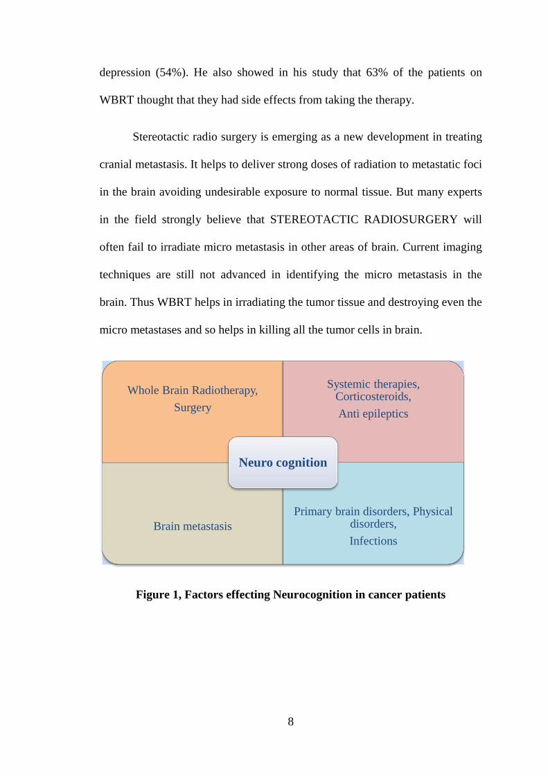
Figure 1, Factors effecting Neurocognition in cancer patients
Whole Brain Radiotherapy,Surgery
Systemic therapies, Corticosteroids,Anti epileptics
Brain metastasisPrimary brain disorders, Physical
disorders,Infections
Neuro cognition
9
Neurocognition in WBRT
Cancer treatment associated cognitive decline is a serious matter of
concern. The major manifestation of WBRT is the development of
neurocognitive impairment. The time duration for WBRT induced cognitive
decline is found to vary among various studies.(18) The impairment ranges
from inattention, impaired short and long term memory, poor reasoning and in
the end frank dementia. Even with the development of several newer advances
like Stereotactic radio surgery; WBRT still continues to be widely used due to
the total survival benefit obtained out of it.
Vascular hypothesis of radiation induced brain injury(19)
Radiation has profound effect on the neurons and surrounding
vasculature(20). Vascular theory mainly argues that radiation exposure causes
damage to the endothelial cells and suppresses its proliferations. Thus it breaks
down blood brain barrier and leads to inflammatory damage to the interstitium.
Several studies in this regard have shown the development of vessel wall
thickening, vessel dilation, and endothelial cell nuclear enlargement in cells
exposed to radiation(20). Studies by Warrington et al have shown that in rats,
the radiation therapy causes exposure of the interstitium and capillary
rarefaction. Inflammatory cytokines, TNF alpha, IL-12, IL 18 are mainly
involved in the damage of the tissues. Due to the above reasons the blood brain
barrier gets breached causing the exposure of matrix metalloproteinase – 2 and
metalloproteinase – 2 tissue inhibitor(21). Radiation also causes degradation of
10
collagen type IV, which is an extracellular matrix component of the blood
vessel basement membrane, and report changes in the mRNA and protein
expression of VEGF, Ang-1, Tie-2, and Ang-2.(22) The earliest known damage
is seen at 10 weeks post radiation. There is profound micro vascular deficit,
metabolic disturbance, immune response and neuronal hypoxia which is
together said to be responsible for cognitive decline occurring after
WBRT.(20).
Next culprit is hippocampal neurogenesis. Neurocognitive impairment
occurs due to problem in hippocampal neurogenesis.(23). Radiation-induced
cognitive impairment occurs in up to 90% of adult brain tumor patients who
survive >6 months after whole brain radiotherapy(24).
Dentate gyri, CA 3, CA 1 are the predominant areas of hippocampal
neurogenesis. Radiation induced damage of these areas are associated with
poor regeneration of neurons and hence said to be the cause for cognitive
decline.
Majority of the studies mainly concentrate on the hippocampal injury for
cognitive decline.(24) Other possible causes include:
• NMDA receptor alterations.
• Disrupted arc expression in hippocampal neurons
• Genetic factors
• Neuronal inflammation/ oxidative stress.
11
Neurocognitive tests show varied results based on the domain measured in
different population.
Cellular and molecular pathway of neurocognitive decline(25)
Molecular mechanism for radiation induced cognitive decline is ill-
defined. In the past it was considered that radiation induced DNA damage was
the cause of neuronal cell death. There was also the endothelial cell death and
glial cell loss which was indirectly considered to cause cognitive decline. The
latest research with the use of fMRI scans has given a different picture. The
present hypothesis is that cognitive decline is a manifestation of complex
interaction between various neuronal cells. Astrocytes, microglia, neurons and
oligodendrocytes interact in a complex way with, endothelial cells to produce
dynamic changes.
Parenchymal hypothesis of brain injury(19)
Oligodendrocyte
This theory was basically focused on the oligodendrocyte which is
involved in the formation of myelin sheath. Oligodendrocyte type-2 astrocyte
(O-2A) progenitor cell is the cell that is necessary for the formation of
oligodendrocytes(26). These progenitor cells lose their reproductive capacity
on exposure to whole brain radiation therapy in the rats. The above hypothesis
states that radiation induced loss of O-2A progenitor cells causes failure to
replace oligodendrocytes which ultimately results in demyelination and white
matter necrosis. After whole brain irradiation there was a decrease in number
12
of oligodendrocytes in 24 hrs after irradiation but there was no change in the
number of myelinated axons, myelin sheath thickness, and the cross-sectional
area of myelinated axons in cognitively impaired rats after 12 months of whole
brain irradiation dose of 40 Gy delivered twice a week for 4 weeks to middle-
aged rats(27). Thus the above theory is still unclear in explaining all the
questions regarding cognitive deficits on radiation exposure.
Astrocytes
Astrocytes constitute more than 50% of the glial cell population. They
are involved in modulation of synaptic transmission and secrete important
neurotrophic factors such as fibroblast growth factor to promote neurogenesis.
They also protect neurons and protect endothelial cells. They form a
component of blood brain barrier and secrete many pro inflammatory
cytokines.(28) In case of injury these astrocytes undergo hypertrophic changes
and secrete pro-inflammatory mediators such as cyclooxygenase (Cox)-2 and
the intercellular adhesion molecule (ICAM)-1. During inflammation there is
increased expression of glial fibrillary acidic protein (29)and breach in blood
brain barrier. The resulting gliosis causing radiation induced edema is said to
be the likely cause of brain injury due to irradiation.
Microglia
Microglia constitutes the cells involved in phagocytosis. They maintain
homeostasis by secretion of neurotrophic factors.(30) Irradiation of these cells
results in rounding of cells, retraction of cell processes, formation of
13
chemokines and reactive oxygen species. Rat models have shown increase in
number of activated microglia around hippocampus with increased pro
inflammatory markers after radiation exposure.(31) Anti inflammatory drugs
like indomethacin and ramipril reduce activated microglia in the hippocampus
and perirhinal cortex(21). Thus in rodents they are shown to prevent radiation-
induced cognitive impairment.
Neurons
Historically neurons were considered to be radio resistant. But the newer
studies have shown that radiation causes changes in hippocampal neurons, in
synaptic connectivity and gene expression. Irradiating the rat brain causes.
(i) Neuronal receptor expression of the immediate-early gene activity-
regulated cytoskeleton-associated protein (Arc)(32)
(ii) N methyl-D-aspartic acid (NMDA) receptor subunits
(iii) Glutaminergic transmission
(iv) Hippocampal long-term potentiation(33)
All the above changes are considered to be important for normal
cognitive functioning. Thus, a dynamic interaction of all the radiation induced
changes in all the cells types is said to be the reason behind the neurocognitive
change occurring after radiation exposure.

14
Analysis on glial clonogens in rat models
Experiment on rat with fractional whole brain irradiation resulted in
reduced endothelial cells, vascular length and density. Two months later the
tissue hypoxia and capillary rarefaction increased in hippocampal areas. When
fetal neural stem cells were given IV to these rats the cognition in these rats
improved. After each 5Gy fraction of whole brain radiotherapy the IV neural
stem cells were supplemented and cognition was maintained with no alteration
in vascular length or density.
Oligodendrocyte type-2 astrocyte (O-2A) progenitor cell is said to be the
primary cell death occurring in WBRT. The radiation induced death of these
cells results in demyelination and white matter necrosis. This is considered to
be a possible cause of cognitive decline. But the above change is only
considered to be transient.
Radiation exposure in rodents causes following changes(25)
• Hippocampal long term potentiation
• Neuronal receptor expression of the immediate-early gene activity-
regulated cytoskeleton-associated protein (Arc) ,
• N-methyl-D-aspartic acid (NMDA) receptor subunits,
• Glutaminergic transmission
Wu et al(34) showed that radiation exposure to rat brains resulted in
reduced tyrosine phosphorylation, reduced exposure of NMDA receptors and in
15
contrast increased expression of GABA receptors. NMDA (N-methyl D-
aspartate) and AMPA (a-amino-3-hydroxy-5-methyl-4-isoxazolepropionic
acid) receptors are responsible for excitatory signaling in hippocampus.
Whereas, the GABA (gamma-amino butyric acid) receptors are responsible for
inhibitory neurotransmission. Thus above receptors and the neurotransmitters
play unique role in managing neural plasticity and in turn are vital for normal
Neurocognition.
Radiation leads to acute changes in tyrosine phosphorylation in various
research models. It is found that trafficking of neurotransmitter receptors to and
fro from the cell surface is dependent on tyrosine phosphorylation. Thus,
tyrosine phosphorylation-dependent trafficking of excitatory NMDARs to the
synapse plays a role in LTP.
1) Radiation Alters NMDA receptor phosphorylation thus changes
NMDA and GABA A receptor Localization.
2) Radiation Alters NMDA and GABA responses and thus acutely
Inhibits Long term potentiation.
These changes caused alteration in synaptic responses and reduction of
long term potentiation which in turn resulted in reduced cognition.
16
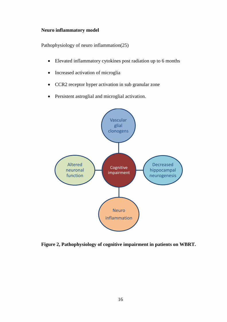
Neuro inflammatory model
Pathophysiology of neuro inflammation(25)
• Elevated inflammatory cytokines post radiation up to 6 months
• Increased activation of microglia
• CCR2 receptor hyper activation in sub granular zone
• Persistent astroglial and microglial activation.
Figure 2, Pathophysiology of cognitive impairment in patients on WBRT.
Cognitive impairment
Vascular glial
clonogens
Altered neuronal function
Neuroinflammation
Decreased hippocampal neurogenesis
17
Neural circuits
Hippocampal formation (dentate gyrus and cornu ammonis) are
considered vital for memory formation. It is involved in learning,
consolidation, retrieval of information, and construction of new memories.
Hippocampal injury results in, the impairment of declarative memory which is
the conscious recollection of facts and events(35). But emerging research is of
the opinion that entire limbic circuit is important for normal cognitive function.
Limbic structures are 2 arch like circuits that are surrounded by ventricles. The
inner arch consists of amygdala, hippocampus [cornu ammonis and dentate
gyrus], fornix, and mamillary bodies. The outer arch consists of
parahippocampal gyrus, cingulum, cingulate gyrus, induseum griseum, and
paraterminal gyrus. The inner and outer arches are separated by the
hippocampal sulcus and corpus callosum.(36)
This circuit is critical to a number of brain functions: integration and
consolidation of new memories, emotional responses and behavior, special
orientation, autonomic responses to external stimuli, and fine motor
coordination (among others).(36) The two structures closely associated with the
hippocampus include the amygdaloid complex and parahippocampal gyrus.(36)
The parahippocampal gyrus is critical to memory encoding and retrieval of
memories, and its ventral-most portion, called the entorhinal cortex, is the
important source of afferent signals to the hippocampus. The amygdaloid
complex, or amygdala, is involved in memory modulation (required for long-
term memory consolidation and the association of memory with emotional and
18
physiological states) and emotional learning (fear reactions, imprinting,
breeding behaviors, etc.).(36) The three structures— the hippocampal
formation, parahippocampal gyrus, and amygdaloid complex—form a
functional unit within the medial temporal lobe and is responsible for true
memory consolidation and learning(36).
The cingulate gyrus is involved in attention, concentration and regulated
autonomic response to emotional stimuli. The cingulum is a white matter tract
adjacent to the cingulate gyrus which connects the parahippocampal gyrus to
cingulate gyrus and prefrontal area. Fornix and mamillary bodies are other
structures closely associated with limbic system. In this way the limbic system
forms vital circuit in connecting hypothalamus, thalamus, prefrontal,
orbitofrontal cortices, and nucleus accumbens. The function of the circuit as a
whole is to process memory, support learning (cognitive, emotional, and
autonomic), regulate emotional states, and assist in spatial orientation. Since
the above circuit is involved in so many vital cognitive functions, the radiation
exposure to it manifests as late toxicity.
Thus from the above studies we can know the possible reason behind the
cognitive decline due to whole brain radiotherapy.
Studies on Neurocogniton of patients on WBRT
MMSE was used for the analysis of the cognitive change in WBRT(9).
In a large scale study done by Sun et al the MMSE assessment showed 36 % of
the population having impaired scores in MMSE after 3 months of whole brain
19
radiotherapy. The study by Aoyama et al showed that 5% patients with
impaired MMSE at 2months which reduced to 14% and 28 % at 3 and 6
months respectively. This study basically used subjects with MMSE score >27
for the analysis. The results showed that up to 40 % of the patients had decline
in MMSE by 12 to 18 months. Thus these studies showed the importance of
MMSE as an important tool in assessment of WBRT patients. Another study
done by Corn et al with 92 subjects showed that exposure of 37.5
Gy/15fractions/19 days led to drop in MMSE scores in 23% by 2 months and in
up to 40% by 18 months. But the largest study(37) by Regine et al with 182
subjects using 30 Gy/10 fractions/12 days. The above showed 46 % having
decline in MMSE scores in just 3 months.
Murray et al(38) published a study by comparing the MMSE of patients
on WBRT with 2 different regimens. He enrolled 445 patients into this phase 3
trial to compare 54 Gy {1.6 Gy b.i.d} against 30Gy in 10 fraction using
MMSE. Study went on from 1991 to 1995 with various assessments. MMSE
scores among the participants ranged from 11 to 30. He also considered that
MMSE < 23 might have possible dementia which was in fact seen in 16 % of
cases before the treatment.(38) The median survival in this study was 4.2 [S.D
3.7-5] months at 95 % C.I. 63 patients died before MMSE was reviewed. So
MMSE was reviewed only for patients who had long term survival. This study
concluded that MMSE is a good scale to assess neurocognitive decline in
WBRT patients. It also stated that 30Gy in 10 fraction prevented MMSE scores
of patients with long term survival to drop less than 23. The poor prognostic
20
factors were males, old age, low MMSE and early drop in MMSE below 23.
Thus most of the above studies were significant in showing the importance of
MMSE in assessment of cognitive decline in WBRT patients.
Hopkins verbal learning test was used to assess the verbal learning skill
in subjects in several studies. This scale assesses the verbal learning ability of
the patients. Several studies were done in this area giving significant results.
Tanaka et al(39) in 2014 used HVLT on patients undergoing WBRT with
different fraction and showed significant results. In this study using Japanese
version of HVLT, 40% of the study population showed significant decline in
total recall scores with 35Gy fraction at 4 months. The decline with 30 Gy, 25
Gy were 7% and 0% respectively from the baseline. But the delayed
recognition scores in 4 months declined by 2 points in 47% and 13% with 35
Gy and 30 Gy respectively.
A significant study with respect to HVLT was by Vinai godi et al (86)
which showed the changes in verbal memory. This study compares the HVLT
scores with total hippocampal sparing WBRT against the controls drawn. This
study shows the decline that happens in Neurocognition when hippocampus is
exposed to radiation. Here the controls that were exposed to radiation showed
up to 30% drop in Neurocognition specifically in HVLT scores. Even among
the 113 cases taken for the study 46 cases had died by 6 months. On analyzing
the HTLV scores of the patients who died within 6 months there was a
statistically significant drop in HVLT scores. Among the rest of the patients
who were exposed to HA WBRT there was 7 % decline in Neurocognition
21
which was not found to be statistically significant. Thus above study could
conclude that neurocognitive decline occurs due to radiation and is indirectly a
factor of age as it determines stem cell population in the dentate gyrus of
hippocampus.
A study done to correlate hippocampal dosimetry on cognitive outcome
was done on 24 patients(40). It showed that when hippocampus is totally
excluded from the radiation field with hippocampal sparing radiotherapy
neurocognition can be partially preserved.
Another study was done by Sun et al(41) which significantly depicted
the memory decline with exposure to WBRT. Total of 356 patients were
recruited for the study. Patients were the ones who were suffering from
advanced Non small cell lung carcinoma. All the set of baseline neurocognitive
assessment was done on the patient with quality of life evaluation. MMSE was
the scale which was used to evaluate the patients for cognitive impairment.
MMSE was used on the patients at 3, 6 and 12 months.
An interesting finding is that there was statistically significant difference
in MMSE scores between WBRT and controls at 3 months [p= 0.04]. But there
was no statistically significant drop in MMSE scores over 12 months [p=0.60].
HVLT was also used on the subjects in this study which gave valuable results.
HVLT immediate recall showed most significant result at 3 months with
p=0.001. The result also remained significant at 6 months [to a lesser extent
with, p=0.045] and at 12 months [p=0.03]. HVLT Delayed Recall also had the
22
most significant change at 3 months (P =0 .001). But the scores showed no
significant difference at 6 months [p=0.81] and later again showed significant
difference at 12 months [p=0.008]. This clear difference showed that MMSE
was less sensitive than HVLT in measuring the cognitive decline occurring due
to radiation exposure. This study further analyzed the quality of life of patients
on WBRT. They used the European Organization for Research and Treatment
of Cancer (EORTC), Quality of Life QLQ-C30 Questionnaire (QLQ-C30) and
BN20 for assessing the quality of life.
The study showed that there was no significant difference in Quality of
life scores at 1 year after exposure to whole brain radiotherapy. They also
mentioned the loss of follow up as one of the major limitations while arriving
at above scores. The compliance rate fell to 34% for neurocognitive function
and 37% for the quality of life scores after 12 months of follow up. The decline
in neurocognitive function here is similar to a study by Wolfson et al(42) which
found a significant increase at 1year of Neurocognitive function decline in the
higher-dose cranial irradiation arms (36 Gy) compared to the standard-dose
arm (25 Gy; P = 0.02).
A study on cognition by Welzel et al (43) was done on 44 participants.
He clearly showed that a Neurocognition decline occurs following WBRT. But
the unique feature is that memory decline is specifically seen immediately in
patients with brain metastasis and not in patients without metastasis. But after 6
to 8 weeks decline is seen in patients with and without metastasis. The deficit is
23
significantly associated with verbal memory but is not to be taken as a reason
for quitting WBRT.
Another study by Roger el al(44) was done on children receiving
radiotherapy and chemotherapy. It showed that the children receiving RT
showed significant reduction in their IQ scores. Overall study follow up of 2
years there was a drop in verbal and performance IQ scores by 14 points (105
to 91). The younger the age of child the severe was the decline in IQ. Children
with age lesser than 7 years, had 25 point drop in IQ scores over 2 years. The
drop in IQ was seen mainly in visual-motor; fine motor, visual-spatial skills
and memory function. Authors were seriously concerned with the cognitive
decline happening in the children as the life span of the children increased.
Brain atrophy is another manifestation of whole brain RT which was
shown by Yuta shibamoto et al.(45). They studied on 101 subjects with median
age of 62 years. Patients with minimum MMSE of 21 and above with no prior
brain surgery were only selected for this study. Radiation of 40Gy as 2 Gy/
day was administered over4 weeks. Cerebrospinal fluid-cranial ratio [CCR]
was used to measure the brain atrophy. Neurocognitive assessment was done
with MMSE scores. Difference in brain atrophy and MMSE scores were
compared with Fishers exact test. They clearly showed mean atrophy index of
1.24 ± 0.39 (SD) at 6 months and 1.32 ± 0.40 at 12 months, and among
them18% and 28% had increase in score by 30% or greater respectively. The
mean MMSE scores almost remained constant according to this study. The
scores dropped by 4 or more points in 11% at 6 months and 12% at 12 months.
24
Since MMSE is less sensitive and covers few domains they recommend group
of neuropsychological tests to find these subtle changes. So this study showed
that brain atrophy may or may not be associated with change in MMSE
scores.(46)
The study by Aoyama et al was very significant in showing the effect of
WBRT on STEREOTACTIC RADIOSURGERY patients. The study says that
STEREOTACTIC RADIOSURGERY alone is better than WBRT in
preventing Leucoencephalopathy, cognitive decline and other side effects.(47)
In contrast avoiding WBRT is associated with significant chance of tumor
recurrence and death among the patients with brain metastasis. This tumor
recurrence in turn causes functional deterioration and poor quality of life.(37)
This study enrolled 132 patients and went on for 3 years. Patients were
randomized to two groups and one group was treated with STEREOTACTIC
RADIOSURGERY alone and other group was given STEREOTACTIC
RADIOSURGERY+ WBRT. WBRT was given as 30Gy in 10 fractions in 2.5
week but was adjusted based on the STEREOTACTIC RADIOSURGERY
dosage. STEREOTACTIC RADIOSURGERY dosage was reduced by 30%
when combined with WBRT. Only 92 patients were available for follow up
review with MMSE scores. After the exposure of the patients for radiation
MMSE scores were recorded and analysis done. Among the patients exposed to
STEREOTACTIC RADIOSURGERY+ WBRT, 14 out of 36 cases had
significant decline in MMSE scores. In contrast among patients with
STEREOTACTIC RADIOSURGERY alone only 12 out of 46 patients had
25
drop in MMSE scores. This study showed that when WBRT is combined with
STEREOTACTIC RADIOSURGERY gave poor outcome in terms of
Neurocognition as measured by MMSE.
A study by Silber et al(48) researched on a group of children suffering
from acute lymphoblastic leukemia and medulloblastoma or posterior fossa
tumor. Using multiple linear regression model they demonstrated that higher
the age lesser is the decline in IQ scores with radiation exposure. With the
exposure of 36Gy the child scored 8.2 points lesser scores in IQ assessment
than 24 Gy and 12.3 points lesser than in those who were exposed to 18 Gy.
Gregor et al(49) performed the evaluation of neuro psychometrics of
long term tumor survivors under remission. 30 patients who were long term
survivors following irradiation were recalled for study. Patients who were
treated initially before 1987 were given WBRT [n = 16] and later patients were
treated with focused irradiation [n= 14].The test basically concentrated on
visuospatial organization, visual memory, and complex information processing.
By analyzing the 16 patients who were on whole brain irradiation he found that
WBRT caused significant decline is cognition compared to focused irradiation.
But the IQ was comparable to pre morbid levels in both groups .(49)The
univariate analysis in this study proved that radiation volume and time from
treatment made the difference and caused the cognitive decline. Multivariate
analysis by logistic regression on the samples proved that the reason for
neurocognitive decline is WBRT exposure.
26
An article by Stephen lutz(50) appeared in cancer which described the
radiation induced damage to the cortex. The author describes that palliative
care in a highly debilitated group of patients should be done with minimum
number of appointments at hospital. So shorter treatment courses to reduce the
burden of side effects is more beneficial for the palliative patients. He further
explains that radiotherapy causes serious damage to neural cells resulting in
apoptosis and vasculopathy. This he tells is the reason for the cognitive decline.
Author is also of the opinion that mild cognitive decline at times starts to arise
even before the cranial radiotherapy. In case of combined therapy decline in
Neurocognition is seen in verbal memory, frontal lobe dysfunction, and motor
incoordination. He criticizes that present palliative care oncologists are
reluctant to treat with short course WBRT for patients’ in spite of knowing the
physical and neurocognitive side effects. (51)He went on to tell that there are
several experiments on rats which demonstrated radiation induced damage
resulting in cognitive impairment.
Wolfson et al(52) performed a study on 264 Small cell lung carcinoma
cases [131 in Arm 1, 67 in Arm 2, and 66 in Arm 3]. The study was done to
measure the incidence of chronic neurotoxicity among patients exposed to
cranial irradiation. The participants in this study had actually achieved
remission with chemotherapy. He randomized the participants into 3 groups
i.e. Arm 1 – total 25 Gy in 10 fraction, Arm 2- total 36 Gy [2 Gy per day over
18 days], Arm 3 total 36 Gy [24 twice daily fractions with each session having
1.5 Gy]. They applied multiple neuropsychological batteries on the patients.
27
After the analysis they showed the cognitive decline was more significant in
the higher radiation exposure [i.e. in arm 2 and 3] compared to lower radiation
[arm 1] with p = 0.02.
The study showed that neurocognitive decline was significantly seen in
36 Gy group which was seen clearly in at least one definite cognitive function
Neurocognitive decline was seen in upto 96% of patients of 36Gy group while
it was seen in 60% of 25 Gy group. Assessments showed abnormalities in
language, visual and spatial scanning, attention, sequencing, and speed among
all three arms. The neurocognitive impairment was attributed to the effects of
chemotherapy on the brain, a paraneoplastic syndrome, aging, an immunologic
dysfunction, or even microscopic cranial metastases at diagnosis resulting in
frontal or sub cortical cognitive defects. In contrast the study showed that there
was no significant difference in terms of survival (50% of both the group were
alive at 1 year).
A study in the journal Neurology by Deangelis(53) dealt with rare
neurocognitive manifestations of radiation exposure. He reported that 12
patients exposed to WBRT went on to develop frank dementia with ataxia in
median duration of 14 months. He also observed that severe disabilities lead to
death in 7 patients. The earliest case he reported was a severe neurocognitive
decline with dementia in 5 months. He showed the incidence of dementia 1.9%
and 5.1% patients in 2 different populations which should be confirmed with
further studies.

28
The degree of neurocognitive outcome is worrisome according to most
of the studies done above but it was mostly shown without categorizing the age
group of the patient’s (54).Benjamin Corn and associates in their study
published in 2007 made the necessary adjustments for age and education. In
this study they compared the effect of radiation on patients due to WBRT alone
against WBRT with thalidomide. This multicentre study used 157 patients in
each arm applied total of 37.5 Gy as 2.5Gy per fraction. MMSE and SQLI
(Spitzer quality of life) scales were used for this study. After the study they
concluded that neurocognitive decline was definitely seen after WBRT. As
they considered MMSE as non specific tool for assessment of Neurocognition,
they compared the patient’s age and education to MMSE scores and made
necessary corrections to arrive at more specific scores. In later analysis they
concluded that thalidomide doesn’t play a special role in improving
Neurocognition during WBRT.
Pospisil et al (55)performed an advanced research using N Acetyl
aspartate as a marker in post WBRT. Using multi voxel MR spectroscopy they
tried to measure the (ht-NAA) N-acetyl aspartate concentration in the
hippocampus of patients who underwent WBRT. They studied 35 patients with
brain metastasis with multiple verbal and memory scales. Follow evaluation
was done on 18 patients at 4 months post WBRT. The concentration of ht-NAA
dropped significantly after radiotherapy on both left (8.64–7.60 mM; −12%,
95%CI: −7.9 to −16.2%) and right (8.52 –7.42 mM; −12.9%, 95%CI: −7.6 to
−16.4%) hippocampus. But the reduction in quality of life was not related to
29
the change in concentration of metabolite. Thus the study was helpful in giving
some practical insight into the neurocognitive decline with WBRT. It also
showed that hippocampal ht-NAA levels may be useful as a biomarker for cell
death in WBRT patients.
When brain metastasis cannot be resected surgically then radiotherapy is
the treatment of choice.(56) But all tissues are not amenable to radiotherapy. In
this condition we use certain tumor selective chemicals that increase the effect
of radiation on the tumor mass. Motexafin gadolinium (MGd) is a new drug
used with WBRT. Motexafin gadolinium (MGd) targets tumors sele ctively and
generates reactive oxygen species intracellularly, lowering the apoptotic
threshold to radiation and chemotherapy. It increases tumor radiation response
in vivo in preclinical models. MGd is paramagnetic, and previous clinical
studies have demonstrated tumor localization using MRI.
Meyers et al(56) performed this study by recruiting 401 patients. They
tried to find the neurocognitive benefits of using MGd with WBRT. 401
patients were randomized in WBRT + MGd [5 mg/ kg/day] or WBRT alone.
Later the randomly divided patients were exposed to 30Gy of WBRT. Among
the participants 50.8% of patients had metastasis only to brain and 80.1% had
multiple metastases. Majority of the patients completed neurocognitive
assessment but, 363 (90.5%) patients had impairment of one or more
neurocognitive tests at baseline. Univariate and stepwise logistic regression
analyses of global neurocognitive impairment was done to get the result. The
results showed that in contrast to WBRT alone, patients on WBRT with MGd
30
had prolonged time-to-neurocognitive progression for memory and executive
function. Hazard ratio was found which favored the use of Motexafin
gadolinium with WBRT.
The overall neurocognitive scores deteriorated over time. The study
compared neurocognitive function against survival of the patients. They found
that impairment in memory (HVLT recognition, recall, and delayed recall),
motor speed and dexterity (peg board test for dominant and non dominant
hands), executive function (Trail making Test B), and global neurocognitive
impairment (> three tests impaired) determined poor survival. A multivariate
Cox proportional hazards analysis of survival with all eight neurocognitive
tests included in the model showed that HVLT recognition and recall served as
independent predictors of survival (P = 0.0323 and P = 0.0342,respectively). In
this study neurocognitive assessment was also used as an objective measure for
comparing tumor growth, number and survival. Neurocognitive scores where
highly correlated with tumor size at baseline and not statistically correlated
with number of brain metastasis. Thus study suggested that cognition is mainly
affected by tumor burden and not by number of tumor masses. The study
showed that the neurocognitive decline gets significantly delayed when the
MGd is used with WBRT.(56)
A significant delay in time to neurologic progression for patients treated
with MGd with WBRT was found (P = 0.018, unadjusted). But In terms of
survival (median, 5.2 months for MGd v 4.9 months for WBRT; P = .48) or
31
neurologic decline (median, 9.5 months for MGd v 8.3 months for WBRT;
P = .95) no significant difference was found.
Brown et al performed the study using memantine for prevention of
cognitive dysfunction in patients on whole brain radiotherapy(57). It was a
randomized, double-blind, placebo-controlled trial with memantine and
placebo. Here the patients with brain metastasis on treatment WBRT receive
either placebo or memantine (20 mg/d), within 3days of initiating radiotherapy.
The study went on for 24 weeks and various neuropsychological assessments
were done. Patients with malignancy diagnosed in last 5 years and visible mass
on MRI were selected for study. KPS >70, no physical deterioration, Mini
Mental State Exam (MMSE) score > 18, no chronic use of benzodiazepine
and no drug abuse were some of the important eligibility criteria. Patients were
randomized and received placebo or memantine.
Patients were treated with 37.5 Gy of WBRT (15 fractions of 2.5 Gy)
with memantine administration to treatment arm. Neuropsychological
assessment was done at baseline and 8, 16, 24, and 52 weeks. 173 out of 508
patients [34%] recruited for the study died in the 6 months course of treatment.
Patients who completed the tests all the occasion were found to have longer
duration of survival [median overall survival of 12.4] and better quality of
living.
HVLT on the patients showed that there was less decline in cognition
with memantine (median decline of 0) compared to the placebo cases (median
32
decline of –0.90) after 24 weeks(57) which was statistically not significant with
[P= 0.059]. But the result had less statistical power [35%] due to attrition. In
contrast the memantine showed significant benefits compared to placebo in
terms of HVLT-R Delayed Recognition (median decline 0 vs. –1, P=0.0149) at
24 weeks. Similarly the MMSE scores (median decline 0 vs. –1, P=0.0093)
also showed significant difference compared to placebo at 24 weeks. HVLT-R
Delayed Recognition scores (median decline 0 vs. –0.715, P¼.0115) showed
significant difference in favor of memantine at 24 weeks using standardized
scores.
The study showed significant decline occurring in terms of
Neurocognition in placebo arm exposed exclusively to WBRT. At 3 months
51.9% of the patients showed neurocognitive decline and it increased to 64.9%
at 6 months. In contrast the neurocognitive decline was 43.7% and 53.8% in
memantine treated patients at 3 months and 6 months respectively(57). The
study thus showed that WBRT is associated with neurocognitive decline and
usage of memantine resulted in better cognitive outcome.(57) The cognitive
function improved over time as it delayed cognitive decline by reducing the
rate of decline in executive function, memory and processing speed.
Ping fang tsai (40) in his study on 40 patients performed hippocampal
sparing WBRT. He used the Volumetric Modulated Arc Therapy (VMAT) with
two full arcs and two non-coplanar partial arcs. With this technique he could
deliver radiation specifically sparing the hippocampus. He performed
neurocognitive assessment before and after 4 months of performing the WBRT.
33
Neurocognitive assessment scores were available for 24 patients after 4
months. Among them, he showed that at there no significant decline in test
scores.
A study done by Onodea et al(58) actually showed the fluctuating
course of the neurocognitive function after WBRT. In this prospective pilot
study he enrolled 27 patients among which 20 received WBRT and rest
7 received stereotactic irradiation. Neurocognitive data obtained at 4m, 8m and
12 months. The study showed that there was a significant deterioration in
delayed memory compared to the baseline (p = 0.04) at 4 months, and at 8
months. But significant improvements were observed in immediate memory
compared to the baseline (p = 0.008) and 4-months scores (p = 0.005). Strange
result reported in this study was that at 12 months, the immediate memory
scores had returned to the baseline. This pilot study had a very small population
but gave an interesting result for further evaluation and research.
There are several studies that even claim that avoiding radiation
exposure to hippocampus prevents neurocognitive decline .A study by Shin yin
lin(23) performed on patients exposed the patients to hippocampal sparing
whole radiotherapy.25 patients were enrolled for this study. After enrolling
Patients they performed extensive investigations and then planned for WBRT.
One important criterion was that patients didn’t have more than 3 tumor masses
and none the masses were more than 4 cm in diameter. After performing MRI
cases with tumor mass 5 mm around the hippocampus were excluded.
Radiation was delivered as 30 fractions in 12 doses and neurocognitive

34
assessment was performed after 4 months and 12 months. They found that
there is no significant drop in cognitive scores in tests. But the compliance at 4
months for neurocognitive assessment was only 54% which was suggested to
be due to patient related factors. Thus according to this study avoidance of
radiation exposure to hippocampus can protect patient from neurocognitive
impairment.
Jing li (59)in their study showed the association of tumor shrinkage and
neurocognitive function. They showed that the increase in tumor growth is
associated with more neurocognitive decline than the exposure to WBRT(37).
So they followed 135 patients and measured the neurocognitive function at 4
months and 15 months. The analysis was done here based on the size of the
indicator lesion and lesion volume reduction at 2 months was above or below
the population median reduction of 45%.
The results showed that good responders to radiation therapy had better
scores in all the 8 neurocognitive assessment scales used in the study. The
delay of neurocognitive function deterioration in good responders were noted
with better scores in an executive function test Trail making B (131 days’ gain;
P = 0.02), and the fine motor coordination tests Pegboard NDH (110 days’gain;
P = 0.02) and Pegboard DH (93days’ gain; P = 0.05).(59) But memory was
found to have weaker association with tumor volume reduction with values still
showing better preservation of memory with reduction in the volume of mass
lesion.

35
Results showed net gain of 61, 52 and 59 days for recall, recognition,
and delayed recall, respectively. They showed that WBRT induced shrinkage
of tumor mass gave survival benefit and preserved neurocognitive function.
Disease progression is the key reason for poor survival in cancer patients. Here
the patients who respond well to Whole brain radiotherapy have better control
over their executive function and fine motor coordination but, there is still
some deterioration in memory function that happens which gets masked by the
improvement in symptoms obtained with reduction in tumor mass.
If we look into the effect of WBRT on cognition in patients with
primary CNS lymphoma(60) the study by Doolittle et al will share some
insight. 80 participants took part in study. They were divided in to 4 groups and
one of these groups was treated with WBRT+ chemo and other with
methotrexate based regimens alone. Over next 2 years patients under went
neuropsychological evaluation, quality of life assessment and MRI scans. 15
out of 80 patients were treated with methotrexate followed by WBRT. Patients
selected for the study were ones who had minimum 2 years of treatment and
disease remission. Later neuropsychological assessment was done with
Wechsler Adult Intelligence Scale—III Digit Span subtest [Digits Forward,
Digits Backward], Trail Making Test and Brief Test of Attention, verbal
memory (Hopkins Verbal Learning Test– Revised),motor skills with Grooved
Pegboard Test. Quality of life was tested with European Organization for
Research and Treatment of Cancer (EORTC) QOL Questionnaire–30
(QLQ-C30). results showed that patients treated with WBRT had lower

36
cognitive performance. Patients on WBRT had a mean score 0.65 points (95%
CI, 0.20–1.10; p 5 0.0051) lower compared to patients on 3 non-WBRT groups
combined. But in terms of verbal memory there was no significant effect of
WBRT recorded (p = 0.1246).
In terms of quality of life the patients scored poor in terms of physical,
emotional and social functions. The above scores were found to associate with
white matter abnormalities. This study may have small number of population
size with median age of 58 years.(60) But it shows the improvement in verbal
memory in patients exposed to methotrexate regimen with 45 Gy WBRT.
After going through all these research a paper by Cole et al (61)
analyzed the self reported cognitive outcome after radiation therapy for brain
metastasis.50 adult patients were taken up for the study group and were treated
with whole or partial irradiation . None of the patient in the therapy had past
radiation therapy. Breast cancer patients without cranial involvement on
adjuvant therapy were controls. Self reported cognitive ability was measured 6
weeks, 3 months, and 6 months after irradiation. Neurocognitive status was
measured using German questionnaires for self-perceived deficits in attention
(FEDA) and subjectively experienced everyday memory performance (FEAG).
Baseline data showed high degree of self perceived cognitive deficit in both
groups.
On statistical evaluation there was significance in group differences for
the FEDA scales 2 and 3.(61) The results showed a significant increase in

37
fatigue and retardation of daily living activities (P=.002) and also decrease in
motivation (P=.032) in the patients exposed to WBRT. The radiation exposure
also caused an increase of attention deficits in the WBRT patients, but not in
the Control Group.(61) The scores of distractibility and retardation of mental
processes (P=.059) between the WBRT/partial RT and the controls were close
to significance. Thus the study concluded by saying that self reported attention
declined in patients treated with WBRT/ partial RT compared to controls.
Function have been reported in rats [15] in response to single [16] and
fractionated [17, 18] WBRT. Additionally, deficits in spatial learning have
been reported in mouse models [19, 20]. Our laboratory recently demonstrated
that fractionated WBRT induces time-dependent learning and memory deficits
in both the Barnes maze and active avoidance tasks [21]. Importantly, spatial
learning was progressively impaired after WBRT as mice exhibited increased
latency to the escape box and made more errors in the Barnes maze in the
months following treatment. Despite extensive studies demonstrating the
effects of WBRT on cognition in multiple species, the etiology of WBRT-
induced cognitive impairment remains poorly-understood.
In reviewing all these research articles we can get invaluable
information about cognitive outcome of WBRT. WBRT is like a double edged
sword with its multiple benefits and side effects. Research has shown that it
improved the survival of patients on WBRT by causing some degree of
cognitive decline. Radiation exposure in children is associated with IQ decline
which has been established in several follow up studies. There was a high
38
incidence of learning disabilities, academic failure with poor
attention, memory and executive functions along with low intellectual
level. Patients’ also scored poor in quality of life measure.(62) Higher doses of
radiation had greater cognitive decline but still very little is known regarding
their cognitive outcome. Further research is needed in this area to establish
several unknown facts.
Quality of life is measured as a part of several researches on WBRT(63).
Quality of life of cancer patients depends on heterogenous factors. Various
cancers related quality of life scales have been created giving varying
results(63). Each scale takes different domains into consideration while
measuring the quality of life. Functional Assessment of Cancer Therapy - Brain
(FACT-BR) questionnaire is a popular scale to measure quality of life in
disorders of brain. This scale uses 2 subscales namely, Functional Assessment
of Cancer Therapy General (FACT-G) and the Brain Subscale. The FACT-G
covers four primary QoL domains: Physical Well-Being (7 items),
Social/Family Well Being (7 items), Emotional Well-Being (6 items); and
Functional Well-Being (7 items).
European Organization for Research and Treatment of Cancer Quality
of Life Core Questionnaire (EORTC QLQ-C30) is another scale that is used by
some studies to measure quality of life. It consists of 5 functional scales
(physical, role, cognitive, emotional, and social) and 3 symptoms scales
(fatigue, pain, and nausea and vomiting). EORTC QLQ-C30 concentrates more
on social activities and family life, while the FACT-G mainly focuses on social
39
support and relationships. We in our study used 3 different scales concentrating
on various aspects of life.
The study by Sun et al(41) studied the quality of life of patients on
WBRT for NSCLC. This study used the EORTC QLQ-C30 scale and BN 20
for the assessment. After the study they found that there was no statistically
significant decline in quality of life at end of 12 month follow up [P> 0.05].
Jing li(11) in his study used the ADL [activities of daily living] and FACT-Br
[Functional Assessment of Cancer Therapy-Brain-specific] for neurocognitive
assessment of his patients on WBRT. He proved that quality of life in a patient
can be correlated with neurocognitive decline seen in the previous visit. Thus
neurocognitive decline is a predictor of the quality of life in near future.
Another study by Gao et al (64) tries to assess the quality of life
palliative WBRT patients and found again that the quality of life improved
after the treatment with whole brain radiotherapy. This study basically utilized
the health-related QOL (HRQOL) of patients with Brain metastasis. He
selected 46 patients for this study and among them 35 were exclusively
exposed to WBRT and 6 to WBRT + Chemotherapy other 5 to supportive care.
The mean age of the sample was 52.6 years. The analysis showed that survival
was 11.8±0.46 months for patients on WBRT, 11.75±1.00 months for patients
on WBRT + Chemotherapy, and 3±0.79 months Supportive Care (P<0.01). The
HRQOL scores of patients on Whole Brain Radiotherapy were 72.23±0.88
(before therapy) and 78.49±0.87 (after therapy) (P<0.01). Thus study showed
significant improvement in quality of life. But still the average survival in the
study was only 11.8 months.(65) Thus several studies on the cancer patients
40
have shown that radiation therapy to the brain gives significant improvement in
the quality of life. Hence it can be made as part of therapy for patients with
brain metastasis.
Radiotherapy procedure
Whole brain radiotherapy was delivered using Radiotherapy device with
cobalt 60 source.(66) The patient was put on supine position and radiation was
delivered. The head of the patient was immobilized by thermoplastic masks.
Right and left lateral position treatment was given on the same day with
required dose, i.e. 1.5 Gy each to left and right side. Corticosteroids and
mannitol were administered to the patients in the ward to prevent increased
intra cranial tension due to brain edema.
Whole brain radiotherapy treatment should have adequate coverage of
all intracranial contents. It should be planned to entirely cover the entire cranial
fosse and even the skull base. Beam arrangement is done in right and left
lateral opposing fields. Shape of the field can be adjusted and radiation was
applied.
Dosage
Standard dose of 30 Gy in 10 daily fractions or 20 Gy in 5 daily
fractions is used. Any dose higher than 30 Gy in 10 fractions did not give
further benefit in patients with brain metastasis. The Cochrane reviews on this
area showed no further improvement with any altered dosage regimens in terms
of survival, neurological outcome or quality of life or symptom control.
41
AIMS AND OBJECTIVES
• To study the neurocognitive change that can occur due to whole brain
radiotherapy.
• To measure the MMSE, MoCA, HVLT, and Trail making test scores
of patients on WBRT.
• To study the quality of life of the whole brain radiotherapy patients
using Katz ADL, LBADL and WHO QOL Bref scales.
42
METHODOLOGY
The study was conducted in department of radiation oncology, Barnard
institute of radiology, Madras Medical College, Chennai. The thesis abstract
was presented before the institution ethics committee and approval was
obtained. The study was done from February 2017 to august 2017. Patients
suffering from brain metastasis were chosen for study based on inclusion and
exclusion criteria. A detailed informed consent was taken from the patient and
then recruited for our study. Initially their IQ was measured with BKT. Later
whole brain radiotherapy sessions were conducted and relevant data was
collected for study. The relevant scales were applied thrice i.e. immediately
after WBRT, 3 months and after 6 months. The collected data were analyzed
using SPSS package and necessary results obtained.
Inclusion criteria
• Patients undergoing whole brain radiotherapy at, Department of
Radiation oncology, Madras Medical College, Chennai
• Age 40 to 70
• Ability to give informed consent
• Karnofsky performance status >70.
43
Exclusion criteria
Mental retardation (IQ< 70)
KPS <70
Patients not giving consent
Known major psychiatric illness
Patients on chemotherapy
Past therapeutic radiation exposure to brain
Neurocognitive assessment tools
1) MMSE
2) Montreal cognitive assessment test
3) Hopkins verbal learning test
4) Activity of Daily Living Scale
5) Binet Kamath Test
6) WHO QOL
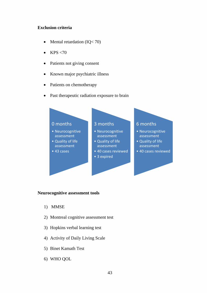
0 months• Neurocognitive assessment
• Quality of life assessment
• 43 cases
3 months• Neurocognitive assessment
• Quality of life assessment
• 40 cases reviewed• 3 expired
6 months• Neurocognitive assessment
• Quality of life assessment
• 40 cases reviewed
44
Scales
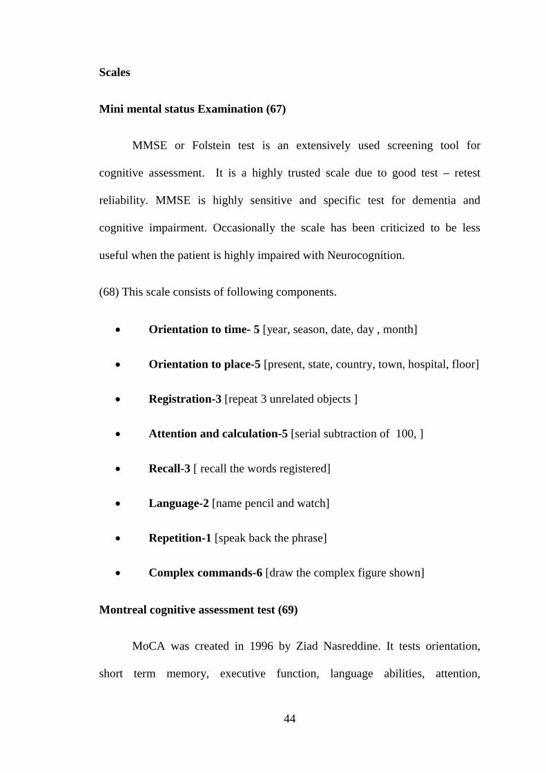
Mini mental status Examination (67)
MMSE or Folstein test is an extensively used screening tool for
cognitive assessment. It is a highly trusted scale due to good test – retest
reliability. MMSE is highly sensitive and specific test for dementia and
cognitive impairment. Occasionally the scale has been criticized to be less
useful when the patient is highly impaired with Neurocognition.
(68) This scale consists of following components.
• Orientation to time- 5 [year, season, date, day , month]
• Orientation to place-5 [present, state, country, town, hospital, floor]
• Registration-3 [repeat 3 unrelated objects ]
• Attention and calculation-5 [serial subtraction of 100, ]
• Recall-3 [ recall the words registered]
• Language-2 [name pencil and watch]
• Repetition-1 [speak back the phrase]
• Complex commands-6 [draw the complex figure shown]
Montreal cognitive assessment test (69)
MoCA was created in 1996 by Ziad Nasreddine. It tests orientation,
short term memory, executive function, language abilities, attention,
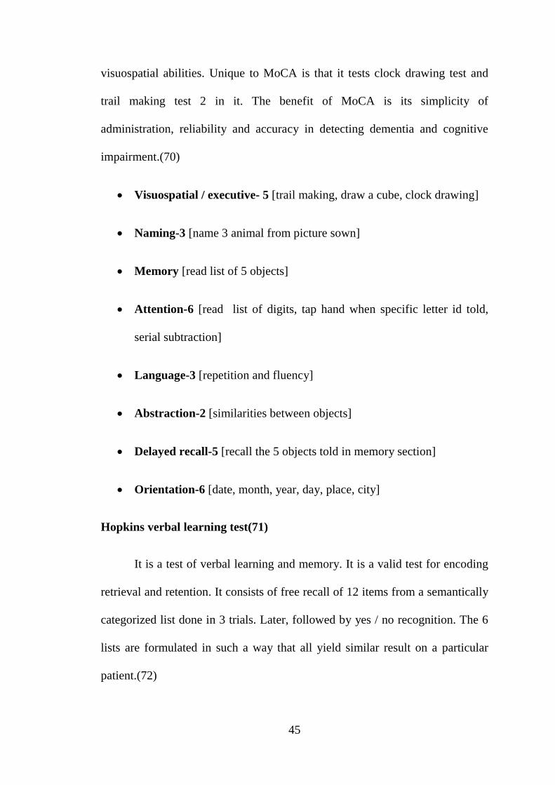
45
visuospatial abilities. Unique to MoCA is that it tests clock drawing test and
trail making test 2 in it. The benefit of MoCA is its simplicity of
administration, reliability and accuracy in detecting dementia and cognitive
impairment.(70)
• Visuospatial / executive- 5 [trail making, draw a cube, clock drawing]
• Naming-3 [name 3 animal from picture sown]
• Memory [read list of 5 objects]
• Attention-6 [read list of digits, tap hand when specific letter id told,
serial subtraction]
• Language-3 [repetition and fluency]
• Abstraction-2 [similarities between objects]
• Delayed recall-5 [recall the 5 objects told in memory section]
• Orientation-6 [date, month, year, day, place, city]
Hopkins verbal learning test(71)
It is a test of verbal learning and memory. It is a valid test for encoding
retrieval and retention. It consists of free recall of 12 items from a semantically
categorized list done in 3 trials. Later, followed by yes / no recognition. The 6
lists are formulated in such a way that all yield similar result on a particular
patient.(72)
46
Test consists of 12 items word list having 4 words from 3 different
semantic categories. The list read out slowly one word every 2 seconds. Then
patients’ free recall is checked in 3 trials. Later 24 word list is read out
including the above 12 words and patient is asked to tell yes to which words
appeared in the 12 word list no to the words that didn’t. 12 extra words that are
read out are distracters or unrelated words. This test is unique in terms of the
duration needed for its administration as it needs 10 min to administer. It is
even easy to administer in demented and debilitated patients.
Trail making test (73)
Trail making test was derived from Taylor number series test. This test
was later revised into present form and included in Halstead Reitan
neuropsychological battery. The 2 varieties include TMT A and TMT B. Part A
consists of numbers in series and the participant needs to connect them
sequentially. In contrast, Part B requires the participant to connect the numbers
and alphabets alternatively in sequence. Part A basically deals with motor
speed and visual search/ attention. Part B mainly deals with higher order
cognition, mental flexibility and executive control. The performance in TMT is
a good indicator of intelligence and it is also said to be sensitive marker of
neurological impairment.
Scoring
Trail A: Average- 29 seconds, Deficient if > 78 seconds, Rule of
Thumb- Most in 90 seconds.
47
Trail B: Average- 75 seconds, Deficient if > 273, Rule of Thumb- Most
in 3 minutes
Katz activities of daily living.(74)
The patients on WBRT often are physically debilitated. Hence this scale
was used measure the daily living difficulties. Katz instrument was devised in
1970 and has been widely used since then mainly for assessing patients with
stroke. This scale helps to assess the daily living abilities of the patient. A fully
functional patient gets a score of 6, while 4 implies moderate impairment and 2
severely impaired. The instrument is found to be highly reliable with reliability
index of 0.87 to 0.94.(75) With each factor examiner checks if the patient is
independent or dependant.
• Bathing
• Dressing
• Toileting
• Transferring
• Continence
• Feeding
The Lawton Instrumental Activities of Daily Living (IADL) Scale(76)
IADL is a good instrument to measure independent living skills. These
skills are considered more complex than the basic activities of daily living as
measured by the Katz Index of ADLs. This scale is considered to be very
48
useful for measuring the level of functioning of a person and to assess the
improvement or deterioration over time. This test consists of 8 domains. They
include, using the telephone, shopping, preparing food, and housekeeping,
doing laundry, using transportation, handling medications and handling
finances. The inter rater reliability was 0.85. The test is easy to administer as it
can be done in 10 min. Only limitation is that the test is self report of the
participant.
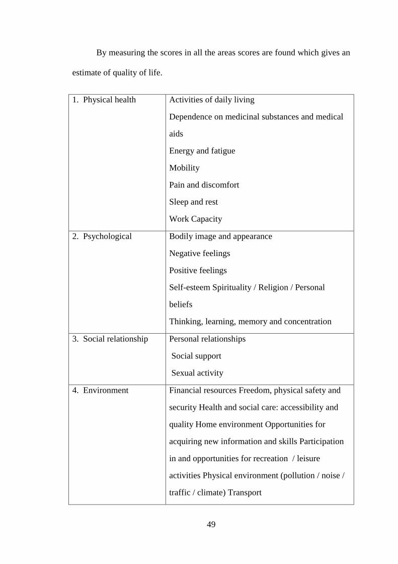
WHO QOL Bref(77)
WHO QOL Bref is one of the internationally acclaimed and validated
scales for measurement of quality of life. It was thoroughly researched in
various countries in a multinational study and standardized. The scale consists
of questions from various domains or areas of life and scored by the patient
accordingly.(78)
The scale basically has 4 domains
• Physical
• Psychological
• Social
• Environmental
49
By measuring the scores in all the areas scores are found which gives an
estimate of quality of life.
1. Physical health Activities of daily living
Dependence on medicinal substances and medical
aids
Energy and fatigue
Mobility
Pain and discomfort
Sleep and rest
Work Capacity
2. Psychological Bodily image and appearance
Negative feelings
Positive feelings
Self-esteem Spirituality / Religion / Personal
beliefs
Thinking, learning, memory and concentration
3. Social relationship Personal relationships
Social support
Sexual activity
4. Environment Financial resources Freedom, physical safety and
security Health and social care: accessibility and
quality Home environment Opportunities for
acquiring new information and skills Participation
in and opportunities for recreation / leisure
activities Physical environment (pollution / noise /
traffic / climate) Transport
50
STATISTICS
Analysis of repeated measure(79)
The term “repeated measure” refers to multiple responses taken in the
sequence on the same experimental unit. Usually the responses are taken over
time. The objective of repeated measure data analysis is to examine and
compare response trends over time. Here multiple measurements are made on
the same experimental units over a period of time. (80) Such data are called
repeated measures
The data of the analysis of repeated measures are analyzed in several
ways using,
• Separate analysis at each time unit
• Univariate analysis of variance
• Univariate and multivariate analysis of time contrast variables.
• Mixed model methodology.
Analysis at each time points measures treatment effect at each individual
time points but makes no comparison between times. Here no inference can be
drawn about trend over time.
Univariate analysis of variance is historically applied to repeatedly
measure data that makes comparison between times. It treats the data as if they
were from split part design. So it is also called split plot in time analysis.
51
Repeated measure analysis needs special attention into covariance structure as
it deals with data in sequence. Ignoring or excluding the covariance will give
incorrect results. Then mixed analysis procedure is used which has 2 steps.(81)
- Model the covariance structure.
- Analyze the time trends for treatment by analyzing and comparing
the means.
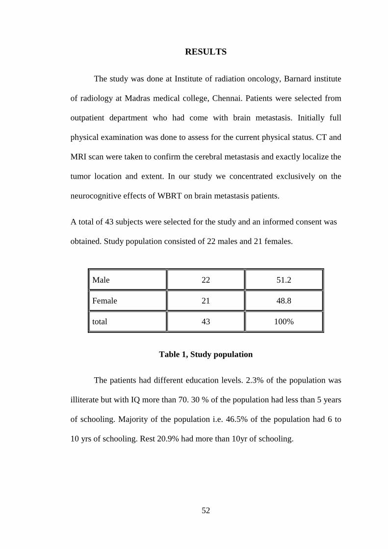
52
RESULTS
The study was done at Institute of radiation oncology, Barnard institute
of radiology at Madras medical college, Chennai. Patients were selected from
outpatient department who had come with brain metastasis. Initially full
physical examination was done to assess for the current physical status. CT and
MRI scan were taken to confirm the cerebral metastasis and exactly localize the
tumor location and extent. In our study we concentrated exclusively on the
neurocognitive effects of WBRT on brain metastasis patients.
A total of 43 subjects were selected for the study and an informed consent was
obtained. Study population consisted of 22 males and 21 females.
Table 1, Study population
The patients had different education levels. 2.3% of the population was
illiterate but with IQ more than 70. 30 % of the population had less than 5 years
of schooling. Majority of the population i.e. 46.5% of the population had 6 to
10 yrs of schooling. Rest 20.9% had more than 10yr of schooling.
Male 22 51.2
Female 21 48.8
total 43 100%
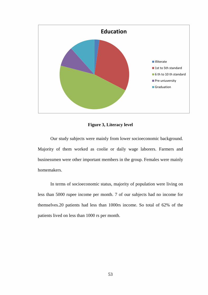
53
Figure 3, Literacy level
Our study subjects were mainly from lower socioeconomic background.
Majority of them worked as coolie or daily wage laborers. Farmers and
businessmen were other important members in the group. Females were mainly
homemakers.
In terms of socioeconomic status, majority of population were living on
less than 5000 rupee income per month. 7 of our subjects had no income for
themselves.20 patients had less than 1000rs income. So total of 62% of the
patients lived on less than 1000 rs per month.
Education
Illiterate
1st to 5th standard
6 th to 10 th standard
Pre uniuversity
Graduation
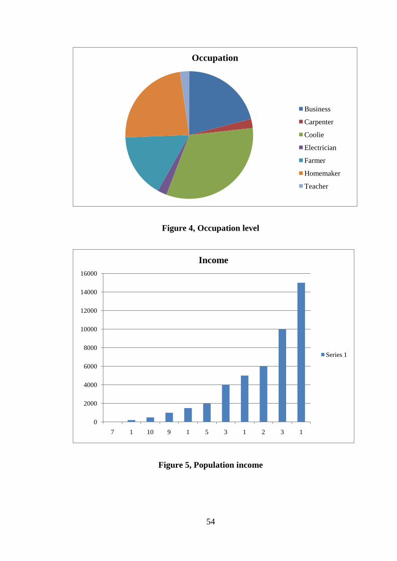
54
Figure 4, Occupation level
Figure 5, Population income
Occupation
Business
Carpenter
Coolie
Electrician
Farmer
Homemaker
Teacher
0
2000
4000
6000
8000
10000
12000
14000
16000
7 1 10 9 1 5 3 1 2 3 1
Income
Series 1

55
Figure 6, Binet kamath IQ test score levels
The intelligence level of the patient plays an important role in the ability
of the patient to participate in the study. Hence Binet Kamath intelligence
assessment test was used to measure the Intelligence quotients. All the patients
had IQ >70. Once the patient was selected for the study immediately IQ was
measured and later his assessment with CT and MRI was continued.
The most important factor in any whole brain radiation therapy is the
type of the primary tumor. Type of tumor helps in understanding the sensitivity
of the tumor to radiation and indirectly helps in estimating the prognosis.
Majority of the patients i.e. more than 50% patients in our study had lung
cancer. Most of the patients had small cell lung carcinoma and few had Non
small cell variety. Next major variety was breast cancer and was almost
exclusively found in females. Melanocytoma and renal cell carcinoma are 2
rare varieties found as metastasis to brain.
BKT IQ assessment scores
70-74
75-79
80-84
85-89
90-95
96-99
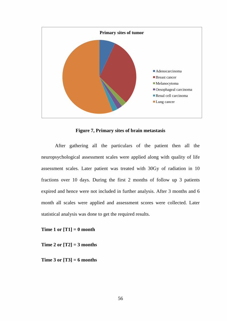
56
Figure 7, Primary sites of brain metastasis
After gathering all the particulars of the patient then all the
neuropsychological assessment scales were applied along with quality of life
assessment scales. Later patient was treated with 30Gy of radiation in 10
fractions over 10 days. During the first 2 months of follow up 3 patients
expired and hence were not included in further analysis. After 3 months and 6
month all scales were applied and assessment scores were collected. Later
statistical analysis was done to get the required results.
Time 1 or [T1] = 0 month
Time 2 or [T2] = 3 months
Time 3 or [T3] = 6 months
Primary sites of tumor
AdenocarcinomaBreast cancerMelanocytomaOesophageal carcinomaRenal cell carcinomaLung cancer
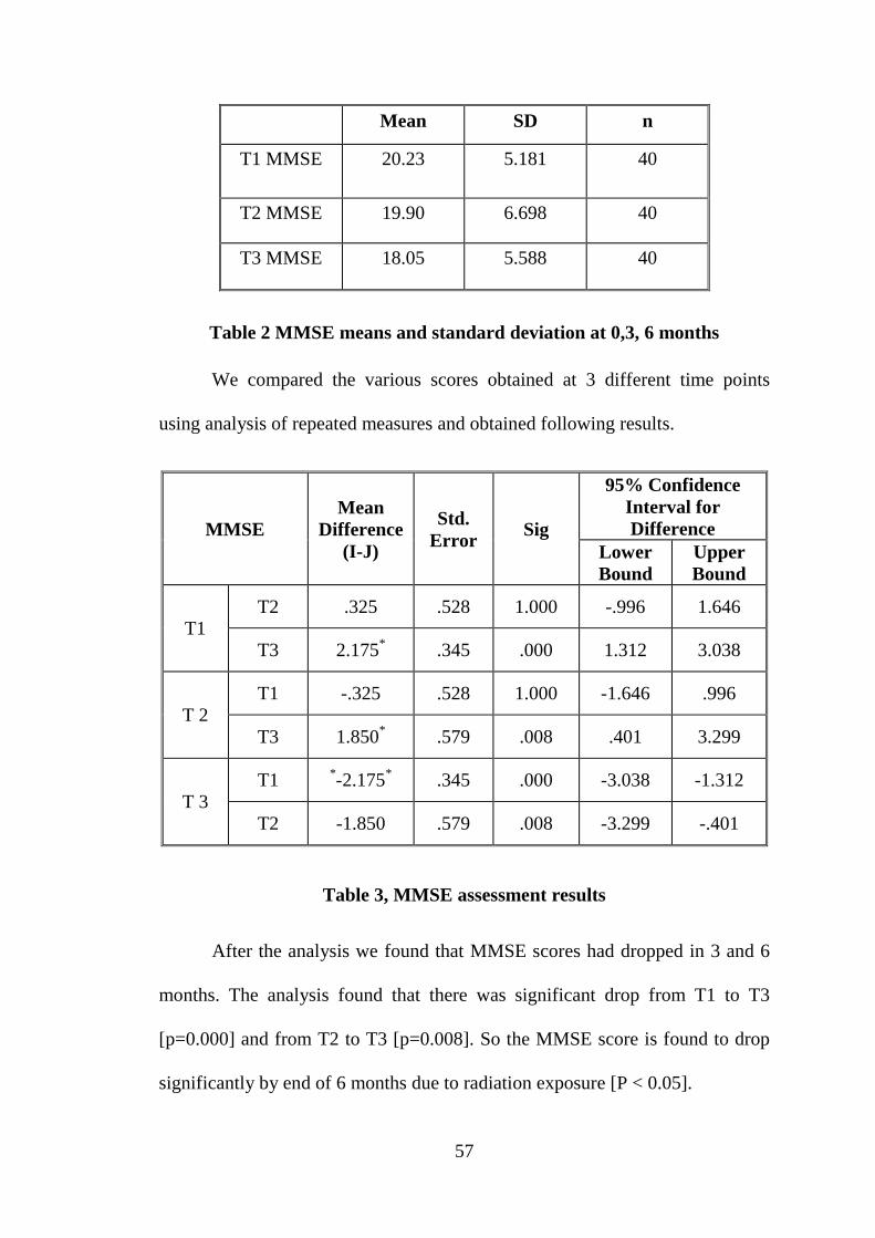
57
Mean SD n
T1 MMSE
20.23 5.181 40
T2 MMSE 19.90 6.698 40
T3 MMSE 18.05 5.588 40
Table 2 MMSE means and standard deviation at 0,3, 6 months
We compared the various scores obtained at 3 different time points
using analysis of repeated measures and obtained following results.
MMSE Mean
Difference (I-J)
Std. Error Sig
95% Confidence Interval for Difference
Lower Bound
Upper Bound
T1 T2 .325 .528 1.000 -.996 1.646
T3 2.175* .345 .000 1.312 3.038
T 2 T1 -.325 .528 1.000 -1.646 .996
T3 1.850* .579 .008 .401 3.299
T 3 T1 *-2.175* .345 .000 -3.038 -1.312
T2 -1.850 .579 .008 -3.299 -.401
Table 3, MMSE assessment results
After the analysis we found that MMSE scores had dropped in 3 and 6
months. The analysis found that there was significant drop from T1 to T3
[p=0.000] and from T2 to T3 [p=0.008]. So the MMSE score is found to drop
significantly by end of 6 months due to radiation exposure [P < 0.05].

58
Montreal cognitive
assessment
Mean Difference
(I-J)
Std. Error Sig
95% Confidence Interval for Difference
Lower Bound
Upper Bound
T1 T2 -.375 .642 1.000 -1.982 1.232
T3 2.875* .565 .000 1.461 4.289
T 2 T1 .375 .642 1.000 -1.232 1.982
T3 3.250* .400 .000 2.250 4.250
T 3 T1 -2.875* .565 .000 -4.289 -1.461
T2 -3.250* .400 .000 -4.250 -2.250
Table 4, Montreal cognitive assessment results at 0,3,6 months
Time 1 Time 2 Time 3
MCog assessment 19.20±5.25 18.08±2.269 15.95±5.71
Visuospatial 2.85±1.37 18.83±7.00 2.68±1.385
Naming 2.80±0.464 2.65±0.700 2.68±0.572
Attention 4.25±1.48 4.10±1.823 3.08±1.526
Language 1.33±0.917 1.53±1.037 1.13±0.966
Abstract 1.13±0.563 0.98±0.733 0.9±0.563
Delayed recall 2.70±0.791 2.63±1.25 1.95±0.714
Orientation 4.18±1.412 4.05±1.632 3.58±1.43
Table 5, MoCA results, with individual score analysis

59
We applied another assessment scale for Neurocognition on patient i.e.
Montreal cognitive assessment scale. This scale also showed that there was
significant decline in Neurocognition from T1:T3 and T2: T3. Mean was
found to drop from T1 to T2 but was not found to be significant. So there is
further evidence to prove the cognitive decline during first 6 months of
radiotherapy.
For further detail in understanding the area of cognitive decline we even
compared each components of MoCA. With pair wise comparison so we found
that there was decline in mean scores of visuospatial function happening from
T1 toT3 but it was not found to be significant. In terms of naming skills too
there was significant decline in cognitive skills.
Attention span was found to drop significantly from T1 to T3 and T2 to
T3. Hence radiation has its negative role in reducing the attention.
Language skills are also found to fall found to all over 6 months. The
mean language score at T1 which was 1.33±0.917 has dropped to 1.13±0.966.
When comparing the scores between 2 intervals we find that the T2:T3 scores
has a significant drop in scores with a difference of 0.400 and (P=0.003).
Abstract scores have dropped significantly fromT1 to T3 according
MoCA scores. Even the delayed recall and orientation have shown significant
decline over 6 months.
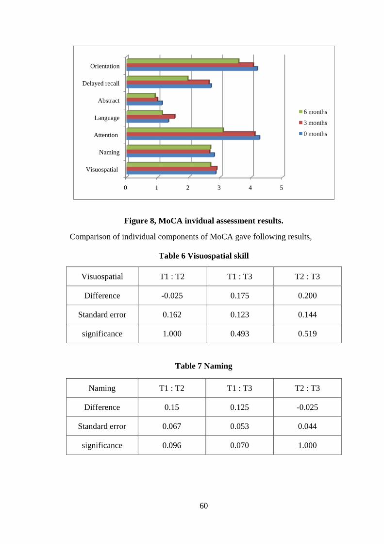
60
Figure 8, MoCA invidual assessment results.
Comparison of individual components of MoCA gave following results,
Table 6 Visuospatial skill
Visuospatial T1 : T2 T1 : T3 T2 : T3
Difference -0.025 0.175 0.200
Standard error 0.162 0.123 0.144
significance 1.000 0.493 0.519
Table 7 Naming
Naming T1 : T2 T1 : T3 T2 : T3
Difference 0.15 0.125 -0.025
Standard error 0.067 0.053 0.044
significance 0.096 0.070 1.000
0 1 2 3 4 5
Visuospatial
Naming
Attention
Language
Abstract
Delayed recall
Orientation
6 months
3 months
0 months
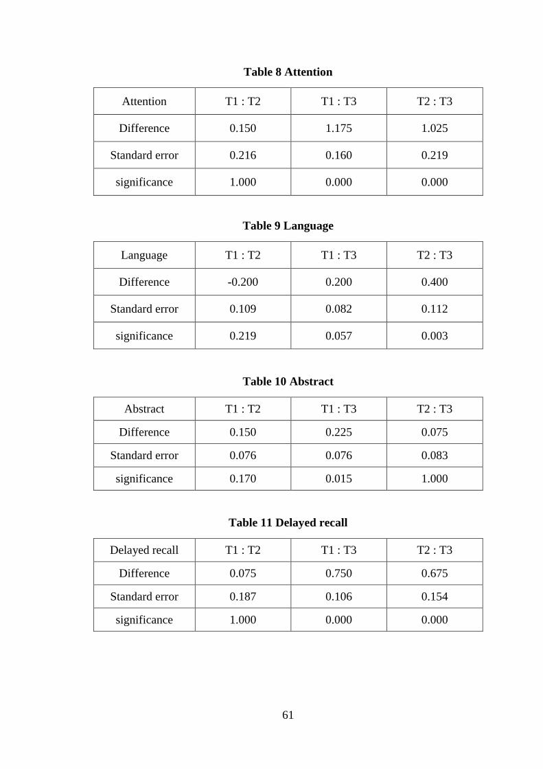
61
Table 8 Attention
Attention T1 : T2 T1 : T3 T2 : T3
Difference 0.150 1.175 1.025
Standard error 0.216 0.160 0.219
significance 1.000 0.000 0.000
Table 9 Language
Language T1 : T2 T1 : T3 T2 : T3
Difference -0.200 0.200 0.400
Standard error 0.109 0.082 0.112
significance 0.219 0.057 0.003
Table 10 Abstract
Abstract T1 : T2 T1 : T3 T2 : T3
Difference 0.150 0.225 0.075
Standard error 0.076 0.076 0.083
significance 0.170 0.015 1.000
Table 11 Delayed recall
Delayed recall T1 : T2 T1 : T3 T2 : T3
Difference 0.075 0.750 0.675
Standard error 0.187 0.106 0.154
significance 1.000 0.000 0.000
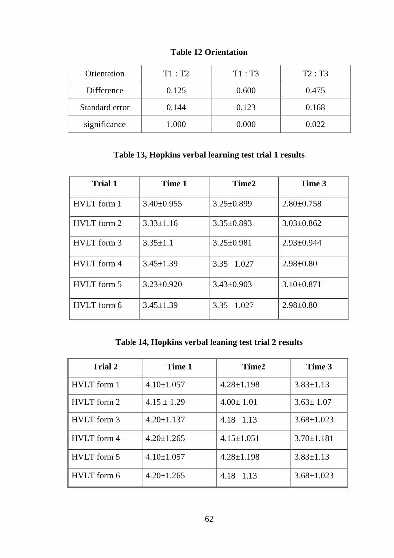
62
Table 12 Orientation
Orientation T1 : T2 T1 : T3 T2 : T3
Difference 0.125 0.600 0.475
Standard error 0.144 0.123 0.168
significance 1.000 0.000 0.022
Table 13, Hopkins verbal learning test trial 1 results
Trial 1 Time 1 Time2 Time 3
HVLT form 1 3.40±0.955 3.25±0.899 2.80±0.758
HVLT form 2 3.33±1.16 3.35±0.893 3.03±0.862
HVLT form 3 3.35±1.1 3.25±0.981 2.93±0.944
HVLT form 4 3.45±1.39 3.35±1.027 2.98±0.80
HVLT form 5 3.23±0.920 3.43±0.903 3.10±0.871
HVLT form 6 3.45±1.39 3.35±1.027 2.98±0.80
Table 14, Hopkins verbal leaning test trial 2 results
Trial 2 Time 1 Time2 Time 3
HVLT form 1 4.10±1.057 4.28±1.198 3.83±1.13
HVLT form 2 4.15 ± 1.29 4.00± 1.01 3.63± 1.07
HVLT form 3 4.20±1.137 4.18±1.13 3.68±1.023
HVLT form 4 4.20±1.265 4.15±1.051 3.70±1.181
HVLT form 5 4.10±1.057 4.28±1.198 3.83±1.13
HVLT form 6 4.20±1.265 4.18±1.13 3.68±1.023
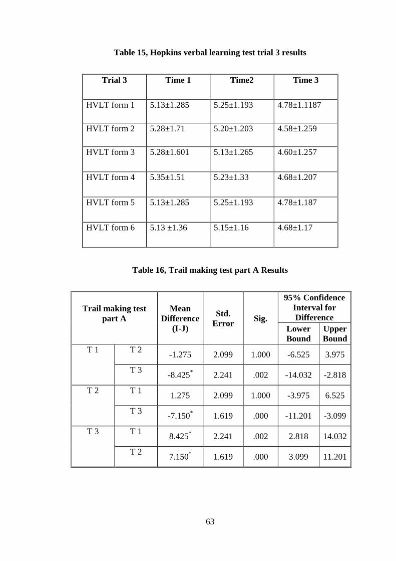
63
Table 15, Hopkins verbal learning test trial 3 results
Trial 3 Time 1 Time2 Time 3
HVLT form 1
5.13±1.285 5.25±1.193 4.78±1.1187
HVLT form 2 5.28±1.71 5.20±1.203 4.58±1.259
HVLT form 3 5.28±1.601 5.13±1.265 4.60±1.257
HVLT form 4 5.35±1.51 5.23±1.33 4.68±1.207
HVLT form 5 5.13±1.285 5.25±1.193 4.78±1.187
HVLT form 6 5.13 ±1.36 5.15±1.16 4.68±1.17
Table 16, Trail making test part A Results
Trail making test part A
Mean Difference
(I-J)
Std. Error Sig.
95% Confidence Interval for Difference
Lower Bound
Upper Bound
T 1
T 2 -1.275 2.099 1.000 -6.525 3.975
T 3 -8.425* 2.241 .002 -14.032 -2.818
T 2 T 1 1.275 2.099 1.000 -3.975 6.525
T 3 -7.150* 1.619 .000 -11.201 -3.099
T 3 T 1 8.425* 2.241 .002 2.818 14.032
T 2 7.150* 1.619 .000 3.099 11.201
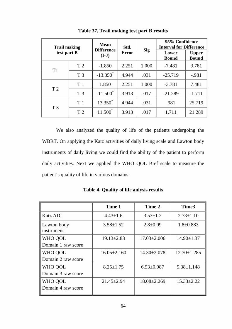
64
Table 37, Trail making test part B results
Trail making test part B
Mean Difference
(I-J)
Std. Error Sig
95% Confidence Interval for Difference
Lower Bound
Upper Bound
T1 T 2 -1.850 2.251 1.000 -7.481 3.781
T 3 -13.350* 4.944 .031 -25.719 -.981
T 2 T 1 1.850 2.251 1.000 -3.781 7.481
T 3 -11.500* 3.913 .017 -21.289 -1.711
T 3 T 1 13.350* 4.944 .031 .981 25.719
T 2 11.500* 3.913 .017 1.711 21.289
We also analyzed the quality of life of the patients undergoing the
WBRT. On applying the Katz activities of daily living scale and Lawton body
instruments of daily living we could find the ability of the patient to perform
daily activities. Next we applied the WHO QOL Bref scale to measure the
patient’s quality of life in various domains.
Table 4, Quality of life anlysis results
Time 1 Time 2 Time3
Katz ADL 4.43±1.6 3.53±1.2 2.73±1.10
Lawton body instrument
3.58±1.52 2.8±0.99 1.8±0.883
WHO QOL Domain 1 raw score
19.13±2.83
17.03±2.006 14.90±1.37
WHO QOL Domain 2 raw score
16.05±2.160 14.30±2.078 12.70±1.285
WHO QOL Domain 3 raw score
8.25±1.75 6.53±0.987 5.38±1.148
WHO QOL Domain 4 raw score
21.45±2.94 18.08±2.269 15.33±2.22
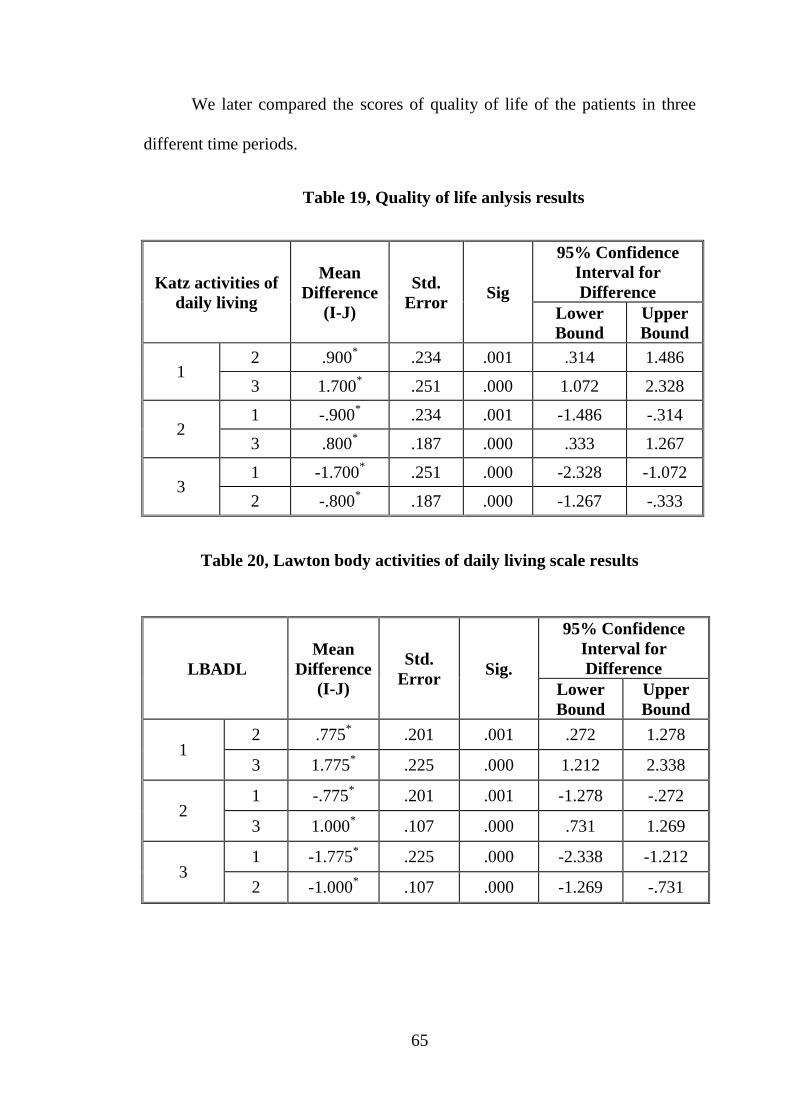
65
We later compared the scores of quality of life of the patients in three
different time periods.
Table 19, Quality of life anlysis results
Katz activities of daily living
Mean Difference
(I-J)
Std. Error Sig
95% Confidence Interval for Difference
Lower Bound
Upper Bound
1 2 .900* .234 .001 .314 1.486
3 1.700* .251 .000 1.072 2.328
2 1 -.900* .234 .001 -1.486 -.314
3 .800* .187 .000 .333 1.267
3 1 -1.700* .251 .000 -2.328 -1.072
2 -.800* .187 .000 -1.267 -.333
Table 20, Lawton body activities of daily living scale results
LBADL Mean
Difference (I-J)
Std. Error Sig.
95% Confidence Interval for Difference
Lower Bound
Upper Bound
1 2 .775* .201 .001 .272 1.278
3 1.775* .225 .000 1.212 2.338
2 1 -.775* .201 .001 -1.278 -.272
3 1.000* .107 .000 .731 1.269
3 1 -1.775* .225 .000 -2.338 -1.212
2 -1.000* .107 .000 -1.269 -.731

66
Table 51, WHO QOL Bref domain 1 raw scale analysis
Domain 1 T1 : T2 T1 : T3 T2 : T3
Difference 2.100 4.225 2.125
Standard error 0.347 0.501 0.330
significance 0.000 0.000 0.000
Table 62, WHO domain 2 raw scale analysis
Domain 2 T1 : T2 T1 : T3 T2 : T3
Difference 1.750 3.350 1.600
Standard error 0.370 0.368 0.295
significance 0.000 0.000 0.000
Table 73, WHO domain 3 raw score analysis
Domain 3 T1 : T2 T1 : T3 T2 : T3
Difference 1.725 2.875 1.150
Standard error 0.235 0.306 0.174
significance 0.000 0.000 0.000
Table 84, WHO domain 4 raw score analysis
Domain 4 T1 : T2 T1 : T3 T2 : T3
Difference 3.375 6.125 2.750
Standard error 0.437 0.502 0.370
significance 0.000 0.000 0.000
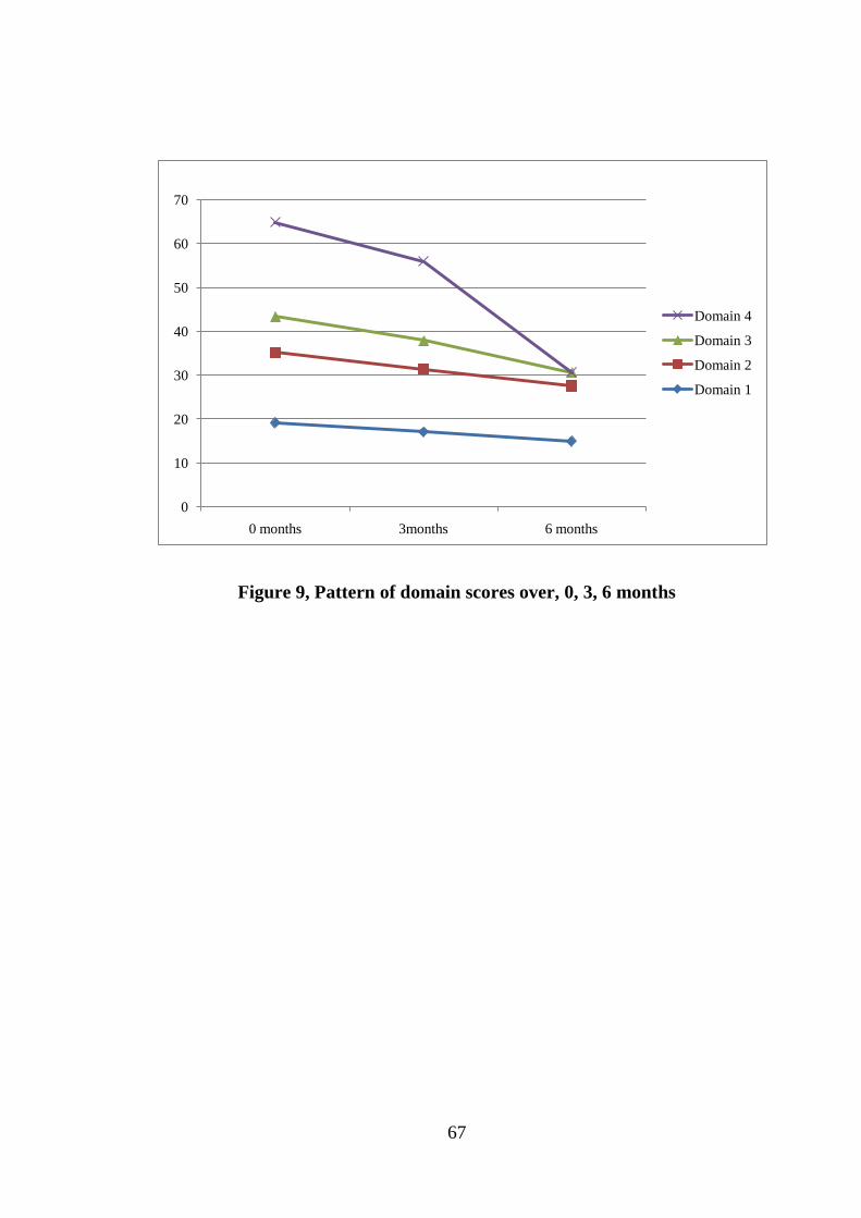
67
Figure 9, Pattern of domain scores over, 0, 3, 6 months
0
10
20
30
40
50
60
70
0 months 3months 6 months
Domain 4
Domain 3
Domain 2
Domain 1
68
DISCUSSION
WBRT is a unique therapy for brain metastasis. Several randomized and
non randomized studies have been conducted in these cases but have yielded
unequivocal results. To date there are very few studies that measure the effect
of WBRT on neurocognitive function and quality of life.(82) These studies
examined patients with varieties of malignancies in different age groups by
exposing them to different radiation regimens. Thus, we have a whole set of
research with different findings and results(82)..
In our study we analyzed data gathered from many patients with brain
metastasis who were treated with WBRT. We tried to understand the
neurocognitive changes that happen over 6 months.
Our study was conducted at a tertiary care hospital with patients chosen
for WBRT. The study was done in one of the largest apex institute and the
sample can fairly represent the population in the locality. 22 males and 21
females were recruited for our study and among them 3 expired and were not
available for total 6 month follow up.
In our study the median age of the population was 58 years which was
close the sample taken in international studies. Median age was 59 in the study
conducted Brown et al(57) with majority among them being females.
The sample consisted of fairly equal number of males and females thus
covering the major cancers affecting both the sex. The major cancer in our
patients was lung cancer which consisted of both small cell and non small cell
69
lung cancers. These numbers went on with the numbers obtained in the
international studies(3). Second highest in incidence we observed in the study
was the breast cancer commonly seen.
The occupation wise majority of our population was daily wage
workers. Their work place exposure to carcinogens may be a reason for
increased incidence of tumors and in turn brain metastasis which needs further
exploration. Income wise categorization was also done on our patients and we
found that majority of our patients were from low socioeconomic level and had
income less than 5000 per month.
Binet Kamath Test was the intelligence assessment test applied to see if
the patient could go through the assessment. Patients with IQ < 70 where
excluded from the study in the first place. There are studies done on the
children on WBRT showing the decline in IQ scores with radiation
exposure.(83) But all the studies show decline in IQ results occurring in due
course at least few years after the treatment. The study by Annett et al
(84)showed that the trajectory of decline in neuropsychological functioning,
specifically verbal IQ. The neuropsychological impact of WBRT was evident
only at 48 months and hence they suggested long term follow up.
Mini Mental Status Examination
MMSE is an important scale for assessing Neurocognition. Its
importance is brought out in a study by Murray et al(38) which tried to prove
its significance in patients on WBRT. We used the same scale in our study to
70
observe some interesting findings. Our MMSE scores showed the drop in the
neurocognitive scores occurring over 6 months (p=0.003). This was in
accordance with many of the research that had happened before. Several
authors have studied the MMSE scores in different settings and were able to
estimate the outcome.
As mentioned in our literature review, Meta analysis by Sun et al(41)
showed significant decline in Neurocognition over months. Similarly study by
Regine et al(37) and Murray et al followed the study population [MMSE scores
28 to 30] over 2 and 3 months in 29% of cases. They concluded that the drop in
MMSE is mainly due to poorly controlled metastasis and higher stages of
tumor. So our MMSE decline goes on with many other international studies
done in similar setup. When comparing the MMSE scores in 3 different times
(0, 3, 6 month) the statistics again gave us significant decline over 6 months.
Exposure to radiation in WBRT is associated with significant neuronal damage
and altered neuronal functioning which has led to drop in MMSE scores.
Meyers(56) and brown study made a significant study with Motexafin
Gadolinium in Whole brain radiotherapy and showed some important findings.
Their phase 3 study with 401 patients showed that almost 90.5% of their cases
had neurocognitive decline at baseline. Among them 42.4% had decline in at
least 4 or more areas.
In case of severe impairment in cognition the MMSE scale is debatable
with its sensitivity as demonstrated by Meyers and Wefel(14). Study by Olson
71
et al (85)showed that MoCA is superior in identifying the cognitive decline in
patients than MMSE. His study compared MMSE and MoCA and proved
that(85) MoCA identified 80% of the cases while MMSE found just 30
with[P=0.0001]. Hence we went on to apply a more specific and sensitive
assessment scale to our patients.
Montreal Cognitive Assessment scale is another neurocognitive scale
used on our patients to demonstrate the effect of the radiation exposure. Our
study with patients on WBRT showed a decline in MoCA neurocognitive
scores over 6 months (p=0.000). Hence the MoCA scores showed significant
results in demonstrating decline in cognitive function. We went to further
analyze each scores obtained in various domains of the MoCA.
Hopkins Verbal Learning Test [HVLT]
Hopkins Verbal Learning Test is a verbal learning test which measures
the new learning ability. It has high reliability and repeatability for measuring
verbal learning and memory. This test is found to be highly sensitive and
specific for various dementias and cognitive impairment. In Our study HVLT
applied on the patients showed declining scores over 6 months. Each of the 6
forms was applied with 3 trials each time over 0, 3 and 6 months. We found
that on comparing the results of each sitting the score declined and hence
verbal memory deteriorated significantly. The study Sun et al (41) was also
depicting the result similar to our study in saying that HVLT results dropped
72
significantly due to exposure of WBRT. The initial 3 months of results showed
p=0.001 which was close to the results obtained in our study.
Similarly, the study by Meyers et al(56) was also significant in proving
the effect of radiation on the verbal learning ability of the patients. After 4
months of follow up of these patients they found drastic decline in neuro
cognitive scores basically memory function .In 12 months the HVLT showed
significant decline among 48% of the patients. Overall the poor responders to
therapy showed decline in all areas with fall in median score in 8 months.
[HVLT recognition p= 0.0011, HVLT recall p = 0.0018, HVLT delayed recall
p= 0.0377]
From the above results with respect to HVLT we can infer that verbal
learning shows significant decline due to exposure to whole brain radiation.
Trail making test
Trail making test is used a part of assessment of visual attention and task
shifting. The test can provide information about visual search speed, scanning,
speed of processing, mental flexibility, as well as executive functioning.
A study on trail making test done by Grosshans et al(87) used trail
making test as a part of neurocognitive assessment showed that Trail Making
Test B score[p=0.001] dropped due to WBRT. the study by Meyers et al(56)
used trail making test and showed impairment in 44% of its subjects. Using the
cox model they showed that there was a significant decline in Trail Making
Test B scores with p=0.022. When they made a comparison with the indicator
73
lesion volume they again found a correlation with trail making result scores [p=
0.001].but there was no correlation with the number of tumor masses. Our
study our used the trail making test part A and part B on the patients. With 3
month of follow up of our patients we did not find a significant decline in
scores. But on 6 month follow up there was a significant decline in both trail
making test part A and part B. Even in terms of trail making test our results go
on with some of the standard studies done elsewhere. Thus we found a
significant deficit occurring in our patients after exposure to whole brain
radiotherapy.
Quality of life assessment
“Katz activities of daily living’’ was used in our study to basically
measure the daily activities of the patients on WBRT. Often the patients on
WBRT will be debilitated and hence a scale covering basic activities will give
a picture on the real life status of these people. In our study this Katz scores are
found to drop significantly over 6 months. We compared the individual scores
measured at 3 settings and patient’s activity level was found to drop at each
settings. One thing we could conclude is that with the exposure to WBRT
participants become more debilitated and restricted in their daily activities.
Since Katz score measure 6 areas of one’s life we also used Lawton
body ADL scale to get some added information. Even with this scale the, we
found to have reduced quality of life over 6 months.
74
As the above scales cover most basic of the daily life activities we used WHO
QOL Bref to measure the QOL in other domains. Literature did not reveal any
study that used WHO QOL scale to measure the quality of life of patients on
WBRT for brain metastasis. They preferred the QOL for brain damage and
other scales in routine research. Our study revealed that the patients had
decrease in quality of life in all domains over 6 Months. When we compared
the individual scores at 3 durations still we found significant decrease
happening as disease progress. In contrast a study by Tang et al(88) on
radiation induced brain injury showed similar decline in QOL over 4 month to
6 months. In the past research several studies tried to estimate the quality of life
and gave varying results. Most studies used overall survival and local tumor
control as one the end point.
In the present day due to the developments in the treatment modalities
the overall survival of the cancer patients has increased. Due to this the
knowledge of quality of life and its management is absolutely necessary. The
longer the patient survives with metastatic disease, the better systemic therapy
and palliative care is necessary. Neurocognitive function plays an important
role in QOL of the patient. The present treatment decisions are at times taken
based on better quality of life even at the cost of survival benefits. The
measurement of quality of life in cancer patients is difficult as loss of patients
is a common entity often due to death, debility or poor motivation. Karnofsky
Performance Status (KPS) is one of the measurement tools often used in
radiotherapy literature to assess the ability of the patient to undergo certain
75
procedure. But, it is poor in measuring the cognitive abilities of the patient.
KPS is said to have poor reliability, validity and inter rater reliability of 29%.
The study by Gao et al (64) as discussed showed improvement in quality
of life according to HRQOL scale. This was in contrast to the values obtained
in our study because the mean age of the population in our study was 57.81
years and our study used different scales to measure the quality of living.
Another important difference would be that our study considered a group of
patients with variety of primaries whereas the study by Gao et al(64) basically
studied on the patients having non small cell lung cancer.
We analyzed the whole lot of data obtained in our study and understood
cognitive changes happening among our patients. Radiation damage occurs due
to a complex and dynamic interaction of various mechanisms. It ranges from
changes in neurotransmitters to cell death. WBRT causes multiple side effects;
still according to research it is proved to be effective in increasing the survival
of the patients.
The stereotactic radio surgery is the latest development in the treatment
of brain tumors. By focusing the calculated amount of radiation a specified
focus this process give higher radiation exposure to the tumor mass. Meanwhile
it also counters the radiation exposure to normal brain tissue. Non-randomized
studies on STEREOTACTIC RADIOSURGERY alone in patients with four or
more brain metastases have shown that the overall survival is comparable to
WBRT. But the greatest problem with STEREOTACTIC RADIOSURGERY is
76
the high chance of tumor recurrence(89). This reason has maintained the
WBRT in a superior position compared to STEREOTACTIC
RADIOSURGERY.
Other newer advances in the field include the Hippocampal sparing
WBRT. It is a technique where the radiation exposure to the hippocampal stem
cell compartment is avoided using intensity modulated radiotherapy
(IMRT)(90). Here the sub granular zone of the hippocampus is exposed to
lesser radiation compared to rest of the brain which receives normal WBRT.
RTOG-0933 developed the guidelines for Hippocampal sparing WBRT where
the dose to 100% of hippocampus was limited to 9 Gray (Gy) with a maximum
dose to the hippocampus limited to 16 Gy. The rest of the brain parenchyma
receives a dose of 30 Gy in 10 fractions(90). Neurocognitive assessment was
done using HVLT showed only 7 % decline in scores compared to WBRT
which showed 30% decline [p=0.0003]. But other research also warns of the
high chances of tumor recurrence.
Ghia et al(91) studied the use of intensity-modulated radiotherapy in the
vicinity of the tumor mass. They showed that 5 mm remains an acceptable
distance from the hippocampus were a tumor mass can be targeted with
radiation causing minimal effect on cognition.
Composite tomotherapy with WBRT has been a new research area.(92)
It has a benefit of providing a homogeneous whole brain dose distribution
equivalent to conventional WBRT. The procedure also provides radio
77
surgically-equivalent dose distributions to individual metastases and
hippocampal avoidance of radiation exposure.
In the latest research the American Society for Radiation Oncology
(ASTRO) recommended the use of WBRT along with SRS in their ‘choose
wisely campaign’. The main reason for advocating this procedure was the
cognitive decline and poor QOL associated with WBRT. They suggest that
WBRT should be used only for leptomeningeal disease and multiple large brain
metastases not manageable with SRS. At present the use of WBRT on patients
undergoing surgery still questionable. Some specialists have started to use
stereotactic radio surgery on the surgical cavity and routinely follow up
radiologically.
Neuroprotection strategies
A set of studies have developed Neuroprotection strategies with WBRT
using pharmacological means(90). They include the use of PPARα 25, rennin
angiotensin system blockers, Experimental thalidomide, and motexafin
gadolinium(93). The use of agents commonly used in the treatment of
Alzheimer’s disease, including donepezil, memantine ,methylphenidate and
alpha-tocopherol, has been suggested(18). a study conducted in Hong Kong
compared Vitamin E against placebo in treating temporal lobe radio necrosis
and found improvement in global cognition, memory and executive
function(94). Patients treated on donepezil showed good improvement in terms
78
of memory, and motor speed and dexterity. Studies are still on to find definite
results in this regard.
Despite all the treatment and care the prognosis remains poor in patients
with brain metastasis. 90% of patients with brain metastases have impaired
neurocognitive function at diagnosis. Among them 60% will show further
declines in Neurocognition within 2-6 months of whole brain
radiotherapy(95). One of the greatest challenges as a therapist is to distinguish
treatment effects from progressive disease because the prognosis remains poor
in many patients

79
CONCLUSION
At the present neurocognitive decline is an important concern for the
patients undergoing WBRT for brain metastasis and tumors. But until today no
treatment has demonstrated better efficacy than WBRT in managing the brain
metastasis(96). WBRT has given the best response in terms of palliation in
brain metastasis with significantly good overall survival. Thus it has been
widely used until today in spite of known side effects.
.
80
LIMITATION
• The most important limitation of this study is population size. Brain
metastasis have poor prognosis and hence few cases will be exposed to
radiation for palliative purpose.
• Duration of the study is also for only 6 months. We allotted participants
and followed the cases for only 6 months. But there are few rare cases
on WBRT surviving up to 5 years which were not taken for our study.
• The absence of control group. But here patients can be considered as his
own control as we are assessing the neurocognitive change over 6
months.
• Lack of advanced neuropsychological battery for assessment of the
patients. Advanced neuropsychological assessment battery might give
more specific results.
• Corticosteroids used on the patients during WBRT might influence
neurocognitive function.
81
FUTURE DIRECTION
• Randomized controlled studies on the radiation induced cognitive
impairment.
• Better patient selection criteria to identify patients who respond to
specific procedures.
• Neurocognitive exercises and Cognitive enhancers to manage patients
on WBRT.
• Neuro protective agents
• Newer research in Stereotactic radio surgery and hippocampal
avoidance whole brain radiotherapy.
82
BIBLIOGRAPHY
1. Zimm S, Wampler GL, Stablein D, Hazra T, Young HF. Intracerebral
metastases in solid-tumor patients: natural history and results of
treatment. Cancer. 1981 Jul 15;48(2):384–94.
2. Brain metastases. Histology, multiplicity, surgery, and survival. -
PubMed - NCBI [Internet]. [cited 2017 Sep 23]. Available from:
https://www.ncbi.nlm.nih.gov/pubmed/8859192
3. Nayak L, Lee EQ, Wen PY. Epidemiology of Brain Metastases. Curr
Oncol Rep. 2012 Feb;14(1):48–54.
4. Whole brain radiotherapy for brain metastasis [Internet]. [cited 2017
Sep 22]. Available from: https://www.ncbi.nlm.nih.gov/pmc/
articles/PMC3656558/
5. Expert Panel on Radiation Oncology-Brain Metastases, Videtic GMM,
Gaspar LE, Aref AM, Germano IM, Goldsmith BJ, et al. American
College of Radiology appropriateness criteria on multiple brain
metastases. Int J Radiat Oncol Biol Phys. 2009 Nov 15;75(4):961–5.
6. Sperduto PW, Chao ST, Sneed PK, Luo X, Suh J, Roberge D, et al.
Diagnosis-specific prognostic factors, indexes, and treatment
outcomes for patients with newly diagnosed brain metastases: a multi-
institutional analysis of 4,259 patients. Int J Radiat Oncol Biol Phys.
2010 Jul 1;77(3):655–61.
83
7. Hall WA, Djalilian HR, Nussbaum ES, Cho KH. Long-term survival
with metastatic cancer to the brain. Med Oncol Northwood Lond Engl.
2000 Nov;17(4):279–86.
8. Tallet AV, Azria D, Barlesi F, Spano J-P, Carpentier AF, Gonçalves
A, et al. Neurocognitive function impairment after whole brain
radiotherapy for brain metastases: actual assessment. Radiat Oncol
Lond Engl. 2012 May 28;7:77.
9. Herman MA, Tremont-Lukats I, Meyers CA, Trask DD, Froseth C,
Renschler MF, et al. Neurocognitive and functional assessment of
patients with brain metastases: a pilot study. Am J Clin Oncol. 2003
Jun;26(3):273–9.
10. McTyre E, Scott J, Chinnaiyan P. Whole brain radiotherapy for brain
metastasis. Surg Neurol Int. 2013;4(Suppl 4):S236-244.
11. Li J, Bentzen SM, Li J, Renschler M, Mehta MP. Relationship
between neurocognitive function and quality of life after whole-brain
radiotherapy in patients with brain metastasis. Int J Radiat Oncol Biol
Phys. 2008 May 1;71(1):64–70.
12. Habets EJJ, Dirven L, Wiggenraad RG, Verbeek-de Kanter A,
Lycklama À Nijeholt GJ, Zwinkels H, et al. Neurocognitive
functioning and health-related quality of life in patients treated with
stereotactic radiotherapy for brain metastases: a prospective study.
Neuro-Oncol. 2016 Mar;18(3):435–44.
84
13. Roentgen-ray therapy of cerebral metastases - Chao - 1954 - Cancer -
Wiley Online Library [Internet]. [cited 2017 Sep 24]. Available from:
http://onlinelibrary.wiley.com/doi/10.1002/1097-
0142(195407)7:4%3C682::AID-
CNCR2820070409%3E3.0.CO%3B2-S/abstract
14. McDuff SGR, Taich ZJ, Lawson JD, Sanghvi P, Wong ET, Barker FG,
et al. Neurocognitive assessment following whole brain radiation
therapy and radiosurgery for patients with cerebral metastases. J
Neurol Neurosurg Psychiatry. 2013 Dec;84(12):1384–91.
15. Cochran DC, Chan MD, Aklilu M, Lovato JF, Alphonse NK, Bourland
JD, et al. The effect of targeted agents on outcomes in patients with
brain metastases from renal cell carcinoma treated with Gamma Knife
surgery. J Neurosurg. 2012 May;116(5):978–83.
16. Tallet A, Dhermain F, Taillia H, Ricard D, Mornex F, Métellus P.
[Cognition and radiation therapy for brain metastases: a new paradigm
to define]. Bull Cancer (Paris). 2013 Jan 1;100(1):69–74.
17. Kondziolka D, Niranjan A, Flickinger JC, Lunsford LD. Radiosurgery
with or without whole-brain radiotherapy for brain metastases: the
patients’ perspective regarding complications. Am J Clin Oncol. 2005
Apr;28(2):173–9.
18. Pham A, Lo SS, Sahgal A, Chang EL. Neurocognition and quality-of-
life in brain metastasis patients who have been irradiated focally or
85
comprehensively. Expert Rev Qual Life Cancer Care. 2016 Jan
2;1(1):45–60.
19. Greene-Schloesser D, Robbins ME, Peiffer AM, Shaw EG, Wheeler
KT, Chan MD. Radiation-induced brain injury: A review. Front Oncol.
2012;2:73.
20. Warrington JP, Ashpole N, Csiszar A, Lee YW, Ungvari Z, Sonntag
WE. Whole brain radiation-induced vascular cognitive impairment:
mechanisms and implications. J Vasc Res. 2013;50(6):445–57.
21. Lee TC, Greene-Schloesser D, Payne V, Diz DI, Hsu F-C, Kooshki M,
et al. Chronic administration of the angiotensin-converting enzyme
inhibitor, ramipril, prevents fractionated whole-brain irradiation-
induced perirhinal cortex-dependent cognitive impairment. Radiat Res.
2012 Jul;178(1):46–56.
22. Lee WH, Cho HJ, Sonntag WE, Lee YW. Radiation Attenuates
Physiological Angiogenesis by Differential Expression of VEGF,
Ang-1, Tie-2 and Ang-2 in Rat Brain. Radiat Res. 2011 Sep
30;176(6):753–60.
23. Lin S-Y, Yang C-C, Wu Y-M, Tseng C-K, Wei K-C, Chu Y-C, et al.
Evaluating the impact of hippocampal sparing during whole brain
radiotherapy on neurocognitive functions: A preliminary report of a
prospective phase II study. Biomed J. 2015 Oct;38(5):439–49.
86
24. Greene-Schloesser D, Robbins ME. Radiation-induced cognitive
impairment--from bench to bedside. Neuro-Oncol. 2012 Sep;14 Suppl
4:iv37-44.
25. Greene-Schloesser D, Moore E, Robbins ME. Molecular pathways:
radiation-induced cognitive impairment. Clin Cancer Res Off J Am
Assoc Cancer Res. 2013 May 1;19(9):2294–300.
26. Raff MC, Miller RH, Noble M. A glial progenitor cell that develops in
vitro into an astrocyte or an oligodendrocyte depending on culture
medium. Nature. 1983 Jun 2;303(5916):390–6.
27. Shi L, Linville MC, Iversen E, Molina DP, Yester J, Wheeler KT, et
al. Maintenance of white matter integrity in a rat model of radiation-
induced cognitive impairment. J Neurol Sci. 2009 Oct 15;285(1–
2):178–84.
28. Seifert G, Schilling K, Steinhäuser C. Astrocyte dysfunction in
neurological disorders: a molecular perspective. Nat Rev Neurosci.
2006 Mar;7(3):194–206.
29. Zhou H, Liu Z, Liu J, Wang J, Zhou D, Zhao Z, et al. Fractionated
radiation-induced acute encephalopathy in a young rat model:
cognitive dysfunction and histologic findings. AJNR Am J
Neuroradiol. 2011 Dec;32(10):1795–800.
87
30. Chiang CS, McBride WH, Withers HR. Radiation-induced astrocytic
and microglial responses in mouse brain. Radiother Oncol J Eur Soc
Ther Radiol Oncol. 1993 Oct;29(1):60–8.
31. Monje ML, Toda H, Palmer TD. Inflammatory blockade restores adult
hippocampal neurogenesis. Science. 2003 Dec 5;302(5651):1760–5.
32. Rosi S, Andres-Mach M, Fishman KM, Levy W, Ferguson RA, Fike
JR. Cranial irradiation alters the behaviorally induced immediate-early
gene arc (activity-regulated cytoskeleton-associated protein). Cancer
Res. 2008 Dec 1;68(23):9763–70.
33. Vlkolinský R, Krucker T, Nelson GA, Obenaus A. 56Fe-Particle
Radiation Reduces Neuronal Output and Attenuates
Lipopolysaccharide-Induced Inhibition of Long-Term Potentiation in
the Mouse Hippocampus. Radiat Res. 2008 May 1;169(5):523–30.
34. Wu PH, Coultrap S, Pinnix C, Davies KD, Tailor R, Ang KK, et al.
Radiation Induces Acute Alterations in Neuronal Function. Tofilon PJ,
editor. PLoS ONE. 2012 May 25;7(5):e37677.
35. Trippa F. Radiation-induced neurocognitive deficits in patients with
brain metastases. Rev Oncol [Internet]. 2014 [cited 2017 Sep 29];
Available from: http://www.reviewsinoncology.com/common/php/
portiere.php?ID=a4fc22957276a1ebb863b0b508e9b1f9
88
36. TheLimbicBrain,KluwerAcademic/Plenum,New York, NY, USA,
2001. - Google Search [Internet]. [cited 2017 Sep 27]. Available from:
https://www.google.co.in/search?q=TheLimbicBrain%2CKluwerAcad
emic%2FPlenum%2CNew+York%2C+NY%2C+USA%2C+2001.&rl
z=1C1DFOC_enIN628IN628&oq=TheLimbicBrain%2CKluwerAcade
mic%2FPlenum%2CNew+York%2C+NY%2C+USA%2C+2001.&aqs
=chrome..69i57.532j0j8&sourceid=chrome&ie=UTF-8
37. Regine WF, Scott C, Murray K, Curran W. Neurocognitive outcome in
brain metastases patients treated with accelerated-fractionation vs.
accelerated-hyperfractionated radiotherapy: an analysis from Radiation
Therapy Oncology Group Study 91-04. Int J Radiat Oncol Biol Phys.
2001 Nov 1;51(3):711–7.
38. Murray KJ, Scott C, Zachariah B, Michalski JM, Demas W, Vora NL,
et al. Importance of the mini-mental status examination in the
treatment of patients with brain metastases: a report from the
Radiation Therapy Oncology Group protocol 91-04. Int J Radiat Oncol
Biol Phys. 2000 Aug 1;48(1):59–64.
39. Tanaka K, Kanemoto A, Ohta A, Sato H, Kawaguchi G, Kaidu M, et
al. Assessment of Verbal Learning and Memory After Whole-Brain
Radiation Therapy With Different Dose Fractionation Using the
Hopkins Verbal Learning Test: The Revised Japanese Version. Int J
Radiat Oncol • Biol • Phys. 2015 Nov 1;93(3):E100.
89
40. Tsai P-F, Yang C-C, Chuang C-C, Huang T-Y, Wu Y-M, Pai P-C, et
al. Hippocampal dosimetry correlates with the change in
neurocognitive function after hippocampal sparing during whole brain
radiotherapy: a prospective study. Radiat Oncol Lond Engl. 2015 Dec
10;10:253.
41. Sun A, Bae K, Gore EM, Movsas B, Wong SJ, Meyers CA, et al.
Phase III trial of prophylactic cranial irradiation compared with
observation in patients with locally advanced non-small-cell lung
cancer: neurocognitive and quality-of-life analysis. J Clin Oncol Off J
Am Soc Clin Oncol. 2011 Jan 20;29(3):279–86.
42. Wolfson A, Bae K, Komaki R, Meyers C, Movsas B, Pechoux CL, et
al. Secondary Endpoints of a Phase II Randomized Trial (RTOG
0212): Impact of Different Total Doses and Schedules of Prophylactic
Cranial Irradiation on Chronic Neurotoxicity and Quality of Life for
Patients with Limited Disease Small-cell Lung Cancer. Int J Radiat
Oncol • Biol • Phys. 2009 Nov 1;75(3):S34.
43. Welzel G, Fleckenstein K, Schaefer J, Hermann B, Kraus-
Tiefenbacher U, Mai SK, et al. Memory function before and after
whole brain radiotherapy in patients with and without brain
metastases. Int J Radiat Oncol Biol Phys. 2008 Dec 1;72(5):1311–8.
44. Packer RJ, Sutton LN, Atkins TE, Radcliffe J, Bunin GR, D’Angio G,
et al. A prospective study of cognitive function in children receiving
90
whole-brain radiotherapy and chemotherapy: 2-year results. J
Neurosurg. 1989 May;70(5):707–13.
45. Shibamoto Y, Baba F, Oda K, Hayashi S, Kokubo M, Ishihara S-I, et
al. Incidence of brain atrophy and decline in mini-mental state
examination score after whole-brain radiotherapy in patients with
brain metastases: a prospective study. Int J Radiat Oncol Biol Phys.
2008 Nov 15;72(4):1168–73.
46. Aoyama H, Tago M, Kato N, Toyoda T, Kenjyo M, Hirota S, et al.
Neurocognitive function of patients with brain metastasis who
received either whole brain radiotherapy plus stereotactic radiosurgery
or radiosurgery alone. Int J Radiat Oncol Biol Phys. 2007 Aug
1;68(5):1388–95.
47. Burri SH, Asher A, Shaffrey M. Brain metastases treated with
radiosurgery alone: an alternative to whole brain radiotherapy?
Neurosurgery. 2004 Apr;54(4):1033-1034; author reply 1034-1035.
48. Silber JH, Radcliffe J, Peckham V, Perilongo G, Kishnani P, Fridman
M, et al. Whole-brain irradiation and decline in intelligence: the
influence of dose and age on IQ score. J Clin Oncol Off J Am Soc Clin
Oncol. 1992 Sep;10(9):1390–6.
49. Gregor A, Cull A, Traynor E, Stewart M, Lander F, Love S.
Neuropsychometric evaluation of long-term survivors of adult brain

91
tumours: relationship with tumour and treatment parameters.
Radiother Oncol J Eur Soc Ther Radiol Oncol. 1996 Oct;41(1):55–9.
50. Lutz S. Palliative whole-brain radiotherapy fractionation: convenience
versus cognition? Cancer. 2007 Dec 1;110(11):2363–5.
51. Monje ML, Palmer T. Radiation injury and neurogenesis. Curr Opin
Neurol. 2003 Apr;16(2):129–134.
52. Wolfson AH, Bae K, Komaki R, Meyers C, Movsas B, Le Pechoux C,
et al. Primary analysis of a phase II randomized trial Radiation
Therapy Oncology Group (RTOG) 0212: impact of different total
doses and schedules of prophylactic cranial irradiation on chronic
neurotoxicity and quality of life for patients with limited-disease
small-cell lung cancer. Int J Radiat Oncol Biol Phys. 2011 Sep
1;81(1):77–84.
53. DeAngelis LM, Delattre JY, Posner JB. Radiation-induced dementia in
patients cured of brain metastases. Neurology. 1989 Jun;39(6):789–96.
54. Corn BW, Moughan J, Knisely JPS, Fox SW, Chakravarti A, Yung
WKA, et al. Prospective evaluation of quality of life and
neurocognitive effects in patients with multiple brain metastases
receiving whole-brain radiotherapy with or without thalidomide on
Radiation Therapy Oncology Group (RTOG) trial 0118. Int J Radiat
Oncol Biol Phys. 2008 May 1;71(1):71–8.
92
55. Pospisil P, Kazda T, Hynkova L, Bulik M, Dobiaskova M, Burkon P,
et al. Post-WBRT cognitive impairment and hippocampal neuronal
depletion measured by in vivo metabolic MR spectroscopy: Results of
prospective investigational study. Radiother Oncol J Eur Soc Ther
Radiol Oncol. 2017 Mar;122(3):373–9.
56. Meyers CA, Smith JA, Bezjak A, Mehta MP, Liebmann J, Illidge T, et
al. Neurocognitive function and progression in patients with brain
metastases treated with whole-brain radiation and motexafin
gadolinium: results of a randomized phase III trial. J Clin Oncol Off J
Am Soc Clin Oncol. 2004 Jan 1;22(1):157–65.
57. Brown PD, Pugh S, Laack NN, Wefel JS, Khuntia D, Meyers C, et al.
Memantine for the prevention of cognitive dysfunction in patients
receiving whole-brain radiotherapy: a randomized, double-blind,
placebo-controlled trial. Neuro-Oncol. 2013 Oct;15(10):1429–37.
58. Onodera S, Aoyama H, Tha KK, Hashimoto N, Toyomaki A, Terae S,
et al. The value of 4-month neurocognitive function as an endpoint in
brain metastases trials. J Neurooncol. 2014 Nov;120(2):311–9.
59. Li J, Bentzen SM, Renschler M, Mehta MP. Regression after whole-
brain radiation therapy for brain metastases correlates with survival
and improved neurocognitive function. J Clin Oncol Off J Am Soc
Clin Oncol. 2007 Apr 1;25(10):1260–6.
93
60. Doolittle ND, Korfel A, Lubow MA, Schorb E, Schlegel U, Rogowski
S, et al. Long-term cognitive function, neuroimaging, and quality of
life in primary CNS lymphoma. Neurology. 2013 Jul 2;81(1):84–92.
61. Cole AM, Scherwath A, Ernst G, Lanfermann H, Bremer M,
Steinmann D. Self-reported cognitive outcomes in patients with brain
metastases before and after radiation therapy. Int J Radiat Oncol Biol
Phys. 2013 Nov 15;87(4):705–12.
62. Roman DD, Sperduto PW. Neuropsychological effects of cranial
radiation: current knowledge and future directions. Int J Radiat Oncol
Biol Phys. 1995 Feb 15;31(4):983–98.
63. Wong J, Hird A, Kirou–Mauro A, Napolskikh J, Chow E. Quality of
life in brain metastases radiation trials: a literature review. Curr Oncol.
2008 Oct;15(5):25–45.
64. Gao Y, Gao F, Ma J-L, Zhao D-L. Palliative whole-brain radiotherapy
and health- related quality of life for patients with brain metastasis in
cancer. Neuropsychiatr Dis Treat. 2015;11:2185–90.
65. Rosenman J, Choi NC. Improved quality of life of patients with small-
cell carcinoma of the lung by elective irradiation of the brain. Int J
Radiat Oncol • Biol • Phys. 1982 Jun 1;8(6):1041–3.
66. Selek U, Lo SS, Chang EL. Radiation Therapy for Brain Metastasis.
In: Decision Making in Radiation Oncology [Internet]. Springer,
94
Berlin, Heidelberg; 2011 [cited 2017 Oct 5]. p. 3–23. (Medical
Radiology). Available from: https://link.springer.com/chapter/
10.1007/978-3-642-13832-4_1
67. Tombaugh TN, McIntyre NJ. The mini-mental state examination: a
comprehensive review. J Am Geriatr Soc. 1992 Sep;40(9):922–35.
68. Vertesi A, Lever JA, Molloy DW, Sanderson B, Tuttle I, Pokoradi L,
et al. Standardized Mini-Mental State Examination. Use and
interpretation. Can Fam Physician. 2001 Oct;47:2018–23.
69. Nasreddine ZS, Phillips NA, Bédirian V, Charbonneau S, Whitehead
V, Collin I, et al. The Montreal Cognitive Assessment, MoCA: a brief
screening tool for mild cognitive impairment. J Am Geriatr Soc. 2005
Apr;53(4):695–9.
70. Smith T, Gildeh N, Holmes C. The Montreal Cognitive Assessment:
Validity and Utility in a Memory Clinic Setting. Can J Psychiatry.
2007 May;52(5):329–32.
71. Brandt J. The hopkins verbal learning test: Development of a new
memory test with six equivalent forms. Clin Neuropsychol. 1991
Apr;5(2):125–42.
72. Belkonen S. Hopkins Verbal Learning Test. In: Kreutzer JS, DeLuca J,
Caplan B, editors. Encyclopedia of Clinical Neuropsychology
[Internet]. New York, NY: Springer New York; 2011 [cited 2017 Sep
95
25]. p. 1264–5. Available from: http://link.springer.com/10.1007/978-
0-387-79948-3_1127
73. Tombaugh TN. Trail Making Test A and B: Normative data stratified
by age and education. Arch Clin Neuropsychol. 2004 Mar
1;19(2):203–14.
74. Fuentes-García A. Katz Activities of Daily Living Scale. In: Michalos
AC, editor. Encyclopedia of Quality of Life and Well-Being Research
[Internet]. Dordrecht: Springer Netherlands; 2014 [cited 2017 Sep 25].
p. 3465–8. Available from: http://link.springer.com/10.1007/978-94-
007-0753-5_1572
75. Ciesla JR, Shi L, Stoskopf CH, Samuels ME. Reliability of Katz’s
Activities of Daily Living Scale when used in telephone interviews.
Eval Health Prof. 1993 Jun;16(2):190–203.
76. Graf C. The Lawton instrumental activities of daily living scale. Am J
Nurs. 2008 Apr;108(4):52-62; quiz 62-63.
77. 76.pdf [Internet]. [cited 2017 Sep 25]. Available from:
http://www.who.int/mental_health/media/en/76.pdf
78. World Health Organization Quality of Life Instruments (WHOQOL-
BREF) - Seattle Quality of Life Group - Measure and Improve Health
Disparities in Children, Adolescents, Adults from Stigmatized
96
Populations [Internet]. [cited 2017 Sep 25]. Available from:
http://depts.washington.edu/seaqol/WHOQOL-BREF
79. Littell RC, Henry PR, Ammerman CB. Statistical analysis of repeated
measures data using SAS procedures. J Anim Sci. 1998
Apr;76(4):1216–31.
80. Analysis of Repeated Measures using sas - Google Search [Internet].
[cited 2017 Oct 4]. Available from: https://www.google.co.in/search?
rlz=1C1DFOC_enIN628IN628&q=Analysis+of+Repeated+Measures
+using+sas&oq=Analysis+of+Repeated+Measures+using+sas&gs_l=p
sy-ab.3..0i22i30k1l3.41441.44441.0.45265.10.10.0.0.0.0.577.1671.
0j4j2j5-1.7.0....0...1.1.64.psy-ab..3.7.1666...0.0.C8cwLtdv3Z4
81. Scheiner SM, Gurevitch J. Design and Analysis of Ecological
Experiments. Oxford University Press; 2001. 432 p.
82. Scoccianti S, Ricardi U. Treatment of brain metastases: Review of
phase III randomized controlled trials. Radiother Oncol. 2012 Feb
1;102(2):168–79.
83. Netson KL, Conklin HM, Wu S, Xiong X, Merchant TE. A 5-year
investigation of children’s adaptive functioning following conformal
radiation therapy for localized ependymoma. Int J Radiat Oncol Biol
Phys. 2012 Sep 1;84(1):217–223.e1.
97
84. Annett RD, Hile S, Bedrick E, Kunin-Batson AS, Krull KR, Embry L,
et al. Neuropsychological functioning of children treated for acute
lymphoblastic leukemia: impact of whole brain radiation therapy.
Psychooncology. 2015 Feb;24(2):181–9.
85. Olson RA, Chhanabhai T, McKenzie M. Feasibility study of the
Montreal Cognitive Assessment (MoCA) in patients with brain
metastases. Support Care Cancer Off J Multinatl Assoc Support Care
Cancer. 2008 Nov;16(11):1273–8.
86. Gondi V, Pugh SL, Tome WA, Caine C, Corn B, Kanner A, et al.
Preservation of memory with conformal avoidance of the hippocampal
neural stem-cell compartment during whole-brain radiotherapy for
brain metastases (RTOG 0933): a phase II multi-institutional trial. J
Clin Oncol Off J Am Soc Clin Oncol. 2014 Dec 1;32(34):3810–6.
87. Grosshans DR, Meyers CA, Allen PK, Davenport SD, Komaki R.
Neurocognitive function in patients with small cell lung cancer : effect
of prophylactic cranial irradiation. Cancer. 2008 Feb 1;112(3):589–95.
88. Tang Y, Luo D, Rong X, Shi X, Peng Y. Psychological Disorders,
Cognitive Dysfunction and Quality of Life in Nasopharyngeal
Carcinoma Patients with Radiation-Induced Brain Injury. PLOS ONE.
2012 Jun 11;7(6):e36529.
89. Lamba N, Muskens IS, DiRisio AC, Meijer L, Briceno V, Edrees H, et
al. Stereotactic radiosurgery versus whole-brain radiotherapy after
98
intracranial metastasis resection: a systematic review and meta-
analysis. Radiat Oncol Lond Engl [Internet]. 2017 Jun 24 [cited 2017
Oct 3];12. Available from: https://www.ncbi.nlm.nih.gov/pmc/articles/
PMC5483276/
90. Dye NB, Gondi V, Mehta MP. Strategies for preservation of memory
function in patients with brain metastases. Chin Clin Oncol. 2015
Jun;4(2):24.
91. Ghia A, Tomé WA, Thomas S, Cannon G, Khuntia D, Kuo JS, et al.
Distribution of brain metastases in relation to the hippocampus:
implications for neurocognitive functional preservation. Int J Radiat
Oncol Biol Phys. 2007 Jul 15;68(4):971–7.
92. Whole Brain Radiation Therapy with Hippocampal Avoidance and
Simultaneously Integrated Brain Metastases Boost: A Planning Study
[Internet]. [cited 2017 Sep 29]. Available from:
https://www.ncbi.nlm.nih.gov/ pmc/ articles/PMC2350212/
93. Strategies to mitigate the effects of whole-brain radiation therapy on
neurocognitive function in patients with brain metastases. (PDF
Download Available) [Internet]. [cited 2017 Sep 29]. Available from:
https://www.researchgate.net/publication/315741748_Strategies_to_m
itigate_the_effects_of_whole-brain_radiation_therapy_on_
neurocognitive_function_in_patients_with_brain_metastases

99
94. Chan AS, Cheung M-C, Law SC, Chan JH. Phase II study of alpha-
tocopherol in improving the cognitive function of patients with
temporal lobe radionecrosis. Cancer. 2004 Jan 15;100(2):398–404.
95. Pinkham MB, Sanghera P, Wall GK, Dawson BD, Whitfield GA.
Neurocognitive Effects Following Cranial Irradiation for Brain
Metastases. Clin Oncol R Coll Radiol G B. 2015 Nov;27(11):630–9.
96. Sundström JT, Minn H, Lertola KK, Nordman E. Prognosis of patients
treated for intracranial metastases with whole-brain irradiation. Ann
Med. 1998 Jun;30(3):296–9.
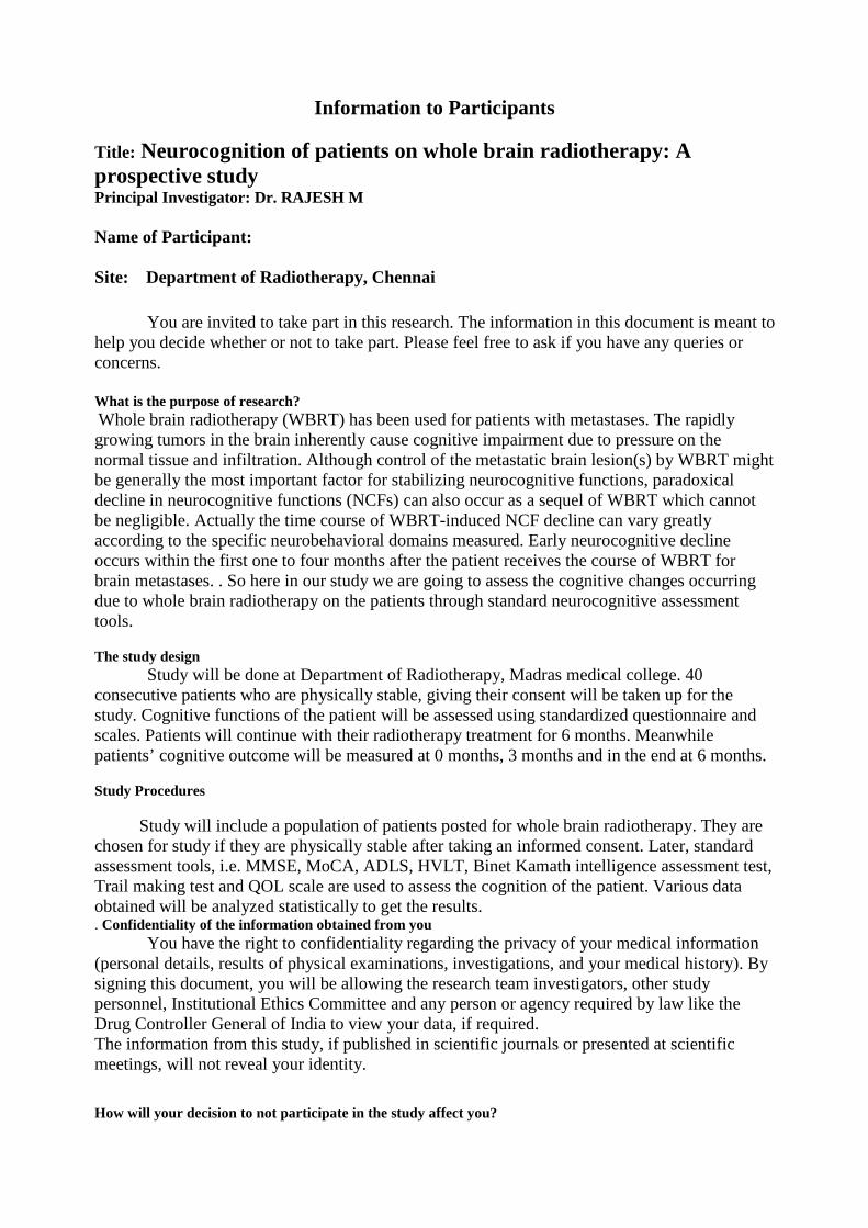
Information to Participants
Title: Neurocognition of patients on whole brain radiotherapy: A prospective study Principal Investigator: Dr. RAJESH M Name of Participant: Site: Department of Radiotherapy, Chennai
You are invited to take part in this research. The information in this document is meant to help you decide whether or not to take part. Please feel free to ask if you have any queries or concerns. What is the purpose of research? Whole brain radiotherapy (WBRT) has been used for patients with metastases. The rapidly growing tumors in the brain inherently cause cognitive impairment due to pressure on the normal tissue and infiltration. Although control of the metastatic brain lesion(s) by WBRT might be generally the most important factor for stabilizing neurocognitive functions, paradoxical decline in neurocognitive functions (NCFs) can also occur as a sequel of WBRT which cannot be negligible. Actually the time course of WBRT-induced NCF decline can vary greatly according to the specific neurobehavioral domains measured. Early neurocognitive decline occurs within the first one to four months after the patient receives the course of WBRT for brain metastases. . So here in our study we are going to assess the cognitive changes occurring due to whole brain radiotherapy on the patients through standard neurocognitive assessment tools. The study design Study will be done at Department of Radiotherapy, Madras medical college. 40 consecutive patients who are physically stable, giving their consent will be taken up for the study. Cognitive functions of the patient will be assessed using standardized questionnaire and scales. Patients will continue with their radiotherapy treatment for 6 months. Meanwhile patients’ cognitive outcome will be measured at 0 months, 3 months and in the end at 6 months.
Study Procedures Study will include a population of patients posted for whole brain radiotherapy. They are chosen for study if they are physically stable after taking an informed consent. Later, standard assessment tools, i.e. MMSE, MoCA, ADLS, HVLT, Binet Kamath intelligence assessment test, Trail making test and QOL scale are used to assess the cognition of the patient. Various data obtained will be analyzed statistically to get the results. . Confidentiality of the information obtained from you
You have the right to confidentiality regarding the privacy of your medical information (personal details, results of physical examinations, investigations, and your medical history). By signing this document, you will be allowing the research team investigators, other study personnel, Institutional Ethics Committee and any person or agency required by law like the Drug Controller General of India to view your data, if required. The information from this study, if published in scientific journals or presented at scientific meetings, will not reveal your identity. How will your decision to not participate in the study affect you?
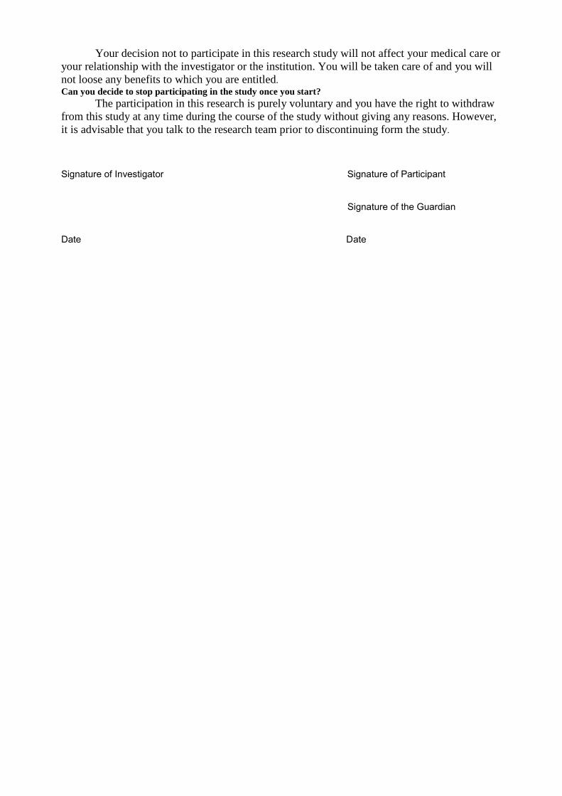
Your decision not to participate in this research study will not affect your medical care or your relationship with the investigator or the institution. You will be taken care of and you will not loose any benefits to which you are entitled. Can you decide to stop participating in the study once you start?
The participation in this research is purely voluntary and you have the right to withdraw from this study at any time during the course of the study without giving any reasons. However, it is advisable that you talk to the research team prior to discontinuing form the study.
Signature of Investigator Signature of Participant
Signature of the Guardian
Date Date
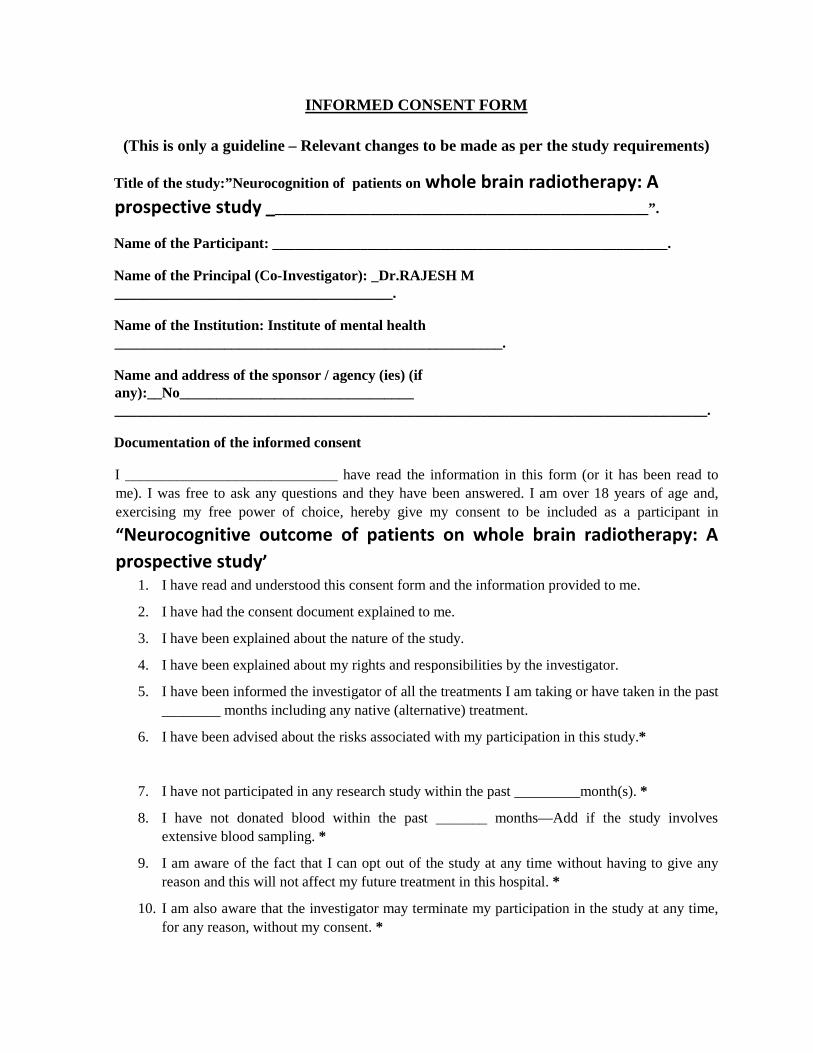
(This is only a guideline – Relevant changes to be made as per the study requirements)
INFORMED CONSENT FORM
Title of the study:”Neurocognition of patients on whole brain radiotherapy: A prospective study ____________________________________________________”.
Name of the Participant: ______________________________________________________.
Name of the Principal (Co-Investigator): _Dr.RAJESH M ______________________________________.
Name of the Institution: Institute of mental health _____________________________________________________.
Name and address of the sponsor / agency (ies) (if any):__No________________________________ _________________________________________________________________________________.
Documentation of the informed consent
I _____________________________ have read the information in this form (or it has been read to me). I was free to ask any questions and they have been answered. I am over 18 years of age and, exercising my free power of choice, hereby give my consent to be included as a participant in
“Neurocognitive outcome of patients on whole brain radiotherapy: A prospective study’
1. I have read and understood this consent form and the information provided to me.
2. I have had the consent document explained to me.
3. I have been explained about the nature of the study.
4. I have been explained about my rights and responsibilities by the investigator.
5. I have been informed the investigator of all the treatments I am taking or have taken in the past ________ months including any native (alternative) treatment.
6. I have been advised about the risks associated with my participation in this study.*
7. I have not participated in any research study within the past _________month(s). *
8. I have not donated blood within the past _______ months—Add if the study involves extensive blood sampling. *
9. I am aware of the fact that I can opt out of the study at any time without having to give any reason and this will not affect my future treatment in this hospital. *
10. I am also aware that the investigator may terminate my participation in the study at any time, for any reason, without my consent. *

11. I hereby give permission to the investigators to release the information obtained from me as result of participation in this study to the sponsors, regulatory authorities, Govt. agencies, and IEC. I understand that they are publicly presented.
12. I have understand that my identity will be kept confidential if my data are publicly presented
13. I have had my questions answered to my satisfaction.
14. I have decided to be in the research study. I am aware that if I have any question during this study, I should contact the investigator. By signing this consent form I attest that the information given in this document has been clearly explained to me and understood by me, I will be given a copy of this consent document.
For adult participants:
Name and signature / thumb impression of the participant (or legal representative if
participant incompetent)
Name _________________________ Signature_________________ Date________________ Name and Signature of impartial witness (required for illiterate patients): Name _________________________ Signature_________________ Date________________ Address and contact number of the impartial witness:
Name and Signature of the investigator or his representative obtaining consent: Name _________________________ Signature_________________ Date________________
Name and Signature of the investigator or his representative obtaining consent: Name _________________________ Signature_________________ Date________________


1
Mini-Mental State Examination (MMSE)
Patient’s Name: Date: Instructions: Ask the questions in the order listed. Score one point for each correct response within each question or activity. Maximum
Score Patient’s
Score Questions
5 “What is the year? Season? Date? Day of the week? Month?”
5 “Where are we now: State? County? Town/city? Hospital? Floor?”
3
The examiner names three unrelated objects clearly and slowly, then asks the patient to name all three of them. The patient’s response is used for scoring. The examiner repeats them until patient learns all of them, if possible. Number of trials: ___________
5 “I would like you to count backward from 100 by sevens.” (93, 86, 79, 72, 65, …) Stop after five answers. Alternative: “Spell WORLD backwards.” (D-L-R-O-W)
3 “Earlier I told you the names of three things. Can you tell me what those were?”
2 Show the patient two simple objects, such as a wristwatch and a pencil, and ask the patient to name them.
1 “Repeat the phrase: ‘No ifs, ands, or buts.’”
3 “Take the paper in your right hand, fold it in half, and put it on the floor.” (The examiner gives the patient a piece of blank paper.)
1 “Please read this and do what it says.” (Written instruction is “Close your eyes.”)
1 “Make up and write a sentence about anything.” (This sentence must contain a noun and a verb.)
1
“Please copy this picture.” (The examiner gives the patient a blank piece of paper and asks him/her to draw the symbol below. All 10 angles must be present and two must intersect.)
30 TOTAL (Adapted from Rovner & Folstein, 1987)
Source: www.medicine.uiowa.edu/igec/tools/cognitive/MMSE.pdf Provided by NHCQF, 0106-410
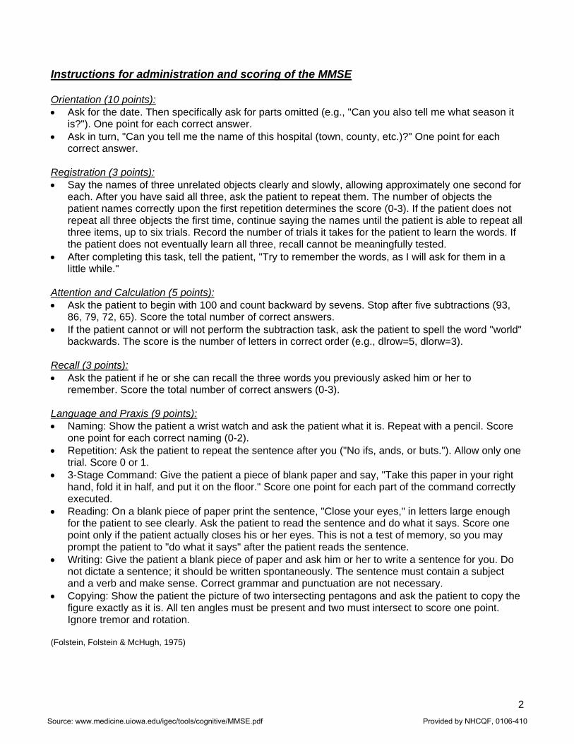
2
Instructions for administration and scoring of the MMSE Orientation (10 points): • Ask for the date. Then specifically ask for parts omitted (e.g., "Can you also tell me what season it
is?"). One point for each correct answer. • Ask in turn, "Can you tell me the name of this hospital (town, county, etc.)?" One point for each
correct answer. Registration (3 points): • Say the names of three unrelated objects clearly and slowly, allowing approximately one second for
each. After you have said all three, ask the patient to repeat them. The number of objects the patient names correctly upon the first repetition determines the score (0-3). If the patient does not repeat all three objects the first time, continue saying the names until the patient is able to repeat all three items, up to six trials. Record the number of trials it takes for the patient to learn the words. If the patient does not eventually learn all three, recall cannot be meaningfully tested.
• After completing this task, tell the patient, "Try to remember the words, as I will ask for them in a little while."
Attention and Calculation (5 points): • Ask the patient to begin with 100 and count backward by sevens. Stop after five subtractions (93,
86, 79, 72, 65). Score the total number of correct answers. • If the patient cannot or will not perform the subtraction task, ask the patient to spell the word "world"
backwards. The score is the number of letters in correct order (e.g., dlrow=5, dlorw=3). Recall (3 points): • Ask the patient if he or she can recall the three words you previously asked him or her to
remember. Score the total number of correct answers (0-3). Language and Praxis (9 points): • Naming: Show the patient a wrist watch and ask the patient what it is. Repeat with a pencil. Score
one point for each correct naming (0-2). • Repetition: Ask the patient to repeat the sentence after you ("No ifs, ands, or buts."). Allow only one
trial. Score 0 or 1. • 3-Stage Command: Give the patient a piece of blank paper and say, "Take this paper in your right
hand, fold it in half, and put it on the floor." Score one point for each part of the command correctly executed.
• Reading: On a blank piece of paper print the sentence, "Close your eyes," in letters large enough for the patient to see clearly. Ask the patient to read the sentence and do what it says. Score one point only if the patient actually closes his or her eyes. This is not a test of memory, so you may prompt the patient to "do what it says" after the patient reads the sentence.
• Writing: Give the patient a blank piece of paper and ask him or her to write a sentence for you. Do not dictate a sentence; it should be written spontaneously. The sentence must contain a subject and a verb and make sense. Correct grammar and punctuation are not necessary.
• Copying: Show the patient the picture of two intersecting pentagons and ask the patient to copy the figure exactly as it is. All ten angles must be present and two must intersect to score one point. Ignore tremor and rotation.
(Folstein, Folstein & McHugh, 1975)
Source: www.medicine.uiowa.edu/igec/tools/cognitive/MMSE.pdf Provided by NHCQF, 0106-410
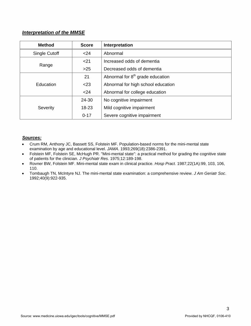
3
Interpretation of the MMSE
Method Score Interpretation
Single Cutoff <24 Abnormal
Range <21
>25
Increased odds of dementia
Decreased odds of dementia
Education
21
<23
<24
Abnormal for 8th grade education
Abnormal for high school education
Abnormal for college education
Severity
24-30
18-23
0-17
No cognitive impairment
Mild cognitive impairment
Severe cognitive impairment Sources: • Crum RM, Anthony JC, Bassett SS, Folstein MF. Population-based norms for the mini-mental state
examination by age and educational level. JAMA. 1993;269(18):2386-2391. • Folstein MF, Folstein SE, McHugh PR. "Mini-mental state": a practical method for grading the cognitive state
of patients for the clinician. J Psychiatr Res. 1975;12:189-198. • Rovner BW, Folstein MF. Mini-mental state exam in clinical practice. Hosp Pract. 1987;22(1A):99, 103, 106,
110. • Tombaugh TN, McIntyre NJ. The mini-mental state examination: a comprehensive review. J Am Geriatr Soc.
1992;40(9):922-935.
Source: www.medicine.uiowa.edu/igec/tools/cognitive/MMSE.pdf Provided by NHCQF, 0106-410
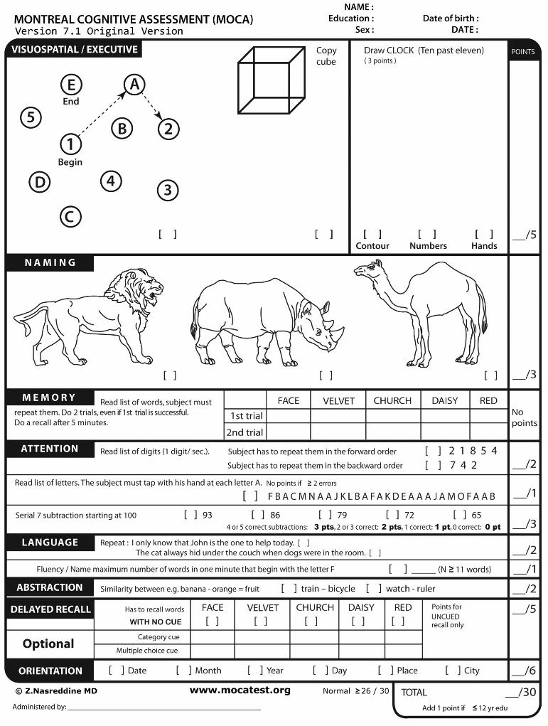
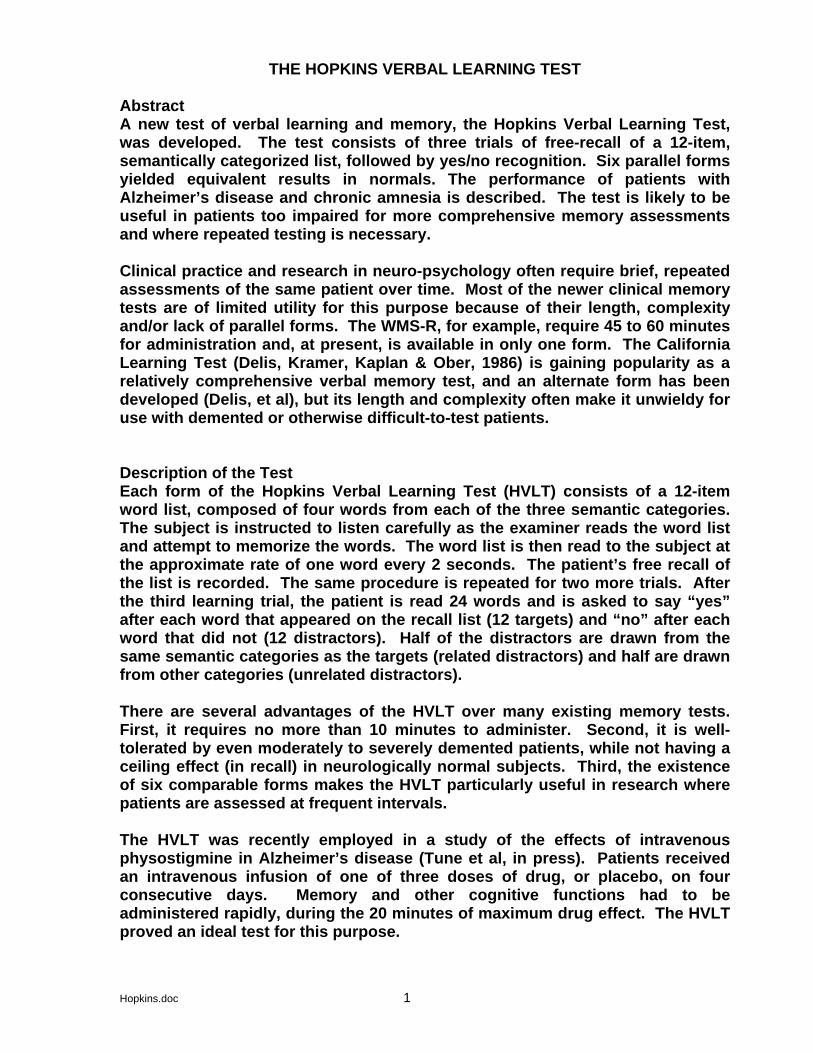
THE HOPKINS VERBAL LEARNING TEST
Abstract A new test of verbal learning and memory, the Hopkins Verbal Learning Test, was developed. The test consists of three trials of free-recall of a 12-item, semantically categorized list, followed by yes/no recognition. Six parallel forms yielded equivalent results in normals. The performance of patients with Alzheimer’s disease and chronic amnesia is described. The test is likely to be useful in patients too impaired for more comprehensive memory assessments and where repeated testing is necessary. Clinical practice and research in neuro-psychology often require brief, repeated assessments of the same patient over time. Most of the newer clinical memory tests are of limited utility for this purpose because of their length, complexity and/or lack of parallel forms. The WMS-R, for example, require 45 to 60 minutes for administration and, at present, is available in only one form. The California Learning Test (Delis, Kramer, Kaplan & Ober, 1986) is gaining popularity as a relatively comprehensive verbal memory test, and an alternate form has been developed (Delis, et al), but its length and complexity often make it unwieldy for use with demented or otherwise difficult-to-test patients. Description of the Test Each form of the Hopkins Verbal Learning Test (HVLT) consists of a 12-item word list, composed of four words from each of the three semantic categories. The subject is instructed to listen carefully as the examiner reads the word list and attempt to memorize the words. The word list is then read to the subject at the approximate rate of one word every 2 seconds. The patient’s free recall of the list is recorded. The same procedure is repeated for two more trials. After the third learning trial, the patient is read 24 words and is asked to say “yes” after each word that appeared on the recall list (12 targets) and “no” after each word that did not (12 distractors). Half of the distractors are drawn from the same semantic categories as the targets (related distractors) and half are drawn from other categories (unrelated distractors). There are several advantages of the HVLT over many existing memory tests. First, it requires no more than 10 minutes to administer. Second, it is well-tolerated by even moderately to severely demented patients, while not having a ceiling effect (in recall) in neurologically normal subjects. Third, the existence of six comparable forms makes the HVLT particularly useful in research where patients are assessed at frequent intervals. The HVLT was recently employed in a study of the effects of intravenous physostigmine in Alzheimer’s disease (Tune et al, in press). Patients received an intravenous infusion of one of three doses of drug, or placebo, on four consecutive days. Memory and other cognitive functions had to be administered rapidly, during the 20 minutes of maximum drug effect. The HVLT proved an ideal test for this purpose.
Hopkins.doc 1
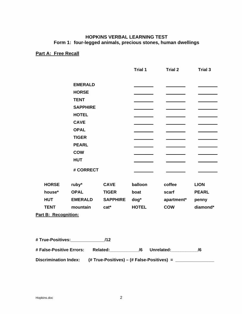
HOPKINS VERBAL LEARNING TEST Form 1: four-legged animals, precious stones, human dwellings
Part A: Free Recall
Trial 1 Trial 2 Trial 3 EMERALD ________ ________ ________HORSE ________ ________ ________TENT ________ ________ ________SAPPHIRE ________ ________ ________HOTEL ________ ________ ________CAVE ________ ________ ________OPAL ________ ________ ________TIGER ________ ________ ________PEARL ________ ________ ________COW ________ ________ ________HUT ________ ________ ________ # CORRECT
________ ________ ________
HORSE ruby* CAVE balloon coffee LION house* OPAL TIGER boat scarf PEARL HUT EMERALD SAPPHIRE dog* apartment* penny TENT mountain cat* HOTEL COW diamond*
Part B: Recognition: # True-Positives:______________/12 # False-Positive Errors: Related:____________/6 Unrelated:___________/6 Discrimination Index: (# True-Positives) – (# False-Positives) = ________________
Hopkins.doc 2
HOPKINS VERBAL LEARNING TEST Form 2: kitchen utensils, alcoholic beverages, weapons
Part A: Free Recall
Trial 1 Trial 2 Trial 3
FORK ________ ________ ________RUM ________ ________ ________PAN ________ ________ ________PISTOL ________ ________ ________SWORD ________ ________ ________SPATULA ________ ________ ________BOURBON ________ ________ ________VODKA ________ ________ ________POT ________ ________ ________COW ________ ________ ________HUT ________ ________ ________
WINE ________ ________ ________# CORRECT ________ ________ ________
Part B: Recognition:
spoon* PISTOL doll whiskey* FORK POT harmonica can opener* SWORD pencil gun* VODKA knife* RUM trout BOMB PAN gold WINE lemon SPATULA BOURBON beer* RIFLE # True-Positives:______________/12 # False-Positive Errors: Related:____________/6 Unrelated:___________/6 Discrimination Index: (# True-Positives) – (# False-Positives) = ________________
Hopkins.doc 3
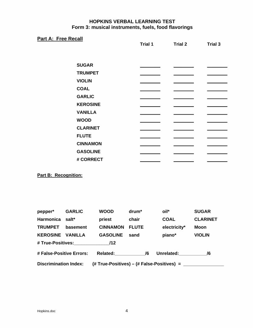
HOPKINS VERBAL LEARNING TEST Form 3: musical instruments, fuels, food flavorings
Part A: Free Recall
Trial 1 Trial 2 Trial 3
SUGAR ________ ________ ________TRUMPET ________ ________ ________VIOLIN ________ ________ ________COAL ________ ________ ________GARLIC ________ ________ ________KEROSINE ________ ________ ________VANILLA ________ ________ ________WOOD ________ ________ ________CLARINET ________ ________ ________FLUTE ________ ________ ________CINNAMON ________ ________ ________
GASOLINE ________ ________ ________# CORRECT ________ ________ ________
Part B: Recognition:
pepper* GARLIC WOOD drum* oil* SUGAR Harmonica salt* priest chair COAL CLARINETTRUMPET basement CINNAMON FLUTE electricity* Moon KEROSINE VANILLA GASOLINE sand piano* VIOLIN # True-Positives:______________/12 # False-Positive Errors: Related:____________/6 Unrelated:___________/6 Discrimination Index: (# True-Positives) – (# False-Positives) = ________________
Hopkins.doc 4
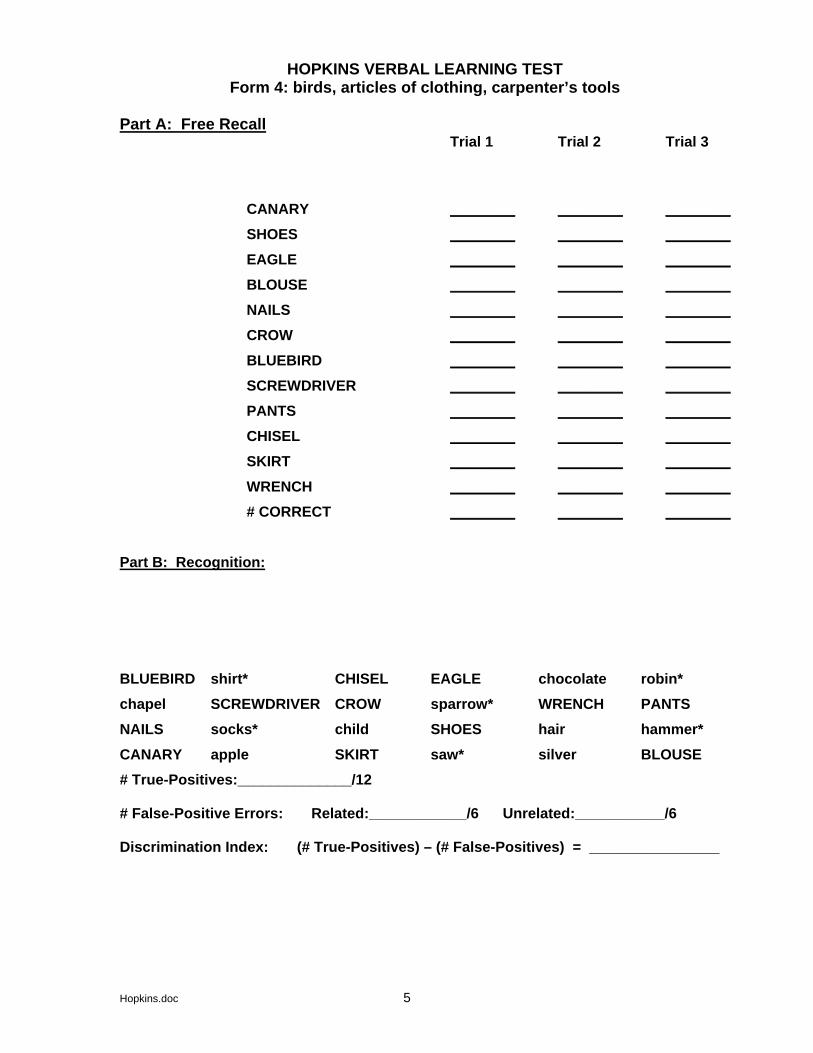
HOPKINS VERBAL LEARNING TEST Form 4: birds, articles of clothing, carpenter’s tools
Part A: Free Recall
Trial 1 Trial 2 Trial 3
CANARY ________ ________ ________SHOES ________ ________ ________EAGLE ________ ________ ________BLOUSE ________ ________ ________NAILS ________ ________ ________CROW ________ ________ ________BLUEBIRD ________ ________ ________SCREWDRIVER ________ ________ ________PANTS ________ ________ ________CHISEL ________ ________ ________SKIRT ________ ________ ________
WRENCH ________ ________ ________# CORRECT ________ ________ ________
Part B: Recognition:
BLUEBIRD shirt* CHISEL EAGLE chocolate robin* chapel SCREWDRIVER CROW sparrow* WRENCH PANTS NAILS socks* child SHOES hair hammer*CANARY apple SKIRT saw* silver BLOUSE # True-Positives:______________/12 # False-Positive Errors: Related:____________/6 Unrelated:___________/6 Discrimination Index: (# True-Positives) – (# False-Positives) = ________________
Hopkins.doc 5

HOPKINS VERBAL LEARNING TEST Form 5: occupations/professions, sports, vegetables
Part A: Free Recall
Trial 1 Trial 2 Trial 3 TEACHER ________ ________ ________BASKETBALL ________ ________ ________LETTUCE ________ ________ ________DENTIST ________ ________ ________TENNIS ________ ________ ________BEAN ________ ________ ________ENGINEER ________ ________ ________POTATO ________ ________ ________PROFESSOR ________ ________ ________GOLF ________ ________ ________CORN ________ ________ ________SOCCER ________ ________ ________# CORRECT ________ ________ ________
Part B: Recognition: # True-Positives:______________/12
TENNIS football* PROFESSOR spinach* lawyer* submarineGOLF DENTIST LETTUCE spider water BEAN BASKETBALL doctor* CORN baseball* TEACHER snake carrot* ENGINEER glove SOCCER POTATO tulip # False-Positive Errors: Related:____________/6 Unrelated:___________/6 Discrimination Index: (# True-Positives) – (# False-Positives) = ________________
Hopkins.doc 6
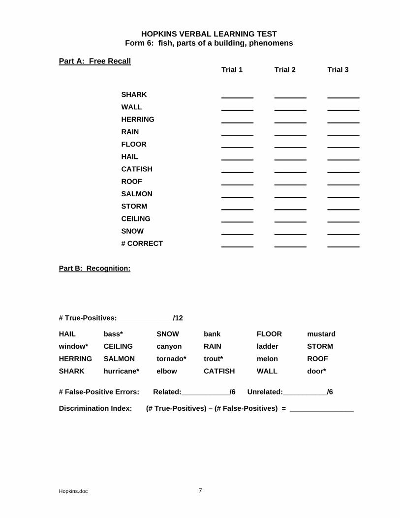
HOPKINS VERBAL LEARNING TEST Form 6: fish, parts of a building, phenomens
Part A: Free Recall
Trial 1 Trial 2 Trial 3 SHARK ________ ________ ________WALL ________ ________ ________HERRING ________ ________ ________RAIN ________ ________ ________FLOOR ________ ________ ________HAIL ________ ________ ________CATFISH ________ ________ ________ROOF ________ ________ ________SALMON ________ ________ ________STORM ________ ________ ________CEILING ________ ________ ________SNOW ________ ________ ________# CORRECT ________ ________ ________
Part B: Recognition: # True-Positives:______________/12
HAIL bass* SNOW bank FLOOR mustard window* CEILING canyon RAIN ladder STORM HERRING SALMON tornado* trout* melon ROOF SHARK hurricane* elbow CATFISH WALL door* # False-Positive Errors: Related:____________/6 Unrelated:___________/6 Discrimination Index: (# True-Positives) – (# False-Positives) = ________________
Hopkins.doc 7
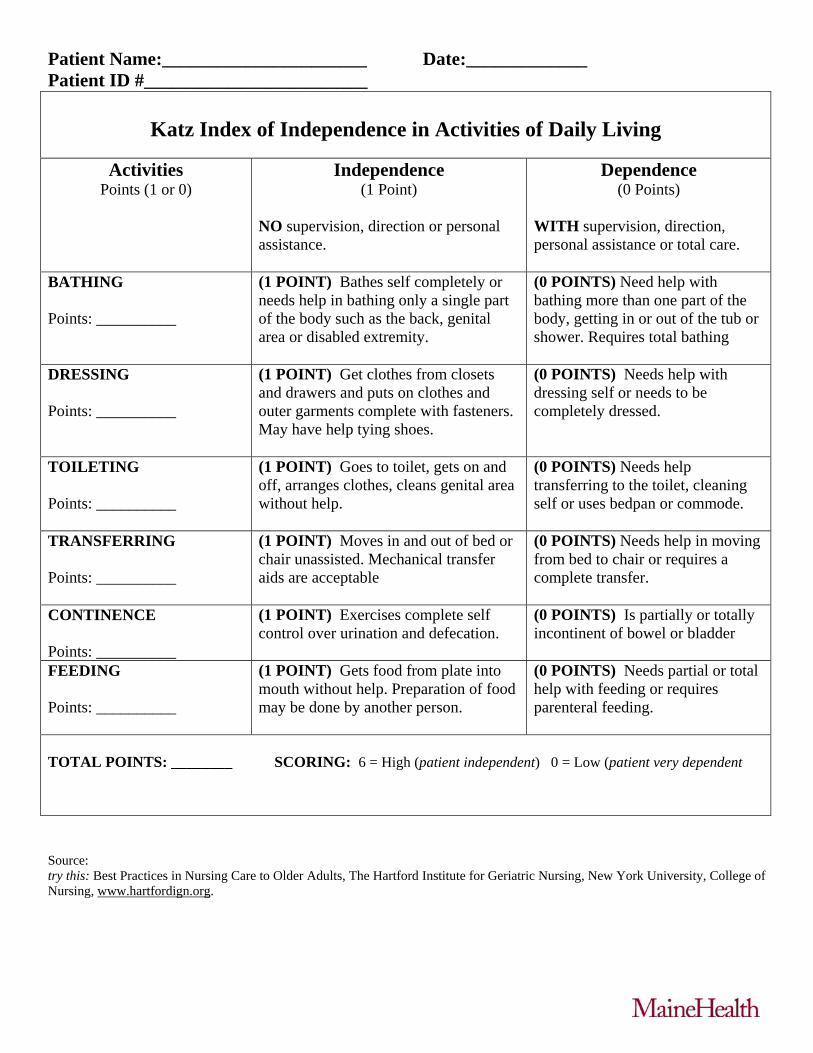
Patient Name:______________________ Date:_____________ Patient ID #________________________
Katz Index of Independence in Activities of Daily Living
Activities Points (1 or 0)
Independence (1 Point)
NO supervision, direction or personal assistance.
Dependence (0 Points)
WITH supervision, direction, personal assistance or total care.
BATHING Points: __________
(1 POINT) Bathes self completely or needs help in bathing only a single part of the body such as the back, genital area or disabled extremity.
(0 POINTS) Need help with bathing more than one part of the body, getting in or out of the tub or shower. Requires total bathing
DRESSING Points: __________
(1 POINT) Get clothes from closets and drawers and puts on clothes and outer garments complete with fasteners. May have help tying shoes.
(0 POINTS) Needs help with dressing self or needs to be completely dressed.
TOILETING Points: __________
(1 POINT) Goes to toilet, gets on and off, arranges clothes, cleans genital area without help.
(0 POINTS) Needs help transferring to the toilet, cleaning self or uses bedpan or commode.
TRANSFERRING Points: __________
(1 POINT) Moves in and out of bed or chair unassisted. Mechanical transfer aids are acceptable
(0 POINTS) Needs help in moving from bed to chair or requires a complete transfer.
CONTINENCE Points: __________
(1 POINT) Exercises complete self control over urination and defecation.
(0 POINTS) Is partially or totally incontinent of bowel or bladder
FEEDING Points: __________
(1 POINT) Gets food from plate into mouth without help. Preparation of food may be done by another person.
(0 POINTS) Needs partial or total help with feeding or requires parenteral feeding.
TOTAL POINTS: ________ SCORING: 6 = High (patient independent) 0 = Low (patient very dependent
Source: try this: Best Practices in Nursing Care to Older Adults, The Hartford Institute for Geriatric Nursing, New York University, College of Nursing, www.hartfordign.org.
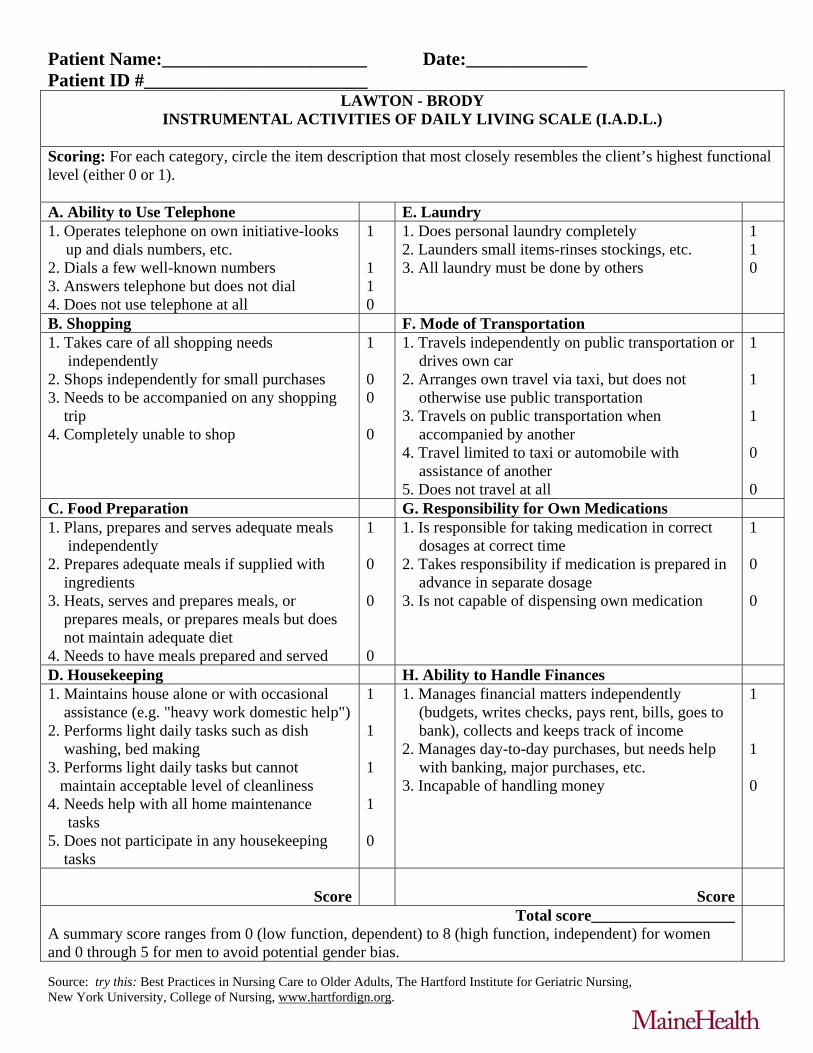
Patient Name:______________________ Date:_____________ Patient ID #________________________
LAWTON - BRODY INSTRUMENTAL ACTIVITIES OF DAILY LIVING SCALE (I.A.D.L.)
Scoring: For each category, circle the item description that most closely resembles the client’s highest functional level (either 0 or 1). A. Ability to Use Telephone E. Laundry 1. Operates telephone on own initiative-looks
up and dials numbers, etc. 2. Dials a few well-known numbers 3. Answers telephone but does not dial 4. Does not use telephone at all
1 1 1 0
1. Does personal laundry completely 2. Launders small items-rinses stockings, etc. 3. All laundry must be done by others
1 1 0
B. Shopping F. Mode of Transportation 1. Takes care of all shopping needs independently 2. Shops independently for small purchases 3. Needs to be accompanied on any shopping trip 4. Completely unable to shop
1 0 0 0
1. Travels independently on public transportation or drives own car
2. Arranges own travel via taxi, but does not otherwise use public transportation
3. Travels on public transportation when accompanied by another
4. Travel limited to taxi or automobile with assistance of another
5. Does not travel at all
1 1 1 0 0
C. Food Preparation G. Responsibility for Own Medications 1. Plans, prepares and serves adequate meals independently 2. Prepares adequate meals if supplied with ingredients 3. Heats, serves and prepares meals, or prepares meals, or prepares meals but does not maintain adequate diet 4. Needs to have meals prepared and served
1 0 0 0
1. Is responsible for taking medication in correct dosages at correct time
2. Takes responsibility if medication is prepared in advance in separate dosage
3. Is not capable of dispensing own medication
1 0 0
D. Housekeeping H. Ability to Handle Finances 1. Maintains house alone or with occasional assistance (e.g. "heavy work domestic help") 2. Performs light daily tasks such as dish washing, bed making 3. Performs light daily tasks but cannot maintain acceptable level of cleanliness 4. Needs help with all home maintenance tasks 5. Does not participate in any housekeeping tasks
1 1 1 1 0
1. Manages financial matters independently (budgets, writes checks, pays rent, bills, goes to bank), collects and keeps track of income
2. Manages day-to-day purchases, but needs help with banking, major purchases, etc.
3. Incapable of handling money
1 1 0
Score
Score
Total score__________________A summary score ranges from 0 (low function, dependent) to 8 (high function, independent) for women and 0 through 5 for men to avoid potential gender bias.
Source: try this: Best Practices in Nursing Care to Older Adults, The Hartford Institute for Geriatric Nursing, New York University, College of Nursing, www.hartfordign.org.

THE WORLD HEALTH ORGANIZATION
QUALITY OF LIFE (WHOQOL) -BREF
The World Health Organization Quality of Life (WHOQOL)-BREF
© World Health Organization 2004
All rights reserved. Publications of the World Health Organization can be obtained from Marketing and Dissemination, World Health Organization, 20 Avenue Appia, 1211 Geneva 27, Switzerland (tel: +41 22 791 2476; fax: +41 22 791 4857; email: [email protected]). Requests for permission to reproduce or translate WHO publications—whether for sale or for noncommercial distribution—should be addressed to Publications, at the above address (fax: +41 22 791 4806; email: [email protected]).
The designations employed and the presentation of the material in this publication do not imply the expression of any opinion whatsoever on the part of the World Health Organization concerning the legal status of any country, territory, city or area or of its authorities, or concerning the delimitation of its frontiers or boundaries. Dotted lines on maps represent approximate border lines for which there may not yet be full agreement.
The mention of specific companies or of certain manufacturers’ products does not imply that they are endorsed or recommended by the World Health Organization in preference to others of a similar nature that are not mentioned. Errors and omissions excepted, the names of proprietary products are distinguished by initial capital letters.
The World Health Organization does not warrant that the information contained in this publication is complete and correct and shall not be liable for any damages incurred as a result of its use.

WHOQOL-BREF
The following questions ask how you feel about your quality of life, health, or other areas of your life. I will read out each question to you, along with the response options. Please choose the answer that appears most appropriate. If you are unsure about which response to give to a question, the first response you think of is often the best one. Please keep in mind your standards, hopes, pleasures and concerns. We ask that you think about your life in the last four weeks.
Very poor Poor Neither poor
nor good Good Very good
1. How would you rate your quality of life?
1 2 3 4 5
Very dissatisfied Dissatisfied
Neither satisfied nor dissatisfied
Satisfied Very satisfied
2. How satisfied are you with your health?
1 2 3 4 5
The following questions ask about how much you have experienced certain things in the last four weeks.
Not at all A little A moderate
amount Very much An extreme
amount
3. To what extent do you feel that physical pain prevents you from doing what you need to do?
5 4 3 2 1
4. How much do you need any medical treatment to function in your daily life?
5 4 3 2 1
5. How much do you enjoy life? 1 2 3 4 5
6. To what extent do you feel your life to be meaningful?
1 2 3 4 5
Not at all A little A moderate
amount Very much Extremely
7. How well are you able to concentrate?
1 2 3 4 5
8. How safe do you feel in your daily life?
1 2 3 4 5
9. How healthy is your physical environment?
1 2 3 4 5
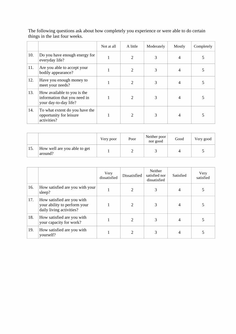
The following questions ask about how completely you experience or were able to do certain things in the last four weeks.
Not at all A little Moderately Mostly Completely
10. Do you have enough energy for everyday life?
1 2 3 4 5
11. Are you able to accept your bodily appearance?
1 2 3 4 5
12. Have you enough money to meet your needs?
1 2 3 4 5
13. How available to you is the information that you need in your day-to-day life?
1 2 3 4 5
14. To what extent do you have the opportunity for leisure activities?
1 2 3 4 5
Very poor Poor Neither poor
nor good Good Very good
15. How well are you able to get around?
1 2 3 4 5
Very dissatisfied Dissatisfied
Neither satisfied nor dissatisfied
Satisfied Very satisfied
16. How satisfied are you with your sleep?
1 2 3 4 5
17. How satisfied are you with your ability to perform your daily living activities?
1 2 3 4 5
18. How satisfied are you with your capacity for work?
1 2 3 4 5
19. How satisfied are you with yourself?
1 2 3 4 5
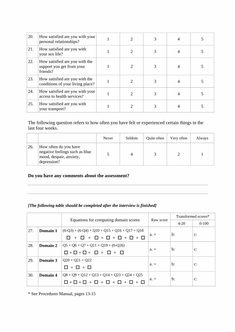
20. How satisfied are you with your personal relationships?
1 2 3 4 5
21. How satisfied are you with your sex life?
1 2 3 4 5
22. How satisfied are you with the support you get from your friends?
1 2 3 4 5
23. How satisfied are you with the conditions of your living place?
1 2 3 4 5
24. How satisfied are you with your access to health services?
1 2 3 4 5
25. How satisfied are you with your transport?
1 2 3 4 5
The following question refers to how often you have felt or experienced certain things in the last four weeks.
Never Seldom Quite often Very often Always
26. How often do you have negative feelings such as blue mood, despair, anxiety, depression?
5 4 3 2 1
Do you have any comments about the assessment?
[The following table should be completed after the interview is finished]
Transformed scores* Equations for computing domain scores Raw score 4-20 0-100
27. Domain 1 (6-Q3) + (6-Q4) + Q10 + Q15 + Q16 + Q17 + Q18
+ + + + + + a. = b: c:
28. Domain 2 Q5 + Q6 + Q7 + Q11 + Q19 + (6-Q26)
+ + + + + a. = b: c:
29. Domain 3 Q20 + Q21 + Q22
+ + a. = b: c:
30. Domain 4 Q8 + Q9 + Q12 + Q13 + Q14 + Q23 + Q24 + Q25
+ + + + + + + a. = b: c:
* See Procedures Manual, pages 13-15
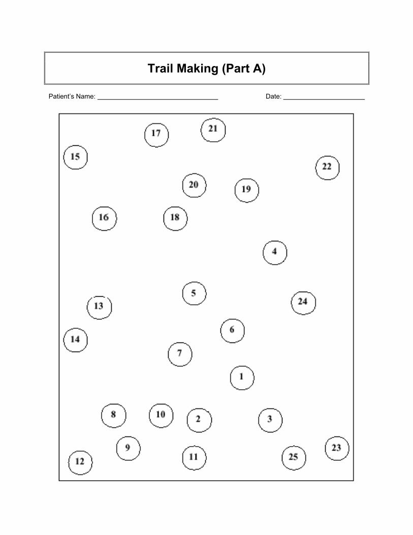
Trail Making (Part A)
Patient’s Name: Date:
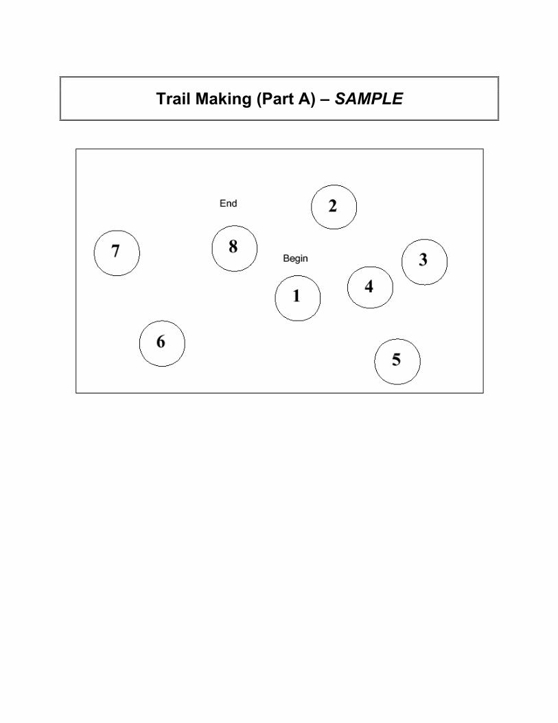
Trail Making (Part A) – SAMPLE
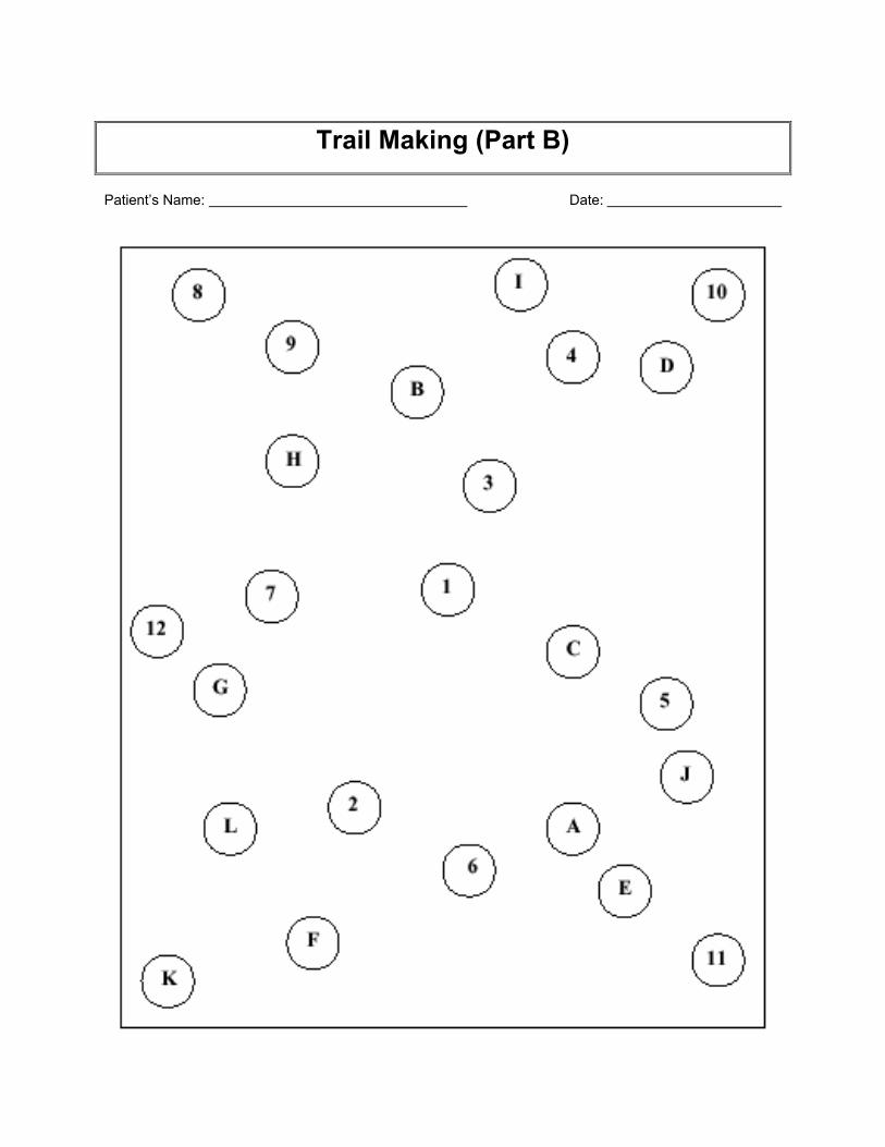
Trail Making (Part B)
Patient’s Name: Date:
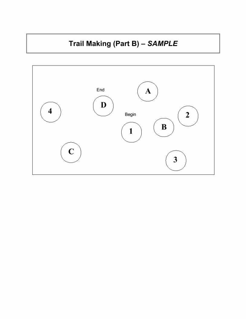
Trail Making (Part B) – SAMPLE
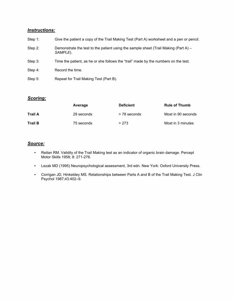
Instructions:
Step 1: Give the patient a copy of the Trail Making Test (Part A) worksheet and a pen or pencil.
Step 2: Demonstrate the test to the patient using the sample sheet (Trail Making (Part A) –SAMPLE).
Step 3: Time the patient, as he or she follows the “trail” made by the numbers on the test.
Step 4: Record the time.
Step 5: Repeat for Trail Making Test (Part B).
Scoring:
Average Deficient Rule of Thumb
Trail A 29 seconds > 78 seconds Most in 90 seconds
Trail B 75 seconds > 273 Most in 3 minutes
Source:
• Reitan RM. Validity of the Trail Making test as an indicator of organic brain damage. PerceptMotor Skills 1958; 8: 271-276.
• Lezak MD (1995) Neuropsychological assessment, 3rd edn. New York: Oxford University Press.
• Corrigan JD, Hinkeldey MS. Relationships between Parts A and B of the Trail Making Test. J ClinPsychol 1987;43:402–9.
Comparison of recognition scores and discrimination scores over 0, 3 and 6 months of HVLT:
Form 1 recognition scores
Mean Difference
(I-J) Std. Error Sig.b
95% Confidence Interval for Differenceb
Lower Bound
Upper Bound
1 2 .325 .252 .612 -.304 .954 3 -3.475* .291 .000 -4.203 -2.747
2 1 -.325 .252 .612 -.954 .304 3 -3.800* .153 .000 -4.182 -3.418
3 1 3.475* .291 .000 2.747 4.203
2 3.800* .153 .000 3.418 4.182
Form 1 discrimination index
Mean Difference
(I-J) Std. Error Sig.b
95% Confidence Interval for Differenceb
Lower Bound
Upper Bound
1 2 .075 .184 1.000 -.385 .535 3 3.075* .210 .000 2.550 3.600
2 1 -.075 .184 1.000 -.535 .385 3 3.000* .203 .000 2.493 3.507
3 1 -3.075* .210 .000 -3.600 -2.550
2 -3.000* .203 .000 -3.507 -2.493
Form 2 recognition scores
Mean Difference
(I-J) Std. Error Sig.b
95% Confidence Interval for Differenceb
Lower Bound
Upper Bound
1 2 -.150 .166 1.000 -.566 .266 3 .350 .234 .426 -.234 .934
2 1 .150 .166 1.000 -.266 .566 3 .500* .179 .024 .052 .948
3 1 -.350 .234 .426 -.934 .234
2 -.500* .179 .024 -.948 -.052
Form 2 discrimination index
Mean Difference
(I-J) Std. Error Sig.b
95% Confidence Interval for Differenceb
Lower Bound
Upper Bound
1 2 -.500 .226 .100 -1.067 .067 3 .425 .226 .203 -.141 .991
2 1 .500 .226 .100 -.067 1.067 3 .925* .194 .000 .439 1.411
3 1 -.425 .226 .203 -.991 .141
2 -.925* .194 .000 -1.411 -.439
Form 3 recognition scores
Mean Difference
(I-J) Std. Error Sig.b
95% Confidence Interval for Differenceb
Lower Bound
Upper Bound
1 2 -.200 .209 1.000 -.724 .324 3 .525 .280 .204 -.175 1.225
2 1 .200 .209 1.000 -.324 .724 3 .725* .273 .034 .043 1.407
3 1 -.525 .280 .204 -1.225 .175
2 -.725* .273 .034 -1.407 -.043
Form 3 discrimination index
Mean Difference
(I-J) Std. Error Sig.b
95% Confidence Interval for Differenceb
Lower Bound
Upper Bound
1 2 -.150 .204 1.000 -.661 .361 3 .400 .189 .122 -.072 .872
2 1 .150 .204 1.000 -.361 .661 3 .550* .196 .023 .060 1.040
3 1 -.400 .189 .122 -.872 .072
2 -.550* .196 .023 -1.040 -.060
Form 4 recognition scores
Mean Difference
(I-J) Std. Error Sig.b
95% Confidence Interval for Differenceb
Lower Bound
Upper Bound
1 2 -.325 .219 .437 -.873 .223 3 .250 .189 .578 -.222 .722
2 1 .325 .219 .437 -.223 .873 3 .575* .192 .015 .094 1.056
3 1 -.250 .189 .578 -.722 .222
2 -.575* .192 .015 -1.056 -.094
Form 4 discrimination index
Mean Difference
(I-J) Std. Error Sig.a
95% Confidence Interval for Differencea
Lower Bound
Upper Bound
1 2 -.025 .181 1.000 -.477 .427 3 .400 .240 .309 -.200 1.000
2 1 .025 .181 1.000 -.427 .477 3 .425 .214 .164 -.112 .962
3 1 -.400 .240 .309 -1.000 .200
2 -.425 .214 .164 -.962 .112
Form 5 recognition scores
Mean Difference
(I-J) Std. Error Sig.a
95% Confidence Interval for Differencea
Lower Bound
Upper Bound
1 2 -.350 .303 .765 -1.108 .408 3 0.000 .209 1.000 -.522 .522
2 1 .350 .303 .765 -.408 1.108 3 .350 .204 .284 -.161 .861
3 1 0.000 .209 1.000 -.522 .522
2 -.350 .204 .284 -.861 .161
Form 5 discrimination index
Mean Difference
(I-J) Std. Error Sig.b
95% Confidence Interval for Differenceb
Lower Bound
Upper Bound
1 2 .150 .210 1.000 -.377 .677 3 .525* .206 .044 .011 1.039
2 1 -.150 .210 1.000 -.677 .377 3 .375 .192 .173 -.105 .855
3 1 -.525* .206 .044 -1.039 -.011
2 -.375 .192 .173 -.855 .105
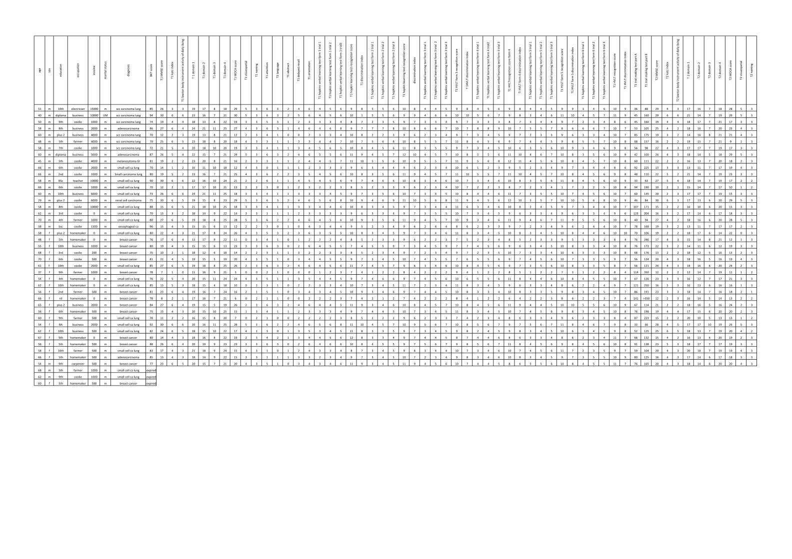
age
sex
education
occupatio
n
income
marita
l status
diagno
sis
BKT score
T1 M
MSE sc
ore
T1 katz ind
ex
T1 lawton bo
dy instrumen
t activity
of d
aily living
T 1 do
main 1
T1 dom
ain 2
T1 dom
ain 3
T1 dom
ain 4
T1 M
OCA
score
T1 visuo
spatial
T1 nam
ing
T1 atten
tion
T1 language
T1 abstract
T1 delayed
recall
T1 orie
ntation
T1 hop
kins verbal learning test fp
rm 1 trial 1
T1 hop
kin verbal learning
test fo
rm 1 trial 2
T1 hop
kin verbal learning
test fo
rm 1 trial3
T1 hop
kin learning
test re
cognition
score
T1 discrim
ination inde
x
T1 hop
kins verbal learning test fo
rm 2 trial 1
T1 hop
kins verbal learning test fo
rm 2 trial 2
T1 hop
kins verbal learning test fo
rm 2 trial 3
T1 hop
kin kearning
test re
cognition
score
discrim
ination inde
x
T1 hop
kins verbal learning test fo
rm 3 trial 1
T1 hop
kins verbal learning test fo
rm 3 trial 2
T1 hop
kins verbal learning test fo
rm 3 trial 3
T1 HVLT form
3 re
cognition
score
T 1H
VLT discrim
ination inde
x
T1 hop
kins verbal learning test fo
rm 4 trial 1
T1 hop
kins verbal learning test fo
rm 4 trial2
T1 hop
kins verbal learning test fo
rm 4 trial 3
T1 HVLTrecognitio
n score form
4
TI HVLT form
4 discrim
ination inde
x
T1 hop
kins verbal learning test fo
rm 5 trial 1
T1 hop
kins verbal learning test fo
rm 5 trial 2
T1 hop
kins verbal learning test fo
rm 5 trial 3
T1 HVLT form
5 re
cognition
score
T1 HVLT form
5 discrmination inde
x
T1 hop
kins verbal learning test fo
rm 6 trial 1
T1 hop
kins verbal learning test fo
rm 6 trial 2
T1 hop
kins verbal learning test fo
rm 6 trial 3
T1 HVLT recognition
score
T1 HVLT discrim
ination inde
x
T1 trail m
aking test part A
T1 trail m
aking test part B
T2 M
MSE sc
ore
T2 katz ind
ex
T2 lawton bo
dy instrumen
t activity
of d
aily living
T 2 do
main 1
T2 dom
ain 2
T2 dom
ain 3
T2 dom
ain 4
T2 M
OCA
score
T2 visuo
spatial
T2 nam
ing
51 m 10th electrician 15000 m scc carcinoma lung 85 26 3 3 19 17 8 18 29 5 3 6 3 2 4 6 4 5 6 9 8 5 5 6 10 8 4 4 5 9 8 4 5 6 9 8 4 4 5 9 9 3 4 6 10 9 36 88 29 4 3 17 16 7 18 28 5 3
40 m diploma business 10000 UM scc carcinoma lung 94 30 6 6 23 16 7 21 30 5 3 6 3 2 5 6 4 5 6 10 1 5 5 6 9 9 4 6 6 10 10 5 6 7 9 8 3 4 6 11 10 4 5 7 11 9 45 160 29 6 6 21 14 7 19 29 5 3
50 m 9th coolie 1000 m scc carcinoma lung 74 19 4 4 18 13 8 22 19 3 3 5 3 1 2 3 3 3 4 8 7 2 3 5 9 7 3 3 4 9 7 3 3 3 8 7 3 4 4 9 7 2 3 4 8 6 45 160 19 4 4 18 17 7 21 17 3 3
58 m 8th business 2000 m adenocarcinoma 86 27 6 4 24 21 11 25 27 4 3 6 3 1 4 6 4 6 8 9 7 7 7 8 10 8 6 6 7 10 7 8 8 9 10 7 5 5 7 8 6 6 6 7 10 7 53 105 25 4 2 18 16 7 20 23 4 3
60 m plus 2 business 4000 m scc carcinoma lung 70 12 2 3 19 13 8 21 12 2 3 4 1 0 0 2 3 3 4 10 8 2 2 3 9 6 2 3 4 9 7 3 4 5 9 7 2 3 3 9 6 3 3 4 10 7 85 175 19 3 2 14 16 8 21 21 4 3
48 m 5th farmer 4000 m scc carcinoma lung 70 25 6 5 23 18 8 20 18 4 3 3 1 1 3 3 4 4 7 10 7 5 6 8 10 8 5 5 7 11 8 4 5 6 9 7 4 4 5 9 8 5 5 7 10 8 68 157 16 2 2 19 15 7 21 9 1 3
58 m 7th coolie 1000 m scc carcinoma lung 72 21 5 4 20 18 10 20 19 3 3 4 1 1 3 4 5 6 5 10 8 4 5 6 11 8 3 5 5 9 7 3 4 5 10 6 3 5 6 10 9 3 4 6 9 8 56 98 22 4 3 17 17 7 19 17 3 3
60 m diploma business 5000 m adenocarcinoma 87 26 5 4 22 15 7 21 29 5 3 6 3 2 4 6 5 5 6 11 9 4 5 7 12 10 4 5 7 10 8 5 5 6 11 10 4 6 7 10 8 3 5 6 10 9 42 100 26 4 3 18 14 5 18 29 5 3
45 m 5th coolie 4000 m melanocytoma GI 81 19 2 2 23 20 8 21 16 2 3 3 1 1 2 4 4 5 7 11 10 5 5 6 10 9 5 5 7 11 9 5 6 8 12 11 4 5 6 10 8 4 5 7 10 8 60 111 22 2 2 16 13 7 20 18 2 3
66 m 6th coolie 2000 m small cell ca lung 70 14 1 2 16 11 10 18 12 4 3 0 1 1 1 2 3 3 3 9 6 3 4 4 9 6 2 3 4 10 6 1 2 3 9 5 2 3 4 9 7 3 4 4 8 6 92 225 13 3 3 13 11 7 17 19 4 3
66 m 2nd coolie 1000 m Small carcinoma lung 80 19 5 2 23 16 7 21 25 4 3 6 2 2 3 5 4 5 6 10 8 3 5 6 11 9 4 5 7 11 10 5 5 7 11 10 4 5 7 10 8 4 5 6 9 8 48 110 22 5 2 21 14 7 19 23 3 3
58 m BSc teacher 10000 m small cell ca lung 90 30 6 6 22 16 10 24 21 2 2 6 1 1 4 5 4 5 6 9 7 4 6 6 10 8 3 4 6 10 7 3 4 6 10 8 3 5 6 11 8 4 5 6 10 9 33 63 27 5 4 18 14 7 19 17 2 2
66 m 6th coolie 1000 m small cell ca lung 70 12 2 1 17 17 10 21 13 2 2 3 0 1 2 3 2 2 3 8 5 2 3 3 9 6 2 3 4 10 7 2 2 3 8 7 2 3 4 1 7 2 2 5 10 8 94 190 10 2 1 15 14 7 17 10 1 2
60 m 10th business 6000 m small cell ca lung 73 26 6 6 24 21 11 25 18 3 3 4 1 1 3 3 3 4 5 9 7 3 5 6 10 7 3 4 5 10 8 4 4 6 11 7 3 5 5 10 7 4 5 6 10 7 60 145 20 2 3 17 17 7 19 15 3 3
29 m plus 2 coolie 6000 m renal cell carcinoma 75 30 6 5 19 15 8 23 29 5 3 6 3 2 4 6 5 6 8 10 9 4 6 9 11 10 5 6 8 11 9 4 5 6 12 10 3 5 7 10 10 5 6 8 10 9 46 84 30 6 3 17 13 6 20 29 5 3
58 m 8th coolie 10000 m small cell ca lung 88 15 6 5 21 18 10 25 18 3 3 4 1 1 3 3 4 4 6 10 8 3 4 5 9 7 3 4 4 11 6 3 4 6 10 8 2 4 5 9 7 3 4 6 10 7 107 171 15 2 2 16 10 6 20 15 3 3
62 m 3rd coolie 0 m small cell ca lung 70 13 3 2 18 14 9 22 14 3 3 1 1 1 2 3 3 3 3 9 6 3 3 4 9 7 3 5 5 10 7 3 4 5 9 6 3 3 4 9 6 3 3 4 9 6 123 204 16 3 2 17 14 6 17 18 3 3
70 m 4th farmer 1000 m small cell ca lung 80 27 6 5 19 18 8 25 28 5 3 6 2 2 4 6 4 5 6 10 9 3 5 6 11 9 4 5 7 10 9 3 4 6 11 9 4 6 7 11 9 5 5 6 10 8 40 94 27 4 2 18 16 6 20 28 5 3
58 m bsc coolie 1500 m oesophageal ca 96 15 4 3 15 15 6 13 12 2 2 3 0 1 0 4 3 4 4 9 5 2 3 4 9 6 2 4 4 8 6 2 3 3 9 7 2 3 4 9 6 2 4 4 10 7 78 168 19 3 2 13 11 7 17 17 2 3
58 f plus 2 homemaker 0 m small cell ca lung 80 22 4 3 21 17 8 24 26 4 3 5 3 2 3 6 3 5 5 10 8 3 4 5 9 7 3 4 6 11 8 3 4 5 10 8 3 4 5 10 8 4 4 6 10 10 70 106 19 2 2 18 17 6 14 22 0 3
48 f 5th homemaker 0 m breast cancer 76 17 6 4 13 17 9 22 11 0 3 4 1 0 1 2 2 2 4 8 5 2 3 3 9 6 2 2 3 7 5 2 3 4 8 5 2 3 3 9 5 3 3 3 8 4 76 246 17 4 3 15 14 8 21 12 1 3
55 f 10th business 1000 m breast cancer 80 19 4 3 15 15 6 13 23 3 3 6 3 0 2 6 4 5 5 7 4 5 5 9 7 3 4 5 9 7 7 4 5 6 9 6 3 4 5 10 8 3 3 4 10 8 76 173 22 3 2 14 11 6 12 19 3 3
68 f 3rd coolie 200 m breast cancer 75 10 2 1 18 12 6 18 14 2 2 3 1 1 3 2 2 3 3 8 5 2 3 4 9 7 2 3 4 9 7 2 3 5 10 7 3 3 4 10 6 3 3 3 10 8 68 176 13 2 2 18 12 5 16 13 2 3
70 f 6th coolie 500 m breast cancer 81 22 4 5 19 15 5 19 20 4 3 5 1 0 3 4 4 5 5 9 7 3 4 5 10 7 4 5 5 7 6 5 5 6 9 7 4 5 6 10 7 3 3 5 9 7 56 124 20 4 3 18 16 5 16 19 4 3
61 f 10th coolie 2000 m small cell ca lung 85 27 6 5 19 18 8 25 26 2 3 6 3 2 4 6 4 5 6 11 7 4 5 7 9 6 3 4 6 10 8 4 5 6 9 7 3 5 6 10 8 3 3 5 8 7 56 121 24 4 3 18 16 6 20 24 2 3
37 f 9th farmer 1000 m breast cancer 78 7 1 0 11 16 9 21 3 0 0 2 1 0 0 0 1 2 3 7 4 1 2 2 8 4 2 2 2 9 4 1 2 2 8 5 1 2 2 7 3 1 2 2 8 4 114 350 12 2 2 12 14 7 19 11 1 2
54 f 4th homemaker 0 m small cell ca lung 76 22 5 4 20 15 11 24 24 4 3 5 1 1 3 5 4 4 5 9 7 4 6 6 9 7 4 5 6 10 6 5 5 6 11 8 4 4 6 10 8 4 5 5 10 7 67 126 23 3 3 16 12 7 17 21 3 3
62 f 10th homemaker 0 m small cell ca lung 85 13 5 3 18 15 4 18 10 0 2 3 1 0 2 2 3 3 4 10 7 3 4 5 11 7 2 3 4 11 8 3 4 5 9 6 3 3 4 8 6 2 2 4 9 7 121 250 16 5 3 16 13 6 16 16 1 3
56 f 2nd farmer 500 m breast cancer 81 23 6 4 19 16 7 24 16 2 1 5 1 0 3 4 3 4 5 10 9 3 4 6 9 7 4 4 5 10 8 3 3 4 10 8 3 3 5 9 8 3 4 5 10 7 86 191 23 3 3 18 14 7 16 18 2 1
66 f nil homemaker 0 m breast cancer 78 8 2 1 17 16 7 21 6 0 2 1 1 0 0 2 2 2 3 7 4 2 1 2 7 4 2 2 2 8 4 1 2 2 6 4 2 2 3 8 6 2 2 3 7 4 141 >350 12 2 3 16 14 5 14 13 2 2
65 f plus 2 business 2000 m breast cancer 84 27 6 4 19 15 5 19 26 2 3 6 3 2 4 6 4 4 5 11 9 3 4 6 10 8 4 5 7 10 8 4 5 6 11 9 4 4 5 10 10 5 5 6 10 9 67 114 21 2 2 18 16 5 16 24 2 3
56 f 6th homemaker 500 m breast cancer 75 15 4 3 20 15 10 25 15 1 3 4 1 1 2 3 3 3 4 9 7 4 4 5 10 7 3 4 5 11 8 3 4 5 10 7 4 5 6 9 8 3 4 5 10 8 76 198 19 4 4 17 15 6 20 20 2 3
60 f 9th farmer 500 m small cell ca lung 78 11 2 2 16 15 8 20 7 0 2 1 0 0 2 2 2 2 3 8 5 2 2 2 9 6 2 3 3 7 4 2 3 4 8 6 3 3 4 9 6 2 3 3 8 4 87 223 15 2 2 20 10 5 13 13 1 2
54 f BA business 2000 m small cell ca lung 92 30 6 6 20 16 11 25 28 5 3 6 2 2 4 6 5 6 8 11 10 4 5 7 10 9 5 6 7 10 8 5 6 7 9 7 5 6 7 11 8 4 6 7 9 8 33 66 28 4 5 17 17 10 19 26 5 3
62 f 10th business 500 m small cell ca lung 82 24 6 5 18 15 10 22 17 4 1 3 0 1 3 5 3 4 5 11 8 3 3 5 9 7 3 4 5 9 8 2 4 5 9 8 3 4 5 10 6 3 4 5 9 8 52 125 25 6 5 18 13 7 19 20 4 2
67 f 9th homemaker 0 m breast cancer 80 14 4 3 18 16 8 22 19 2 3 4 2 1 3 4 4 5 6 12 8 3 3 4 9 7 4 4 5 8 7 4 4 6 8 6 3 3 4 8 6 2 3 4 11 7 66 132 15 4 2 16 13 6 20 19 2 3
56 f 5th homemaker 500 m breast cancer 88 26 6 4 20 19 9 23 23 3 3 6 3 0 2 6 4 6 6 10 8 4 5 5 9 7 5 6 7 9 8 5 6 7 11 8 4 5 6 9 8 4 5 6 10 8 91 138 23 5 3 18 17 7 17 19 3 3
58 f 10th farmer 500 m small cell ca lung 83 17 4 3 21 18 9 24 15 4 3 1 0 1 2 4 3 3 4 8 7 3 4 5 9 8 3 4 4 10 7 3 4 6 10 7 4 5 6 11 7 2 3 5 9 7 59 104 20 4 3 20 18 7 19 19 4 3
66 f 5th homemaker 500 m adenocarcinoma 85 13 4 3 18 14 9 22 15 3 3 1 1 1 3 3 2 3 4 9 7 3 4 5 10 7 2 3 4 9 8 3 4 6 10 8 3 4 5 9 7 3 5 5 10 9 80 125 16 4 3 17 14 6 17 18 3 3
54 m 9th carpenter 500 m breast cancer 77 23 6 5 20 15 7 21 20 4 3 5 1 0 3 4 3 4 6 11 9 3 4 5 11 9 4 5 6 10 7 4 4 5 8 6 5 5 6 10 8 4 5 5 11 7 76 165 20 4 3 18 14 6 20 20 4 3
68 m 5th farmer 1000 m small cell ca lung expired
62 m 9th coolie 1000 m small cell ca lung expired
60 f 5th homemaker 500 m breast cancer expired
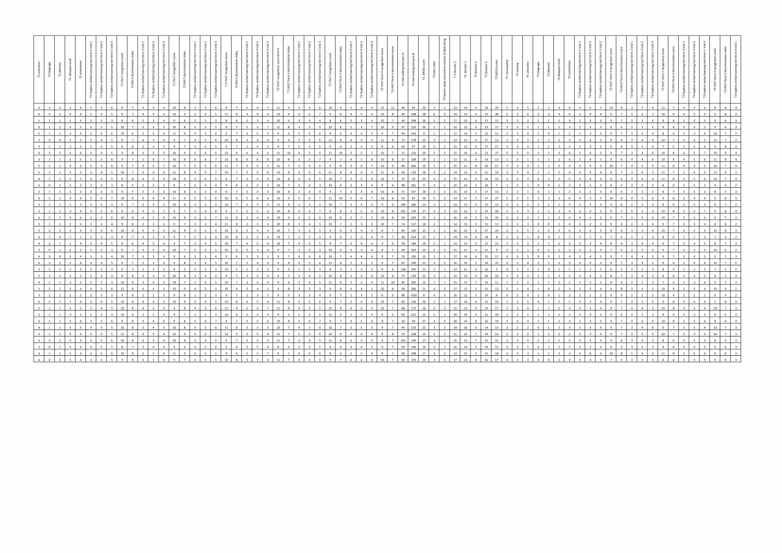
T2 atten
tion
T2 language
T2 abstract
T2 delayed
recall
T2 orie
ntation
T2 hop
kins verbal learning test fo
rm 1 trial 1
T2 hop
kins verbal learning test fo
rm 1 trial 2
T2 hop
kins verbal learning test fo
rm 1 trial 3
T2 HVLT recognition
score
T2 HVLT discrim
ination inde
x
T2 hop
kins verbal learning test fo
rm 2 trial 1
T2 hop
kins verbal learning test fo
rm 2 trial 2
T2 hop
kins verbal learning test fo
rm 2 trial 3
T2 HVLT recognition
score
T2 HVLT discrim
ination inde
x
T2 hop
kins verbal learning test fo
rm 3 trial 1
T2 hop
kins verbal learning test fo
rm 3 trial 2
T2 hop
kins verbal learning test fo
rm 3 trial 3
T 2 HVLT recognition
score
T2 HVLT discrim
ination inde
x
T2 hop
kins verbal learning test fo
rm 4 trial 1
T2 hop
kins verbal learning test fo
rm 4 trial 2
T2 hop
kins verbal learning test fo
rm 4 trial 3
T2 HVLT recognition
score form
4
T 2 HVLT form
4 discrim
ination inde
x
T2 hop
kins verbal learning test fo
rm 5 trial 1
T2 hop
kins verbal learning test fo
rm 5 trial 2
T2 hop
kins verbal learning test fo
rm 5 trial 3
T2 HVLT recognition
score
T2 HVLT form
5 discrim
ination inde
x
T2 hop
kins verbal learning test fo
rm 6 trial 1
T2 hop
kins verbal learning test fo
rm 6 trial 2
T2 hop
kins verbal learning test fo
rm 6 trial 3
T2 HVLT form
6 re
cognition
score
T2 HVLT form
6 discrim
ination score
T2 trail m
aking test part A
T2 trail m
aking test part B
T3 M
MSE sc
ore
T3 katz ind
ex
T3 lawton bo
dy instrumen
t activity
of d
aily living
T 3 do
main 1
T3 dom
ain 2
T3 dom
ain 3
T3 dom
ain 4
T3 M
OCA
score
T3 visuo
spatial
T3 nam
ing
T3 atten
tion
T3 language
T3 abstract
T3 delayed
recall
T3 orie
ntation
T3 hop
kins verbal learning test fo
rm 1 trial 1
T3 hop
kins verbal learning test fo
rm 1 trial 2
T3 hop
kins verbal learning test fo
rm 1 trial 3
T3 HVLT form
3 re
cognition
score
T3 HVLT form
3 discrim
ination score
T3 hop
kins verbal learning test fo
rm 2 trial 1
T3 hop
kins verbal learning test fo
rm 3 trial 2
T3 hop
kins verbal learning test fo
rm 2 trial 3
T3 HVLT form
2 re
cognition
score
T3 HVLT form
2 discrim
ination score
T3 hop
kins verbal learning test fo
rm 3 trial 1
T3 hop
kins verbal learning test fo
rm 3 trial 2
T3 hop
kins verbal learning test fo
rm 3 tria
l 3
T3 HVLT form
3 re
cognition
score
T3 HVLT form
3 discrim
ination inde
x
T3 hop
kins verbal learning test fo
rm 4 trial 1
6 3 2 3 6 5 5 6 9 7 4 5 6 10 8 3 5 6 9 7 5 6 7 11 9 4 4 6 10 9 5 6 6 12 11 44 92 29 3 2 13 14 5 16 25 5 3 5 2 1 3 6 4 5 7 10 9 3 5 6 11 7 4 4 6 9 8 4
6 3 2 4 6 3 4 5 9 7 4 4 5 10 9 3 4 5 11 9 3 4 6 10 9 4 6 7 9 8 4 5 6 10 8 45 168 28 6 5 14 13 5 15 28 5 3 6 3 2 3 6 3 4 4 9 7 5 6 7 10 9 4 5 5 9 8 4
5 1 1 1 4 2 3 3 8 6 2 3 4 9 6 2 3 3 8 6 2 3 4 10 6 3 4 4 8 6 3 3 4 10 7 40 168 16 2 2 17 13 6 17 15 3 3 2 1 1 1 4 2 3 3 9 7 2 3 4 8 6 2 3 5 9 6 3
6 1 1 3 5 4 5 5 10 7 3 4 5 10 8 4 5 5 9 7 5 5 7 11 8 4 5 5 10 8 5 4 7 10 9 67 124 20 2 1 14 12 4 13 17 3 3 4 1 1 2 3 2 3 4 8 6 3 3 4 9 6 3 3 5 9 6 3
5 1 1 3 4 4 6 6 10 6 5 5 6 11 9 4 5 6 9 7 4 5 5 9 6 5 5 6 9 6 4 4 5 9 7 90 163 9 1 1 12 14 5 15 13 2 3 4 1 0 1 2 2 3 3 9 7 3 4 4 8 6 3 3 4 10 7 2
1 0 0 2 2 3 4 5 9 7 4 3 6 9 7 4 5 6 10 8 3 4 6 9 6 3 5 6 11 8 4 5 5 11 8 71 178 15 2 2 13 12 6 17 13 1 3 3 1 1 2 2 3 4 5 9 6 3 4 4 10 7 3 4 5 11 7 2
4 1 1 2 3 2 4 5 8 6 2 4 5 9 7 3 4 5 9 7 2 4 5 8 7 2 4 5 9 6 2 4 5 8 6 60 97 19 2 2 15 13 5 17 17 3 3 4 1 1 2 3 2 3 5 9 8 3 4 4 7 5 2 4 4 9 8 2
6 3 2 4 6 3 4 5 9 8 3 5 5 10 9 3 4 6 10 9 4 4 6 11 10 4 5 6 11 10 5 5 7 10 7 51 110 29 5 3 15 10 4 13 27 5 3 5 3 2 3 6 3 4 5 9 7 3 4 6 10 8 4 5 7 10 9 4
3 1 1 3 5 5 5 6 9 7 5 6 7 10 8 6 6 7 10 8 6 6 9 10 8 5 5 7 9 7 4 5 6 10 8 57 108 19 2 1 13 11 6 14 13 1 3 1 1 1 2 4 3 4 5 8 6 4 4 6 10 8 4 5 6 11 8 4
5 1 1 2 3 4 4 6 9 7 4 5 7 10 7 5 5 6 11 7 4 5 5 11 7 4 6 6 9 8 5 6 7 10 9 84 206 13 1 1 15 11 8 20 17 4 3 3 1 1 2 3 3 4 5 10 7 4 5 6 11 8 4 4 5 10 7 4
5 2 2 3 5 3 4 5 10 7 3 4 6 11 8 4 5 7 10 7 3 5 6 10 8 3 5 6 11 8 4 4 5 11 8 56 125 18 3 2 14 13 5 15 19 3 3 4 2 1 2 4 3 3 4 9 7 3 4 5 11 7 2 4 5 11 9 3
4 1 1 2 5 3 4 5 9 8 4 5 6 10 9 2 4 5 9 7 3 4 5 10 8 3 4 5 10 7 3 5 6 10 7 37 76 25 4 2 15 15 5 16 15 2 2 3 0 1 2 5 3 4 4 9 6 3 4 4 11 8 3 5 6 10 7 3
2 0 1 1 3 2 2 3 8 5 2 2 3 9 7 2 3 4 9 6 2 2 3 10 7 2 2 3 10 6 2 3 4 9 6 98 201 9 2 1 15 13 5 10 7 1 2 1 0 0 1 2 2 2 3 8 5 2 2 2 8 6 2 3 2 9 6 2
2 1 1 3 2 3 3 4 9 7 2 3 5 10 8 3 3 4 9 7 3 4 5 10 8 2 3 4 9 7 3 4 6 10 8 71 157 18 3 2 15 13 5 17 13 2 2 1 0 1 1 2 2 2 3 9 6 2 2 3 8 7 2 2 3 8 5 3
6 3 2 4 6 5 6 7 10 9 4 6 8 11 9 4 5 6 10 9 5 6 6 10 9 4 6 7 11 10 5 6 7 10 8 53 87 29 5 3 14 11 5 17 27 5 3 5 3 2 3 6 4 5 5 10 8 4 5 6 9 8 3 4 6 9 8 3
2 1 1 2 3 3 3 4 9 7 3 4 5 10 8 3 3 4 10 7 3 3 4 11 8 2 3 4 10 7 3 4 4 9 6 100 186 14 2 1 14 12 4 13 14 3 3 2 1 1 1 3 3 3 4 9 6 2 2 4 8 6 2 3 4 9 7 2
3 1 1 3 4 4 5 6 9 6 4 4 7 9 7 4 6 8 9 7 5 5 6 10 8 4 6 7 9 8 5 5 6 10 8 101 176 17 3 2 13 12 5 15 18 3 3 3 1 1 3 4 4 5 5 9 7 5 5 6 10 8 5 5 7 9 8 4
6 2 2 4 6 3 4 6 10 8 4 5 6 10 8 3 5 7 11 9 3 4 6 10 9 4 5 6 10 8 3 5 5 10 8 56 103 25 2 1 16 14 5 16 25 5 3 4 2 2 3 6 4 4 5 9 7 3 4 6 10 7 3 5 6 9 7 3
3 1 1 2 5 3 3 4 9 8 2 3 5 9 7 3 3 4 11 8 2 3 4 10 8 3 4 5 10 7 2 3 3 10 7 74 157 18 2 1 14 13 5 15 12 2 2 3 0 0 1 4 2 2 3 9 6 2 3 4 9 7 3 3 4 8 6 2
5 2 2 3 6 3 4 6 10 8 4 4 5 11 8 3 3 4 10 8 3 4 4 10 7 4 3 5 9 8 3 4 5 8 7 85 120 15 2 1 16 13 6 17 24 2 3 5 2 2 3 6 4 5 5 8 6 3 3 4 10 7 3 5 5 10 8 3
4 1 0 2 1 2 3 3 9 7 3 3 3 9 7 2 3 4 10 8 3 3 4 10 7 2 2 3 9 6 2 3 4 9 7 80 254 15 2 2 14 14 6 18 8 1 3 1 0 0 1 2 2 2 3 7 4 2 2 3 8 4 1 2 3 7 4 2
4 2 1 2 4 3 4 5 9 6 4 5 6 9 7 3 4 5 10 7 4 5 6 10 7 4 5 5 9 7 5 6 6 9 6 78 186 19 2 1 14 13 5 12 15 3 3 3 2 1 2 3 2 3 4 8 6 3 4 4 9 7 3 4 5 8 7 3
3 0 1 2 2 2 2 4 9 7 3 3 4 10 7 2 4 5 10 9 3 3 4 9 7 3 3 3 10 6 2 3 4 8 5 64 163 13 2 1 15 11 4 11 9 2 2 1 0 1 1 2 2 2 3 7 4 2 3 4 9 7 2 3 3 10 6 2
6 0 0 3 4 3 3 4 10 7 3 3 4 9 6 3 3 4 9 6 3 3 3 9 7 4 6 6 10 7 4 4 6 9 7 70 130 15 2 1 17 14 4 15 17 4 3 5 0 0 2 4 3 4 5 9 7 4 4 5 9 7 3 4 5 9 7 3
6 3 2 2 6 4 4 5 9 7 3 3 4 9 8 3 4 5 10 7 3 4 4 9 8 4 5 6 11 6 3 5 5 9 7 62 130 22 3 2 16 14 5 16 22 4 3 4 2 1 3 5 4 5 5 9 7 3 4 5 9 6 3 4 4 10 7 5
2 1 1 2 2 2 3 3 9 5 2 3 3 8 5 2 2 3 10 5 2 2 3 9 4 2 2 3 8 5 1 3 3 9 6 136 350 11 2 1 14 15 6 16 5 0 1 1 1 0 1 1 1 2 2 6 3 2 2 2 8 4 1 2 2 7 5 2
5 1 1 3 5 3 3 4 9 8 3 4 5 10 8 3 4 4 9 7 3 5 6 9 7 3 4 5 10 8 3 4 6 10 8 75 134 21 3 1 15 13 5 16 19 3 3 4 1 1 2 5 3 4 5 8 7 3 4 5 8 6 3 4 5 9 7 3
4 1 1 3 3 2 3 4 10 8 3 4 5 10 7 3 4 5 10 7 3 3 4 9 6 3 4 5 11 8 3 3 4 11 10 85 205 13 2 1 15 13 7 14 11 1 2 2 1 1 2 2 2 2 3 8 4 2 2 3 7 4 2 3 4 9 7 2
5 2 1 3 4 4 5 6 11 8 4 4 5 10 9 4 5 5 10 9 3 4 5 9 8 3 3 3 9 8 4 4 5 10 8 99 206 21 4 2 17 13 5 15 13 2 1 4 1 0 2 3 3 3 4 9 8 3 4 5 10 9 3 3 4 10 9 2
3 1 1 2 2 2 3 3 9 6 2 2 3 9 8 1 2 3 9 7 2 3 3 8 5 3 3 4 9 7 2 3 2 8 5 98 >350 8 4 3 16 12 5 14 6 0 2 0 1 0 1 2 2 2 2 9 5 2 2 2 10 4 2 2 2 9 4 2
6 3 2 2 6 3 4 5 10 8 4 4 5 10 9 3 4 5 10 8 4 5 6 11 8 3 5 6 9 7 3 4 4 10 7 82 136 20 2 1 17 14 4 15 19 1 3 5 4 1 2 5 3 3 4 9 7 3 3 5 9 8 3 4 4 1 8 4
5 1 1 3 5 3 4 5 10 8 4 5 5 9 8 4 5 6 11 9 5 5 7 11 8 4 5 5 10 8 4 5 5 10 7 68 175 16 2 2 15 12 6 19 17 3 3 4 1 1 2 3 3 3 4 9 7 4 4 5 10 8 3 3 4 10 7 3
2 1 1 3 3 2 3 4 10 6 2 2 3 9 7 2 2 3 10 6 2 3 4 9 5 2 3 5 11 5 2 2 4 8 5 92 215 12 2 1 16 10 3 11 10 1 2 1 1 1 2 2 2 3 2 9 6 2 2 3 8 6 2 2 3 9 6 3
6 2 1 3 6 4 5 6 9 8 4 4 6 9 8 5 6 7 9 7 4 5 6 10 8 5 6 7 9 7 4 5 6 8 7 26 59 27 4 3 18 14 8 16 19 4 3 4 1 1 2 4 3 4 5 9 7 4 6 6 10 8 5 5 6 8 6 4
4 1 1 3 5 4 4 5 10 8 3 4 5 10 8 3 5 6 11 8 3 3 5 10 7 4 5 6 10 7 3 5 5 9 7 45 133 22 3 3 14 10 6 14 13 3 2 2 0 1 2 3 3 3 4 9 7 3 4 6 9 7 3 3 4 10 7 3
4 1 1 4 4 4 4 5 11 8 4 5 6 10 9 4 6 7 10 9 3 5 6 11 7 5 5 6 10 8 4 6 6 9 8 72 128 13 2 2 15 13 7 19 13 2 3 2 1 1 2 2 3 3 4 9 7 3 4 4 10 7 3 4 5 10 7 3
4 2 1 2 4 4 5 6 10 8 3 4 6 10 8 3 3 4 9 7 3 4 5 11 7 3 3 5 11 6 4 4 5 9 7 101 145 17 3 2 15 12 7 14 15 3 3 4 1 1 1 3 3 3 4 9 6 3 3 5 8 6 3 3 5 8 5 3
3 0 1 4 4 4 5 5 8 7 3 4 6 9 6 4 5 6 9 6 3 5 6 8 6 3 3 5 8 6 4 4 5 9 7 65 106 16 4 2 16 14 5 19 13 3 3 1 0 1 2 3 3 3 4 8 5 3 4 5 9 8 3 4 5 9 6 3
3 1 1 3 4 4 5 6 10 8 5 5 6 11 9 4 5 5 9 6 5 5 7 9 7 4 5 6 8 6 3 5 6 9 7 60 108 17 4 3 13 12 5 15 18 3 3 3 1 1 3 4 4 6 6 10 8 5 4 6 11 8 5 6 6 9 6 4
6 0 0 3 4 4 4 5 9 8 4 5 6 9 7 4 5 5 10 8 3 5 6 11 7 4 4 6 9 7 4 6 6 10 7 82 170 19 3 2 17 13 8 16 17 4 3 4 0 0 2 4 3 4 5 7 5 3 4 5 8 6 3 3 5 9 8 3
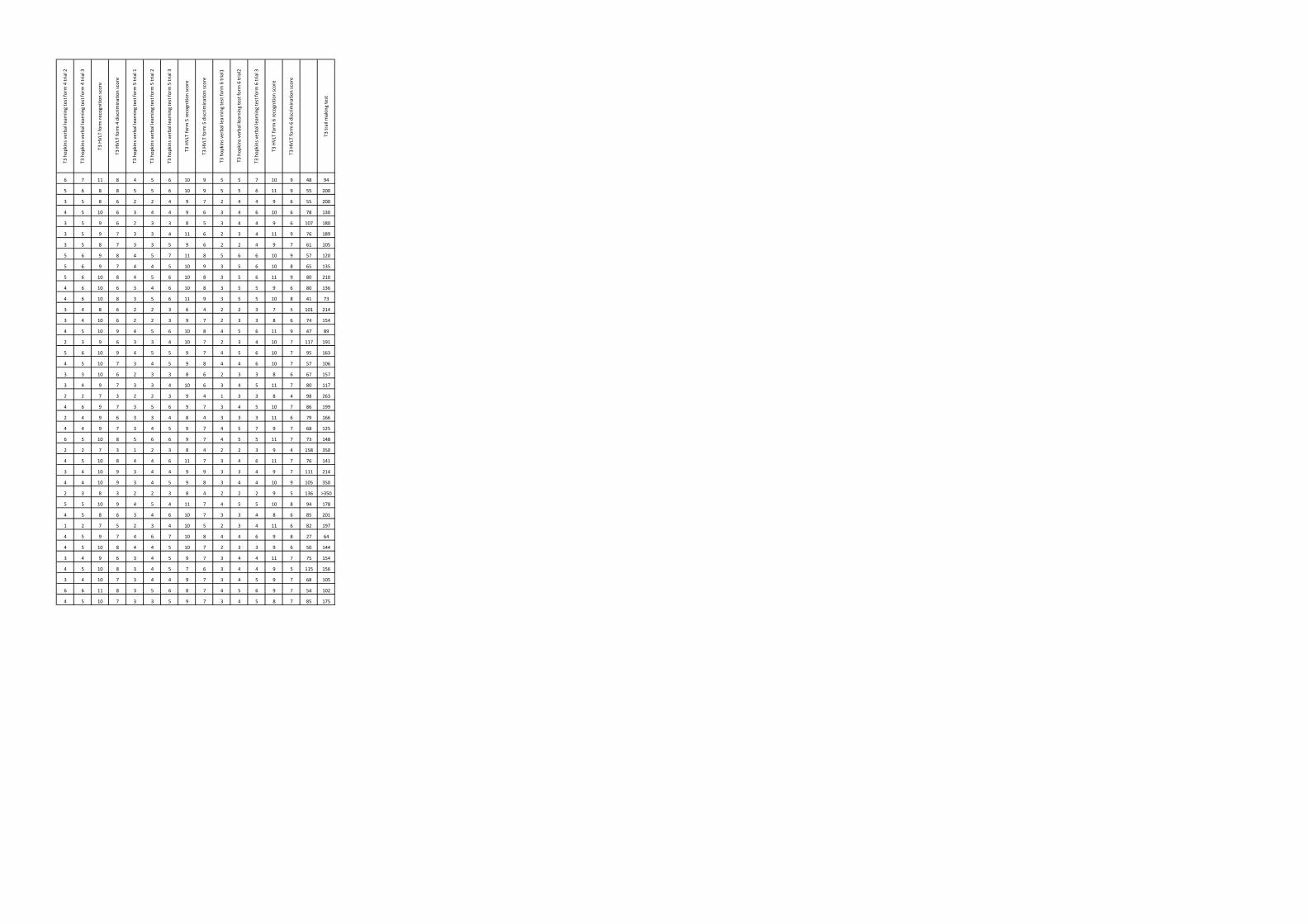
T3 hop
kins verbal learning test fo
rm 4 trial 2
T3 hop
kins verbal learning test fo
rm 4 trial 3
T3 HVLT form
recognition
score
T3 HVLT form
4 discrim
ination score
T3 hop
kins verbal learning test fo
rm 5 trial 1
T3 hop
kins verbal learning test fo
rm 5 trial 2
T3 hop
kins verbal learning test fo
rm 5 trial 3
T3 HVLT form
5 re
cognition
score
T3 HVLT form
5 discrim
ination score
T3 hop
kins verbal learning test fo
rm 6 trial1
T3 ho
pkins v
erbal learning test fo
rm 6 trial2
T3 hop
kins verbal learning test fo
rm 6 trial 3
T3 HVLT form
6 re
cognition
score
T3 HVLT form
6 discrim
ination score
T3 trail m
aking test
6 7 11 8 4 5 6 10 9 5 5 7 10 9 48 94
5 6 8 8 5 5 6 10 9 5 5 6 11 9 55 200
3 5 8 6 2 2 4 9 7 2 4 4 9 6 55 200
4 5 10 6 3 4 4 9 6 3 4 6 10 6 78 130
3 5 9 6 2 3 3 8 5 3 4 4 9 6 107 180
3 5 9 7 3 3 4 11 6 2 3 4 11 9 76 189
3 5 8 7 3 3 5 9 6 2 2 4 9 7 61 105
5 6 9 8 4 5 7 11 8 5 6 6 10 9 57 120
5 6 9 7 4 4 5 10 9 3 5 6 10 8 65 135
5 6 10 8 4 5 6 10 8 3 5 6 11 9 80 210
4 6 10 6 3 4 6 10 8 3 5 5 9 6 80 136
4 6 10 8 3 5 6 11 9 3 5 5 10 8 41 73
3 4 8 6 2 2 3 6 4 2 2 3 7 5 101 214
3 4 10 6 2 2 3 9 7 2 3 3 8 6 74 154
4 5 10 9 4 5 6 10 8 4 5 6 11 9 47 89
2 3 9 6 3 3 4 10 7 2 3 4 10 7 117 191
5 6 10 9 4 5 5 9 7 4 5 6 10 7 95 163
4 5 10 7 3 4 5 9 8 4 4 6 10 7 57 106
3 3 10 6 2 3 3 8 6 2 3 3 8 6 67 157
3 4 9 7 3 3 4 10 6 3 4 5 11 7 80 117
2 2 7 3 2 2 3 9 4 1 3 3 8 4 98 263
4 6 9 7 3 5 6 9 7 3 4 5 10 7 86 199
2 4 9 6 3 3 4 8 4 3 3 3 11 6 79 166
4 4 9 7 3 4 5 9 7 4 5 7 9 7 68 125
6 5 10 8 5 6 6 9 7 4 5 5 11 7 73 148
2 2 7 3 1 2 3 8 4 2 2 3 9 4 158 350
4 5 10 8 4 4 6 11 7 3 4 6 11 7 76 141
3 4 10 9 3 4 4 9 9 3 3 4 9 7 111 214
4 4 10 9 3 4 5 9 8 3 4 4 10 9 105 350
2 3 8 3 2 2 3 8 4 2 2 2 9 5 136 >350
5 5 10 9 4 5 4 11 7 4 5 5 10 8 94 178
4 5 8 6 3 4 6 10 7 3 3 4 8 6 85 201
1 2 7 5 2 3 4 10 5 2 3 4 11 6 82 197
4 5 9 7 4 6 7 10 8 4 4 6 9 8 27 64
4 5 10 8 4 4 5 10 7 2 3 3 9 6 50 144
3 4 9 6 3 4 5 9 7 3 4 4 11 7 75 154
4 5 10 8 3 4 5 7 6 3 4 4 9 5 115 156
3 4 10 7 3 4 4 9 7 3 4 5 9 7 68 105
6 6 11 8 3 5 6 8 7 4 5 6 9 7 54 102
4 5 10 7 3 3 5 9 7 3 4 5 8 7 85 175


![[The relations between neurocognition and quality of life in people with schizophrenia]](https://static.fdokumen.com/doc/165x107/633d1d32b6ae16c04d04f64f/the-relations-between-neurocognition-and-quality-of-life-in-people-with-schizophrenia.jpg)