Nephroprotective Plants: A Review on the Use in Pre-Renal ...
24
Citation: Tienda-Vázquez, M.A.; Morreeuw, Z.P.; Sosa-Hernández, J.E.; Cardador-Martínez, A.; Sabath, E.; Melchor-Martínez, E.M.; Iqbal, H.M.N.; Parra-Saldívar, R. Nephroprotective Plants: A Review on the Use in Pre-Renal and Post-Renal Diseases. Plants 2022, 11, 818. https://doi.org/10.3390/ plants11060818 Academic Editors: Antonella Smeriglio and Fabrizio Araniti Received: 22 February 2022 Accepted: 16 March 2022 Published: 18 March 2022 Publisher’s Note: MDPI stays neutral with regard to jurisdictional claims in published maps and institutional affil- iations. Copyright: © 2022 by the authors. Licensee MDPI, Basel, Switzerland. This article is an open access article distributed under the terms and conditions of the Creative Commons Attribution (CC BY) license (https:// creativecommons.org/licenses/by/ 4.0/). plants Review Nephroprotective Plants: A Review on the Use in Pre-Renal and Post-Renal Diseases Mario Adrián Tienda-Vázquez 1,† , Zoé P. Morreeuw 1,† , Juan Eduardo Sosa-Hernández 1 , Anaberta Cardador-Martínez 1 , Ernesto Sabath 2,3 , Elda M. Melchor-Martínez 1, * , Hafiz M. N. Iqbal 1, * and Roberto Parra-Saldívar 1, * 1 Tecnologico de Monterrey, School of Engineering and Sciences, Monterrey 64849, Mexico; [email protected] (M.A.T.-V.); [email protected] (Z.P.M.); [email protected] (J.E.S.-H.); [email protected] (A.C.-M.) 2 Departamento de Nefrología, Hospital General de Querétaro, Queretaro 76175, Mexico; [email protected] 3 Facultad de Ciencias Naturales, Universidad Autónoma de Querétaro, Juriquilla 76230, Mexico * Correspondence: [email protected] (E.M.M.-M.); hafi[email protected] (H.M.N.I.); [email protected] (R.P.-S.) † These authors contributed equally to this work. Abstract: Kidney diseases are expected to become the fifth leading cause of death by 2040. Several physiological failures classified as pre-, intra-, and post-renal factors induce kidney damage. Diabetes, liver pathologies, rhabdomyolysis, and intestinal microbiota have been identified as pre-renal factors, and lithiasis or blood clots in the ureters, prostate cancer, urethral obstructions, prostate elongation, and urinary tract infections are post-renal factors. Additionally, the nephrotoxicity of drugs has been highlighted as a crucial factor inducing kidney injuries. Due to the adverse effects of drugs, it is necessary to point to other alternatives to complement the treatment of these diseases, such as nephroprotective agents. Plants are a wide source of nephroprotective substances and can have beneficial effects in different levels of the physiological pathways which lead to kidney damage. In traditional medicines, plants are used as antioxidants, anti-inflammatories, diuretics, and anticancer agents, among other benefits. However, the mechanism of action of some plants empirically used remains unknown and scientific data are required to support their nephroprotective effects. The present work reviewed the plants with a beneficial effect on kidney diseases. The classification of nephroprotective plants according to the clinical definition of pre-renal, intrinsic, and post-renal factors is proposed to orient their use as complementary treatments. Keywords: plants; pre-renal diseases; post-renal diseases; secondary nephroprotection 1. Introduction The kidneys play an important role in human physiology, maintaining fluid home- ostasis, regulating blood pressure, erythrocyte production and bone density, regulating hormonal balance, and filtering and removing nitrogenous and other waste products [1,2]. Chronic kidney disease (CKD) is characterized by a progressive loss of functions while acute kidney injury (AKI) is an abrupt reduction in kidney function. Both CKD and AKI have increased worldwide and are considered one of the leading public health problems [2,3]. A projection of health concerns by 2040 ranked CKD as the fifth leading cause of death worldwide [3]. In addition, kidney diseases have been recognized as risk factors for severe forms of COVID-19 [4]. The increase of their prevalence is associated with the increase of diabetes Mellitus (DM) and hypertension as the main reported causes of kidney dys- functions, although various factors can trigger this physiopathology. Factors are classified, based on the pathway they led to kidney damage, as pre-renal, intrinsic, and post-renal factors (Figure 1). Plants 2022, 11, 818. https://doi.org/10.3390/plants11060818 https://www.mdpi.com/journal/plants
-
Upload
khangminh22 -
Category
Documents
-
view
1 -
download
0
Transcript of Nephroprotective Plants: A Review on the Use in Pre-Renal ...

�����������������
Citation: Tienda-Vázquez, M.A.;
Morreeuw, Z.P.; Sosa-Hernández, J.E.;
Cardador-Martínez, A.; Sabath, E.;
Melchor-Martínez, E.M.; Iqbal,
H.M.N.; Parra-Saldívar, R.
Nephroprotective Plants: A Review
on the Use in Pre-Renal and
Post-Renal Diseases. Plants 2022, 11,
818. https://doi.org/10.3390/
plants11060818
Academic Editors: Antonella
Smeriglio and Fabrizio Araniti
Received: 22 February 2022
Accepted: 16 March 2022
Published: 18 March 2022
Publisher’s Note: MDPI stays neutral
with regard to jurisdictional claims in
published maps and institutional affil-
iations.
Copyright: © 2022 by the authors.
Licensee MDPI, Basel, Switzerland.
This article is an open access article
distributed under the terms and
conditions of the Creative Commons
Attribution (CC BY) license (https://
creativecommons.org/licenses/by/
4.0/).
plants
Review
Nephroprotective Plants: A Review on the Use in Pre-Renal andPost-Renal DiseasesMario Adrián Tienda-Vázquez 1,†, Zoé P. Morreeuw 1,† , Juan Eduardo Sosa-Hernández 1 ,Anaberta Cardador-Martínez 1 , Ernesto Sabath 2,3, Elda M. Melchor-Martínez 1,* , Hafiz M. N. Iqbal 1,*and Roberto Parra-Saldívar 1,*
1 Tecnologico de Monterrey, School of Engineering and Sciences, Monterrey 64849, Mexico;[email protected] (M.A.T.-V.); [email protected] (Z.P.M.); [email protected] (J.E.S.-H.);[email protected] (A.C.-M.)
2 Departamento de Nefrología, Hospital General de Querétaro, Queretaro 76175, Mexico; [email protected] Facultad de Ciencias Naturales, Universidad Autónoma de Querétaro, Juriquilla 76230, Mexico* Correspondence: [email protected] (E.M.M.-M.); [email protected] (H.M.N.I.); [email protected] (R.P.-S.)† These authors contributed equally to this work.
Abstract: Kidney diseases are expected to become the fifth leading cause of death by 2040. Severalphysiological failures classified as pre-, intra-, and post-renal factors induce kidney damage. Diabetes,liver pathologies, rhabdomyolysis, and intestinal microbiota have been identified as pre-renal factors,and lithiasis or blood clots in the ureters, prostate cancer, urethral obstructions, prostate elongation,and urinary tract infections are post-renal factors. Additionally, the nephrotoxicity of drugs hasbeen highlighted as a crucial factor inducing kidney injuries. Due to the adverse effects of drugs,it is necessary to point to other alternatives to complement the treatment of these diseases, suchas nephroprotective agents. Plants are a wide source of nephroprotective substances and can havebeneficial effects in different levels of the physiological pathways which lead to kidney damage. Intraditional medicines, plants are used as antioxidants, anti-inflammatories, diuretics, and anticanceragents, among other benefits. However, the mechanism of action of some plants empirically usedremains unknown and scientific data are required to support their nephroprotective effects. Thepresent work reviewed the plants with a beneficial effect on kidney diseases. The classification ofnephroprotective plants according to the clinical definition of pre-renal, intrinsic, and post-renalfactors is proposed to orient their use as complementary treatments.
Keywords: plants; pre-renal diseases; post-renal diseases; secondary nephroprotection
1. Introduction
The kidneys play an important role in human physiology, maintaining fluid home-ostasis, regulating blood pressure, erythrocyte production and bone density, regulatinghormonal balance, and filtering and removing nitrogenous and other waste products [1,2].Chronic kidney disease (CKD) is characterized by a progressive loss of functions while acutekidney injury (AKI) is an abrupt reduction in kidney function. Both CKD and AKI haveincreased worldwide and are considered one of the leading public health problems [2,3].A projection of health concerns by 2040 ranked CKD as the fifth leading cause of deathworldwide [3]. In addition, kidney diseases have been recognized as risk factors for severeforms of COVID-19 [4]. The increase of their prevalence is associated with the increaseof diabetes Mellitus (DM) and hypertension as the main reported causes of kidney dys-functions, although various factors can trigger this physiopathology. Factors are classified,based on the pathway they led to kidney damage, as pre-renal, intrinsic, and post-renalfactors (Figure 1).
Plants 2022, 11, 818. https://doi.org/10.3390/plants11060818 https://www.mdpi.com/journal/plants

Plants 2022, 11, 818 2 of 24Plants 2022, 11, 818 2 of 25
Figure 1. Pre-renal and post-renal diseases.
According to clinical criteria, pre-renal diseases are related to a decrease in renal per-fusion or alteration in the systemic circulation, which will first compromise the glomeru-lar filtration rate (GFR) and secondly lead to more severe alterations in the kidney struc-ture. These dysfunctions are reflected in clinical analyses by changes in biomarker levels; for example, an increase in serum creatinine; and fluctuations in urine flow [5,6]. Several diseases have been identified as pre-renal factors such as bleeding, trauma, shock, hyper-tension, cirrhosis, diabetes, systemic infections, hypotension, autoimmune diseases, rhab-domyolysis, disorders of the gut microbiota, liver damage, and intravascular volume de-pletion [7]. The factors that directly cause kidney damage are intrinsic, whether heavy metals, trauma, Wegener’s granulomatosis, proteinuria, congenital abnormalities, drug toxicity, renal atheroembolic disease, arthralgias, lupus erythematosus, or kidney cancer. Histologically, the main diagnoses are ischemic acute tubular necrosis, nephrotoxic acute tubular necrosis, and glomerulonephritis [8]. Any other health disorders which can indi-rectly induce kidney failures that occur after the physiological action of the kidney are post-renal diseases. Among these post-renal factors, mainly related to urinary flow disor-ders, are the ureter and urethra obstruction due to blood clots, lithiasis, or tumor growth, which can lead to increased pressure inside tubules, and as a consequence, the GFR is compromised, and a urinary tract infection (UTI) [9] is caused.
Kidney diseases can be addressed at several levels according to the physiological pathway of the original cause. Each pre-renal and post-renal disease currently has phar-macological treatments; however, most of the drugs used cause adverse effects and some-times lead to intrinsic kidney damage. Among them are non-steroidal anti-inflammatory drugs (NSAID), proton pump inhibitors, antibiotics, and chemotherapy [10–13]. Ne-phrotoxicity of drugs administrated as treatments for pre-renal and post-renal diseases, as well as for other diseases, is now considered as a risk factor of acute and chronic kidney conditions. To avoid the adverse effects caused by medication, different alternatives have been sought to treat these pathologies.
Plants have been traditionally used as treatments for various diseases, among them several pathologies identified as pre-, intra-, and post-renal factors. The medicinal
Figure 1. Pre-renal and post-renal diseases.
According to clinical criteria, pre-renal diseases are related to a decrease in renal per-fusion or alteration in the systemic circulation, which will first compromise the glomerularfiltration rate (GFR) and secondly lead to more severe alterations in the kidney structure.These dysfunctions are reflected in clinical analyses by changes in biomarker levels; forexample, an increase in serum creatinine; and fluctuations in urine flow [5,6]. Severaldiseases have been identified as pre-renal factors such as bleeding, trauma, shock, hy-pertension, cirrhosis, diabetes, systemic infections, hypotension, autoimmune diseases,rhabdomyolysis, disorders of the gut microbiota, liver damage, and intravascular vol-ume depletion [7]. The factors that directly cause kidney damage are intrinsic, whetherheavy metals, trauma, Wegener’s granulomatosis, proteinuria, congenital abnormalities,drug toxicity, renal atheroembolic disease, arthralgias, lupus erythematosus, or kidneycancer. Histologically, the main diagnoses are ischemic acute tubular necrosis, nephrotoxicacute tubular necrosis, and glomerulonephritis [8]. Any other health disorders which canindirectly induce kidney failures that occur after the physiological action of the kidneyare post-renal diseases. Among these post-renal factors, mainly related to urinary flowdisorders, are the ureter and urethra obstruction due to blood clots, lithiasis, or tumorgrowth, which can lead to increased pressure inside tubules, and as a consequence, theGFR is compromised, and a urinary tract infection (UTI) [9] is caused.
Kidney diseases can be addressed at several levels according to the physiologicalpathway of the original cause. Each pre-renal and post-renal disease currently has pharma-cological treatments; however, most of the drugs used cause adverse effects and sometimeslead to intrinsic kidney damage. Among them are non-steroidal anti-inflammatory drugs(NSAID), proton pump inhibitors, antibiotics, and chemotherapy [10–13]. Nephrotoxicityof drugs administrated as treatments for pre-renal and post-renal diseases, as well as forother diseases, is now considered as a risk factor of acute and chronic kidney conditions.To avoid the adverse effects caused by medication, different alternatives have been soughtto treat these pathologies.
Plants have been traditionally used as treatments for various diseases, among them sev-eral pathologies identified as pre-, intra-, and post-renal factors. The medicinal characteris-tics of plants have been attributed to their secondary metabolites, which can protect against

Plants 2022, 11, 818 3 of 24
pathogens or have important physiological benefits to prevent some diseases [14,15]. Plantsprovide a wide range of bioactive compounds which act as antioxidants, anti-inflammatory,diuretic, anticancer, and antimicrobial [16–18]. Further, nephroprotective agents from plantsmitigate processes such as interstitial nephritis, altered intraglomerular hemodynamics,tubular necrosis, or glomerulonephritis [19]. Previous works have already reviewed theusages of plants and phytochemicals as nephroprotective agents providing an importantunderstanding of how extracts or single compounds interfere with molecular pathways tomitigate kidney diseases [2,20]. However, they usually focused on intrinsic damage suchas nephrotoxicity, omitting the pre-renal and post-renal factors, and to our knowledge, noclassification of nephroprotective plants according to pre-, intra-, and post-renal diseaseshave been reported.
Furthermore, since it is complicated to replace the effect granted by drugs, nephropro-tective plants have to be considered as a complement of treatments rather than substitutivetreatments [21–23]. The beneficial complementarity of traditional and modern medicinefor primary health care is promoted by the World Health Organization (WHO). The WHOreported that a large part of the world population is using plants products as their primarysource of health care. While the lack of fundamental understanding about the mechanismof actions of medicinal plants has limited their uses worldwide, especially in countrieswhere modern medicines dominate the health systems [24]. However, the usage of medici-nal plants as a key component of complementary and alternative medicine has acquiredrenewed interest. As a success story, traditional Chinese medicine, based mainly on plants,is globally accepted and used at every level of the China health system; guidelines forrecommendations and uses of plants have already been implemented by the governmentand academic institutions [25,26]. Similarly, ancestral medicines of India, such as Ayurveda,have proved their efficiency as treatments of several diseases and play an important role inthe economy of the Indian health sector [27,28]. On the other hand, nearly 25% of modernmedicines are derived from natural products, many of which were first used in traditionalmedicines. Thus, plants and products from traditional medicines are a resource for primaryhealth care, but also for innovation and discovery [26,29,30].
In this context, plants used as nephroprotective agents for pre-renal, intrinsic and post-renal disease were reviewed. Scientific information on the effect of the nephroprotectiveplants on the pathophysiologies that lead to kidney injuries was described to support theiruses as a complementary pharmacological treatment.
2. A Combined Therapeutic Approach
Medicinal plants are used for wellbeing in every ancient civilization, and some ofthe traditional uses of plants are still employed, such as in traditional Chinese medicineand Ayurveda. Traditional usage of plants as treatments is based on knowledge sharedover generations. [26,31–36]. In contrast, occidental medicine, in some cases, is based ona lifetime intake of drugs; therefore, patients become dependent on drugs just to survive,especially older people. Despite their therapeutic effects, drugs also have a wide range ofadverse effects such as nephrotoxicity [37]. In addition, drugs can even generate abstinencesyndrome, generating drug dependence in patients [38–41].
In the last 20 years, plants and their extracts have been used as healthy alternatives inthe form of food supplements, herbal medicines, natural health products, phytomedicine,or dietary supplements [42]. It is now well-known that plants and their phytochemicalsexert specific therapeutic effects for different health disorders, acting as antioxidants,antimicrobials, anti-inflammatories, diuretics, and anticancer agents, among other benefits.For renal pathophysiology, a plant-based diet has proven its benefits [43], in particular in apatient with mild proteinuria and diabetic nephropathy [44]. Likewise, herbal formulationshelp reduce the dialysis requirements by curing the causes and altering symptoms of renalfailures and are also helpful in mitigating the side effects of dialysis [21]. Thus, the currentapproach of using plants as a complementary treatment provides a double benefit; theysupport the fight against the disease and at the same time act as nephroprotective agents.

Plants 2022, 11, 818 4 of 24
The positive combined approach of occidental and traditional medicine raises newquestions about plants and natural products for therapeutic uses. Indian and Chinesemedicines are widely accepted due to their effectiveness, with fewer side effects reportedin comparison to modern systems of medicine. Furthermore, plants in the management ofdiseases are presented as a cost-effective alternative [45,46]. However, there is still a lack ofknowledge about how plants act as therapeutic agents. Further, effective doses for plantintakes as food or supplements as treatment are usually not established, and dose-relatedtoxicity of plant chemicals is questioned [47–49].
3. Nephroprotective Plants
Now, thanks to advances in molecular techniques, scientific procedures, and protocols,the therapeutic effects can be demonstrated by in vitro or in vivo validated models andeven clinical trials. These studies aimed to validate the benefits of plants and derivates,discarding plants that have a false or placebo effect. Perspectives are usually to recommendthe use of plants as supplementary treatment or to develop naturally sourced drugs withlower toxicity than synthetic drugs.
To that end, key parameters of the studies are the origin of plants, the preparation ofthe plants as therapeutic products, the phytochemicals, the model of the study (clinical orin vivo), the model used to simulate the targeted disease, and the observed mechanism ofaction by the biomarkers analysis (Table 1). Previous reviews have already exhaustivelylisted the plants and their phytochemicals for health benefits. Hence, the present reviewfocuses on the diversity of plants that have scientifically demonstrated their biologicalactivities against renal associated disorders. The mechanisms of action of plant productsare described below according to the physiological pathway they regulate. Plant extractsand compounds depicted in Table 1 are efficient against pre-renal or post-renal conditionsand prevent intrinsic kidney damage. Plants that act first-line directly protecting thekidney were classified as intrinsic, and those that mitigate the disease or factor and at thesame time protect the kidney from secondary damages were classified as pre-renal andpost-renal. Based on clinical definitions, plants from these last categories could be calledsecondary nephroprotectors.
Table 1. Plants and their activity against pre-renal, intrinsic, and post-renal diseases.
Plant Origin Extract/Compounds
Model ofStudy Disease Mechanism of
Action Renal Effect Reference
Tinospora crispa Southeast Asiaand Africa Genkwanin Albino Wistar
rats + CCl4Hepaticinjury
IncreaseSOD activity Pre-renal [50]
Cirsium vulgareand
Cirsiumehrenbergii
Amazon basin Lupeol acetate Male Wistarrats + CCl4
Hepaticinjury
Prevent depletionof glycogen,
antioxidant andanti-inflammatory
effects
Pre-renal [51]
Suaedavermiculata South Africa
Quercetin,quercetin-3-O-rutinoside, andkaempferol-O-
(acetyl)-hexoside-pentoside
Male SpragueDawley rats
+ CCl4
Hepaticinjury
Decrease ASTand ALT Pre-renal [52]
DescurainiaSophia
Europe andnorthern
Africa
Dried seed ethylalcohol Extract
Swiss albinomice + ac-
etaminophen
Hepaticinjury byNSAID
Reduceinflammation,
swellingand necrosis
Pre-renal [53]
Plants 2022, 11, 818 5 of 24
Table 1. Cont.
Plant Origin Extract/Compounds
Model ofStudy Disease Mechanism of
Action Renal Effect Reference
Eurycomalongifolia Indonesia
Standardizedaqueous extract
of the roots(“Physta” from
the brandBiotropics)
Wistar rats +paracetamol
Hepaticinjury byNSAID
Increaseantioxidant
enzymes, improvesbiomarkers of
kidney function,and histopathol-
ogy changes
Pre-renal [54]
Arbutus pavarii Libya
Gallic acid,epicatechin,
dimeric forms ofB-type
Proanthocyanidins,quercetin,
flavonoids, andphenolic acid
Disc DiffusionAssay
Hepaticinjury by
antibiotics
Exert bacteriostaticand bactericidal
effect, againstdifferent
methicillin-resistant
Staphylococcusaureus strains
Pre-renal [55]
Punicagranatum India
Dulcitol,loganin,
bergenin,quercitrin,
cosmosin, folicacid,
khayanthone
Wistar rats +gentamicin
Antibiotics/infections
Improve kidneyfunction
biomarkers,exerted antioxidant
activity, andameliorated histo-
logical changes
Pre-renaland intrinsic [56]
Punicagranatum India
Fruit peelethanolic extract.
Polyphenols.
Wistar rats +gentamicin
Antibiotic-induced liverand kidney
damage
Protect the tissuesagainst
ROS-mediatedoxidative damageand modulate the
inflammatoryresponse
Pre-renaland intrinsic [57]
Cocoa(Theobroma
cacao)Mexico
Hydroalcoholicextract ofNatural
Forasterococoa powder.
Zuckerdiabeticfatty rats
Type 2 DM Decreaseglucose levels Pre-renal [58]
Coffea arabica Africa Pulpaqueous extract
Wistar rats +high-fat diet Type 2 DM Raise
catalase levelsPre-renal
and intrinsic [59]
Eysenhardtiapolystachya Mexico Methanolic
bark extractMice +
streptozotocin Type 2 DM Decreaseoxidative stress
Pre-renaland intrinsic [60]
Anchomanesdifformis Africa Aqueous
leaf extract
Wistar rats +fructose and
streptozotocin
DM relatedwith
pancreas
Induce dissociationof Nrf2/keap,
activating Nrf2,reduced
oxidative stress
Pre-renal [45]
Hibiscussabdariffa
Asia, Africa,Central
America
Calyxaqueous extract
Wistar rats +30% sucrose
Metabolicsyndrome
Increase theantioxidant
systems includingnon-enzymatic and
enzymatic effect
Pre-renal [61]
Hibiscussabdariffa
Asia, Africa,Central
America
Calyx aqueousextract and driedpowdered calyx
Hibiscus acid,anthocyanins,
chlorogenic acid
Humans withuncontrolledhypertension
Hypertension Regulateblood pressure Pre-renal [46]

Plants 2022, 11, 818 6 of 24
Table 1. Cont.
Plant Origin Extract/Compounds
Model ofStudy Disease Mechanism of
Action Renal Effect Reference
Cichoriumintybus
SaudiArabia
1,4-naphthalenedione,
oleic acid,β-asarone,
naphtho furanone,p-
methoxycinnamate,hexadecanoic acid
Wistar albinorats +
ISO-inducedmyocardial
ischemiamodel
Hypertension
Improve thesystolic functionand increase thelevels of LVEF
and LVFS
Pre-renaland
post-renal[62]
Gardeniajasminoides Asia Geniposide
Wistar SHR,and WistarKyoto rats
Myocardialischemia
Exert antioxidantactivity and
decreased CK-MB,AST, ALT, andMDA levels.
Pre-renaland
post-renal[63]
Curcuma longa India Curcumin C57BL/6J miceGlycerol Rhabdomyolysis
Reduce ROS,inflammation, and
histopathol-ogy changes
Pre-renal [64]
Curcuma longa India Curcumin Wistar rats +doxorubicin Nephrotoxicity Increase enzymatic
antioxidant activityPre-renal
and intrinsic [65]
Passion fruit(Passiflora spp.) North America
Methanolic peelextract
Gallic acid,Ellagic acid,
Kaempferol andQuercetinglycosides
Albino rats +paracetamol Nephrotoxicity
Keep urea andcreatinine at
normal levels
Pre-renaland intrinsic [66]
Euphorbiaparalias
Europe,western Asia,and northern
Africa
Methanolicextract of
aerial parts
Sprague-Dawley rats +thioacetamide
Nephrotoxicity Reduce levels ofurea and creatinine
Pre-renaland intrinsic [67]
Pistaciaatlantica
North Africa,Middle East,
Iran, andAfghanistan
Leafhydroethanolic
extract
Wistar rats +gentamicin Nephrotoxicity
Decrease levels ofurea, creatinine,
and uric acid
Pre-renaland intrinsic [68]
Costus afer Africa Aqueousleaf extract
Wistar rats +cyclosporine Nephrotoxicity
Decrease serumpotassium and
BUN levels
Pre-renaland Intrinsic [69]
Cranberry(Vaccinium sp.) North America
“Exocyan” brandnatural
cranberry extract.“Nutrican” brand
cranberry drytextract.
UropathogenicEscherichia coli
Urinary tractinfections
Decrease E. coliadhesion, and
reduce bacterialmotility and
biofilm formation
Post-renal [70,71]
Descurainiasophia
Europe andnorthern
Africa
Aqueousseed extract
Male Wistarrats +
Ammoniumchloride +
ethylene glycol
Lithiasis
Decrease thedeposition of
calcium oxalateand amount oftissue damage
Post-renal [72]
Equisetumarvense Spain
Drystandardized
extract of aerialparts.
Alcoholic extractof sterile stems
Clinical trialHealthy male
volunteers
Urinaryretention
andinfections
Diuretic action andeffective againstCandida tropicalis,Candida glabrata,Candida albicans,Staphylococcus
epidermidis,Streptococcusmutans and
Staphylococcusaureus
Post-renal [73,74]

Plants 2022, 11, 818 7 of 24
Table 1. Cont.
Plant Origin Extract/Compounds
Model ofStudy Disease Mechanism of
Action Renal Effect Reference
Cynanchumwilfordii Korea
4-hydroxyacetop-
henone and2,4-
hydroxyacetop-henone
Male Sprague-Dawley rats
+ testosterone
Benignprostatic
hyperplasia
Decreasedtestosterone and
DHT, viadownregulation ofandrogen receptor5α gene expression
Post-renal [75]
Pumpkin(Cucurbita pepo) Mexico
Oil-freehydroethanolic
pumpkin seed ex-tract.Phytosterols
and fatty acids
Openmono-center
trial men withsymptomatic
benignprostatic
hyperplasia.
Benignprostatic
hyperplasia
Decrease residualurine volume andnocturia.Inhibit 5α
reductase anddecrease
DHT level.
Post-renal [76,77]
Broccoli(Brassica
oleracea var.Italica)
Italy
Broccoli SproutsPowder from
Natural SproutsCompany, LLC
TRAMP Prostate cancer
Decrease HDACexpression, and
decline theacetylation of
histone H3 lysine18 and H3K9
Post-renal [78]
CCl4: tetrachloride; SOD: superoxide dismutase; AST: aspartate aminotransferase; ALT: alanine aminotransferase;NSAID: Non-steroidal anti-inflammatory; DHT: dihydrotestosterone; DM: diabetic Mellitus; DM2: diabetesmellitus type 2; ISO: isoprenaline; LVEF: left ventricular ejection fraction; LVFS: left ventricular fraction shortening;SHR: spontaneously hypertensive rats; CK-MB: creatinine kinase myocardial band; MDA: malondialdehyde; ROS:reactive oxygen species; TRAMP: transgenic adenocarcinoma of the mouse prostate; HDAC: histone deacetylated.
4. The Role of Plants in Renal Pathophysiology4.1. Pre-Renal Factors4.1.1. Diabetes Mellitus
Diabetes mellitus (DM) is a disease characterized by hyperglycemia and insulin re-sistance. In type 1 diabetes, the pancreatic β-cells responsible for insulin production arecompromised and cause an elevation in glucose level. Type 2 diabetes, is a low response ofinsulin secreted to the target tissues [72,73]. Over time, DM is a risk factor for CKD, anddiabetic nephropathy is the most common cause of end-stage renal disease [45,79]. Thepathophysiology induced by DM develops in multifactorial forms and can even triggerother renal risk factors.
Diabetes causes glomerular hyperfiltration and increases intraglomerular pressuredue to the high amounts of glucose to filter. The sodium-glucose transport proteins (SLGT)and glucose transporter (GLUT) transporters in the proximal convoluted tubules haveto overwork, leading to renal hyperperfusion. The tubules cannot handle the amountof glucose, and its excretion through the urine induces osmotic diuresis. Moreover, thiscan also cause microalbuminuria, macroalbuminuria, nephrotic syndrome, and chronicrenal failure, gradually. Hyperglycemia also causes cellular dehydration by increasing theosmotic pressure of the extracellular fluid. Therefore, a hypovolemic state can be reachedbecause of the loss of urine, due to a few reabsorptions, and intracellular dehydration.The kidney, through baroreceptors, detects low pressure and continuously activates therenin angiotensin aldosterone system (RAAS), leading to hypertension [80–82]. On theother hand, insulin resistance causes mitochondrial superoxide overproduction, activatingprotein kinase C (PKC) pathways via aldose sorbitol accumulation and the formation ofadvanced glycation end-products (AGEs). The reactive oxygen species (ROS) inducessevere damage through lipid peroxidation, oxidizing low-density lipoprotein (LDL). ROSalso activate inflammatory response activating the signaling pathway with the transcriptionfactors Bcl2, NF-κB which promote the expression of inflammatory cytokines (IL-1β, IL-2,IL-6, IL-12 and IL-18, TNF-α and MCP1) and apoptosis cascade [45,80]. Finally, glomerularhypertrophy occurs with thickening of the basement membrane and mesangial expansion,

Plants 2022, 11, 818 8 of 24
which can lead to glomerulosclerosis, hemodynamic dysregulation, and tubulointerstitialfibrosis (Figure 2) [45,79].
Plants 2022, 11, 818 9 of 25
IL-6, IL-12 and IL-18, TNF-α and MCP1) and apoptosis cascade [45,80]. Finally, glomeru-lar hypertrophy occurs with thickening of the basement membrane and mesangial expan-sion, which can lead to glomerulosclerosis, hemodynamic dysregulation, and tubulointer-stitial fibrosis (Figure 2) [45,79].
Figure 2. Pathophysiology of kidney damage induced by diabetes. IGP: intraglomerular pressure; SGLT: sodium-glucose transporter; GLUT: glucose transporter; RAAS: renin angiotensin aldoste-rone system.
Cocoa powder can help to counteract hyperglycemia by regulating glucose homeo-stasis and insulin resistance. The 10% cocoa diet for Zucker diabetic fatty rats restored the glucose transporters (SGLT-2 and GLUT-2) and prevented the inactivation of the glyco-genesis regulating the GSK-3 (glycogen synthase kinase 3)/GS (glycogen synthase) path-way and phosphorylation (G-6-PASE: glucose 6 phosphatase). In addition, cocoa reverses the decrease of the phosphorylated levels of tyrosine-phosphorylated insulin [58]. Aque-ous extract of Coffea arabica pulp, rich in polyphenols, efficiently prevents hyperglycemia, insulin resistance, and lipid metabolism disorders. The coffee pulp extract raised levels of the liver antioxidant enzymes catalase (CAT) and copper-zinc superoxide dismutase (Cu-Zn SOD), blocked the stress-sensitive signaling pathway by reducing the expression levels of p-PKCα/PKCα, and improved cationic transport. Such effects were observed with the coffee extract administrated as supplements at 1000 mg/kg body weight (BW) in a model of type 2 diabetes in rats, induced by a high-fat diet, and the effect was compared with metformin as antidiabetic treatment (30 mg/kg BW), and a coffee pulp extract/metformin combined treatment (1000/30 mg/kg BW) [59]. Likewise, in Wistar rats with renal damages caused by diabetes and the nephrotoxic drug streptozotocin, the use of leaf aqueous ex-tract of Anchomanes difformis reverses tissue damage of mesangial cells, glomerular hyper-trophy, and membrane damage. At the molecular level, the extract reduced the serum concentration of urea, reduced oxidative stress by increasing the levels of CAT and SOD, and had anti-inflammatory effects by reducing the expression of NF-κB and Bcl2, thus, decreasing the IL-10, IL-18 and TNFɑ, IL-18 levels. In this work, no significant difference was found between the two tested concentrations: 200 and 400 mg/kg BW, and the effect was not specifically associated with phytochemicals [45]. In contrast, similar antioxidant effects of Hibiscus sabdariffa infusion in the metabolic syndrome rat model have been at-tributed to the high content of antioxidant phytochemicals such as polyphenols, anthocy-anins, flavonoids and phenolic acids. In addition to enhanced antioxidant physiological pathways, phenolic compounds participate in neutralizing ROS by donating a hydrogen atom or an electron. The beneficial effect on body weight, lipid metabolism, insulin home-ostasis, and renal function was obtained with a treatment of 2% of H. sabdariffa infusion in drinking water [61]. Additionally, isolated flavonoids from Eysenhardtia polystachya have
Figure 2. Pathophysiology of kidney damage induced by diabetes. IGP: intraglomerular pressure;SGLT: sodium-glucose transporter; GLUT: glucose transporter; RAAS: renin angiotensin aldos-terone system.
Cocoa powder can help to counteract hyperglycemia by regulating glucose homeosta-sis and insulin resistance. The 10% cocoa diet for Zucker diabetic fatty rats restored theglucose transporters (SGLT-2 and GLUT-2) and prevented the inactivation of the glycogen-esis regulating the GSK-3 (glycogen synthase kinase 3)/GS (glycogen synthase) pathwayand phosphorylation (G-6-PASE: glucose 6 phosphatase). In addition, cocoa reverses thedecrease of the phosphorylated levels of tyrosine-phosphorylated insulin [58]. Aqueousextract of Coffea arabica pulp, rich in polyphenols, efficiently prevents hyperglycemia, in-sulin resistance, and lipid metabolism disorders. The coffee pulp extract raised levels of theliver antioxidant enzymes catalase (CAT) and copper-zinc superoxide dismutase (Cu-ZnSOD), blocked the stress-sensitive signaling pathway by reducing the expression levelsof p-PKCα/PKCα, and improved cationic transport. Such effects were observed with thecoffee extract administrated as supplements at 1000 mg/kg body weight (BW) in a modelof type 2 diabetes in rats, induced by a high-fat diet, and the effect was compared withmetformin as antidiabetic treatment (30 mg/kg BW), and a coffee pulp extract/metformincombined treatment (1000/30 mg/kg BW) [59]. Likewise, in Wistar rats with renal damagescaused by diabetes and the nephrotoxic drug streptozotocin, the use of leaf aqueous extractof Anchomanes difformis reverses tissue damage of mesangial cells, glomerular hypertrophy,and membrane damage. At the molecular level, the extract reduced the serum concentra-tion of urea, reduced oxidative stress by increasing the levels of CAT and SOD, and hadanti-inflammatory effects by reducing the expression of NF-κB and Bcl2, thus, decreasingthe IL-10, IL-18 and TNFα, IL-18 levels. In this work, no significant difference was foundbetween the two tested concentrations: 200 and 400 mg/kg BW, and the effect was notspecifically associated with phytochemicals [45]. In contrast, similar antioxidant effects ofHibiscus sabdariffa infusion in the metabolic syndrome rat model have been attributed to thehigh content of antioxidant phytochemicals such as polyphenols, anthocyanins, flavonoidsand phenolic acids. In addition to enhanced antioxidant physiological pathways, phenoliccompounds participate in neutralizing ROS by donating a hydrogen atom or an electron.The beneficial effect on body weight, lipid metabolism, insulin homeostasis, and renalfunction was obtained with a treatment of 2% of H. sabdariffa infusion in drinking water [61].Additionally, isolated flavonoids from Eysenhardtia polystachya have been tested for their

Plants 2022, 11, 818 9 of 24
antidiabetic and nephroprotective effects in diabetic mice with renal damages induced bystreptozotocin. The results revealed that 20 mg/kg BW of the purified extract significantlyreduced oxidative damages in both the kidneys and the liver, and such effect has been re-lated to the high antioxidant capacity of the characterized phytochemicals [60]. In additionto polyphenols, antioxidative effects, and mitigation of serum lipid abnormalities observedin diabetic Wistar rats treated with Agave lechuguilla by-product extracts were attributed tothe saponins (triterpenoid glycosides), and the effective concentration was established at300 mg/kg BW [83].
The use of plants as a supplementary treatment for diabetes and renal protectionis supported by the reviewed studies. A common factor of the plants to counteract theeffects of diabetes-related pathologies is their antioxidant and anti-inflammatory properties.Such bioactivities have been mostly attributed to phenolic compounds. However, othersphytochemicals can also be responsible for the observed effects and further targeted studiesare required to elucidate their therapeutic potential. On the other hand, dose responsesstudies need to be carried out to establish the posology.
4.1.2. Hypertension
Hypertension is a disease characterized by permanent or continuous high pressure;according to international guidelines on hypertension, systolic blood pressure values mustbe higher than 140 mmHg and diastolic blood pressure higher than 90 mmHg. However,pharmacological treatments depend on the patient’s age and comorbidities [84–87]. Thekidney has self-regulating mechanisms to dampen fluctuations in systemic blood pressureto avoid an increase in intraglomerular pressure. However, at constant high blood pressure,these kidney mechanisms fail because the incoming vessels become weaker, stiffer, orthinner, and a phenomenon called myogenic reflex occurs. During the first compensatoryresponse, the depolarization of the membrane, by increasing the intracellular calciumflux through the L-type channels, causes a change in caliber (contraction) in the afferentarteriole. When this first mechanism fails, there is an increase in intraglomerular filtrationand pressure, also associated with an increase in the sodium chloride load. To compensate,another autoregulatory mechanism called tubuloglomerular feedback switches on, whichis detected in the distal tubule by the macula densa cells. The failure in compensatoryself-regulatory mechanisms ended in glomerulonephritis, the development of glomeru-losclerosis, and a rupture of the fenestra, leading to the filtration of larger molecules suchas proteins, which leads to proteinuria. As proteins accumulate in the tubules, they activateprofibrotic, proinflammatory, and cytotoxic pathways, causing tubulointerstitial damageand kidney scarring (Figure 3) [88–90].
Plants 2022, 11, 818 10 of 25
been tested for their antidiabetic and nephroprotective effects in diabetic mice with renal damages induced by streptozotocin. The results revealed that 20 mg/kg BW of the purified extract significantly reduced oxidative damages in both the kidneys and the liver, and such effect has been related to the high antioxidant capacity of the characterized phyto-chemicals [60]. In addition to polyphenols, antioxidative effects, and mitigation of serum lipid abnormalities observed in diabetic Wistar rats treated with Agave lechuguilla by-product extracts were attributed to the saponins (triterpenoid glycosides), and the effec-tive concentration was established at 300 mg/kg BW [83].
The use of plants as a supplementary treatment for diabetes and renal protection is supported by the reviewed studies. A common factor of the plants to counteract the effects of diabetes-related pathologies is their antioxidant and anti-inflammatory properties. Such bioactivities have been mostly attributed to phenolic compounds. However, others phytochemicals can also be responsible for the observed effects and further targeted stud-ies are required to elucidate their therapeutic potential. On the other hand, dose responses studies need to be carried out to establish the posology.
4.1.2. Hypertension Hypertension is a disease characterized by permanent or continuous high pressure;
according to international guidelines on hypertension, systolic blood pressure values must be higher than 140 mmHg and diastolic blood pressure higher than 90 mmHg. How-ever, pharmacological treatments depend on the patient’s age and comorbidities [84–87]. The kidney has self-regulating mechanisms to dampen fluctuations in systemic blood pressure to avoid an increase in intraglomerular pressure. However, at constant high blood pressure, these kidney mechanisms fail because the incoming vessels become weaker, stiffer, or thinner, and a phenomenon called myogenic reflex occurs. During the first compensatory response, the depolarization of the membrane, by increasing the intra-cellular calcium flux through the L-type channels, causes a change in caliber (contraction) in the afferent arteriole. When this first mechanism fails, there is an increase in intra-glomerular filtration and pressure, also associated with an increase in the sodium chloride load. To compensate, another autoregulatory mechanism called tubuloglomerular feed-back switches on, which is detected in the distal tubule by the macula densa cells. The failure in compensatory self-regulatory mechanisms ended in glomerulonephritis, the de-velopment of glomerulosclerosis, and a rupture of the fenestra, leading to the filtration of larger molecules such as proteins, which leads to proteinuria. As proteins accumulate in the tubules, they activate profibrotic, proinflammatory, and cytotoxic pathways, causing tubulointerstitial damage and kidney scarring (Figure 3) [88–90].
Figure 3. Pathophysiology of kidney damage induced by hypertension. IGP: intraglomerular pres-sure; GFR: glomerular filtration rate; NaCl: sodium chloride. Figure 3. Pathophysiology of kidney damage induced by hypertension. IGP: intraglomerularpressure; GFR: glomerular filtration rate; NaCl: sodium chloride.

Plants 2022, 11, 818 10 of 24
In this context, tincture obtained from the herbaceous plant Cichorium intybus showedpromising cardioprotective and nephroprotective effects in the rat isoprenaline-induced my-ocardial ischemia model. Therapeutic effects were evidenced by the increased antioxidantand anti-inflammatory mechanisms activity and the decreased creatinine kinase myocardialband (CK-MB), aspartate aminotransferase (AST), and malondialdehyde (MDA) levels.Such effects have been related to the high antioxidant capacity determined in vitro andattributed to the polyphenolic acids and flavonoids quantified in the extracts. The highernephroprotective effect was obtained with 100 mg/mL in drinking water compared tolower concentrations, highlighting that higher concentrations may have adverse oxidantand inflammatory effects [62]. Similarly, the anti-hypertensive effect of polyphenol-richH. sabdariffa infusion has been demonstrated in humans with uncontrolled hypertension.Proof of effect has been obtained with doses ranging from 10,000 to 20,000 mg/d, however,the authors hypothesized that a lower dose could be sufficient, from 2500 to 5000 mg/d,and could help to minimize the probability of gastric acidity that has been observed as aside effect in very few cases [46]. The mechanism of blood pressure regulation has beenfurther described in spontaneously hypertensive rats and Wistar-Kyoto rats supplementedwith 360 mg/kg BW of Gardenia jasminoides ethanolic extracts or 25 and 50 mg/kg BW ofthe purified active compounds geniposide (terpene iridoid glycoside). This work showedthat geniposide decreased the left ventricular end-diastolic diameter (LVEDD) and left ven-tricular end-systolic diameter (LVESD), and improved the systolic function by increasingthe left ventricle ejection fraction (LVEF) and left ventricular fraction shortening (LVFS).At a molecular level, the analysis of myocardial injury biomarkers revealed that genipo-side treatment enhanced cardiac function by activating the energy metabolic pathway(AMPK/SirT1/FOXO1), and decreased apoptosis rate by regulating the p38/Bcl2/BAXpathway [63]. The effects of G. jasminoides have been only partially explained by the geni-poside; thus, other bioactive compounds must also have cardioprotective benefits and haveto be characterized. From another perspective, the synergic effect of phytochemicals onplants extracts could potentialize the therapeutical effect.
The good results obtained, particularly in the human trial, encourage further studieson the anti-hypertensive properties of plants to determine effective doses, characterize thebioactive phytochemicals, evaluate the potential side effects, and related their uses withnephroprotective effects.
4.1.3. Hepatic Injury
The liver is the main organ in charge of metabolizing xenobiotics, and metabolicprocesses such as hydroxylation, conjugation, acylation, reduction, oxidation, sulfonation,and glucuronidations [50]. The main causes of liver damage are high doses of non-steroidalanti-inflammatory drugs, alcohol consumption, leptospirosis, infections, acetaminophenpoisoning, antibiotics, and viral hemorrhagic fever [91,92].
Liver disease has 4 different stages; stages 1 and 2 belong to the compensatory phasecharacterized by being asymptomatic, whereas stages 3 and 4 belong to the decompensatoryphase, characterized by ascites, variceal bleeding, hepatic encephalopathy, ending in thelast stage in sepsis and kidney failure. Cirrhosis is representative of the last stage ofchronic liver disease, commonly called cirrhosis, characterized by regenerative nodules,progressive fibrosis, and chronic inflammatory response, leading to hypertension. Multiplemechanisms that cause acute kidney damage, secondarily to cirrhosis, have been observed.An alteration in the liver circulatory system as a consequence of chronic inflammation andhypertension results in an excessive release of vasodilators such as carbon monoxide (CO)and nitric oxide (NO). The foregoing decreases vascular resistance causing heart failure,which is initially compensated by an increase in heart rate. As the disease progresses, GFRdecreases, leading to the activation of endogenous vasoconstrictor systems such as thesympathetic nervous system (SNS), endothelin (ET), arginine vasopressin (AT), RAAS,thromboxane A2 and leukotrienes, which in turn causes edema and ascites (Figure 4).

Plants 2022, 11, 818 11 of 24
Secondary to the above, there is an increase in intestinal permeability, causing bacterialtranslocation, systemic inflammation, and oxidative stress [91,93].
Plants 2022, 11, 818 12 of 25
Figure 4. Pathophysiology of kidney damage induced by liver disorders. NO: nitric oxide; CO: car-bon monoxide; GFR: glomerular filtration rate; RAAS: renin angiotensin aldosterone system; SNS: sympathetic nervous system; ET: endothelin; AT: arginine vasopressin.
The model of liver damage induced by carbon tetrachloride (CCl4) is characterized by lipid peroxidation and subsequent MDA. In addition, the CCl3O2• radical interacts with the polyunsaturated fatty acids of the endoplasmic reticulum which is reflected in high concentrations of bilirubin, serum glutamic-pyruvic transaminase (SGPT), serum glutamic oxaloacetate transaminase (SGOT), and alkaline phosphatase (ALP). Over time, such metabolic alteration generates necrosis of liver tissue. In the CCl4-induced Wistar rat model, methanol extract of the medicinal plant Tinospora crispa significantly moderated the elevation of all the biomarkers and improved antioxidant response increasing the lev-els of SOD. The maximum hepatoprotective activity was observed at 400 mg/kg BW, which was the highest concentration tested in the trial. Among the phytochemicals char-acterized in the extract, the in silico prediction of activity spectra for substances suggested that the diterpenoid tinocrisposide has the highest hepatoprotective potential. However, the computer-aided pharmacodynamic analysis revealed that this compound is not a suit-able drug candidate, in fact, only the flavonoid genkwanin appeared as a safe hepatopro-tective natural product drug. This study highlighted the paradox between therapeutic property and potential toxicity of the phytochemicals [50]. In the same model, nonpolar extracts of the flowers Cirsium vulgare and Cirsium ehrenbergii showed comparable hepa-toprotective effects with a dose-dependent response evidence between 250 and 500 mg/kg BW. The main molecule found in the extracts is lupeol acetate (triterpenoid), thus they assumed it is the protective agent. The study suggested that lupeol acetate has antioxidant properties avoiding damage caused by oxidative stress; it inhibits pro-inflammatory en-zymes and prevents glycogen depletion [51]. Although such therapeutic properties have to be confirmed by evaluating isolated lupeol acetate effect in vivo or predicting its activ-ity through in silico PASS and pharmacodynamic analysis. The aqueous-ethanolic extract of the edible halophyte plant Suaeda vermiculata, with high content of antioxidant flavo-noids, proved to capture free radicals generated by CCl4, this extract achieved a decrease in AST and ALT, in male Sprague Dawley rats. In addition to the hepatoprotective, nephroprotective, and cardioprotective effects demonstrated at 250 mg/kg BW, the safety of the extract was confirmed at 5 g/kg BW, and IC50 was established at 56.19 and 78.40 µg/mL by in vitro assay. Furthermore, the IC50 of the cytotoxic drug doxorubicin was 2.77-fold reversed when co-administered with the S. vermiculata extracts, this effect was de-fined as synergetic [52]. Likewise, curcumin powder (polyphenol compound obtained from the rhizome of the Curcuma longa) protected the kidney from damage caused by dox-orubicin in Wistar rats at 200 mg/kg BW. The biomarkers analysis revealed that curcumin increases antioxidant enzyme activity (GPx (glutathione peroxidase), CAT, and SOD), prevents lipid peroxidation, reduces inflammation by modulating cytokine levels (TNF-
Figure 4. Pathophysiology of kidney damage induced by liver disorders. NO: nitric oxide; CO: carbonmonoxide; GFR: glomerular filtration rate; RAAS: renin angiotensin aldosterone system; SNS: sympa-thetic nervous system; ET: endothelin; AT: arginine vasopressin.
The model of liver damage induced by carbon tetrachloride (CCl4) is characterizedby lipid peroxidation and subsequent MDA. In addition, the CCl3O2• radical interactswith the polyunsaturated fatty acids of the endoplasmic reticulum which is reflected inhigh concentrations of bilirubin, serum glutamic-pyruvic transaminase (SGPT), serumglutamic oxaloacetate transaminase (SGOT), and alkaline phosphatase (ALP). Over time,such metabolic alteration generates necrosis of liver tissue. In the CCl4-induced Wistar ratmodel, methanol extract of the medicinal plant Tinospora crispa significantly moderated theelevation of all the biomarkers and improved antioxidant response increasing the levelsof SOD. The maximum hepatoprotective activity was observed at 400 mg/kg BW, whichwas the highest concentration tested in the trial. Among the phytochemicals characterizedin the extract, the in silico prediction of activity spectra for substances suggested thatthe diterpenoid tinocrisposide has the highest hepatoprotective potential. However, thecomputer-aided pharmacodynamic analysis revealed that this compound is not a suitabledrug candidate, in fact, only the flavonoid genkwanin appeared as a safe hepatoprotectivenatural product drug. This study highlighted the paradox between therapeutic propertyand potential toxicity of the phytochemicals [50]. In the same model, nonpolar extracts ofthe flowers Cirsium vulgare and Cirsium ehrenbergii showed comparable hepatoprotectiveeffects with a dose-dependent response evidence between 250 and 500 mg/kg BW. Themain molecule found in the extracts is lupeol acetate (triterpenoid), thus they assumed it isthe protective agent. The study suggested that lupeol acetate has antioxidant propertiesavoiding damage caused by oxidative stress; it inhibits pro-inflammatory enzymes andprevents glycogen depletion [51]. Although such therapeutic properties have to be con-firmed by evaluating isolated lupeol acetate effect in vivo or predicting its activity throughin silico PASS and pharmacodynamic analysis. The aqueous-ethanolic extract of the ediblehalophyte plant Suaeda vermiculata, with high content of antioxidant flavonoids, provedto capture free radicals generated by CCl4, this extract achieved a decrease in AST andALT, in male Sprague Dawley rats. In addition to the hepatoprotective, nephroprotective,and cardioprotective effects demonstrated at 250 mg/kg BW, the safety of the extract wasconfirmed at 5 g/kg BW, and IC50 was established at 56.19 and 78.40 µg/mL by in vitroassay. Furthermore, the IC50 of the cytotoxic drug doxorubicin was 2.77-fold reversed whenco-administered with the S. vermiculata extracts, this effect was defined as synergetic [52].Likewise, curcumin powder (polyphenol compound obtained from the rhizome of theCurcuma longa) protected the kidney from damage caused by doxorubicin in Wistar ratsat 200 mg/kg BW. The biomarkers analysis revealed that curcumin increases antioxidant
Plants 2022, 11, 818 12 of 24
enzyme activity (GPx (glutathione peroxidase), CAT, and SOD), prevents lipid peroxidation,reduces inflammation by modulating cytokine levels (TNF-α, NF-κB, IL-1β, iNOS, andCOX-2), and mitigates toxicity by limiting apoptosis activation (caspase 3) [65].
Another type of hepatotoxicity is acetaminophen-induced toxicity (APAP) which isdue to the production of the reactive metabolite N-acetyl-p-benzoquinone imine (NAPQI)through sulphation and glucuronidation metabolic pathways. At high concentrations,liver GSH is overwhelmed and NADQI, which is not scavenged reacts with mitochondrialproteins of the hepatocytes. Mitochondrial damage increases oxidative stress and subse-quently leads to hepatocyte necrosis [92]. Moreover, nitric oxide (NO) levels rise in theproximal convoluted tubules of the kidney, the glomerulus, and distal convoluted tubules;this vasodilator alters the kidney circulatory system. In a Swiss albino mice study withacetaminophen-induced damage, the co-administration of Descurainia sophia seed extract at300 mg/kg was significantly protected from nephrotoxicity. Proximal convoluted tubulestructure was preserved, inflammation, swelling, and necrosis were reduced, and lowerlevels of uric acid, creatinine, and blood urea nitrogen (BUN) were observed [53].
In another toxicity model, thioacetamide (TAA) recreate acute liver injury and cir-rhosis similarly as with CCL4 and APAPA. When it is metabolized, the highly reactivethioacetamide-S-dioxide is produced with subsequent elevation of oxidative stress andinflammatory cytokines levels [94]. In male Sprague-Dawley rats thioacetamide-inducedhepatotoxicity and nephrotoxicity were successfully mitigated by an herbal extract. Thehigh content of antioxidant polyphenols in Euphorbia paralias extracts improved the redoxstatus of the kidney tissue, reducing the serum creatinine, serum urea and increasingCAT and SOD levels. The histological analysis revealed that the extract, administratedat 200 mg/kg body weight, efficiently prevented damage to the nephron of blood vesselcongestion and glomerular damage [67].
Hepatotoxicity and subsequent nephrotoxicity can also be induced by non-steroidalanti-inflammatory (NSAID) drugs such as paracetamol, one of the most consumed anal-gesics worldwide. In a study of the paracetamol-induced toxicity model in Wistar ratstreated with Eurycoma longifolia, protection was observed by decreasing levels of creatinine,urea, albumin, and total serum protein. At a tissue level, it achieved the preservationof glomeruli, interstitium, and tubules in the kidneys. The medicinal herb extract pre-sented an effect at 200 mg/kg BW, with a dose-dependent effect observed when increasedat 400 mg/kg BW [54]. Similarly, passion fruit (Passiflora spp.) peel extract maintainedthe biomarkers serum renal function such as urea and creatinine at normal levels whenco-administrated with paracetamol in albino rats. The authors also highlighted a dose-dependent nephroprotective activity from 150 to 500 mg/kg BW. This effect has beenassociated with the antioxidant potential of flavonoids and tannins as the main phytochem-icals found in the extracts [66].
When pre-renal diseases reach their final stages, damages are irreversible, and organtransplants are required. Cyclosporine-A is used as an immunosuppressive drug to enhancetransplantation success, although overdoses of this molecule induce organ damage. Inalbino male Wistar rats with Cyclosporine-A-induced nephrotoxicity, the leaf extract ofmedicinal plant Costus afer, at 200 mg/kg BW, decreased serum potassium and bloodurea nitrogen levels, inhibited the elevation of MDA, increased antioxidant defense, andprevented any structural change (glomerular and tubular histology) [69].
Hepatic injuries are mainly due to toxic substance ingestion, and hepatotoxicity andnephrotoxicity are closely related. The protective effect of the plant products is reportedfrom 200 mg/kg BW with a dose-dependent response. Such therapeutical potential ofplants, in several toxicity models and combined with widely used drugs, supports their useto treat pre-renal disease as well as to prevent secondary renal damages. The mentionedwork underlined the action mechanisms of plants products, although, further analysis isnecessary to accurately relate the observed effect with phytochemicals, which could bethrough targeted in vivo analysis or new in silico approaches.

Plants 2022, 11, 818 13 of 24
4.1.4. Antibiotic Drugs Damage
Additional risk factors are infections that also can be generated by multi-resistantbacteria; in these cases, the use of certain antibiotics such as polymyxins and colistin shouldbe considered as a last resort. Plants are widely recognized as a source of antibacterialagents and are of interest for being active against antibiotic-resistant strains. For instance,crude extracts and solvent fractions of leaves and stems of the medicinal plant Arbutuspavarii have been evaluated on methicillin-resistant Staphylococcus aureus (MRSA) strains.The in vitro assays revealed that all the extracts and fractions exerted bacteriostatic andbactericidal effects. The metabolite profiling suggested that phenolic acids and flavonoids,as the main phytochemicals in the extracts and fractions, are responsible for the antibacterialactivity [55]. Antimicrobial properties of plants have been widely reported, although themajority of the study is conducted in vitro, and results have to be confirmed throughin vivo assays to further recommend their uses as treatment of infectious diseases. Thatcould explain why antibiotics are still largely used.
Antibiotics can lead to hepatotoxicity and nephrotoxicity. The toxic effect is generatedby anchoring in the membrane of the proximal tubule; on the edge of the brush, thereare negative charges and these antibiotics have a polycationic ring in their structure, forsubsequent internalization and cellular damage. Additionally, it upregulates cholesterolbiosynthesis and increases urinary cholesterol levels. The nephrotoxicity of gentamicinfollows an anchorage in the same area of the proximal tubule, subsequent internalizationby endocytosis, rupture within the cell, the release of proteases, damage to the organelles,generation of ROS, ending in necrosis [56,95]. Some antibiotics could cause AKI throughmitochondrial injury with subsequent ROS production and change in metabolic energyconsumption pathway, alterations in renal circulation at micro and macro levels, and tissuedamage [13]. The use of antibiotics is essential in daily life to fight infections, which, ifnot treated, could cause sepsis shock; here it can be observed that fighting these diseaseswith antibiotic drugs also causes hepatotoxicity and nephrotoxicity. In vivo, plants haveproved their potential in reverting antibiotic-induced damages in liver and kidney tissues.In Wistar rats treated with gentamicin and Atlas mastic tree (Pistacia atlantica), leaf extractssimultaneously showed lower antibiotic-induced nephropathy with a dose-dependentresponse evidenced between 200 and 800 mg/kg body weight. The protective effectwas attributed to the antioxidant and anti-inflammatory effect of the phenolic acids andflavonoids. The reduction of inflammation was evidenced by a decrease of the serum lipidprofile level and an increase of high-density lipoproteins level (HDL). Protective effectsagainst oxidative damage were reflected in the reduction of MDA prevalence by increasingthe plasma antioxidant capacity with higher activity of CAT and SOD and higher vitamin Clevel [68]. Similarly, in the same gentamicin-induced nephropathy model, the leaf extract ofPunica granatum (pomegranate) decreased serum creatinine, urea, and albumin levels as wellas urine albumin. In addition, this extract eliminated hydroxyl radicals and singlet oxygen,increased the number of antioxidant enzymes such as CAT, SOD, and GSH, decreased MDAand expression of TNF-α, and lastly, in the tissue it improved morphological alterationssuch as tubular atrophy, necrosis, vascularization and congestion of peritubular bloodvessels. Such effect was demonstrated at 200 and 400 mg/kg BW, whereas at 100 mg/kg BW,incomplete nephroprotection was obtained [56]. In contrast, 100 mg/kg BW of pomegranatefruit peel extract showed effective hepatoprotective and nephroprotective properties inco-treatment with high doses of the antibiotic vancomycin, and better protection washighlighted when administrated prior to vancomycin treatment [57]. This result suggestsan antagonist effect of the antibiotic and the plant extract.
Hence, plants and their different parts can be used to reverse antibiotic toxicity notonly in the kidney but also in the liver and gut, particularly acting as antioxidants andanti-inflammatory. The strategy for complementary treatment such as the mode of adminis-tration and doses must be further studied to guarantee the therapeutic effect of both plantproducts and antibiotic drugs.

Plants 2022, 11, 818 14 of 24
4.1.5. The Gut Microbiota
The intestinal microbiota studies have gained importance since it has been proven thatits alteration leads to the production of uremic retention solutes (URS) and is directly relatedto a deterioration in kidney function. One of these toxic metabolites is trimethylamineN-Oxide (TMAO). The TMA molecule is produced by the microbiota from its dietaryprecursors such as carnitine, choline, and betaine obtained mainly from animal proteinintakes. Later, in the liver, it is oxidized thanks to monooxygenase, released into circulation,and reaches the kidneys, in this part, the kidneys have to work to excrete the metabolite.TMAO increases endogenous inflammation, promotes atherogenesis, and modulates lipidmetabolism. It has been shown in vivo studies and clinical trials that the intake of vegetableprotein decreases the TMAO levels [96,97], supporting the benefice of a plant-based dietand the use of plant supplements to treat pre-renal diseases.
Antidiabetics, antibiotics, analgesics and antipyretics, and other drugs, in additionto causing liver and kidney damage, are also responsible for alterations in the intestinalmicrobiota, causing diarrhea, among other physiological disorders. In diarrhea, the de-crease in probiotics is affected and there is an overgrowth of opportunistic pathogens.One of the most common usages of plants as complementary pharmaceutical treatmentis as prebiotics. Several phytochemicals have already proved to positively modulate gutmicrobiota, enhancing probiotics growth and limiting pathogens developments. Amongthem, polyphenol resveratrol is a compound synthesized by a high diversity of plants.Due to its low bioavailability, it is not early metabolized thus reaches the colon and inter-acts with the gut microbiota, changing the composition of the microbial community. Bychanging the microbiota, tight junctions can be increased to form a barrier that preventsharmful metabolic waste from crossing and arriving at the liver; this interaction is calledthe gut-liver axis. Resveratrol (50 mg/kg BW) repaired the tight junction in non-alcoholicfatty liver disease induced by the high-fat diet in C57BL/6J mice model. It also increasedOlsenella and Allocaculu genus, which exhibit a beneficial change for the disease [98]. In aC57BL/6 mouse model with lincomycin hydrochloride-induced diarrhea, several medicinalherb residues (Dioscorea opposita rhizome, Pseudostellaria heterophylla root tuber, Crataeguspinnatifida fruit, Citrus reticulata pericarp and Hordeum vulgare fruit) fermented with probi-otics (Bacillus subtilis, Aspergillus oryzae and Lactobacillus plantarum M3) were tested for theirbeneficial effect on gut microbiota. The fermentation supernatant significantly inhibiteddiarrhea caused by the antibiotics, and enhanced bacterial diversity and restored Lactobacil-lus johnsonii dominance in the gut microbial community. Furthermore, antioxidant andantibacterial properties were demonstrated in vitro [99]. In this last referenced work, theauthors encourage the use of medicinal herb residues previously processed by pharma-cological firms to obtain new therapeutical products. This highlights that the potential ofplants in pharmacology is far from being fully exploited.
4.1.6. Rhabdomyolysis
Rhabdomyolysis is a syndrome characterized by muscular sarcolemma injuries. Twopathways have been identified, failure in energy production by sodium-potassium AT-Pase and calcium ATPase pumps, and the activation of calcium-dependent phospho-lipases and proteases by an increase in intracellular calcium. These enzymes destroythe membrane and cytoskeleton proteins causing necrosis. Because of necrosis, elec-trolytes and intracellular proteins such as myoglobin, creatine kinase, lactate dehydro-genase, aspartate transaminase and aldose are released into the systemic circulation.Rhabdomyolysis syndrome is mainly caused by metabolic, genetic, structural, inflam-matory and/or traumatic causes such as crush syndrome, muscular hypoxia, intenseexercise, genetic defects, drug and/or medication abuse [100,101]. In addition to thesefactors, there is an association with antibiotics such as cefditoren, daptomycin, cefaclor, nor-floxacin, erythromycin, clarithromycin, azithromycin, meropenem, cefdinir, trimethoprim-sulfamethoxazole, piperacillin-tazobactam, linezolid and ciprofloxacin [102].

Plants 2022, 11, 818 15 of 24
The rhabdomyolysis subsequently causes kidney damage through the activationof platelets and the heme group (product of muscle necrosis); this group interacts withmacrophage antigen 1 (Mac-1) and promotes citrullination of histones, ROS production,and subsequent macrophage extracellular trap (MET) formation. Kidney damage occursthrough damage to cells of the proximal convoluted tubule due to the accumulation of ROS,lipid peroxidation, and precipitation of myoglobin with uromodulin (Figure 5) [100,103].
Sarcolemma damage leads to
Muscle fiber
Intracellular calcium
Energy production
Na+ K+ ATPaseCa2+ ATPase
myoglobin, creatine kinase, lactate dehydrogenase, aspartate transaminase and/or aldose
Phospholipases and proteases
Destroy membrane and cytoskeleton proteins
Systemic circulation
1
2
3
4
5
6
Figure 5. Pathophysiology of kidney damage induced by rhabdomyolysis.
Because this disease must be treated carefully, it is important to take extra care ofthe kidney, avoiding any adverse effects from medications. For example, some medicinalherbs (Pteridium sp.) have been investigated for being responsible for rhabdomyolysis andmultiple organ dysfunction in patients with no particular medical history and one withhypertension. This plant contains flavonoids, cardiac glycosides, saponins and phenols;however, the toxicity could not be attributed to one phytochemicals in particular [104].In contrast, the effects exerted by curcumin have been presented as a promising optionfor the management of rhabdomyolysis. In a glycerol-induced rhabdomyolysis model ofC57BL/6J mice, curcumin reduced ROS production by the activation of the Nrf2/HO-1 axis,reversed the decrease of renal GSH levels, and reduced the activation of NF-κB and ERKpro-inflammatory pathways. Moreover, histopathology showed that curcumin improvedtubular cell death and lumen dilatation, interstitial edema, and loss of brush border. Sucheffects were obtained using 1000 mg/kg BW of curcumin as preventive treatment andafter rhabdomyolysis induction. Furthermore, HO-1 was identified as a key pathwayinvolved in the nephroprotective effect of curcumin [64]. The use of plants to prevent renalinjuries requires specific attention when the pre-renal factor is rhabdomyolysis due to thepotential adverse effect of some plants’ phytochemicals. In this context, purified extractsand compounds must be preferred rather than complex extracts to avoid negative effectsand provide a therapeutic alternative.
4.2. Post-Renal4.2.1. Urinary Tract Infections
Urinary tract infections (UTI) are common ailments, mainly caused by bacteria such asEscherichia coli, Klebsiella pneumoniae, Proteus mirabilis, Enterococcus faecalis, Staphylo-coccus saprophyticus, Proteus spp., Streptococcus agalactiae, and Pseudomonas aeruginosa.When the pathogens are positioned in the bladder and prostate, they cause cystitis andprostatitis respectively, being classified as lower UTIs. When the uropathogens reachthe kidney, they are classified as upper UTIs. Renal damage caused by UTI is charac-terized by severe inflammation in the interstitial tubule and subsequent fibrosis, whichis called pyelonephritis (Figure 6). During fibrosis, kidney scars are created; due to this

Plants 2022, 11, 818 16 of 24
non-functional tissue, they cause a decrease in kidney activity, which eventually leads tothe development of chronic kidney disease [105–107].
Plants 2022, 11, 818 17 of 25
this non-functional tissue, they cause a decrease in kidney activity, which eventually leads to the development of chronic kidney disease [105–107].
Figure 6. Pathophysiology of pyelonephritis due to urinary tract infections. Ascending infection from the bladder to the kidneys.
Different herbal strategies have been used to avoid the use of antibiotics and also combat this type of infection. Diuretic plants are widely recommended in case of UTI to help in eliminating uropathogens from the organism, thus, preventing secondary renal infection. The diuretic effect of Equisetum arvense (Field Horsetail) claimed in the tradi-tional use of this herbaceous plant was confirmed through a randomized, double-blind clinical trial, with 900 mg/d of a dry extract containing 0.026% of flavonoids was admin-istrated to healthy male volunteers [73]. At the same dose, the dry cranberry extract was administrated to male and female volunteers and resulted in significant inhibition of E. coli adhesion in male urine analyzed ex vivo while no significant effect was found with female urine. The nephroprotective effect was demonstrated in vitro with a decrease of bacterial adhesion in human A498 kidney cells. In addition to the antimicrobial properties of the phenol identified in cranberry hypothesized that the antiadhesive effect is due to endogenous compounds produced by the human organisms. They observed an increase in THP level in urine, this glycoprotein is a strong inhibitor of type 1 fimbriae adhesion, although such response was only found in male urine [71]. In another work, the use of propolis potentiates the effect of cranberry extract, reducing bacterial motility and biofilm formation in vitro regardless of the resistance of the tested strains to antibiotics [70].
The two clinical trials demonstrated the potential of diuretic plants as a preventive treatment for UTI and subsequent renal infections. However, the gender-specific physio-logical response highlighted provides new insight into the mechanism of action of plants as therapeutic agents and questions the use of male models as default in the in vivo assays to underlying physiological processes and develop new pharmacological treatments.
4.2.2. Urinary Tract Obstructions Lithiasis is the term used to refer to kidney stones; this could be triggered by low
activity, diet, or genetics. The supersaturation of crystals such as calcium oxalate, magne-sium phosphate, ammonium, uric acid, and cystine lead to stones formation. In some cases, stones could cause infections, pain, obstruct the flow, and hemorrhage (Figure 7) [108]. Stone prevalence is increased in case of hypercalciuria, hyperoxaluria, hypocitra-turia, UTI, and low urinary volume. The probability of flow obstruction increases with the size of the stones [109]. Obstruction drastically reduces renal function, causing a decrease in glomerular filtration rate and contraction of afferent arterioles. In addition, oxalate de-livers free radicals that can lead to lipid peroxidation and subsequent tissular changes. Lithiasis induced by ammonium chloride with ethylene glycol in a male Wistar rat model was successfully treated with an aqueous extract of Descurainia sophia seeds, a plant used for its diuretic property. The extract prevented tissue damage by decreasing calcium oxa-late deposits in collecting ducts and urinary space. The dose-dependent response between
Figure 6. Pathophysiology of pyelonephritis due to urinary tract infections. Ascending infectionfrom the bladder to the kidneys.
Different herbal strategies have been used to avoid the use of antibiotics and alsocombat this type of infection. Diuretic plants are widely recommended in case of UTI tohelp in eliminating uropathogens from the organism, thus, preventing secondary renalinfection. The diuretic effect of Equisetum arvense (Field Horsetail) claimed in the traditionaluse of this herbaceous plant was confirmed through a randomized, double-blind clinicaltrial, with 900 mg/d of a dry extract containing 0.026% of flavonoids was administrated tohealthy male volunteers [73]. At the same dose, the dry cranberry extract was administratedto male and female volunteers and resulted in significant inhibition of E. coli adhesion inmale urine analyzed ex vivo while no significant effect was found with female urine. Thenephroprotective effect was demonstrated in vitro with a decrease of bacterial adhesionin human A498 kidney cells. In addition to the antimicrobial properties of the phenolidentified in cranberry hypothesized that the antiadhesive effect is due to endogenouscompounds produced by the human organisms. They observed an increase in THP levelin urine, this glycoprotein is a strong inhibitor of type 1 fimbriae adhesion, although suchresponse was only found in male urine [71]. In another work, the use of propolis potentiatesthe effect of cranberry extract, reducing bacterial motility and biofilm formation in vitroregardless of the resistance of the tested strains to antibiotics [70].
The two clinical trials demonstrated the potential of diuretic plants as a preventivetreatment for UTI and subsequent renal infections. However, the gender-specific physio-logical response highlighted provides new insight into the mechanism of action of plantsas therapeutic agents and questions the use of male models as default in the in vivo assaysto underlying physiological processes and develop new pharmacological treatments.
4.2.2. Urinary Tract Obstructions
Lithiasis is the term used to refer to kidney stones; this could be triggered by low ac-tivity, diet, or genetics. The supersaturation of crystals such as calcium oxalate, magnesiumphosphate, ammonium, uric acid, and cystine lead to stones formation. In some cases,stones could cause infections, pain, obstruct the flow, and hemorrhage (Figure 7) [108].Stone prevalence is increased in case of hypercalciuria, hyperoxaluria, hypocitraturia, UTI,and low urinary volume. The probability of flow obstruction increases with the size ofthe stones [109]. Obstruction drastically reduces renal function, causing a decrease inglomerular filtration rate and contraction of afferent arterioles. In addition, oxalate deliversfree radicals that can lead to lipid peroxidation and subsequent tissular changes. Lithiasis
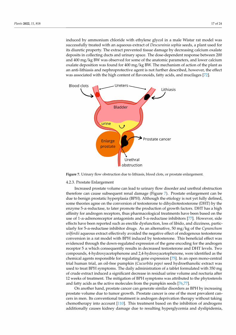
Plants 2022, 11, 818 17 of 24
induced by ammonium chloride with ethylene glycol in a male Wistar rat model wassuccessfully treated with an aqueous extract of Descurainia sophia seeds, a plant used forits diuretic property. The extract prevented tissue damage by decreasing calcium oxalatedeposits in collecting ducts and urinary space. The dose-dependent response between 200and 400 mg/kg BW was observed for some of the anatomic parameters, and lower calciumoxalate deposition was found for 400 mg/kg BW. The mechanism of action of the plant asan anti-lithiasis and nephroprotective agent is not further described, however, the effectwas associated with the high content of flavonoids, fatty acids, and mucilages [72].
Plants 2022, 11, 818 18 of 25
and 400 mg/kg BW was observed for some of the anatomic parameters, and lower calcium oxalate deposition was found for 400 mg/kg BW. The mechanism of action of the plant as an anti-lithiasis and nephroprotective agent is not further described, however, the effect was associated with the high content of flavonoids, fatty acids, and mucilages [72].
Figure 7. Urinary flow obstruction due to lithiasis, blood clots, or prostate enlargement.
4.2.3. Prostate Enlargement Increased prostate volume can lead to urinary flow disorder and urethral obstruction
therefore can cause subsequent renal damage (Figure 7). Prostate enlargement can be due to benign prostatic hyperplasia (BPH). Although the etiology is not yet fully defined, some theories agree on the conversion of testosterone to dihydrotestosterone (DHT) by the en-zyme 5-α-reductase, to later promote the production of growth factors. DHT has a high affinity for androgen receptors, thus pharmacological treatments have been based on the use of 1-α-adrenoreceptor antagonists and 5-α-reductase inhibitors [77]. However, side effects have been reported such as erectile dysfunction, loss of libido, and dizziness, par-ticularly for 5-α-reductase inhibitor drugs. As an alternative, 50 mg/kg of the Cynanchum wilfordii aqueous extract effectively avoided the negative effect of endogenous testos-terone conversion in a rat model with BPH induced by testosterone. This beneficial effect was evidenced through the down-regulated expression of the gene encoding for the an-drogen receptor 5 α which consequently results in decreased testosterone and DHT levels. Two compounds, 4-hydroxyacetophenone and 2,4-hydroxyacetophenone, were identified as the chemical agents responsible for regulating gene expression [75]. In an open mono-central trial human trial, an oil-free pumpkin (Cucurbita pepo) seed hydroethanolic extract was used to treat BPH symptoms. The daily administration of a tablet formulated with 350 mg of crude extract induced a significant decrease in residual urine volume and noc-turia after 12 weeks of treatment. The mitigation of BPH symptoms was attributed to the phytosterols and fatty acids as the active molecules from the pumpkin seeds [76,77].
On another hand, prostate cancer can generate similar disorders as BPH by increas-ing prostate volume due to tumor growth. Prostate cancer is one of the most prevalent cancers in men. Its conventional treatment is androgen deprivation therapy without tak-ing chemotherapy into account [110]. This treatment based on the inhibition of androgens additionally causes kidney damage due to resulting hyperglycemia and dyslipidemia, which alters glomerular filtration [111]. Further, it can cause ischemic injury by attenuat-ing vasodilation in the area of the renal vessels derived from testosterone and nitric oxide, and this type of injury cannot be prevented by estrogen, since it generates hypogonadism and estrogen levels decrease [112]. In addition, the growth of abnormalities in the epithe-lium of the prostate can obstruct urinary flow, leading to a decrease in GFR. In of 15% transgenic adenocarcinoma of the mouse prostate (TRAMP) model, 15% of broccoli sprout powder in the diet reduced the cancer incidence and progression. At a molecular level, broccoli influenced specific epigenetic mechanisms such as HDAC expression, the
Figure 7. Urinary flow obstruction due to lithiasis, blood clots, or prostate enlargement.
4.2.3. Prostate Enlargement
Increased prostate volume can lead to urinary flow disorder and urethral obstructiontherefore can cause subsequent renal damage (Figure 7). Prostate enlargement can bedue to benign prostatic hyperplasia (BPH). Although the etiology is not yet fully defined,some theories agree on the conversion of testosterone to dihydrotestosterone (DHT) by theenzyme 5-α-reductase, to later promote the production of growth factors. DHT has a highaffinity for androgen receptors, thus pharmacological treatments have been based on theuse of 1-α-adrenoreceptor antagonists and 5-α-reductase inhibitors [77]. However, sideeffects have been reported such as erectile dysfunction, loss of libido, and dizziness, partic-ularly for 5-α-reductase inhibitor drugs. As an alternative, 50 mg/kg of the Cynanchumwilfordii aqueous extract effectively avoided the negative effect of endogenous testosteroneconversion in a rat model with BPH induced by testosterone. This beneficial effect wasevidenced through the down-regulated expression of the gene encoding for the androgenreceptor 5 α which consequently results in decreased testosterone and DHT levels. Twocompounds, 4-hydroxyacetophenone and 2,4-hydroxyacetophenone, were identified as thechemical agents responsible for regulating gene expression [75]. In an open mono-centraltrial human trial, an oil-free pumpkin (Cucurbita pepo) seed hydroethanolic extract wasused to treat BPH symptoms. The daily administration of a tablet formulated with 350 mgof crude extract induced a significant decrease in residual urine volume and nocturia after12 weeks of treatment. The mitigation of BPH symptoms was attributed to the phytosterolsand fatty acids as the active molecules from the pumpkin seeds [76,77].
On another hand, prostate cancer can generate similar disorders as BPH by increasingprostate volume due to tumor growth. Prostate cancer is one of the most prevalent can-cers in men. Its conventional treatment is androgen deprivation therapy without takingchemotherapy into account [110]. This treatment based on the inhibition of androgensadditionally causes kidney damage due to resulting hyperglycemia and dyslipidemia,

Plants 2022, 11, 818 18 of 24
which alters glomerular filtration [111]. Further, it can cause ischemic injury by attenuatingvasodilation in the area of the renal vessels derived from testosterone and nitric oxide, andthis type of injury cannot be prevented by estrogen, since it generates hypogonadism andestrogen levels decrease [112]. In addition, the growth of abnormalities in the epithelium ofthe prostate can obstruct urinary flow, leading to a decrease in GFR. In of 15% transgenicadenocarcinoma of the mouse prostate (TRAMP) model, 15% of broccoli sprout powderin the diet reduced the cancer incidence and progression. At a molecular level, broccoliinfluenced specific epigenetic mechanisms such as HDAC expression, the acetylation ofhistone (H3 lysine 18), the H3K9 trimethylation in the ventral prostate lobe, H3K18 acetyla-tion levels, the mean area/foci for H3K9me3, the NAD(P)H quinone dehydrogenase 1 gene(NQO1) and p16 mRNA levels. The therapeutic effect was attributed to the sulforaphanefound at 400 mg/kg in the formulated diet [78].
In the case of both BPH and prostate cancer, the mentioned studies promote thebeneficial effect of plant-based diet and plants intakes as a supplement to prevent post-renal diseases and subsequent renal pathologies.
5. Conclusions and Future Perspectives
Kidney disease can be triggered by multiple factors or diseases, classified as pre-renal, intrinsic, and post-renal according to the physiological pathway via which they leadto kidney injury. The pharmacological treatments for the diseases raised in this review,in most cases, generate adverse effects and end in nephrotoxicity. In contrast, plantsand their bioactive molecules offer nephroprotective effects, acting as antioxidants, anti-inflammatories, antibiotics, anti-cancer, and diuretics, among other effects. The reviewedworks undoubtedly support the use of plants and their bioactive molecules to mitigaterisks factors and drug-induced kidney injuries. Furthermore, these results provide abetter understanding of how plants act as molecular modulators to alleviate pre-renaland post-renal disorders that indirectly or subsequently can lead to the development ofintrinsic kidney disease. Indirect nephroprotective effects have been demonstrated froma wide diversity of plants, including all parts (roots, herbs, leaves, flowers, fruits, andseeds), as well as their by-products such as medicinal plant residue, fruits peels, and pulps.Likewise, therapeutic effects were reported from raw material, crude extracts, and purifiedcompounds. Tested plant products provided nephroprotection at a concentration up to200 mg/kg body weight in male rat models and 900 mg/d in human experiments. Ingeneral, no toxicity was evidenced at the highest tested concentration. The observed effectswere mostly attributed to phenolic compounds, although few of the studies quantified andcharacterized the other phytochemicals.
The main perspective that can be highlighted from this review is that it broadens thepanel of nephroprotective plants by considering not only plants with intrinsic effects orthat can reverse nephrotoxicity, but also plants with beneficial effects on pre- and post-renaldiseases. Therefore, a new classification of secondary nephroprotective plants is proposed.
Furthermore, to implement the use of plants and products as complementary treat-ments rather than substitutive treatment, key parameters must be further studied such asthe ratio for co-administration of nephroprotective plant agents and nephrotoxic drugs, thedose-dependent toxicity, and qualitative and quantitative characterization of phytochemi-cals in the plant products. To achieve this, novel tools such as in silico pharmacodynamicscan be particularly useful for identifying new nephroprotective agents and specificallydesigning in vivo and clinicals assays. In addition, both in vivo and clinical trials shouldbe improved by considering female models.
Author Contributions: Conceptualization, methodology, and writing—original draft preparationM.A.T.-V. and Z.P.M.; visualization, E.S., J.E.S.-H., A.C.-M., E.M.M.-M., H.M.N.I. and R.P.-S.; super-vision, E.M.M.-M., H.M.N.I. and R.P.-S.; project administration, E.M.M.-M., H.M.N.I. and R.P.-S.;funding acquisition, E.M.M.-M., H.M.N.I. and R.P.-S. All authors have read and agreed to the pub-lished version of the manuscript.

Plants 2022, 11, 818 19 of 24
Funding: This research received no external funding. The APC was funded by Tecnologico deMonterrey Grants for Scientific Papers Publication.
Data Availability Statement: Not applicable.
Acknowledgments: This research was funded by Consejo Nacional de Ciencia y Tecnología, for thescholarship granted to Mario Adrián Tienda Vázquez, CVU: 858269 and Tecnológico de Monterrey.CONACYT is thankfully acknowledged for partially supporting this work under Sistema Nacional deInvestigadores (SNI) program awarded to Elda M. Melchor-Martínez (CVU: 230784), Juan EduardoSosa-Hernández (CVU: 375202), Hafiz M.N. Iqbal (CVU: 735340) and Roberto Parra-Saldívar (CVU:35753), Ernesto Sabath (CVU: 46607), and Anaberta Cardador-Martínez (CVU:121490). The authorsappreciate the support from Tecnológico de Monterrey for the literature services.
Conflicts of Interest: The authors declare no conflict of interest.
Abbreviations
AKI: acute kidney injury; ALP: alkaline phosphatase; ALT: alanine aminotransferase; ANP: atrialnatriuretic peptide; APAP: acetaminophen; AST: aspartate aminotransferase; BNP: brain natriureticpeptide; BPH: bening prostatic hyperplasia; BUN: blood urea nitrogen; BW: body weight; CAT:catalase; CKD: chronic kidney disease; CK-MB: creatinine kinase myocardial band; DM: diabetesmellitus; DHT: dihydrotestosterone; ET: endothelin; GSH: glutathione; GFR: glomerular filtrationrate; GLUT: glucose transporter; GPx: glutathione peroxidase; GS: glycogen synthase; GSK3: Glyco-gen synthase kinase-3; G-6-PASE: glucose 6-phosphatase; HDAC: histone deacetylase; HO-1: hemeoxygenase 1; H-FABP: heart-type fatty acid-binding protein; IGP: intraglomerular pressure; IL: in-terleukine ISO: isoprenaline; CCl4: tetrachloride; LDH: lactate dehydrogenase; LDL: low-densitylipoprotein; LVEF: left ventricular ejection fraction; LVFS: left ventricular fraction shortening; MDA:malondialdehyde; NO: nitric oxide; NSAID: Non-steroidal anti-inflammatory drug; RAAS: reninangiotensin aldosterone system; ROS: reactive oxygen species; SGLT: sodium-glucose transporter;SHR: spontaneously hypertensive rats; α-SMA: smooth muscle actin; SOD: superoxide dismutase;TAA: thioacetamide; THP: Tamm-Horsfall glycoprotein; TNF-α: tumor necrosis factor alpha; TRAMP:transgenic adenocarcinoma of the mouse prostate.
References1. Little, M.H.; Combes, A.N. Kidney organoids: Accurate models or fortunate accidents. Genes Dev. 2019, 33, 1319–1345. [CrossRef]
[PubMed]2. Sujana, D.; Saptarini, N.M.; Sumiwi, S.A.; Levita, J. Nephroprotective activity of medicinal plants: A review on in silico-, in vitro-,
and in vivo-based studies. J. Appl. Pharm. Sci. 2021, 11, 113–127. [CrossRef]3. Jager, K.J.; Kovesdy, C.; Langham, R.; Rosenberg, M.; Jha, V.; Zoccali, C. A single number for advocacy and communication-
worldwide more than 850 million individuals have kidney diseases. Nephrol. Dial. Transplant. 2019, 34, 1803–1805. [CrossRef][PubMed]
4. Henry, B.M.; Lippi, G. Chronic kidney disease is associated with severe coronavirus disease 2019 (COVID-19) infection. Int. Urol.Nephrol. 2020, 52, 1193–1194. [CrossRef]
5. Chawla, L.S.; Bellomo, R.; Bihorac, A.; Goldstein, S.L.; Siew, E.D.; Bagshaw, S.M.; Bittleman, D.; Cruz, D.; Endre, Z.;Fitzgerald, R.L.; et al. Acute kidney disease and renal recovery: Consensus report of the Acute Disease Quality Initiative (ADQI)16 Workgroup. Nat. Rev. Nephrol. 2017, 13, 241–257. [CrossRef] [PubMed]
6. KDIGO. Kidney disease: Improving global outcomes (KDIGO) acute kidney injury workgroup. KDIGO clinical practice guidelinefor acute kidney injury. Kidney. Int. Suppl. 2012, 2, 1–138.
7. Manzoor, H.; Bhatt, H. Prerenal Kidney Failure; StatPearls Publishing: Las Vegas, NV, USA, 2020.8. Matuszkiewicz-Rowinska, J.; Małyszko, J. Acute kidney injury, its definition, and treatment in adults: Guidelines and reality. Pol.
Arch. Intern Med. 2020, 130, 1074–1080. [CrossRef] [PubMed]9. Makris, K.; Spanou, L. Acute Kidney Injury: Definition, Pathophysiology and Clinical Phenotypes. Clin. Biochem. Rev. 2016, 37,
85–98. [PubMed]10. Horie, S.; Oya, M.; Nangaku, M.; Yasuda, Y.; Komatsu, Y.; Yanagita, M.; Kitagawa, Y.; Kuwano, H.; Nishiyama, H.; Ishioka, C.; et al.
Guidelines for treatment of renal injury during cancer chemotherapy 2016. Clin. Exp. Nephrol. 2018, 22, 210–244. [CrossRef][PubMed]
11. Lucas, G.N.C.; Leitão, A.C.C.; Alencar, R.L.; Xavier, R.M.F.; Daher, E.D.F.; Silva, G.B.D. Pathophysiological aspects of nephropathycaused by non-steroidal anti-inflammatory drugs. J. Bras. Nefrol. 2019, 41, 124–130. [CrossRef] [PubMed]

Plants 2022, 11, 818 20 of 24
12. Petejova, N.; Martinek, A.; Zadrazil, J.; Kanova, M.; Klementa, V.; Sigutova, R.; Kacirova, I.; Hrabovsky, V.; Svagera, Z.; Stejskal, D.Acute kidney injury in septic patients treated by selected nephrotoxic antibiotic agents—Pathophysiology and biomarkers—Areview. Int. J. Mol. Sci. 2020, 21, 7115. [CrossRef] [PubMed]
13. Wu, B.; Li, D.; Xu, T.; Luo, M.; He, Z.; Li, Y. Proton pump inhibitors associated acute kidney injury and chronic kidney disease:Data mining of US FDA adverse event reporting system. Sci. Rep. 2021, 11, 1–8. [CrossRef] [PubMed]
14. Lawson, S.K.; Satyal, P.; Setzer, W.N. The volatile phytochemistry of seven native american aromatic medicinal plants. Plants2021, 10, 1061. [CrossRef] [PubMed]
15. Isah, T. Stress and defense responses in plant secondary metabolites production. Biol. Res. 2019, 52, 39. [CrossRef] [PubMed]16. Das, S.; Vasudeva, N.; Sharma, S. Kidney disorders and management through herbs: A Review. J. Phytopharm. 2019, 8, 21–27.
[CrossRef]17. Efferth, T.; Saeed, M.E.; Mirghani, E.; Alim, A.; Yassin, Z.; Saeed, E.; Khalid, H.E.; Daak, S. Integration of phytochemicals and
phytotherapy into cancer precision medicine. Oncotarget 2017, 8, 50284–50304. [CrossRef] [PubMed]18. Governa, P.; Baini, G.; Borgonetti, V.; Cettolin, G.; Giachetti, D.; Magnano, A.R.; Miraldi, E.; Biagi, M. Phytotherapy in the
Management of Diabetes: A Review. Molecules 2018, 23, 105. [CrossRef] [PubMed]19. Sabiu, S.; O’Neill, F.H.; Ashafa, A.O.T. The purview of phytotherapy in the management of kidney disorders: A systematic review
on Nigeria and South Africa. Afr. J. Tradit. Complement. Altern. Med. 2016, 13, 38–47.20. Basist, P.; Parveen, B.; Zahiruddin, S.; Gautam, G.; Parveen, R.; Khan, M.A.; Krishnan, A.; Shahid, M.; Ahmad, S. Potential
nephroprotective phytochemicals: Mechanism and future prospects. J. Ethnopharm. 2022, 283, 114743. [CrossRef] [PubMed]21. Ahmad, Q.Z. An Appraisal of Nephroprotection and the Scope of Natural Products in Combating Renal Disorders. J. Nephrol.
Ther. 2014, 04, 170. [CrossRef]22. Stub, T.; Quandt, S.A.; Arcury, T.A.; Sandberg, J.C.; Kristoffersen, A.E.; Musial, F.; Salamonsen, A. Perception of risk and
communication among conventional and complementary health care providers involving cancer patients’ use of complementarytherapies: A literature review. BMC Complement. Altern. Med. 2016, 16, 353. [CrossRef]
23. Khalil, H.; Huang, C. Adverse drug reactions in primary care: A scoping review. BMC Health Serv. Res. 2020, 20, 5. [CrossRef]24. World Health Organization (WHO). WHO Traditional Medicines Strategy 2014–2023; World Health Organization: Geneva, Switzer-
land, 2013.25. Li, J.; Li, B.; Zhao, X.K.; Tu, J.Y.; Li, Y. A critical review to grading systems and recommendations of traditional Chinese medicine
guidelines. Health Qual. Life Outcomes 2020, 18, 174. [CrossRef] [PubMed]26. Mai, W.; Wei, A.; Lin, X.; Wang, F.; Ye, J.; Chen, P. Efficacy and safety of traditional Chinese medicine injection with mecobalamin
in treating diabetic peripheral neuropathy: A protocol for systematic review and meta-analysis. Medicine 2021, 100, e23702.[CrossRef]
27. Kessler, C.S.; Morandi, A.; Kumar, A.; Dhiman, K.S.; Gupta, S.; Icke, K.; Bühner, C.; Stapelfeldt, E.; Wischnewsky, M.;Kronpaß, L.; et al. Reliability of Ayurvedic Diagnosis for Knee Osteoarthritis Patients: A Nested Diagnostic Study Withina Randomized Controlled Trial. J. Altern. Complement. Med. 2019, 25, 910–919. [CrossRef] [PubMed]
28. Peterson, C.T.; Denniston, K.; Chopra, D. Therapeutic uses of Triphala in ayurvedic medicine. J. Altern Complement. Med. 2017, 23,607–614. [CrossRef] [PubMed]
29. Atanasov, A.G.; Waltenberger, B.; Pferschy-Wenzig, E.-M.; Linder, T.; Wawrosch, C.; Uhrin, P.; Temml, V.; Wang, L.; Schwaiger, S.;Heiss, E.H.; et al. Discovery and resupply of pharmacologically active plant-derived natural products: A review. Biotechnol. Adv.2015, 33, 1582–1614. [CrossRef]
30. Atanasov, A.G.; Zotchev, S.B.; Dirsch, V.M.; Supuran, C.T. Natural products in drug discovery: Advances and opportunities. Nat.Rev. Drug Discov. 2021, 20, 200–216. [CrossRef] [PubMed]
31. Li, S.; He, Y.; Zhang, H.; Zheng, R.; Xu, R.; Liu, Q.; Tang, S.; Ke, X.; Huang, M. Formulation of traditional Chinese medicine andits application on intestinal flora of constipated rats. Microb. Cell Fact. 2020, 19, 212. [CrossRef] [PubMed]
32. Luo, H.; Zhao, Y.; Hua, H.; Zhang, Y.; Zhang, X.; Fang, Q.; Li, Q.; Zhang, Y.; Tan, P.; Yang, A.; et al. Research progress on qualityassurance of genuine Chinese medicinal in Sichuan. Chin. Med. 2021, 16, 19. [CrossRef]
33. Nyakudya, T.; Tshabalala, T.; Dangarembizi, R.; Erlwanger, K.; Ndhlala, A.R. The Potential Therapeutic Value of Medicinal Plantsin the Management of Metabolic Disorders. Molecules 2020, 25, 2669. [CrossRef] [PubMed]
34. Rastogi, S. Emanating the specialty clinical practices in Ayurveda: Preliminary observations from an arthritis clinic and itsimplications. J. Ayurveda Integr. Med. 2020, 12, 52–57. [CrossRef] [PubMed]
35. Yoo, S.; Kim, K.; Nam, H.; Lee, D. Discovering Health Benefits of Phytochemicals with Integrated Analysis of the MolecularNetwork, Chemical Properties and Ethnopharmacological Evidence. Nutrients 2018, 10, 1042. [CrossRef] [PubMed]
36. Pan, S.-Y.; Litscher, G.; Gao, S.-H.; Zhou, S.-F.; Yu, Z.-L.; Chen, H.-Q.; Zhang, S.-F.; Tang, M.-K.; Sun, J.-N.; Ko, K.-M. HistoricalPerspective of Traditional Indigenous Medical Practices: The Current Renaissance and Conservation of Herbal Resources.Evid.-Based Complement. Altern. Med. 2014, 2014, 525340. [CrossRef] [PubMed]
37. Campagne, O.; Mager, D.E.; Brazeau, D.; Venuto, R.C.; Tornatore, K.M. The impact of tacrolimus exposure on extrarenal adverseeffects in adult renal transplant recipients. Br. J. Clin. Pharmacol. 2019, 85, 516–529. [CrossRef] [PubMed]
38. Kumar, S.; Dobos, G.J.; Rampp, T. The Significance of Ayurvedic Medicinal Plants. J. Evid.-Based Complement. Altern. Med. 2017,22, 494–501. [CrossRef]

Plants 2022, 11, 818 21 of 24
39. Pfister, B.; Jonsson, J.; Gustafsson, M. Drug-related problems and medication reviews among old people with dementia. BMCPharmacol. Toxicol. 2017, 18, 52. [CrossRef]
40. Stocco, G.; Lucafò, M.; Decorti, G. Pharmacogenomics of Antibiotics. Int. J. Mol. Sci. 2020, 21, 5975. [CrossRef]41. Phe, K.; Shields, R.K.; Tverdek, F.P.; Aitken, S.; Guervil, D.J.; Lam, W.-Y.M.; Musgrove, R.J.; Luce, A.M.; Tam, V.H. Predicting
the risk of nephrotoxicity in patients receiving colistimethate sodium: A multicentre, retrospective, cohort study. J. Antimicrob.Chemother. 2016, 71, 3585–3587. [CrossRef]
42. Dwyer, J.T.; Coates, P.M.; Smith, M.J. Dietary Supplements: Regulatory Challenges and Research Resources. Nutrients 2018, 10, 41.[CrossRef]
43. McGraw, N.J.; Krul, E.S.; Grunz-Borgmann, E.; Parrish, A.R. Soy-based renoprotection. World J. Nephrol. 2016, 5, 233–257.[CrossRef] [PubMed]
44. Moorthi, R.N.; Vorland, C.J.; Gallant, K.M.H. Diet and Diabetic Kidney Disease: Plant Versus Animal Protein. Curr. Diabetes Rep.2017, 17, 15. [CrossRef] [PubMed]
45. Alabi, T.; Brooks, N.; Oguntibeju, O. Leaf Extracts of Anchomanes difformis Ameliorated Kidney and Pancreatic Damage in Type 2Diabetes. Plants 2021, 10, 300. [CrossRef] [PubMed]
46. Al-Anbaki, M.; Cavin, A.-L.; Nogueira, R.; Taslimi, J.; Ali, H.; Najem, M.; Mahmood, M.S.; Khaleel, I.A.; Mohammed, A.S.;Hasan, H.R.; et al. Hibiscus sabdariffa, a Treatment for Uncontrolled Hypertension. Pilot Comparative Intervention. Plants 2021,10, 1018. [CrossRef] [PubMed]
47. Nasri, H.; Hedayatollah, S. Toxicity and safety of medicinal plants. J. HerbMed. Pharmacol. 2013, 2, 21–22. Available online:http://eprints.skums.ac.ir/4798/1/9.pdf (accessed on 15 March 2022).
48. Kharchoufa, L.; Bouhrim, M.; Bencheikh, N.; Addi, M.; Hano, C.; Mechchate, H.; Elachouri, M. Potential Toxicity of MedicinalPlants Inventoried in Northeastern Morocco: An Ethnobotanical Approach. Plants 2021, 10, 1108. [CrossRef]
49. Oketch-Rabah, H.A.; Roe, A.L.; Rider, C.V.; Bonkovsky, H.L.; Giancaspro, G.I.; Navarro, V.; Paine, M.F.; Betz, J.M.; Marles, R.J.;Casper, S.; et al. United States Pharmacopeia (USP) comprehensive review of the hepatotoxicity of green tea extracts. Toxicol. Rep.2020, 7, 386–402. [CrossRef]
50. Rakib, A.; Ahmed, S.; Islam, M.A.; Haye, A.; Uddin, S.M.N.; Uddin, M.M.N.; Hossain, M.K.; Paul, A.; Emran, T.B. Antipyreticand hepatoprotective potential of Tinospora crispa and investigation of possible lead compounds through in silico approaches.Food Sci. Nutr. 2020, 8, 547–556. [CrossRef] [PubMed]
51. Fernández-Martínez, E.; Jiménez-Santana, M.; Centeno-Álvarez, M.; Torres-Valencia, J.M.; Shibayama, M.; Cariño-Cortés, R.Hepatoprotective Effects of Nonpolar Extracts from Inflorescences of Thistles Cirsium vulgare and Cirsium ehrenbergii on AcuteLiver Damage in Rat. Pharmacogn. Mag. 2018, 13, S860–S867. [CrossRef]
52. Mohammed, S.A.A.; Khan, R.A.; El-readi, M.Z.; Emwas, A.H.; Sioud, S.; Poulson, B.G.; Jaremko, M.; Eldeeb, H.M.; Al-Omar, M.S.;Mohammed, H.A. Suaeda vermiculata aqueous-ethanolic extract-based mitigation of ccl4-induced hepatotoxicity in rats, andhepg-2 and hepg-2/adr cell-lines-based cytotoxicity evaluations. Plants 2020, 9, 1291. [CrossRef] [PubMed]
53. Moshaie-Nezhad, P.; Bahari, Z.; Jangravi, Z.; Zarei, S.M.; Iman, M. The Effect of Descurainia Sophia Seed Extract on NephrotoxicityMarkers Induced by Acetaminophen in Mice. J. Adv. Med Biomed. Res. 2021, 29, 139–144. [CrossRef]
54. Chinnappan, S.M.; George, A.; Thaggikuppe Krishnamurthy, P.; Choudhary, Y.; Choudhary, V.K.; Ramani, Y.; Dewangan, R.Nephroprotective Effect of Herbal Extract Eurycoma longifolia on Paracetamol-Induced Nephrotoxicity in Rats. Evid.-basedComplement. Altern. Med. 2019, 2019, 4916519. [CrossRef]
55. Buzgaia, N.; Awin, T.; Elabbar, F.; Abdusalam, K.; Lee, S.Y.; Rukayadi, Y.; Abas, F.; Shaari, K. Antibacterial Activity of Arbutuspavarii Pamp against Methicillin-Resistant Staphylococcus aureus (MRSA) and UHPLC-MS/MS Profile of the Bioactive Fraction.Plants 2020, 9, 1539. [CrossRef] [PubMed]
56. Mestry, S.N.; Gawali, N.B.; Pai, S.A.; Gursahani, M.S.; Dhodi, J.B.; Munshi, R.; Juvekar, A.R. Punica granatum improves renalfunction in gentamicin-induced nephropathy in rats via attenuation of oxidative stress. J. Ayurveda Integr. Med. 2020, 11, 16–23.[CrossRef]
57. El Bohi, K.M.; Abdel-Motal, S.M.; Khalil, S.R.; Abd-Elaal, M.M.; Metwally, M.M.M.; Elhady, W.M. The efficiency of pomegranate(Punica granatum) peel ethanolic extract in attenuating the vancomycin-triggered liver and kidney tissues injury in rats. Environ.Sci. Pollut. Res. 2021, 28, 7134–7150. [CrossRef] [PubMed]
58. Álvarez-Cilleros, D.; López-Oliva, E.; Goya, L.; Martín, M.Á.; Ramos, S. Cocoa intake attenuates renal injury in Zucker Diabeticfatty rats by improving glucose homeostasis. Food Chem. Toxicol. 2019, 127, 101–109. [CrossRef] [PubMed]
59. Boonphang, O.; Ontawong, A.; Pasachan, T.; Phatsara, M.; Duangjai, A.; Amornlerdpison, D.; Jinakote, M.; Srimaroeng, C.Antidiabetic and Renoprotective Effects of Coffea arabica Pulp Aqueous Extract through Preserving Organic Cation TransportSystem Mediated Oxidative Stress Pathway in Experimental Type 2 Diabetic Rats. Molecules 2021, 26, 1907. [CrossRef] [PubMed]
60. Perez-Gutierrez, R.M.; Garcia-Campoy, A.H.; Muñiz-Ramirez, A. Properties of Flavonoids Isolated from the Bark of Eysenhardtiapolystachya and Their Effect on Oxidative Stress in Streptozotocin-Induced Diabetes Mellitus in Mice. Oxid. Med. Cell. Longev.2016, 2016, 9156510. [CrossRef] [PubMed]
61. Rodríguez-Fierros, F.L.; Guarner-Lans, V.; Soto, M.E.; Manzano-Pech, L.; Díaz-Díaz, E.; Soria-Castro, E.; Rubio-Ruiz, M.E.;Jiménez-Trejo, F.; Pérez-Torres, I. Modulation of Renal Function in a Metabolic Syndrome Rat Model by Antioxidants in Hibiscussabdariffa L. Molecules 2021, 26, 2074. [CrossRef] [PubMed]

Plants 2022, 11, 818 22 of 24
62. Epure, A.; Pârvu, A.E.; Vlase, L.; Benedec, D.; Hanganu, D.; Gheldiu, A.-M.; Toma, V.A.; Oniga, I. Phytochemical Profile,Antioxidant, Cardioprotective and Nephroprotective Activity of Romanian Chicory Extract. Plants 2020, 10, 64. [CrossRef][PubMed]
63. Hou, Y.; Yuan, P.; Fu, Y.; Zhang, Q.; Gao, L.; Wei, Y.; Zheng, X.; Feng, W. Geniposide from Gardenia jasminoides var. radicansMakino Attenuates Myocardial Injury in Spontaneously Hypertensive Rats via Regulating Apoptotic and Energy MetabolismSignalling Pathway. Drug Des. Dev. Ther. 2021, 15, 949–962. [CrossRef] [PubMed]
64. Guerrero-hue, M.; Garc, C.; Palomino-antol, A.; Rubio-Navarro, A.; Cristina, V.; Herencia, C.; Martín-Sanchez, D.; Farré-Alins, V.;Egea, J.; Cannata, P.; et al. Curcumin reduces renal damage associated with rhabdomyolysis by decreasing ferroptosis-mediatedcell death. FASEB J. 2019, 33, 8961–8975. [CrossRef] [PubMed]
65. Benzer, F.; Kandemir, F.M.; Kucukler, S.; Comaklı, S.; Caglayan, C. Chemoprotective effects of curcumin on doxorubicin-inducednephrotoxicity in wistar rats: By modulating inflammatory cytokines, apoptosis, oxidative stress and oxidative DNA damage.Arch. Physiol. Biochem. 2018, 124, 448–457. [CrossRef] [PubMed]
66. Nerdy, N.; Ritarwan, K. Hepatoprotective Activity and Nephroprotective Activity of Peel Extract from Three Varieties of thePassion Fruit (Passiflora Sp.) in the Albino Rat. Open Access Maced. J. Med. Sci. 2019, 7, 536–542. [CrossRef] [PubMed]
67. Al-Yousef, H.M.; Alqahtani, A.S.; Ghani, A.S.; El-Toumy, S.A.; El-Dougdoug, W.I.; Hassan, W.H.B.; Hassan, H.M. Nephroprotec-tive, cytotoxic and antioxidant activities of Euphorbia paralias. Saudi J. Biol. Sci. 2021, 28, 785–792. [CrossRef]
68. Heidarian, E.; Jafari-Dehkordi, E.; Valipour, P.; Ghatreh-Samani, K.; Ashrafi-Eshkaftaki, L. Nephroprotective and Anti-Inflammatory Effects of Pistacia atlantica Leaf Hydroethanolic Extract Against Gentamicin-Induced Nephrotoxicity in Rats. J. Diet.Suppl. 2017, 14, 489–502. [CrossRef]
69. Ezejiofor, A.N.; Udowelle, N.A.; Orisakwe, O.E. Nephroprotective and antioxidant effect of aqueous leaf extract of Costus afer Kergawl on cyclosporin-a (Csa) induced nephrotoxicity. Clin. Phytosci. 2016, 2, 11. [CrossRef]
70. Ranfaing, J.; Dunyach-Remy, C.; Lavigne, J.-P.; Sotto, A. Propolis potentiates the effect of cranberry (Vaccinium macrocarpon) inreducing the motility and the biofilm formation of uropathogenic Escherichia coli. PLoS ONE 2018, 13, e0202609. [CrossRef]
71. Scharf, B.; Sendker, J.; Dobrindt, U.; Hensel, A. Influence of Cranberry Extract on Tamm-Horsfall Protein in Human Urine and itsAntiadhesive Activity Against Uropathogenic Escherichia coli. Planta Med. 2018, 85, 126–138. [CrossRef]
72. Saremi, J.; Hasheminasab, M.; Sadeghi, N.; Jahromi, H.K. Preventive and curative effects of aqueous extracts of Descurainia sophiaL on nephrolithiasis induced in rats. Asian J. Pharm. 2018, 2018, 228–233.
73. Carneiro, D.M.; Freire, R.C.; Honório, T.C.D.D.; Zoghaib, I.; Cardoso, F.F.D.S.E.S.; Tresvenzol, L.M.F.; De Paula, J.R.; Sousa, A.L.L.;Jardim, P.C.B.V.; Da Cunha, L.C. Randomized, Double-Blind Clinical Trial to Assess the Acute Diuretic Effect ofEquisetumarvense(Field Horsetail) in Healthy Volunteers. Evid.-Based Complement. Altern. Med. 2014, 2014, 760683. [CrossRef] [PubMed]
74. Pallag, A.; Filip, G.A.; Olteanu, D.; Clichici, S.; Baldea, I.; Jurca, T.; Micle, O.; Vicas, L.; Marian, E.; Soritău, O.; et al. Equisetumarvense L. Extract Induces Antibacterial Activity and Modulates Oxidative Stress, Inflammation, and Apoptosis in EndothelialVascular Cells Exposed to Hyperosmotic Stress. Oxid. Med. Cell. Longev. 2018, 2018, 3060525. [CrossRef] [PubMed]
75. Lee, G.; Shin, J.; Choi, H.; Jo, A.; Pan, S.; Bae, D.; Lee, Y.; Choi, C. Cynanchum wilfordii Ameliorates Testosterone-Induced BenignProstatic Hyperplasia by Regulating 5α-Reductase and Androgen Receptor Activities in a Rat Model. Nutrients 2017, 9, 1070.[CrossRef] [PubMed]
76. Leibbrand, M.; Siefer, S.; Schön, C.; Perrinjaquet-Moccetti, T.; Kompek, A.; Csernich, A.; Bucar, F.; Kreuter, M.H. Effects of anOil-Free Hydroethanolic Pumpkin Seed Extract on Symptom Frequency and Severity in Men with Benign Prostatic Hyperplasia:A Pilot Study in Humans. J. Med. Food 2019, 22, 551–559. [CrossRef]
77. Csikós, E.; Horváth, A.; Ács, K.; Papp, N.; Balázs, V.L.; Dolenc, M.S.; Kenda, M.; Glavac, N.K.; Nagy, M.; Protti, M.; et al. Treatmentof Benign Prostatic Hyperplasia by Natural Drugs. Molecules 2021, 26, 7141. [CrossRef]
78. Beaver, L.M.; Löhr, C.V.; Clarke, J.; Glasser, S.T.; Watson, G.W.; Wong, C.P.; Zhang, Z.; Williams, D.E.; Dashwood, R.H.;Shannon, J.; et al. Broccoli Sprouts Delay Prostate Cancer Formation and Decrease Prostate Cancer Severity with a ConcurrentDecrease in HDAC3 Protein Expression in Transgenic Adenocarcinoma of the Mouse Prostate (TRAMP) Mice. Curr. Dev. Nutr.2018, 2, nzy002. [CrossRef]
79. Stephens, J.W.; Brown, K.E.; Min, T. Chronic kidney disease in type 2 diabetes: Implications for managing glycaemic control,cardiovascular and renal risk. Diabetes Obes. Metab. 2020, 22, 32–45. [CrossRef]
80. Bharadwaj, D.; Singh, A. Genetic basis for increased risk for vascular diseases in diabetes. In Mechanisms of Vascular Defects inDiabetes Mellitus; Kartha, C., Ramachandran, S., Pillai, R.M., Eds.; India Springer: New Delhi, India, 2017; pp. 27–45.
81. Cupisti, A.; Giannese, D.; Moriconi, D.; D’Alessandro, C.; Torreggiani, M.; Piccoli, G.B. Nephroprotection by SGLT2i in CKDPatients: May It Be Modulated by Low-Protein Plant-Based Diets? Front. Med. 2020, 7, 622593. [CrossRef]
82. Hall, J.; Hall, M. Adrenocorcical hormones. In Guyton and Hall Textbook of Medical Physiology, 14th ed.; Elsevier: Amsterdam, TheNetherlands, 2020; pp. 955–987.
83. Esquivel-Gutiérrez, E.R.; Manzo-Avalos, S.; Peña-Montes, D.J.; Saavedra-Molina, A.; Morreeuw, Z.P.; Reyes, A.G. Hypolipi-demic and Antioxidant Effects of Guishe Extract from Agave lechuguilla, a Mexican Plant with Biotechnological Potential, onStreptozotocin-Induced Diabetic Male Rats. Plants 2021, 10, 2492. [CrossRef]
84. Cardador-Martínez, A. Medicinal plants with the potential to treat hypertension: Studies that were done in the last decade.In Medicinal Plants for the Treatment of Metabolic Disorders, 1st ed.; Cardador Martínez, A., Rodriguez, V., Manzano-Santana, P.,Alonzo, M., Eds.; Nova Medicine & Health: Mexico, NM, USA, 2020; pp. 24–25.
Plants 2022, 11, 818 23 of 24
85. O’Shea, P.; Griffin, T.P.; Fitzgibbon, M. Hypertension: The role of biochemistry in the diagnosis and management. Clin. Chim.Acta 2017, 465, 131–143. [CrossRef]
86. Kjeldsen, S.; Feldman, R.D.; Lisheng, L.; Mourad, J.-J.; Chiang, C.-E.; Zhang, W.; Wu, Z.; Liu, L.; Williams, B. Updated Nationaland International Hypertension Guidelines: A Review of Current Recommendations. Drugs 2014, 74, 2033–2051. [CrossRef][PubMed]
87. James, P.A.; Oparil, S.; Carter, B.L.; Cushman, W.C.; Dennison-Himmelfarb, C.; Handler, J.; Lackland, D.T.; LeFevre, M.L.;MacKenzie, T.D.; Ogedegbe, O.; et al. 2014 evidence-based guideline for the management of high blood pressure in adults:Report from the panel members appointed to the Eighth Joint National Committee (JNC 8). JAMA 2014, 311, 507–520. [CrossRef][PubMed]
88. Pugh, D.; Gallacher, P.J.; Dhaun, N. Management of Hypertension in Chronic Kidney Disease. Drugs 2019, 79, 365–379. [CrossRef][PubMed]
89. Saxena, T.; Ali, A.O.; Saxena, M. Pathophysiology of essential hypertension: An update. Expert Rev. Cardiovasc. Ther. 2018, 16,879–887. [CrossRef]
90. Ku, E.; McCulloch, C.E.; Vittinghoff, E.; Lin, F.; Johansen, K.L. Use of Antihypertensive Agents and Association with Risk ofAdverse Outcomes in Chronic Kidney Disease: Focus on Angiotensin-Converting Enzyme Inhibitors and Angiotensin ReceptorBlockers. J. Am. Hear. Assoc. 2018, 7, e009992. [CrossRef] [PubMed]
91. Levi, M.; Takahashi, S.; Wang, X.; Levi, M. The Kidney in Liver Disease. In The Liver Biology and Pathobiol, 6th ed.; John Wiley &Sons, Ltd.: Chichister, UK, 2020; pp. 619–638.
92. Ramachandran, A.; Visschers, R.G.J.; Duan, L.; Akakpo, J.Y.; Jaeschke, H. Mitochondrial dysfunction as a mechanism ofdrug-induced hepatotoxicity: Current understanding and future perspectives. J. Clin. Transl. Res. 2018, 4, 75–100.
93. Rajakumar, A.; Appuswamy, E.; Kaliamoorthy, I.; Rela, M. Renal Dysfunction in Cirrhosis: Critical Care Management. Indian J.Crit. Care Med. 2021, 25, 207–214. [CrossRef]
94. Ramachandran, A.; Jaeschke, H. Experimental Models of Hepatotoxicity for the Testing of Natural Products [Internet]; ReferenceModule in Chemistry, Molecular Sciences and Chemical Engineering; Elsevier Inc.: Amsterdam, The Netherlands, 2016; pp. 1–9.[CrossRef]
95. Weber, E.J.; Lidberg, K.A.; Wang, L.; Bammler, T.K.; Macdonald, J.W.; Li, M.J.; Redhair, M.; Atkins, W.M.; Tran, C.; Hines, K.; et al.Human kidney on a chip assessment of polymyxin antibiotic nephrotoxicity. JCI Insight 2018, 3, e123673. [CrossRef]
96. Winther, S.A.; Øllgaard, J.C.; Hansen, T.W.; von Scholten, B.J.; Reinhard, H.; Ahluwalia, T.S.; Wang, Z.; Gæde, P.; Parving, H.-H.;Hazen, S.; et al. Plasma trimethylamine N-oxide and its metabolic precursors and risk of mortality, cardiovascular and renaldisease in individuals with type 2-diabetes and albuminuria. PLoS ONE 2021, 16, e0244402. [CrossRef] [PubMed]
97. Wiese, G.N.; Biruete, A.; Moorthi, R.N.; Moe, S.M.; Lindemann, S.R.; Gallant, K.M.H. Plant-Based Diets, the Gut Microbiota, andTrimethylamine N-Oxide Production in Chronic Kidney Disease: Therapeutic Potential and Methodological Considerations. J.Ren. Nutr. 2021, 31, 121–131. [CrossRef]
98. Du, F.; Huang, R.; Lin, D.; Wang, Y.; Yang, X.; Huang, X.; Zheng, B.; Chen, Z.; Huang, Y.; Wang, X.; et al. Resveratrol ImprovesLiver Steatosis and Insulin Resistance in Non-alcoholic Fatty Liver Disease in Association with the Gut Microbiota. Front.Microbiol. 2021, 12, 611323. [CrossRef] [PubMed]
99. Meng, F.; Chen, T.; Ma, D.; Wang, X.; Zhao, X.; Tian, P.; Wang, H.; Hai, Z.; Shen, L.; Tang, X.; et al. Reclamation of Herb ResiduesUsing Probiotics and Their Therapeutic Effect on Diarrhea. Mediat. Inflamm. 2017, 2017, 4265898. [CrossRef] [PubMed]
100. Boudhabhay, I.; Poillerat, V.; Grunenwald, A.; Torset, C.; Leon, J.; Daugan, M.V.; Lucibello, F.; El Karoui, K.; Ydee, A.; Chauvet, S.Complement activation is a crucial driver of acute kidney injury in rhabdomyolysis. Kidney Int. 2021, 99, 581–597. [CrossRef][PubMed]
101. Cortés, R.; Kleinsteuber, K.; Vargas, C.P.; Avaria, M.D.L. Ángeles Rabdomiólisis metabólica: Actualización. Rev. Médica Clínica LasCondes 2018, 29, 553–559. [CrossRef]
102. Teng, C.; Baus, C.; Wilson, J.P.; Frei, C.R. Rhabdomyolysis Associations with Antibiotics: A Pharmacovigilance Study of the FDAAdverse Event Reporting System (FAERS). Int. J. Med. Sci. 2019, 16, 1504–1509. [CrossRef] [PubMed]
103. Okubo, K.; Kurosawa, M.; Kamiya, M.; Urano, Y.; Suzuki, A.; Yamamoto, K.; Hase, K.; Homma, K.; Sasaki, J.; Miyauchi, H.; et al.Macrophage extracellular trap formation promoted by platelet activation is a key mediator of rhabdomyolysis-induced acutekidney injury. Nat. Med. 2018, 24, 232–238. [CrossRef] [PubMed]
104. Li, F.; Chen, A.-B.; Duan, Y.-C.; Liao, R.; Xu, Y.-W.; Tao, L.-L. Multiple organ dysfunction and rhabdomyolysis associated withmoonwort poisoning: Report of four cases. World J. Clin. Cases 2020, 8, 479–486. [CrossRef]
105. Kang, C.-I.; Kim, J.; Park, D.W.; Kim, B.-N.; Ha, U.-S.; Lee, S.-J.; Yeo, J.; Min, S.K.; Lee, H.; Wie, S.-H. Clinical Practice Guidelinesfor the Antibiotic Treatment of Community-Acquired Urinary Tract Infections. Infect. Chemother. 2018, 50, 67–100. [CrossRef][PubMed]
106. Kline, K.A.; Lewis, A.L. Gram-Positive Uropathogens, Polymicrobial Urinary Tract Infection, and the Emerging Microbiota of theUrinary Tract. Microbiol. Spectr. 2016, 4, 459–502. [CrossRef] [PubMed]
107. Olson, P.D.; McLellan, L.K.; Liu, A.; Briden, K.E.; Tiemann, K.M.; Daugherty, A.L.; Hruska, K.A.; Hunstad, D.A. Correction: Renalscar formation and kidney function following antibiotic-treated murine pyelonephritis. Dis. Model. Mech. 2018, 11, 1371–1379.[CrossRef]

Plants 2022, 11, 818 24 of 24
108. Nirumand, M.C.; Hajialyani, M.; Rahimi, R.; Farzaei, M.H.; Zingue, S.; Nabavi, S.M.; Bishayee, A. Dietary Plants for the Preventionand Management of Kidney Stones: Preclinical and Clinical Evidence and Molecular Mechanisms. Int. J. Mol. Sci. 2018, 19, 765.[CrossRef] [PubMed]
109. Santos, F.M.D.; Peres, A.K.; Mandotti, M.R.; Peres, L.A.B. Metabolic investigation in patients with nephrolithiasis. Einstein 2017,15, 452–456. [CrossRef] [PubMed]
110. Wang, G.; Zhao, D.; Spring, D.J.; Depinho, R.A. Genetics and biology of prostate cancer. Genes Dev. 2018, 32, 1105–1140. [CrossRef][PubMed]
111. Cardwell, C.R.; O’Sullivan, J.M.; Jain, S.; Hicks, B.M.; Devine, P.A.; McMenamin, Ú.C. Hormone therapy use and the risk of acutekidney injury in patients with prostate cancer: A population-based cohort study. Prostate Cancer Prostatic Dis. 2021, 24, 1055–1062.[CrossRef]
112. Daskivich, T.J. Androgen deprivation therapy and acute kidney injury in prostate cancer: Room for debate? Prostate CancerProstatic Dis. 2021, 24, 933–934. [CrossRef] [PubMed]