Nature of Nonfunctional Envelope Proteins on the Surface of Human Immunodeficiency Virus Type 1
15
10.1128/JVI.80.5.2515-2528.2006. 2006, 80(5):2515. DOI: J. Virol. Roux, Dennis R. Burton and James M. Binley Michael B. Zwick, Michael Franti, Lynn Morris, Kenneth H. Charmagne S. Cayanan, Henry Grise, Paul Corcoran, Penny L. Moore, Emma T. Crooks, Lauren Porter, Ping Zhu, Virus Type 1 Immunodeficiency on the Surface of Human Nature of Nonfunctional Envelope Proteins http://jvi.asm.org/content/80/5/2515 Updated information and services can be found at: These include: REFERENCES http://jvi.asm.org/content/80/5/2515#ref-list-1 at: This article cites 73 articles, 44 of which can be accessed free CONTENT ALERTS more» articles cite this article), Receive: RSS Feeds, eTOCs, free email alerts (when new http://journals.asm.org/site/misc/reprints.xhtml Information about commercial reprint orders: http://journals.asm.org/site/subscriptions/ To subscribe to to another ASM Journal go to: on March 21, 2014 by guest http://jvi.asm.org/ Downloaded from on March 21, 2014 by guest http://jvi.asm.org/ Downloaded from
-
Upload
independent -
Category
Documents
-
view
0 -
download
0
Transcript of Nature of Nonfunctional Envelope Proteins on the Surface of Human Immunodeficiency Virus Type 1

10.1128/JVI.80.5.2515-2528.2006.
2006, 80(5):2515. DOI:J. Virol. Roux, Dennis R. Burton and James M. BinleyMichael B. Zwick, Michael Franti, Lynn Morris, Kenneth H.Charmagne S. Cayanan, Henry Grise, Paul Corcoran, Penny L. Moore, Emma T. Crooks, Lauren Porter, Ping Zhu, Virus Type 1
Immunodeficiencyon the Surface of Human Nature of Nonfunctional Envelope Proteins
http://jvi.asm.org/content/80/5/2515Updated information and services can be found at:
These include:
REFERENCEShttp://jvi.asm.org/content/80/5/2515#ref-list-1at:
This article cites 73 articles, 44 of which can be accessed free
CONTENT ALERTS more»articles cite this article),
Receive: RSS Feeds, eTOCs, free email alerts (when new
http://journals.asm.org/site/misc/reprints.xhtmlInformation about commercial reprint orders: http://journals.asm.org/site/subscriptions/To subscribe to to another ASM Journal go to:
on March 21, 2014 by guest
http://jvi.asm.org/
Dow
nloaded from
on March 21, 2014 by guest
http://jvi.asm.org/
Dow
nloaded from

JOURNAL OF VIROLOGY, Mar. 2006, p. 2515–2528 Vol. 80, No. 50022-538X/06/$08.00�0 doi:10.1128/JVI.80.5.2515–2528.2006Copyright © 2006, American Society for Microbiology. All Rights Reserved.
Nature of Nonfunctional Envelope Proteins on the Surface of HumanImmunodeficiency Virus Type 1
Penny L. Moore,1,2 Emma T. Crooks,1 Lauren Porter,3 Ping Zhu,3 Charmagne S. Cayanan,4†Henry Grise,3 Paul Corcoran,1 Michael B. Zwick,4 Michael Franti,5 Lynn Morris,2
Kenneth H. Roux,3 Dennis R. Burton,4 and James M. Binley1,4*Torrey Pines Institute for Molecular Studies, 3550 General Atomics Court, San Diego, California 921211; National Institute for
Communicable Diseases, Sandringham, Johannesburg, South Africa2; Department of Biological Science and Institute ofMolecular Biophysics, Florida State University, Tallahassee, Florida 323063; Departments of Immunology and
Molecular Biology, The Scripps Research Institute, La Jolla, California 920374; and Progenics Pharmaceuticals,Inc., Tarrytown, New York 105915
Received 19 September 2005/Accepted 5 December 2005
Human immunodeficiency virus type 1 (HIV-1) neutralizing antibodies are thought be distinguished fromnonneutralizing antibodies by their ability to recognize functional gp120/gp41 envelope glycoprotein (Env)trimers. The antibody responses induced by natural HIV-1 infection or by vaccine candidates tested to dateconsist largely of nonneutralizing antibodies. One might have expected a more vigorous neutralizing response,particularly against virus particles that bear functional trimers. The recent surprising observation thatnonneutralizing antibodies can specifically capture HIV-1 may provide a clue relating to this paradox. Spe-cifically, it was suggested that forms of Env, to which nonneutralizing antibodies can bind, exist on virussurfaces. Here, we present evidence that HIV-1 particles bear nonfunctional gp120/gp41 monomers andgp120-depleted gp41 stumps. Using a native electrophoresis band shift assay, we show that antibody-trimerbinding predicts neutralization and that the nonfunctional forms of Env may account for virus capture bynonneutralizing antibodies. We hypothesize that these nonfunctional forms of Env on particle surfaces serveto divert the antibody response, helping the virus to evade neutralization.
An effective human immunodeficiency virus type 1 (HIV-1)vaccine will likely need a component that is able to stimulatebroadly neutralizing antibodies (45). However, progress in thisarea of vaccine research has been slow (26). A better under-standing of the mechanisms by which the virus evades neutral-ization may provide key information necessary to accelerateprogress in vaccine design.
Functional HIV-1 envelope surface glycoprotein (Env)spikes consist of compact trimers of noncovalently associatedgp120 (surface subunit) and gp41 (transmembrane subunit)(29) (depicted schematically in Fig. 1A). If we assume thatantibody binding to these trimers predicts neutralization, ashas been proposed (28), then functional trimers would appearto be a logical basis for a vaccine. However, without exception,all vaccine approaches based on this premise, as well as naturalHIV infection, result in antibody responses directed to Envthat efficiently recognize nonfunctional forms of Env, for ex-ample, monomeric gp120, but which are largely nonneutraliz-ing (7). If trimer binding is a prerequisite for neutralization, itappears then that nonneutralizing antibodies are somehowgenerated against a form of Env other than the trimer (53).
Possible alternative immunogenic forms of Env include sol-uble monomeric gp120 and gp160. However, it is also possiblethat nonfunctional forms of Env exist on the surfaces of virus
particles. Relating to this possibility, nonneutralizing monoclo-nal antibodies (MAbs) have been shown to capture infectiousvirus in a highly specific manner (13, 17, 50, 54). Initially, it wasproposed that nonneutralizing MAbs somehow capture thevirus through functional trimers (17, 50). However, the behav-ior of two MAbs, b6 and b12, suggested otherwise. TheseMAbs are both directed to epitopes that overlap the CD4binding site of gp120 but differ in that the former is nonneu-tralizing but the latter is potently neutralizing. Although b6 caninhibit virus capture by immobilized b12, it does not affectb12’s neutralization activity (31, 54). Thus, an alternative ex-planation for virus capture by nonneutralizing antibodies isthat it occurs via an as yet unidentified alternative form of Envthat is recognized by both neutralizing and nonneutralizingMAbs.
Here, we further investigated the possibility of nonfunc-tional Env on HIV surfaces. Some potential candidates aredepicted in Fig. 1B to E. To explain virus capture by nonneu-tralizing anti-gp120 MAbs, the nonneutralizing face of gp120(71) would be expected to be exposed on the nonfunctionalEnv. One possibility is uncleaved gp160 (Fig. 1B), the Envprecursor (9, 44, 52). In natural infection, uncleaved gp160may be released from infected cells (53). However, whole in-activated HIV particles incorporate only fully processed Env(44). Another possibility is provided by gp120 shedding fromthe virus surface, leaving behind depleted gp41 stumps (Fig.1C) (13, 26, 46). A further possibility is an alternative trimerisoform (Fig. 1D), a nonfunctional conformational variant ofthe trimer, for which there is a precedent in rhabdoviruses(40). Finally, as a complement to gp120 shedding, trimers
* Corresponding author. Mailing address: Torrey Pines Institute forMolecular Studies, 3550 General Atomics Court, San Diego, CA92121. Phone: (858) 909 5142. Fax: (858) 455 3804. E-mail: [email protected].
† Present address: Kalypsys, Inc., 10420 Wateridge Circle, San Diego,CA 92121.
2515
on March 21, 2014 by guest
http://jvi.asm.org/
Dow
nloaded from

might dissociate along the axis of trimerization, resulting ingp120/gp41 monomers (Fig. 1E).
MATERIALS AND METHODS
Plasmids and mutagenesis. Plasmid pCAGGS (49) was used to express mem-brane-bound forms of Env from the primary R5 isolate JR-FL expressing wild-type, full-length gp160, denoted gp160WT, and a mutant referred to asgp160�CT, truncated after amino acid 708 (HXB2 numbering system), leaving 3amino acids of the gp41 cytoplasmic tail, as reported previously (6). The SOSmutant involved the introduction of cysteines at specific sites, resulting in anintermolecular disulfide bond between gp120 and gp41, as described previously(6, 9). A mutation, termed gp160UNC, to change the wild-type SU/TM cleavagesite from REKR to GEKR, eliminating gp160 precursor processing to gp120/gp41, has also been described (9). All amino acid substitutions were made byQuikchange site-directed mutagenesis (Stratagene, Inc.).
Using similar methods, we constructed pCAGGS plasmids expressing thesimian immunodeficiency virus (SIV) SIVmac239 and SIVmac316 gp160�CTEnv genes. We used the plasmid pNL4-3.Luc.R-E- (10, 22) to induce particlebudding. This plasmid expresses an HIV-1 genome that is truncated to removethe env and nef genes, has a frameshift mutation in the vpr gene and carries theluciferase gene in place of nef. A plasmid expressing vesicular stomatitis virus Gprotein (pVSV-G) has been described previously (22, 42).
VLPs and inactivated virus preparations. Virus-like particles (VLPs) wereproduced by transient transfection of 293T cells with pNL4-3.Luc.R-E- (22), apCAGGS-based (49) Env-expressing plasmid and, for certain virus capture ex-periments, also with pVSV-G, by calcium phosphate precipitation. Two dayslater, supernatants were collected. These VLP-containing supernatants wereused directly for virus capture and infectivity analyses, but concentrated VLPswere required for blue native polyacrylamide gel electrophoresis (BN-PAGE)and sodium dodecyl sulfate (SDS)-PAGE. To concentrate VLPs, the method ofWilley was adapted (70), first preclearing cell debris by low-speed centrifugation,filtration through a 0.45-�M filter, and then pelleting particles at 50,000 � g for1 h, followed by a second spin in microcentrifuge tubes at 25,000 � g to removeresidual culture medium. VLPs were resuspended in phosphate-buffered saline(PBS) to 1,000 times the original concentration in the supernatant. Whereappropriate, VLPs were inactivated using 1 mM aldrithiol, as described previ-ously (38). VLPs were referred to as WT-VLPs (Env is unmodified, wild type),SOS-VLPs (Env is stabilized by the SOS disulfide bridge), and UNC-VLPs (Envin VLPs is uncleaved by use of a plasmid expressing an Env cleavage sitemutation) (6).
Large volumes of virus supernatants prepared from primary mitogen-stimu-lated peripheral blood mononuclear cell cultures of primary viruses were pro-vided by Meng Wang (The Scripps Research Institute, La Jolla, CA) and DavidMontefiori (Duke University Medical Center, Durham, NC). These were inac-tivated and concentrated as described for VLPs. Concentrated inactivated prep-arations of HIV-1 ADA, BaL, and MN, and SIVmac239 gp160�CT viruses werealso obtained from Larry Arthur and Jeff Lifson, AIDS Vaccine Program, Na-tional Cancer Institute, Frederick, MD. The viruses were grown in SupT1 cells(MN) or SupT1 cells engineered to express CCR5 (ADA, BaL, and SIVmac239).
Monoclonal antibodies and HIVIG. A panel of anti-HIV-1 gp120 MAbs wasobtained. Their epitopes are described with reference to the concept that gp120comprises a conserved core comprising of domains C1 through C5, separated by
variable loops V1 to V5 (64). MAbs were b12 and b6 (directed to conformation-dependent epitopes overlapping the CD4 binding site of gp120) (15); 2G12(directed to a unique glycan-dependent epitope) (16, 56, 59, 67); X5 (directed toa CD4-inducible epitope) (37); 447-52D and PA1 (directed to the V3 loop) (61,62); c11 (directed to a conformational epitope involving the C1 and C5 domainsof gp120); B12, against an epitope in the C2 domain of gp120 (1); P7 (directedto a conformational epitope involving the C1 domain) (24); and 133/290 (di-rected to a linear C1 epitope) (12).
MAbs against gp41 were 2F5 (48) and Fab Z13 (74) (directed to neutralizingepitopes in the C-terminal region of the gp41 ectodomain); 7B2 (9) and Fab T2(7) (directed to the cluster I region); and 2.2B (9) and T3 (7) (directed to thecluster II region). We used a variant of Z13, termed Z13e1, a high-affinity mutantof the parent Z13, selected by phage display (M.B.Z., unpublished data). Theanti-SIV MAbs included 5B11, 11F12, 2.6C, 17A, 8C7, 3E9, 7D3, 311H, and3.10A (21, 25). Fab LS4 directed to Ebola virus GP was used as a control (43).
MAbs b12, 2G12, Z13, and 2F5 are broadly neutralizing (11, 66). MAb 447-52D neutralizes a subset of viruses with the GPGR sequence motif in the V3 loop(11, 65). Fab X5 can neutralize HIV isolates adapted to growth in T-cell lines butis weak against primary isolates (37). MAbs c11, P7, 7B2, 2.2B, Fab T2, Fab T3,B12, and b6 are all weakly or nonneutralizing against primary viruses.
For some experiments, monovalent Fab fragments were prepared by digestingthe parent immunoglobulin G (IgG) with immobilized pepsin (followed by re-duction of the Fab2 disulfide bond to make Fab�) or papain (Pierce), accordingto the manufacturer’s instructions.
MAbs 2F5 and 2G12 were provided by Hermann Katinger (Polymun ScientificInc., Vienna, Austria). MAbs 7B2 and 2.2B were provided by James Robinson(Tulane University). MAb B12 was provided by George Lewis (Institute ofHuman Virology, Baltimore, MD). Mouse MAb PA1 was provided by ProgenicsPharmaceuticals Inc. Purified human immune globulin from an HIV-1-infecteddonor (HIVIG) was provided by John Mascola (Vaccine Research Center, Na-tional Institutes of Health, Washington, DC). MAb 447-52D was obtained fromthe National Institutes of Health AIDS Reference and Reagent Program. Theanti-SIV MAbs were provided by James Robinson (Tulane University) andJames Hoxie (University of Pennsylvania).
Purified intravenous IgG collected from HIV-negative donors (IVIG) wasobtained from Armour Pharmaceuticals, Kankakee, IL.
Purified recombinant gp120 and CD4-based proteins and gp120 ELISA. Four-domain soluble CD4 (sCD4), CD4-IgG2, and JR-FL gp120 have been describedelsewhere (4) and were provided by Progenics Pharmaceuticals, Inc. Two-do-main sCD4, consisting of domains one and two of the CD4 ectodomain, wasobtained from the AIDS Reference and Reagent Repository. An enzyme-linkedimmunosorbent assay (ELISA) to measure MAb binding to gp120 directlycoated on Immulon II ELISA wells at 5 �g/ml using the AMPAK substrate andamplifier system has been described previously (8).
Methods to analyze Env in VLPs. (i) SDS-PAGE. Sodium dodecyl sulfate-polyacrylamide gel electrophoresis was performed as previously described (9).Reduced and nonreduced samples were prepared by boiling for 5 min in Lae-mmli sample buffer (62.5 mM Tris-HCl, pH 6.8, 2% SDS, 25% glycerol, 0.01%bromophenol blue) in the presence or absence of 20 mM dithiothreitol (9).Western blots were probed with MAbs PA1 and B12 diluted to 1 �g/ml in PBScontaining 4% nonfat milk, then detected with a goat anti-mouse alkaline phos-phatase conjugate (Jackson) (at 1:3,000) and developed using 5-bromo-4-chloro-
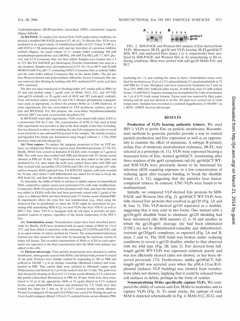
FIG. 1. Potential forms of Env on the HIV-1 membrane. gp120 is shown in red with the outer neutralizing face in light shading and the innernonneutralizing face in darker shading. Carbohydrate moieties are depicted as “tree”-like structures. gp41 is comprised of N-terminal (yellow) andC-terminal (green) transmembrane domains, separated by a disulfide-constrained loop. The membrane-proximal gp41 region exposed on thetrimer is depicted in dark green. A) Functional Env trimer; B) uncleaved gp160 precursor (depicted here as a trimer; however, it may also existas other oligomeric forms); C) gp120 shedding; D) alternative trimer isoform exposing the nonneutralizing face of gp120; E) gp120/gp41monomers.
2516 MOORE ET AL. J. VIROL.
on March 21, 2014 by guest
http://jvi.asm.org/
Dow
nloaded from
3-indolylphosphate (BCIP)/nitroblue tetrazolium (NBT) colorimetric reagents(Sigma-Aldrich).
(ii) BN-PAGE. To analyze Env derived from VLPs under native conditions, wedevised a modified BN-PAGE protocol (57, 60, 61). To liberate Env, VLPs wereincubated in an equal volume of solubilization buffer (0.12% Triton X-100 in 1mM EDTA/1.5 M aminocaproic acid) and one microliter of a protease inhibitorcocktail (Sigma). An equal volume of 2� sample buffer containing 100 mMmorpholinepropanesulfonic acid (MOPS), 100 mM Tris-HCl, pH 7.7, 40% glyc-erol, and 0.1% Coomassie blue was then added. Samples were loaded onto a 4to 12% Bis-Tris NuPAGE gel (Invitrogen). Ferritin (Amersham) was used as asize standard. Samples were electrophoresed at 4°C for 3 h at 100 V with 50 mMMOPS/50 mM Tris, pH 7.7, containing 0.002% Coomassie blue as cathode bufferand the same buffer without Coomassie blue as the anode buffer. The gel wasthen Western blotted onto polyvinylidene difluoride. Excess Coomassie blue dyewas removed after blotting by washing with 30% methanol/10% acetic acid then100% methanol.
The blot was then transferred to blocking buffer (4% nonfat milk in PBS) for30 min and probed using 1 �g/ml each of MAbs 2G12, b12, and 447-52D(anti-gp120 cocktail), or 20 �g/ml each of 4E10 and 2F5 (anti-gp41 cocktail).Goat anti-human and/or mouse Fc and Fab�2 alkaline phosphatase conjugateswere used, as appropriate, to detect the primary MAbs at 1:3,000 (Jackson). Insome experiments, Env was cross-linked on VLP membrane surfaces, prior tolysis and BN-PAGE. For this purpose, the cross-linker bis(sulfosuccinimidyl)suberate (BS3) was used, as previously described (19).
In BN-PAGE band shift experiments, VLPs were mixed with either sCD4 ora monovalent Fab for 5 min. The concentration of sCD4 or Fab used in bandshift experiments was recorded as that in the final sample at the time of loading.Env was detected as above, but omitting the anti-Fab conjugates in order to avoidcross-reactivity to any unbound Fab present in the samples. The density of nativeand liganded Env bands was determined using ImageJ software v. 1.33u (NIHfreeware; http://rsb.info.nih.gov/ij/).
(iii) Virus capture. To analyze the antigenic properties of Env on VLP sur-faces, we adopted the MAb-virus capture assay described previously (17, 50, 54).Briefly, MAbs were coated on Immulon II ELISA wells overnight at 5 �g/ml inPBS. The wells were then washed with PBS and blocked with 3% bovine serumalbumin in PBS for 30 min. VLP supernatant was then added to the plate andincubated for 3 h, after which the wells were washed three times with PBS andthen overlaid with susceptible CF2.CD4Th.synCCR5 (33) cell targets (104 cells/well) to measure MAb-captured virus. For SOS-VLP capture, cells were overlaidfor 30 min, after which 5 mM dithiothreitol was added for 10 min to break theSOS bond (6), and then the medium was changed.
To determine the ability of MAb in solution to inhibit capture by immobilizedMAb, competitive capture assays were performed (54), with some modifications.Competitive MAb (10 �g/ml) was first premixed with virus, and then the mixturewas added to ELISA wells that had been previously coated with 5 �g/ml of thesame or a different MAb and then blocked with 3% bovine serum albumin/PBS.In experiments where the virus Env was noninfectious (e.g., when using theuncleaved Env to pseudotype or when the VLPs might be neutralized by pre-mixing with neutralizing MAb b12), we used VLPs that bear VSV-G as well asHIV-1 Env. Since VSV-G is fusogenic with all cell types, it provides an inde-pendent readout of capture, regardless of the fusion competency of the HIV-1Env.
(iv) Neutralization assays. Neutralization assays have been described previ-ously (6). Briefly, VLPs were incubated with an equal volume of MAb for 1 h at37°C and then added to microtiter wells containing CF2.CD4Th.synCCR5 cellsin an equal volume of culture medium for 2 hours. The nonneutralized infectiousfraction was then assayed two days later by measuring the luciferase activity intarget cell lysates. The recorded concentration of MAb or sCD4 in each exper-iment was expressed as the final concentration after the MAb-virus mixture wasadded to the cells.
(v) Electron microscopy. Virus particle preparations were attached to carbonmembranes, subsequently reacted with MAbs, and labeled with protein G-coated10 nm gold. Particles were initially washed by suspending in 200 �l PBS andpelleted at 100,000 � g in an Airfuge centrifuge (Beckman Coulter) and resus-pended in PBS. Thin carbon films were attached to 600-mesh copper grids(Polysciences) and floated on 5 �l of the washed virus for 15 min. The grids werethen blocked by floating on 20 �l of 0.1% bovine serum albumin, 0.1% cold waterfish gelatin (Amersham Biosciences) in PBS for 30 min. Grids were then trans-ferred to 10 �l of the appropriate MAb at 10 �g/ml diluted in 0.1% acylatedbovine serum albumin/PBS (Aurion) and incubated for 2 h. Grids were thenwashed five times for 2 min on 20 �l 0.1% acylated bovine serum albumin.Protein G-conjugated 10-nm gold (Ted Pella) was reacted by floating the grid on10 �l of gold conjugate diluted 1:20 in 0.1% acylated bovine serum albumin /PBS,
incubating for 1 h, and washing five times as above. Gold-labeled viruses werefixed by incubation on 10 �l of 2.5% glutaraldehyde/1% paraformaldehyde in 75mM PBS for 15 min. Phosphate ions were removed by incubation for 5 min on20 �l 20% BSB (20% buffered saline borate, 20 mM boric acid, 0.5 mM sodiumborate, 15 mM NaCl). Negative staining was accomplished by 2 min of incubationon 5 �l of 0.5% fresh uranyl formate. Excess stain was removed by filter paperblotting and the grid was allowed to air dry. All steps were carried out at roomtemperature. Samples were recorded at a nominal magnification of 100,000� ona JEOL 1200EX electron microscope.
RESULTS
Production of VLPs bearing authentic trimers. We usedHIV-1 VLPs to probe Env on particle membranes. Recombi-nant methods to generate particles provide a way to controlparticle production and Env expression as well as an opportu-nity to examine the effect of mutations. A subtype B primaryisolate Env of moderate neutralization resistance, JR-FL, wasselected as a prototype (6, 11). VLPs were expressed bearing atruncated form of Env, termed gp160�CT, terminating afterthree residues of the gp41 cytoplasmic tail (6). gp160�CT WT-and SOS-VLPs were previously shown to be functional forinfection (SOS requiring exposure to a low concentration ofreducing agent after receptor binding, to break the disulfidebond) (3, 6, 52), suggesting that the particles bear fusion-competent trimers. In contrast, UNC-VLPs were found to benonfunctional.
Initially, we compared VLP-derived Env proteins by SDS-PAGE and Western blot (Fig. 2). gp160�CT WT-VLPs borefully cleaved Env proteins that resolved as gp120 (Fig. 2A andB, lane 3). This VLP-derived gp120 separated as a doublet,suggesting that it may exist in two forms. One mutant with agp120-gp41 disulfide bond to eliminate gp120 shedding hadbeen introduced (the SOS mutant) (2, 6, 9) and another inwhich the gp120-gp41 cleavage site had been eliminated(UNC) (6) led to dithiothreitol-reducible and dithiothreitol-resistant gp120/gp41 complexes, as expected (Fig. 2A and B,lanes 2 and 4). The SOS bond was broken under reducingconditions to reveal a gp120 doublet, similar to that observedwith the wild type (Fig. 2B, lane 2). Env derived from full-length gp160 WT-VLPs was expressed relatively poorly andwas less efficiently cleaved (data not shown), as has been ob-served previously (72). Furthermore, unlike gp160�CT, full-length gp160 was secreted even when the pNL4-3.Luc.R-E-plasmid (induces VLP budding) was omitted from transfec-tions (data not shown), implying that it could be released fromcell surfaces as debris, perhaps in the form of vesicles.
Nonneutralizing MAbs specifically capture VLPs. We com-pared the ability of various anti-Env MAbs to neutralize and tocapture VLPs (Fig. 3). To assist clarity, the epitope of eachMAb is depicted schematically in Fig. 4. MAbs b12, 2G12, and
FIG. 2. SDS-PAGE and Western blot analysis of Env derived fromVLPs. Monomeric JR-FL gp120 and VLPs bearing JR-FLgp160�CTSOS, WT, and uncleaved Envs (lanes 1 to 4, respectively) were ana-lyzed by SDS-PAGE and Western blot in A) nonreducing or B) re-ducing conditions. Blots were probed with anti-gp120 MAbs PA1 andB12.
VOL. 80, 2006 NONFUNCTIONAL Env ON HIV PARTICLE SURFACES 2517
on March 21, 2014 by guest
http://jvi.asm.org/
Dow
nloaded from
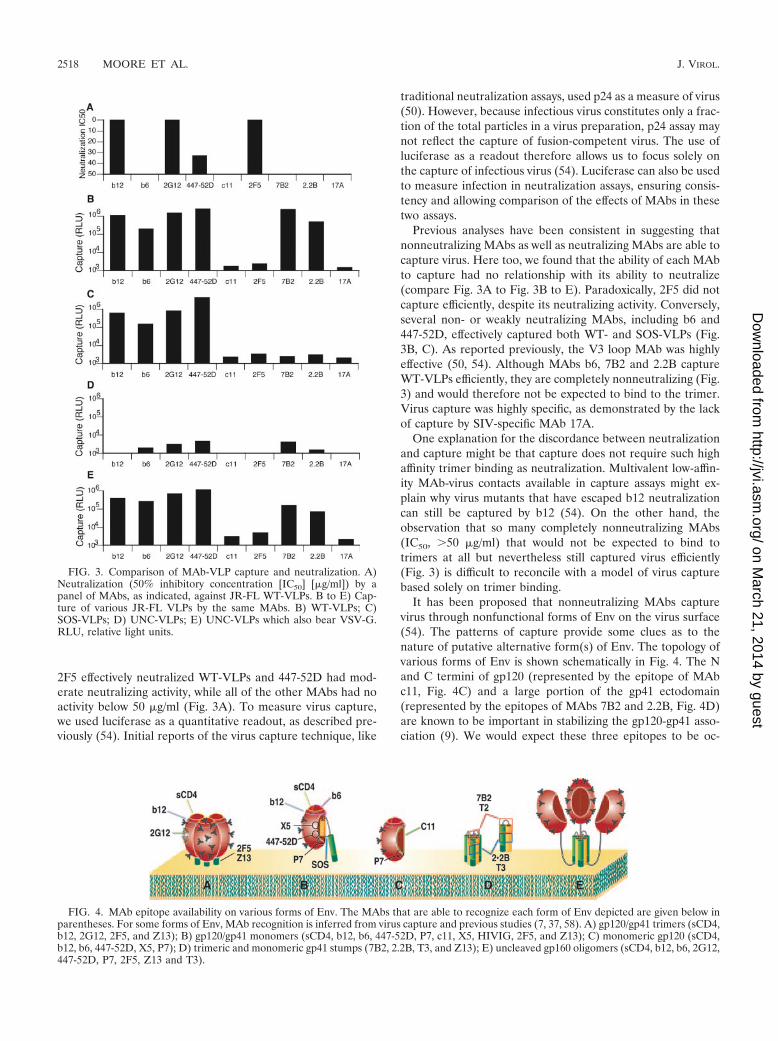
2F5 effectively neutralized WT-VLPs and 447-52D had mod-erate neutralizing activity, while all of the other MAbs had noactivity below 50 �g/ml (Fig. 3A). To measure virus capture,we used luciferase as a quantitative readout, as described pre-viously (54). Initial reports of the virus capture technique, like
traditional neutralization assays, used p24 as a measure of virus(50). However, because infectious virus constitutes only a frac-tion of the total particles in a virus preparation, p24 assay maynot reflect the capture of fusion-competent virus. The use ofluciferase as a readout therefore allows us to focus solely onthe capture of infectious virus (54). Luciferase can also be usedto measure infection in neutralization assays, ensuring consis-tency and allowing comparison of the effects of MAbs in thesetwo assays.
Previous analyses have been consistent in suggesting thatnonneutralizing MAbs as well as neutralizing MAbs are able tocapture virus. Here too, we found that the ability of each MAbto capture had no relationship with its ability to neutralize(compare Fig. 3A to Fig. 3B to E). Paradoxically, 2F5 did notcapture efficiently, despite its neutralizing activity. Conversely,several non- or weakly neutralizing MAbs, including b6 and447-52D, effectively captured both WT- and SOS-VLPs (Fig.3B, C). As reported previously, the V3 loop MAb was highlyeffective (50, 54). Although MAbs b6, 7B2 and 2.2B captureWT-VLPs efficiently, they are completely nonneutralizing (Fig.3) and would therefore not be expected to bind to the trimer.Virus capture was highly specific, as demonstrated by the lackof capture by SIV-specific MAb 17A.
One explanation for the discordance between neutralizationand capture might be that capture does not require such highaffinity trimer binding as neutralization. Multivalent low-affin-ity MAb-virus contacts available in capture assays might ex-plain why virus mutants that have escaped b12 neutralizationcan still be captured by b12 (54). On the other hand, theobservation that so many completely nonneutralizing MAbs(IC50, �50 �g/ml) that would not be expected to bind totrimers at all but nevertheless still captured virus efficiently(Fig. 3) is difficult to reconcile with a model of virus capturebased solely on trimer binding.
It has been proposed that nonneutralizing MAbs capturevirus through nonfunctional forms of Env on the virus surface(54). The patterns of capture provide some clues as to thenature of putative alternative form(s) of Env. The topology ofvarious forms of Env is shown schematically in Fig. 4. The Nand C termini of gp120 (represented by the epitope of MAbc11, Fig. 4C) and a large portion of the gp41 ectodomain(represented by the epitopes of MAbs 7B2 and 2.2B, Fig. 4D)are known to be important in stabilizing the gp120-gp41 asso-ciation (9). We would expect these three epitopes to be oc-
FIG. 3. Comparison of MAb-VLP capture and neutralization. A)Neutralization (50% inhibitory concentration [IC50] [�g/ml]) by apanel of MAbs, as indicated, against JR-FL WT-VLPs. B to E) Cap-ture of various JR-FL VLPs by the same MAbs. B) WT-VLPs; C)SOS-VLPs; D) UNC-VLPs; E) UNC-VLPs which also bear VSV-G.RLU, relative light units.
FIG. 4. MAb epitope availability on various forms of Env. The MAbs that are able to recognize each form of Env depicted are given below inparentheses. For some forms of Env, MAb recognition is inferred from virus capture and previous studies (7, 37, 58). A) gp120/gp41 trimers (sCD4,b12, 2G12, 2F5, and Z13); B) gp120/gp41 monomers (sCD4, b12, b6, 447-52D, P7, c11, X5, HIVIG, 2F5, and Z13); C) monomeric gp120 (sCD4,b12, b6, 447-52D, X5, P7); D) trimeric and monomeric gp41 stumps (7B2, 2.2B, T3, and Z13); E) uncleaved gp160 oligomers (sCD4, b12, b6, 2G12,447-52D, P7, 2F5, Z13 and T3).
2518 MOORE ET AL. J. VIROL.
on March 21, 2014 by guest
http://jvi.asm.org/
Dow
nloaded from
cluded when gp120 and gp41 are properly associated (as in Fig.4A and B). We found that c11 was unable to capture (Fig. 3),suggesting that no forms of Env exist on particles that resemblemonomeric gp120 dissociated from gp41 (Fig. 4C). However,the capture of WT-VLPs by 7B2 and 2.2B suggests that gp120shedding exposes gp41 stumps (Fig. 2B), perhaps explainingwhy antibodies with these specificities are so common in HIV-positive sera (13). The failure of 7B2 and 2.2B to captureSOS-VLPs (Fig. 3C) is fully consistent, because the SOS mu-tation eliminates gp120 shedding, so that these epitopes re-main sequestered (Fig. 4B).
Effective readout of UNC-VLP virus capture was problem-atic because the uncleaved mutation rendered the particlesnoninfectious (Fig. 3D). As a solution, we generated UNC-VLPs that also bear VSV-G protein. VSV-G is a highly fuso-genic amphotropic molecule that amplifies the readout of cap-tured virus, irrespective of the functional status of the HIVspikes through which capture actually occurs. The patternUNC�VSV-G-VLP capture was found to be similar to WT-VLPs, although 7B2 and 2.2B were less efficient (Fig. 3E). Thesimilarity of WT- and UNC-VLPs in virus capture contrastswith studies suggesting that cleavage does affect epitope expo-sure (9, 52).
In keeping with these earlier analyses, we believe cleavagedoes in fact significantly affect epitope exposure, for reasonsthat will become clear in further experiments outlined below,using BN-PAGE. To determine whether the discordance be-tween capture and neutralization extends to any other lentivi-ral Envs, we evaluated SIVmac239 gp160�CT WT-VLPs. Sev-eral anti-SIV MAbs, and the recombinant chimeric protein,CD4-IgG2, effectively captured these VLPs (not shown). How-ever, only the CD4-IgG2 molecule was able to neutralize.Thus, virus capture by nonneutralizing MAbs may be a generalphenomenon for lentiviruses.
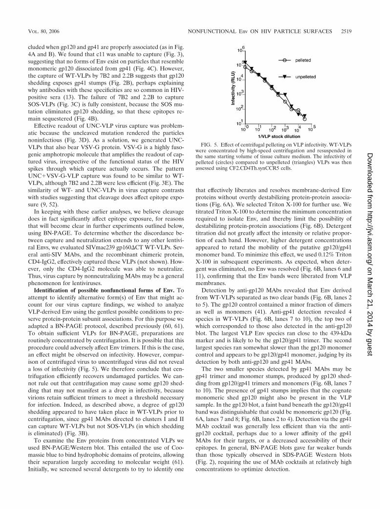
Identification of possible nonfunctional forms of Env. Toattempt to identify alternative form(s) of Env that might ac-count for our virus capture findings, we wished to analyzeVLP-derived Env using the gentlest possible conditions to pre-serve protein-protein subunit associations. For this purpose weadapted a BN-PAGE protocol, described previously (60, 61).To obtain sufficient VLPs for BN-PAGE, preparations areroutinely concentrated by centrifugation. It is possible that thisprocedure could adversely affect Env trimers. If this is the case,an effect might be observed on infectivity. However, compar-ison of centrifuged virus to uncentrifuged virus did not reveala loss of infectivity (Fig. 5). We therefore conclude that cen-trifugation efficiently recovers undamaged particles. We can-not rule out that centrifugation may cause some gp120 shed-ding that may not manifest as a drop in infectivity, becausevirions retain sufficient trimers to meet a threshold necessaryfor infection. Indeed, as described above, a degree of gp120shedding appeared to have taken place in WT-VLPs prior tocentrifugation, since gp41 MAbs directed to clusters I and IIcan capture WT-VLPs but not SOS-VLPs (in which sheddingis eliminated) (Fig. 3B).
To examine the Env proteins from concentrated VLPs weused BN-PAGE/Western blot. This entailed the use of Coo-massie blue to bind hydrophobic domains of proteins, allowingtheir separation largely according to molecular weight (61).Initially, we screened several detergents to try to identify one
that effectively liberates and resolves membrane-derived Envproteins without overtly destabilizing protein-protein associa-tions (Fig. 6A). We selected Triton X-100 for further use. Wetitrated Triton X-100 to determine the minimum concentrationrequired to isolate Env, and thereby limit the possibility ofdestabilizing protein-protein associations (Fig. 6B). Detergenttitration did not greatly affect the intensity or relative propor-tion of each band. However, higher detergent concentrationsappeared to retard the mobility of the putative gp120/gp41monomer band. To minimize this effect, we used 0.12% TritonX-100 in subsequent experiments. As expected, when deter-gent was eliminated, no Env was resolved (Fig. 6B, lanes 6 and11), confirming that the Env bands were liberated from VLPmembranes.
Detection by anti-gp120 MAbs revealed that Env derivedfrom WT-VLPs separated as two clear bands (Fig. 6B, lanes 2to 5). The gp120 control contained a minor fraction of dimersas well as monomers (41). Anti-gp41 detection revealed 4species in WT-VLPs (Fig. 6B, lanes 7 to 10), the top two ofwhich corresponded to those also detected in the anti-gp120blot. The largest VLP Env species ran close to the 439-kDamarker and is likely to be the gp120/gp41 trimer. The secondlargest species ran somewhat slower than the gp120 monomercontrol and appears to be gp120/gp41 monomer, judging by itsdetection by both anti-gp120 and gp41 MAbs.
The two smaller species detected by gp41 MAbs may begp41 trimer and monomer stumps, produced by gp120 shed-ding from gp120/gp41 trimers and monomers (Fig. 6B, lanes 7to 10). The presence of gp41 stumps implies that the cognatemonomeric shed gp120 might also be present in the VLPsample. In the gp120 blot, a faint band beneath the gp120/gp41band was distinguishable that could be monomeric gp120 (Fig.6A, lanes 7 and 8; Fig. 6B, lanes 2 to 4). Detection via the gp41MAb cocktail was generally less efficient than via the anti-gp120 cocktail, perhaps due to a lower affinity of the gp41MAbs for their targets, or a decreased accessibility of theirepitopes. In general, BN-PAGE blots gave far weaker bandsthan those typically observed in SDS-PAGE Western blots(Fig. 2), requiring the use of MAb cocktails at relatively highconcentrations to optimize detection.
FIG. 5. Effect of centrifugal pelleting on VLP infectivity. WT-VLPswere concentrated by high-speed centrifugation and resuspended inthe same starting volume of tissue culture medium. The infectivity ofpelleted (circles) compared to unpelleted (triangles) VLPs was thenassessed using CF2.CD4Th.synCCR5 cells.
VOL. 80, 2006 NONFUNCTIONAL Env ON HIV PARTICLE SURFACES 2519
on March 21, 2014 by guest
http://jvi.asm.org/
Dow
nloaded from

We next asked whether the species observed in Fig. 6 aretruly present in intact particles, or if they arise from dissocia-tion of the weakest protein-protein associations during samplepreparation for BN-PAGE. To investigate this, we incubatedWT- or SOS-VLPs with a cross-linker (Fig. 7). Effective cross-linking of WT-VLPs was confirmed by subjecting samples todenaturing and reducing conditions and probing with anti-gp120 MAbs, revealing three bands (Fig. 7A, compare lanes 5to lane 6). These bands are likely to be trimers, dimers andmonomers. The dimers probably reflect incompletely cross-linked trimer. The existence of a distinct monomer band evenin the conditions of excess cross-linker is consistent with itbeing present on intact particles (Fig. 7A, lane 6). Withoutcross-linker, Env resolved as a gp120 doublet (Fig. 7A, lane 5),as observed previously (Fig. 2).
In native conditions, cross-linking increased the proportionof trimer (Fig. 7A, compare lanes 1 to lane 2) but did noteliminate the monomer. Similarly, probing blots with gp41-specific antibodies revealed gp41 stumps in the wild type (vis-ible as faint bands in Fig. 7B, lanes 1 and 2), but not SOS (Fig.7B, lanes 3 and 4), regardless of cross-linking. Thus, althoughthe conditions of BN-PAGE may cause some trimer dissocia-tion, the gp120/gp41 monomer and gp41 stumps appear to existon the surface of intact particles.
The SOS mutation was useful to simplify the identificationof any putative defective form(s) of Env that did not arise from
gp120 shedding. When probed with anti-gp120 MAbs, SOSEnv also resolved as a mixture of gp120/gp41 trimers andmonomers (Fig. 7A, lanes 3 and 4). Denaturing/reducing con-ditions, as for WT, resulted in a gp120 doublet (Fig. 7A, lane7). The smallest two bands in the wild type probed with anti-gp41 MAbs (Fig. 7B, compare lanes 1 and 3) do not appear inSOS, consistent with their proposed identity as gp41 stumps.
To determine if the nonfunctional species is present on thesurface of other virus isolates and, moreover, if it is relevant tolive viruses, we examined concentrated, inactivated prepara-tions of HIV-1ADA and HIV-1MN that had been grown inSupT1 cell lines in BN-PAGE. Like JR-FL VLPs, HIV-1ADA
and HIV-1MN bore Env monomers and trimers (Fig. 8A andB). The monomers are likely to be gp120/gp41 and not solelymonomeric (shed gp120), since they were detected by the anti-gp41 cocktail (Fig. 8B). The different migration properties oftrimers and monomers may arise from variation in sequenceand/or glycosylation. The MN trimer migrated relatively slowlyin BN-PAGE compared to the other Envs (Fig. 8A and B,compare lane 3 to lanes 1 and 2), perhaps because MN trimersare less compact. To control for a possible producer cell effect,we also examined PBMC-grown JR-CSF and found that thisalso exhibited trimers and monomers (Fig. 8C). Finally, inac-tivated SIVmac239 particles also showed trimer and monomerbands, implying that the monomer is likely to be a broadlyconserved species (not shown). Overall, these data indicatethat nonfunctional forms of Env are relevant to infectiousHIV.
Band shift assays visualize sCD4 binding to native Env. Toinvestigate the possibility that the nontrimer Env species might
FIG. 6. Effect of detergent type and concentration on the pattern of Env separation by BN-PAGE. A) Various detergents were used tosolubilize WT-VLPs, as follows: 0.5% Tween 80 (lane 1); 0.5% Tween 20 (lane 2); 10 mM �-octylglucoside (lane 3); 0.25% each of Igepal andTriton X-100 (lane 4); 0.25% Igepal (lane 5); 0.25% Triton X-100 (lane 6); 0.25% sodium deoxycholate (lane 7); 25 mM CHAPS (cholamido-propyldimethylammoniopropanesulfonate) (lane 8). Monomeric gp120 was included as a control (lane 9). B) Progressively lower concentrationsof Triton X-100 were used to lyse WT-VLPs in each lane, as indicated. Monomeric gp120 was included as a control in lane 1. Western blots wereprobed with the anti-gp120 cocktail (b12, 447-52D, and 2G12) (lanes 1 to 6) or an anti-gp41 cocktail (2F5 and 4E10) (lanes 7 to 10).
FIG. 7. Effect of cross-linking on the proportion of functional andnonfunctional forms of Env. WT-VLPs (lanes 1, 2, 5, and 6 of part A;lanes 1 and 2 of part B) and SOS-VLPs (lanes 3, 4, 7, and 8 of part A;lanes 3 and 4 of part B) were incubated with (lanes 2, 4, 6, and 8) orwithout (lanes 1, 3, 5, and 7) 1 mM BS3 before BN-PAGE. Monomericgp120 was included as a control (lane 9). Samples in lanes 5 to 8 wereboiled in 1% SDS/50 mM dithiothreitol prior to loading. Western blotswere probed with A) the anti-gp120 cocktail (b12, 447-52D, and 2G12)or B) the anti-gp41 cocktail (2F5 and 4E10).
FIG. 8. Behavior of inactivated HIV preparations in BN-PAGE.Env from VLPs and virus preparations was compared. A) JR-FLgp160�CT WT-VLPs (lane 1); HIV-1ADA (lane 2); and HIV-1MN (lane3) Western blots were probed with an anti-gp120 cocktail, consisting ofb12, 447-52D, and 2G12. B) The same samples in panel A were probedwith the anti-gp41 cocktail of 2F5 and 4E10. C) JR-FL WT-VLPs (lane1) and peripheral blood mononuclear cell-produced JR-CSF (lane 2)were probed with the anti-gp120 cocktail.
2520 MOORE ET AL. J. VIROL.
on March 21, 2014 by guest
http://jvi.asm.org/
Dow
nloaded from

account for the ability of nonneutralizing MAbs to capturevirus, we wished to probe their topology using MAbs in BN-PAGE. Probing Western blot strips with various MAbs failedto show distinctions in MAb reactivities with gp120/gp41 trim-ers and monomers, probably because the Env is partially de-natured during blotting (not shown). A native band shift BN-PAGE protocol was therefore developed to determine the truereactivity patterns of Env on intact particles. We initially testedthe binding of two-domain and four-domain sCD4 to WT-VLPs (Fig. 9). The monomer and trimer bands were shifted assCD4 bound and retarded their migration (Fig. 9). The mag-nitude of the shift was proportional to the size of the sCD4ligand (molecular weight for two-domain sCD4 was for �25kDa; four-domain sCD4, �50 kDa). Similar results using HIV-1ADA and HIV-1MN preparations (not shown) demonstratedthat the technique was broadly applicable.
Trimer binding correlates with neutralization whereas non-neutralizing MAbs recognize nonfunctional Env. MAb bindingassays using cells expressing surface Env have provided evi-dence for a correlation between neutralization and binding totrimers. However, the binding of some nonneutralizing MAbsrevealed some apparent contradictions (28, 52). Nonneutraliz-ing MAb binding might stem from heterogeneity of Env on cellsurfaces, including uncleaved gp160 or other nonnative spe-cies. Examining MAb binding using BN-PAGE band shift as-says might have the advantage of visually distinguishing thetrimer from other forms of Env.
To develop an MAb-based band shift protocol, Env-MAbcomplexes should be detected via an Env component, so thatliganded and unliganded Env fractions can be distinguished.To prevent interference by unbound MAb ligand in the sam-ple, we used Fabs to form Env-Fab complexes and detected theEnv component in blots using whole gG MAbs, followed by ananti-Fc conjugate. The use of monovalent Fabs also eliminatedthe possibility of multivalent Env-MAb complexes that mightbe difficult to identify.
We analyzed the patterns of MAb band shifts with WT-,SOS-, and UNC-VLPs and HIV-1MN (Fig. 10). Western blotswere probed either with anti-gp120 (Fig. 10A, B, E, and F) oranti-gp41 cocktails (Fig. 10C and D). The identity of the gp120/gp41 monomer band in WT-VLPs and HIV-1MN was con-firmed by the observation that monomer shifts caused by gp41MAbs Z13e1 and 2F5 could be detected by gp120 MAbs (Fig.10A and E, lanes 9 and 10) and complementary data showingthat monomer shifts caused by gp120 MAbs were detectable bythe anti-gp41 cocktail (Fig. 10C, lanes 2 to 7).
Bacterially produced Fabs (b12, b6, P7, X5, Z13e1, T2, andT3) gave cleaner shifts than Fabs prepared from IgG by papaindigestion, probably because the former are inherently homog-enous and free of any contaminating Fc. In general, blots fromband shift experiments were slightly fainter than from regularBN-PAGE, because only anti-Fc conjugate was used (i.e., noanti-Fab conjugate) and also because, in some cases, Fab li-gands obscure the epitopes of whole IgG probes used fordetection. Overall, there was good consistency in the shiftpatterns of WT-VLPs and SOS-VLPs (Fig. 10A to D). TheSOS monomer species was generally more distinct, probablybecause gp120 shedding is eliminated. SOS-VLPs are there-fore particularly useful in determining the patterns of MAbbinding to the gp120/gp41 monomer.
A comparison of the neutralizing and nonneutralizing pro-totype Fabs, b6 and b12, showed that both were able to shiftmonomers, but only the neutralizing Fab b12 shifted trimers(Fig. 10A to E, lanes 2 and 3). In contrast, b6 shifted theoligomeric uncleaved Env species (Fig. 10F, lane 3), consistentwith exposure of the nonneutralizing face of gp120 (Fig. 10E)(9, 52). Analysis of other Fabs revealed that those that shiftednative trimers (2G12, 2F5, and Z13e1) also had strong neu-tralizing activity (Fig. 10A to E, lanes 4, 9, and 10). Of partic-ular interest, 2G12 induced “supershifts” compared to otherFabs, consistent with the notion that 2G12 exhibits a unique“domain exchange” conformation (16), in which the Fab armsare locked together in an unusually large 100-kDa Fab-Fabcomplex.
Fabs 447-52D and X5 appeared to partially shift SOS trim-ers (Fig. 10B and D, lanes 5 and 7). They did not, however,cause convincing shifts of WT-VLP trimers. This difference isreflected by a slightly increased neutralization sensitivity ofSOS-VLPs compared to WT-VLPs to neutralization by certainMAbs (6). It is possible that MAb 447-52D neutralizationoccurs when it binds to trimers after CD4 engagement (39).However, band shift assays are limited to the native form oftrimer and cannot fully recapitulate the patterns of epitopeexposure that may occur during infection, when epitopes suchas that of 447-52D might become exposed. The lack of X5neutralization contrasts with a previous report using virusesbearing full-length JR-FL Env (37). This may be becausegp160�CT Env fuses more rapidly than full-length gp160 (3),reducing the availability of fusion intermediates for X5 toneutralize. HIVIG is derived from pooled human HIV-positivesera and almost certainly contains a small fraction of neutral-izing antibodies. However, these neutralizing antibodies ap-pear to not be at a concentration high enough to shift thetrimer in BN-PAGE, as evidenced by the fact that the IC50 ofHIVIG exceeds 40 �g/ml.
Compared to the trimer, the monomeric gp120/gp41 speciesexhibited completely different binding patterns. Almost allFabs were able to mediate shifts. The only exceptions were thegp41-specific Fabs T2 and T3 (Fig. 10A to D), whose epitopesappear to be obscured by proper gp120/gp41 association, asobserved in SOS-VLP capture (Fig. 3C). However, MAb T3recognized gp41 stumps from which gp120 had dissociated, asevidenced by a loss in native gp41 trimer stumps, accompaniedby a gain in signal close to the gp120/gp41 monomer (Fig. 10C,lane 12). HIVIG and Z13e1 also appeared to shift the gp41stumps, while 2F5 and T2 did not.
FIG. 9. Analysis of sCD4 binding to native Env proteins in BN-PAGE band shifts. SOS-VLPs were incubated either alone (lane 1) orwith two-domain (lane 2) or four-domain (lane 3) sCD4 at a finalconcentration of 20 �g/ml. Samples were then separated by BN-PAGE. Western blots were probed with an anti-gp120 MAb cocktailcontaining 2G12, b12, and 447-52D. Ferritin 12-mer (220 kDa) and24-mer (439 kDa) served as molecular size markers.
VOL. 80, 2006 NONFUNCTIONAL Env ON HIV PARTICLE SURFACES 2521
on March 21, 2014 by guest
http://jvi.asm.org/
Dow
nloaded from

HIVMN shifts showed a similar pattern to JR-FL WT-VLPs(Fig. 10, compare parts A, C, and E). Because of the slowermobility of MN trimers (observed above in Fig. 8), electro-phoresis was run longer to better resolve the bands. MAbs b12,2G12, 2F5, and Z13e1 all clearly shifted MN trimers (Fig. 10E,lanes 2, 4, 9, and 10). The monomer bands were more difficultto resolve, as was discerning any shifts. However, close inspec-tion of lanes containing ligands (Fig. 10E, lanes 2 to 12) com-pared to the unliganded control (Fig. 10E, lane 1) reveals thatmany of the Fabs (particularly lanes 4, 6, 8, 9, and 10) causedshifts similar to JR-FL WT-VLPs.
The oligomeric species of uncleaved protein exhibited a dif-ferent pattern compared to native trimers and was recognizedby all the Fabs except T2 (Fig. 10F, lane 11). This highlights theimportance of gp120/gp41 cleavage in the proper folding ofEnv and indicates possible limitations in vaccine approachesthat involve uncleaved Env proteins. However, the monomericuncleaved species behaved very similarly to the monomersfrom WT-VLPs bearing cleaved Env and may possibly derivefrom a small amount of cleaved material in UNC-VLPs (Fig.2). The similarities in overall behavior of uncleaved andcleaved gp120/gp41 monomers in WT-VLPs could explain the
similarities noted earlier in the patterns of behavior betweenUNC- and WT-VLPs in virus capture (Fig. 3).
To investigate a possible quantitative relationship betweenneutralization and trimer binding, graded concentrations offour-domain sCD4 were added to WT-VLPs in parallel bandshift and neutralization assays. The IC50 of trimer shift (0.5�g/ml) and neutralization (IC50 0.8 �g/ml) closely corre-sponded (Fig. 10G and H). MAbs b12, 2G12, 2F5, and Z13e1trimer shift IC50s similarly corresponded with neutralizationIC50s (not shown). As documented above, monomer bindingdid not correlate with neutralization, because several Fabsrecognized the monomer without neutralizing the virus. How-ever, monomer binding did appear to correlate with virus cap-ture. In particular, strong monomer binding by Fabs 447-52D,2G12, and four-domain sCD4 reflected their efficient viruscapture ability (Fig. 2 and 4 and data not shown).
Fabs T2 and T3 bound neither to trimers nor to monomers,but were nevertheless able to capture WT-VLPs with moderateefficiency (data not shown; capture by MAbs 7B2 and 2.2B thatrecognize similar epitopes is shown in Fig. 3). Capture by FabT3 probably occurs through gp41 stumps (13), though it isunclear how T2 might capture. In contrast, Fabs 2F5 and
FIG. 10. MAb binding to native Env in BN-PAGE band shifts. WT-VLPs (A and C), SOS-VLPs (B and D), inactivated MN virus (E), andUNC-VLPs (F) were incubated with Fabs at a final concentration of 20 �g/ml and then processed for BN-PAGE. Western blots in panels A, B,E, and F were probed with the anti-gp120 cocktail containing 2G12, b12, and 447-52D; blots in panels C and D were probed with the anti-gp41cocktail containing 2F5 and 4E10. Labels: T, gp120/gp41 trimer; M, gp120/gp41 monomer; O, uncleaved oligomers (most likely dimers, trimers,and tetramers). The Fabs used (neutralization IC50 titers against JR-FL gp160�CT WT in �g/ml) are as follows: lane 1, no Fab; lane 2, b12 (0.1�g/ml); lane 3, b6 (�40 �g/ml); lane 4, 2G12 (0.5 �g/ml); lane 5, 447-52D (9 �g/ml); lane 6, P7 (�40 �g/ml); lane 7, X5 (�40 �g/ml); lane 8, HIVIG(�40 �g/ml); lane 9, 2F5 (8 �g/ml); lane 10, Z13e1 (19 �g/ml); lane 11, T2 (�40 �g/ml); lane 12, T3 (�40 �g/ml). In panels G and H four-domainsCD4 was titrated in band shifts (lanes 1 to 8 correspond to four-domain sCD4 concentrations of 0.003, 0.01, 0.03, 0.1, 0.3, 1, 3, and 10 �g/ml,respectively), and in a neutralization assay against WT-VLPs.
2522 MOORE ET AL. J. VIROL.
on March 21, 2014 by guest
http://jvi.asm.org/
Dow
nloaded from

Z13e1 bound monomer tightly (IC50 0.8 �g/ml each) butwere unable to capture WT-VLPs efficiently. It is possible thatthe 2F5/Z13 epitopes on gp120/gp41 monomers (Fig. 10) areaccessible to Fabs in solution, but when the Fabs are immobi-lized in capture wells, the gp120 moiety of gp120/gp41 mono-mers sterically limits access to the 2F5/Z13 epitopes on gp41.
Remaining questions concerning the existence of monomeron intact particles. It could still be argued that the monomerobserved in BN-PAGE is solely the product of treatment ofVLPs with mild detergent and conditions of BN-PAGE. Un-doubtedly, sample preparation does cause some trimer to fallapart into monomers (Fig. 7). To determine definitively ifmonomer is present on intact particles, it is important to elim-inate the possibility of MAb binding to forms of Env thatbecome available only after detergent treatment. This can beachieved by separating the MAb-virus incubation step fromdetergent treatment and sample preparation for BN-PAGE.Thus, we developed a new protocol. We incubated MAbs withSOS-VLPs, then pelleted virus and washed away any unboundMAb with two 1-ml washes of PBS, and then performed BN-PAGE. We prepared control samples in parallel, where VLPswere washed and spun in the same way, after which MAb wasadded, along with detergent, as per the original protocol. Inthe new format (Fig. 11, lanes 2 to 7), the neutralizing andnonneutralizing MAbs largely behaved as they did by the orig-inal protocol (Fig. 10B and Fig. 11, lanes 8 to 13). Fabs b12 and2G12 shifted both trimers and monomers and the nonneutral-izing Fab b6 shifted only the monomer. The fact that b6 rec-ognized the monomer in this format suggests that the mono-mer is likely to be available on intact particles. Someunliganded monomer that resolved in this format may havebeen the product of trimer dissociation during sample prepa-ration. Control Fab LS4 (Ebola virus GP-specific) did notcause any shift (Fig. 11, lanes 7 and 13).
Interestingly, Fab Z13WT failed to cause convincing shifts inthe new format (Fig. 11, lane 6), in contrast to the originalformat (Fig. 11, lane 12). This suggests that the Z13 epitopemay be sequestered on native spikes and becomes fully avail-able only after the Env trimers are liberated by detergent. Z13may therefore exhibit greater neutralizing activity after Env
has been activated, perhaps by receptor binding (6, 23). Thefact that Z13 behaves differently in our two protocols alsoconfirms that the washing procedure intended to remove un-bound Fab was effective, providing further evidence that b6binds to gp120/gp41 monomers on intact particles.
Immunoelectron microscopy reveals nonneutralizing MAbbinding to VLPs. We further investigated direct binding ofnonneutralizing MAbs to intact particles by immunoelectronmicroscopy. We compared b6 and b12 as prototype monomer-and monomer/trimer-reactive MAbs. Both MAbs bind to mo-nomeric JR-FL gp120 equally well (Fig. 12A), but only b12neutralizes JR-FL WT-VLPs (Fig. 12B). In electron micro-graphs, we used MAbs b6 and b12 at 10 �g/ml each, a con-centration at which both MAbs bind monomeric gp120 tosaturation (Fig. 12A), but only b12 neutralizes (Fig. 12B).MAb binding was detected by subsequent incubation with 10-nm gold particles coupled with protein G (Fig. 12C and D).
MAb b12 binding in fields encompassing 1,470 virions wasscored. Of 182 gold particles observed, 131 were associatedwith 120 virions (i.e., 8.2% of virions were labeled) and 51 goldparticles were not associated with virions. MAb b6 also boundto the particles, albeit to a lesser extent. Of 99 gold particlesobserved in fields encompassing 1,491 virions, 73 were associ-ated with 71 virions (i.e., 4.9% of virions were labeled) and 26gold particles were not associated with virions. The majority(�75%) of the gold not bound to particles appeared to beassociated with aggregated protein of cellular/viral debris andso may have been binding to legitimate target molecules. Rep-resentative examples of b12 and b6 immunogold-labeled par-ticles are shown (Fig. 12C and D). Similar results were ob-tained with inactivated BaL virus particles (data not shown). Acontrol in which virions were incubated with pooled normalintravenous IgG resulted in essentially no gold particle bindingto JR-FL VLPs (of 1,500 virions, only four gold particles werevirion associated and 17 gold particles were in the background,most of which were not associated with debris; not shown).This was also true for protein G-gold alone (not shown), con-firming the specificity of binding. The fact that b6 bound toJR-FL VLPs suggests that its target, the gp120/gp41 monomer(Fig. 10), is present on intact particles. The observation that b6binding was less pronounced compared to b12 is consistentwith b12’s having a greater range of targets, including bothgp120/gp41 trimers and monomers.
Reciprocal competitive binding relationships of b6 and b12in virus capture are consistent with the presence of gp120/gp41monomers on intact particles. Because b6 blocks b12 bindingto monomeric Env but not trimeric Env, it might be expectedthat neutralizing MAb b12 can capture virus even in the pres-ence of excess b6 (Fig. 10A and C, lanes 2 and 3). This wouldprovide further evidence that the monomer exists on intactparticles. To test this hypothesis, we analyzed MAb bindingrelationships in competitive virus capture assays, in which aninhibitory MAb was mixed with VLPs and then added to wellscoated with the same or a different MAb. A previous study (54)demonstrated that capture by b12 could be inhibited by b6.However, that study did not investigate the ability of b12 toinhibit b6 capture, because b12 competitor neutralized thevirus, and thus any virus captured by b6 was neutralized andtherefore could not be detected using HIV infectivity as areadout.
FIG. 11. Separation of the MAb-VLP binding step from detergenttreatment and sample preparation indicates that monomer is presenton intact particles. We further investigated whether gp120/gp41 mono-mers are a relevant species on intact particles by separating MAbbinding from sample preparation for BN-PAGE. Various Fabs wereincubated with SOS-VLPs either before (lanes 2 to 7) or after (lanes 8to 13) two washes with 1 ml PBS, after which VLPs were detergenttreated and run on BN-PAGE as usual (lane 1, JR-FL gp120; lanes 2and 8, no Fab; lanes 3 and 9, Fab b12; lanes 4 and 10, Fab b6; lanes 5and 11, Fab 2G12; lanes 6 and 12, Fab Z13 WT; lanes 7 and 13, FabLS4 directed to Ebola virus GP) for 1 h before being pelleted, washedtwo times to completely remove any unbound Fab (verified in parallelcontrols), and then the washed VLP sample was resuspended in PBSand prepared for BN-PAGE as shown.
VOL. 80, 2006 NONFUNCTIONAL Env ON HIV PARTICLE SURFACES 2523
on March 21, 2014 by guest
http://jvi.asm.org/
Dow
nloaded from
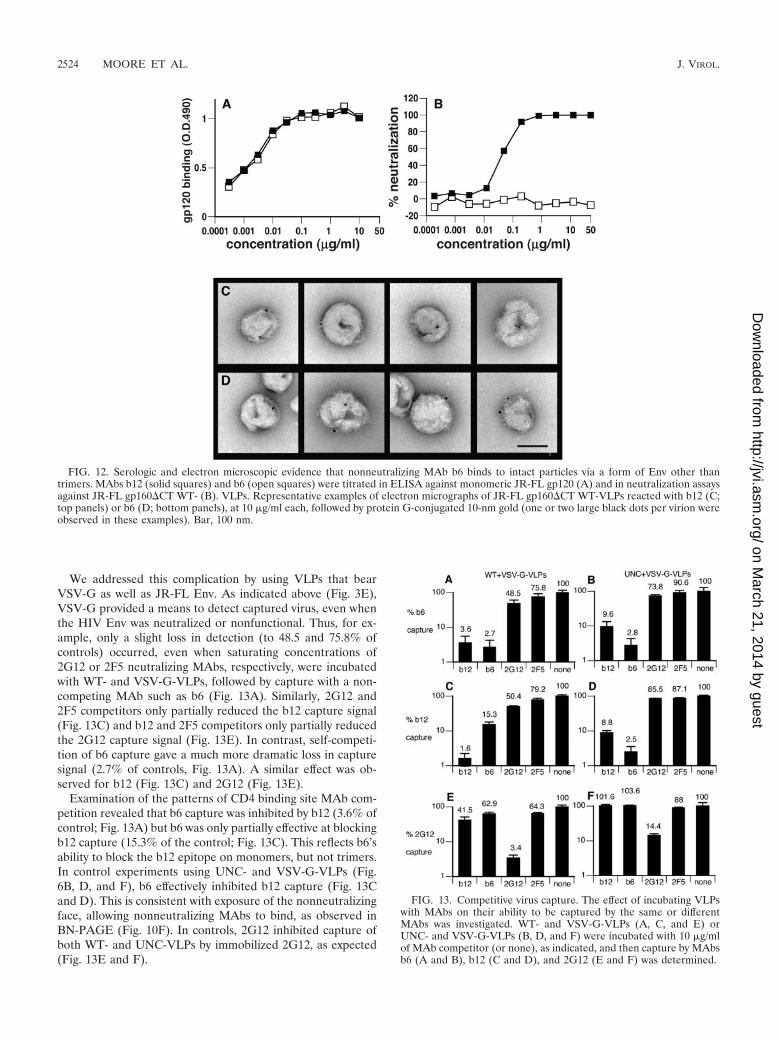
We addressed this complication by using VLPs that bearVSV-G as well as JR-FL Env. As indicated above (Fig. 3E),VSV-G provided a means to detect captured virus, even whenthe HIV Env was neutralized or nonfunctional. Thus, for ex-ample, only a slight loss in detection (to 48.5 and 75.8% ofcontrols) occurred, even when saturating concentrations of2G12 or 2F5 neutralizing MAbs, respectively, were incubatedwith WT- and VSV-G-VLPs, followed by capture with a non-competing MAb such as b6 (Fig. 13A). Similarly, 2G12 and2F5 competitors only partially reduced the b12 capture signal(Fig. 13C) and b12 and 2F5 competitors only partially reducedthe 2G12 capture signal (Fig. 13E). In contrast, self-competi-tion of b6 capture gave a much more dramatic loss in capturesignal (2.7% of controls, Fig. 13A). A similar effect was ob-served for b12 (Fig. 13C) and 2G12 (Fig. 13E).
Examination of the patterns of CD4 binding site MAb com-petition revealed that b6 capture was inhibited by b12 (3.6% ofcontrol; Fig. 13A) but b6 was only partially effective at blockingb12 capture (15.3% of the control; Fig. 13C). This reflects b6’sability to block the b12 epitope on monomers, but not trimers.In control experiments using UNC- and VSV-G-VLPs (Fig.6B, D, and F), b6 effectively inhibited b12 capture (Fig. 13Cand D). This is consistent with exposure of the nonneutralizingface, allowing nonneutralizing MAbs to bind, as observed inBN-PAGE (Fig. 10F). In controls, 2G12 inhibited capture ofboth WT- and UNC-VLPs by immobilized 2G12, as expected(Fig. 13E and F).
FIG. 12. Serologic and electron microscopic evidence that nonneutralizing MAb b6 binds to intact particles via a form of Env other thantrimers. MAbs b12 (solid squares) and b6 (open squares) were titrated in ELISA against monomeric JR-FL gp120 (A) and in neutralization assaysagainst JR-FL gp160�CT WT- (B). VLPs. Representative examples of electron micrographs of JR-FL gp160�CT WT-VLPs reacted with b12 (C;top panels) or b6 (D; bottom panels), at 10 �g/ml each, followed by protein G-conjugated 10-nm gold (one or two large black dots per virion wereobserved in these examples). Bar, 100 nm.
FIG. 13. Competitive virus capture. The effect of incubating VLPswith MAbs on their ability to be captured by the same or differentMAbs was investigated. WT- and VSV-G-VLPs (A, C, and E) orUNC- and VSV-G-VLPs (B, D, and F) were incubated with 10 �g/mlof MAb competitor (or none), as indicated, and then capture by MAbsb6 (A and B), b12 (C and D), and 2G12 (E and F) was determined.
2524 MOORE ET AL. J. VIROL.
on March 21, 2014 by guest
http://jvi.asm.org/
Dow
nloaded from
DISCUSSION
Why have gp120/gp41 monomers not been described previ-ously? Collectively, our data support gp120/gp41 monomers asa primary nonfunctional form of Env on the surface of HIV-1.Less surprisingly, gp41 stumps from which gp120 has shed alsodecorate the particle surfaces (13). The reason why monomershave not been previously reported may be partly due to a lackof techniques available to investigate this possibility. More-over, until recently, researchers had little reason to suspect thatalternative forms of Env exist on the HIV-1 surface. Indeed, itis widely perceived that strong gp41-gp41 associations effec-tively stabilize trimers (20). However, in native trimers, gp41 islikely to be in a “prefusion” conformation that may be some-what less stable than the fusion-active forms of gp41 previouslystudied. In fact, cryoelectron microscopic analysis of nativevirion-associated SIV Env spikes shows a pronounced gap sep-arating the three membrane-proximal segments (i.e., the gp41“stalk”) (Zhu et al., unpublished results). In fact, gp120-gp120associations may be just as important in stabilizing native tri-mers (36, 41).
Electron microscopy studies have shed some light on alter-native forms of Env on particle surfaces. Atomic force micros-copy revealed what were interpreted as Env “monomers” (34).However, paradoxically, this method did not identify trimers.In contrast, electron tomographs of HIV and SIV particlesrevealed distinct Env trimers (73), but no other forms deemedto be Env were observed. This may be because (i) monomersare difficult to resolve because of their smaller size and lessdistinctive structure, (ii) they are simply too few in number,and/or (iii) they cannot be distinguished from other virionsurface molecules. Indeed, electron micrographs of HIV-1 re-veal some small, less regular structures that might be mono-mers (73 and unreported data).
In retrospect, the observation that soluble gp140SOS readilydissociates into gp120/gp41 monomers (9, 61) provided a par-adigm for membrane-associated gp120/gp41 monomers ob-served here. There have been a few other hints in the literatureregarding the possibility of gp120/gp41 monomers. For exam-ple, one group suggested that there might be a monomer-trimer equilibrium (5), based on the proposed inhibitory mech-anism of a gp41 N-helix intercalating peptide that may bind tovirus if trimers, at least temporarily, dissociate into monomers.In addition, a study of cross-linked Env derived from HIVpreparations revealed a fraction of low-molecular-weight Envmaterial that, considering the new information presented here,might well have been gp120/gp41 monomers (18).
How does the gp120/gp41 monomer originate? Our analysissuggested the presence of gp120/gp41 monomers and gp41stumps on virus surfaces, but no unbalanced gp120/gp41 oli-gomers (e.g., gp41 trimeric stumps from which gp120 has onlypartially shed). This indicates that the trimer dissociates in aconcerted manner either at the gp120/gp41 interface (resultingin gp120 shedding) or at the vertices of oligomerization (toform gp120/gp41 monomers). While gp120 shedding probablyoccurs only after the virus has matured, it is possible that thegp120/gp41 monomer arises as a by-product of Env synthesis.Our detection of gp120 doublets in SDS-PAGE analysis ofVLP Env (Fig. 2) provides a possible lead. gp120 doublets wereobserved previously in virus-derived Env (51) but, so far, an
explanation for their existence has not been put forward. Thepresent data suggest that gp120 doublet bands might originatefrom gp120/gp41 trimers and gp120/gp41 monomers. If weassume that monomers exist during the late stages of Envsynthesis, we might expect that, due to the presumed increasedaccessibility of N-linked sites, they are glycosylated at a certainsite(s) not exposed on trimers. Thus, the more heavily glyco-sylated upper band of the doublet might derive from monomer,and the lower band from trimer.
The possibility that the monomer arises during Env synthesisis rendered even more plausible when one considers that theuncleaved Env precursor exists as a heterogeneous mix ofoligomers (Fig. 4F). It is possible that proteolytic cleavage ofthis mixed precursor leads to a similarly mixed final Env prod-uct of monomers and trimers. Other possibilities to account forthe emergence of a subspecies of monomer during Env syn-thesis include incomplete signal peptide cleavage and partialcleavage of gp120/gp41 at a putative secondary cleavage site(44). On the other hand, much like gp120 shedding from ma-ture virus, the gp120/gp41 monomer may progressively accu-mulate on the surface of mature virus. For example, premature“misfiring” of the metastable trimer complex or proteolyticcleavage of the V3 loop might induce dissociation of maturetrimers.
What are the consequences of nonfunctional Env? Sincegp120/gp41 monomers and gp41 stumps are not thought to beinvolved in virus binding and fusion, nonneutralizing antibod-ies may bind to virus without directly affecting virus infection.However, antibody coating of nonfunctional Env on particlesurfaces may have other consequences distinct from conven-tional neutralization (14). The recently described ability ofnonneutralizing antibodies to potently inhibit HIV-1 infectionof macrophages, most likely by Fc receptor-mediated phago-cytosis (32), could be explained by the availability of nonfunc-tional targets on the virus, to which the nonneutralizing anti-bodies bind. Thus, even in the absence of conventionalneutralization, antibody coating may impose certain restric-tions on the cell targets the virus can infect. In addition, non-functional Env may well explain early reports of high concen-trations of circulating immune complexes of infectious virus inHIV-positive subjects, despite the typically low concentrationof neutralizing antibodies in sera (27, 47). Furthermore, non-neutralizing antibody coating may prolong the persistence ofparticles by facilitating their trapping by follicular dendriticcells (30, 63).
Nonfunctional Env may also have consequences for vaccinedesign. Since MAb binding to trimers is associated with neu-tralization, it has been difficult to reconcile why particles bear-ing trimers elicit mostly nonneutralizing antibodies. As thecritical functional unit in viral infectivity, Env has been underintense pressure to evolve immune evasion tactics. The non-functional forms of Env we describe may prove to be oneimmune evasion tactic, serving to divert the attention of B cellsfrom trimers.
One hypothetical problem with nonfunctional forms of Envacting as decoys is that we might expect B cells to respondequally to monomers, trimers and gp41 stumps on particlesurfaces, in the same way that combination vaccines such asthat for measles, mumps, and rubella induce equivalent anti-body responses against each component as they do when each
VOL. 80, 2006 NONFUNCTIONAL Env ON HIV PARTICLE SURFACES 2525
on March 21, 2014 by guest
http://jvi.asm.org/
Dow
nloaded from
component is administered separately (68). However, the pre-sentation of the various forms of Env together on the sameparticle creates an entirely different scenario. Virus particlescan be considered as a single target. Affinity selection of ger-minal center B cells mandates the amplification of higher-affinity clones, at the expense of others. Therefore, the devel-opment of antibody responses toward each of the foreignantigens on particle surfaces might depend on their relativeaccessibility to antibody binding. Thus, the high accessibility ofnonfunctional forms of Env may explain their apparent immu-nodominance. Antibodies that react with gp120/gp41 mono-mer or gp41 stumps do not tend to cross-react with the trimer,probably because their epitopes impinge on the nonneutraliz-ing face (Fig. 4). Even B cells that recognize trimers tend tobind as well to monomers on the same particle and may losetheir trimer-binding property during further maturationagainst the monomer.
Another question raised by these data is the significance ofthe fact that the monomer is a minor species. It is possible thatthe immunogenicity of monomers depends on expressionabove a certain threshold. However, our evidence that themonomer appears to play a dominant role in virus capture andis a major target of HIVIG prepared from HIV-positive donorplasma is consistent with the monomer eliciting strong anti-body responses during natural infection. We therefore proposethat accessibility, not quantity, is the major factor driving therelative immunogenicity of monomers and trimers. We sug-gest, then, that trimers are a secondary target for antibodies,consistent with the slow development of autologous neutral-ization in natural infection. In accordance with this idea, wehave observed that immunizations of small animals with SOS-VLPs induced high titer anti-gp120 antibodies directed againstepitopes that are highly accessible in virus capture and onmonomers but ineffective at neutralizing the JR-FL parentvirus (J. Binley, unpublished).
An awareness of HIV-1’s full armory of evasion mechanismsmay provide new solutions for vaccine research (35, 55, 69).The observation that simple trimer binding predicts neutral-ization (Fig. 10) reaffirms this as the primary target for anti-body-based vaccine development. Devising ways to minimizeor eliminate responses to nonfunctional Env and, at the sametime, to focus responses on functional trimers is thereforeworth investigating in future vaccine research.
ACKNOWLEDGMENTS
This work was supported by NIH grants AI49566 and AI058763 andthe AIDS and Infectious Disease Science Center (J.M.B.), AI055461(K.H.R.), and AI33292 (D.R.B.). P.L.M. was supported through theColumbia University-Southern Africa Fogarty AIDS InternationalTraining and Research Program (grant number D.43 TW00231),funded by the Fogarty International Center. Additional support wasprovided by Progenics Pharmaceuticals, Inc.
We thank Rogier Sanders, Pascal Poignard, and Norbert Schulke forinsightful discussions and advice; Welkin Johnson for providing a SIV-mac316 molecular clone; Ned Landau for providing the pVSV-G plas-mid; John Mascola for providing HIVIG; Jeff Robinson, GeorgeLewis, James Hoxie, and Hermann Katinger for providing variousantibodies; Jeff Lifson and Larry Arthur for providing inactivated HIVand SIV preparations; Meng Wang and David Montefiori for provid-ing PBMC-produced viruses; the AIDS Reagent Repository for 447-52D and two-domain sCD4; and Michelle Nolasco for assistance withgraphic artwork.
REFERENCES
1. Abacioglu, Y. H., T. R. Fouts, J. D. Laman, E. Claassen, S. H. Pincus, J. P.Moore, C. A. Roby, R. Kamin-Lewis, and G. K. Lewis. 1994. Epitope map-ping and topology of baculovirus-expressed HIV-1 gp160 determined with apanel of murine monoclonal antibodies. AIDS Res. Hum. Retroviruses 10:371–381.
2. Abrahamyan, L. G., R. M. Markosyan, J. P. Moore, F. S. Cohen, and G. B.Melikyan. 2003. Human immunodeficiency virus type 1 Env with an inter-subunit disulfide bond engages coreceptors but requires bond reduction afterengagement to induce fusion. J. Virol. 77:5829–5836.
3. Abrahamyan, L. G., S. R. Mkrtchyan, J. Binley, M. Lu, G. B. Melikyan, andF. S. Cohen. 2005. The cytoplasmic tail slows the folding of human immu-nodeficiency virus type 1 Env from a late prebundle configuration into thesix-helix bundle. J. Virol. 79:106–115.
4. Allaway, G. P., K. L. Davis-Bruno, G. A. Beaudry, E. B. Garcia, E. L. Wong,A. M. Ryder, K. W. Hasel, M. C. Gauduin, R. A. Koup, J. S. McDougal, etal. 1995. Expression and characterization of CD4-IgG2, a novel heterotet-ramer that neutralizes primary HIV type 1 isolates. AIDS Res. Hum. Ret-rovir. 11:533–539.
5. Bewley, C. A., J. M. Louis, R. Ghirlando, and G. M. Clore. 2002. Design ofa novel peptide inhibitor of HIV fusion that disrupts the internal trimericcoiled-coil of gp41. J. Biol. Chem. 277:14238–14245.
6. Binley, J. M., C. S. Cayanan, C. Wiley, N. Schulke, W. C. Olson, and D. R.Burton. 2003. Redox-triggered infection by disulfide-shackled human immu-nodeficiency virus type 1 pseudovirions. J. Virol. 77:5678–5684.
7. Binley, J. M., H. J. Ditzel, C. F. Barbas 3rd, N. Sullivan, J. Sodroski, P. W.Parren, and D. R. Burton. 1996. Human antibody responses to HIV type 1glycoprotein 41 cloned in phage display libraries suggest three majorepitopes are recognized and give evidence for conserved antibody motifs inantigen binding. AIDS Res. Hum. Retrovir. 12:911–924.
8. Binley, J. M., P. J. Klasse, Y. Cao, I. Jones, M. Markowitz, D. D. Ho, andJ. P. Moore. 1997. Differential regulation of the antibody responses to Gagand Env proteins of human immunodeficiency virus type 1. J. Virol. 71:2799–2809.
9. Binley, J. M., R. W. Sanders, B. Clas, N. Schuelke, A. Master, Y. Guo, F.Kajumo, D. J. Anselma, P. J. Maddon, W. C. Olson, and J. P. Moore. 2000.A recombinant human immunodeficiency virus type 1 envelope glycoproteincomplex stabilized by an intermolecular disulfide bond between the gp120and gp41 subunits is an antigenic mimic of the trimeric virion-associatedstructure. J. Virol. 74:627–643.
10. Binley, J. M., R. W. Sanders, A. Master, C. S. Cayanan, C. L. Wiley, L.Schiffner, B. Travis, S. Kuhmann, D. R. Burton, S. L. Hu, W. C. Olson, andJ. P. Moore. 2002. Enhancing the proteolytic maturation of human immu-nodeficiency virus type 1 envelope glycoproteins. J. Virol. 76:2606–2616.
11. Binley, J. M., T. Wrin, B. Korber, M. B. Zwick, M. Wang, C. Chappey, G.Stiegler, R. Kunert, S. Zolla-Pazner, H. Katinger, C. J. Petropoulos, andD. R. Burton. 2004. Comprehensive cross-clade neutralization analysis of apanel of anti-human immunodeficiency virus type 1 monoclonal antibodies.J. Virol. 78:13232–13252.
12. Binley, J. M., R. Wyatt, E. Desjardins, P. D. Kwong, W. Hendrickson, J. P.Moore, and J. Sodroski. 1998. Analysis of the interaction of antibodies witha conserved enzymatically deglycosylated core of the HIV type 1 envelopeglycoprotein 120. AIDS Res. Hum. Retrovir. 14:191–198.
13. Burrer, R., S. Haessig-Einius, A. M. Aubertin, and C. Moog. 2005. Neutral-izing as well as non-neutralizing polyclonal immunoglobulin (Ig)G frominfected patients capture HIV-1 via antibodies directed against the principalimmunodominant domain of gp41. Virology 333:102–113.
14. Burton, D. R. 2002. Antibodies, viruses and vaccines. Nat. Rev. Immunol.2:706–713.
15. Burton, D. R., J. Pyati, R. Koduri, S. J. Sharp, G. B. Thornton, P. W. Parren,L. S. Sawyer, R. M. Hendry, N. Dunlop, P. L. Nara, et al. 1994. Efficientneutralization of primary isolates of HIV-1 by a recombinant human mono-clonal antibody. Science 266:1024–1027.
16. Calarese, D. A., C. N. Scanlan, M. B. Zwick, S. Deechongkit, Y. Mimura, R.Kunert, P. Zhu, M. R. Wormald, R. L. Stanfield, K. H. Roux, J. W. Kelly,P. M. Rudd, R. A. Dwek, H. Katinger, D. R. Burton, and I. A. Wilson. 2003.Antibody domain exchange is an immunological solution to carbohydratecluster recognition. Science 300:2065–2071.
17. Cavacini, L., and M. Posner. 2004. Native HIV type 1 virion surface struc-tures: relationships between antibody binding and neutralization or lessonsfrom the viral capture assay. AIDS Res. Hum. Retrovir. 20:435–441.
18. Center, R. J., R. D. Leapman, J. Lebowitz, L. O. Arthur, P. L. Earl, and B.Moss. 2002. Oligomeric structure of the human immunodeficiency virus type1 envelope protein on the virion surface. J. Virol. 76:7863–7867.
19. Center, R. J., P. Schuck, R. D. Leapman, L. O. Arthur, P. L. Earl, B. Moss,and J. Lebowitz. 2001. Oligomeric structure of virion-associated and solubleforms of the simian immunodeficiency virus envelope protein in the prefu-sion activated conformation. Proc. Natl. Acad. Sci. USA 98:14877–14882.
20. Chan, D. C., D. Fass, J. M. Berger, and P. S. Kim. 1997. Core structure ofgp41 from the HIV envelope glycoprotein. Cell 89:263–273.
21. Cole, K. S., M. Alvarez, D. H. Elliott, H. Lam, E. Martin, T. Chau, K.
2526 MOORE ET AL. J. VIROL.
on March 21, 2014 by guest
http://jvi.asm.org/
Dow
nloaded from
Micken, J. L. Rowles, J. E. Clements, M. Murphey-Corb, R. C. Montelaro,and J. E. Robinson. 2001. Characterization of neutralization epitopes ofsimian immunodeficiency virus (SIV) recognized by rhesus monoclonal an-tibodies derived from monkeys infected with an attenuated SIV strain. Vi-rology 290:59–73.
22. Connor, R. I., B. K. Chen, S. Choe, and N. R. Landau. 1995. Vpr is requiredfor efficient replication of human immunodeficiency virus type-1 in mono-nuclear phagocytes. Virology 206:935–944.
23. de Rosny, E., R. Vassell, S. Jiang, R. Kunert, and C. D. Weiss. 2004. Bindingof the 2F5 monoclonal antibody to native and fusion-intermediate forms ofhuman immunodeficiency virus type 1 gp41: implications for fusion-inducingconformational changes. J. Virol. 78:2627–2631.
24. Ditzel, H. J., P. W. Parren, J. M. Binley, J. Sodroski, J. P. Moore, C. F.Barbas 3rd, and D. R. Burton. 1997. Mapping the protein surface of humanimmunodeficiency virus type 1 gp120 using human monoclonal antibodiesfrom phage display libraries. J. Mol. Biol. 267:684–695.
25. Edinger, A. L., M. Ahuja, T. Sung, K. C. Baxter, B. Haggarty, R. W. Doms,and J. A. Hoxie. 2000. Characterization and epitope mapping of neutralizingmonoclonal antibodies produced by immunization with oligomeric simianimmunodeficiency virus envelope protein. J. Virol. 74:7922–7935.
26. Emini, E. A., and W. C. Koff. 2004. AIDS/HIV. Developing an AIDS vaccine:need, uncertainty, hope. Science 304:1913–1914.
27. Fiscus, S. A., E. B. Wallmark, J. D. Folds, J. Fryer, and C. M. van der Horst.1991. Detection of infectious immune complexes in human immunodefi-ciency virus type 1 (HIV-1) infections: correlation with plasma viremia andCD4 cell counts. J. Infect. Dis. 164:765–769.
28. Fouts, T. R., J. M. Binley, A. Trkola, J. E. Robinson, and J. P. Moore. 1997.Neutralization of the human immunodeficiency virus type 1 primary isolateJR-FL by human monoclonal antibodies correlates with antibody binding tothe oligomeric form of the envelope glycoprotein complex. J. Virol. 71:2779–2785.
29. Freed, E. O., and M. A. Martin. 2001. HIVs and their replication, p. 1971–2041. In D.M. Knipe et al. (ed.), Fields virology. Lippincott Williams andWilkins, Philadelphia, Pa.
30. Heath, S. L., J. G. Tew, A. K. Szakal, and G. F. Burton. 1995. Folliculardendritic cells and human immunodeficiency virus infectivity. Nature 377:740–744.
31. Herrera, C., C. Spenlehauer, M. S. Fung, D. R. Burton, S. Beddows, and J. P.Moore. 2003. Nonneutralizing antibodies to the CD4-binding site on thegp120 subunit of human immunodeficiency virus type 1 do not interfere withthe activity of a neutralizing antibody against the same site. J. Virol. 77:1084–1091.
32. Holl, V., S. Hemmerter, R. Burrer, S. Schmidt, A. Bohbot, A. M. Aubertin,and C. Moog. 2004. Involvement of Fc gamma RI (CD64) in the mechanismof HIV-1 inhibition by polyclonal IgG purified from infected patients incultured monocyte-derived macrophages. J. Immunol. 173:6274–6283.
33. Kolchinsky, P., E. Kiprilov, and J. Sodroski. 2001. Increased neutralizationsensitivity of CD4-independent human immunodeficiency virus variants.J. Virol. 75:2041–2050.
34. Kuznetsov, Y. G., J. G. Victoria, W. E. Robinson, Jr., and A. McPherson.2003. Atomic force microscopy investigation of human immunodeficiencyvirus (HIV) and HIV-infected lymphocytes. J. Virol. 77:11896–11909.
35. Kwong, P. D., M. L. Doyle, D. J. Casper, C. Cicala, S. A. Leavitt, S. Majeed,T. D. Steenbeke, M. Venturi, I. Chaiken, M. Fung, H. Katinger, P. W.Parren, J. Robinson, D. Van Ryk, L. Wang, D. R. Burton, E. Freire, R. Wyatt,J. Sodroski, W. A. Hendrickson, and J. Arthos. 2002. HIV-1 evades anti-body-mediated neutralization through conformational masking of receptor-binding sites. Nature 420:678–682.
36. Kwong, P. D., R. Wyatt, Q. J. Sattentau, J. Sodroski, and W. A. Hendrickson.2000. Oligomeric modeling and electrostatic analysis of the gp120 envelopeglycoprotein of human immunodeficiency virus. J. Virol. 74:1961–1972.
37. Labrijn, A. F., P. Poignard, A. Raja, M. B. Zwick, K. Delgado, M. Franti, J.Binley, V. Vivona, C. Grundner, C. C. Huang, M. Venturi, C. J. Petropoulos,T. Wrin, D. S. Dimitrov, J. Robinson, P. D. Kwong, R. T. Wyatt, J. Sodroski,and D. R. Burton. 2003. Access of antibody molecules to the conservedcoreceptor binding site on glycoprotein gp120 is sterically restricted onprimary human immunodeficiency virus type 1. J. Virol. 77:10557–10565.
38. Lifson, J. D., J. L. Rossio, M. Piatak, Jr., J. Bess, Jr., E. Chertova, D. K.Schneider, V. J. Coalter, B. Poore, R. F. Kiser, R. J. Imming, A. J. Scarzello,L. E. Henderson, W. G. Alvord, V. M. Hirsch, R. E. Benveniste, and L. O.Arthur. 2004. Evaluation of the safety, immunogenicity, and protective effi-cacy of whole inactivated simian immunodeficiency virus (SIV) vaccines withconformationally and functionally intact envelope glycoproteins. AIDS Res.Hum. Retrovir. 20:772–787.
39. Lusso, P., P. L. Earl, F. Sironi, F. Santoro, C. Ripamonti, G. Scarlatti, R.Longhi, E. A. Berger, and S. E. Burastero. 2005. Cryptic nature of a con-served, CD4-inducible V3 loop neutralization epitope in the native envelopeglycoprotein oligomer of CCR5-restricted, but not CXCR4-using, primaryhuman immunodeficiency virus type 1 strains. J. Virol. 79:6957–6968.
40. Maillard, A. P., and Y. Gaudin. 2002. Rabies virus glycoprotein can fold intwo alternative, antigenically distinct conformations depending on mem-brane-anchor type. J. Gen. Virol. 83:1465–1476.
41. Malvoisin, E., M. P. Kieny, and F. Wild. 1997. Self-association of truncatedforms of HIV-1 gp120. Virus Res. 49:163–172.
42. Mariani, R., G. Rutter, M. E. Harris, T. J. Hope, H. G. Krausslich, and N. R.Landau. 2000. A block to human immunodeficiency virus type 1 assembly inmurine cells. J. Virol. 74:3859–3870.
43. Maruyama, T., P. W. Parren, A. Sanchez, I. Rensink, L. L. Rodriguez, A. S.Khan, C. J. Peters, and D. R. Burton. 1999. Recombinant human monoclo-nal antibodies to Ebola virus. J. Infect. Dis. 179(Suppl. 1):S235–239.
44. McCune, J. M., L. B. Rabin, M. B. Feinberg, M. Lieberman, J. C. Kosek,G. R. Reyes, and I. L. Weissman. 1988. Endoproteolytic cleavage of gp160 isrequired for the activation of human immunodeficiency virus. Cell 53:55–67.
45. McMichael, A. J., and T. Hanke. 1999. Is an HIV vaccine possible? Nat.Med. 5:612–614.
46. Moore, J., J. McKeating, R. Weiss, and Q. Sattentau. 1990. Dissociation ofgp120 from HIV-1 virions induced by soluble CD4. Science 250:1139–1142.
47. Morrow, W. J., M. Wharton, R. B. Stricker, and J. A. Levy. 1986. Circulatingimmune complexes in patients with acquired immune deficiency syndromecontain the AIDS-associated retrovirus. Clin. Immunol. Immunopathol. 40:515–524.
48. Muster, T., F. Steindl, M. Purtscher, A. Trkola, A. Klima, G. Himmler, F.Ruker, and H. Katinger. 1993. A conserved neutralizing epitope on gp41 ofhuman immunodeficiency virus type 1. J. Virol. 67:6642–6647.
49. Niwa, H., K. Yamamura, and J. Miyazaki. 1991. Efficient selection forhigh-expression transfectants with a novel eukaryotic vector. Gene 108:193–199.
50. Nyambi, P. N., M. K. Gorny, L. Bastiani, G. van der Groen, C. Williams, andS. Zolla-Pazner. 1998. Mapping of epitopes exposed on intact human im-munodeficiency virus type 1 (HIV-1) virions: a new strategy for studying theimmunologic relatedness of HIV-1. J. Virol. 72:9384–9391.
51. Owens, R. J., and R. W. Compans. 1990. The human immunodeficiency virustype 1 envelope glycoprotein precursor acquires aberrant intermoleculardisulfide bonds that may prevent normal proteolytic processing. Virology179:827–833.
52. Pancera, M., and R. Wyatt. 2005. Selective recognition of oligomeric HIV-1primary isolate envelope glycoproteins by potently neutralizing ligands re-quires efficient precursor cleavage. Virology 332:145–156.
53. Parren, P. W., D. R. Burton, and Q. J. Sattentau. 1997. HIV-1 antibody–debris or virion? Nat. Med. 3:366–367.
54. Poignard, P., M. Moulard, E. Golez, V. Vivona, M. Franti, S. Venturini, M.Wang, P. W. Parren, and D. R. Burton. 2003. Heterogeneity of envelopemolecules expressed on primary human immunodeficiency virus type 1 par-ticles as probed by the binding of neutralizing and nonneutralizing antibod-ies. J. Virol. 77:353–365.
55. Richman, D. D., T. Wrin, S. J. Little, and C. J. Petropoulos. 2003. Rapidevolution of the neutralizing antibody response to HIV type 1 infection.Proc. Natl. Acad. Sci. USA 100:4144–4149.
56. Sanders, R. W., M. Venturi, L. Schiffner, R. Kalyanaraman, H. Katinger,K. O. Lloyd, P. D. Kwong, and J. P. Moore. 2002. The mannose-dependentepitope for neutralizing antibody 2G12 on human immunodeficiency virustype 1 glycoprotein gp120. J. Virol. 76:7293–7305.
57. Sanders, R. W., M. Vesanen, N. Schuelke, A. Master, L. Schiffner, R. Kaly-anaraman, M. Paluch, B. Berkhout, P. J. Maddon, W. C. Olson, M. Lu, andJ. P. Moore. 2002. Stabilization of the soluble, cleaved, trimeric form of theenvelope glycoprotein complex of human immunodeficiency virus type 1.J. Virol. 76:8875–8889.
58. Sattentau, Q. J., S. Zolla-Pazner, and P. Poignard. 1995. Epitope exposureon functional, oligomeric HIV-1 gp41 molecules. Virology 206:713–717.
59. Scanlan, C. N., R. Pantophlet, M. R. Wormald, E. Ollmann Saphire, R.Stanfield, I. A. Wilson, H. Katinger, R. A. Dwek, P. M. Rudd, and D. R.Burton. 2002. The broadly neutralizing anti-human immunodeficiency virustype 1 antibody 2G12 recognizes a cluster of 1–�2 mannose residues on theouter face of gp120. J. Virol. 76:7306–7321.
60. Schagger, H., W. A. Cramer, and G. von Jagow. 1994. Analysis of molecularmasses and oligomeric states of protein complexes by blue native electro-phoresis and isolation of membrane protein complexes by two-dimensionalnative electrophoresis. Anal. Biochem. 217:220–230.
61. Schulke, N., M. S. Vesanen, R. W. Sanders, P. Zhu, M. Lu, D. J. Anselma,A. R. Villa, P. W. Parren, J. M. Binley, K. H. Roux, P. J. Maddon, J. P.Moore, and W. C. Olson. 2002. Oligomeric and conformational properties ofa proteolytically mature, disulfide-stabilized human immunodeficiency virustype 1 gp140 envelope glycoprotein. J. Virol. 76:7760–7776.
62. Sharon, M., N. Kessler, R. Levy, S. Zolla-Pazner, M. Gorlach, and J. An-glister. 2003. Alternative conformations of HIV-1 V3 loops mimic betahairpins in chemokines, suggesting a mechanism for coreceptor selectivity.Structure (Camb). 11:225–236.
63. Smith-Franklin, B. A., B. F. Keele, J. G. Tew, S. Gartner, A. K. Szakal, J. D.Estes, T. C. Thacker, and G. F. Burton. 2002. Follicular dendritic cells andthe persistence of HIV infectivity: the role of antibodies and Fc gammareceptors. J. Immunol. 168:2408–2414.
64. Starcich, B. R., B. H. Hahn, G. M. Shaw, P. D. McNeely, S. Modrow, H. Wolf,E. S. Parks, W. P. Parks, S. F. Josephs, R. C. Gallo, et al. 1986. Identification
VOL. 80, 2006 NONFUNCTIONAL Env ON HIV PARTICLE SURFACES 2527
on March 21, 2014 by guest
http://jvi.asm.org/
Dow
nloaded from

and characterization of conserved and variable regions in the envelope geneof HTLV-III/LAV, the retrovirus of AIDS. Cell 45:637–648.
65. Trkola, A., T. Ketas, V. N. Kewalramani, F. Endorf, J. M. Binley, H.Katinger, J. Robinson, D. R. Littman, and J. P. Moore. 1998. Neutralizationsensitivity of human immunodeficiency virus type 1 primary isolates to an-tibodies and CD4-based reagents is independent of coreceptor usage. J. Vi-rol. 72:1876–1885.
66. Trkola, A., A. B. Pomales, H. Yuan, B. Korber, P. J. Maddon, G. P. Allaway,H. Katinger, C. F. Barbas 3rd, D. R. Burton, D. D. Ho, et al. 1995. Cross-clade neutralization of primary isolates of human immunodeficiency virustype 1 by human monoclonal antibodies and tetrameric CD4-IgG. J. Virol.69:6609–6617.
67. Trkola, A., M. Purtscher, T. Muster, C. Ballaun, A. Buchacher, N. Sullivan,K. Srinivasan, J. Sodroski, J. P. Moore, and H. Katinger. 1996. Humanmonoclonal antibody 2G12 defines a distinctive neutralization epitope on thegp120 glycoprotein of human immunodeficiency virus type 1. J. Virol. 70:1100–1108.
68. Usonis, V., S. Meriste, V. Bakasenas, I. Lutsar, F. Collard, M. Stoffel, and N.Tornieporth. 2005. Immunogenicity and safety of a combined hepatitis Aand B vaccine administered concomitantly with either a measles-mumps-rubella or a diphtheria-tetanus-acellular pertussis-inactivated poliomyelitisvaccine mixed with a Haemophilus influenzae type b conjugate vaccine ininfants aged 12–18 months. Vaccine 23:2602–2606.
69. Wei, X., J. M. Decker, S. Wang, H. Hui, J. C. Kappes, X. Wu, J. F. Salazar-Gonzalez, M. G. Salazar, J. M. Kilby, M. S. Saag, N. L. Komarova, M. A.Nowak, B. H. Hahn, P. D. Kwong, and G. M. Shaw. 2003. Antibody neutral-ization and escape by HIV-1. Nature 422:307–312.
70. Willey, R. L., M. A. Martin, and K. W. Peden. 1994. Increase in soluble CD4binding to and CD4-induced dissociation of gp120 from virions correlateswith infectivity of human immunodeficiency virus type 1. J. Virol. 68:1029–1039.
71. Wyatt, R., and J. Sodroski. 1998. The HIV-1 envelope glycoproteins: fuso-gens, antigens, and immunogens. Science 280:1884–1888.
72. Yamshchikov, G. V., G. D. Ritter, M. Vey, and R. W. Compans. 1995.Assembly of SIV virus-like particles containing envelope proteins using abaculovirus expression system. Virology 214:50–58.
73. Zhu, P., E. Chertova, J. Bess, Jr., J. D. Lifson, L. O. Arthur, J. Liu, K. A.Taylor, and K. H. Roux. 2003. Electron tomography analysis of envelopeglycoprotein trimers on HIV and simian immunodeficiency virus virions.Proc. Natl. Acad. Sci. USA 100:15812–15817.
74. Zwick, M. B., A. F. Labrijn, M. Wang, C. Spenlehauer, E. O. Saphire, J. M.Binley, J. P. Moore, G. Stiegler, H. Katinger, D. R. Burton, and P. W.Parren. 2001. Broadly neutralizing antibodies targeted to the membrane-proximal external region of human immunodeficiency virus type 1 glycopro-tein gp41. J. Virol. 75:10892–10905.
2528 MOORE ET AL. J. VIROL.
on March 21, 2014 by guest
http://jvi.asm.org/
Dow
nloaded from