Human Immunodeficiency Virus 1 Envelope Proteins Induce Interleukin 1,Tumor Necrosis Factor cx, and...
11
Human Immunodeficiency Virus 1 Envelope Proteins Induce Interleukin 1,Tumor Necrosis Factor cx, and Nitric Oxide in Glial Cultures Derived from Fetal, Neonatal, and Adult Human Brain By Prasad Koka,* Kongyuan He,* Jerome A. Zack,~ Scott Kitchen,w Warwick Peacock, II Itzhak Fried,II ~ Thanh Tran,* Sharam S.Yashar,* and Jean E. Merrill* From the Departments of*Neurology, ~ Medidne-Hematology / Oncology, w and Immunology, IISurgery-Neurosurgery, and ~[ Psychiatry and BiobehavioraI Sciences, University of California, Los Angeles, School of Medicine, Los Angeles, California 90024 Summary Although microglia are the only cells found to be productively infected in the central nervous system of acquired immunodeficiency disease syndrome (AIDS) patients, there is extensive white and gray matter disease nonetheless. This neuropathogenesis is believed to be due to in- direct mechanisms other than infection with human immunodeficiency virus 1 (HIV-1). Cy- tokines and toxic small molecules have been implicated in the clinical and histopathological findings in CNS AIDS. Previously, we have demonstrated in rodent glial cultures the presence of biologically active epitopes ofgpl20 and gp41 that are capable of inducing interleukin 1 and tumor necrosis factor ix. In this study, we map the HIV-1 envelope epitopes that induce nitric oxide, inducible nitric oxide synthase, interleukin 1, and tumor necrosis factor ix in human glial cultures. Epitopes in the carboxy terminus of gp120 and the amino terminus of gp41 induce these proinflammatory entities. In addition, we compare HIV-1 infection and pathology in glial cells derived from human brain taken at different states of maturation (fetal, neonatal, and adult brain) in an effort to address some of the clinical and histological differences seen in vivo. This study demonstrates that, in the absence of virus infection and even in the absence of dis- tinct viral tropism, human glia respond like rodent glia to non-CD4-binding epitopes of gp120/gp41 with cytokine and nitric oxide production. Differences among fetal, neonatal, and adult glial cells' infectivity and cytokine production indicate that, in addition to functional dif- ferences of glia at different stages of development, cofactors in vitro and in vivo may also be critical in facilitating the biological responses of these cells to HIV-1. H IV-1 disease produces a primary neurologic/neuro- psychiatric disorder, although the precise contribu- tion of virus to either brain or spinal cord pathology or to clinical symptoms is thought not to be due to actual infec- tion of neurons or oligodendrocytes (1). Infection in fetal brain is difficult to detect even by PCk or in situ hybrid- ization, and virus isolation from these brains is rare. When infection occurs in fetal brain, it does so in few cells and may be latent or defective (2). HIV-1 nucleic acids and proteins are more easily detected in pediatric than in fetal brain; nevertheless, infection in neonatal brain is at a lower level than in adult brain, and productive infection is not usually evident (3). Because pathology occurs in cells not infected with vi- res, it is possible that cytokines and small toxic molecules are indirectly at work in central nervous system (CNS) 1 AIDS (4). In AIDS patients, HIV-1 infection increases the levels of several proinflammatory cytokines, such as IL-1, IL-6, and TNF-IX in vivo and in vitro (for review see re- ference 4). TNF-o~ and IL-1 have been found to be ele- vated in CNS AIDS brain tissue, with significantly higher amounts in demented HIV-l-infected patients (5). Levels of TNF-o~ have been correlated with spinal cord vacuolar 1Abbreviations used in this paper: ANOVA, analysis of variance; AZT, azi- dothymidine; CNS, central nervous system; CYTO, cytokines; GCT, gi- ant cell tumor supernatant; iNOS, inducible nitric oxide synthase; Dil-0t- LDL, acetylated low-density lipoprotein; mP,NA, messenger RNA; NO, nitric oxide; NO~-, total nitric oxide; P,CA-1, Ricinus communis aggluti- nin 1; WP,., wild type. 941 J. Exp. Med. 9 The Rockefeller University Press 9 0022-1007/95/10/941/11 $2.00 Volume 182 October 1995 941-952 on March 28, 2014 jem.rupress.org Downloaded from Published October 1, 1995
-
Upload
independent -
Category
Documents
-
view
4 -
download
0
Transcript of Human Immunodeficiency Virus 1 Envelope Proteins Induce Interleukin 1,Tumor Necrosis Factor cx, and...
H u m a n Immunodef i c i ency Virus 1 Envelope Proteins Induce Interleukin 1 ,Tumor Necrosis Factor cx, and Nitric Oxide in Glial Cultures Derived from Fetal, Neonatal , and Adult H u m a n Brain By Prasad Koka,* Kongyuan He,* Jerome A. Zack,~ Scott Kitchen,w Warwick Peacock, II Itzhak Fried,II ~ Thanh Tran,* Sharam S.Yashar,* and Jean E. Merrill*
From the Departments of*Neurology, ~ Medidne-Hematology / Oncology, w and Immunology, IISurgery-Neurosurgery, and ~[ Psychiatry and BiobehavioraI Sciences, University of California, Los Angeles, School of Medicine, Los Angeles, California 90024
Summary Although microglia are the only cells found to be productively infected in the central nervous system o f acquired immunodeficiency disease syndrome (AIDS) patients, there is extensive white and gray matter disease nonetheless. This neuropathogenesis is believed to be due to in- direct mechanisms other than infection with human immunodeficiency virus 1 (HIV-1). Cy- tokines and toxic small molecules have been implicated in the clinical and histopathological findings in C N S AIDS. Previously, we have demonstrated in rodent glial cultures the presence o f biologically active epitopes o f g p l 2 0 and gp41 that are capable o f inducing interleukin 1 and tumor necrosis factor ix. In this study, we map the HIV-1 envelope epitopes that induce nitric oxide, inducible nitric oxide synthase, interleukin 1, and tumor necrosis factor ix in human glial cultures. Epitopes in the carboxy terminus of gp120 and the amino terminus o f gp41 induce these proinflammatory entities. In addition, we compare HIV-1 infection and pathology in glial cells derived from human brain taken at different states of maturation (fetal, neonatal, and adult brain) in an effort to address some of the clinical and histological differences seen in vivo. This study demonstrates that, in the absence o f virus infection and even in the absence of dis- tinct viral tropism, human glia respond like rodent glia to non -CD4-b ind ing epitopes o f gp120/gp41 with cytokine and nitric oxide production. Differences among fetal, neonatal, and adult glial cells' infectivity and cytokine production indicate that, in addition to functional dif- ferences of glia at different stages of development, cofactors in vitro and in vivo may also be critical in facilitating the biological responses of these cells to HIV-1.
H IV-1 disease produces a primary neurologic /neuro- psychiatric disorder, although the precise contribu-
tion of virus to either brain or spinal cord pathology or to clinical symptoms is thought not to be due to actual infec- tion of neurons or oligodendrocytes (1). Infection in fetal brain is difficult to detect even by P C k or in situ hybrid- ization, and virus isolation from these brains is rare. W h e n infection occurs in fetal brain, it does so in few cells and may be latent or defective (2). HIV-1 nucleic acids and proteins are more easily detected in pediatric than in fetal brain; nevertheless, infection in neonatal brain is at a lower level than in adult brain, and productive infection is not usually evident (3).
Because pathology occurs in cells not infected with vi- res, it is possible that cytokines and small toxic molecules
are indirectly at work in central nervous system (CNS) 1 AIDS (4). In AIDS patients, HIV-1 infection increases the levels o f several proinflammatory cytokines, such as IL-1, IL-6, and TNF-IX in vivo and in vitro (for review see re- ference 4). TNF-o~ and IL-1 have been found to be ele- vated in CNS AIDS brain tissue, with significantly higher amounts in demented HIV- l - i n f ec t ed patients (5). Levels o f TNF-o~ have been correlated with spinal cord vacuolar
1Abbreviations used in this paper: ANOVA, analysis of variance; AZT, azi- dothymidine; CNS, central nervous system; CYTO, cytokines; GCT, gi- ant cell tumor supernatant; iNOS, inducible nitric oxide synthase; Dil-0t- LDL, acetylated low-density lipoprotein; mP, NA, messenger RNA; NO, nitric oxide; NO~-, total nitric oxide; P, CA-1, Ricinus communis aggluti- nin 1; WP,., wild type.
941 J. Exp. Med. �9 The Rockefeller University Press �9 0022-1007/95/10/941/11 $2.00 Volume 182 October 1995 941-952
on March 28, 2014
jem.rupress.org
Dow
nloaded from
Published October 1, 1995
myelopathy and brain encephalitis (5). The findings o f HIV-1 induction o f IL-1, TNF-o~, and IL-6 in macro- phages and glia are interesting since all three o f these cyto- kines up-regulate HIV-1 replication in T cells or M O (1, 6).
The primary cell type infected in vivo in C N S AIDS is the microglial cell (1). The CD4- independen t infections in human astrocytes (malignant or primary fetal) were usually transient, nonproduct ive/ la tent , or partially defective and did not lead to in vitro cytopathicity (7-9). In human adult brain, CD4* macrophages were shown to fuse and die after in vitro infection by a macrophage-tropic strain o f HIV-1 (10), but there is some controversy remaining as to whether human microglia are CD4 + and whether they can be in- fected (11, 12).
Recently, human macrophages, astrocytes, and astrocy- tomas have been shown to produce nitric oxide (NO) via inducible N O synthase ( iNOS) in response to cytokines and gp120 (13-16). i N O S has been seen in AIDS retina, suggesting N O as a possible mechanism for C N S damage (17); N O damages neurons and oligodendrocytes in vitro. Because gp120 and gp41 are able to induce IL-1 and TNF-ot in glial cultures (18, 19), we have mapped epitopes in HIV-1 envelope that induce IL-1, TNF-cx, and i N O S at the pro- tein and messenger 1KNA (mlKNA) level. In the absence o f significant productive infection, gp120 carboxy terminus and gp41 amino terminus epitopes give rise to cytokines and N O product ion in human glial cultures from tissue taken at different stages o f development.
Materials and Methods
Primary Human Glial Cells: Establishment of Cultures and Glial Cell Type Identification. The human glial cultures were estab- lished from adult, neonatal, and fetal brain tissue. The adult and neonatal brain tissue was obtained from epilepsy patients under- going surgical resections for intractable seizures. Tissues from four adults were obtained; the patients' average age was 23.8 -+ 4.0 yr (range 18.0-32.0 yr). The tissue was derived from frontal and temporal lobe regions. The neonatal brain tissues were procured from 10 cases, with an average age of 3.1 + 2.0 yr (range 1.4-5.0 yr). These tissues were mainly from temporal lobectomies. Entire brains of 10 aborted fetuses were obtained from Advanced Bio- science Resources, Inc. (Alameda, CA). They were from fetuses 19.8 + 1.8 wk old (range 17.5-24.0 wk).
Human brain tissue was prepared as previously described (18) and cultured in Iscove's modified Dulbecco's medium (Irvine Sci- entific, Santa Aria, CA) containing 10% non-heat-inactivated FCS (Gemini Bioproducts, Inc., Calabasas, CA) as well as L-glutamine (2 mM) and gentamicin sulfate (50 !~g/ml), both purchased from lrvine Scientific. Once cultures were established (1-4 wk), they were split at passage 1 and maintained in two variations of Is- cove's medium (see above) containing glial growth factors. One medium contained 10% giant cell tumor supernatant (GCT; AIDS Research and Reference Reagent Program, Rockvitle, MD); this was used to duplicate culture conditions previously described for infection of human adult microglia (10). A second medium (cy- tokines [CYTO]) contained known concentrations of microglial growth factors: M-CSF, GM-CSF, and IL-3 (each used at 2.8 ng/ml, Immunex Corp., Seattle, WA).
For phenotyping, viable or fixed glial cells were stained as previ- ously described (18). Astrocytes were detected using a polyclonal antiglial fibrillary acidic protein antibody (1:100; Boehringer Mann- heim Corp., Indianapolis, IN). Microglia were labeled using mon- oclonat EBM-11 (anti-CD68, I:50; DAKO Corp., Carpinteria, CA), fluorescein-conjugated Ridnus communis aggludnin 1 (RCA-I), 1:50 (Vector Laboratories, Inc., Burlingame, CA), and, for recep- tors for acetylated low-density lipoprotein, 1 tzg/ml 1,1 '-dioctade- cyl-l-3,3,3'3'-tetramethyl indocarbocyanine perchlorate-conju- gated LDL (DiI-~-LDL) (Biomedical Technologies, Inc,, Stoughton, MA). A proportion ofastrocytes also stains with RCA-1. Polyclonal Factor VIII antibody (1:50; DAKO Corp.) was used to detect endothelial cells. Staining for neurons was with anti-neuron- specific enolase (1:50; DAKO Corp.), and for oligodendrocytes with polyclonal antibody to galactocerebroside (1:20, Boehringer Mannheim Corp.). The major histocompatibility complex class II antigens were detected on astrocytes and microglia with an anti- HLA-DR antibody (1:50, Becton Dickinson & Co., Mountain View, CA). The presence of CD4 antigen on astrocytes and nil- croglia was also assessed (1:100, OKT4A; Ortho Diagnostic Sys- tems, Inc., Raritan, NJ). For pathological assessment, cells were stained with a buffered differential Wright's stain (Camco Quik Stain; Baxter Scientific Products, McGaw Park, IL).
Preparation and Quantitation of Reconlbinant env Proteins from env:vaccinia Virus Infections. Recombinant viruses were provided to us by Drs. Bernard Moss and Patricia Earl (National Institutes of Health, Bethesda, MD). These included six serial truncation mutants from the carboxy terminus of the full-length gp160 engi- neered into vaccinia virus and expressed in HeLa cells as previ- ously described (19). Controls included medium alone and wild- type (WR) vaccinia virus-infected and mock-infected (medium alone) HeLa cells, vPE or W R virus titers were determined, stocks were prepared, and vPE proteins were quantitated on Western blots (19). When titered on CV-1 or HeLa cells, no infectious vaccinia virus was detected in these vPE or W R supernatants.
Treatment of Glial Cuhures with env Proteins from Recombinant Vaccinia Viruses. Glial cells at 10('/nil in 24-well plates (Falcon Labware, Becton Dickinson & Co.) were preconditioned over- night with the serumless UltraCHO medium (BioWhittaker, Inc., Walkersville, MD) followed by exposure to medium alone or me- dium containing equivalent moles of recombinant vPE env pro- teins, WIK, or mock-infected control supernatants. To control for any effects of vaccinia virus proteins, W R control supernatants were added to glial cells for the same length of time and at a con- centration equal to the highest concentration of any of the vPE supernatants. After replacement of medium and an additional 2-h incubation, the glial cell supernatants were harvested. Theretbre, the time designated for glial cell supematant harvests for IL-I and TNF-c~ is a total time of 4 h after initial exposure to env proteins or control supernatants. While other time points (8 h, 24 h) were examined, 4 h proved to be optimal. These gliat cell supernatants contained no HIV-1 or vaccinia virus proteins as determined by analysis of Western blots using anti-gp160 or anti-gp41 and anti- vaccinia virus. This is probably due to the fact that the glial cells were washed after their 2 h exposure to supernatants from Heka cells. For NO measurements, the incubation of glial cells with env proteins proceeded for 1 wk before the supernatants were harvested, which proved to be a peak production time point.
Treatment of Glial Cultures with Bacterially Produced Recombinant env Proteins and Heat-inactivated HIV-I Virus. R.ecombinant HIV-1 env proteins (gp160, gp120, and gp41) and the control gag protein (p24) produced in bacteria were purchased from American BioTechnologies (Cambridge, MA) and used to stimu-
942 HIV-I env Induces IL-1, TNF, and NO in Human Glial Cultures
on March 28, 2014
jem.rupress.org
Dow
nloaded from
Published October 1, 1995
late glial cell cultures at 2 ~glml. As described previously (18), HIV-1NL4_ 3 and HIVrqFN_SX were gradient purified, heat inacti- vated at 65~ for 45 rain, and added to glial cells at a p24 concen- tration of 100 ng/ml. For total NO (NOx-) production and IL-1/ TNF-ot/ iNOS Northern blot analyses, stimulation with IL-113 (5 ng/ml) plus IFN-',/ (70 ng/ml) (both cytokines from Immunex Corp.) was used as a positive control. With recombinant env pro- teins and heat-inactivated HIV-1 strains, stimulation of glial cells was performed for 4 h and 1 wk for assessment of IL-1/TNF-ci and NO, respectively. As a control for the bacterially expressed recombinant envelope proteins, the gag protein of HIV-1, p24, was used as a negative control for the production of NO.
HIV- 1 Infection of Glial Cells. Gradient-purified virus (18-20) from the T cell-tropic strain of HIV-1 (NL4-3) and the MO- tropic strains JR-FL and JR-CSF as well as a hybrid recombinant strain (NFN-SX) (18, 21) were used to attempt to infect human glial cultures. Glial cultures and control PBLs at 106 cells/culture in media with GCT or cytokines were exposed to 104-105 infec- tious U (300-500 ng/culture, p24 titer as measured by the HIV-1- specific ELISA [Abbott Laboratories, North Chicago, IL]). At this multiplicity of infection, as expected, maximal infection of the PBLs with the virus occurred at 1 viral DNA copy per 1,000 cells. After exposure to virus, 10% of the total cells was assayed by PCR. As fewer brain cells were exposed to virus, the relative amount of virus added per cell was ~50-fold higher for the brain-derived cells. In one experiment, tissue was processed into a glial cell suspension but was not cultured; rather, this fresh cell suspension was placed in Iscove's medium (with no additive or with GCT or CYTO) and exposed to virus immediately.
Endotoxin levels of all reagents were measured by the limulus amebocyte lysate assay kit (BioWhittaker, Inc., Walkersville, MD). Only those recombinant proteins, azidothymidine (AZT) or heat-inactivated viruses, live viruses, vPE, WR, and HeLa su- pernatants that had an endotoxin content of ~<10 pg/ml were used to treat the glial cells (19).
PCR Analysis of HIV-I Entry and Infection in Glial Cells. At the times designated above, DNA was recovered and subjected to 25 cycles of PCR amplification according to techniques previ- ously described (18, 20). Quantitation was accomplished by ana- lyzing in parallel a standard curve of cloned HIV-1jR_cs F DNA linearized with EcoRl, which does not cleave viral sequences. Entry of virus into cells was determined by choosing primer pairs for PCR that detect initiation (primer pairs M667/AA55) and completion (primer pairs M667/M661) of reverse transcription. [~-globin DNA amplification was used as an internal control for the amount of DNA being amplified in glial cells (20).
IL-1, TNF-ee, and NOx- Assays. As previously described by this laboratory, IL-1 activity was measured using the LBRM 331A5 conversion assay, and TNF-o~ was measured by cytotoxic- ity ofL929 cells (18). NO production was measured at day 3 and day 7 as described earlier. The nitrite (NO2-) plus nitrate (NO3-) determination yields total NO levels and was performed using boiling vanadium (III) with detection by means of chemilumines- cence (22, 23).
Northern Blot Analysis of Cytokine and iNOS mRNA. The presence, regulation, or induction o f m R N A for IL-1 and TNF-o~ in glial ceils after env protein exposure was monitored by North- ern blot analysis. Glial cells were incubated with env proteins from supernatants of vaccinia virus recombinants. As a positive control, cells were stimulated with IL-113 (5 ng/ml) plus IFN-y (70 ng/ml) (both gifts from Immunex Corp.); the negative con- trols were W R infected, HeLa cell supernatant, or medium alone. The cell pellets were harvested after a 1-h and a 3-h exposure for
cytokine mRNA and a 6-h exposure for iNOS mRNA. Total R N A was isolated using previously published techniques (19).
Human IL-113 mRNA was probed using a 1.05-kb PstI cDNA fragment contained in pBR322 vector. Human TNF-cl mRNA was detected by hybridization with a 0.82-kb EcoRI-digested cDNA fragment in pUC vector; both probes were purchased from American Type Culture Collection (Rockville, MD). The human hepatocyte iNOS cDNA fragment of 2.1 kb was digested from pBluescript SK with EcoRI and BamHI (obtained from Dr. D. Geller, University of Pittsburgh, Pittsburgh, PA) (24). Probing with the cDNA for cyclophilin n ~ N A was used as a control for the amount of total RNA loaded in each lane. mRNA was de- tected using a 0.7-kb BamHI cDNA fragment of rat cyclophilin gene, isolated from pCD vector (obtained from Dr. J. G. Sut- cliffe, The Scripps Research Institute, La Jolla, CA). For cyto- kines and iNOS, the exposure was 6 to 9 d, while, for cyclophilin, exposure was overnight. For reprobing of the same membrane with another cDNA, the membrane was stripped of the hybrid- ized probe with 0.1 • SSC/0,1% SDS at boiling temperature for 20 rain. The autoradiograms were quantitated using a laser densi- tometer (Ultroscan XL; LKB Instruments, Inc., Gaithersburg, MD) to estimate the levels of mRNA of genes that were tran- scribed. The ratio of cytokine mRNA induced by treated glial cells was compared with the mRNA levels of cyclophilin gene.
Statistical Analysis. The levels of IL-1, TNF-cq and NOx- produced by stimulation of gila were analyzed for significance by means of a multivariate analysis of variance (ANOVA). The paired Student's t test was performed comparing IL-1, TNF-cx, or NOx- activity in post facto analyses.
Results
Cellular Composition of Human Mixed Glial Cultures. Markers for cell identification in fetal, neonatal, and adult cultures grown in two types o f med ium are shown in Table 1. There are no significant numbers o f oligodendrocytes, endothelial cells, or neurons in these cultures. H L A - D R molecules were seen predominant ly on astrocytes and not microglia. CD4 molecules were not present on fetal glia. W e believe the minor populat ion o f CD4-posi t ive cells in neonatal and adult brain cultures may be b lood-borne mac- rophages or pefivascular microglia (also blood-derived) contaminants and not parenchymal microglia. As enumer- ated by glial fibrillary acidic protein or e~-LDL, there were equal proport ions o f astrocytes and microglia, respectively, in fetal, neonatal, and adult mixed glial cultures whether they were cultured in C Y T O or GCT. Microglia were also identified by EBM-11 and R C A - 1 . EBM-11 identified fewer microglia than did cI-LDL in fetal and neonatal cul- tures and disappeared entirely from microglia in adult cul- tures. R C A - 1 identified all microglia and stained up to 50% of astrocytes in fetal and neonatal cultures, but not adult cultures (Table 1).
Infection of and Pathology in Human Mixed Glial Cell Cul- ture Exposed to HIV-1. There are very few published studies on the infection o f primary cultures o f human glia, and the data are not always in agreement. Because o f this, and because there is controversy as to the presence o f CD4 on microglia in vivo and in vitro and whether it is the gp120
943 Koka et al.
on March 28, 2014
jem.rupress.org
Dow
nloaded from
Published October 1, 1995
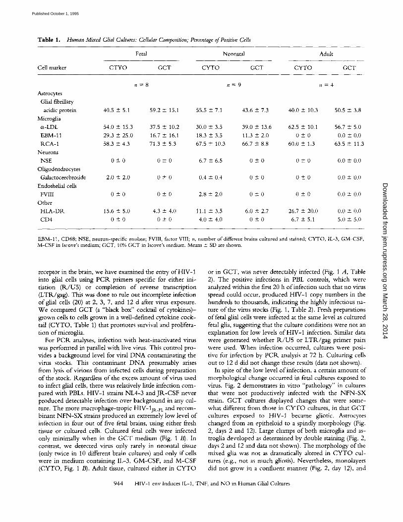
Table 1. Human Mixed Glial Cultures: Cellular Composition; Percentage of Positive Cells
Fetal Neonatal Adult
CeHma~er CTYO GCT CYTO GCT CYTO GCT
n = 8 n = 9 n = 4
Astrocytes
Glial fibfiUary
acidic protein 40.5 -+ 5.1 59.2 + 15.1 55.5 -+ 7.1 43.6 -+ 7.3 40.0 + 10.3 50.5 -+ 3.8
Microglia
tx-LDL 54.0 -+ 15.3 37.5 4- 10.2 30.0 + 3.5 39.0 + 13.6 62.5 + 10.1 56.7 + 5.0
EBM-11 29.3 -+ 25.0 16.7 + 16.1 18.3 + 3.5 11.3 4- 2.0 0 _+ 0 0.0 + 0.0
P, CA-1 58.3 -- 4.3 71.3 + 5.3 67.5 + 10.3 66.7 - 8.8 60.0 + 1.3 63.5 + 11.3
Neurons
NSE 0 + 0 0 -4- 0 6.7 + 6.5 0 -+ 0 0 + 0 0.0 --- 0.0
Oligodendrocytes
Galactocerebroside 2.0 _+ 2.0 0 + 0 0.4 4- 0.4 0 + 0 0 + 0 0.0 +- 0.0
Endothelial cells
FVIII 0 + 0 0 + 0 2.8 + 2.0 0 - 0 0 + 0 0.0 --+ 0.0
Other
HLA-DR. 15.6 -+ 5.0 4.3 + 4.0 11.1 + 3.5 6.0 + 2.7 26.7 --- 20.0 0.0 + 0.0
CD4 0 --- 0 0 4- 0 4.0 +- 4.0 0 __- 0 6.7 + 5.1 5.0 + 5.0
EBM-11, CD68; NSE, neuron-specific enolase; FVIlI, factor VIII; n, number of different brains cultured and stained; CYTO, IL-3, GM-CSF, M-CSF in Iscove's medium; GCT, 10% GCT in Iscove's medium. Means + SD are shown.
receptor in the brain, we have examined the entry o f HIV-1 into glial cells using P C R primers specific for either ini- tiation ( R / U 5 ) or complet ion o f reverse transcription (LTR./gag). This was done to rule out incomplete infection o f glial cells (20) at 2, 3, 7, and 12 d after virus exposure. W e compared G C T (a "black box" cocktail of cytokines)- grown cells to cells grown in a well-defined cytokine cock- tail (CYTO, Table 1) that promotes survival and prolifera- tion o f microglia.
For P C R analyses, infection with heat-inactivated virus was performed in parallel with live virus. This control p ro- vides a background level for viral D N A contaminating the virus stocks. This contaminant D N A presumably arises from lysis o f virions from infected cells during preparation o f the stock. Regardless o f the excess amount o f virus used to infect glial cells, there was relatively little infection com- pared with PBLs. HIV-1 strains NL4-3 a n d J R - C S F never produced detectable infection over background in any cul- ture. The more macrophage- t ropic HIV-1jR_FL and recom- binant N F N - S X strains produced an extremely low level o f infection in four out o f five fetal brains, using either fresh tissue or cultured cells. Cultured fetal cells were infected only minimally when in the G C T med ium (Fig. 1 /3). In contrast, we detected virus only rarely in neonatal tissue (only twice in 10 different brain cultures) and only i f cells were in medium containing IL-3, G M - C S F , and M - C S F (CYTO, Fig. 1 B). Adult tissue, cultured either in C Y T O
or in GCT, was never detectably infected (Fig. 1 A, Table 2). The positive infections in PBL controls, which were analyzed within the first 20 h o f infection such that no virus spread could occur, produced HIV-1 copy numbers in the hundreds to thousands, indicating the highly infectious na- ture o f the virus stocks (Fig. 1, Table 2). Fresh preparations o f fetal glial cells were infected at the same level as cultured fetal glia, suggesting that the culture conditions were not an explanation for low levels o f HIV-1 infection. Similar data were generated whether R J U 5 or L T R / g a g pr imer pairs were used. W h e n infection occurred, cultures were posi- tive for infection by PCR. analysis at 72 h. Culturing cells out to 12 d did not change these results (data not shown).
In spite o f the low level o f infection, a certain amount o f morphological change occurred in fetal cultures exposed to virus. Fig. 2 demonstrates in vitro "pathology" in cultures that were not productively infected with the N F N - S X strain. G C T cultures displayed changes that were some- what different from those in C Y T O cultures, in that G C T cultures exposed to HIV-1 became gliotic. Astrocytes changed from an epitheloid to a spindly morphology (Fig. 2, days 2 and 12). Large clumps o f both microglia and as- troglia developed as determined by double staining (Fig. 2, days 2 and 12 and data not shown). The morphology o f the mixed glia was not as dramatically altered in C Y T O cul- tures (e.g., not as much gliosis). Nevertheless, monolayers did not grow in a confluent manner (Fig. 2, day 12), and
944 HIV-1 env Induces IL-1, TNF, and NO in Human Glial Cultures
on March 28, 2014
jem.rupress.org
Dow
nloaded from
Published October 1, 1995
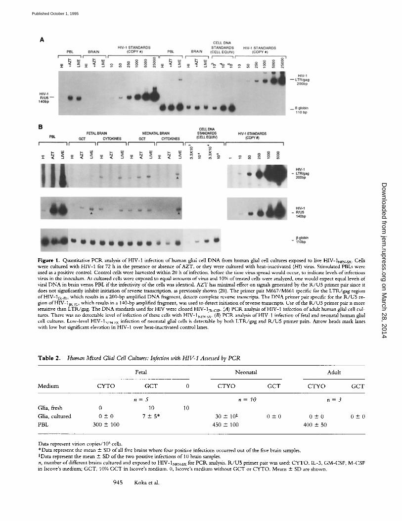
Figure 1. Quantitative PCIk analysis of HIV-1 infection of human glial cell DNA from human glial cell cultures exposed to live HIV-1NFN_SX. Cells were cultured with HIV-1 for 72 h in the presence or absence of AZT, or they were cultured with heat-inactivated (H/) virus. Stimulated PBLs were used as a positive control. Control cells were harvested within 20 h of infection, before the time virus spread would occur, to indicate levels of infectious vires in the inoculum. As cultured cells were exposed to equal amounts of virus and 10% of treated cells were analyzed, one would expect equal levels of viral D N A in brain versus PBL if the infectivity of the cells was identical. AZT has minimal effect on signals generated by the R / U 5 primer pair since it does not significantly inhibit initiation of reverse transcription, as previously shown (20). The primer pair M667/M661 specific for the LTR/gag region of HIV-13r~_r which results in a 200-bp amplified DNA fragment, detects complete reverse transcripts. The D N A primer pair specific for the 1K/U5 re- gion of HIV-1jp,_FL, which results in a 140-bp amplified fragment, was used to detect initiation of reverse transcripts. Use of the R / U 5 primer pair is more sensitive than LTR/gag. The D N A standards used for HIV were cloned HIV-1jR.cs r. (A) P C R analysis of HIV-1 infection of adult human glial cell cul- tures. There was no detectable level of infection of these cells with HIV-1NrN_SX. (B) PCIK analysis of HIV-1 infection of fetal and neonatal human glial cell cultures. Low-level HIV-1NrN_SX infection of neonatal glial cells is detectable by both LTR/gag and R / U 5 primer pairs. Arrow heads mark lanes with low but significant elevation in HIV-1 over heat-inactivated control lanes.
T a b l e 2. Human Mixed Glial Cell Cultures: Infection with HIV- 1 Assessed by PCR
Fetal Neona ta l Adul t
M e d i u m C Y T O G C T 0 C T Y O G C T C T Y O G C T
n = 5 n = l O n = 3
Glia, fresh 0 10 10
Glia, cu l tu red 0 +- 0 7 + 5* 30 + 10~ 0 + 0 0 + 0
PBL 300 -+ 100 450 -+ 100 400 + 50
0 - 0
Data represent virion copies/106 cells. * Data represent the mean +- SD of all five brains where four positive infections occurred out of the five brain samples. SData represent the mean + SD of the two positive infections o f 10 brain samples. n, number o f different brains cultured and exposed to HIV-1NFN_SX for PCIK analysis. R / U 5 primer pair was used; C Y T O , IL-3, GM-CSF, M-CSF in Iscove's medium; GCT, 10% G C T in Iscove's medium. 0, Iscove's medium without G C T or CYTO. Means - SD are shown.
945 Koka et al.
on March 28, 2014
jem.rupress.org
Dow
nloaded from
Published October 1, 1995
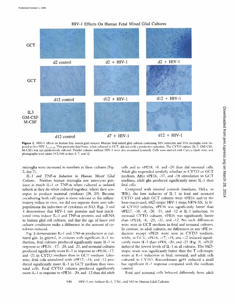
Figure 2. HIV-1 effects on human fetal mixed glial cultures. Human fetal mixed glial cultures containing 50% astrocytes and 50% microglia were ex- posed to five HIV-1NFN_SX. This particular fetal brain, when cultured in GCT, did not yield a productive infection. The CYTO culture (IL-3, GM-CSF, M-CSF) was not productively infected. Parallel cultures without HIV-1 were also examined (control). Ceils were stained with Cameo Quik stain, and photographs were taken (• at days 2, 7, and 12.
microglia were increased in numbers in these cultures (Fig. 2, day 7).
IL-1 and TNF-oe Induction in Human Mixed Glial Cultures. Neither human microglia nor astrocytes pro- duce as much IL-1 or TNF-o/ when cultured as isolated subsets as they do when cultured together, where they syn- ergize to produce maximal cytokines (28, 29). Because coculturing both cell types is more relevant to the inflam- matory milieu in vivo, we did not separate them into sub- populations for induction o f cytokines or N O . Figs. 3 and 4 demonstrate that HIV-1 env proteins and heat inacti- vated virus induce IL-1 and TNF-o~ proteins and mP, N A in human glial cell cultures, and that the age of tissue and culture conditions make a difference in the amount o f cy- tokines induced.
Fig. 3 demonstrates IL-1 and TNF-o~ product ion in cul- tured glia. In general, in cultures with significant IL-1 in- duction, fetal cultures produced significantly more IL-1 in response to vPE 16, -17, -20, and -21, and neonatal cultures produced significantly more IL-1 in response to vPE16, -17, and -21 in C Y T O medium than in G C T medium. Like- wise, fetal cells stimulated with vPE17, -18, and -12 pro- duced significantly more IL-1 in G C T medium than neo- natal cells. Fetal C Y T O cultures produced significantly more IL-1 in response to vPE16, -20, and -12 than did adult
cells and to vPE18, -8, and -20 than did neonatal ceils. Adult glia responded similarly whether in C Y T O or G C T medium. After vPE16, -17, and -18 stimulation in G C T medium, adult glia produced significantly more IL-1 than fetal cells.
Compared with internal controls (medium, HeLa, or W R ) , the best inducers o f IL-I in fetal and neonatal C Y T O and adult G C T cultures were vPE16 and /o r the heat-inactivated, M•- t rop ic HIV-1 strain N F N - S X . In fe- tal C Y T O cultures, vPE16 was significantly better than vPE17, -18, -8, -20, -21, and -12 at IL-1 induction. In neonatal C Y T O cultures, vPE16 was significantly better than vPEt8 , -8, -20, -21, and -12. N o such differences were seen in G C T medium in fetal and neonatal cultures. In contrast, in adult cultures, no differences in any vPE in- duction except vPE21 were seen in C Y T O medium, while, in GCT, vPE16, -17, -18, and -12 induced signifi- cantly more IL-1 than vPE8, -20, and -21 (Fig. 3). vPE21 induced the lowest levels o f IL-1 in all cultures. The M Q - tropic strain was significantly better than the T cel l- t ropic strain at IL-1 induction in fetal, neonatal, and adult cells cultured in C Y T O . Recombinan t gp41 induced a small but significant IL-1 response compared with the medium control.
Fetal and neonatal cells behaved differently from adult
946 HIV-I env Induces IL-I, TNF, and NO in Human Glial Cultures
on March 28, 2014
jem.rupress.org
Dow
nloaded from
Published October 1, 1995
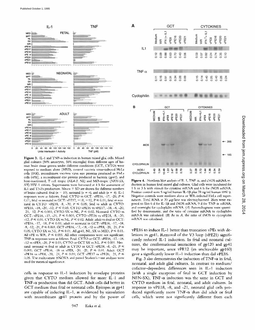
Figure 3. IL-1 and TNF-c~ induction in human mixed glial cells. Mixed glial cultures (50% astrocytes, 50% microglia) from different ages of hu- man brain tissue grown under different conditions (GCT, CYTO) were exposed to medium alone (MED), control vaccinia virus-infected HeLa cells (WR), recombinant vaccinia virus env proteins produced in HeLa cells (vPE), a recombinant env protein produced in bacteria (gp41), and heat-inactivated, T cell-tropic (NL4-3, NL) and MO-tropic (NFN-SX, SX) HIV-1 virions. Supernatants were harvested at 4 h for assessment of ILl and TNFa production. Means - SD are shown for different numbers of brains cultured: fetal (n = 10), neonatal (n = 4), and adult (n = 4). IL-1 responses were as follows. Fetal: CYTO vs GCT: vPE16, -17, 20, P <~ 0.01; fetal vs neonatal m GCT: vPE17, - 18, - 12, P ~< 0.01; fetal vs neo- natal in CYTO: vPE18, -8, -20, P ~< 0.05; fetal vs adult in CYTO: vPE16, -18, -20, -12, P ~< 0.05; CYTO vPE16 vs vPE17, -18, -8, -20, -21, -12, P ~ 0.001; CYTO SX vs NL, P ~< 0.02. Neonatal: CYTO vs GCT: vPE16, -17, -21, P ~< 0.001; CYTO vPE16 vs vPE18, -8, -20, -12, P ~< 0.01; CYTO SX vs NL, P <~ 0.02. Adult: adult vs fetal in GCT: vPE16, -17, -18, P ~< 0.01; adult vs neonatal in GCT: vPE16, -17, -18, -8, -12, -21, P ~< 0.001; GCT vPE16, -17, -18, -12 vs vPE8, -20, 21, P ~< 0.05; CYTO SX vs NL, P ~< 0.01. All gp41, NL, SX vs MED, P <~ 0.01. All vPE vs WR, P ~< 0.001. All other comparisons were not significant. TNF-0t responses were as follows. Fetal: CYTO or GCT: vPE 16, -17, -18, -12 vs vPE8, -20, P ~< 0.01; CYTO or GCT SX vs NL, P ~< 0.001. Neo- natal: neonatal vs fetal or adult in CYTO or GCT: vPE18, -8, -21, P ~< 0.001; GCT vPE16, -18 vs vPE8, -20, -21, P ~< 0.01. Adult: GCT vPE16 vs vPE8, -20, -21, P ~< 0.01; GCT vPE17 vs vPE20, -21, P ~< 0.05. The multivariate ANOVA and paired Student's t test analyses were used for statistical significance.
cells in r e s p o n s e to IL-1 i n d u c t i o n b y e n v e l o p e p r o t e i n s
g i v e n tha t C Y T O m e d i u m a l l o w e d for m o r e IL-1 a n d
T N F - r p r o d u c t i o n t h a n d i d G C T . A d u l t cells d i d b e t t e r in
G C T m e d i u m t h a n fetal o r n e o n a t a l cells. E p i t o p e s in gp41
are capab le o f i n d u c i n g IL-1 , as e v i d e n c e d b y s t i m u l a t i o n
w i t h r e c o m b i n a n t gp41 p r o t e i n a n d b y t h e p o w e r o f
Figure 4. Northern blot analysis oflL-1, TNF-ot, and iNOS mRNA in- duction in human fetal mixed glial cultures. Glial cells were incubated for 1 h or 3 h with stimuli for cytokine mR.NA and 6 h for iNOS ml<NA. Positive control were 5 ng/ml human IL-113 plus 70 ng/ml human IFN-y. Negative controls were medium alone or WP,-infected HeLa cell super- natants. Total 1<NA at 30 p.g/Iane was electrophoresed. Blots were ex- posed to film 6 d for IL-115 and iNOS mRNA, 9 d for TNF-c~ mR.NA, and overnight for cyclophilin mRNA. (A) Autoradiograms were quanti- fied by densitometry, and the ratio of cytokine mRNA to cyclophilin m R N A was calculated. (B) As in A, the ratio of iNOS to cydophilin mRNA was calculated.
v P E 1 6 to i n d u c e IL-1 b e t t e r t h a n t r u n c a t i o n v P E s w i t h d e -
l e t i ons in gp41 . R e m o v a l o f t h e V 3 l o o p (vPE21) s ign i f i -
can t ly r e d u c e d IL-1 i n d u c t i o n . In fetal a n d n e o n a t a l c u l -
tures , t h e c o n f o r m a t i o n a l i n t e r a c t i o n o f g p 1 2 0 a n d gp41
m a y b e i m p o r t a n t , s i nce v P E 1 2 (an u n c l e a v a b l e g p l 6 0 )
gave a s ign i f i can t ly l o w e r IL-1 i n d u c t i o n t h a n d id v P E 1 6 .
Fig. 3 also d e m o n s t r a t e s t h e i n d u c t i o n o f T N F - ( x in fetal,
n e o n a t a l , a n d adu l t glial cu l tu res . In c o n t r a s t to m e d i u m /
c o f a c t o r - d e p e n d e n t d i f f e r e n c e s s e e n in IL-1 i n d u c t i o n
( w i t h a s ing le e x c e p t i o n o f fetal in G C T i n d u c t i o n b y
N F N - S X ) , T N F - ( i i n d u c t i o n was t h e s a m e in G C T a n d
C Y T O m e d i u m in fetal, n e o n a t a l , a n d adu l t cu l tu res . In
r e s p o n s e to v P E 1 8 , -8 , a n d - 2 1 , n e o n a t a l glial cells p r o -
d u c e d s ign i f i can t ly m o r e TNF-oL t h a n d i d adu l t o r fetal
cells, w h i c h w e r e n o t s ign i f i can t ly d i f f e r en t f r o m e a c h
947 Koka et al.
on March 28, 2014
jem.rupress.org
Dow
nloaded from
Published October 1, 1995
other at any data point. As with IL-1, adult, neonatal, and fetal cultures specifcally produced the same amount o f TNF-ot in response to heat-inactivated virus and recombi- nant gp41.
In fetal cultures, vPE16, -17, -18, and -12 induced the same levels o f TNF-ot but significantly more than was in- duced by vPE8 and -20, which produced similar TNF-oc This suggests that gp41 is important and the V3 loop is not important in TNF-ot production. Likewise, in neonatal and adult cultures, removal of all gp41 epitopes (as in vPE8, -20, -21) significantly reduced TNF-0~ in contrast to what was seen for IL-1. vPE21 did not induce significant TNF-o~ in any cultures. In contrast to IL-1 induction, gp41 stimulated as much TNF-o~ as did vPE16 and heat-inactivated virus induction. These data suggest a more important role for gp41 in TNF-~x than IL-1 induction. In fetal cultures, the MO- t rop i c strain induced significantly more TNF-o~ than did the T cell- tropic strain; there was no tropism in TNF-0r induction in neonatal or adult cultures.
To determine whether the induction o f lL-1 and TNF-c i was being regulated at the mlKNA level, Nor thern blot analyses o f lL-1 and TNF-oe mlKNA from fetal cultures be- fore and after env protein stimulation were performed compared with the housekeeping cyclophilin gene. In Fig. 4 A, it can be seen that the negative controls, medium, or W1K vaccinia virus did not induce IL-1 or TNF-cx mlKNA. Likewise, vPE21, which did not induce protein, also did not induce mlKNA. The positive control I F N - y / I L - 1 was
a strong inducer o f both cytokine mlKNAs in either me- dium (GCT or CYTO) . For tL-1, vPE16 was a better in- ducer than vPE8 or vPE20, a difference that was greater in C Y T O than G C T in parallel with protein data (Fig. 3). The differences among vPE16, -18, and -20 or between C Y T O and G C T were not as pronounced in TNF-o~ in- duction.
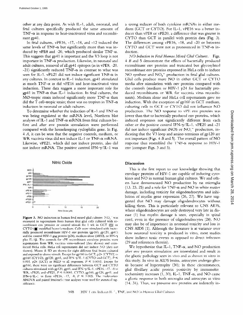
N O Induction in Fetal Human Mix-ed Glial Cultures. Figs. 4 B and 5 demonstrate the effects o f bacterially produced recombinant env proteins and truncated but glycosylated recombinant env proteins expressed in HeLa cells to induce N O synthase and N O • product ion in fetal glial cultures. Glial cells produce more N O in either G C T or C Y T O media after stimulation with env proteins compared with the controls (medium or HIV-1 p24 for bacterially pro- duced recombinants or W1K for vaccinia virus recombi- nants). Med ium alone and HeLa cell supernatants gave no induction. Wi th the exception o f gp160 in G C T medium, culturing cells in G C T or C Y T O did not influence N O production. The N O response to vPE env proteins was lower than that to bacterially produced env proteins, which induced responses not significantly different from each other or the positive control I F N - y / I L - 1 . vPE20 and -21 did not induce significant iNOS or N O x - production, in- dicating that the V3 loop and amino terminus o f g p l 2 0 are not important (Figs. 4 B and 5). The overall pattern of N O response thus resembled the TNF-er response to HIV-1 env (compare Figs. 3 and 5).
Figure 5. NO induction in human fetal mixed glial cultures. NO~- was measured in supernatants from human fetal glial cells cultured with re- combinant env proteins or control stimuli for 1 wk in GCT ([]) or CYTO ([]) modified Iscove's medium. Cells were stimulated with bacte- rially produced recombinant HIV-1 env proteins (gp160, gp120, gp41) and the control HIV-1 gag protein (p24), medium alone (MED), or IFN-3' plus IL-I~. The controls for vPE recombinant envelope proteins were supernatants from W1K vaccinia vires-infected (data shown) and unin- fected HeLa cells. HeLa cell supematants did not induce NO (data not shown). Means "4- SD are shown for eight different fetal brains cultured and exposed to above stimuli. Except for gp160 in GCT, p24 (CYTO) vs gp160 (CYTO), gp120, gp41, and IFN-y/IL-1 (CYTO and GCT), P ~< 0.001. p24 (GCT) or MED vs all responses, P ~< 0.0001. Except for gp160, there were no significant differences between GCT and CYTO cultures stimulated with gp120, gp41, and IFN-~//IL-1. vPE16, -17, -8 vs WIK, vPE20, and vPE21, P ~< 0.0001. CYTO, gp160, gp120, gp41, and IFN-"//IL-1 vs from vPE16, -17, -8, P ~< 0.0(15. The nmltivariate ANOVA and paired Student's t test analyses were used for statistical sig- nificance.
Discuss ion
This is the first report to our knowledge showing that envelope proteins o f HIV-1 are capable o f inducing cyto- kines and N O in normal human glial cultures. W e and oth- ers have demonstrated N O product ion by rat microglia (13, 23, 25) and a role for TNF-0~ and N O in white matter damage, including toxicity for oligodendrocytes and inhi- bit ion o f myelin gene expression (26, 27). W e have sug- gested that N O may damage oligodendrocytes wi thout killing them. This is particularly relevant to CNS AIDS, where oligodendrocytes are only destroyed very late in dis- ease (1) but myelin damage is seen, especially in spinal cord, even in the presence o f ol igodendrocytes (28). N O may also be o f importance in neuronal toxicity and loss in CNS AIDS (1). Although the literature is at variance over how neuronal toxicity is produced in vitro, most studies show indirect toxic events as opposed to direct infection (29 and references therein).
W e hypothesize that IL-1, TNF-ci , and N O product ion after env protein stimulation are interrelated and result in the gliotic pathology seen in vivo and as shown in vitro in this study. In vivo in AIDS brains, astrocytes undergo glio- sis because o f hyper t rophy (30); in these circumstances, glial fibrillary acidic protein positivity by immunohis- tochemistry increases (1, 30). IL-1, T N F - a , and N O cause a gliotic response in both microglia and astrocytes in vitro (14, 31). Thus, we presume env proteins are indirectly in-
948 HIV-1 env Induces IL-1, TNF, and NO in Human Glial Cultures
on March 28, 2014
jem.rupress.org
Dow
nloaded from
Published October 1, 1995
ducing the microgliosis shown in vitro in this study. While N O may cause gliosis, we would predict little or no other perceptible toxic effect in these cultures. W e and others have shown that N O inhibits mitochondrial function in as- trocytes, but it does not kill them. N O has virtually no toxic effect on microglia (23, 27).
I f the production of N O in our cultures is the indirect result o f IL-1 and TNF-cx production or a direct effect o f gp120/gp41 on NFKB, it is not clear why there is a rather long interval between IL-1 and TNF-e~ production (peak 4-24 h) and peak N O production (3-7 d). Posttranslational regulation o f i N O S may be an important component in the delay in N O production. This is currently being examined. As we have suggested in these studies, HIV-1 need not in- fect glial cells to contribute to pathology in the CNS. While it is clear that HIV-1 infects CNS microglial cells in vivo, productively infected cells are rare in the brains of pe- diatric AIDS patients. The in vivo infections are latent or defective (1-3, 32, 33). Even the culturing of microglia from infected pediatric brain tissue requires 30 d before vi- ral replication is readily apparent, indicating the extremely slow virus life cycle or poor infectivity leading to slow spread of infection (34). In vitro infection o f astrocytes from fetal brain results in a transient, partially defective viral replication (7-9). In vivo, astrocytes are not productively infected (1) but nef protein is expressed in a small propor- tion of astrocytes in pediatric AIDS brain tissues (35). A re- stricted or latent infection in glia is therefore thought to lead to indirect but extensive neuropathogenesis.
While several reports claim microglia in brain tissue and in tissue culture have CD4 on their surface (10, 34), which contributes to HIV-1 infection (10), many other studies have failed to detect CD4 on parenchymal microglia in fe- tal or adult brain from normal or HIV- l -pos i t i ve brain tis- sue (11, 12, 36, 37), nor has CD4 been seen to be inducible on microglia in vitro (37). It is thought that the CD4 + cells in brain are blood-derived perivascular CNS macrophages (11, 36). Since we document a significant number of microglia in our cultures (which are CD4- ) , either the Watkins study (10) and follow-up studies from the same
laboratory were demonstrating infection of blood-borne, perivascular brain macrophages and not microglia, or other culture conditions in the Watkins study allowed for CD4 expression and/or infection. The infection we achieved in fetal cultures, while significantly above controls and on a fairly consistent basis, was extremely low compared with PBL controls. The fetal tissue we used was taken during the second trimester and was therefore older than that in one study (11) but similar to that in another (12). Nevertheless, the level o f infection in our study was significantly lower than that reported by Lee et al. (12). Additionally, the in- ability to infect human glia in vitro and the differences seen in TNF-o~, IL-1, and N O production when cells are in dif- ferent med ium conditions suggest that the in vitro cofactors are not identical to in vivo cofactors, or that in vivo infec- tion of glia is indirectly achieved by fusion of infected blood monocytes with brain cells.
In summary, this study reconfirms two previous studies on rat glial cells demonstrating a functional cytokine re- sponse to n o n - C D 4 epitopes in gp120 and to the amino terminus ofgp41 (18, 19). IL-1, TNF-cx, and iNOS can be induced at the m R N A level by env proteins and HIV-1 vi- rus in the absence of infection. Their production is induced and regulated differently, given that media with different cofactors and different env domains give different IL-1 or TNF-c* responses. The responses in adult, neonatal, and fe- tal glial cultures are also different, even though the cellular composition o f the cultures appears to be identical. The V3 loop and carboxy terminus o f gp120 and to a lesser extent gp41 are important for IL-1. The V3 loop of gp120 is less important and gp41 is more important in TNF-ot than in IL-1 induction. While the macrophage-tropic strain of HIV-1 tends to be a better inducer of cytokines, a signifi- cant IL-1 and TNF-0~ response occurs with a T cell-tropic strain of HIV-1. Further mapping of distinct sequences within gp120 and gp41 that are responsible for T N F and IL-1 production and the role of these cytokines in N O in- duction is part of the ongoing research in this lab. Elucida- tion of these interactions will provide a basis for designing therapeutic interventions to use in treating CNS AIDS.
We thank Dr. Antoine E1 Hajjar and Dr. Munther Hijazin for the tissue culture work, Ben Techagaiciyawa- nis and Evan Jones for cytokine bioassays, and Joan Miley for the manuscript preparation. We also wish to thank Genentech Inc. (South San Francisco, CA) for the supply of human TNF-cx and Immunex Corp. (Se- attle, WA) for human IL-113 and IFN-3,.
This work was supported in part by grants from the National Institutes of Health (RO-I NS30768) and from the Conrad N. Hilton Foundation (J. E. Merrill); National Institute of Mental Health fellowship (5T32MHI19200) for postdoctoral interdisciplinary training in HIV-AIDS (P. Koka); Howard Hughes Medical Institute Undergraduate Research Fellowship and Naumburg Research Award (T. Tran); and the President's Undergraduate Fellowship (T. Tran, S. S. Yashar)
Address correspondence to Dr. Jean E. Merrill, Department of Immunology, Berlex Biosciences, 15049 San Pablo Avenue, POB 4099, Richmond, CA 94804-0099.
Received for publication 3 November 1995 and in revised form 10 May 1995.
949 Koka et al.
on March 28, 2014
jem.rupress.org
Dow
nloaded from
Published October 1, 1995
References 1. Levy, J.A. 1993. Pathogenesis of human immunodeficiency
virus infection. Microbiol. Rev. 57:183-289. 2. Mano, H., andJ.C. Chermann. 1991. Fetal human immuno-
deficiency virus type 1 infection of different organs in the second trimester. A I D S Res. Hum. Retroviruses. 7:83-88.
3. Kure, K., J.F. Llena, W.D. Lyman, R. Soeiro, K.M. Weiden- heim, A. Hirano, and D.W. Dickson. 1991. Human immuno- deficiency virus-1 infection of the nervous system: an autopsy study of 268 adult, pediatric, and fetal brains. Hum. Pathol. 22:700-710.
4. Merrill, J.E., and O. Martinez-Maza. 1993. Cytokines in AIDS-associated neurons and immune system dysfunction. In Neurobiology of Cytokines Part B: Methods in Neuro- science. E.B. De Souza, editor. Academic Press, Inc., San Di- ego, CA. 243-266.
5. Tyor, W.R.., J.D. Glas, J.W. Griffin, P.S. Becker, J.C. McArthur, L. Bezman, and D.E. Griffin. 1992. Cytokine ex- pression in the brain during the acquired immunodeficiency syndrome. Ann. Neurol. 31:349-360.
6. Poli, G., P. Bressler, A. Kinter, E. Duh, W.C. Timmer, A. lLabson, J.S. Justement, S. Stanley, and A.S. Fauci. 1990. In- terleukin 6 induces human immunodeficiency virus expres- sion in infected monocytic cells alone and in synergy with tumor necrosis factor er by transcriptional and posttranscrip- tional mechanisms.J. Exp. Med. 172:151-158.
7. Christofinis, G., L. Papadaki, Q. Sattentau, R_.B. Ferns, and 1<. Tedder. 1987. HIV replicates in cultured human brain ceils. A I D S (Phila.). 1:229-234.
8. Tornatore, C., A. Nath, K. Amemiya, and E.O. Major. 1991. Persistent human immunodeficiency virus type 1 infection in human fetal glial cells reactivated by T-cell factors or by the cytokines tumor necrosis factor alpha and interleukin-I beta. J. l/itvl. 65:6094-6100.
9. Teglbjaerg, L.L.S., J.-E.S. Hansen, H. Dalboge, C.M. Nielsen, L.R. Mathieseu, and O. Nielsen. 1991. Detection of human immunodeficiency virus DNA in cultured human glial cells by means of the polymerase chain reaction. Acta Neurol. &and. 83:179-182.
10. Watkins, B.A., H.H. Dorn, W.B. Kelly, K.C. Armstrong, B.J. Potts, F. Michaels, C.V. Kutfta, and M. Dubois-Dalcq. 1990. Specific tropism of HIV-1 for microglial cells in pri- mary human brain cultures. Scie,ce (Wash. DC). 249:549- 553.
1 I. Peudenier, S., C. Hery, L. Montagnier, and M. Tardieu. 1993. Human microglial cells characterization in cerebral tis- sue and in primary culture and study of their suscepubility to HIV-1 infection. Ann. Neurol. 29:152-161.
12. Lee, S.C., W.C. Hatch, W. Liu, X. Kress, W.1). Lyman, and D.W. Dickson. 1993. Productive infection of human fetal nficroglia by HIV-I. Am._l. Pathol. 143:1032-1(139.
13. Murphy, S., M.L. Simmons, L. Aqullo, A. Garcia, D.L. Fein- stein, E. Galea, 1).J. Reis, D. Minc-Golomb, and J.P. Schwartz. 1993. Synthesis of nitric oxide in CNS glial cells. Tre~Ms Neurosci. 16:323-328.
14. Lee, S.C., D.W. Dickson, W. Liu, and C.F. Brosnan. 1993. Induction of nitric oxide synthase activity in human astro- cytes by interleukin-l[3 and interferon y . j . Neuroimmunol. 46:19-24.
15. Mollace, V., M. Colasanti, T. Persichini, G. Bagetta, G.M. Lauro, and G. Nistico. 1993. HIV gp120 glycoprotein stimu- lates the inducible isoform of NO synthase in human cul- tured astrocytoma cells. Biochem. Biophys. Res. Col~lmltn. 194:
439-445. 16. Pietraforte, D., E. Tritarelli, V. Testa, and M. Minetti. 1994.
gp120 HIV envelope glycoprotein increases the production of nitric oxide in human monocyte-derived macrophages. J. Leukocyte Biol, 55:175-182.
17. Dighiero, P., I. Reux, J,-J. Hauw, A.-M. Fillet, Y. Courtois, and O. Goureau. 1994. Expression of nitric oxide synthase in cytomegalovirus infected cells of retinas from AIDS patients. Neurosci. Lett. 166:31-34.
18. Merrill, J.E., Y. Koyanagi, J. Zack, L Thomas, F. Martin, and I.S.Y. Chen. 1992. Induction ofinterleukin-1 and tumor necrosis factor alpha in brain cultures with human mmmno- deficiency virus type 1.J. Virol. 66:2217-2225.
19. Koka, P., K. He., D. Camerini, T. Tran, S.S. Yashar, andJ.E. Merrill. 1995. The mapping of HIV-I gp160 epitopes re- quired for ILl and TNFc~ production in glial cells. J. Neu- roimmunol. 57:179-191.
2(t. Zack, J.A., S.J. Arrigo, S.R. Weitsman, A.S. Go, A.M. Hais- lip, and I.S.Y. Chen. 1990. HIV-I entry into quiescent pri- mary lymphocytes: molecular analysis reveals a labile, latent, viral structure. Cell. 61:213-222.
21. O'Brien, W.A., I.S.Y. Chen, I).D. Ho, and E.S. 1)aar. 1992. Mapping genetic determinants for human mmmnodeficiency virus type 1 resistance to soluble CD4. _1. Vinyl. 66:3125- 3130.
22. Bush, P.A., N.E. Gonzalez, J.M. Griscavage, and L.J. Ignarro. 1992. Nitric oxide synthase from cerebellum catalyzes the formation of equimolar quantities of nitric oxide and citrul- line from L-arginine. Biochem. Biophys. Res. Commun. 185: 960-966.
23. Mitrovic, B., B.A. St. Pierre, A.J. Mackenzie-Graham, and J.E. Merrill. 1994. The role of nitric oxide in glial pathology. Anu. N Y Acad. Sci. 738:436-446.
24. Geller, D.A., C.J. Lowenstein. K.A. Shapiro, A.K. Nussler, M. l)iSilvio, S.C. Wang, D.K. Nakayama, R..L. Simmons, S.H. Snyder, and T.R. Billiar. 1993. Molecular cloning and expression of inducible nitric oxide synthase from human hepatocytes. Proc. Natl. Acad. Sci. USA. 90:349t-3495.
25. Chao, C.C., S. Hu, T.W. Molitor, E.G. Shaskan, and P.K. Peterson. 1992. Microglia-mediated neuronal injury via a ni- tric oxide mechanism. J. lmmunol. 149:27-36.
26. Merrill, J.E., L.J. Ignarro, M.P. Sherman, J. Melinek, and T. Lane. 1993. Microglial cell cytotoxicity of oligodendrocytes is mediated through nitric oxide.J, hnmunol. 151:2132-2141.
27. Mitrovic, B., L.J. Ignarro, S. Montestruque, A. Smoll, and J.E. Merrill. 1994. Nitric oxide as a potential pathological mechanism in demyetination: its difibrential effects on pri- mary glial cells in vitro. Neuroscience. 61:575-585.
28. Esiri, M.M., C.S. Morris, and PAL. Millard. 1991. Fate ofoli- godendrocytes in HIV-1 infection. AIDS (Phila.). 5:1081- 1 (/88.
29. Lipton, S.A. 1994. Neuronal injury associated with HIV-1 and potential treatment with calcium channel and NMI)A antagonists. Dev. Neurosci. 16:145-15l.
3(1. Weis, S., H. Haug, and H. Budka. 1993. Astroglial changes in cerebral cortex of AIDS brains: a morphometric and inmm- nohistochemical investigation. Neun~pathol. Appl. Neurobiol. 19:329-335.
31. Merrill, J.E. 1991. Effects ofinterleukin-I and tumor necrosis factor c~ on astrocytes, microglia, oligodendrocytes, and glial procursors in vitro. Dev. Neurosci. 13:13()-137.
32. Vazeux, 1<., C. Lacroix-Ciudo, S. Blanche, M.-C. Cumont,
950 HIV-1 env Induces IL-1, TNF, and NO in Human Glial Cultures
on March 28, 2014
jem.rupress.org
Dow
nloaded from
Published October 1, 1995
D. Henin, F. Gray, L., Boccon-Gibod, and M. Tardieu. 1992. Low levels of human immunodeficiency virus replica- tion in the brain tissue of children with severe acquired im- munodeficiency syndrome encephalopathy. Am. J. Pathol. 140:137-144.
33. Dickson, D.W., A.L. Belman, and Y.D. Park. 1989. Central nervous system pathology in pediatric AIDS: an autopsy study. APMIS. 8(Suppl.):40-45.
34. Brinkman, R., A. Schwinn, O. Narayan, C. Zink, H.W. Kreth, W. Reggendorf, R. Domes, S. Schwender, H. Im- rich, and V. terMeulen. 1992. Human immunodeficiency vi- rus infection in microglia: correlation between cells infected in the brain and cells cultured from infectious brain tissue. Ann. Neurol. 31:361-365.
35. Saito, Y., L.R. Sharer, L.G. Epstein, J. Michaels, M. Mintz, M. Louder, K. Golding, T.A. Cvetkovich, and B.M. Blum- berg. 1993. Overexpression of nefas a marker for restricted HIV-1 infection of astrocytes in post mortem pediatric cen- tral nervous tissues. Neurol. 44:474-480.
36. Williams, K., A. Bar Or, E. Ulvestad, A. Olivier, J.P. Antel, and V.W. Yong. 1992. Biology of adult human microglia in culture: comparisons with peripheral blood monocytes and astrocytes. J. Neuropathol. & Exp. Neurol. 51:538-549.
37. Peudenier, S., C. Hery, K.H. Ng, and M. Tardieu. 1991. HIV receptors within the brain: a study of CD4 and MHC-II on human neurons, astrocytes, and microglial cells. Res. Virol. 142:145-149.
951 Koka et al.
on March 28, 2014
jem.rupress.org
Dow
nloaded from
Published October 1, 1995