Calcium signaling in vertebrate embryonic patterning and morphogenesis
Am. J. Hum. Genet. 73:957–966, 2003
957
Report
Mutations in Capillary Morphogenesis Gene-2 Result in the AllelicDisorders Juvenile Hyaline Fibromatosis and Infantile Systemic HyalinosisOonagh Dowling,1 Analisa Difeo,1 Maria C. Ramirez,1 Turgut Tukel,1,6,8 Goutham Narla,2Luisa Bonafe,9 Hulya Kayserili,7 Memnune Yuksel-Apak,7 Amy S. Paller,10 Karen Norton,3Ahmad S. Teebi,11 Valerie Grum-Tokars,12 Gail S. Martin,13 George E. Davis,13
Marc J. Glucksman,12 and John A. Martignetti1,4,5
Departments of 1Human Genetics, 2Division of Liver Diseases, 3Radiology, and 4Pediatrics and 5The Derald H. Ruttenberg Cancer Center,Mount Sinai School of Medicine, New York; 6Istanbul Medical Faculty, Department of Pediatrics, and 7Child Health Institute, Division ofMedical Genetics, and 8Institute for Experimental Medicine, Department of Genetics, Istanbul University, Istanbul; 9Division of MolecularPaediatrics, Centre Hospitalier Universitaire Vaudois, Lausanne, Switzerland; 10Departments of Pediatrics and Dermatology, Children’sMemorial Hospital, Northwestern University’s Feinberg School of Medicine, Chicago; 11Department of Genetics, The Hospital for SickChildren, Toronto; 12Midwest Proteome Center and Department of Biochemistry & Molecular Biology, Finch University of Health Sciences,Chicago Medical School, North Chicago; and 13Department of Pathology, System Health Science Center, Texas A&M University, CollegeStation
Juvenile hyaline fibromatosis (JHF) and infantile systemic hyalinosis (ISH) are autosomal recessive syndromesof unknown etiology characterized by multiple, recurring subcutaneous tumors, gingival hypertrophy, joint con-tractures, osteolysis, and osteoporosis. Both are believed to be allelic disorders; ISH is distinguished from JHF byits more severe phenotype, which includes hyaline deposits in multiple organs, recurrent infections, and death withinthe first 2 years of life. Using the previously reported chromosome 4q21 JHF disease locus as a guide for candidate-gene identification, we identified and characterized JHF and ISH disease-causing mutations in the capillary mor-phogenesis factor–2 gene (CMG2). Although CMG2 encodes a protein upregulated in endothelial cells duringcapillary formation and was recently shown to function as an anthrax-toxin receptor, its physiologic role is unclear.Two ISH family-specific truncating mutations, E220X and the 1-bp insertion P357insC that results in translationof an out-of-frame stop codon, were generated by site-directed mutagenesis and were shown to delete the CMG-2 transmembrane and/or cytosolic domains, respectively. An ISH compound mutation, I189T, is predicted to createa novel and destabilizing internal cavity within the protein. The JHF family-specific homoallelic missense mutationG105D destabilizes a von Willebrand factor A extracellular domain alpha-helix, whereas the other mutation, L329R,occurs within the transmembrane domain of the protein. Finally, and possibly providing insight into the patho-physiology of these diseases, analysis of fibroblasts derived from patients with JHF or ISH suggests that CMG2mutations abrogate normal cell interactions with the extracellular matrix.
Juvenile hyaline fibromatosis (JHF [MIM 228600]) andinfantile systemic hyalinosis (ISH [MIM 236490]) are au-tosomal recessive disorders that present in infancy withpapulonodular skin lesions, particularly of the perianal,perinasal, and perioral areas. Affected individuals often
Received July 1, 2003; accepted for publication July 23, 2003; elec-tronically published September 12, 2003.
Address for correspondence and reprints: Dr. John A. Martignetti,Department of Human Genetics, Mount Sinai School of Medicine,Room 14-70B, 1425 Madison Avenue, New York, NY 10029. E-mail:[email protected]
� 2003 by The American Society of Human Genetics. All rights reserved.0002-9297/2003/7304-0022$15.00
develop several associated features, including multiplesubcutaneous tumors, gingival hypertrophy, flexion con-tractures of joints, osteolytic lesions, and osteopenia(Landing and Nadorra 1986; Fayad et al. 1987; Keseret al. 1999). ISH is distinguished by an earlier onset,more painful and severe course, and, as revealed by his-tological examination, widespread deposition of hyalinematerial throughout the skin, gastrointestinal tract, en-docrine glands, and muscle (Landing and Nadorra 1986).In addition, ISH has been associated with an increasedsusceptibility to bone fractures, infections, and death ininfancy (Stucki et al. 2001). Diagnosis is generally madeon the basis of clinical findings, including distribution
958 Am. J. Hum. Genet. 73:957–966, 2003
Figure 1 Radiological features of affected individual in family JHF1. A, Frontal view of both hands, revealing diffuse osteopenia andnarrowing of interarticular spaces. Multiple subluxations and contractures are present. B, Lateral view of the knee, revealing marked narrowingof the joint space (arrow) and profound osteopenia.
of skin lesions and biopsy results that typically reveal thepresence of an abundant extracellular, acidophilic hyalinematerial. The etiology of these two disorders, suggestedelsewhere to be allelic because of their significant pheno-typic overlaps (Mancini et al. 1999), is unknown.
By use of a positional-cloning approach, the JHF dis-ease gene was recently localized to chromosome 4q21(Rahman et al. 2002). The 5.3-cM/6.9-Mb locus isbounded by microsatellite marker D4S2393 centromeri-cally and D4S395 telomerically (Kong et al. 2002; Rah-man et al. 2002). In an attempt to further refine the locusand to investigate the possibility that these clinically over-
lapping autosomal recessive disorders, JHF and ISH, areindeed allelic, we first ascertained four unrelated familieswith established clinical diagnoses and features consis-tent with these syndromes (fig. 1; table 1). After obtain-ing informed consent and institutional review board ap-proval from the corresponding institutions, blood sam-ples were collected from family members, and genomicDNA was isolated. Using a dense set of microsatellitemarkers spanning the linked region, we haplotyped allavailable family members to look for regions that werehomozygous-by-descent. Haplotype analysis was per-formed using eight fluorescently labeled microsatellite
Reports 959
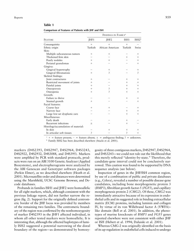
Table 1
Comparison of Features of Patients with JHF and ISH
FEATURE
FINDINGS IN FAMILYa
JHF1 JHF2 ISH1 ISH2b
Consanguinity � � � �Ethnic origin Turkish African American Turkish SwissSkin:
Multiple subcutaneous tumors � � � �Thickened firm skin � � � �Pearly nodules � � � �Perianal granulomas � � � �
Gingiva:Gingival hypertrophy � � � �Gingival fibromatosis � ? � ?
Skeletal findings:Joint contractures � � � �Restricted movement of joints � � � �Painful joints � � � �Osteoporosis � ? � �Osteopenia � ? � �
Growth:Failure to thrive � � � �Stunted growth � � � �
Facial features:Coarse face � � � �Narrow face � � � �Large low-set dysplastic ears � � � �
Miscellaneous:Early death � � � �Recurrent infections � � � �
Histology/accumulation of material:In skin � � � �In articular soft tissues ? � ? �
a � p feature present; � p feature absent; � p ambiguous finding; ? p unknown.b Family ISH2 has been described elsewhere (Stucki et al. 2001).
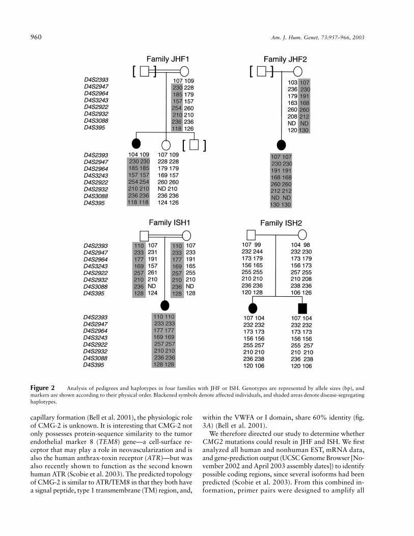
markers (D4S2393, D4S2947, D4S2964, D4S3243,D4S2922, D4S2932, D4S3088, and D4S395). Markerswere amplified by PCR with standard protocols, prod-ucts were run on an ABI 3100 Genetic Analyzer (AppliedBiosystems), and electropherograms were analyzed bythe ABI Genescan and Genotyper software packages(Perkin Elmer), as we described elsewhere (Heath et al.2001). Microsatellite order and distances were determinedusing the Marshfield, UCSC Genome Browser, and De-code databases.
Probands in families ISH1 and JHF2 were homoallelicfor all eight markers, which, although consistent with theprevious linkage report, did not further narrow the re-gion (fig. 2). Support for the originally defined centrom-eric border of the JHF locus was provided by membersof the remaining two families. The centromeric bound-ary of the region was confirmed by the nonhomozygosityof marker D4S2393 in the JHF1 affected individual, inwhom all other tested markers were homoallelic. It isinteresting that, although the affected haplotypes of fami-ly ISH2 suggested a potential narrowing of the distalboundary of the region—as demonstrated by homozy-
gosity of three contiguous markers, D4S2947, D4S2964,and D4S3243—we could not rule out the likelihood thatthis merely reflected “identity-by-state.” Therefore, thecandidate-gene interval could not be conclusively nar-rowed. This caution was found to be supported by DNAsequence analysis (see below).
Inspection of genes in the JHF/ISH common region,by use of a combination of public and private databases(e.g., Celera), revealed a number of possible disease-genecandidates, including bone morphogenetic protein 3(BMP3), fibroblast growth factor-5 (FGF5), and capillarymorphogenesis protein 2 (CMG2). Of these, CMG2 wasimmediately attractive because of its expression in endo-thelial cells and its suggested role in binding extracellularmatrix (ECM) proteins, including laminin and collagenIV, by virtue of its von Willebrand factor A (VWFA)–like domain (Bell et al. 2001). In addition, the pheno-types of murine knockouts of BMP3 and FGF5 genesreported elsewhere were not consistent with either JHFor ISH (Hebert et al. 1994; Daluiski et al. 2001).
Whereas CMG-2 was originally identified on the basisof its up-regulation in endothelial cells induced to undergo
960 Am. J. Hum. Genet. 73:957–966, 2003
Figure 2 Analysis of pedigrees and haplotypes in four families with JHF or ISH. Genotypes are represented by allele sizes (bp), andmarkers are shown according to their physical order. Blackened symbols denote affected individuals, and shaded areas denote disease-segregatinghaplotypes.
capillary formation (Bell et al. 2001), the physiologic roleof CMG-2 is unknown. It is interesting that CMG-2 notonly possesses protein-sequence similarity to the tumorendothelial marker 8 (TEM8) gene—a cell-surface re-ceptor that may play a role in neovascularization and isalso the human anthrax-toxin receptor (ATR)—but wasalso recently shown to function as the second knownhuman ATR (Scobie et al. 2003). The predicted topologyof CMG-2 is similar to ATR/TEM8 in that they both havea signal peptide, type 1 transmembrane (TM) region, and,
within the VWFA or I domain, share 60% identity (fig.3A) (Bell et al. 2001).
We therefore directed our study to determine whetherCMG2 mutations could result in JHF and ISH. We firstanalyzed all human and nonhuman EST, mRNA data,and gene-prediction output (UCSC Genome Browser [No-vember 2002 and April 2003 assembly dates]) to identifypossible coding regions, since several isoforms had beenpredicted (Scobie et al. 2003). From this combined in-formation, primer pairs were designed to amplify all
Reports 961
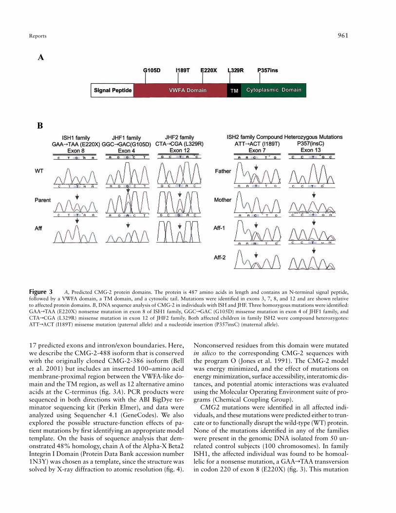
Figure 3 A, Predicted CMG-2 protein domains. The protein is 487 amino acids in length and contains an N-terminal signal peptide,followed by a VWFA domain, a TM domain, and a cytosolic tail. Mutations were identified in exons 3, 7, 8, and 12 and are shown relativeto affected protein domains. B, DNA sequence analysis of CMG-2 in individuals with ISH and JHF. Three homozygous mutations were identified:GAArTAA (E220X) nonsense mutation in exon 8 of ISH1 family, GGCrGAC (G105D) missense mutation in exon 4 of JHF1 family, andCTArCGA (L329R) missense mutation in exon 12 of JHF2 family. Both affected children in family ISH2 were compound heterozygotes:ATTrACT (I189T) missense mutation (paternal allele) and a nucleotide insertion (P357insC) (maternal allele).
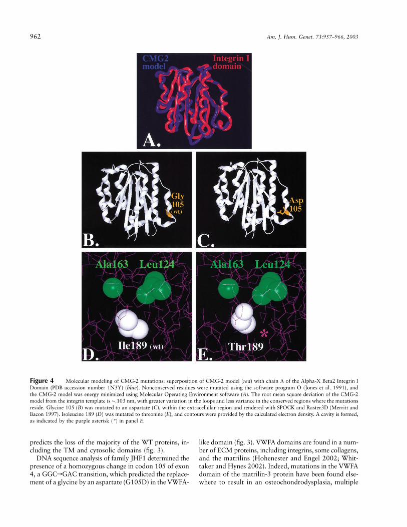
17 predicted exons and intron/exon boundaries. Here,we describe the CMG-2-488 isoform that is conservedwith the originally cloned CMG-2-386 isoform (Bellet al. 2001) but includes an inserted 100–amino acidmembrane-proximal region between the VWFA-like do-main and the TM region, as well as 12 alternative aminoacids at the C-terminus (fig. 3A). PCR products weresequenced in both directions with the ABI BigDye ter-minator sequencing kit (Perkin Elmer), and data wereanalyzed using Sequencher 4.1 (GeneCodes). We alsoexplored the possible structure-function effects of pa-tient mutations by first identifying an appropriate modeltemplate. On the basis of sequence analysis that dem-onstrated 48% homology, chain A of the Alpha-X Beta2Integrin I Domain (Protein Data Bank accession number1N3Y) was chosen as a template, since the structure wassolved by X-ray diffraction to atomic resolution (fig. 4).
Nonconserved residues from this domain were mutatedin silico to the corresponding CMG-2 sequences withthe program O (Jones et al. 1991). The CMG-2 modelwas energy minimized, and the effect of mutations onenergy minimization, surface accessibility, interatomicdis-tances, and potential atomic interactions was evaluatedusing the Molecular Operating Environment suite of pro-grams (Chemical Coupling Group).
CMG2 mutations were identified in all affected indi-viduals, and these mutations were predicted either to trun-cate or to functionally disrupt the wild-type (WT) protein.None of the mutations identified in any of the familieswere present in the genomic DNA isolated from 50 un-related control subjects (100 chromosomes). In familyISH1, the affected individual was found to be homoal-lelic for a nonsense mutation, a GAArTAA transversionin codon 220 of exon 8 (E220X) (fig. 3). This mutation
962 Am. J. Hum. Genet. 73:957–966, 2003
Figure 4 Molecular modeling of CMG-2 mutations: superposition of CMG-2 model (red) with chain A of the Alpha-X Beta2 Integrin IDomain (PDB accession number 1N3Y) (blue). Nonconserved residues were mutated using the software program O (Jones et al. 1991), andthe CMG-2 model was energy minimized using Molecular Operating Environment software (A). The root mean square deviation of the CMG-2model from the integrin template is ∼.103 nm, with greater variation in the loops and less variance in the conserved regions where the mutationsreside. Glycine 105 (B) was mutated to an aspartate (C), within the extracellular region and rendered with SPOCK and Raster3D (Merritt andBacon 1997). Isoleucine 189 (D) was mutated to threonine (E), and contours were provided by the calculated electron density. A cavity is formed,as indicated by the purple asterisk (*) in panel E.
predicts the loss of the majority of the WT proteins, in-cluding the TM and cytosolic domains (fig. 3).
DNA sequence analysis of family JHF1 determined thepresence of a homozygous change in codon 105 of exon4, a GGCrGAC transition, which predicted the replace-ment of a glycine by an aspartate (G105D) in the VWFA-
like domain (fig. 3). VWFA domains are found in a num-ber of ECM proteins, including integrins, some collagens,and the matrilins (Hohenester and Engel 2002; Whit-taker and Hynes 2002). Indeed, mutations in the VWFAdomain of the matrilin-3 protein have been found else-where to result in an osteochondrodysplasia, multiple
Reports 963
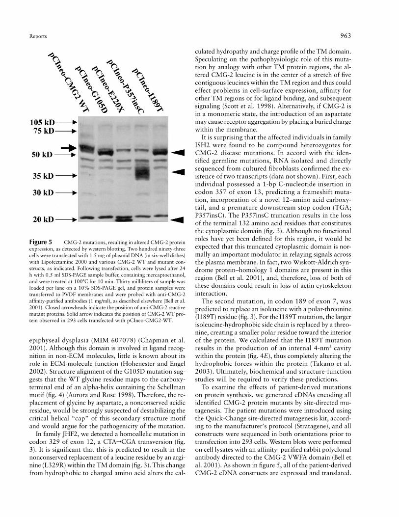
Figure 5 CMG-2 mutations, resulting in altered CMG-2 proteinexpression, as detected by western blotting. Two hundred ninety-threecells were transfected with 1.5 mg of plasmid DNA (in six-well dishes)with Lipofectamine 2000 and various CMG-2 WT and mutant con-structs, as indicated. Following transfection, cells were lysed after 24h with 0.5 ml SDS-PAGE sample buffer, containing mercaptoethanol,and were treated at 100�C for 10 min. Thirty milliliters of sample wasloaded per lane on a 10% SDS-PAGE gel, and protein samples weretransferred to PVDF membranes and were probed with anti-CMG-2affinity-purified antibodies (1 mg/ml), as described elsewhere (Bell et al.2001). Closed arrowheads indicate the position of anti-CMG-2 reactivemutant proteins. Solid arrow indicates the position of CMG-2 WT pro-tein observed in 293 cells transfected with pCIneo-CMG2-WT.
epiphyseal dysplasia (MIM 607078) (Chapman et al.2001). Although this domain is involved in ligand recog-nition in non-ECM molecules, little is known about itsrole in ECM-molecule function (Hohenester and Engel2002). Structure alignment of the G105D mutation sug-gests that the WT glycine residue maps to the carboxy-terminal end of an alpha-helix containing the Schellmanmotif (fig. 4) (Aurora and Rose 1998). Therefore, the re-placement of glycine by aspartate, a nonconserved acidicresidue, would be strongly suspected of destabilizing thecritical helical “cap” of this secondary structure motifand would argue for the pathogenicity of the mutation.
In family JHF2, we detected a homoallelic mutation incodon 329 of exon 12, a CTArCGA transversion (fig.3). It is significant that this is predicted to result in thenonconserved replacement of a leucine residue by an argi-nine (L329R) within the TM domain (fig. 3). This changefrom hydrophobic to charged amino acid alters the cal-
culated hydropathy and charge profile of the TM domain.Speculating on the pathophysiologic role of this muta-tion by analogy with other TM protein regions, the al-tered CMG-2 leucine is in the center of a stretch of fivecontiguous leucines within the TM region and thus couldeffect problems in cell-surface expression, affinity forother TM regions or for ligand binding, and subsequentsignaling (Scott et al. 1998). Alternatively, if CMG-2 isin a monomeric state, the introduction of an aspartatemay cause receptor aggregation by placing a buried chargewithin the membrane.
It is surprising that the affected individuals in familyISH2 were found to be compound heterozygotes forCMG-2 disease mutations. In accord with the iden-tified germline mutations, RNA isolated and directlysequenced from cultured fibroblasts confirmed the ex-istence of two transcripts (data not shown). First, eachindividual possessed a 1-bp C-nucleotide insertion incodon 357 of exon 13, predicting a frameshift muta-tion, incorporation of a novel 12–amino acid carboxy-tail, and a premature downstream stop codon (TGA;P357insC). The P357insC truncation results in the lossof the terminal 132 amino acid residues that constitutesthe cytoplasmic domain (fig. 3). Although no functionalroles have yet been defined for this region, it would beexpected that this truncated cytoplasmic domain is nor-mally an important modulator in relaying signals acrossthe plasma membrane. In fact, two Wiskott-Aldrich syn-drome protein–homology 1 domains are present in thisregion (Bell et al. 2001), and, therefore, loss of both ofthese domains could result in loss of actin cytoskeletoninteraction.
The second mutation, in codon 189 of exon 7, waspredicted to replace an isoleucine with a polar-threonine(I189T) residue (fig. 3). For the I189T mutation, the largerisoleucine-hydrophobic side chain is replaced by a threo-nine, creating a smaller polar residue toward the interiorof the protein. We calculated that the I189T mutationresults in the production of an internal 4-nm3 cavitywithin the protein (fig. 4E), thus completely altering thehydrophobic forces within the protein (Takano et al.2003). Ultimately, biochemical and structure-functionstudies will be required to verify these predictions.
To examine the effects of patient-derived mutationson protein synthesis, we generated cDNAs encoding allidentified CMG-2 protein mutants by site-directed mu-tagenesis. The patient mutations were introduced usingthe Quick-Change site-directed mutagenesis kit, accord-ing to the manufacturer’s protocol (Stratagene), and allconstructs were sequenced in both orientations prior totransfection into 293 cells. Western blots were performedon cell lysates with an affinity–purified rabbit polyclonalantibody directed to the CMG-2 VWFA domain (Bell etal. 2001). As shown in figure 5, all of the patient-derivedCMG-2 cDNA constructs are expressed and translated.
964 Am. J. Hum. Genet. 73:957–966, 2003
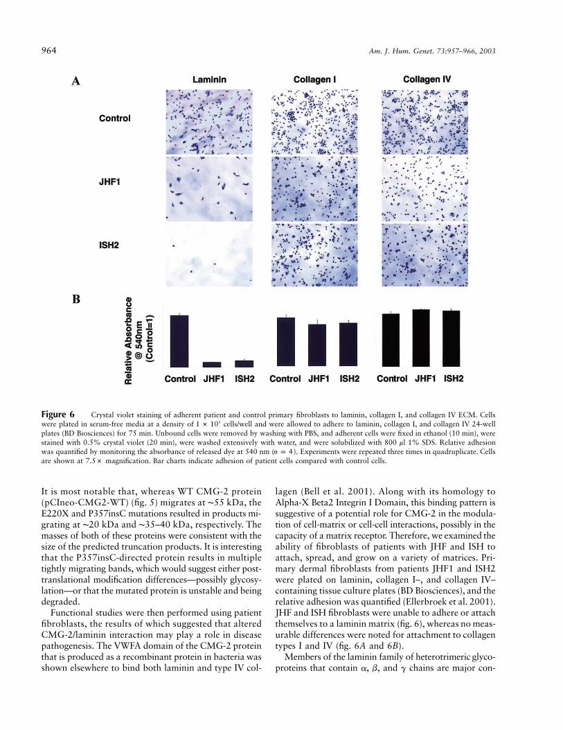
Figure 6 Crystal violet staining of adherent patient and control primary fibroblasts to laminin, collagen I, and collagen IV ECM. Cellswere plated in serum-free media at a density of cells/well and were allowed to adhere to laminin, collagen I, and collagen IV 24-well51 # 10plates (BD Biosciences) for 75 min. Unbound cells were removed by washing with PBS, and adherent cells were fixed in ethanol (10 min), werestained with 0.5% crystal violet (20 min), were washed extensively with water, and were solubilized with 800 ml 1% SDS. Relative adhesionwas quantified by monitoring the absorbance of released dye at 540 nm ( ). Experiments were repeated three times in quadruplicate. Cellsn p 4are shown at 7.5# magnification. Bar charts indicate adhesion of patient cells compared with control cells.
It is most notable that, whereas WT CMG-2 protein(pCIneo-CMG2-WT) (fig. 5) migrates at ∼55 kDa, theE220X and P357insC mutations resulted in products mi-grating at ∼20 kDa and ∼35–40 kDa, respectively. Themasses of both of these proteins were consistent with thesize of the predicted truncation products. It is interestingthat the P357insC-directed protein results in multipletightly migrating bands, which would suggest either post-translational modification differences—possibly glycosy-lation—or that the mutated protein is unstable and beingdegraded.
Functional studies were then performed using patientfibroblasts, the results of which suggested that alteredCMG-2/laminin interaction may play a role in diseasepathogenesis. The VWFA domain of the CMG-2 proteinthat is produced as a recombinant protein in bacteria wasshown elsewhere to bind both laminin and type IV col-
lagen (Bell et al. 2001). Along with its homology toAlpha-X Beta2 Integrin I Domain, this binding pattern issuggestive of a potential role for CMG-2 in the modula-tion of cell-matrix or cell-cell interactions, possibly in thecapacity of a matrix receptor. Therefore, we examined theability of fibroblasts of patients with JHF and ISH toattach, spread, and grow on a variety of matrices. Pri-mary dermal fibroblasts from patients JHF1 and ISH2were plated on laminin, collagen I–, and collagen IV–containing tissue culture plates (BD Biosciences), and therelative adhesion was quantified (Ellerbroek et al. 2001).JHF and ISH fibroblasts were unable to adhere or attachthemselves to a laminin matrix (fig. 6), whereas no meas-urable differences were noted for attachment to collagentypes I and IV (fig. 6A and 6B).
Members of the laminin family of heterotrimeric glyco-proteins that contain a, b, and g chains are major con-
Reports 965
stituents of basement membranes, which are ECMs foundin close contact with individual cells and cell layers (Joneset al. 2000). Acting through specific receptors, laminin iscrucial for the formation of direct contacts between theECM and cells. As would be expected, inherited defectsin laminins are associated with human disease (McGowanand Marinkovich 2000). For example, epidermolysisbullosa letalis (MIM 226700) is caused by mutations inany of the three laminin-5–associated glycoproteins, a3(LAMA3), b3 (LAMB3), or g2 (LAMC2) (Pulkkinen andUitto 1999). In addition, and beyond their structuralroles, laminins help control cellular activities by allowingthe bridging together of information between adjacentcells through interaction with cell surface receptors. Itis striking that mutations in the epithelial-expressed, het-erodimer-linked laminin-receptor proteins, integrin-b4gene (ITGB4), and integrin-a6 gene (ITGA6) cause dis-ease in a subset of these patients but with additional gas-trointestinal manifestations: epidermolysis bullosa withpyloric atresia (MIM 226730) (Vidal et al. 1995; Ruzziet al. 1997). Mutations in any component of dystroglycan,a major receptor for a2-laminins in the muscle sarco-lemma, result in a range of muscular dystrophies that canbe characterized by loss of basement-membrane architec-ture and function (Colognato and Yurchenco 2000). Also,basement-membrane assembly is thought to be regulatedby epithelial-mesenchymal interactions (Lonai 2003), andCMG-2 may play a role in such interactions.
The discovery that CMG2 mutations result in the al-lelic disorders JHF and ISH provides a noninvasive mo-lecular diagnostic tool, defines these two diseases as be-ing on either end of the same disease spectrum, andhighlights novel information on the in vivo function ofthis integrin-like cell surface molecule and its role in keydevelopmental and physiological processes. The dermal,gastrointestinal, and skeletal findings present in thesesyndromes could result from dysregulation in basement-membrane architecture, possibly arising from compro-mised cell-matrix or cell-cell interactions. Histologic re-ports have identified cells embedded within a fibrillo-granular material with cellularity inversely proportionalto the age of the lesion and with abnormal accumulationof extracellular deposits that apparently originate fromdermal blood vessels (Stucki et al. 2001). This strength-ens the hypothesis that CMG-2 plays a role in basementmembrane–matrix homeostasis and architecture duringdevelopment and morphogenesis (Bell et al. 2001) andprovides the starting point for future studies.
Acknowledgments
We thank the families and referring physicians, especiallyDr. Judith Dillner (Mount Sinai School of Medicine), for theirparticipation in this study, and Abigail Guce and Vanessa Men-doza (Mount Sinai School of Medicine), for their expert tech-
nical assistance. This work was supported, in part, by NationalInstitutes of Health National Center for Research Resourcesgrant S10 RR 015935 (to M.J.G.) and National Heart, Lung,and Blood Institute grant HL59373 (to G.E.D).
Electronic-Database Information
URLs for data presented herein are as follows:
Celera, http://www.celera.com/ (for identification of candidategenes)
Center for Medical Genetics, Marshfield Medical ResearchFoundation, http://research.marshfieldclinic.org/genetics/
Decode, http://www.decodegenetics.com/ (for the human geneticmap)
Online Mendelian Inheritance in Man (OMIM), http://www.ncbi.nlm.nih.gov/Omim/ (for JHF, ISH, epidermolysis bul-losa letalis, epidermolysis bullosa with pyloric atresia, andmultiple epiphyseal dysplasia)
Protein Data Bank, http://www.rcsb.org/pdb/UCSC Genomic Bioinformatics, http://genome.cse.ucsc.edu/
References
Aurora R, Rose GD (1998) Helix capping. Protein Sci 7:21–38Bell SE, Mavila A, Salazar R, Bayless KJ, Kanagala S, Maxwell
SA, Davis GE (2001) Differential gene expression duringcapillary morphogenesis in 3D collagen matrices: regulatedexpression of genes involved in basement membrane matrixassembly, cell cycle progression, cellular differentiation andG-protein signaling. J Cell Sci 114:2755–2773
Chapman KL, Mortier GR, Chapman K, Loughlin J, GrantME, Briggs MD (2001) Mutations in the region encoding thevon Willebrand factor A domain of matrilin-3 are associatedwith multiple epiphyseal dysplasia. Nat Genet 28:393–396
Colognato H, Yurchenco PD (2000) Form and function: thelaminin family of heterotrimers. Dev Dyn 218:213–234
Daluiski A, Engstrand T, Bahamonde ME, Gamer LW, AgiusE, Stevenson SL, Cox K, Rosen V, Lyons KM (2001) Bonemorphogenetic protein-3 is a negative regulator of bone den-sity. Nat Genet 27:84–88
Ellerbroek SM, Wu YI, Overall CM, Stack MS (2001) Func-tional interplay between type I collagen and cell surface matrixmetalloproteinase activity. J Biol Chem 276:24833–24842
Fayad MN, Yacoub A, Salman S, Khudr A, Der Kaloustian VM(1987) Juvenile hyaline fibromatosis: two new patients andreview of the literature. Am J Med Genet 26:123–131
Heath KE, Campos-Barros A, Toren A, Rozenfeld-Granot G,Carlsson LE, Savige J, Denison JC, Gregory, MC, White JG,Barker DF, Greinacher A, Epstein CJ, Glucksman MJ, Mar-tignetti JA (2001) Nonmuscle myosin heavy chain IIA mu-tations define a spectrum of autosomal dominant macro-thrombocytopenias: May-Hegglin anomaly and Fechtner,Sebastian, Epstein, and Alport-like syndromes. Am J HumGenet 69:1033–1045
Hebert JM, Rosenquist T, Gotz J, Martin GR (1994) FGF5 asa regulator of the hair growth cycle: evidence from targetedand spontaneous mutations. Cell 78:1017–1025
Hohenester E, Engel J (2002) Domain structure and organisationin extracellular matrix proteins. Matrix Biol 21:115–128
966 Am. J. Hum. Genet. 73:957–966, 2003
Jones JC, Dehart GW, Gonzales M, Goldfinger LE (2000) Lam-inins: an overview. Microsc Res Tech 51:211–213
Jones TA, Zou JY, Cowan SW, Kjeldgaard A (1991) Improvedmethods for building protein models in electron density mapsand the location of errors in these models. Acta CrystallogrA 47:110–119
Keser G, Karabulut B, Oksel F, Calli C, Ustun EE, Akalin T,Kocanaogullari H, Gumudis G, Doganavsargil E (1999) Twosiblings with juvenile hyaline fibromatosis: case reports andreview of the literature. Clin Rheumatol 18:248–252
Kong A, Gudbjartsson DF, Sainz J, Jonsdottir GM, Gudjons-son SA, Richardsson B, Sigurdardottir S, Barnard J, Hall-beck B, Masson G, Shlien A, Palsson ST, Frigge ML, Thor-geirsson TE, Gulcher JR, Stefansson K (2002) A high-reso-lution recombination map of the human genome. Nat Genet31:241–247
Landing BH, Nadorra R (1986) Infantile systemic hyalinosis:report of four cases of a disease, fatal in infancy, apparentlydifferent from juvenile systemic hyalinosis. Pediatr Pathol 6:55–79
Lonai P (2003) Epithelial mesenchymal interactions, the ECMand limb development. J Anat 202:43–50
Mancini GM, Stojanov L, Willemsen R, Kleijer WJ, HuijmansJG, van Diggelen OP, de Klerk JB, Vuzevski VD, Oranje AP(1999) Juvenile hyaline fibromatosis: clinical heterogeneity inthree patients. Dermatology 198:18–25
McGowan KA, Marinkovich MP (2000) Laminins and humandisease. Microsc Res Tech 51:262–279
Merritt EA, Bacon DJ (1997) Raster3D: photorealisticmoleculargraphics. Methods Enzymol 277:505–524
Pulkkinen L, Uitto J (1999) Mutation analysis and moleculargenetics of epidermolysis bullosa. Matrix Biol 18:29–42
Rahman N, Dunstan M, Teare MD, Hanks S, Edkins SJ, Hughes
J, Bignell GR, Mancini G, Kleijer W, Campbell M, Keser G,Black C, Williams N, Arbour L, Warman M, Superti-FurgaA, Futreal, PA, Pope FM (2002) The gene for juvenile hyalinefibromatosis maps to chromosome 4q21. Am J Hum Genet71:975–980
Ruzzi L, Gagnoux-Palacios L, Pinola M, Belli S, Meneguzzi G,D’Alessio M, Zambruno G (1997) A homozygous mutationin the integrin alpha6 gene in junctional epidermolysis bul-losa with pyloric atresia. J Clin Invest 99:2826–2831
Scobie HM, Rainey GJ, Bradley KA, Young JA (2003) Humancapillary morphogenesis protein 2 functions as an anthraxtoxin receptor. Proc Natl Acad Sci USA 100:5170–5174
Scott JP 3rd, Scott JP 2nd, Chao YL, Newman PJ, Ward CM(1998) A frameshift mutation at Gly975 in the transmem-brane domain of GPIIb prevents GPIIb-IIIa expression—analysis of two novel mutations in a kindred with type Iglanzmann thrombasthenia. Thromb Haemost 80:546–550
Stucki U, Spycher MA, Eich G, Rossi A, Sacher P, SteinmannB, Superti-Furga A (2001) Infantile systemic hyalinosis insiblings: clinical report, biochemical and ultrastructural find-ings, and review of the literature. Am J Med Genet 100:122–129
Takano K, Yamagata Y, Yutani K (2003) Buried water mole-cules contribute to the conformational stability of a protein.Protein Eng 16:5–9
Vidal F, Aberdam D, Miquel C, Christiano AM, Pulkkinen L,Uitto J, Ortonne JP, Meneguzzi G (1995) Integrin beta 4 mu-tations associated with junctional epidermolysis bullosa withpyloric atresia. Nat Genet 10:229–234
Whittaker CA, Hynes RO (2002) Distribution and evolutionof von Willebrand/integrin a domains: widely dispersed do-mains with roles in cell adhesion and elsewhere. Mol BiolCell 13:3369–3387
Copyright © 2022 FDOKUMEN