Multiple luteinizing hormone receptor (LHR) protein variants, interspecies reactivity of anti-LHR...
18
BioMed Central Page 1 of 18 (page number not for citation purposes) Reproductive Biology and Endocrinology Open Access Research Multiple luteinizing hormone receptor (LHR) protein variants, interspecies reactivity of anti-LHR mAb clone 3B5, subcellular localization of LHR in human placenta, pelvic floor and brain, and possible role for LHR in the development of abnormal pregnancy, pelvic floor disorders and Alzheimer's disease Antonin Bukovsky* 1 , Korakod Indrapichate 2 , Hiroshi Fujiwara 3 , Maria Cekanova 1 , Maria E Ayala 4 , Roberto Dominguez 4 , Michael R Caudle 1 , Jay Wimalsena 1 , Robert F Elder 1 , Pleas Copas 1 , James S Foster 1 , Romaine I Fernando 1 , Donald C Henley 1 and Nirmala B Upadhyaya 1 Address: 1 Laboratory for Development, Differentiation and Cancer, Department of Obstetrics and Gynecology, The University of Tennessee Graduate School of Medicine, 1924 Alcoa Highway, Knoxville, TN, 37920, USA, 2 School of Biology, Institute of Science, Suranaree University of Technology, Nakhon Ratchasima 30000, Thailand, 3 Department of Gynecology and Obstetrics, Faculty of Medicine, Kyoto University, Sakyoku, Kyoto 60601, Japan and 4 Laboratory of Biology of Reproduction, Facultad de Estudios Profesionales Zaragoza, UNAM, Mexico D.F., Mexico Email: Antonin Bukovsky* - [email protected]; Korakod Indrapichate - [email protected]; Hiroshi Fujiwara - [email protected]; Maria Cekanova - [email protected]; Maria E Ayala - [email protected]; Roberto Dominguez - [email protected]; Michael R Caudle - [email protected]; Jay Wimalsena - [email protected]; Robert F Elder - [email protected]; Pleas Copas - [email protected]; James S Foster - [email protected]; Romaine I Fernando - [email protected]; Donald C Henley - [email protected]; Nirmala B Upadhyaya - [email protected] * Corresponding author Abstract Distinct luteinizing hormone receptor (LHR) protein variants exist due to the posttranslational modifications. Besides ovaries, LHR immunoreactivity (LHRI) was also found in other tissues, such as the brain, fallopian tube, endometrium, trophoblast and resident tissue macrophages. The 3B5 mouse monoclonal antibody was raised against purified rat LHR. In rat, porcine and human ovaries, the 3B5 identified six distinct LHR bands migrating at ~92, 80, 68, 59, 52 and 48 kDa. Characteristic LHRI was detected in rat, human and porcine corpora lutea. During cellular differentiation, subcellular LHR distribution changed from none to granular cytoplasmic, perinuclear, surface, nuclear and no staining. There were also differences in vascular LHR expression – lack of LHRI in ovarian vessels and strong staining of vessels in other tissues investigated. In normal human term placentae, villous LHRI was associated with blood sinusoids and cytotrophoblast cells, and rarely detected in trophoblastic syncytium. In all abnormal placentae, the LHRI of sinusoids was absent, and syncytium showed either enhanced (immature placental phenotypes) or no LHRI (aged placental phenotype). LHRI in human brain was identified in microglial cells (CD68+ resident macrophages). Protein extracts from human vaginal wall and levator ani muscle and fascia showed strong ~92 and 68 kDa species, and LHRI was detected in smooth muscle cells, fibroblasts, resident macrophages and nuclei of skeletal muscle fibers. Our observations indicate that, in contrast to the theory on the role of vascular hormone receptors in preferential pick up of circulating hormones, there is no need to enhance selective pick up rather only prevent LH/CG transport to inappropriate sites. Abnormal placental LHR expression may play a role in the development of abnormal pregnancy. Expression of LHR in the pelvic floor compartments suggests that high LH levels in postmenopausal women may contribute to the pelvic floor relaxation and increased incidence of pelvic floor disorders. Since chorionic gonadotropin increases secretion of a variety of cytokines by monocytes, and induces their inflammatory reaction and phagocytic activity, high LH levels in aging individuals may also activate microglia (mononuclear phagocyte system in the central nervous system) and contribute to the development of Alzheimer's disease and other inflammation-mediated neurodegenerative diseases. Published: 3 June 2003 Reproductive Biology and Endocrinology 2003, 1:46 Received: 18 March 2003 Accepted: 3 June 2003 This article is available from: http://www.RBEj.com/content/1/1/46 © 2003 Bukovsky et al; licensee BioMed Central Ltd. This is an Open Access article: verbatim copying and redistribution of this article are permitted in all media for any purpose, provided this notice is preserved along with the article's original URL.
Transcript of Multiple luteinizing hormone receptor (LHR) protein variants, interspecies reactivity of anti-LHR...

BioMed Central
Reproductive Biology and Endocrinology
ss
Open AcceResearchMultiple luteinizing hormone receptor (LHR) protein variants, interspecies reactivity of anti-LHR mAb clone 3B5, subcellular localization of LHR in human placenta, pelvic floor and brain, and possible role for LHR in the development of abnormal pregnancy, pelvic floor disorders and Alzheimer's diseaseAntonin Bukovsky*1, Korakod Indrapichate2, Hiroshi Fujiwara3, Maria Cekanova1, Maria E Ayala4, Roberto Dominguez4, Michael R Caudle1, Jay Wimalsena1, Robert F Elder1, Pleas Copas1, James S Foster1, Romaine I Fernando1, Donald C Henley1 and Nirmala B Upadhyaya1Address: 1Laboratory for Development, Differentiation and Cancer, Department of Obstetrics and Gynecology, The University of Tennessee Graduate School of Medicine, 1924 Alcoa Highway, Knoxville, TN, 37920, USA, 2School of Biology, Institute of Science, Suranaree University of Technology, Nakhon Ratchasima 30000, Thailand, 3Department of Gynecology and Obstetrics, Faculty of Medicine, Kyoto University, Sakyoku, Kyoto 60601, Japan and 4Laboratory of Biology of Reproduction, Facultad de Estudios Profesionales Zaragoza, UNAM, Mexico D.F., Mexico
Email: Antonin Bukovsky* - [email protected]; Korakod Indrapichate - [email protected]; Hiroshi Fujiwara - [email protected]; Maria Cekanova - [email protected]; Maria E Ayala - [email protected]; Roberto Dominguez - [email protected]; Michael R Caudle - [email protected]; Jay Wimalsena - [email protected]; Robert F Elder - [email protected]; Pleas Copas - [email protected]; James S Foster - [email protected]; Romaine I Fernando - [email protected]; Donald C Henley - [email protected]; Nirmala B Upadhyaya - [email protected]
* Corresponding author
AbstractDistinct luteinizing hormone receptor (LHR) protein variants exist due to the posttranslational modifications. Besidesovaries, LHR immunoreactivity (LHRI) was also found in other tissues, such as the brain, fallopian tube, endometrium,trophoblast and resident tissue macrophages. The 3B5 mouse monoclonal antibody was raised against purified rat LHR.In rat, porcine and human ovaries, the 3B5 identified six distinct LHR bands migrating at ~92, 80, 68, 59, 52 and 48 kDa.Characteristic LHRI was detected in rat, human and porcine corpora lutea. During cellular differentiation, subcellularLHR distribution changed from none to granular cytoplasmic, perinuclear, surface, nuclear and no staining. There werealso differences in vascular LHR expression – lack of LHRI in ovarian vessels and strong staining of vessels in other tissuesinvestigated. In normal human term placentae, villous LHRI was associated with blood sinusoids and cytotrophoblast cells,and rarely detected in trophoblastic syncytium. In all abnormal placentae, the LHRI of sinusoids was absent, and syncytiumshowed either enhanced (immature placental phenotypes) or no LHRI (aged placental phenotype). LHRI in human brainwas identified in microglial cells (CD68+ resident macrophages). Protein extracts from human vaginal wall and levatorani muscle and fascia showed strong ~92 and 68 kDa species, and LHRI was detected in smooth muscle cells, fibroblasts,resident macrophages and nuclei of skeletal muscle fibers. Our observations indicate that, in contrast to the theory onthe role of vascular hormone receptors in preferential pick up of circulating hormones, there is no need to enhanceselective pick up rather only prevent LH/CG transport to inappropriate sites. Abnormal placental LHR expression mayplay a role in the development of abnormal pregnancy. Expression of LHR in the pelvic floor compartments suggests thathigh LH levels in postmenopausal women may contribute to the pelvic floor relaxation and increased incidence of pelvicfloor disorders. Since chorionic gonadotropin increases secretion of a variety of cytokines by monocytes, and inducestheir inflammatory reaction and phagocytic activity, high LH levels in aging individuals may also activate microglia(mononuclear phagocyte system in the central nervous system) and contribute to the development of Alzheimer'sdisease and other inflammation-mediated neurodegenerative diseases.
Published: 3 June 2003
Reproductive Biology and Endocrinology 2003, 1:46
Received: 18 March 2003Accepted: 3 June 2003
This article is available from: http://www.RBEj.com/content/1/1/46
© 2003 Bukovsky et al; licensee BioMed Central Ltd. This is an Open Access article: verbatim copying and redistribution of this article are permitted in all media for any purpose, provided this notice is preserved along with the article's original URL.
Page 1 of 18(page number not for citation purposes)

Reproductive Biology and Endocrinology 2003, 1 http://www.RBEj.com/content/1/1/46
BackgroundThe luteinizing hormone receptor (LHR) plays a funda-mental role in ovarian responsiveness to pituitary LH. TheLHR consists of a 335 residue extracellular domain whichcontains six N-linked glycosylation sites [1]. Posttransla-tional changes in glycosylation and phosphorylationresult in several LHR variants migrating between ~93 and44 kDa [2–17]. Lower molecular weight forms (48 and 44kDa species) appear to represent a glycosylated extracellu-lar domain expressed in mammalian cells (truncatedreceptor) and retain hormone binding specificity. Theyare not secreted from cells, but remain trapped intracellu-larly [18]. In addition to various glycosylated LHR vari-ants, western blotting also yielded a 170 kDa bandrepresenting an LHR dimer [19]. LH binds to LHR variantswith different affinities, and highest affinity appears to beassociated with the fully glycosylated receptor (~90 kDa)[19]. Chorionic gonadotropin (CG), which is importantfor corpus luteum (CL) rescue and maintenance of preg-nancy, also binds to LHR, although with a 10-fold lowerbinding affinity compared with that of LH [20].
The mouse anti-rat LHR monoclonal antibody (mAb),clone 3B5, was developed against purified rat LHR [21].The antibody showed immunoreactivity with rat granu-losa cells of mature (preovulatory) follicles, ovarian thecaland interstitial cells, granulosa-lutein cells of developing,mature and regressing CL, and with testicular Leydig cells,and no reactivity with rat kidneys [22]. During the last tenyears, affinity purified 3B5 antibody has been used in sev-eral immunohistochemical studies [23–26]. To ourknowledge, however, no analysis of the 3B5 antibody bywestern blot has been reported.
In porcine ovaries, LHR expression was detected in granu-losa and theca cells of preovulatory follicles, but not ingranulosa lutein cells of the mature CL [27]. In humanovaries, LHR expression was also detected in granulosaand theca cells of preovulatory follicles, but mature CLshowed strong expression in luteal cells, which disap-peared during luteal regression. CL from early humanpregnancies showed various intensities of LHR expression(from weak to intense) on the surface and in the cytoplas-mic regions of luteal cells [24].
LHR expression was also detected in nonpregnant humanuterus (glandular and luminal epithelial cells, stromalcells, myometrial cells and vascular smooth muscle cells),syncytiotrophoblast of midterm but not term human pla-centa, fetal membranes and decidua [28], human and ratbrain [29,30], rat prostate [14,15], human trophoblastand syncytiotrophoblast from early pregnancies [31], por-cine fallopian tubes and umbilical cord [16,32], ovarian,decidual, endometrial and luteal macrophages [22,33],and ovarian cancer cell cultures and tumor tissues [34].
Beside its involvement in the regulation of ovarian func-tion, the LHR appears to be involved in some additionaleffects. LHR immunoreactivity in porcine fallopian tubeswas confined to the epithelium and smooth muscle cells,and in vitro LH treatment caused relaxation of the oviduct[35]. Also, LHR stimulation plays a role in the inductionand maintenance of myometrial quiescence [36–38].Since smooth muscles belong among important compart-ments of the pelvic floor, high LH levels after menopausemight be involved in the increased incidence of pelvicfloor disorders, a common condition in postmenopausalwomen [39]. Since the brain was reported to express func-tional LHR [29,30], there is also a possibility that high LHlevels in aging individuals may in some way participate inthe development and progression of neurodegenerativeprocess of several degenerative neurological diseases,including Alzheimer's disease.
The aim of the present study was to investigate subcellularLHR distribution and determine LHR protein variantsidentified by 3B5 mAb in various rat, human and porcinetissues. During cellular differentiation, a transition fromcytoplasmic to perinuclear, surface, nuclear and no LHRimmunoreactivity was detected. In protein extracts fromrat, porcine and human ovaries six distinct LHR bandswere observed, migrating between ~92 and 48 kDa.
Materials and MethodsRatsRats of the CII-ZV strain, born and maintained in the Lab-oratory of Biology of Reproduction, Zaragoza, Mexico,were maintained on a 14L-10D cycle (lights on 0500–1900 h). The postnatal rats were weaned at 21 days of age,and they attained sexual maturity during the 6th week oflife. Ovaries were studied in proestrous rats, at the age oftwo months. Daily vaginal smears were collected forabout ten days prior to sacrifice.
Porcine tissuesTwenty porcine ovaries with fallopian tubes and someadditional porcine tissues were obtained from a localabattoir and tissue samples were frozen within one hour.With respect to the follicular and luteal development, por-cine ovaries were macroscopically classified as follows:Early/mid follicular – medium sized antral follicles (3–5mm) accompanied by shrinking fibrous corpora lutea ofsimilar size; late follicular – large antral follicles (>5 mm)not accompanied by any luteal tissue; early/ mid luteal –large corpora lutea (~10 mm) characterized by yellowishcolor; and late luteal – CL of diminishing size (~7 mm)with massive bleeding into the central cavity (bluishcolor) and no accompanying follicles.
Page 2 of 18(page number not for citation purposes)

Reproductive Biology and Endocrinology 2003, 1 http://www.RBEj.com/content/1/1/46
Human tissuesFrozen blocks of human tissues from previous studieswere obtained as described previously [40–44]. Some tis-sues also originated from the Cooperative Human TissueNetwork, Columbus, Ohio. Functional stage of ovarieswas classified according to the endometrial morphology[45]. Ovaries and endometrial samples were obtainedfrom hysterectomy specimens. We also utilized frozenblocks and protein extracts from human placental chori-onic villi, and protein extracts from trophoblast culturesand cultured amniotic fibroblasts and placental mononu-clear cells [46]. Placental mononuclear cells were col-lected from the first digestion, which contains notrophoblast cells [46]. The study was approved by theInstitutional Animal Care and Use Committee and Insti-tutional Review Board.
Tissue processing and peroxidase immunohistochemistryAll chemicals, except where specified otherwise, were pur-chased from Sigma Chemical Co., St. Louis, MO. Tissuesamples were collected into cryomold biopsy vinyl speci-men molds (Tissue-Tek Cryomold Biopsy, Miles Inc.Diagnostic Division, Elkhart, IN) and embedded in anoptimum cutting temperature formulation of water-solu-ble glycols and resins (O.C.T. compound; Miles). Themolds with specimens were frozen by floating on liquidnitrogen and stored at -80°C until use. Frozen tissues weresliced into 7 µm serial sections using a cryostat microtomewith specimen retraction during return travel (Carl ZeissMicrom HM 505 E; MICROM Laborgeräte GmbH, Wal-dorf, Germany) and ten to twelve distinct tissue sectionswere placed on each slide. The slides were dried ~2 h atroom temperature, fixed 5 min in acetone, dried 30 min,and stored at -80°C until immunoperoxidase staining wasperformed. Prior to staining, to prevent water condensa-tion, the slides were transferred at -80°C into pre-chilledair protected boxes containing Drierite granules (W.A.Hammond Drierite Co, Ltd, Xenia, OH), and the boxeswith slides were equilibrated to -20°C, +4 (cold room)and room temperature (20 min each step) beforeopening.
Slides were incubated overnight (cold room) with pri-mary antibodies (see below). Specimens were then trans-ferred to room temperature, extensively washed in freshlyprepared PBS, pH 7.22, and incubated 20 minutes withswine anti-mouse IgG peroxidase conjugate (SwAM;SEVAC Praha, Prague, Czech Republic) diluted 1:20, andpreabsorbed with rat kidney homogenate to remove non-specific background [46]. Control slides were similarlyprocessed, but primary antibody was replaced with PBS.Antigen-antibody complexes were detected by standarddiaminobenzidine technique (brown color). Some slideswere counterstained with hematoxylin, and all slides weredehydrated and mounted. Dual color immunohistochem-
istry experiments were performed as described previously[47].
Evaluation was performed on a Leitz DM RB (Leica Inc.,Wetzlar, Germany) microscope equipped with differentialinterference contrast and a DEI-470 CCD Video CameraSystem (Optronics Engineering, Goleta, CA) with detailenhancement. The video images were captured by CG-7color frame grabber (Scion Corporation, Frederick, MD)supported by Scion Image public software developed atthe National Institutes of Health (Wayne Rasband, NIH,Bethesda, MD), and ported into Microsoft® Power-Point®
97 SR-2 (Microsoft Corporation, Redmont, WA).
Primary antibodiesFor LHR immunohistochemistry, the 3B5 affinity purifiedantibody was used at 50 µg/ml. The antibody is IgG1 class,developed and characterized previously [21,22]. The anti-body was stored at -80°C, either as a lyophylizate, or indiluted aliquots. For immunoblots, the antibody concen-tration was 10 µg/ml. We also used mouse anti-humanThy-1 mAb, clone F15-42-01 [48] (Dr. Rosemarie Dal-chau, Institute of Child Health, London, UK), a marker ofplacental decidual cells (and fibroblasts and brain tissue)[46], mouse anti-rat Thy-1, clone OX-7 [49] (Dr Alan F.Williams, University of Oxford, Oxford, UK), mAbsagainst CD68, a marker of tissue macrophages, and CD14,a marker of monocytes (Dako Corporation, Carpinteria,CA), and leukocyte-common antigen, clone F10-89-4 [50](Dr. John F. Fabre, Institute of Child Health, London,UK).
Tissue extracts and western blot analysisFor preparation of protein extracts from rat, human andporcine tissues for western blot analysis, 150 cryostattissue sections (7 µm) were collected into microcentrifugetubes and lysed by adding ice-cold lysis buffer (20 mMTris pH 7.5, 200 mM NaCl, 0.25% Nonidet P-40; 400 µl/100 mg of tissue sections) containing 1 mM sodiumorthovanadate, 10 mM sodium fluoride, and 1 mM phe-nylmethylsulfonyl fluoride. Tissue culture cells were col-lected and processed as described previously [46].
After 15 min on ice, the tissue and cell lysates were soni-cated using a SonicatorTM Cell Disruptor (Heat Systems-Ultrasonic, Inc., Plainview, NY) for 5 seconds, and centri-fuged at 11000 × g for 20 min at 4°C. Supernatants werestored at -80°C. For western blot analysis, protein concen-trations were determined by Bradford assay (Bio-Rad,Hercules, CA). Equal amounts of unboiled protein (60µg) were loaded onto reducing 10% SDS-Tris-glycinepolyacrylamide gels, transferred to nitrocellulose (Bio-Rad), and processed as described previously [51], withsome modifications.
Page 3 of 18(page number not for citation purposes)

Reproductive Biology and Endocrinology 2003, 1 http://www.RBEj.com/content/1/1/46
Briefly, membranes were washed in Tris-buffered salinecontaining 0.05% Tween 20 (TBST) and non-specificbinding sites were blocked by immersing the membranein blocking reagent (0.5% casein in TBST) for 1 hour atroom temperature on an orbital shaker. After blocking,the membranes were briefly rinsed in two changes ofTBST, and washed once for 15 minutes and twice for 5minutes in fresh changes of TBST. The membranes weresubjected to overnight incubation (4°C) with mouse anti-rat LHR monoclonal antibody in blocking reagent.
The membranes were washed and incubated with peroxi-dase labeled secondary antibody – goat anti-mouse IgGand IgM (Jackson Immunoresearch, West Grove, PA)diluted 1:2000 – for 1 hour at room temperature. Beforestaining, the secondary antibody was diluted 1:60 (5 µl/300 µl), absorbed with rat kidney homogenate (150 mg/300 µl) for 20 min, centrifuged and diluted 1:2000 to thefinal concentration. This absorption eliminated any non-specific background. After incubation, the membraneswere washed 1 × 15 min, 2 × 10 min and 4 × 5 min in freshchanges of TBST, and incubated for 1 hour in blockingbuffer at room temperature. Bound antibodies weredetected by a chemiluminescent detection system (ECL;Amersham Pharmacia Biotech, Piscataway, NJ) as recom-mended by the manufacturer's protocol. For exposure (1min, 5 min, and up to 1 h) we used Kodak XAR film (East-man Kodak, Rochester, NY).
For protein load control, the membranes were reprobedwith actin mouse mAb, clone C4 (Boehringer MannheimCorp., Indianapolis, IN) at 0.25 µg/ml, and developed asabove.
Absorption studiesWe also included absorption studies with porcine ovarianhomogenates. Primary antibody (100 µg) in 2 ml PBS wasincubated 20 min with 500 mg of porcine ovarianhomogenate (late follicular phase) at room temperature.After centrifugation (11000 × g for 10 min at 4°C), thesupernatant was diluted to 10 ml with blocking reagentand used for immunostaining of nitrocellulosemembranes.
ResultsRat tissues (Figure 1)In rat ovaries, the LHR immunoreactivity was detected inthe mature and regressing CL (MCL and RCL, Fig. 1A) andovarian interstitial cells (i), follicular theca interna (t, Fig.1B), and mature granulosa cells (mg, Fig. 1B vs. control,1C). Note a lack of immunoreactivity in ovarian vessels(v, Fig. 1A). The antibody also identified granular cyto-plasmic LHR immunoreactivity in immature granulosacells (ig, Fig. 1B) of preantral and early antral follicles,suggestive of early LHR synthesis. This may represent syn-
thesis of a receptor extracellular domain not yet associatedwith the cell surface, but remaining trapped intracellularly[18].
Maturation of preovulatory follicles was associated with atransition of scarce granular cytoplasmic LHR immunore-activity (ig, Fig. 1D) to LHR accumulation in more differ-entiated granulosa cells (dg). LHR immunoreactivity wasalso found in surface epithelium of the rat uterine cavity,epithelium of uterine glands (se and ug, Fig. 1E), nuclei ofstromal cells (arrowhead), and in uterine vessels (v). Thelatter contrasted with a lack of staining of ovarian vessels(see above).
Human tissues (Figure 2)During follicular selection (mid follicular phase, as deter-mined by endometrium morphology) dominant folliclesin human ovaries showed granular cytoplasmic LHRexpression in granulosa cells (g, Fig. 2A), similar to thosein immature rat follicles (see above). Theca interna (ti)showed more distinct LHR immunoreactivity, character-ized by accumulation of LHR cytoplasmic granules. Follic-ular vessels (vascular layer, vl, and thecal vessels, tv) werevirtually unstained. Some LHR immunoreactivity was alsodetected in theca externa cells (te).
Early luteal development (fresh CL) was associated withvery strong cytoplasmic LHR immunostaining of granu-losa lutein cells migrating into the follicular cavity (gl, Fig.2B). Adjacent theca-lutein cells (tl) also exhibited promi-nent staining, but showed distinct vacuolization. No LHRexpression accompanied developing luteal vessels (v). Inthe mature CL, granulosa lutein cells showed diminutionof cytoplasmic LHR immunoreactivity and strong surfacestaining (Fig. 2C). Luteal cells in the old CL (late lutealperiod) showed marked diminution of LHR expression(Fig. 2D). Regression of luteal tissue (CL investigated dur-ing the follicular phase) showed residual staining oftheca-lutein cells and no staining of regressing granulosalutein cells (Fig. 2E vs. ctr).
In CL of pregnancy (CLP; 3 months), less or more distinctgranular cytoplasmic LHR immunoreactivity (arrowhead,Fig. 2F vs. ctr) and no surface staining was detected. Thissuggests that high CG levels accompanying early preg-nancy may preserve CLP through the cytoplasmic recep-tor. In other words, selective pick up of CG through thesurface LHR expression in granulosa lutein cells duringthe luteal phase after conception appears not to berequired if the circulating CG is very high.
In fallopian tubes, distinct cytoplasmic LHR expressionwas associated with the luminal aspect of epithelial cells(arrowhead, Fig. 2G). In endometrium (late follicularphase – note stromal edema), LHR immunoreactivity was
Page 4 of 18(page number not for citation purposes)

Reproductive Biology and Endocrinology 2003, 1 http://www.RBEj.com/content/1/1/46
associated with endometrial glands (g, Fig. 2H) and withthe surface epithelium of the uterine cavity (se). There wasalso granular cytoplasmic expression in endometrial stro-mal cells (s), precursors of decidual cells, and similargranular staining was observed in smooth muscle cells oflate follicular phase myometrium (data not shown). 3B5antibody also stained human brain vessels (v, Fig. 2I) andshowed strong LHR immunoreactivity with microglialcells (arrowhead; see Fig. 8).
Porcine tissues (Figure 3)Porcine preovulatory follicles showed distinct LHR stain-ing of theca interna, lack of LHR expression in vascularlayer under the follicular membrane, and transition ofgranular cytoplasmic to surface LHR immunoreactivity ingranulosa cells (Fig. 3A,3B,3C, vs. control D). Mature CLshowed strong surface LHR immunoreactivity of lutealcells (Fig. 7E), and old corpora lutea showed diminutionof LHR immunoexpression (Fig. 3F). Ovarian surface epi-thelium, the source of most ovarian tumors, was also
Figure 1LHR expression in the rat ovaries and uterus. A) Strong LHR immunoreactivity is apparent in mature (MCL) and regressing CL (RCL) and interstitial cells (i). Note unstained vessels (v). B) Strong staining of theca interna (t) and weaker staining of mature granulosa cells (mg) in preovulatory follicle. Note scarce cytoplasmic granules with LHR immunoreactivity in immature granu-losa cells (ig). C) Control. D) Increased staining during granulosa cell differentiation (dg) in large antral follicle. E) In the rat uterus, the LHR immunostaining is apparent in the surface epithelium (se), uterine glands (ug) and vasculature (v). Uterine stro-mal cells show nuclear staining (arrowhead). No hematoxylin counterstain.
BB
DD igig
dgdg
ii
tt
igig
mgmg
tt
ii
ugug
sese
100 m100 m
vv
AA
RCLRCL
MCLMCL
ii vv
CCigig
mgmg
tt
ii
igig
EE
Page 5 of 18(page number not for citation purposes)

Reproductive Biology and Endocrinology 2003, 1 http://www.RBEj.com/content/1/1/46
Figure 2Reactivity of 3B5 with human tissues. A) Dominant follicle (DOF, midfollicular phase) – g, granulosa; vl, vascular layer; ti, theca interna; tv, thecal vessels; te, theca externa. B) Fresh CL – gl, granulosa lutein cells; tl, theca lutein cells; v, proliferating luteal vessels. C) Mature CL – midluteal phase. D) Old CL (OCL, late luteal phase). E) Regressing CL (RCL, mid-follicular phase of the subsequent cycle). F) Corpus luteum of pregnancy (CLP, 3rd month of pregnancy) Arrowhead indicates cytoplasmic LHR immunoreactivity. G) Fallopian tube. Arrowhead indicates LHR immunoreactivity at the luminal aspects of epithelial cells. H) Endometrium (EM, late follicular phase – note stromal edema) – se, surface epithelium; g, glands; s, endometrial stromal cells. I) Staining of brain microvasculature (v) and microglial cells (arrowhead – see Fig. 8). Insets show control staining (ctr). Hematox-ylin counterstain except panel F.
AA BB
CC DD
FFEE
GG HH
3B5 ctr
DO
F
OC
LF
CL
MC
Lglgl
tltl
vv
vv
gg
titi
tete
vlvl
tvtv
ctr
RC
L
EM
glgl
tltl
glgl
tltl
FT
sese
gg
ctrctr
CL
P
ss
II
Br
vv 100 m100 m
Page 6 of 18(page number not for citation purposes)

Reproductive Biology and Endocrinology 2003, 1 http://www.RBEj.com/content/1/1/46
Figure 3LHR immunostaining in porcine tissues. A–C) Preovulatory follicles (late follicular phase) show a transition from granular cyto-plasmic to surface LHR immunoreactivity in granulosa cells (arrowheads), lack of staining of vascular layer, increase of LHR staining in theca interna and extreme dilatation of follicular microvasculature (v). D) Control for C. E) Mature CL. F) Old CL. G) Ovarian surface epithelium (arrowhead). H) Fallopian tube (FT). I) Skin vessels show strong LHR immunoreactivity of the entire vascular wall (arrowhead). J) In ovarian vasculature, the LHR immunoreactivity is limited to the nuclei of some endothe-lial cells (arrowhead) but other components of vascular wall are unstained. Insets in E–J show control staining. Details in text. Hematoxylin counterstain except panels A–D, H and I.
AA CCBBP
OF
100 m100 m
titi
vlvl titi
titi
vlvl vlvl
vvgg gg gg
titi
vlvl
gg
DD
EE
MC
L
OC
L
ctrctr ctrctr
Ov S
E
ctrctr
Ov v
es
Skin
ve
s
ctrctr
FT
ctrctrctrctr
HHGG
FF
JJII
Page 7 of 18(page number not for citation purposes)

Reproductive Biology and Endocrinology 2003, 1 http://www.RBEj.com/content/1/1/46
stained (arrowhead, Fig. 3G). In porcine fallopian tube(Fig. 3H), the LHR immunostaining was similar to that inhuman oviduct. Strong LHR immunoreactivity in vascularwalls was detected in the skin (Fig. 3I), which does notrepresent a classic LH/CG target. This contrasted with alack of staining in ovarian vessels, except weak nuclearstaining of endothelial cells (arrowhead, Fig. 3J). Insets inFig. 7E,7F,7G,7H,7I,7J show the control procedure.
Observations of LHR immunoreactivity in porcine tissuesindicate that LHR distribution is similar to that in humantissues. They also show LHR expression in the ovarian sur-face epithelium (not investigated in human ovaries) andastonishing differences in vascular walls of nonclassic(strong LHR in skin vessels) and classic LHR targets (weekexpression in ovarian vessels) – see Ref. [40] and Discus-sion beneath for a role of vascular LHR expression in theLH/CG delivery.
Multiple rat, human and porcine LHR protein variants (Figures 4 and 5)Western blot analysis of protein extracts from normal ratovaries and human and porcine mature CL with 3B5 anti-body showed six distinct bands migrating between ~92
and ~48 kDa (lanes 2, 4 and 6, Fig. 4; see also Fig. 5A).Although weak, some LHR immunoreactivity was alsodetected in the rat kidney protein extract (lane 1). Thisobservation is consistent with a lack of LHR immunoreac-tivity in rat kidney cryostat sections reported previously[22].
Distinct LHR expression was detected in rat proestrousovaries (lane 2, Fig. 4), which contain LHR+ CL at variousstages of development and regression, and developingand mature preovulatory follicles. However, in porcinepreovulatory ovaries (late follicular phase), which containnumerous preovulatory follicles but no luteal tissue, verystrong LHR immunoreactivity was also detected (lane 3,Fig. 4). Regression of porcine luteal tissue (RCL; lane 5)was associated with a marked decline of LHR expression.This confirmed immunohistochemical observations.
In human tissues, mature CL (MCL; lane 6) showed LHRbands similar to that in mature porcine CL. Very strongexpression of all six LHR variants was detected in the fal-lopian tube (FT; lane 7), which is the site of first contactwith the developing embryo secreting CG, a signalrequired for CL preservation.
Figure 4Western blot analysis of rat, porcine and human tissues with 3B5. KN, kidney; Ov/P, ovary in proestrus; POF, preovulatory fol-licle; MCL, mature CL; RCL, regressing CL (follicular phase); FT, fallopian tube; CV, chorionic villi (term placenta); T:0, time 0 trophoblast culture; EC, early culture (24 h); LC, late culture (72 h); MC, cultured placental mesenchymal cells; FB, cultured amniotic fibroblasts; VagW, vaginal wall (epithelium, connective tissue and smooth muscle bundles); LM, levator ani muscle; LF, levator ani fascia. Details in text.
KN Ov/P MCL MCLPOF RCL
1 2 3 4 5 6 7 8 9 10 11 12 13 14 15 16
LM LF
68>
52>
48>
80>
92>
59>
LHR
kDa VagWCV EC LCT:0FT MC FB
Rat Porcine Human
44>
170>
Page 8 of 18(page number not for citation purposes)

Reproductive Biology and Endocrinology 2003, 1 http://www.RBEj.com/content/1/1/46
Placental chorionic villi (CV; lane 8) also showed six dis-tinct bands similar to those detected in the rat, humanand porcine ovaries. However, time 0 and early and latetrophoblast cultures derived from the same placenta
(lanes 9–11) showed additional high molecular weightLHR variants (~170 kDa), possibly LHR homodimers[19], which were barely detectable or undetectable inother tissues, including placental CV. Moreover, there
Figure 53B5 absorbed with porcine ovaries (western blot control). Reactivity of 3B5 in western blots (A) diminishes after preabsorp-tion with porcine ovaries [porcine ovary absorbed (POA) 3B5] (B). C) Actin control for protein loading. For abbreviations see Fig. 4.
kDa
Rat
Ov/P
Porc
MCL MCL FT CV EC LCT:0
3B5
POA
3B5
actin
1 2 3 4 5 6 7 8
Human
68>
52>
48>
80>
92>
59>
44>
170>
68>
52>
48>
80>
92>
59>
44>
170>B
A
C
Page 9 of 18(page number not for citation purposes)

Reproductive Biology and Endocrinology 2003, 1 http://www.RBEj.com/content/1/1/46
were additional bands detected in trophoblast cultures(~85, 60, and 46 kDa species) and a lack of a ~52 kDa var-iant as compared to the luteal, FT and CV protein extracts.The 44 kDa species, probably a partially degraded formfrom LHR turnover [18], was observed in placentalcultures (lanes 9–13) and pelvic floor compartments(lanes 14–16).
We also investigated cultures of placental mesenchymalcells (MC), which are always associated with trophoblastcultures [40,41], and amniotic fibroblasts (FB). Placentalmesenchymal cells showed a prominent LHR band at ~68kDa (lane 12). Note the virtual lack of other bands char-acteristic of trophoblast cultures. Placental amnioticfibroblasts showed two distinct LHR bands migrating at~59 and 48 kDa (lane 13). Similar ~59 and 48 kDa specieswere distinctly expressed in trophoblast cultures.
Western blot analysis of pelvic floor compartmentsshowed strong expression of ~92 and 68 kDa species inprotein extracts from the vaginal wall (VagW; lane 14),which consists of vaginal epithelium, longitudinal andcircular smooth muscle cell bundles, and connective tis-sue. A less prominent band was detected at ~80 kDa.Three similar bands were also found in human levator animuscle and fascia (lanes 15 and 16). These observationsindicate that pelvic floor compartments represent poten-tial targets for the LH/CG effect. Absorption with porcineovaries resulted in severe diminution of 3B5 reactivitywith rat, porcine and human tissues in western blots (Fig.5B vs. 5A).
Altogether, these data indicate that the 3B5 antibody iden-tifies six LHR protein variants showing distinct expressionin rat, porcine and human tissues, including human pla-centa, with some variants also detected in human femalepelvic floor compartments.
Human chorionic villi and placental membranes (Figure 6)In normal placentae at term, the LHR immunoreactivity inmature chorionic villi was found in mononucleatedcytotrophoblast cells under the syncytial layer (arrow-heads, Fig 6A) and villous vascular sinusoids (v), but mostof the syncytiotrophoblast was virtually unstained (whiteasterisks vs. black). Mononucleated trophoblast cellsshowed variable nuclear LHR immunoreactivity (Fig. 6B,detail from A). The most prominent nuclear staining wasfound in trophoblast cells merging with the syncytium(full arrowheads vs. white). The red arrowhead indicatesdiminution of LHR expression in the cell which joined thesyncytial layer. Staining of villous sinusoids was associ-ated with basal (abluminal) aspects of endothelial cells(arrowheads, Fig. 6C). Developing (immature) chorionicvilli were occasionally observed and showed more pro-nounced granular cytoplasmic LHR immunoreactivity in
the syncytial layer (asterisk, Fig. 6D), a feature alsodetected in immature granulosa cells (see above).
In contrast to normal placentae, granular cytoplasmicstaining of the syncytial layer dominated in some moder-ately abnormal placentae (asterisks, Fig. 6E), whichshowed mild compensatory dilatation of villous sinu-soids (v) – see Ref. [52] for definition of placental abnor-mality. In such placentae, the LHR+ mononucleatedtrophoblast cells were barely detectable, as well as LHRexpression in villous sinusoids (white arrowheads).
Differences between placental types were more obvious atlower magnification. Figure 6F (vs. control 6I), shows nor-mal placenta with prominent LHR staining of villoussinusoids (full arrowhead) and no staining of syncytium(white asterisk). Figure 6G shows LHR immunostainingof syncytium (black asterisk) and lack of vascular LHR(white arrowhead), resembling immature chorionic villi(immature placental phenotypes, characteristic for intrau-terine growth retardation and maternal diabetes [52] –note lack of dilatation of villous sinusoids). Figure 6Hshows that neither surface (white asterisk) nor significantvascular LHR immunoreactivity (white arrowhead) wasdetected in placentae with extreme dilatation of villoussinusoids (aged placental phenotype [52]).
In placental membranes, occasionally found multilayeredamniotic epithelium showed no or fine granular cytoplas-mic staining of epithelial cells adjacent to the basementmembrane (full arrowhead, Fig. 6J), followed byperinuclear (yellow arrowhead) and nuclear LHR immu-noexpression (red arrowhead). Cells most distant fromthe basement membrane showed no staining (whitearrowhead).
Extravillous trophoblast cells in placental membranes andplacental basal plate (Fig. 6K) exhibited usually perinu-clear (full arrowheads) and occasionally nuclear LHRexpression (data not shown). In contrast, however,nuclear or no staining was prevalent in adjacent decidualcells (white arrowheads). Fig. 6L shows a semi-parallelsection stained for Thy-1 glycoprotein, a characteristicmarker of human decidual cells [46]. Note Thy-1+ decid-ual cells (white arrowhead) and unstained extravilloustrophoblast cells (full arrowhead). Dual color immuno-histochemistry for LHR (brown) and CD68 (blue; Fig.6M) indicates that LHR+ decidual cells also show expres-sion of CD68 (white arrowhead – note brown and bluecolor) of decidual macrophages (red arrowhead), i.e.,marker also characteristic for regressing luteal cells [40].Note a lack of macrophages among trophoblast cells.
Page 10 of 18(page number not for citation purposes)

Reproductive Biology and Endocrinology 2003, 1 http://www.RBEj.com/content/1/1/46
Figure 6LHR immunoreactivity in chorionic villi and placental membranes. A) LHR+ mononucleated trophoblast (arrowheads) and vil-lous vascular sinusoids (v) in normal term placentae. Note unstained syncytium (white asterisks) or sporadic LHR+ granules (black asterisk). B) Detail from (A) (turned 90°) shows cytotrophoblast cells merging with syncytium (full arrowheads vs. white arrowhead), followed by diminution of LHR immunoreactivity (red arrowhead). C) Villous sinusoids. D) Immature (developing) villus in normal placenta. E) Abnormal placenta (note enhanced dilatation of all sinusoids – compare with A). Panels (F–H) show staining for LHR (DAB-Ni substrate) in various placental types (see text) vs. control (I). J) Occasionally detected multi-layered amniotic epithelium shows a transition from granular cytoplasmic (full arrowhead) to perinuclear (yellow arrowhead) and nuclear LHR immunoreactivity (red arrowhead). Note unstained surface cells (white arrowhead). K) Extravillous trophob-last cells (basal plate) with strong perinuclear staining (full arrowheads) variable LHR immunoreactivity in decidual cells (white arrowheads). L) Parallel section shows Thy-1+ decidual cells (white arrowhead) and unstained trophoblast cells (full arrow-head). M) Dual color immunohistochemistry: extravillous trophoblast expressing LHR (full arrowhead), decidual cells express-ing both markers (white arrowhead) and decidual macrophages expressing CD68 only (red arrowhead). Hematoxylin counterstain except panels F–I and panel M (dual color immunohistochemistry). Bar in C, for B and C; bar in H, for F–I. For other panels see bar in A.
BB
CC
**
**
vv
JJ
AA
KK LL
50 m50 m
Thy-1Thy-1
**2
0
m2
0
m
vv
**
**
**
EE
**
vv
**
**
vv
DD
GG
FF1
00
m
10
0
mHH
MM
LHR/CD68LHR/CD68
**
** **II
ctrctr
Page 11 of 18(page number not for citation purposes)
Reproductive Biology and Endocrinology 2003, 1 http://www.RBEj.com/content/1/1/46
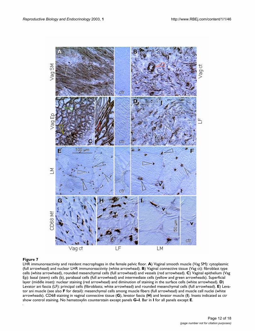
Figure 7LHR immunoreactivity and resident macrophages in the female pelvic floor. A) Vaginal smooth muscle (Vag SM): cytoplasmic (full arrowhead) and nuclear LHR immunoreactivity (white arrowhead). B) Vaginal connective tissue (Vag ct): fibroblast type cells (white arrowhead), rounded mesenchymal cells (full arrowhead) and vessels (red arrowhead). C) Vaginal epithelium (Vag Ep): basal (stem) cells (b), parabasal cells (full arrowhead) and intermediate cells (yellow and green arrowheads). Superficial layer (middle inset): nuclear staining (red arrowhead) and diminution of staining in the surface cells (white arrowhead). D) Levator ani fascia (LF): principal cells (fibroblasts; white arrowhead) and rounded mesenchymal cells (full arrowhead). E) Leva-tor ani muscle (see also F for detail): mesenchymal cells among muscle fibers (full arrowhead) and muscle cell nuclei (white arrowheads). CD68 staining in vaginal connective tissue (G), levator fascia (H) and levator muscle (I). Insets indicated as ctr show control staining. No hematoxylin counterstain except panels G–I. Bar in I for all panels except E.
Page 12 of 18(page number not for citation purposes)

Reproductive Biology and Endocrinology 2003, 1 http://www.RBEj.com/content/1/1/46
Figure 8LHR immunostaining in the gray matter of the human brain parietal lobe. A) LHR+ microglial cells with characteristic dendritic processes (full arrowhead) and flattened nuclei (yellow arrowheads). Green arrowhead indicates unstained nerve cell. B) Area occupied by microglia (full and yellow arrowheads) contains unstained nerve cells (green arrowheads). However, area lacking microglia contains LHR+ nerve cells (red arrowheads). C) Staining for leukocyte common antigen (LCA) reveals picture similar to that in (A). Note dendritic processes of microglial cells (full arrowheads), and interaction with neuronal (red arrowhead) and other glial elements (white arrowhead). D) Anti human Thy-1 (hThy-1) shows diffuse staining of brain tissue except for the vascular endothelium (v). E) Control staining with anti-rat Thy-1 (rThy-1). F–K) Schematic drawings of interstitial (glial) cells in the human brain and differences in their nuclei (from Ref. [54]). F) Fibrous astrocytes – white matter. G) Oligodendroglial cells – satellites to nerve cells. H) Protoplasmic astrocytes – grey matter. I) Microglial cells – mesodermal origin, gray and white matter. Yellow arrowhead indicates flattened nucleus of microglial cell. J) Irregularly oval nuclei of astrocytes (full arrowhead) and oval nuclei of oligodendroglia (white arrowhead). K) Small, often flattened nuclei of microglial cells. L) CD68 staining shows flattened nucleus of microglial cell (yellow arrowhead). M) No staining was observed with CD14, a marker of mono-cytes and immature tissue macrophages. Hematoxylin counterstain: weak in panels A and B, moderate in C-E, L and M. Bar in M, for A-E, L and M.
AA BB
DDCC EE
FF
HH
GG
II
KKJJ
88
CD68CD68 CD14CD14
LCALCA
3B53B5 3B53B5
hThy-1hThy-1 rThy-1rThy-1
vv
vv
50 m50 m
LL MM
Page 13 of 18(page number not for citation purposes)

Reproductive Biology and Endocrinology 2003, 1 http://www.RBEj.com/content/1/1/46
Pelvic floor (Figure 7)Previously documented LH-dependent relaxation of theporcine oviduct [53] raises a question if high LH levels inpostmenopausal women do not contribute to the relaxa-tion of the pelvic floor, and hence to the increased inci-dence of pelvic floor disorders in these females. Weinvestigated connective tissues and muscular compart-ments of the pelvic floor, in order to determine cellularequivalents to LHR bands detected in western blots andpossible cellular targets for the LH effect.
Strong cytoplasmic and nuclear LHR immunoreactivitywas found in smooth muscle cells in the vaginal wall (Fig.7A vs. ctr). Vaginal connective tissue fibroblasts (whitearrowhead, Fig. 7B) and other stromal elements (fullarrowhead), as well as vaginal vessels (red arrowhead),also showed distinct LHR immunoreactivity.
In vaginal epithelial layers, a model for differentiation ofepithelial cells [42], basal (stem) cells were unstained (b,Fig. 7C), dense cytoplasmic staining was found in paraba-sal (immature) cells (full arrowhead), followed by perinu-clear (yellow arrowhead) and cell surface LHRimmunoreactivity (green arrowhead). Nuclear stainingwas found in lower superficial cells (red arrowhead inadjacent panel), and cornified superficial cells showedoverall diminution of LHR immunoreactivity (whitearrowhead).
Additional pelvic floor structure, the levator ani fascia,was studied and exhibited nuclear LHR immunoreactivityin principal cells (fibroblasts; white arrowhead, Fig. 7D)and other mesenchymal cells with rounded nuclei (fullarrowhead), most probably resident macrophages (seebelow). Mesenchymal cells in levator ani muscle alsoshowed LHR immunoreactivity (full arrowheads, Fig. 7Eand 7F). In addition, nuclear LHR staining was alsodetected in levator ani striated muscle fibers, some ofwhich were centrally (abnormally) located in this sample(white arrowheads, Fig. 7E and 7F). Note unstained mus-cle vessels (v) accompanied by LHR+ stromal cells.
Distribution of resident macrophages, as shown in Fig.7G,7H,7I, suggests that rounded mesenchymal cellsshowing LHR immunoreactivity in pelvic floor connectivetissues might be represented by this type of cells. Noteassociation of some resident macrophages with vascula-ture (v).
These observations indicate that principal components ofthe pelvic floor (smooth muscle cells, fibroblasts and stri-ated muscle fibers), which are responsible for pelvic floorintegrity, are potential targets for LH/CG hormonaleffects. Additional cells exhibiting LHR immunoreactivity
in pelvic floor connective tissues appear to be residentmacrophages.
Human brain (Figure 8)Neurodegenerative diseases, including Alzheimer's dis-ease, also show increased incidence in aging individuals.Therefore, a question arises if the rise in LH, reflectingdiminution of sex steroids, can also contribute to theinitiation and progression of neurodegeneration. Westudied LHR expression in the gray matter of the humanparietal lobe cortex. Strong LHR immunoreactivity wasassociated with small cells with flattened nuclei (yellowarrowheads, Fig. 8A) and numerous spiny processes (fullarrowhead). Adjacent nerve cells were unstained (greenarrowheads, Fig. 8B), but distant nerve cells showed mod-erate staining (red arrowheads).
Staining for the leukocyte common antigen (Fig. 8C) andobservation for characteristic morphology and nuclearshape (Fig. 8I vs. 8F,8G,8H; Fig. 8K vs. 8J – adapted fromRef. [54]) as well as expression of CD68 of resident mac-rophages (Fig. 8L) indicate that LHR immunoreactivity inFig. 8A identifies microglial cells of mesenchymal (meso-dermal) origin. Yet, Fig. 8M indicates that these cells donot express CD14 of monocytes (and primitive tissuemacrophages [42]). This suggests that they are highly dif-ferentiated dendritic type cells. Figure 8D shows diffuseexpression of Thy-1 in the cortex, identified with anti-human Thy-1 antibody. Figure 8E is a control using theanti-rat Thy-1 antibody (not reacting with human tissues).
These data indicate that microglial cells and some nervecells in the gray matter of the human brain cortex may beinfluenced by LH/CG proteohormones. Yet, loss of graymatter from areas of the parietal and temporal lobes is themost obvious consequence of Alzheimer's disease[55,56].
DiscussionOur observations indicate that the 3B5 anti-LHR antibodyreacts with rat, porcine and human tissues. In westernblots, the 3B5 antibody identifies six distinct bandsmigrating between ~92 and 48 kDa, and this reactivitywith protein extracts from all three species diminisheswhen the antibody is preabsorbed with porcine ovaries.We also show changes in subcellular LHR distributionduring differentiation of various cell types. To our knowl-edge, LHR expression in fibroblasts, striated muscle cells,and microglial cells (CD68+ resident macrophages) in thecentral nervous system is presented for the first time. Wealso report tissue differences in vascular LHR expression.
During cellular differentiation, subcellular LHR distribu-tion changed from granular cytoplasmic to perinuclear,surface and nuclear staining; virtually no staining was
Page 14 of 18(page number not for citation purposes)

Reproductive Biology and Endocrinology 2003, 1 http://www.RBEj.com/content/1/1/46
detected in stem cells and most differentiated and agedcells. Yet, cell surface staining was observed only on classicLH/CG targets, granulosa and theca cells of preovulatoryfollicles and mature luteal cells, but also in the vaginalepithelium. This indicates that these cells are prepared forthe selective accumulation of LH/CG signals from thecirculation.
There was also a striking difference in vascular LHRexpression between ovaries and other tissues. In ovarian(and also striated muscle) vessels the LHR expression wasvirtually absent (Fig. 1A, 2A, 3J), while other tissues inves-tigated, including uterus, brain, skin and vagina, showeddistinct vascular LHR immunoreactivity. We also reportedpreviously that in the rat the endothelial LHR expressionis absent in testicular vessels, and vessels in other tissuesinvolved in LH/CG transport into (pituitary) and from theblood (kidney) [22]. Saturation of LHR in ovarian targetsmay be dependent on the precise delivery of smallamounts of circulating LH/CG from the blood into theextravascular space. A receptor mediated endothelialtransport of hCG has been suggested to represent a modelfor general involvement of specific receptors in transportof other plasma proteins [57,58]. Yet, application of thistheory on our data will indicate that the LH/CG will bedelivered to the uterus, brain, skin and vagina rather thanto the ovary. Hence, the opposite mechanism should beconsidered – endothelial LHR expression may suppressthe transport of LH/CG from vessels to the extravascularspace. The LH/CG molecule is smaller than that of albu-min or IgG, i.e., proteins exhibiting ubiquitous distribu-tion, so there is no need to enhance rather only preventLH/CG transport to inappropriate sites. Binding of LH/CG to endothelial cells expressing LHR may prevent LH/CG transport from circulation by electrostatic forces, andthe lack of endothelial LHR expression may be associatedwith LH/CG transport mediated by a general mechanisminvolved in protein exchange [40].
We report that normal term placentae show virtually noLHR expression in trophoblastic syncytium while syncy-tium of some abnormal placentae (immature pheno-types) exhibits LHR immunoreactivity. This resembleshigh LHR expression in first trimester placenta [28], whichis a source of high hCG levels [59]. Yet, hCG levels fallduring the second trimester, and elevated maternalmidtrimester hCG is associated with higher rates of mater-nal and neonatal complications (pregnancy-inducedhypertension, preeclampsia, gestational diabetes, andperinatal death) [60–63].
Hence, some abnormal term placentae appear to preservesyncytium in a younger state, accompanied by high secre-tion of hCG. In contrast, vascular LHR expression wasdetected in sinusoids of chorionic villi from normal term
placentae, but was virtually absent in all abnormal placen-tae. If the vascular LHR expression represents a barrier forhCG transport from chorionic villi to the fetal blood (seeabove), lack of this barrier may cause high levels of fetalhCG resulting in perinatal morbidity and mortality [63],regardless of normal or abnormal hCG production.
Western blot analysis of placental villi and trophoblastcultures indicates that cultured trophoblast cells showadditional bands, including ~170 kDa species, possibly anLHR homodimer [19]. Although the ~170 kDa speciespersisted in late cultures, other (additional) LHR variantsdiminished. This suggests an activation of isolated tro-phoblast cells and enhanced LHR synthesis with forma-tion of additional glycosylated LHR variants, particularlyin time 0 and early cultures. When compared to chorionicvilli, trophoblast cultures showed stronger ~59 and 48kDa species, characteristic for cultured fibroblasts. Indeed,it has been reported, that differentiation of cultured tro-phoblast is associated with activation of accompanyingfibroblasts [64].
Early differentiation of granulosa, luteal, and other celltypes expressing LHR was associated with cytoplasmicLHR immunoreactivity, reflecting receptor synthesis priorits surface expression. In addition, the CL of pregnancy,the function of which is highly dependent on CG, alsoshowed cytoplasmic expression. Hence, it is possible thathigh levels of circulating LH/CG, e.g., during pregnancyand after menopause, may influence not only cells withsurface LH expression, but also cells with cytoplasmic,and perhaps exclusively nuclear LHR expression (pelvicfloor compartments, including striated muscle). In otherwords, cells lacking surface LHR may not be influenced bytemporary increase of LH production during thepreovulatory period of the ovarian cycle due to the "cellmembrane LHR barrier," and the LH effect is probablyalso prevented by the "vascular LHR barrier." Yet, high CGlevels during pregnancy may pass the "cell membrane bar-rier" and act through the cytoplasmic LHR in the CL ofpregnancy and other cells with cytoplasmic expression,and such effect could be enhanced if the "vascular LHRbarrier" is absent – abnormal placentae of aged pheno-type and similar putative age-related vascular changes inother tissues.
But the LH/CG action through the cytoplasmic/perinu-clear and nuclear LHRs opens a possibility of more directLH/CG action within the cells, which may not require aclassic second messenger pathway (cyclic AMP dependentsignaling mechanism) involvement, or this system can actthrough the intracellular LHRs. Evidence is accumulatingthat a number of other factors modulate the actions ofgonadotropins in the ovary and testis via activation ofalternative signaling pathways and via LHR protein vari-
Page 15 of 18(page number not for citation purposes)

Reproductive Biology and Endocrinology 2003, 1 http://www.RBEj.com/content/1/1/46
ants, and alternative second messenger pathways for thetransmission of the LHR activation effect exist, which maynot include stimulation of cyclic AMP levels [65–69]. Aquestion also arises if the nuclear LHR expression in ter-minally differentiated cells (e.g., striated muscle fibers)may not indicate a possibility of the direct effect of LH/CGon modulation of protein synthesis, reflecting certaineffects of sex steroids on their nuclear receptors.
LH/CG causes a relaxation of smooth muscle cells, whichexpress LHR [36–38] (Fig. 7A). The data presented alsoindicate that additional principal cell types in the pelvicfloor show LHR expression, including stromal and fascialfibroblasts and striated muscle cells. This is associatedwith relatively high expression of LHR protein in westernblots, and a fully glycosylated ~92 kDa species in particu-lar. Interestingly, strong ~59 and 48 kDa proteinsexpressed in cultured fibroblasts were not detected in pel-vic floor lysates, but a distinct ~68 kDa band from mesen-chymal cells was apparent. We speculate that a ~92 kDaspecies in the pelvic floor is a result of interaction of resi-dent macrophages with pelvic floor fibroblasts.
The hCG secreted by trophoblastic syncytium during preg-nancy may play an important role in the physiologicadaptation of the body, and preparation of the pelvicfloor for labor in particular. However, when compared tohCG, LH has a 10-fold higher LHR binding affinity [20].Consequently, high LH levels after menopause may causepathologic relaxation of the pelvic floor resulting in pelvicfloor disorders.
Strong LHR expression in microglial cells in the brain cor-tex is of particular interest. Microglial cells are residentmacrophages in the central nervous system, and LHRexpression has been described in other types of humanresident macrophages (ovary, decidua, endometrium andcorpora lutea) [22,33]. Pathological activation of micro-glia has been reported in a wide range of conditions suchas Alzheimer's disease, cerebral ischemia, prion diseases,multiple sclerosis, AIDS dementia, and other degenerativeneurological diseases [70,71]. Some of these degenerativediseases are associated with advanced age and high levelsof circulating LH. High LH levels might pass the presump-tive LH barrier of brain vessels expressing LHR, or vascularLHR expression might diminish with age, as in abnormalterm placentae. Yet, elevated maternal mid-trimester cho-rionic gonadotropin is associated with fetal cerebralblood flow redistribution and adverse perinatal outcome[63]. In addition, estrogens, which are known to cause adiminution of high LH levels in postmenopausal women,have been reported to be effective in the prevention andtreatment of Alzheimer's disease [72–74]. Microgliabelong among cells of the mononuclear phagocyte system[75]. Since CG increases secretion of a variety of cytokines
by monocytes, and induces their inflammatory reactionand phagocytic activity [76–79], high LH levels in agingindividuals may also activate resident macrophages in thecentral nervous system and contribute to the developmentof Alzheimer's disease and other inflammation-mediatedneurodegenerative diseases.
Outside of the area occupied by microglial cells in the greymatter of the cerebral cortex, weaker LHR expression wasalso detected in some nerve cells. This suggests that suchnerve cells could be directly influenced by LH/CG proteo-hormones (with unknown consequences), while thenerve cells among microglial branching processes are pro-tected from such an effect.
ConclusionIn conclusion, our observations concur with and expandcurrent knowledge on LHR expression in gonadal andnongonadal tissues reported by other investigators. Weshow that the 3B5 antibody identifies six distinct LHRprotein variants in three different mammalian species.Subcellular LHR localization varies during cellular differ-entiation. In contrast to the theory on the role of vascularhormone receptors in preferential pick up of circulatinghormones, there may be no need to enhance selective pickup rather only prevent LH/CG transport to inappropriatesites. Abnormal placental LHR expression may play a rolein the development of abnormal pregnancy and fetal out-come, and expression of LHR in the pelvic floor compart-ments suggests that high LH levels in postmenopausalwomen may contribute to pelvic floor relaxation andincreased incidence of pelvic floor disorders. High LH lev-els in aging individuals may also participate in the devel-opment of inflammation-mediated neurodegenerativediseases, including Alzheimer's disease.
AcknowledgmentSupported by Physician's Medical Education and Research Foundation grants No. 93051 and 93066, Knoxville, Tennessee, to A.B. We thank Dr. John F. Fabre and Dr. Rosemarie Dalchau of the Institute of Child Health, London, UK, and Dr Alan F. Williams, University of Oxford, Oxford, UK, for generous gift of F10-89-4, F15-42-01 and OX-7 monoclonal antibodies.
References1. Minegishi T, Nakamura K, Takakura Y, Miyamoto K, Hasegawa Y,
Ibuki Y, Igarashi M and Minegishi T: Cloning and sequencing ofhuman LH/hCG receptor cDNA Biochem Biophys Res Commun1990, 172:1049-1054.
2. Minegishi T, Delgado C and Dufau ML: Phosphorylation and glyc-osylation of the luteinizing hormone receptor Proc Natl Acad SciU S A 1989, 86:1470-1474.
3. Petaja-Repo UE, Merz WE and Rajaniemi HJ: Significance of theglycan moiety of the rat ovarian luteinizing hormone/chori-onic gonadotropin (CG) receptor and human CG for recep-tor-hormone interaction Endocrinology 1991, 128:1209-1217.
4. Merz WE: The primate placenta and human chorionicgonadotropin Exp Clin Endocrinol 1994, 102:222-234.
5. Zhang R, Cai H, Fatima N, Buczko E and Dufau ML: Functional gly-cosylation sites of the rat luteinizing hormone receptorrequired for ligand binding J Biol Chem 1995, 270:21722-21728.
Page 16 of 18(page number not for citation purposes)
http://www.ncbi.nlm.nih.gov/entrez/query.fcgi?cmd=Retrieve&db=PubMed&dopt=Abstract&list_uids=2244890
http://www.ncbi.nlm.nih.gov/entrez/query.fcgi?cmd=Retrieve&db=PubMed&dopt=Abstract&list_uids=2244890
http://www.ncbi.nlm.nih.gov/entrez/query.fcgi?cmd=Retrieve&db=PubMed&dopt=Abstract&list_uids=2922394
http://www.ncbi.nlm.nih.gov/entrez/query.fcgi?cmd=Retrieve&db=PubMed&dopt=Abstract&list_uids=2922394
http://www.ncbi.nlm.nih.gov/entrez/query.fcgi?cmd=Retrieve&db=PubMed&dopt=Abstract&list_uids=1999142
http://www.ncbi.nlm.nih.gov/entrez/query.fcgi?cmd=Retrieve&db=PubMed&dopt=Abstract&list_uids=1999142
http://www.ncbi.nlm.nih.gov/entrez/query.fcgi?cmd=Retrieve&db=PubMed&dopt=Abstract&list_uids=1999142
http://www.ncbi.nlm.nih.gov/entrez/query.fcgi?cmd=Retrieve&db=PubMed&dopt=Abstract&list_uids=7995344
http://www.ncbi.nlm.nih.gov/entrez/query.fcgi?cmd=Retrieve&db=PubMed&dopt=Abstract&list_uids=7995344
http://www.ncbi.nlm.nih.gov/entrez/query.fcgi?cmd=Retrieve&db=PubMed&dopt=Abstract&list_uids=7665591

Reproductive Biology and Endocrinology 2003, 1 http://www.RBEj.com/content/1/1/46
6. Vu-Hai MT, Huet JC, Echasserieau K, Bidart JM, Floiras C, PernolletJC and Milgrom E: Posttranslational modifications of the lutro-pin receptor: mass spectrometric analysis Biochemistry 2000,39:5509-5517.
7. Wimalasena J, Moore P, Weibe JP, Abel JA and Chen TT: The por-cine LH/hCG receptor, characterization and purification JBiol Chem 1985, 260:10689-10697.
8. Wimalasena J, Abel JA Jr, Wiebe JP and Chen TT: The porcine ovar-ian luteinizing hormone/human chorionic gonadotropinreceptor II. Is the purified receptor an oligomer of identicalsubunits? J Biol Chem 1986, 261:9416-9420.
9. Keinanen KP, Kellokumpu S, Metsikko MK and Rajaniemi HJ: Purifi-cation and partial characterization of rat ovarian lutropinreceptor J Biol Chem 1987, 262:7920-7926.
10. Sojar HT and Bahl OP: Characterization of rat ovarian lutropinreceptor. Role of thiol groups in receptor association J BiolChem 1989, 264:2552-2559.
11. Alpaugh K, Indrapichate K, Abel JA, Rimerman R and Wimalasena J:Purification and characterization of the human ovarian LH/hCG receptor and comparison of the properties of mamma-lian LH/hCG receptors Biochem Pharmacol 1990, 9:2093-2103.
12. Hipkin RW, Sanchez-Yague J and Ascoli M: Identification and char-acterization of a luteinizing hormone/chorionic gonadotro-pin (LH/CG) receptor precursor in a human kidney cell linestably transfected with the rat luteal LH/CG receptor com-plementary DNA Mol Endocrinol 1992, 6:2210-2218.
13. Lakkakorpi JT, Pietila EM, Aatsinki JT and Rajaniemi HJ: Human cho-rionic gonadotrophin (CG)-induced down-regulation of therat luteal LH/CG receptor results in part from the down-reg-ulation of its synthesis, involving increased alternativeprocessing of the primary transcript J Mol Endocrinol 1993,10:153-162.
14. Reiter E, McNamara M, Closset J and Hennen G: Expression andfunctionality of luteinizing hormone/chorionic gonadotropinreceptor in the rat prostate Endocrinology 1995, 136:917-923.
15. Tao YX, Lei ZM, Woodworth SH and Rao CV: Novel expressionof luteinizing hormone/chorionic gonadotropin receptorgene in rat prostates Mol Cell Endocrinol 1995, 111:R9-12.
16. Gawronska B, Paukku T, Huhtaniemi I, Wasowicz G and Ziecik AJ:Oestrogen-dependent expression of LH/hCG receptors inpig Fallopian tube and their role in relaxation of the oviductJ Reprod Fertil 1999, 115:293-301.
17. VuHai-Luuthi MT, Jolivet A, Jallal B, Salesse R, Bidart JM, Houllier A,Guiochon-Mantel A, Garnier J and Milgrom E: Monoclonal antibod-ies against luteinizing hormone receptor. Immunochemicalcharacterization of the receptor Endocrinology 1990, 127:2090-2098.
18. Thomas DM and Segaloff DL: Hormone-binding properties andglycosylation pattern of a recombinant form of the extracel-lular domain of the luteinizing hormone/chorionicgonadotropin receptor expressed in mammalian cells Endo-crinology 1994, 135:1902-1912.
19. Roche PC and Ryan RJ: Purification, characterization, andamino-terminal sequence of rat ovarian receptor for lutein-izing hormone/human choriogonadotropin J Biol Chem 1989,264:4636-4641.
20. Bousfield GR, Butnev VY, Gotschall RR, Baker VL and Moore WT:Structural features of mammalian gonadotropins Mol CellEndocrinol 1996, 125:3-19.
21. Indrapichate K, Meehan D, Lane TA, Chu S-Y, Rao CV, Johnson D,Chen TT and Wimalasena J: Biological actions of monoclonalluteinizing hormone/human chorionic gonadotropin recep-tor antibodies Biol Reprod 1992, 46:265-278.
22. Bukovsky A, Chen TT, Wimalasena J and Caudle MR: Cellular local-ization of luteinizing hormone receptor immunoreactivity inthe ovaries of immature, gonadotropin-primed and normalcycling rats Biol Reprod 1993, 48:1367-1382.
23. Teerds KJ and Dorrington JH: Immunolocalization of transform-ing growth factor alpha and luteinizing hormone receptor inhealthy and atretic follicles of the adult rat ovary Biol Reprod1995, 52:500-508.
24. Takao Y, Honda T, Ueda M, Hattori N, Yamada S, Maeda M, FujiwaraH, Mori T and Wimalasena J: Immunohistochemical localizationof the LH/HCG receptor in human ovary: HCG enhances cellsurface expression of LH/HCG receptor on luteinizing gran-ulosa cells in vitro Mol Hum Reprod 1997, 3:569-578.
25. Bukovsky A, Ayala ME, Dominguez R, Keenan JA, Wimalasena J,McKenzie PP and Caudle MR: Postnatal androgenization inducespremature aging of rat ovaries Steroids 2000, 65:190-205.
26. Bukovsky A, Ayala ME, Dominguez R, Keenan JA, Wimalasena J, ElderRF and Caudle MR: Changes of ovarian interstitial cell hor-mone receptors and behavior of resident mesenchymal cellsin developing and adult rats with steroid-induced sterilitySteroids 2002, 67:277-289.
27. Meduri G, Vu Hai MT, Jolivet A, Takemori S, Kominami S, DriancourtMA and Milgrom E: Comparison of cellular distribution of LHreceptors and steroidogenic enzymes in the porcine ovary JEndocrinol 1996, 148:435-446.
28. Reshef E, Lei ZM, Rao CV, Pridham DD, Chegini N and Luborsky JL:The presence of gonadotropin receptors in nonpregnanthuman uterus, human placenta, fetal membranes, anddecidua J Clin Endocrinol Metab 1990, 70:421-430.
29. Lei ZM, Rao CV, Kornyei JL, Licht P and Hiatt ES: Novel expressionof human chorionic gonadotropin/luteinizing hormonereceptor gene in brain Endocrinology 1993, 132:2262-2270.
30. AL Hader AA, Lei ZM and Rao CV: Neurons from fetal rat brainscontain functional luteinizing hormone/chorionic gonado-tropin receptors Biol Reprod 1997, 56:1071-1076.
31. Tao YX, Lei ZM, Hofmann GE and Rao CV: Human intermediatetrophoblasts express chorionic gonadotropin/luteinizinghormone receptor gene Biol Reprod 1995, 53:899-904.
32. Wasowicz G, Derecka K, Stepien A, Pelliniemi L, Doboszynska T,Gawronska B and Ziecik AJ: Evidence for the presence of lutei-nizing hormone-chorionic gonadotrophin receptors in thepig umbilical cord J Reprod Fertil 1999, 117:1-9.
33. Zhang YM, Lei ZM and Rao CV: Human macrophages containluteinizing and chorionic gonadotropin receptors J Soc GynecolInvestig 1998, 5(Suppl):92A.
34. Gawronska B, Leuschner C, Enright FM and Hansel W: Effects of alytic peptide conjugated to beta HCG on ovarian cancer:studies in vitro and in vivo Gynecol Oncol 2002, 85:45-52.
35. Gawronska B, Stepien A and Ziecik AJ: Effect of estradiol and pro-gesterone on oviductal LH-receptors and LH-dependentrelaxation of the porcine oviduct Theriogenology 2000, 53:659-672.
36. Ziecik AJ: Luteinizing hormone/human chorionic gonadotro-pin receptors in myometrium and their role in uterinemotility Arch Immunol Ther Exp (Warsz) 1990, 38:141-144.
37. Fanchin R, Ayoubi JM, Righini C, Olivennes F, Schonauer LM and Fry-dman R: Uterine contractility decreases at the time of blasto-cyst transfers Hum Reprod 2001, 16:1115-1119.
38. Kurtzman JT, Wilson H and Rao CV: A proposed role for hCG inclinical obstetrics Semin Reprod Med 2001, 19:63-68.
39. Brown JS, Grady D, Ouslander JG, Herzog AR, Varner RE and PosnerSF: Prevalence of urinary incontinence and associated riskfactors in postmenopausal women. Heart & Estrogen/Pro-gestin Replacement Study (HERS) Research Group ObstetGynecol 1999, 94:66-70.
40. Bukovsky A, Caudle MR, Keenan JA, Wimalasena J, Upadhyaya NBand Van Meter SE: Is corpus luteum regression an immune-mediated event? Localization of immune system compo-nents, and luteinizing hormone receptor in human corporalutea Biol Reprod 1995, 53:1373-1384.
41. Bukovsky A, Caudle MR, Keenan JA, Wimalasena J, Foster JS and VanMeter SE: Quantitative evaluation of the cell cycle-relatedretinoblastoma protein and localization of Thy-1 differentia-tion protein and macrophages during follicular developmentand atresia, and in human corpora lutea Biol Reprod 1995,52:776-792.
42. Bukovsky A, Caudle MR, Keenan JA, Upadhyaya NB, Van Meter S,Wimalasena J and Elder RF: Association of mesenchymal cellsand immunoglobulins with differentiating epithelial cells BMCDev Biol 2001, 1:11.
43. Copas P, Bukovsky A, Asbury B, Elder RF and Caudle MR: Estrogen,progesterone and androgen receptor expression in levatorani muscle and fascia J Womens Health Gend Based Med 2001,10:785-795.
44. Bukovsky A, Copas P, Caudle MR, Cekanova M, Dassanayake T,Asbury B, Van Meter SE, Elder RF, Brown JB and Cross SB: Abnor-mal expression of p27kip1 protein in levator ani muscle ofaging women with pelvic floor disorders – a relationship to
Page 17 of 18(page number not for citation purposes)
http://www.ncbi.nlm.nih.gov/entrez/query.fcgi?cmd=Retrieve&db=PubMed&dopt=Abstract&list_uids=2993284
http://www.ncbi.nlm.nih.gov/entrez/query.fcgi?cmd=Retrieve&db=PubMed&dopt=Abstract&list_uids=2993284
http://www.ncbi.nlm.nih.gov/entrez/query.fcgi?cmd=Retrieve&db=PubMed&dopt=Abstract&list_uids=3013888
http://www.ncbi.nlm.nih.gov/entrez/query.fcgi?cmd=Retrieve&db=PubMed&dopt=Abstract&list_uids=3013888
http://www.ncbi.nlm.nih.gov/entrez/query.fcgi?cmd=Retrieve&db=PubMed&dopt=Abstract&list_uids=3013888
http://www.ncbi.nlm.nih.gov/entrez/query.fcgi?cmd=Retrieve&db=PubMed&dopt=Abstract&list_uids=3584147
http://www.ncbi.nlm.nih.gov/entrez/query.fcgi?cmd=Retrieve&db=PubMed&dopt=Abstract&list_uids=3584147
http://www.ncbi.nlm.nih.gov/entrez/query.fcgi?cmd=Retrieve&db=PubMed&dopt=Abstract&list_uids=3584147
http://www.ncbi.nlm.nih.gov/entrez/query.fcgi?cmd=Retrieve&db=PubMed&dopt=Abstract&list_uids=2914921
http://www.ncbi.nlm.nih.gov/entrez/query.fcgi?cmd=Retrieve&db=PubMed&dopt=Abstract&list_uids=2914921
http://www.ncbi.nlm.nih.gov/entrez/query.fcgi?cmd=Retrieve&db=PubMed&dopt=Abstract&list_uids=1491699
http://www.ncbi.nlm.nih.gov/entrez/query.fcgi?cmd=Retrieve&db=PubMed&dopt=Abstract&list_uids=1491699
http://www.ncbi.nlm.nih.gov/entrez/query.fcgi?cmd=Retrieve&db=PubMed&dopt=Abstract&list_uids=1491699
http://www.ncbi.nlm.nih.gov/entrez/query.fcgi?cmd=Retrieve&db=PubMed&dopt=Abstract&list_uids=8484864
http://www.ncbi.nlm.nih.gov/entrez/query.fcgi?cmd=Retrieve&db=PubMed&dopt=Abstract&list_uids=8484864
http://www.ncbi.nlm.nih.gov/entrez/query.fcgi?cmd=Retrieve&db=PubMed&dopt=Abstract&list_uids=8484864
http://www.ncbi.nlm.nih.gov/entrez/query.fcgi?cmd=Retrieve&db=PubMed&dopt=Abstract&list_uids=7867600
http://www.ncbi.nlm.nih.gov/entrez/query.fcgi?cmd=Retrieve&db=PubMed&dopt=Abstract&list_uids=7867600
http://www.ncbi.nlm.nih.gov/entrez/query.fcgi?cmd=Retrieve&db=PubMed&dopt=Abstract&list_uids=7867600
http://www.ncbi.nlm.nih.gov/entrez/query.fcgi?cmd=Retrieve&db=PubMed&dopt=Abstract&list_uids=7544305
http://www.ncbi.nlm.nih.gov/entrez/query.fcgi?cmd=Retrieve&db=PubMed&dopt=Abstract&list_uids=7544305
http://www.ncbi.nlm.nih.gov/entrez/query.fcgi?cmd=Retrieve&db=PubMed&dopt=Abstract&list_uids=7544305
http://www.ncbi.nlm.nih.gov/entrez/query.fcgi?cmd=Retrieve&db=PubMed&dopt=Abstract&list_uids=2226303
http://www.ncbi.nlm.nih.gov/entrez/query.fcgi?cmd=Retrieve&db=PubMed&dopt=Abstract&list_uids=2226303
http://www.ncbi.nlm.nih.gov/entrez/query.fcgi?cmd=Retrieve&db=PubMed&dopt=Abstract&list_uids=2226303
http://www.ncbi.nlm.nih.gov/entrez/query.fcgi?cmd=Retrieve&db=PubMed&dopt=Abstract&list_uids=7956911
http://www.ncbi.nlm.nih.gov/entrez/query.fcgi?cmd=Retrieve&db=PubMed&dopt=Abstract&list_uids=7956911
http://www.ncbi.nlm.nih.gov/entrez/query.fcgi?cmd=Retrieve&db=PubMed&dopt=Abstract&list_uids=7956911
http://www.ncbi.nlm.nih.gov/entrez/query.fcgi?cmd=Retrieve&db=PubMed&dopt=Abstract&list_uids=2925659
http://www.ncbi.nlm.nih.gov/entrez/query.fcgi?cmd=Retrieve&db=PubMed&dopt=Abstract&list_uids=2925659
http://www.ncbi.nlm.nih.gov/entrez/query.fcgi?cmd=Retrieve&db=PubMed&dopt=Abstract&list_uids=2925659
http://www.ncbi.nlm.nih.gov/entrez/query.fcgi?cmd=Retrieve&db=PubMed&dopt=Abstract&list_uids=9027339
http://www.ncbi.nlm.nih.gov/entrez/query.fcgi?cmd=Retrieve&db=PubMed&dopt=Abstract&list_uids=9027339
http://www.ncbi.nlm.nih.gov/entrez/query.fcgi?cmd=Retrieve&db=PubMed&dopt=Abstract&list_uids=1536902
http://www.ncbi.nlm.nih.gov/entrez/query.fcgi?cmd=Retrieve&db=PubMed&dopt=Abstract&list_uids=1536902
http://www.ncbi.nlm.nih.gov/entrez/query.fcgi?cmd=Retrieve&db=PubMed&dopt=Abstract&list_uids=1536902
http://www.ncbi.nlm.nih.gov/entrez/query.fcgi?cmd=Retrieve&db=PubMed&dopt=Abstract&list_uids=8318590
http://www.ncbi.nlm.nih.gov/entrez/query.fcgi?cmd=Retrieve&db=PubMed&dopt=Abstract&list_uids=8318590
http://www.ncbi.nlm.nih.gov/entrez/query.fcgi?cmd=Retrieve&db=PubMed&dopt=Abstract&list_uids=8318590
http://www.ncbi.nlm.nih.gov/entrez/query.fcgi?cmd=Retrieve&db=PubMed&dopt=Abstract&list_uids=7756445
http://www.ncbi.nlm.nih.gov/entrez/query.fcgi?cmd=Retrieve&db=PubMed&dopt=Abstract&list_uids=7756445
http://www.ncbi.nlm.nih.gov/entrez/query.fcgi?cmd=Retrieve&db=PubMed&dopt=Abstract&list_uids=7756445
http://www.ncbi.nlm.nih.gov/entrez/query.fcgi?cmd=Retrieve&db=PubMed&dopt=Abstract&list_uids=9268134
http://www.ncbi.nlm.nih.gov/entrez/query.fcgi?cmd=Retrieve&db=PubMed&dopt=Abstract&list_uids=9268134
http://www.ncbi.nlm.nih.gov/entrez/query.fcgi?cmd=Retrieve&db=PubMed&dopt=Abstract&list_uids=9268134
http://www.ncbi.nlm.nih.gov/entrez/query.fcgi?cmd=Retrieve&db=PubMed&dopt=Abstract&list_uids=8778222
http://www.ncbi.nlm.nih.gov/entrez/query.fcgi?cmd=Retrieve&db=PubMed&dopt=Abstract&list_uids=8778222
http://www.ncbi.nlm.nih.gov/entrez/query.fcgi?cmd=Retrieve&db=PubMed&dopt=Abstract&list_uids=1688865
http://www.ncbi.nlm.nih.gov/entrez/query.fcgi?cmd=Retrieve&db=PubMed&dopt=Abstract&list_uids=1688865
http://www.ncbi.nlm.nih.gov/entrez/query.fcgi?cmd=Retrieve&db=PubMed&dopt=Abstract&list_uids=1688865
http://www.ncbi.nlm.nih.gov/entrez/query.fcgi?cmd=Retrieve&db=PubMed&dopt=Abstract&list_uids=8477671
http://www.ncbi.nlm.nih.gov/entrez/query.fcgi?cmd=Retrieve&db=PubMed&dopt=Abstract&list_uids=8477671
http://www.ncbi.nlm.nih.gov/entrez/query.fcgi?cmd=Retrieve&db=PubMed&dopt=Abstract&list_uids=8477671
http://www.ncbi.nlm.nih.gov/entrez/query.fcgi?cmd=Retrieve&db=PubMed&dopt=Abstract&list_uids=9160703
http://www.ncbi.nlm.nih.gov/entrez/query.fcgi?cmd=Retrieve&db=PubMed&dopt=Abstract&list_uids=9160703
http://www.ncbi.nlm.nih.gov/entrez/query.fcgi?cmd=Retrieve&db=PubMed&dopt=Abstract&list_uids=9160703
http://www.ncbi.nlm.nih.gov/entrez/query.fcgi?cmd=Retrieve&db=PubMed&dopt=Abstract&list_uids=8547486
http://www.ncbi.nlm.nih.gov/entrez/query.fcgi?cmd=Retrieve&db=PubMed&dopt=Abstract&list_uids=8547486
http://www.ncbi.nlm.nih.gov/entrez/query.fcgi?cmd=Retrieve&db=PubMed&dopt=Abstract&list_uids=8547486
http://www.ncbi.nlm.nih.gov/entrez/query.fcgi?cmd=Retrieve&db=PubMed&dopt=Abstract&list_uids=2288470
http://www.ncbi.nlm.nih.gov/entrez/query.fcgi?cmd=Retrieve&db=PubMed&dopt=Abstract&list_uids=2288470
http://www.ncbi.nlm.nih.gov/entrez/query.fcgi?cmd=Retrieve&db=PubMed&dopt=Abstract&list_uids=2288470
http://www.ncbi.nlm.nih.gov/entrez/query.fcgi?cmd=Retrieve&db=PubMed&dopt=Abstract&list_uids=8562694
http://www.ncbi.nlm.nih.gov/entrez/query.fcgi?cmd=Retrieve&db=PubMed&dopt=Abstract&list_uids=8562694
http://www.ncbi.nlm.nih.gov/entrez/query.fcgi?cmd=Retrieve&db=PubMed&dopt=Abstract&list_uids=8562694
http://www.ncbi.nlm.nih.gov/entrez/query.fcgi?cmd=Retrieve&db=PubMed&dopt=Abstract&list_uids=7780000
http://www.ncbi.nlm.nih.gov/entrez/query.fcgi?cmd=Retrieve&db=PubMed&dopt=Abstract&list_uids=7780000

Reproductive Biology and Endocrinology 2003, 1 http://www.RBEj.com/content/1/1/46
Publish with BioMed Central and every scientist can read your work free of charge
"BioMed Central will be the most significant development for disseminating the results of biomedical research in our lifetime."
Sir Paul Nurse, Cancer Research UK
Your research papers will be:
available free of charge to the entire biomedical community
peer reviewed and published immediately upon acceptance
cited in PubMed and archived on PubMed Central
yours — you keep the copyright
Submit your manuscript here:http://www.biomedcentral.com/info/publishing_adv.asp
BioMedcentral
the cellular differentiation and degeneration BMC Clin Pathol2001, 1:4.
45. Dallenbach-Hellweg G: Histopathology of the Endometrium Berlin:Springer-Verlag; 1971.
46. Bukovsky A, Cekanova M, Caudle MR, Wimalasena J, Foster JS,Henley DC and Elder RF: Expression and localization of estro-gen receptor-alpha protein in normal and abnormal termplacentae and stimulation of trophoblast differentiation byestradiol Reprod Biol Endocrinol 2003, 1:13.
47. Bukovsky A, Caudle MR, Cekanova M, Fernando RI, Wimalasena J,Foster JS, Henley DC and Elder RF: Placental expression ofestrogen receptor beta and its hormone binding variant –comparison with estrogen receptor alpha and a role forestrogen receptors in asymmetric division and differentia-tion of estrogen-dependent cells Reprod Biol Endocrinol 2003,1:36.
48. McKenzie JL and Fabre JW: Human Thy-1: unusual localizationand possible functional significance in lymphoid tissues JImmunol 1981, 126:843-850.
49. Barclay AN: Different reticular elements in rat lymphoid tis-sue identified by localization of Ia, Thy-1 and MRC OX 2antigens Immunology 1981, 44:727-736.
50. Ritter MA, Sauvage CA, Pegram SM, Myers CD, Dalchau R and FabreJW: The human leucocyte-common (LC) molecule: dissec-tion of leukaemias using monoclonal antibodies directedagainst framework and restricted antigenic determinantsLeuk Res 1985, 9:1249-1254.
51. McKenzie PP, Foster JS, House S, Bukovsky A, Caudle MR andWimalasena J: Expression of G1 cyclins and cyclin-dependentkinase-2 activity during terminal differentiation of culturedhuman trophoblast Biol Reprod 1998, 58:1283-1289.
52. Bukovsky A, Cekanova M, Caudle MR, Wimalasena J, Foster JS,Keenan JA and Elder RF: Variability of placental expression ofcyclin E low molecular weight variants Biol Reprod 2002, 67:568-574.
53. Peters MA, Teerds KJ, van dG I, de Rooij DG and van Sluijs FJ: Useof antibodies against LH receptor, 3beta-hydroxysteroiddehydrogenase and vimentin to characterize different typesof testicular tumour in dogs Reproduction 2001, 121:287-296.
54. Ranson SW: The Anatomy of the Nervous System Philadelphia: W. B.Saunders Co; 1941.
55. Bowen DM, Najlerahim A, Procter AW, Francis PT and Murphy E:Circumscribed changes of the cerebral cortex in neuropsy-chiatric disorders of later life Proc Natl Acad Sci U S A 1989,86:9504-9508.
56. Bozzao A, Floris R, Baviera ME, Apruzzese A and Simonetti G: Diffu-sion and perfusion MR imaging in cases of Alzheimer's dis-ease: correlations with cortical atrophy and lesion load AJNRAm J Neuroradiol 2001, 22:1030-1036.
57. Ghinea N and Milgrom E: Transport of protein hormonesthrough the vascular endothelium J Endocrinol 1995, 145:1-9.
58. Ghinea N and Milgrom E: A new function for the LH/CG recep-tor: transcytosis of hormone across the endothelial barrierin target organs Semin Reprod Med 2001, 19:97-101.
59. Phillips CN, McCue PA, Whitsett CF and Priest JH: Differentiationin human chorionic villus cultures: hCG and HLA expressionPrenat Diagn 1989, 9:227-242.
60. Heikkila A, Makkonen N, Heinonen S and Kirkinen P: Elevatedmaternal serum hCG in the second trimester increases pre-maturity rate and need for neonatal intensive care in primi-parous preeclamptic pregnancies Hypertens Pregnancy 2001,20:99-106.
61. Merviel P, Muller F, Guibourdenche J, Berkane N, Gaudet R, Breart Gand Uzan S: Correlations between serum assays of humanchorionic gonadotrophin (hCG) and human placental lac-togen (hPL) and pre-eclampsia or intrauterine growthrestriction (IUGR) among nulliparas younger than 38 yearsEur J Obstet Gynecol Reprod Biol 2001, 95:59-67.
62. Davidson EJ, Riley SC, Roberts SA, Shearing CH, Groome NP andMartin CW: Maternal serum activin, inhibin, human chorionicgonadotrophin and alpha-fetoprotein as second trimesterpredictors of pre-eclampsia BJOG 2003, 110:46-52.
63. Hershkovitz R, Erez O, Sheiner E, Landau D, Mankuta D and MazorM: Elevated maternal mid-trimester chorionic gonadotropin> or = 4 MoM is associated with fetal cerebral blood flowredistribution Acta Obstet Gynecol Scand 2003, 82:22-27.
64. Bukovsky A: Mesenchymal cells in tissue homeostasis andcancer Mod Asp Immunobiol 2000, 1:43-47.
65. Themmen AP, Hoogerbrugge JW, Rommerts FF and van der MolenHJ: Effects of LH and an LH-releasing hormone agonist on dif-ferent second messenger systems in the regulation of ster-oidogenesis in isolated rat Leydig cells J Endocrinol 1986,108:431-440.
66. Aguirre C, Jayes FC and Veldhuis JD: Luteinizing hormone (LH)drives diverse intracellular calcium second messengersignals in isolated porcine ovarian thecal cells: preferentialrecruitment of intracellular Ca2+ oscillatory cells by higherconcentrations of LH Endocrinology 2000, 141:2220-2228.
67. Shenker A: Activating mutations of the lutropin choriogona-dotropin receptor in precocious puberty Receptors Channels2002, 8:3-18.
68. Stepien A and Ziecik AJ: Second messenger systems in theaction of LH and oxytocin on porcine endometrial cells invitro Theriogenology 2002, 57:2217-2227.
69. LaPolt PS, Leung K, Ishimaru R, Tafoya MA and You-Hsin CJ: Rolesof cyclic GMP in modulating ovarian functions Reprod BiomedOnline 2003, 6:15-23.
70. Nakamura Y: Regulating factors for microglial activation BiolPharm Bull 2002, 25:945-953.
71. Liu B and Hong JS: Role of microglia in inflammation-mediatedneurodegenerative diseases: mechanisms and strategies fortherapeutic intervention J Pharmacol Exp Ther 2003, 304:1-7.
72. Fillit H: Estrogens in the pathogenesis and treatment ofAlzheimer's disease in postmenopausal women Ann N Y AcadSci 1994, 743:233-239.
73. Green PS and Simpkins JW: Estrogens and estrogen-like non-feminizing compounds. Their role in the prevention andtreatment of Alzheimer's disease Ann N Y Acad Sci 2000, 924:93-8.
74. Monk D and Brodaty H: Use of estrogens for the prevention andtreatment of Alzheimer's disease Dement Geriatr Cogn Disord2000, 11:1-10.
75. Sasaki A, Horikoshi Y, Yokoo H, Nakazato Y and Yamaguchi H:Antiserum against human glucose transporter 5 is highlyspecific for microglia among cells of the mononuclear phago-cyte system Neurosci Lett 2003, 338:17-20.
76. Schafer A, Pauli G, Friedmann W and Dudenhausen JW: Humanchoriogonadotropin (hCG) and placental lactogen (hPL)inhibit interleukin-2 (IL-2) and increase interleukin-1 beta(IL-1 beta), -6 (IL-6) and tumor necrosis factor (TNF-alpha)expression in monocyte cell cultures J Perinat Med 1992, 20:233-240.
77. Reinisch N, Sitte BA, Kahler CM and Wiedermann CJ: Human cho-rionic gonadotrophin: a chemoattractant for human bloodmonocytes, neutrophils and lymphocytes J Endocrinol 1994,142:167-170.
78. Shirshev SV, Lyalina OG and Zamorina SA: Role of potassium ionsin monocyte-regulating effects of chorionic gonadotropin BullExp Biol Med 2000, 130:1099-1101.
79. Kosaka K, Fujiwara H, Tatsumi K, Yoshioka S, Sato Y, Egawa H,Higuchi T, Nakayama T, Ueda M and Maeda M: Human chorionicgonadotropin (HCG) activates monocytes to produce inter-leukin-8 via a different pathway from luteinizing hormone/HCG receptor system J Clin Endocrinol Metab 2002, 87:5199-5208.
Page 18 of 18(page number not for citation purposes)
http://www.ncbi.nlm.nih.gov/entrez/query.fcgi?cmd=Retrieve&db=PubMed&dopt=Abstract&list_uids=7462633
http://www.ncbi.nlm.nih.gov/entrez/query.fcgi?cmd=Retrieve&db=PubMed&dopt=Abstract&list_uids=7462633
http://www.ncbi.nlm.nih.gov/entrez/query.fcgi?cmd=Retrieve&db=PubMed&dopt=Abstract&list_uids=6119291
http://www.ncbi.nlm.nih.gov/entrez/query.fcgi?cmd=Retrieve&db=PubMed&dopt=Abstract&list_uids=6119291
http://www.ncbi.nlm.nih.gov/entrez/query.fcgi?cmd=Retrieve&db=PubMed&dopt=Abstract&list_uids=6119291
http://www.ncbi.nlm.nih.gov/entrez/query.fcgi?cmd=Retrieve&db=PubMed&dopt=Abstract&list_uids=2415779
http://www.ncbi.nlm.nih.gov/entrez/query.fcgi?cmd=Retrieve&db=PubMed&dopt=Abstract&list_uids=2415779
http://www.ncbi.nlm.nih.gov/entrez/query.fcgi?cmd=Retrieve&db=PubMed&dopt=Abstract&list_uids=9603265
http://www.ncbi.nlm.nih.gov/entrez/query.fcgi?cmd=Retrieve&db=PubMed&dopt=Abstract&list_uids=9603265
http://www.ncbi.nlm.nih.gov/entrez/query.fcgi?cmd=Retrieve&db=PubMed&dopt=Abstract&list_uids=9603265
http://www.ncbi.nlm.nih.gov/entrez/query.fcgi?cmd=Retrieve&db=PubMed&dopt=Abstract&list_uids=2574463
http://www.ncbi.nlm.nih.gov/entrez/query.fcgi?cmd=Retrieve&db=PubMed&dopt=Abstract&list_uids=2574463
http://www.ncbi.nlm.nih.gov/entrez/query.fcgi?cmd=Retrieve&db=PubMed&dopt=Abstract&list_uids=2574463
http://www.ncbi.nlm.nih.gov/entrez/query.fcgi?cmd=Retrieve&db=PubMed&dopt=Abstract&list_uids=7798014
http://www.ncbi.nlm.nih.gov/entrez/query.fcgi?cmd=Retrieve&db=PubMed&dopt=Abstract&list_uids=7798014
http://www.ncbi.nlm.nih.gov/entrez/query.fcgi?cmd=Retrieve&db=PubMed&dopt=Abstract&list_uids=2717532
http://www.ncbi.nlm.nih.gov/entrez/query.fcgi?cmd=Retrieve&db=PubMed&dopt=Abstract&list_uids=2717532
http://www.ncbi.nlm.nih.gov/entrez/query.fcgi?cmd=Retrieve&db=PubMed&dopt=Abstract&list_uids=3009676
http://www.ncbi.nlm.nih.gov/entrez/query.fcgi?cmd=Retrieve&db=PubMed&dopt=Abstract&list_uids=3009676
http://www.ncbi.nlm.nih.gov/entrez/query.fcgi?cmd=Retrieve&db=PubMed&dopt=Abstract&list_uids=3009676
http://www.ncbi.nlm.nih.gov/entrez/query.fcgi?cmd=Retrieve&db=PubMed&dopt=Abstract&list_uids=7802416
http://www.ncbi.nlm.nih.gov/entrez/query.fcgi?cmd=Retrieve&db=PubMed&dopt=Abstract&list_uids=7802416
http://www.ncbi.nlm.nih.gov/entrez/query.fcgi?cmd=Retrieve&db=PubMed&dopt=Abstract&list_uids=1453299
http://www.ncbi.nlm.nih.gov/entrez/query.fcgi?cmd=Retrieve&db=PubMed&dopt=Abstract&list_uids=1453299
http://www.ncbi.nlm.nih.gov/entrez/query.fcgi?cmd=Retrieve&db=PubMed&dopt=Abstract&list_uids=1453299
http://www.ncbi.nlm.nih.gov/entrez/query.fcgi?cmd=Retrieve&db=PubMed&dopt=Abstract&list_uids=7964276
http://www.ncbi.nlm.nih.gov/entrez/query.fcgi?cmd=Retrieve&db=PubMed&dopt=Abstract&list_uids=7964276