Strain-specific interspecies interactions between co-isolated ...
15
1 Vol.:(0123456789) Scientific Reports | (2022) 12:3374 | https://doi.org/10.1038/s41598-022-07018-5 www.nature.com/scientificreports Strain‑specific interspecies interactions between co‑isolated pairs of Staphylococcus aureus and Pseudomonas aeruginosa from patients with tracheobronchitis or bronchial colonization Meissiner Gomes‑Fernandes 1,2,3,7 , Andromeda‑Celeste Gomez 2,4,7 , Marc Bravo 2,4 , Pol Huedo 2,4 , Xavier Coves 2,4 , Cristina Prat‑Aymerich 1,5,6 , Isidre Gibert 2,4 , Alicia Lacoma 1,5* & Daniel Yero 2,4* Dual species interactions in co‑isolated pairs of Staphylococcus aureus and Pseudomonas aeruginosa from patients with tracheobronchitis or bronchial colonization were examined. The genetic and phenotypic diversity between the isolates was high making the interactions detected strain‑specific. Despite this, and the clinical origin of the strains, some interactions were common between some co‑isolated pairs. For most pairs, P. aeruginosa exoproducts affected biofilm formation and reduced growth in vitro in its S. aureus counterpart. Conversely, S. aureus did not impair biofilm formation and stimulated swarming motility in P. aeruginosa. Co‑culture in a medium that mimics respiratory mucus promoted coexistence and favored mixed microcolony formation within biofilms. Under these conditions, key genes controlled by quorum sensing were differentially regulated in both species in an isolate‑dependent manner. Finally, co‑infection in the acute infection model in Galleria mellonella larvae showed an additive effect only in the co‑isolated pair in which P. aeruginosa affected less S. aureus growth. This work contributes to understanding the complex interspecies interactions between P. aeruginosa and S. aureus by studying strains isolated during acute infection. e concern about polymicrobial infections has been increasing, especially regarding the biofilm environment, where Staphylococcus aureus and Pseudomonas aeruginosa, members of the microbiota and potential patho- gens, interact with each other 1 . Further, an important number of human infections are associated with biofilms development, especially those associated with indwelling medical devices such as orotracheal tubes and urinary catheters 1,2 . It has been postulated that the most common lifestyle adopted by bacteria in nature is to grow in biofilm mode where the microbial community protects itself from antimicrobial agents, including both drugs and host defenses. Indeed the biofilm environment favors the development of antimicrobial resistance through a variety of mechanisms 3 and thus facilitates the emergence of multidrug resistant (MDR) microorganisms. OPEN 1 Microbiology Department, Hospital Universitari Germans Trias i Pujol, Institut d’Investigació en Ciències de la Salut Germans Trias i Pujol, Universitat Autònoma de Barcelona (UAB), Badalona, Spain. 2 Institut de Biotecnologia i de Biomedicina (IBB), UAB, Barcelona, Spain. 3 CAPES Foundation, Ministry of Education of Brazil, Brasília, Brazil. 4 Departament de Genètica i de Microbiologia, UAB, Barcelona, Spain. 5 CIBER Enfermedades Respiratorias, CIBER, Instituto de Salud Carlos III, Badalona, Spain. 6 Julius Center for Health Sciences and Primary Care, University Medical Center Utrecht, Utrecht University, Utrecht, The Netherlands. 7 These authors contributed equally: Meissiner Gomes-Fernandes and Andromeda-Celeste Gomez. * email: [email protected]; daniel.yero@ uab.cat
-
Upload
khangminh22 -
Category
Documents
-
view
5 -
download
0
Transcript of Strain-specific interspecies interactions between co-isolated ...

1
Vol.:(0123456789)
Scientific Reports | (2022) 12:3374 | https://doi.org/10.1038/s41598-022-07018-5
www.nature.com/scientificreports
Strain‑specific interspecies interactions between co‑isolated pairs of Staphylococcus aureus and Pseudomonas aeruginosa from patients with tracheobronchitis or bronchial colonizationMeissiner Gomes‑Fernandes1,2,3,7, Andromeda‑Celeste Gomez2,4,7, Marc Bravo2,4, Pol Huedo2,4, Xavier Coves2,4, Cristina Prat‑Aymerich1,5,6, Isidre Gibert2,4, Alicia Lacoma1,5* & Daniel Yero2,4*
Dual species interactions in co‑isolated pairs of Staphylococcus aureus and Pseudomonas aeruginosa from patients with tracheobronchitis or bronchial colonization were examined. The genetic and phenotypic diversity between the isolates was high making the interactions detected strain‑specific. Despite this, and the clinical origin of the strains, some interactions were common between some co‑isolated pairs. For most pairs, P. aeruginosa exoproducts affected biofilm formation and reduced growth in vitro in its S. aureus counterpart. Conversely, S. aureus did not impair biofilm formation and stimulated swarming motility in P. aeruginosa. Co‑culture in a medium that mimics respiratory mucus promoted coexistence and favored mixed microcolony formation within biofilms. Under these conditions, key genes controlled by quorum sensing were differentially regulated in both species in an isolate‑dependent manner. Finally, co‑infection in the acute infection model in Galleria mellonella larvae showed an additive effect only in the co‑isolated pair in which P. aeruginosa affected less S. aureus growth. This work contributes to understanding the complex interspecies interactions between P. aeruginosa and S. aureus by studying strains isolated during acute infection.
The concern about polymicrobial infections has been increasing, especially regarding the biofilm environment, where Staphylococcus aureus and Pseudomonas aeruginosa, members of the microbiota and potential patho-gens, interact with each other1. Further, an important number of human infections are associated with biofilms development, especially those associated with indwelling medical devices such as orotracheal tubes and urinary catheters1,2. It has been postulated that the most common lifestyle adopted by bacteria in nature is to grow in biofilm mode where the microbial community protects itself from antimicrobial agents, including both drugs and host defenses. Indeed the biofilm environment favors the development of antimicrobial resistance through a variety of mechanisms3 and thus facilitates the emergence of multidrug resistant (MDR) microorganisms.
OPEN
1Microbiology Department, Hospital Universitari Germans Trias i Pujol, Institut d’Investigació en Ciències de la Salut Germans Trias i Pujol, Universitat Autònoma de Barcelona (UAB), Badalona, Spain. 2Institut de Biotecnologia i de Biomedicina (IBB), UAB, Barcelona, Spain. 3CAPES Foundation, Ministry of Education of Brazil, Brasília, Brazil. 4Departament de Genètica i de Microbiologia, UAB, Barcelona, Spain. 5CIBER Enfermedades Respiratorias, CIBER, Instituto de Salud Carlos III, Badalona, Spain. 6Julius Center for Health Sciences and Primary Care, University Medical Center Utrecht, Utrecht University, Utrecht, The Netherlands. 7These authors contributed equally: Meissiner Gomes-Fernandes and Andromeda-Celeste Gomez. *email: [email protected]; [email protected]
2
Vol:.(1234567890)
Scientific Reports | (2022) 12:3374 | https://doi.org/10.1038/s41598-022-07018-5
www.nature.com/scientificreports/
Staphylococcus aureus and P. aeruginosa are two of the major MDR nosocomial pathogens4 commonly associ-ated with lower respiratory tract infections (LRTI) in diverse clinical settings5–7. Both microorganisms are among the most common in the pulmonary microbiota displaying a range of cooperative and competitive interactions, and their co-isolation has been observed in a wide range of airway diseases8. In biofilm-related infections, such as the ones occurring in cystic fibrosis (CF) or chronic wounds, in which S. aureus and P. aeruginosa have been co-isolated, disease outcomes have been seen more severe compared to single species infections1. Due to the intrinsic mechanisms of adaptation, survival and resistance to multiple classes of antibiotics9,10, their strain- and environment-specific interactions could play an important role in disease progression, the antimicrobial therapy choice and the clinical outcome11–13. While interactions between S. aureus and P. aeruginosa have been widely reported during the course of chronic pulmonary diseases, such as CF and chronic obstructive pulmonary dis-ease (COPD)14, little has been investigated regarding these interactions in acute LRTI or bronchial colonization. Transition from occasional colonization into persistent respiratory infections could be facilitated, among many other factors, by the ability of pathogenic bacteria to form biofilms and tolerate antibiotic treatments13,15. In addition, the properties of S. aureus and P. aeruginosa strains associated with acute LRTI differ from those that chronically colonize the airways of CF patients16,17. The complex network of interactions between them involve changes in microbial behavior18,19 which in turn could enhance adaptation mechanisms or pathogenicity of microorganisms in the respiratory tract, such as the mixed-species biofilm formation20.
Both pathogens utilize cell–cell communication via different quorum sensing (QS) systems to coordinate the expression of multiple virulence factors and resistance determinants upon environmental stress conditions, as well as to compete with other members of the microbial community1,21. In S. aureus, the agr (accessory gene regulator) system is the most well studied QS network contributing to virulence in model biofilm-associated infections, although its exact role varies according to the type of infection model used22. Surfactant peptides secreted by S. aureus in a QS-controlled fashion have been associated with biofilm detachment and consequent bacterial dissemination23. On the other hand, among the variety of QS signaling molecules in P. aeruginosa, quinolone-based signals, with 2-heptyl-3-hydroxy-4-quilonone (PQS) as the main effector molecule, seem to induce the production of virulence factors in the presence of Gram-positive organisms, such as S. aureus 17,21,24. Two other QS systems in P. aeruginosa (LasR and RhIR) work regulating PQS through its main effector molecule, N-acylhomoserine lactone (AHL)1.
The study of the microbial synergies or interferences between S. aureus and P. aeruginosa in the context of acute LRTI or bronchial colonization could help to establish the real clinical role of these microorganisms in polymicrobial infections, as well as to gain further insight on how both pathogens interact with each other in the respiratory tract.
In this work, we investigated co-cultures and mixed biofilms formed in vitro by clinical strains of S. aureus and P. aeruginosa co-isolated from clinical respiratory samples after careful assessment on clinical diagnosis. We also determined the influence of their exoproducts and cross-interference in their QS system activities in planktonic and biofilm cultures using an artificial sputum medium. Finally, the interaction between both species was further assessed in the Galleria mellonella acute infection model. Differential genotypes and virulence phenotypes were observed among the strains studied here, and yet common traits were still detected in the interaction between each coexisting pairs of S. aureus and P. aeruginosa. This work also highlights the importance of working with clinical strains over other studies using laboratory strains.
ResultsInter‑strain phenotypic diversity among the S. aureus and P. aeruginosa co‑isolated pairs. Five co-isolated pairs of S. aureus and P. aeruginosa were obtained from five patients admitted to the ICU from Hospi-tal Universitari Germans Trias i Pujol that were classified as having tracheobronchitis (3) and bronchial coloni-zation (2) (Table 1). The clinical strains were first characterized based on species-specific virulence phenotypes (Supplementary Tables S1 and S2). In addition, genetic classification of the isolated S. aureus strains was done by DNA microarray. The five S. aureus strains belong to four clonal complexes and those strains with the same
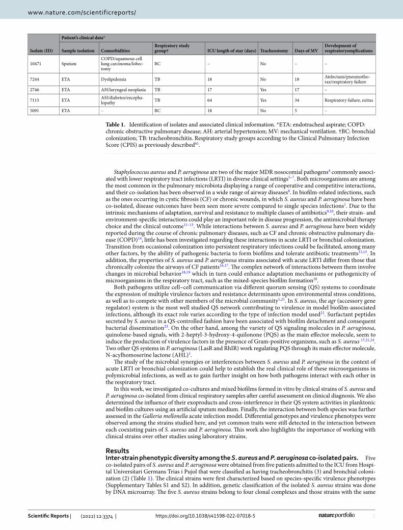
Table 1. Identification of isolates and associated clinical information. *ETA: endotracheal aspirate; COPD: chronic obstructive pulmonary disease; AH: arterial hypertension; MV: mechanical ventilation. †BC: bronchial colonization; TB: tracheobronchitis. Respiratory study groups according to the Clinical Pulmonary Infection Score (CPIS) as previously described61.
Isolate (ID)
Patient’s clinical data*
Sample isolation ComorbiditiesRespiratory study group† ICU length of stay (days) Tracheostomy Days of MV
Development of respiratoryomplications
10471 SputumCOPD/squamous cell lung carcinoma/lobec-tomy
BC – No – –
7244 ETA Dyslipidemia TB 18 No 18 Atelectasis/pneumotho-rax/respiratory failure
2746 ETA AH/laryngeal neoplasia TB 17 Yes 17 –
7115 ETA AH/diabetes/encepha-lopathy TB 64 Yes 34 Respiratory failure, exitus
5091 ETA – BC 18 No 5 –

3
Vol.:(0123456789)
Scientific Reports | (2022) 12:3374 | https://doi.org/10.1038/s41598-022-07018-5
www.nature.com/scientificreports/
multilocus sequence typing (MLST) profile display different virulence genes as determined by DNA microarray. The agr genotyping also revealed the phenotypic diversity of the strains, and Agr was inferred to be functional in the isolates SAR2746, SAR7115 and SAR5091 as determined through δ-haemolysin detention in CAMP assay. Strains SAR7244 and SAR10471 were non-haemolytic on blood agar.
P. aeruginosa isolates also displayed significant diversity in terms of their genomic sequences, colony mor-phology and virulence related phenotypes. The MLST analysis shows that the five strains belong to different sequence types (STs) and the P. aeruginosa MLST database indicates a novel ST for strain PAR7244 (Table S2). Furthermore, none of the detected STs are part of the same clonal complex and none share more than two alleles in common. Virulence factors, such as swarming motility, fluorescent siderophore production, pyocyanin pig-ment and protease secretion were differentially expressed by all strains (Table S2 and Fig. S1).
Clinical isolates of P. aeruginosa induce slow‑growing colonies in S. aureus. To investigate how the P. aeruginosa and S. aureus co-isolated pairs from patients with tracheobronchitis or bronchial colonization coexist and interact with each other, we first studied growth competition by co-culturing the two organisms in agar plates. The presence of P. aeruginosa inhibited growth of S. aureus on TSA plates as indicated by the halo formed around the bacterial colonies (Fig. 1A). Inside the inhibition halos, total killing was only observed in a narrow zone surrounding the P. aeruginosa colony except for strain SAR10471; beyond this area S. aureus colo-nies grew slowly and lost pigmentation. This effect caused by the diffusion of an inhibitory substance in a solid medium, could indicate the secretion of antistaphylococcal molecules by P. aeruginosa. Besides, the presence of P. aeruginosa supernatant in the medium also induced the formation of these small non-pigmented colonies, except for the isolated pair with ID 10471 (Fig. 1B and C). S. aureus SAR10471 was less affected if considering the inhibition halo diameter and no slow-growing colonies were observed in the presence of PAR10471 super-natant. The observed phenotypic changes in the other four S. aureus strains upon contact with P. aeruginosa were transient. For these isolates, the inhibitory effect of P. aeruginosa on S. aureus growth could be related to the production of phenazine pigments such as pyocyanin according to the culture medium used (Fig. S1). When the strain PAR10471 was grown on TSB, no pigment production was detected. Interestingly, this strain is the one that showed the lowest proteolytic activity (Table S2).
Pseudomonas aeruginosa affect growth and biofilm formation in S. aureus, but show syner‑gism in the early stages of biofilm formation in artificial sputum medium. To study competition between P. aeruginosa and S. aureus during biofilm formation, we first evaluated growth interference in plank-tonic conditions. For this purpose the two species were inoculated simultaneously in 0.5X TSB with 1% glucose and allowed to grow until the end of the mid-logarithmic phase. The numbers of viable cells collected from each species in both mixed and axenic (control) cultures are shown in Fig. 2A and B. For all pairs, except for the co-isolated pairs identified as 10471 and 7244, the presence of P. aeruginosa inhibits the growth of S. aureus in mixed cultures with more than tenfold reductions in viable cells. It must be noted that the strains of S. aureus SAR10471 and SAR7244 are genetically related and belong to the same clonal complex. In general, the growth of P. aerugi-nosa was not significantly affected in mixed cultures. In the competition assays in TSA plates, strains SAR10471 and SAR7244 were the least affected by their corresponding P. aeruginosa isolates if we take into account the diameter of the inhibition halos (Fig. 1).
We also evaluated the effect of one species exoproducts on static biofilm formation on polystyrene plates by the other species using planktonic supernatants (Fig. 2C, D). Biofilm formation was tested in both 0.5X TSB sup-plemented with 1% glucose, a media that promotes robust biofilm growth for S. aureus in vitro25 and enhances their survival in the mixed biofilm26, and a TSB-based artificial sputum medium (ASM) to mimic growth in the cystic fibrosis lung habitat27. First, regarding the intrinsic capacity of each strain to form biofilm in both growth media (without the influence of the other species), a large phenotypic variability was observed between the strains of both species. For example, the S. aureus strains SAR2746 and SAR5091 with functional Agr form much less biofilm than the other ones. On the other hand, the P. aeruginosa strain PAR7244 is a strong biofilm former, while the strain PAR5091 almost does not form biofilm under the tested conditions. Despite this variability, and in almost all cases, the presence of P. aeruginosa supernatant significantly decreased biofilm formation by S. aureus under the two growth conditions used (Fig. 2C, E), with the exception of strain SAR2746 in TSB and strain SAR7115 in modified ASM. On the contrary, the presence of S. aureus supernatant did not affect biofilm formation by P. aeruginosa, except for the strain PAR7244 which showed variable results (Fig. 2D, F).
We studied mixed biofilm on polystyrene plates after 24 h of static growth in both TSB-based media (Fig. 3). Both strains were inoculated simultaneously at a ratio of 10:1 (S. aureus: P. aeruginosa) to prevent P. aeruginosa from totally outcompeting S. aureus. Under these conditions, and after 24 h of growth, the total amount of bio-film formed was lower compared to single-species biofilm (Fig. 3A). Even so, both species were collected from the formed biofilms but in different proportions (Fig. 3B). Irrespective of the co-isolated pair and the growth medium, the ratio of the two species in the biofilms after 24 h cultivation was lower than in the initial inocu-lum, with a higher proportion of P. aeruginosa. Differential cfu counting showed that the ASM-based medium increases this ratio in favor of S. aureus in the pairs 10471, 7244 and 5091 (Fig. 3B). The dual species biofilm in both media under static conditions on microscopy dishes were also visualized by confocal microscopy and representative images are presented in Figs. 3C and S2. When grown as a single species biofilm, S. aureus formed extensive patches of biomass after 24 h of growth, except for strains SAR10471 and SAR7115 that grew forming microcolony structures in artificial mucus medium (Fig. S2). On the contrary, all P. aeruginosa strains appeared as microcolonies in both growth conditions after 24 h of growth, except for strain PAR7244 that formed uniform sheets of biomass. This is probably because TSB is less capable of supporting P. aeruginosa biofilm formation under static conditions28, and also because this species takes up to 3 days to develop mature biofilm in ASM29,30.

4
Vol:.(1234567890)
Scientific Reports | (2022) 12:3374 | https://doi.org/10.1038/s41598-022-07018-5
www.nature.com/scientificreports/
1047
1
SAR in TSA SAR in TSA withPAR supernatantPAR spotted on SAR lawn
7244
2746
7115
5091
A B C
Figure 1. Interspecies growth competition assay on agar plates. (A) Colonies of P. aeruginosa (PAR) grown on a lawn of S. aureus (SAR) of the same co-isolated pair on tryptic soy agar (TSA) plates. Enlarged view of the boxed area is shown in the right panel. (B and C) S. aureus was streak-inoculated onto TSA plates with or without P. aeruginosa supernatant. Strain names are according to the origin of the sample (see Tables 1, S1 and S2).

5
Vol.:(0123456789)
Scientific Reports | (2022) 12:3374 | https://doi.org/10.1038/s41598-022-07018-5
www.nature.com/scientificreports/
During co-culture, attachment of S. aureus cells to the surface of plastic dishes was negatively affected and the strains merely formed some localized cell aggregates. The initial formation of these microcolonies was favored in the modified ASM medium (Fig. 3C). In co-culture in the sputum-like medium, P. aeruginosa cells appear to have clustered around cellular aggregates formed by S. aureus, promoting the initial stage of mixed biofilm formation.
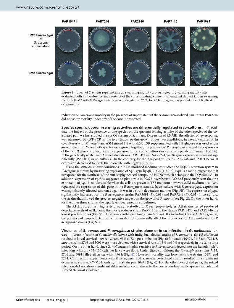
Exposure to S. aureus supernatant influences swarming motility in P. aeruginosa. The effect of the presence of S. aureus strain, tested as culture supernatant, on swarming motility of the P. aeruginosa co-isolated strain was evaluated and the results are summarized in Fig. 4. In the three P. aeruginosa strains PAR5091,
A B
C D
E F
P. aeruginosa growthModified TSB with agitation (8 hrs)
PAR 10471 PAR 7244 PAR 2746 PAR 7115 PAR 5091105
106
107
108
109
101 0
Axenic cultureCo-culture
cfu/
mL
S. aureus growthModified TSB with agitation (8 hrs)
SAR 10471 SAR 7244 SAR 2746 SAR 7115 SAR 5091105
106
107
108
109
101 0
Axenic cultureCo-culture
cfu/
mL
SAR 10471 SAR 7244 SAR 2746 SAR 7115 SAR 50910
1
2
3
4Modified TSBModified TSB with planktonicsupernatant P. aeruginosa
***
***
*
Rel
ativ
e B
iofil
m (O
D 5
50/6
00nm
)
PAR 10471 PAR 7244 PAR 2746 PAR 7115 PAR 5091
Modified TSBModified TSB with planktonicsupernatant S. aureus
***
0
0.5
1.0
1.5
2.0
2.5
Rel
ativ
e B
iofil
m (O
D 5
50/6
00nm
)
PAR 10471 PAR 7244 PAR 2746 PAR 7115 PAR 5091
Modified ASMModified ASM with planktonicsupernatant S. aureus
*
0
0.5
1.0
1.5
2.0
Rel
ativ
e B
iofil
m (O
D 5
50/6
00nm
)
SAR 10471 SAR 7244 SAR 2746 SAR 7115 SAR 50910
2
4
6 Modified ASMModified ASM with planktonicsupernatant P. aeruginosa
**
***
**
***
Rel
ativ
e B
iofil
m (O
D 5
50/6
00nm
)
Figure 2. Effect of the interaction between clinical co-isolated pairs of S. aureus and P. aeruginosa on bacterial growth and biofilm formation. (A and B) Growth of S. aureus (SAR) and P. aeruginosa (PAR) strains in axenic culture and in co-culture with the corresponding co-isolated pair. The values represent the mean of viable bacterial count per ml (cfu/ml) after 8 h of cultivation at 37 °C with agitation. (C and E) Effect of the planktonic supernatants from P. aeruginosa on biofilm formation by the co-isolated S. aureus strain both in modified TSB or ASM medium at 37 °C. (D and F) Effect of the planktonic supernatants from S. aureus on biofilm formation by the co-isolated P. aeruginosa strain both in modified TSB or ASM medium at 37 °C. In all panels modified TSB consists of 0.5X TSB with 1% glucose, and modified ASM (artificial sputum medium) is a 1:1 mix of ASM (see methods) and 0.5X TSB with 1% glucose. When added, the planktonic supernatants are diluted 1:10 in culture medium. Error bars indicate standard deviation of biological replicates. *p < 0.05, **p < 0.01, ***p < 0.001 by Unpaired t test.

6
Vol:.(1234567890)
Scientific Reports | (2022) 12:3374 | https://doi.org/10.1038/s41598-022-07018-5
www.nature.com/scientificreports/
PAR7115 and PAR7244 exhibiting a dendritic swarm pattern, swarming motility increased in the presence of cell-free supernatants from its S. aureus counterpart. On the contrary, P. aeruginosa PAR10471 strain showed
A
B
C
Dual species biofilmModified TSB static (24 hrs)
10471 7244 2746 7115 5091101
102
103
104
105
106
107
108
109
P. aeruginosaS. aureus
cfu/
wel
l(b
iofil
mfr
actio
n)
Dual species biofilmModified ASM static (24 hrs)
10471 7244 2746 7115 5091101
102
103
104
105
106
107
108
109
P. aeruginosaS. aureus
cfu/
wel
l(b
iofil
mfr
actio
n)
Dual species biofilmModified TSB static (24 hrs)
10471 7244 2746 7115 50910.0
0.1
0.2
0.3
0.4
0.5R
elat
ive
Bio
film
(OD
550/
600n
m)
Dual species biofilmModified ASM static (24 hrs)
10471 7244 2746 7115 50910.0
0.1
0.2
0.3
0.4
0.5
Rel
ativ
eB
iofil
m(O
D55
0/60
0nm
)
10471
7115 5091
7244 2746 10471
7115 5091
7244 2746
Modified TSB (24 hrs) microscopy dishes Modified ASM (24 hrs) microscopy dishes
Figure 3. Attachment and microcolony formation in dual species static biofilms of clinical S. aureus and P. aeruginosa. Biofilm biomass measured by crystal violet method (A) and population of each bacterium in the mixed biofilm (B). Biofilms were grown under static conditions on polystyrene microtiter plates in 0.5X TSB supplemented with 1% glucose or in artificial sputum medium (ASM) mixed 1:1 with 0.5X TSB 1% glucose for 24 h. (C) Representative confocal laser scanning microscopy images of co-culture biofilms grown under static conditions on microscopy dishes for 24 h, and stained fluorescently for differentiation between species. Texas Red-X selectively binds to the surface of gram-positive bacteria to fluoresce red, while SYTOX counterstains the Gram negative fixed cells and fluoresces green. Scale bars represent 5 μm. In all experiments bacterial inoculation ratio was 10:1 (S. aureus:P. aeruginosa) and error bars indicate standard deviation of three biological replicates. Strain names are according to the origin of the sample (see Tables 1, S1 and S2).
7
Vol.:(0123456789)
Scientific Reports | (2022) 12:3374 | https://doi.org/10.1038/s41598-022-07018-5
www.nature.com/scientificreports/
reduction on swarming motility in the presence of supernatant of the S. aureus co-isolated pair. Strain PAR2746 did not show motility under any of the conditions tested.
Species specific quorum‑sensing activities are differentially regulated in co‑cultures. To eval-uate the impact of the presence of one species on the quorum sensing activity of the other species of the co-isolated pair, we first studied the agr QS system of S. aureus. Expression of RNAIII, the effector of agr response, was measured by qRT-PCR in the five clinical strains grown under two conditions, in axenic cultures or in co-cultures with P. aeruginosa. ASM mixed 1:1 with 0.5X TSB supplemented with 1% glucose was used as the growth medium. When both species were grown together, the presence of P. aeruginosa affected the expression of the rnaIII gene compared with its expression in the axenic cultures in a strain-dependent manner (Fig. 5A). In the genetically related and Agr negative strains SAR10471 and SAR7244, rnaIII gene expression increased sig-nificantly (P < 0.001) in co-cultures. On the contrary, for the Agr positive strains SAR2746 and SAR7115 rnaIII expression decreased to levels that correlate with negative strains.
Using the same co-culture conditions in ASM modified medium, we studied the HQNO secretion system in P. aeruginosa strains by measuring expression of pqsL gene by qRT-PCR (Fig. 5B). PqsL is a mono-oxygenase that is required for the synthesis of the anti-staphylococcal compound HQNO which belongs to the PQS family31. In addition, expression of pqsL is suggested to play a role in PQS biosynthesis32. We had previously seen that the expression of pqsL is not detectable when the cells are grown in TSB medium; however, ASM medium positively regulated the expression of this gene in the P. aeruginosa strains. In co-culture with S. aureus pqsL expression was significantly affected, and once again it was in a strain dependent manner (Fig. 5B). The expression of pqsL significantly increased for the P. aeruginosa strains PAR5091 (P < 0.01) and PAR7244 (P < 0.05) in co-culture, the strains that showed the greatest negative impact on the growth of S. aureus (see Fig. 2). On the other hand, for the other three strains, the pqsL levels decreased in co-cultures.
The AHL quorum sensing system was also studied in P. aeruginosa isolates. All strains tested produced detectable levels of AHL, being the most producer strain PAR7115 and the strains PAR10471 and PAR2746 the lowest producer ones (Fig. S3). All strains synthesized long chain 3-oxo-AHLs including C8 and C10. In general, the presence of exoproducts from S. aureus did not significantly affect the production of AHL molecules by P. aeruginosa strains (Fig. S3).
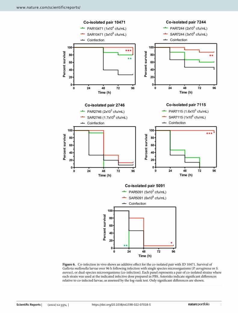
Virulence of S. aureus and P. aeruginosa strains alone or in co‑infection in G. mellonella lar‑vae. Acute infection of G. mellonella larvae with individual clinical strains of S. aureus (1–6 × 10⁶ cfu/larva) resulted in larval survival between 80 and 95% at 72 h post-infection (Fig. 6) for strains 10471, 7115 and 7244. S. aureus strains 2746 and 5091 were more virulent with a survival rate of 13% and 7% respectively in the same time period. On the other hand, since G. mellonella is highly sensitive to P. aeruginosa injected into the hemolymph33, infections with only 15–100 cells per larva were done. Under these conditions, the P. aeruginosa strains 7115, 2746 and 5091 killed all larvae within 96 h (Fig. 6). However, mortality was lower with the strains 10471 and 7244. Co-infection experiments with P. aeruginosa and S. aureus co-isolated strains resulted in a significant decrease in survival (P < 0.01) only for the strain pair 10471 (Fig. 6). For the other co-isolated pairs, the mixed infection did not show significant differences in comparison to the corresponding single species inocula that showed the most virulence.
BM2 swarm agar +
S. aureus supernatant
BM2 swarm agar
PAR10471 PAR5091 PAR2746 PAR7115 PAR7244
Figure 4. Effect of S. aureus supernatants on swarming motility of P. aeruginosa. Swarming motility was evaluated both in the absence and presence of the corresponding S. aureus supernatant diluted 1:10 in swarming medium (BM2 with 0.5% agar). Plates were incubated at 37 °C for 20 h. Images are representative of triplicate experiments.

8
Vol:.(1234567890)
Scientific Reports | (2022) 12:3374 | https://doi.org/10.1038/s41598-022-07018-5
www.nature.com/scientificreports/
DiscussionThe interaction between clinical strains of P. aeruginosa and S. aureus has been extensively studied in recent years both in vivo and in vitro mostly from chronic infections1,8,21,34–37. Most of these studies have shown that when both bacteria are grown together, P. aeruginosa surpasses or decreases the S. aureus population. In the present work, this inhibitory effect of P. aeruginosa over its S. aureus counterpart has been observed for various clinical pairs co-isolated from the respiratory tract of patients with acute LRTI or bronchial colonization. However the strength of such an inhibitory effect has been strain specific. It has been shown that P. aeruginosa outcompetes S. aureus for the limited availability of required nutrients in the medium38. P. aeruginosa also produces an array of antimicrobial factors, which include respiratory inhibitors like hydrogen cyanide, pyocyanin, and the antistaphy-lococcal compound HQNO, as well as proteases that degrade wall components of S. aureus21,39,40. Particularly pyocyanin diverts electron flow and thus increases levels of products of oxygen reduction41. After lysis S. aureus could be an iron source for P. aeruginosa in cultures under iron-limiting conditions which contributes to the success of the latter39. Supplementary Fig. S4 summarizes the main interactions studied here between members of each co-isolated pair, and the phenotypic changes due to these interactions. Considering the high genetic vari-ability of the clinical isolates and their clinical origin, it is not surprising that the interactions are strain-specific. Despite this, and the strong inhibitory effect exerted by P. aeruginosa on S. aureus under standard laboratory conditions, both species cooperate to survive in the mucosal environment of the respiratory tract.
The regulation of quorum sensing (QS) systems could play an important role in these dual species interac-tions. Secretion of anti-staphylococcal compounds is largely dependent on the modulation of the QS activity in P. aeruginosa in co-cultures. For example, HQNO is regulated by the quorum-sensing system PQS and it is involved in the increase of reactive oxygen species activity by disrupting the respiratory chain1,42. It has been shown that a P. aeruginosa mutant, deficient in the production of HQNO, does not efficiently kill S. aureus19. Exposure of S. aureus to HQNO has also been shown to down-regulate agr expression20. Beside this, several long-chain AHLs inhibit growth and virulence of S. aureus by reducing the agr activity43. All P. aeruginosa strains studied here synthesize long chain AHLs, and in general AHL production was not significantly affected by the presence of S. aureus exoproducts. However, co-culture with S. aureus seems to affect HQNO produc-tion in P. aeruginosa in a strain-specific manner. On the other hand, in S. aureus isolates with functional agr QS system, the exoproducts from co-isolated P. aeruginosa strains inhibit the expression of rnaIII, the regulatory
A
B
SAR10471 SAR7244 SAR 2746 SAR7115 SAR50910
2
4
6S. aureus in axenic cultureS. aureus-P. aeruginosa co-culture
***
***
******
S. aureus rnaIII
Relativege
neex
pres
sion
(rna
III/g
yrA)
PAR10471 PAR7244 PAR 2746 PAR7115 PAR50910
2
4
6 P. aeruginosa in axenic cultureS. aureus-P. aeruginosa co-culture
***
*
* **
P. aeruginosa pqsL
Relativege
neex
pres
sion
(pqs
L/rpo
D)
Figure 5. Differential deregulation of genes controlled by quorum sensing in co-culture. Relative expression of quorum sensing genes in co-cultures of S. aureus and P. aeruginosa in modified artificial sputum (ASM) medium (1:1 mix of ASM and 0.5X TSB with 1% glucose) assayed by qRT-PCR. (A) Effect of P. aeruginosa on rnaIII expression in S. aureus. Horizontal dashed line denotes the cut-off used to discriminate between Agr functional and dysfunctional strains (see methods). (B) Effect of S. aureus on pqsL expression in P. aeruginosa. In all experiments error bars indicate standard deviation of three biological replicates.
9
Vol.:(0123456789)
Scientific Reports | (2022) 12:3374 | https://doi.org/10.1038/s41598-022-07018-5
www.nature.com/scientificreports/
Co-isolated pair 10471
0 24 48 72 960
20
40
60
80
100
PAR10471 (1x10⁴ cfu/mL)
SAR10471 (3x10⁸ cfu/mL)Coinfection
Time (h)
Perc
ent s
urvi
val
Co-isolated pair 7244
0 24 48 72 960
20
40
60
80
100
PAR7244 (2x10³ cfu/mL)
SAR7244 (3x10⁸ cfu/mL)Coinfection
Time (h)
Perc
ent s
urvi
val
Co-isolated pair 2746
0 24 48 72 960
20
40
60
80
100
PAR2746 (2x10³ cfu/mL)
SAR2746 (1.7x10⁸ cfu/mL)Coinfection
Time (h)
Perc
ent s
urvi
val
Co-isolated pair 7115
0 24 48 72 960
20
40
60
80
100
PAR7115 (1.6x10³ cfu/mL)
SAR7115 (1x10⁸ cfu/mL)Coinfection
Time (h)
Perc
ent s
urvi
val
Co-isolated pair 5091
0 24 48 72 960
20
40
60
80
100
PAR5091 (5x10³ cfu/mL)
SAR5091 (6x10⁸ cfu/mL)Coinfection
Time (h)
Perc
ent s
urvi
val
* **
**
***
***
**
Figure 6. Co-infection in vivo shows an additive effect for the co-isolated pair with ID 10471. Survival of Galleria mellonella larvae over 96 h following infection with single species microorganisms (P. aeruginosa or S. aureus), or dual-species microorganisms (co-infection). Each panel represents a pair of co-isolated strains where each strain was used at the indicated infective dose prepared in PBS. Asterisks indicate significant differences relative to co-infected larvae, as assessed by the log-rank test. Only significant differences are shown.
10
Vol:.(1234567890)
Scientific Reports | (2022) 12:3374 | https://doi.org/10.1038/s41598-022-07018-5
www.nature.com/scientificreports/
effector molecule of this system. On the contrary, co-culture with P. aeruginosa activates rnaIII gene expres-sion in strains with non-functional agr systems, which could also justify the specific strain pattern during the interaction between both species. QS network rewiring has been proposed as a strategy to coexist in the mucoid environment of the respiratory tract44. The observed increase in agr activity for strains SAR10471 and SAR7244 in co-cultures could correlate with a significant decrease in biofilm formation in the presence of P. aeruginosa supernatants (Fig. S4). Agr-mediated QS regulates many virulence factors in S. aureus that are important for the establishment of infection45,46. For instance, a functional Agr system is required for colonization in some infection models (e.g. epidermal), whereas reduced agr expression has been associated with increased adherence and biofilm production46,47.
Although this work is focused on strains isolated from patients with acute LRTI or bronchial colonization, the coexistence of P. aeruginosa and S. aureus can complicate both the course of the disease and the treatment decision making, promoting the transition into recalcitrant and chronic infections. For example, in CF patients carrying both microorganisms, co-infection is associated with worse disease outcomes and both pathogens seem to contribute additively to the disease severity1,48. Although as discussed before, P. aeruginosa impairs S. aureus growth, the co-occurrence of both microorganisms might also result in increased expression of some staphylo-coccal virulence factors49. Furthermore, S. aureus can survive in the presence of P. aeruginosa by the selection of tiny, non-pigmented colonies known as small-colony variants (SCVs). This phenotype is typically transient and it appears upon contact with exoproducts secreted by P. aeruginosa, such as pyocyanin and HQNO, that block the respiratory chain of S. aureus40,50,51. This phenotype can also be irreversible due to permanent genetic changes. S. aureus is able to persist in the airways of patients with CF by transforming into SCVs40, and this phenotype has been observed in biofilms after antibiotic therapies52. We have shown formation of slow-growing colonies of S. aureus by co-culturing on agar plates with P. aeruginosa, as demonstrated by others using CF patient co-isolates53. Regarding dual species biofilm formation, it has been shown that if S. aureus cells are able to survive killing by P. aeruginosa, certain extracellular factors of P. aeruginosa protect them within the biofilm matrix54,55. Interactions between S. aureus and P. aeruginosa could be reciprocally beneficial to each organism in terms of initial colo-nization and pathogenicity where co-aggregation plays a crucial role56. Here we also show that in co-culture, S. aureus cells start to attach and co-aggregate with P. aeruginosa on the plastic surface despite the strong inhibitory effect exerted by the latter. This synergistic effect is favored in conditions mimicking the CF pulmonary mucus.
On the other hand, the presence of molecules secreted by S. aureus increases P. aeruginosa virulence by enhancing the production of extracellular virulence factors such rhamnolipids, pyocyanin, elastase and qui-nolone-based signals1. A more recent study showed that S. aureus produces proteinaceous factors responsible for promoting P. aeruginosa surface motility57. In the present study exposure to S. aureus supernatant influenced swarming motility in the P. aeruginosa co-isolated strain, however this did not affect its ability to form biofilm. It has also been found by others that P. aeruginosa isolates from co-infected patients are less competitive with S. aureus36. Additionally, the presence of N-acetyl glucosamine from Gram-positive bacteria has been shown in vivo to increase the virulence of pathogenic P. aeruginosa in a G. mellonella infection model58. We have also detected this additive effect in the acute infection model in G. mellonella, but only for the co-isolated pair which showed the smallest competitive effect of P. aeruginosa on S. aureus growth (Fig. S4). Competition between S. aureus and P. aeruginosa has been also shown by others in the mouse model of acute lung infection53,59. In these studies it was also demonstrated that S. aureus profits from the presence of P. aeruginosa and the interactions between both species could be strain specific.
The processes and interactions governing biofilm development are directly involved in the pathogenesis of polymicrobial infections60. The functional and structural organization of both microorganisms in biofilms is com-plex and it is mainly regulated by their QS systems2. Within the biofilm, cells can be transformed phenotypically to become more resistant to treatments promoting the chronicity of infections. Dual species interactions within biofilms are dependent on both S. aureus and P. aeruginosa strains1,56, and this might explain the inconsistency between each co-isolated pair through all the experiments. Significant phenotypic differences were observed among the co-isolates studied here including differences in traits important for successful lung colonization. Considering the origin of the strains and their phenotypic diversity, our results add more clues on the complex interactions that take place between both species in the respiratory tract during acute LRTI or simply during a temporary colonization. Although strain-specific, these interactions could play an important role in disease progression and the clinical outcome in LRTI. This study also suggests the importance of the QS in dual species biofilm-related infections, making interference on these systems a promising therapeutic option. To deepen this knowledge, further studies should be conducted, especially how global gene expression and metabolome are affected in co-cultures.
MethodsClinical isolates. Ten clinical isolates were included in the study. S. aureus and P. aeruginosa strains included were isolated simultaneously in the same respiratory sample (4 endotracheal aspirates and one sputum sample) (co-isolated) from five patients admitted to the ICU from Hospital Universitari Germans Trias i Pujol, a tertiary care hospital in Badalona (Spain) (Table 1). The patients were classified into respiratory study groups according to the Clinical Pulmonary Infection Score (CPIS) and as previously described61. At inclusion, all patients except one were under mechanical ventilation. Ethical approval was provided and the need for informed consent was waived by the Institutional Review Board: Comitè d’Ètica de la Investigació de l’Hospital Germans Trias i Pujol. Patient consent was waived because material used were strains and clinical data were anonymized. Sample col-lection followed the standard of care and was based on suspicion of infection.
Frozen glycerol stocks (15%) of the isolates were stored at -80˚C until use. For some experiments, the reference strains P. aeruginosa PAO1 and S. aureus RN6390B were included as controls. Unless otherwise stated, cultures

11
Vol.:(0123456789)
Scientific Reports | (2022) 12:3374 | https://doi.org/10.1038/s41598-022-07018-5
www.nature.com/scientificreports/
were routinely grown in tryptic soy broth (TSB; BD Difco, Le Pont de Claire, France) at 37 °C and maintained on LB agar plates.
Mono‑culture and co‑culture planktonic growth. All growth curves experiments were done in 0.5X TSB supplemented with 1% glucose at 37 °C with agitation on a rotary shaker at 200 rpm. Overnight individual cultures of S. aureus and P. aeruginosa strains were diluted to an OD550 of 1.0 and used to inoculate (1:20) axenic or mixed cultures. For co-culture growth curves, bacteria were inoculated simultaneously at an equal ratio (1:1). Samples were collected every hour, serial diluted in PBS, and then seeded onto the selective media MacConkey agar (BD Difco) for P. aeruginosa and Mannitol salt agar (Conda, Madrid, Spain) for S. aureus. The agar plates were incubated at 37 °C for 24 h and the number of viable cells was determined.
Preparation of cell‑free culture supernatant. Overnight cultures for each individual strain of both species were prepared in 100 mL of 0.5X TSB supplemented with 1% glucose for 18 h at 37 °C with shaking at 200 rpm. Planktonic supernatants were obtained after centrifugation at 4000 g for 20 min at 4 °C, followed by sterile filtration using a 0.22 µm-pore-size filter (Merck Millipore, Germany) and storage at − 20 °C until use. Before the use, supernatants were adjusted to pH 7.0, supplemented with 0.4% glucose and oxygenated by mix-ing.
Growth competition assay on agar plates. To prepare a bacterial lawn, overnight cultures of S. aureus were adjusted to OD550 of 0.01 and spread onto tryptic soy agar (TSA) plates using a sterile cotton swab. Five microliters of logarithmic cultures of P. aeruginosa isolates were spotted on the competitor lawns using a micro-pipette. In addition, fresh colonies of S. aureus were picked and streak-inoculated onto TSA plates with or with-out 10% (v/v) culture supernatant of P. aeruginosa grown in TSB. Plates were incubated 24 h at 37 °C.
Haemolysin activity in S. aureus. Hemolytic activities and Agr functionality of S. aureus were inferred using the classical CAMP synergistic haemolysis test on blood agar plates62. In CAMP assay, strains were tested against S. aureus strain RN4220 which produces only β-haemolysin. After incubation at 37 °C for 24 h, the plates were refrigerated to increase β-hemolytic activity.
DNA microarray‑based genotyping of S. aureus. Genotypic characterization of S. aureus clinical isolates was performed with a species-specific genotyping DNA microarray (Alere Technologies GmbH, Jena, Germany), which detects specific resistance and virulence genes and their allelic variants and allows the assign-ment of isolates to MLST clonal complexes. PCR amplification and hybridization were performed following the manufacturer’s instructions. An image of the array was recorded and analyzed using a designated reader and software (Arraymate, Iconoclust, Alere Technologies)63.
MLST typing of P. aeruginosa isolates. For each isolate, DNA was extracted using the DNeasy Ultra Clean Microbial kit (Qiagen). Nextera XT kit (Illumina Inc., San Diego, United States) was used for dual-indexed library preparation and paired-end sequencing was performed using the Illumina MiSeq platform (Illumina Inc., San Diego, United States) at the Germans Trias i Pujol Research Institute. The whole DNA content of five strains was sequenced with the Illumina technology. Contig sequences were uploaded to the Pseudomonas MLST database (https:// pubml st. org/ organ isms/ pseud omonas- aerug inosa) to define the sequence types (STs) according to the scheme previously proposed for P. aeruginosa64.
Colony morphology and virulence phenotypes in P. aeruginosa. Pigment production and colony morphology was assessed by growing strains on King A medium (Conda) at 37 °C for 24 h followed by visual inspection. The fluorescence of colonies was also detected under UV light using a table transilluminator. Swarm-ing motility was assayed by point inoculation on BM2 swarm agar plates containing 0.5% (w/v) agar as previ-ously described65. Qualitative determination of proteolytic activity was performed on skimmed milk agar at a final concentration of 1.5% skim milk. Tested strains were spotted on protease plates and incubated 24 h at 37 °C, the formation of a clear zone around the colony was considered as protease positive.
Biofilm formation on polystyrene microtiter plates. Biofilm formation was determined in 96-well polystyrene microtiter plates (BRANDplates, no. 781662, Germany) with a staining method using crystal violet25,66. All biofilms were obtained using 0.5X TSB supplemented with 1% glucose or an artificial sputum medium (ASM)27 mixed 1:1 with 0.5X TSB 1% glucose. Briefly, overnight cultures of individual strains grown in LB broth were diluted in the culture media used for biofilm assay to an OD550 of 0.01 for P. aeruginosa and 0.05 for S. aureus. The diluted cultures were transferred to wells of polystyrene microtiter plates (200 µl per well) and incubated at 37 °C for 24 h under an aerobic atmosphere without agitation (static biofilm). After incubation, the OD600 of each well was measured using a multiwell plate reader to measure planktonic growth in each well. The wells were then gently washed three times with distilled water, dried for 60 min at 60 °C, and stained with 0.1% (w/v) crystal violet for 15 min. Then, the plate was rinsed with distilled water, dried at 37 °C and the crystal violet was extracted with 30% (v/v) acetic acid. The amount of biofilm was quantified by measuring the OD550 of dis-solved CV using the plate reader. Biofilm formation was normalized to cell growth (OD620) to calculate relative biofilm formation. Each reported value corresponds to the arithmetic mean and standard deviation of two bio-logical replicates, each with 8 to 12 replicates per strain and condition. To evaluate the effect of one species super-natant on biofilm formation by the other species of the co-isolated pair, 10% planktonic supernatant was added
12
Vol:.(1234567890)
Scientific Reports | (2022) 12:3374 | https://doi.org/10.1038/s41598-022-07018-5
www.nature.com/scientificreports/
to growth media and the assay was performed as previously described. For co-culture biofilm, overnight cultures of clinical strains were diluted to an OD550 0.01 for P. aeruginosa and 0.1 for S. aureus and mixed (1:1, v/v). Each well was inoculated with 200 µl of each bacterial suspension and incubated at 37 °C for 24 h. To enumerate viable bacteria in the biofilms, after removal of planktonic culture, and after several washes of the plates with PBS, the biofilm fraction was removed by treatment with 250 µl of 0.1% Triton X-100 in PBS. Biofilm bacteria were then scraped and vortexed in the plate for 2 min and biofilm fractions were then serially diluted and plated in selective media. Additional non-treated wells were stained with crystal violet for total biomass quantification.
Confocal laser scanning microscopy (CLSM) of S. aureus and P. aeruginosa biofilms. Single species and co-culture biofilms of S. aureus and P. aeruginosa were grown in microscopy-quality 35 mm sterile plastic dishes (µ-Dish ibidi, no. 81156, Germany). Biofilms were grown using 0.5X TSB supplemented with 1% glucose or ASM mixed 1:1 with 0.5X TSB 1% glucose. For single species biofilm cultivation, overnight cultures of clinical strains grown in TSB were diluted in both biofilm media to an OD550 of 0.01 and 0.05 for P. aeruginosa and S. aureus respectively. For co-culture biofilm, overnight cultures of clinical strains were diluted to an OD550 0.01 for P. aeruginosa and 0.1 for S. aureus and mixed (1:1, v/v). Each dish was inoculated with 1 mL of each bacterial suspension and incubated at 37 °C for 24 h. Then, the dishes were gently washed with distilled water, allowed to air dry for several minutes in inverted position and 1 mL of 4% paraformaldehyde in PBS (0.1 M) was added. After incubation for 15 min, the dishes were gently washed twice with distilled water. Bacterial staining was made using the reagents SYTOX Green and Texas Red-X-WGA (ViaGram Red+ Bacterial Gram Stain and Viability Kit V-7023; Molecular Probes) for general nucleic acid and Gram-positive specific staining, respectively. Cells with damaged membranes stain fluorescent green with SYTOX Green. Texas Red-X-WGA was diluted 1:50 and SYTOX Green was diluted 1:500, and incubation was for 30 min. Images were recorded on an inverted Leica TCS-SP5 confocal microscope using an × 63/1.4 objective (Servei Microscòpia, UAB, Barce-lona). The wavelengths of the excitation and emission lasers were, respectively: green channel (480/500 nm) to SYTOX Green and red channel (490/635 nm) to Texas Red-X-WGA. 3D images were obtained using the Imaris software package (V 6.1; Bitplane). The experiments were carried out in duplicate and at least two sections were acquired with an interval between sections of 0.17 μm.
Reverse transcription and quantitative RT‑PCR. RNA was extracted from single species cultures or co-cultures using the RNeasy Mini Kit (Qiagen, USA) according to the manufacturer’s instructions. All experi-ments were done in 10 mL of ASM mixed 1:1 with 0.5X TSB 1% glucose at 37 °C with agitation. Culture inocula-tion was performed in the same way as for CLSM. Samples were collected after 24 h of growth and were adjusted to an OD550 of 1.0, 0.5 mL of bacterial cultures were treated with two volumes of RNAprotect (Qiagen, USA), incubated for 5 min at room temperature and centrifuged at 5000 g. The pellet was resuspended in Tris–EDTA buffer containing 400 μg/ml lysozyme (Sigma-Aldrich, USA) for P. aeruginosa and/or 200 μg/mL of lysostaphin (Ambion, USA) for S. aureus and incubated for 30 min at 37˚C, followed by proteinase K treatment for 20 min under agitation. RNA was quantified using NanoDrop ND-1000 spectrophotometer (Thermo Fischer, USA) and the absence of DNA was verified by PCR. Reverse transcription was performed using Maxima First Strand cDNA Synthesis system (Thermo Fischer, USA) according to manufacturer’s instructions. For detection and quantification of agr functionality in S. aureus, the quantitative RT-PCR was done as previously described67 with modifications. Amplification of rnaIII gene was done with primers rnaIIIFW, 5’-GAA GGA GTG ATT TCA ATG GCA CAA G-3’, and rnaIIIRV, 5’ GAA AGT AAT TAA TTA TTC ATC TTA TTT TTT AGT GAA TTTG-3’. For P. aer-uginosa isolates the expression of pqsL gene was used to examine the PQS system as previously described36. The primers used for the analysis of the pqsL gene expression were: pqsLFW, 5’- CGG TAT CGC CTC CTA CGT G-3′, and pqsLRV 5′-GGA AGC TCA CCA CCA GTC G-3′. The amplification products were detected using the SsoAd-vanced universal SYBR Green supermix (Bio-Rad, USA). Results from experiments performed in triplicate were normalized to the expression of species-specific reference genes. Gyrase gyrB and RNA polymerase sigma factor rpoD genes were used as the normalizing genes for S. aureus and P. aeruginosa respectively using the following oligonucleotide pairs: gyrFW, 5′-CCA GGT AAA TTA GCC GAT TGC-3′ and gyrRV, 5′-AAA TCG CCT GCG TTC TAG AG-3′; and rpoFW, 5′-CGA TCG GTG ACG ACG AAG AT-3′ and rpoRV, 5′-GTC ACA TCG AAC TGC TTG CC-3’. S. aureus RN6390B was used as a positive control for rnaIII expression. We defined a strain to be Agr functional if rnaIII expression levels were at least tenfold lower than the positive control.
Detection of AHLs production. AHLs were detected in bioassays by determining β-galactosidase activity using a sensor strain of Agrobacterium tumefaciens KYC55 carrying the plasmids pJZ372, pJZ384 and pJZ410, as previously described68,69 with few modifications. Briefly, A. tumefaciens KYC55 strain was grown in 20 mL of BM2 medium at 30 °C for 18 h. Cells were harvested and reconstituted with 10 mL of fresh BM2 and added to 40 mL of temperate BM2 medium containing 2% of Noble Agar (BD Difco) and 80 µg/mL of X-Gal. AHLs produced by P. aeruginosa strains were extracted with acidified ethyl acetate from cell-free culture supernatants as previously described70 and were evaluated both in bioassay plates and by thin layer chromatography (TLC) coupled to bioassay70. Presence of blue halos or spots indicates AHL production. P. aeruginosa PAO1 was used as a positive control of AHLs production.
To assess the effect of exoproducts of S. aureus on the AHL synthesis by P. aeruginosa, overnight cultures of P. aeruginosa strains grown in LB were diluted in fresh medium to an OD550 of 1.0. The AHL bioassay plates were prepared with and without the addition of 1:10 diluted supernatant of the corresponding S. aureus co-isolate and were seeded with 2 µL of the diluted culture and incubated at 30 °C for 24 h. The diameters of the blue halos were compared between both conditions.
13
Vol.:(0123456789)
Scientific Reports | (2022) 12:3374 | https://doi.org/10.1038/s41598-022-07018-5
www.nature.com/scientificreports/
Infection of Galleria mellonella larvae. Larvae of Galleria mellonella were obtained from our own hatchery and only healthy-looking larvae weighing 200–300 mg and showing no signs of melanization were used in the experiments. To prepare bacterial inocula, P. aeruginosa and S. aureus isolates were grown overnight at 37°C in 10 mL of LB and 0.5X TSB supplemented with 1% glucose, respectively. Then, cells from individual cultures were sedimented by centrifugation at 5000 g, washed in PBS and adjusted to contain approximately the desired dose. This inoculum size was selected based on pilot studies that have determined the optimal infec-tive dose of each strain required to kill G. mellonella over a 24–96 h period. The selected doses ranged from 101 to 102 cfu per larvae for P. aeruginosa and from 106 to 4 × 10⁷ cfu per larvae for S. aureus isolates. Larvae were infected with P. aeruginosa or S. aureus or co-infected with P. aeruginosa and S aureus of the same co-isolated pair. Mixed infected larvae were inoculated with the same bacterial burden of P. aeruginosa and S aureus as those inoculated with single species microorganisms. Fifteen larvae per group were infected through the left proleg with 10 μL of each bacterial suspension using a 50 μl Hamilton Microliter syringe and incubated in the dark at 30 °C in empty petri dishes. Bacterial burden of the doses was confirmed by plating on LB agar medium. Mortal-ity was determined every 24 h. Larvae were considered dead when they no longer responded to touch and turned black which correlates with total melanization.
Statistical analysis. All statistical analyses were performed by GraphPad Prim software (ver. 5.0; GraphPad Inc, San Diego, USA), considering as statistically significant a p-value less than 0.05. For infection experiments Kaplan–Meier survival curves were plotted using GraphPad Prism and statistical significance was determined using the log-rank (Mantel-Cox) test.
Statement. Experiments were performed in accordance with relevant guidelines and regulations.
Data availabilityWhole Genome Shotgun projects for sequences identified in this study have been deposited at DDBJ/ENA/Gen-Bank (accession codes are listed in Table S2). MLST information is available at the Pseudomonas MLST database.
Received: 28 September 2021; Accepted: 9 February 2022
References 1. Hotterbeekx, A., Kumar-Singh, S., Goossens, H. & Malhotra-Kumar, S. In vivo and In vitro Interactions between Pseudomonas
aeruginosa and Staphylococcus spp. Front. Cell. Infect. Microbiol. 7, 106 (2017). 2. Fernandez-Barat, L. & Torres, A. Biofilms in ventilator-associated pneumonia. Fut. Microbiol. 11, 1599–1610 (2016). 3. Uruen, C., Chopo-Escuin, G., Tommassen, J., Mainar-Jaime, R. C. & Arenas, J. Biofilms as promoters of bacterial antibiotic resist-
ance and tolerance. Antibiotics 10, 3 (2020). 4. Santajit, S. & Indrawattana, N. Mechanisms of antimicrobial resistance in ESKAPE pathogens. Biomed. Res. Int. 2016, 2475067
(2016). 5. Kollef, M. H. et al. Epidemiology and outcomes of health-care-associated pneumonia: results from a large US database of culture-
positive pneumonia. Chest 128, 3854–3862 (2005). 6. Rello, J., Lisboa, T. & Koulenti, D. Respiratory infections in patients undergoing mechanical ventilation. Lancet Respir. Med. 2,
764–774 (2014). 7. Antibacterial agents in clinical development: an analysis of the antibacterial clinical development pipeline, including tuberculosis.
(ed (eds Organization WH). World Health Organization (2017). 8. Limoli, D. H. & Hoffman, L. R. Help, hinder, hide and harm: What can we learn from the interactions between Pseudomonas
aeruginosa and Staphylococcus aureus during respiratory infections?. Thorax 74, 684–692 (2019). 9. Zemanick, E. T., Sagel, S. D. & Harris, J. K. The airway microbiome in cystic fibrosis and implications for treatment. Curr. Opin.
Pediatr. 23, 319–324 (2011). 10. Hotterbeekx, A. et al. The endotracheal tube microbiome associated with Pseudomonas aeruginosa or Staphylococcus epidermidis.
Sci. Rep. 6, 36507 (2016). 11. Kumar, A. & Ting, Y. P. Presence of Pseudomonas aeruginosa influences biofilm formation and surface protein expression of
Staphylococcus aureus. Environ. Microbiol. 17, 4459–4468 (2015). 12. Cohen, T. S. et al. Staphylococcus aureus alpha toxin potentiates opportunistic bacterial lung infections. Sci. Transl. Med. 8, 329ra331
(2016). 13. Prat, C. & Lacoma, A. Bacteria in the respiratory tract-how to treat? Or do not treat?. Int. J. Infect. Dis. 51, 113–122 (2016). 14. Cullen, L. & McClean, S. Bacterial adaptation during chronic respiratory infections. Pathogens 4, 66–89 (2015). 15. Bayes, H. K., Ritchie, N., Irvine, S. & Evans, T. J. A murine model of early Pseudomonas aeruginosa lung disease with transition to
chronic infection. Sci. Rep. 6, 35838 (2016). 16. Marks, M. I. Clinical significance of Staphylococcus aureus in cystic fibrosis. Infection 18, 53–56 (1990). 17. Sadikot, R. T., Blackwell, T. S., Christman, J. W. & Prince, A. S. Pathogen-host interactions in Pseudomonas aeruginosa pneumonia.
Am. J. Respir. Crit. Care Med. 171, 1209–1223 (2005). 18. Korgaonkar, A., Trivedi, U., Rumbaugh, K. P. & Whiteley, M. Community surveillance enhances Pseudomonas aeruginosa virulence
during polymicrobial infection. Proc. Natl. Acad. Sci. USA 110, 1059–1064 (2013). 19. Filkins, L. M. et al. Coculture of staphylococcus aureus with pseudomonas aeruginosa drives S. aureus towards fermentative
metabolism and reduced viability in a cystic fibrosis model. J. Bacteriol. 197, 2252–2264 (2015). 20. Mitchell, G. et al. Staphylococcus aureus sigma B-dependent emergence of small-colony variants and biofilm production following
exposure to Pseudomonas aeruginosa 4-hydroxy-2-heptylquinoline-N-oxide. BMC Microbiol. 10, 33 (2010). 21. Abisado, R. G., Benomar, S., Klaus, J. R., Dandekar, A. A. & Chandler, J. R. Bacterial quorum sensing and microbial community
interactions. mBio https:// doi. org/ 10. 1128/ mBio. 01749- 18 (2018). 22. Yarwood, J. M., Bartels, D. J., Volper, E. M. & Greenberg, E. P. Quorum sensing in Staphylococcus aureus biofilms. J. Bacteriol. 186,
1838–1850 (2004). 23. Otto, M. Staphylococcal infections: Mechanisms of biofilm maturation and detachment as critical determinants of pathogenicity.
Annu. Rev. Med. 64, 175–188 (2013).

14
Vol:.(1234567890)
Scientific Reports | (2022) 12:3374 | https://doi.org/10.1038/s41598-022-07018-5
www.nature.com/scientificreports/
24. Chirathanamettu, T. R. & Pawar, P. D. Quorum sensing-induced phenotypic switching as a regulatory nutritional stress response in a competitive two-species biofilm: An individual-based cellular automata model. J. Biosci. https:// doi. org/ 10. 1007/ s12038- 020- 00092-9 (2020).
25. Kwasny, S. M. & Opperman, T. J. Static biofilm cultures of Gram-positive pathogens grown in a microtiter format used for anti-biofilm drug discovery. Curr. Protoc. Pharmacol. https:// doi. org/ 10. 1002/ 04711 41755. ph13a 08s50 (2010).
26. Cendra, M. D., Blanco-Cabra, N., Pedraz, L. & Torrents, E. Optimal environmental and culture conditions allow the in vitro coexistence of Pseudomonas aeruginosa and Staphylococcus aureus in stable biofilms. Sci. Rep-Uk https:// doi. org/ 10. 1038/ s41598- 019- 52726-0 (2019).
27. Sriramulu, D. D., Lunsdorf, H., Lam, J. S. & Romling, U. Microcolony formation: a novel biofilm model of Pseudomonas aeruginosa for the cystic fibrosis lung. J. Med. Microbiol. 54, 667–676 (2005).
28. Haney, E. F., Trimble, M. J., Cheng, J. T., Valle, Q. & Hancock, R. E. W. Critical assessment of methods to quantify biofilm growth and evaluate antibiofilm activity of host defence peptides. Biomolecules 8, 29 (2018).
29. Ramsey, M. M. & Whiteley, M. Pseudomonas aeruginosa attachment and biofilm development in dynamic environments. Mol. Microbiol. 53, 1075–1087 (2004).
30. Kirchner, S. et al. Use of artificial sputum medium to test antibiotic efficacy against Pseudomonas aeruginosa in conditions more relevant to the cystic fibrosis lung. J. Vis. Exp. https:// doi. org/ 10. 3791/ 3857 (2012).
31. Lin, J., Cheng, J., Wang, Y. & Shen, X. The Pseudomonas quinolone signal (PQS): Not just for quorum sensing anymore. Front. Cell Infect. Microbiol. 8, 230 (2018).
32. D’Argenio, D. A. et al. Growth phenotypes of Pseudomonas aeruginosa lasR mutants adapted to the airways of cystic fibrosis patients. Mol. Microbiol. 64, 512–533 (2007).
33. Hill, L., Veli, N. & Coote, P. J. Evaluation of Galleria mellonella larvae for measuring the efficacy and pharmacokinetics of antibiotic therapies against Pseudomonas aeruginosa infection. Int. J. Antimicrob. Agents 43, 254–261 (2014).
34. Pallett, R. et al. Anaerobiosis influences virulence properties of Pseudomonas aeruginosa cystic fibrosis isolates and the interaction with Staphylococcus aureus. Sci. Rep. 9, 6748 (2019).
35. Trizna, E. Y. et al. Bidirectional alterations in antibiotics susceptibility in Staphylococcus aureus-Pseudomonas aeruginosa dual-species biofilm. Sci. Rep. 10, 14849 (2020).
36. Limoli, D. H. et al. Pseudomonas aeruginosa Alginate Overproduction Promotes Coexistence with Staphylococcus aureus in a Model of Cystic Fibrosis Respiratory Infection. MBio https:// doi. org/ 10. 1128/ mBio. 00186- 17 (2017).
37. Fugere, A. et al. Interspecific small molecule interactions between clinical isolates of Pseudomonas aeruginosa and Staphylococcus aureus from adult cystic fibrosis patients. PLoS One 9, e86705 (2014).
38. Nguyen, A. T. & Oglesby-Sherrouse, A. G. Interactions between Pseudomonas aeruginosa and Staphylococcus aureus during co-cultivations and polymicrobial infections. Appl. Microbiol. Biotechnol. 100, 6141–6148 (2016).
39. Mashburn, L. M., Jett, A. M., Akins, D. R. & Whiteley, M. Staphylococcus aureus serves as an iron source for Pseudomonas aerugi-nosa during in vivo coculture. J. Bacteriol. 187, 554–566 (2005).
40. Biswas, L., Biswas, R., Schlag, M., Bertram, R. & Gotz, F. Small-colony variant selection as a survival strategy for Staphylococcus aureus in the presence of Pseudomonas aeruginosa. Appl. Environ. Microbiol. 75, 6910–6912 (2009).
41. Hassan, H. M. & Fridovich, I. Mechanism of the antibiotic action pyocyanine. J. Bacteriol. 141, 156–163 (1980). 42. Hazan, R. et al. Auto poisoning of the respiratory chain by a quorum-sensing-regulated molecule favors biofilm formation and
antibiotic tolerance. Curr. Biol. 26, 195–206 (2016). 43. Qazi, S. et al. N-acylhomoserine lactones antagonize virulence gene expression and quorum sensing in Staphylococcus aureus.
Infect. Immun. 74, 910–919 (2006). 44. Camus, L., Briaud, P., Vandenesch, F. & Moreau, K. How bacterial adaptation to cystic fibrosis environment shapes interactions
between Pseudomonas aeruginosa and Staphylococcus aureus. Front. Microbiol. 12, 617784 (2021). 45. Le, K. Y. & Otto, M. Quorum-sensing regulation in staphylococci-an overview. Front. Microbiol. 6, 1174 (2015). 46. Nakamura, Y. et al. Staphylococcus Agr virulence is critical for epidermal colonization and associates with atopic dermatitis devel-
opment. Sci. Transl. Med. https:// doi. org/ 10. 1126/ scitr anslm ed. aay40 68 (2020). 47. Sakoulas, G., Moise, P. A. & Rybak, M. J. Accessory gene regulator dysfunction: an advantage for Staphylococcus aureus in health-
care settings?. J. Infect. Dis. 199, 1558–1559 (2009). 48. Sagel, S. D. et al. Impact of Pseudomonas and Staphylococcus infection on inflammation and clinical status in young children with
cystic fibrosis. J. Pediatr. 154, 183–188 (2009). 49. Pastar, I. et al. Interactions of methicillin resistant Staphylococcus aureus USA300 and Pseudomonas aeruginosa in polymicrobial
wound infection. PLoS One 8, e56846 (2013). 50. Voggu, L. et al. Microevolution of cytochrome bd oxidase in Staphylococci and its implication in resistance to respiratory toxins
released by Pseudomonas. J. Bacteriol. 188, 8079–8086 (2006). 51. Hoffman, L. R. et al. Selection for Staphylococcus aureus small-colony variants due to growth in the presence of Pseudomonas
aeruginosa. Proc. Natl. Acad. Sci. USA 103, 19890–19895 (2006). 52. Singh, R., Ray, P., Das, A. & Sharma, M. Role of persisters and small-colony variants in antibiotic resistance of planktonic and
biofilm-associated Staphylococcus aureus: An in vitro study. J. Med. Microbiol. 58, 1067–1073 (2009). 53. Millette, G. et al. Despite antagonism in vitro, Pseudomonas aeruginosa Enhances Staphylococcus aureus colonization in a murine
lung infection model. Front Microbiol. 10, 2880 (2019). 54. Billings, N. et al. The extracellular matrix Component Psl provides fast-acting antibiotic defense in Pseudomonas aeruginosa
biofilms. PLoS Pathog 9, e1003526 (2013). 55. Yang, L. et al. Pattern differentiation in co-culture biofilms formed by Staphylococcus aureus and Pseudomonas aeruginosa. FEMS
Immunol. Med. Microbiol. 62, 339–347 (2011). 56. Alves, P. M. et al. Interaction between Staphylococcus aureus and Pseudomonas aeruginosa is beneficial for colonisation and patho-
genicity in a mixed biofilm. Pathog. Dis. https:// doi. org/ 10. 1093/ femspd/ fty003 (2018). 57. Limoli, D. H. et al. Interspecies interactions induce exploratory motility in Pseudomonas aeruginosa. eLife https:// doi. org/ 10. 7554/
eLife. 47365 (2019). 58. Whiley, R. A. et al. Differential potentiation of the virulence of the Pseudomonas aeruginosa cystic fibrosis liverpool epidemic
strain by oral commensal Streptococci. J. Infect. Dis. 209, 769–780 (2014). 59. Baldan, R. et al. Adaptation of Pseudomonas aeruginosa in Cystic Fibrosis airways influences virulence of Staphylococcus aureus
in vitro and murine models of co-infection. PLoS One 9, e89614 (2014). 60. Elias, S. & Banin, E. Multi-species biofilms: Living with friendly neighbors. FEMS Microbiol. Rev. 36, 990–1004 (2012). 61. Lacoma, A. et al. Genotypic and phenotypic characterization of Staphylococcus aureus Isolates from the respiratory tract in mechan-
ically-ventilated patients. Toxins 13, 122 (2021). 62. Traber, K. E. et al. agr function in clinical Staphylococcus aureus isolates. Microbiology 154, 2265–2274 (2008). 63. Monecke, S., Slickers, P. & Ehricht, R. Assignment of Staphylococcus aureus isolates to clonal complexes based on microarray
analysis and pattern recognition. FEMS Immunol. Med. Microbiol. 53, 237–251 (2008). 64. van Mansfeld, R. et al. Pseudomonas aeruginosa genotype prevalence in Dutch cystic fibrosis patients and age dependency of
colonization by various P. aeruginosa sequence types. J. Clin. Microbiol. 47, 4096–4101 (2009).

15
Vol.:(0123456789)
Scientific Reports | (2022) 12:3374 | https://doi.org/10.1038/s41598-022-07018-5
www.nature.com/scientificreports/
65. Overhage, J., Lewenza, S., Marr, A. K. & Hancock, R. E. Identification of genes involved in swarming motility using a Pseudomonas aeruginosa PAO1 mini-Tn5-lux mutant library. J. Bacteriol. 189, 2164–2169 (2007).
66. Merritt, J. H., Kadouri, D. E. & O’Toole, G. A. Growing and analyzing static biofilms. Curr. Protocols Microbiol. https:// doi. org/ 10. 1002/ 97804 71729 259. mc01b 01s22 (2011).
67. Gomes-Fernandes, M. et al. Accessory gene regulator (Agr) functionality in Staphylococcus aureus derived from lower respiratory tract infections. PloS One 12, e0175552 (2017).
68. Farrand, S. K., Qin, Y. & Oger, P. Quorum-sensing system of Agrobacterium plasmids: Analysis and utility. Methods Enzymol. 358, 452–484 (2002).
69. Steindler, L. & Venturi, V. Detection of quorum-sensing N-acyl homoserine lactone signal molecules by bacterial biosensors. FEMS Microbiol. Lett. 266, 1–9 (2007).
70. Martinez, P. et al. Stenotrophomonas maltophilia responds to exogenous AHL signals through the LuxR solo SmoR (Smlt1839). Front. Cell Infect. Microbiol. 5, 41 (2015).
AcknowledgementsThis work was partially supported by PI17/01139 (PI. CPA), integrated in the Plan Nacional de I+D+I and cofunded by the ISCIII Subdirección General de Evaluación and the Fondo Europeo de Desarrollo Regional (FEDER). IG and DY (PI) also thanks Catalan AGAUR for support (2017 SGR 1062) and Spanish MICINN (PID2019-111364RB-I00). MGF was funded by CAPES Foundation, Ministry of Education of Brazil (Brasília, Brazil). We thank V. Saludes and D. Panisello from Microbiology Department, Laboratori Clínic Metropolitana Nord (Hospital Universitari Germans Trias i Pujol) for their kind support in performing sequencing experiments. We also thank L. Sumoy and J. F. Sánchez-Herrero from IGTP High Content Genomics and Bioinformatics Unit (Institut d’Investigació en Ciències de la Salut Germans Trias i Pujol) for analyzing sequencing results.
Author contributionsM.G.F., A.C.G., M.B., P.H., X.C., and D.Y. conducted the experiments; M.G.F., A.C.G., C.P.A., A.L. and D.Y. designed the experiments and participated in the analysis and interpretation of experimental data; M.G.F., A.C.G., A.L. and D.Y. wrote the paper; C.P.A. and I.G. supervised research, and all authors revised the manuscript.
Competing interests The authors declare no competing interests.
Additional informationSupplementary Information The online version contains supplementary material available at https:// doi. org/ 10. 1038/ s41598- 022- 07018-5.
Correspondence and requests for materials should be addressed to A.L. or D.Y.
Reprints and permissions information is available at www.nature.com/reprints.
Publisher’s note Springer Nature remains neutral with regard to jurisdictional claims in published maps and institutional affiliations.
Open Access This article is licensed under a Creative Commons Attribution 4.0 International License, which permits use, sharing, adaptation, distribution and reproduction in any medium or
format, as long as you give appropriate credit to the original author(s) and the source, provide a link to the Creative Commons licence, and indicate if changes were made. The images or other third party material in this article are included in the article’s Creative Commons licence, unless indicated otherwise in a credit line to the material. If material is not included in the article’s Creative Commons licence and your intended use is not permitted by statutory regulation or exceeds the permitted use, you will need to obtain permission directly from the copyright holder. To view a copy of this licence, visit http:// creat iveco mmons. org/ licen ses/ by/4. 0/.
© The Author(s) 2022