In planta expression of a mature Der p 1 allergen isolated from an Italian strain of...
13
ORIGINAL PAPER In planta expression of a mature Der p 1 allergen isolated from an Italian strain of Dermatophagoides pteronyssinus Gianpiero Marconi • Emidio Albertini • Adriano Mari • Paola Palazzo • Andrea Porceddu • Lorenzo Raggi • Luigi Bolis • Hovirag Lancioni • Antonella Palomba • Livia Lucentini • Luisa Lanfaloni • Francesco Marcucci • Mario Falcinelli • Fausto Panara Received: 7 April 2009 / Accepted: 25 August 2011 / Published online: 9 September 2011 Ó Springer Science+Business Media B.V. 2011 Abstract European (Dermatophagoides pteronys- sinus) and American (Dermatophagoides farinae) house dust mite species are considered the most common causes of asthma and allergic symptoms worldwide. Der p 1 protein, one of the main allergens of D. pteronyssinus, is found in high concentration in mites faecal pellets, which can became easily airborne and, when inhaled, can cause perennial rhinitis and bronchial asthma. Here we report the isolation of the Der p 1 gene from an Italian strain of D. pteronyssinus and the PVX- mediated expression of its mature form (I-rDer p 1) in Nicotiana benthamiana plants. Human sera from characterized allergic patients were used for IgE binding inhibition assays to test the immunological reactivity of I-rDer p 1 produced in N. benthamiana plants. The binding properties of in planta produced I-rDer p 1 versus the IgE of patients sera were comparable to those obtained on Der p 1 preparation immobilized on a microarray. In this paper we provide a proof of concept for the production of an immunologically active form of Der p 1 using a plant viral vector. These results pave the way for the development of diagnostic allergy tests based on in planta produced allergens. Keywords House dust mite allergens Transient expression PVX Dermatophagoides pteronyssinus Gianpiero Marconi and Emidio Albertini have contributed equally to this work. Electronic supplementary material The online version of this article (doi:10.1007/s11248-011-9551-5) contains supplementary material, which is available to authorized users. G. Marconi E. Albertini (&) L. Raggi L. Bolis M. Falcinelli Dipartimento di Biologia Applicata, University of Perugia, Borgo XX Giugno 74, 06121 Perugia, Italy e-mail: [email protected] A. Mari P. Palazzo IDI-IRCCS Center for Molecular Allergology, Rome, Italy A. Porceddu Dipartimento Scienze Agronomiche e Genetica Vegetale Agraria, University of Sassari, Sassari, Italy H. Lancioni A. Palomba L. Lucentini L. Lanfaloni F. Panara Dipartimento Biologia Cellulare e Ambientale, University of Perugia, Perugia, Italy F. Marcucci Dipartimento di Specialita ` Chirurgiche e Sanita ` Pubblica, University of Perugia, Perugia, Italy 123 Transgenic Res (2012) 21:523–535 DOI 10.1007/s11248-011-9551-5
Transcript of In planta expression of a mature Der p 1 allergen isolated from an Italian strain of...
ORIGINAL PAPER
In planta expression of a mature Der p 1 allergen isolatedfrom an Italian strain of Dermatophagoides pteronyssinus
Gianpiero Marconi • Emidio Albertini • Adriano Mari • Paola Palazzo •
Andrea Porceddu • Lorenzo Raggi • Luigi Bolis • Hovirag Lancioni •
Antonella Palomba • Livia Lucentini • Luisa Lanfaloni • Francesco Marcucci •
Mario Falcinelli • Fausto Panara
Received: 7 April 2009 / Accepted: 25 August 2011 / Published online: 9 September 2011
� Springer Science+Business Media B.V. 2011
Abstract European (Dermatophagoides pteronys-
sinus) and American (Dermatophagoides farinae)
house dust mite species are considered the most
common causes of asthma and allergic symptoms
worldwide. Der p 1 protein, one of the main
allergens of D. pteronyssinus, is found in high
concentration in mites faecal pellets, which can
became easily airborne and, when inhaled, can cause
perennial rhinitis and bronchial asthma. Here we
report the isolation of the Der p 1 gene from an
Italian strain of D. pteronyssinus and the PVX-
mediated expression of its mature form (I-rDer p 1)
in Nicotiana benthamiana plants. Human sera from
characterized allergic patients were used for IgE
binding inhibition assays to test the immunological
reactivity of I-rDer p 1 produced in N. benthamiana
plants. The binding properties of in planta produced
I-rDer p 1 versus the IgE of patients sera were
comparable to those obtained on Der p 1 preparation
immobilized on a microarray. In this paper we
provide a proof of concept for the production of an
immunologically active form of Der p 1 using a
plant viral vector. These results pave the way for the
development of diagnostic allergy tests based on in
planta produced allergens.
Keywords House dust mite allergens �Transient expression � PVX � Dermatophagoides
pteronyssinus
Gianpiero Marconi and Emidio Albertini have contributed
equally to this work.
Electronic supplementary material The online version ofthis article (doi:10.1007/s11248-011-9551-5) containssupplementary material, which is available to authorized users.
G. Marconi � E. Albertini (&) � L. Raggi �L. Bolis � M. Falcinelli
Dipartimento di Biologia Applicata, University
of Perugia, Borgo XX Giugno 74, 06121 Perugia, Italy
e-mail: [email protected]
A. Mari � P. Palazzo
IDI-IRCCS Center for Molecular Allergology,
Rome, Italy
A. Porceddu
Dipartimento Scienze Agronomiche e Genetica
Vegetale Agraria, University of Sassari, Sassari, Italy
H. Lancioni � A. Palomba � L. Lucentini �L. Lanfaloni � F. Panara
Dipartimento Biologia Cellulare e Ambientale,
University of Perugia, Perugia, Italy
F. Marcucci
Dipartimento di Specialita Chirurgiche e Sanita Pubblica,
University of Perugia, Perugia, Italy
123
Transgenic Res (2012) 21:523–535
DOI 10.1007/s11248-011-9551-5
Introduction
Forty-five years ago, Voorhorst et al. (1967) remarked
the importance of house dust mites (HDM) as the origin
of main environmental allergens responsible of several
pathologies in sensitized human population (Platts-
Mills and de Weck 1989; Steen et al. 1994; Fernandez-
Caldas 1997; Robinson et al. 1997; Asturias et al. 1999;
Thomas et al. 2002). Dermatophagoides pteronyssinus
is the source of two classes of major allergens (Der p 1
and Der p 2) (Smith et al. 2001; Thomas et al. 2002;
Takai et al. 2005) affecting the allergic people living
in tempered climates (Fernandez-Caldas 1997;
Platts-Mills et al. 2000). To date, over 30 different
IgE-binding proteins have been identified in D. pter-
onyssinus and for 19 of them their gene sequences have
been cloned and characterized (Chua et al. 1993, 1996;
Lin et al. 1994; Smith et al. 1994; Asturias et al. 1998;
Thomas et al. 2002; Lee et al. 2004). Der p 1 protein is
found in high concentration in mites faecal pellets,
which can became easily airborne and, when inhaled,
can cause perennial rhinitis and bronchial asthma
(Platts-Mills and Chapman 1988; Jacquet et al. 2000;
Platts-Mills and Woodfolk 2011). This allergen (about
25 kDa) is synthesized as a precursor of 320 amino acid
residues which includes a 18-amino acid signal peptide
and a 80-amino acid N-terminal prosequence (pro-Der
p 1) (Chua et al. 1988; Thomas et al. 1988; Jacquet et al.
2000). The protein contains two potential N-glycosyl-
ation sites, one located inside the prosequence region
and another at position 52–54 (N52) of the mature form
(Chua et al. 1988; Jacquet et al. 2000). Der p 1 primary
structure displays homologies with the cysteine pro-
teinase archetype papain, including the conservation of
the active site residues (Chua et al. 1988; Thomas et al.
1988; Topham et al. 1994; Jacquet et al. 2000). Upon
maturation the Der p 1 pro-region is removed by an
autocatalytic process which can be mimicked in vitro at
60�C and low pH (Vernet et al. 1991; Mach et al. 1994;
Jacquet et al. 2000). The maturation process, but not the
glycosylation, seems to be crucial for Der p 1 immu-
nological activity. Greene et al. (1991) have identified
at least five distinct regions (residues 1–56, 53–59,
98–140, 166–194 and 188–222) responsible for the
reactivity versus IgE and IgG in sera of allergic
individuals, demonstrating that they are all located in
the mature protein (Greene et al. 1991). In addition,
Takai et al. (2005) compared natural Der p 1 against
matured Der p 1 and pro-Der p 1 proteins expressed in
yeasts. While the mature Der p 1 retained similar
molecular weight, secondary structures, and its aller-
genicity is equivalent to natural Der p 1, the Der p 1 pro-
forms exhibited different secondary structures and a
lower allergenicity than the mature form suggesting that
the recombinant mature form of Der p 1 could be an
useful tool for various in vitro and in vivo analyses
(Kraft et al. 1998; Takai et al. 2002; Valenta 2002).
Moreover, with the aim of using Pichia pastoris for
producing recombinant proteins mimicking the natural
ones, the same research group (Takai et al. 2002) has
generated Der p 1 mutants in which the N-glycosylation
motif within the mature sequence was disrupted (Der
p 1-N52Q). This study has demonstrated that Der
p 1-N52Q protein retains both a molecular weight
equivalent to the natural Der p 1 and a full enzymatic and
IgE-binding activities. Lienard et al. (2007) produced
immunologically active recombinant Der p 1 and Der
p 2 proteins in BY-2 tobacco cells suspensions but their
attempts for a direct expression of the mature form of
Der p 1 without the propeptide domain failed. To date,
only Yang et al. (2007) have described the expression of
the mature form of Der p 1 in transgenic rice seeds using
a GluB promoter and a targeting signal for the
endoplasmic reticulum (ER) showing that glycosylated
Der p 1 had a significantly reduced IgE binding ability
when compared with unglycosylated Der p 1.
Plants are a particularly attractive system for
expressing pharmaceutical recombinant proteins
(Yusibov et al. 2006; Floss et al. 2007). In fact as
economical biomass producer, plants are virtually free
of contamination from bacterial toxins or animal
pathogens (Cunningham and Porter 1998). Foreign
genes can be expressed at high levels in plant cells
employing a wide variety of strategies ranging from
stable integration in the nucleus or plastid genomes to
transient expression mediated by plant viral vectors
(Gleba et al. 2005).
Transient expression of foreign genes, mediated by
viral vectors such as the tobacco mosaic virus (TMV)
and potato virus X (PVX), has become a valuable tool
in plant biotechnology. Transient expression systems,
based on the PVX, proved very efficient in obtaining
high level of expression in less than 6–8 weeks
(Bendahmane et al. 1995; Porta and Lomonossoff
1998). Potato virus X, a member of the Potexviruses, is
a filamentous rod-shaped virus which contains a single
plus-sense RNA molecule of about 6400 nucleotides
capped at the 50 end (Sonenberg et al. 1978) and
524 Transgenic Res (2012) 21:523–535
123
polyadenylated at the 30 end (Morozov et al. 2000). Its
genome contains five open reading frames (ORFs)
encoding for an RdRp (ORF 1) of 166 kDa, a set of
three movement proteins (ORF 2–4) of 25, 12 and
8 kDa and the ORF 5 encoding for the 25 kDa virus
coat protein (CP). A PVX-based expression vector,
named pGR107, was designed to combine the advan-
tages of the Agrobacterium tumefaciens-mediated
transfection strategy and the infection power of the
PVX virus (http://www.tsl.ac.uk/). This engineered
plant virus is a binary expression vector derived from
pGreen 0000 (Hellens et al. 2000). pGR107 expression
vector contains multiple cloning sites where genes of
interest (GOIs) can be inserted and which expression is
controlled by a duplicated PVX coat protein promoter.
The PVX genome, containing the GOI, is surrounded
by the left border (LB) and the right border (RB);
A. tumefaciens transfers the expression construct to
plant cells and allows the integration into nuclear
genome of plants (Wagner et al. 2004).
Up to now, the functional and molecular character-
ization of the major D. pteronyssinus allergens were
obtained using commercial mite strains, although
molecular evidences demonstrated the existence of
polymorphic variants of Der p 1 and Der p 2 isolated
from Australian and Eastern Asia HDM populations
respect to those obtained from commercial cultures
(Voorhorst et al. 1967; Chua et al. 1993, 1996;
HuangFu et al. 2006).
In this paper, the isolation of the I-Der p 1 gene from
an Italian strain of D. pteronyssinus and the PVX-
mediated expression of its mature form (I-rDer p 1) in
N. benthamiana plants is reported and discussed. The
purification of the recombinant protein and the in vitro
immunoreactivity of I-rDer p 1 with human allergic
sera are also reported. We therefore demonstrate, as
proof of concept, that by using a plant viral vector it is
possible to produce a mature form of I-Der p 1 which
could replace complex mixtures of mite extracts to
perform specific diagnostic test for allergy.
Materials and methods
RNA extraction cDNA, synthesis and cloning
178 of I-Der p 1 gene
An Italian strain of D. pteronyssinus was bred in a pure
culture at the CRA-ABP laboratories in Florence, Italy.
Thirty specimens were drawn and immediately trans-
ferred in liquid nitrogen. Samples were ground in a
mortar in presence of liquid nitrogen and total RNA
was extracted by adding 200 ll of TRIzol� (Invitro-
gen, Carlsbad, CA, USA) and following the manufac-
turer’s instruction (Chomczynski and Sacchi 1987).
Isolated RNA was resuspended in 30 ll of sterile water
treated with diethylpyrocarbonate (DEPC) and con-
served at -80�C. RNA purity and concentration were
assessed with a GeneQuant apparatus (GE healthcare,
Waukesha, WI, USA) and 1% denaturant agarose gel
electrophoresis. cDNA was obtained from 1 lg of total
RNA using ImPromII Reverse Transcription System
(Promega, Madison, WI, USA). Twenty-five ng of the
single-stranded cDNA were then amplified using the
illustra PuReTaq Ready-To-Go PCR Beads (GE
healthcare) and specific primers (forward 50-ACT
AACGCCTGCAGTATCAA-30; reverse 50-AGAGA
ATGACAACATATGGA-30), designed on the mature
portion of the Australian HDM Der p 1 (accession n.
U11695). Amplification protocol was as follow: initial
denaturing step at 94�C for 3 min, 30 cycles at 94�C for
30 s, 55�C for 30 s and 72�C for 30 s, and a final
incubation at 72�C for 10 min.
A 670 bp cDNA fragment was visualized on 1.5%
agarose gel electrophoresis, purified using the Wizard�
SV Gel and PCR Clean-Up System (Promega) and
cloned into the pGEM-T Easy vector system I
(Promega). Sequencing reactions were performed
using the BigDye Terminator Cycle Sequencing kit
1.1 and run on an ABI 377 DNA sequencer (Applied
Biosystem, Foster city, CA, USA).
The resulting sequences were used to design
specific primers (50-CCGTGTTGGGAAGCACAA
TCGACTA-30 and 50-ACGCACAAGGTGTCGAT
TATTGG-30) to be used for 50and 30-RACE experi-
ments employing the BD SMART RACE cDNA
amplification kit (Clontech, Mountain View, CA). The
full-length cDNA clone was reconstructed from 50 to
30 RACE fragments using the Vector NTI Suite 8
Contig Express (InforMax). Specific primers and the
Advantage 2 PCR mix (Clontech) were used for
performing end-to-end amplifications and to obtain
the entire transcriptional unit. A fragment of the
predicted size (about 1099 bp) corresponding to the
I-Der p 1 gene was amplified and directly sequenced.
In order to avoid sequence artefacts, cDNA isolation,
cloning and sequencing phases were repeated in three
independent experiments. Sequence homology was
Transgenic Res (2012) 21:523–535 525
123
verified using Mega3 (Kumar et al. 2004) and NCBI-
BLAST tools (Altschul et al. 1997).
Construction of pGR107_GAT
and pGR107_GAT.Derp1_FLAG_Cterm
The pGR107 vector was modified to make it compat-
ible with the Gateway cloning system. The plasmid
was linearized with SalI restriction enzyme (New
England Biolabs, Ipswich, MA, USA), purified and
treated with Klenow polymerase (New England Bio-
labs) in order to obtain blunt ends. The resulting
plasmid was ligated with reading frame cassette A of
the Gateway Vector Conversion System (Invitrogen)
and denominated pGR107_GAT destination vector.
The mature portion of I-Der p 1 cDNA was amplified
using primers DER-50ATG (50-ATGACTAACGCC
TGCAATATCA-30) and DER-30FLAG (50-TTACT
TGTCGTCGTCGTCCTTGTAGTCGAGAATGACAA
CATATGG-30), cloned into PCR8/GW/TOPO TA
cloning vector (Invitrogen) and sequenced to confirm
the absence of errors. The resulting entry vector was
named PCR8/GW.Derp1_FLAG_Cterm.
The FLAG-tag double stranded oligolinker was
prepared by mixing 500 pmol of FLAG upper (50-AA
TGGACTACAAGGACGACGACGACAAG-30) and
FLAG lower (50-ATCACTTGTCGTCGTCGTCCT
TGTAGTCCAT-30) oligos. The oligonucleotide mix-
tures were heated at 95�C for 10 min and then cooled
at room temperature to allow annealing. The oligolin-
ker was cloned into PCR8/GW/TOPO TA cloning
vector (Invitrogen) and then the sequence was veri-
fied; the resulting entry vector was named PCR8/
GW.FLAG.
Gateway LR Clonase II Enzyme Mix (Invitrogen)
was used for two indipendent recombination reactions
employing pGR107_GAT destination vector and
PCR8/GW.Derp1_FLAG_Cterm or PCR8/GW.FLAG
entry vectors. The resulting expression vectors were
named pGR107_GAT.Derp1_FLAG_Cterm and
pGR107_GAT.FLAG (empty vector).
Agroinfiltration
The pGR107_GAT.FLAG and pGR107_GAT.Derp1_
FLAG_Cterm vectors were independently introduced
into an A. tumefaciens strain (GV3101) by electro-
poration (Nagel et al. 1990), plated on LB agar
medium (Tryptone 10 g/l, Yeast extract 5 g/l, Sodium
chloride 5 g/l, agar 10 g/l) supplemented with tetra-
cycline (5 lg/ml), gentamicin (25 lg/ml), rifampicin
(50 lg/ml) and kanamycin (50 lg/ml), and grown at
28�C for 3 days. A single agrobacterium colony was
used to inoculate an overnight 5 ml culture of LB
medium with appropriate antibiotics. One ml of
agrobacteria culture was then inoculated in 50 ml
LB medium containing 10 mM 2-(N-morpholino)-
ethanesulfonic acid (MES, pH5.5), 20 lM acetosy-
ringone, tetracycline (5 lg/ml) and kanamycin
(50 lg/ml). After overnight culture at 28�C, agrobac-
terial cells were collected by centrifugation for 15 min
at 3,0009g and resuspended in 10 mM MES (pH5.5)
plus 10 mM MgCl2 and 100 lM acetosyringone.
Bacterial suspension was adjusted to a final OD600
concentration of 1. Agrobacterium-mediated tran-
sient transformation was conducted on 30 days old
N. benthamiana plants grown at 22�C with a light/dark
cycle of 16/8 h, respectively. Bacterial suspection was
infiltrated into intercellular spaces of intact leaves (2
leaves for each plant) using 1 ml plastic syringe
without needle.
cDNA synthesis and RT-PCR analysis
Total RNAs were isolated from 0.1 g of fresh symp-
tomatic leaves 10 days after agroinfection using the
GenElute Mammalian Total RNA miniprep kit
(Sigma-Aldrich, St. Louis, MO) according to the
manufacturer’s instructions. Total RNA was purified
from residual genomic DNA using the DNA-free kit
(Ambion, Norwalk, CT, USA). First strand cDNA was
synthesized from 1 lg of total RNA in a volume of
50 ll according to Sambrook and Russell (2001).
For Der p 1 gene detection, specific prim-
ers DER_ATG (50-ATGACTAACGCCTGCAATAT
CA-30) and DER_stop (50-TTAGAGAATGACAA
CATATGGATA-3) were used. Tubulin gene was
employed as housekeeping and was amplified by
using the TUB_for (50-GTGGAGTGGATCCCCAAC
AA-30) and TUB_rew (50-AAAGCCTTCCTCCT
GAACATGG-30) specific primers.
RT-PCR reactions were performed in total volume
of 25 ll containing 1 ll of the first-strand cDNA,
1 lM of each primer (DER_ATG, DER_stop, TUB_
for, TUB_rew), 19 PCR Buffer, 1.5 mM MgCl2,
0.6 lL dNTPs and 2U Taq DNA Polymerase
(Sigma). Samples were denatured at 94�C for 1 min,
and subjected to 25 cycles of denaturation at 94�C
526 Transgenic Res (2012) 21:523–535
123
1 min, annealing at 60�C for 1 min and extension at
72�C for 1 min, plus a final step at 72�C for 5 min.
Protein extraction and quantification
Nicotiana benthamiana leaves derived from plant
infected either with pGR107_GAT.FLAG or pGR107_
GAT.Derp1_FLAG_Cterm and from healthy plants
(mock), were ground to a fine powder in liquid nitrogen
using mortar and pestel. Resulting powder (0.2 g) was
resuspended in 0.5 ml of extraction buffer (EB)
(50 mM Tris-HCl, 150 mM NaCl, 1% Triton X-100,
1 mM EDTA, pH8) supplemented with complete
protease plant inhibitor (Sigma). Homogenates were
centrifuged at 15,0009g at 4�C for 20 min and the
supernatants were quantified. Protein concentrations
derived from total soluble protein and immunoprecip-
itated I-rDer p 1 were determined using the Bradford
method according to the Bio-Rad Microassay proce-
dure (Bio-Rad, Berkeley, CA, USA) (Bradford 1976).
A standard curve with known concentration of BSA
was produced; absorption at OD590 nm was used to
estimate sample protein concentration while protein
quantification was performed using a BECKMAN
DU650 spectrophotometer.
Western blot analysis
SDS–PAGE
Equal amounts of total soluble proteins (TPS) were
fractioned on 12% SDS-polyacrylamide gels according
to Sambrook and Russell (2001). Proteins were trans-
ferred to a Hybond C ? membrane (GE Healthcare)
using a wet-electroblotting apparatus (Bio-Rad) at 20 V
for 12 h at 4�C. The resulting blots were blocked with
5% of non-fat dry milk (Bio-Rad), melted in Tween-
Tris Buffered Saline (T-TBS), for 1 h at room temper-
ature with gentle agitation. A rabbit polyclonal anti-CP
antibody (1:10,000; kindly provided by CNR-IVV) and
an anti-rabbit IgG peroxidase conjugated (1:5,000;
Sigma) were used as primary and secondary antibodies,
respectively in anti-virus coat protein immunoblotting.
Indirect and direct antibodies were used for Der p 1
recombinant protein detections. For indirect detection, a
mouse monoclonal anti-FLAG antibody (1:5,000;
Sigma) and an anti-mouse peroxidase conjugate
(1:5,000; Sigma) were used for western blot analysis
on TPS samples whereas for immunoprecipitated
samples a mouse monoclonal anti-FLAG peroxidase
conjugate (1:1,000; Sigma) was used. Direct identifi-
cation of rDer p 1 was performed using a mouse
monoclonal antibody anti-Der p 1 Mite Group 1, clone
4C1 (1:200; Indoor biotechnologies) and an anti-mouse
peroxidase conjugate (1:5,000; Sigma) as primary and
secondary antibodies, respectively. Hybridized blots
were exposed with Kodak AR films (Kodak, Rochester,
New York, USA) after the chemiluminescent reaction
carried out with the SuperSignal� West Pico HRP
Substrate Kit (Thermo scientific, Pierce protein RP,
Rockford, IL USA).
For quantification of I-rDer p 1 in agroinfected
plants, a N-Terminal Met-FLAG fusion protein of
E. coli bacterial alkaline phosphatase (BAP) (Sigma)
was used as reference standard (25, 50, 75 and 100 ng)
in western blot analysis with mouse monoclonal anti-
FLAG antibody. Following exposure to film, the blots
were analysed using LabImage 1D 2006 software
(Kapelan Bio-Imaging GmbH, Leipzig, Germany) by
comparison of bands with known quantities of standard
(Met-FLAG.BAP; Sigma) and rDer p 1_FLAG_Cterm
fusion protein.
Native-PAGE
For western blot analysis total soluble proteins and
purified I-rDer p 1 were loaded on 7.5% polyacryl-
amide gels according to Hames and Rickwood (1990).
After a 1 h pre-electrophoresis at 110 V, electropho-
resis was carried out at the same voltage for 150 min at
4�C in a Acetic Acid–beta-alanine buffer (pH 4.5).
Proteins were transferred using wet-electroblotting
apparatus (Bio-Rad) at 400 mA for 1 h at 4�C in a
Tris-glycine–methanol buffer (Towbin et al. 1979) to a
nitrocellulose membrane (GenScript, Piscataway, NJ,
USA). The immunoblot was performed with One-Step
complete Western Kit (GenScript) according to man-
ufacturer’s instructions using the monoclonal antibody
anti-Der p 1 Mite Group 1, clone 4C1 (1:1,000; Indoor
biotechnologies). Signals were detected by enhanced
chemiluminescence as described above. Purified
House dust mite nDer p 1 protein was used as positive
control (Stallergenes, SA, Antony, France).
Immunoprecipitation
Leaves derived from plants infected with pGR107_
GAT.FLAG, pGR107_GAT.Derp 1_FLAG_Cterm
Transgenic Res (2012) 21:523–535 527
123
and healthy plants were used for immunoprecipita-
tion analysis using anti-FLAG M2 affinity Gel
(Sigma). Tissues (0.2 g) were homogenized (previ-
ously pulverized to a fine powder with a mortar and
pestle under liquid nitrogen) using 0.8 ml of EB
supplemented with 19 complete protease plant
inhibitor (Sigma). The homogenates were centri-
fuged at 15,0009g at 4�C for 30 min and the
supernatants were quantified as described above.
Anti-FLAG M2 affinity agarose gel was suspended
before use to make a uniform suspension of resin in
the vials. The ratio of suspension to packed volume
should be 2:1. Two hundred microliter of the
suspension were transferred into a fresh test tubes
and centrifuged at 8,2009g for 30 s. Supernatant
was removed and the packed gel was washed with
1 ml of TBS solution, centrifuged at 8,2009g for
30 s and the supernatants removed again. The resins
were washed one more time with 1 ml of TBS
before continuing with immunoprecipitation. All
samples were added to the washed resins and
incubated overnight at 4�C with gentle shaking.
After shaking, samples were centrifuged for 30 s at
8,2009g and the supernatants removed. Agarose
gels were washed 3 times with 1 ml of TBS as
described above and final resins were used for the
elution step performed at room temperature with
0.1 M glycine HCl (pH 3.5). Samples were gently
shook for 5 min and then centrifuged 30 s at
8,2009g. Supernatants were transferred to fresh
tubes containing 2.5 ll of Tris 0.5 M (pH 11) and
used for further analyses.
N-glycosylation analysis
Purified I-rDer p 1 and RNase B (New England
Biolabs) were treated to remove carbohydrate resi-
dues from protein according to manufacturer’s
instructions. Briefly, two samples, each composed
of 1 lg of I-rDer p 1 and 1 ll of 109 Glycoprotein
Denaturing Buffer in 10 ll of reaction mix, were
denatured at 100�C for 10 min. After cooling,
samples were incubated at 37�C for 12 h in presence
or absence of 1,000 U of PNGase F, 1 ll of 109 G7
Reaction Buffer and 2 ll of 10% NP-40 in a final
reaction volume of 20 ll. The treated/untreated
purified proteins were analyzed by SDS–PAGE
Coomassie Brillant Blue Staining and western blot-
ting assay with anti-Der p 1 antibody.
IgE inhibition assay
IgE inhibition experiments were performed as previ-
ously reported by Krause et al. (2009). Briefly, 20 ll
of pooled patients’ sera were incubated overnight with
20 ll of a solution containing the purified I-rDer p 1
allergen preparations at the concentration of 1 mg/ml.
After o.n. incubation, the IgE binding inhibition
was evaluated by running the ISAC 103 microarray
IgE assay (VBC-Genomics, Phadia AB, Uppsala,
Sweden), where Der p 1 and Der f 1 allergens are
immobilized. Purified proteins derived from plants
infected with empty viral vector (pGR107_GAT.
FLAG), mock plants were used as control samples.
Buffer solution was used as reference value for no
inhibition IgE values.
The assay was run as a single point highest
inhibition achievable assay (SPHIAa). For control
purposes of the specific IgE inhibition obtained on
mite molecules, several other allergens that were
recognized by the IgE of the same pool were used. A
no-inhibition value was required to record the exper-
iment as valuable.
Results
Der p 1 cDNA cloning
A two step strategy was employed to isolate the full-
length cDNA of the I-Der p 1 gene (EU881509). A
cDNA fragment of 669 bp encoding for the mature
Der p 1 enzyme was isolated by RT-PCR with two
degenerated primers. Sequence information of this
fragment was used to design two specific primers
suitable for 50 and 30-RACE experiments. A cDNA
clone of 1099 bp, compatible with the expected total
length of Der p 1 gene (Online resources, Figure 1S)
was thus isolated by RACE_PCR. Five prime and 3
prime-end specific primers were designed and used for
performing end-to-end amplifications and to obtain
the entire transcriptional unit. The obtained fragments
were then directly sequenced. Sequence analysis
confirmed the isolation of the full-length cDNA
isoform which included a 49-base-long 50UTR, a
coding region of 963 bases and a 86 bp 30UTR. The
putative I-Der p 1 is a 320 amino acids polypeptides,
composed of a signal peptide of 18 residues, a
propeptide of 80 amino acids and a mature portion
528 Transgenic Res (2012) 21:523–535
123
of 222 residues with a calculated molecular mass of
about 25 kDa (ACG58378).
When compared with the only available Der p 1 full-
length cDNA sequence (Online resources, Figure 1S,
U11695), a total of eleven polymorphisms were
noticed. One synonymous and 4 non synonymous
mutations were located in the coding region (Online
resources, Figure 2S). In particular, 1 (non silent
mutation Val/Thr-4), 2 (one silent Val/Val-54 and
one non silent Phe/Ser-93) and 1 (non silent mutation
Ser/Asn-103) mutations were found in signal peptide,
propeptide and mature enzyme (Online resources,
Figure 2S, AAB60215), respectively. The 50UTR
contained 6 SNPs within the first ten nucleotides
whereas no differences were found in the 30UTR
(Online resources, Figure 1S). Moreover, when com-
pared with other available protein sequences derived
from Der p 1 and Der f 1 accessions (Online resources,
Figure 2S), I-Der p 1 protein showed 3 unique
aminoacids: Thr, Ser and Asn at position ?4, ?93
and ?103, respectively. This confirm that Der p 1 gene
isolated from an Italian house dust mite encoding a new
specific protein variant.
Construction of PVX expression vectors
and agroinfiltration
A plant viral vector, based on the potato virus X, was
employed for the expression of the I-Der p 1 allergen
in N. benthamiana plants. In particular, Gateway
cloning sites (Landy 1989) were inserted into the
pGR107 vector (www.pbltechnology.com) (Online
resources, Figure 3SA) between two copies of the CP
sub-genomic promoter; the resulting destination
vector was named pGR107_GAT (Online resources,
Figure 3SB). Since the mature form of Der p 1 was
reported to be immunologically active, it was decided
to express only the N-terminal 222 aa which corre-
sponds to the mature form of I-Der p 1 after prote-
olytic cleavage (Kraft et al. 1998; Takai et al. 2002;
Valenta 2002). The corresponding coding sequence
was thus amplified with specific primers containing
clamp sequences for allowing the insertion of an
ATG codon at the 50 end and a FLAG epitope
(DYKDDDDK) at the 30 end. The amplicon was
cloned into PCR8/GW vector (Invitrogen) and
sequenced to verify the sequence integrity. The
resulting entry vector, named PCR8/GW.Derp1_
FLAG_Cterm, was used in a LR recombination with
the pGR107_GAT (Online resources, Figure 3SB)
destination vector to generate the expression vector
pGR107_GAT.Derp1_FLAG_Cterm (Online resour-
ces, Figure 3SD). An LR recombination reaction was
also performed using PCR8/GW.FLAG entry and the
pGR107_GAT vectors to generate the pGR107_GAT.
FLAG empty vector (Online resources, Figure 3SC),
which was used as a control.
Overnight cultures of Agrobacterium tumefaciens
cells (strain GV3101) transformed with either
pGR107_GAT.Derp1_FLAG_Cterm or pGR107_GAT.
FLAG were used to infiltrate 4 weeks old leaves of
N. benthamiana. Controls were also carried out with
mock infections. N. benthamiana leaves were collected
10 days after agroinfiltration and stored a -80�C.
I-Der p 1 expression in Nicotiana benthamiana
plants
The transcription of Der p 1 gene was investigated by
RT-PCR with specific primers located at the 50 and 30
ends of the I-Der p 1 sequence. An amplification
product of the expected size was detected both in
cDNA samples derived from plants agroinfiltrated
with pGR107_GAT.Derp1_FLAG_Cterm vector and
in pGR107_GAT.Derp1_FLAG_Cterm plasmid (posi-
tive control) (Online resources, Figure 4SC and D),
whereas no amplification products were observed in
mock plants or plants infected with the empty vector
(Online resources, Figure 4SA and B). No amplifica-
tion products were observed in samples where RT
enzyme was omitted (data not shown). Direct sequenc-
ing of amplicons confirmed the integrity of Der p 1
sequence, replicated as additional viral ORF (Online
resources, Figure 2S, I-rDer p 1).
In order to verify whether recombinant viral vectors
were correctly propagated in plant cells, total soluble
protein, extracted from symptomatic leaves collected
after agroinfiltration, were analyzed with anti-CP,
anti-FLAG (indirect detection) or anti-Der p 1 (direct
detection) antibodies through SDS–PAGE and native
conditions.
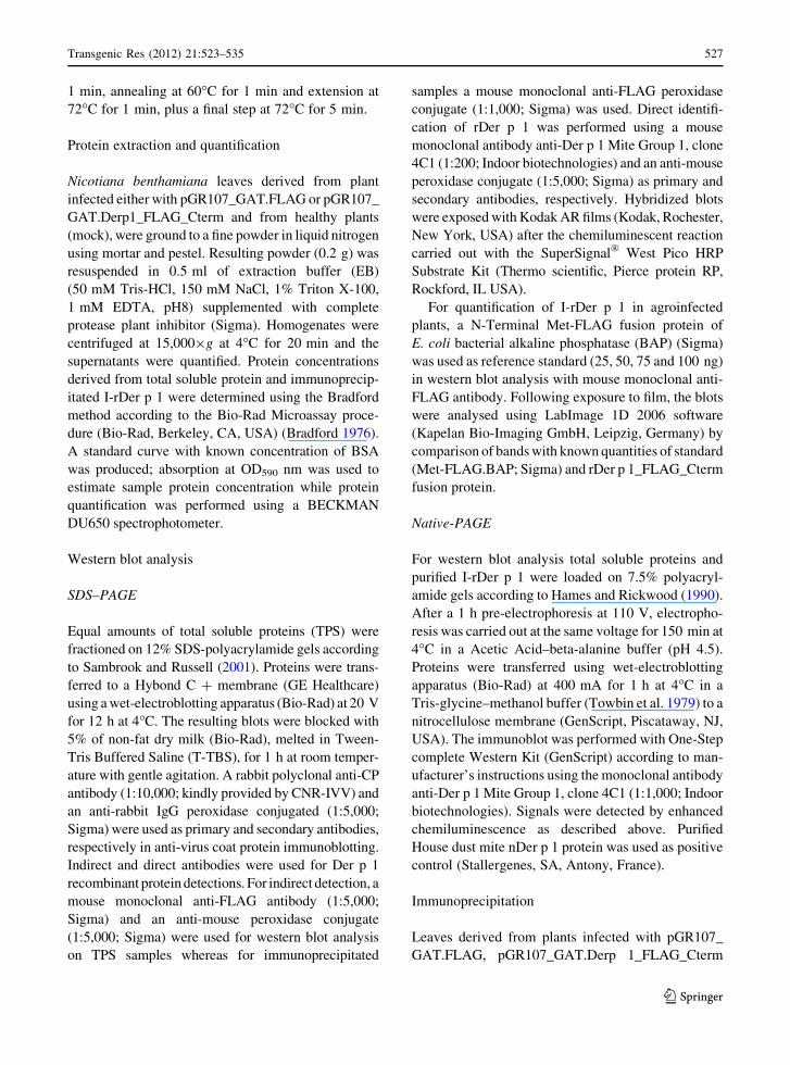
In western blot analysis with polyclonal anti-CP
antibody, the coat protein of potato virus X was
detectable as a band of about 25 kDa in both
pGR107_GAT.FLAG and pGR107_GAT.Derp1_
FLAG_Cterm samples (Fig. 1; lanes a2 and a3) but
not in mock inoculated plants (Fig. 1; lane a1)
suggesting an efficient virus replication.
Transgenic Res (2012) 21:523–535 529
123
Duplicated blots were probed with monoclonal anti-
FLAG and anti-Der p 1 antibodies in SDS–PAGE
conditions. A single band corresponding to a molecular
weight of about 27 kDa was visualized in western blots
hybridized with monoclonal anti-FLAG antibody for
PVX sample expressing I-Der p 1 (Fig. 1, lane b3).
Since the FLAG peptide has a very low molecular
weight (about 1 kDa), no signal was detected in
samples from plants infected with the empty vector
(Fig. 1, lanes b1 and b2). Moreover, western blotting
with SDS–PAGE conditions using anti-Der p 1 anti-
body was able to detect the expected 27 kDa band
either in plants or house dust mite extracts (data not
shown). Interestingly, under native conditions, anti-
Der p 1 antibody detected the expected band in both
commercial purified nDer p 1 (Fig. 1; lane c1) and in
planta protein extracts (Fig. 1; lane c4).
The anti-FLAG antibody also allowed an estima-
tion of the accumulation level of I-rDer p 1 fusion
protein (Derp1_FLAG_Cterm) in N. benthamiana.
Quantifications were performed in western blots,
according to Gotoh et al. (2002), by comparing signals
in plant samples with known amounts of N-Terminal
Met-FLAG bacterial alkaline phosphatase (BAP)
fusion protein of E. coli (Met-FLAG.BAP, Sigma)
using the LabImage 1D 2006 software. The estimated
level of accumulation of rDer p 1 in N. benthamiana
plant cells was about 3% of total soluble proteins.
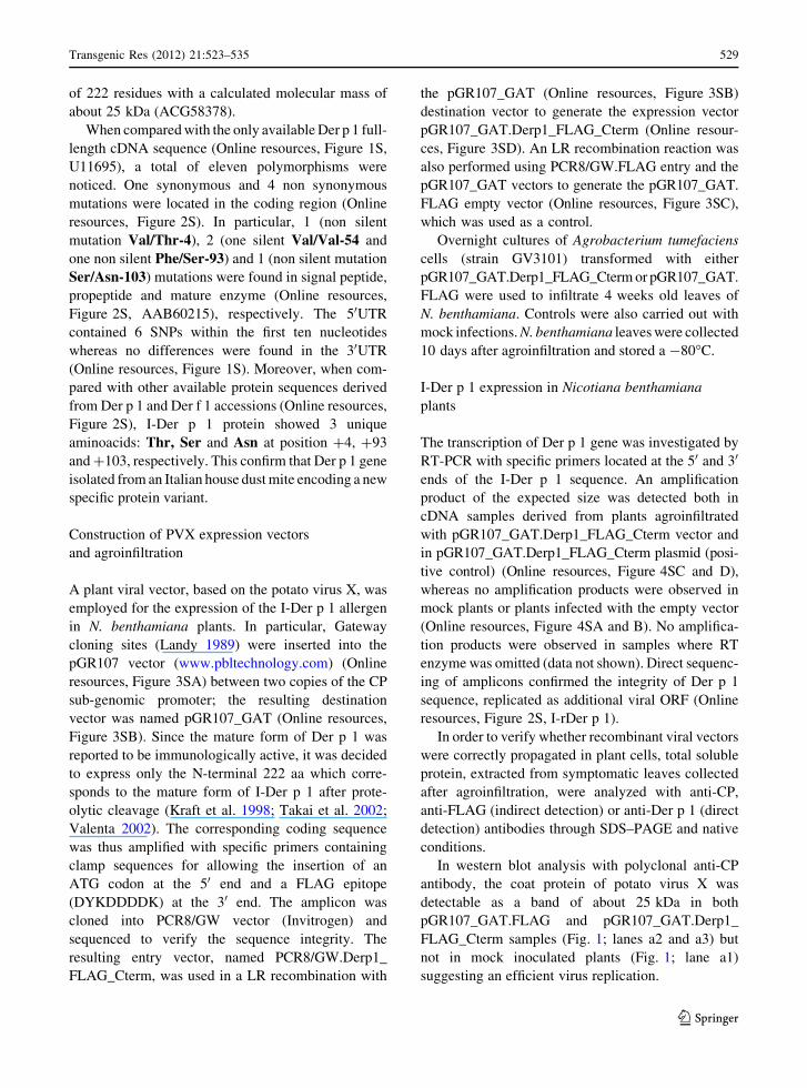
I-rDer p 1 purification and immunoblotting
The FLAG-tag epitope, fused at the C-terminus region
of I-rDer p 1 protein, allowed immunoprecipitations
with an anti-FLAG antibody coupled to M2 agarose
beads. The efficiency of immunoprecipitation was
analyzed by estimating the amount of recombinant
protein in the different fractions (Fig. 2a–c) through
western blotting with either anti-FLAG antibody and
SDS–PAGE conditions (Fig. 2a, b) or anti-Der p 1
antibody and Native-PAGE conditions (Fig. 2c).
Purified proteins were also stained with Coomassie
Brillant Blue to verify the presence of visible proteins
in eluate samples (Fig. 2d).
A single hybridization signal of 27 kDa was
visualized in western blotting with anti-FLAG anti-
body both in eluate (containing the purified protein,
Fig. 2, lane a3) and beads fraction after elution (Fig. 2,
lane a3) while no signal was detected in supernatant
(data not shown). Moreover, a single band was
visualized in Coomassie stained gels (Fig. 2, lane
d3), and in western blotting with anti-Der p 1 antibody
(Fig. 2, lane a4).
As expected, no bands were visualized in immu-
noprecipitated samples derived either from mock
plants (Fig. 2, lanes a1, b1, c2 and d1) or from plants
infected with empty viral vector (Fig. 2, lanes a2, b2,
c3 and d2). The purification process yielded about
150 lg of I-rDer p 1 per gram of leaves infected with
pGR107_GAT.Derp1_FLAG_Cterm vector.
N-glycosylation analysis of I-rDer p 1
To determine whether I-rDer p 1 was glycosylated the
purified protein fraction was treated with PNGase F
Fig. 1 Der p 1 mature form protein is expressed in planta.
Western blot analysis were carried out with rabbit polyclonal
anti-CP antibody (a) and mouse monoclonal anti-FLAG
(b) under SDS–PAGE conditions and with mouse monoclonal
anti-Der p 1 antibody under Native-PAGE conditions (c). Total
soluble proteins were isolated from mock plants (a1, b1, c2),
from plants agroinfiltrated with empty vector (a2, b2, c3) and
from plants agroinfiltrated with vector containing I-Der p 1 (a3,
b3, c4). House dust mite extracts were used as positive control in
Native-PAGE (c1). Standard Molecular weights are reported on
the left side of figure a and b (kDa kiloDaltons)
530 Transgenic Res (2012) 21:523–535
123
enzyme which release all carbohydrate residues from
protein. Figure 3a and b shows that the electrophoretic
mobility of I-rDer p 1 before (lanes 1a and 1b) and
after (lanes 2a and 2b) de-glycosylation does not
change. These results suggest that recombinant mature
Der p 1 expressed in our study is not glycosylated.
RNase B protein was also treated and the results of the
SDS–PAGE Coomassie Brilliant Blue before and after
de-glycosylation demonstrated the goodness of the
experimental procedures (Supplementary material,
Figure 5S).
I-rDer p 1 ISAC IgE inhibition assay
IgE inhibition results, obtained by incubating the
I-rDer p 1 with pooled sera from mite allergic patients
recognizing Der p 1 and Der f 1, are reported in Fig. 4.
As clearly shown, an almost total IgE inhibition of
both homologous mite molecules immobilized on the
ISAC microarray has been achieved, strongly sug-
gesting that the I-rDer p 1 preparation bears all the IgE
binding epitopes. As expected no IgE inhibition
reaction was observed (data not shown) when purified
proteins derived from tissues of plants infected with
empty viral vector and mock were used.
Discussion
In developed countries more than 20% of the popu-
lation is affected by IgE mediated allergic diseases,
such as seasonal and perennial rhinitis asthma and
atopic dermatitis (Wills-Karp et al. 2001). Diagnosis
of IgE mediated allergies is usually conducted by
provocation testing with extracts prepared from sus-
pected allergen sources. Alternatively, IgE inhibition
Fig. 2 Der p 1 recombinant protein is immunoprecipitated.
After extraction, proteins were subjected to immunoprecipita-
tion with an anti-FLAG antibody coupled to M2 agarose beads
and used for western blotting with anti-FLAG monoclonal
antibody on both eluate (containing the purified protein, a) and
beads fraction after elution (b). In particular proteins were
immunoprecipitated from mock plants (lanes a1, b1), from
plants agroinfiltrated with pGR107_GAT.FLAG (a2, b2) and
with pGR107_GAT.Derp1_FLAG_Cterm (a3, b3) expression
vectors. Western blotting were also carried out under Native-
PAGE conditions (c) using anti-Der p 1 antibody against
positive control (c1, commercial purified nDer p 1), immuno-
precipitated proteins from mock plants (c2), from plants
agroinfiltrated with empty vector (c3) and pGR107_GAT.
Derp1_FLAG_Cterm (c4). Staining with Coomassie Brillant
Blue (d) of immunoprecipitated proteins from mock-inoculated
control plants (d1), plants infected with empty vector (d2) and
plants infected with viral vector containing I-Der p 1 (d3) was
performed. d4 is an empty lane while d5 is MW marker.
Standard Molecular weight (kDa kiloDaltons) are reported on
the left side of a, b and d figures
Fig. 3 N-glycosylation analysis of I-rDer p 1. SDS–PAGE
coomassie brillant blue staining (a) and western blot analysis
(b) of I-rDer p 1 before (1a, 1b) and after (2a, 2b) PNGase F de-
glycosylation treatment
Transgenic Res (2012) 21:523–535 531
123
testing can be applied to obtain in vitro measurements
of specific serum IgE antibodies. In both cases allergen
extracts, consisting of variable mixtures of allergic and
non allergic components, are used for in vitro and in
vivo diagnosis of allergy. With such diagnostic
extracts it is almost impossible to detect whether the
patient is sensitized to a variety of unrelated allergens
or cross sensitized to few but cross reactive allergens.
Moreover, extract-based diagnosis cannot provide
information regarding the individual reactivity pro-
files of allergic patients and thus fail to identify the
precise target molecules for specific immunotherapy.
Recombinant allergen mimicking the immunological
properties of native allergens would be welcome for
both diagnostic and therapeutic purposes.
Plants have recently joined the competition for the
development of an efficient and cheap system for the
production of pharmaceutical proteins (Ma et al.
2005). Compared to other expression systems, such as
bacterial or mammalian cell cultures, plants are
characterized by a lower risk of getting contaminated
by human pathogens (e.g. animal viruses or prions). In
the present paper a plant viral vector based on the
potato virus X was used to express the mature form of
Der p 1, the major allergen of house dust mite. In
particular, we have inserted Gateway cloning sites into
the pGR107 vector with aiming at agroinfiltrations
rather than plasmidic DNA infections. Gateway tech-
nology facilitates high throughput cloning of target
sequences by making use of the bacteriophage lambda
site-specific recombination system (Landy 1989),
whereas the agroinfiltration increases the infection
efficiency (Gleba et al. 2005).
As far as we know this is the first time that the I-Der
p 1 (Der p 1 of an Italian strain of the house dust mite)
variant was isolated, sequenced and expressed. The
sequence shows several polymorphisms compared to
full or partial Der p 1 sequences obtained from house
dust mites belonging to other geographical regions
(Voorhorst et al. 1967; Chua et al. 1988, 1993; Thomas
et al. 1988, 2004; Arlian et al. 2001; Smith et al. 2001;
Asokananthan et al. 2002; Takai et al. 2002; De Boer
2003; Wolfowicz et al. 2003; Pichavant et al. 2005). In
particular, I-Der p 1 variant shows several mutations at
nucleotide level when compared with all the other
available sequences. Three amino acid substitutions
were observed, one in the signal peptide, one in the
propeptide and one in the mature enzyme sequence.
These 3 substitutions distinguished the Italian strain
from all others. It is worth noting that these substitu-
tions do not pertain neither to the active site (Cys34;
His170; Asn190) nor the disulfide bonds (Cys31-
Cys71; Cys65-Cys103; Cys4-Cys117) but affect the
portions of the molecule located in the signal peptide,
in the propeptide and in the NH2-terminal end of the
mature enzyme. In particular, amino acidic mutation
Phe/Ser-6 is located in the binding region between the
propeptide and the mature enzyme.
It is noteworthy that previous studies carried out in
E. coli and yeasts have failed in expressing the mature
form of Der p 1. The same results were reported for
tobacco cell suspension transformed with a cDNA
coding for the mature form of Der p 1. On the contrary,
the pro-Der p1 protein targeted to the endoplasmic
reticulum of tobacco BY2 cells was correctly pro-
cessed to mature form which retained both enzymatic
activity and immunogenic properties. Yang et al.
(2007) have described the expression of the mature
form of Der p 1 in transgenic rice seeds using a
construct based on the GluB promoter and a targeting
signal for the ER. Possible problems with glycosyla-
tion in Golgi were avoided by including a KDEL
signal for protein retention in the ER. The authors have
also demonstrated that glycosylated Der p 1 had a
significantly reduced IgE binding capacity when
compared with unglycosylated Der p 1. Based on
these results they proposed that the high variability in
IgE binding reduction among sera depended on
glycosylation levels (Yang et al. 2007). To circumvent
this problem, we decided to express a cytoplasmic
Fig. 4 IgE inhibition analysis. IgE inhibition of Ir-Der p 1 was
evaluated by running the ISAC 103 microarray IgE assay where
Der p 1 and Der f 1 allergens are immobilized. I-rDer p 1 showed
an almost full inhibition of the IgE binding. Buffer solution was
used as reference value for no inhibition IgE values
532 Transgenic Res (2012) 21:523–535
123
version of mature Der p 1 by using an in whole plant
transient expression system. Interestingly, we report
evidences that anti-Der p 1 antibody (clone 4C1,
Indoor biotechnologies) recognized commercial puri-
fied n-Der p 1 and our I-rDer p 1 in Native-PAGE
suggesting that I-rDer p 1 is correctly folded, at least at
the level of epitope recognized by the antibody used.
Comparison between I-rDer p 1 and N-Terminal
Met-FLAG fusion protein of E. coli Bacterial Alkaline
Phosphatase, allowed us to estimate that the recombi-
nant allergen represented about 3% of total soluble
proteins in N. benthamiana leaves. Our results dem-
onstrate that the execution of immunoprecipitations,
using the FLAG-tag for isolating I-rDer p 1 proteins
from crude extracts, is very simple and efficient.
Nevertheless, FLAG-based immunoprecipitation sys-
tem for Der p 1 purification need to be optimized since
we have demonstrated that a significant aliquot of the
bound protein tends to remain attached to the anti-
FLAG antibody immobilized to the agarose beads.
Our future aims are to test other immunoprecipitation
systems employing other tags (e.g. 6HIS or CMyC).
Moreover, when the I-rDer p 1 has been tested for
IgE inhibition on a well established immune assay, we
obtained an almost full inhibition of the IgE binding.
These findings corroborates that the obtained recom-
binant product mimics the already available ones and
thus it could be useful for diagnostic purposes and
might be employed for allergen standardization in
total mite extracts.
In conclusion, in this paper we have shown that a
modified PVX viral vector can be successfully used to
express I-Der p 1 protein suitable for diagnostic
purposes. A better molecular and immunological
characterization is needed to ascertain whether in
planta produced I-Der p 1 might also be used for
therapeutic treatments.
Acknowledgments We owe a big thank to Prof. Luca Santi and
Luigi Russi for critical reading of this manuscript and helpful
discussions. We are also very grateful to M. Castagnoli of CRA-
ABP (Consiglio per la Ricerca e la Sperimentazione in
Agricoltura, Centro di ricerca per l’agrobiologia e la pedologia,
Florence, Italy) for providing HDM and to Prof. David Baulcombe
(The Sainsbury Laboratory, Norwich, UK, www.pbltechnology.
com) for kindly providing the pGR107 vector. This work was
funded by Progetto PRIN 2005 ‘‘Transient expression of human
proinsulin and of other therapeutics proteins in Nicotiana benth-amiana’’ (EA) and PRIN 2006 ‘‘Production, purification and in
vitro characterization of some italian-strain recombinant allergens
of Dermatophagoides pteronyssinus’’ (FP).
References
Altschul SF, Madden TL, Schaffer AA, Zhang J, Zhang Z,
Miller W, Lipman DJ (1997) Gapped BLAST and PSI-
BLAST: a new generation of protein database search pro-
grams. Nucl Acids Res 25:3389–3402
Arlian LG, Neal JS, Morgan MS, Vyszenski-Moher DL, Rapp
CM, Alexander AK (2001) Reducing relative humidity is a
practical way to control dust mites and their allergens in
homes in temperate climates. J Allergy Clin Immunol
107:99–104
Asokananthan N, Graham PT, Stewart DJ, Bakker AJ, Eidne
KA, Thompson PJ, Stewart GA (2002) House dust mite
allergens induce proinflammatory cytokines from respira-
tory epithelial cells: the cysteine protease allergen, Der p 1,
activates protease-activated receptor (PAR)-2 and inacti-
vates PAR-1. J Immunol 169:4572–4578
Asturias JA, Arilla MC, Gomez-Bayon N, Martınez A, Martınez
J, Palacios R (1998) Sequencing and high level expression
in Escherichia coli of the tropomyosin allergen (Der p 10)
from Dermatophagoides pteronyssinus. Biochim Biophys
Acta Gene Struct Expr 1397:27–30
Asturias J, Gomez-Bayon N, Arilla MC, Martinez A, Palacios R,
Sanchez-Gascon F, Martinez J (1999) Molecular charac-
terization of American cockroach tropomyosin (Peripla-neta americana allergen 7), a cross-reactive allergen.
J Immunol 162:4342–4348
Bendahmane A, Kohn BA, Dedi C, Baulcombe DC (1995)
The coat protein of potato virus X is a strain-specific
elicitor of Rx1-mediated virus resistance in potato. Plant
J 8:933–941
Bradford M (1976) A rapid and sensitive method for the quan-
tification of microgram quantities of protein utilizing the
principle of protein-dye binding. Anal Biochem 72:
248–254
Chomczynski P, Sacchi N (1987) Single-step method of RNA
isolation by acid guanidinium thiocyanate-phenol-chloro-
form extraction. Anal Biochem 162:156–159
Chua KY, Stewart GA, Thomas WR, Simpson RJ, Dilworth RJ,
Plozza TM, Turner KJ (1988) Sequence analysis of cDNA
coding for a major house dust mite allergen, Der p 1.
Homology with cysteine proteases. J Exp Med 167:175–182
Chua KY, Kehal PK, Thomas WR (1993) Sequence polymor-
phism of the cDNA clones encoding the mite allergen Der
p 1. Int Arch Allergy Immunol 101:364–368
Chua KY, Huang CH, Shen HD, Thomas WR (1996) Analysis of
sequence polymorphism of a major mite allergen Der p 2.
Clin Exp Allergy 26:829–837
Cunningham C, Porter AJR (eds) (1998) Recombinant proteins
from plants. Production and isolation of clinically useful
compounds. Methods in Biotechnology, vol 3. Humana
Press, Totowa, NJ
De Boer R (2003) The effect of sub-floor heating on house-dust-
mite populations on floors and in furniture. Exp Appl
Acarol 29:315–330
Fernandez-Caldas E (1997) Mite species of allergologic
importance in Europe. Allergy 52:383–387
Floss DM, Falkenburg D, Conrad U (2007) Production of vaccines
and therapeutic antibodies for veterinary applications in
transgenic plants: an overview. Transgenic Res 16:315–332
Transgenic Res (2012) 21:523–535 533
123
Gleba Y, Klimyuk V, Marillonnet S (2005) Magnifection: a new
platform for expressing recombinant vaccines in plants.
Vaccine 23:2042–2048
Gotoh M, Yada T, Sato T, Akashima T, Iwasaki H, Mochizuki
H, Inaba N, Togayachi A, Kudo T, Watanabe H, Kimata K,
Narimatsu H (2002) Molecular cloning and characteriza-
tion of a novel chondroitin Sulfate glucuronyltransferase
that transfers glucuronic acid to N-acetylgalactosamine.
J Biol Chem 41:38179–38188
Greene WK, Cyster JG, Chua KY, O’Brien RM, Thomas WR
(1991) IgE and IgG binding of peptides expressed from
fragments of cDNA encoding the major house dust mite
allergen Der p I. J Immunol 147:3768–3773
Hames BD, Rickwood D (1990) Gel electrophoresis of proteins.
A practical approach, 2nd edn edn. IRL Press, Oxford,
pp 1–270
Hellens RP, Edwards EA, Leyland NR, Bean S, Mullineaux PM
(2000) pGreen: a versatile and flexible binary Ti vector for
Agrobacterium-mediated plant transformation. Plant Mol
Biol 42:819–832
HuangFu T, Lim LH, Chua KY (2006) Efficacy evaluation of
Der p 1 DNA vaccine for allergic asthma in an experi-
mental mouse model. Vaccine 24:4576–4581
Jacquet A, Haumont M, Massaer M, Daminet V, Garcia L,
Mazzu P, Jacobs P, Bollen A (2000) Biochemical and
immunological characterization of a recombinant precur-
sor form of the house dust mite allergen Der p 1 produced
by Drosophila cells. Clin Exp Allergy 30:677–684
Kraft D, Ferreira F, Ebner C, Valenta R, Breiteneder H, Susani
M, Breitenbach M, Scheiner O (1998) Recombinant
allergens: the future of the diagnosis and treatment of
atopic allergy. Allergy 53:62–66
Krause S, Reese G, Randow S, Zennaro D, Quaratino D, Palazzo
P, Ciardiello MA, Petersen A, Becker WM, Mari A (2009)
Lipid transfer protein (Ara h 9) as a new peanut allergen
relevant for a Mediterranean allergic population. J Allergy
Clin Immunol 4:771–778
Kumar S, Tamura K, Nei M (2004) MEGA3: integrated soft-
ware for molecular evolutionary genetics analysis and
sequence alignment. Brief Bioinform 5:150–163
Landy A (1989) Dynamic, structural, and regulatory aspects of
lambda site-specific recombination. Annu Rev Biochem
58:913–949
Lee CS, Tsai LC, Chao PL, Lin CY, Hung MW, Chien AI,
Chiang YT, Han SH (2004) Protein sequence analysis of a
novel 103-kDa Dermatophagoides pteronyssinus mite
allergen and prevalence of serum immunoglobulin e
reactivity to rDer p 11 in allergic adult patients. Clin Exp
Allergy 34:354–362
Lienard D, Tran Dinh O, van Oort E, Van Overtvelt L, Bonneau
C, Wambre E, Bardor M, Cosette P, Didier-Laurent A, de
Borne FD, Delon R, van Ree R, Moingeon P, Faye L,
Gomord V (2007) Suspension-cultured BY-2 tobacco cells
produce and mature immunologically active house dust
mite allergens. Plant Biotechnol J 5:93–108
Lin KL, Hsieh KH, Thomas WR, Chiang BL, Chua KY (1994)
Characterization of Der p V allergen, cDNA analysis, and
IgE-mediated reactivity to the recombinant protein.
J Allergy Clin Immunol 94:989–996
Ma J, Barros E, Bock R, Christou P, Dale PJ, Dix PJ, Fischer R,
Irwin J, Mahoney R, Pezzotti M, Schillberg S, Sparrow P,
Stoger E, Twyman RM (2005) Molecular farming for new
drugs and vaccines. EMBO Rep 6:593–599
Mach L, Mort JS, Glossl J (1994) Maturation of human proca-
thepsin B. Proenzyme activation and proteolytic process-
ing of the precursor to the mature proteinase, in vitro, are
primarily unimolecular processes. J Biol Chem 269:
13030–13035
Morozov Sy, Gorbulev Vg, Novlkov Vk, Agranovsky Aa,
Kozlov Yv, Atabekov Jg, Aa Baev (2000) The primary
structure of the 50- and 30-terminal regions of the RNA of
potato virus X. Dokl Akad Nauk 259:723–725
Nagel R, Elliot A, Masel A, Birch RG, Manners JM (1990)
Electroporation of binary Ti plasmid vector into Agro-bacterium tumefaciens and Agrobacterium rhizogenes.
FEMS Microbiol Lett 67:325–328
Pichavant M, Charbonnier AS, Taront S, Brichet A, Wallaert B,
Pestel J, Tonnel AB, Gosset P (2005) Asthmatic bronchial
epithelium activated by the proteolytic allergen Der p 1
increases selective dendritic cell recruitment. J Allergy
Clin Immunol 115:771–778
Platts-Mills TA, Chapman MD (1988) Dust mites: immunology,
allergic disease, and environmental control. J Allergy Clin
Immunol 82:841
Platts-Mills TA, de Weck AL (1989) Dust mite allergens and
asthma, a worldwide problem. J Allergy Clin Immunol
83:415–427
Platts-Mills TA, Woodfolk JA (2011) Allergens and their role in
the allergic immune response. Immunol Rev 242:51–68
Platts-Mills TA, Blumenthal K, Perzanowski M, Woodfolk JA
(2000) Determinants of clinical allergic disease. The rele-
vance of indoor allergens to the increase in asthma. Am J
Respir Crit Care Med 162:S128–S133
Porta C, Lomonossoff GP (1998) Scope for using plant viruses
to present epitopes from animal pathogens. Rev Med Virol
8:25–41
Robinson C, Kalsheker NA, Srinivasan N, King CM, Garrod
DR, Thompson PJ, Stewart GA (1997) On the potential
significance of the enzymatic activity of mite allergens to
immunogenicity. Clues to structure and function revealed
by molecular characterization. Clin Exp Allergy 27:10–21
Sambrook J, Russell DW (2001) Molecular cloning: a labora-
tory manual. Cold Spring Harbor, NY., Cold Spring Harbor
Laboratory Press
Smith WA, Chua KY, Kuo MC, Rogers BL, Thomas WR (1994)
Cloning and sequencing of the Dermatophagoides pter-onyssinus group III allergen, Der p III. Clin Exp Allergy
24:220–228
Smith WA, Hales BJ, Jarnicki AG, Thomas WR (2001) Aller-
gens of wild house dust mites: environmental Der p 1 and
Der p 2 sequence polymorphisms. J Allergy Clin Immunol
107:985–992
Sonenberg N, Shatkin AJ, Ricciardi RP, Rubin M, Goodman
RM (1978) Analysis of terminal structures of RNA from
potato virus X. Nucleic Acids Res 5:2501–2512
Steen N, Hutchinson A, McColl E, Eccles MP, Hewison J,
Meadows KA, Blades SM, Fowler P (1994) Development
of a symptom based outcome measure for asthma. Br Med J
309:1065–1068
Takai T, Mineki R, Nakazawa T, Takaoka M, Yasueda H,
Murayama K, Okumura K, Ogawa H (2002) Maturation of
the activities of recombinant mite allergens Der p 1 and Der
534 Transgenic Res (2012) 21:523–535
123
f 1, and its implication in the blockade of proteolytic
activity. FEBS Lett 531:265–272
Takai T, Kato T, Yasueda H, Okumura K, Ogawa H (2005)
Analysis of the structure and allergenicitiy of recombinant
pro- and mature Der p 1 and Der f 1: major conformational
IgE-epitopes blocked by prodomains. J Allergy Clin
Immunol 115:555–563
Thomas WR, Stewart GA, Simpson RJ, Chua KY, Plozza TM,
Dilworth RJ, Nisbet A, Turner KJ (1988) Cloning and
expression of DNA coding for the major house dust mite
allergen Der p 1 in Escherichia coli. Int Arch Allergy Appl
Immunol 85:127–129
Thomas WR, Smith WA, Hales BJ, Mills KL, O’Brien RM
(2002) Characterization and immunobiology of house dust
mite allergens. Int Arch Allergy Immunol 129:1–18
Thomas WR, Smith WA, Hales BJ (2004) The allergenic
specificities of the house dust mite. Chang Gung Med J
27:563–569
Topham CM, Srinivasan N, Thorpe CJ, Overington JP, Kal-
sheker NA (1994) Comparative modelling of major house
dust mite allergen Der p I: structure validation using an
extended environmental amino acid propensity table.
Protein Eng 7:869–894
Towbin H, Staehelin T, Gordon J (1979) Electrophoretic
transfer of proteins from polyacrylamide gels to nitrocel-
lulose sheets: procedure and some applications. Proc Natl
Acad Sci USA 76:4350–4354
Valenta R (2002) The future of antigen-specific immunotherapy
of allergy. Nat Rev Immunol 2:446–453
Vernet T, Khouri HE, Laflamme P, Tessier DC, Musil R, Gour-
Salin BJ, Storer AC, Thomas DY (1991) Processing of the
papain precursor. Purification of the zymogen and char-
acterization of its mechanism of processing. J Biol Cem
266:21451–214517
Voorhorst R, Spieksma-Boezeman MIA, Spieksma FTM,
Varekamp H, Leupen MJ, Lyklema AW (1967) The house
dust mite (Dermatophagoides pteronyssinus) and the
allergens it produces. Identity with the house dust allergen.
J Allergy 39:325–339
Wagner B, Fuchs H, Adhami F, Ma Y, Scheiner O, Breiteneder
H (2004) Plant virus expression systems for transient pro-
duction of recombinant allergens in Nicotiana benthami-ana. Methods 32:227–234
Wills-Karp M, Santeliz J, Karp CL (2001) The germeless theory
of allergic disease: revisiting the hygiene hypothesis. Nat
Rev Immunol 1:69–75
Wolfowicz CB, HuangFu T, Chua KY (2003) Expression and
immunogenicity of the major house dust mite allergen Der
p 1 following DNA immunization. Vaccine 21:1195–1204
Yang L, Kajiura H, Suzuki K, Hirose S, Fujiyama K (2007)
Generation of a transgenic rice seed-based edible vaccine
against house dust mite allergy. Biochem Biophys Res
Commun 365:334–339
Yusibov V, Rabindran S, Commandeur U, Twyman RM,
Fischer R (2006) The potential of plant virus vectors for
vaccine production. Drugs R&D 7:203–217
Transgenic Res (2012) 21:523–535 535
123