The domestication of the probiotic bacterium Lactobacillus acidophilus
Standards in Genomic Sciences (2013) 9:142-159 DOI:10.4056/sigs.4287962
The Genomic Standards Consortium
Genome sequence of Phaeobacter daeponensis type strain (DSM 23529T), a facultatively anaerobic bacterium isolated from marine sediment, and emendation of Phaeobacter daeponensis
Marco Dogs1, Hazuki Teshima2, Jörn Petersen3, Anne Fiebig3, Olga Chertkov2, Hajnalka Dalingault2, Amy Chen4, Amrita Pati5, Lynne A. Goodwin2,5, Patrick Chain5, John C. Detter2,5, Natalia Ivanova5, Alla Lapidus5, Manfred Rohde6, Sabine Gronow3, Nikos C. Kyrpides5, Tanja Woyke5, Meinhard Simon1, Markus Göker3*, Hans-Peter Klenk3, Thorsten Brinkhoff1
1Institute for Chemistry and Biology of the Marine Environment, University of Oldenburg, Oldenburg, Germany. 2Los Alamos National Laboratory, Bioscience Division, Los Alamos, New Mexico, USA 3Leibniz Institute DSMZ – German Collection of Microorganisms and Cell Cultures,
Braunschweig, Germany 4Biological Data Management and Technology Center, Lawrence Berkeley National
Laboratory, Berkeley, California, USA 5DOE Joint Genome Institute, Walnut Creek, California, USA 6HZI – Helmholtz Centre for Infection Research, Braunschweig, Germany
*Correspondence: Markus Göker ([email protected])
Keywords: Marine microbiology, facultative anaerobe, indigoidine, Rhodobacteraceae, Roseobacter clade
TF-218T is the type strain of the species Phaeobacter daeponensis Yoon et al. 2007, a facultatively anaerobic Phaeobacter species isolated from tidal flats. Here we describe the draft genome sequence and annotation of this bacterium together with previously unreported aspects of its phenotype. We analyzed the genome for genes involved in secondary metabo-lite production and its anaerobic lifestyle, which have also been described for its closest rela-tive Phaeobacter caeruleus. The 4,642,596 bp long genome of strain TF-218T contains 4,310 protein-coding genes and 78 RNA genes including four rRNA operons and consists of five re-plicons: one chromosome and four extrachromosomal elements with sizes of 276 kb, 174 kb, 117 kb and 90 kb. Genome analysis showed that TF-218T possesses all of the genes for indigoidine biosynthesis, and on specific media the strain showed a blue pigmentation. We also found genes for dissimilatory nitrate reduction, gene-transfer agents, NRPS/ PKS genes and signaling systems homologous to the LuxR/I system.
Introduction The genus Phaeobacter currently is comprised of five species (P. daeponensis, P. gallaeciensis, P. inhibens, P. arcticus and P. caeruleus) and is a part of the marine Roseobacter clade within the Alphaproteobacteria [1-5]. The genus name was derived from the dark brownish pigmentation of the type species P. gallaeciensis (phaeos = dark, brown) [3]. Strain TF-218T, however, was de-scribed as not pigmented. Strain TF-218T (= KCTC 12794T = JCM 13606T = DSM 23529T) is the type strain of the species Phaeobacter daeponensis [1].
It was isolated from tidal flats at Daepo Beach (Yellow Sea), Korea, which led to the species name of P. daeponensis [1]. Secondary metabolite production is a well-known feature within the Roseobacter clade [6], especially within the Phaeobacter cluster, which shows high efficiency for secondary metabolite production [7]. Examples include biosynthesis of the antibiot-ics tropdithietic acid (TDA) or indigoidine, quor-um sensing by N-acyl homoserine lactones (AHLs), and presence of genes coding for nonribosomal
Dogs et al.
http://standardsingenomics.org 143
peptide synthases (NRPS) and polyketide syn-thases (PKS) [6-11]. Furthermore, P. daeponensis was the first described facultatively anaerobic Phaeobacter species, which is capable of nitrate reduction [1]. Here we present the draft genome sequence and annotation of P. daeponensis TF-218T. We analyzed the genome for special features with a focus on secondary metabolite production. Novel aspects of the strain phenotype are also reported.
Classification and features 16S rRNA gene sequence analysis Figure 1 shows the phylogenetic neighborhood of P. daeponensis in a 16S rRNA gene sequence based tree. The sequences of the four 16S rRNA gene copies in the genome of strain DSM 23529T differ from each other by up to two nucleotides, and dif-fer by up to two nucleotides from the previously published 16S rRNA gene sequence (DQ81486) [Table 1]. A representative genomic 16S rRNA gene se-quence of P. daeponensis TF-218T was compared with the Greengenes database for determining the weighted relative frequencies of taxa and (trun-cated) keywords as previously described [21]. The most frequently occurring genera were Ruegeria (31.6%), Phaeobacter (28.8%), Silicibacter (13.6%), Roseobacter (13.3%) and Nautella (3.6%) (713 hits in total). Regarding the five hits to sequences from the species, the average identi-ty within HSPs was 99.9%, whereas the average coverage by HSPs was 19.0%. Regarding the 45 hits to sequences from other species of the genus, the average identity within HSPs was 97.8%, whereas the average coverage by HSPs was 18.9%. Among all other species, the one yielding the highest score was Roseobacter gallaeciensis (AY881240), which corresponded to an identity of 98.6% and an HSP coverage of 18.8%. (Note that the Greengenes database uses the INSDC (= EMBL/NCBI/DDBJ) annotation, which is not an authoritative source for nomenclature or classifi-cation.) The highest-scoring environmental se-quence was AF253467 (Greengenes short name 'Key aromatic-ring-cleaving enzyme protocatechuate 34-dioxygenase ecologically im-portant marine Roseobacter lineage d on Indulin seawater'), which showed an identity of 99.8% and an HSP coverage of 18.8%. The most frequent-ly occurring keywords within the labels of all en-vironmental samples which yielded hits were
'microbi' (2.8%), 'marin' (2.7%), 'coral' (2.4%), 'diseas' (1.8%) and 'water' (1.8%) (492 hits in to-tal). The most frequently occurring keywords within the labels of those environmental samples which yielded hits of a higher score than the high-est scoring species were 'marin' (17.4%), 'sediment' (8.5%), 'aromatic-ring-cleav, ecolog, enzym, import, indulin, kei, lineag, protocatechu, roseobact, seawat' (4.4%), 'coco, island, near, site' (4.3%) and 'redox-stratifi, reef, sandi' (4.3%) (4 hits in total).
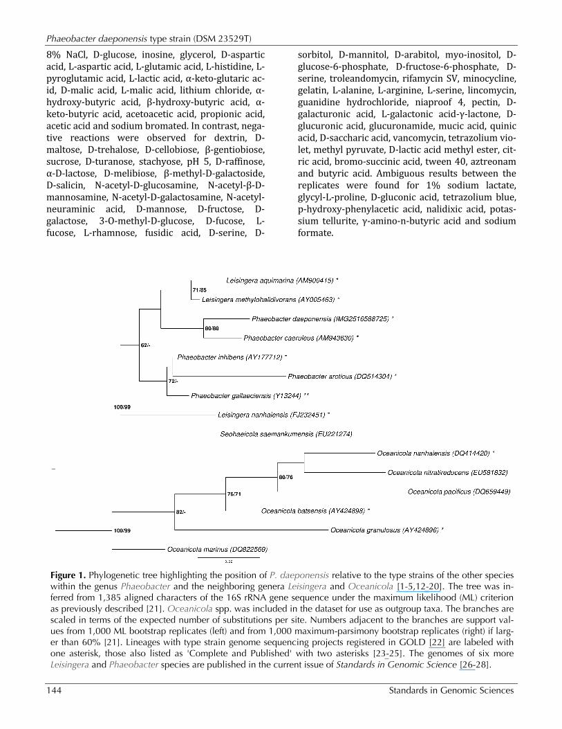
Morphology and physiology P. daeponensis TF-218T is a Gram-negative, facultatively anaerobic, mesophilic marine bacte-rium with an optimal growth temperature of 37°C and an optimal salt-tolerance between 0.1 and 8% (w/v) NaCl. The optimal pH for growth is between 7.0 and 8.0 with pH 5.5 being the lowest possible pH at which growth occurs. Strain TF-218T pos-sesses oval cells 0.4-0.9 x 0.7-2.0 µm in size (Fig-ure 2) and is motile by means of a single polar fla-gellum. On marine agar circular, slightly convex, smooth, glistering, yellowish-white colonies 1.5-2.5 mm in diameter are formed [1]. TF-218T utiliz-es D-glucose, glycerol, leucine, serine, acetate, cit-rate and succinate [1]. In addition to the findings reported in [1], we ob-served that strain DSM 23529T is able to form blue colonies on YTSS medium, as described for the closely related strain Y4I [11]. This is probably due to the presence of genes for indigoidine bio-synthesis in the genome (see below). The utilization of carbon compounds by P. daeponensis was also determined for this study using Generation-III microplates in an OmniLog phenotyping device (BIOLOG Inc., Hayward, CA, USA). The microplates were inoculated at 28°C with a cell suspension at a cell density of 95-96% turbidity and dye IF-A. Further additives were vit-amins, micronutrients and sea-salt solutions. The exported measurement data were further ana-lyzed with the opm package for R [30,31], using its functionality for statistically estimating parame-ters from the respiration curves and translating them into negative, ambiguous, and positive reac-tions. The strain was studied in two independent biological replicates, and reactions with a different behavior between the two repetitions were re-garded as ambiguous. For P. daeponensis strain DSM 23529T, positive re-actions were observed for pH 6, 1% NaCl, 4% NaCl,
Phaeobacter daeponensis type strain (DSM 23529T)
144 Standards in Genomic Sciences
8% NaCl, D-glucose, inosine, glycerol, D-aspartic acid, L-aspartic acid, L-glutamic acid, L-histidine, L-pyroglutamic acid, L-lactic acid, α-keto-glutaric ac-id, D-malic acid, L-malic acid, lithium chloride, α-hydroxy-butyric acid, β-hydroxy-butyric acid, α-keto-butyric acid, acetoacetic acid, propionic acid, acetic acid and sodium bromated. In contrast, nega-tive reactions were observed for dextrin, D-maltose, D-trehalose, D-cellobiose, β-gentiobiose, sucrose, D-turanose, stachyose, pH 5, D-raffinose, α-D-lactose, D-melibiose, β-methyl-D-galactoside, D-salicin, N-acetyl-D-glucosamine, N-acetyl-β-D-mannosamine, N-acetyl-D-galactosamine, N-acetyl-neuraminic acid, D-mannose, D-fructose, D-galactose, 3-O-methyl-D-glucose, D-fucose, L-fucose, L-rhamnose, fusidic acid, D-serine, D-
sorbitol, D-mannitol, D-arabitol, myo-inositol, D-glucose-6-phosphate, D-fructose-6-phosphate, D-serine, troleandomycin, rifamycin SV, minocycline, gelatin, L-alanine, L-arginine, L-serine, lincomycin, guanidine hydrochloride, niaproof 4, pectin, D-galacturonic acid, L-galactonic acid-γ-lactone, D-glucuronic acid, glucuronamide, mucic acid, quinic acid, D-saccharic acid, vancomycin, tetrazolium vio-let, methyl pyruvate, D-lactic acid methyl ester, cit-ric acid, bromo-succinic acid, tween 40, aztreonam and butyric acid. Ambiguous results between the replicates were found for 1% sodium lactate, glycyl-L-proline, D-gluconic acid, tetrazolium blue, p-hydroxy-phenylacetic acid, nalidixic acid, potas-sium tellurite, γ-amino-n-butyric acid and sodium formate.
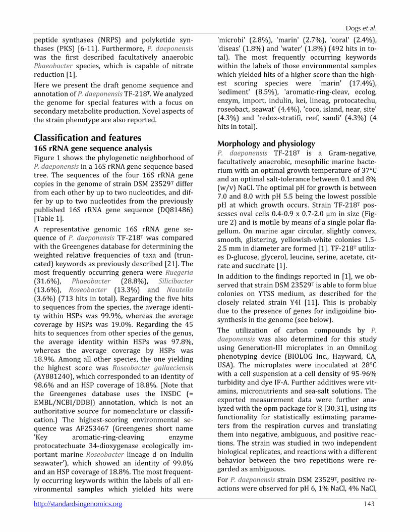
Figure 1. Phylogenetic tree highlighting the position of P. daeponensis relative to the type strains of the other species within the genus Phaeobacter and the neighboring genera Leisingera and Oceanicola [1-5,12-20]. The tree was in-ferred from 1,385 aligned characters of the 16S rRNA gene sequence under the maximum likelihood (ML) criterion as previously described [21]. Oceanicola spp. was included in the dataset for use as outgroup taxa. The branches are scaled in terms of the expected number of substitutions per site. Numbers adjacent to the branches are support val-ues from 1,000 ML bootstrap replicates (left) and from 1,000 maximum-parsimony bootstrap replicates (right) if larg-er than 60% [21]. Lineages with type strain genome sequencing projects registered in GOLD [22] are labeled with one asterisk, those also listed as 'Complete and Published' with two asterisks [23-25]. The genomes of six more Leisingera and Phaeobacter species are published in the current issue of Standards in Genomic Science [26-28].
Dogs et al.
http://standardsingenomics.org 145
Figure 2. Scanning electron micrograph of P. daeponensis DSM 23529T
Chemotaxonomy The principal fatty-acid profile of strain TF-128T consisted of major amounts of unsaturated fatty acid C18:1ω7c (57.7%) and 11-methyl C18:1ω7c (16.6%) in addition to straight-chain fatty acids (12.8%) and hydroxyl fatty acids (9.9%). Apart from the differences in the proportions, the fatty acid profile is similar to those of the type strains of P. gallaeciensis, P. inhibens and P. caeruleus. The major polar lipids of strain TF-218T are phosphatidylcholine, phosphatidylglycerol, phosphatidylethanolamine, two unidentified lipids and an aminolipid [1].
Genome sequencing and annotation Genome project history This organism was selected for sequencing on the basis of the DOE Joint Genome Institute Community Sequencing Program (CSP) 2010, CSP 441 “Whole genome type strain sequences of the genera Phaeobacter and Leisingera – a monophyletic group of physiologically highly diverse organisms”. The
genome project is deposited in the Genomes On Line Database [22] and the complete genome se-quence is deposited in GenBank. Sequencing and annotation were performed by the DOE Joint Ge-nome Institute (JGI) using state-of-the-art sequenc-ing technology [40]. A summary of the project in-formation is shown in Table 2.
Growth conditions and DNA isolation A culture of DSM 23529T was grown aerobically in DSMZ medium 514 [41] at 37°C. Genomic DNA was isolated using a Jetflex Genomic DNA Purifica-tion Kit (GENOMED 600100) following the stand-ard protocol provided by the manufacturer, but modified by an incubation time of 40 min, the in-cubation on ice over night on a shaker, the use of an additional 25 µl proteinase K, and the addition of 200 µl protein precipitation buffer. DNA is available from DSMZ through the DNA Bank Net-work [42].
Phaeobacter daeponensis type strain (DSM 23529T)
146 Standards in Genomic Sciences
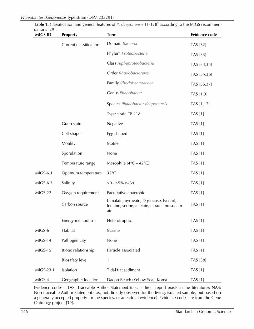
Table 1. Classification and general features of P. daeponensis TF-128T according to the MIGS recommen-dations [29]. MIGS ID Property Term Evidence code
Current classification Domain Bacteria TAS [32]
Phylum Proteobacteria TAS [33]
Class Alphaproteobacteria TAS [34,35]
Order Rhodobacterales TAS [35,36]
Family Rhodobacteraceae TAS [35,37]
Genus Phaeobacter TAS [1,3]
Species Phaeobacter daeponensis TAS [1,17]
Type strain TF-218 TAS [1]
Gram stain Negative TAS [1]
Cell shape Egg-shaped TAS [1]
Motility Motile TAS [1]
Sporulation None TAS [1]
Temperature range Mesophile (4°C – 42°C) TAS [1]
MIGS-6.1 Optimum temperature 37°C TAS [1]
MIGS-6.3 Salinity >0 - >9% (w/v) TAS [1]
MIGS-22 Oxygen requirement Facultative anaerobic TAS [1]
Carbon source L-malate, pyruvate, D-glucose, lycerol, leucine, serine, acetate, citrate and succin-ate
TAS [1]
Energy metabolism Heterotrophic TAS [1]
MIGS-6 Habitat Marine TAS [1]
MIGS-14 Pathogenicity None TAS [1]
MIGS-15 Biotic relationship Particle associated TAS [1]
Biosafety level 1 TAS [38]
MIGS-23.1 Isolation Tidal flat sediment TAS [1]
MIGS-4 Geographic location Daepo Beach (Yellow Sea), Korea TAS [1]
Evidence codes - TAS: Traceable Author Statement (i.e., a direct report exists in the literature); NAS: Non-traceable Author Statement (i.e., not directly observed for the living, isolated sample, but based on a generally accepted property for the species, or anecdotal evidence). Evidence codes are from the Gene Ontology project [39].
Dogs et al.
http://standardsingenomics.org 147
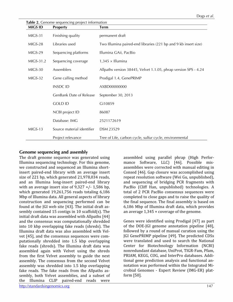
Table 2. Genome sequencing project information MIGS ID Property Term
MIGS-31 Finishing quality permanent draft
MIGS-28 Libraries used Two Illumina paired-end libraries (221 bp and 9 kb insert size)
MIGS-29 Sequencing platforms Illumina GAii, PacBio
MIGS-31.2 Sequencing coverage 1,345 × Illumina
MIGS-30 Assemblers Allpaths version 38445, Velvet 1.1.05, phrap version SPS - 4.24
MIGS-32 Gene calling method Prodigal 1.4, GenePRIMP
INSDC ID AXBD00000000
GenBank Date of Release September 30, 2013
GOLD ID Gi10859
NCBI project ID 86087
Database: IMG 2521172619
MIGS-13 Source material identifier DSM 23529
Project relevance Tree of Life, carbon cycle, sulfur cycle, environmental
Genome sequencing and assembly The draft genome sequence was generated using Illumina sequencing technology. For this genome, we constructed and sequenced an Illumina short-insert paired-end library with an average insert size of 221 bp, which generated 21,978,034 reads, and an Illumina long-insert paired-end library with an average insert size of 9,327 +/- 1,586 bp, which generated 19,261,756 reads totaling 6,186 Mbp of Illumina data. All general aspects of library construction and sequencing performed can be found at the JGI web site [43]. The initial draft as-sembly contained 15 contigs in 10 scaffold(s). The initial draft data was assembled with Allpaths [44] and the consensus was computationally shredded into 10 kbp overlapping fake reads (shreds). The Illumina draft data was also assembled with Vel-vet [45], and the consensus sequences were com-putationally shredded into 1.5 kbp overlapping fake reads (shreds). The Illumina draft data was assembled again with Velvet using the shreds from the first Velvet assembly to guide the next assembly. The consensus from the second Velvet assembly was shredded into 1.5 kbp overlapping fake reads. The fake reads from the Allpaths as-sembly, both Velvet assemblies, and a subset of the Illumina CLIP paired-end reads were
assembled using parallel phrap (High Perfor-mance Software, LLC) [46]. Possible mis-assemblies were corrected with manual editing in Consed [46]. Gap closure was accomplished using repeat resolution software (Wei Gu, unpublished), and sequencing of bridging PCR fragments with PacBio (Cliff Han, unpublished) technologies. A total of 2 PCR PacBio consensus sequences were completed to close gaps and to raise the quality of the final sequence. The final assembly is based on 6,186 Mbp of Illumina draft data, which provides an average 1,345 × coverage of the genome.
Genes were identified using Prodigal [47] as part of the DOE-JGI genome annotation pipeline [48], followed by a round of manual curation using the JGI GenePRIMP pipeline [49]. The predicted CDSs were translated and used to search the National Center for Biotechnology Information (NCBI) nonredundant database, UniProt, TIGR-Fam, Pfam, PRIAM, KEGG, COG, and InterPro databases. Addi-tional gene prediction analysis and functional an-notation was performed within the Integrated Mi-crobial Genomes - Expert Review (IMG-ER) plat-form [50].
Phaeobacter daeponensis type strain (DSM 23529T)
148 Standards in Genomic Sciences
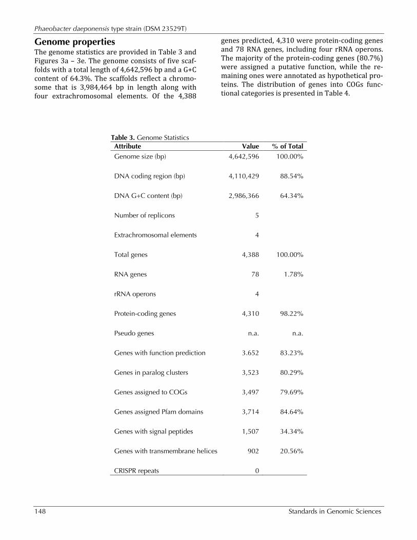
Genome properties The genome statistics are provided in Table 3 and Figures 3a – 3e. The genome consists of five scaf-folds with a total length of 4,642,596 bp and a G+C content of 64.3%. The scaffolds reflect a chromo-some that is 3,984,464 bp in length along with four extrachromosomal elements. Of the 4,388
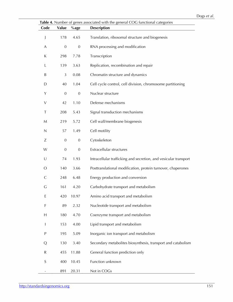
genes predicted, 4,310 were protein-coding genes and 78 RNA genes, including four rRNA operons. The majority of the protein-coding genes (80.7%) were assigned a putative function, while the re-maining ones were annotated as hypothetical pro-teins. The distribution of genes into COGs func-tional categories is presented in Table 4.
Table 3. Genome Statistics Attribute Value % of Total Genome size (bp) 4,642,596 100.00%
DNA coding region (bp) 4,110,429 88.54%
DNA G+C content (bp) 2,986,366 64.34%
Number of replicons 5
Extrachromosomal elements 4
Total genes 4,388 100.00%
RNA genes 78 1.78%
rRNA operons 4
Protein-coding genes 4,310 98.22%
Pseudo genes n.a. n.a.
Genes with function prediction 3.652 83.23%
Genes in paralog clusters 3,523 80.29%
Genes assigned to COGs 3,497 79.69%
Genes assigned Pfam domains 3,714 84.64%
Genes with signal peptides 1,507 34.34%
Genes with transmembrane helices 902 20.56%
CRISPR repeats 0
Dogs et al.
http://standardsingenomics.org 149
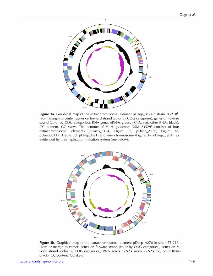
Figure 3a. Graphical map of the extrachromosomal element pDaep_B174in strain TF-218T. From margin to center: genes on forward strand (color by COG categories), genes on reverse strand (color by COG categories), RNA genes (tRNAs green, rRNAs red, other RNAs black), GC content, GC skew. The genome of P. daeponensis DSM 23529T consists of four extrachromosomal elements (pDaep_B174; Figure 3b, pDaep_A276; Figure 3c, pDaep_C117; Figure 3d, pDaep_D91) and one chromosome (Figure 3e, cDaep_3984), as evidenced by their replication initiation system (see below).
Figure 3b. Graphical map of the extrachromosomal element pDaep_A276 in strain TF-218T. From or margin to center: genes on forward strand (color by COG categories), genes on re-verse strand (color by COG categories), RNA genes (tRNAs green, rRNAs red, other RNAs black), GC content, GC skew.
Phaeobacter daeponensis type strain (DSM 23529T)
150 Standards in Genomic Sciences
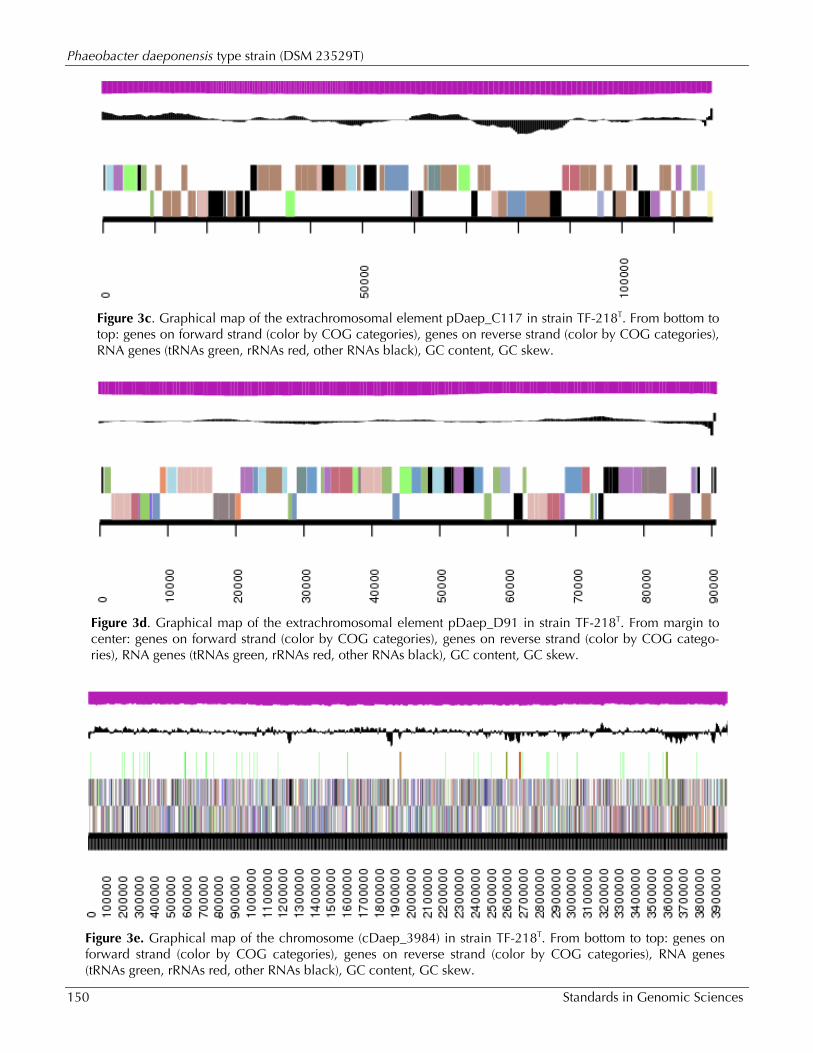
Figure 3c. Graphical map of the extrachromosomal element pDaep_C117 in strain TF-218T. From bottom to top: genes on forward strand (color by COG categories), genes on reverse strand (color by COG categories), RNA genes (tRNAs green, rRNAs red, other RNAs black), GC content, GC skew.
Figure 3d. Graphical map of the extrachromosomal element pDaep_D91 in strain TF-218T. From margin to center: genes on forward strand (color by COG categories), genes on reverse strand (color by COG catego-ries), RNA genes (tRNAs green, rRNAs red, other RNAs black), GC content, GC skew.
Figure 3e. Graphical map of the chromosome (cDaep_3984) in strain TF-218T. From bottom to top: genes on forward strand (color by COG categories), genes on reverse strand (color by COG categories), RNA genes (tRNAs green, rRNAs red, other RNAs black), GC content, GC skew.
Dogs et al.
http://standardsingenomics.org 151
Table 4. Number of genes associated with the general COG functional categories Code Value %age Description
J 178 4.65 Translation, ribosomal structure and biogenesis
A 0 0 RNA processing and modification
K 298 7.78 Transcription
L 139 3.63 Replication, recombination and repair
B 3 0.08 Chromatin structure and dynamics
D 40 1.04 Cell cycle control, cell division, chromosome partitioning
Y 0 0 Nuclear structure
V 42 1.10 Defense mechanisms
T 208 5.43 Signal transduction mechanisms
M 219 5.72 Cell wall/membrane biogenesis
N 57 1.49 Cell motility
Z 0 0 Cytoskeleton
W 0 0 Extracellular structures
U 74 1.93 Intracellular trafficking and secretion, and vesicular transport
O 140 3.66 Posttranslational modification, protein turnover, chaperones
C 248 6.48 Energy production and conversion
G 161 4.20 Carbohydrate transport and metabolism
E 420 10.97 Amino acid transport and metabolism
F 89 2.32 Nucleotide transport and metabolism
H 180 4.70 Coenzyme transport and metabolism
I 153 4.00 Lipid transport and metabolism
P 195 5.09 Inorganic ion transport and metabolism
Q 130 3.40 Secondary metabolites biosynthesis, transport and catabolism
R 455 11.88 General function prediction only
S 400 10.45 Function unknown
- 891 20.31 Not in COGs
Phaeobacter daeponensis type strain (DSM 23529T)
152 Standards in Genomic Sciences
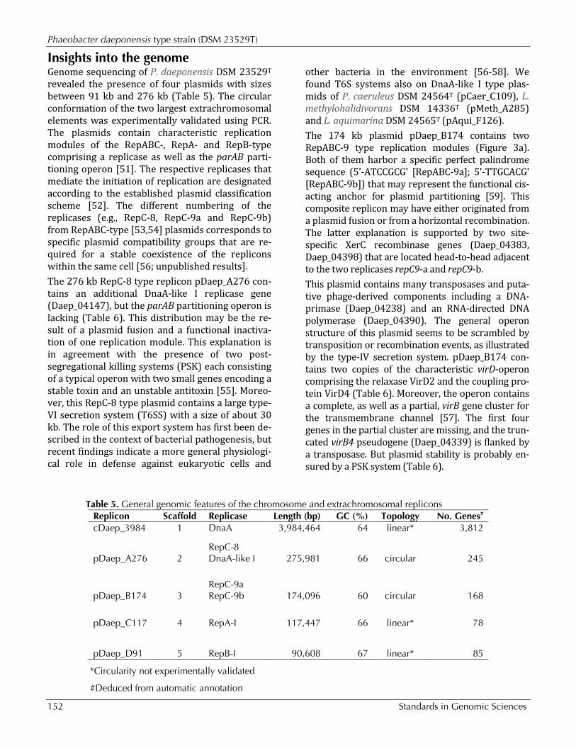
Insights into the genome Genome sequencing of P. daeponensis DSM 23529T revealed the presence of four plasmids with sizes between 91 kb and 276 kb (Table 5). The circular conformation of the two largest extrachromosomal elements was experimentally validated using PCR. The plasmids contain characteristic replication modules of the RepABC-, RepA- and RepB-type comprising a replicase as well as the parAB parti-tioning operon [51]. The respective replicases that mediate the initiation of replication are designated according to the established plasmid classification scheme [52]. The different numbering of the replicases (e.g., RepC-8, RepC-9a and RepC-9b) from RepABC-type [53,54] plasmids corresponds to specific plasmid compatibility groups that are re-quired for a stable coexistence of the replicons within the same cell [56; unpublished results]. The 276 kb RepC-8 type replicon pDaep_A276 con-tains an additional DnaA-like I replicase gene (Daep_04147), but the parAB partitioning operon is lacking (Table 6). This distribution may be the re-sult of a plasmid fusion and a functional inactiva-tion of one replication module. This explanation is in agreement with the presence of two post-segregational killing systems (PSK) each consisting of a typical operon with two small genes encoding a stable toxin and an unstable antitoxin [55]. Moreo-ver, this RepC-8 type plasmid contains a large type-VI secretion system (T6SS) with a size of about 30 kb. The role of this export system has first been de-scribed in the context of bacterial pathogenesis, but recent findings indicate a more general physiologi-cal role in defense against eukaryotic cells and
other bacteria in the environment [56-58]. We found T6S systems also on DnaA-like I type plas-mids of P. caeruleus DSM 24564T (pCaer_C109), L. methylohalidivorans DSM 14336T (pMeth_A285) and L. aquimarina DSM 24565T (pAqui_F126). The 174 kb plasmid pDaep_B174 contains two RepABC-9 type replication modules (Figure 3a). Both of them harbor a specific perfect palindrome sequence (5'-ATCCGCG' [RepABC-9a]; 5'-TTGCACG' [RepABC-9b]) that may represent the functional cis-acting anchor for plasmid partitioning [59]. This composite replicon may have either originated from a plasmid fusion or from a horizontal recombination. The latter explanation is supported by two site-specific XerC recombinase genes (Daep_04383, Daep_04398) that are located head-to-head adjacent to the two replicases repC9-a and repC9-b. This plasmid contains many transposases and puta-tive phage-derived components including a DNA-primase (Daep_04238) and an RNA-directed DNA polymerase (Daep_04390). The general operon structure of this plasmid seems to be scrambled by transposition or recombination events, as illustrated by the type-IV secretion system. pDaep_B174 con-tains two copies of the characteristic virD-operon comprising the relaxase VirD2 and the coupling pro-tein VirD4 (Table 6). Moreover, the operon contains a complete, as well as a partial, virB gene cluster for the transmembrane channel [57]. The first four genes in the partial cluster are missing, and the trun-cated virB4 pseudogene (Daep_04339) is flanked by a transposase. But plasmid stability is probably en-sured by a PSK system (Table 6).
Table 5. General genomic features of the chromosome and extrachromosomal replicons Replicon Scaffold Replicase Length (bp) GC (%) Topology No. Genes# cDaep_3984 1 DnaA 3,984,464 64 linear* 3,812
pDaep_A276 2 RepC-8 DnaA-like I 275,981 66 circular 245
pDaep_B174 3 RepC-9a RepC-9b 174,096 60 circular 168
pDaep_C117 4 RepA-I 117,447 66 linear* 78
pDaep_D91 5 RepB-I 90,608 67 linear* 85
*Circularity not experimentally validated
#Deduced from automatic annotation
Dogs et al.
http://standardsingenomics.org 153
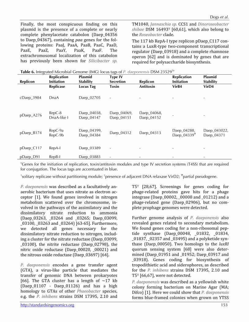
Finally, the most conspicuous finding on this plasmid is the presence of a complete or nearly complete phenylacetate catabolon (Daep_04356 to Daep_04367), containing paa genes for the fol-lowing proteins: PaaJ, PaaA, PaaB, PaaC, PaaD, PaaE, PaaZ, PaaY, PaaK, PaaF. The extrachromosomal localization of this catabolon has previously been shown for Silicibacter sp.
TM1040, Jannaschia sp. CCS1 and Dinoroseobacter shibae DSM 16493T [60,61], which also belong to the Roseobacter clade. The 117 kb RepA-I type replicon pDaep_C117 con-tains a LuxR-type two-component transcriptional regulator (Daep_03918) and a complete rhamnose operon [62] and is dominated by genes that are required for polysaccharide biosynthesis.
Table 6. Integrated Microbial Genome (IMG) locus tags of P. daeponensis DSM 23529T†
Replicon Replication Initiation
Plasmid Stability
Type IV Secretion Replicon
Replication Initiation
Plasmid Stability
Replicase Locus Tag Toxin Antitoxin VirB4 VirD4
cDaep_3984 DnaA Daep_02705 - - - -
pDaep_A276 RepC-8 DnaA-like I1
Daep_04038, Daep_04147
Daep_04069, Daep_04151
Daep_04068, Daep_04152
- -
pDaep_B174 RepC-9a RepC-9b
Daep_04399, Daep_04384
Daep_04312 Daep_04313 Daep_04288, Daep_04339Ψ
Daep_043022, Daep_043712
pDaep_C117 RepA-I Daep_03389 - - - -
pDaep_D91 RepB-I Daep_03883 - - - -
†Genes for the initiation of replication, toxin/antitoxin modules and type IV secretion systems (T4SS) that are required for conjugation. The locus tags are accentuated in blue. 1solitary replicase without partitioning module; 2presence of adjacent DNA relaxase VirD2; Ψpartial pseudogene.
P. daeponensis was described as a facultatively an-aerobic bacterium that uses nitrate as electron ac-ceptor [1]. We found genes involved in nitrogen metabolism scattered over the chromosome, in-volved in the pathways of the assimilatory and the dissimilatory nitrate reduction to ammonia (Daep_03263, _03264 and _03265; Daep_03099, _03100, _03263 and _03264) [63-65]. Furthermore, we detected all genes necessary for the dissimilatory nitrate reduction to nitrogen, includ-ing a cluster for the nitrate reductase (Daep_03099, _03100), the nitrite reductase (Daep_02798), the nitric oxide reductase (Daep_00020, _00021) and the nitrous oxide reductase (Daep_03697) [64].
P. daeponensis encodes a gene transfer agent (GTA), a virus-like particle that mediates the transfer of genomic DNA between prokaryotes [66]. The GTA cluster has a length of ~17 kb (Daep_01107 - Daep_01126) and has a high homology to GTAs of other Phaeobacter species, e.g. the P. inhibens strains DSM 17395, 2.10 and
T5T [28,67]. Screenings for genes coding for phage-related proteins gave hits for a phage integrase (Daep_00002, _00008 and _01212) and a phage-related gene (Daep_02906), but no com-plete prophage genomes were detected.
Further genome analysis of P. daeponensis also revealed genes related to secondary metabolism. We found genes coding for a non-ribosomal pep-tide synthase (Daep_00048, _01832, _01834, _01837, _02357 and _03495) and a polyketide syn-thase (Daep_00050). Two homologs to the luxRI quorum sensing system [68] were also deter-mined (Daep_01951 and _01952; Daep_03917 and _03918). Genes coding for biosynthesis of tropodithietic acid and siderophores, as described for the P. inhibens strains DSM 17395, 2.10 and T5T [66,67], were not detected. P. daeponensis was described as a yellowish white colony forming bacterium on Marine Agar (MA; Difco) [1]. Here we could show that P. daeponensis forms blue-framed colonies when grown on YTSS
Phaeobacter daeponensis type strain (DSM 23529T)
154 Standards in Genomic Sciences
broth [11]. In the genome we found genes proba-bly encoding indigoidine biosynthesis [11]. The respective operon (Daep_03493, _03494, _03495, _03496, _03497 and _03498) is similar to the op-eron recently described for the closely related strain Phaeobacter sp. Y4I [11]. The luxRI genes and the gene Daep_01773 show homology to the quorum-sensing systems and the clpA gene of Phaeobacter sp. strain Y4I, respectively. Strain Y4I lost its pigmentation by transposon insertions in each of the two luxRI quorum-sensing systems, revealing that pigment production in strain Y4I is regulated via quorum sensing [11]. Transposon insertion in gene clpA of strain Y4I, coding for a universal regulatory chaperone protein ClpA, which degrades abnormal and regulatory pro-teins, led to a higher pigment production. The presence of the biosynthesis operon and the regu-latory systems indicates that P. daeponensis is also able to produce indigoidine in a similar way as strain Y4I. Phylogenetic analysis shows that P. daeponensis and P. caeruleus form a cluster together with the
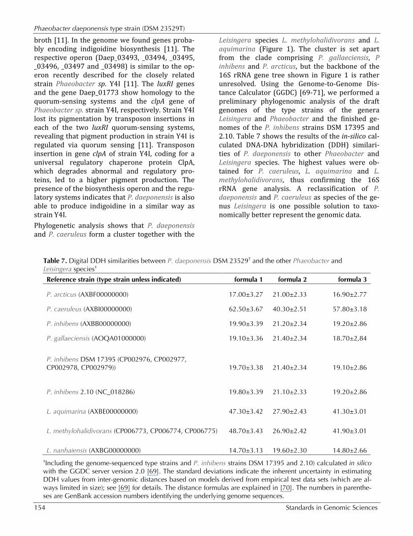
Leisingera species L. methylohalidivorans and L. aquimarina (Figure 1). The cluster is set apart from the clade comprising P. gallaeciensis, P inhibens and P. arcticus, but the backbone of the 16S rRNA gene tree shown in Figure 1 is rather unresolved. Using the Genome-to-Genome Dis-tance Calculator (GGDC) [69-71], we performed a preliminary phylogenomic analysis of the draft genomes of the type strains of the genera Leisingera and Phaeobacter and the finished ge-nomes of the P. inhibens strains DSM 17395 and 2.10. Table 7 shows the results of the in-silico cal-culated DNA-DNA hybridization (DDH) similari-ties of P. daeponensis to other Phaeobacter and Leisingera species. The highest values were ob-tained for P. caeruleus, L. aquimarina and L. methylohalidivorans, thus confirming the 16S rRNA gene analysis. A reclassification of P. daeponensis and P. caeruleus as species of the ge-nus Leisingera is one possible solution to taxo-nomically better represent the genomic data.
Table 7. Digital DDH similarities between P. daeponensis DSM 23529T and the other Phaeobacter and Leisingera species†
Reference strain (type strain unless indicated) formula 1 formula 2 formula 3
P. arcticus (AXBF00000000) 17.00±3.27 21.00±2.33 16.90±2.77
P. caeruleus (AXBI00000000) 62.50±3.67 40.30±2.51 57.80±3.18
P. inhibens (AXBB00000000) 19.90±3.39 21.20±2.34 19.20±2.86
P. gallaeciensis (AOQA01000000) 19.10±3.36 21.40±2.34 18.70±2,84
P. inhibens DSM 17395 (CP002976, CP002977, CP002978, CP002979)) 19.70±3.38 21.40±2.34 19.10±2.86
P. inhibens 2.10 (NC_018286) 19.80±3.39 21.10±2.33 19.20±2.86
L. aquimarina (AXBE00000000) 47.30±3.42 27.90±2.43 41.30±3.01
L. methylohalidivorans (CP006773, CP006774, CP006775) 48.70±3.43 26.90±2.42 41.90±3.01
L. nanhaiensis (AXBG00000000) 14.70±3.13 19.60±2.30 14.80±2.66 †Including the genome-sequenced type strains and P. inhibens strains DSM 17395 and 2.10) calculated in silico with the GGDC server version 2.0 [69]. The standard deviations indicate the inherent uncertainty in estimating DDH values from inter-genomic distances based on models derived from empirical test data sets (which are al-ways limited in size); see [69] for details. The distance formulas are explained in [70]. The numbers in parenthe-ses are GenBank accession numbers identifying the underlying genome sequences.
Dogs et al.
http://standardsingenomics.org 155
Even though discrepancies between the current classification of the group and the genomic data apparently exist, it is also obvious that P. caeruleus, which forms blue colonies [5], is the closest known relative of P. daeponensis (Table 7). For this reason, the formation of blue colonies by P. daeponensis DSM 23529T on YTSS medium [11] observed in this study, confirmed by the presence of genes for indigoidine biosynthesis in the ge-nome, is probably of taxonomic relevance. This
warrants an update of the taxonomic description of P. daeponensis.
Emended description of the species Phaeobacter daeponensis Yoon et al. 2007 The description of the species Phaeobacter daeponensis is the one given by Yoon et al. 2007 [1], with the following modification. Forms blue colonies when cultivated on YTSS medium.
Acknowledgements The authors would like to gratefully acknowledge the assistance of Iljana Schröder for growing P. daeponensis cultures and Evelyne-Marie Brambilla for DNA extraction and quality control (both at DSMZ). The work conducted by the U.S. Department of Energy Joint Genome Institute was supported by the Office of Sci-ence of the U.S. Department of Energy under contract
No. DE-AC02-05CH11231; the work conducted by members of the Roseobacter consortium was sup-ported by the German Research Foundation (DFG) Transregio-SFB 51. We also thank the European Com-mission which supported phenotyping via the Microme project 222886 within the Framework 7 program.
References 1. Yoon JH, Kang SJ, Lee SY, Oh TK. Phaeobacter
daeponensis sp. nov., isolated from a tidal flat of the Yellow Sea in Korea. Int J Syst Evol Microbiol 2007; 57:856-861. PubMed http://dx.doi.org/10.1099/ijs.0.64779-0
2. Ruiz-Ponte C, Cilia V, Lambert C, Nicolas JL. Roseobacter gallaeciensis sp. nov., a new marine bacterium isolated from rearings and collectors of the scallop Pecten maximus. Int J Syst Bacteriol 1998; 48:537-542. PubMed http://dx.doi.org/10.1099/00207713-48-2-537
3. Martens T, Heidorn T, Pukall R, Simon M, Tindall BJ, Brinkhoff T. Reclassification of Roseobacter gallaeciensis Ruiz-Ponte et al. 1998 as Phaeobacter gallaeciensis gen. nov., comb. nov., description of Phaeobacter inhibens sp. nov., re-classification of Ruegeria algicola (Lafay et al. 1995) Uchino et al. 1999 as Marinovum algicola gen. nov., comb. nov., and emended descriptions of the genera Roseobacter, Ruegeria and Leisingera. Int J Syst Evol Microbiol 2006; 56:1293-1304. PubMed http://dx.doi.org/10.1099/ijs.0.63724-0
4. Zhang DC, Li HR, Xin YH, Liu HC, Chi ZM, Zhou PJ, Yu Y. Phaeobacter arcticus sp. nov., a psy-chrophilic bacterium isolated from the Arctic. Int J Syst Evol Microbiol 2008; 58:1384-1387. PubMed http://dx.doi.org/10.1099/ijs.0.65708-0
5. Vandecandelaere I, Segaert E, Mollica A, Faimali M, Vandamme P. Phaeobacter caeruleus sp. nov., a blue-coloured, colony-forming bacterium iso-
lated from a marine electroactive biofilm. Int J Syst Evol Microbiol 2009; 59:1209-1214. PubMed http://dx.doi.org/10.1099/ijs.0.002642-0
6. Martens T, Gram L, Grossart HP, Kessler D, Mül-ler R, Simon M, Wenzel SC, Brinkhoff T. Bacteria of the Roseobacter clade show potential for sec-ondary metabolite production. Microb Ecol 2007; 54:31-42. PubMed http://dx.doi.org/10.1007/s00248-006-9165-2
7. Bruhn JB, Nielsen KF, Hjelm M, Hansen M, Bresciani J, Schulz S, Gram L. Ecology, inhibitory activity and morphogenesis of a potential marine fish larvae probiotic bacteria, Roseobacter strain 27-4. Appl Environ Microbiol 2005; 71:7263-7270. PubMed http://dx.doi.org/10.1128/AEM.71.11.7263-7270.2005
8. Brinkhoff T, Bach G, Heidorn T, Liang L, Schlingloff A, Simon M. Antibiotic production by a Roseobacter clade-affiliated species from the German Wadden Sea and its antagonistic effects on indigenous isolates. Appl Environ Microbiol 2004; 70:2560-2565. PubMed http://dx.doi.org/10.1128/AEM.70.4.2560-2565.2003
9. Rao D, Webb JS, Kjelleberg S. Competitive inter-actions in mixed-species biofilms containing the marine bacterium Pseudoalteromonas tunicata. Appl Environ Microbiol 2005; 71:1729-1736. PubMed
Phaeobacter daeponensis type strain (DSM 23529T)
156 Standards in Genomic Sciences
http://dx.doi.org/10.1128/AEM.71.4.1729-1736.2005
10. Ruiz-Ponte C, Samain JF, Sanchez JL, Nicolas JL. The benefit of a Roseobacter species on the sur-vival of scallop larvae. Mar Biotechnol (NY) 1999; 1:52-59. PubMed http://dx.doi.org/10.1007/PL00011751
11. Cude WN, Mooney J, Tavanaei AA, Hadden MK, Frank AM, Gulvik CA, May AL, Buchan A. Pro-duction of the antimicrobial secondary metabolite indigoidine contributes to competitive surface colonization by the marine roseobacter Phaeobacter sp. strain Y4I. Appl Environ Microbiol 2012; 78:4771-4780. PubMed http://dx.doi.org/10.1128/AEM.00297-12
12. Gu J, Guo B, Wang YN, Yu SL, Inamori R, Qu R, Ye YG, Wu XL. Oceanicola nanhaiensis sp. nov., isolated from sediments of the South China Sea. Int J Syst Evol Microbiol 2007; 57:157-160. Pub-Med http://dx.doi.org/10.1099/ijs.0.64532-0
13. Lin KY, Sheu SY, Chang PS, Cho JC, Chen WM. Oceanicola marinus sp. nov., a marine alphaproteobacterium isolated from seawater col-lected off Taiwan. Int J Syst Evol Microbiol 2007; 57:1625-1629. PubMed http://dx.doi.org/10.1099/ijs.0.65020-0
14. Schaefer JK, Goodwin KD, McDonald IR, Murrell JC, Oremland RS. Leisingera methylohalidivorans gen. nov., sp. nov., a marine methylotroph that grows on methyl bromide. Int J Syst Evol Microbiol 2002; 52:851-859. PubMed http://dx.doi.org/10.1099/ijs.0.01960-0
15. Sun F, Wang B, Liu X, Lai Q, Du Y, Li G, Luo J, Shao Z. Leisingera nanhaiensis sp. nov., isolated from marine sediment. Int J Syst Evol Microbiol 2010; 60:275-280. PubMed http://dx.doi.org/10.1099/ijs.0.010439-0
16. Thrash JC, Cho JC, Vergin KL, Giovannoni SJ. Ge-nome sequences of Oceanicola granulosus HTCC2516T and Oceanicola batsensis HTCC2597T. J Bacteriol 2010; 192:3549-3550. PubMed http://dx.doi.org/10.1128/JB.00412-10
17. Vandecandelaere I, Segaert E, Mollica A, Faimali M, Vandamme P. Leisingera aquimarina sp. nov., isolated from a marine electroactive biofilm, and emended descriptions of Leisingera methylohalidivorans Schaefer et al. 2002, Phaeobacter daeponensis Yoon et al. 2007 and Phaeobacter inhibens Martens et al. 2006. Int J Syst Evol Microbiol 2008; 58:2788-2793. PubMed http://dx.doi.org/10.1099/ijs.0.65844-0
18. Yoon JH, Kang SJ, Lee SY, Oh KH, Oh TK. Seohaeicola saemankumensis gen. nov., sp. nov., isolated from a tidal flat. Int J Syst Evol Microbiol 2009; 59:2675-2679. PubMed http://dx.doi.org/10.1099/ijs.0.011312-0
19. Yuan J, Lai Q, Wang B, Sun F, Liu X, Du Y, Li G, Gu L, Zheng T, Shao Z. Oceanicola pacificus sp. nov., isolated from a deep-sea pyrene-degrading consortium. Int J Syst Evol Microbiol 2009; 59:1158-1161. PubMed http://dx.doi.org/10.1099/ijs.0.003400-0
20. Zheng Q, Chen C, Wang YN, Jiao N. Oceanicola nitratireducens sp. nov., a marine alphaproteobacterium isolated from the South China Sea. Int J Syst Evol Microbiol 2010; 60:1655-1659. PubMed http://dx.doi.org/10.1099/ijs.0.016311-0
21. Göker M, Cleland D, Saunders E, Lapidus A, No-lan M, Lucas S, Hammon N, Deshpande S, Cheng JF, Tapia R, et al. Complete genome sequence of Isosphaera pallida type strain (IS1BT). Stand Ge-nomic Sci 2011; 4:63-71. PubMed http://dx.doi.org/10.4056/sigs.1533840
22. Liolios K, Chen IM, Mavromatis K, Tavernarakis N, Hugenholtz P, Markowitz VM, Kyrpides NC. The Genomes OnLine Database (GOLD) in 2009:status of genomic and metagenomic pro-jects and their associated metadata. Nucleic Acids Res 2010; 38:D346-D354. PubMed http://dx.doi.org/10.1093/nar/gkp848
23. Riedel T, Teshima H, Petersen J, Fiebig A, Daven-port K, Dalingault H, Erkkila T, Gu W, Munk C, Xu Y, et al. Genome sequence of the Leisingera aquimarina type strain (DSM 24565T), a member of the Roseobacter clade rich in extrachromosomal elements. Stand Genomic Sci 2013; 8:389-402. http://dx.doi.org/10.4056/sigs.3858183
24. Beyersmann PG, Chertkov O, Petersen J, Fiebig A, Chen A, Pati A, Ivanova N, Lapidus A, Goodwin LA, Chain P, et al. Genome sequence of Phaeobacter caeruleus type strain (DSM 24564T), a surface-associated member of the marine Roseobacter clade. Stand Genomic Sci 2013; 8:403-419. http://dx.doi.org/10.4056/sigs.3927626
25. Freese H, Dalingault H, Petersen J, Pradella S, Fiebig A, Davenport K, Teshima H, Chen A, Pati A, Ivanova N, et al. Genome sequence of the plasmid and phage-gene rich marine Phaeobacter arcticus type strain (DSM 23566T). Stand Ge-nomic Sci 2013; 8:450-464. http://dx.doi.org/10.4056/sigs.383362
Dogs et al.
http://standardsingenomics.org 157
26. Breider S, Teshima H, Petersen J, Fiebig A, Chertkov O, Dalingault H, Chen A, Pati A, Ivanova N, Lapidus A, et al. Genome sequence of Leisingera nanhaiensis strain DSM 23252T isolated from marine sediment. Stand Genomic Sci 2013; 8:389-402.
27. Buddruhs N, Chertkov O, Fiebig A, Petersen J, Chen A, Pati A, Ivanova N, Lapidus A, Goodwin LA, Chain P, et al. Complete genome sequence of the marine methyl-halide oxidizing Leisingera methylohalidivorans type strain (DSM 14336T), a member of the Roseobacter clade. Stand Ge-nomic Sci 2013; In press.
28. Dogs M, Voget S, Teshima H, Petersen J, Fiebig A, Davenport K, Dalingault H, Chen A, Pati A, Ivanova N, et al. Genome sequence of Phaeobacter inhibens strain T5T, a secondary-metabolite producing member of the marine Roseobacter clade, and emendation of the spe-cies Phaeobacter inhibens. Stand Genomic Sci 2013; In press.
29. Field D, Garrity G, Gray T, Morrison N, Selengut J, Sterk P, Tatusova T, Thomson N, Allen MJ, Angiuoli SV, et al. The minimum information about a genome sequence (MIGS) specification. Nat Biotechnol 2008; 26:541-547. PubMed http://dx.doi.org/10.1038/nbt1360
30. Vaas LAI, Sikorski J, Michael V, Göker M, Klenk HP. Visualization and curve-parameter estimation strategies for efficient exploration of phenotype microarray kinetics. PLoS ONE 2012; 7:e34846. PubMed http://dx.doi.org/10.1371/journal.pone.0034846
31. Vaas LAI, Sikorski J, Hofner B, Buddruhs N, Fiebig A, Klenk HP, Göker M. opm: An R package for analysing OmniLog® Phenotype MicroArray Da-ta. Bioinformatics 2013; 29:1823-1824. PubMed http://dx.doi.org/10.1093/bioinformatics/btt291
32. Woese CR, Kandler O, Wheelis ML. Towards a natural system of organisms. Proposal for the do-mains Archaea, Bacteria and Eucarya. Proc Natl Acad Sci USA 1990; 87:4576-4579. PubMed http://dx.doi.org/10.1073/pnas.87.12.4576
33. Garrity GM, Bell JA, Lilburn T. Phylum XIV. Proteobacteria phyl. nov. In: DJ Brenner, NR Krieg, JT Staley, GM Garrity (eds), Bergey's Man-ual of Systematic Bacteriology, second edition, vol. 2 (The Proteobacteria), part B (The Gammaproteobacteria), Springer, New York, 2005, p. 1.
34. Garrity GM, Bell JA, Lilburn T. Class I. Alphaproteobacteria class. nov. In: Garrity GM,
Brenner DJ, Krieg NR, Staley JT (eds), Bergey's Manual of Systematic Bacteriology, Second Edi-tion, Volume 2, Part C, Springer, New York, 2005, p. 1.
35. Validation List No. 107. List of new names and new combinations previously effectively, but not validly, published. Int J Syst Evol Microbiol 2006; 56:1-6. PubMed http://dx.doi.org/10.1099/ijs.0.64188-0
36. Garrity GM, Bell JA, Lilburn T. Order III. Rhodobacterales ord. nov. In: Garrity GM, Bren-ner DJ, Krieg NR, Staley JT (eds), Bergey's Manual of Systematic Bacteriology, Second Edition, Vol-ume 2, Part C, Springer, New York, 2005, p. 161.
37. Garrity GM, Bell JA, Lilburn T. Family I. Rhodobacteraceae fam. nov. In: Garrity GM, Brenner DJ, Krieg NR, Staley JT (eds), Bergey's Manual of Systematic Bacteriology, Second Edi-tion, Volume 2, Part C, Springer, New York, 2005, p. 161.
38. BAuA. Classification of Bacteria and Archaea in risk groups. TRBA 2010; 466:93.
39. Ashburner M, Ball CA, Blake JA, Botstein D, But-ler H, Cherry JM, Davis AP, Dolinski K, Dwight SS, Eppig JT, et al. Gene ontology: tool for the unification of biology. The Gene Ontology Con-sortium. Nat Genet 2000; 25:25-29. PubMed http://dx.doi.org/10.1038/75556
40. Mavromatis K, Land ML, Brettin TS, Quest DJ, Copeland A, Clum A, Goodwin L, Woyke T, Lapidus A, Klenk HP, et al. The fast changing landscape of sequencing technologies and their impact on microbial genome assemblies and an-notation. PLoS ONE 2012; 7:e48837. PubMed http://dx.doi.org/10.1371/journal.pone.0048837
41. List of growth media used at the DSMZ: http://www.dmsz.de/catalogues/cataloque-microorganisms/culture-technology/list-of-media-for-microorganisms.html.
42. Gemeinholzer B, Dröge G, Zetzsche H, Haszprunar G, Klenk HP, Güntsch A, Berendsohn WG, Wägele JW. The DNA Bank Network: the start from a German initiative. Biopreserv Biobank 2011; 9:51-55. http://dx.doi.org/10.1089/bio.2010.0029
43. The DOE Joint Genome Institute. www.jgi.doe.gov
44. Butler J, MacCallum I, Kleber M, Shlyakhter IA, Belmonte MK, Lander ES, Nusbaum C, Jaffe DB. ALLPATHS: de novo assembly of whole-genome shotgun microreads. Genome Res 2008; 18:810-
Phaeobacter daeponensis type strain (DSM 23529T)
158 Standards in Genomic Sciences
820. PubMed http://dx.doi.org/10.1101/gr.7337908
45. Zerbino DR, Birney E. Velvet: algorithms for de novo short read assembly using de Bruijn graphs. Genome Res 2008; 18:821-829. PubMed http://dx.doi.org/10.1101/gr.074492.107
46. Phrap and Phred for Windows. MacOS, Linux, and Unix. http://www.phrap.com
47. Hyatt D, Chen GL, LoCascio PF, Land ML, Lar-imer FW, Hauser LJ. Prodigal: prokaryotic gene recognition and translation initiation site identifi-cation. BMC Bioinformatics 2010; 11:119. Pub-Med http://dx.doi.org/10.1186/1471-2105-11-119
48. Mavromatis K, Ivanova NN, Chen IM, Szeto E, Markowitz VM, Kyrpides NC. The DOE-JGI Standard operating procedure for the annotations of microbial genomes. Stand Genomic Sci 2009; 1:63-67. PubMed http://dx.doi.org/10.4056/sigs.632
49. Pati A, Ivanova NN, Mikhailova N, Ovchinnikova G, Hooper SD, Lykidis A, Kyrpides NC. GenePRIMP: a gene prediction improvement pipeline for prokaryotic genomes. Nat Methods 2010; 7:455-457. PubMed http://dx.doi.org/10.1038/nmeth.1457
50. Markowitz VM, Ivanova NN, Chen IMA, Chu K, Kyrpides NC. IMG ER: a system for microbial ge-nome annotation expert review and curation. Bio-informatics 2009; 25:2271-2278. PubMed http://dx.doi.org/10.1093/bioinformatics/btp393
51. Petersen J. Phylogeny and compatibility: plasmid classification in the genomics era. Arch Microbiol 2011; 193:313-321. PubMed
52. Petersen J, Brinkmann H, Berger M, Brinkhoff T, Päuker O, Pradella S. Origin and evolution of a novel DnaA-like plasmid replication type in Rhodobacterales. Mol Biol Evol 2011; 28:1229-1240. PubMed http://dx.doi.org/10.1093/molbev/msq310
53. Cevallos MA, Cervantes-Rivera R, Gutierrez-Rios RM. The repABC plasmid family. Plasmid 2008; 60:19-37. PubMed http://dx.doi.org/10.1016/j.plasmid.2008.03.001
54. Castillo-Ramírez S, Vazque-Castellanos JF, Gon-zalez V, Cevallos MA. Horizontal gene transfer and diverse functional constrains within a com-mon replication-partitioning system in Alphaproteobacteria: the repABC operon. BMC Genomics 2009; 10:536. PubMed http://dx.doi.org/10.1186/1471-2164-10-536
55. Zielenkiewicz U, Ceglowski P. Mechanisms of plasmid stable maintenance with special focus on plasmid addiction systems. Acta Biochim Pol 2001; 48:1003-1023. PubMed
56. Schwarz S, Hood RD, Mougous JD. What is type VI secretion doing in all those bugs? Trends Microbiol 2010; 18:531-537. PubMed http://dx.doi.org/10.1016/j.tim.2010.09.001
57. del Solar G, Giraldo R, Ruiz-Echevarria MJ, Espi-nosa M, Diaz-Orejes R. Replication and control of circular bacterial plasmids. Microbiol Mol Biol Rev 1998; 62:434-464. PubMed
58. Cascales E, Christie PJ. The versatile bacterial type IV secretion systems. Nat Rev Microbiol 2003; 1:137-149. PubMed http://dx.doi.org/10.1038/nrmicro753
59. Petersen J, Brinkmann H, Pradella S. Diversity and evolution of repABC type plasmids in Rhodobacterales. Environ Microbiol 2009; 11:2627-2638. PubMed http://dx.doi.org/10.1111/j.1462-2920.2009.01987.x
60. Moran MA, Belas R, Schell MA, González JM, Sun F, Sun S, Binder BJ, Edmonds J, Ye W, Orcutt B, et al. Ecological genomics of marine Roseobacters. Appl Environ Microbiol 2007; 73:4559-4569. PubMed http://dx.doi.org/10.1128/AEM.02580-06
61. Wagner-Döbler I, Ballhausen B, Berger M, Brinkhoff T, Buchholz I, Bunk B, Cypionka H, Daniel R, Drepper T, Gerdts G, et al. The com-plete genome sequence of the algal symbiont Dinoroseobacter shibae: a hitchhiker's guide to life in the sea. ISME J 2010; 4:61-77. PubMed http://dx.doi.org/10.1038/ismej.2009.94
62. Giraud MF, Naismith JH. The rhamnose pathway. Curr Opin Struct Biol 2000; 10:687-696. PubMed http://dx.doi.org/10.1016/S0959-440X(00)00145-7
63. Stolz JF, Basu P. Evolution of nitrate reductase: molecular and structural variations on common function. ChemBioChem 2002; 3:198-206. Pub-Med http://dx.doi.org/10.1002/1439-7633(20020301)3:2/3<198::AID-CBIC198>3.0.CO;2-C
64. Morozkina EV, Zvyagilskaya RA. Nitrate reductase: structure, functions, and effect of stress factors. Biochemistry (Mosc) 2007; 72:1151-1160. PubMed http://dx.doi.org/10.1134/S0006297907100124
Dogs et al.
http://standardsingenomics.org 159
65. Cabello P, Roldan MD, Moreno-Vivian C. Nitrate reduction and the nitrogen cycle in archaea. Mi-crobiology 2004; 150:3527-3546. PubMed http://dx.doi.org/10.1099/mic.0.27303-0
66. Lang AS, Beatty JT. The gene transfer agent of Rhodobacter capsulatus and “constitutive trans-duction” in prokaryotes. Arch Microbiol 2001; 175:241-249. PubMed http://dx.doi.org/10.1007/s002030100260
67. Thole S, Kalhoefer D, Voget S, Berger M, Engelhardt T, Liesegang H, Wollherr A, Kjelleberg S, Daniel R, Simon M, et al. Phaeobacter gallaeciensis genomes from globally opposite lo-cations reveal high similarity of adaption to sur-face life. ISME J 2012; 6:2229-2244. PubMed http://dx.doi.org/10.1038/ismej.2012.62
68. Fuqua C, Winans SC, Greenberg EP. Census and consensus in bacterial ecosystems: the LuxR-LuxI family of quorum-sensing transcriptional regula-tors. Annu Rev Microbiol 1996; 50:727-751.
PubMed http://dx.doi.org/10.1146/annurev.micro.50.1.727
69. Meier-Kolthoff JP, Auch AF, Klenk HP, Göker M. Genome sequence-based species delimitation with confidence intervals and improved distance functions. BMC Bioinformatics 2013; 14:60. PubMed http://dx.doi.org/10.1186/1471-2105-14-60
70. Auch AF, Klenk HP, Göker M. Standard operating procedure for calculating genome-to-genome dis-tances based on high-scoring segment pairs. Stand Genomic Sci 2010; 2:142-148. PubMed http://dx.doi.org/10.4056/sigs.541628
71. Auch AF, Von Jan M, Klenk HP, Göker M. Digital DNA-DNA hybridization for microbial species delineation by means of genome-to-genome se-quence comparison. Stand Genomic Sci 2010; 2:117-134. PubMed http://dx.doi.org/10.4056/sigs.531120
Copyright © 2022 FDOKUMEN