Multiple loci associated with indices of renal function and chronic kidney disease
15
Multiple Novel Loci are Associated with Indices of Renal Function and Chronic Kidney Disease Anna Köttgen (1),* , Nicole L Glazer (2),* , Abbas Dehghan (3),(24),* , Shih-Jen Hwang (4),* , Ronit Katz (5) , Man Li (1) , Qiong Yang (6) , Vilmundur Gudnason (19),(20) , Lenore J Launer (21) , Tamara B Harris (21) , Albert V Smith (19) , Dan E Arking (7) , Brad C Astor (1) , Eric Boerwinkle (8) , Georg B Ehret (7),(23) , Ingo Ruczinski (9) , Robert B Scharpf (9) , Yii-Der Ida Chen (10) , Ian H de Boer (11) , Talin Haritunians (10) , Thomas Lumley (5) , Mark Sarnak (12) , David Siscovick (13) , Emelia J Benjamin (14) , Daniel Levy (4) , Ashish Upadhyay (15) , Yurii S Aulchenko (3) , Albert Hofman (3) , Fernando Rivadeneira (16) , André G Uitterlinden (16) , Cornelia M van Duijn (3) , Daniel I Chasman (22) , Guillaume Paré (22) , Paul M Ridker (22) , WH Linda Kao (1) , Jacqueline C Witteman (3),(24),** , Josef Coresh (1),(9),** , Michael G Shlipak (17),** , and Caroline S Fox (4),(18),** (1) Department of Epidemiology, Johns Hopkins University, Baltimore, MD, USA (2) Cardiovascular Health Research Unit and Department of Medicine, University of Washington, Seattle, WA, USA (3) Department of Epidemiology, Erasmus Medical Center, Rotterdam, The Netherlands (4) NHLBI’s Framingham Heart Study and the Center for Population Studies, Framingham, MA, USA (5) Department of Biostatistics, University of Washington, Seattle, WA, USA (6) Department of Biostatistics, Boston University, Boston, MA, USA (7) McKusick-Nathans Institute of Genetic Medicine, Johns Hopkins Medical Institutions, Baltimore, MD, USA (8) Human Genetics Center and Division of Epidemiology, University of Texas Health Science Center at Houston, Houston, TX, USA (9) Department of Biostatistics, Johns Hopkins University, Baltimore, MD, USA (10) Medical Genetics Institute, Cedars-Sinai Medical Center, Los Angeles, CA, USA (11) Division of Nephrology, Department of Medicine, University of Washington, Seattle, WA, USA (12) Tufts-New England Medical Center, Boston, MA, USA (13) Departments of Medicine and Epidemiology, Cardiovascular Health Research Unit, University of Washington, Seattle, WA, USA (14) Boston University Schools of Medicine and Public Health, Boston, MA, USA (15) Renal Section, Boston Medical Center and Boston University School of Medicine, Boston, MA, USA (16) Department of Internal Medicine, Erasmus Medical Center, Rotterdam, The Netherlands (17) General Internal Medicine Division; San Francisco VA Medical Center; University of California, San Francisco, CA, USA (18) Brigham and Women’s Hospital Division of Endocrinology and Harvard Medical School, Correspondence: Caroline S. Fox, MD, MPH, Address: National Heart, Lung, and Blood Institute’s Framingham Heart Study, 73 Mt Wayte Ave, Suite 2, Framingham, MA 01702, USA, Phone: +1 (508) 935-3447, Fax: +1 (508) 626-1262, [email protected]. Jacqueline C. Witteman, PhD, Department of Epidemiology & Biostatistics, Erasmus Medical Center, Rotterdam, P.O. Box 2040, 3000 CA Rotterdam, The Netherlands, Phone: +31 10-70 43489, Fax: +31 10-70 44657, [email protected]. Josef Coresh, MD, PhD, Address: Welch Center for Prevention, Epidemiology & Clinical Research, 2024 E., Monument St., Baltimore, MD 21287, USA, Phone: +1 (410) 955-0495, Fax: +1 (410) 955-0476, [email protected]. Michael G. Shlipak, MD, MPH, Address: General Internal Medicine Section, Veterans Affairs Medical Center (111A1), 4150 Clement St., San Francisco, CA 94121, USA, Phone: +1 (415) 750-2093, Fax: +1 (415) 379-5573, [email protected]. * These authors have contributed equally to this work ** Corresponding authors jointly contributed to this work Author Contributions: AK, NLG, AD, SJH, QY, IHdB, TL, DS, DL, VG, JC, MGS, and CSF contributed to the design of this analysis; AK, NLG, AD, SJH, QY, EB, IR, IHdB, TL, DS, DL, AVS, VG, WHLK, JCW, JC, MGS, and CSF contributed to the interpretation of the results; AK, NLG, AD, SJH, JC, MGS, and CSF drafted the manuscript; all others reviewed and commented on the manuscript; AK, NLG, AD, SJH, RK, ML, QY, DEA, GBE, IR, RBS, TL, FR, CMvD, AVS, DIC, GP, and JC contributed to statistical methods and analysis; DS, EJB, DL, VG, JC, and CSF contributed to recruitment and follow up of subjects; EB, YIC, TH, FR, AGU contributed to genotyping; and ML, QY, YSA, FR, AGU, AVS, and GP contributed to bioinformatics. NIH Public Access Author Manuscript Nat Genet. Author manuscript; available in PMC 2011 February 15. Published in final edited form as: Nat Genet. 2009 June ; 41(6): 712–717. doi:10.1038/ng.377. NIH-PA Author Manuscript NIH-PA Author Manuscript NIH-PA Author Manuscript
Transcript of Multiple loci associated with indices of renal function and chronic kidney disease

Multiple Novel Loci are Associated with Indices of RenalFunction and Chronic Kidney Disease
Anna Köttgen(1),*, Nicole L Glazer(2),*, Abbas Dehghan(3),(24),*, Shih-Jen Hwang(4),*,Ronit Katz(5), Man Li(1), Qiong Yang(6), Vilmundur Gudnason(19),(20), Lenore JLauner(21), Tamara B Harris(21), Albert V Smith(19), Dan E Arking(7), Brad C Astor(1), EricBoerwinkle(8), Georg B Ehret(7),(23), Ingo Ruczinski(9), Robert B Scharpf(9), Yii-Der IdaChen(10), Ian H de Boer(11), Talin Haritunians(10), Thomas Lumley(5), Mark Sarnak(12),David Siscovick(13), Emelia J Benjamin(14), Daniel Levy(4), Ashish Upadhyay(15), Yurii SAulchenko(3), Albert Hofman(3), Fernando Rivadeneira(16), André G Uitterlinden(16),Cornelia M van Duijn(3), Daniel I Chasman(22), Guillaume Paré(22), Paul M Ridker(22), WHLinda Kao(1), Jacqueline C Witteman(3),(24),**, Josef Coresh(1),(9),**, Michael GShlipak(17),**, and Caroline S Fox(4),(18),**
(1) Department of Epidemiology, Johns Hopkins University, Baltimore, MD, USA (2) CardiovascularHealth Research Unit and Department of Medicine, University of Washington, Seattle, WA, USA(3) Department of Epidemiology, Erasmus Medical Center, Rotterdam, The Netherlands (4)
NHLBI’s Framingham Heart Study and the Center for Population Studies, Framingham, MA, USA(5) Department of Biostatistics, University of Washington, Seattle, WA, USA (6) Department ofBiostatistics, Boston University, Boston, MA, USA (7) McKusick-Nathans Institute of GeneticMedicine, Johns Hopkins Medical Institutions, Baltimore, MD, USA (8) Human Genetics Centerand Division of Epidemiology, University of Texas Health Science Center at Houston, Houston,TX, USA (9) Department of Biostatistics, Johns Hopkins University, Baltimore, MD, USA (10)
Medical Genetics Institute, Cedars-Sinai Medical Center, Los Angeles, CA, USA (11) Division ofNephrology, Department of Medicine, University of Washington, Seattle, WA, USA (12) Tufts-NewEngland Medical Center, Boston, MA, USA (13) Departments of Medicine and Epidemiology,Cardiovascular Health Research Unit, University of Washington, Seattle, WA, USA (14) BostonUniversity Schools of Medicine and Public Health, Boston, MA, USA (15) Renal Section, BostonMedical Center and Boston University School of Medicine, Boston, MA, USA (16) Department ofInternal Medicine, Erasmus Medical Center, Rotterdam, The Netherlands (17) General InternalMedicine Division; San Francisco VA Medical Center; University of California, San Francisco, CA,USA (18) Brigham and Women’s Hospital Division of Endocrinology and Harvard Medical School,
Correspondence: Caroline S. Fox, MD, MPH, Address: National Heart, Lung, and Blood Institute’s Framingham Heart Study, 73 MtWayte Ave, Suite 2, Framingham, MA 01702, USA, Phone: +1 (508) 935-3447, Fax: +1 (508) 626-1262, [email protected] C. Witteman, PhD, Department of Epidemiology & Biostatistics, Erasmus Medical Center, Rotterdam, P.O. Box 2040,3000 CA Rotterdam, The Netherlands, Phone: +31 10-70 43489, Fax: +31 10-70 44657, [email protected]. Josef Coresh,MD, PhD, Address: Welch Center for Prevention, Epidemiology & Clinical Research, 2024 E., Monument St., Baltimore, MD 21287,USA, Phone: +1 (410) 955-0495, Fax: +1 (410) 955-0476, [email protected]. Michael G. Shlipak, MD, MPH, Address: GeneralInternal Medicine Section, Veterans Affairs Medical Center (111A1), 4150 Clement St., San Francisco, CA 94121, USA, Phone: +1(415) 750-2093, Fax: +1 (415) 379-5573, [email protected].*These authors have contributed equally to this work**Corresponding authors jointly contributed to this workAuthor Contributions:AK, NLG, AD, SJH, QY, IHdB, TL, DS, DL, VG, JC, MGS, and CSF contributed to the design of this analysis; AK, NLG, AD, SJH,QY, EB, IR, IHdB, TL, DS, DL, AVS, VG, WHLK, JCW, JC, MGS, and CSF contributed to the interpretation of the results; AK,NLG, AD, SJH, JC, MGS, and CSF drafted the manuscript; all others reviewed and commented on the manuscript; AK, NLG, AD,SJH, RK, ML, QY, DEA, GBE, IR, RBS, TL, FR, CMvD, AVS, DIC, GP, and JC contributed to statistical methods and analysis; DS,EJB, DL, VG, JC, and CSF contributed to recruitment and follow up of subjects; EB, YIC, TH, FR, AGU contributed to genotyping;and ML, QY, YSA, FR, AGU, AVS, and GP contributed to bioinformatics.
NIH Public AccessAuthor ManuscriptNat Genet. Author manuscript; available in PMC 2011 February 15.
Published in final edited form as:Nat Genet. 2009 June ; 41(6): 712–717. doi:10.1038/ng.377.
NIH
-PA Author Manuscript
NIH
-PA Author Manuscript
NIH
-PA Author Manuscript
Boston, MA, USA (19) Icelandic Heart Association Research Institute, Kopavogur, Iceland (20)
University of Iceland, Reykjavik, Iceland (21) Laboratory of Epidemiology, Demography, andBiometry, Intramural Research Program, National Institute on Aging (22) Division of PreventiveMedicine, Brigham and Women’s Hospital, Boston, MA, USA (23) Division of Cardiology, GenevaUniversity Hospital, Switzerland (24) Member of the Netherlands Consortium on Healthy Aging(NCHA)
AbstractChronic kidney disease (CKD) has a heritable component and is an important global public healthproblem because of its high prevalence and morbidity.1 We conducted genome-wide associationstudies (GWAS) to identify susceptibility loci for glomerular filtration rate estimated by serumcreatinine (eGFRcrea), cystatin C (eGFRcys), and CKD (eGFRcrea<60 ml/min/1.73m2) inEuropean-ancestry participants of four populations-based cohorts (ARIC, CHS, FHS, RS;n=19,877, 2,388 CKD cases), and tested for external replication in 21,466 participants (1,932CKD cases). Significant associations (p<5*10−8) were identified for SNPs with [1] CKD at theUMOD locus; [2] eGFRcrea at the UMOD, SHROOM3, and GATM/SPATA5L1 loci; [3] eGFRcysat the CST and STC1 loci. UMOD encodes the most common protein in human urine, Tamm-Horsfall protein,2 and rare mutations in UMOD cause Mendelian forms of kidney disease.3 Ourfindings provide new insights into CKD pathogenesis and underscore the importance of commongenetic variants influencing renal function and disease.
Keywordschronic kidney disease; renal function; epidemiology; genetics; genome-wide association study;single nucleotide polymorphism
CKD affects 10–3% of US adults.4 Estimates from Europe are similar,5 and incidence andprevalence are increasing worldwide. Its most severe form, end-stage renal disease, requiresdialysis and currently affects over 500,000 US adults.6 In addition to conferring risk for end-stage renal disease, CKD increases the risk of cardiovascular disease7 and all-causemortality.8
Multiple studies such as familial aggregation studies have provided evidence for a geneticcomponent to kidney disease. Heritability estimates of eGFRcrea are reported between 0.41and 0.75 in individuals with the major CKD risk factors hypertension or diabetes,9, 10 and as0.33 in a population-based sample.11 Heritability estimates of GFRcys are similar. Whilerare genetic variants causing different forms of monogenetic kidney disease have beenidentified, common CKD susceptibility variants have been difficult to detect reproduciblyby linkage or candidate gene studies.12
To discover such variants, we conducted meta-analyses of study-specific GWAS for indicesof renal function, eGFRcrea and eGFRcys, and for CKD from four population-based,unselected cohorts of the CHARGE Consortium13: Atherosclerosis Risk in CommunitiesStudy, Cardiovascular Health Study (CHS), Framingham Heart Study (FHS), and RotterdamStudy (RS). As a direct measurement of kidney function is not feasible in population-basedstudies, we applied commonly used estimating equations to determine eGFRcrea14 andeGFRcys.15 Population-based measures of GFR are imperfect,16 and using two differentbiomarkers to estimate GFR can therefore help to uncover true signals. CKD was defined aseGFRcrea <60 ml/min/1.73m2 according to National Guidelines,17 as detailed in themethods. Genotypes for >2.5 million SNPs were imputed within each study using reference
Köttgen et al. Page 2
Nat Genet. Author manuscript; available in PMC 2011 February 15.
NIH
-PA Author Manuscript
NIH
-PA Author Manuscript
NIH
-PA Author Manuscript
genotype data from the HapMap CEU population. Study-specific details on genotyping andimputation are provided in Supplementary Table 1. SNPs showing evidence of suggestive(p<4*10−7) or significant (p<5*10−8) genome-wide association were tested for in silicoreplication in independent study samples, the Age Gene/Environment Susceptibility-Reykjavik Study (AGES) and the Women’s Genome Health Study (WGHS). Detailedinformation on the study samples are provided in the Supplementary Methods.
Characteristics of the four discovery and two validation study samples are shown in Table 1;19,877 participants with 2,388 CKD cases and 21,466 participants with 1,932 CKD casescontributed information, respectively. CKD prevalence was higher in cohorts with olderparticipants, ranging from 6.3% (WGHS) to 24.3% (AGES). Characteristics among CKDcases are provided in Supplementary Table 2. Figure 1 summarizes meta-analysis results forCKD, eGFRcrea, and eGFRcys across the discovery samples. The observed versus expectedp-value distributions (quantile-quantile plots) are shown in Supplementary Figure 1: study-specific genomic inflation factors did not indicate substantial inflation of the test statisticsfor any of the traits.
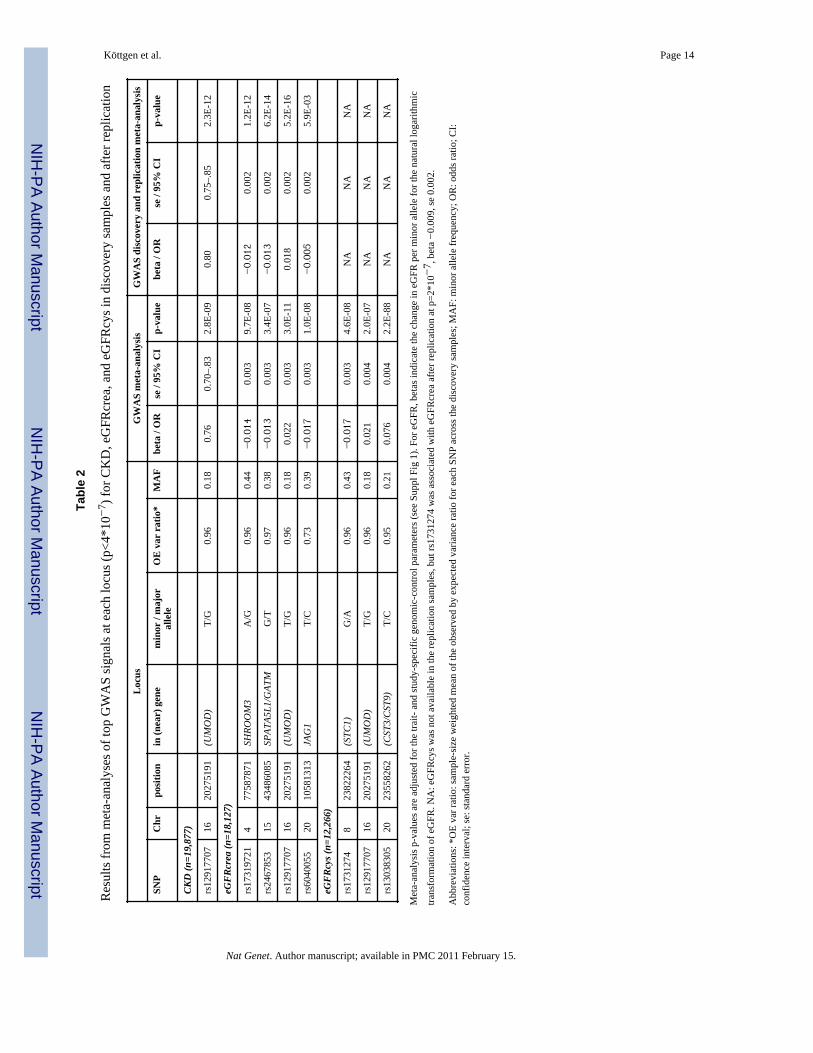
Table 2 lists the most significant SNP at each genomic locus associated with CKD,eGFRcrea, and eGFRcys, and replication results. Study-specific results are presented inSupplementary Table 3. For CKD, we identified SNP rs12917707 in a highly evolutionaryconserved region 3.6 kb upstream from the uromodulin (UMOD) gene on chromosome 16(Fig. 2A, p=5*10−16 across discovery and replication samples, Table 2). Seven SNPs in orupstream of UMOD in high LD (r2>0.8) with rs12917707 were also associated with CKD ata genome-wide significant level. The minor T allele at rs12917707 was associated with 20%reduced risk of CKD (meta-analysis OR=0.80, p-value=2*10−12, Table 2). The associationof rs12917707 with CKD, which was not significant in the FHS Study, showed someheterogeneity across studies (p-heterogeneity=0.02). Findings were consistent in modelsadjusting for major CKD risk factors including systolic blood pressure, hypertensionmedication intake and diabetes mellitus, as well as stratified for age, sex, hypertension, anddiabetes status (Figure 3). Prospective information from the ARIC Study demonstrated thatthe T allele of rs12917707 was associated with a lower relative risk of incident CKD (HR0.81, 95%CI 0.72–.92, p=0.001) over 14.7 years of follow-up (n=952 cases, see Methods).
Rare UMOD mutations cause autosomal-dominant forms of kidney disease, medullary cystickidney disease type 2 (MCKD2), familial juvenile hyperuricemic nephropathy (FJHN), andglomerulocystic kidney disease (GCKD) (OMIM #603860; #162000; #609886).3, 18, 19 Asthe syndromes caused by rare UMOD mutations are often accompanied by hyperuricemiaand gout, we explored the association of rs12917707 with these traits; no significantassociations were observed. While this does not exclude the presence of rare UMODvariants among our study participants, our study identifies another example of a genomicrisk locus containing susceptibility variants across the spectrum of risk allele frequencies.
UMOD knock-out mice are reported to have 63% lower creatinine clearance than wildtypemice.20 UMOD encodes for the most abundant protein in the urine of healthy individuals,Tamm-Horsfall protein. The physiological functions of Tamm-Horsfall protein are not wellunderstood but may include protection against inflammation and infection.2 A possible roleof the UMOD gene in renal development was also recently reported.21 UMOD is transcribedexclusively in renal tubular cells of the thick ascending limb of the loop of Henle. Ourfindings therefore suggest a common mechanism for CKD pathogenesis localized to thenephron’s loop of Henle, which has previously received little attention. The major riskfactors for kidney disease, hypertension and diabetes, are thought to affect the glomerulusprimarily, and glomerular damage is typically characterized by albuminuria. Our findings,however, indicate that the association is consistent across strata of hypertension and
Köttgen et al. Page 3
Nat Genet. Author manuscript; available in PMC 2011 February 15.
NIH
-PA Author Manuscript
NIH
-PA Author Manuscript
NIH
-PA Author Manuscript
diabetes, and we observed no association with albuminuria. Thus, our findings provide newinsights into CKD pathogenesis and highlight the need to understand the production andfunctions of Tamm-Horsfall protein within the kidney. The very broad CKD definition wechose, including a variety of causes of CKD such as hypertension and diabetes, indicatesthat the search for susceptibility variants for complex diseases may not only benefit fromphenotypic refinement, but also from evaluating a broad phenotype definition in order toidentify common disease mechanisms.
Four loci were identified in association with eGFRcrea: the strongest association was forSNP rs12917707 at the UMOD locus (p-overall=5*10−16, Table 2). The top SNP at thesecond significant eGFRcrea locus was the intronic SNP rs17319721 located in a highlyevolutionary conserved region in shroom family member 3 (SHROOM3) on chromosome 4(Fig. 2B, p-overall=1*10−12, Table 2). The SHROOM3 gene product is expressed in humankidney and reported to play a role in epithelial cell shape regulation.22 The association at thethird eGFRcrea locus, the intronic SNP rs6040055 in jagged 1 (JAG1) on chromosome 20(Supplementary Figure 2, p=1*10−8, Table 2), did not replicate (p-overall=0.006). Thefinding may therefore be a false positive, although a biological role of JAG1 in kidneydisease is supported by rare JAG1 mutations causing Alagille syndrome (OMIM #118450).23 Lastly, the intronic SNP rs2467853 in spermatogenesis associated 5-like 1 (SPATA5L1) atthe GATM/SPATA5L1 locus on chromosome 15 was significantly associated with eGFRcrea(Fig. 2C, p-overall=6*10−14, Table 2). GATM encodes glycine amidinotransferase, anenzyme involved in creatine biosynthesis. SNPs at this locus are therefore likely related toserum levels of creatinine without influencing susceptibility to kidney disease (Table 3).Although rs2467853 is located in SPATA5L1, strong LD extends into the region of theGATM gene.
We identified three loci in association with eGFRcys: the strongest association was for theintergenic SNP rs13038305 between cystatin C (CST3) and cystatin 9 (CST9) (Fig. 2D,p=2.2*10−88, Table 2). SNPs within the cystatin (CST) superfamily gene cluster onchromosome 20 have been previously reported as associated with serum cystatin C levels.24
The genes in the CST super-family encode cystatin proteins. SNPs in these genes likelyinfluence serum levels of cystatin C and therefore estimated eGFRcys, but not true GFR orCKD susceptibility (Table 3). Secondly, we identified the intergenic SNP rs1731274,located 54 kb from the stanniocalcin 1 (STC1) gene on chromosome 8 (Fig. 2E, p=4.6*10−8,Table 2). STC1 encodes stanniocalcin 1, a hormone regulating calcium homeostasis in fish.In mammals, it is highly expressed in the renal nephron and may influence local calcium andphosphate homeostasis via a paracrine mechanism.25 A recent study in STC1 transgenicmice reported STC1 as a renal protective protein with a potent anti-inflammatory role.26 Asthe replication samples did not have cystatin C measurements available, we explored theassociation of rs1731274 with eGFRcrea across the discovery and replication samples(p=2*10−7, Table 2). Finally, rs12917707 at the UMOD locus was associated with eGFRcysat p=2*10−7.
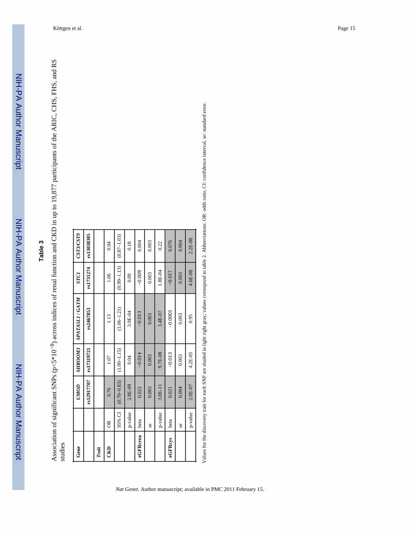
Table 3 presents the association of all genome-wide significant SNPs across the three renaltraits. SNPs in UMOD, SHROOM3, and STC1 showed direction-consistent associationacross traits. For example, rs12917707 at UMOD was associated with both higher eGFRcreaand eGFRcys representing better kidney function, and with lower odds of CKD, conferringdisease-protection. SNPs at the GATM/SPATA5L1 and CST regions were only associatedwith the respective discovery trait, the association of rs2467853 at the GATM/SPATA5L1locus with CKD likely results from the eGFRcrea-based definition of CKD. All SNPsassociated with CKD, eGFRcrea, and eGFRcys at p<4*10−7 are listed in SupplementaryTables 4, 5, and 6, respectively.
Köttgen et al. Page 4
Nat Genet. Author manuscript; available in PMC 2011 February 15.
NIH
-PA Author Manuscript
NIH
-PA Author Manuscript
NIH
-PA Author Manuscript
Together, loci for eGFRcrea explain 0.7% of the eGFRcrea variance [0.43% without theGATM locus], and loci for eGFRcys explain 3.2% of the eGFRcys variance [0.24% withoutthe CST locus], suggesting that additional yet undiscovered genetic variants impactvariability in renal function. In accordance with small absolute differences observed in otherGWAS for continuous human traits, the multivariable adjusted eGFR difference acrossgenotypes for any one locus was small. Since risk alleles may act in an additive fashion, wecreated a risk score for each individual as the sum of risk alleles at UMOD, SHROOM3, andSTC1. These analyses were performed in the ARIC Study, the largest individual studycontributing data and with available prospective information. The mean eGFRcrea was 10ml/min/1.73m2 lower in individuals with all 6 risk alleles across the 3 loci compared tothose with 0 risk alleles (p=2*10−8 per each unit score increase). CKD prevalence rangedfrom 0% in those without any risk alleles to 12.1% in individuals carrying all six risk alleles.
The association of SNPs in the UMOD gene with indices of renal function and CKDimplicate a common pathophysiologic mechanism localized to the nephron’s loop of Henle.As opposed to the renal glomerulus, this region has previously received little attention.Thus, studies to understand the production and functions of Tamm-Horsfall protein arewarranted to eventually lead to novel prevention and intervention options to reduce CKDrisk.
In summary, we identified and validated common variants at several novel loci conferringsusceptibility for kidney dysfunction and CKD in large, unselected population-based studies.
MethodsStudy Samples
Four large, population-based cohorts of the CHARGE consortium had GWAS data availableand formed the discovery sample: ARIC, CHS, FHS, and RS. Detailed information aboutthese cohorts, including the design papers, is provided in the Supplementary Methods.Briefly, the studies were initiated to study cardiovascular disease and its risk factors anddiseases related to aging. The population-based ARIC cohort recruited 15,792 middle-agedparticipants from 1987–1989 in four US communities. The population-based CHS cohortrecruited 5,888 participants 65 years from 1989–1990 and 1992–1993 in four UScommunities. The FHS is a community-based study with a family component, including theOriginal (n=5,209, recruited 1948) and Offspring (n=5,214, recruited 1971) component. Thecommunity-based RS recruited 7,983 participants aged 55 years or older from 1990–1993.Two independent study samples were used to replicate results. In AGES, 5,764 survivors ofthe Reykjavik Study were examined from 2002–2006 and contributed to information. TheWGHS is a sample drawn in 2006 from the original Women’s Health Study. Eachparticipant provided written informed consent, and Institutional Review Boards of theparticipating institutions approved the study protocols. African American participants fromARIC and CHS did not contribute information to the present study.
Genotyping and ImputationDetailed information about genotyping and imputation methods is provided inSupplementary Table 1, and details about data cleaning are provided in the SupplementaryMethods. Briefly, all studies directly genotyped between 300,000 and 900,000 SNPs usingwhole-genome genotyping arrays by either Affymetrix (6.0 [ARIC], 500K and 50K gene-centric [FHS]) or Illumina (Human CNV370 [AGES, CHS], 550K [RS], HumanHap300Duo-Plus or a combination of HumanHap300 and iSelect [WGHS]). All genotyping wasperformed according to the manufacturer’s instructions between 2006–2008. Using thePhase II CEU HapMap individuals as a reference panel, genotypes were imputed to a
Köttgen et al. Page 5
Nat Genet. Author manuscript; available in PMC 2011 February 15.
NIH
-PA Author Manuscript
NIH
-PA Author Manuscript
NIH
-PA Author Manuscript
common set of ~2.5 million high-quality HapMap SNPs. Software used for imputation wereBimBam v0.9927 (CHS) and MACH v1.0.15/16 (all others,http://www.sph.umich.edu/csg/abecasis/MACH/); FHS accounted for relatedness ofparticipants. Imputed genotypes were expressed as an allelic dosage, a fractional valuebetween 0–2. The WGHS did not impute genotypes.
Outcomes: eGFRcrea, eGFRcys, CKDSerum creatinine was measured using a modified kinetic Jaffe reaction in all studies butAGES, where an enzymatic method was used. eGFRcrea was calculated using theModification of Diet in Renal Disease (MDRD) Study equation14: eGFRcrea (ml/min/1.73m2) = 186.3*serum creatinine (mg/dl)−1.154 * age−0.203 * 0.742 (if female). To becomparable across studies, creatinine values in all studies were calibrated using regression toage, sex, and race adjusted mean values from a nationally representative US survey asdescribed previously.28 Cystatin C was measured by a particle-enhancedimmunonephelometric assay (N Latex Cystatin C, Dade Behring) at ARIC visit 4, CHSbaseline exam, and FHS offspring exam 7 with a nephelometer (BNII, Dade-Behring).eGFRcys was then calculated using the formula eGFRcys = 76.7*(serum cystatin C)−1.19.15
CKD was defined as eGFRcrea <60 ml/min/1.73m2 according to National KidneyFoundation guidelines.17
CKD in CHS, RS, WGHS, and AGES was defined based on a single measurement of serumcreatinine at the baseline visit. FHS and ARIC used a cumulative definition of CKD basedon serum creatinine measurements at several study visits as detailed in the SupplementaryMethods. Incident CKD in ARIC was defined as eGFRcrea <60 ml/in/1.73m2 at study visits2 or 4 in individuals with eGFRcrea ≥60 ml/in/1.73m2 at study visit 1, or a kidney-diseasespecific ICD code on a hospital discharge record or death certificate from study inception in1987 through January 1, 2005.29
Information on age and sex was collected at each study visit, and race was self-reported.Potential population stratification was assessed as detailed in the Supplementary Methods
Statistical analysisGWAS was conducted within each cohort for eGFRcrea, eGFRcys, and CKD, followed bymeta-analysis of the study-specific associations for each trait. SNPs showing genome-widesignificant association with any of the three traits in meta-analyses were then explored fortheir association with the other two traits.
The phenotype for the eGFR analyses in all studies was created by calculating a naturallogarithmic transformation of eGFR obtained from the respective equations for eGFRcreaand eGFRcys. All studies but CHS then created sex-specific age- and study-site (ARIC) orcohort (FHS) adjusted residuals. CHS adjusted for age, sex, and study site in multivariableregression models. Incident CKD in ARIC was analyzed using multivariable-adjusted Coxproportional hazards regression. Software packages used by the individual studies toconduct linear and logistic regression are listed in Supplementary Table 1. FHS accountedfor the relatedness of individuals in the analyses as detailed in the Supplementary Methods.Pedigree correlations were adjusted for using the robust variance option. All studies used anadditive genetic model.
Meta-analysis was conducted using inverse-variance weighting as implemented in METAL(http://www.sph.umich.edu/csg/abecasis/Metal/index.html). Prior to meta-analysis, thegenomic control parameter was calculated within each study for each trait to assess potentialinflation of the test statistics. If the parameter was larger than 1, an adjustment wasperformed by scaling the test statistics to the inflation factor. Only SNPs with minor allele
Köttgen et al. Page 6
Nat Genet. Author manuscript; available in PMC 2011 February 15.
NIH
-PA Author Manuscript
NIH
-PA Author Manuscript
NIH
-PA Author Manuscript
frequency (MAF) ≥2% were analyzed based on the number of CKD cases, corresponding toapproximately 50 carriers of the minor allele with CKD. Statistical heterogeneity wasevaluated using Cochrane’s chi-square test (Q-test).
The most significant SNP at genomic loci with evidence of suggestive association(p<4*10−7) for any of the traits were tested for replication in the independent samples. Thisthreshold corresponds to1/2.5 million tests conducted and corresponds to one or lessexpected false positive findings.30 A threshold of 5*10−8 was used to indicate genome-widesignificance, corresponding to a Bonferroni correction for the estimated 1 million commonindependent SNPs across the genome (0.05/1 million).31 The SNAP program with theHapMap CEU sample as a reference was used to identify the best proxy in the WGHSdataset, to evaluate LD to nearby coding SNPs, and to evaluate LD between imputed SNPsand proxy SNPs that were directly genotyped(http://www.broad.mit.edu/mpg/snap/ldsearch.php).
Supplementary MaterialRefer to Web version on PubMed Central for supplementary material.
AcknowledgmentsWe are indebted to the staff and participants of the AGES Reykjavik Study, the ARIC Study, the CHS Study, theFHS Study, the Rotterdam Study, and the WGHS Study for their important contributions.
.AGES: The Age, Gene/Environment Susceptibility Reykjavik Study has been funded by NIH contract N01-AG-12100, the NIA Intramural Research Program, Hjartavernd (the Icelandic Heart Association), and the Althingi(the Icelandic Parliament). We thank Thor Aspelund and Gudny Eiriksdottir for their contribution to collecting,analyzing and preparing the AGES Reykjavik Study data.
ARIC: The Atherosclerosis Risk in Communities Study is carried out as a collaborative study supported byNational Heart, Lung, and Blood Institute contracts N01-HC-55015, N01-HC-55016, N01-HC-55018, N01-HC-55019, N01-HC-55020, N01-HC-55021, N01-HC-55022, R01HL087641, R01HL59367 and R01HL086694;National Human Genome Research Institute contract U01HG004402; and National Institutes of Health contractHHSN268200625226C. The authors thank the staff and participants of the ARIC study for their importantcontributions. Infrastructure was partly supported by Grant Number UL1RR025005, a component of the NationalInstitutes of Health and NIH Roadmap for Medical Research. AK was supported by a German Research FoundationFellowship. WHLK was supported by K01DK067207.
CHS: The CHS research reported in this article was supported by contract numbers N01-HC-85079 through N01-HC-85086, N01-HC-35129, N01 HC-15103, N01-HC-55222, N01-HC-75150, N01-HC-45133, grant numbers U01HL080295 and R01 HL087652, and R01 AG027002 from the National Heart, Lung, and Blood Institute, withadditional contribution from the National Institute of Neurological Disorders and Stroke. A full list of principalCHS investigators and institutions can be found at http://www.chs-nhlbi.org/pi.htm. DNA handling and genotypingwas supported in part by National Center for Research Resources grant M01RR00069 to the Cedars-Sinai GeneralClinical Research Center Genotyping core and National Institute of Diabetes and Digestive and Kidney Diseasesgrant DK063491 to the Southern California Diabetes Endocrinology Research Center.
FHS: This research was conducted in part using data and resources from the Framingham Heart Study of theNational Heart Lung and Blood Institute of the National Institutes of Health and Boston University School ofMedicine. The analyses reflect intellectual input and resource development from the Framingham Heart Studyinvestigators participating in the SNP Health Association Resource (SHARe) project. This work was partiallysupported by the National Heart, Lung and Blood Institute’s Framingham Heart Study (Contract No. N01-HC-25195) and its contract with Affymetrix, Inc for genotyping services (Contract No. N02-HL-6-4278). A portionof this research utilized the Linux Cluster for Genetic Analysis (LinGA-II) funded by the Robert Dawson EvansEndowment of the Department of Medicine at Boston University School of Medicine and Boston Medical Center.
RS: The Rotterdam Study is supported by the Erasmus Medical Center and Erasmus University Rotterdam; theNetherlands Organization for Scientific Research; the Netherlands Organization for Health Research andDevelopment (ZonMw); the Research Institute for Diseases in the Elderly; The Netherlands Heart Foundation; theMinistry of Education, Culture and Science; the Ministry of Health Welfare and Sports; the European Commission;and the Municipality of Rotterdam. Support for genotyping was provided by the Netherlands Organization for
Köttgen et al. Page 7
Nat Genet. Author manuscript; available in PMC 2011 February 15.
NIH
-PA Author Manuscript
NIH
-PA Author Manuscript
NIH
-PA Author Manuscript
Scientific Research (NWO) (175.010.2005.011, 911.03.012) and Research Institute for Diseases in the Elderly(RIDE). This study was further supported by the Netherlands Genomics Initiative (NGI)/Netherlands Organisationfor Scientific Research (NWO) project nr. 050-060-810. We thank Pascal Arp, Mila Jhamai, Dr MichaelMoorhouse, Marijn Verkerk and Sander Bervoets for their help in creating the Rotterdam database and MaximStruchalin for his contributions to the imputations of the Rotterdam data.
WGHS: The WGHS was supported by the National Heart, Lung, and Blood Institute (HL 043851) and the NationalCancer Institute (CA 047988). Collaborative scientific and genotyping support was provided by Amgen, Inc.
References1. Levey AS, et al. Chronic kidney disease as a global public health problem: approaches and
initiatives - a position statement from Kidney Disease Improving Global Outcomes. Kidney Int2007;72:247–259. [PubMed: 17568785]
2. Serafini-Cessi F, Malagolini N, Cavallone D. Tamm-Horsfall glycoprotein: biology and clinicalrelevance. Am J Kidney Dis 2003;42:658–676. [PubMed: 14520616]
3. Hart TC, et al. Mutations of the UMOD gene are responsible for medullary cystic kidney disease 2and familial juvenile hyperuricaemic nephropathy. J Med Genet 2002;39:882–892. [PubMed:12471200]
4. Coresh J, et al. Prevalence of chronic kidney disease in the United States. JAMA 2007;298:2038–2047. [PubMed: 17986697]
5. Zhang QL, Rothenbacher D. Prevalence of chronic kidney disease in population- based studies:systematic review. BMC Public Health 2008;8:117. [PubMed: 18405348]
6. National Institutes of Health. US Renal Data System, USRDS 2008 Annual Data Report: Atlas ofChronic Kidney Disease and End-Stage Renal Disease in the United States. National Institute ofDiabetes and Digestive and Kidney Diseases; Bethesda, MD:
7. Sarnak MJ, et al. Kidney disease as a risk factor for development of cardiovascular disease: astatement from the American Heart Association Councils on Kidney in Cardiovascular Disease,High Blood Pressure Research, Clinical Cardiology, and Epidemiology and Prevention. Circulation2003;108:2154–2169. [PubMed: 14581387]
8. Weiner DE, et al. Chronic kidney disease as a risk factor for cardiovascular disease and all-causemortality: a pooled analysis of community-based studies. J Am Soc Nephrol 2004;15:1307–1315.[PubMed: 15100371]
9. Langefeld CD, et al. Heritability of GFR and albuminuria in Caucasians with type 2 diabetesmellitus. Am J Kidney Dis 2004;43:796–800. [PubMed: 15112169]
10. Bochud M, et al. Heritability of renal function in hypertensive families of African descent in theSeychelles (Indian Ocean). Kidney Int 2005;67:61–69. [PubMed: 15610228]
11. Fox CS, et al. Genomewide linkage analysis to serum creatinine, GFR, and creatinine clearance ina community-based population: the Framingham Heart Study. J Am Soc Nephrol 2004;15:2457–2461. [PubMed: 15339995]
12. Freedman BI, Satko SG. Genes and renal disease. Curr Opin Nephrol Hypertens 2000;9:273–277.[PubMed: 10847329]
13. Psaty BM, et al. Cohorts for Heart and Aging Research in Genomic Epidemiology (CHARGE)Consortium: Design of prospective meta-analyses of genome-wide association studies from fivecohorts. Circulation: Cardiovascular Genetics. 2009 in press.
14. Levey AS, et al. A more accurate method to estimate glomerular filtration rate from serumcreatinine: a new prediction equation. Modification of Diet in Renal Disease Study Group. AnnIntern Med 1999;130:461–470. [PubMed: 10075613]
15. Stevens LA, et al. Estimating GFR using serum cystatin C alone and in combination with serumcreatinine: a pooled analysis of 3,418 individuals with CKD. Am J Kidney Dis 2008;51:395–406.[PubMed: 18295055]
16. Stevens LA, Coresh J, Greene T, Levey AS. Assessing kidney function--measured and estimatedglomerular filtration rate. N Engl J Med 2006;354:2473–2483. [PubMed: 16760447]
Köttgen et al. Page 8
Nat Genet. Author manuscript; available in PMC 2011 February 15.
NIH
-PA Author Manuscript
NIH
-PA Author Manuscript
NIH
-PA Author Manuscript
17. National Kidney Foundation. K/DOQI clinical practice guidelines for chronic kidney disease:evaluation, classification, and stratification. Am J Kidney Dis 2002;39:S1–266. [PubMed:11904577]
18. Vylet’al P, et al. Alterations of uromodulin biology: a common denominator of the geneticallyheterogeneous FJHN/MCKD syndrome. Kidney Int 2006;70:1155–1169. [PubMed: 16883323]
19. Rampoldi L, et al. Allelism of MCKD, FJHN and GCKD caused by impairment of uromodulinexport dynamics. Hum Mol Genet 2003;12:3369–3384. [PubMed: 14570709]
20. Bachmann S, et al. Renal effects of Tamm-Horsfall protein (uromodulin) deficiency in mice. Am JPhysiol Renal Physiol 2005;288:F559–67. [PubMed: 15522986]
21. Benetti E, et al. Immature renal structures associated with a novel UMOD sequence variant. Am JKidney Dis 2009;53:327–331. [PubMed: 18950917]
22. Nishimura T, Takeichi M. Shroom3-mediated recruitment of Rho kinases to the apical celljunctions regulates epithelial and neuroepithelial planar remodeling. Development2008;135:1493–1502. [PubMed: 18339671]
23. Anad F, et al. Alagille syndrome and deletion of 20p. J Med Genet 1990;27:729–737. [PubMed:2074558]
24. Hwang SJ, Yang Q, Meigs JB, Pearce EN, Fox CS. A genome-wide association for kidneyfunction and endocrine-related traits in the NHLBI’s Framingham Heart Study. BMC Med Genet2007;8 (Suppl 1):S10. [PubMed: 17903292]
25. Yoshiko Y, Aubin JE. Stanniocalcin 1 as a pleiotropic factor in mammals. Peptides 2004;25:1663–1669. [PubMed: 15476933]
26. Huang L, et al. Anti-Inflammatory and Renal Protective Actions of Stanniocalcin-1 in a Model ofAnti-Glomerular Basement Membrane Glomerulonephritis. Am J Pathol. 2009
27. Servin B, Stephens M. Imputation-based analysis of association studies: candidate regions andquantitative traits. PLoS Genet 2007;3:e114. [PubMed: 17676998]
28. Coresh J, et al. Calibration and random variation of the serum creatinine assay as critical elementsof using equations to estimate glomerular filtration rate. Am J Kidney Dis 2002;39:920–929.[PubMed: 11979335]
29. Hsu CC, et al. Apolipoprotein E and progression of chronic kidney disease. JAMA2005;293:2892–2899. [PubMed: 15956634]
30. Gordon A, Glazko G, Qui X, Yakovlev A. Control of the mean number of false discoveries,Bonferroni and stability of multiple testing. The Annals of Applied Statistics 2007;1:179–190.
31. Pe’er I, Yelensky R, Altshuler D, Daly MJ. Estimation of the multiple testing burden forgenomewide association studies of nearly all common variants. Genet Epidemiol 2008;32:381–385. [PubMed: 18348202]
Köttgen et al. Page 9
Nat Genet. Author manuscript; available in PMC 2011 February 15.
NIH
-PA Author Manuscript
NIH
-PA Author Manuscript
NIH
-PA Author Manuscript
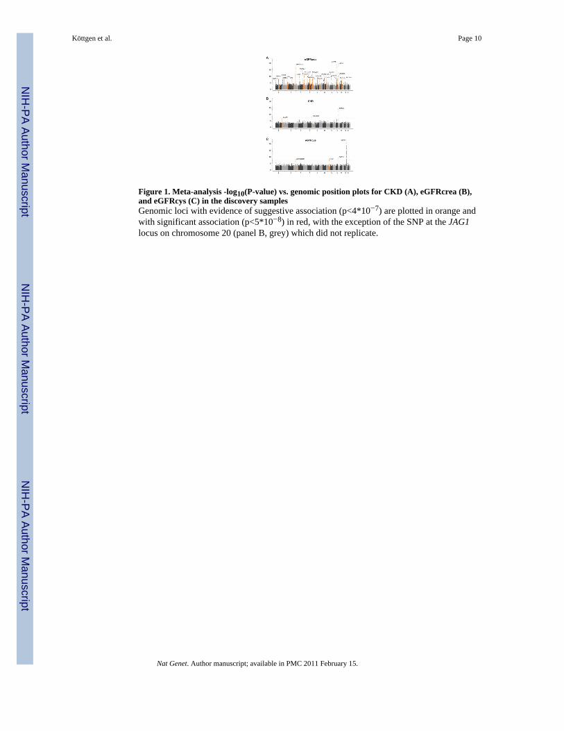
Figure 1. Meta-analysis -log10(P-value) vs. genomic position plots for CKD (A), eGFRcrea (B),and eGFRcys (C) in the discovery samplesGenomic loci with evidence of suggestive association (p<4*10−7) are plotted in orange andwith significant association (p<5*10−8) in red, with the exception of the SNP at the JAG1locus on chromosome 20 (panel B, grey) which did not replicate.
Köttgen et al. Page 10
Nat Genet. Author manuscript; available in PMC 2011 February 15.
NIH
-PA Author Manuscript
NIH
-PA Author Manuscript
NIH
-PA Author Manuscript

Figure 2. Genetic architecture of the genome-wide significant susceptibility loci for renal diseasein the discovery samples: (A): UMOD gene region, (B): SHROOM3 gene region, (C): GATM/SPATA5L1 gene region, (D) CST genes region, (E) STC1 gene region−log10 P-values are plotted versus genomic position (Build 36). The most significant SNPin each region is plotted in blue. LD based on the HapMap CEU sample is color-coded: red(r2 to top SNP 0.8–.0), orange (0.5–.8), yellow (0.2–.5), and white (<0.2). Gene annotationsare based on Build 36 and arrows present direction of transcription. P-values are obtainedfrom the discovery traits: CKD (UMOD), eGFRcrea (SHROOM3, GATM), eGFRcys (CST,STC1).
Köttgen et al. Page 11
Nat Genet. Author manuscript; available in PMC 2011 February 15.
NIH
-PA Author Manuscript
NIH
-PA Author Manuscript
NIH
-PA Author Manuscript

Figure 3. Meta-analysis of the odds of CKD per each additional copy of the minor T allele atUMOD rs12917707 across strata of major kidney disease risk factorsError bars correspond to 95% confidence intervals. Meta-analysis values obtained from thediscovery samples.
Köttgen et al. Page 12
Nat Genet. Author manuscript; available in PMC 2011 February 15.
NIH
-PA Author Manuscript
NIH
-PA Author Manuscript
NIH
-PA Author Manuscript

NIH
-PA Author Manuscript
NIH
-PA Author Manuscript
NIH
-PA Author Manuscript
Köttgen et al. Page 13
Tabl
e 1
Cha
ract
eris
tics o
f the
stud
y sa
mpl
es
AR
ICC
HS
FHS
RS
AG
ES
WG
HS
Coho
rt in
form
atio
ndi
scov
ery
repl
icat
ion
Stud
y D
esia
gnpr
ospe
ctiv
e m
ulti-
cent
erPo
pula
tion-
base
d pr
ospe
ctiv
e m
ulti-
cent
erC
omm
unity
-bas
ed fa
mily
mul
ti-ge
nera
tion
Popu
latio
n-ba
sed
pros
pect
ive
Popu
latio
n-ba
sed
pros
pect
ive
Tria
l
Ana
lyze
d sa
mpl
e si
ze, C
KD
/eG
FRcr
ea/e
GFR
cys
8069
/ 65
25 /
6430
3278
/ 32
78 /
2844
4140
/ 39
34 /
2992
4390
/ 43
90 /
NA
3219
/ 32
19 /
NA
1824
7 / 1
8247
/ N
A
Sam
ple
char
acte
ristic
s*m
ean
(SD
) / %
(n)
mea
n (S
D) /
% (n
)m
ean
(SD
) / %
(n)
mea
n (S
D) /
% (n
)m
ean
(SD
) / %
(n)
mea
n (S
D) /
% (n
)
Age
, yea
rs63
.1 (5
.6)
72.4
(5.4
)62
.4 (1
1.6)
70.0
(9.0
)76
.4 (5
.5)
54.7
(7.1
)
Mal
e46
.7 (3
047)
39.2
(128
6)45
.5 (1
890)
38.6
(169
4)42
(135
2)0
Hyp
erte
nsio
n pr
eval
ence
43.0
(279
3)52
.4 (1
719)
57.4
(237
7)34
.1 (1
497)
81 (2
596)
24.4
(446
0)
Dia
bete
s mel
litus
13.7
(893
)11
.8 (3
85)
9.8
(406
)10
.7 (4
70)
11.5
(368
)2.
6 (4
74)
eGFR
cys
tatin
, ml/m
in/1
.73m
284
.1 (1
9.7)
79.9
(18.
3)77
.9 (1
6.9)
NA
NA
NA
eGFR
cre
atin
ine,
ml/m
in/1
.73m
280
.6 (1
7.2)
80.0
(22.
6)85
.2 (2
3.5)
77.1
(17.
2)73
.0 (2
0)90
.4 (2
2.8)
CK
D, e
GFR
<60
ml/m
in/1
.73m
29.
1 (7
31)
18.7
(612
)10
.7 (4
45)
13.7
(600
)24
.3 (7
81)
6.3
(115
1)
* Info
rmat
ion
on d
emog
raph
ics,
hype
rtens
ion,
and
dia
bete
s are
asc
erta
ined
at t
he v
isit
eGFR
crea
was
mea
sure
d. S
ampl
e si
zes f
or C
KD
and
eG
FRcr
ea in
AR
IC a
nd F
HS
diff
er a
s the
se st
udie
s use
a c
umul
ativ
e de
finiti
on o
f CK
D a
s det
aile
d in
the
met
hod
sect
ion.
Abb
revi
atio
ns: A
RIC
: Ath
eros
cler
osis
Ris
k in
Com
mun
ities
Stu
dy, C
HS:
Car
diov
ascu
lar H
ealth
Stu
dy, F
HS:
Fra
min
gham
Hea
rt St
udy,
RS:
Rot
terd
am S
tudy
, AG
ES: A
ge G
ene/
Envi
ronm
ent S
usce
ptib
ility
-Rey
kjav
ik S
tudy
, WG
HS:
Wom
en’s
Gen
ome
Hea
lth S
tudy
, SD
:st
anda
rd d
evia
tion,
CK
D: c
hron
ic k
idne
y di
seas
e, e
GFR
: est
imat
ed g
lom
erul
ar fi
ltrat
ion
rate
.
Nat Genet. Author manuscript; available in PMC 2011 February 15.
NIH
-PA Author Manuscript
NIH
-PA Author Manuscript
NIH
-PA Author Manuscript
Köttgen et al. Page 14
Tabl
e 2
Res
ults
from
met
a-an
alys
es o
f top
GW
AS
sign
als a
t eac
h lo
cus (
p<4*
10−
7 ) fo
r CK
D, e
GFR
crea
, and
eG
FRcy
s in
disc
over
y sa
mpl
es a
nd a
fter r
eplic
atio
n
Loc
usG
WA
S m
eta-
anal
ysis
GW
AS
disc
over
y an
d re
plic
atio
n m
eta-
anal
ysis
SNP
Chr
posi
tion
in (n
ear)
gen
em
inor
/ m
ajor
alle
leO
E v
ar r
atio
*M
AF
beta
/ O
Rse
/ 95
% C
Ip-
valu
ebe
ta /
OR
se /
95%
CI
p-va
lue
CKD
(n=1
9,87
7)
rs12
9177
0716
2027
5191
(UM
OD
)T/
G0.
960.
180.
760.
70–.
832.
8E-0
90.
800.
75–.
852.
3E-1
2
eGFR
crea
(n=1
8,12
7)
rs17
3197
214
7758
7871
SHRO
OM
3A
/G0.
960.
44−0.014
0.00
39.
7E-0
8−0.012
0.00
21.
2E-1
2
rs24
6785
315
4348
6085
SPAT
A5L1
/GAT
MG
/T0.
970.
38−0.013
0.00
33.
4E-0
7−0.013
0.00
26.
2E-1
4
rs12
9177
0716
2027
5191
(UM
OD
)T/
G0.
960.
180.
022
0.00
33.
0E-1
10.
018
0.00
25.
2E-1
6
rs60
4005
520
1058
1313
JAG
1T/
C0.
730.
39−0.017
0.00
31.
0E-0
8−0.005
0.00
25.
9E-0
3
eGFR
cys (
n=12
,266
)
rs17
3127
48
2382
2264
(STC
1)G
/A0.
960.
43−0.017
0.00
34.
6E-0
8N
AN
AN
A
rs12
9177
0716
2027
5191
(UM
OD
)T/
G0.
960.
180.
021
0.00
42.
0E-0
7N
AN
AN
A
rs13
0383
0520
2355
8262
(CST
3/C
ST9)
T/C
0.95
0.21
0.07
60.
004
2.2E
-88
NA
NA
NA
Met
a-an
alys
is p
-val
ues a
re a
djus
ted
for t
he tr
ait-
and
stud
y-sp
ecifi
c ge
nom
ic-c
ontro
l par
amet
ers (
see
Supp
l Fig
1).
For e
GFR
, bet
as in
dica
te th
e ch
ange
in e
GFR
per
min
or a
llele
for t
he n
atur
al lo
garit
hmic
trans
form
atio
n of
eG
FR. N
A: e
GFR
cys w
as n
ot a
vaila
ble
in th
e re
plic
atio
n sa
mpl
es, b
ut rs
1731
274
was
ass
ocia
ted
with
eG
FRcr
ea a
fter r
eplic
atio
n at
p=2
*10−
7 , b
eta −
0.00
9, se
0.0
02.
Abb
revi
atio
ns: *
OE
var r
atio
: sam
ple-
size
wei
ghte
d m
ean
of th
e ob
serv
ed b
y ex
pect
ed v
aria
nce
ratio
for e
ach
SNP
acro
ss th
e di
scov
ery
sam
ples
; MA
F: m
inor
alle
le fr
eque
ncy;
OR
: odd
s rat
io; C
I:co
nfid
ence
inte
rval
; se:
stan
dard
err
or.
Nat Genet. Author manuscript; available in PMC 2011 February 15.
NIH
-PA Author Manuscript
NIH
-PA Author Manuscript
NIH
-PA Author Manuscript
Köttgen et al. Page 15
Tabl
e 3
Ass
ocia
tion
of si
gnifi
cant
SN
Ps (p
<5*1
0−8 )
acr
oss i
ndic
es o
f ren
al fu
nctio
n an
d C
KD
in u
p to
19,
877
parti
cipa
nts o
f the
AR
IC, C
HS,
FH
S, a
nd R
Sst
udie
s
Gen
eU
MO
DSH
ROO
M3
SPAT
A5L1
/ G
ATM
STC1
CST3
/CST
9
rs12
9177
07rs
1731
9721
rs24
6785
3rs
1731
274
rs13
0383
05
Trai
t
CK
DO
R0.
761.
071.
131.
060.
94
95%
CI
(0.7
0–0.
83)
(1.0
0–1.
15)
(1.0
6–1.
21)
(0.9
9–1.
13)
(0.8
7–1.
03)
p-va
lue
2.8E
-09
0.04
3.9E
-04
0.09
0.18
eGFR
crea
beta
0.02
2−0.014
−0.013
−0.009
0.00
4
se0.
003
0.00
30.
003
0.00
30.
003
p-va
lue
3.0E
-11
9.7E
-08
3.4E
-07
1.8E
-04
0.22
eGFR
cys
beta
0.02
1−0.013
−0.0001
−0.017
0.07
6
se0.
004
0.00
30.
003
0.00
30.
004
p-va
lue
2.0E
-07
4.2E
-05
0.95
4.6E
-08
2.2E
-88
Val
ues f
or th
e di
scov
ery
trait
for e
ach
SNP
are
shad
ed in
ligh
t rig
ht g
ray;
val
ues c
orre
spon
d to
tabl
e 2.
Abb
revi
atio
ns: O
R: o
dds r
atio
, CI:
conf
iden
ce in
terv
al, s
e: st
anda
rd e
rror
.
Nat Genet. Author manuscript; available in PMC 2011 February 15.