Multicriteria-based ranking for risk management of food-born ...
324
Multicriteria-based ranking for risk management of food-borne parasites MICROBIOLOGICAL RISK ASSESSMENT SERIES 23 ISSN 1726-5274
-
Upload
khangminh22 -
Category
Documents
-
view
0 -
download
0
Transcript of Multicriteria-based ranking for risk management of food-born ...

Multicriteria-based ranking for risk management of food-borne parasites
M I C R O B I O L O G I C A L R I S K A S S E S S M E N T S E R I E S
23ISSN 1726-5274

Infectious diseases caused by food-borne parasites have not received the same level of attention as other food-borne biological and chemical hazards. Nevertheless, they cause a high burden of disease in humans, may have prolonged, severe, and sometimes fatal outcomes, and result in considerable hardship
negative
by animals or
do not capture the true
a
te-commodity
this
Alimentarius
individuals
MRAbookCover23.indd 1 07/04/14 15:49
For further information on the joint FAO/WHO
Activities on microbiological risk assessment, please contact:
Food Safety and Codex Unit
Food and Agriculture Organization of the United Nations
Viale delle Terme di Caracalla
00153 Rome, Italy
Fax: +39 06 57054593
E-mail: [email protected]
Web site: http://www.fao.org/food/food-safety-quality
or
Department of Food Safety and Zoonoses
World Health Organization
20 Avenue Appia
1211 Geneva 27
Switzerland
Fax: +41 22 7914807
E-mail: [email protected]
Web site: http://www.who.int/foodsafety
Cover design:
Food and Agriculture Organization of the United Nations
and the World Health Organization
Cover picture:
© Dennis Kunkel Microscopy, Inc.
Publications of the World Health Organization
can be obtained from:
WHO Press
World Health Organization, 20 Avenue Appia
1211 Geneva 27, Switzerland
Tel: +41 22 7913264
Fax: +41 22 7914857
E-mail: [email protected]
or
on the Internet from http://www.who.int/bookorders
Publications of the Food and Agriculture Organization of the United Nations
can be purchased from:
Sales and Marketing Group,
Food and Agriculture Organization of the United Nations
Viale delle Terme di Caracalla, 00153 Rome, Italy
Fax: +39 06 57053360
E-mail: [email protected]
or
on the Internet from http://www.fao.org/icatalog/inter-e.htm
Report of a Joint FAO/WHO Expert Meeting, 3–7 September 2012, FAO Headquarters, Rome, Italy
Food and Agriculture Organization of the United Nations World Health Organization
2014
Multicriteria-based ranking for risk management of food-borne parasites
23M I C R O B I O L O G I C A L R I S K A S S E S S M E N T S E R I E S

The designations employed and the presentation of material in this information product do not imply the expression of any opinion whatsoever on the part of the Food and Agriculture Organization of the United Nations (FAO) or of the World Health Organization (WHO) concerning the legal status of any country, territory, city or area or of its authorities, or concerning the delimitation of its frontiers or boundaries. The mention of specific companies or products of manufacturers, whether or not these have been patented, does not imply that these are or have been endorsed or recommended by FAO or WHO in preference to others of a similar nature that are not mentioned. All reasonable precautions have been taken by FAO and WHO to verify the information contained in this publication. However, the published material is being distributed without warranty of any kind, either expressed or implied. The responsibility for the interpretation and use of the material lies with the reader. In no event shall FAO and WHO be liable for damages arising from its use.
WHO Library Cataloguing-in-Publication Data:Multicriteria-based ranking for risk management of food-borne parasites: report of a Joint FAO/WHO Expert Meeting, 3-7 September 2012, FAO Headquarters, Rome, Italy.1.Food contamination. 2.Food parasitology. 3.Parasites. 4.Risk management – methods. I.World Health Organization. II.Food and Agriculture Organization of the United Nations.
ISBN 978 92 4 156470 0 (WHO) (NLM classification: WA 701)ISBN 978-92-5-108199-0 (print) (FAO)E-ISBN 978-92-5-108200-3 (PDF) (FAO)ISSN 1726-5274
Recommended citation:FAO/WHO [Food and Agriculture Organization of the United Nations/World Health Organization]. 2014. Multicriteria-based ranking for risk management of food-borne parasites. Microbiological Risk Assessment Series No. 23. Rome. 302pp
FAO and WHO encourage the use, reproduction and dissemination of material in this information product. Except where otherwise indicated, material may be copied, downloaded and printed for private study, research and teaching purposes, or for use in non-commercial products or services, provided that appropriate acknowledgement of FAO and WHO as the source and copyright holder is given and that their endorsement of users’ views, products or services is not implied in any way.All requests for translation and adaptation rights, and for resale and other commercial use rights should be made via www.fao.org/contact-us/licencerequest or addressed to [email protected].
FAO information products are available on the FAO website (www.fao.org/publications) and can be purchased through [email protected].
© FAO/WHO 2014
Contents
12
3
4
Acknowledgments x
Contributors xi
Abbreviations used in the report xiv
Executive Summary xv
Background 1
Objectives and approach 4
2.1 Identification of parasites 7
2.2 Definition of primary and secondary parasite and food pathways 7
2.3 Definition of criteria for parasite scoring 8
2.4 Scoring parasites according to criteria 1 1
2.5 Definition of criteria weights 12
2.6 Calculation of parasite scores 13
Results 14
3.1 The global ranking of food-borne parasites
3.2 Trade scores for the ranked parasites 14
3.3 Socio-economic impacts for the ranked parasites 18
3.4 Conclusions 20
Risk management options for the higher ranked parasites 23
4.1 General risk management considerations 23
4.2 Generic risk management options 24
4.3 Some specific considerations for risk management 26
Conclusions and recommendations 32
References 35
5
iv
ANNEXES
Annex 1 Identification of food-borne parasites for consideration 40
Annex 2 Food-borne parasite ranking exercise: summary card 44
Annex 3 Food-borne parasite ranking exercise form: explanation of criteria 45Criterion No. 1. Number of global food-borne illnesses 45Criterion No. 2. Geographical distribution (endemic regions) 46Criterion No. 3. Acute Morbidity Severity 47Criterion No. 4. Chronic Morbidity Severity 48Criterion No. 5. Fraction chronic 49Criterion No. 6. Mortality rate 50Criterion No. 7. Increasing trend in disease 50Criterion No. 8. International trade 50Criterion No. 9. Distributional impacts (socio-economic impact) 51Criterion No. 10. Quality of evidence 52Comments 52References 52
Annex 4. Criteria weights worksheet 53
Annex 5. Sensitivity analysis 54
Annex 6. Risk management actions 62
Annex 7. Specific information for the ranked parasites 63
A7.1 Anisakidae and anisakiasis 63General information 63Geographical distribution 63Disease 64Trade relevance 65Impact on economically vulnerable populations 65References 65
A7.2 Ascaris spp. 66General information 66Geographical distribution 67Disease 67Trade relevance 68Impact on economically vulnerable populations 68Other relevant information 68References 69
A7.3 Balantidium coli 70General information 70Geographical distribution 70Disease 70
v
Trade relevance 71Impact on economically vulnerable populations 71References 71
A7.4 Cryptosporidium spp. 72General information 72Geographical distribution 73Disease 74Trade relevance 74Impact on economically vulnerable populations 74References 75
A7.5 Cyclospora cayetanensis 77General information 77Geographical distribution 78Disease 78Trade relevance and impact on economically vulnerable
populations 79References 79
A7.6 Diphyllobothrium spp. 82General information 82Geographical distribution 82Disease 83Trade relevance 84Impact on economically vulnerable populations 84Other relevant information 84References 84
A7.7 Echinococcus granulosus 88General information 88Geographical distribution 89Disease 89Trade relevance of cystic echinococcosis 90Impact of CE on economically vulnerable populations 91References 92
A7.8 Echinococcus multilocularis 95General information 95Geographical distribution 95Disease 96Trade relevance 97Impact on economically vulnerable populations 98References 98
A7.9 Entamoeba histolytica 101General information 101Geographical distribution 101Disease 101Trade relevance 102

vi
Impact on economically vulnerable populations 102References 102
A7.10 Fasciola spp. 104General information 104Geographical distribution 104Disease 104Trade relevance 105Impact on economically vulnerable populations 105References 106
A7.11 Giardia duodenalis 108General information 108Geographical distribution 108Disease 108Trade relevance 110Impact on economically vulnerable populations 110References 110
A7.12 Heterophyidae and heterophyidiasis 112General information 112Geographical distribution 112Disease 112Trade relevance 113Impact on economically vulnerable populations 113Other relevant information 113References 114
A7.13 Opisthorchiidae 115General information 115Geographical distribution (endemic regions) 115Disease 115Trade relevance 117Impact on economically vulnerable populations 117Other relevant information 117References 117
A7.14 Paragonimus spp. 119General information 119Geographical distribution 119Disease 120Trade relevance 122Impact on economically vulnerable populations 122References 122
A7.15 Sarcocystis spp. 124General information 124Geographical distribution 124Prevalence in food animals 124Prevalence in humans 125

vii
Disease 126Trade relevance 126Impact on economically vulnerable populations 126References 127
A7.16 Spirometra spp. 129General information 129Geographical distribution 129Disease 130Trade relevance and impact on vulnerable populations 130References 131
A7.17 Taenia saginata 133General information 133Geographical distribution 133Disease 134Trade relevance 135Impact on economically vulnerable populations 135References 135
A7.18 Taenia solium 137General information on the parasite 137Geographical distribution 137Disease 137Trade relevance 138Impact on economically vulnerable populations 138References 139
A7.19 Toxocara spp. 141General information 141Geographical distribution 141Disease 142Trade relevance 143Impact on economically vulnerable populations 143References 143
A7.20 Toxoplasma gondii 145General Information 145Geographical distribution 146Disease 147Trade relevance and Impact on economically vulnerable
populations 149References 149
A7.21 Trichinella spp. other than T. spiralis 152General information 152Geographical distribution 152Disease 153Trade relevance 154Impact on economically vulnerable populations 154References 154

viii
A7.22 Trichinella spiralis 156General information 156Geographical distribution 156Disease 156Trade relevance 157Impact on economically vulnerable populations 158Other relevant information 158References 158
A7.23 Trichuris trichiura 160General information 160Geographical distribution 160Disease 160Trade relevance 161Impact on economically vulnerable populations 161References 161
A7.24 Trypanosoma cruzi 163General information 163Geographical distribution 163Disease 164Chagas disease by oral transmission 164Trade relevance 165Impact on economically vulnerable populations 165References 165
A7.25 Glossary of Parasitological Terms 167
Annex 8. Regional Reports 171
Annex 8.1 – Africa 172A8.1.1 Introduction 172A8.1.2 Data availability in humans, and food attribution 172A8.1.3 Agri-food trade 179A8.1.4 Consumer perception 179A8.1.5 Social sensitivity 179A8.1.6 Risk management 180
Annex 8.2 – Asia 182
A8.2.1 Introduction 182
A8.2.2 Description of individual foodborne parasitic diseases 182A8.2.2.1 Meat-borne parasite infections 182A8.2.2.2 Fish- and shellfish-borne parasites 184A8.2.2.3 Plant (fruit and vegetable)-borne parasites 187A8.2.3 Risk management strategies 190A8.2.4 Sources consulted 190
Annex 8.3 – Australia 218A8.3.1 Preparation 218A8.3.2 Data availability in humans and food attribution 218
ix
A8.3.3 Agri-food trade 226A8.3.4 Consumer perception 227A8.3.5 Social sensitivity 227A8.3.6 Risk management 228A8.3.7 Sources cited in the discussion 228
Annex 8.4 – Europe 230A8.4.1 Preparation 230A8.4.2 Data availability in humans and food attribution 230A8.4.3 Data on the burden of disease and food attribution 231A8.4.4 Data on parasite prevalence, incidence and concentration
in the main food categories 231A8.4.5 Agri-food trade 231A8.4.6 Consumer perception 233A8.4.7 Social sensitivity 234A8.4.8 Risk management 235A8.4.9 Sources cited in the text of the Europe section discussion 235
Annex 8.5 – Near East 249A8.5.1 Compilation of data availability on food borne parasites
relevant to the Near East 249A8.5.2 Agri-food trade 250A8.5.3 Consumer perception and social sensitivity 250A8.5.4 Risk management 251A8.5.5 Sources cited in the discussion 251
Annex 8.6 – North America with notes on Central America 267A8.6.1 Report preparation 267A8.6.2 Data availability on human occurrences and food
attribution 267A8.6.3 Data on the burden of disease and food attribution 267A8.6.4 Agri-food trade 267A8.6.5 Consumer perception 268A8.7.6 Social sensitivity 268A8.6.7 Risk management 268
Annex 8.7 – South America 268A8.7.1 Report preparation 268A8.7.2 Data availability in humans and food attribution 268A8.7.3 Agri-food trade 268A8.7.4 Consumer perception 268A8.7.5 Social sensitivity 287A8.7.6 Risk management 287

x
Acknowledgments
The Food and Agriculture Organization of the United Nations and the World Health Organization would like to express their appreciation to all those who con-tributed to the preparation of this report through their participation in the expert meeting and the provision of their time, expertise, data and other relevant infor-mation both before and after the meeting. Special appreciation is extended to Mr Michael Batz for his work on the design and facilitation of the multi criteria-based ranking exercise, and to Dr Andrijana Rajic for her valuable help, particularly in the design and implementation of the pre-meeting activities, as well as the meeting approach. All contributors are listed on the following pages.
Appreciation is also extended to all those who responded to the calls for data that were issued by FAO and WHO, and brought to our attention data in official docu-mentation or not readily available in the mainstream literature.
Final editing for language and preparation for publication was by Thorgeir Lawrence.

xi
Contributors
EXpERtS
Pascal Boireau, Director, Laboratory for Animal Health, Maisons Alfort, 23 av. du Général de Gaulle, BP 67, 94703 Maisons-Alfort, France.
Jorge E. Bolpe, Head, Departamento de Zoonosis Rurales de Azul, Ministerio de Salud de la Provincia de Buenos Aires, Calle España Nº 770 (7300) Azul, Provincia de Buenos Aires Argentina.
Allal Dakkak, Professor, Parasitology Unit, Department of Pathology and Veterinary Public Health, OIE Reference Laboratory for Echinococcsis/Hydatidosis, Institut Agronomique et Veterinaire Hassan II., B.P. 6202 Rabat-Instituts, Morocco.
Brent Dixon, Head, Food-borne Viruses, Parasites and Other Disease Agents, Microbiology Research Division, Bureau of Microbial Hazards, Food Directorate, HPFB, Health Canada, Ottawa, Ontario, Canada.
Ronald Fayer, Senior Scientist, United States Department of Agriculture, Agricultural Research Service, Environmental Microbial and Food Safety Laboratory, Beltsville, Maryland 20705, USA.
Jorge E. Gómez Marín, Director, Centro de Investigaciones Biomédicas de la Universidad del Quindio, Avenida Bolívar 12N, Código Postal 630004, Armenia, Colombia.
Erastus Kang’ethe, Professor, Department of Public Health, Pharmacology and Toxicology, University of Nairobi, Kenya.
Malcolm Kennedy, Professor, Graham Kerr Building, University of Glasgow, Glasgow G12 8QQ, Scotland, UK.
Samson Mukaratirwa, Professor and Head, School of Biological and Conservation Sciences, University of KwaZulu-Natal, Private Bag X54001, Durban 4000, South Africa.
K. Darwin Murrell, Adjunct Professor, WHO/FAO Collaborating Centre for Emerging Parasitic Zoonoses, Danish Centre for Experimental Parasitology, Department of Veterinary Disease Biology, Faculty of Life Sciences, University of Copenhagen, Frederiksberg, Denmark.
Tomoyoshi Nozaki, Director, Department of Parasitology, National Institute of Infectious Diseases, 1-23-1 Toyama, Shinjuku, Tokyo 162-8640, Japan.

xii
Ynés Ortega, Associate Professor, Center for Food Safety, University of Georgia, 1109 Experiment St., Griffin, GA 30223, USA.
Subhash C. Parija, Professor and Head, Department of Microbiology, Jawaharlal Institute of Post-graduate Medical Education and Research, Puducherry 605 006, India.
Lucy Robertson, Professor, Parasitology Laboratory, Section for Microbiology, Immunology and Parasitology, Institute for Food Safety and Infection Biology, Norwegian School of Veterinary Science, PO Box 8146 Dep, 0033 Oslo, Norway.
Mohammad Bagher Rokni, Department of Medical Parasitology and Mycology, School of Public Health and Institute of Public Health Research, Tehran University of Medical Sciences, Iran.
Patrizia Rossi, Senior Research Scientist, Unit of Gastroenteric and Tissue Parasitic Diseases, Department of Infectious, Parasitic and Immunomediated Diseases, Istituto Superiore di Sanita. Viale Regina Elena 299, 00161 Rome, Italy.
Said Shalaby, Research Professor and Chairman, Department. of Research and Application of Complementary Medicine Medical Division, National Research Center, Cairo, Egypt.
Paiboon Sithithaworn, Professor, Department of Parasitology and Liver Fluke and Chol angio carcinoma Research Centre, Faculty of Medicine, Khon Kaen University, Khon Kaen 40002, Thailand.
Rebecca Traub, Senior Lecturer, Veterinary Public Health, School of Veterinary Sciences, University of Queensland, Australia.
Nguyen van De, Professor, Department of Parasitology, Hanoi Medical University, Viet Nam.
Joke W.B. van der Giessen, Director, National Reference Laboratory for Food-borne Parasites, National Institute of Public Health and the Environment (RIVM), Laboratory for Zoonoses and Environmental Microbiology, Antonie van Leeuwenhoeklaan 9, P.O. Box 1,3720 BA Bilthoven, The Netherlands.
RESOuRCE pERSONS
Michael Batz, Head of Food Safety Programs, Emerging Pathogens Institute, University of Florida, Gainesville, USA.
Annamaria Bruno, Joint FAO/WHO Food Standards Programme, Codex Secretariat, Rome.
Verna Carolissen, Joint FAO/WHO Food Standards Programme, Codex Secretariat, Rome.

xiii
Steve Hathaway, Director, Science and Risk Assessment Standards Branch, Ministry of Agriculture and Forestry, Pastoral House 25, PO Box 2526, Wellington 6140, New Zealand.
Iddya Karunasagar, Fisheries and Aquaculture Department, Food and Agriculture Organization of the United Nations.
Gillian Mylrea, Deputy Head, Department of International Trade, OIE World Organisation for Animal Health, 12, Rue de Prony, 75017 Paris, France.
Patrick Otto, Animal Production and Health Division, Food and Agriculture Organization of the United Nations.
Edoardo Pozio, Director, Unit of Gastroenteric and Tissue Parasitic Diseases, Department of Infectious, Parasitic and Immunomediated Diseases, Istituto Superiore di Sanita, Viale Regina Elena 299, 00161 Rome, Italy.
Andrijana Rajic, Nutrition and Consumer Protection Division, Food and Agriculture Organization of the United Nations.
SECREtARIAt
Sarah Cahill, Nutrition and Consumer Protection Division, Food and Agriculture Organization of the United Nations.
Marisa Caipo, Nutrition and Consumer Protection Division, Food and Agriculture Organization of the United Nations.
Mina Kojima, Department of Food Safety and Zoonoses , World Health Organization.
Simone Magnino, Department of Food Safety and Zoonoses , World Health Organization.
Kaye Wachsmuth, International Public Health Consultant, PO Box 4488, DeLand, FL 32721, USA.
DEClARAtIONS OF INtERESt
All participants completed a Declaration of Interests form in advance of the meeting. None were considered to present any potential conflict of interest.

xiv
Abbreviations used in the report
CAC Codex Alimentarius Commission
CCFH Codex Committee on Food Hygiene
FAO Food and Agriculture Organization of the United Nations
FERG WHO Food-borne Disease Epidemiology Reference Group
GAP Good Agricultural Practice
GHP Good Hygiene Practice
HACCP Hazard Analysis and Critical Control Points
OIE World Organisation for Animal Health
WHO World Health Organization

xv
Executive Summary
At the 42nd Session (December 2010) of the Codex Committee on Food Hygiene (CCFH), the Committee requested that FAO and WHO
“review the current status of knowledge on parasites in food and their public health and trade impact in order to provide CCFH with advice and guidance on the parasite-commodity combinations of particular concern, issues that need to be addressed by risk managers, and the options available to them.”
On the basis of this information, CCFH would determine the feasibility of devel-oping general guidance as a framework for annexes that would address specific parasite-commodity combinations.
To address this request FAO and WHO initiated a series of activities that culminat-ed in an expert meeting on 3–7 September 2012. Preceding the meeting, relevant data were identified and collated through a formal “call-for-data” and by written reports from experts representing the African, Asian, Australian, European, Near Eastern, North American and South American Regions. Some 93 potential parasites were initially identified for consideration. Preliminary work was also un-dertaken on the development of a ranking tool and experts provided inputs to this through an on-line questionnaire. This preliminary ranking work combined with additional discussions during the meeting, resulted in a list of 24 parasites for ranking. Experts further identified specific vehicles of transmission for each of the 24 parasites.
It is important to note that food-borne parasitic diseases present some unique challenges, and are often referred to as neglected diseases. Notification to public health authorities is not compulsory for most parasitic diseases, and therefore official reports do not reflect the true prevalence or incidence of the disease occur-rences (under-reporting). The parasites have complicated life cycles, which may include multiple hosts, some of which could become food, or the parasites them-selves could contaminate food. The disease can present with prolonged incubation periods (up to several years), be sub-clinical or asymptomatic, and epidemiological studies associating illness with a specific food type may not be possible.
With technical guidance, the experts defined global criteria for evaluating the 24 food-borne parasites and rated each parasite along these criteria. The criteria can be summarized as: (1) number of global illnesses; (2) global distribution; (3) mor-

xvi
bidity-acute; (4) morbidity-chronic; (5) percentage chronic; (6) mortality; (7) in-creasing illness potential; (8) trade relevance; and (9) socio-economic impact. Each criterion was then weighted by the experts in terms of their importance. The three criteria for disease severity (3, 4 and 5) were combined into one criterion, giving a total of 7 criteria weights, reflecting the relative importance of each criterion to the overall score. The overall score for each parasite was calculated by normalized parasite criteria scores multiplied by fractional weights, and summed.
The primary outputs of the expert meeting were the development of the ranking tool and the actual global ranking, based primarily on public health concerns, i.e. 85% of weighting. The global ranking of food-borne parasites by “importance” and their primary food vehicle in descending order was:
Taenia solium – PorkEchino coccus granulosus – Fresh produceEchino coccus multi locularis – Fresh produceToxoplasma gondii – Meat from small ruminants, pork, beef, game (red meat
and organs)Crypto sporidium spp. – Fresh produce, fruit juice, milkEntamoeba histolytica – Fresh produceTrichinella spiralis – PorkOpisthorchiidae – Freshwater fishAscaris spp. – Fresh produceTrypanosoma cruzi – Fruit juicesGiardia duodenalis – Fresh produceFasciola spp. – Fresh produce (aquatic plants)Cyclospora cayetanensis – Berries, fresh produceParagonimus spp. – Freshwater crustaceansTrichuris trichiura – Fresh produceTrichinella spp. – Game meat (wild boar, crocodile, bear, walrus, etc.)Anisakidae – Salt water fish, crustaceans, and cephalopodsBalantidium coli – Fresh produceTaenia saginata – BeefToxocara spp. – Fresh produceSarcocystis spp. – Beef and porkHeterophyidae – Fresh and brackish water fishDiphyllobothriidae – Fresh and salt water fishSpirometra spp. – Fish, reptiles and amphibians

xvii
This ranking should be considered a “snapshot” and representative only of the in-formation available at the time, the criteria used for ranking, and the weightings assigned to those criteria. Also, some of these parasites had very similar rankings, so it might be more relevant to consider the parasites in groups of concern, e.g. top 5, or top 10, rather than the individual ranking position. With more information or with changing human and animal behaviour, and with climate change effects, parasite scoring and subsequent ranking could also change. As with many phases of risk analysis, it may be important to repeat and update the process on a regular basis. In fact, with heavily weighted public health criteria, the ranking results in part reflect risk defined as a function of the probability of an adverse health effect, and the severity of that effect consequential to a hazard in food. If the parasites are ranked only on trade criteria scores, the order of importance changes: Trichinella spiralis, Taenia solium, Taenia saginata, Anisakidae and Cyclospora cayetanensis are the top five. In this way, individual criteria can be considered, e.g. by CCFH, outside of the total scoring and weighting processes to assure that specific concerns can be addressed transparently and separately if needed.
Since criteria weights were calculated separately from the individual parasite scoring, alternative weighting schemes reflecting the judgments of risk managers could be used to generate alternative ranking, using the scoring of the parasites undertaken by the expert meeting. Thus, the ranking process that was developed was considered to be as important an output of the meeting as the ranking result, since it allows the global ranking to be updated through changes in scoring and to reflect the priorities of different groups of risk managers or stakeholders through different weighting. The process can be completely re-run at national or regional level using data more specific to that particular country or region.
Finally, the meeting also highlighted some considerations for risk management including possible approaches for the control of some of these food-borne parasites. Reference is also made to existing risk management texts as appropriate. This in-formation, together with the global ranking of the parasites, the identification of the primary food vehicles and information on food attribution, is aimed to assist Codex in terms of establishing their priorities and determining the next steps in terms of managing these hazards. However, it should be noted that management of specific parasites may then require further scientific input, which it was not feasible to provide as part of this present process.

xviii

Chapter 1 -baCkground 1
1Background
Infectious diseases caused by food-borne parasites, generally defined as
“ Any organism that lives in or on another organism without benefiting the host organism; commonly refers to pathogens, most commonly in reference to protozoans and helminths.”
(CDC, NO DATE)
are often referred to as neglected diseases, and from the food safety perspective parasites have not received the same level of attention as other food-borne bio-logical and chemical hazards. Nevertheless, they cause a high burden of disease in humans. The infections may have prolonged, severe, and sometimes, fatal outcomes, and result in considerable hardship in terms of food safety, security, quality of life, and negative impacts on livelihoods.
Food-borne parasites can be transmitted by ingesting fresh or processed foods that have been contaminated with the transmission stages (spores, cysts, oocysts, ova, larval and encysted stages) via the environment; by animals (often from their faeces); or by people (often due to inadequate hygiene). Food-borne parasites can also be transmitted through the consumption of raw and under-cooked or poorly processed meat and offal from domesticated animals, wild game and fish containing infective tissue stages (Slifko, Smith and Rose, 2000). Despite the fact that the parasite does not replicate outside a live host, food processing techniques in common use can artificially amplify the quantity of contaminated food that reaches the consumer, increasing the number of human cases (e.g. sausage made from meats of different origin).

Multi criteria-based ranking for risk ManageMent of food-borne parasites2
Notification to public health authorities is not compulsory for most parasitic diseases, and therefore official reports do not reflect the true prevalence or incidence of the disease (under-reporting) that occurs. Although the global impact of food-borne diseases on public health is largely unknown due to limited data, the burden of disease caused by some parasites has been estimated by the WHO Food-borne Disease Epidemiology Reference Group (FERG). FERG (Fürst, Keiser and Utzinger, 2012) assessed the global burden of human food-borne trematodiasis with data for the year 2005, and estimated that 56.2 million people were infected by food-borne trematodes, of which 7.8 million suffered from severe sequelae and 7158 died worldwide. This and other FERG papers include individual parasites and country data, as well as disability calculations, but reports do not routinely provide food attribution data.
The complexities of the epidemiology and life cycle of each parasite play a central role in the identification, prevention and control of the risks associated with food-borne parasitic diseases. Surveillance for parasitic diseases is complicated by the often prolonged incubation periods, sub-clinical nature and unrecognized, chronic sequelae. The spread of food-borne parasitic diseases is enhanced by changes in human behaviour, demographics, environment, climate, land use and trade, among other drivers. (Orlandi et al., 2002; Macpherson, 2005; Broglia and Kapel, 2011). Some examples worth mentioning in the context of this report are the globaliza-tion of food trade, which offers new opportunities for dissemination; variations in food preferences and consumption patterns, such as the expected global increase in meat consumption in emerging countries over the next 20 years; the increasing tendency to eat meat, fish or seafood raw, under-cooked, smoked, pickled or dried; or the demand for exotic foods such as bush meat or wild game. The impact of climate change on parasite life cycles in the environment will depend on several factors, such as the number of hosts (one, two or more) involved in the transmis-sion, the presence or absence of intermediate hosts or vectors, free living stages1 and reservoir host species (Mas-Coma, Valero and Bargues, 2009; Polley and Thompson, 2009). The potential for climate change could affect parasite host(s) habitats, present a greater likelihood of contamination due to extreme weather events, and create increased pressure on some food sources (Davidson et al., 2011).
Options for control of some parasites that can cause human and zoonotic diseases have been addressed collaboratively by FAO, WHO and the World Organisation for Animal Health (OIE). Extensive guidelines for the surveillance, management, prevention and control of taeniosis/cysticercosis and trichinellosis have been published in 2005 and 2007, respectively, and OIE is currently revising the chapter in the Terrestrial Animal Health Code for Trichinella spp., Echino coccus granulosus
1 For the purposes of food-borne animal parasite discussions, a free-living stage is a stage of a parasite that lives outside of its host or hosts (Rohr et al., 2011).

Chapter 1 - baCkgrounD 3
and Echino coccus multi locularis. Aquaculture product standards are addressed by the Codex Alimentarius Commission (CAC) and the FAO Fisheries and Aquacul-ture Department. EU directives for food-borne parasites already exist. However, increased multidisciplinary collaboration is needed for risk-based prevention and control of parasites at all stages of the production-to-consumption continuum. Such control is necessary to safeguard public health and minimize production problems and economic losses caused by parasites.
One of the CAC committees, the Codex Committee on Food Hygiene (CCFH), is currently developing “Guidelines for the Control of Specific Zoonotic Parasites in Meat: Trichinella spiralis and Cysticercus bovis2”, working in close cooperation with OIE. In undertaking this work the Committee recognized the need to address food-borne parasites more broadly, based on their risk to human health as well as their socio-economic and trade impacts, and, if needed, to provide more general guidance for their control. Therefore, at its 42nd Session (December 2010) the Committee requested that FAO and WHO
“ review the current status of knowledge on parasites in food and their public health and trade impact in order to provide the CCFH with advice and guidance on the parasite-commodity combinations of particular concern, the issues that need to be addressed by risk managers, and the options available to them.”
On the basis of this information, CCFH would evaluate the feasibility of develop-ing a general guidance document that would provide a framework where annexes could address specific parasite×commodity combinations. FAO and WHO convened an Expert Meeting on Food-borne Parasites on 3–7 September 2012 at FAO Headquarters, Rome, Italy, to respond to the request of the CCFH.
2 Clarification note to the CCFH: During the expert meeting, the more precise taxonomic term Taenia saginata was used instead of the older and less formal designation, Cysticercus bovis. The human disease is taeniasis due to the tapeworm form, while the cattle disease is cysticercosis due to the metacestode (cysticercus) form (Flisser, Craig and Ito, 2011).

Multi criteria-based ranking for risk ManageMent of food-borne parasites4
2Objectives and approach
The objectives of the meeting were as follows:
• Todeveloparankedlistoffood-borneparasitesofglobalimportance.• To identify the foods of greatest concern for themost important food-
borne parasites.• Toprovideanoverviewof theriskmanagementoptionsandapproaches
available for the control of the most highly ranked food-borne parasites.
A systematic, evidence-based approach was taken to prioritize the food-borne parasites of global importance. An expert-based, multi criteria ranking tool was designed, and implemented during the meeting. It built on data gathered in advance of the meeting by means of an FAO/WHO formal “call for data” and through electronic working procedures facilitated by the FAO/WHO Secretariat. Additional data came from detailed presentations at the meeting itself. Results of this ranking exercise achieved the first objective and informed systematic discus-sions to address the second and third objectives.
The meeting was attended by 21 internationally recognized experts in food-borne parasites from 20 countries covering all global regions, together with 9 resource people and the FAO/WHO secretariat, as well as additional resource people from FAO and WHO (see list of Contributors in the front matter). The expert meeting was chaired by Dr Joke van der Giessen, Dr Brent Dixon served as Vice-Chair and Dr Rebecca Traub served as Rapporteur.

Chapter 2 - ObjeCtives and apprOaCh 5
The process used to rank food-borne parasites and identify risk management strat-egies is shown in Figure 1. The process comprised 6 primary steps: (1) Identifica-tion of parasites for ranking; (2) Identification of key foods of concern for each parasite; (3) Identification and definition of criteria by which each parasite would be evaluated; (4) Expert scoring of each parasite based on the criteria; (5) Weight importance of each criterion in overall parasite scoring; and (6) Calculation of parasite scores and subsequent ranking. As shown in the figure, some steps can be further broken down into stages, many of which began prior to the meeting. The figure also shows which activities in the process were primarily conducted by the FAO/WHO secretariat and which were done entirely by experts or by experts with FAO/WHO facilitation.
The expert-based parasite ranking exercise was developed following a multi criteria assessment (MCA) approach. It was specifically based on a number of similar as-sessments conducted for zoonotic and infectious diseases in the past few years (e.g. Anderson et al., 2011; Cardoen et al., 2009; Havelaar et al., 2010; Humblet et al., 2012; Krause et al., 2008; Ng and Sargeant, 2012). Most of these ranking approach-es follow a similar multi criteria approach in which a set of hazards are evaluated with a set of criteria, including but not limited to public health, and then overall scores are computed based on a weighting of those criteria. There is no standard methodology for conducting a multi criteria assessment, however, as such ranking exercises are designed for specific risk management contexts, they are inevitably constrained by resources, time and data availability.
The multi criteria-based ranking process included a number of efforts to collect, collate and share data and acquired knowledge. Published information was collected from the peer reviewed literature. This included the publications from the FERG Parasitic Diseases Task Force, FAO/WHO/OIE guidelines and others.
In the 2011 call for data, FAO and WHO requested information on (1) impact of food-borne parasitic diseases; (1A) impact on public health and (1B) socio-economic impact; (2) monitoring and inspection systems; (3) control and man-agement; (4) risk assessment and risk profiles; and (5) risk ranking. Twenty-two member countries and one regional body (EU) responded. Results showed that most had adopted surveillance systems for food-borne parasitic diseases (n=20); monitoring and inspection systems for food-borne parasites (n=15); and appro-priate control and management measures (n=15). However, data or information, or both, on socio-economic impact, were very limited, as were risk assessments, profiles and ranking. Most of the respondents recognized that Trichinella, Cryptosporidium, Echino coccus, Giardia, Toxoplasma and Taenia were important as food-borne pathogens.

Multi criteria-based ranking for risk ManageMent of food-borne parasites6
FIGuRE 1. Flow chart of the multi criteria ranking exercise
Written reports were produced in advance of the meeting for each of seven geo-graphic regions: Africa, Asia, Pacific (only included Australia), Europe, Near East, North America and South America. Presentations based on these reports were made by the experts at the meeting. The regional reports considered the current overall quantity and quality of data at the regional and global levels; burden of disease and food attribution; data on parasite prevalence, incidence and concen-tration in the main food categories; agri-food trade; consumer perception; social sensitivity; and risk management options. These reports were used by the experts in their deliberations during the meeting (see Annex 8 of this report).
An online questionnaire was sent to the 21 experts to examine the importance of criteria by which parasites might be evaluated and to elicit experts’ initial judgments on the global and regional importance of each of 93 parasites. The questionnaire also captured information about the background and expertise of each expert.
Call for data & country responses
Initial food categories scheme
Parasite overall score calculation & ranking
Complete parasite list
pRIOR tO MEEtING lEGEND
FAO/WHO Secretariat
Draft parasite screening
Written regional reports
Draft criteria
Online questionnaire
Final parasite screening
Expert panel
Expert parasite scoring
Risk management recommendations
Parasite/food pairs
Final food categories
Regional presentations
Finalize criteria
Weight criteria
Chapter 2 - ObjeCtives and apprOaCh 7
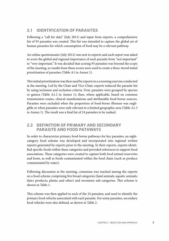
2.1 IDENtIFICAtION OF pARASItES
Following a “call for data” (July 2011) and input from experts, a comprehensive list of 93 parasites was created. This list was intended to capture the global set of human parasites for which consumption of food may be a relevant pathway.
An online questionnaire (July 2012) was sent to experts and each expert was asked to score the global and regional importance of each parasite from “not important” to “very important.” It was decided that scoring 93 parasites was beyond the scope of the meeting, so results from these scores were used to create a three-tiered initial prioritization of parasites (Table A1 in Annex 1).
This initial prioritization was then used by experts in a screening exercise conducted at the meeting. Led by the Chair and Vice-Chair, experts reduced the parasite list by using inclusion and exclusion criteria. First, parasites were grouped by species or genera (Table A1.2 in Annex 1); then, where applicable, based on common transmission routes, clinical manifestations and attributable food-borne sources. Parasites were excluded when the proportion of food-borne illnesses was negli-gible or when parasites were only relevant in a limited geographic area (Table A1.3 in Annex 1). The result was a final list of 24 parasites to be ranked.
2.2 DEFINItION OF pRIMARy AND SECONDARy pARASItE AND FOOD pAtHWAyS
In order to characterize primary food-borne pathways for key parasites, an eight-category food scheme was developed and incorporated into regional written reports generated by experts prior to the meeting. In their reports, experts identi-fied specific foods within these categories and provided references to support food associations. These categories were created to capture both food animal reservoirs and hosts, as well as foods contaminated within the food chain (such as produce contaminated by water).
Following discussion at the meeting, consensus was reached among the experts on a food scheme comprising five broad categories (land animals; aquatic animals; dairy products; plants; and other) and seventeen sub-categories. This scheme is shown in Table 1.
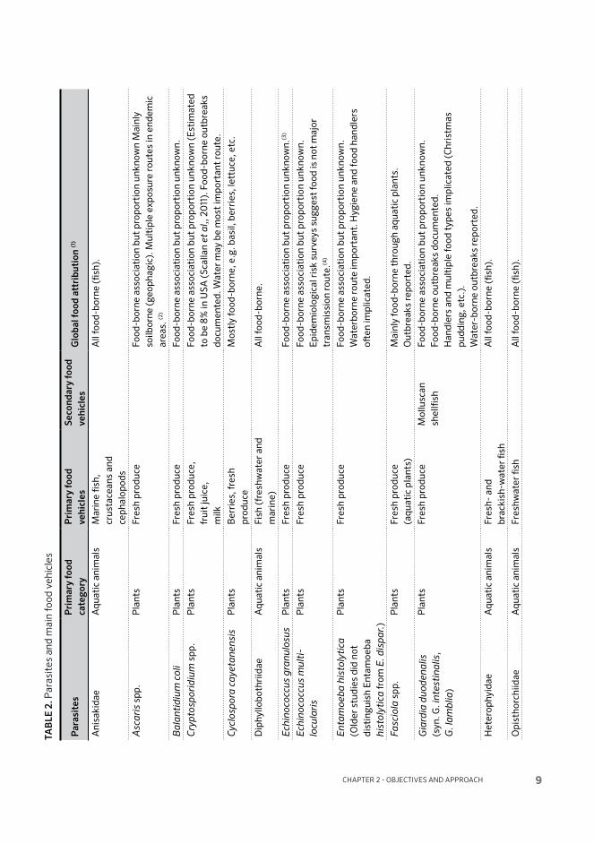
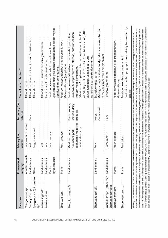
This scheme was then applied to each of the 24 parasites, and used to identify the primary food vehicles associated with each parasite. For some parasites, secondary food vehicles were also defined, as shown in Table 2.

Multi criteria-based ranking for risk ManageMent of food-borne parasites8
TAblE 1. Food category scheme
Food category Food subcategory
Land animals Beef
Pork
Poultry
Small ruminants
Other meat
Game and wild animals
Aquatic animals Marine fish
Freshwater fish
Shellfish
Aquatic mammals
Dairy products Dairy products
Plants Berries
Fruit juices
Other fruit
Leafy greens
Other vegetables
Fresh produce (refers to 2 or more of the above)
Other Other foods
2.3 DEFINItION OF CRItERIA FOR pARASItE SCORING
Based on previous prioritization studies and risk management needs, five cate-gories were considered for the analysis: public health, microbial ecology, animal health, agribusiness and trade, and socio-economic impact. A number of potential criteria in these categories were included in the online questionnaire to appraise the applicability of these criteria and to elicit experts’ judgment on which criteria were more important. This information was used to generate an expansive list of 41 potential criteria in these five categories.
The FAO/WHO Secretariat narrowed the list of potential criteria to 11 and presented these to the experts at the meeting. Following extensive discussions on the list of criteria, consensus was reached on a final list of 9 criteria. Of these criteria, 5 relate to the quantity and severity of global disease, while two others relate to the global distribution of these illnesses and the potential for short-term emergence of increased disease. The remaining two criteria relate to the potential for the parasite (in its primary and secondary foods, defined previously) to affect trade, and the impact of the parasite on economically vulnerable communities.
Chapter 2 - ObjeCtives and apprOaCh 9
TAb
lE 2
. Par
asit
es a
nd m
ain
food
veh
icle
s
Par
asit
es
Pri
mar
y fo
od
cate
gory
Pri
mar
y fo
od
vehi
cles
Seco
ndar
y fo
od
vehi
cles
Glo
bal f
ood
attr
ibut
ion
(1)
Ani
saki
dae
Aqu
atic
ani
mal
sM
arin
e fis
h,
crus
tace
ans
and
ceph
alop
ods
All
food
-bor
ne (fi
sh).
Asc
aris
spp
.P
lant
sFr
esh
prod
uce
Food
-bor
ne a
ssoc
iati
on b
ut p
ropo
rtio
n un
know
n M
ainl
y so
ilbor
ne (g
eoph
agic
). M
ulti
ple
expo
sure
rout
es in
end
emic
ar
eas.
(2)
Bal
anti
diu
m c
oli
Pla
nts
Fres
h pr
oduc
eFo
od-b
orne
ass
ocia
tion
but
pro
port
ion
unkn
own.
Cry
pto
spor
idiu
m s
pp.
Pla
nts
Fres
h pr
oduc
e,fr
uit
juic
e,m
ilk
Food
-bor
ne a
ssoc
iati
on b
ut p
ropo
rtio
n un
know
n (E
stim
ated
to
be
8% in
USA
(Sca
llan
et a
l,, 2
011
). F
ood-
born
e ou
tbre
aks
docu
men
ted.
Wat
er m
ay b
e m
ost
impo
rtan
t ro
ute.
Cyc
losp
ora
caye
tane
nsis
Pla
nts
Ber
ries
, fre
sh
prod
uce
Mos
tly
food
-bor
ne, e
.g. b
asil,
ber
ries
, let
tuce
, etc
.
Dip
hyllo
both
riid
ae
Aqu
atic
ani
mal
sFi
sh (f
resh
wat
er a
nd
mar
ine)
All
food
-bor
ne.
Ech
ino c
occu
s g
ran
ulos
usP
lant
sFr
esh
prod
uce
Food
-bor
ne a
ssoc
iati
on b
ut p
ropo
rtio
n un
know
n.(3
)
Ech
ino c
occu
s m
ulti
lo
cula
ris
Pla
nts
Fres
h pr
oduc
eFo
od-b
orne
ass
ocia
tion
but
pro
port
ion
unkn
own.
Epi
dem
iolo
gica
l ris
k su
rvey
s su
gges
t fo
od is
not
maj
or
tran
smis
sion
rout
e.(4
)
Ent
amoe
ba
his
toly
tica
(O
lder
stu
dies
did
not
di
stin
guis
h E
ntam
oeba
hi
stol
ytic
a fr
om E
. dis
par
.)
Pla
nts
Fres
h pr
oduc
eFo
od-b
orne
ass
ocia
tion
but
pro
port
ion
unkn
own.
W
ater
born
e ro
ute
impo
rtan
t. H
ygie
ne a
nd fo
od h
andl
ers
often
impl
icat
ed.
Fasc
iola
spp
.P
lant
sFr
esh
prod
uce
(aqu
atic
pla
nts)
Mai
nly
food
-bor
ne t
hrou
gh a
quat
ic p
lant
s.
Out
brea
ks re
port
ed.
Gia
rdia
du
oden
alis
(s
yn. G
. int
esti
nal
is,
G. l
amb
lia)
Pla
nts
Fres
h pr
oduc
eM
ollu
scan
sh
ellfi
shFo
od-b
orne
ass
ocia
tion
but
pro
port
ion
unkn
own.
Fo
od-b
orne
out
brea
ks d
ocum
ente
d.
Han
dler
s an
d m
ulti
ple
food
typ
es im
plic
ated
(Chr
istm
as
pudd
ing,
etc
.).
Wat
er-b
orne
out
brea
ks re
port
ed.
Het
erop
hyid
aeA
quat
ic a
nim
als
Fres
h- a
nd
brac
kish
-wat
er fi
shA
ll fo
od-b
orne
(fish
).
Opi
stho
rchi
idae
Aqu
atic
ani
mal
sFr
eshw
ater
fish
All
food
-bor
ne (fi
sh).
Multi criteria-based ranking for risk ManageMent of food-borne parasites10
Par
asit
es
Pri
mar
y fo
od
cate
gory
Pri
mar
y fo
od
vehi
cles
Seco
ndar
y fo
od
vehi
cles
Glo
bal f
ood
attr
ibut
ion
(1)
Par
agon
imus
spp
.A
quat
ic a
nim
als
Fres
hwat
er c
rust
acea
A
ll fo
od-b
orne
.
Sar
cocy
stis
spp
.La
nd a
nim
als
Bee
fP
ork
All
food
-bor
ne fo
r S.
sui
hom
inis
and
S. b
ovih
omin
is
Spar
gano
sis
– S
pir
omet
ra
spp.
Oth
erFr
og, s
nake
mea
tA
ll fo
od-b
orne
Taen
ia s
agin
ata
Land
ani
mal
sB
eef
All
food
-bor
ne. T
aeni
osis
exc
lusi
vely
mea
tbor
ne.
Taen
ia s
oliu
mLa
nd a
nim
als
Por
kTa
enio
sis
excl
usiv
ely
mea
tbor
ne.
Pla
nts
(cys
tice
rcos
is)
Fres
h pr
oduc
eFo
od-b
orne
ass
ocia
tion
but
pro
port
ion
unkn
own.
Cy
stic
erco
sis
mai
nly
soilb
orne
; con
tam
inat
ed p
lant
s m
ay b
e si
gnifi
cant
in s
ome
regi
ons.
Toxo
cara
spp
.P
lant
sFr
esh
prod
uce
Food
-bor
ne a
ssoc
iati
on b
ut p
ropo
rtio
n un
know
n.
Mai
nly
soilb
orne
(geo
phag
y).
Toxo
pla
sma
gon
dii
Land
ani
mal
s M
eat
from
sm
all
rum
inan
ts, p
ork,
be
ef, g
ame
mea
t (r
ed
mea
t an
d or
gans
)
Fres
h pr
oduc
e,
seaf
ood,
dai
ry
prod
ucts
Food
-bor
ne a
ssoc
iati
on (f
resh
pro
duce
) but
pro
port
ion
unkn
own.
Mul
tipl
e ro
utes
of i
nfec
tion
, but
tra
nsm
issi
on
thro
ugh
mea
t is
impo
rtan
t.
(Mea
tbor
ne T
oxop
lasm
a in
fect
ions
est
imat
ed t
o be
22%
in
USA
, Boy
er e
t al
., 20
11; 5
3% in
Chi
le, M
uñoz
et
al.,
2010
; 26
% in
Col
ombi
a, L
ópez
et
al.,
200
5)
Wat
erbo
rne
outb
reak
s do
cum
ente
d.
Tric
hin
ella
sp
iral
isLa
nd a
nim
als
Por
kH
orse
, G
ame
mea
tE
xclu
sive
ly m
eatb
orne
.M
akin
g sa
usag
e or
sim
ilar
food
pro
duct
s in
crea
ses
the
risk
to
the
con
sum
er fr
om a
sin
gle
anim
al.
Tric
hin
ella
spp
. (ot
her t
han
Tric
hin
ella
sp
iral
is)
Land
ani
mal
sG
ame
mea
t (5
)P
ork
Exc
lusi
vely
mea
tbor
ne.
Tric
huri
s tr
ichi
ura
Pla
nts
Fres
h pr
oduc
eFo
od-b
orne
ass
ocia
tion
but
pro
port
ion
unkn
own.
Mai
nly
soilb
orne
.
Tryp
anos
oma
cruz
i P
lant
sFr
uit
juic
esFo
od-b
orne
out
brea
ks d
ocum
ente
d.Fr
uit
juic
e in
lim
ited
geo
grap
hic
area
. Mai
nly
tran
smit
ted
by
inse
cts
Not
es: (
1) T
he in
form
atio
n in
thi
s co
lum
n is
bas
ed o
n pe
er re
view
ed p
ublic
atio
ns in
the
sci
enti
fic li
tera
ture
, unp
ublis
hed
repo
rts
and
expe
rt o
pini
on, w
hich
may
be
the
only
app
roac
h to
est
imat
e fo
od
attr
ibut
ion
for
som
e of
the
par
asit
ic d
isea
ses
on a
glo
bal b
asis
. (2)
Asc
aris
spp
. egg
s ca
n be
com
e ub
iqui
tous
in a
n en
dem
ic a
rea,
mak
ing
attr
ibut
ion
diffi
cult
if n
ot im
poss
ible
. (3)
The
incu
bati
on p
erio
d fo
r
Ech
ino c
occu
s g
ranu
losu
s ca
n be
as
long
as
5 to
15 y
ears
; it
is n
ot p
ossi
ble
to p
reci
sely
iden
tify
an
expo
sure
occ
urri
ng m
any
year
s pr
evio
usly
. How
ever
, the
re a
re m
any
arti
cles
indi
cati
ng t
hat
E. g
ranu
losu
s
eggs
con
tam
inat
e pl
ants
, and
evi
denc
e th
at p
eopl
e in
the
end
emic
, dev
elop
ing
coun
trie
s co
nsum
e ve
geta
bles
, inc
ludi
ng r
aw. I
t is
alm
ost
impo
ssib
le t
o pi
npoi
nt t
he fo
od s
ourc
e be
caus
e th
e tr
ansm
issi
on
rout
es a
re v
arie
d (e
.g. c
onta
ct w
ith
dog,
oth
er c
anid
s (f
ox, w
olf)
, soi
l, et
c.).
(4) T
he in
cuba
tion
per
iod
for
Ech
ino c
occu
s m
ulti
locu
lari
s ca
n be
5–1
5 ye
ars,
and
the
dis
ease
, alv
eola
r ec
hino
cocc
us, i
s di
agno
sed
at a
n ad
vanc
ed s
tage
; it
is n
ot p
ossi
ble
to p
reci
sely
iden
tify
an
expo
sure
occ
urri
ng m
any
year
s pr
evio
usly
. (5)
Wild
boa
r, c
roco
dile
, bea
r, w
alru
s, e
tc.

Chapter 2 - ObjeCtives and apprOaCh 11
The final criteria selected for scoring were: (1) Number of global food-borne illnesses (manifesting disease); (2) Global distribution (number of regions); (3) Acute morbidity severity (disability weight); (4) Chronic morbidity severity (disability weight); (5) Fraction of illness that is chronic (%); (6) Case-fatality ratio (%); (7) Likelihood of increased human burden (%); (8) How relevant is this para-site-food pathway for international trade?; and (9) What is the scope of the impact on economically vulnerable communities?
For each of these 9 criteria, between three and five scoring levels were defined. For 7 criteria, these scoring levels were defined quantitatively, while the remaining two were qualitative. Scoring levels were intended to allow for appropriate differ-entiation among the 24 parasites. These criteria, along with a question pertaining to data quality, are shown in Annex 2. Note that question 8, on international trade concerns, relates specifically to the pathogen in its primary food vehicle, whereas all other questions refer to the parasite in general.
2.4 SCORING pARASItES ACCORDING tO CRItERIA
Experts were divided into five groups of 4 to 5 people, organized so that each group had, to the extent possible, coverage across regions and expertise. Each group was given three documents: a summary card form for each parasite (see Annex 2), a document explaining each criterion and how to score it (Annex 3), and a list of parasites. The lists of parasites provided to each group were staggered in order to maintain equal numbers of scores across parasites, because all groups were unlikely to complete summary cards for all 24 parasites.
Each group used available material, such as regional written reports, published literature and WHO material on disability weights, coupled with online searches, to facilitate a discussion of each criterion for each parasite. Each group scored a summary card for each parasite on their list. Preliminary criteria scores were tabulated into spreadsheets for each group, and preliminary scores were presented back to the group. Discussions around large disparities in preliminary scores allowed the group to identify some differences in interpreting criteria. Once the expert panel reached consensus and greater clarity and agreement on criteria defi-nitions, groups re-convened to review their scores. Following a second tabulation of preliminary results and similar discussion on criteria definitions, a third round of scoring was conducted to obtain final group parasite criteria scores.
Ultimately, two groups scored all 24 parasites and the remaining groups scored 21, 18 and 14 parasites respectively. Thus, 11 parasites had 5 sets of criteria scores, 7 parasites had 4 sets of scores, and 6 parasites had 3 sets of scores.
Multi criteria-based ranking for risk ManageMent of food-borne parasites12
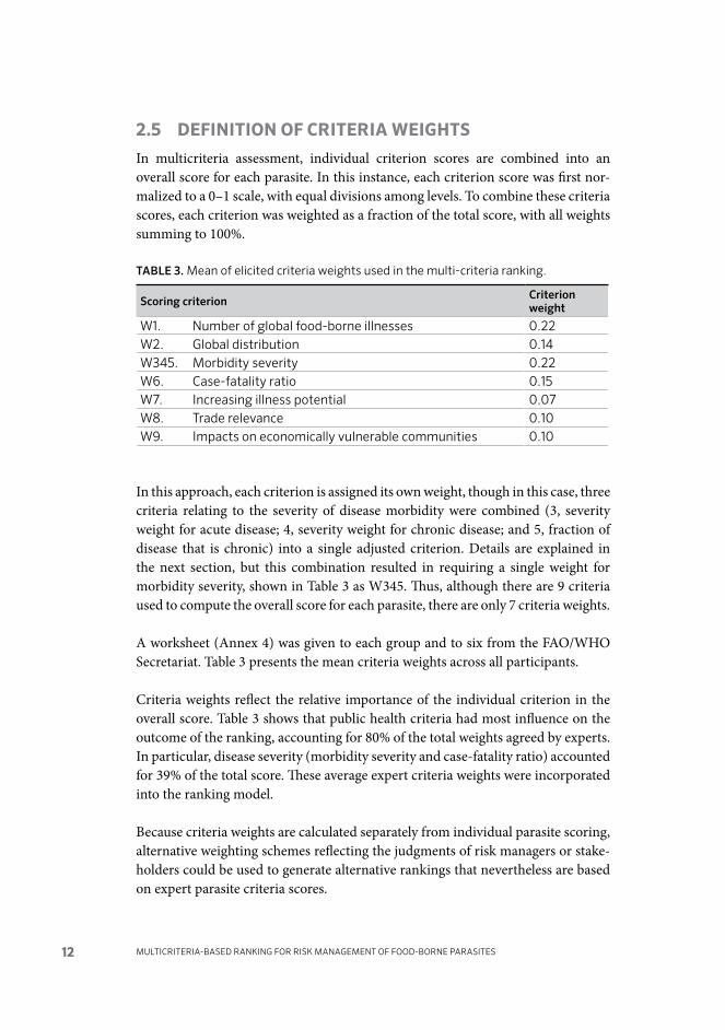
2.5 DEFINItION OF CRItERIA WEIGHtS
In multi criteria assessment, individual criterion scores are combined into an overall score for each parasite. In this instance, each criterion score was first nor-malized to a 0–1 scale, with equal divisions among levels. To combine these criteria scores, each criterion was weighted as a fraction of the total score, with all weights summing to 100%.
TAblE 3. Mean of elicited criteria weights used in the multi-criteria ranking.
Scoring criterion Criterion weight
W1. Number of global food-borne illnesses 0.22W2. Global distribution 0.14W345. Morbidity severity 0.22W6. Case-fatality ratio 0.15W7. Increasing illness potential 0.07W8. Trade relevance 0.10W9. Impacts on economically vulnerable communities 0.10
In this approach, each criterion is assigned its own weight, though in this case, three criteria relating to the severity of disease morbidity were combined (3, severity weight for acute disease; 4, severity weight for chronic disease; and 5, fraction of disease that is chronic) into a single adjusted criterion. Details are explained in the next section, but this combination resulted in requiring a single weight for morbidity severity, shown in Table 3 as W345. Thus, although there are 9 criteria used to compute the overall score for each parasite, there are only 7 criteria weights.
A worksheet (Annex 4) was given to each group and to six from the FAO/WHO Secretariat. Table 3 presents the mean criteria weights across all participants.
Criteria weights reflect the relative importance of the individual criterion in the overall score. Table 3 shows that public health criteria had most influence on the outcome of the ranking, accounting for 80% of the total weights agreed by experts. In particular, disease severity (morbidity severity and case-fatality ratio) accounted for 39% of the total score. These average expert criteria weights were incorporated into the ranking model.
Because criteria weights are calculated separately from individual parasite scoring, alternative weighting schemes reflecting the judgments of risk managers or stake-holders could be used to generate alternative rankings that nevertheless are based on expert parasite criteria scores.
Chapter 2 - ObjeCtives and apprOaCh 13
2.6 CAlCulAtION OF pARASItE SCORES
The overall score for each parasite is given by the following equation:
Score = C1*W1+C2*W2+{C3*(1-C5)+C4*C5}*W345+C6*W6+C7*W7+C8*W8+C9*W9
where C are parasite-specific normalized criteria scores and W are constant criteria weights that are the same for all parasites. Criteria 3, 4 and 5 are combined to produce a single morbidity criteria; it is essentially the weighted average of acute and chronic disease severity. Thus, criteria 3, 4 and 5 have one associated weight, denoted in the equation as W345. Otherwise the calculation is straightforward: normalized parasite criteria scores are multiplied by fractional weights, and summed. Overall scores therefore range from 0 to 1.
A spreadsheet model was developed to calculate overall scores for each parasite based on all group summary cards and averaged criteria weights. The resulting scores were then ranked to produce the current list of global food-borne parasites.

Multi criteria-based ranking for risk ManageMent of food-borne parasites14
3Results
3.1 tHE GlOBAl RANkING OF FOOD-BORNE pARASItES
The results of the ranking exercise, where the top ranking parasites are arranged on the x-axis from left to right in decreasing rank order and the average weights (in percentage) on the y-axis, are presented in Figure 2. This figure was obtained from the average of all elicited weights for the criteria. Among the top ranked parasites are those that have already been singled out by WHO as neglected tropical diseases (NTD), and identified by FERG as priorities for further burden of illness studies.
As noted in Chapter 2, this ranking is a combination of scoring the parasites based on predefined criteria and weighting the criteria based on the importance assigned to them by the expert meeting participants. Since many of the criteria were public health related, there were not big differences between the final ranking and the outcome of the scoring exercise alone, where all criteria are consid-ered to have equal weight. Sensitivity analysis was carried out using alternative criteria weighting schemes (see Annex 5). Figure A5.3 in Annex 5 compares the ranks for global foodborne parasites scored across alternative criteria weighting schemes. Figure A5.5 in Annex 5 presents the scores for the public health criteria only, weighted equally, compared with baseline ranking based on all criteria and elicited weights. These figures are included for reference and indicate that the top 4 parasites remain the same based on expert scoring. It is also interesting to note that the gradually declining trend along the x-axis from left to right remains generally the same. Therefore the weighting of criteria did not radically change the ranks,

15Chapter 3 - results 15
and the public health criteria alone were not so different from the expert ranking. This also reflects the dominance of public health-related criteria in the ranking tool.
A short overview of the top 8 parasites in the above ranking is provided below. Further information relevant to the management of these parasites is provided in Chapter 4. As risk managers consider individual parasites, there will be a need to go into more depth for each. Specific information on the 24 ranked parasites was generated after the meeting and can be found in Annex 7.
Taenia soliumTaenia solium (ranked 1st in Figure 2) is estimated to infect millions of persons worldwide. It is unique in that the larval or cysticercus stage can infect humans
0.00 10.00 20.00 30.00 40.00 50.00 60.00 70.00 80.00
Spirometra spp.
Diphyllobothriidae
Heterophyidae
Sarcocystis spp.
Toxocara spp.
Taenia saginata
Balantidium coli
Anisakidae
Trichinella spp.*
Trichuris trichiura
Paragonimus spp.
Cyclospora cayetanensis
Fasciola spp.
Giardia duodenalis
Trypanosoma cruzi
Ascaris spp.
Opisthorchiidae
Trichinella spiralis
Entamoeba histolytica
Cryptosporidium spp.
Toxoplasma gondii
Echinococcus multilocularis
Echinococcus granulosus
Taenia solium
Normalized overall score
FIGuRE 2. Global ranking of food-borne parasites using a multi criteria ranking tool for scoring parasites, with weighting of scoring criteria based on criteria scores and weights elicited from expert meeting participants (Note: Trichinella spp.* includes Trichinella species except T. spiralis).
Multi criteria-based ranking for risk ManageMent of food-borne parasites16
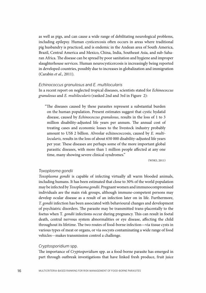
as well as pigs, and can cause a wide range of debilitating neurological problems, including epilepsy. Human cysticercosis often occurs in areas where traditional pig husbandry is practiced, and is endemic in the Andean area of South America, Brazil, Central America and Mexico, China, India, Southeast Asia, and sub-Saha-ran Africa. The disease can be spread by poor sanitation and hygiene and improper slaughterhouse services. Human neurocysticercosis is increasingly being reported in developed countries, possibly due to increases in globalization and immigration (Carabin et al., 2011).
Echino coccus granulosus and E. multilocularis In a recent report on neglected tropical diseases, scientists stated for Echino coccus granulosus and E. multilocularis (ranked 2nd and 3rd in Figure 2):
“The diseases caused by these parasites represent a substantial burden on the human population. Present estimates suggest that cystic hydatid disease, caused by Echino coccus granulosus, results in the loss of 1 to 3 million disability-adjusted life years per annum. The annual cost of treating cases and economic losses to the livestock industry probably amount to US$ 2 billion. Alveolar echinococcosis, caused by E. multilocularis, results in the loss of about 650 000 disability-adjusted life years per year. These diseases are perhaps some of the more important global parasitic diseases, with more than 1 million people affected at any one time, many showing severe clinical syndromes.”
(WHO, 2011)
Toxoplasma gondiiToxoplasma gondii is capable of infecting virtually all warm blooded animals, including humans. It has been estimated that close to 30% of the world population may be infected by Toxoplasma gondii. Pregnant women and immuno compromised individuals are the main risk groups, although immune-competent persons may develop ocular disease as a result of an infection later on in life. Furthermore, T. gondii infection has been associated with behavioural changes and development of psychiatric disorders. The parasite may be transmitted trans-placentally to the foetus when T. gondii infections occur during pregnancy. This can result in foetal death, central nervous system abnormalities or eye disease, affecting the child throughout its lifetime. The two routes of food-borne infection—via tissue cysts in various types of meat or organs, or via oocysts contaminating a wide range of food vehicles—makes transmission control a challenge.
Crypto sporidium spp.The importance of Crypto sporidium spp. as a food-borne parasite has emerged in part through outbreak investigations that have linked fresh produce, fruit juice

17Chapter 3 - results 17
and dairy products with disease. In the USA, it is estimated that 8% of the annual food-borne disease burden may be attributed to this parasite. For most people, symptomatic cryptosporidiosis is characterized by acute watery diarrhoea, often accompanied by abdominal pain, nausea or vomiting, low grade fever, headache and general malaise. Most patients recover within 2–3 weeks, but highly immuno-compromised patients may suffer chronic illness, also leading to severe disease and sometimes death. For most parasitic infections there is some treatment available, but for Crypto sporidium spp. infections in the immuno compromised, there is none. There is also increasing evidence that cryptosporidiosis may have long-term effects, such as chronic gastro intestinal conditions. In addition, it is noted that cryptosporidium oocysts are very resistant to chlorine commonly used to treat water.
Entamoeba histolyticaEntamoeba histolytica, as with Crypto sporidium spp., is probably primarily trans-mitted through food handlers and contaminated water, which can enter the food chain causing illnesses attributed to fresh produce; it should be noted that, unlike some Crypto sporidium spp., E. histolytica is not zoonotic. Amoebiasis is tradition-ally limited to dysenteric-like symptoms, with abdominal pain, bloody or mucoid diarrhoea, and tenesmus, but has the ability to invade extra-intestinal tissues also, e.g. inducing liver abscesses, and extra-hepatic spread of E. histolytica is associated with relatively high mortality (20–75%). One of the problems with its detection is that microscopy methods used for E. histolytica do not differentiate it from non-pathogenic species. This parasitic disease is of importance globally, but occurs predominately in developing countries and may be transmitted with immigrant populations to developed areas. Unlike Crypto sporidium spp., E. histolytica is sus-ceptible to chlorine.
Trichinella spiralisTrichinella spiralis, like all Trichinella species, has a unique lifecycle in that there is no environmental transmission stage – thus all cases are due to ingestion of meat containing the encysted larvae; meat types typically associated with T. spiralis include pork, horse meat, and game. Globally, there were 65 818 human infec-tions reported between 1986 and 2009, with most of these reported for hospital-ized patients in Romania, where 42 patient deaths were reported. However, there may be increased exposure through human behavioural trends, e.g. consumption of raw horse meat, dog meat, wild boar, and other sylvatic animal meats, as well as practices of free-range animal husbandry (infected animals are asymptomatic).
OpisthorchiidaeThe Opisthorchiidae family includes various digenean parasites, of which the most medically important are Clonorchis sinensis, Opisthorchis viverrini and Opisthorchis

Multi criteria-based ranking for risk ManageMent of food-borne parasites18
felineus. All are transmitted to humans via ingestion of the encysted metacercaria in the flesh or skin of freshwater fish. Opisthorchiasis/clonorchiasis occurs autoch-thonously in southeast Asia, eastern Europe, and central Asia. FERG reported over 8 million infections globally in 2005, almost all of which occurred in southeast Asia, where over 300 000 people were heavily infected and 1323 died. Disability-adjusted life years was 74 367. The FERG report further states that awareness of this food-borne problem is limited; only Japan and South Korea have established successful control programmes for fish-borne trematodiases. Opisthorchiasis is particularly worrisome in its potential to be carcinogenic; case-control studies have suggested that a substantial proportion of chol angio carcinoma in some Asian countries can be due to infection with O. viverrini.
SummaryThe fact that this is a global ranking may mean that some diseases that are severe and often fatal, but limited to a particular region, are not highly ranked. One example is Chagas disease, transmission of which is at present largely restricted to parts of Central and South America, with FERG reporting over 11 000 deaths due to Trypanosoma cruzi worldwide in 2004. However, survival of the trypomasti-gotes in fruits and juices might present an unknown risk for global dissemination in the world market.
The parasites currently being considered by the CCFH were ranked seventh (T. spiralis) and nineteenth (T. saginata/C. bovis) for overall importance by the experts.
3.2 tRADE SCORES FOR tHE RANkED pARASItES
The data used to rank parasites and generate Figure 2 are used to produce Figure 3, in which only the average trade criteria scores for each parasite are displayed. This figure suggests that there may be additional or separate trade issues that could be considered by risk managers such as Codex and national food authorities.
The parasites currently contemplated by the CCFH, T. spiralis and T saginata/C. bovis, were considered among the most important for trade, based on criteria scores. In the regional reports, Trichinella spiralis, Taenia saginata, Taenia solium and/or Echino coccus granulosus were mentioned as current or potential trade concerns in the African, Australian, European, Near Eastern and South American Regions. The North American and Asian Regions did not address this issue directly.
It may be of interest to risk managers that the Anisakidae that ranked lower (17th) in overall importance, scored higher for the trade criteria, and were mentioned in
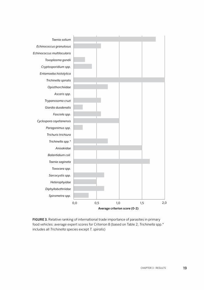
19Chapter 3 - results 19
0,0 0,5 1,0 1,5 2,0
Average criterion score (0-2)
Spirometra spp.
Diphyllobothriidae
Heterophyidae
Sarcocystis spp.
Toxocara spp.
Taenia saginata
Balantidium coli
Anisakidae
Trichinella spp.*
Trichuris trichiura
Paragonimus spp.
Cyclospora cayetanensis
Fasciola spp.
Giardia duodenalis
Trypanosoma cruzi
Ascaris spp.
Opisthorchiidae
Trichinella spiralis
Entamoeba histolytica
Cryptosporidium spp.
Toxoplasma gondii
Echinococcus multilocularis
Echinococcus granulosus
Taenia solium
FIGuRE 3. Relative ranking of international trade importance of parasites in primary food vehicles: average expert scores for Criterion 8 (based on Table 2; Trichinella spp.* includes all Trichinella species except T. spiralis)

Multi criteria-based ranking for risk ManageMent of food-borne parasites20
several country reports as a class of organisms important to the country. These are probably countries that trade or consume fish extensively.
Conversely, parasites of concern in the overall ranking may not rank high as a trade concern. An example is Toxoplasma gondii, which might be prevalent in meat products but is microscopic and does not affect the appearance of the products, and there is no rapid, inexpensive, accurate test available. Therefore, for trade purposes, it would be ranked lower than the easily visible and detectable parasites.
3.3 SOCIO-ECONOMIC IMpACtS FOR tHE RANkED pARASItES
The data analysed to rank parasites and generate Figure 2 are also used to produce Figure 4, which presents average scores for the socio-economic impact criterion. The figure indicates that there may be additional or separate socio-economic concerns not addressed in the overall ranking or in trade issues. An example of this is Cyclospora cayetanensis, which may require further investigation. It is probable that this reflects the known and on-going, socio-economic impacts on Guatema-lan berry farmers, following the relatively extensive outbreaks of cyclosporiasis in North America during the 1990s. Outbreaks were primarily associated with berries imported from Guatemala.
Diseases caused by Taenia solium (ranked 1st) and Echino coccus granulosus and E. multi locularis (ranked 3rd and 4th, respectively) contribute to economic losses in human and animal populations in many parts of the world. They are considered preventable diseases that can be controlled or eliminated and should be prioritized (Carabin et al., 2005). Stigmatization and social isolation, attached to the occur-rence of epilepsy caused by neurocysticercosis (T. solium infection), are examples of societal impact presented in the African Regional report, that are difficult to quantify but add to the socio-economic burden of this disease.
The parasites currently contemplated by the CCFH, T. spiralis and T. saginata/C. bovis were not considered important in terms of the socio-economic criterion.
3.4 CONCluSIONS
The ranking exercise has provided a picture of the food-borne parasites of global importance today and has created a seemingly useful tool that is transparent and reproducible. The tool can be used with emphasis on different criteria and with or without weight factors. It is imperative that future use of this ranking tool and

21Chapter 3 - results 21
0,0 0,5 1,0 1,5 2,0 2,5 3,0
Average criterion score (0-3)
Spirometra spp.
Diphyllobothriidae
Heterophyidae
Sarcocystis spp.
Toxocara spp.
Taenia saginata
Balantidium coli
Anisakidae
Trichinella spp.*
Trichuris trichiura
Paragonimus spp.
Cyclospora cayetanensis
Fasciola spp.
Giardia duodenalis
Trypanosoma cruzi
Ascaris spp.
Opisthorchiidae
Trichinella spiralis
Entamoeba histolytica
Cryptosporidium spp.
Toxoplasma gondii
Echinococcus multilocularis
Echinococcus granulosus
Taenia solium
FIGuRE 4. Relative ranking of socio-economic impacts of parasites to vulnerable communities: average expert scores for Criterion 9 (based on Table 2; Trichinella spp.* includes all Trichinella species except T. spiralis)

Multi criteria-based ranking for risk ManageMent of food-borne parasites22
strategy be undertaken in a transparent manner. By using this approach, the results can be compared when the procedure is repeated.
The experts ranked the most important parasites by using multi criteria analysis during the meeting. The results shown in Figure 2 indicate that the method clearly defines those parasites that are highly ranked and those considered of lower rank. While Taenia solium clearly came out on top, there were less marked differences between the parasites that ranked second, third and fourth. Similarly those that ranked fifth, sixth and seventh are very close together, suggesting that the indi-vidual ranking is less important than the overall picture that the ranking provides in terms of food-borne parasites. As noted in the explanation of the weighting of the criteria, public health importance was the primary driver of ranking, with almost equal importance being given to illness and severity. This importance given to severity will have contributed to the high ranking of Echino coccus granulosus, ranked second, followed by E. multilocularis.
Toxoplasma gondii ranked fourth. The predominant disease burden of this parasite is confined mainly to substantial risks in pregnancy to the unborn, and in immuno compromised people (e.g. HIV/AIDS, transplantation patients). However, acquired toxoplasmosis also may contribute also an additional, substan-tial disease burden; many uncertainties still exist. The ranking order is affected by data availability; in the absence of data, or when data is limited, it is more difficult to categorize a parasite×food commodity. New data may influence ranking order. For example, the increasing number of papers linking toxoplasmosis with chronic illness (Havelaar et al., 2012), including mental illness (Henriquez et al., 2009) may push this parasite further up the ranking in the near future. Therefore, the parasite ranking list developed here should not be considered to be absolute or static; in order to remain current and fit for purpose, it must be updated periodically. The tool can be used also for prioritizing regional and national agendas for policy or research activities. There may be more specific data at national or regional level, as well as differing judgments on the importance of the various criteria, which could lead to a different ranking at a local level.
23CHAPTER 4 - RISk MANAGEMENT OPTIONS FOR THE HIGHER RANkED PARASITE
4Risk management options for the higher ranked parasites
The identification of ranked parasites in Figure 2 is based not only on scientific evidence where available (including both published and unpublished data), but also on expert experience and opinion, and is weighted primarily by the public health concerns of the experts. The ranking of parasites by overall importance is the primary input to the risk managers in CCFH, who will then consider other issues relevant to management priorities and actions.
The ranking approach used in the expert meeting can be applied at the national level, where scoring may change, based on data availability and where weights may be placed on different criteria, based on the national situation or risk management issue.
Risk managers need to ensure that aspects other than the initial ranking by the experts that need to be considered in the decision-making process should also be evidence-based where possible, and done in a transparent manner. This section outlines some of these other considerations.
4.1 GENERAl RISk MANAGEMENt CONSIDERAtIONS
It is important to recognize at all levels—global, regional and local—that there is a significant lack of information regarding food attribution for many parasitic diseases (Table 2). This is especially true for parasitic infections in which there may

Multi criteria-based ranking for risk ManageMent of food-borne parasites24
be a prolonged period (possibly many years) before symptoms appear (e.g. Echinococcus spp.) or those producing a chronic progression of disease (Ascaris spp., Trypanosoma cruzi and Trichuris trichiura). Food may be an important vehicle of transmission, but these parasites are not considered to be exclusively food-borne. For example, food may not be the primary transmission vehicle for Echinococcus spp.; however, the experts still considered these parasites as potential food-borne risks and advocate that further evidence be gathered to close this knowledge gap. Echino coccus granulosus and E. multi locularis ranked 2nd and 3rd, respectively, based largely on the potential severity of their associated diseases.
4.2 GENERIC RISk MANAGEMENt OptIONS
As with other food-borne biological hazards, there are some generic good practices that are relevant for the control of food-borne parasites but are not necessarily unique to parasites. The importance of such practices may therefore already be captured in various existing risk-management documents. However, the recogni-tion of parasites as being somewhat neglected warrants mention of any relevant control measures and management options.
4.2.1 Primary production and pre-harvestWhile many of the parasites of concern are meat or fish-borne, for many others the entry into the food chain is via water or soil, or both. For example, Ascaris, Crypto sporidium, Cyclospora, Echinococcus and Giardia are essentially transmit-ted through the faecal-oral route, but may be transmitted by contaminated water during primary production of foods such as fresh produce. Thus, the primary pro-duction and pre-harvest stage of the food chain are critical in terms of control of numerous parasites, and it was considered that parasites may not be adequately considered in Good Agriculture Practices (GAPs). Some important considerations are highlighted here.
Parasites transmitted by the faecal-oral route Given the importance of the faecal-oral route of transmission for some parasites, areas for cultivation of fresh produce, particularly for raw consumption, need to be assessed in terms of their susceptibility to faecal contamination, whether from run-off from wild animals, farm animals, domestic animals or and humans, and the necessary measures taken to manage the identified risk.
The importance of on-farm sanitation and hygiene in interrupting the life cycle of parasites and minimizing the opportunity for the faecal-oral route of transmission needs to be recognized, with appropriate installation and use of the relevant facilities promoted, e.g. functional on-farm latrines, and adequate hand-washing facilities.

Chapter 4 - risk management options for the higher ranked parasite 25
The use of organic fertilizer, particularly on produce, should be monitored closely in order to ensure that it is composted adequately to destroy parasite transmission stages prior to use. However, it should be noted that the effectiveness of compost-ing in destroying or inactivating parasites is uncertain, and should be considered a knowledge gap.
Zoonotic parasitesFor those parasites with an indirect life cycle, special consideration must be given to breaking the cycle at the level of the intermediate host, such as snail (intermedi-ate host) control in the case of trematode parasites in aquaculture.
The role of dogs and cats (domestic or feral) in transmission of certain parasites needs to be highlighted and farmers and other relevant stakeholders educated on good practices, e.g. no feeding of raw or untreated carcasses or offal of livestock and fish to domestic dogs and cats, or allowing wild canids and felids access to dead livestock, aborted foetuses, etc., and fish products; population control of semi-domesticated, stray or feral dogs and cats in close vicinity to the farm or aquaculture ponds.
Mass treatment of reservoir hosts, such as livestock, at frequent intervals in a sustainable fashion should ensure reduction in environmental contamination of infective stages. This applies to dogs in the case of echinococcosis by Echino coccus granulosus.
Water is an important vehicle for transmission for a number of food-borne parasites. Thus attention to water quality throughout the food-chain, from primary production through processing to consumption is very important.
Although not specific to primary production, monitoring and surveillance were considered to be important tools in the control of parasites, and for complete ef-fectiveness may need to begin at the pre-harvest stage. For example, the ability to trace back infected animals at the abattoir level will allow identification of ‘high risk’ animals or fish populations or regions, and help allocation and targeting of resources for control. Furthermore, the ability to trace back fresh produce to the country, and even farm, of origin will allow identification of ‘high risk’ regions for subsequent risk management decisions. Monitoring and surveillance programmes can identify potentially emerging trends and risks for regional incursion (displaced forest animals or hosts in expanding urban environments).
4.2.2 Post-harvestWhile post-harvest opportunities for control will be very dependent on the commodity of concern, it was considered that current Good Hygiene Practice (GHP), and HACCP plans for processing, etc., might not address parasitic hazards adequately.
Multi criteria-based ranking for risk ManageMent of food-borne parasites26
In terms of processing, many parasite stages in meat and fish are susceptible to freezing as a process step and to controlled cooking at the process and consumer levels. However, the time×temperature combinations can be important, and in the cases of some parasites, such as E. multilocularis eggs, lower temperature domestic freezing may not be adequate. Irradiation can be an effective control measure and guidelines are available for its use in the control of Toxoplasma and Trichinella. Other control measures such as curing, salting, drying and high pressure process-ing need evaluation for specific parasites and food commodity contexts. Vacuum packing and chilling do not alter the viability of parasites in meat (e.g. Toxoplasma tissue cysts in meat).
4.2.3 EducationEducation and awareness raising was identified as an important component of food-borne parasite control, and in some cases may be the only feasible option available. Education should be directed to actors throughout the food chain from farm and abattoir workers to food handlers (consumers and food retail outlets), and should address the gamut from good animal husbandry practices to hygiene and sanitation measures. In terms of consumer education there may also be a need to address specific high risk population groups. For consumers, especially those who are pregnant or immuno compromised (e.g. individuals with HIV/AIDS), advice on the preparation and consumption of high risk foods such as fresh produce and tubers, carrots etc., adequate cooking of meat and fish prior to consumption and the importance of hygiene, e.g. hand-washing, is critical.
4.3 SOME SpECIFIC CONSIDERAtIONS FOR RISk MANAGEMENt
During the meeting specific consideration was given to the management of the eight top ranked parasites, and some of the important aspects for consideration by risk managers in deciding how to address these parasites. These considerations for Taenia solium, Echino coccus granulosus, E. multi locularis, Toxoplasma gondii, Crypto sporidium spp., Entamoeba histolytica, Trichinella spiralis and the Opisthor-chiidae family are summarized in Table 4. Where they were identified, details of existing risk management texts or guidelines are provided. It should be noted that providing more specific input on the top eight ranked parasites was a function of the time available at the expert meeting rather than any technical consideration.
In addition, Table 5 provides some information on the global trade in those com-modities identified as primary vehicles for the ranked parasites, thus providing an overview of their importance.

Chapter 4 - risk management options for the higher ranked parasite 27
TAb
lE 4
. Som
e sp
ecifi
c ri
sk m
anag
emen
t co
nsid
erat
ions
rela
ting
to
the
top
eigh
t ra
nked
par
asit
es
Par
asit
e
Hos
ts a
nd m
ain
tran
s mis
sion
rout
es
– Fo
od c
hain
s of
co
ncer
n
Seve
rity
of
illne
ss
Ove
r arc
hing
fa
ctor
s fo
r co
nsid
er at
ion
in
risk
man
age m
ent
Exam
ples
of m
anag
emen
t opt
ions
and
cha
lleng
es a
long
the
food
ch
ain
Exam
ples
of r
isk
man
age m
ent
text
s an
d gu
idan
ceO
n-fa
rmP
ost-
harv
est o
r pr
oces
sing
Ret
ail a
nd
cons
umer
Taen
ia s
oliu
mTw
o tr
ansm
issi
on
rout
es:
Und
erco
oked
por
k –
adul
t ta
pew
orm
in
fect
ion
(tae
nios
is)
T. s
oliu
m e
ggs
– en
viro
nmen
t (e
.g.
via
fres
h pr
oduc
e) –
la
rval
sta
ge in
fect
ion
(cys
tice
rcos
is)
Taen
iosi
s –
a re
lati
vely
be
nign
dis
ease
. Cy
stic
erco
sis
a se
vere
, po
tent
ially
fata
l, in
fect
ion
Cla
ssifi
ed a
s er
adic
able
but
ex
pen s
ive
and
chal
leng
ing
to
cont
rol.
H
uman
hos
t m
eans
beh
avio
ur
impo
rtan
t in
tr
ansm
issi
on
Con
trol
str
at eg
ies
linke
d to
whe
ther
a
geog
raph
ic a
rea
is c
ateg
oriz
ed
high
or
low
ris
k
Vac
cine
use
and
ch
emo-
ther
apeu
tic
cont
rol i
n pi
gs.
Goo
d pi
g hu
sban
dry
prac
tice
s cr
itic
al fo
r su
stai
nabl
e co
ntro
l. Sa
fe a
ppli c
atio
n of
m
anur
e to
con
trol
en
viro
n men
tal
cont
amin
atio
n.
Mai
ntai
n hi
gh w
ater
qu
alit
y.
Safe
sla
ught
er
prac
tice
s an
d an
eff
ecti
ve in
spec
tion
sy
stem
. Fo
od h
andl
er
educ
atio
n to
tar
get
pers
onal
hyg
iene
.
Con
sum
er
educ
atio
n on
the
ro
le o
f hum
ans
in
tran
smis
sion
. C
onsu
mpt
ion
of r
aw
prod
uce
in e
ndem
ic
area
s is
hig
h ri
sk.
Coo
king
is e
ffec
tive
.
FAO
/WH
O/O
IE
200
5.
Ech
ino c
occu
s g
ranu
losu
sFr
esh
prod
uce
Shee
p, c
attl
e,
goat
s an
d pi
gs a
re
inte
rmed
iate
hos
ts
Seve
re c
linic
al
synd
rom
e,
cyst
ic h
ydat
id
dise
ase
Not
com
pli c
ated
to
con
trol
bu
t re
quir
es
coor
dina
tion
am
ong
rele
vant
au
thor
itie
s.
Eff
ecti
ve
trea
tmen
t of
do
gs n
eeds
co
nfine
men
t an
d in
cine
r ati
on o
f fa
eces
for
2 da
ys
post
tre
atm
ent,
th
en re
peat
in 4
5 da
ys.
Elim
inat
ion
of t
he
para
site
in d
ogs.
E
duca
tion
of f
arm
ers
to m
inim
ize
on-f
arm
co
ntam
in at
ion
and
infe
ctio
n of
ani
mal
s.
Con
trol
of w
ater
use
d,
incl
udin
g fr
uit
and
vege
tabl
e pr
oduc
tion
. Sh
eep
and/
or
dog
vacc
inat
ion
may
be
a fu
ture
opt
ion.
Eff
ecti
ve in
spec
tion
sy
stem
to
ensu
re
GH
P a
nd G
AP.
A
bilit
y to
tra
ce b
ack
to fa
rm.
Kee
ping
dog
s aw
ay
from
pot
enti
ally
in
fect
ed o
ffal
and
fr
om n
ear
abat
toir
s.
Ech
inoc
occu
s eg
gs
are
not
susc
epti
ble
to fr
eezi
ng
(exc
ept
whe
n co
re
tem
p era
ture
of
food
is m
inus
80
°C
for
48
hrs
or m
inus
70
°C fo
r 4
day
s).
Edu
cati
on o
f fo
od h
andl
ers
and
cons
umer
s re
gard
ing
food
pr
epar
atio
n an
d pe
rson
al h
ygie
ne.
Ech
inoc
occu
s eg
gs
are
not
susc
epti
ble
to fr
eezi
ng
(exc
ept
whe
n co
re
tem
p era
ture
of
food
is m
inus
80
°C
for
48
hrs
or m
inus
70
°C fo
r 4
day
s).
WH
O/O
IE,
200
1.O
IE, 2
00
5a.
WH
O, 2
011
.

Multi criteria-based ranking for risk ManageMent of food-borne parasites28
Par
asit
e
Hos
ts a
nd m
ain
tran
s mis
sion
rout
es
– Fo
od c
hain
s of
co
ncer
n
Seve
rity
of
illne
ss
Ove
r arc
hing
fa
ctor
s fo
r co
nsid
er at
ion
in
risk
man
age m
ent
Exam
ples
of m
anag
emen
t opt
ions
and
cha
lleng
es a
long
the
food
ch
ain
Exam
ples
of r
isk
man
age m
ent
text
s an
d gu
idan
ceO
n-fa
rmP
ost-
harv
est o
r pr
oces
sing
Ret
ail a
nd
cons
umer
Ech
ino c
occu
s m
ulti
lo
cula
ris
Fres
h pr
oduc
e.
New
tre
nd m
ay
be m
igra
tion
of
the
para
site
s w
ith
sylv
atic
incu
rsio
n in
to re
side
ntia
l ar
eas.
Seve
re c
linic
al
synd
rom
e,
alve
olar
ech
ino-
cocc
osis
Far
mor
e ch
alle
ngin
g to
co
ntro
l tha
n E
. gra
nu lo
sus
give
n th
e pr
e dom
in an
ce o
f a
sylv
atic
cyc
le
invo
lvin
g fo
xes
and
rode
nts.
Diffi
cult
to
cont
rol i
n w
ildlif
e.
Ant
helm
in th
ic
impr
eg na
ted
bait
for
foxe
s in
per
i-ur
ban
area
s or
aro
und
farm
s –
may
be
diffi
cult
to
sus
tain
and
als
o ex
pens
ive.
Ech
inoc
occu
s eg
gs
are
not
susc
epti
ble
to fr
eezi
ng
Edu
cati
on o
f foo
d ha
ndle
rs a
nd
cons
umer
s m
ay b
e th
e m
ost
feas
ible
fo
rm o
f con
trol
in
ende
mic
are
as.
Was
hing
pro
duce
no
t be
suffi
cien
t to
re
mov
e pa
rasi
tes.
E
chin
ococ
cus
eggs
no
t su
scep
tibl
e to
free
zing
(see
ab
ove)
.
WH
O/O
IE,
200
1.
OIE
, 20
05a
. W
HO
, 20
11.
Toxo
pla
sma
gon
dii
Mea
t an
d off
al fr
om
a ra
nge
of a
nim
als
(pig
s, c
attl
e, s
heep
, go
ats,
gam
e) m
ay
cont
ain
infe
ctio
us
tach
yzoi
tes.
O
ocys
ts c
an
cont
amin
ate
fres
h pr
oduc
e an
d m
ollu
scan
she
llfish
. O
ocys
ts c
an b
e a
sour
ce o
f on-
farm
in
fect
ion
for
dom
esti
c an
imal
s.
Mild
to
mod
erat
e to
se
vere
, can
ca
use
abor
tion
s an
d co
n gen
ital
de
fect
s.
May
be
linke
d to
ch
roni
c m
enta
l an
d ne
urol
ogic
se
quel
ae in
ad
ults
.
Cha
lleng
ing
to
cont
rol b
ecau
se
ther
e ar
e m
ulti
ple
poss
ible
veh
icle
s.
Non
-foo
d-bo
rne
infe
ctio
ns
com
plic
ated
by
the
dom
esti
c “h
ouse
” ca
t as
a k
now
n pa
rasi
te re
serv
oir
and
sour
ce o
f in
fect
ion.
Con
trol
feas
ible
in
hous
ed o
r fe
edlo
t pi
gs a
nd c
attl
e (c
an
be c
onfir
med
by
sero
logi
cal t
esti
ng
- To
xop
lasm
a-fr
ee
desi
gnat
ion)
Not
feas
ible
in fr
ee
rang
e fa
rmed
ani
mal
s.
Vac
cine
ava
ilabl
e fo
r sh
eep,
but
tis
sue
cyst
s st
ill p
rese
nt in
mea
t.
Com
mer
cial
free
zers
ca
n ki
ll ti
ssue
cys
ts
in m
eat;
dom
esti
c fr
eeze
rs o
r co
olin
g un
der
gas
or
vacu
um m
ay n
ot b
e eff
ecti
ve.
Susc
epti
ble
to
past
euri
zati
on a
nd
cook
ing.
Te
stin
g of
mea
t &
org
an p
rod u
cts
not
a vi
able
opt
ion
as c
ysts
are
sm
all
and
rand
omly
di
stri
bute
d.
Lack
of s
tan d
ard i
zed
met
h ods
mea
ns t
hat
fres
h pr
oduc
e is
not
ro
utin
ely
test
ed fo
r T.
gon
dii
oocy
sts.
Edu
cati
on
of h
igh-
risk
co
nsum
er g
roup
s is
impe
rati
ve;
this
incl
udes
pr
egna
nt w
omen
an
d im
mun
o-co
mpr
omis
ed
indi
vidu
als.
Jone
s an
d D
ubey
, 20
12.
Kijl
stra
and
Jo
nger
t, 2
00
8.

Chapter 4 - risk management options for the higher ranked parasite 29
Par
asit
e
Hos
ts a
nd m
ain
tran
s mis
sion
rout
es
– Fo
od c
hain
s of
co
ncer
n
Seve
rity
of
illne
ss
Ove
r arc
hing
fa
ctor
s fo
r co
nsid
er at
ion
in
risk
man
age m
ent
Exam
ples
of m
anag
emen
t opt
ions
and
cha
lleng
es a
long
the
food
ch
ain
Exam
ples
of r
isk
man
age m
ent
text
s an
d gu
idan
ceO
n-fa
rmP
ost-
harv
est o
r pr
oces
sing
Ret
ail a
nd
cons
umer
Cry
pto
spor
idiu
m s
pp.
Fres
h pr
oduc
e, fr
uit
juic
e, a
nd m
ilk.
Like
ly e
ntry
into
th
e fo
od c
hain
is
by w
ater
and
/or
cont
amin
atio
n by
fo
od h
andl
ers.
Th
e en
tire
wat
er
syst
em m
ust
be
addr
esse
d, e
.g.
rese
rvoi
r, p
ipin
g,
etc.
Mild
to
mod
erat
e to
sev
ere
and
chro
nic
(im
mun
o co
mpr
o mis
ed)
Con
trol
mea
sure
s fo
r w
ater
qua
lity
thro
ugho
ut t
he
wat
er s
uppl
y an
d fo
od c
hain
. O
ocys
ts:
(a) v
ery
resi
stan
t to
chl
orin
e,
(b) d
etec
tion
m
etho
ds d
o no
t as
sess
via
bilit
y,
(c) c
an s
urvi
ve
wit
hin,
and
be
prot
ecte
d by
, the
st
oma
of fr
esh
frui
ts a
nd le
afy
vege
tabl
es.
Com
post
ing
may
be
insu
ffici
ent
to
inac
tiva
te o
ocys
ts.
No
hous
ing
of c
alve
s (a
nd o
ther
live
stoc
k) in
ar
eas
whe
re p
rodu
ce is
gr
own.
Th
orou
gh w
ashi
ng o
f fa
rm e
quip
men
t, e
.g.
colle
ctio
n ba
sket
s.
Ded
icat
ed u
se o
f eq
uipm
ent
may
hel
p co
ntro
l.
Com
mer
cial
tan
ks
for
was
hing
pro
duce
ca
n be
com
e co
ntam
inat
ed.
Tran
spor
t ve
hicl
es,
stor
age
equi
pmen
t &
room
s im
por t
ant
sour
ces
of c
ross
-co
ntam
inat
ion.
Su
scep
tibl
e to
pa
steu
riza
tion
and
fr
eezi
ng.
ISO
dev
elop
ing
a st
anda
rd fo
r de
tect
ion
in fo
od.
but
expe
nsiv
e,
effici
ency
may
be
low
, sm
all s
ampl
e si
ze m
ay n
ot a
ccou
nt
for
hete
roge
neou
s di
stri
buti
on.
Con
sum
er
educ
atio
n is
cri
tica
l fo
r H
IV+
and
othe
r im
mun
o com
pro-
mis
ed in
divi
dual
s, a
t ri
sk fo
r se
vere
and
ch
roni
c in
fect
ions
. Fr
esh
prod
uce
may
be
hig
h ri
sk.
Was
hing
of
fres
h fr
uits
and
ve
geta
bles
re
com
men
ded
but
will
not
rem
ove
all
oocy
sts.
ISO
Sta
ndar
ds.
US-
EPA
St
anda
rds.
U
K-D
WI
Stan
dard
s.
Rob
erts
on a
nd
Faye
r, 2
012
.
Ent
amoe
ba
hist
olyt
ica
Fres
h pr
oduc
e.
Larg
ely
wat
er-b
orne
an
d as
soci
ated
wit
h fo
od h
andl
ers.
Th
e cy
sts
are
sens
itiv
e to
chl
orin
e w
ashe
s.
Mild
to
mod
erat
ely
seve
re.
Dia
rrho
eal
illne
ss, l
iver
ab
sces
s.
Dia
gnos
is o
f E
. his
toly
tica
re
quir
es
spec
ific
tool
s to
diff
eren
tiat
e it
from
no
n-pa
thog
enic
E
. dis
par
and
E.
mos
h kov
skii.
Illne
sses
in t
he
past
hav
e be
en
linke
d to
lack
of
hygi
ene
duri
ng fo
od
prep
arat
ion
and
cons
umpt
ion.
Thee
l and
Pri
tt,
2012
Par
asit
e
Hos
ts a
nd m
ain
tran
s mis
sion
rout
es
– Fo
od c
hain
s of
co
ncer
n
Seve
rity
of
illne
ss
Ove
r arc
hing
fa
ctor
s fo
r co
nsid
er at
ion
in
risk
man
age m
ent
Exam
ples
of m
anag
emen
t opt
ions
and
cha
lleng
es a
long
the
food
ch
ain
Exam
ples
of r
isk
man
age m
ent
text
s an
d gu
idan
ceO
n-fa
rmP
ost-
harv
est o
r pr
oces
sing
Ret
ail a
nd
cons
umer
Ech
ino c
occu
s m
ulti
lo
cula
ris
Fres
h pr
oduc
e.
New
tre
nd m
ay
be m
igra
tion
of
the
para
site
s w
ith
sylv
atic
incu
rsio
n in
to re
side
ntia
l ar
eas.
Seve
re c
linic
al
synd
rom
e,
alve
olar
ech
ino-
cocc
osis
Far
mor
e ch
alle
ngin
g to
co
ntro
l tha
n E
. gra
nu lo
sus
give
n th
e pr
e dom
in an
ce o
f a
sylv
atic
cyc
le
invo
lvin
g fo
xes
and
rode
nts.
Diffi
cult
to
cont
rol i
n w
ildlif
e.
Ant
helm
in th
ic
impr
eg na
ted
bait
for
foxe
s in
per
i-ur
ban
area
s or
aro
und
farm
s –
may
be
diffi
cult
to
sus
tain
and
als
o ex
pens
ive.
Ech
inoc
occu
s eg
gs
are
not
susc
epti
ble
to fr
eezi
ng
Edu
cati
on o
f foo
d ha
ndle
rs a
nd
cons
umer
s m
ay b
e th
e m
ost
feas
ible
fo
rm o
f con
trol
in
ende
mic
are
as.
Was
hing
pro
duce
no
t be
suffi
cien
t to
re
mov
e pa
rasi
tes.
E
chin
ococ
cus
eggs
no
t su
scep
tibl
e to
free
zing
(see
ab
ove)
.
WH
O/O
IE,
200
1.
OIE
, 20
05a
. W
HO
, 20
11.
Toxo
pla
sma
gon
dii
Mea
t an
d off
al fr
om
a ra
nge
of a
nim
als
(pig
s, c
attl
e, s
heep
, go
ats,
gam
e) m
ay
cont
ain
infe
ctio
us
tach
yzoi
tes.
O
ocys
ts c
an
cont
amin
ate
fres
h pr
oduc
e an
d m
ollu
scan
she
llfish
. O
ocys
ts c
an b
e a
sour
ce o
f on-
farm
in
fect
ion
for
dom
esti
c an
imal
s.
Mild
to
mod
erat
e to
se
vere
, can
ca
use
abor
tion
s an
d co
n gen
ital
de
fect
s.
May
be
linke
d to
ch
roni
c m
enta
l an
d ne
urol
ogic
se
quel
ae in
ad
ults
.
Cha
lleng
ing
to
cont
rol b
ecau
se
ther
e ar
e m
ulti
ple
poss
ible
veh
icle
s.
Non
-foo
d-bo
rne
infe
ctio
ns
com
plic
ated
by
the
dom
esti
c “h
ouse
” ca
t as
a k
now
n pa
rasi
te re
serv
oir
and
sour
ce o
f in
fect
ion.
Con
trol
feas
ible
in
hous
ed o
r fe
edlo
t pi
gs a
nd c
attl
e (c
an
be c
onfir
med
by
sero
logi
cal t
esti
ng
- To
xop
lasm
a-fr
ee
desi
gnat
ion)
Not
feas
ible
in fr
ee
rang
e fa
rmed
ani
mal
s.
Vac
cine
ava
ilabl
e fo
r sh
eep,
but
tis
sue
cyst
s st
ill p
rese
nt in
mea
t.
Com
mer
cial
free
zers
ca
n ki
ll ti
ssue
cys
ts
in m
eat;
dom
esti
c fr
eeze
rs o
r co
olin
g un
der
gas
or
vacu
um m
ay n
ot b
e eff
ecti
ve.
Susc
epti
ble
to
past
euri
zati
on a
nd
cook
ing.
Te
stin
g of
mea
t &
org
an p
rod u
cts
not
a vi
able
opt
ion
as c
ysts
are
sm
all
and
rand
omly
di
stri
bute
d.
Lack
of s
tan d
ard i
zed
met
h ods
mea
ns t
hat
fres
h pr
oduc
e is
not
ro
utin
ely
test
ed fo
r T.
gon
dii
oocy
sts.
Edu
cati
on
of h
igh-
risk
co
nsum
er g
roup
s is
impe
rati
ve;
this
incl
udes
pr
egna
nt w
omen
an
d im
mun
o-co
mpr
omis
ed
indi
vidu
als.
Jone
s an
d D
ubey
, 20
12.
Kijl
stra
and
Jo
nger
t, 2
00
8.

Multi criteria-based ranking for risk ManageMent of food-borne parasites30
Par
asit
e
Hos
ts a
nd m
ain
tran
s mis
sion
rout
es
– Fo
od c
hain
s of
co
ncer
n
Seve
rity
of
illne
ss
Ove
r arc
hing
fa
ctor
s fo
r co
nsid
er at
ion
in
risk
man
age m
ent
Exam
ples
of m
anag
emen
t opt
ions
and
cha
lleng
es a
long
the
food
ch
ain
Exam
ples
of r
isk
man
age m
ent
text
s an
d gu
idan
ceO
n-fa
rmP
ost-
harv
est o
r pr
oces
sing
Ret
ail a
nd
cons
umer
Tric
hine
lla s
pir
alis
Por
k, h
orse
and
ga
me
mea
t.A
cute
illn
ess,
lo
w fa
talit
y ra
teTr
ichi
nello
sis
occu
rs w
orld
wid
e.
Ther
e ar
e sp
ecifi
c re
com
men
d ati
ons
for
Tric
hine
lla-f
ree
pig
farm
ing
and
nati
onal
he
rd c
erti
fica t
ion
pro g
ram
mes
.
Pro
cess
ing
such
as
saus
age
mak
ing
can
spre
ad o
ne in
fect
ed
anim
al a
mon
g m
any
prod
ucts
, inc
reas
ing
the
risk
from
a
sing
le c
onta
m in
ated
an
imal
.
Con
trol
led
in
man
y co
untr
ies,
w
here
pre
sent
, by
cons
umer
edu
cati
on
to c
ook
pork
th
orou
ghly
. C
ontr
olle
d by
som
e re
ligio
us a
nd e
thni
c di
etar
y re
stri
ctio
ns.
FAO
/WH
O/O
IE
200
7.
OIE
, 20
05b
Opi
sth o
rchi
idae
Fres
h wat
er fi
sh.
The
para
site
occ
urs
in fi
sh in
the
wild
an
d ve
ry r
arel
y in
tho
se g
row
n un
der
com
mer
cial
aq
uacu
ltur
e.
Mul
tipl
e ho
sts,
e.g
. fa
rm a
nim
als.
Seve
re
infe
ctio
ns,
chro
nic
sequ
elae
, ca
rcin
o gen
ic
pote
ntia
l
It m
ay b
e im
poss
ible
to
cont
rol t
he
infe
ctio
n in
w
ild-c
augh
t fis
h.
Cur
rent
ly,
of re
gion
al
impo
rtan
ce fo
r A
sia.
Dis
cour
age
the
feed
ing
of u
nste
ri liz
ed n
ight
so
il (i
.e. h
uman
faec
es)
to c
omm
erc i
ally
fa
rmed
fish
. A
rchi
tect
ure
and
loca
tion
of c
om m
er ci
al
pond
s im
port
ant
to
avoi
d co
n tam
inat
ion
by
faec
al r
un-o
ff.
Dur
ing
food
pr
epar
atio
n an
d co
nsum
ptio
n,
Opi
stho
rchi
idae
ca
n be
con
trol
led
by fr
eezi
ng a
nd b
y th
orou
gh c
ooki
ng.
FAO
, 20
12

Chapter 4 - risk management options for the higher ranked parasite 31
TAblE 5. Commodity-trade volumes and monetary values of the primary food vehicles of transmission of the higher ranked parasites
Food CategoryTrade volume (tonne) 2010 or 2009 (1)
Trade value (1000 uS$) 2010 or 2009 (1)
Beef and veal 5 208 618 23 893 619
Pork 3 728 741 10 061 812
Goat meat 53 431 239 167
Sheep meat 962 169 5 110 599
Game/wild animal meat 55 198 477 096
Marine fish (edible product) 22 431 962 49 163 711
Freshwater fish (edible product) 3 627 385 17 797 345
Freshwater crustaceans (edible product) 31 818 226 837
Marine crustaceans (edible product) 2 947 344 19 591 627
Molluscan shellfish (bivalves) (edible product) 466 790 2 148 135
Berries 123 417 571 570
Fruit juice 2 707 796 3 527 824
Other fruits 1 955 370 1 660970
Vegetables, fresh 2 444 437 3 251 556
Sources: The information is based on that available for the year 2010 in the FAO Statistical database (FAOSTAT) as of 19 October 2012. (1) Information for fish, crustaceans and bivalves are for the year 2009, based on the latest available data from FAO Fisheries and Aquaculture Statistics Service, 2012.

Multi criteria-based ranking for risk ManageMent of food-borne parasites32
5Conclusions and recommendations
Providing risk managers with the information they need for decision-making is a critical element of food safety management. This meeting of technical experts was convened with the objective of providing information for globally important food-borne parasites. Given the breadth of the area of food-borne parasites, FAO and WHO concluded that addressing the task required a structured and trans-parent approach that made optimal use of existing information and was able to build on existing and relevant initiatives underway in both organizations. This led to the development of a multi criteria-based ranking tool, and challenged all the participants to use the available information and their expertise and apply it to the ranking exercise. While this initiative took substantial effort, the meeting concluded that the output, a transparent, reproducible and qualitative (with quan-titative inputs) approach to ranking food-borne parasitic hazards of global impor-tance and the application of that tool to produce a global ranking of food-borne hazards of concern was significant, and should provide CCFH with the requested overview of the parasite-commodity combinations of concern.
It is important to acknowledge that the present ranking is global and based on the state of knowledge and experience in 2012. Taking a global perspective, it is
Chapter 5 - ConClusions and reCommendations 33
not expected that this would necessarily reflect parasite ranking at national level, where more precise information may be available and specific local conditions can be taken into consideration. For the current ranking, it is fully recognized that this could change as more research, data and information on food-borne parasites become available for further analysis and ranking refinement. Like many phases of risk analysis, this process is potentially most useful if it is replicated and updated on a continuous basis.
Furthermore, it is well recognized that initiatives such as the FERG initiative to assess the global burden of food-borne disease will in the medium term provide much more extensive information in terms of the public health importance and burden of food-borne diseases and be critical to furthering our understanding and knowledge. However, like any in-depth study, they are also resource and time intensive. In the meantime, ranking approaches such as the one described here allow the use of whatever information is available at a particular point in time to identify those parasites (or other hazards) of greatest concern and also to take into account aspects other than the public health element. The systematic and transpar-ent approach means that they can be updated as new information comes on board and can be considered as one means of translating existing knowledge on food-borne parasites into a format that focuses the risk manager’s attention.
The meeting concluded that food-borne parasites had not always received the attention they deserved based on their public health, trade and socio-economic importance. It was hoped that exercises such as this would serve to increase the awareness of food-borne parasites at a global level. Although it was recognized that the current meeting was aimed at providing advice to the CCFH, managing food-borne parasites is clearly a multidisciplinary task with a critical role for partners, not only those working with different parts of the food chain, but also in diverse disciplines addressing water, wildlife, the environment and more.
The meeting recognized that the ranking alone is not adequate for decision-making, and that the establishment of priorities by risk managers also requires consideration of other factors. Therefore, the experts aimed to provide additional information which could facilitate the decision-making process, including the primary food vehicles of concern for each of the parasites, knowledge on food attribution, and some information in relation to control of these parasites. An example of how these different elements could then be used by risk managers is presented in Annex 6. However, this report does not profess to be fully comprehen-sive, but rather raises awareness of certain aspects to be considered in the prelimi-nary risk management phase. The existing materials, particularly for management of zoonotic parasites at the primary production stage, were fully acknowledged, and the meeting highlighted the importance of updating such texts. For example,

Multi criteria-based ranking for risk ManageMent of food-borne parasites34
the meeting recommended that the FAO/WHO/OIE guidelines for the surveil-lance, prevention and control of trichinellosis (2007) be periodically reviewed and updated to reflect technological advances.
The meeting also recognized that there are numerous knowledge gaps that hamper our efforts to control food-borne parasites, including the difficulty of attributing food or other vehicles for the transmission of parasite infection and illness. The importance of ongoing research into food-borne transmission of parasites was emphasized. One example is where recent studies suggest that, for Toxoplasma gondii, oocyst infection attributed to produce might be much more important than previously thought. While it was not within the scope of this meeting to address such aspects in detail, the meeting did recommend that if Codex decides to move forward with development of risk management guidance for specific parasites, then it should request more specific scientific input on the individual parasites.
35REFERENCES
References
Anderson, M., Jaykus, L.-A., Beaulieu, S. & Dennis, S. 2011. Pathogen- produce pair attribution risk ranking tool to prioritize fresh produce commodity and pathogen combinations for further evaluation (P3ARRT). Food Control, 22: 1865–1872.
Boyer, K., Hill, D., Mui, E., Wroblewski, K., Karrison, T., Dubey, J.P., Sautter, M., Noble, A.G., Withers, S., Swisher, C., Heydemann, P., Hosten, T., Babiarz, J., Lee, D., Meier, P., McLeod, R. and the Toxoplasmosis Study Group. 2011. Un-recognized ingestion of Toxoplasma gondii oocysts leads to congenital toxoplas-mosis and causes epidemics in North America. Clinical and Infectious Diseases, 53(11): 1081–1089.
Broglia, A. & Kapel, C. 2011. Changing dietary habits in a changing world: Emerging drivers for the transmission of food-borne parasitic zoonoses. Veterinary Parasitology, 182: 2–13.
Carabin, H., Budke, C.M., Cowan, L.D., Willingham III, A.L. & Torgerson, P.R. 2005. Methods for assessing the burden of parasitic zoonoses: echinococcosis and cysti-cercosis. Trends in Parasitology, 21(7): 327–333.
Carabin, H., Ndimubanzi, P.C., Budke, C.M., Nguyen, H., Qian, Y., Cowan, L.D., Stoner, J.A., Rainwater, E. & Dickey, M. 2011. Clinical manifestations associated with neurocysticercosis: a systematic review. PLoS Neglected Tropical Diseases, 5(5): e1152. (Online. doi: 10.1371/journal.pntd.0001152).
Cardoen, S., Van Huffel, X., Berkvens, D., Quoilin, S., Ducoffre, G., Saegerman, C., Speybroeck, N., Imberechts, H., Herman, L., Ducatelle, R. & Dierick, K. 2009. Evidence-based semiquantitative methodology for prioritization of foodborne zoonoses. Foodborne Pathogens and Disease, 6(9): 1083–1096.
CDC (Centers for Disease Control). No date [online]. Parasites Glossary. Available at http://www.cdc.gov/parasites/glossary.html#p Accessed 2013-07-03.
Davidson, R., Simard, M., Kutz, S.J., Kapel, C.M.O., Hamnes, I.S. & Robertson, L.J. 2011. Arctic parasitology: why should we care? Trends in Parasitology, 27(6): 238–244.
FAO. 2012. Assessment and management of seafood safety and quality – Current prac-tices and emerging issues. Prepared by I. Karunasagar, L. Ababouch and J. Ryder. FAO Fisheries and Aquaculture Technical Paper, No. 574.
FAO/WHO/OIE. 2007. Guidelines for the surveillance, management, prevention and con-trol of trichinellosis. Edited by J. Dupouy-Camet and K.D. Murrell. 119 p. Available at http://www.trichinellosis.org/uploads/FAO-WHO-OIE_Guidelines.pdf

Multi criteria-based ranking for risk ManageMent of food-borne parasites36
Flisser, A., Craig, P.S. & Ito, A. 2011. Cysticercosis and taeniosis: Taenia solium, Taenia saginata, and Taenia asiatica. pp. 625–642, in: S.R. Palmer, Lord Soulsby, P.R. Torg-erson and D.W.G. Brown (editors). Zoonoses: Biology, Clinical Practice and Public Health Control. Oxford University Press, New York, USA.
Fürst, T., Keiser, J. & Utzinger, J. 2012. Global burden of human food-borne trematodiasis: a systematic review and meta-analysis. Lancet Infectious Diseases, 12(3): 210–221.
Havelaar, A.H., van Rosse, F., Bucura, C., Toetenel, M.A., Haagsma, J.A., Kurowicka, D., Heesterbeek, J.H., Speybroeck, N., Langelaar, M.F., van der Giessen, J.W., Cooke, R.M. & Braks, M.A. 2010. Prioritizing emerging zoonoses in the Nether-lands. PLoS One, 5(11): e13965. [Online doi: 10.1371/journal.pone.0013965.]
Havelaar, A.H., Haagsma, J.A., Mangen, M.J., Kemmeren, J.M., Verhoef, L.P., Vijgen, S.M., Wilson, M., Friesema, I.H., Kortbeek, L.M., van Duynhoven, Y.T. & van Pelt, W. 2012. Disease burden of foodborne pathogens in the Netherlands, 2009. International Journal of Food Microbiology, 156(3): 231–238.
Henriquez, S.A., Brett, R., Alexander, J., Pratt, J. & Roberts, C.W. 2009. Neuropsy-chiatric disease and Toxoplasma gondii infection. Neuroimmunomodulation, 16(2): 122–133.
Humblet, M.F., Vandeputte, S., Albert, A., Gosset, C., Kirschvink, N., Haubruge, E., Fecher-Bourgeois, F., Pastoret, P.P. & Saegerman, C. 2012. Multidisci-plinary and evidence-based method for prioritizing diseases of food-producing animals and zoonoses. Emerging Infectious Diseases, 18(4). [Online. doi: 10.3201/eid1804.111151].
Jones, J.L. & Dubey, J.P. 2012. Food-borne toxoplasmosis. Clinical Infectious Diseases, 55(6): 845–851.
Kijlstra, A. & Jongert, E. 2008. Toxoplasma-safe meat: close to reality? Trends in Parasi-tology, 25(1): 18–22.
Krause, G. & and the Working Group on Prioritization at the Robert Koch Institute. 2008. How can infectious diseases be prioritized in public health? EMBO Reports, 9: S22–S27 [doi:10.1038/embor.2008.76]
López-Castillo, C.A., Díaz-Ramirez, J., Gómez-Marín, J.E. 2005. [Risk factors for Toxoplasma gondii infection in pregnant women in Armenia, Colombia] [In Spanish]. Revista de salud publica (Bogota), 7(2): 180–190.
Macpherson, C.N.L. 2005. Human behaviour and the epidemiology of parasitic zoono-ses. International Journal for Parasitology, 35(11-12): 1319–1331.
Mas-Coma, S., Valero, M.A. & Bargues, M.D. 2009. Climate change effects on trema-todiases, with emphasis on zoonotic fascioliasis and schistosomiasis. Veterinary Parasitology, 163: 264–280.

37REFERENCES
Muñoz-Zanzi, C.A., Fry, P., Lesina, B. & Hill, D. 2010. Toxoplasma gondii oocyst-specif-ic antibodies and source of infection. Emerging Infectious Diseases, 16(10): 1591–1593.
Ng, V. & Sargeant, J.M. 2012. A stakeholder-informed approach to the identification of criteria for the prioritization of zoonoses in Canada. PLoS One, 7(1): e29752. [On-line. doi: 10.1371/journal.pone.0029752.]
OIE (World Organisation for Animal Health). 2005a. Terrestrial Animal Health Code, Chapter 8.4, Echinococcosis/Hydatidosis. See: http://www.oie.int/en/internation-al-standard-setting/terrestrial-code/access-online/ Accessed 2013-07-06. Note that Chapter 8.4 is currently (July 2013) under revision, revising Chapter 8.4 for E. Granulosu, with a proposed new chapter for E. multi locularis.
OIE. 2005b. Terrestrial Animal Health Code, Chapter 8.13, Trichinella spp. See:http://www.oie.int/en/international-standard-setting/terrestrial-code/access-online/ Ac-cessed 2013-07-06. Note that Chapter 8.13 is currently (July 2013) under revision.
Orlandi, P.A., Chu, D.-M.T., Bier, J.W. & Jackson, G.J. 2002. Parasites and the food sup-ply. Food Technology 56(4): 72–81.
Polley, L. & Thompson, R.C.A. 2009. Parasite zoonoses and climate change: molecular tools for tracking shifting boundaries. Trends in Parasitology, 25(9): 285–291.
Robertson, L.J. & Fayer, R. 2012. Crypto sporidium. pp. 33–64 (Chapter 2), in: L.J. Rob-ertson and H.V. Smith (editors). Foodborne Protozoan Parasites. Nova Scotia Pub-lishers, Inc., Hauppauge, NY, USA.
Rohr, J.R., Dobson, A.P., Johnson, P.T.J., Kilpatrick, A.M., Paull, S.H., Raffel, T.R., Ruiz-Moreno, D.R. & Thomas, M,B. 2011. Frontiers in climate change – disease research. Trends in Ecology and Evolution, 26(6): 270–277.
Scallan, E., Hoekstra, R.M., Angulo, F.J., Tauxe, R.V., Widdowson, M.A., Roy, S.L., Jones, J.L. & Griffin, P.M. 2011. Foodborne illness acquired in the United States – major pathogens. Emerging Infectious Diseases, 17(1): 7–15.
Slifko, T.R., Smith, H.V. & Rose, J.B. 2000. Emerging parasite zoonoses associated with water and food. International Journal for Parasitology, 30: 1379–1393.
Theel, E. & Pritt, B.S. 2012. Balantidium coli and Entamoeba histolytica. pp. 2–32 (Chap-ter 1) in: L.J. Robertson and H.V. Smith (editors). Foodborne Protozoan Parasites. Nova Scotia Publishers, Inc., Hauppauge, NY, USA.
WHO. 2011. Report of the WHO Informal Working Group on cystic and alveolar echi-nococcosis, surveillance, prevention and control, with the participation of FAO and OIE. 22–23 June, 2011. Department of Control of Neglected Tropical Diseases, WHO, Geneva, Switzerland. 20 p. Available at www.who.int/entity/neglected_dis-eases/diseases/echinococcosis/en/ Accessed 2013-07-04.

Multi criteria-based ranking for risk ManageMent of food-borne parasites38
WHO/FAO/OIE. 2005. Guidelines for the surveillance, management, prevention and control of taeniosis/cysticercosis. Edited by K.D. Murrell and seven others. 99 p. Available at http://www.oie.int/doc/ged/d11245.pdf Accessed 2013-07-05.
WHO/OIE. 2001. WHO/OIE Manual on Echinococcosis in Humans and Animals: a Public Health Problem of Global Concern. Edited by J. Eckert, M.A. Gemmell, F.-X. Meslin and Z.S. Pawlowski. 285 p. Available at http://whqlibdoc.who.int/publications/2001/929044522X.pdf Accessed 2013-07-05.

Annexes

Multi criteria-based ranking for risk ManageMent of food-borne parasites40
Annex 1
Identification of food-borne parasites for consideration
An online questionnaire was utilized to prioritize 93 listed parasites at a regional and global level with respect to their public health significance and trade implications. The questionnaire also provided a valuable resource for the experts in developing criteria to be used for the ranking process (Table A1.1). The results were grouped into four tiers related to the global relevance of the listed parasites. Tier 1 and Tier 2 parasites were classified as “important” (‘very’ or ‘somewhat’) from a global perspective by at least 50% (n=25) and 40% (n=12) of experts, respectively.
TAblE A1.1 Tiered list of parasites under consideration
Tier 1 parasites (identified by more than 50% of the experts as being globally important)
Anisakis simplex Echino coccus granulosus Toxocara canis
Anisakis spp. Echino coccus multi locularis Toxocara cati
Ascaris lumbricoides Entamoeba histolytica Toxoplasma gondii
Clonorchis sinensis Fasciola gigantica Trichinella britovi
Crypto sporidium hominis. Fasciola hepatica Trichinella pseudospiralis
Crypto sporidium parvum Giardia lamblia Trichinella spiralis
Crypto sporidium spp. Taenia saginata Trichuris trichiura
Diphyllobothrium latum Taenia solium Trypanosoma cruzi
Diphyllobothrium spp.
Tier 2 parasites (scored by more than 40% of experts as being globally important)
Ancylostoma duodenale Gnathostoma spinigerum Opisthorchis felineus
Balantidium coli Hymenolepis nana Sarcocystis spp.
Cyclospora cayetanensis Metagonimus spp. Taenia asiatica
Enterobius vermicularis Necator americanus Trichinella nativa

Chapter 1 -IntroduCtIon 41
Tier 3 parasites (those with the greatest number of “very important” global scores or regional scores and those with the highest cumulative importance scores, i.e. sum of number of experts indicating a parasite is global and regionally important)
Angiostrongylus cantonensis Opisthorchis viverrini Sarcocystis hominis
Blastocystis spp. Paragonimus heterotremus Strongyloides stercolaris
Capillaria philippinensis Paragonimus spp. Trichinella murelli
Fasciolopsis buski Paragonimus westermani
Tier 4 – Remaining parasites
Alaria alata Echinostoma revolutum Nanophyetus salmincola
Alaria americana Echinostoma spp. Paragonimus kellicoti
Alaria spp. Gastrodiscoides hominis Pseudoterranova decipiens
Ancylostoma ceylanicum Gnathostoma binucleatum Sarcocystis fayeri
Angiostrongylus costaricensis Gnathostoma hispidu Sarcocystis suihominis
Baylisascaris Haplorchis pumilo Spirometra erinacei
Blastocystis hominis Haplorchis spp. Spirometra mansoni
Capillaria hepatica Haplorchis taichui Spirometra mansonoides
Centrocestus spp. Heterophyes spp. Spirometra ranarum
Contracaecum/Phocascaris Hymenolepis diminuta Spirometra spp.
Cystoisospora belli Kudoa septempunctata Taenia multiceps
Dicrocoelium dendriticum Lecithodendriid flukes Taenia serialis
Dientamoeba fragilis Linguatula serrata Trichinella papuae
Dioctophyme renale Mesocestoides lineatus Trichinella zimbabwensis
Diplogonoporus grandis Mesocestoides variabilis Trichostrongylus spp.
The Tier 3 list comprised those with the greatest number of “very important” global scores or regional scores, and those with the highest cumulative importance scores (sum of number of experts indicating a parasite is global or regionally important ), while Tier 4 contained the remaining parasites. The experts decided to further screen this 4-tiered list through grouping of parasites by genus or family (Table A1.2), and where applicable, based on common routes of transmission, clinical manifestations, and food-borne sources of infection. This resulted in a list of 24 parasites for the ranking exercise (Table 2 in Section 2.2 in the main report). Table A1.3 lists parasites that were considered important by the experts at the regional or national level but were excluded at the global level for the stated reasons.
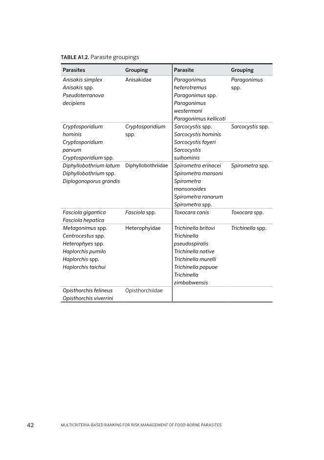
Multi criteria-based ranking for risk ManageMent of food-borne parasites42
TAblE A1.2. Parasite groupings
Parasites Grouping Parasite Grouping
Anisakis simplex
Anisakis spp.Pseudoterranova
decipiens
Anisakidae Paragonimus
heterotremus
Paragonimus spp.Paragonimus
westermani
Paragonimus kellicoti
Paragonimus spp.
Crypto sporidium
hominis
Crypto sporidium
parvum
Crypto sporidium spp.
Crypto sporidium spp.
Sarcocystis spp.Sarcocystis hominis
Sarcocystis fayeri
Sarcocystis
suihominis
Sarcocystis spp.
Diphyllobothrium latum
Diphyllobothrium spp.Diplogonoporus grandis
Diphyllobothriidae Spirometra erinacei
Spirometra mansoni
Spirometra
mansonoides
Spirometra ranarum
Spirometra spp.
Spirometra spp.
Fasciola gigantica
Fasciola hepatica
Fasciola spp. Toxocara canis Toxocara spp.
Metagonimus spp.Centrocestus spp.Heterophyes spp.
Haplorchis pumilo
Haplorchis spp.
Haplorchis taichui
Heterophyidae Trichinella britovi
Trichinella
pseudospiralis
Trichinella native
Trichinella murelli
Trichinella papuae
Trichinella
zimbabwensis
Trichinella spp.
Opisthorchis felineus
Opisthorchis viverrini
Opisthorchiidae

Chapter 1 -IntroduCtIon 43
TAblE A1.3. Parasites excluded from original list
broad Category Parasites EXCluDED Criteria for exclusion
Meat-borne Taenia asiatica Regional
Taenia serialis Unlikely / rare zoonosis
Fish- and shellfish-borne
Capillaria philippinensis Regional – Philippines
Contracaecum/
Phocascaris
Proportion of cases attributable to food-borne infection negligible
Echinostoma spp. Regional – SE Asia
Gnathostoma spp. Regional – SE Asia
Kudoa septempunctata Regional – SE Asia
Lecithodendrid flukes Regional – SE Asia
Plant (fruit- and vegetable-borne, including berries, fruit juice)
Blastocystis spp. Proportion of cases attributable to food-borne infection negligible
Strongyloides stercoralis Proportion of cases attributable to food-borne infection negligible
Ancylostoma spp. Proportion of cases attributable to food-borne infection negligible
Necator americanus Proportion of cases attributable to food-borne infection negligible
“Other” Angiostrongylus
cantonensis
Regional – Asia Pacific
Hymenolepis spp. Proportion of cases attributable to food-borne infection negligible

Multi criteria-based ranking for risk ManageMent of food-borne parasites44
Annex 2
Food-borne parasite ranking exercise: summary cardGroup: Parasite/food:
Criterion bin 0 bin 1 bin 2 bin 3 bin 4 Score
Number of global food-borne illnesses (manifesting disease)
<10 000 10 000 – 100 000
100 000 – 1 000 000
1 000 000 – 10 000 000
>107
Global distribution (number of regions)
N/A 1 2 3-4 >4
Acute morbidity severity (disability weight)
0 (none)<0.03
(very mild)0.03–0.1
(mild)0.1–0.30
(moderate)>0.30
(severe)
Chronic morbidity severity (disability weight)
0 (none)<0.03
(very mild)0.03-0.1
(mild)0.1–0.30
(moderate)>0.30
(severe)
Fraction of illness that is chronic (%)
0%chronic<25%
chronic25–50% chronic
50–75% chronic
>75% chronic
Case-fatality ratio (%)
0% 0–0.1% 0.1–1% 1–10% >10%
Likelihood of increased human burden (%)
None0–25% (low)
25–50% (moderate)
75–100% (high)
100% (still
increasing)
How relevant is this parasite-food pathway for international trade?
Not at allSome
relevanceHigh
relevance
Scope of impact to economically vulnerable communities?
None Low Moderate High
What is the quality of available evidence for this parasite?
Very Poor Poor Adequate Good Very Good
Further comments relevant for the discussion on risk management

Annex 3 - Food-borne pArAsite rAnking exercise Form: explAnAtion oF criteriA 45
Annex 3
Food-borne parasite ranking exercise form: explanation of criteria
The summary card used to conduct this exercise should be considered an expert elicitation. We are asking for your expert judgment on 9 scored parameters, each of which is intended to capture some aspect of the global importance of each parasite. We realize that data may not be available to support your scores, but we ask you to use your knowledge of the literature and your considered opinion to answer these questions.
Please indicate group and parasite/food pathway on the sheet.
We ask that for each parasite you estimate each criterion, using the levels marked in the bins to indicate your group score. You may circle multiple bins as marked in the example below to indicate a broader range of values, but it is CRITICAL that you come to a SINGLE CONSENSUS BEST GUESS SCORE for the criterion and mark it numerically in the rightmost column.
Please take notes on a separate piece of paper to indicate important assumptions or data sources that you would like to record. Please mark on that paper the group and parasite/food pathway so we can associate it with your scores.
Please remark additional comments relevant to risk management below and on the back.
Example:
bin 0 bin 1 bin 2 bin 3 bin 4 Score
Mortality rate
(case-fatality ratio) (%)0% 0–0.1% 0.1–1% 1–10% >10% 2
Criterion No. 1. Number of global food-borne illnessesCriterion: Number of individuals worldwide that manifest clinical illness
Explanation: This criterion measures the magnitude of global food-borne disease as the number of people worldwide who have clinical
Group: Parasite/food:
Criterion bin 0 bin 1 bin 2 bin 3 bin 4 Score
Number of global food-borne illnesses (manifesting disease)
<10 000 10 000 – 100 000
100 000 – 1 000 000
1 000 000 – 10 000 000
>107
Global distribution (number of regions)
N/A 1 2 3-4 >4
Acute morbidity severity (disability weight)
0 (none)<0.03
(very mild)0.03–0.1
(mild)0.1–0.30
(moderate)>0.30
(severe)
Chronic morbidity severity (disability weight)
0 (none)<0.03
(very mild)0.03-0.1
(mild)0.1–0.30
(moderate)>0.30
(severe)
Fraction of illness that is chronic (%)
0%chronic<25%
chronic25–50% chronic
50–75% chronic
>75% chronic
Case-fatality ratio (%)
0% 0–0.1% 0.1–1% 1–10% >10%
Likelihood of increased human burden (%)
None0–25% (low)
25–50% (moderate)
75–100% (high)
100% (still
increasing)
How relevant is this parasite-food pathway for international trade?
Not at allSome
relevanceHigh
relevance
Scope of impact to economically vulnerable communities?
None Low Moderate High
What is the quality of available evidence for this parasite?
Very Poor Poor Adequate Good Very Good
Further comments relevant for the discussion on risk management

Multi criteria-based ranking for risk ManageMent of food-borne parasites46
symptoms of illness and who were infected by food. If you do not feel that you can estimate this number directly, you can calculate it based on numbers you may feel more comfortable with. Namely, it can be considered a function of the global preva-lence of infection multiplied by the percent of infections that are result from food consumption multiplied by the percent of infec-tions that are symptomatic multiplied by the global population of (7 billion people). For parasites that are generally regarded as acute infections (e.g. Trichinella), it is incidence times percent symptomatic times percent food-borne times global population.
For example: Your best estimate is that Parasite A has a global prevalence of about 20%, of which about 50% you believe to be food-borne. Of these infections, the literature suggests that 10% of infections are symptomatic. This equates to a global illness rate of 1% (20% × 50% × 10%). Thus, you would estimate about 70 000 000 cases (1% of 7×10⁹).
Ranges:
bin 0 bin 1 bin 2 bin 3 bin 4
<10 000
illnesses
10 000 – 100 000
illnesses
100 000 – 1 000 000
illnesses
1 000 000 – 10 000 000
illnesses
>10 000 000
illnesses
Criterion No. 2. Geographical distribution (endemic regions)Criterion: Number of regions in which this parasite is geographically dis-
tributed (in which it shows a natural cycle)
Explanation: This criterion reflects the global distribution of the parasite across world regions as a simple count of the number of major regions (Africa, Asia, Europe, Near East, North America, Latin America and the Carribbean, Pacific) in which the disease is regularly found.
Ranges:
bin 0 bin 1 bin 2 bin 3 bin 4
Not applicable 1 region 2 regions 3-4 regions > 4 regions

Annex 3 - Food-borne pArAsite rAnking exercise Form: explAnAtion oF criteriA 47
Criterion No. 3. Acute Morbidity SeverityCriterion: Loss of health-related quality of life due to acute infection
Explanation: This criterion reflects the degree to which an acute manifestation of illness reduces health-related quality of life. The value of the criterion is anchored between 0 (full health, asymptomatic, no illness) to 1 (worst possible health state or death). It depends on both the severity and duration of illness. For a large number of health conditions, including many infectious diseases and some parasitic diseases, disability weights have already been published.
Ranges:
bin 0 bin 1 bin 2 bin 3 bin 4
0
(none)
< 0.03
(very mild)
0.03 –0.01
(mild)
0.01–0.30
(moderate)
>0.30
(severe)
Decision rules: If a pathogen causes more than one acute disease, a population weighted average is applied. Calculate your best guess point estimates for identified acute conditions and weight by likeli-hood. Then assign a bin using the ranges above.
Refer to the table of disability weights below (from Havelaar et al., 2010; Annex 1) or see WHO publications. The Global Burden of Disease 2004 Update includes a summary table on page 33 (WHO, 2008).
This annex to the 2004 report includes more detailed disability weights (WHO, 2004; see http://www.who.int/healthinfo/global_burden_disease/GBD2004_Dis-abilityWeights.pdf)
Table of disability weights for acute and chronic conditions
Very mild (disability weight <0.03)
Duration (in days)
Moderate (0.1<disability weight <0.3)
Duration (in days)
Otitis media 14 Inflammatory bowel disorder 183
Hepatitis 30 Reactive arthritis 183
Folliculitis 7 Tuberculosis 365
Cystitis 14 Chronic pulmonary disease
(bronchitis, asthma,
emphysema)
365
Gastroenteritis, severe 10–15
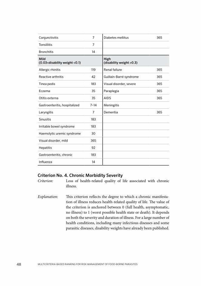
Multi criteria-based ranking for risk ManageMent of food-borne parasites48
Conjunctivitis 7 Diabetes mellitus 365
Tonsillitis 7
Bronchitis 14
Mild (0.03<disability weight <0.1)
High (disability weight >0.3)
Allergic rhinitis 119 Renal failure 365
Reactive arthritis 42 Guillain-Barré syndrome 365
Tinea pedis 183 Visual disorder, severe 365
Eczema 35 Paraplegia 365
Otitis externa 35 AIDS 365
Gastroenteritis, hospitalized 7–14 Meningitis
Laryngitis 7 Dementia 365
Sinusitis 183
Irritable bowel syndrome 183
Haemolytic uremic syndrome 30
Visual disorder, mild 365
Hepatitis 92
Gastroenteritis, chronic 183
Influenza 14
Criterion No. 4. Chronic Morbidity SeverityCriterion: Loss of health-related quality of life associated with chronic
illness.
Explanation: This criterion reflects the degree to which a chronic manifesta-tion of illness reduces health-related quality of life. The value of the criterion is anchored between 0 (full health, asymptomatic, no illness) to 1 (worst possible health state or death). It depends on both the severity and duration of illness. For a large number of health conditions, including many infectious diseases and some parasitic diseases, disability weights have already been published.
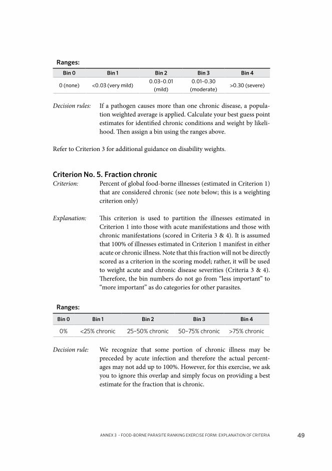
Annex 3 - Food-borne pArAsite rAnking exercise Form: explAnAtion oF criteriA 49
Ranges:
bin 0 bin 1 bin 2 bin 3 bin 4
0 (none) <0.03 (very mild)0.03–0.01
(mild)
0.01–0.30
(moderate)>0.30 (severe)
Decision rules: If a pathogen causes more than one chronic disease, a popula-tion weighted average is applied. Calculate your best guess point estimates for identified chronic conditions and weight by likeli-hood. Then assign a bin using the ranges above.
Refer to Criterion 3 for additional guidance on disability weights.
Criterion No. 5. Fraction chronicCriterion: Percent of global food-borne illnesses (estimated in Criterion 1)
that are considered chronic (see note below; this is a weighting criterion only)
Explanation: This criterion is used to partition the illnesses estimated in Criterion 1 into those with acute manifestations and those with chronic manifestations (scored in Criteria 3 & 4). It is assumed that 100% of illnesses estimated in Criterion 1 manifest in either acute or chronic illness. Note that this fraction will not be directly scored as a criterion in the scoring model; rather, it will be used to weight acute and chronic disease severities (Criteria 3 & 4). Therefore, the bin numbers do not go from “less important” to “more important” as do categories for other parasites.
Ranges:
bin 0 bin 1 bin 2 bin 3 bin 4
0% <25% chronic 25–50% chronic 50–75% chronic >75% chronic
Decision rule: We recognize that some portion of chronic illness may be preceded by acute infection and therefore the actual percent-ages may not add up to 100%. However, for this exercise, we ask you to ignore this overlap and simply focus on providing a best estimate for the fraction that is chronic.

Multi criteria-based ranking for risk ManageMent of food-borne parasites50
Criterion No. 6. Mortality rateCriterion: Case-fatality ratio
Explanation: This criterion estimates the likelihood that a given cases of illness will result in death. Mortality rate is dependent on disease symptoms and severity, as well as underlying health of the infected person.
Ranges:
bin 0 bin 1 bin 2 bin 3 bin 4
0% 0 –0.1% 0.1–1% 1–10% >10%
Criterion No. 7. Increasing trend in diseaseCriterion: Likelihood of a significant increase in human illness.
Explanation: This criterion reflects the potential for the human health burden associated with this particular parasite to increase in the near term, for example through changes in food production, process-ing and consumption.
Ranges:
bin 0 bin 1 bin 2 bin 3 bin 4
None0–25%
(low)
25–50%
(moderate, or unsure)
75–100%
(high)
100%
(still increasing)
Criterion No. 8. International tradeCriterion: Relevance of the parasite and its PRIMARY food sources or
vehicles to affect international trade.
Explanation: This qualitative criterion estimates the degree to which this par-ticular parasite and its main food sources or vehicles may affect international trade. While the characteristics of the parasite or disease severity relate to trade, it is largely a function of the food source or vehicle; if the primary food-borne pathway is not widely traded, or not currently traded from a region in which the parasite is currently endemic, it may not be likely to have an impact on trade. At the same time, if the parasite is in a food product that is widely traded or if there are current issues associ-ated with the parasite-food pathway, it is of greater relevance.
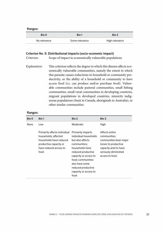
Annex 3 - Food-borne pArAsite rAnking exercise Form: explAnAtion oF criteriA 51
Ranges:
bin 0 bin 1 bin 2
No relevance Some relevance High relevance
Criterion No. 9. Distributional impacts (socio-economic impact)Criterion: Scope of impact to economically vulnerable populations
Explanation: This criterion reflects the degree to which this disease affects eco-nomically vulnerable communities, namely the extent to which this parasite causes reductions in household or community pro-ductivity, or the ability of a household or community to have access food (i.e. can produce and/or purchase food). Vulner-able communities include pastoral communities, small fishing communities, small rural communities in developing countries, migrant populations in developed countries, minority indig-enous populations (Inuit in Canada, aboriginals in Australia), or other similar communities.
Ranges:
bin 0 bin 1 bin 2 bin 3
None Low
Primarily affects individual
households; affected
households have reduced
productive capacity or
have reduced access to
food
Moderate
Primarily impacts
individual households
but also affects
communities;
households have
reduced productive
capacity or access to
food; communities
also have some
reduced productive
capacity or access to
food
High
Affects entire
communities;
communities bear major
losses to productive
capacity and/or have
seriously diminished
access to food.

Multi criteria-based ranking for risk ManageMent of food-borne parasites52
Criterion No. 10. Quality of evidenceCriterion: Quality of available evidence to support judgments (Not a scored
criterion)
Explanation: This question reflects the extent to which you feel you were able to assess criterion-based data or information for a specific parasite.
Ranges:
bin 0 bin 1 bin 2 bin 3 bin 4
Very Poor Poor Adequate Good Very Good
CommentsLastly, please indicate comments, if any, that should be considered in the discus-sion of risk management for this parasite food pathway
References
Havelaar, A.H., van Rosse, F., Bucura, C., Toetenel, M.A., Haagsma, J.A., Kurowicka, D., Heesterbeek, J.H., Speybroeck, N., Langelaar, M.F., van der Giessen, J.W., Cooke, R.M. & Braks, M.A. 2010. Prioritizing emerging zoonoses in the Nether-lands. PLoS One, 5(11): e13965. [Online doi: 10.1371/journal.pone.0013965.]
WHO. 2004. Global burden of disease 2004 update: Disability weights for diseases and conditions. Available at http://www.who.int/healthinfo/global_burden_disease/ GBD2004_DisabilityWeights.pdf Accessed 2013-07-05.
WHO. 2008. Global Burden of Disease 2004 Update. See http://www.who.int/healthinfo/global_burden_disease/GBD_report_2004update_part3.pdf Accessed 2013-07-05.

Annex 4 - CriteriA weights worksheet 53
Annex 4
Criteria weights worksheet
The overall parasite score is given by two equations:
C1*W1 + C2*W2 + C345*W345 + C6*W6 + C7*W7 + C8*W8 + C9*W9 Eq. 1 C345={C3*(1-C5) + C4*C5} Eq. 2
where C are criteria scores normalized to a 0–1 scale and W are the criteria weights, which sum to 100%. Eq. 2 calculates the average severity weight for the parasite, an average of chronic disability weight and acute disability weight using the fraction of illnesses that are chronic. Thus, criteria 3, 4 and 5 have one associ-ated weight, denoted as W345. Otherwise the calculation is straightforward: nor-malized parasite criteria scores are multiplied by fractional weights.
Criteria weights are simply the fraction of the total score reflected by the criteria in question. Therefore, if you think 25% of the overall score should be driven by C1, W1 should be marked with a 25. For comparison purposes, equal weighting of all criteria would result in a value of 14.285%.
Make sure that all numbers sum to 100%, and that no criterion weight is less than 5%. Please use integers only (no decimal points).
Criterion Weighting Code
Criterion Weight (Fraction of Total Score)
C1. Number of global food-borne illnesses W1
C2. Global distribution W2
C3. Acute morbidity severity
C4. Chronic morbidity severity
C5. Chronic illness fraction
W345
C6. Case fatality rate W6
C7. Increasing illness potential W7
C8. Trade relevance W8
C9. Impacts to economically vulnerable
communitiesW9
SUM 100%

Multi criteria-based ranking for risk ManageMent of food-borne parasites54
Annex 5
Sensitivity analysis
In addition to the results presented in the main text, a number of analyses were conducted to examine elicited scores and the sensitivity of the ranking results to different model inputs.
First, variability was examined across groups in elicited scores. Figure A5.1 shows group scores for all nine criteria, averaged over all parasites scored by each group. Because each group scored a slightly different set of parasites, the average scores are not directly comparable, but they do show some interesting patterns. Criterion 2, on the global distribution of disease of each parasite, shows consis-
0.0 0.1 0.2 0.3 0.4 0.5
Scaled Criteria Scores (0-1)
0.6 0.7 0.8 0.9 1.0
1. Number of illnesses
2. Global distribution
3. Acute Severity
4. Chronic Severity
5. Fraction Chronic
6. Case Fatality Rate
7. Illness trends
8. Trade revlevancy
9. Vulnerable communities
Group 1Group 2Group 3Group 4Group 5
FIGuRE A5.1. Group scores by criteria, averaged across parasites

Annex 5 - SenSitivity AnAlySiS 55
tently high average scores, which suggests that experts generally agreed that the set of screened pathogens reflects parasites of global relevancy. The severity of chronic disease consistently scored higher than the severity of acute disease for this set of parasites. Overall, scores for illness trends were low, suggesting that current, endemic disease may be more of a concern than disease movement or emergence.
FIGuRE A5.2. Means and ranges of normalized risk scores across expert groups under baseline model conditions
0 10 20 30 40 50 60 70 80 90
Average criterion score (0-2)
Taenia solium
Echinococcus granulosus
Echinococcus multilocularis
Toxoplasma gondii
Cryptosporidium spp.
Entamoeba histolytica
Trichinella spiralis
Opisthorchiidae
Ascaris spp.
Trypanosoma cruzi
Giardia duodenalis
Fasciola spp.
Cyclospora cayetanensis
Paragonimus spp.
Trichuris trichiura
Trichinella spp.*
Anisakidae
Balantidium coli
Taenia saginata
Toxocara spp.
Sarcocystis spp.
Heterophyidae
Diphyllobothriidae
Spirometra spp.

Multi criteria-based ranking for risk ManageMent of food-borne parasites56
Just as there is variability across expert groups in average criteria scores across parasites, there is variability across groups for specific parasites. This is shown in Figure A5.2, which shows the mean normalized risk scores presented in the main text of the report, as well as the ranges of estimates across groups. Some parasites, such as Toxoplasma gondii, have relatively little variability across experts, while parasites such as Echinococcus multilocularis and Paragonimus, have notably larger variability. This variance can be interpreted as a signal of the strength of scientific knowledge about a given parasite: the greater the variance, the greater the uncer-tainty in information available to experts.
In examining sensitivity of the model itself, alternative weighting schemes were of particular interest.
In some multi-criteria decision analyses, different groups of experts are used to score the individual criteria and to develop the weights that define how criteria scores will be combined into a final risk score. That is, subject matter experts are elicited for criteria scores, while risk managers are elicited for criteria weights. In part due to time and resource constraints, weights were elicited from expert groups, as well as from the FAO/WHO Secretariat, acting as risk managers. The mean of elicited weights across all participants was used for the baseline model and ranking.
Criterion weights were roughly similar across experts and risk managers, as shown in the rounded values presented in Table A5.1. Risk managers tended to put greater weight on potential for increased illness, trade relevance and impacts to economi-cally vulnerable communities than did experts, but all participants tended to put greater weight on public health criteria. These are compared with an equal weighting scheme, in which each criterion is treated as of equal importance in the overall risk score.
Sensitivity analyses of rankings were conducted around three alternative weighting schemes: mean of expert weights, mean of risk manager weights, and equal criteria weighting. Table A5.2 and Figure A5.3 show multicriteria risk scores (normalized to 0–100) for global foodborne parasites for the baseline and the three alternative schemes mentioned above. Although different schemes result in slightly different scores, the ranking is fairly robust among the alternative schemes.
Sensitivity analyses of rankings were conducted around three alternative weighting schemes: mean of expert weights, mean of risk manager weights, and equal criteria weighting. Table A5.2 and Figure A5.3 show multicriteria risk scores (normalized to 0–100) for global foodborne parasites for the baseline and the three alternative schemes mentioned above. Although different schemes result in slightly different scores, the ranking is fairly robust among the alternative schemes.

Annex 5 - SenSitivity AnAlySiS 57
Given the similarities in expert and Secretariat scores, the biggest differences can be seen in the equal weighting scheme, though even under that scheme the ranks of the first four parasites are identical. Figure A5.4 shows how these alternativee schemes affect the rank order of parasites in the overall ranking. The dots show the baseline rank, while the vertical lines display the range of ranks across the three alternative scenarios. This figure shows that ranks are quite stable, with some parasite-specific deviation. Those with the greatest deviation in scores are Taenia saginata and Cyclospora cayetanensis, followed by Trichinella spiralis, Ascaris spp., Paragonimus spp., Anisakidae and Toxocara spp. Most of these are parasites with higher scores in trade relevancy or impacts on socio-economically vulnerable pop-ulations, as the equal weighting scheme increases the importance of these criteria.
TAblE A5.2. Normalized multicriteria risk scores for global foodborne parasites under alternative criteria weighting schemes
Weighting scheme
baseline Expert Secretariat Equal
Taenia solium 72.9 73.1 72.7 70.7
Echinococcus granulosus 63.6 65.9 61.8 60.5
Echinococcus multilocularis 61.6 65.0 58.8 56.8
Toxoplasma gondii 61.0 64.9 57.7 53.0
TAblE A5.1. Mean of elicited criteria weights used in multicriteria ranking
Scoring criterion Weighting
baseline Expert Secretariat Equal
W1. Number of global
foodborne illnesses
0.22 0.24 0.20 0.14
W2. Global distribution 0.14 0.15 0.12 0.14
W345. Morbidity severity 0.22 0.23 0.21 0.14
W6. Case-fatality ratio 0.15 0.16 0.15 0.14
W7. Increasing illness potential 0.07 0.06 0.09 0.14
W8. Trade relevance 0.10 0.06 0.13 0.14
W9. Impacts on economically
vulnerable communities
0.10 0.09 0.11 0.14
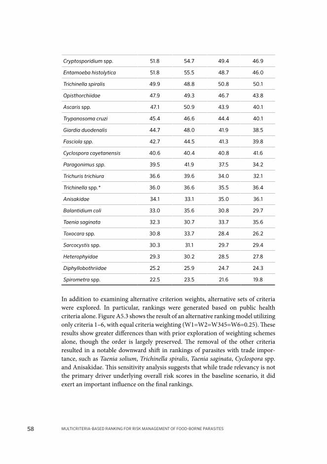
Multi criteria-based ranking for risk ManageMent of food-borne parasites58
Cryptosporidium spp. 51.8 54.7 49.4 46.9
Entamoeba histolytica 51.8 55.5 48.7 46.0
Trichinella spiralis 49.9 48.8 50.8 50.1
Opisthorchiidae 47.9 49.3 46.7 43.8
Ascaris spp. 47.1 50.9 43.9 40.1
Trypanosoma cruzi 45.4 46.6 44.4 40.1
Giardia duodenalis 44.7 48.0 41.9 38.5
Fasciola spp. 42.7 44.5 41.3 39.8
Cyclospora cayetanensis 40.6 40.4 40.8 41.6
Paragonimus spp. 39.5 41.9 37.5 34.2
Trichuris trichiura 36.6 39.6 34.0 32.1
Trichinella spp.* 36.0 36.6 35.5 36.4
Anisakidae 34.1 33.1 35.0 36.1
Balantidium coli 33.0 35.6 30.8 29.7
Taenia saginata 32.3 30.7 33.7 35.6
Toxocara spp. 30.8 33.7 28.4 26.2
Sarcocystis spp. 30.3 31.1 29.7 29.4
Heterophyidae 29.3 30.2 28.5 27.8
Diphyllobothriidae 25.2 25.9 24.7 24.3
Spirometra spp. 22.5 23.5 21.6 19.8
In addition to examining alternative criterion weights, alternative sets of criteria were explored. In particular, rankings were generated based on public health criteria alone. Figure A5.3 shows the result of an alternative ranking model utilizing only criteria 1–6, with equal criteria weighting (W1=W2=W345=W6=0.25). These results show greater differences than with prior exploration of weighting schemes alone, though the order is largely preserved. The removal of the other criteria resulted in a notable downward shift in rankings of parasites with trade impor-tance, such as Taenia solium, Trichinella spiralis, Taenia saginata, Cyclospora spp. and Anisakidae. This sensitivity analysis suggests that while trade relevancy is not the primary driver underlying overall risk scores in the baseline scenario, it did exert an important influence on the final rankings.

Annex 5 - SenSitivity AnAlySiS 59
0 10 20 30 40 50 60 70 80
Normalized overall score
Taenia solium
Echinococcus granulosus
Echinococcus multilocularis
Toxoplasma gondii
Cryptosporidium spp.
Entamoeba histolytica
Trichinella spiralis
Opisthorchiidae
Ascaris spp.
Trypanosoma cruzi
Giardia duodenalis
Fasciola spp.
Cyclospora cayetanensis
Paragonimus spp.
Trichuris trichiura
Trichinella spp.*
Anisakidae
Balantidium coli
Taenia saginata
Toxocara spp.
Sarcocystis spp.
Heterophyidae
Diphyllobothriidae
Spirometra spp.
Baseline weights
Expert weights
Secretariat weights
Equal weights
FIGuRE A5.3. Comparison of multicriteria risk scores of global foodborne parasites across alternative criterion weighting schemes

Multi criteria-based ranking for risk ManageMent of food-borne parasites60
FIGuRE A5.4. Rank scores of global foodborne parasites across alternative criterion weighting schemes, presented as ranges around baseline ranks
0 5 10 15 20 25
Rank
Taenia solium
Echinococcus granulosus
Echinococcus multilocularis
Toxoplasma gondii
Cryptosporidium spp.
Entamoeba histolytica
Trichinella spiralis
Opisthorchiidae
Ascaris spp.
Trypanosoma cruzi
Giardia duodenalis
Fasciola spp.
Cyclospora cayetanensis
Paragonimus spp.
Trichuris trichiura
Trichinella spp.*
Anisakidae
Balantidium coli
Taenia saginata
Toxocara spp.
Sarcocystis spp.
Heterophyidae
Diphyllobothriidae
Spirometra spp.
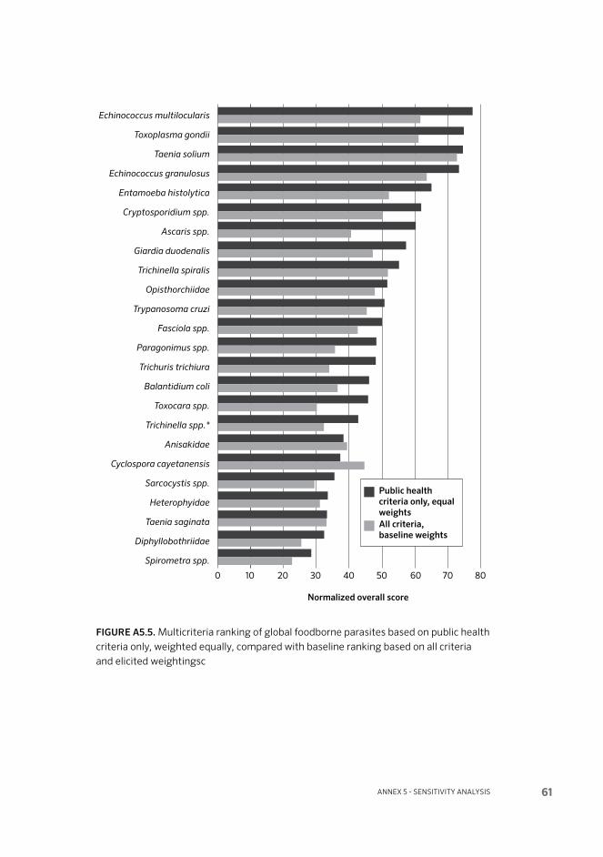
Annex 5 - SenSitivity AnAlySiS 61
0 10 20 30 40 50 60 70 80
Normalized overall score
Echinococcus multilocularis
Toxoplasma gondii
Taenia solium
Echinococcus granulosus
Entamoeba histolytica
Cryptosporidium spp.
Ascaris spp.
Giardia duodenalis
Trichinella spiralis
Opisthorchiidae
Trypanosoma cruzi
Fasciola spp.
Paragonimus spp.
Trichuris trichiura
Balantidium coli
Toxocara spp.
Trichinella spp.*
Anisakidae
Cyclospora cayetanensis
Sarcocystis spp.
Heterophyidae
Taenia saginata
Diphyllobothriidae
Spirometra spp.
Public health criteria only, equal weightsAll criteria, baseline weights
FIGuRE A5.5. Multicriteria ranking of global foodborne parasites based on public health criteria only, weighted equally, compared with baseline ranking based on all criteria and elicited weightingsc
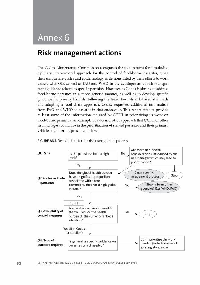
Multi criteria-based ranking for risk ManageMent of food-borne parasites62
Annex 6
Risk management actions
The Codex Alimentarius Commission recognizes the requirement for a multidis-ciplinary inter-sectoral approach for the control of food-borne parasites, given their unique life-cycles and epidemiology as demonstrated by their efforts to work closely with OIE as well as FAO and WHO in the development of risk manage-ment guidance related to specific parasites. However, as Codex is aiming to address food-borne parasites in a more generic manner, as well as to develop specific guidance for priority hazards, following the trend towards risk-based standards and adopting a food-chain approach, Codex requested additional information from FAO and WHO to assist it in that endeavour. This report aims to provide at least some of the information required by CCFH in prioritizing its work on food-borne parasites. An example of a decision-tree approach that CCFH or other risk managers could use in the prioritization of ranked parasites and their primary vehicle of concern is presented below.
FIGuRE A6.1. Decision tree for the risk management process
Q1. Rank
Q2. Global vs trade importance
Q3. Availability of control measures
Q4. Type of standard required
Is the parasite / food a high rank?
Are there non-health considerations introduced by the risk manager which may lead to prioritization?
Does the global health burden have a significant proportion associated with a food commodity that has a high global volume?
Are control measures available that will reduce the health burden cf. the current (ranked) situation?
CCFH prioritise the work needed (include review of existing standards)
Is general or specific guidance on parasite control needed?
CCFH
Yes (If in Codex jurisdiction)
Stop (inform other agencies? E.g. WHO, FAO)
Stop
Stop
No
No
Yes
No
Yes
Separate risk management process

Annex 7 - Specific informAtion for the rAnked pArASiteS 63
Annex 7
Specific information for the ranked parasites
For a Glossary of Parasitological Terms, see Annex section A7.25
After the meeting, the experts developed informative summaries for the resulting 24 ranked parasites, for use by risk managers or any interested stakeholder. A glossary was also provided by the expert group to help the reader with the termi-nology.
A7.1 ANISAkIDAE AND ANISAkIASIS
General information Anisakiasis refers to infection of people with nematode larvae belonging to the nematode Family Anisakidae, and it is a serious zoonotic disease. Although there are several zoonotic species in this family, the two species most often associat-ed with anisakiasis are Anisakis simplex the ‘herring worm’ and Pseudoterranova decipiens, the ‘cod worm’ (Chai, Murrell and Lymbery, 2005). The complex life history of A. simplex involves a marine intermediate host (euphasid crustacean), a paratenic host (marine fish or squid) and a definitive host (marine mammal).
Anisakiasis occurs when people ingest third-stage larvae that occur in the viscera or muscle of a wide range of marine fish and squids. Humans are accidental inter-mediate hosts in which the parasites rarely develop further; this invasion can cause gastro intestinal abscesses.
Geographical distributionAnisakiasis occurs throughout the world, but is reported most frequently from Asia (especially Japan) and Western Europe, where risky food behaviour customs (i.e., eating raw, lightly cooked, or marinated fish in dishes such as sushi, salted or smoked herring, gravlax, and ceviche) are common (Lymbery and Cheah, 2007). Recent molecular genetic studies have shown that these species, A. simplex and P. decipiens, actually comprise a number of sibling species, often with distinct geo-graphical and host ranges, or both (Mattiucci et al., 2005). Within the Anisakis simplex complex are: A. simplex (sensu stricto), found in the northern Atlantic; A. simplex C, found in the northern Pacific and southern waters below 30°N; and
Multi criteria-based ranking for risk ManageMent of food-borne parasites64
A. pegreffi, found in the Mediterranean Sea. Three species have also been described for the Pseudoterranova decipiens complex: P. decipiens A in the northeast Atlantic and Norwegian Sea, P. decipiens C in the northwest Atlantic and Barents Sea, and P. decipiens B throughout northern waters. Where the ranges of these species overlap, they appear to preferentially utilize different definitive host species.
Historically, most authors estimate there have been 15 000 to 20 000 total human cases. There has been an increase in reported prevalence throughout the world in the last two decades, probably due to better diagnostic tools, increased demand for seafood, and a growing demand for raw or lightly cooked food, although none of these factors has been rigorously evaluated. The areas of highest prevalence are Japan (after eating sushi and sashimi), and along the Pacific coast of South America (from eating ceviche, seviche or cebiche).
DiseaseWhen humans eat infected fish harbouring live third-stage larvae, the larvae migrate to the gastro intestinal mucosa, where they die, but induce the formation of abscesses. Presumptive diagnosis in humans may be made on the basis of the patient’s recent food habits (Gutierrez, 2011). Definitive diagnosis requires dem-onstration of worms by gastroscopy or surgery. No treatment is recommended for transient infection. In the gastro intestinal form (embedded larvae), surgery or gas-troscopic procedure is also curative.
There is little information on chronic morbidity. However, the development of allergy to the parasite’s allergens (even when the fish is thoroughly cooked) is now recognized. Gastro allergic anisakiasis is an acute IgE-mediated generalized reaction, manifested by urticaria and anaphylaxis, with or without accompanying gastro intestinal symptoms (Audicana and Kennedy, 2008). Occupational allergy, including asthma, conjunctivitis and contact dermatitis, has also been observed in fish-processing workers.
There is little information available on illness fraction or case fatality rates, probably because most cases are acute and treated.
Trade relevanceAnisakid infections are a trade issue because of regulations imposed by countries on imports. Many countries have regulations requiring inspection of fish for zoonotic parasites, and for inactivating any nematode larvae, etc., that may be present. Regulations and inactivation methods may differ in specifics between countries. (See EU, no date; and Chapter 5 in FDA, no date).

Annex 7 - Specific informAtion for the rAnked pArASiteS 65
Impact on economically vulnerable populationsDeveloping countries can be affected by the necessity of taking steps to ensure fish exports are free of live anisakids. The inactivation methods described above can be expensive, and when not completely successful lead to rejection of exports by importing countries, especially fresh fish products.
References
Audicana, M.T. & Kennedy, M. 2008. Anisakis simplex: from obscure infectious worm to inducer of immune hypersensitivity. Clinical Microbiology Reviews, 21(2): 360–379.
Chai, J.Y., Murrell, K.D. & Lymbery, A.J. 2005. Fish-borne parasitic zoonoses: status and issues. International Journal for Parasitology, 35(11-12): 1233–1254.
EU. No date. EU import conditions for seafood and other fishery products. Web site. Available at http://ec.europa.eu/food/international/trade/im_cond_fish_en.pdf Accessed 2013-06-23.
FDA (Federal Drug Administration). No date. Chapter 5 – Parasites, in: Fish and Fishery Products Hazards and Controls Guidance. 4th Edition, November 2011. Available at http://www.fda.gov/downloads/Food/GuidanceRegulation/UCM252393.pdf Accessed 2013-08-24
Gutierrez, Y. 2011. Anisakidae. pp. 778–787 (Ch. 12), in: R.L. Guerrant, D.H. Walker and P.F. Weller (editors). Tropical Infectious Diseases: Principles, Pathogens and Practice. 3rd edition. Saunders, Philadelphia, USA.
Lymbery, A.J. & Cheah, F.Y. 2007. Anisakid nematodes and anisakiasis. pp. 185–207, in: K.D. Murrell and B. Fried (editors). Foodborne parasitic zoonoses. Springer, New York, USA.
Mattiucci, S., Nascetti, G., Dailey, M., Webb, S.C., Barros, N.B., Cianchi, R. & Bul-lini, L. 2005. Evidence for a new species of Anisakis Dujardin, 1845: morphological description and genetic relationships between congeners (Nematoda: Anisakidae). Systematic Parasitology, 61(3): 157–161.

Multi criteria-based ranking for risk ManageMent of food-borne parasites66
A7.2 AscAris Spp.
General informationAscaris lumbricoides is the large intestinal roundworm (nematode) of humans, and the infection is termed ‘ascariasis’ or ‘ascariosis’, and rarely ‘ascarosis’. ‘Ascariasis’ is the most widely used, and will be used here. The adult worms are large (females up to 35 cm, males up to 31 cm in length), and individual worms can weigh as much as 7 or 8 g (Elkins and Haswellelkins, 1989). The adults occupy the small intestine and the female lays large numbers of eggs (estimated to be in the hundreds of thousands of eggs per day per female) (Brown and Cort, 1927; O’Lorcain and Holland, 2000). The eggs are voided in the faeces and are sticky, thick shelled and highly persistent in the environment such that they can survive for several years in soil. They contaminate water supplies following rain or flooding, vegetables either directly from soil or by irrigation, and probably the hands and clothing of agricul-tural workers or other people in contact with contaminated soil. The practice of using human faeces as fertilizer in subsistence farming presents a significant risk of continued transmission.
Ascaris suum is the large roundworm of pigs and is considered to be a separate species from A. lumbricoides. The two species are virtually indistinguishable mor-phologically, immunologically and biochemically, although there are some dis-tinguishing immunological and biochemical features (e.g. Kennedy et al., 1987). DNA-based surveys have indicated that A. suum is mostly confined to pigs, and A. lumbricoides to humans, but that there is evidence of cross-infections such that A. suum may present a significant risk to humans Peng and Criscione, 2012; Zhou et al., 2012; Nejsum et al., 2005). In regions endemic for both parasites it appears mostly, but not exclusively, to be the case that adult worms of A. lumbricoides have a host preference for humans, and adult worms of A. suum have a host preference for pigs (Peng and Criscione, 2012). In regions where A. lumbricoides does not occur in humans, there have been cases of infection with A. suum that have been attributed to contamination from pig farms (Nejsum et al., 2005; Anderson, 1995).
In both species, infection occurs by ingestion of viable eggs, which hatch in the small intestine, releasing the infective-stage larvae of the parasite, which is in its third developmental stage (L3). The larvae then undergo a tissue migration involving the liver then the lungs. In the lungs the larvae break through to the air spaces, migrate up the trachea, are then swallowed and thereby re-introduced to the gastro intestinal tract, where they mature to adult worms in the small intestine.
The global prevalence of human ascariasis in the 1990s was estimated to be ap-proximately 1.5 billion with 100–200 million people affected clinically, a large pro-portion of whom were children (reviewed in O’Lorcain and Holland (2000) and
Annex 7 - Specific informAtion for the rAnked pArASiteS 67
Peng and Criscione (2012). More recent estimates are slightly lower at 1.2 billion people infected, which is largely due to China’s large-scale treatment programmes (reviewed in O’Lorcain and Holland (2000) and Peng and Criscione (2012)), but some estimates remain as high as 2 billion people currently infected.
Geographical distributionThe distribution of both species of Ascaris is essentially global, but with low preva-lences in countries with well-developed sanitation systems, and very high preva-lences in regions with poor sanitation. The greater association with tropical and subtropical countries may merely be because many of these have poorer overall sanitation systems and parasite control programmes, and the viability and develop-ment to infectivity of eggs is favoured under warm, moist conditions. Exposure of humans to eggs of A. suum will be less likely in regions where pigs are not farmed.
DiseaseAscariasis in humans presents mainly in the gut (small intestine and ileum) and the lungs, though larval migration through the liver and peritoneum likely also cause damage (O’Lorcain and Holland, 2000). In the gut, the worms can occur in such numbers that blockage and rupture or perforation can occur in extreme cases. The parasites have also been known to cause death by migrating into and blocking the pancreatic or bile ducts. The worms in the gut can cause malabsorp-tion and anorexia, which will contribute to malnutrition (O’Lorcain and Holland, 2000). The malabsorption may be due to a loss of brush border enzymes, erosion and flattening of the villi, and inflammation of the lamina propria, and premature cessation of lactase production has also been intimated (O’Lorcain and Holland, 2000). Migration of larvae through the lungs can cause severe immune hypersensi-tivity responses (Loeffler’s Syndrome) that may be life-threatening. This appears to be more common in arid areas when periodic rains mobilise dormant Ascaris eggs from soil and other sources such as latrines, resulting in a high level of contamina-tion of water and food supplies. It is highly likely that severe pulmonary reactions can be caused by exposure to the eggs of either A. lumbricoides or A. suum (as is known in sheep and cattle from exposure to A. suum eggs, and in experimentally infected animals such as rats, mice and rabbits, the larvae of either species reach the lungs). Infection with adult Ascaris can be detected by observation of eggs in faeces, although this requires the presence of a reproductive female. A. suum in-fections of humans in developed countries is often with single or low numbers of worms. Loeffler’s Syndrome can be detected by X-ray appearance of shadows on the lungs (Loeffler, 1956), and detection of larvae and eosinophils in sputum or throat swabs. A characteristic feature of infection with parasitic worms is high levels of IgE antibody and eosinophils in blood, and eosinophils and mast cells in infected tissues; there is evidence that allergic-type immune responses may be part

Multi criteria-based ranking for risk ManageMent of food-borne parasites68
of the protective response to Ascaris (McSharry et al., 1999). Important allergens have been described from Ascaris. See O’Lorcain and Holland (2000) for further detail on ascariasis disease symptoms and other effects.
Trade relevanceContamination of fresh produce with Ascaris eggs has not been an issue in trade up to now. The main risk here is through fresh vegetables that have been contaminated with eggs directly from the soil in which they were grown, or the water with which they were irrigated or treated and prepared post-harvest. Trade in pigs can clearly also be a source of infection to new areas. The robustness of Ascaris eggs means that they can survive for long periods during transport, and they can survive low temperatures, including freezing to some degree, desiccation and chemical attack, though not cooking. Once soil is contaminated with viable eggs, it can remain so for up to a decade.
Impact on economically vulnerable populations The potential impact of ascariasis is chronic and insidious for communities, and can be severe and even life-threatening for individuals. In addition to the overt disease symptoms due to A. lumbricoides infection detailed above, there is evidence that chronic infection can affect the growth rate and final height, of children, and their cognitive development (reviewed in O’Lorcain and Holland, 2000, and Bundy, Walson and Watkins, 2013), which is likely to be particularly so in double or multiple infections with other species of worm parasites. In regions where pigs are kept, the risks of intestinal infection with adult A. suum may be low, but migratory larvae will still cause damage to liver and lungs, and it is likely that the risk of Loeffler’s Syndrome will be similar with either species of Ascaris. Lamentably little research has been carried out on the prevalence, morbidity and mortality due to Loeffler’s Syndrome or less acute but chronic, repetitive damage to the lungs, in humans infected with either parasite. This paucity of information on the pulmonary stage of infection is particularly unfortunate given the possibil-ity that lung damage could exacerbate lung infections and consequent mortality in children (O’Lorcain and Holland, 2000).
Other relevant informationDrugs for the treatment for the intestinal stage of ascariasis are cheap, readily available and relatively free of side-effects. Treatments for the tissue migratory phases would be rare, partly because of the difficulty of diagnosis, and possibly also the risk of causing deleterious reactions to dead larvae in liver, lungs or elsewhere (O’Lorcain and Holland, 2000). There are no vaccines available against ascaria-sis. There continues to be a debate about whether infection with Ascaris or other helminth parasites increases or decreases the risk of allergic reactions to environ-mental allergens (Pinelli et al., 2009).

Annex 7 - Specific informAtion for the rAnked pArASiteS 69
References
General sourcesHolland, C.V. (Editor). 2013. Ascaris: The Neglected Parasite. CABI Publishing, Walling-
ford, Oxfordshire, UK, and Cambridge, MA, USA.
Specific sources citedAnderson, T.J.C. 1995. Ascaris infections in humans from North America – molecular
evidence for cross-infection. Parasitology, 110: 215–219.
Brown, H.W. & Cort, W.W. 1927. The egg production of Ascaris lumbricoides. Journal of Parasitology, 14: 88–90.
Bundy, D.A.P., Walson, J.L. & Watkins, K.L. 2013. Worms, wisdom, and wealth: why deworming can make economic sense. Trends in Parasitology, 29: 142–148.
Elkins, D.B. & Haswellelkins, M. 1989. The weight-length profiles of Ascaris lumbricoides within a human community before mass treatment and following re-infection. Parasitology, 99: 293–299.
Kennedy, M.W., Qureshi, F., Haswellelkins, M. & Elkins, D.B. 1987. Homology and heterology between the secreted antigens of the parasitic larval stages of Ascaris lumbricoides and Ascaris suum. Clinical and Experimental Immunology, 67: 20–30.
Loeffler, W. 1956. Transient lung infiltrations with blood eosinophilia. International Archives of Allergy and Applied Immunology, 8: 54–59.
McSharry, C., Xia, Y., Holland, C.V. & Kennedy, M.W. 1999. Natural immunity to Ascaris lumbricoides associated with immunoglobulin E antibody to ABA-1 allergen and inflammation indicators in children. Infection and Immunity, 67: 484–489.
Nejsum, P., Parker, E.D. Jr, Frydenberg, J., Roepstorff, A., Boes, J., Haque, R., Astrup, I., Prag, J. & Skov Sørensen, U.B. 2005. Ascariasis is a zoonosis in Denmark. Journal of Clinical Microbiology, 43(3): 1142–1148.
O’Lorcain, P. & Holland, C.V. 2000. The public health importance of Acaris lumbricoides. Parasitology, 121: S51–S71.
Peng, W. & Criscione, C.D. 2012. Ascariasis in people and pigs: new inferences from DNA analysis of worm populations. Infection Genetics and Evolution, 12: 227–235.
Pinelli, E., Willers, S.M., Hoek, D., Smit, H.A., Kortbeek, L.M., Hoekstra, M., de Jong-ste, J., van Knapen, F., Postma, D., Kerkhof, M., Aalberse, R., van der Giessen, J.W. & Brunekreef, B. 2009 Prevalence of antibodies against Ascaris suum and its association with allergic manifestations in 4-year-old children in The Netherlands: the PIAMA birth cohort study. European Journal of Clinical Microbiology & Infectious Diseases, 28(11): 1327–1334.
Zhou, C., Li, M., Yuan, K., Deng, S. & Peng, W. 2012. Pig Ascaris: an important source of human ascariasis in China. Infection Genetics and Evolution, 12: 1172–1177.

Multi criteria-based ranking for risk ManageMent of food-borne parasites70
A7.3 BAlAntidium coli
General information Balantidium coli is a protozoan parasitic species that causes the disease balantidia-sis (CDC, no date; Anon., 2003). It is the only member of the ciliate phylum known to be pathogenic to humans (CDC, no date; Anon., 2003a). Infection occurs when the cysts are ingested, usually through contaminated food or water. This parasite lives in the caecum and colon of humans. B. coli has two developmental stages, a trophozoite stage and a cyst stage. Trophozoites multiply and encyst due to the dehydration of faeces. It can thrive in the gastro intestinal tract as long as there is a balance between the protozoan and the host without causing dysenteric symptoms. Infection most likely occurs in people with malnutrition due to the low stomach acidity or people with immune compromised systems (Anon., 2003b; Schuster and Ramirez-Avila, 2008).
Geographical distributionThe disease is considered to be rare and occurs in less than 1% of the human population. Most infections occur in developing countries where faeces are more likely to get in contact with food and drinking water. In addition to humans, pigs and other animals carry the disease. People who raise pigs have a greater risk of getting infected with balantidiasis. Co-infections with other parasites are likely to aggravate the damage wrought by each individual parasite, and they probably share common sources of infection (i.e. contaminated water) (Roberts and Janovy, 2009).
Balantidiasis in humans is common in the Philippines, but it can be found anywhere in the world, especially among those that are in close contact with swine. It has been noted in Latin America, Bolivia, Southeast Asia and New Guinea.
DiseaseCommon symptoms of balantidiasis include chronic diarrhoea, occasional dysentery (diarrhoea with passage of blood or mucus), nausea, foul breath, colitis (inflammation of the colon), abdominal pain, weight loss, deep intestinal ulcer-ations, and possibly perforation of the intestine. Fulminating acute balantidiasis is when the disease comes very suddenly and with great intensity. Haemorrhag-ing can occur, which can lead to shock and death. Untreated fulminating acute balantidiasis is reported to have a fatality rate of 30%. In acute disease, explosive diarrhoea may occur as often as every twenty minutes. Perforation of the colon may also occur in acute infections which can lead to life-threatening situations. If balantidiasis is not treated the persistent diarrhoea leads to high fluid loss and dehydration. If abdominal bleeding occurs, it can lead to death (Schuster and Ramirez-Avila, 2008).

Annex 7 - Specific informAtion for the rAnked pArASiteS 71
Trade relevanceThere have not yet been significant trade issues with respect to findings of B. coli in foods, but with the increasing number of surveillance studies reporting positive results worldwide, and the growing number of produce-associated illness outbreaks, more trade issues resulting in import restrictions and recalls may occur in the future. B. coli causes reduced production performance in the animals affected, which has an impact on the economy, both locally for farmers and nation-ally for the country (Roberts and Janovy, 2009).
Impact on economically vulnerable populations The disease is a problem primarily in developing countries, where water sources may be contaminated with swine or human faeces. Balantidiasis infections can be prevented by following appropriate hygiene practices (such as not using human faeces as fertilizer in agriculture; washing hands after going to the toilet and before meals; washing vegetables; and cooking meat properly). Infective B. coli cysts are killed by heat (Schuster and Ramirez-Avila, 2008).
References
Anon[ymous]. 2003a. The Parasite: Balantidium coli. The Disease: Balantidiasis. In-troduction. Web site prepared by A. Ramachandran. Last updated 2003-05-23; accessed 2013-06-18. Online at http://www.stanford.edu/group/parasites/Para-Sites2003/Balantidium/Balantidium_coli_ParaSite.htm.
Anon. 2003b. The Parasite: Balantidium coli. The Disease: Balantidiasis. Morphology. Web site prepared by A. Ramachandran. Last updated 2003-05-23; accessed 2013-06-18. Online at http://www.stanford.edu/group/parasites/ParaSites2003/Balan-tidium/Morphology.htm
CDC (Centres for Disease Control and Prevention). No date. Parasites and Health: Bal-antidiasis [Balantidium coli]. Online. See: http://www.dpd.cdc.gov/dpdx/HTML/Balantidiasis.htm Accessed 2013-06-18.
Schuster, F.L. & Ramirez-Avila, L. 2008. Current world status of Balantidium coli. Clinical Microbiology Review, 21(4): 626–638.
Roberts, L.S. & Janovy, J. Jr. 2009. Foundations of Parasitology. 8th Edition. McGraw-Hill, New York, USA.

Multi criteria-based ranking for risk ManageMent of food-borne parasites72
A7.4 cryptosporidium Spp.
General information Cryptosporidium spp. are protozoan parasites reported worldwide in a large number of different hosts, including humans. The infectious stages of Cryptosporidium spp., known as oocysts, are shed with the faeces of the host and can survive for long periods under cool and moist conditions.
Routes of transmission include waterborne, person-to-person, zoonotic and food-borne. The waterborne route is numerically the most important means of trans-mission of cryptosporidiosis. Numerous waterborne outbreaks of cryptosporidi-osis have occurred worldwide as a result of oocyst contamination of drinking water sources and recreational water. The largest waterborne illness outbreak of any kind in the United States of America occurred in the spring of 1993, when an estimated 403 000 people became ill with cryptosporidiosis in Milwaukee, Wisconsin. Food-borne transmission of cryptosporidiosis is thought to be much less common than waterborne or person-to-person transmission; about 8% of domestically acquired cases in the United States of America are food-borne (Scallan et al., 2011). It is, however, emerging as an important public health issue.
Food-borne outbreaks of cryptosporidiosis associated with the consumption of fresh produce have been reported mainly in the United States of America and in northern Europe (Dixon et al., 2011; Robertson and Chalmers, 2013). The foods implicated in these outbreaks have included green onions, sandwich-bar ingredi-ents, parsley, carrots, red peppers, and lettuce. In some cases these outbreaks were attributed to infected food-handlers. A large outbreak affecting approximately 300 people occurred in the UK in 2012 and was associated with the consumption of pre-cut bagged salad products (HPA, 2013). There have also been four cryptospo-ridiosis outbreaks associated with drinking unpasteurized apple cider, all in the United States of America. Unpasteurized milk has been associated with outbreaks of cryptosporidiosis in Australia and the UK. Chicken salad was implicated in an outbreak in the United States of America and may have been contaminated by a food worker who also operated a daycare facility.
Numerous surveys performed worldwide have reported the presence of Cryptosporidium oocysts on a wide variety of fresh produce items (Dixon et al., 2013). Cryptosporidium oocysts have also been reported worldwide in the gills and tissues of oysters and other molluscan shellfish, including clams, cockles and mussels (Fayer, Dubey and Lindsay, 2004).
Annex 7 - Specific informAtion for the rAnked pArASiteS 73
Control measures to reduce the likelihood of contamination of produce at the pre-harvest stage with Cryptosporidium include the use of good quality water for irriga-tion, mixing of pesticides, or washing and processing; restricting access of livestock and other animals to crop lands and surface waters; monitoring the health of farm workers and encouraging good hygiene; and using only composted manure as fertilizer. Post-harvest control measures include the use of good quality water for washing and processing produce; monitoring and enforcing good personal hygiene in food handlers; prevention of cross-contamination; and the incorpora-tion of HACCP plans. At the consumer level, good hygiene and avoidance of cross-contamination are again important control measures. Thorough washing of fresh produce is recommended, but probably will not be fully effective in removing all contaminating oocysts. Although oocysts are somewhat resistant to freezing, they can be inactivated by storing produce at -20°C for >24 hours, or at -15°C for at least a week. Alternatively, oocysts will be readily destroyed in foods that are sub-sequently cooked.
Geographical distributionIn recent years, human infection with Cryptosporidium spp. has emerged as a global public health problem. Prevalence, however, is very difficult to determine as data is not available from many countries. In one estimate, the prevalence of Cryptosporidium in patients with gastro enteritis was 1–4% in Europe and North America, and 3–20% in Africa, Asia, Australia, and South and Central America (Current and Garcia, 1991). Laberge and Griffiths (1996) estimated that the preva-lence rates based on oocyst excretion were 1–3% in industrialized countries, and up to 10% in developing countries. Cryptosporidiosis has been reported in 106 countries worldwide (Fayer, 2008).
Approximately twelve species of Cryptosporidium, and several genotypes, have been reported in humans. However, 90% of reported human infections involve C. hominis, which is found primarily in humans, and C. parvum, which is an important zoonotic species. C. hominis is thought to account for more human cases than C. parvum in North America, Australia, Asia, sub-Saharan Africa and some parts of Europe. Generally speaking, C. parvum is more prevalent in rural or agricultural regions, probably as a result of zoonotic transmission. In recent years, C. meleagridis has been reported more commonly in humans. For example, Cama et al. (2008) reported a relatively high prevalence of infection with C. meleagridis in children in Peru. Similarly, C. cuniculus was found to be the third most commonly identified species, after C. parvum and C. hominis, in sporadic cases of cryptospo-ridiosis in the UK (Chalmers et al., 2011). Several other Cryptosporidium species and genotypes are only occasionally found in humans (Xiao, 2010).

Multi criteria-based ranking for risk ManageMent of food-borne parasites74
DiseaseCryptosporidiosis is an enteric disease which is self-limiting in immuno competent individuals. The disease is characterized by watery diarrhoea and a variety of other symptoms, including, abdominal pain, weight loss, nausea, vomiting, fever and malaise (Chalmers and Davies, 2010). Symptoms in some immuno compromised patients become chronic, debilitating and potentially life-threatening. Crypto-sporidiosis accounts for up to 6% of all reported diarrhoeal illnesses in immuno-competent persons (Chen et al., 2002). Twenty-four percent of AIDS patients with diarrhoea are infected with Cryptosporidium spp. (Guerrant, 1997). In the United States of America, Scallan et al. (2011) reported a hospitalization rate of 25%, and a death rate of 0.3%, in laboratory-confirmed cases of cryptosporidiosis. In addition to the patients’ immune status, there is some evidence that clinical manifestations of cryptosporidiosis may also be partially dependent upon the species of Cryptosporidium involved in the infection. With the exception of Nitazoxanide, which is approved in the United States of America for treating diarrhoea caused by Cryptosporidium in immuno competent patients, drug development has been largely un-successful against cryptosporidiosis.
Trade relevance There have not yet been significant trade issues with respect to the finding of Cryptosporidium oocysts in foods, but with the increasing number of surveil-lance studies reporting positive results in a wide variety of foods worldwide, and the growing number of produce-associated illness outbreaks, more trade issues resulting in import restrictions and recalls may occur in the future. As has already been seen with respect to Cyclospora cayetanensis in fresh berries, these actions could have significant impacts on the agricultural industry and the economy of developing countries that produce and export fresh produce. An ISO international standard for the detection and enumeration of Cryptosporidium and Giardia in fresh leafy green vegetables and berry fruits is currently being drafted and may have implications for trade in future as more standardized food testing is done.
Impact on economically vulnerable populationsAlong with giardiasis, cryptosporidiosis was included in the WHO Neglected Diseases Initiative in 2004. Diseases included in this initiative “occur mainly in de-veloping countries where climate, poverty and lack of access to services influence outcomes”, and where they “impair the ability of those infected to achieve their full potential, both developmentally and socio-economically” (Savioli, Smith and Thompson, 2006). As such, cryptosporidiosis in particular may have considerable negative impacts on economically vulnerable populations.
Annex 7 - Specific informAtion for the rAnked pArASiteS 75
References
Cama, V.A., Bern, C., Roberts, J., Cabrera, L., Sterling, C.R., Ortega, Y., Gilman, R.H. & Xiao, L. 2008. Cryptosporidium species and subtypes and clinical manifestations in children, Peru. Emerging Infectious Diseases, 14: 1567–1574.
Chalmers, R. M. & Davies, A.P. 2010. Minireview: clinical cryptosporidiosis. Experimental Parasitology, 124: 138–146.
Chalmers, R.M., Elwin, K., Hadfield, S.J. & Robinson, G. 2011. Sporadic human cryp-tosporidiosis caused by Cryptosporidium cuniculus, United Kingdom, 2007–2008. Emerging Infectious Diseases, 17: 536–538.
Chen, X.M., Keithly, J.S., Paya, C.V. & LaRusso, N.F. 2002. Cryptosporidiosis. New England Journal of Medicine, 346: 1723–1731.
Current, W.L. & Garcia, L.S. 1991. Cryptosporidiosis. Clinical Microbiology Reviews, 4: 325–358.
Dixon, B.R., Fayer, R., Santin, M., Hill, D.E. & Dubey, J.P. 2011. Protozoan parasites: Cryptosporidium, Giardia, Cyclospora and Toxoplasma. pp. 349–370, in: J. Hoorfar (editor). Rapid detection, characterization and enumeration of foodborne pathogens. ASM Press, Washington D.C., USA.
Dixon, B., Parrington, L., Cook, A., Pollari, F. & Farber, J. 2013. Detection of Cyclospora, Cryptosporidium and Giardia in ready-to-eat packaged leafy greens in Ontario. Canada. Journal of Food Protection, 76: 307–313.
Fayer, R. 2008. Biology. pp. 1–42, in: R. Fayer and L. Xiao (editors). Cryptosporidium and Cryptosporidiosis. 2nd edition. CRC Press and IWA Publishing, Boca Raton, FL, USA.
Fayer, R., Dubey, J.P. & Lindsay, D.S. 2004. Zoonotic protozoa: from land to sea. Trends in Parasitology, 20: 531–536.
Guerrant, R.L. 1997. Cryptosporidiosis: an emerging, highly infectious threat. Emerging Infectious Diseases, 3: 51–57.
HPA (Health Protection Agency). 2012. Investigation into an outbreak of Cryptosporidium infection in spring 2012. Online HPA Press Release 2013-03-19. See: http://www.hpa.org.uk/NewsCentre/ NationalPressReleases/2013PressReleases/130319Investigationintoanoutbreakofcryptosporidium/ Accessed 2013-06-18.
Laberge, I. & Griffiths, M.W. 1996. Prevalence, detection and control of Cryptosporidium in food. International Journal of Food Microbiology, 32: 1–26.
Robertson, L.J. & Chalmers, R.M. 2013. Food-borne cryptosporidiosis: is there really more in Nordic countries? Trends in Parasitology, 29: 3–9.
Multi criteria-based ranking for risk ManageMent of food-borne parasites76
Savioli, L., Smith, H. & Thompson, A. 2006. Giardia and Cryptosporidium join the “Ne-glected Diseases Initiative”. Trends in Parasitology, 22: 203–208.
Scallan, E., Hoekstra, R.M., Angulo, F.J., Tauxe, R.V., Widdowson, M.A., Roy, S.L., Jones, J.L. & Griffin, P.M. 2011. Food-borne illness acquired in the United States – major pathogens. Emerging Infectious Diseases, 17(1): 7–15.
Xiao, L. 2010. Molecular epidemiology of cryptosporidiosis: an update. Experimental Parasitology, 124: 80–89.

Annex 7 - Specific informAtion for the rAnked pArASiteS 77
A7.5 cyclosporA cAyetAnensis
General information Cyclospora cayetanensis is a coccidian parasite that can be acquired by ingestion of contaminated raw produce (vegetables, herbs and fruits) and possibly drinking water. Sporulated oocysts excyst in the gastro intestinal tract and invade the epi-thelial cells of the small intestine, where asexual and sexual multiplication occurs. Unsporulated oocysts are formed and excreted in the faeces of the infected indi-vidual. It takes 7–15 days under ideal environmental conditions for these oocysts to sporulate and become infectious. Oocysts measure 8–10 µm in diameter and autofluoresce when exposed to UV light. Sporulated oocysts consist of two spo-rocysts, each containing two sporozoites. C. cayetanensis seems to be specifically anthroponotic. A few reports described Cyclospora oocysts in the faeces of dogs, ducks and chickens, but unsuccessful experimental infections and lack of histo-pathological evidence of infection do not support the availability of an interme-diate or definitive host other than human (Ortega and Sanchez, 2010) and these undoubtedly represented spurious passage of oocysts. In the past decade, other Cyclospora species have been described in non-human primates, but molecular in-formation has confirmed that these species are not C. cayetanensis (Eberhard et al., 1999).
In the United States of America, it is estimated that annually the mean number of episodes of gastro enteritis caused by Cyclospora is 11 407 (CI: 137–37 673) with a 6.5% hospitalization rate (Scallan et al., 2011). To date, no deaths have been reported due to Cyclospora infections and there is no evidence that Cyclospora is endemic in the United States of America.
Waterborne transmission can occur (Rabold et al., 1994). Oocysts have been iden-tified in water used for human consumption in various studies; however, food-borne transmission has been reported more frequently and has been linked to lettuce, basil, snow peas and berries (black berries and rasp berries) (Shields and Olson, 2003) that were consumed raw, and frequently associated with social events. In 1996, 1465 cases of cyclosporiasis, associated with consumption of Guatemalan raspberries, were reported in the United States of America and Canada. In 1997, 1012 more cases were reported associated with the consumption of Guatemalan raspberries, and 342 cases implicated contaminated basil. In 1998, raspberry im-portations were not permitted into the United States of America whereas importa-tion into Canada continued. That year, 315 cases of cyclosporiasis were reported in Canada, again implicating raspberries imported from Guatemala (Herwaldt, 2000). Since then, Cyclospora cases have been reported in the United States of America every year, and in most instances no specific food commodity has been as-sociated with those outbreaks. Outbreaks of Cyclospora have also been reported in

Multi criteria-based ranking for risk ManageMent of food-borne parasites78
Europe (Doller et al., 2002). In most instances, reports from Europe describe cases associated with travel to endemic areas (Cann et al., 2000; Clarke and McIntyre, 1996; Green et al., 2000). In December 2000, 34 persons acquired cyclosporiasis in Germany. The food items implicated as a result of the epidemiological investiga-tion (butterhead lettuce (from Southern France), mixed lettuce (from Bari, Italy), and chives (from Germany)) were not available for microbiological examination (Doller et al., 2002).
Geographical distributionCyclospora has been reported to be endemic in China, Cuba, Guatemala, Haiti, India, Mexico, Nepal, Peru and Turkey. Other reports from travellers suggest that Cyclospora could also be endemic in other tropical regions, including Bali, Dominican Republic, Honduras, Indonesia, Papua New Guinea and Thailand (Ortega and Sanchez, 2010). The prevalence of Cyclospora in these regions has changed as the socio-economic conditions of the populations have changed. There are reports of infection in parts of Africa, but the absence of infection has been noted in many studies that looked specifically for it, and further study and con-firmation of the distribution of the organisms in this part of the world is required.
Disease Cyclosporiasis is characterized by watery diarrhoea, nausea, abdominal pain and anorexia. Low-grade fever, flatulence, fatigue and weight loss have also been reported. Biliary disease, Guillain-Barrè Syndrome and Reiter’s Syndrome have been reported to follow Cyclospora infections (Ortega and Sanchez, 2010).
The severity of illness is higher in children, the elderly, and immuno compromised individuals. Symptomatic cyclosporiasis is common in naïve (non-endemic) pop-ulations. Illness usually lasts 7–15 days, but in immuno compromised and a few immuno competent individuals it can last up to 3 months (Bern et al., 2002). Re-currence has been reported in HIV patients. The drug of choice to control infection is trimethoprim sulfamethoxazole, but in patients who are allergic to sulfa, cipro-floxacin has been used as an alternative treatment. If not treated, the host’s immune response should eventually control the infection (Pape et al., 1994).
In endemic areas, children under 10 years frequently acquire the infection, and as they grow and have repeated exposures, infections can be less symptomatic and of shorter duration (Bern et al., 2000, 2002; Hoge et al., 1995; Ortega et al., 1993). The environmental conditions that favour Cyclospora endemicity are not fully elu-cidated, nor are the conditions that allow for a marked seasonality characteristic in locations where Cyclospora is endemic (Lopez et al., 2003; Madico et al., 1997; Schlim et al., 1999).
Annex 7 - Specific informAtion for the rAnked pArASiteS 79
Trade relevance and impact on economically vulnerable populations Cyclospora has affected international trade and susceptible populations. This was very evident during the 1995–1997 outbreaks in the United States of America. Importation of berries (Herwaldt, 2000), particularly raspberries, was affected, causing significant financial losses to the producers, exporters and importers. In 1996, United States of America strawberry growers were affected as it was assumed that cases of cyclosporiasis were linked with California strawberries. The California Strawberry Commission estimated that this false assumption led to US$ 16 million in lost revenue to the growers in California during the month of June in that year. Later it was determined that these outbreaks were associated with the consump-tion of imported Guatemalan raspberries (Herwaldt et al., 1997). In 1996, before the Cyclospora outbreaks occurred, the number of raspberry growers in Guatemala was estimated to be 85. By 2002, only 3 remained. For many growers the decision to leave the industry was based on losses due to the lack of foreign demand of their berries and export markets closures (Calvin, Flores and Foster, 2003). The losses resulting from these outbreaks were significant not only financially but also for the reputation of the Guatemalan berry industry and the communities involved.
The global burden and prevalence of this parasite worldwide need to be consid-ered. Its effect in global trade has been notorious in commodities imported from endemic areas. However, effects on the economy and health of the population in endemic countries, where exports are not an element of consideration in terms of outbreaks in developed countries, need to be further studied.
References
Bern, C., Hernandez, B., Lopez, M.B., Arrowood, M.J., De Merida, A.M. & Klein, R.E. 2000. The contrasting epidemiology of Cyclospora and Cryptosporidium among outpatients in Guatemala. American Journal of Tropical Medicine and Hygiene, 63: 231–235.
Bern, C., Ortega, Y., Checkley, W., Roberts, J.M., Lescano, A.G., Cabrera, L., Veraste-gui, M., Black, R.E., Sterling, C. & Gilman, R.H. 2002. Epidemiologic differences between cyclosporiasis and cryptosporidiosis in Peruvian children. Emerging Infectious Diseases, 8: 581–585.
Calvin, L., Flores, L. & Foster, W. 2003. Case Study: Guatemalan raspberries and Cyclospora. Food Safety in Food Security and Food Trade series, Focus 10, Brief 7. Part of the 2020 Vision For Food, Agriculture, and the Environment programme. IFPRI, Washington DC, USA. See http://www.ifpri.org/sites/default/files/publica-tions/focus10_07.pdf Accessed 2013-06-18.

Multi criteria-based ranking for risk ManageMent of food-borne parasites80
Cann, K.J., Chalmers, R.M., Nichols, G. & O’Brien, S.J. 2000. Cyclospora infections in England and Wales: 1993 to 1998. Communicable Diseases and Public Health, 3: 46–49.
Clarke, S.C. & McIntyre, M. 1996. The incidence of Cyclospora cayetanensis in stool sam-ples submitted to a district general hospital. Epidemiology & Infection, 117: 189–193.
Doller, P.C., Dietrich, K., Filipp, N., Brockmann, S., Dreweck, C., Vonthein, R., Wag-ner-Wiening, C. & Wiedenmann, A. 2002. Cyclosporiasis outbreak in Germany associated with the consumption of salad. Emerging Infectious Diseases, 8: 992–994.
Eberhard, M.L., da Silva, A.J., Lilley, B.G. & Pieniazek, N.J. 1999. Morphologic and molecular characterization of new Cyclospora species from Ethiopian monkeys: C. cercopitheci sp.n., C. colobi sp.n., and C. papionis sp.n. Emerging Infectious Diseases, 5: 651–658.
Green, S. T., McKendrick, M.W., Mohsen, A.H., Schmid, M.L. & Prakasam, S.F. 2000. Two simultaneous cases of Cyclospora cayatensis enteritis returning from the Do-minican Republic. Journal of Travel Medicine, 7(1): 41–42.
Herwaldt, B.L. 2000. Cyclospora cayetanensis: a review, focusing on the outbreaks of cy-closporiasis in the 1990s. Clinical Infectious Diseases, 31(4): 1040–1057.
Herwaldt, B.L., Ackers, M.L. and 52 others [The Cyclospora Working Group]. 1997. An outbreak in 1996 of cyclosporiasis associated with imported raspberries. New England Journal of Medicine, 336(22): 1548–1556.
Hoge, C.W., Echeverria, P., Rajah, R., Jacobs, J., Malthouse, S., Chapman, E., Jimenez, L.M. & Shlim, D.R. 1995. Prevalence of Cyclospora species and other enteric pathogens among children less than 5 years of age in Nepal. Journal of Clinical Microbiology, 33: 3058–3060.
Lopez, A.S., Bendik, J.M., Alliance, J.Y., Roberts, J.M., da Silva, A.J., Moura, I.N., Ar-rowood, M.J. Eberhard, M.L. & Herwaldt, B.L. 2003. Epidemiology of Cyclospora cayetanensis and other intestinal parasites in a community in Haiti. Journal of Clinical Microbiology, 41: 2047–2054.
Madico, G., McDonald, J., Gilman, R.H., Cabrera, L. & Sterling, C.R. 1997. Epidemiol-ogy and treatment of Cyclospora cayetanensis infection in Peruvian children. Clinical Infectious Diseases, 24(5): 977–981.
Ortega, Y.R. & Sanchez, R. 2010. Update on Cyclospora cayetanensis, a food-borne and waterborne parasite. Clinical Microbiology Reviews, 23: 218–234.
Ortega, Y.R., Sterling, C.R., Gilman, R.H., Cama, V.A. & Diaz, F. 1993. Cyclospora spe-cies – a new protozoan pathogen of humans. New England Journal of Medicine, 328(18): 1308–1312.
Chapter 1 -IntroduCtIon 81
Pape, J.W., Verdier, R.I., Boncy, M., Boncy, J. & Johnson, W.D. Jr. 1994. Cyclospora infection in adults infected with HIV. Clinical manifestations, treatment, and pro-phylaxis. Annals of Internal Medicine, 121: 654–657.
Rabold, J.G., Hoge, C.W., Shlim, D.R., Kefford, C., Rajah, R. & Echeverria, P. 1994. Cyclospora outbreak associated with chlorinated drinking water. Lancet, 344: 1360–1361.
Scallan, E., Hoekstra, R.M., Angulo, F.J., Tauxe, R.V., Widdowson, M.A., Roy, S.L., Jones, J.L. & Griffin, P.M. 2011. Food-borne illness acquired in the United States – major pathogens. Emerging Infectious Diseases, 17:7-15.
Shields, J.M. & Olson, B.H. 2003. Cyclospora cayetanensis: a review of an emerging para-sitic coccidian. International Journal of Parasitology, 33: 371–391.
Shlim, D.R., Hoge, C.W., Rajah, R., Scott, R.M., Pandy, P. & Echeverria, P. 1999. Per-sistent high risk of diarrhea among foreigners in Nepal during the first 2 years of residence. Clinical Infectious Diseases, 29: 613–616.
Multi criteria-based ranking for risk ManageMent of food-borne parasites82
A7.6 diphylloBothrium Spp.
General informationHuman diphyllobothriasis is a fish-borne zoonosis distributed world wide and it is transmitted by cestodes belonging to the genus Diphyllobothrium. The life cycle of these tapeworms involves two intermediate hosts (zooplankton and some marine and fresh water fish species, especially those anadromous species that migrate from salt to fresh water to spawn), and piscivorous mammals and birds as definitive hosts. Fourteen of the 50 known species of Diphyllobothrium so far described are known to infect humans (Scholz et al., 2009). The occurrence of the disease is closely linked to the consumption of raw or undercooked freshwater or marine fishes.
Diphyllobothriasis is considered a mild illness and is not reportable, therefore the estimates of global illnesses attributed to this fish-borne zoonosis are based on limited human surveys and clinical case reports. Chai, Murrell and Lymbery (2005) estimated global infection at 20 million. Dorny and co-workers (2009) estimated that in about 20% of the infections, clinical manifestations occur.
Geographical distributionAmericasUntil 1982, diphyllobothriasis was a reportable disease in the United States of America, with 125–200 cases reported during the period 1977–1981 (Ruttenber et al., 1984). In North America, most cases occur in the Great Lakes region and Alaska, although cases have been reported elsewhere (Cushing and Bacal, 1934; Margolis, Rausch and Robertson, 1973; Turgeon, 1974). The following species of Diphyllobothrium were documented as infecting humans in North America: D latum, D dendriticum, D. dalliae, D. lanceolatum, D. ursi, D. alascense and, just recently, D. nihonkaiense (reviewed by Scholz et al., 2009).
Human infections are commonly reported within the Southern Cone of South America, most commonly with D. latum and D. pacificum (Mercado et al., 2010), which includes Chile (Mercado et al., 2010; Torres et al., 1993), Argentina (Semenas, Kreiter and Urbanski, 2001) and Peru (Lumbreras et al., 1982) on the Pacific coast. In Chile, 0.4–1.4% of the population shed Diphyllobothrium eggs in high-risk zones (Torres et al., 1993; Navarrete and Torres, 1994).
AsiaIn Japan, it is estimated that, on average, about 100 cases per year of diphyllobo-thriasis occur (Oshima and Kliks, 1987), and in the Republic of Korea, at least 47 cases have been reported since 1971 (Lee et al., 2007; Jeon et al., 2009), most commonly with D. nihonkaiense. In China, 12 cases (Guo et al., 2012) of infection
Annex 7 - Specific informAtion for the rAnked pArASiteS 83
with D. latum were reported in 2009–2011; however, these figures are likely to be a gross underestimate of true incidence. Sporadic reports of clinical illness have also been reported in Malaysia (Rohela et al., 2002, 2006), India (Devi et al., 2007; Duggal et al., 2011; Ramana et al., 2011) and Taiwan (Chou et al., 2006; Lou et al., 2007). In easternmost Russia, where D. klebanovskii is considered the important zoonotic species, human prevalence usually ranges from 1.0 to 3.3%. Since the completion of the Krasnoyaek Reservoir on the Enisel River the prevalence of D. klebanovskii has risen as high as 7.7% in people living along the reservoir shore (Scholz et al., 2009; Chai, Murrell and Lymbery, 2005).
EuropeD. latum has been considered to be the principal species infecting humans in Europe, with D. dendriticum present in northern Europe. The incidence appears to be on the decline overall. In Scandinavian countries it persists in several regions. Currently Switzerland, Sweden, Finland and Estonia report more than 10 cases per year (440 in Estonia in 1997), while Lithuania, Poland, Hungary, Italy and France average 2–10 cases annually. Only sporadic cases occur in Norway, Austria and Spain. Over 30 cases have been identified on the Swiss shores of Lake Maggiore since 1990, and 70 cases on the Swiss and French shores of Lake Leman between 1993 and 2002 (Dupouy-Camet and Peduzzi, 2004).
DiseaseSeverity of acute morbidityAcutely, patients may experience vomiting, abdominal discomfort, cramps, diarrhoea and shed ribbon-like proglottids in their faeces (Lumbreras et al., 1982; Ramana et al., 2011; Wicht et al., 2008).
Severity of chronic morbidityThis was reviewed by Scholtz et al. (2009). In addition to chronic relapsing diarrhoea and abdominal discomfort (Wicht et al., 2008; Choi, Lee and Yang, 2012), prolonged or heavy infection may cause megaloblastic anaemia due to a parasite-mediated dissociation of the vitamin B12-intrinsic factor complex within the gut lumen, making B12 unavailable to the host. Approximately 80% of the B12 intake is absorbed by the worm, with a differential absorption rate of 100:1 in relation to absorption by the host (Scholz et al., 2009).
Chronic illness fractionAbout 40% of infected individuals may show low B12 levels, but only 2% or less develop clinical anaemia, which is hyperchromic and macrocytic and may be as-sociated with low platelets or low white blood cell counts (Scholz et al., 2009). This deficiency may produce damage to the nervous system, including peripheral neuropathy or central nervous system degenerative lesions (Scholz et al., 2009).

Multi criteria-based ranking for risk ManageMent of food-borne parasites84
Case fatality ratesNo reports.
Increase in human illness potentialIncreased human illness is unlikely with regards to severity, but is potentially an emerging zoonosis due to increased globalization associated with travel and trade, as well as increases in global popularity of eating dishes such as sushi and sashimi. Risks are mostly associated with wild-caught fish given the primarily sylvatic nature of the parasite’s life cycle.
Trade relevanceAs the demands for ‘premium’ quality fish and fishery products increase, harvest-ing and export of wild-caught fish from diphyllobothriid-endemic areas that are transported chilled (not frozen) pose the greatest risk to trade (Chetrick, 2007). Inactivation of larvae (plerocercoids) requires cooking fish at 55°C for at least 5 minutes, or freezing it at -18°C for at least 24 h before consumption. An increasing number of human cases of diphyllobothriasis due to ‘exotically’ located Diphyllobothrium species are being reported (de Marval et al., 2013). To date, D. nihonkaiense infection has been reported in three Swiss (Wicht, de Marval and Peduzzi, 2007; Shimizu et al., 2008) and two French locals (Paugam et al., 2009; Yera et al., 2006) and a case of D. dendriticum (de Marval et al., 2013) in a Swiss local that had most likely consumed salmon imported from Finland (Wicht et al., 2008).
Impact on economically vulnerable populations True incidence and contribution to morbidity remain unascertained. The zoonosis is likely to have impacts, especially within developing communities, due to the neglected nature of parasitism.
Other relevant information In those areas where mass drug administration programmes are carried out and known to be endemic for diphyllobothriasis, it may be important to consider the inclusion of praziquantel and educational measures aimed at discouraging the practice of eating insufficiently cooked fish.
References
Chai, J.Y., Murrell, K.D. & Lymbery, A.J. 2005. Fish-borne parasitic zoonoses: status and issues. International Journal for Parasitology, 35(11-12): 1233–1254.
Chetrick, J. 2007. Sales of premium products to EU drive record U.S. seafood exports. FAS Worldwide, March 2007: 1–3. Online. Available at https://www.fas.usda.gov/info/fasworldwide/2007/03-2007/EUSeafood.pdf Accessed 2013-06-20.
Annex 7 - Specific informAtion for the rAnked pArASiteS 85
Choi, H.J., Lee, J. &Yang, H.J. 2012. Four human cases of Diphyllobothrium latum infec-tion. Korean Journal of Parasitology, 50(2): 143–146.
Chou, H.F., Yen, C.M., Liang, W.C. & Jong, Y.J. 2006. Diphyllobothriasis latum: the first child case report in Taiwan. Kaohsiung Journal of Medical Science, 22(7): 346–351.
Cushing, H.B. & Bacal, H.L. 1934. Diphyllobothrium latum (Fish Tapeworm) infestation in eastern Canada with particular reference to its increasing prevalence. Canadian Medical Association Journal, 30(4): 377–384.
de Marval, F., Gottstein, B., Weber, M. & Wicht, B. 2010 Imported diphyllobothriasis in Switzerland: molecular methods to define a clinical case of Diphyllobothrium infection as Diphyllobothrium dendriticum, August 2010. EuroSurveillance, 18(3): ii, 20355.
Devi, C.S., Shashikala, Srinivasan, S., Murmu, U.C., Barman, P. & Kanungo, R. 2007. A rare case of diphyllobothriasis from Pondicherry, South India. Indian Journal of Medical Microbiology, 25(2): 152–154.
Dorny, P., Praet, N., Deckers, N. & Gabriel, S. 2009. Emerging food-borne parasites. Veterinary Parasitology, 163(3): 196–206.
Duggal, S., Mahajan, R.K., Duggal, N. & Hans, C. 2011. Case of sparganosis: a diagnos-tic dilemma. Indian Journal of Medical Microbiology, 29(2): 183–186.
Dupouy-Camet, J. & Peduzzi, R. 2004. Current situation of human diphyllobothriasis in Europe. EuroSurveillance, 9(5): 31–35.
Guo, A.J., Liu, K., Gong, W., Luo, X.N., Yan, H.B., Zhao, S.B., Hu, S.N. & Jia, W.Z. 2012. Molecular identification of Diphyllobothrium latum and a brief review of di-phyllobothriosis in China. Acta Parasitologica, 57(3): 293–296.
Jeon, H.K., Kim, K.H., Huh, S., Chai, J.Y., Min, D.Y., Rim, H.J. & Eom, K.S. 2009. Morphologic and genetic identification of Diphyllobothrium nihonkaiense in Ko-rea. Korean Journal of Parasitology, 47(4): 369–375.
Lee, E.B., Song, J.H., Park, N.S., Kang, B.K., Lee, H.S., Han, Y.J., Kim, H.J., Shin, E.H. & Chai, J.Y. 2007. A case of Diphyllobothrium latum infection with a brief review of diphyllobothriasis in the Republic of Korea. Korean Journal of Parasitology, 45(3): 219–223.
Lou, H.Y., Tsai, P.C., Chang, C.C., Lin, Y.H., Liao, C.W., Kao, T.C., Lin, H.C., Lee, W.C. & Fan, C.K. 2007. A case of human diphyllobothriasis in northern Taiwan after eating raw fish fillets. Journal of Microbiology, Immunology and Infection, 40(5): 452–456.
Lumbreras, H., Terashima, A., Alvarez, H., Tello, R. & Guerra, H. 1982. Single dose treatment with praziquantel (Cesol R, EmBay 8440) of human cestodiasis caused by Diphyllobothrium pacificum. Tropenmedizin und Parasitologie, 33(12): 5–7.
Multi criteria-based ranking for risk ManageMent of food-borne parasites86
Margolis, L., Rausch, R.L. & Robertson, E. 1973. Diphyllobothrium ursi from man in British Columbia – first report of this tapeworm in Canada. Canadian Journal of Public Health, 64(6): 588–589.
Mercado, R., Yamasaki, H., Kato, M., Munoz, V., Sagua, H., Torres, P. & Castillo, D. 2010. Molecular identification of the Diphyllobothrium species causing diphyllo-bothriasis in Chilean patients. Parasitology Research, 106(4): 995–1000.
Navarrete, N. & Torres, P. 1994. [Prevalence of infection by intestinal helminths and pro-tozoa in school children from a coastal locality in the province of Valdivia, Chile] [Article in Spanish]. Boletín Chileno de Parasitología, 49(3-4): 79–80.
Oshima, T. & Kliks, M. 1987. Effects of marine mammal parasites on human health. International Journal of Parasitology, 17(2): 415–421.
Paugam, A., Yera, H., Poirier, P., Lebuisson, A. & Dupouy-Camet, J. 2009. [Diphyllobothrium nihonkaiense infection: a new risk in relation with the consumption of salmon] [Article in French]. Presse Med [La Presse Médicale], 38(4): 675–657.
Ramana, K., Rao, S., Vinaykumar, M., Krishnappa, M., Reddy, R., Sarfaraz, M., Kon-dle, V., Ratnamani, M. & Rao, R. 2011. Diphyllobothriasis in a nine-year-old child in India: a case report. Journal of Medical Case Reports, 5: 332.
Rohela, M., Jamaiah, I., Chan, K.W. & Yusoff, W.S. 2002. Diphyllobothriasis: the first case report from Malaysia. Southeast Asian Journal of Tropical Medicine and Public Health, 33(2): 229–230.
Rohela, M., Jamaiah, I., Goh, K.L. & Nissapatorn, V. 2006. A second case of diphyl-lobothriasis in Malaysia. Southeast Asian Journal of Tropical Medicine and Public Health, 37(5): 896–898.
Ruttenber, A.J., Weniger, B.G., Sorvillo, F., Murray, R.A. & Ford, S.L. 1984. Diphyl-lobothriasis associated with salmon consumption in Pacific coast states. American Journal of Tropical Medicine and Hygiene, 33(3): 455–459.
Scholz, T., Garcia, H.H., Kuchta, R. & Wicht, B. 2009. Update on the human broad tape-worm (genus Diphyllobothrium), including clinical relevance. Clinical Microbiology Reviews, 22(1): 146–160.
Semenas, L., Kreiter, A. & Urbanski, J. 2001. New cases of human diphyllobothriasis in Patagonia, Argentina. Revista de Saude Publica, 35(2): 214–216.
Shimizu, H., Kawakatsu, H., Shimizu, T., Yamada, M., Tegoshi, T., Uchikawa, R. & Arizono, N. 2008. Diphyllobothriasis nihonkaiense: possibly acquired in Switzer-land from imported Pacific salmon. Internal Medicine, 47(14): 1359–1362.

Annex 7 - Specific informAtion for the rAnked pArASiteS 87
Torres P., Franjola, R., Weitz, J.C., Peña, G. & Morales, E. 1993. [New records of human diphyllobothriasis in Chile (1981–1992), with a case of multiple Diphyllobothrium latum infection] [Article in Spanish]. Boletín Chileno de Parasitología, 48(3-4): 39–43.
Turgeon, E.W. 1974. Letter: Diphyllobothrium latum (fish tapeworm) in the Sioux Look-out zone. Canadian Medical Association Journal, 111(6): 507, 509.
Wicht, B., de Marval, F. & Peduzzi, R. 2007. Diphyllobothrium nihonkaiense (Yamane et al., 1986) in Switzerland: first molecular evidence and case reports. Parasitology International, 56(3): 195–199.
Wicht, B., de Marval, F., Gottstein, B. & Peduzzi, R. 2008. Imported diphyllobothria-sis in Switzerland: molecular evidence of Diphyllobothrium dendriticum (Nitsch, 1824). Parasitology Research, 102(2): 201–204.
Yera, H., Estran, C., Delaunay, P., Gari-Toussaint, M., Dupouy-Camet, J. & Marty, P. 2006. Putative Diphyllobothrium nihonkaiense acquired from a Pacific salmon (Oncorhynchus keta) eaten in France; genomic identification and case report. Parasitology International, 55(1): 45–49.

Multi criteria-based ranking for risk ManageMent of food-borne parasites88
A7.7 echinococcus grAnulosus
General informationEchinococcus granulosus is a small (3–7 mm) cestode (tapeworm) belonging to the Taeniidae family. It belongs to the Echinococcus genus, which includes six species. The most important species of the genus in terms of public health importance and geographical distribution are E. granulosus, which causes cystic echinococcosis (CE) and E. multilocularis, which causes alveolar echinococcosis (AE).
Echinococcus species require two mammalian hosts for completion of their life-cycles (end and intermediate hosts). Tapeworm segments containing eggs (gravid proglottids) or free eggs are passed in the faeces of the definitive host, a carnivore. The eggs are ingested by intermediate hosts (many mammalian species), in which the larval stage (metacestodes) and infectious elements (protoscoleces) develop and cause CE. The cycle is completed if an infected intermediate host is eaten by a suitable carnivore. A common source of infection for carnivores is offal from infected livestock.
Infection of humans is due to accidental ingestion of E. granulosus eggs passed into the environment with faeces from definitive hosts (dogs are the main sources). E. granulosus is maintained in domestic and wildlife reservoirs, and its transmis-sion is influenced by human activities, behaviour, hygiene, environmental factors and the lack of cooperation among public health, agriculture and local authori-ties. Eggs of E. granulosus are highly resistant to environmental conditions and can remain infective for many months (up to about 1 year in a moist environment at lower ranges of temperatures of about +4°C to +15°C). Eggs are sensitive to desic-cation, and are killed within 4 days at a relative humidity of 25%, and within 1 day at 0%. Heating to 60–80°C will kill these eggs in less than 5 minutes. Most impor-tantly, E. granulosus eggs can survive freezing temperatures (Eckert et al., 1992; Gemmell and Lawson, 1986)
There are at least ten genetic variants (G1 to G10) of E. granulosus, of which seven (sheep strain G1, Tasmanian sheep strain G2, buffalo strain G3, cattle strain G5 (E. ortleppi), camel strain G6, pig strain G7/G9 and cervid strain G8) have been shown to be infectious for humans. The strain most often associated with human CE appears to be the common sheep strain (G1).
CE is not considered to be ‘strictly’ a food-borne disease because the infection occurs by ingestion of the Echinococcus eggs via contact with contaminated soil, infected dogs, or by consumption of food (mainly vegetables) or water contami-nated with infected dog faeces. Food may be an important vehicle of transmis-sion, but it may not be the primary vehicle for transmission for these parasites.
Annex 7 - Specific informAtion for the rAnked pArASiteS 89
However, given the wide distribution and relatively high incidence and severity of CE, and since CE is one of the major contributors to the global burden of parasitic zoonoses (Torgerson and Macpherson, 2011), it is necessary to consider its food-borne route.
There are continuing challenges in diagnosing CE in different host species, including humans (Barnes et al., 2012). In addition, no global estimates exist to date of CE burden in humans, and the incidence data is gathered from published literature that is generally based on surgical cases. Consequently, human cases of CE are systematically under reported by health care systems. Serra et al. (1999) and Nazirov, Ilkhamov and Ambekov (2002) reported that up to 75% of clinic- or hospital-diagnosed cases are never recorded in local or national databases or published reports.
One of the major factors influencing the prevalence of CE is close contact with untreated dogs, the habit and popular tradition of eating raw or inadequately cooked foods, and drinking water contaminated with Echinococcus eggs.
Geographical distributionE. granulosus has a worldwide geographical distribution, with endemic foci present in every continent. Its distribution and prevalence depends on the presence of large numbers of sheep, cattle, goat and camel flocks that are the intermediate hosts of the parasite, and their close contact with dogs, the main final host, which transmit the infection to humans. At the same time, the highest prevalence of CE in human and animal hosts is found in countries of the temperate zones, including several parts of Eurasia (the Mediterranean regions, southern and central parts of Russia, central Asia, China), Australia, some parts of America (especially South America) and north and east Africa (Dakkak, 2010; Eckert et al., 2001; Grosso et al., 2012; Thompson and McManus, 2002). Due to the wide geographical distribution and extent greater than previously believed, CE is currently considered an emerging or re-emerging disease (Grosso et al., 2012; Thompson and MacManus, 2002; Torgerson et al., 2003).
Human CE, which is the most common Echinococcus spp. infection, probably accounts for more than 95% of the estimated 3 million global cases, with human AE causing only 0.3–0.5 million cases (Zhang, Ross and McManus, 2008). The annual incidence of CE can range from less than 1 to >200 per 100 000 inhabitants in various endemic areas (Pawlowski, Eckert and Vuitton, 2001; Dakkak, 2010).
DiseaseThe oncospheres released from ingested E. granulosus eggs enter the blood stream after penetration of the intestinal mucosa, and are distributed to the liver and other

Multi criteria-based ranking for risk ManageMent of food-borne parasites90
sites, where development of cysts begins. The liver is the most common site of the echinococcal cyst (>65%), followed by the lungs (25%); the cyst is seen less fre-quently in the spleen, kidneys, heart, bone or central nervous system (Moro and Schantz, 2009). The cysts vary greatly in size and shape, and may be present in large numbers in one organ. The location of cysts and cyst morphology depends on host factors and on the E. granulosus strain.
The incubation period ranges between 2 and 15 years in general, and clinical mani-festations of CE are variable and determined by the site, size and condition of the cysts. It has been shown that rates of growth of cysts are variable, ranging from 1 to 5 cm in diameter per year (Moro and Schantz, 2009), and that the cysts of E. granulosus can grow to more than 20 cm in diameter in humans, but the clinical manifestations are generally mild and the disease remains asymptomatic for a considerable period. Thus, CE is a chronic cyst-forming disease characterized by long-term growth of the cysts in internal organs for several years (Spruance, 1974). The slowly growing hydatid cysts can attain a volume of several litres and contain many thousands of infectious elements (protoscoleces). Due to the slow-growing nature of the cyst, even if the infection is frequently acquired in childhood, most cases with localization of cysts in the liver and lung become symptomatic and are diagnosed in adult patients. At the same time, cysts located in the brain or eye can cause severe clinical symptoms even when small; thus, most cases of intracerebral echinococcosis are diagnosed in children (Moro and Schantz, 2009).
The signs and symptoms of hepatic echinococcosis can include hepatic enlarge-ment (with or without a palpable mass in the right upper quadrant), right epigas-tric pain, nausea, biliary duct obstruction and vomiting. Pulmonary involvement can produce chest pain, cough and haemoptysis. CE is rarely fatal, but occasion-ally death occurs because of anaphylactic shock, or cardiac tamponade (Bouraoui, Trimeche and Mahdhaoui, 2005). Rupture of the cysts and sudden release of the contents can precipitate allergic reactions and produce fever, urticaria, eosinophil-ia and mild to fatal anaphylactic shock, as well as cyst dissemination that results in multiple secondary echinococcosis disease. Larval growth in bones is atypical; when it occurs, invasion of marrow cavities and spongiosa is common and causes extensive erosion of the bone.
The mortality rate, among surgical cases, is about 2 to 4%, and it increases consid-erably if surgical and medical treatment and care are inadequate (Zhang, Ross and McManus, 2008; Dakkak, 2010).
Trade relevance of cystic echinococcosisA number of scientific publications have reported that E. granulosus might be imported either with intermediate or definitive hosts (Boubaker et al., 2013).

Annex 7 - Specific informAtion for the rAnked pArASiteS 91
This could represent a threat for those countries currently free from the parasite. Therefore, consideration may need to be given to the development of tools for pre-mortem diagnosis of hydatidosis in farm animals, which could be used to minimize the risk of importation of infected livestock. There must also be increased awareness of the possible occurrence of biological strains of the parasite that might be of greater or lower infectivity for humans.
At present no data are available on the actual prevalence of E. granulosus eggs in food or in drinking water in general. Even less is known about that which is traded internationally. Greater consideration of the possible occurrence of parasite strains that might be of greater or lower infectivity for humans may be important. However, the development of specific DNA detection techniques would provide an important diagnostic tool.
Action in definitive hosts is an effective means to strengthen the prevention of the introduction of the disease due to importation of dogs, cats and wild carnivores. Indeed, the World Organisation for Animal Health (OIE) has issued important recommendations in this regard:
“Veterinary Authorities of importing countries should require the presen-tation of an international veterinary certificate attesting that the animals were treated against echinococcosis/hydatidosis prior to shipment, and that the treatment used is recognized as being effective” (OIE, 2012).
Impact of CE on economically vulnerable populationsAs a cosmopolitan disease, CE represents an increasing public health and socio-economic concern in many areas of the world ((Eckert, Conraths and Tackmann, 2000; Garippa, Varcasia and Scala, 2004), and already results in a high disease burden in under developed regions of the world, including areas of North Africa, the Near East, South America, Central Asia, and China (Wang, Wang and Liu, 2008). It affects both human and animal health and has important socio-economic consequences. However, the socio-economic impact of the disease is not fully un-derstood in most endemic countries because it is necessary to consider not only human and animal health, but also agriculture, trade and market factors. Evalu-ation of the costs to national economies has been reviewed by Budke, Deplazes and Torgerson (2006). However, the true impact of CE may still be substantially under-represented.
In humans, costs associated with CE have been shown to have a great impact on affected individuals, their families, and the community as a whole (Budke, Deplazes and Torgerson, 2006; Torgerson, 2003). CE represents a substantial burden on the human population, and current estimates suggest that the disease results in the loss

Multi criteria-based ranking for risk ManageMent of food-borne parasites92
of 1 to 3 million disability-adjusted life years (DALYs) per annum (Torgerson and Craig, 2011). The World Health Organization (WHO) considered CE as: “… not only one of the most widespread parasitic diseases, but also one of the most costly to treat and prevent in terms of public health.” (Eckert et al., 2001). Furthermore, in most reports, between 1 and 4% of CE cases are fatal (Budke, Deplazes and Torgerson, 2006; Dakkak, 2010; Torgerson, 2003).
In livestock, there is a direct cost (mainly the loss of revenue through offal condem-nation) and indirect costs (reductions in the growth, fecundity and milk produc-tion of infected animals) that are included in the estimate of the total costs associ-ated with CE. According to Benner et al. (2010), indirect losses account for almost 99% of the total cost associated with CE. Torgerson and Craig (2011) estimated that the annual cost of treating cases and economic losses to the livestock industry probably amounts to US$ 2 billion.
References
Barnes, T.S., Deplazes, P., Gottstein, B., Jenkins, D.J., Mathis, A., Siles-Lucas, M., Torgerson, P.R., Ziadinov, I.D. & Heath, D. 2012. Challenges for diagnosis and control of cystic hydatid disease. Acta Tropica, 123: 1–7
Benner, C., Carabin, H., Sánchez-Serrano, L.P., Budke, C.M. & Carmena, D. 2010. Analysis of the economic impact of cystic echinococcosis in Spain. Bulletin of the World Health Organization, 88: 49–57.
Boubaker, G., Macchiaroli, N., Prada L., Cucher, M.A., Rosenzvit, M.C., Ziadinov, I., Deplazes, P., Saarma, U., Babba, H., Gottstein, B. & Spiliotis, M. 2013. A Multiplex PCR for the simultaneous detection and genotyping of the Echinococcus granulosus Complex. PLoS Neglected Tropical Diseases, 7(1): e2017. Online. See doi:10.1371/journal.pntd.0002017
Bouraoui, H., Trimeche, B. & Mahdhaoui, A. 2005. Echinococcosis of the heart: clini-cal and echocardiographic features in 12 patients. Acta Cardiologica, 60(1): 39–41.
Budke, C.M., Deplazes, P. and Torgerson, P.R. 2006. Global socio economic impact of cystic echinococcosis. Emerging Infectious Diseases, 12: 296–303.
Dakkak, A. 2010. Echinococcosis/Hydatidosis: a severe threat in Mediterranean coun-tries. Veterinary Parasitology, 174: 2–11.
Eckert J., Conraths F.J. & Tackmann K. 2000. Echinococcosis: an emerging or re-emerg-ing zoonosis? International Journal of Parasitology, 30(12-13): 1283–1294.
Eckert, J., Kutzer, E., Romel, M., Bürger, H.J. & Körting, W. (editors). 1992. Veterinärmedizinische Parasitologie. 4th edition. Verlag Paul Parey, Berlin, Germany. 905 p.

Annex 7 - Specific informAtion for the rAnked pArASiteS 93
Eckert, J., Schantz, P.M., Gasser, R.B., Torgerson, P.R., Bessonov, A.S., Movsessian, S.O., Thakur, A., Grimm, F. & Nikogossian, M.A. 2001. Geographic distribution and prevalence of cystic echinococcosis. pp. 20–66, in: J. Eckert, M.A. Gemmel, F.X. Meslin and Z.S. Pawlowski (editors). WHO/OIE Manual on Echinococcus in humans and animals: A public health problem of global concern. World Health Organization, Geneva, Switzerland, and World Organization for Animal Health, Paris, France.
Garippa, G., Varcasia, A. & Scala, A. 2004. Cystic echinococcosis in Italy from the 1950s to present. Parassitologia, 46(4): 387–391.
Gemmell, M.A. & Lawson, J.R. 1986. Epidemiology and control of hydatid disease. pp 189–216, in: R.C.A. Thompson (editor). The biology of Echinococcus and hydatid disease. Allen & Unwin, London, UK.
Grosso, G., Gruttadauria, S., Biondi, A., Marventano, S., Mistretta, A., Grosso, G., Gruttadauria, S., Biondi, A., Marventano, S. & Mistretta, A. 2012. Worldwide epidemiology of liver hydatidosis including the Mediterranean area. World Journal of Gastroenterology, 18: 1425–1437.
Moro, P. & Schantz, P.M. 2009. Echinococcosis: a review. International Journal of Infectious Diseases, 13(2): 125–133.
Nazirov, F.G., Ilkhamov, I.L. & Ambekov, N.C. 2002. Echinococcosis in Uzbekistan: types of problems and methods to improve treatment. Medical Journal of Uzbekistan, 23: 2–5.
OIE (World Organisation for Animal Health). 2012. Echinococcosis/Hydatidosis. Rec-ommendations applicable to OIE Listed diseases and other diseases of importance to international trade. Volume 2, Chapter 8.4, art. 8.4.2, in: Terrestrial Animal Health Code. OIE, Paris, France.
Pawlowski, Z., Eckert, J. & Vuitton, D. 2001. Echinococcosis in humans: clinical as-pects, diagnosis and treatment. pp. 20–66, in: J. Eckert, M.A. Gemmel, F.X. Meslin and Z.S. Pawlowski (editors). WHO/OIE Manual on Echinococcus in humans and animals: A public health problem of global concern. World Health Organization, Geneva, Switzerland, and World Organization for Animal Health, Paris, France.
Serra, I., Garcia, V., Pizzaro, A., Luzoro, A., Cavada, G. & Lopez, J.A. 1999. [A univer-sal method to correct underreporting of communicable diseases. Real incidence of hydatidosis in Chile, 1985-1994] [Article in Spanish]. Revista Médica de Chile, 127(4): 485–492.
Spruance, L. 1974. Latent period of 53 years in a case of hydatid cyst disease. Archives of Internal Medicine, 1: 741–742.

Multi criteria-based ranking for risk ManageMent of food-borne parasites94
Thompson, R.C. & McManus, D.P. 2002. Towards a taxonomic revision of the genus Echinococcus. Trends in Parasitology, 18: 452–457.
Torgerson P.R. 2003. Economic effects of echinococcosis. Acta Tropica, 85: 113–118.
Torgerson, P.R. & Craig, P. 2011. Updated global burden of cystic and alveolar echino-coccosis. p. 1, in: Report of the WHO Informal Working Group on cystic and al-veolar echinococcosis surveillance, prevention and control, with the participation of FAO and OIE, Geneva, 22–23 June 2011. Available at http://whqlibdoc.who.int/publications/2011/9789241502924_eng.pdf Accessed 2013-06-20.
Torgerson, P.R. & Macpherson, C.N. 2011. The socioeconomic burden of parasitic zoo-noses: global trends. Veterinary Parasitology, 182: 79–95.
Torgerson, P.R., Karaeva, R.R., Corkeri, N., Abdyjaparov, T.A., Kuttubaev, O.T. & Shaikenov, B.S. 2003. Human cystic echinococcosis in Kyrgystan: an epidemio-logical study. Acta Tropica, 85: 51–61.
Wang, Z., Wang, X. & Liu, X. 2008. Echinococcosis in China, a review of the epidemiol-ogy of Echinococcus spp. Ecohealth, 5: 115–126.
Zhang, W., Ross, A.G. & McManus, D.P. 2008. Mechanisms of immunity in hydatid disease: implications for vaccine development. Journal of Immunology, 181: 6679–6685.

Annex 7 - Specific informAtion for the rAnked pArASiteS 95
A7.8 echinococcus multiloculAris
General informationThe fox tapeworm, Echinococcus multilocularis (Cestoda: Cyclophyllidea: Taeniidae) is mostly associated with a sylvatic life cycle, with foxes of the genera Vulpes and Alopex usually serving as definitive hosts, although other wild canids (e.g. raccoon dogs, wolves, coyotes) may also act as definitive hosts. A synan-thropic cycle also occurs, in which domestic dogs usually act as definitive hosts; although domestic cats (and possibly wild felids) may serve as definitive hosts, experimental infections suggest that cats would appear to have only a minor role in the maintenance of E. multilocularis in endemic areas, and infections in cats may be of minimal public health significance (Thompson et al., 2006).
For both sylvatic and synanthropic cycles, various different genera of rodents and lagomorphs may act as intermediate hosts, being infected by ingestion of the eggs released from the tapeworms in the faeces of the definitive hosts. The most common potential intermediate hosts include rodents in the genera Microtus, Arvicola and Ondatra, and lagomorphs in the genera Ochotona, depending on location. A number of other mammals, including humans and pigs, may also be infected with the eggs of the parasite; in humans, this may result in the disease state known as alveolar echinococcosis (AE). However, as metacestode develop-ment in these non-rodent mammals seems to be incomplete or retarded, and also as these animals are less likely to be later consumed by the definitive hosts, they do not seem to play a role in the perpetuation of the life cycle, and they are usually referred to as aberrant or accidental intermediate hosts (Böttcher et al., 2013).
Geographical distributionData on the prevalence of AE in humans is scattered and patchy, probably partly due to diagnostic challenges, particularly in early stages of infection. However, improved diagnostics, such as specific serological tests in combination with imaging techniques, have increased diagnostic possibilities.
In North America, only a couple of cases of human AE have been recorded, despite a high prevalence and intensity of infection in wild canids and despite some popu-lations, such as fox and coyote trappers, being highly exposed. In 2008, the EU reported an annual incidence of 1 case per 10 million inhabitants (EFSA, 2010), whereas reports from the United States of America indicate a much lower incidence (Bristow et al., 2012). It has been suggested that this difference in incidence may represent genetic differences between strains of parasites, rather than differences in exposure risks or diagnostic capabilities between populations (Davidson et al., 2012). Although E. multilocularis infections in wildlife in Europe appear to be in-creasing and expanding in prevalence, and the pattern of prevalence in humans

Multi criteria-based ranking for risk ManageMent of food-borne parasites96
is following the same trend (Schweiger et al., 2007), human infection neverthe-less continues to be considered as rare. For example, the mean annual incidence of human cases per 100 000 population, recorded with consistent methods, more than doubled in Switzerland, from 0.10 between 1993 and 2000, to 0.26 between 2001 and 2005 (Moro and Schantz, 2009), while in Latvia and Lithuania patient numbers seem to have been rising since 2002 (Bruzinskaite et al., 2007; Keis et al., 2007), indicating emergence of this infection in some parts of Europe. Expanding fox populations associated with rabies vaccination in some areas may contribute to the spread of this infection.
While E. multilocularis infection apparently does not occur in Australia, Africa, South or Central America, countries in Asia and Europe, as well as North America, remain important endemic areas. In particular, Russia and adjacent countries (Belarus, Ukraine, Moldova, Turkey, Armenia, Azerbaijan, Kazakhstan, Turkmen-istan, Uzbekistan, Tajikistan, Kyrgyzstan and Mongolia), nine provinces or autono-mous regions in China (Tibet, Sichuan, Inner Mongolia, Gansu, Ningxia, Qinghai, Xinjiang, Heilongjiang and Shaanxi) and the Japanese island of Hokkaido are important endemic foci (Davidson et al., 2012). Indeed, by far the largest numbers of human cases are reported from three main foci in China, with prevalences ranging from 0.2% in northwestern Xinjiang to 4% in Gansu and Northwestern Sichuan (Craig, 2006). Specific individual villages report even higher prevalence, with 16% reported from the village of Ban Ban Wan, Gansu (Vuitton et al., 2011).
DiseaseAdult E. multilocularis tapeworms normally cause little harm to the definitive host and infection is asymptomatic. In intermediate hosts, including humans, ingested eggs develop to oncospheres, which penetrate the intestinal wall and are carried via blood to the liver in particular, but also to other organs, where they form multilocular cysts causing the disease, AE. From ingestion of eggs to onset of clinical symptoms (incubation time) in people may be from months to years, or even decades, depending on the location of the cysts and their speed of growth. In the vast majority of human AE cases, metacestodes of E. multilocularis initially develop in the liver (Kern, 2010), with cysts varying from a few millimetres up to 15–20 cm or more in diameter. These cysts can also reproduce aggressively by asexual lateral budding. This gradual invasion of adjacent tissue in a tumour-like manner is the basis for the severity of this disease. Metacestodes may also spread from the liver to other internal organs, such as the lungs, spleen, heart and kidney. Symptoms of severe hepatic dysfunction appear in the advanced clinical stage, in addition to symptoms from other affected organs.
The proportion of cases of AE that are actually food-borne is difficult to estimate, as diagnosis usually occurs long after infection and it may be difficult to associate

Annex 7 - Specific informAtion for the rAnked pArASiteS 97
an infection with a food-borne event many years previously. It should be noted that the tapeworm eggs excreted in the faeces of definitive hosts may contaminate various types of edible plants, including fruits and vegetables, as well as drinking water. The eggs are extremely tolerant of environmental conditions, as the onco-sphere membrane surrounds and protects the infective part of the egg from the environment. E. multilocularis eggs are also extremely freeze-tolerant; freezing the eggs at -20°C does not affect their infectivity. However, the eggs are sensitive to desiccation and heat. Thus, although there is a large potential for food-borne infection via raw produce, it is difficult to obtain evidence for this, and consump-tion of raw outdoor produce did not emerge as an important risk factor for AE in a German study in which other factors had considerably higher odds ratios (Kern et al., 2004). Other reports suggest that owning pet dogs with access to the outdoors may be the highest risk factor for AE (Stehr-Green et al., 1988; Kreidl et al., 1998). Nevertheless, the severity of the chronic morbidity associated with AE, and the potential for food-borne transmission without it necessarily being recognized, means that food-borne transmission should not be dismissed.
While there is negligible acute morbidity associated with AE, its chronic morbidity is severe and infection is potentially fatal. Most patients suffering from a chronic carrier status need continuous medical treatment and follow-up examinations. Surgery and various endoscopic or percutaneous interventions are required. In addition to anti-infective therapy with benzimidazoles, earlier diagnosis and long-term medical care has increased patients survival time during the last 35 years (Kern, 2010).
Trade relevanceAlthough globalization of trade suggests that E. multilocularis could also be intro-duced to countries via fresh produce, particularly with respect to the longevity of the infective eggs, a risk assessment from Norway concluded that import of E. multilocularis to mainland Norway (currently E. multilocularis-free) via fresh produce is unlikely (VKM, 2012). Import of this parasite to currently E. multilocularis-free regions seems to be more likely to occur via transport in either definitive or inter-mediate hosts, as has previously been documented (for example, introduction to Svalbard; Davidson et al., 2012).
Different regions of the world have veterinary regulations for treatment of dogs, wild canids and cats to avoid the import of the infection. For example, within the EU there is a specific regulation regarding preventive health measures for the control of E. multilocularis infection in dogs (EU, 2003). From an international perspective, the OIE terrestrial code provides recommendations for the importa-tion of dogs, wild canids and cats from an infected country.
Multi criteria-based ranking for risk ManageMent of food-borne parasites98
Impact on economically vulnerable populations It should be noted that in communities where access to either diagnosis or prolonged (life-long) treatment, or both, is limited, then the potential impact of infection is considerable. AE is a serious public health problem mainly in the more sparsely populated regions of China (including the Tibetan plateau and Inner Mongolia) and is often associated with pastoral minority communities. Failure to diagnose AE (or its misdiagnosis) leads to advanced disease, making treatment difficult and prognosis poor; cases studies in rural China have indicated that poor public health infrastructure may result in diagnostic and treatment challenges for AE (McManus et al., 2011). Thus, although the prognosis for AE is reasonable when treatment is available, the prognosis is bleak in the absence of treatment or with failure for diagnosis (Torgerson et al., 2010), and in economically vulnerable populations annual mortality may be similar to the incidence. The disease burden from AE has been compared to that of rabies (Torgerson et al., 2010), with annual AE mortality estimated as being approximately one-third of that due to rabies, which has been estimated at approximately 55 000. The authors note that, unlike with rabies, there is no vaccine for AE, and therefore although AE is rare globally, in some highly endemic communities in China (and possibly other economical-ly vulnerable populations) it imposes high burden, and is likely to be one of the leading causes of death.
References
Böttcher, D., Bangoura, B., Schmäschke, R., Müller, K., Fischer, S., Vobis, V., Meiler, H., Wolf, G., Koller, A., Kramer, S., Overhoff, M., Gawlowska, S. & Schoon, H.A. 2013. Diagnostics and epidemiology of alveolar echinococcosis in slaughtered pigs from large-scale husbandries in Germany. Parasitology Research, 112(2): 629–636.
Bristow, B.N., Lee, S., Shafir, S., Sorvillo, F. 2012. Human echinococcosis mortality in the United States, 1990-2007. PLoS Neglected Tropical Diseases, 6(2): e1524. On-line. doi: 10.1371/journal.pntd.0001524
Bruzinskaite, R., Marcinkute, A., Strupas, K., Sokolovas, V., Deplazes, P., Mathis, A., Eddi, C. & Sarkūnas, M. 2007. Alveolar echinococcosis, Lithuania. Emerging Infectious Diseases, 13(10): 1618–1619.
Craig, P.S. 2006. Epidemiology of human alveolar echinococcosis in China. Parasitology International, 55: S221–S225.
Davidson, R.K., Romig, T., Jenkins, E., Tryland, M. & Robertson, L.J. 2012. The impact of globalization on distribution of Echinococcus multilocularis. Trends in Parasitology, 28(6): 239–247.
Annex 7 - Specific informAtion for the rAnked pArASiteS 99
EFSA (European Food Safety Authority). 2010. The community summary report on trends and sources of zoonoses, zoonotic agents and food-borne outbreaks in the European Union in 2008. EFSA Journal, 8: 1496.
EU (European Union). 2003. Commission Delegated Regulation (EU) No 1152/2011 of 14 July 2011 supplementing Regulation (EC) No 998/2003 of the European Parlia-ment and of the Council as regards preventive health measures for the control of Echinococcus multilocularis infection in dogs (Text with EEA relevance). Available at http://eur-lex.europa.eu/LexUriServ/ LexUriServ.do?uri=OJ:L:2011:296:0006:0012:EN:PDF Accessed 2013-06-20.
Keiss, J., Sondore, V., Cernusenko, A., Viksna, L. & Rozentale, B. 2007. Current trends in echinococcosis in Latvia. Abstract number: 1733_48 presented at the 17th Eu-ropean Congress of Clinical Microbiology and Infectious Diseases, ICC, Munich, Germany, 31 March - 04 April 2007. International Journal of Antimicrobial Agents, 29(Special issue: Problem Pathogens): S122–S123.
Kern, P. 2010. Clinical features and treatment of alveolar echinococcosis. Current Opinion in Infectious Diseases, 23: 505–512.
Kern, P., Ammon, A., Kron, M., Sinn, G., Sander, S., Petersen, L.R., Gaus, W. & Kern, P. 2004. Risk factors for alveolar echinococcosis in humans. Emerging Infectious Diseases, 10(12): 2088–2093.
Kreidl, P., Allerberger, F., Judmaier, G., Auer, H., Aspöck, H. & Hall, A.J. 1998. Domes-tic pets as risk factors for alveolar hydatid disease in Austria. American Journal of Epidemiology, 147(10): 978–981.
McManus, D.P., Li, Z., Yang, S., Gray, D.J. & Yang, Y.R. 2011. Case studies emphasising the difficulties in the diagnosis and management of alveolar echinococcosis in rural China. Parasites & Vectors, 4: Article 196. Online. DOI: 10.1186/1756-3305-4-196
Moro, P. & Schantz, P.M. 2009. Echinococcosis: a review. International Journal of Infectious Diseases, 13(2): 125–133.
Schweiger, A., Ammann, R.W., Candinas, D., Clavien, P.A., Eckert, J., Gottstein, B., Halkic, N., Muellhaupt, B., Prinz, B.M., Reichen, J., Tarr, P.E., Torgerson, P.R. & Deplazes, P. 2007. Human alveolar echinococcosis after fox population increase, Switzerland. Emerging Infectious Diseases, 13(6): 878–882
Stehr-Green, J.K., Stehr-Green, P.A., Schantz, P.M., Wilson, J.F. & Lanier, A. 1988. Risk factors for infection with Echinococcus multilocularis in Alaska. American Journal of Tropical Medicine and Hygiene, 38(2): 380–385.
Thompson, R.C., Kapel, C.M., Hobbs, R.P. & Deplazes, P. 2006. Comparative de-velopment of Echinococcus multilocularis in its definitive hosts. Parasitology, 132(5): 709–716.

Multi criteria-based ranking for risk ManageMent of food-borne parasites100
Torgerson, P.R., Keller, K., Magnotta, M. & Ragland, N. 2010. The global burden of alveolar echinococcosis. PLoS Neglected Tropical Diseases, 4(6): e722. Online. doi:10.1371/journal.pntd.0000722
VKM (Vitenskapskomitteen for Mattrygghet/Norwegian Scientific Committee for Food Safety). 2012. Assessment of risk of introduction and establishment of Echinococcus multilocularis to mainland Norway. Prepared by L. Robertson, J. Lassen, M. Tryland and R.K. Davidson. Available at http://vkm.no/dav/d35674e4f0.pdf Accessed 2013-06-20.
Vuitton, D.A., Wang, Q., Zhou, H.X., Raoul, F., Knapp, J., Bresson-Hadni, S., Wen, H. & Giraudoux, P. 2011. A historical view of alveolar echinococcosis, 160 years after the discovery of the first case in humans: part 1. What have we learnt on the dis-tribution of the disease and on its parasitic agent? China Medical Journal (English). 124(18): 2943–2953.

Annex 7 - Specific informAtion for the rAnked pArASiteS 101
A7.9 entAmoeBA histolyticA
General informationEntamoeba histolytica is an intestinal protozoan and causes amoebic colitis, dysentery, and extraintestinal abscesses. Amoebiasis is the second leading cause of death from protozoan disease worldwide (Haque et al., 2003; Stanley, 2003). E. histolytica has a life cycle consisting of the infectious cyst form and the tropho-zoite, the invasive and disease causing stage. The incidence of amoebiasis was pre-viously overestimated as two or more morphologically indistinguishable species were thought to be responsible for disease. However, differentiation by molecular diagnosis such as PCR (rRNA, peroxiredoxin, tRNA-linked short tandem repeats) led to the consensus that only E. histolytica (and maybe E. moshkovskii) is invasive and causes disease, whereas E. dispar is commensal and non-invasive. The current estimation of the disease burden is approximately 50 million infections, resulting in an estimated 40 000 to 110 000 deaths annually (PAHO, 1998). Infection mostly occurs by ingestion of food or water contaminated with faeces containing E. histolytica cysts. However, direct ingestion of faeces by oral and anal sex, particularly among men who have sex with men, and also by faecal smearing among persons with intellectual disabilities, are considered to be the major route of infection in industrialized countries (Weinke et al., 1990; Nozaki, 2000). Since waterborne routes are primarily important in developing countries, the exact proportion of food-borne association with amoebiasis is not known.
Geographical distributionAmoebiasis is distributed throughout the world and is a potential health risk in all countries where water and food are not adequately separated from faecal con-tamination. In Mexico, serological studies showed that >8% of the population had amoebiasis (Caballero-Salcedo et al., 1994). In Hue City, Viet Nam, the annual incidence of amoebic liver abscess was reported to be 21 cases per 100 000 in-habitants (Blessmann et al., 2002). In the United States of America, about three thousand cases of amoebiasis were recorded in 1993, comprising mostly immi-grants from Central and South America, Asia and the Pacific Islands (MMWR, 1994). Travellers to endemic countries and regions are also at risk of amoebiasis infections. For instance, 10% of about 500 individuals with diarrhoea after travel-ling to a developing country were diagnosed with amoebiasis (Jelinek et al., 1996), and 3% of about 3000 German travellers returning from the tropical regions were infected with E. histolytica (Weinke et al., 1990).
DiseaseLess than 10% of individuals infected with E. histolytica develop symptoms (Haque et al., 2003; Stanley, 2003; Ali and Nozaki, 2007). Clinical symptoms of amoebic colitis include bloody diarrhoea with multiple mucoid stools, abdominal pain and

Multi criteria-based ranking for risk ManageMent of food-borne parasites102
tenderness. Fulminant amoebic colitis is characterized by profuse bloody diarrhoea, fever, pronounced leucocytosis, and severe abdominal pain, and occasionally seen in individuals at risk, including pregnant women, immuno compromised individu-als, including those with AIDS, diabetes or alcoholism. Amoebic liver abscess is the most common extra intestinal manifestation of an amoebic infection. Symptoms associated with amoebic liver abscess are fever, right upper quadrant pain and hepatic tenderness, and sometimes include cough, anorexia and weight loss. Pleu-ropulmonary amoebiasis, amoebic brain abscess and amoebic skin abscess also oc-casionally occur. In most cases, amoebic infection is cured by drug treatment or is self-limiting, and persistent and chronic infection does not usually occur. Pro-tective acquired immunity against amoebiasis does not last long, particularly in children, which leads to repeated infections. The case fatality rate of amoebiasis is not well known. However, in Japan, 10 deaths were reported among the 2574 confirmed cases in 2003–2006 (IASR, 2007). The case fatality rate in developing countries may be significantly higher.
Trade relevanceAs mentioned above, attribution of food-borne association with overall incidence of amoebiasis is not very clear. As transmission occurs through consumption of fresh produce, trade involving all endemic countries and regions may have an impact on transmission of the parasite. However, amoebiasis is considered to be mostly irrelevant to international trade. It is important to improve hygiene and awareness of potential food-borne transmission in food handlers.
Impact on economically vulnerable populations Children, particularly malnourished children, are more susceptible than adults (Haque et al., 2003). Trade-associated impact to these populations is not known.
References
Ali, V. & Nozaki, T. 2007. Current therapeutics, their problems, and sulfur-containing-amino-acid metabolism as a novel target against infections by “amitochondriate” protozoan parasites. Clinical Microbiology Reviews, 2007, 20:164-87.
Blessmann, J., Van Linh, P., Nu, P.A., Thi, H.D., Muller-Myhsok, B., Buss, H. & Tan-nich, E. 2002. Epidemiology of amebiasis in a region of high incidence of amebic liver abscess in central Vietnam. American Journal of Tropical Medicine and Hygiene, 66(5): 578–583.
Caballero-Salcedo, A., Viveros-Rogel, M., Salvatierra, B., Tapia-Conyer, R., Sepulve-da-Amor, J., Gutierrez, G. & Ortiz-Ortiz, L. 1994. Sero epidemiology of amebiasis in Mexico. American Journal of Tropical Medicine and Hygiene, 50(4): 412–429.

Annex 7 - Specific informAtion for the rAnked pArASiteS 103
Haque, R., Huston, C.D., Hughes, M., Houpt, E. & Petri, W.A. Jr. 2003. Amebiasis. New England Journal of Medicine, 348: 1565–1573.
IASR (Infectious Agents Surveillance Reports). 2007. Amoebiasis 2003–2006. Infectious Agent Surveillance Reports, 28: 103–104.
Jelinek, T., Peyerl, G., Loånscher, T. & Nothdurft, H.D. 1996. Evaluation of an antigen-capture enzyme immunoassay for detection of Entamoeba histolytica in stool sam-ples. European Journal of Clinical Microbiology and Infectious Disease, 15: 752–755.
MMWR (Morbidity and Mortality Weekly Report). 1994. Summary of notifiable dis-eases, United States. Morbidity and Mortality Weekly Report, 42: 1–73.
Nozaki, T. 2000. Current problems of amebiasis in Japan and recent advances in amebia-sis research. Japan Journal of Infectious Disease, 53: 229–237.
PAHO (Pan American Health Organization). 1998. Mexico. pp. 357–378, in: Health in the Americas. Pan American Health Organization, Washington DC, USA.
Stanley, S.L. Jr. 2003. Amoebiasis. Lancet, 361(9362): 1025–1034.
Weinke, T., Friedrich-Jaenicke, B., Hopp, P. & Janitschke, K. 1990. Prevalence and clinical importance of Entamoeba histolytica in two high-risk groups: travellers returning from the tropics and male homosexuals. Journal of Infectious Diseases, 161: 1029–1031.

Multi criteria-based ranking for risk ManageMent of food-borne parasites104
A7.10 FAsciolA Spp.
General informationOver 80 different species of food-borne trematodes have been reported from human infections (Fürst, Keiser, and Utzinger. 2012; Chai, 2007). Worldwide, about 56.2 million people were infected with food-borne trematodes (including Fasciola) in 2005: 7.9 million presented severe sequelae and 7158 died (Fürst, Keiser, and Utzinger. 2012).
Fasciola (Fasciolidae) is a plant-borne trematode. Two species have been found to affect humans: Fasciola hepatica and F. gigantica. Fascioliasis is an important disease in sheep, cattle and humans and is chiefly confined to the liver, where the most important pathogenic sequelae are hepatic lesions and fibrosis, and chronic inflammation of the bile ducts (Mas-Coma, Esteban and Bargues, 1999; Mas-Coma, Bargues and Valero, 2005).
The emergence of fascioliasis appears to be partly related to climate change, where mainly anthropogenic modifications of the environment have increased the geo-graphical range of intermediate hosts (aquatic snails) and livestock (WHO, 1995; Mas-Coma, Valero and Bargues, 2009). The World Health Organization includes human fascioliasis on its list of priorities among neglected tropical diseases (NTDs) (WHO, 2008)
Geographical distributionFascioliasis is widely distributed among herbivorous animals and humans, through-out most of the world. Human fascioliasis infection estimates increased from the 2000 reported in 1990 to 17 million people in 1992, and in 51 different countries in 1998 (Esteban, Bargues and Mas-Coma, 1998). Fascioliasis occurs worldwide in over 50 countries, especially where sheep or cattle are reared. In general, F. hepatica is present in Europe, Africa, Asia, the Americas and Oceania, and F. gigantica is mainly distributed in Africa and Asia (WHO, 2007). Fürst, Keiser, and Utzinger (2012) reported 2 646 515 fascioliasis patients globally, including North, Central and Latin America; North and Eastern Africa; Near East; Asia; and Europe. It has been reported, in Viet Nam, that fascioliasis is an emerging problem, increasing from 12 provinces with 500 cases in 2000, to 52 provinces with over 20 000 cases in 2012 (De et al., 2003; De, Le and Waikagul, 2006; De, 2012).
DiseasePeople usually become infected from eating raw watercress or other water plants contaminated with immature Fasciola larvae (metacercariae). On ingestion, the larval flukes migrate through the intestinal wall, into the abdominal cavity, migrate to the liver and finally into the bile ducts, where they develop into mature, egg-

Annex 7 - Specific informAtion for the rAnked pArASiteS 105
laying adult flukes (Mas-Coma, Valero and Bargues, 2009; Esteban, Bargues and Mas-Coma, 1998).
Acute morbidityIn the liver the most important pathogenic sequelae are hepatic lesions such as liver tumours or abscesses, and in some cases, bleeding, and the occurrence of ectopic lesions when immature flukes deviate during migration and enter into other organs (Mas-Coma, Esteban and Bargues, 1999). The major clinical symptoms are abdominal pain, fever, dyspepsia, fatty food intolerance, weight loss, digestive disorders, jaundice, allergy, enlarged liver, lithiasis of the bile duct or the gall bladder, urticaria, and respiratory symptoms. The usual signs are hepa-tomegaly and splenomegaly, ascites, anaemia, chest signs, jaundice, vomiting and bleeding from the bile duct (De, 2011; Chen and Mott, 1990; Esteban, Bargues and Mas-Coma, 1998). The major sub-clinical symptoms are tumours or liver abscesses detected by ultrasound, CT scans or MRI; eosinophilia; and positive ELISA test by Fasciola antigen.
The pathology typically is most pronounced in the bile ducts and liver. However, fascioliasis is treatable, for example with Triclabendazole (Egaten) (WHO, 2007) (see also CDC, 2013).
Chronic morbidityChronic infection may cause expansion and thickening of the bile duct wall, and degenerative lesions in liver tissue resulting in liver cirrhosis. In some cases, parasites in the liver tissue may be calcified or become incorporated in a granuloma (Mas-Coma, Esteban and Bargues, 1999; Esteban, Bargues and Mas-Coma, 1998). Fascioliasis patients may experience weight loss, fever, and abdominal pain, which may result in a loss of strength and physical activity; high case fatality rates are reported (Mas-Coma, Esteban and Bargues, 1999).
Trade relevanceThe import of domesticated livestock such as sheep, goats, oxen, zebu cattle, buffaloes, pigs, donkeys, horses, mules, yaks, camels, dromedaries, llamas and alpacas can lead to introduction of Fasciola into non-endemic areas (Mas-Coma, Esteban and Bargues, 1999;; Mas-Coma, Bargues and Valero, 2005). Because of this, export activity may be negatively affected, but there are no international re-strictions known.
Impact on economically vulnerable populations Fascioliasis may have a major impact on community human health due to the as-sociated mortality, morbidity and disability. The costs for diagnosis, hospitaliza-tion and treatment are expensive, especially for rural populations in low income

Multi criteria-based ranking for risk ManageMent of food-borne parasites106
countries. Farm income can be affected because of direct effects on animal health and on economic value of livestock and their products. It is important to recognize that because of climate change the distribution of vector snails, reservoir hosts and suitable ecological habitats may increase, thereby leading to greater public health problems and economic impact on livestock producers and their communities. Increasing parasite resistance to the most effective drug, Triclabendazole, may also exacerbate these impacts.
References
CDC (Centers for Disease Control). 2013. Fact sheet: Parasites - Fascioliasis (Fasciola infection). Web page. Available at http://www.cdc.gov/parasites/fasciola/index.html Accessed 2013-06-23.
Chai, J.-Y. 2007. Intestinal flukes. pp. 53–115, in: K.D. Murrell and B. Fried (editors). World Class Parasites. Vol. 11. Springer, Dordrecht, The Netherlands.
Chen, M.G. & Mott, K.E. 1990. Progress in assessment of morbidity due to Fasciola hepatica infection: a review of recent literature. Tropical Diseases Bulletin, 87: R1–R38.
De, N.V. 2011. Fascioliasis infection in tumour liver patients in Hanoi hospitals 2006–2010. Tropical Medicine and International Health, 16: 274.
De, N.V. 2012. An update on the parasitic diseases in Vietnam. pp. 9–21, in: International Scientific Proceeding of Mekong-Sante-110, 10–12 May 2012, Hanoi Medical Uni-versity, Viet Nam.
De, N.V., Le, T.H. & Waikagul, J. 2006. Plant-borne trematodes and fascioliasis in Viet-nam. Presented at 5th Seminar on Food- and Water-borne Parasitic Zoonoses (5th FBPZ), 28–30 November 2006, Bangkok, Thailand.
De, N.V., Murrell, K.D., Cong, le D., Cam, P.D., Chau, le V., Toan, N.D. & Dalsgaard, A. 2003. The food-borne trematode zoonoses of Vietnam. Southeast Asian Journal of Tropical Medicine and Public Health, 34(Suppl. 1): 12–34.
Esteban, J.G., Bargues, M.D. & Mas-Coma, S. 1998. Geographical distribution, diagno-sis and treatment of human fascioliasis: a review. Research and Reviews in Parasitology, 58: 13–42.
Fürst, T., Keiser, J. & Utzinger, J. 2012. Global burden of human food-borne trematodia-sis: a systematic review and meta-analysis. Lancet Infectious Diseases, 12(3): 210–221.
Annex 7 - Specific informAtion for the rAnked pArASiteS 107
Mas-Coma, S. 2004. Human fascioliasis: Epidemiological patterns in human endemic ar-eas of South America, Africa and Asia. Southeast Asian Journal of Tropical Medicine and Public Health, 35(Suppl. 1): 1–11.
Mas-Coma, S. & Bargues, M.D. 1999. Human liver flukes: a review. pp. 411–434, in: J.P. Dalton (editor). Research Reviews in Parasitology. CAB International Publishing, Wallingford, UK.
Mas-Coma, S., Bargues, M.D. & Valero, M.A. 2005. Fascioliasis and other plant-borne trematode zoonoses. International Journal of Parasitology, 35(11-12): 1255–1278.
Mas-Coma, M.S., Esteban, J.G. & Bargues, M.D. 1999. Epidemiology of human fas-cioliasis: a review and proposed new classification. Bulletin of the World Health Organization, 77(4): 340–346.
Mas-Coma, S., Valero, M.A. & Bargues, M.D. 2009. Fasciola, lymnaeids and human fascioliasis, with a global overview on disease transmission, epidemiology, evolu-tionary genetics, molecular epidemiology and control. Advances in Parasitology, 69: 41–146.
WHO (World Health Organization). 1995. Control of food-borne trematode infections. WHO Technical Reports Series, No. 849. 157 p.
WHO. 2007. Report of the WHO Informal Meeting on use of triclabendazole in fas-cioliasis control. Geneva, Switzerland, 17–18 October 2006. Available at http://www.who.int/neglected_diseases/ preventive_chemotherapy/WHO_CDS_NTD_PCT_2007.1.pdf Accessed 2013-06-21.
WHO. 2008. Fact sheet on fascioliasis. Action Against Worms, WHO, Geneva, Switzer-land.

Multi criteria-based ranking for risk ManageMent of food-borne parasites108
A7.11 giArdiA duodenAlis
General informationGiardia duodenalis, Giardia intestinalis, and Giardia lamblia are the names used to refer to the same flagellated, binucleated protozoan, but opinions differ regarding the name G. intestinalis. Recently, numerous biological and genetic analyses have shown that the same Giardia species present in humans are also found in a range of other mammalian species, so there is no taxonomic basis for the use of the name G. lamblia. For purposes of consistency we will use G. duodenalis.
The protozoan G. duodenalis is the most frequent intestinal parasite for humans in many countries [1]. Although G. duodenalis is the only species found in humans and many other mammals, including pets and livestock, it is now considered a multi species complex whose members can be assigned to at least seven distinct as-semblages or groups of strains (Feng and Xiao, 2011; Cacciò and Ryan, 2008). Only assemblages A and B have been detected in humans and in a wide range of other mammalian hosts, whereas the remaining assemblages, C to H, are likely to be host specific and have not yet been described infecting humans. One sub-assemblage of the A assemblage, the AII, has been described as infecting only humans (Feng and Xiao, 2011; Cacciò and Ryan, 2008).
Geographical distributionG. duodenalis has a global distribution, causing an estimated 8×10⁸ cases per year, and is the most common intestinal parasite of humans in many countries. In Asia, Africa and Latin America, about 200 million people have symptomatic giardiasis, with some 500 000 new cases reported each year (Lal et al., 2013). Infection rates for giardiasis in humans are generally lower in developed countries. Food-borne transmission could occur through manure application to cropland; irrigation with contaminated water; and infected consumables such as meat and milk (Nash et al., 1987). Most food-borne outbreaks of giardiasis has been related to direct con-tamination by a food handler, but a role for zoonotic transmission is also suggested (e.g. the consumption of a Christmas pudding contaminated with rodent faeces, and tripe soup made from the offal of an infected sheep) (Nash et al., 1987). Unfor-tunately, no information is available on the proportion of food-borne sources for total G. duodenalis human infections (Nash et al., 1987).
DiseaseSeverity of acute morbidityApproximately 50% of exposed individuals clear the infection without clinical symptoms, and approximately 5% to 15% of individuals shed cysts asymptomati-cally (Caeiro et al., 1999). The remaining 35% to 45% of individuals have symptom-atic infection (Caeiro et al., 1999). Giardia causes a generally self-limited clinical

Annex 7 - Specific informAtion for the rAnked pArASiteS 109
illness characterized by diarrhoea, abdominal cramps, bloating, weight loss and malabsorption. It is not fully understood why some individuals develop clinical giardiasis while others remain asymptomatic. Host factors and strain variation of the parasite are both likely to be involved.
Severity of chronic morbidityChronic giardiasis may follow the acute phase of illness or may develop in the absence of an antecedent acute illness. Symptoms of chronic giardiasis may include loose stools but usually not diarrhoea; steatorrhoea; profound weight loss; malab-sorption; or malaise. The manifestations may wax and wane over many months. Even in cases of otherwise asymptomatic infection, malabsorption of fats, sugars, carbohydrates and vitamins may occur. This can lead to hypo albuminaemia and deficiencies of vitamin A, B12 and folate. Acquired lactose intolerance occurs in up to 40% of patients; clinically, this manifests as exacerbation in intestinal symptoms following ingestion of dairy products (Cantey et al., 2011). Recovery can take many weeks, even after clearance of the parasite (Cantey et al., 2011). In some patients, persistence of infection is associated with development of malabsorption and weight loss (Ortega and Adam, 1997; Ish-Horowicz et al., 1989). Children with chronic giardiasis may present growth retardation, protuberance of the abdomen, spindly extremities, oedema and pallor. Hypochromic microcytic anaemia is common. One study among Columbian children suggested that giardiasis was a strong predictor of stunted growth (Botero-Garcés et al., 2009).
Chronic illness fractionChronic symptoms can develop in up to half of symptomatic individuals. In one study of experimentally infected individuals, 84% had a self-limited illness (mean duration 18 days); the remainder became chronically infected (Nash et al., 1987).
Case fatality ratesNo mortality has been reported
Increase in human illness potentialCultural practices and trends drive food selection and preparation, influencing the extent of exposure to parasitic protozoa through food. In Morocco, where untreated wastewater is traditionally used for irrigation, crops were contaminat-ed with Giardia cysts (Amahmid, Asmama and Bouhoum, 1999). Giardiasis in resident children was linked to the use of raw wastewater in agriculture (Melloul et al., 2002). In some high-income countries, the popularity of raw salads, sushi and other seafood, and of drinks prepared from imported berries, has increased the risk of food-borne cryptosporidiosis and giardiasis (Graczyk, Graczyk and Naprawska, 2011).

Multi criteria-based ranking for risk ManageMent of food-borne parasites110
Trade relevanceCurrently Giardia is not considered relevant for trade. However, raising awareness of potential transmission through food, implementing appropriate food safety measures, and the development of cross-border transport protocols may need to be discussed in correspondence with new knowledge and information available about this parasite and its diversity. Consequently, it can be anticipated that food groups, such as fresh fruits and vegetables, may require new food safety controls for these parasites.
Impact on economically vulnerable populationsChildren are more frequently infected than adults, particularly those from devel-oping countries and those malnourished. Giardia infection in early childhood is associated with poor cognitive function and failure to thrive (Berkman et al., 2002).
References
Amahmid, O., Asmama, S. & Bouhoum, K. 1999. The effect of waste water reuse in ir-rigation on the contamination level of food crops by Giardia cysts and Ascaris eggs. International Journal of Food Microbiology, 49(1-2): 19–26.
Berkman, D.S., Lescano, A.G., Gilman, R.H., Lopez, S. & Black, M.M. 2002. Effects of stunting, diarrhoeal disease, and parasitic infection during infancy on cognition in late childhood: a follow-up study. Lancet, 359(9306): 564–571.
Botero-Garcés, J.H., García-Montoya, G.M., Grisales-Patiño, D., Aguirre-Acevedo, D.C. & Alvarez-Uribe, M.C. 2009. Giardia intestinalis and nutritional status in children participating in the complementary nutrition program, Antioquia, Co-lombia, May to October 2006. Revista do Instituto de Medicina Tropical de São Paulo, 51(3): 155–162.
Cacciò, S.M. & Ryan, U. 2008. Molecular epidemiology of giardiasis. Molecular and Biochemical Parasitology, 160(2): 75–80.
Caeiro, J.P., Mathewson, J.J., Smith, M.A., Jiang, Z.D., Kaplan, M.A. & Dupont, H.L. 1999. Etiology of outpatient pediatric nondysenteric diarrhea: a multicenter study in the United States. Pediatric Infectious Disease Journal, 18(2): 94–97.
Cantey, P.T., Roy, S., Lee, B., Cronquist, A., Smith, K., Liang, J. & Beach, M.J. 2011. Study of nonoutbreak giardiasis: novel findings and implications for research. American Medical Journal, 124(12): 1175.e1-8. Online. doi: 10.1016/j.amjmed.2011.06.012.
Feng, Y. & Xiao, L. 2011. Zoonotic potential and molecular epidemiology of Giardia spe-cies and giardiasis. Clinical Microbiology Reviews, 24(1): 110–140.
Annex 7 - Specific informAtion for the rAnked pArASiteS 111
Graczyk, Z., Graczyk, T.K. & Naprawska, A. 2011. A role of some food arthropods as vectors of human enteric infections. Central European Journal of Biology, 6(2): 145–149.
Ish-Horowicz, M., Korman, S.H., Shapiro, M., Har-Even, U., Tamir, I., Strauss, N. & Deckelbaum, R.J. 1989. Asymptomatic giardiasis in children. Pediatric Infectious Disease Journal, 8(11): 773–779.
Lal, A., Baker, M.G., Hales, S. & French, N.P. 2013. Potential effects of global environ-mental changes on cryptosporidiosis and giardiasis transmission. Trends in Parasitology, 29(2): 83–90
Melloul, A., Amahmid, O., Hassani, L. & Bouhoum, K. 2002. Health effect of human wastes use in agriculture in El Azzouzia (the wastewater spreading area of Mar-rakesh city, Morocco). International Journal of Environmental Health Research, 12(1): 17–23.
Nash, T.E., Herrington, D.A., Losonsky, G.A. & Levine, M.M. 1987. Experimental hu-man infections with Giardia lamblia. Journal of Infectious Diseases, 156(6): 974–984.
Ortega, Y.R. & Adam, R.D. 1997. Giardia: overview and update. Clinical Infectious Diseases, 25(3): 545–549.

Multi criteria-based ranking for risk ManageMent of food-borne parasites112
A7.12 HEtEROpHyIDAE AND HEtEROpHyIDIASIS
General informationFish-borne intestinal trematodes (flukes), predominately the Heterophyidae, have many biological and epidemiological traits in common with the liver flukes and usually co-occur (Chai, Murrell and Lymbery, 2005). More than 35 species are reported to be zoonotic; species of Metagonimus, Haplorchis, Heterophyes and Centrocestus are the most prevalent. The number of species of fish (intermediate host) reported to be susceptible to infection with infective metacercariae is very large, more than 70, including both freshwater and marine species (Chai, 2007). An important epidemiological feature is the wide variety of reservoir hosts for these flukes, including fish-eating birds and wild and domestic mammals, especially cats, dogs and pigs. Human fondness for raw or lightly prepared fish foods is the primary human risk factor, and responsible for the wide geographical distribution of the human infections.
Geographical distributionHeterophyid infections occur worldwide because of the wide distribution of reservoir and fish host species, and risky human food behaviours that include consuming raw or lightly processed or cooked fish, especially in Asia, but also Europe, Africa, Near East, and North and South America (WHO, 1995).
It has been estimated by WHO (2004) that heterophyids infect 40 to 50 million people worldwide, and that approximately 600 million are at risk for fish-borne flukes. Importantly, it is not possible to accurately determine the number of cases based on clinical and epidemiological data because of diagnostic confusion in dis-tinguishing between heterophyid faecal eggs (the primary detection procedure) and those of the liver flukes in clinical and prevalence surveys. It is likely that under-reporting of intestinal flukes and the over-reporting of the liver flukes (commonly Clonorchis sinensis and Opisthorchis spp., especially in SE Asia and China). A second reason is that the milder clinical picture with intestinal infec-tions may result in many “hidden infections”.
DiseaseDisease caused by intestinal flukes (heterophyidiasis) is generally not considered as significant in clinical importance as that of liver fluke infections. This may not be an accurate assessment because heterophyid infections, until recently, have not been widely recognized. More recent reports demonstrate that several heterophyid species can cause significant pathology, although infrequently fatal, in the heart, brain and spinal cord of humans (which may be related to invasion of the circula-tory system by worm eggs). Disease is usually related to worm burdens (generally true for most helminth infections in the intestine) and although many infections

Annex 7 - Specific informAtion for the rAnked pArASiteS 113
are probably sub-clinical, heavy infections are often associated with diarrhoea, mucus-rich faeces, catarrhal inflammation, abdominal pain, dyspepsia, anorexia, nausea and vomiting, the most prominent symptoms being malabsorption and diarrhoea. A recent report on Haplorchis taichui infection in Thailand revealed that mucosal ulceration, mucosal and sub-mucosal haemorrhages, fusion and shorten-ing of villi, chronic inflammation, and fibrosis of the sub-mucosa can occur.
Because the extent of intestinal fluke infections have only recently been recog-nized, there is little basis on which to estimate overall health impact. Case fatality rates especially have not been estimated because disease is usually related to worm burdens and while serious in heavy infections, the majority of epidemiological data suggests most infections are moderate to light, and hence most are probably sub-clinical. A recent estimate of intestinal trematode infections suggested morbidity estimates for DALYs as 83 699 (Fürst, Keiser and Utzinger, 2012). However, this was based on aggregation of all intestinal fluke infections, fish-borne and otherwise, and not specific to just heterophyids.
Trade relevanceImporting countries apply the regulatory standards for safety and quality relevant to parasite contamination similar to that imposed for anisakids and cestodes. For example of these regulations, these are detailed in the US FDA and EC-EUFSA regulations (EU, no date; and Chapter 5 in FDA, no date).
Impact on economically vulnerable populationsImpact is not easily estimated because the communities most at risk consume locally produced fish and are not commonly involved in the large-scale aquacul-ture that accounts for most exports of fish. However, as poverty levels are reduced in rural areas, the awareness and demand for higher quality and safer fish can be expected to rise; this could have a negative impact on fish farmers who produce for the local or national markets (WHO, 2004).
Other relevant informationBecause of the importance in heterophyid epidemiology of non-human reservoir hosts (e.g. fish-eating birds, dogs, cats) (Anh et al., 2009), attempts to change long-entrenched food behaviours of people (i.e. consumption of raw fish) or the applica-tion of human mass drug treatment strategies are not likely to have a sustainable impact on fish infections. Instead, efforts should be made to improve fish produc-tion practices to control risk of fish infections.
Multi criteria-based ranking for risk ManageMent of food-borne parasites114
References
Anh, L.T.N., Phuong, T.N., Murrell, K.D., Johansen, M.V., Dalsgaard A., Thu, T.L, Chi, K.T.B. & Thamsborg, S.M. 2009. Animal reservoir hosts and fish-borne zoo-notic trematode infections on fish farms, Vietnam. Emerging Infectious Diseases, 15: 540–546.
Chai, J.Y. 2007. Intestinal flukes. pp. 53–115 (Ch. 2), in: K.D. Murrell and B. Fried (edi-tors). Foodborne Parasitic Zoonoses. Springer, New York, USA.
Chai, J.Y., Murrell, K.D. & Lymbery, A. 2005. Fishborne zoonoses: status and issues. International Journal of Parasitology, 35: 1233–1254.
Chai, J.Y., Murrell, K.D. & Lymbery, A.J. 2005. Fish-borne parasitic zoonoses: status and issues. International Journal for Parasitology, 35(11-12): 1233–1254.
EU (European Union). No date. EU import conditions for seafood and other fishery products. Available at http://ec.europa.eu/food/international/trade/im_cond_fish_en.pdf Accessed 2013-06-21.
FDA (Federal Drug Administration). No date. Chapter 5 – Parasites, in: Fish and Fishery Products Hazards and Controls Guidance. 4th Edition, November 2011. Available at http://www.fda.gov/ downloads/Food/GuidanceRegulation/UCM252393.pdf Accessed 2013-08-24
Fürst, T., Keiser, J. & Utzinger, J. 2012. Global burden of human food-borne trematodia-sis: a systematic review and meta-analysis. Lancet Infectious Diseases, 12(3): 210–221.
WHO (World Health Organization). 1995. Control of food-borne trematode infections. WHO Technical Reports Series, No. 849. 157 p.
WHO. 2004. Report of the Joint WHO/FAO Workshop on food-borne trematode infec-tions in Asia. Ha Noi, Viet Nam, 26–28 November 2002. World Health Organiza-tion Regional office for the Western Pacific. 158 p. Available at http://whqlibdoc.who.int/wpro/2004/RS_2002_GE_40(VTN).pdf Accessed 2013-06-21.

Annex 7 - Specific informAtion for the rAnked pArASiteS 115
A7.13 OpIStHORCHIIDAE
General informationOpisthorchiidae is a group of fish-borne zoonotic trematodes that includes the liver flukes Opisthorchis viverrini, Clonorchis sinensis and O. felineus. The life cycle of the liver fluke involves freshwater snails (Bithynia spp.) as first intermediate hosts, cyprinid fish as second intermediate hosts, humans as the definitive hosts, and cats and dogs as reservoir hosts. Humans are infected by consumption of undercooked fish containing viable metacercariae, and the infection induces hepatobiliary pathology that eventually leads to bile duct cancer, cholangio carcinoma (CCA), the leading cause of death in Asia. Because of a strong link to CCA, O. viverrini and C. sinensis are known as type 1 carcinogens (IARC, 2012).
It is estimated that the number of people infected with liver fluke may be as many as 25 million with 10 million for O. viverrini, 15 million for C. sinensis and about 1 million for O. felineus (WHO 1995). Up to 700 million (10% of the global popu-lation) are at risk of infection when the third species, O. felineus, is considered (Keiser and Utzinger, 2005). Their contribution to the global disease burden in terms of disability-adjusted life years (DALYs) reflects substantial impact on health and well-being of the infected victims in developing countries (Fürst, Keiser and Utzinger, 2012). Infection by the liver fluke causes various non-specific gastro-intestinal symptoms in some infected individuals, which are related to the intensity of infection. In C. sinensis alone, an estimated 2.5 million people may have some form of illness (Hong and Fang, 2012).
Geographical distribution (endemic regions)Human liver flukes cause public health problems in many parts of the world, par-ticularly in Asia and Europe. C. sinensis is endemic in southern China, Korea, Taiwan, northern Viet Nam and also in Russia. O. viverrini is endemic in the Lower Mekong Basin, including Thailand, Lao People’s Democratic Republic (Lao PDR), Cambodia and central Viet Nam (WHO, 1995). O. felineus is found in the former USSR and in Central Eastern Europe, and a recent review indicated that it is endemic in 13 European countries (Pozio et al., 2013).
DiseaseLiver fluke infections primarily induce chronic inflammatory diseases of the hepatobiliary system and may subsequently cause bile duct cancer (cholangio-carcinoma). Benign hepatobiliary diseases are characterized by cholangitis, ob-structive jaundice, hepato megaly, periductal fibrosis, chole cystitis, and chole-lithiasis. Most of these manifestations are mild and asymptomatic. However, once advanced CCA develops, clinical manifestation such as jaundice occurs in approxi-mately half of the cases, while the other half may have no specific symptoms.
Multi criteria-based ranking for risk ManageMent of food-borne parasites116
Severity of acute morbidityThere is little evidence of acute morbidity and it is rarely reported. This is probably due to the nature of low dose infection over many years rather than a heavy or massive infection. Acute symptoms may occur in cases with heavy infection, including epigastric pain and tenderness, fever, jaundice and diarrhoea.
Severity of chronic MorbidityChronic morbidity is more common in liver fluke infections since the parasite survives more than 10 years in humans. The illness may occur in a small percent-age of infected individuals and includes weakness, flatulence or dyspepsia, and abdominal pain in the right upper quadrant (Upatham et al., 1984). However, preclinical hepatobiliary abnormalities can be determined by radiological exami-nation such as ultrasonography, MRI and CT. These include advanced periduc-tal fibrosis, chronic chole cystitis, gall stones, pyogenic cholangitis, abscesses and cholangio carcinoma.
Chronic illness fractionChronic illness occurs in a small fraction of infected people and some of the infected individuals (less than 10%) may develop severe disease and also cholangio-carcinoma (CCA). CCA is a complication of a liver fluke infection (opisthorchia-sis or clonorchiasis) but once it develops, it is fatal and curative treatment is not available. Unlike hepato cellular carcinoma (hepatoma), a specific early marker or biomarker for diagnosis is not available for CCA. Several risk factors for CCA are documented and in addition to the liver fluke infection by O. viverrini or C. sinensis, cholangio carcinoma associates with other conditions such as primary schlerosing cholangitis, gall stones as well as viral hepatitis.
Case fatality ratesCase fatality as a result of CCA is high and in the endemic areas of opisthorchiasis, such as in northeast Thailand, the district-based incidence of CCA varied from 90 to 300 per 100 000 (Sriamporn et al., 2004). Most CCA cases have poor prognosis and even with surgical treatment survival is short, depending on the stage of cancer and also the health care system. Most CCA patients survive for less than 5 years.
Increase in human illness potential Generally the risk of infection is confined to the endemic localities where active trans-mission occurs with ongoing transmission in human and intermediate hosts (snail and fish). However, with cross-border migration and aquaculture trading, there is a possibility that it may pose a threat outside endemic areas. Moreover, infection of the liver fluke is normally contracted by ingestion of native fish species (mostly cyprinid), but aquaculture fishery has been increasing and several species of cyprinid carps are cultured, and hence may have potential for transmission of the liver flukes.

Annex 7 - Specific informAtion for the rAnked pArASiteS 117
Trade relevanceCurrently, the liver fluke has little trade relevance because the main sources of infection are native species of fish circulated locally in endemic countries. Aquacul-ture of fresh-water cyprinid or fin fish (low-value aquaculture) are often operated by small-scale farmers to serve domestic consumers. Generally, this farm practice does not meet export standards set by importing countries such as EU, Japan and United States of America, and thus may have low or little relevance for interna-tional trade. However, evidence in aquaculture in Viet Nam and China indicated potential contamination with zoonotic fish-borne trematodes, including C. sinensis in aquaculture for international trade. Therefore, import of fishery products from the liver fluke-endemic areas, particularly Asia, may create a risk of infection to consumers. As such, prevention is required from the farm level and throughout the market chain.
Impact on economically vulnerable populationsThe impact on vulnerable populations in endemic areas is high. There are poten-tially severe socio-economic consequences if the infected people finally develop CCA and if they are income earners in the family and community. Currently, no data on healthcare costs for CCA treatment in endemic countries (i.e. Thailand and Lao PDR) are available, although it can be expected that the cost of such healthcare might be high since treatment of CCA either by surgery or palliative care is costly.
Other relevant information Concerted and comprehensive effort is required for sustainable prevention and control of the liver flukes and is vital for reduction of CCA. Although the liver fluke is recognized as one of the Neglected Tropical Diseases, the problem is difficult to solve because it links not only with public health aspects but also socio-economic and cultural dimensions. Therefore, in addition to conventional chemotherapy by mass drug administration, health education, including on food safety issues, to raise awareness starting at school-age-level as well as to community members is needed for successful outcomes.
References
Fürst, T., Keiser, J. & Utzinger, J. 2012. Global burden of human food-borne trematodia-sis: a systematic review and meta-analysis. Lancet Infectious Diseases, 12(3): 210–221.
Hong, S.T. & Fang, Y. 2012. Clonorchis sinensis and clonorchiasis: an update. Parasitology International, 61(1): 17–24.

Multi criteria-based ranking for risk ManageMent of food-borne parasites118
IARC (World Health Organization International Agency for Research On Cancer). 2012. A review of human carcinogens (6 vols). Monographs on the Evaluation of Carcinogenic Risks to Humans. IARC Monograph, 100.
Keiser, J. & Utzinger, J. 2005. Emerging food-borne trematodiasis. Emerging Infectious Diseases, 11(10): 1507–1514.
Pozio, E., Armignacco, O., Ferri, F. & Gomez-Morales, M.A. 2013. Opisthorchis felineus, an emerging infection in Italy and its implication for the European Union. Acta Tropica, 126(1): 54–62.
Sriamporn, S., Pisani, P., Pipitgool, V., Suwanrungruang, K., Kamsa-ard, S., & Par-kin, D.M. 2004. Prevalence of Opisthorchis viverrini infection and incidence of cholangio carcinoma in Khon Kaen, northeast Thailand. Tropical Medicine & International Health, 9(5): 588–594.
Upatham, E.S., Viyanant, V., Kurathong, S., Rojborwonwitaya, J., Brockelman, W.Y., Ardsungnoen, S., Lee, P. & Vajrasthira, S. 1984. Relationship between prevalence and intensity of Opisthorchis viverrini infection, and clinical symptoms and signs in a rural community in northeast Thailand. Bulletin of the World Health Organization, 62(3): 451–461.
WHO (World Health Organization). 1995. Control of food-borne trematode infections. WHO Technical Reports Series, No. 849. 157 p.
Annex 7 - Specific informAtion for the rAnked pArASiteS 119
A7.14 pArAgonimus Spp.
General information Paragonimiasis, also recognized as endemic haemoptysis, oriental lung fluke infection, etc., is a food-borne parasitic infection caused by the lung fluke of the family Paragonimidae that triggers a sub-acute to chronic inflammatory disease of the lung. Among the 30 species of trematodes (flukes) of the genus Paragonimus that are able to infect humans and animals, the most common agent for human infection is P. westermani (John and Petri, 2006: 198).
There are about 15 species of Paragonimus known to infect humans. P. heterotremus is the aetiologic agent of human paragonimiasis in P.R. China, Lao PDR, Viet Nam and Thailand. Species of Paragonimus are reported to infect humans in other places, including P. africanus in Africa and P. kellicotti in North America.
P. westermani was reported for the first time in the lungs of a human followed by recognition of the eggs in the sputum in 1880. The intermediate host and details of the parasite’s life cycle were reported between 1916 and 1922 (Manson, 1881; Cox, 2002).
Paragonimus has two agents of intermediate hosts as well as humans in its life cycle. Intermediate hosts are various snails and crab species. Transmission of the parasite P. westermani to humans primarily occurs through the consumption of raw or undercooked seafood. Diagnosis is based on stool or sputum examination for the parasite’s eggs until 2 to 3 months after infection. However, eggs are also occasionally encountered in effusion fluid or biopsy material. Antibody detection is useful in light infections and in the diagnosis of extrapulmonary paragonimiasis. Praziquantel is the drug of choice, with recommended dosage of 75 mg/kg per day, divided into 3 doses over 2 days (Pachucki et al., 1984).
Geographical distributionHuman paragonimiasis occurs in three endemic focal areas: Asia (P.R. China, Japan, Korea, Lao PDR, Philippines, Viet Nam, Taiwan and Thailand); South and Central America (Ecuador, Peru, Costa Rica and Columbia); and Africa (Cameroon, Gambia and Nigeria) (Sripa et al., 2010). There have been some reports of the disease in the United States of America during the past 15 years because of the increase in immigrants.
Approximately 200 million people have been exposed and 20 million people have been infected worldwide with this parasite (WHO, 1995). The total number of in-fections can be seen in Table 1. Some detailed prevalences (Sripa et al., 2010) are: China, 4.1–5.1% in 24 provinces; Viet Nam, 0.5–15% in 10/64 provinces; Thailand,

Multi criteria-based ranking for risk ManageMent of food-borne parasites120
cases reported in 23/68 provinces; Japan, cases reported with over 200 cases; Phil-ippines, 27.2–40% in some areas; and India, endemic to north eastern states, up to 50%.
DiseaseSeverity of acute morbidityThe acute phase consists of various manifestations, including diarrhoea, abdominal pain, fever, cough, urticaria, hepato spleno megaly, pulmonary abnormalities and eosinophilia (CDC, no date).
Severity of chronic morbidityThe chronic phase might embrace pulmonary manifestations such as cough, ex-pectoration of discoloured sputum, haemoptysis and chest radiographic abnor-malities. It is possible that the disease could be confused with TB. Flukes occasion-ally invade and reside in the pleural space without parenchymal lung involvement. Extra-pulmonary locations of the adult worms result in more severe manifesta-tions, especially when the brain is involved. Extra-pulmonary paragonimiasis is rarely seen in humans because the worms migrate to the lungs, but cysts can develop in the brain and abdominal adhesions resulting from infection have been reported. Haemoptysis is the most common sign of the disease.
Table 1 shows the number of cerebral infections in patients infected with paragoni-miasis. Accordingly, the three parameters of Years Lost to Disability (YLD), Years of Life Lost (YLL) and Disbility-adjusted Life Years (DALYs) can be seen in this table which shows the importance of the disease.
Chronic illness fractionNo reports could be found on chronic illness cases, but column 3 in Table 1 depicts an estimation of cases that might result in chronic infection.
Case fatality ratesAccording to Table 1 and based on Global Burden of Disease (GBD) 2010 study regions, in 2005 the number of global deaths would have been 244 cases (Fürst, Keiser and Utzinger, 2012).
Increase in human illness potential There are many reports that show the increasing risk of illness potential in endemic regions. Many cases of eating roast crabs in the field amongst schoolchildren have been reported, as well as frequent consumption of seasoned crabs by adult villagers, and papaya salad with crushed raw crab (Stanford University, no date; Song et al., 2008). In addition to this characteristic feature of the villagers’ food culture, area residents drink fresh crab juice as a traditional cure for measles, and this was also

Annex 7 - Specific informAtion for the rAnked pArASiteS 121
thought to constitute a route for infection. Kung Plah, Kung Ten (raw crayfish salad) and Nam Prik Poo (crab sauce) are popular and widely consumed dishes in Thailand. Kinagang, which is semi-cooked fresh-water mountainous crabs, are eaten as an appreciated dish in the Philippines. In Viet Nam, people have the habit of eating undercooked crabs. All these data show the increasing risk of the disease in regions where eating crab is a part of the culture.
When live crabs are crushed during preparation, the metacercariae may contami-nate the fingers or utensils of the kitchen staff. Accidental transfer of infective cysts can occur via food preparers who handle raw seafood and subsequently contami-nate cooking utensils and other foods (Yokogawa, 1965). Consumption of animals that feed on crustaceans can also transmit the parasite, such as eating raw boar meat. Food preparation techniques such as pickling and salting do not neutralize the causative agent. In some countries, crabs are soaked in wine for 3–5 minutes, and so called “drunken crabs” are eaten by people or cats and dogs; hence it is an important risk factor for transmission of the disease (Yokogawa, 1965). In the United States of America, significant behavioural and recreational risk factors
TAblE 1 Summary of parasite-specific and region-specific modelled point estimates for paragonimiasis in 2005, based on Global Burden of Disease (GBD) 2010 study regions
Regions Total no. infected
No. of heavy
infections
No. of cerebral
infections
No. of deaths YlD Yll DAlYs
Asia, east
(China)
22 320 640 4 909 332 159 953 235 175 997 12 442 188 439
Latin America,
Andean
(Ecuador,
Peru)
630 173 131 345 4420 8 6 960 443 7 403
Asia,
southeast
(Laos)
203 334 43 876 1 467 1 780 87 867
Asia Pacific,
high income
(South Korea)
957 176 20 0 1 0 1
Global 23 155 105 5 084 729 165 860 244 183 738 12 972 196 710
Notes: YLD = Years Lost to Disability; YLL = Years of Life Lost; DALY = Disbility-adjusted Life Years. Source: Fürst, Keiser and Utzinger, 2012.
Multi criteria-based ranking for risk ManageMent of food-borne parasites122
include eating raw crayfish while on canoeing trips on local rivers, eating raw crayfish while on canoeing trips in Missouri, and eating raw crayfish while intoxi-cated (Diaz, 2011).
In addition, raw or undercooked meat of paratenic hosts such as boar, bear, wild pig or rat, where juvenile worms can survive in the muscles for years, is also an important source of human infection. Animals such as pigs, dogs and a variety of feline species can also harbour P. westermani (CDC, No date).
Trade relevance Paragonimiasis is a neglected disease that has received relatively little attention from public health authorities. Interest in Paragonimus species outside endemic areas is increasing because of the risk of infection through consumption of crus-taceans traded far from their point of origin in today’s globalized food supply. No trade limitations currently exist with regard to Paragonimus spp., but it might be of importance for the international trade of seafood from endemic areas.
Impact on economically vulnerable populationsIn many countries endemic for paragonimiasis, it is very difficult to change the habits of consuming raw or semi-cooked crabs and crayfish. Unfortunately, in some poor countries involved with this disease, intersectoral collaboration between gov-ernmental sectors, such as agriculture, aquaculture, public health and education and finance, is weak and this can cause an increase in the disease rate.
References
CDC (Centers for Disease Control). No date. Parasites and Health: Paragonimiasis. Available at http://www.dpd.cdc.gov/DPDx/html/Frames/MR/Paragonimiasis/body_Paragonimiasis_page2.htm Accessed 2013-06-21.
Cox, F.E.G. 2002. History of human parasitology. Clinical Microbiology Reviews, 15(4): 595–612.
Diaz, J.H. 2011. Boil before eating: paragonimiasis after eating raw crayfish in the Mis-sissippi River Basin. Journal of Louisiana State Medical Society, 163(5): 261–266.
Fürst, T., Keiser, J. & Utzinger, J. 2012. Global burden of human food-borne trematodia-sis: a systematic review and meta-analysis. Lancet Infectious Diseases, 12(3): 210–221.
John, D.T. & Petri, W.A. (editors). 2006. Markell and Voge’s Medical Parasitology. 9th edition. Elsevier. 480 p.

Annex 7 - Specific informAtion for the rAnked pArASiteS 123
Manson, P. 1881. Distoma ringeri. Medical Times Gazette, 2: 8–9.
Pachucki, C.T., Levandowski, R.A., Brown, V.A., Sonnenkalb, B.H. & Vruno, M.J. 1984. American Paragonimiasis treated with praziquantel. New England Journal of Medicine, 311: 582–583.
Song, H.-Y., Min, D.-Y., Rim, H.-J., Vonghachack Youthanavanh, Bouakhasith Daluny, Vongsouvan Sengdara, Banouvong Virasack & Phommasak Bounlay. 2008. Skin test for paragonimiasis among schoolchildren and villagers in Namback District, Luangprabang Province, Lao PDR. Korean Journal of Parasitology, 46(3): 179–182.
Sripa, B., Kaewkes, S., Intapan, P.M., Maleewong, W. & Brindley, P.J. 2010. Food-borne trematodiases in Southeast Asia epidemiology, pathology, clinical manifestation and control. Advances in Parasitology, 72: 305–350.
Stanford University. No date [online]. Paragonimus westermani. http://www.stanford.edu/group/parasites/ParaSites2009/FatimaHassan_Paragonimus/FatimaHassan_Paragonimus%20westermani.htm Accessed 2013-08-24.
WHO (World Health Organization). 1995. Control of food-borne trematode infections. WHO Technical Reports Series, No. 849. 157 p.
Yokogawa, M. 1965. Paragonimus and paragonimiasis. Advances in Parasitology, 3: 99–158.

Multi criteria-based ranking for risk ManageMent of food-borne parasites124
A7.15 sArcocystis Spp.
General informationThe genus Sarcocystis consists of obligate intracellular protozoan parasites with a two-host life cycle described as a prey-predator, herbivore-carnivore or intermedi-ate-definitive host relationship. Humans can serve as intermediate hosts for some species of Sarcocystis and as definitive hosts for other species. Care must be taken to understand these roles and the potential sources of infection for each.
In the intermediate host, sarcocysts develop in skeletal muscles, tongue, oesoph-agus, diaphragm and cardiac muscle, and occasionally in spinal cord and brain (Fayer, 2004a, b). Mature sarcocysts of different species vary in size from micro-scopic to macroscopic, and in the structure of the wall that surrounds 100s to 1000s of crescent-shaped bodies called bradyzoites. After flesh (meat) from the interme-diate host is eaten by the carnivore definitive host the sarcocyst wall is digested, bradyzoites are liberated and enter cells in the intestine. Each bradyzoite develops into a sexual stage and after fertilization the oocyst stage is formed. Mature oocysts (containing two sporocysts each with four sporozoites) are excreted in the faeces and contaminate the environment. When a susceptible intermediate host ingests the oocysts in water or food they pass to the small intestine, where the sporozoites are released. Sporozoites penetrate the gut epithelium and enter endothelial cells in blood vessels throughout the body giving rise to several generations of asexual stages. The number of asexual generations and their primary sites of development differ for each species of Sarcocystis. The terminal generation of asexual develop-ment occurs in muscle cells. Maturation varies with the species and can take 2 months or more until bradyzoites form and sarcocysts become infectious for the definitive host. Sarcocysts may persist for months or years.
Geographical distributionSarcocystis species have been found as sarcocysts in the muscles of fish, reptiles, birds, and mammals worldwide.
Prevalence in food animalsPrevalence data for all Sarcocystis infections must be interpreted carefully. They often reflect the findings of physicians, public health workers, veterinarians or sci-entists with specific interests. Much data are unreported and no truly large-scale population surveys have been conducted. Based on examination of tissues from abattoirs, a high percentage of cattle worldwide have been found infected with S. cruzi (infectious from cattle only to canines), the most prevalent species. Because S. hominis (infectious from cattle to humans) and S. hirsuta (infectious from cattle to felines) are difficult to distinguish except by electron microscopy, some preva-lence data may be erroneous. S. hominis has not been detected in the United States

Annex 7 - Specific informAtion for the rAnked pArASiteS 125
of America, whereas up to 63% of cattle in Germany have been reported to be infected. S. suihominis (infectious from pigs to humans) was found more prevalent in Germany than Austria, but little information is available from other countries. In Brazil, all 50 samples of raw kibbe (beef) from 25 Arabian restaurants in Sao Paulo contained sarcocysts (Pena, Ogassawara and Sinhorini, 2001). Based on wall structure, S. hominis, S. hirsuta and S. cruzi were found in 94, 70 and 92% of the samples. The overall prevalence of Sarcocystis in pigs appears low, at 3–36% worldwide. S. suihominis and S. hominis have been reported in slaughtered pigs and cattle, respectively, raised in Japan (Saito et al., 1998, 1999).
Although humans acquire gastro intestinal sarcocystosis by ingesting raw or un-dercooked meat from cattle or pigs harbouring mature cysts of S. hominis or S. suihominis, other species of meat animals that harbour Sarcocystis include sheep, goats, bison, water buffalo, yaks, a variety of wild ruminants, horses, camels, llamas and species of pigs other than the domesticated Sus scrofa (Dubey, Speer and Fayer, 1989). Many species of reptiles, birds, and mammals that harbour sarcocysts serve as food animals in various parts of the world (Dubey, Speer and Fayer, 1989).
Prevalence in humansBased on limited, somewhat focal surveys, intestinal sarcocystosis in humans was reported as more prevalent in Europe than any other continent (Dubey, Speer and Fayer, 1989). A prevalence of 10.4% of faecal specimens was found in children in Poland and 7.3% of samples from Germany. Of 1228 apprentices from the Hanoi-Haiphong area of Viet Nam who worked in Central Slovakia in 1987–1989, 14 (1.1%) had sporocysts of Sarcocystis spp. detected in their stool (Straka et al., 1991). Kibbe positive for S. hominis was fed to 7 human volunteers; 6 excreted sporocysts, 2 developed diarrhoea (Pena, Ogassawara and Sinhorini, 2001). After eating raw beef, a patient in Spain with abdominal discomfort, loose stools, and sporulated oocysts in the faeces was diagnosed with S. hominis (Clavel et al., 2001). In Tibet, where Sarcocystis was detected in 42.9% of beef specimens examined from the marketplace, S. hominis and S. suihominis were found in stools from 21.8% and 0–7% of 926 persons, respectively (Yu et al., 1991).
Muscular sarcocystosis in humans is rarely reported, with only about 100 cases until recently (Fayer 2004a, b). In such cases, humans harbour the sarcocyst stage and therefore serve as the intermediate host. Based on all other Sarcocystis life cycles, infected human tissues must be eaten by a carnivore to complete the life cycle. Because there is no known predatory or scavenging cycle in nature in which human tissues are eaten regularly by carnivores, humans most likely become infected accidentally by ingestion of food or water contaminated with faeces from a carnivore that participates in a primate-carnivore cycle involving an unknown

Multi criteria-based ranking for risk ManageMent of food-borne parasites126
species of Sarcocystis. Most have been from Asia and Southeast Asia, although cases from Central and South America, Africa, Europe and the United States of America have been reported (McLeod et al., 1980; Mehrotra et al., 1996). An outbreak in 7 persons of a 15 member military team occurred in Malaysia (Arness et al., 1999). During 2011, 32 patients 21–59 years of age, all residents in Europe, complained of mild to severe myalgia with onset a median of 11 days after departing Tioman Island, Malaysia (Esposito, 2011). All cases consumed ice in beverages, 7 (70%) brushed teeth with tap water, and 6 (60%) ate fresh produce.
DiseaseHumans serve as definitive hosts after eating undercooked or raw meat containing mature cysts. S. hominis is acquired from eating beef, and S. suihominis is acquired from eating pork. The cycles must be human-cattle-human and human-pig-hu-man. Like most other species of Sarcocystis, S. hominis and S. suihominis are ge-netically programmed to complete their life cycles in specific intermediate hosts or within closely related host species. For example, sporocysts of S. hominis infect cattle and not pigs whereas those of S. suihominis infect pigs but not cattle.
Human volunteers that ate raw beef containing S. hominis became infected and shed oocysts in their faeces. One person who became ill 3 to 6 hours after eating the beef had nausea, stomach ache and diarrhoea (Aryeetey and Piekarski, 1976; Rommel and Heydorn, 1972). Other volunteers who ate raw pork containing S. suihominis had signs after 6 to 48 hours, including bloat, nausea, loss of appetite, stomach ache, vomiting, diarrhoea, difficult breathing and rapid pulse (Rommel and Heydorn, 1972; Heydorn, 1977).
Humans can also serve as intermediate hosts with asexual stages developing throughout the body and cysts forming in striated muscles. In such cases, humans apparently are accidental hosts because it is extremely rare that carnivores eat humans and unless that happens frequently a cycle cannot be maintained. Vascu-litis, fever, myalgias, bronchospasm, pruritic rashes, lymph adeno pathy, and sub-cutaneous nodules associated with eosinophilia, elevated erythrocyte sedimenta-tion rate, and elevated creatinine kinase levels can last for weeks to several months (Fayer, 2004a, b). An American who, 4 years earlier, travelled extensively in Asia, had for over a year intermittent lesions on his arms, legs, soles of his feet, and trunk, beginning as subcutaneous masses associated with overlying erythaema (MacLeod et al., 1980).
Trade relevanceOnly those meat products that contain grossly visible cysts are recognized as infected. Although rarely reported in recent decades, they have been found pre-dominantly in sheep in North America and recently in alpacas in Peru, but the
Annex 7 - Specific informAtion for the rAnked pArASiteS 127
impact on trade is unknown. Eosinophilic myositis (a greenish sheen on portions of beef carcasses that resulted in condemnation of parts or entire carcasses) was once attributed solely to Sarcocystis infections, but other causes may be possible. Some countries might have import restrictions related to sarcocysts in meat, which might complicate trade in animals or meat due to the lack of diagnostic tools.
Impact on economically vulnerable populationsSarcocysts have been identified in carcasses of alpacas in the altiplano of Peru, which have been found unfit for consumption and of no commercial value, resulting in economic loss to local farmers (Vitaliano Cama, 2013, pers. comm.). Documentation of the impact is not available.
References
Arness, M.K., Brown, J.D., Dubey, J.P., Neafie, R.C. & Granstrom, D.E. 1999. An out-break of acute eosinophilic myositis due to human Sarcocystis parasitism. American Journal of Tropical Medicine and Hygiene, 61: 548–553.
Aryeetey, M.E. & Piekarski, G. 1976. Serologische Sarcocystis-studien an Menschen und Ratten. Zeitschrift fur Parasitenkunde, 50: 109–124.
Clavel, A., Doiz, O., Varea, M., Morales, S., Castillo, F.J., Rubio, M.C. & Gomez-Lus, R. 2001. Molestias abdominales y heces blandas en consumidor habitual de carne de vacuno poco cocinada. Enfermedades Infecciosas y Microbiología Clínica, 19: 29–30.
Dubey, J.P., Speer, C.A. & Fayer, R. 1989. Sarcocystis of animals and man. CRC Press, USA. 215 p.
Esposito, D.H. 2011. Muscular sarcocystosis in travelers returning from Tioman Island, Malaysia – 2011. Presented at International Congress on Emerging Infectious Dis-ease (Abstract).
Fayer, R. 2004a. Sarcocystis of humans. In: D.S. Lindsay and L.M. Weiss (editors). Opportunistic Infections: Toxoplasma, Sarcocystis and Microsporidia. Kluwer Academic Publishers, Boston, USA. 256 p.
Fayer, R. 2004b. Sarcocystis in human infections. Clinical Microbiology Reviews, 17(4): 894–902.
Heydorn, A.O. 1977. Sarkosporidien enfiziertes Fleisch als mogliche Krankheitsurache fur den Menschen. Archiv für Lebensmittelhygiene, 28: 27–31.
McLeod, R., Hirabayashi, R.N., Rothman, W. & Remington, J.R. 1980. Necrotizing vas-culitis and Sarcocystis: a cause and effect relationship? Southern Medical Journal, 73(10): 1380–1383.

Multi criteria-based ranking for risk ManageMent of food-borne parasites128
Mehrotra, R., Bisht, D., Singh, P.A., Gupta, S.C. & Gupta, R.K. 1996. Diagnosis of hu-man Sarcocystis infection from biopsies of the skeletal muscle. Pathology, 28: 281–282.
Pena, H.F., Ogassawara, S. & Sinhorini, I.L. 2001. Occurrence of cattle Sarcocystis spe-cies in raw kibbe from Arabian food establishments in the city of Sao Paolo, Brazil, and experimental transmission to humans. Journal of Parasitology, 87: 1459–1465.
Rommel, M. & Heydorn, A.O. 1972. Beitrage zum Lebenszyklus der Sarkosporidien. III. Isospora hominis (Railiet und Lucet, 1891) Wenyon, 1923, eine Dauerform des Sar-kosporidien des Rindes und des Schweins. Berliner und Münchener Tierärztliche Wochenschrift, 85: 143–145.
Saito, M., Shibata, Y., Ohno, A., Kubo, M., Shimura, K. & Itagaki, H. 1998. Sarcocystis suihominis detected for the first time from pigs in Japan. Japan. Journal of Veterinary Medical Science, 60: 307–309.
Saito, M., Shibata, Y., Kubo, M., Sakakibara, I., Yamada, A. & Itagaki, H. 1999. First isolation of Sarcocystis hominis from cattle in Japan. Japan. Journal of Veterinary Medical Science, 61: 307–309.
Straka, S., Skracikova, J., Konvit, I., Szilagyiova, M. & Michal, L. 1991. Sarcocystis spe-cies in Vietnamese workers. Ceskoslovenska Epidemiologie Mikrobiologie Immunologie, 40: 204–208.
Yu, S. 1991. [Field survey of Sarcocystis infection in the Tibet autonomous region] [In Chinese] Zhongguo Yi Xue Ke Xue Yuan Xue Bao, 13: 29–32.
Annex 7 - Specific informAtion for the rAnked pArASiteS 129
A7.16 spirometrA Spp.
General informationSparganosis is one of the rare forms of metacestode infections caused by the pseudo phyllidean tapeworms of the genus Spirometra. The plerocercoid larvae of three species of Spirometra namely Spirometra mansoni (or Spirometra erinaceieuropaei), S. mansonoides and S. proliferum are implicated in human disease (Khurana et al., 2012).
The adult worm inhabits the small intestine of felines, which are the usual defini-tive hosts, although adult worms have also been reported in the human intestinal tract (Wang, Tang and Yang, 2012). The adult cestode worms are hermaphrodites and consist of scolex with a pair of grooves resembling lips and several proglottids. The terminal proglottid releases numerous ovoid eggs through the uterine pore. The eggs hatch in water to liberate the ciliated, free swimming larva called the cora-cidium. The coracidium is ingested by the freshwater crustacean Cyclops, the first intermediate host in which the procercoid larva is formed. The procercoid larva develops into the plerocercoid larva in the second intermediate hosts, the amphibi-ans or reptiles that acquire the infection on ingesting the infected Cyclops. Humans contract sparganosis either by drinking water containing infected copepods or by the ingestion of inadequately cooked meat of the infected amphibians or reptiles containing the plerocercoid larva. Practices such as application of frog flesh or blood as poultices on open wounds can also cause sparganosis (Parija, 2011).
Geographical distributionAlthough cases of sparganosis has been reported sporadically from numerous countries across the world, China and a few South East Asian countries, including Thailand, South Korea and Viet Nam, contribute the majority of the case load. From 1927 to 2011, more than 1000 cases of sparganosis have been reported from China (Li et al., 2011). Thailand reported 52 cases in the period 1943 to 2010. The major reason for this geographical predilection is the local social and cultural practices (Anantaphruti, Nawa and Vanvanitchai, 2011).
Studies from China show that around 30% of the wild frogs and 30% of the frogs sold in markets for consumption were infected with any of the three species of Spirometra. Also, faecal examination of stray dogs and cats in one of the provinces of China showed that around 20% of the stray dogs and over 30% of the stray cats had eggs of Spirometra (Cui et al., 2011).
Even though the worm, its hosts and the favourable ecological setting are present, sparganosis is a rare entity in India (Saleque, Juyal and Bhatia, 1990). Only five cases of sparganosis have been reported to date from India: two cases of cerebral

Multi criteria-based ranking for risk ManageMent of food-borne parasites130
sparganosis, two cases of visceral sparganosis and a case of ocular sparganosis. (Khurana et al., 2012; Sundaram, Prasad and Reddy, 2003; Duggal et al., 2011; Kudesia et al., 1998; Sen et al., 1989). The most probable reason for the low preva-lence in India would be the absence of practices such as consumption and poultic-ing of frog meat.
DiseaseThe disease in humans is due to the migration of the plerocercoid larvae from the intestine to different sites of the body. Most common localizations of sparganum are in the subcutaneous connective tissue and superficial skeletal muscles, where it forms nodular lesions that are usually painful and associated with pruritis (Qin, Feng and Zheng, 2011). Other manifestations include ocular, cerebral and visceral sparganosis.
Ocular sparganosis clinically presents as redness and oedema of the eyelids and conjunctivas; forward displacement of the eyeball from the orbit (proptosis); sub-conjunctival granulomatous lesions; and migratory hyperaemic masses of the eyelid or conjunctiva (Ye et al., 2012). The clinical manifestations of cerebral spar-ganosis resemble that of brain tumour, with seizures, headache or focal neuro-logical disturbances (Finsterer and Auer, 2012). Migration of the larvae to internal organs leads to visceral sparganosis. Although the preferred localizations are the intestinal wall, perirenal fat and the intestinal wall, along with its peritoneal attach-ments (mesentry), virtually any organ can be affected. Sparganosis of liver, lung, pericardium, breast and scrotum have been reported (Khurana et al., 2012; Huang, Gong and Lu, 2012; Lee et al., 2011; Hong et al., 2010). Disseminated spargano-sis is a rare entity caused by S. proliferum, whose sparganum is pleomorphic with irregular branches and proliferative buds that detach and migrate to different sites, where they repeat the process and invade other organs (Stief and Enge, 2011).
While sparganosis is rarely fatal, it causes significant morbidity, which manifests acutely as in ocular and visceral forms, while cerebral sparganosis can result in chronic neurological sequelae (Qin, Feng and Zheng, 2011). In mainland China and Guangdong province, where most cases of the disease has been reported, spar-ganosis has been associated with significant morbidity and work absenteeism (Li et al., 2011). Treatment includes the surgical removal of worm or nodule, with or without administration of anti-parasitic agents such as pyquiton or metronidazole (Anon., 1990).
Trade relevance and impact on vulnerable populationsSparganosis is a significant disease of the eastern world due to the habit of eating frog meat and the usage of frog muscles as poultices. In other parts of the world it occurs as a result of drinking raw water containing infected Cyclops. As the disease

Annex 7 - Specific informAtion for the rAnked pArASiteS 131
has a wide variation in clinical presentation, it is often misdiagnosed or neglected (Cui et al., 2011). Increased public awareness about the risks associated with eating or poulticing raw frog and strengthened food safety measures are needed to control the disease transmission in endemic regions (Li et al., 2011). Export of frog meat from endemic regions to other parts of the world might be restricted due to Spirometra infections.
References
Anantaphruti, M.T., Nawa, Y. & Vanvanitchai, Y. 2011. Human sparganosis in Thailand: an overview. Acta Tropica, 118(3): 171–176.
Anon[ymous]. 1990. Helminthic infections. pp. 235–236, in: Review of Parasitic Zoonoses. 1st ed. A.I.T.B.S. Publishers Distributors, New Delhi, India.
Cui, J., Lin, X.M., Zhang, H.W., Xu, B.L. & Wang, Z.Q. 2011. Sparganosis, Henan Prov-ince, central China. Emerging Infectious Diseases, 17(1): 146–147.
Duggal, S., Mahajan, R.K., Duggal, N. & Hans, C. 2011. Case of sparganosis: a diagnos-tic dilemma. Indian Journal of Medical Microbiology, 29(2): 183–186.
Finsterer, J. & Auer, H. 2012. Parasitoses of the human central nervous system. Journal of Helminthology, 10: 1–14.
Hong, S.J., Kim, Y.M., Seo, M. & Kim, K.S. 2010. Breast and scrotal sparganosis: so-nographic findings and pathologic correlation. Journal of Ultrasound Medicine, 29(11): 1627–1633.
Huang, F., Gong, H.Y. & Lu, M.H. 2012. Pulmonary sparganosis mansoni: a case report. Tropical Biomedicine, 29(2): 220–223.
Khurana, S., Appannanavar, S., Bhatti, H.S. & Verma, S. 2012. Sparganosis of liver: a rare entity and review of literature. BMJ Case Reports published online 6 December 2012. doi: 10.1136/bcr-2012-006790
Kudesia, S., Indira, D.B., Sarala, D., Vani, S., Yasha, T.C., Jayakumar, P.N. & Shankar, S.K. 1998. Sparganosis of brain and spinal cord: unusual tapeworm infestation (re-port of two cases). Clinical Neurology and Neurosurgery, 100(2): 148–152.
Lee, J.H., Kim, G.H., Kim, S.M., Lee, S.Y., Lee, W.Y., Bae, J.W., Shin, K.S., Hwang, K.K., Kim, D.W., Cho, M.C. 2011. A case of sparganosis that presented as a recur-rent pericardial effusion. Korean Circulation Journal, 41(1): 38–42.
Li, M.-W., Song, H.-Q., Li, C., Lin, H.-Y., Xie, W.-T., Lin, R.-Q. & Zhu, X.-Q. 2011. Sparganosis in mainland China. International Journal of Infectious Diseases, 15(3): e154–156. Online. doi: 10.1016/j.ijid.2010.10.001

Multi criteria-based ranking for risk ManageMent of food-borne parasites132
Parija, S.C. 2011. Helminthic infections. pp. 203–204, in: Textbook of Medical Parasitology. 3rd ed. All India Publishers and Distributors, New Delhi, India.
Qin, Y., Feng, Y. & Zheng, L. 2011. [A case of sparganosis mansoni] [In Chinese]. Zhongguo Ji Sheng Chong Xue Yu Ji Sheng Chong Bing Za Zhi, 29(4): 246.
Saleque, A., Juyal, P.D. & Bhatia, B.B. 1990. Spirometra sp. in a domestic cat in India. Veterinary Parasitology, 35(3): 273–276.
Sen, D.K., Muller, R., Gupta, V.P. & Chilana, J.S. 1989. Cestode larva (Sparganum) in the anterior chamber of the eye. Tropical Geographic Medicine, 41(3): 270–273.
Stief, B. & Enge, A. 2011. Proliferative peritonitis with larval and cystic parasitic stages in a dog. Veterinary Pathology, 48(4): 911–914.
Sundaram, C., Prasad, V.S.S.V. & Reddy, J.J.M. 2003. Cerebral sparganosis. Journal of the Association of Physicians of India, 51: 1107–1109.
Wang, H., Tang, Y. & Yang, Y. 2012. [A case of Spirometra mansoni infection with both plerocercoid larvae and adult worm] [In Chinese]. Zhongguo Ji Sheng Chong Xue Yu Ji Sheng Chong Bing Za Zhi, 30(1): 40.
Ye, H., Du, Y., Liu, G., Luo, X. & Yang, H. 2012. Clinical features of 8 cases of orbital sparganosis in southern China. Canadian Journal of Ophthalmology, 47(5): 453–457.

Annex 7 - Specific informAtion for the rAnked pArASiteS 133
A7.17 tAeniA sAginAtA
General information Taenia saginata is an intestinal zoonotic cestode with humans as definitive hosts. Formerly defined as Cysticercus bovis, the metacestode larval stage occurs in the inter-mediate host (cattle) as cysts, causing T. saginata cysticercosis (Abuseir et al., 2007). Upon ingestion of these cysticerci, an adult tapeworm will develop in the host’s small intestine and will reach maturity within two to three months. An adult tapeworm can measure 3 m up to 12 m and will release gravid proglottids that contain between 30 000–50 000 eggs (Murrell et al., 2005). These proglottids leave the host by active migration through the anus or in the stools. The eggs contain a larva (oncosphere) and are infective for the intermediate host (cattle) immediately after release from the human host. Cattle become infected orally during grazing when the environ-ment is contaminated with eggs shed by human faeces directly (animal care takers) or via sewage plants after flooding or sewage sediment distributed on pastures (Cabarat, Geerts and Madeline, 2002). Eggs hatch in the intestine and the onco-spheres liberated from the eggs, penetrate the intestinal wall and circulate through the lymphatic system and blood stream. Following migration in the animal’s body, the larvae will develop into cysticerci after 8 to 10 weeks in muscle tissues, including the heart, and other predilection sites such as tongue, diaphragm and the masseter muscles (Abuseir et al., 2007). Humans acquire the infection by consumption of raw or undercooked beef containing live cysticerci of T. saginata.
Geographical distributionGlobally, T. saginata is the most widely distributed human Taenia tapeworm, with an estimated 60 million human infections worldwide (Craig and Ito, 2007). Human tapeworm infections occur wherever cattle husbandry is prevalent and where human faeces are not disposed of properly. Despite this, T. saginata is also present in industrialized countries with good sanitary systems, because indirect transmis-sion to cattle pastures via contaminated sewage sludge might also occur (Cabarat, Geerts and Madeline, 2002). (Cabarat, Geerts and Madeline, 2002) reported global human taeniasis prevalence results from the last 25 years ranging from less than 0.01 to 10% in Europe and up to 36% in Dagestan. It is unclear whether the data available reflects only T. saginata or also includes T. solium infections, since Taenia eggs of all species are morphologically alike.
Not many studies have been conducted in humans in many African countries, and in many instances there is difficulty in differential diagnoses with T. solium eggs. Bovine cysticercosis occurs in most of the African countries, but the epidemio-logical patterns in the African countries are far from being completely understood because there is a lack of surveillance systems, with consequent unavailability of data with which to quantify the disease burden.

Multi criteria-based ranking for risk ManageMent of food-borne parasites134
In the Near East, the prevalence of human T. saginata (taeniasis) is infrequently reported, as is bovine cysticercosis from meat inspection. In Europe, every single carcass of bovines above 6 weeks of age needs to be examined for bovine cysticer-cosis, but this does not lead to accurate data of the prevalence in cattle due to low sensitivity of the method and poor reporting systems. In addition, no prevalence data have been reported in humans.
T. saginata has a global distribution, but the number of global food-borne illnesses is still not very clear due to difficulties in differential diagnosis with other Taenia infections, the asymptomatic nature of most of the infections, and rare complica-tions, such as bowel obstructions (Craig and Ito, 2007). There are an estimated 12 million carriers in Africa, and an incidence up to 30% in some regions has been stated (Gracey, Collins and Huey, 1999). Based on meat inspection data in various European countries, the prevalence in cattle ranges between 0.01 and 7% (Abuseir et al., 2007), but due to the lack of sensitivity of the post-mortem meat inspection there is an underestimation of the prevalence by a factor of 5 to 50 times (Dorny et al., 2000).
In conclusion, despite the global distribution of T. saginata, the real prevalence of this tapeworm in humans and in cattle is underestimated due to imperfect diag-nostic testing and poor reporting systems in cattle and the asymptomatic character of the disease in humans.
DiseasePatients harbouring adult T. saginata tapeworms are either asymptomatic or suffer from anal pruritis and discharge faecal proglottids. In some cases there might be non-specific symptoms like vomiting, nausea, epigastric pain, diarrhoea and weight loss. T. saginata is also a rare cause of ileus, pancreatitis, cholecystitis and cholangitis. In some endemic countries, T. saginata can cause an acute cholangitis (Uygur-Bayramiçli et al., 2012).
Severity of acute morbidityLow, with most infected people asymptomatic. In some cases there is more severe illness due to epigastric fullness, nausea, diarrhoea and vomiting. Rare cases of acute cholangitis have been reported.
Severity of chronic morbidityLow. Weight loss can occur. In some patients there are more severe symptoms, as T. saginata have been reported as the cause of ileus, pancreatitis, cholecystitis and cholangitis.

Annex 7 - Specific informAtion for the rAnked pArASiteS 135
Chronic illness fractionUnknown, but asymptomatic carriers are most frequent.
Case fatality rateNot known, but probably non-existent.
Increase in human illnessUnknown.
Trade relevance In Europe, bovine carcasses require mandatory meat inspection under EC regu-lation No. 854/2004. In the event of positive findings during meat inspection, positive carcasses are condemned (heavily infected) or frozen if lightly infected, to inactivate cysticerci before consumption. Therefore, economic losses occur and, due to the global distribution of the parasite, might be relevant.
Impact on economically vulnerable populations The impact in terms of number of infections might be high when beef is eaten raw or undercooked. This is of particular relevance in the absence of adequate hygienic conditions and appropriate veterinary public health control measures. However, since most infections in humans are asymptomatic, the impact in terms of number of reported illnesses is rather low.
The economic losses might be relevant due to carcass devaluation or condemnation in those vulnerable communities with poor hygiene when beef is traded, although data are lacking to show the relevance of this point.
References
Abuseir, S., Kuhne, M., Schnieder, T., Klein, G. & Epe, C. 2007. Evaluation of a serologi-cal method for the detection of Taenia saginata cysticercosis using serum and meat juice samples. Parasitology Research, 101: 131–137.
Cabarat, J., Geerts, S. & Madeline, M. 2002. The use of urban sewage sludge on pastures: the cysticercosis threat. Veterinary Research, 33: 575–597
Craig, P. & Ito, A. 2007. Intestinal parasites. Current Opinions in Infectious Diseases, 20: 524–532.
Dorny, P., Vallée, I., and 19 others. 2010. Development of harmonised schemes for the monitoring and reporting of Cysticercus in animals and foodstuffs in the European Union. Scientific Report submitted to EFSA. 30 p. Available at http://www.efsa.europa.eu/en/supporting/doc/34e.pdf Accessed 2013-06-22.

Multi criteria-based ranking for risk ManageMent of food-borne parasites136
Gracey, J.F., Collins, D.S. & Huey, R. 1999. Diseases caused by helminth and arthropod parasites. pp. 243–259, 635–699, in: J.F. Gracey, D.S. Collins and R.J. Huey (Au-thors). Meat Hygiene. 10th edition. Saunders, UK.
Murrell, K.D., Dorny, P., Flisser, A., Geerts, S., Kyvsgaard, N.C., McManus, D.P., Nash, T.E. & Pawlowski, Z.S. (Editors). 2005. WHO/FAO/OIE Guidelines for the surveillance, prevention and control of taeniosis/cysticercosis. OIE, Paris, France. 139 p. Available at http://www.oie.int/doc/ged/d11245.pdf Accessed 2013-08-24
Uygur-Bayramiçli, O., Ak, O., Dabak, R., Demirhan, G., Ozer, S. 2012. Taenia saginata a rare cause of acute cholangitis: a case report. Acta Clinica Belgia, 67(6): 436–437.

Annex 7 - Specific informAtion for the rAnked pArASiteS 137
A7.18 tAeniA solium
General information on the parasiteHumans are definitive hosts of Taenia solium and will shed eggs in their stool (taeniasis). Ingestion of T. solium eggs will lead to the development of cysticerci in pigs, and also in humans (cysticercosis). Cysticerci can develop in almost any tissue, but involvement of the central nervous system, known as neurocysticerco-sis, is the clinically most important manifestation of the disease in humans and may lead to epilepsy and death (Sorvillo, DeGiorgio and Waterman, 2007). The presence of cysticerci in pork also makes pork unsafe for human consumption and greatly reduces its market value.
Humans acquire taeniasis (adult tapeworm infection) by eating raw or under-cooked pork with cysticerci, the larval form of T. solium (Sorvillo, DeGiorgio and Waterman, 2007). The cysticerci evaginate and attach to the intestinal wall of the small intestine and within approximately two months develop into adult tapeworms, which can grow to more than 3 m long (Flisser, 1994). The distal pro-glottids detach from the worm when their eggs are mature and pass out into the environment with the human faeces. These eggs are infective to the same (auto-in-fection) or other humans as well as pigs if they are ingested following direct contact with tapeworm carriers, ingestion of infected faecal matter or from consuming water or food contaminated with human faeces (Garcia et al., 2003).
Geographical distributionT. solium cysticercosis is one of the most common parasitic diseases worldwide and the estimated prevalence is greater than 50 million people (Psarros, Zouros and Coimbra. 2003; Hawk et al., 2005).
The prevalence of T. solium infection varies greatly according to the level of sanita-tion, pig husbandry practices and eating habits in a region. The parasite is endemic in several developing countries, including in Central and South America, sub-Sa-haran Africa, South East Asia and Western Pacific (Schantz, 2002). In developed countries, such as the United States of America and parts of Europe, T. solium cys-ticercosis is considered as an emerging disease due to increased immigration and international travel (Schantz, 2002; Pal, Carpio and Sander, 2000).
DiseaseClinical manifestations of T. solium cysticercosis are related to individual differ-ences in the number, size, and topography of lesions, and the efficiency of the host’s immune response to the parasites (Nash and Neva, 1984). Neuro cysticercosis and ophthalmic cysticercosis are associated with substantial morbidity (Garcia, Gonzalez and Gilman, 2011). Epileptic seizures are the commonest presentation of neuro cysticercosis and generally represent the primary or sole manifestation of

Multi criteria-based ranking for risk ManageMent of food-borne parasites138
the disease. Seizures occur in 50–80% of patients with parenchymal brain cysts or calcifications, but are less common in other forms of the disease (Schantz, Wilkins and Tsang, 1998; Chopra, Kaur and Mahajan, 1981; Del Brutto et al., 1992).
Severity of acute morbidity T. solium neuro cysticercosis is considered responsible for over 10% of acute case admissions to the neurological ward of countries where it is endemic (Montresor and Palmer, 2006).
Severity of chronic morbidity Seizure disorders raise the risk of injuries, and in New Guinea the introduction of cysticercosis was followed by an epidemic of serious burns when convulsions caused people to fall into open cooking fires (Bending and Cartford, 1983). The estimated economic consequences due to chronic disability are heavy (Flisser, 1988; Carabin et al., 2006; Praet et al., 2009).
Case fatality ratesSeveral large facility-based case series studies have reported that the number of deaths from cysticercosis is relatively low and that the case-fatality rate is <1% (Sorvillo, DeGiorgio and Waterman, 2007). Global deaths due to cysticercosis were estimated in 1990 to be 700 (Range (0 to 2800) and in 2010 1200 (Range 0 to 4300) for all ages and both sexes combined (Lozano et al., 2012).
Increase in human illness potentialWith the introduction of pigs into rural farming communities by donor agencies in most countries in Africa and the short reproductive cycle of pigs, human infection with T. solium should be considered emergent, and is spreading rapidly in this region. Public health efforts for its control in pig and human populations are active in many countries.
Trade relevanceVeterinary public health efforts for control of this parasite in pigs are active in many endemic countries. In most African countries carcasses may not be released even for domestic market unless they have been inspected or tested, or both, to ascertain the absence of infection. The challenge is in the enforcement of legisla-tions on meat inspection in resource-poor communities rearing outdoor pigs.
In non-endemic regions veterinary public health measurements are in place.
Impact on economically vulnerable populations Neurocysticercosis due to T. solium infection is one of the main causes of epilepsy in rural African communities (Pal, Carpio and Sander, 2000). This comes with

Annex 7 - Specific informAtion for the rAnked pArASiteS 139
social stigma to those affected by the parasite (Placencia et al., 1995) and the disease has substantial global impact in terms of disability adjusted life years (DALYs) and monetary losses (Carabin et al., 2006; Praet et al., 2009; Lozano et al., 2012).
T. solium is considered to have economic impact when it comes to monetary loss due to carcass devaluation or condemnation (Carabin et al., 2006). The parasite has high prevalence in both pigs and humans where sanitation is poor, pigs are allowed to roam freely (free-range), or meat inspection is absent or inadequate (Garcia et al., 2003; Bern et al., 1999). These features are mainly associated with resource-poor communities or small-holder livestock farmers in the developing countries.
References
Bending, J.J. & Catford, J.C. 1983. Epidemic of burns in New Guinea due to cerebral cysticercosis [letter]. Lancet, 1(8330): 922.
Bern, C., Garcia, H.H., Evans, C., Gonzalez, A.E., Verastegui, M., Tsang, V.C.W. &. Gilman, R.H. 1999. Magnitude of the disease burden from neurocysticercosis in a developing country. Clinical Infectious Diseases, 29: 1203-9.
Carabin, H., Krecek, R.C., Cowan, L.D., Michael, L., Foyaca-Sibat, H., Nash, T. & Willingham, A.L. 2006. Estimation of the cost of Taenia solium cysticercosis in Eastern Cape Province, South Africa. Tropical Medicine and International Health, 11: 906–916.
Chopra, J.S., Kaur, U. & Mahajan, R.C. 1981. Cysticerciasis and epilepsy: a clinical and serological study. Transaction of Royal Society of Tropical Medicine and Hygiene, 75: 518–520.
Del Brutto, O.H., Santibanez, R., Noboa, C.A., Aguirre, R., Diaz, E. & Alarcon, T.A. 1992. Epilepsy due to neurocysticercosis: analysis of 203 patients. Neurology, 42: 389–392.
Flisser, A. 1988. Neurocysticercosis in Mexico. Parasitology Today, 4: 131–137.
Flisser, A. 1994. Taeniasis and Cysticercosis due to Taenia solium. pp. 77–116, In: Tsieh Sun (editor). Progress in Clinical Parasitology. CRC Press, Boca Raton, FL, USA.
García, H.H., Gonzalez, A.E. & Gilman, R.H. 2011. Cysticercosis of the central ner-vous system: how should it be managed? Current Opinion in Infectious Diseases, 24(5): 423–427.
García, H.H., Gonzalez, A.E., Evans, C.A.W. & Gilman, R.H. 2003. Taenia solium cys-ticercosis. Lancet, 362(9383: 547–556.

Multi criteria-based ranking for risk ManageMent of food-borne parasites140
Hawk, M.W., Shahlaie, K., Kim, K.D. & Theis, J.H. 2005. Neurocysticercosis: a review. Surgery and Neurology, 63: 123–132.
Lozano, R., Naghavi, M., Foreman, K., Lim, S., Shibuya, K. and the Working Group. 2012. Global and regional mortality from 235 causes of death for 20 age groups in 1990 and 2010: a systematic analysis for the Global Burden of Disease Study 2010. Lancet, 380(9859): 2095–2128. See also same authors, 2013, Lancet, 381(9867): 628–628.
Montresor, A. & Palmer, K. 2006. Taeniasis/cysticercosis trend worldwide and rationale for control. Parasitology International, 55: 301–303.
Nash, T.E. & Neva, F.A. 1984. Recent advances in the diagnosis and treatment of cerebral cysticercosis. New England Journal of Medicine, 311: 1492–1496.
Pal, D.K., Carpio, A. & Sander, J.W. 2000. Neurocysticercosis and epilepsy in developing countries. Journal of Neurology, Neurosurgery and Psychiatry, 68: 137–143.
Placencia, M., Farmer, P.J., Jumbo, L., Sander, J.W. & Shorvon, S.D. 1995. Levels of stigmatization of patients with previously untreated epilepsy in northern Ecuador. Neuroepidemiology, 14: 147–154.
Praet, N., Speybroeck, N., Manzanedo, R., Berkvens, D., Nsame Nforninwe, D., Zoli, A., Quet, F., Preux, P.M., Carabin, H. & Geerts, S. 2009. The disease burden of Taenia solium cysticercosis in Cameroon. PLoS Neglected Tropical Diseases, 3(3): e406. Online. doi: 10.1371/journal.pntd.0000406.
Psarros, T.G., Zouros, A. & Coimbra, C. 2003. Neurocysticercosis: a neurosurgical per-spective. Southern Medical Journal, 96: 1019–1022.
Schantz, P.M. 2002. Overview of global distribution and transmission. In: G. Singh and S. Prabhakar (editors). Taenia solium Cysticercosis: from basic to clinical science. CAB International, Wallingford, UK
Schantz, P.M., Wilkins, P.P. & Tsang, V.C.W. 1998. Immigrants, imaging and immunob-lots: the emergence of neurocysticercosis as a significant public health problem. pp. 213–241, in: W.M. Scheld, W.A. Craig and J.M. Hughes (editors). Emerging Infections 2. ASM Press, Washington DC, USA.
Sorvillo, F.J., DeGiorgio, C. & Waterman, S.H. 2007. Deaths from Cysticercosis, United States. Emerging Infectious Diseases, 13: 230–235.

Annex 7 - Specific informAtion for the rAnked pArASiteS 141
A7.19 toxocArA Spp.
General informationHuman toxocariasis is a zoonotic helminth infection caused by the migration of the larvae of Toxocara canis (mainly) and T. cati from dogs and cats respective-ly. Eggs of the parasite are shed in the faeces of dogs and cats, and the infective larvae then develop within the environmentally robust eggs until maturation of the infective stage larvae. Infective eggs can survive in soil for several years. Human infection primarily occurs upon ingestion of embryonated eggs. The larvae hatch in the intestine, penetrate the intestinal wall and migrate through the liver, lungs and heart, ultimately disseminating to other organs and the central nervous system (Hotez and Wilkins, 2009). The larvae do not develop further in humans, but remain under developmental arrest and can survive for many years. During their migrations they release antigens that result in systematic immune and local in-flammatory responses, and commonly elicit eosinophilia and immunoglobulin E antibodies.
Other routes of infection include the consumption of raw vegetables grown in kitchen gardens contaminated with faeces of dogs and cats containing embryo-nated eggs, which may result in chronic low-dose infections. Rarely, the infection is associated with consumption of raw meat from potential paratenic hosts (in non-canid hosts, during migration, the larvae encyst in muscles and are infective), such as chicken (Nagakura et al., 1989), lamb (Salem and Schantz, 1992) or rabbit (Stürchler, Weiss and Gassner, 1990).
Geographical distributionToxocariasis is a worldwide zoonosis (Utzinger et al., 2012). Eggs of T. canis and T. cati are found worldwide in soil that is open to contamination by dogs and cats. The eggs of these species occur in 2 to 88% of soil samples collected in various countries and regions.
Seroprevalence surveys in Western countries of apparently healthy adults from urban areas indicate from 2 to 5% infection compared with 14.2–37% of adults in rural areas (Magnaval, Glickman and Dorchies, 1994a). In tropical countries the seroprevalence of Toxocara infection has been found to be higher, ranging from 63.2% (Chomel et al., 1993) to 92.8% (Magnaval et al., 1994b).
The proportion of human illness attributable to a food source is very low compared with that due to contact with soil (geophagia) and the global burden of disease at-tributable to toxocariasis is unknown (Utzinger et al., 2012).
Multi criteria-based ranking for risk ManageMent of food-borne parasites142
DiseaseToxocariasis manifests itself in three syndromes, namely visceral larval migrans (VLM), ocular larval migrans (OLM) and neurological toxocariasis. Ocular tox-ocariasis occurs when Toxocara larvae migrate to the eye. Symptoms and signs include vision loss, eye inflammation or damage to the retina. Typically, only one eye is affected. It can be mistakenly diagnosed as childhood retinoblastoma, with consequent inappropriate enucleation of the eye. Visceral toxocariasis occurs when Toxocara larvae migrate to various body organs, such as the liver or central nervous system. Symptoms of visceral toxocariasis include fever, fatigue, coughing, wheezing or abdominal pain. The clinical signs of neurological toxocariasis, as with VLM, are non-specific (Magnaval et al., 1997), leading to possible under-diagnosis of this condition. Quattrocchi et al. (2012) has shown that there is a highly signifi-cant association (p<0.001) between people with epilepsy and levels of antibodies to Toxocara (Odds Ratio of 1.92). In addition, there have been studies associating Toxocara infections with allergic asthma (Tonelli, 2005; Pinelli et al., 2008).
Severity of acute morbidityMany people who are infected with Toxocara are asymptomatic, while others present mild or more severe symptoms after the infection, and may develop overt ocular and visceral toxocariasis. The most severe cases are rare, but are more likely to occur in young children, who often play in contaminated areas, or eat soil (pica) contaminated by dog or cat faeces (CDC, 2013).
Severity of chronic morbidity Because of the occult nature of the infection and the non-specificity of the symptoms, the global scale of chronic morbidity is not known. Ocular toxocariasis is a particular exception to this, although prevalence appears to be relatively low and no data exists in many countries. In the United States of America between September 2009 and September 2010, 68 patients were diagnosed with ocular tox-ocariasis (CDC, 2011). Of these 30 had clinical data and of these 25 (83%) reported vision loss and 17 (68%) of these had permanent vision loss. VLM involving the brain is thought to be rare, but this may merely be because of under-recognition and -detection. Because toxocariasis tends to be an occult infection, the true incidence of infection and morbidity is probably greatly underestimated.
Increase in human illness potentialOne of the main drawbacks to diagnosis of toxocariasis has been lack of diagnos-tic tools and clinical symptoms that are not specific to the disease condition in humans. Considering that dogs and cats are the hosts of T. canis and T. cati, the reporting of human cases may continue to increase as diagnostic methods improve and infection in dogs (e.g. 25% (Barriga, 1988) and cats 30-60% (Petithory et al.,
Annex 7 - Specific informAtion for the rAnked pArASiteS 143
1996) remain high. In western countries, the above quoted seroprevalence surveys clearly demonstrate high infection rates, especially in children.
Trade relevanceToxocariasis may have little trade relevance at present because the main vehicle of transmission remains through raw vegetables and meats from paratenic hosts. The embryonated eggs of Toxocara canis could develop at a low threshold temperature of 11.8°C and have been shown to survive for 6 weeks between +1 and –2°C (Azam et al., 2012). The fact that the larval stages and eggs can survive under these envi-ronmental conditions and the increase in international trade of the food vehicles mentioned above would pre-empt the trade relevance for this food-borne parasite.
Impact on economically vulnerable populations The population at risk is children under 7 years with geophagic or pica character-istics. In this section of the population, infection, though rarely resulting in death, can cause untold suffering if it develops into ocular and neurological forms. The costs of treatment and chronic disabilities associated with these two forms are the major losses to affected populations.
References
Azam, D., Ukpai, O.M., Said, A., Abd-Allah, G.A. & Morgan, E.R. 2012. Temperature and the development and survival of infective Toxocara canis larvae. Parasitology Research, 110(2): 649–656.
Barriga, O.O. 1988. A critical look at the importance, prevalence and control of toxo-cariasis and the possibilities of immunological control. Veterinary Parasitology, 29: 195–234.
CDC (Centers for Disease Control). 2011. Ocular Toxocariasis – United States, 2009–2010. Morbidity and Mortality Weekly Report, 60, No. 22.
CDC. 2013. Parasites – Toxocariasis (also known as Roundworm Infection). Web page, last updated 2013-01-10. Available at http://www.cdc.gov/parasites/toxocariasis/index.html Accessed 2013-06-23.
Chomel, B.B., Kasten, R., Adams, C., Lambillotte, D., Theis, J., Goldsmith, R., Koss, J., Chioino, C., Widjana, D.P. & Sutisna, P. 1993. Serosurvey of some major zoonotic infections in children and teenagers in Bali, Indonesia. Southeast Asian Journal of Tropical Medicine and Public Health, 24(2): 321–326.
Hotez, P.J. & Wilkins, P.P. 2009. Toxocariasis: America’s most common neglected infec-tion of poverty and a helminthiasis of global importance? PLoS Neglected Tropical Diseases, 3(3): e400. Online doi:10.1371/journal.pntd.0000400
Multi criteria-based ranking for risk ManageMent of food-borne parasites144
Magnaval, J.-F., Glickman, L.T. & Dorchies, P. 1994a. La toxocarose, une zoonose hel-minthique majeure. Revue de Medecine Veterinaire, 145(9): 611–627.
Magnaval, J.-F., Michault, A., Calon, N., Charlet, J.P. 1994b. Epidemiology of human toxocariasis in La Reunion. Transaction of Royal Society of Tropical Medicine and Hygiene, 88: 531–533.
Magnaval, J.-F., Galindo, V., Glickman, L.T. & Clanet, M. 1997. Human Toxocara infec-tion of the central nervous system and neurological disorders: a case-control study. Parasitology, 115(5): 537–543.
Magnaval, J.F., Glickman, L.T., Dorchies, P. & Morassin, B. 2001 Highlights of human toxocariasis. Review. Korean Journal of Parasitology, 39(1): 1–11.
Nagakura, K., Tachibana, H., Kaneda, Y. & Kato, Y. 1989. Toxocariasis possibly caused by ingesting raw chicken. Journal of Infectious Diseases, 160: 735–736.
Petithory, J.C., Vandemeule Broucke, E., Jousserand, P. & Bisognani, A.C. 1996. Preva-lence de Toxocara cati chez le chat en France. Bulletin de la Societe Francaise de Parasitologie, 14: 179–184.
Pinelli, E., Brandes, S., Dormans, J., Gremmer, E. & van Loveren, H. 2008. Infection with the roundworm Toxocara canis leads to exacerbation of experimental allergic airway inflammation. Clinical and Experimental Allergy, 38(4): 649–658.
Quattrocchi, G., Nicoletti, A., Marin, B., Bruno, E., Druet-Cabanac, M. & Preux, P.M. 2012. Toxocariasis and epilepsy: systematic review and meta-analysis. PLoS Neglected Tropical Diseases, 6(8): e1775. Online. doi:10.1371/journal.pntd.0001775.
Salem, G. & Schantz, P. 1992. Toxocaral visceral larva migrans after ingestion of raw lamb liver. Clinical Infectious Diseases, 15: 743–744.
Stürchler, D., Weiss, N. & Gassner, M. 1990. Transmission of toxocariasis. Journal of Infectious Diseases, 162: 571.
Tonelli, E. 2005. [Toxocariasis and asthma: a relevant association] [In Portuguese]. Journal Pediatrics (Rio Janeiro). 81(2): 95–96.
Utzinger, J., Becker, S.L., Knopp, S., Blum, J., Neumayr, A.L., Keiser, J. & Hatz, C.F. 2012. Neglected tropical diseases: diagnosis, clinical management, treatment and control. Swiss Medical Weekly, 142: w13727. Online 142:w13727. doi: 10.4414/smw.2012.13727
Annex 7 - Specific informAtion for the rAnked pArASiteS 145
A7.20 toxoplAsmA gondii
General InformationToxoplasma is a protozoan parasite belonging to the Phylum Apicomplexa and is infectious to practically all warm-blooded animals, including humans, livestock, birds and marine mammals. There is only one species in the Toxoplasma genus: Toxoplasma gondii. Based on molecular analyses, in conjunction with mouse virulence information, T. gondii from Europe and North America has been classi-fied into 3 genetic types (I, II, III), of which type I isolates are lethal to mice, irre-spective of dose, while types II and III are generally avirulent for mice. In Europe, genotype II is predominant in humans and animals. Strains that did not fall into these three clonal types were previously considered atypical, but a fourth clonal type has been recently recognized, mostly in wildlife (Khan et al., 2011). In South America, particularly Brazil, a greater diversity of genotypes has been detected that also tend to be more virulent (Clementino Andrade et al., 2013; Carneiro et al., 2013), with a heavier burden of clinical disease (Dubey et al., 2012a, b).
The overall life cycle of Toxoplasma contains two distinct cycles: the sexual entero-epithelial cycle and the asexual cycle. The definitive hosts of T. gondii are members of the cat family (Felidae), thus the sexual cycle of the parasite occurs only within the intestinal epithelial cells of felids. Oocysts are the zygotic stage of the life cycle, and are excreted unsporulated in cat faeces. Speed of oocyst sporulation in the environment depends on factors such as temperature and humidity, but usually takes around three days. The oocysts are environmentally robust, and can retain infectivity in a cool damp environment for months (Guy, Dubey and Hill, 2012).
The asexual cycle occurs when consumption of tissue cysts (see below) or oocysts results in infection of the intestine, and the tachyzoite form of the parasite multi-plies asexually in the cells of lamina propria by repeated divisions until the cells rupture. Tachyzoites from ruptured cells are released into surrounding tissues resulting in systemic infection. Circulating tachyzoites infect new cells through-out the body, with cells in cardiac and skeletal muscle and the central nervous system more often infected. After several more rounds of asexual division, tissue cysts are formed and these remain intracellular. Tissue cysts of T. gondii range from 5 µm to over 100 µm in size and contain bradyzoites, which are infectious when ingested with the tissue surrounding them. If ingested by a felid, then the sexual enteroepithelial cycle occurs; if ingested by any other host, then the asexual cycle, as described in the previous paragraph occurs. Additionally, if a female host is pregnant when first infected, then circulating tachyzoites may move through the placenta to the foetus (intra uterine or congenital transmission).

Multi criteria-based ranking for risk ManageMent of food-borne parasites146
Geographical distributionToxoplasma gondii is perhaps the most widespread protozoan parasite affecting humans, and it has been estimated that between 1 and 2 billion of the world’s popu-lation is infected at any one time (Montoya and Liesenfeld, 2004). It should be emphasized that the majority of these do not manifest clinical illness (see later).
Infection in humans occurs worldwide, but prevalence varies significantly between populations. Between 11 and 40% of adults in the United States of America and UK have been found to be seropositive, but in other countries in western Europe, typical seroprevalence rates vary from 11 to 28% in Scandinavia, to 42% in Italy, and up to 67% in Belgium (Guy, Dubey and Hill, 2012). In some regions of Brazil, infection rates of over 70% have been reported, while rates of around 40% have been reported from various African countries. In Asia, infection rates vary from less than 10% to over 70% (Guy, Dubey and Hill, 2012). With the exception of congenital transmission, the majority of infections with T. gondii are considered to be food-borne, as described below, although waterborne outbreaks can also be of local importance, and water-borne infection has been suggested to be the major source of Toxoplasma infection in developing countries (Petersen, Kijlstra and Stanford, 2012).
There are three potentially infectious stages of Toxoplasma: tachyzoites, bradyzo-ites and oocysts, two of which (bradyzoites and oocysts) are of particular relevance to food-borne transmission. Bradyzoites may be ingested with the tissue of an infected intermediate host, while oocysts may be ingested with any produce that has the potential to be contaminated with the faeces of an infected felid. In addition, though probably of less significance, tachyzoites excreted in the milk might result in milk-borne infection. Outbreaks of toxoplasmosis associated with consumption of unpasteurized goats’ milk have been reported (Guy, Dubey and Hill, 2012), and consumption of such milk is considered a risk factor for T. gondii infection in the United States of America (Jones et al., 2009).
Human infection via bradyzoites in meat is dependent on various factors, including prevalence of Toxoplasma infection in meat animals, cultural factors regarding meat consumption and meat preparation, and factors (such as age and immuno-logical status) of the person exposed. Parasite factors are probably of relevance also. Virtually all edible portions of an animal can harbour viable T. gondii tissue cysts, and most species of livestock are susceptible to infection (Dubey, 2009a; Guy, Dubey and Hill, 2012). In some countries sheep and goats are the most important hosts of T. gondii, and the main source of infection to humans (Dubey, 2009b). In other countries, for example United States of America, lamb and mutton are considered relatively minor food commodities (Guy, Dubey and Hill, 2012). Of

Annex 7 - Specific informAtion for the rAnked pArASiteS 147
the major meat animal species investigated in United States of America to date, pig is the only species that has been found to frequently harbour the parasite (Dubey and Jones, 2008), although prevalence has declined in areas where they are predominantly raised indoors (Guy, Dubey and Hill, 2012). However, elevated infection in organic pigs indicates that consumption of under-cooked organic pork may represent an increasing infection route (Dubey et al., 2012a, b). The risk of acquiring toxoplasmosis from beef also demonstrates regional variability, with some European studies suggesting that it can be a significant contributor to human infection (Cook et al., 2000; Opsteegh et al., 2011). Although poultry are also susceptible to infection with T. gondii, and theoretically pose a source of infection to humans, the relatively limited lifespan of poultry and the fact that they tend to be well-cooked before consumption, limits their importance as sources of infection for humans (Kijlstra and Jongert, 2008). Indeed, chickens have not been indicated as a source of human infection in the United States of America, despite high infection rates in some flocks (Guy, Dubey and Hill, 2012). Game animals are also considered to be potentially important sources of meat-borne toxoplas-mosis, particularly as such meat is often consumed undercooked (Opsteegh et al., 2011), with wild boar and venison particularly implicated in Europe (Kijlstra and Jongert, 2008). In other parts of the world, other game meats may be of equal or greater importance; for example, kangaroos are considered to be highly susceptible to T. gondii infection (Kijlstra and Jongert, 2008). In Arctic regions, consumption of undercooked game meat, particularly from marine mammals, seems to be an important risk factor for human infection (Davidson et al., 2011).
Human infection via oocysts occurs when a person ingests something that has been contaminated with faeces from an infected cat. As Toxoplasma oocysts are not infective at excretion, direct infection from handling an infected cat or cleaning the litter box daily is unlikely. As oocysts are very hardy (and, unlike bradyzoites, can survive freezing), contamination of produce provides a route for transmission. It is possible that the importance of the oocyst infection route has been generally under-estimated previously. In various outbreaks, as well as individual infections, use of a test detecting sporozoites has indicated that oocysts have been the source of infection rather than bradyzoites, indicating the importance of this route of infection (Boyer et al., 2012). Oocysts may also contaminate water, and can result in water-borne infections and outbreaks, or may contaminate fresh produce or other food items.
DiseaseThe clinical picture of infection with Toxoplasma is greatly influenced by the immune status of the infected person, and also by the virulence of the strain of parasite. In the immuno competent, T. gondii infection is usually asymptomatic,

Multi criteria-based ranking for risk ManageMent of food-borne parasites148
but may cause a mild to moderate illness, in which typical symptoms include low grade fever, lymphadenopathy, fatigue, muscle pain, sore throat and headache. In some cases, ocular toxoplasmosis may occur, which may be accompanied by partial or total loss of vision. The rate of ocular toxoplasmosis seems to differ according to unknown factors, but is more common in South America, Central America, the Caribbean and parts of tropical Africa than in Europe and North America, and is quite rare in China (Petersen, Kijlstra and Stanford, 2012). In addition, ocular disease appears to be more severe in South America than in other continents, pre-sumably due to the presence of extremely virulent genotypes of the parasite.
Although latent Toxoplasma infection is generally accepted as being generally benign in the immuno competent, some studies have suggested that the parasite may affect behaviour (Flegr, 2007), perhaps being a contributory, or even causative, factor in various psychiatric disorders, including depression, anxiety and schizo-phrenia (Henriquez et al., 2009; Flegr, 2013). It has been proposed that Toxoplasma may affect dopamine levels within the brain, resulting in alterations in CNS function (Flegr, 2013). Should the association between Toxoplasma infection and psychiatric dysfunction be proven, then the overall burden of disease and risk to health and well-being due to this parasite should be re-evaluated (Guy, Dubey and Hill, 2012; Flegr, 2013).
In the immuno compromised and immuno deficient (such as HIV-patients and those receiving profound immuno suppressive therapy), severe or life-threatening disease can result either from acute Toxoplasma infection or re-activation of a pre-viously latent infection. Here, encephalitis is the most clinically significant mani-festation, but retino choroiditis, pneumonitis and other systemic disease may also occur. In patients with acquired immunodeficiency syndrome (AIDS), toxoplas-mic encephalitis is the most common cause of intracerebral mass lesions and ranks highly on the list of diseases resulting in the death of AIDS patients.
Congenital toxoplasmosis is another serious potential manifestation of T. gondii infection; this is not food-borne infection per se, but may result from food-borne infection of the mother. In an immuno competent mother, it is generally accepted that Toxoplasma is passed on to the foetus from an infection acquired immedi-ately before or during pregnancy, i.e. prior to onset of the latent phase of infection. However, rare cases of transplacental infection have been reported in which the mother has had a previous latent infection. The risk of trans placental infection increases throughout pregnancy, but the risk of severe disease or foetal death decreases. Symptoms commonly associated with trans placental infection include spontaneous termination, foetal death, ventricular dilatation and intracranial cal-cification (Guy, Dubey and Hill, 2012). Neonates may present with hydrocephalus,
Annex 7 - Specific informAtion for the rAnked pArASiteS 149
seizures, retinochoroiditis, spasticity, deafness, hepatosplenomegaly, jaundice or rash, and children that are asymptomatic at birth, may suffer from mental retar-dation or retinochoroidal lesions later in life. Children who have been infected late on in the pregnancy are usually asymptomatic or have only mild complica-tions. Again, there is variation according to strain of Toxoplasma, with more severe symptoms apparently associated with congenital toxoplasmosis in South America (Gómez-Marin et al., 2011).
Trade relevance and Impact on economically vulnerable populationsAs toxoplasmosis has a global distribution, the trade relevance is generally con-sidered minimal. However, import and export of chilled (non-frozen) meat (including beef and horse) may enable spread of the different genotypes of Toxoplasma, with particular concern being the import of more virulent strains into new areas (Pomares et al., 2011).
An elevation in vulnerable populations (e.g. immunologically compromised) who are more likely to experience clinical illness from infection with T. gondii may indicate that this parasite is of increasing importance.
Thus, the main concerns appear to be that populations that are vulnerable to clinical toxoplasmosis may be increasing, while more virulent strains may have the potential to spread with traded produce, meat, and animals.
References
Boyer, K., Hill, D., Mui, E., Wroblewski, K., Karrison, T., Dubey, J.P., Sautter, M., Noble, A.G., Withers, S., Swisher, C., Heydemann, P., Hosten, T., Babiarz, J., Lee, D., Meier, P., McLeod, R. and the Toxoplasmosis Study Group. 2011. Un-recognized ingestion of Toxoplasma gondii oocysts leads to congenital toxoplas-mosis and causes epidemics in North America. Clinical and Infectious Diseases, 53(11): 1081–1089.
Carneiro, A.C., Andrade, G.M., Costa, J.G., Pinheiro, B.V., Vasconcelos-Santos, D.V., Ferreira, A.M., Su, C., Januário, J.N. & Vitor, R.W. 2013. Genetic characteriza-tion of Toxoplasma gondii revealed highly diverse genotypes for isolates from new-borns with congenital toxoplasmosis in south-eastern Brazil. Journal of Clinical Microbiology, 51(3): 901–907.
Clementino Andrade, M.M., Pinheiro, B.V., Cunha, M.M., Carneiro, A.C., Andrade Neto, V.F. & Vitor, R.W. 2013. New genotypes of Toxoplasma gondii obtained from farm animals in northeast Brazil. Research in Veterinary Science, 94(3): 587–589.

Multi criteria-based ranking for risk ManageMent of food-borne parasites150
Cook, A.J., Gilbert, R.E., Buffolano, W., Zufferey, J., Petersen, E., Jenum, P.A., Foulon, W., Semprini, A.E. & Dunn, D.T. 2000. Sources of Toxoplasma infection in preg-nant women: European multicentre case-control study. European Research Net-work on Congenital Toxoplasmosis. British Medical Journal, 321(7254): 142–147.
Davidson, R., Simard, M., Kutz, S.J., Kapel, C.M., Hamnes, I.S. & Robertson, L.J. 2011. Arctic parasitology: why should we care? Trends in Parasitology, 27(6): 239–245.
Dubey, J.P. 2009a. Toxoplasmosis of Animals and Humans. 2nd ed. CRC Press, Boca Ra-ton, FL, USA.
Dubey, J.P. 2009b. Toxoplasmosis in sheep – the last 20 years. Veterinary Parasitology, 163(1-2): 1–14.
Dubey, J.P. & J.L. Jones. 2008. Toxoplasma gondii infections in humans and animals in the United States. International Journal of Parasitology, 38: 1257–1278.
Dubey, J.P., Hill, D.E., Rozeboom, D.W., Rajendran, C., Choudhary, S., Ferreira, L.R., Kwok, O.C. & Su, C. 2012a. High prevalence and genotypes of Toxoplasma gondii isolated from organic pigs in northern United States of America. Veterinary Parasitology, 188(1-2): 14–18.
Dubey, J.P., Lago, E.G., Gennari, S.M., Su, C. & Jones, J.L. 2012b. Toxoplasmosis in humans and animals in Brazil: high prevalence, high burden of disease, and epide-miology. Parasitology, 139(11):1375-424.
Flegr, J. 2007. Effects of Toxoplasma on human behaviour. Schizophrenia Bulletin, 33(3): 757–760.
Flegr, J. 2013. How and why Toxoplasma makes us crazy. Trends in Parasitology, 29(4): 156–163.
Gómez-Marin, J.E., de la Torre, A., Angel-Muller, E. and 30 others. 2011. First Colom-bian multicentric newborn screening for congenital toxoplasmosis. PLoS Neglected Tropical Diseases, 5(5): e1195 Online doi: 10.1371/journal.pntd.0001195.
Guy, E., Dubey, J.P. & Hill, D.E. 2012. Toxoplasma gondii. pp. 167–188 (Ch. 6), in: L.J. Robertson and H.V. Smith (editors). Foodborne Protozoan Parasites. Nova Bio-medical.
Henriquez,, S.A., Brett, R., Alexander, J., Pratt, J. & Roberts, C.W. 2009. Neuropsychi-atric disease and Toxoplasma gondii infection. Neuroimmunomodulation, 16: 122–133.
Jones, J.L., Dargelas, V., Roberts, J., Press, C., Remington, J.S. & Montoya, J.G. 2009. Risk factors for Toxoplasma gondii infection in the United States. Clinical Infectious Diseases, 49(6): 878–884.

Annex 7 - Specific informAtion for the rAnked pArASiteS 151
Khan, A., Dubey, J.P., Su, C., Ajioka, J.W., Rosenthal, B.M. & Sibley, L.D. 2011. Ge-netic analyses of atypical Toxoplasma gondii strains reveal a fourth clonal lineage in North America. International Journal of Parasitology, 41: 645–655.
Kijlstra, A. & Jongert, E. 2008. Control of the risk of human toxoplasmosis transmitted by meat. International Journal of Parasitology, 38(12): 1359–1370.
Montoya, J. & Liesenfeld, O. 2004. Toxoplasmosis. Lancet, 363: 965–976.
Opsteegh, M., Prickaerts, S., Frankena, K. & Evers, E.G. 2011. A quantitative microbial risk assessment for meat-borne Toxoplasma gondii infection in The Netherlands. International Journal of Food Microbiology, 150(2-3): 103–114.
Petersen, E., Kijlstra, A. & Stanford, M. 2012 Epidemiology of ocular toxoplasmosis. Ocular Immunology and Inflammation, 20(2): 68–75.
Pomares, C., Ajzenberg, D., Bornard, L., Bernardin, G., Hasseine, L., Dardé, M.-L. & Marty, P. 2011, Toxoplasmosis in horse meat. 2011. Emerging Infectious Diseases, 17(7): 1327–1328.

Multi criteria-based ranking for risk ManageMent of food-borne parasites152
A7.21 trichinellA Spp. OtHER tHAN t. spirAlis
General informationNematodes of the genus Trichinella are maintained in nature by sylvatic or domestic cycles. The sylvatic cycle is widespread on all continents, from frigid to torrid zones except Antarctica, and it is maintained by cannibalism and the scavenging behaviour of carnivorous and omnivorous animals. Twelve taxa are recognized in the genus Trichinella, three of them (T. pseudospiralis, T. papuae, T. zimbabwensis) are clustered in the non-encapsulated clade, and the other nine are in the encapsu-lated clade (T. spiralis, T. nativa, T. britovi, T. murrelli, T. nelsoni, T. patagoniensis, Trichinella T6, T8 and T9). All taxa infect mammals, while whereas, T. pseudospiralis infects also birds, and T. papuae and T. zimbabwensis infect also reptiles) (Pozio et al., 2009).
Only humans show the clinical disease, trichinellosis, whereas animals are generally asymptomatic and only those experimentally infected with a huge number of larvae can develop the signs of the disease. Humans acquire the infection by the ingestion of raw or poorly cooked meat of domestic and wild swine, bears, walruses, horses, badgers, dogs, cougars, jackals and turtles. Meat and meat-derived products of all Trichinella-susceptible animals are a risk for humans if consumed raw or semi-raw (Pozio and Murrell, 2006).
Geographical distributionTrichinella parasites are widespread in all continents, except Antarctica, with varying prevalence according to the environmental conditions (low temperature and high humidity versus high temperature and low humidity), wildlife, and human behaviour. For example, the common habit of hunters to leave animal carcasses in the field after skinning, or removing and discarding the entrails, increases the probability of transmission to new hosts (Pozio and Murrell, 2006). T. spiralis and T. pseudospiralis are the only two species with a cosmopolitan distribution for two different reasons: T. spiralis has been spread in the world by humans, while T. pseudospiralis is spread by birds. All the other taxa show a well defined distribution area: T. nativa in arctic and sub-arctic regions; T. britovi in Europe, western Asia, North and West Africa; T. murrelli in United States of America, southern Canada and northern Mexico; T. nelson in eastern and southern Africa; T. patagoniensis in South America; Trichinella T6 in arctic and sub-arctic regions of North America; Trichinella T8 in southwest Africa; and Trichinella T9 in Japan (Pozio et al., 2009).
In 1998, it was estimated that the global prevalence of trichinellosis was about 11 million (Dupouy-Camet, 2000). This estimate was based on the assumption that the number of trichinellosis cases was similar to that of people affected by taeniasis/cysticercosis, because both diseases are transmitted through pork consumption. In

Annex 7 - Specific informAtion for the rAnked pArASiteS 153
2007, an estimate of the yearly incidence suggested around 10 000 infections. This number was estimated by aggregating the highest incidence rate reported in the countries of the world in a ten-year period (Pozio, 2007). However, because of problems related to incomplete data from some regions, and to the quality of diag-nostic criteria of infection, the World Health Organization’s Food-borne Disease Burden Epidemiology Reference Group (FERG) requested a systematic review of the global incidence. The systematic review of the literature available worldwide from 1986 to 2009 founds reports of 65 818 cases and 42 deaths from 41 countries (Murrell and Pozio, 2011). Most of the infections (87%) have been documented in Europe, with about half of those being from Romania.
DiseaseSeverity of acute morbidityIn most persons, the onset of the acute stage is sudden, with general weakness, chills, headache, fever (up to 40°C), excessive sweating and tachycardia. In nearly all cases, symmetrical eyelid and periocular oedema occur, and oedema frequently affects the entire face. The blood vessels of conjunctivae become inflamed, and in some persons petechiae, intraconjunctival haemorrhages and haemorrhages of nail beds occur. These symptoms are accompanied by eosinophilia, and usually by leucocytosis. This symptomatology is followed by pain in various muscle groups, which may restrict motility. The intensity of muscle pain reflects the severity of the disease. Pain develops in nuchal and trunk muscles, in the muscles of the upper and lower extremities, and, less frequently, in masseter muscles. Pain occurs upon movement (Pozio, Gomez Morales and Dupouy-Camet, 2003).
Severity of chronic morbidityIt is quite difficult to distinguish what may be considered as “chronic trichinel-losis”. Nonetheless, there have been reports of persons who, months or even years after the acute stage, continued to suffer from chronic pain, general discomfort, tingling, numbness and excessive sweating, and who showed signs of paranoia and a syndrome of persecution. The persistence of these symptoms has been more frequently observed among persons who had suffered severe trichinellosis. Up to ten years from infection, there have been reports of impaired muscle strength, conjunctivitis, impaired coordination and the presence of IgG antibodies, and live larvae have been detected in muscles up to 39 years after infection, yet without clinical signs or symptoms (Pozio, Gomez Morales and Dupouy-Camet, 2003).
Chronic illness fractionChronic trichinellosis is very rarely documented; however, all cases in which trich-inellosis has been defined as “chronic” have been reported in persons who had not been treated in a timely manner (i.e. early in the invasion of the muscles by larvae) (Pozio, Gomez Morales and Dupouy-Camet, 2003).

Multi criteria-based ranking for risk ManageMent of food-borne parasites154
Case fatality ratesIn a 24-year period (1986–2009), 42 deaths were reported worldwide, of which 24 were documented in Europe (Murrell and Pozio, 2011).
Increase in human illness potentialSocial, political and economic factors; food behaviour; increase in animal popula-tions susceptible to Trichinella; and the common habit of hunters to leave animal carcasses in the field after skinning or removing and discarding the entrails, are responsible for the re emergence of trichinellosis in humans.
Trade relevanceTrade was of important relevance for horse meat in the past (Liciardi et al., 2009). Game meat (mainly from wild boar and bear) illegally imported from endemic to non-endemic countries was the source of infection for hundreds of people. Since Trichinella-infected pigs are backyard or free-ranging, they are consumed at the local level and do not reach the market. Most marketed pigs are reared in high containment-level farms and consequently are Trichinella free.
Impact on economically vulnerable populationsTrichinella spp. circulate at relatively high prevalence in backyard or free-ranging pigs of poor rural areas without efficient veterinary services. However, the behaviour of the human population and the environmental conditions play an important role in the circulation of these zoonotic parasites. In addition, since Trichinella spp. circulate in wildlife, the hunters, their relatives and friends consuming game meat of Trichinella-susceptible animals can be exposed to the infection regardless of their economic and social status if game is not tested by the veterinary services.
References
Dupouy-Camet, J. 2000. Trichinellosis: a worldwide zoonosis. Veterinary Parasitology, 93: 191–200.
Liciardi, M., Marucci, G., Addis, G., Ludovisi, A., Gomez Morales, M.A., Deiana, B., Cabaj, W. & Pozio, E. 2009. Trichinella britovi and Trichinella spiralis mixed infec-tion in a horse from Poland. Veterinary Parasitology, 161: 345–348.
Murrell, K.D. & Pozio, E. 2011. Worldwide occurrence and impact of human trichinel-losis, 1986–2009. Emerging Infectious Diseases, 17: 2194–2202.
Pozio, E. 2007. World distribution of Trichinella spp. infections in animals and humans. Veterinary Parasitology, 149: 3–21.

Annex 7 - Specific informAtion for the rAnked pArASiteS 155
Pozio, E., Gomez Morales, M.A. & Dupouy-Camet, J. 2003. Clinical aspects, diagnosis and treatment of trichinellosis. Expert Reviews in Antiinfection Therapy, 1: 471–482.
Pozio, E., Hoberg, E., La Rosa, G. & Zarlenga, D.S. 2009. Molecular taxonomy, phylog-eny and biogeography of nematodes belonging to the Trichinella genus. Infection Genetics and Evolution, 9(4): 606–616.
Pozio, E. & Murrell, K.D. 2006. Systematics and epidemiology of Trichinella. Advances in Parasitology, 63: 367–439.
Multi criteria-based ranking for risk ManageMent of food-borne parasites156
A7.22 trichinellA spirAlis
General informationTrichinella spiralis is an intracellular parasitic nematode of mammalian striated muscles. It is responsible for trichinellosis, a zoonosis resulting from consump-tion of raw or undercooked meat from infected animals (e.g. pork, game animals). Human outbreaks have been regularly reported during the last century (Ancelle et al., 2005; Khumjui et al., 2008). Trichinellosis is regarded as an emerging or re-emerging disease in some parts of the world (particularly in Eastern Europe, Asia, etc.). Trichinella infections are mainly due to food or culinary habits, with pork being the major source of contamination for humans (Devine, 2003; Blaga et al., 2009). The Trichinella genus is divided in two clades (Gottstein, Pozio and Noeckler, 2009) with (i) encapsulated species due to the production of a collagen capsule sur-rounding the parasite: T. spiralis, T. nativa, T. britovi, T. murrelli, T. nelsoni, T. patagoniensis (Krivokapich et al., 2012) and 3 genotypes; and (ii) the non-encapsu-lated species that do not form a thick collagen capsule in muscle: T. pseudospiralis, T. papuae and T. zimbabwensis. Most of these species and genotypes are involved in human infections and clinical signs.
Geographical distributionIt was estimated that more than 11 million people are infected worldwide (Dup-ouy-Camet, 2000), but this figure should be carefully used as it is based on sero-logical studies.
Even if Trichinella can be found worldwide in wild animals, the parasite is endemic in pig breeding in several countries in eastern Europe, Russia (in some areas), China (in various provinces), South Asia (Laos, Thailand) and in South America (except Brazil). For example, an overall study in China described more than 500 human outbreaks, numbering 25 161 reported cases with 240 deaths (Liu and Boireau, 2002). It was underlined that this reported quantity was probably sig-nificantly under estimated because adequate diagnostic techniques might not have been available in China at the time.
DiseaseSeverity of acute morbidityIn animals the disease is considered as asymptomatic, whereas in humans, trichi-nellosis is a serious disease that can cause much suffering and rarely may result in death. The symptoms follow the parasitic life cycle, with an enteric phase, a migratory phase and a muscle phase. During the invasion of intestinal epithelium by the worms, intestinal pains and diarrhoea can be observed (Gottstein, Pozio and Noeckler, 2009). Severe signs and symptoms such as fever (39–40°C) and facial oedema may result from the migration of the larvae within blood vessels.
Annex 7 - Specific informAtion for the rAnked pArASiteS 157
The establishment of new-born larvae within the muscle cell and the encystment of muscle larvae (ML) are responsible for myalgia and asthenia. The most frequently affected muscles are the muscles of the cervix, trunk, upper and lower extremities, and also less frequently the masseters. Severe myalgia generally lasts for two to three weeks.
Severity of chronic morbidity and chronic illness fractionA small percentage of trichinellosis cases become “clinically chronic” and may be associated with recurrent muscle pain, a difficulty in eye accommodation, and in-testinal disorders in the case of repeated infection. Brain abnormalities were also reported by several authors (Gottstein, Pozio and Noeckler, 2009). The fraction of chronic illness is difficult to establish precisely as it depends on the initial infective dose of ML ingested and the density of ML spread in the organism. During large outbreaks, like those reported following the consumption of contaminated horse meat, less than 10% of human cases become chronic.
Case fatality ratesA study on the reported trichinellosis cases in China (Liu and Boireau, 2002) allowed for an estimate of mortality (0.9%). This figure confirmed the previous estimation for human mortality.
Increase in human illness potential A recent report in India underlines the possibility of reaching 30% mortality in the absence of treatment during severe infection (Sethi et al., 2012).
Trade relevanceDomestic pigs, horses and susceptible wild animals intended for human con-sumption are submitted to compulsory veterinary controls to ensure the meat is Trichinella free. The method for Trichinella detection is based on direct iden-tification of the parasite after artificial digestion of muscle sample harvested on carcasses (Gajadhar et al., 2009). The reference method is described in both EU regulation and ICT (EU, 2005; ICT, no date) recommendations. Briefly, EU regu-lation requests that pigs must be systematically sampled at slaughterhouses and submitted to Trichinella detection (1 g for domestic swine, 2 g for breeding sows and boars, taken in the pillar of the diaphragm). Other animals (horse meat, wild game meat and other species sensitive to Trichinella infection) must be analysed with at least 5 g of muscle from tongue or jaw muscle for horsemeat and at least 5 g of muscle from foreleg, tongue or diaphragm for wild boar. Derogations for meat of domestic swine are possible when pig holdings have been officially recognized as being controlled housing as defined by the competent authorities.

Multi criteria-based ranking for risk ManageMent of food-borne parasites158
Impact on economically vulnerable populations Few studies on trichinellosis have been performed in low- or middle-income countries and there is a need for research in this field.
Other relevant informationCritical control points in pre- and post-harvest raising of pigs are described in OIE and ICT guidelines and also derogations are given in the EU regulation. The main points are:
• Prevention of livestock contamination Feed must be purchased from an approved company that produces feed following good production practices. Feed and feed storage must be maintained in closed silos where rodents cannot enter. Feeding livestock with uncooked food waste, rodents or other wildlife are practices that expose animals to a risk of contamination by Trichinella.
• Meat processing Meat of domestic swine that has undergone a freezing treatment according to EU regulation or ICT recommendations and under the supervision of competent authorities can be exempted from Trichinella examination. For example, pork of a thickness up to 15 cm needs to be frozen at -15°C for at least 20 days to be considered safe. However, Trichinella found in game meats (mainly T. nativa and to a lesser extent T. britovi) may be resistant to freezing and therefore frozen meat may still pose a public health risk. If meat cannot be controlled by a fully implemented direct examination, ICT recommends adequate treatment by cooking the meat to an internal temperature of 71°C. Appropriate treatment of meat cannot be ensured by the use of microwaves, drying or smoking.
Prevention of human infection is accomplished by meat inspection, by meat pro-cessing and by prevention of exposure of food animals to infected meat. Game meats should always be considered as a potential source of infection, and therefore game meats should be tested or cooked thoroughly.
References
Ancelle, T., De Bruyne, A., Poisson, D. & Dupouy-Camet, J. 2005. Outbreak of trichi-nellosis due to consumption of bear meat from Canada. France, September 2005. Eurosurveillance, 10(10: e051013.3)
Blaga, R., Durand, B., Antoniu, S., Gherman, C., Cretu, C., Cozma, V. & Boireau, P. 2007. A dramatic increase in the incidence of human trichinellosis in Romania over the past 25 years: impact of political changes and regional food habits. American Journal of Tropical Medicine and Hygiene, 76: 983–986
Annex 7 - Specific informAtion for the rAnked pArASiteS 159
Devine, R. 2003. La consommation des produits carnés. INRA Productions Animales, 16: 325–327.
Dupouy-Camet, J. 2000. Trichinellosis: a worldwide zoonosis. Veterinary Parasitology, 93: 191–200.
EU (European Union). 2005. Commission Regulation (EC) No 2075/2005 of 5 Decem-ber 2005 laying down specific rules on official controls for Trichinella in meat. Available at http://eur-lex.europa.eu/LexUriServ/LexUriServ.do?uri=OJ:L:2005:338:0060:0082:EN:PDF Accessed 2013-06-23.
Gajadhar, A.A., Pozio, E., Gamble, H.R., Nockler, K., Maddox-Hyttel, C., Forbes, L.B., Vallee, I., Rossi, P., Marinculic, A. & Boireau, P. 2009. Trichinella diagnostics and control: mandatory and best practices for ensuring food safety. Veterinary Parasitology, 159: 197–205.
Gottstein, B., Pozio, E. & Noeckler, K. 2009. Epidemiology, diagnosis, treatment, and control of trichinellosis. Clinical Microbiology Reviews, 22: 127–145.
ICT (International Commission on Trichinellosis). No date. Recommendations on methods for the control of Trichinella in domestic and wild animals intended for human consumption. Prepared by the ICT Standards for Control Guidelines Com-mittee. 22 p. Available at http://www.trichinellosis.org/uploads/ICT_Recommen-dations_for_Control_English.pdf Accessed 2013-06-23.
Khumjui, C., Choomkasien, P., Dekumyoy, P., Kusolsuk, T., Kongkaew, W., Chala-maat, M. & Jones, J.L. 2008. Outbreak of trichinellosis caused by Trichinella papuae, Thailand, 2006. Emerging Infectious Diseases, 14: 1913–1915.
Krivokapich, S.J., Pozio, E., Gatti, G.M., Gonzalez Prous, C.L., Ribicich, M., Marucci, G., La Rosa, G. & Confalonieri, V. 2012. Trichinella patagoniensis n.sp. (Nemato-da), a new encapsulated species infecting carnivorous mammals in South America. International Journal for Parasitology, 42: 903–910
Liu, M. & Boireau, P. 2002. Trichinellosis in China: epidemiology and control. Trends in Parasitology, 18: 553–556.
Sethi, B., Butola, K.S., Kumar, Y. & Mishra, J.P. [2012]. Multiple outbreaks of trichinel-losis with high mortality rate. Tropical Doctor, 42(4): 243–243.

Multi criteria-based ranking for risk ManageMent of food-borne parasites160
A7.23 trichuris trichiurA
General informationTrichuris trichiura is a nematode commonly known as the whipworm due to its particular shape (it looks like a whip with wider “handles” at the posterior end). Females are approximately 35–50 mm long, males 30–45 mm. The female T. trichiura produces 2000–10 000 single-celled eggs per day. Eggs are deposited from human faeces to soil where, after two to three weeks depending on the tem-perature and humidity (hot and humid climatic conditions are optimal for their development), they become embryonated and enter the “infective” stage. When these embryonated infective eggs are ingested by humans, they hatch in the small intestine, exploiting the intestinal microflora as hatching stimulus, where they grow and moult. The young worms move to the caecum and penetrate the mucosa with the cephalic end, and there they complete their development to adult worms. The life cycle from time of ingestion of eggs to development of mature worms takes approximately three months. During this time, there may be limited signs of infection in stool samples due to lack of egg production and shedding. The female T. trichiura begin to lay eggs after three months of maturity, and worms can live up to two to three years.
Geographical distributionT. trichiura, together with Ascaris lumbricoides, Ancylostoma duodenal and Necator americanus, is a soil-transmitted helminth. It is distributed worldwide, infecting an estimated 600 million people, especially in tropical and subtropical areas, with the greatest numbers occurring in Africa, southern India, China, Southeast Asia and the Americas. In 2010, the global population at risk was estimated at 5 023 million (Asian Group Report, this publication), with a Global Burden of Disease (GBD) estimated at 1.0–6.4 million DALYs (WHO, 2102a) in the world (236 000 DALYs in Africa) (African Group Report, this publication).
Infection occurs through ingestion of eggs by eating raw, unwashed vegetables, by drinking contaminated water, or by not washing the hands after handling contami-nated soil (a common transmission route for children).
DiseaseMorbidity is related to the number of worms harboured (WHO, 2012b). Light in-fections (<100 worms) are frequently asymptomatic, while bloody diarrhoea and dysentery may occur in heavy infections, with rectal prolapse possible in severe cases. Vitamin A deficiency may also result due to infection. Mechanical damage to the mucosa may occur, as well as toxic or inflammatory damage to the intes-tines of the host. Trichuriasis is one of the seven most common Neglected Tropical Diseases (NTDs) (GNNTD, 2012).

Annex 7 - Specific informAtion for the rAnked pArASiteS 161
Intensity of infection is classified by WHO according to the number of eggs per gram (epg) of faeces, excreted by infected persons: from 1–999 epg the infection is considered light, from 1000–9999 epg moderate, and >10 000 epg the infection is heavy intensity (WHO, 2011).
The burden of disease due to T. trichiura is mainly attributed to its chronic and insidious impact on the health and quality of life of those infected, rather than to the mortality it causes. Infections of heavy intensity impair physical growth and cognitive development and are a cause of micro nutrient deficiencies, leading to poor school performance and absenteeism in children, reduced work productivity in adults and adverse pregnancy outcomes.
In countries of high endemicity of the soil-transmitted helminth parasites, preven-tive chemotherapy (i.e. repeated administration of anthelmintic drugs to at-risk populations) is the main strategy to control morbidity. However, rapid re-infection of humans occurs after successful de-worming, and therefore effective preventive measures are required to achieve public health goals with optimal efficiency and sustainability.
In 2001, the World Health Organization endorsed preventive chemotherapy as the global strategy to control soil-transmitted helminthiasis (WHO, 2012b). The key component of this strategy is regular administration of anthelmintic drugs to at-risk groups: children, women of childbearing age, and adults in high-risk oc-cupations, such as nightsoil re-use and farming. Although this strategy reduces illness caused by soil-transmitted helminths, it does not prevent rapid re-infection. To interrupt transmission and to achieve local elimination of helminthiasis, inte-grated control approaches that include access to sanitation and other complemen-tary interventions of a primary prevention nature are needed (Ziegelbauer, 2012).
Trade relevanceCurrently this parasite is not considered an issue in trade. Due to the faecal-oral route of transmission for T. trichiura, the primary production and pre-harvest stage of the food chain are critical in terms of control of this parasite, and areas for cultivation of fresh produce, particularly for raw consumption, need to be assessed in terms of their susceptibility to faecal contamination.
Impact on economically vulnerable populationsPoor hygiene, especially lack of sanitation occurring wherever there is poverty, is associated with soil-transmitted helminthiases, such as T. trichiura, and also con-tributes to the faecal contamination of foods. People infected with soil-transmitted helminths have parasite eggs in their faeces. In areas where there are no latrine

Multi criteria-based ranking for risk ManageMent of food-borne parasites162
systems, the soil (and water) around the village or community becomes contami-nated with faeces containing worm eggs. Children are especially vulnerable to infection due to their high exposure risk.
References
GNNTD (Global Network on Neglected Tropical Diseases). 2012. Web site. http://www.globalnetwork.org/trichuriasis Accessed 2013-06-23.
WHO (World Health Organization). 2011. Helminth control in school age children. A guide for managers of control programmes. 2nd ed. 90 p. Available at http://whqlibdoc.who.int/publications/2011/9789241548267_eng.pdf Accessed 2013-06-23.
WHO. 2012a. Research priorities for helminth infections. Technical report of the TDR disease reference group on helminth infections. WHO Technical Report Series, no. 972. 174 p. Available at http://apps.who.int/iris/bitstream/10665/75922/1/WHO_TRS_972_eng.pdf Accessed 2013-06-23.
WHO. 2012b. Soil-transmitted helminthiases: eliminating soil-transmitted helmin-thiases as a public health problem in children. Progress report 2001–2010 and strategic plan 2011–2020. 79 p. Available at http://whqlibdoc.who.int/publica-tions/2012/9789241503129_eng.pdf Accessed 2013-06-23.
Ziegelbauer, K., Speich, B., Mäusezahl, D., Bos, R., Keiser, J. & Utzinger, J. 2012. Ef-fect of sanitation on soil-transmitted helminth infection: systematic review and meta-analysis. PLoS Medicine, 9(1): e1001162. Online; doi: 10.1371/journal.pmed.1001162.

Annex 7 - Specific informAtion for the rAnked pArASiteS 163
A7.24 trypAnosomA cruzi
General information Chagas disease, or American trypanosomiasis, a primarily vector-borne parasitic disease in the Americas, is a human infection caused by the protozoan parasite Trypanosoma cruzi. The disease can also be transmitted through transfusion, through transplant, congenitally and by oral transmission (WHO, 2003; Bern et al., 2011). T. cruzi is a flagellate that belongs to the Kinetoplastida order, Trypanosomatidae family, characterized by the presence of one flagellum and a single mitochondrion, where the kinetoplast is located. The parasite T. cruzi is not a homogeneous popu-lation and is composed of a pool of strains which circulate both in the domestic and sylvatic cycles involving humans, vectors and animal reservoirs of the parasite (Bern et al., 2011).
Geographical distributionAccording to information from 21 countries located throughout Mexico, Central America and South America, where the disease is endemic, the number of infected people today is estimated at 7 694 500 (1.448% of the population) (PAHO/WHO, 2012). The number of new cases per year due to vector transmission is estimated at 41 200 (7775 per 100 000) and the number of new cases of congenital Chagas disease per year has been estimated at 14 385. In addition, in 2008, 11 000 people died from the disease (WHO, 207; PAHO/WHO, 2012; WHO. 2010).
Animal reservoirsTo date, over 100 mammalian species have been reported as natural hosts for T. cruzi, and all mammals are considered to be susceptible to infection. The epi-demiologically important reservoirs vary geographically according to the biology and ecology of mammals and vectors, and how these interactions translate to risk of human exposure. Although T. cruzi has a wide host range, opossums and arma-dillos are important reservoirs throughout the Americas (Bern et al., 2011).
VectorsThere are more than 130 triatomine species (blood sucking reduviid insects) in the Americas, many of which can be infected by and transmit T. cruzi. However, a small number of highly domiciliated vectors are important in the human epi-demiology of the disease. The major triatomine species that colonize domestic and peridomestic environments and play an important role in the epidemiology of Chagas disease in Latin America are: Triatoma infestans in Argentina, Brazil, Chile, Paraguay, southern Peru and Uruguay; Rhodnius prolixus in Colombia, El Salvador, Guatemala, Honduras, southern Mexico, Nicaragua and Venezuela; Triatoma dimidiate in Belize, Colombia, Costa Rica, Ecuador, El Salvador, Guatemala, Honduras, Mexico, Nicaragua, Panama, northern Peru and Venezuela;

Multi criteria-based ranking for risk ManageMent of food-borne parasites164
Panstrongylus megistus in Argentina, Brazil, Paraguay, Uruguay; and Triatoma brasiliensis in north-eastern Brazil (WHO, 2003; Bern et al., 2011).
DiseaseIn the Americas, T. cruzi infection is most commonly acquired through contact with faeces of an infected triatomine bug (vector-borne transmission) that can enter the human body through a bite wound, intact conjunctiva or other mucous membranes. Infection can also occur from: mother-to-baby (congenital), contami-nated blood products (transfusions), transplanted organs from infected donors, laboratory accidents, food or drink contaminated with vector faeces (oral trans-mission) or consumption of raw meat from infected mammalian sylvatic hosts (Nóbrega et al., 2009; Dias, Amato Neto and Luna, 2011; Toso, Vial and Galanti, 2011; PAHO, 2009).
The acute phase of infection usually lasts around two months immediately after infection and is characterized by a variety of clinical manifestations and parasites that may be found in the blood. Most cases have no or few symptoms, but there may be a skin chancre (chagoma) or unilateral purplish orbital oedema (Romaña’s sign) with local lymph adeno pathy and fever over several weeks. More general symptoms include: headache, myalgia, dyspnoea, oedema in inferior extremities or face, abdominal pain, cough, hepato megaly, rash, painful nodules, spleno megaly, generalized oedema, diarrhoea, multiple lymph adeno pathy, myocarditis and, more rarely, meningo encephalitis
Following the acute phase, most infected people enter into a prolonged asymp-tomatic form of the disease (called ‘chronic indeterminate’) during which few or no parasites are found in blood, but with positive anti-T. cruzi serology. However, 10–40% will go on over the next decades to develop cardiac or digestive manifesta-tions, or both. Cardiac sequelae include: conduction disorders, arrhythmia, cardio-myopathy, heart failure, cardiac aneurysm and secondary thrombo embolism. Digestive lesions include mega oesophagus and mega colon (WHO, 2003; Bern et al., 2011).
Chagas disease by oral transmissionFollowing advances in the control of vectors and transmission of Chagas disease via blood transfusion in the endemic regions of America, alternative mechanisms of transmission have become more important, and several outbreaks reported in Brazil, Colombia and Venezuela have occurred due to transmission of T. cruzi through an oral route and have been attributed to contaminated fruit, palm wine or sugar cane juice (Nóbrega et al., 2009; Alarcón de Noya et al., 2010; Dias, Amato Neto and Luna, 2011; Toso, Vial and Galanti, 2011; PAHO, 2009).

Annex 7 - Specific informAtion for the rAnked pArASiteS 165
The clinical presentation of Chagas disease contracted through oral transmission is different from that observed in vector-borne infection, with more severe acute morbidity and higher mortality. After an incubation period of 5 to 22 days post-ingestion, the disease is expressed with acute manifestations of fever, gastric ir-ritation, abdominal pain, vomiting, jaundice and bloody diarrhoea. As a result, in many cases patients develop severe myocarditis and meningeal irritation. Lethality can reach a relatively high level (up to 35.2%, with an average rate of 7.1%) (Alarcón de Noya et al., 2010; PAHO, 2009; Bern et al., 2011).
Trade relevanceThe food-borne transmission route for T. cruzi is a new, emerging hazard, and the extent of the possible trade impact has not been fully assessed.
The precise stage of food handling at which contamination occurs is unknown, although various foods, such as fruit juice, sugar cane and açaí palm, are involved, possibly contaminated with infected triatomine faeces during processing. Oral transmission of Chagas disease is always dependent on infected vectors or res-ervoirs as T. cruzi does not multiply in food, therefore the disease is relevant in countries with vector-borne transmission and, additionally, outbreaks contracted through oral transmission have been detected. The adoption of good food hygiene measures, as well as proper cooking of wild meat from endemic areas minimizes the risk of transmission. In the case of prepared foods produced in areas with triatomine bugs, high standards of proper cooking or pasteurization become essential. Pasteurization of açaí pulp is being adopted for the product exported to other regions of the Amazon in Brazil and abroad (Dias, Amato Neto and Luna, 2011; PAHO, 2009).
Impact on economically vulnerable populationsFood-borne transmission of T. cruzi may occur more often than is currently rec-ognized. Most outbreaks are small, often affecting family groups in rural areas, and unusually in urban populations of South America (Nóbrega et al., 2009; Alarcón de Noya et al., 2010; Dias, Amato Neto and Luna, 2011; Toso, Vial and Galanti, 2011). This form of transmission is considered an emerging threat to public health; the negative socio-economic impact is due to the high morbidity and mortality in the community affected by outbreaks.
References
Alarcón de Noya, B., Díaz-Bello, Z., Colmenares, C., Ruiz-Guevara, R., Mauriello, L., Zavala-Jaspe, R., Suarez, J.A., Abate, T., Naranjo, L., Paiva, M., Rivas, L., Castro, J., Márques, J., Mendoza, I., Acquatella, H., Torres, J. & Noya, O. 2010. Large

Multi criteria-based ranking for risk ManageMent of food-borne parasites166
urban outbreak of orally acquired acute Chagas disease at a school in Caracas, Ven-ezuela. Journal of Infectious Diseases, 201(9): 1308–1315.
Bern, C., Kjos, S., Yabsley, M.J. & Montgomery, S.P. 2011. Trypanosoma cruzi and Cha-gas’ Disease in the United States. Clinical Microbiology Reviews, 24(4): 655–681.
Dias, J.C., Amato Neto V. & Luna, E.J. 2011. [Alternative transmission mechanisms of Trypanosoma cruzi in Brazil and proposals for their prevention] [Article in Portu-guese]. Revista da Sociedad Brasileira de Medicina Tropical, 44(3): 375–379.
Nóbrega, A.A., Garcia, M.H., Tatto, E., Obara, M.T., Costa, E., Sobel, J. & Araujo, W.N. 2009. Oral transmission of Chagas disease by consumption of açaí palm fruit, Brazil. Emerging Infectious Diseases, 15(4): 653–655.
PAHO (Pan-American Health Organization). 2009. Guía para vigilancia, prevención, control y manejo clínico de la enfermedad de Chagas aguda transmitida por ali-mentos. [In Spanish] – Rio de Janeiro: PANAFTOSA-VP/OPAS/OMS, 2009. 92 p. (Serie de Manuales Técnicos, 12). Doc. PAHO/HSD/CD/539.09. Available at http://bvs.panalimentos.org/local/File/Guia_Enfermedad_Chagas_2009esp.pdf Accessed 2013-06-23.
PAHO/WHO. 2012. Fact sheet. [Estimación cuantitativa de la enfermedad de Chagas en las Américas] [In Spanish]. Doc. OPS/HDM/CD/425.06. Available at http://new.paho.org/hq/index.php?option=com_content&view=article&id=5856&Itemid=4196&lang=es Accessed 2013-06-23.
Toso M., A., Vial F., U. & Galanti, N. 2011. Oral transmission of Chagas’ disease. Revista Medica de Chile, 139(2): 258–266.
WHO (World Health Organization). 2003. Control of Chagas disease: second report of the WHO Expert Committee. Prepared by WHO Expert Committee on the Con-trol of Chagas Disease. OMS Serie de informes técnicos, no. 905. 117 p. Available at http://apps.who.int/iris/bitstream/10665/42738/1/WHO_TRS_905_spa.pdf Ac-cessed 2013-06-23.
WHO. 2007. Reporte del Grupo de trabajo científico sobre la enfermedad de Chagas, Buenos Aires, Argentina, 17–20 April 2005. Edited by F. Guhl and G. Lasdinz. WHO Doc. TDR/GTC/09. Available at http://whqlibdoc.who.int/hq/2007/TDR_SWG_09_spa.pdf Accessed 2013-06-23.
WHO. 2010. Enfermedad de Chagas: control y eliminación: Informe de la Secre-taría A63/17 22 de abril de 2010. Paper prepared for the 63rd World Health Assembly. Geneva, Switzerland. 5 p. Available at http://apps.who.int/iris/bit-stream/10665/4877/1/A63_17-sp.pdf Accessed 2013-06-23.

Annex 7 - Specific informAtion for the rAnked pArASiteS 167
A7.25 GlOSSARy OF pARASItOlOGICAl tERMS
assemblage – the preferred term for a Giardia duodenalis genotype
bradyzoite – the slowly multiplying life cycle stage of some coccidian parasites (e.g. Toxoplasma gondii); found inside tissue cysts in host cells
cestode – tapeworm (Phylum Platyhelminthes, Class Cestoda); all are parasitic (e.g. Diphyllobothrium spp., Echinococcus spp., Taenia spp.)
coccidian – member of a group of protozoan parasites (Phylum Apicomplexa) that inhabit cells lining the host’s intestinal tract (e.g. Cryptosporidium spp., Cyclospora cayetanensis, Toxoplasma gondii)
cyst – environmental life cycle stage of some protozoan parasites, containing tro-phozoites (e.g. Entamoeba histolytica, Giardia duodenalis,); may also refer to tissue cysts of Toxoplasma gondii, sarcocysts of Sarcocystis spp., or hydatid cysts of Echinococcus spp.
cysticercus (pl. cysticerci) – the infectious larval stage of some tapeworms (e.g. Taenia spp.)
DALY (or DALYs) – Disability-Adjusted Life Year; a measure of disease burden calculated by adding YLL and YLD
definitive host – the final host in the life cycle of a parasite and in which sexual reproduction occurs, resulting in the production of the infectious en-vironmental stage (e.g. eggs, cysts or oocysts)
encyst (encystment or encystation) – the formation of an environmentally-re-sistant cyst around some protozoan parasites prior to their shedding with the host’s faeces (e.g. Giardia duodenalis); a result of physiologi-cal and biochemical triggers within the host’s digestive tract; also, the formation of a cyst around helminth larvae at the beginning of the dormant tissue phase of the life cycle (e.g. Taenia spp., Trichinella spp.)
excyst (excystment or excystation) – release of motile, infective life cycle stages of protozoan parasites following ingestion of cysts or oocysts by a host
genotype – a genetically distinct group of organisms within a species
genus – a taxonomic group of organisms with similar attributes consisting of one or more species; typically written in italics followed by the species name

Multi criteria-based ranking for risk ManageMent of food-borne parasites168
helminth – worms belonging to four phyla: Nematoda (roundworms), Platyhel-minths (flatworms e.g. cestodes and trematodes), Acanthocephala (spiny-headed worms) and Nemathophora (hairworms)
hexocanth – see: oncosphere
hydatid cyst – fluid-filled cyst containing larvae (protoscoleces) of the tapeworm, Echinococcus spp.; they develop in liver, lungs, brain and other organs of the intermediate host
incubation period – the period of time between exposure to a parasite and the first symptoms
intermediate host – a host in the life cycle of a parasite in which some specific developmental stage is reached, short of the sexually mature stage; the parasite is subsequently transmitted to the next intermediate host, or to the definitive host, through predation, accidental ingestion or free-living larvae
Loeffler’s syndrome – a disease in which eosinophils accumulate in the lung in response to a parasitic infection (e.g. Ascaris lumbricoides )
metacercariae (sing. metacercaria) – encysted infectious larval stage of trematodes; found in the tissues of intermediate hosts (e.g. Clonorchis sinensis) or attached to aquatic vegetation (e.g. Fasciola hepatica)
merozoite – non-motile life cycle stage of coccidian parasites; produced during the asexual cycle in cells lining the host’s intestinal tract
metacestode – the larval stage of a tapeworm found in an intermediate host (e.g. cysticercus, hydatid cyst)
nematode – roundworm (Phylum Nematoda); includes parasitic species (e.g. Ani-sakidae, Ascaris spp., Toxocara spp., Trichinella spp.)
OLM – ocular larval migrans
oncosphere – the embryo of some tapeworms (e.g. Echinococcus spp., Taenia spp.) which has six hooklets and is surrounded by a membrane and contained within an egg; also referred to as a hexacanth
oocyst – the infectious environmental stage of coccidian parasites, produced through the sexual stage of the life cycle
Annex 7 - Specific informAtion for the rAnked pArASiteS 169
paratenic host – a host not necessary for the development of a parasite but which may facilitate the completion of its life cycle and its dispersion in the environment. In contrast to its development in a secondary host, a parasite in a paratenic host does not undergo any changes into the following stages of its development.
plerocercoid – a larval stage of some cestodes with aquatic life cycles (e.g. Diphyllobothrium spp.); found in tissues of the second intermediate host
procercoid – a larval stage of some cestodes with aquatic life cycles (e.g. Diphyllobothrium spp.); found in the first intermediate host
proglottids – the “segments” of tapeworms; mature proglottids contain both male and female reproductive organs, while gravid proglottids consist of uteri filled with eggs
protoscolex (pl. protoscoleces) – juvenile scolex of some tapeworms (e.g. Echinococcus spp.,Taenia spp.) which bud from the inner lining of the cyst
protozoan – single-celled eukaryotic organism; this group includes parasitic species (e.g. Cryptosporidium spp., Cyclospora cayetanensis, Giardia duodenalis, Toxoplasma gondii)
redia – a digenean trematode (fluke) in the larval stage developed from a sporocyst in the main intermediate host, and in turn forming a number of cercariae
scolex (pl. scoleces) – the “head” or anterior end of tapeworms; equipped with hold-fast structures such as suckers, grooves or hooks, or a combina-tion
species (sing. sp., pl. spp.) – a taxonomic group of organisms within a genus which is distinct from other species based on morphological, biological, and molecular characteristics; the genus and species make up the “scien-tific name” (Latin binomial) of an organism, and are typically written in italics
sporocyst – structures containing sporozoites found within mature oocysts of some coccidian parasites; also, a cyst which contains the rediae larvae of some trematode parasites
sporozoite – motile, infective life cycle stage of coccidian parasites; released from mature oocysts upon ingestion by a host; may be contained within sporocysts

Multi criteria-based ranking for risk ManageMent of food-borne parasites170
sylvatic – referring to diseases affecting and/or cycling through wild animals; dis-tinguished from domestic or synanthropic cycles
synanthropic – referring to diseases or pathogens whose life cycles are ecological-ly associated with humans and domestic animals; distinguished from sylvatic cycles
tachyzoite – motile life cycle stage of some coccidian parasites (e.g. Toxoplasma gondii); undergo rapid multiplication in the host before developing into bradyzoites and forming tissue cysts
tissue cyst – cluster of Toxoplasma gondii bradyzoites surrounded by a cyst wall within cells of the host’s organs and tissues
trematode – fluke (Phylum Platyhelminthes, Class Trematoda); all are parasitic (e.g. Fasciola spp., Heterophyidae, Opisthorchiidae, Paragonimus spp.)
trophozoite – the motile, asexually multiplying stage in the life cycle of many protozoan parasites; present in host cells or attached to cells lining the intestine
viscera – the internal organs of the body; particularly in the thoracic and abdominal cavities
VLM – visceral larval migrans
YLL – a metric describing the Years of Life Lost in a population due to different factors, including infectious diseases
YLD – a metric describing the Years Lost due to Disability in a population due to various factors, including infectious diseases
zoonosis (noun.) – a disease naturally transmitted from one species of animal to another (including those transmitted through a vector), especially to humans
zoonotic (adj.) – designating, causing or involving a zoonosis; transmitted from animals to humans
Annex 8 - regionAl reports 171
Annex 8
Regional Reports
The experts were grouped into seven geographical regions and were asked to prepare and bring to the meeting regional information that considered the current overall quantity and quality of data at the regional and global levels; burden of disease and food attribution; data on parasite prevalence; incidence and concentra-tion in the main food categories; agri-food trade; consumer perception; social sen-sitivity; and risk management options. These reports were used by the experts in their deliberations during the meeting. The seven geographical regions represented were Africa, Asia, Pacific (primarily Australia), Europe, Near East, North America and South America. What little that was available for Central America was added to the North America section.
Note on information sources: The references for the Asia regional report were revised after the meeting, and a few were updated (2013).
Note on taxonomy: There has been confusion concerning the causative agent of giardiasis, and it has variously been named as Giardia duodenalis, Giardia lamblia or Giardia intestinalis. The general consensus is that the parasite should be identi-fied as Giardia duodenalis, with Giardia lamblia and Giardia intestinalis considered synonyms.
Multi criteria-based ranking for risk ManageMent of food-borne parasites172
ANNEX 8.1 – AFRICA
A8.1.1 IntroductionThe group members (Erastus Kang’ethe, Kenya; Allal Dakkak, Morocco; and Samson Mukaratirwa, South Africa) were responsible for collating data on food-borne parasites relevant to the African region, deriving the information from the proposed list and based on their experiences and information available in the liter-ature. Communication and exchange of information among members of the group was through e-mail.
Samson Mukaratirwa, as the Group leader, was responsible for compiling the con-tributions from members, following the specific guidelines from the Secretariat of the FAO/WHO Joint Expert Meetings on Risk Assessment (JEMRA).
A8.1.2 Data availability in humans, and food attributionTo some extent data is available on the prevalence of Taenia solium, T. saginata, Echinococcus granulosus and Toxoplasma gondii, but not enough to quantify the burden of the disease in humans in the region. In many African countries there is virtually no data on prevalence in humans, and there is a general lack of surveil-lance systems, which leads to no availability of data to quantify the burden of the disease. With the advent of the HIV-AIDS pandemic in sub-Saharan Africa there are reports of cases of cryptosporidiosis and toxoplasmosis, but mainly in immu-no-compromised individuals. Efforts have been made in the last decade to estimate the burden of T. solium cysticercosis in sub-Saharan Africa, with some success in Cameroon and South Africa, and in Africa as whole the burden of ascariasis and trichuriasis has been estimated.
For other food borne parasites, more prevalence studies are needed to quantify the disease burden. in humans Although parasites like Toxoplasma gondii, Giardia spp., Cryptosporidium spp. and Trichinella spp. have a global importance, they are still very much under reported in Africa, either because of lack of prioritization by relevant authorities or by being overshadowed by the importance of other parasites, such as Plasmodium spp. There is need to collect data on the prevalence of these parasites in order to estimate the burden of the disease in the region, especially for neglected rural communities, where the prevalence is assumed to be very high.

Annex 8 - regionAl reports 173
TAb
lE A
8.1.
1 D
ata
avai
labi
lity
on t
he b
urde
n of
dis
ease
and
food
att
ribu
tion
at
the
regi
onal
and
glo
bal l
evel
for
Afr
ica
Par
asit
e sp
ecie
s
Dat
a av
aila
bilit
y on
hum
an d
isea
se re
late
d pa
ram
eter
s
Reg
iona
l lev
elG
loba
l lev
el
Dis
ease
in h
uman
sD
isea
se s
ever
ity/
mai
n po
pula
tion
s at
ris
kM
ain
food
sou
rce
and
attr
ibut
ion
Dis
ease
in
hum
ans
Dis
ease
se
veri
ty/
m
ain
popu
lati
ons
at r
isk
Mai
n fo
od
sour
ces
and
attr
ibut
ions
Anc
ylos
tom
a d
uod
enal
eYe
s [2
8, 3
3]
Nor
th A
fric
a: 0
–1.9
%; C
entr
al
Afr
ica:
10–2
0%
; Sou
th a
nd
Wes
t A
fric
a: 5
0-7
0%
.
Yes [2
8, 3
0, 3
3]
Hig
h pr
eval
ence
in
sub-
Saha
ran
Afr
ica
Mai
n m
ode
of t
rans
mis
sion
is
via
ski
n pe
ne tr
atio
n.
Ora
l tra
ns m
issi
on t
hrou
gh
inge
stio
n of
con
tam
inat
ed
vege
tabl
es a
nd d
rink
ing
wat
er m
ay o
ccur
. C
hild
ren
are
at h
igh
risk
[28,
32,
33]
Yes
[28,
32]
Ye
s [2
8, 3
2]
[28,
32]
Asc
aris
lu
mb
rico
ides
Yes
[27]
Ye
s [2
7]
9133
3.5
00
0 D
ALY
s in
Afr
ica
Con
tam
inat
ed w
ater
, fru
its
and
edib
le p
lant
s [2
7]
Yes
[27]
Ye
s [2
7]
1 851
00
0
DA
LYs
in t
he
wor
ld.
Con
tam
inat
ed
wat
er, f
ruit
s an
d ed
ible
pl
ants
[27]
Cry
pto
spor
idiu
m
spp.
Yes
[21]
Mai
nly
in im
mun
o-co
mpr
omis
ed in
divi
dual
s.
Yes
Rel
ated
to
urba
n dw
elle
rs
wit
h po
or s
uppl
y of
pot
able
w
ater
and
HIV
-inf
ecte
d.
Hig
h pa
thog
enic
eff
ects
in
chi
ldre
n ag
ed 6
to
36
mon
ths,
par
ticu
larl
y th
ose
who
are
mal
nour
ishe
d or
po
siti
ve fo
r H
IV in
fect
ion.
Mai
nly
cont
amin
ated
wat
er,
frui
ts a
nd e
dibl
e pl
ants
Yes
[22,
23]
Ye
s [2
2]
Mai
nly
cont
amin
ated
w
ater
and
ed
ible
pl
ants
[22]
Ech
inoc
occu
s g
ranu
losu
sYe
s [1
2, 13
, 14
, 15]
H
ydat
idos
is is
hig
hly
prev
alen
t an
d 3
to 7
sur
gica
l cas
es
per
100
00
0 in
habi
tant
s a
year
in s
ub-S
ahar
an A
fric
a.
Ther
e is
rat
her
cons
picu
ous
conc
en tr
atio
n of
hum
an c
ases
in
NW
Sud
an, N
E U
gand
a,
SE E
thio
pia
and
extr
eme
SE
Suda
n.
Yes
[14
, 15]
Yo
ung
child
ren
are
mos
t oft
en a
ffec
ted
beca
use
of t
heir
con
stan
t ha
nd-t
o-m
outh
beh
avio
ur
Edi
ble
frui
ts, p
lant
s an
d w
ater
con
tam
inat
ed w
ith
eggs
[14
, 15]
Yes
[16]
Cy
stic
hy
dati
d osi
s is
one
of
the
mos
t im
port
ant
zoon
otic
di
seas
es
Yes
[17]
G
loba
l D
ALY
s lo
st
due
to
dise
ase
is
esti
mat
ed a
t 28
5 4
07
Edi
ble
plan
ts, f
ruit
s an
d w
ater
co
ntam
inat
ed
wit
h eg
gs

Multi criteria-based ranking for risk ManageMent of food-borne parasites174
Par
asit
e sp
ecie
s
Dat
a av
aila
bilit
y on
hum
an d
isea
se re
late
d pa
ram
eter
s
Reg
iona
l lev
elG
loba
l lev
el
Dis
ease
in h
uman
sD
isea
se s
ever
ity/
mai
n po
pula
tion
s at
ris
kM
ain
food
sou
rce
and
attr
ibut
ion
Dis
ease
in
hum
ans
Dis
ease
se
veri
ty/
m
ain
popu
lati
ons
at r
isk
Mai
n fo
od
sour
ces
and
attr
ibut
ions
Taen
ia s
agin
ata
Yes
[9, 1
0]
Not
man
y st
udie
s co
nduc
ted
in
hum
ans
and
in m
any
inst
ance
s th
ere
is d
ifficu
lty
in d
iffer
enti
al
Dx
wit
h T.
sol
ium
egg
s. O
ccur
s in
mos
t A
fric
an c
ount
ries
, but
th
e ep
idem
iolo
gica
l pat
tern
s in
the
Afr
ican
cou
ntri
es a
re fa
r fr
om b
eing
com
plet
e.
Yes
[10
. 11]
Scan
ty re
port
s fo
r th
e di
seas
e in
hum
ans.
Mai
n po
pula
tion
s at
ris
k ar
e ru
ral
com
mun
itie
s w
ith
poor
sa
nita
tion
. Dis
ease
is n
ot
cons
ider
ed a
s se
vere
in
hum
ans.
Mea
t [1
0. 1
1]
(und
er co
oked
or
raw
bee
f)Ye
s [1
1]
Yes
[11]
Pop
ulat
ion
in a
reas
w
here
poo
r sa
nita
tion
an
d an
imal
hu
sban
dry
faci
litat
e pa
rasi
te
tran
smis
sion
Mea
t [1
1]
unde
r coo
ked
or r
aw b
eef
Taen
ia s
oliu
m
Yes
[1. 2
] E
xcep
t fo
r th
e M
uslim
regi
ons,
w
here
por
k is
not
eat
en fo
r re
ligio
us re
ason
s, T
. sol
ium
cy
stic
erco
sis
affec
ts v
irtu
ally
al
l cou
ntri
es in
Wes
tern
and
C
entr
al A
fric
aW
est
Afr
ica:
0.6
–17%
; Cen
tral
A
fric
a: 0
.6–2
0%
; Wes
t A
fric
a:
0.1
–6.5
%.
Neu
rocy
stic
erco
sis
is
cons
ider
ed t
o be
the
co
mm
ones
t pa
rasi
tic
dise
ase
of t
he h
uman
ner
vous
sys
tem
Yes
[3. 4
, 5, 6
] U
nder
esti
mat
ed b
ecau
se o
f la
ck o
f che
ap a
nd re
liabl
e D
x te
st. M
onet
ary
burd
en
valu
ed a
t U
S$ 3
4.2
mill
ion
in
the
Eas
tern
Cap
e P
rovi
nce
of S
outh
Afr
ica.
9.0
DA
LYs
lost
per
100
0 p
erso
ns in
C
amer
oon.
Mea
t (u
nder
cook
ed o
r ra
w
pork
); e
dibl
e ra
w p
lant
s an
d fr
uits
con
tam
inat
ed w
ith
eggs
; aut
o inf
ecti
on
Yes [7
, 8]
Yes
[7]
Mea
t (u
nder
cook
ed
or r
aw
pork
); e
dibl
e ra
w p
lant
s an
d fr
uits
co
ntam
inat
ed
wit
h eg
gs;
auto
infe
ctio
n [8
]

Annex 8 - regionAl reports 175
Par
asit
e sp
ecie
s
Dat
a av
aila
bilit
y on
hum
an d
isea
se re
late
d pa
ram
eter
s
Reg
iona
l lev
elG
loba
l lev
el
Dis
ease
in h
uman
sD
isea
se s
ever
ity/
mai
n po
pula
tion
s at
ris
kM
ain
food
sou
rce
and
attr
ibut
ion
Dis
ease
in
hum
ans
Dis
ease
se
veri
ty/
m
ain
popu
lati
ons
at r
isk
Mai
n fo
od
sour
ces
and
attr
ibut
ions
Taen
ia s
agin
ata
Yes
[9, 1
0]
Not
man
y st
udie
s co
nduc
ted
in
hum
ans
and
in m
any
inst
ance
s th
ere
is d
ifficu
lty
in d
iffer
enti
al
Dx
wit
h T.
sol
ium
egg
s. O
ccur
s in
mos
t A
fric
an c
ount
ries
, but
th
e ep
idem
iolo
gica
l pat
tern
s in
the
Afr
ican
cou
ntri
es a
re fa
r fr
om b
eing
com
plet
e.
Yes
[10
. 11]
Scan
ty re
port
s fo
r th
e di
seas
e in
hum
ans.
Mai
n po
pula
tion
s at
ris
k ar
e ru
ral
com
mun
itie
s w
ith
poor
sa
nita
tion
. Dis
ease
is n
ot
cons
ider
ed a
s se
vere
in
hum
ans.
Mea
t [1
0. 1
1]
(und
er co
oked
or
raw
bee
f)Ye
s [1
1]
Yes
[11]
Pop
ulat
ion
in a
reas
w
here
poo
r sa
nita
tion
an
d an
imal
hu
sban
dry
faci
litat
e pa
rasi
te
tran
smis
sion
Mea
t [1
1]
unde
r coo
ked
or r
aw b
eef
Taen
ia s
oliu
m
Yes
[1. 2
] E
xcep
t fo
r th
e M
uslim
regi
ons,
w
here
por
k is
not
eat
en fo
r re
ligio
us re
ason
s, T
. sol
ium
cy
stic
erco
sis
affec
ts v
irtu
ally
al
l cou
ntri
es in
Wes
tern
and
C
entr
al A
fric
aW
est
Afr
ica:
0.6
–17%
; Cen
tral
A
fric
a: 0
.6–2
0%
; Wes
t A
fric
a:
0.1
–6.5
%.
Neu
rocy
stic
erco
sis
is
cons
ider
ed t
o be
the
co
mm
ones
t pa
rasi
tic
dise
ase
of t
he h
uman
ner
vous
sys
tem
Yes
[3. 4
, 5, 6
] U
nder
esti
mat
ed b
ecau
se o
f la
ck o
f che
ap a
nd re
liabl
e D
x te
st. M
onet
ary
burd
en
valu
ed a
t U
S$ 3
4.2
mill
ion
in
the
Eas
tern
Cap
e P
rovi
nce
of S
outh
Afr
ica.
9.0
DA
LYs
lost
per
100
0 p
erso
ns in
C
amer
oon.
Mea
t (u
nder
cook
ed o
r ra
w
pork
); e
dibl
e ra
w p
lant
s an
d fr
uits
con
tam
inat
ed w
ith
eggs
; aut
o inf
ecti
on
Yes [7
, 8]
Yes
[7]
Mea
t (u
nder
cook
ed
or r
aw
pork
); e
dibl
e ra
w p
lant
s an
d fr
uits
co
ntam
inat
ed
wit
h eg
gs;
auto
infe
ctio
n [8
]
Par
asit
e sp
ecie
s
Dat
a av
aila
bilit
y on
hum
an d
isea
se re
late
d pa
ram
eter
s
Reg
iona
l lev
elG
loba
l lev
el
Dis
ease
in h
uman
sD
isea
se s
ever
ity/
mai
n po
pula
tion
s at
ris
kM
ain
food
sou
rce
and
attr
ibut
ion
Dis
ease
in
hum
ans
Dis
ease
se
veri
ty/
m
ain
popu
lati
ons
at r
isk
Mai
n fo
od
sour
ces
and
attr
ibut
ions
Toxo
pla
sma
gon
dii
Yes
[18]
M
ainl
y in
imm
uno-
com
prom
ised
indi
vidu
als.
O
ccur
s in
mos
t A
fric
an
coun
trie
s, w
here
it s
eem
s to
be
freq
uent
, but
the
epi
dem
io-
logi
cal p
atte
rns
in t
he A
fric
an
coun
trie
s ar
e fa
r fr
om c
lear
. Th
e pr
eval
ence
of i
nfec
tion
se
ems
to b
e hi
gh a
nd v
arie
s fr
om 15
to
60%
.
Yes
[19]
R
epor
ts re
late
d to
HIV
in
fect
ion
and
cong
enit
al
infe
ctio
ns.
Yes
Milk
and
raw
or
unde
r coo
ked
mea
t fr
om li
vest
ock
cont
amin
ated
wit
h ta
chyz
oite
s an
d br
adyz
oite
s;
drin
king
of w
ater
and
in
gest
ion
of e
dibl
e pl
ants
co
ntam
inat
ed w
ith
oocy
sts.
Yes
[8]
Hig
h se
ro-
prev
alen
ce
in N
orth
A
mer
ica
(10
%) a
nd
UK
(40
%);
50
to
80%
in
cont
in en
tal
Eur
ope
and
Lati
n A
mer
ica.
H
owev
er,
prev
alen
ce
is s
tead
ily
decr
easi
ng.
Yes
Due
to
HIV
-AID
S pa
n dem
ic in
su
b-Sa
hara
n A
fric
a.
Milk
and
ra
w o
r un
der c
ooke
d m
eat
from
liv
esto
ck
cont
amin
ated
w
ith
tach
yzoi
tes
and
brad
yzoi
tes;
dr
inki
ng o
f w
ater
and
in
gest
ion
of
edib
le p
lant
s co
ntam
inat
ed
wit
h oo
cyst
s [8]
Tric
huri
s tr
ichi
ura
Yes
[27]
Ye
s [27]
23
6 0
00
DA
LYs
in A
fric
aC
onta
min
ated
wat
er, f
ruit
s an
d ed
ible
pla
nts
[27]
Yes
[27]
Ye
s [2
7]
1 012
00
0
DA
LYs
in t
he
wor
ld
Con
tam
inat
ed
wat
er, f
ruit
s an
d ed
ible
pl
ants
[27]
Tric
hine
lla s
ppYe
s [2
4]
Spor
adic
cas
es re
port
ed in
A
fric
a. S
peci
es id
enti
ficat
ion
from
cas
es n
ot a
lway
s do
ne.
Yes
Spor
adic
clin
ical
cas
es
confi
rmed
in h
uman
s bu
t sp
ecie
s no
t de
term
ined
.
Mea
t (u
nder
cook
ed o
r ra
w
mea
t an
d pr
oduc
ts fr
om w
ild
pig,
war
thog
, bus
h pi
g).
Yes
[26]
Ye
s [2
6]
Abo
ut 11
m
illio
n pe
ople
may
be
infe
cted
Mea
t [2
6]
unde
r coo
ked
or r
aw m
eat
and
prod
ucts
fr
om p
igs,
ho
rses
and
w
ildlif
e of
te
mpe
rate
re
gion
s, li
ke
bear
s an
d se
als.
Not
es: D
x =
diag
nost
ic; D
ALY
= d
isab
ility
-adj
uste
d lif
e ye
ar
Multi criteria-based ranking for risk ManageMent of food-borne parasites176
Sources used for Table A8.1.1:
01. O’Neal, S.E., townes, J.M., Wilkins, p.p., Noh, J.C., lee, D., Rodriguez, S., Garcia, H.H. & Stauffer, W.M. 2012. Seroprevalence of antibodies against Taenia solium cysticerci among refugees re-settled in United States. Emerging Infectious Diseases, 18(3): 431-438.
02. Zoli, A., Shey-Njila, O., Assana, E., Nguekam, J.p. , Dorny, p., Brandt, J. & Geerts, S. 2003. Regional status, epidemiology and impact of Taenia solium cysticercosis in Western and Central Africa. Acta Tropica, 87(1) 35–42.
03. phiri, I.k., Ngowi, H., Afonso, S. and 16 others. 2003. The emergence of Taenia solium cysticercosis in Eastern and Southern Africa as a serious agricultural problem and public health risk. Acta Tropica, 87(1): 13–23.
04. Carabin, H., krecek, R.C., Cowan, l.D., Michael, l., Foyaca-Sibat, H., Nash, t. & Willingham, A.l. 2006. Estimation of the cost of Taenia solium cysticercosis in Eastern Cape Province, South Africa. Tropical Medicine & International Health, 11(6): 906–916.
05. praet, N., Speybroeck, N., Manzanedo, R., Berkvens, D., Nforninwe, D.N., Zoli, A., Quet, F., preux, p.M., Carabin, H. & Geerts, S. 2009. The Disease Burden of Taenia solium Cysticercosis in Cameroon. PLOS Neglected Tropical Diseases, 3(3): Art. No. e406.
06. Newell, E., Vyungimana, F., Geerts, S., Vankerckhoven, I., tsang, V.C.W. & Engels, D. 1997. Prevalence of cysticercosis in epileptics and members of their families in Burundi. Transactions of the Royal Society of Tropical Medicine and Hygiene, 91(4): 389–391.
07. pawlowski, Z., Allan, J. & Sarti, E. 2005. Control of Taenia solium taeniasis/cysticercosis: From research towards implementation. International Journal for Parasitology, 35(11-12): 1221–1232.
08. Gajadhar, A.A., Scandrett, W.B. & Forbes, l.B. 2006. Overview of food- and water-borne zoonotic parasites at the farm level. Revue Scientifique et Technique de l’OIE, 25(2): 595–606.
09. Biu, A.A. & Hena, S.A. 2008. Prevalence of human Taeniasis in Maiduguri, Nigeria. International Journal of Biomedical and Health Sciences, 4(1): 25–27; karrar, Z.A. & Rahim, F.A. 1995. Prevalence and risk-factors of parasitic infections among under-5 Sudanese children – a community-based study. East African Medical Journal, 72(2): 103–109.
10. Benazzou, S., Arkha, y., Derraz, S., El Ouahabi, A. & El khamlichi, A. 2010. Orbital hydatid cyst: Review of 10 cases. Journal of CranioMaxillofacial Surgery, 38(4): 274–278.
11. knight-Jones, t.J.D., Mylrea, G.E. & kahn, S. 2010. Animal production food safety: priority pathogens for standard setting by the World Organisation for Animal Health. Revue Scientifique et Technique OIE, 29(3): 523–535.
12. Magambo, J., Njoroge, E. & Zeyhle, E. 2006. Epidemiology and control of echinococcosis in sub-Saharan Africa. Parasitology International, 55(Suppl.): S193–S195.
13. Elmahdi, I.E., Ali, Q.M., Magzoub, M.M.A., Ibrahim, A.M., Saad, M.B. & Romig, t. 2004. Cystic echinococcosis of livestock and humans in central Sudan. Annals of Tropical Medicine and Parasitology, 98(5) 473–479; Mersie, A. 1993. Survey of echinococcosis in eastern Ethiopia. Veterinary Parasitology, 47(1-2): 161–163.
14. Dakkak, A. 2010. Echinococcosis/hydatidosis: A severe threat in Mediterranean countries. Veterinary Parasitology, 174(1-2): 2–11.
15. Romig, t., Omer, R.A., Zeyhle, E., Huttner, M., Dinkel, A., Siefert, l., Elmahdi, I.E., Magambo, J., Ocaido, M., Menezes, C.N., Ahmed, M.E., Mbae, C., Grobusch, M.p. & kern, p. 2011. Echinococcosis in sub-Saharan Africa: Emerging complexity. Veterinary Parasitology, 181(1 – Special Issue): 43–47.
16. Jenkins, D.J., Romig, t. & thompson, R.C.A. 2005. Emergence/re-emergence of Echinococcus spp. – a global update. International Journal for Parasitology, 35(11-12): 1205-1219; Matossian, R.M., Rickard, M.D. & Smyth, J.D. 1977. Hydatidosis: a global problem of increasing importance. Bulletin WHO, 55(4): 499–507

Annex 8 - regionAl reports 177
17. Battelli, G. 2009. Echinococcosis: costs, losses and social consequences of a neglected zoonosis. Veterinary Research Communications, 33: S47–S52; Budke, C.M., Deplazes, p. & torgerson, p.R. 2006. Global socioeconomic impact of cystic echinococcosis. Emerging Infectious Diseases, 12(2): 296–303; torgerson, p.R. 2010. Financial burdens and Disability-Adjusted Life Years in echinococcosis. pp. 1373–1389 (Pt 2, 2.5), in: V.R. Preedy and R.R. Watson (editors). Hand book of diseases burden and quality of life measures. Springer Reference.
18. Dubey, J.p., tiao, N., Gebreyes, W.A. & Jones, J.l. 2012. A review of toxoplasmosis in humans and animals in Ethiopia. Epidemiology and Infection , 140(11): 1935–1938; Bisvigou, u., Mickoto, B., Ngoubangoye, B., Mayi, t.S., Akue, J.p. & Nkoghe, D. 2009. Seroprevalence of toxoplasmosis in a rural population in south-eastern Gabon. Parasite – Journal de la Societe Francaise de Parasitologie, 16(3): 240–242; Joubert, J.J. & Evans, A.C. 1997. Current status of food-borne parasitic zoonoses in South Africa and Namibia. Southeast Asian Journal of Tropical Medicine and Public Health, 28(Suppl. 1): 7–101.
19. lucas, S.B., Hounnou, A., peacock, C. and 15 others. 1993. The mortality and pathology of HIV infection in a West African city. AIDS, 7(12): 1569–1579.
21. Bogaerts, J., lepage, p., Rouvroy, D. & Vandepitte, J. 1984. Cryptosporidium spp., a frequent cause of diarrhea in central Africa. Journal of Clinical Microbiology, 20(5): 874–876; peng, M.M., Meshnick, S.R., Cunliffe, N.A., thindwa, B.D.M., Hart, C.A., Broadhead, R.l. & Xiao, l.H. 2003. Molecular epidemiology of cryptosporidiosis in children in Malawi. Journal of Eukaryotic Microbiology, 50(Suppl.): 557–559; Samie, A., Bessong, p.O., Obi, C.l., Sevilleja, J.E.A.D., Stroup, S., Houpt, E. & Guerrant, R.l. 2006. Cryptosporidium species: Preliminary descriptions of the prevalence and genotype distribution among school children and hospital patients in the Venda region, Limpopo Province, South Africa. Experimental Parasitology, 114(4): 314–322.
22. Savioli, l. & thompson, A. 2006. Giardia and Cryptosporidium join the ‘Neglected Diseases Initiative’. Trends in Parasitology, 22(5): 203–208.
23. Gaash, B. 2006. Cryptosporidiosis. Indian Journal for the Practising Doctor, 3(1): 2006-03 – 2006-04. [Online; see http://www.indmedica.com/journals.php?journalid=3&issueid=74&articleid=955&action=article ]
24. Dupouy-Camet, J., lecam, S., talabani, H. & Ancelle, t. 2009. Trichinellosis acquired in Senegal from warthog ham, March 2009. Eurosurveillance, 14(21): 63–64 [Art. No. 19220];
pozio, E. 2007. World distribution of Trichinella spp. infections in animals and humans. Veterinary Parasitology, 149(1-2 Special Issue): 3–21.
26. Dupouy-Camet, J. 2000. Trichinellosis: a worldwide zoonosis. Veterinary Parasitology, 93(3-4): 191–200.
27. WHO (World Health Organization). 2008. The global burden of disease: 2004 update. Available at: http://www.who.int/healthinfo/global_burden_disease/2004_report_update/en/
28. Haburchak, D.R. 2011. Ascariasis – Pathophysiology. [Online Medscape antry; dated 2011-11-21; accessed 2013-05-31] See: http://emedicine.medscape.com/article/212510-overview#a0104
31. Jiraanankul, V., Aphijirawat, W., Mungthin, M., khositnithikul, R., Rangsin, R., traub, R.J., piyara,j p., Naaglor, t., taamasri, p. & leelayoova, S. 2011. Incidence and risk factors of hookworm infection in a rural community of central Thailand. American Journal of Tropical Medicine and Hygiene, 84(4): 594–598.
33. palmer, p.E.S. & Reeder, M.M. [2008]. The Imaging of Tropical Diseases. See Chapter 12. Online – see http://tmcr.usuhs.mil/tmcr/staff.htm. Originally published by Springer-Verlag, Berlin, Germany.

Multi criteria-based ranking for risk ManageMent of food-borne parasites178
TAblE A8.1.2 Data availability for parasite prevalence or concentration in the main food categories for Africa
taenia saginata
Beef Yes {6–9]
Game Yes {6–9]
echinococcus granulosus
Beef Yes [10]
Game Yes [11]
Other Yes [10] Caprid meat
taenia solium
Pork Yes [1–5]
Fruits Yes [1–5] Contaminated with T. solium eggs.
Vegetables Yes [1–5] Contaminated with T. solium eggs.
Other Yes [1–5] Drinking water contaminated with T. solium eggs.
Sources for Table A8.1.2:
1. (Reg.) phiri, I.k., Ngowi, H., Afonso, S. and 16 others. 2003. The emergence of Taenia solium cysticercosis in Eastern and Southern Africa as a serious agricultural problem and public health risk. Acta Tropica, 87(1): 13–23.
2. (Reg.) krecek, R.C., Michael, l.M., Schantz, p.M., Ntanjana, l., Smith, M.F., Dorny, p., Harrison l.J.S., Grimm, F., praet, N. & Willingham III, A.l. 2008. Prevalence of Taenia solium cysticercosis in swine from a community-based study in 21 villages of the Eastern Cape Province, South Africa. Veterinary Parasitology, 154(1-2): 38–47.
3. (Reg.) Ekong, p.S., Juryit, R., Dika, N.M., Nguku, p. & Musenero, M. 2012. Prevalence and risk factors for zoonotic helminth infection among humans and animals - Jos, Nigeria, 2005–2009. The Pan African Medical Journal, 2012;12:6 [Online -- see http://www.panafrican-med-journal.com/content/article/12/6/full/ ].
4. (Reg.) Ngowi, H.A., kassuku, A.A., Maeda, G.E.M., Boa, M.E. & Willingham, A.l. 2004. A slaughter slab survey for extra-intestinal porcine helminth infections in northern Tanzania. Tropical Animal Health and Production, 36(4): 335–340.
5. (Global) knight-Jones, t.J.D., Mylrea, G.E. & kahn, S. 2010. Animal production food safety: priority pathogens for standard setting by the World Organisation for Animal Health. Revue Scientifique et Technique OIE, 29(3): 523–535.
6. (Reg.) Dorny, p., phiri, I., Gabriel, S., Speybroeck, N. & Vercruysse, J. 2002. A sero-epidemiological study of bovine cysticercosis in Zambia. Veterinary Parasitology, 104(3): 211–215.
7. (Reg.) tolosa, t., tigre, W., teka, G. & Dorny, p. 2009. Prevalence of bovine cysticercosis and hydatidosis in Jimma municipal abattoir, South West Ethiopia. Onderstepoort Journal of Veterinary Research, 76(3): 323–326.
8. (Reg.) Ejima, I.A. & uma, J.O. 2007. Taenia saginata (Goeze, 1782) in cattle slaughtered in Idah Metropolis, Kogi State, Nigeria. The Zoologist, 5: 8–15.
9. (Global) Gajadhar, A.A., Scandrett, W.B. & Forbes, l.B. 2006. Overview of food- and water-borne zoonotic parasites at the farm level. Revue Scientifique et Technique de l’OIE, 25(2): 595–606.
10. (Reg.) Elmahdi, I.E., Ali, Q.M., Magzoub, M.M.A., Ibrahim, A.M., Saad, M.B. & Romig, t. 2004. Cystic echinococcosis of livestock and humans in central Sudan. Annals of Tropical Medicine and Parasitology, 98(5) 473–479.
11. (Reg.) Huttner, M., Siefert, l., Mackenstedt, u. & Romig, t. 2009. A survey of Echinococcus species in wild carnivores and livestock in East Africa. International Journal for Parasitology, 39(11): 1269–1276.
Annex 8 - regionAl reports 179
A8.1.3 Agri-food tradeMost of the above parasites have minor regional or global trade implications, except for T. solium, Trichinella spp. in pork and pork products, and T. saginata in beef and beef products, which do have trade implications. In most countries, carcasses may not be released even for the domestic market unless they have been inspected and/or tested to ascertain absence of infection.
A8.1.4 Consumer perceptionBecause of lack of public awareness campaigns and education concerning the risks of eating certain foods, especially meat and meat products, in many African countries, consumers in Africa are ignorant of the prevalent of food borne parasites. To some extent, consumers in some countries are aware of T. solium and saginata cysticercosis, but in some cases they are ignorant of the importance of meat in-spection and hygiene. In some countries, consumers are aware of the effects of hydatid cysts of Echinococcus granulosus but are ignorant of not how the parasite is transmitted. The risk of human infection from infected meat and vegetables is reduced by cooking, which destroys the pathogen, because of the reduced use of raw vegetables this has limited transmission.
A8.1.5 Social sensitivity Neurocysticercosis due to T. solium infection is one of the main causes of epilepsy in rural African communities. This comes with social stigma for those affected by the parasite. Another disease that might have social sensitivity in the African context is congenital toxoplasmosis, which might cause abortions and foetal de-formities, creating a variety of social problems within a community. The disease has substantial global impact in terms of disability adjusted life years (DALYs) and monetary losses. Furthermore, in most reports, between 1 and 2 hydatid cysts in humans are fatal, depending on their location, and the DALYs are substantial.
T. solium, T. saginata and E. granulosus are considered to have economic impact when it comes to monetary loss due to carcass devaluation or condemnation, which is recognized by a lot of the people. This affects not only human and animal health directly, but also agriculture in general.

Multi criteria-based ranking for risk ManageMent of food-borne parasites180
A8.1.6 Risk management
TAblE A8.1.3 Data availability for risk management options for main parasite-com-modity combinations in Africa.NOTE: The authors were asked to consider all combinations of the particular parasite and the main food categories, namely Beef, Dairy, Pork, Poultry, Game, Seafood, Fruit, Vegetables and Other.
cryptosporidium spp.
Beef Yes [9]
Fruits Yes [12, 13]
Vegetables Yes [12, 13]
Other Yes [12, 13]
echinococcus granulosus
Beef
Other Yes [9–11]
Taenia solium
Pork Yes [1–4]
Other Yes [1–4]
taenia saginata
Beef Yes [6]
Pork Yes [5]
Other Yes [5, 6]
trichinella spiralis
Pork Yes [7, 8]
Game Yes [7, 8]
Sources used for Table A8.1.3:
01. Sikasunge, C.S., phiri, I.k., phiri, A.M., Dorny, p., Siziya, S. & Willingham, A. l. III. 2007. Risk factors associated with porcine cysticercosis in selected districts of Eastern and Southern provinces of Zambia. Veterinary Parasitology, 143(1): 59–66.
02. Mkupasi, E.M., Ngowi, H.A. & Nonga, H.E. 2011. Prevalence of extra-intestinal porcine helminth infections and assessment of sanitary conditions of pig slaughter slabs in Dar es Salaam city, Tanzania. Tropical Animal Health and Production, 43(2): 417–423.
03. krecek, R.C., Mohammed, H., Michael, l.M., Schantz, p.M., Ntanjana, l., Morey, l., Werre, S.R. & Willingham, A.l. III. 2012. Risk factors of porcine cysticercosis in the eastern Cape Province, South Africa. PLOS ONE, 7(5): Art. no. e37718 [Online].
04. Gweba, M., Faleke, O.O., Junaidu, A.u., Fabiyi, J.p. & Fajinmi, A.O. 2010. Some risk factors for Taenia solium cysticercosis in semi-intensively raised pigs in Zuru, Nigeria. Veterinaria Italiana, 46(1): 57–67.
05. Cabaret, J., Geerts, S., Madeline, M., Ballandonne, C. & Barbier, D. 2002. The use of urban sewage sludge on pastures: the cysticercosis threat. Veterinary Research, 33(5): 575–597.
06. Skjerve, E. 1999. Possible increase of human Taenia saginata infections through import of beef to Norway from a high prevalence area. Journal of Food Protection, 62(11): 1314–1319.

Annex 8 - regionAl reports 181
07. theodoropoulos, G., theodoropoulou, H., Skopelitis, G. & Benardis, k. 2009. Assessment of swine farms in Greece in relation to the risk of exposure of pigs to Trichinella. Preventive Veterinary Medicine, 89(3-4): 277–281.
08. Alban, l., Boes, J., kreiner, H., petersen, J.V. & Willeberg, p. 2008. Towards a risk-based surveillance for Trichinella spp. in Danish pig production. Preventive Veterinary Medicine, 87(3-4): 340–357.
09. Buishi, I.E., Njoroge, E.M., Bouamra, O. & Craig, p.S. 2005. Canine echinococcosis in northwest Libya: Assessment of coproantigen ELISA, and a survey of infection with analysis of risk-factors. Veterinary Parasitology, 130(3-4): 223–232.
10. takumi, k., Hegglin, D., Deplazes, p., Gottstein, B., teunis, p. & Van Der Giessen, J. 2012. Mapping the increasing risk of human alveolar echinococcosis in Limburg, The Netherlands. Epidemiology and Infection, 140(5): 867–871.
11. pavlin, B.I., Schloegel, l.M. & Daszak, p. 2009. Risk of importing zoonotic diseases through wildlife trade, United States. Emerging Infectious Diseases, 15(11): 1721–1726.
12. kimani, V.N., Mitoko, G., McDermott, B., Grace, D., Ambia, J., kiragu, M.W., Njehu, A.N., Sinja, J., Monda, J.G. & kang’ethe, E.k. 2012. Social and gender determinants of risk of cryptosporidiosis, an emerging zoonosis, in Dagoretti, Nairobi, Kenya. Tropical Animal Health and Production, 44(Suppl. 1): S17–S23.
13. Grace, D., Monda, J., karanja, N., Randolph, t.F. & kang’ethe, E.k. 2012. Participatory probabilistic assessment of the risk to human health associated with cryptosporidiosis from urban dairying in Dagoretti, Nairobi, Kenya. Tropical Animal Health and Production, 44(Suppl. 1): S33–S40.
Multi criteria-based ranking for risk ManageMent of food-borne parasites182
ANNEX 8.2 – ASIA
A8.2.1 IntroductionFood borne Parasitic diseases are widely distributed in south-east, east and south Asia, and have been major public health problem for the population in the countries and regions. Distribution and endemicity of individual food borne parasitic diseases vary greatly among countries and regions. While a majority of food borne parasitic diseases are restricted to a few countries, or even local areas, there are some diseases prevalent much more widely. This section tries to summarize the current status of food borne parasitic diseases in Asia.
The information for Asia was collected by Nguyen Van De, Viet Nam; Tomoyoshi Nozaki, Japan; Subhash Parija, India; and Paiboon Sithithaworn, Thailand.
One should note that some of statistics regarding endemicity were based on serology and/or microscopy, and thus potentially erroneous due to lack of proper objective diagnostic methods such as PCR and antigen detection of parasites, and confounded by potential cross-reactivity of sera from individuals infected with other parasites in tests using crude or undefined antigens.
A8.2.2 Description of individual food borne parasitic diseasesThe food borne parasitic diseases are considered hierarchically as, firstly, meat-, fish-, shellfish- and plant-borne infections, secondly as protozoan and helminth infections, and thirdly in alphabetical order.
A8.2.2.1 Meat-borne parasite infectionsSarcocystosis (intestinal)Intestinal sarcocystosis domestically is distributed in some countries, including China (29.7%), Malaysia (19.7% of 243 persons had antibodies to sarcocystis), India (11 case reports from 1990 to 2004), Thailand (1.5%) and Japan (case reported). In Viet Nam none were reported. In Japan, a case that may be relevant to interna-tional trade has recently emerged. Sarcocystis infection through consumption of raw horse meat (“Basashi”) is becoming an important social health problem, with 37 clinical complaints related to consumption of fresh market horsemeat reported, mainly from the producing centres in Japan. The Sarcocystis species responsible for cases has been determined by rRNA sequencing to be closest to S. fayeri. Both horse meat imported unfrozen and live horses imported (mainly from North America) and raised in Japan were proven to be infected with the species at high possibility. Proper freezing (-20ºC for >48 hours) can eliminate live parasites.
Toxoplasmosis Toxoplasmosis, caused by the protozoan parasite Toxoplasma gondii, is prevalent in

Annex 8 - regionAl reports 183
Asia. However, data on most the serious form of toxoplasmosis, congenital toxo-plasmosis, is largely unavailable.
In China, the first human case of toxoplasmosis was reported in 1964 in Jiangxi Province. Many human cases have been were reported in China since the first epidemic survey on toxoplasmosis was carried out in Guangxi Province in 1978. Between 2001 and 2004, a national serological survey of 47 444 people in 15 provinces and autonomous regions estimated a mean prevalence of 7.9% by using enzyme-linked immunosorbent assay (ELISA). High seroprevalence of latent T. gondii infection has been found among immuno compromised patients. Preva-lence of T. gondii infection in cancer patients ranged from 24% to 79%. Surveys of T. gondii infection in individuals with tuberculosis and hepatitis B showed that the prevalences were 35.3% and 19.3%, respectively. In India, in the general popula-tion, seropositivities were 10.8–51.8% for IgG and 2–5% for IgM. In females with a bad obstetric history IgG was 49.5%. In HIV-infected subjects, seropositivity for IgG was 70%. In Thailand, the prevalence of toxoplasmosis was 2.6%. In Viet Nam, some cases of toxoplasmosis were reported. In Sri Lanka, the prevalence of toxo-plasmosis was 27.5%. In Japan, the prevalence of toxoplasmosis was 1.8–5.6%. In Malaysia, the prevalence of toxoplasmosis was 10–50%. In Nepal, the prevalence of toxoplasmosis was 45.6%. In Viet Nam, some cases were reported. Food attribu-tion to toxoplasmosis in Asia remains not well understood.
Taeniasis/cysticercosis Human Taeniasis refers to food borne infections with adult tapeworms: Taenia solium, Taenia asiatica (from pigs) or Taenia saginata (from cattle). Cysticercosis is a tissue infection with the larval cysticercus or metacestode stage of tapeworms, and occurs most commonly in pigs and cattle. The larval stage of Taenia solium can also infect humans and cause cysticercosis/neurocysticercosis, which is considered widespread in the developing countries of Latin America, Africa and Asia.
In Viet Nam, the infection rate of Taenia (serology) was 0.5–2% in the plains area, 3.8% in the highlands and 2–6% in mountain areas. Most taeniasis was due to T. saginata and T. asiatica (78–80%) or T. solium (20–22%). Cysticercosis is dis-tributed in many provinces (over than 50 provinces), the prevalence was 5–7% in some villages. In China, the emergence of cysticercosis as a serious public health problem was recognized by the Chinese Government. Human cysticercosis caused by the larval stage of T. solium occurred in 29 provinces/autonomous regions/mu-nicipalities, and about 7 million people were estimated to be infected. Currently, T. solium and cysticercosis are highly endemic, primarily in Yunnan, Sichuan and Guizhou in the south-west, and in Qinghai province and Inner Mongolia in the north-west and northern regions. In Thailand, the prevalence of Taeniasis varies from 0.6 to 5.9%, and cysticercosis was 4% (based on serology). In Japan, 446 cysti-

Multi criteria-based ranking for risk ManageMent of food-borne parasites184
cercosis cases were reported up to 2004. In the Philippines, the reported prevalence of Taeniasis varied greatly, from 0.56 to 10% and cases reported. Indonesia, the prevalence of Taenia was 8–9% and cases reported of cysticercosis. In Bangladesh, case reports identified Taenia spp. In Nepal, the prevalence of Taenia spp. was 43% and cases reported for cysticercosis. In India, the prevalence of T. solium (18.6%), prevalence of neurocysticercosis (NCC) in asymptomatic individuals (15.1%), prevalence of NCC in active epileptics (26.3–56.8%) and prevalence of T. saginata was 5.3%. Note that these statistics regarding endemicity were often based on serology, and thus potentially erroneous due to cross-reactivity. In addition, as these infections occur mostly with domestically, but not internationally, traded meats, these diseases may not currently be a serious issue in Asia.
TrichinellosisTrichinellosis in Asia is restricted to China and a few south-eastern countries. In China, more than 500 outbreaks in 12 of 34 provinces were reported, with 25 685 persons affected and 241 deaths. In Viet Nam, 5 trichinellosis outbreaks were reported, in the province of Yen Bai in 1970, Dien Bien in 2002 and 2004, Son La in 2008 and Thanh Hoa in 2012, with 114 cases and 8 deaths in total. In Thailand, the prevalence of trichinellosis was 0.9–9% (based on serology). In Japan, only one case was reported of trichinellosis. In India, there have been very few case reports, but recently a point source outbreak involved 42 cases. Note that these statistics regarding endemicity were mostly based on serology, and thus potentially erroneous due to cross-reactivity. In addition, as these infections occur mostly with domestically, but not internationally traded meats, these diseases may not currently be a serious issue in Asia.
A8.2.2.2 Fish- and shellfish-borne parasitesAnisakiasis (including Pseudoterranova sp.)Anisakiasis is endemic in eastern Asian countries and regions, including Japan, Korea, mainland China and Taiwan. Due to the increasing popularity of Sushi and Sashimi, its world wide distribution has potentially some relevance to the present FAO/WHO consultation. The worm species most commonly involved in human infections is Anisakis simplex. In Japan, 2 511 cases were reported between 2001 and 2005, and it is estimated—based on a survey using medical practitioners’ receipts for health insurance claims—that a few to several thousand cases occurred annually in Japan. Anisakiasis has not been reported in South-East and South Asian countries, including India, Thailand and Viet Nam. The only cases reported of Pseudoterranova decipiense were from Japan and Taiwan.
Capillariasis Capillaria philippinensis was reported in the Philippines, Japan, Thailand, Taiwan, Indonesia and India, with 3 case reports up to 2012.

Annex 8 - regionAl reports 185
ClonorchiasisThe oriental liver fluke, Clonorchis sinensis, is of socioeconomic importance in East and South-East Asia, including China, Taiwan, Viet Nam, Korea, and, to a lesser extent, in Japan. It is estimated that about 35 million people are infected globally, of whom approximately 15 million are in China in 27 provinces, which is a three-fold increase in the last decade. In Korea there have been 2 million infected, with a prevalence of 1.4–21.0%. In Japan, the prevalence was 1.0–54.2% (1960) and 10.9–66% (1961), but now has almost disappeared. In Viet Nam, the preva-lence is 19.5% (0.2–40%) in 15 of 64 provinces in the north of the country. In Taiwan, prevalence is 10–20%. India has had very few cases reported. Note that these statistics regarding endemicity were very often based on serology, and thus potentially erroneous due to cross-reactivity. In addition, as these infections occur mostly with domestically but not internationally traded meats, these diseases may not currently be a serious issue in Asia.
GnathostomiasisGnathostomiasis is restricted to South-East Asian countries. Cases reported of Gnathostoma spp. include 40 cases in Japan (2000–2011), 86 in China, and 34 in other Asian countries. Cases have been reported in China, Thailand, Viet Nam, India, Laos PDR, Myanmar, Cambodia, Bangladesh, Malaysia, Indonesia, Philip-pines and in India, with 14 cases reported up to 2012.
EchinostomiasisReported prevalences of Echinostoma spp. were 0.04–55.3% in Thailand, 1.5–20.1% in China (based on serology), a single case in Viet Nam and a few cases in India.
kudoa infectionsKudoa infections from consumption of unfrozen raw flatfish (“Hirame”) have been reported only recently in Japan. However, the number of cases is growing since the identification and notification of the causative agent. By 2011, 33 incidents involving 473 cases had been reported, with outbreaks also common. Food poisoning associated with flatfish consumption can be prevented by freezing at -20°C for 4 hours or heating at 90°C for 5 minutes, which inactivates Kudoa septempunctata. However, in view of the high market value of live flatfish, the Fishery Agency is currently taking measures towards Kudoa-free flatfish aquaculture. Currently, unfrozen flatfish is consumed only in East Asia, including Japan and Korea, but Kudoa may have an impact on food trade when flatfish consumption becomes more widely popular.
OpisthorchiasisOpisthorchiasis is restricted to a few SE countries, where eating raw freshwater fish is common. In Thailand, prevalence of opisthorchiasis was 15.7%. In Lao PDR,
Multi criteria-based ranking for risk ManageMent of food-borne parasites186
the prevalence of opisthorchiasis was 37–86%. In Cambodia, opisthorchiasis was found in some cases. In Viet Nam, the prevalence of opisthorchiasis was 1.4–37.9% in 9/64 provinces in the south. In Malaysia, one case was reported of opisthorchia-sis. In India, no cases have yet been reported. As opisthorchiasis occurs mostly in a domestic context, it is irrelevant to international trade in Asia.
ParagonimiasisParagonimus westermani has major socioeconomic importance in some restricted SE Asian countries and China. The parasite is transmitted via snails to freshwater crabs or crayfish, then to humans and other mammals, such as cats and dogs, and causes paragonimiasis. Thus, paragonimiasis is restricted to countries and regions where easting raw crab meat, which is locally distributed, is practised. In China, species of medical importance are Paragonimus westermani, P. szechuanensis, P. heterotremus, P. huetiungensis and P. skrjabini. P. westermani has been reported in humans from 24 provinces of mainland China, with a prevalence of 4.1–5.1%, with the population at risk of paragonimiasis being about 195 million. In Viet Nam, prevalence was 0.5–15% in 10/64 provinces based on serology. Adult worms found in dogs and infected cats, identified by morphology and molecular methods, were P. heterotremus. In Thailand, cases were reported in 23/68 provinces. In Japan, over 200 cases have been reported, but only a few recent cases. In Philippines, preva-lence was 27.2–40% by serology in some areas. In India, it is endemic to the north-eastern states of Manipur, Nagaland and Arunachal Pradesh, where P. heterotremus is the common species, with up to 50% seroprevalence in these regions. Note that these statistics regarding endemicity were mostly based on serology and thus po-tentially erroneous due to cross-reactivity. In addition, as these infections occur mostly through domestically but not internationally traded meats, these diseases may not currently be a serious issue in Asia.
Small intestinal flukesSmall intestinal flukes reported included Heterophiydae (Haplorchis taichui, H. pumilio, H. yokogawai, Metagonimus spp, Centrocestus spp, Lecitodendriids) and Echinostomatidae (Echinostoma spp., Echinochasmus spp.). Many cases were reported of small intestinal flukes in Korea, with 19 species identified. In Viet Nam, small intestinal flukes were widely distributed with a high prevalence (over 50% in some endemic areas), with 6 species in humans. Flukes are common in Thailand, with cases reported from China, Japan and India.
SparganosisA case was reported of Spirometra erinacei causing sparganosis in humans in Japan (an imported case), and Viet Nam and India have had few cases reported.

Annex 8 - regionAl reports 187
A8.2.2.3 Plant (fruit and vegetable)-borne parasitesAmoebiasisEntamoeba histolytica is widely distributed in Asia. For instance, in Viet Nam, prevalence is 2-6% in children; in India, intestinal amoebiasis with E. histolytica or E. dispar (1–58%), intestinal amoebiasis with proven E. histolytica (34.6% of all the samples found to be positive for E. histolytica or E. dispar), extra-intestinal am-oebiasis – amoebic liver abscess (3–9% of all the cases of intestinal amoebiasis). In Japan, in contrast, E. histoltyica infections are restricted to faecal spread though anal intercourse or faecal smearing by persons with intellectual disabilities, and thus its impact on food borne transmission of the disease is very limited. In addition, as its transmission is primarily local and domestic in all endemic countries and regions, amoebiasis may be irrelevant to international trade. Furthermore, as transmission occurs locally and domestically in all endemic countries and regions, amoebiasis is irrelevant to international trade.
CryptosporidiosisIn Viet Nam, an infection rate of cryptosporidiosis was 2.8% reported on a national basis. In India, the infection rate of Cryptosporidiosis was 18.9% in children, who had diarrhoea. In Japan, no food borne case of cryptosporidiosis has been reported. In China, the infection rate of Cryptosporidiosis was 1.36–13.3%. Note that some of these numbers may not be reliable. As its transmission occurs locally and do-mestically in all endemic countries and regions, cryptosporidiosis is probably irrel-evant to international trade. In addition, the main route of transmission is drinking water, and food attribution is not well understood.
GiardiasisGiardiasis is caused by Giardia duo denalis (syn. G. lamblia, G. intestinalis), which constitutes the most common intestinal protozoan worldwide. Contaminated water is an important source of human infection, either through direct consump-tion or through the use of contaminated water in food processing or prepara-tion. Human infection with Giardia duo denalis has been documented in every province of mainland China. The infection rate ranged from 8.67% to 9.07%, which were extracted from 13 areas out of 35 cities at the provincial level. Giar-diasis is more common in children (<10 years old), and the prevalence varied from 5.0% in children aged 5–9 years to 4.2% in children aged 10–14 years. In Viet Nam, the prevalence was 1–10%. In India, countrywide distribution was 8.4–53.8%. However, as its transmission is principally local and domestic in all endemic countries and regions, giardiasis is irrelevant to international trade. In addition, the main route of transmission is drinking water, and food attribution is not well understood.
Multi criteria-based ranking for risk ManageMent of food-borne parasites188
AscariasisAscariasis is among the most common helminth infections worldwide, including Asia. However, as its transmission is local and domestic in all endemic countries and regions, ascariasis is irrelevant to international trade. In China, a recent nationwide survey suggested that Ascaris lumbricoides infection was the most common hel-minthiasis, with an overall prevalence of 47% and an estimated 531 million infec-tions, and was most prevalent in children between 5 and 19 years old. In Viet Nam, the prevalence of ascariasis in communities in most provinces was 10–95%, with the greatest endemic infection rates being 80–95% in the Red River delta region, and least in the south and highland regions (10–40%). In Japan, the prevalence of Ascariasis was 8.2% in 1956, and is currently very low. In India, countrywide it is the commonest intestinal helminth ((28.4–68.3%). However, since its transmission is local and domestic in all endemic countries and regions, ascariasis is irrelevant to international trade.
AngiostrongyliasisAngiostrongyliasis caused by Angiostrongylus cantonensis is a potentially fatal parasitic disease. The biggest outbreak in China thus far could be attributed to a freshwater snail and took place in the capital Beijing in 2006. Of the 160 infected individuals involved in this outbreak, 100 were hospitalized. In Thailand, case reports showed 484 cases from 1965 to 1968. In Viet Nam, over 60 cases were reported from many areas, most of them in children. In Japan, there have been 54 cases reported. In India, there is a single case report. Angiostrongyliasis is region-ally restricted and irrelevant to international trade.
CoenurosisCerebral coenurosis or, more appropriately, central nervous system coenurosis (CNSc), is caused by infection with the larval stage (Coenurus cerebralis) of Taenia multiceps. This disease is very rare in humans and only about 100 cases have ever been recorded in China. Most human cases occur in developing countries, including India.
EchinococcosisEchinococcosis, including cystic echinococcosis (CE) caused by the cestode Echinococcus granulosus and alveolar echinococcosis by E. multilocularis are regarded as among the most serious parasitic zoonoses. In China, the recent nationwide ELISA survey estimated that 380 000 people were infected with echinococcosis and ca. 50 million at risk of infection. In Japan, 373 cases (26 deaths) of alveolar echinococcosis were reported in 1997. Infection of wild foxes still persists, which may pose a public health risk for food borne transmission of echinococcosis. Cases have been reported in South Korea, Mongolia, Thailand, Bangladesh, Nepal and India. Despite the fact that food attribution of echinococcosis is not well under-

Annex 8 - regionAl reports 189
stood in Asia, it may have some relevance to international trade due to the severity of disease outcomes.
FasciolopsiasisFasciolopsis buski is an intestinal trematode of humans and pigs that is acquired by consumption or handling of aquatic plants Fasciolopsiasis is restricted to some part of SE and East Asia, and irrelevant to international trade. In China, the first national survey between 1988 and 1992 revealed that fasciolopsiasis was distrib-uted across 16 provinces and affected a total of 9531 infected people with 10.2–92.9% in some areas. The prevalence of infection in children ranged from 57% in mainland China to 25% in Taiwan. In Viet Nam, the prevalence of fasciolopsiasis was 0.5–3.8% in 16/64 provinces. In Thailand, its prevalence was 10% in children, who had intestinal parasites. Cases have been reported in Taiwan, Cambodia, Laos, Malaysia, Indonesia, Myanmar and India with a prevalence of 0–22.4%.
FascioliasisFascioliasis is restricted to some part of SE Asia, and irrelevant to international trade. In Viet Nam, fascioliasis has been found in 52 of 63 provinces of the country, including 26 provinces in the south and 26 provinces in the north. Samples of Fasciola eggs and adult worms collected from the patients were analysed and iden-tified by molecular methods as Fasciola gigantica. It is suggested that the Vietnam-ese F. gigantica has been hybridized with F. hepatica. In China, a national survey between 1988 and 1992 found 148 people were infected with F. hepatica and 9 with F. gigantica. Cases have been reported in Thailand, Korea, Islamic Republic of Iran, Japan, Malaysia, Singapore, Laos, Cambodia, Philippines and India.
Hookworm disease In Viet Nam, the hookworm infection rate of was 30–85% in the north and 47–68% in the south, most of them being Necator americanus (95–98% of cases). Country-wide distribution in India was 28.9–43%. Cases have been reported in China, Korea and Japan. Significance of food borne transmission of hookworms in Asia is not known.
ToxocariasisIn India, it is endemic in the northern states, up to 33% in Kashmir and with sero-positivity of 6–23% in other northern states. In Viet Nam, one case report indicated hundreds of cases. In Japan, some cases have been reported. Food borne attribution is not known.
TrichuriasisIn Viet Nam, distribution of trichuriasis is as wide as ascariasis, with a prevalence rate of 0.5–89% in surveys, with the infection rate in the north higher than in the south. In Thailand prevalence was 70%, and in Laos it was 41.5%.

Multi criteria-based ranking for risk ManageMent of food-borne parasites190
A8.2.3 Risk management strategiesThe strategies for control of food borne parasites are combination of the regula-tion of an entire food chain from production to consumption. They also include creation of consumer perception and agri-food trade regulation. For some food-borne parasitic diseases, food habits of eating raw materials (e.g. freshwater fish) are the primary cause of endemicity and national, regional, and local activities to increase public awareness are essential. However, these diseases are mostly local and consequently not addressed as part of the meeting.
A8.2.4 Sources consultedAkao, N. & Ohta, N. 2007. Toxocariasis in Japan. Parasitology International, 56(2): 87–93.
Anantaphruti, M.t., yamasaki, H., Nakao, M., Waikagul, J., Watthanakulpanich, D., Nuamtanong, S., Maipanich, W., pubampen, S., Sanguankiat, S., Muennoo, C., Nakaya, k., Sato, M.O., Sako, y., Okamoto, M. & Ito, A. 2007. Sympatric occurrence of Taenia solium, T. saginata, and T. asiatica, Thailand. Emerging Infectious Diseases, 13(9): 1413–1416.
Ando, k. 2005. Gnathostomiasis in Japan. pp. 231–239, in: N. Arizono, J.-Y. Chai, Y. Nawan and Y. Takahashi (editors). Asian Parasitology, Vol. 1. The Federation of Asian Parasitologists Journal Ltd., Chiba, Japan.
Ando, k., Ishikura, k., Nakakugi, t., Shimono, y., tamai, t., Sugawa, M., limviroj, W. & Chinzei, y. 2001. Five cases of Diphyllobothrium nihonkaiense infection with discovery of plerocercoids from an infective source, Oncorhynchus masou ishikawae. Journal of Parasitology, 87(1): 96–100.
Areekul, p., putaporntip, C., pattanawong, u., Sitthicharoenchai, p. & Jongwutiwes, S. 2010. Trichuris vulpis and T. trichiura infections among schoolchildren of a rural community in northwestern Thailand: the possible role of dogs in disease transmission. Asian Biomedicine, 4(1): 49–60.
Arizono, N. 2005. Food-borne helminthiasis in Asia. Asian Parasitology Monographs Series, vol. 1. Federation of Asian Parasitologists, Chiba, Japan. 318 p.
Banerjee, p.S., Bhatia, B.B. & pandit, B.A. 1994. Sarcocystis suihominis infection in human beings in India. Journal of Veterinary Parasitology, 8(1): 57–58.
Borkakoty, B.J., Borthakur, A.k. & Gohain, M. 2007. Prevalence of Toxoplasma gondii infection amongst pregnant women in Assam, India. Indian Journal of Medical Microbiology, 25(4): 431–432.
Chen, X.G., li, H. & lun, Z.R. 2005. Angiostrongyliasis, mainland China. Emerging Infectious Diseases, 11(10): 1645–1647.
Chi, t.t.k., Dalsgaard, A., turnbull, J.F. tuan, p.A. & Murrell, k.D. 2008. Prevalence of zoonotic trematodes in fish from a Vietnamese fish-farming community. Journal of Parasitology, 94(2): 423–428.
Conlan, J.V., khamlome, B., Vongxay, k., Elliot, A., pallant, l., Sripa, B., Blacksell, S.D., Fenwick, S. & thompson, R.C.A. 2012. Soil-transmitted helminthiasis in Laos: a community-wide cross-sectional study of humans and dogs in a mass drug administration environment. American Journal of Tropical Medicine and Hygiene, 86(4): 624–634.
De, N.V. 2004. Taenia and Cysticercosis in Vietnam. In: [Abstracts of the ] Joint International Tropical Medicine Meeting (JITMM 2004), 29 November – 1 December 2004Bangkok, Thailand. Mahidol University, Faculty of Tropical Medicine. 284 p.
van der Hoek, W., De, N.V., konradsen, F., Cam, p.D., Hoa, N.t., toan, N.D. & Cong, le D. 2003 Current status of soil-transmitted helminths in Vietnam. Southeast Asian Journal of Tropical Medicine and Public Health, 34(Suppl.1): 1–11. Review.
De, N.V. & khue. 2009. Zoonotic parasites in humans. [In Vietnamese]. Scientific Book Education Publishing House, Ha Noi. 260 p.
De, N.V. & le, t.H. 2011. Human infections of fish-borne trematodes in Vietnam: Prevalence and molecular specific identification at an endemic commune in Nam Dinh province. Experimental Parasitology, 129(4): 355–361.

Annex 8 - regionAl reports 191
De, N.V., Dorny, p. & Waikagul, J. 2006. Trichinelliasis in Vietnam. pp. 37–42, in: [Proceedings of the ] Seminar on Food- and Water- borne Parasitic Zoonoses (5thFBPZ), 28–30 November 2006.
De, N.V. 2004. Epidemiology, pathology and treatment of paragonimiasis in Viet Nam. Southeast Asian Journal of Tropical Medicine and Public Health, 35(Suppl. 1): 331–336.
De, N.V. 2004. Fish-borne trematodes in Viet Nam. Southeast Asian Journal of Tropical Medicine and Public Health, 35(Suppl. 1): 299–301.
De, N.V. 2012. Fascioliasis in Viet Nam. Pers. comm. in response to request for data.
De, N.V., trung, N.V., Ha, N.H., Nga, V.t., Ha, N.M., thuy, p.t., Duyet, l.V. & Chai, J.y. 2012. An outbreak of trichinosis with molecular identification of Trichinella sp. in Vietnam. Korean Journal of Parasitology, 50(4): 339–343.
Dhumne, M., Sengupta, C., kadival, G., Rathinaswamy, A. & Velumani, A. 2007. National seroprevalence of Toxoplasma gondii in India. Journal of Parasitology, 93(6): 1520–1521.
Dubey, J.p. 1986. A review of toxoplasmosis in pigs. Veterinary Parasitology, 19(3-4): 181–223.
Duggal, S., Mahajan, R.k., Duggal, N. & Hans, C. 2011. Case of sparganosis: A diagnostic dilemma. Case report. Indian Journal of Medical Microbiology, 29(2): 183–186.
Dung, D.t., Van De, N., Waikagul, J., Dalsgaard, A., Chai, J.y., Sohn, W.M. & Murrell, k.D. 2007. Fishborne zoonotic intestinal trematodes, Vietnam. Emerging Infectious Diseases, 13(12): 1828–1833.
Elhence, p., Agarwal, p., prasad, k.N. & Chaudhary, R.k. 2010. Seroprevalence of Toxoplasma gondii antibodies in North Indian blood donors: Implications for transfusion transmissible toxoplasmosis. Transfusion and Apheresis Science, 43(1): 37-40.
Fayer, R. 2004. Sarcocystis spp. in human infections. Clinical Microbiology Reviews, 17(4): 894-+
Fernandez, M.C., Verghese, S., Bhuvaneswari, R., Elizabeth, S.J., Mathew, t., Anitha, A. & Chitra, A.k. 2002. A comparative study of the intestinal parasites prevalent among children living in rural and urban settings in and around Chennai. Journal of Communicable Diseases, 34(1): 35–39.
Fomda, B.A., Ahmad, Z., khan, N.N., tanveer, S. & Wani, S.A. 2007. Ocular toxocariasis in a child: A case report from Kashmir, north India. Indian Journal of Medical Microbiology, 25(4): 411–412.
Grover, M., Dutta, R., kumar, R., Aneja, S. & Mehta, G. 1998. Echinostoma iliocanum infection. Case report. Indian Pediatrics, 35 (June): 549–552.
Hasegawa, H. 2003. Larval spirurin infections. pp. 519–528, in: M. Otsuru, S. Kamegai and S. Hayashi (editors). Progress of Medical Parasitology in Japan, Vol. 8. Meguro Parasitological Museum, Tokyo, Japan.
Hisako kyan, H., taira, M., yamamoto, A., Inaba, C. & Zakimi, S. 2012. Isolation and characterization of Toxoplasma gondii genotypes from goats at an abattoir in Okinawa. Japanese Journal of Infectious Diseases, 65: 167–170.
IASR (Infectious Agent Surveillance Reports). 1999. Multilocular echinococcosis in Hokkaido, Japan. IASR, 20(1 – No. 227).
IASR. 2001. Cryptosporidiosis and giardiasis in Japan. IASR, 22(7 – No. 257): 159–160.
IASR. 2003. Amebic dysentery April 1999 - December 2002. IASR, 24(4 – No. 278): 79–80.
IASR. 2004. Foodborne helminthiases as emerging diseases in Japan. IASR, 25(5 – No. 291): 114–115.
IASR. 2005, Cryptosporidiosis, as of June 2005. IASR, 26(7 – No. 305): 165–166.
IASR. 2007. Amebiasis in Japan, 2003-2006 IASR, 28 (no. 326): 103–104.
Miyazaki, I. 1991. Helminthic zoonoses. SEAMIC publication, no. 62. International Medical Foundation of Japan, Tokyo. 494 p. See: pp. 27–31; 333–345.
Ishikura, H. 2003. Anisakiasis (2) Clinical Pathology and Epidemiology. pp. 451–473, in: M. Otsuru, S. Kamegai and S. Hayashi (editors). Progress of Medical Parasitology in Japan, Vol. 8. Meguro Parasitological Museum, Tokyo, Japan.
Ito, A., Craig, p.S. & Schantz, p.M. (Editors). 2006. Taeniasis/cysticercosis and echinococcosis with focus on Asia and the Pacific. Proceedings of the 5th International Symposium on the Cestode Zoonoses, Asahikawa, Japan, 2006. Parasitology International, 55 (Suppl.): S1–S312.
Jones, J.l. & Dubey, J.p. 2012. Foodborne toxoplasmosis. Clinical Infectious Diseases, 55(6): 845–851.
Multi criteria-based ranking for risk ManageMent of food-borne parasites192
kagei, N. 2003. Anisakiasis (1) Biology. pp. 421–449, in: M. Otsuru, S. Kamegai and S. Hayashi (editors). Progress of Medical Parasitology in Japan, Vol. 8. Meguro Parasitological Museum, Tokyo, Japan.
kamo, H. 2003. Cestodes – General view. pp. 235–236, in: M. Otsuru, S. Kamegai and S. Hayashi (editors). Progress of Medical Parasitology in Japan, Vol. 8. Meguro Parasitological Museum, Tokyo, Japan.
kanai, y., Inoue, t., Mano, t., Nonaka, N., katakura, k. & Oku, y. 2007. Epizootiological survey of Trichinella spp. infection in carnivores, rodents and insectivores in Hokkaido, Japan. Japanese Journal of Veterinary Research, 54: 175–182.
kang, G., Mathew, M.S., Rajan, D.p., Daniel, J.D., Mathan, M.M. & Muliyil, J.p. 1998. Prevalence of intestinal parasites in rural Southern Indians. Tropical Medicine & International Health, 3(1): 70–75.
kawai, t., Sekizuka, t., yahata, y., kuroda, M., kumeda, y., Iijima, y., kamata, y., Sugita-konishi, y. & Ohnishi, t. 2012. Identification of Kudoa septempunctata as the causative agent of novel food poisoning outbreaks in Japan by consumption of Paralichthys olivaceus in raw fish. Clinical Infectious Diseases, 54(8): 1046–1052.
kawashima, k. 2003. Biology of Paragonimus. pp. 165–182, in: M. Otsuru, S. Kamegai and S. Hayashi (editors). Progress of Medical Parasitology in Japan, Vol. 8. Meguro Parasitological Museum, Tokyo, Japan.
keiser, J. & utzinger, J. 2005. Emerging foodborne trematodiasis. Perspective. Emerging Infectious Diseases [Online] http://dx.doi.org/10.3201/eid1110.050614
khairnar, k.S. & parija, S.C. 2007. A novel nested multiplex polymerase chain reaction (PCR) assay for differential detection of Entamoeba histolytica, E. moshkovskii and E. dispar DNA in stool samples. BMC Microbiology, 7: Art. No. 47 [Online]
kijlstra, A. & Jongert, E. 2008. Control of the risk of human toxoplasmosis transmitted by meat. International Journal for Parasitology, 38(12): 1359–1370
kino, H., Hori, W., kobayashi, H., Nakamura, N. & Nagasawa, k. 2002. A mass occurrence of human infection with Diplogonoporus grandis (Cestoda: Diphyllobothriidae) in Shizuoka Prefecture, central Japan. Parasitology International, 51(1): 73–79.
kino, H., Oishi, H., Ohno, y. & Ishiguro, M. 2002. An endemic human infection with Heterophyes nocens Onji et Nishio 1916 at Mikkabi-cho, Shizuoka, Japan. Japanese Journal of Tropical Medicine and Hygiene, 30: 301–304.
kino, H., Suzuki, t., Oishi, H., Suzuki, S., yamagiwa, S. & Ishiguro, M. 2006. Geographical distribution of Metagonimus yokogawai and M. miyatai in Shizuoka Prefecture, Japan, and their site preferences in the sweetfish, Plecoglossus altivelis, and hamsters. Parasitology International, 55: 201–206.
kumari. N., kumar, M., Rai, A. & Acharya, A. 2006. Intestinal trematode infection in North Bihar. Journal of the Nepal Medical Association, 45(161): 204–206.
li, J.H., lin, Z., Qin, y.X. & Du, J. 2004. Sarcocystis suihominis infection in human and pig population in Guangxi [In Chinese; no abstract available]. Zhongguo ji sheng chong xue yu ji sheng chong bing za zhi [Chinese Journal of Parasitology & Parasitic Diseases], 22: 82.
li, t., Ito, A., Craig, p.S., Chen, X., Qiu, D., Zhou, X., Xiao, N. & Qiu, J. 2007. Taeniasis/cysticercosis in China. Southeast Asian Journal of Tropical Medicine and Public Health, 38(Suppl.1): 131–139.
liu, M.y. & Boireau, p. 2002. Trichinellosis in China: epidemiology and control. Trends in Parasitology, 18(12): 553–556.
liu, Q., Wei, F., liu, W., yang, S. & Zhang, X. 2008. Paragonimiasis: an important food-borne zoonosis in China. Trends in Parasitology, 24: 318–323.
Mahajan, R.C. 2005. Paragonimiasis: an emerging public health problem in India. Indian Journal of Medical Research, 121(6): 716–718.
Maji, A.k., Bera, D.k., Manna, B., Nandy, A., Addy, M. & Bandyopadhyay, A.k. 1993. First record of human infection with Echinostoma malayanum in India. Transactions of the Royal Society of Tropical Medicine and Hygiene, 87(6): 673–673.
Malhotra, S., Mehta, D.k., Arora, R., Chauhan, D., Ray, S. & Jain, M. 2006. Ocular angio strongy-liasis in a child – First case report from India. Journal of Tropical Pediatrics, 52(3): 223–225.

Annex 8 - regionAl reports 193
Malla, N., Aggarwal, A.k. & Mahajan, R.C. 2002. A serological study of human toxocariasis in north India. National Medical Journal of India, 15(3): 145–147.
Mirdha, B.R. & Samantray, J.C. 2002. Hymenolepis nana: A common cause of paediatric diarrhoea in urban slum dwellers in India. Journal of Tropical Pediatrics, 48(6): 331–334.
Mirdha, B.R., Gulati, S., Sarkar, t. & Samantray, J.C. 1998 Acute clonorchiasis in a child. Indian Journal of Gastroenterology, 17(4): 155.
Mori, N., Iketani, O., Abe, N., Hirata, k. & Chinone, S. 1996. An outbreak of bovine cysticercosis in Kanagawa Prefecture. Japanese Journal of Veterinary Medical Association, 49: 467–470.
Morimatsu, y., Akao, N., Akiyoshi, H., kawazu, t., Okabe, y. & Aizawa, H. 2006. A familial case of visceral larva migrans after ingestion of raw chicken livers; appearance of specific antibody in broncho alveolar lavage fluid of the patient. American Journal of Tropical Medicine and Hygiene, 75: 303–306.
Muralidhar, S., Srivastava, l., Aggarwal, p., Jain, N. & Sharma D.k. 2000. Fasciolopsiasis--a persisting problem in eastern U.P. – a case report. Indian Journal of Pathology and Microbiology, 43(1) 69–71.
Murrell, k.D. & pozio, E. 2011. Worldwide occurrence and impact of human trichinellosis 1986–2009. Emerging Infectious Diseases, 17(12): 2194–2202.
Narain, k., Rajguru, S.k. & Mahanta, J. 2000. Prevalence of Trichuris trichiura in relation to socio-economic & behavioural determinants of exposure to infection in rural Assam. Indian Journal of Medical Research, 112: 140–146.
Nawa, y. 2005. Angiostrongyliasis cases in Japan. pp. 213–216, in: N. Arizono, J.-Y. Chai, Y. Nawan and Y. Takahashi (editors). Asian Parasitology, Vol. 1. The Federation of Asian Parasitologists Journal Ltd., Chiba, Japan.
Nichpanit, S., Nakai, W., Wongsaroj, t. & Nithikathkul, C. 2010. First large scale of human Sarcocystis hominis in Thailand. Trends Research in Science and Technology, 2(1): 1–5.
Nishida, H. & Shibahara, t. 2003. Epidemiology of paragonimiasis. pp. 201–217, in: M. Otsuru, S. Kamegai and S. Hayashi (editors). Progress of Medical Parasitology in Japan, Vol. 8. Meguro Parasitological Museum, Tokyo, Japan.
Nishiyama, k. & Araki, t. 2003. Cysticercosis cellulosae. Clinical features and epidemiology. pp. 281–292, in: M. Otsuru, S. Kamegai and S. Hayashi (editors). Progress of Medical Parasitology in Japan, Vol. 8. Meguro Parasitological Museum, Tokyo, Japan.
Orlandi, p.A., Chu, D.-M.t., Bier, J.W. & Jackson, G.J. 2002. Parasites and the food supply. Food Technology, 56(4): 72–81.
pancharatnam, S., Jacob, E. & kang, G. 1998. Human diphyllobothriasis: first report from India. Transactions of the Royal Society of Tropical Medicine and Hygiene, 92(2): 179–180.
parija. S.C. 1998. A review of some simple immunoassays in the serodiagnosis of cystic hydatid disease. Acta Tropica, 70(1): 17–24.
parija, S.C. 2000. Rare intestinal protozoal infections in India. Journal of International Medical Sciences Academy, 13(1): 49–54.
parija, S.C. 2005. Parasitic infections of the central nervous system. Journal of Parasitic Diseases, 29(2): 85–96.
parija, S.C., Basile, A.l. & Nalni, p. 1999. Enterobius vermicularis infection in children in Pondicherry. Biomedicine, 19(2): 103–105.
parija S.C. 2002. Epidemiology, clinical features and laboratory diagnosis of amoebiasis in India. Journal of Parasitic Diseases, 26(1): 1–8.
parija, S.C. & Gireesh, A. 2011. Cysticercus cellulosae antigens in the serodiagnosis of neurocysticercosis. Tropical Parasitology, 1(2): 64–72.
parija, S.C. & khairnar, k. 2005. Entamoeba moshkovskii and Entamoeba dispar-associated infections in Pondicherry, India. Journal of Health Population and Nutrition, 23(3): 292–295.
parija, S.C. & khairnar, k. 2008. Mutation detection analysis of a region of 16S-like ribosomal RNA gene of Entamoeba histolytica, Entamoeba dispar and Entamoeba moshkovskii. BMC Infectious Diseases, 8: Art. No. 131. [Online]
parija, S.C. & Raman, G.A. 2100. Anti-Taenia solium larval stage IgG antibodies in patients with epileptic seizures. Tropical Parasitology, 1(1): 20–25.
Multi criteria-based ranking for risk ManageMent of food-borne parasites194
parija, S.C. & Rao, S. 1992. Serological survey of amoebiasis in Pondicherry. Indian Journal of Parasitology, 16(1): 69–72.
parija, S.C. & Sahu, p.S. 2003. A serological study of human cysticercosis in Pondicherry, South India. Journal of Communicable Diseases, 35(4): 283–289.
parija, S.C., Malini, G. & Rao, R.S. 1992. Prevalence of hookworm species in Pondicherry, India. Tropical and Geographical Medicine, 44(4): 378–380.
parija, S.C., Sasikala, A. & Rao, R.S. 1987. Serological survey of hydatid disease in Pondicherry, India. Transactions of the Royal Society of Tropical Medicine and Hygiene, 81(5): 802–803.
parija, S.C., Rao, R.S., Badrinath, S. & Sengupta, D.N. 1983. Hydatid disease in Pondicherry. Journal of Tropical Medicine and Hygiene, 86(3): 113–115.
parija, S.C., Shivaprakas, M.R., Murali, p. & Habeebullah, S. 2001. Toxoplasma IgM antibodies prevalence studies in women with bad obstetrical history. Biomedicine, 21(4): 46–48.
parija, S.C., Balamurungan, N., Sahu, p.S. & Subbaiah, S.p. 2005. Cysticercus antibodies and antigens in serum from blood donors from Pondicherry, India. Revista do Instituto de Medicina Tropical de São Paulo, 47(4): 227–230.
pebam, S., Goni, V., patel, S. kumar, V., Rawall, S. & Bali, k. 2012. Case Report: A 12-year-old child with trichinellosis, pyomyositis and secondary osteomyelitis. Journal of Global Infectious Diseases, 4(1): 84–88.
peng – see Zhou
pillai, G.S., kumar, A., Radhakrishnan, N., Maniyelil, J., Shafi, t., Dinesh, k.R. & karim, S. 2102. Case Report: Intraocular gnathostomiasis: report of a case and review of literature. American Journal of Tropical Medicine and Hygiene, 86(4):620–623.
prasad, k.N., prasad, A., Gupta, R.k., pandey, C.M. & Singh, u. 2007. Prevalence and associated risk factors of Taenia solium Taeniasis in a rural pig farming community of north India. Transactions of the Royal Society of Tropical Medicine and Hygiene, 101(12): 1241–1247.
prasad, k.N., prasad, A., Verma, A. & Singh, A.k. 2008. Human cysticercosis and Indian scenario: a review. Journal of Biosciences, 33(4): 571–582.
pullan, R.l. & Brooker, S.J. 2012. The global limits and population at risk of soil-transmitted helminth infections in 2010. Parasites & Vectors, 5: Art. no. 81 [Online]
putignani, l. & Menichella, D. [Online]. Global distribution, public health and clinical impact of the proto zoan pathogen Cryptosporidium. Interdisciplinary Perspectives on Infectious Diseases, 2010: ID 753512. 39 p.
Qian, M.-B., Chen, y.-D., Fang, y.-y., Xu, l.-Q., Zhu, t.-J., tan, t., Zhou, C.-H., Wang, G.-F., Jia, t.-W., yang, G.-J. & Zhou, X.-N. 2011. Disability weight of Clonorchis sinensis infection: captured from community study and model simulation. PLOS Neglected Tropical Diseases, 5(12): Art. no. e1377. [Online]
Ragunathan, l., kalivaradhan, S.k., Ramadass, S., Nagaraj, M. & Ramesh, k. 2010. Helminthic infections in school children in Puducherry, S. India. Journal of Microbiology Immunology and Infection, 43(3): 228–232.
Ramachandran, J., Ajjampur, S.S.R., Chandramohan, A. & Varghese, G.M. 2012. Cases of human fascioliasis in India: Tip of the iceberg. Journal of Postgraduate Medicine, 58(2): 150–152.
Ramana, kV Rao, S., Vinaykumar, M., krishnappa, M., Reddy, R., Sarfaraz, M., kondle, V., Ratnamani, M.S. & Rao, R. 2011. Diphyllobothriasis in a nine-year-old child in India: a case report. Journal of Medical Case Reports, 5: 332.
Rao, A.V., pravin, t. & parija, S.C. 1999. Intracameral gnathostomiasis: A first case report from Pondicherry. Journal of Communicable Diseases, 31(3): 197–198.
Rao, S.S., Mehra, B., & Narang, R. 2012. The spectrum of hydatid disease in rural central India: An 11-year experience. Annals of Tropical Medicine and Public Health, 5(3): 225–230.
Rayan, p., Verghese, S. & McDonnell, p.A. 2010. Geographical location and age affects the incidence of parasitic infestations in school children. Indian Journal of Pathology and Microbiology, 53(3): 498–502.
Saito, S. 2003. Metagonimus – Research done after 1960. pp. 219–231, in: M. Otsuru, S. Kamegai and S. Hayashi (editors). Progress of Medical Parasitology in Japan, Vol. 8. Meguro Parasitological Museum, Tokyo, Japan.

Annex 8 - regionAl reports 195
Sakae, C., Natphopsuk, S., Settheetham-Ishida, W. & Ishida, t. 2013. Low prevalence of Toxoplasma gondii infection among women in north-eastern Thailand. Journal of Parasitology, 99(1): 172–173.
Sakikawa, M., Noda, S., Hanaoka, M., Nakayama, H., Hojo, S, kakinoki, S., Nakata, M., yasuda, t., Ikenoue, t. & kojima, t. 2012. Anti-toxoplasma antibody prevalence, primary infection rate, and risk factors in a study of toxoplasmosis in 4,466 pregnant women in Japan. Clinical and Vaccine Immunology, 19(3): 365–367.
Sen, D.k., Muller, R., Gupta, V.p. & Chilana, J.S. 1989. Cestode larva (sparganum) in the anterior-chamber of the eye. Tropical and Geographical Medicine, 41(3): 270–273.
Sethi, B., Butola, k.S., kumar, y. & Mishra, J.p. 2012. Multiple outbreaks of trichinellosis with high mortality rate. Tropical Doctor, 42(4): 243–243.
Sheela Devi, C., Shashikala, Srinivasan, S., Murmu, u.C., Barman, p. & kanungo, R. 2007. A rare case of diphyllobothriasis from Pondicherry, South India. Indian Journal of Medical Microbiology, 25(2): 152–154.
Shivaprakash. M.R., parija, S.C. & Sujatha, S. 2001. Seroprevalence of toxoplasmosis in HIV infected patients in Pondicherry. Journal of Communicable Diseases, 33(3): 221–223.
Singh, t.S., Sugiyama, H., umehara, A., Hiese, S. & khalo, k. 2009. Paragonimus heterotremus infection in Nagaland: A new focus of paragonimiasis in India. Indian Journal of Medical Microbiology, 27(2): 123–127.
Sithithaworn, p., Andrews, R.H., Nguyen, V.D., Wongsaroj, t., Sinuon, M., Odermatt, p., Nawa, y., liang, S., Brindley, p.J. & Sripa, B. 2012. The current status of opisthorchiasis and clonorchiasis in the Mekong Basin. Parasitology International, 61(1, Special Issue): 10–16.
Slifko, t.R., Smith, H.V. & Rose, J.B. 2000. Emerging parasite zoonoses associated with water and food. International Journal for Parasitology, 30(12-13): 1379–1393.
Sripa, B., Bethony, J.M., Sithithaworn, p., kaewkes, S., Mairiang, E., loukas, A., Mulvenna, J., laha, t., Hotez, p.J. & Brindley, p.J. 2011. Opisthorchiasis and opisthorchis-associated cholangiocarcinoma in Thailand and Laos. Acta Tropica, 120(Special Issue – Suppl. 1): S158–S168.
Sucilathangam, G., palaniappan, N., Sreekumar, C. & Anna, t. 2010. IgG – Indirect fluorescent antibody technique to detect seroprevalence of Toxoplasma gondii in immunocompetent and immunodeficient patients in southern districts of Tamil Nadu. Indian Journal of Medical Microbiology, 28(4): 354–357.
Sundaram, C., prasad, V.S.S.V. & Reddy, J.J.M. 2003. Cerebral sparganosis. Journal of the Association of Physicians of India, 51(11): 1107–1109.
Suzuki, J., Murata, R., Morozumi, S., Murata, I., tsukuda, H., kojima, t. & togashi, t. 2000. Investigation of Metagonimus yokogawai metacercariae infection in Salangichthys microdon (Shirauo) retailed in Tokyo. Shokuhin Eiseigaku Zasshi, 41: 353–356. [In Japanese with English summary]
Suzuki, J., Murata, R., Sadamatsu, k. & Araki, J. 2010. Detection and identification of Diphyllobothrium nihonkaiense plerocercoids from wild Pacific salmon (Oncorhynchus spp.) in Japan. Journal of Helminthology, 84: 434–440.
Takagi, M., toriumi, H., Endo, t., yamamotom N. & kuroki, t. 2008. [An outbreak of cryptosporidiosis associated with swimming pools]. Kansenshogaku Zasshi, 82(1): 14–19. [In Japanese]
torgerson, p.R. & Macpherson, C.N.l. 2011. The socioeconomic burden of parasitic zoonoses: Global trends. Veterinary Parasitology, 182(1 – Special Issue): 79–95.
traub, R.J., Robertson, I.D., Irwin, p., Mencke, N. & thompson, R.C.A. 2004. The prevalence, intensities and risk factors associated with geohelminth infection in tea-growing communities of Assam, India. Tropical Medicine & International Health, 9(6): 688–701.
uppal, B. & Wadhwa, V. 2005. Rare case of Metagonimus yokogawai. Indian Journal of Medical Microbiology, 23(1): 61–62.
van der Hoek, W., De, N.V., konradsen, F., Cam, p.D., Hoa, N.t., toan, N.D. & Cong, le D. 2003. Current status of soil-transmitted helminths in Vietnam. Southeast Asian Journal of Tropical Medicine and Public Health, 34(Suppl. 1): 1–12.
Vasantha, p.l., Girish, N, & Sai leela, k. 2012. Human intestinal capillariasis: A rare case report from non-endemic area (Andhra Pradesh, India). Indian Journal of Medical Microbiology, 30(2) 236–239.

Multi criteria-based ranking for risk ManageMent of food-borne parasites196
von Sonnenburg, F., Cramer, J.p., Freedman, D.O., plier, D.A., Esposito, D.H., Sotir, M.J. & lankau, E.W. 2012. Notes from the field: acute muscular sarcocystosis among returning travelers – Tioman Island, Malaysia, 2011 (Reprinted from MMWR, 61:37–38). Journal of the American Medical Association, 307(12): 1247–1247.
Wang. 2008. Advances in epidemiology and treatment of cryptosporidiosis. [In Chinese]. Journal of Pathogen Biology, 3: 953–957.
Wang, Z.Q., Cui, J. & Shen, l.J. 2007. The epidemiology of animal trichinellosis in China. Veterinary Journal, 173(2)391–398.
Wani, S.A., Ahmad, E., Zargar, S.A., Dar, p.A., Dar, Z.A. & Jan, t.R. 2008. Intestinal helminths in a population of children from the Kashmir valley, India. Journal of Helminthology, 82(4): 313–317.
Wani, S.A., Ahmad, F., Zargar, S.A., Ahmad, Z., Ahmad, p. & tak, H. 2007. Prevalence of intestinal parasites and associated risk factors among schoolchildren in Srinagar City, Kashmir, India. Journal of Parasitology, 93(6): 1541–1543.
Wani, S.A., Ahmad, F., Zargar, S.A., Amin, A., Dar, Z.A. & Dar, p.A. 2010. Intestinal helminthiasis in children of Gurez valley of Jammu and Kashmir State, India. Journal of Global Infectious Diseases, 2(2): 91–94.
WHO. 2004. Report of the Joint WHO/FAO Workshop on Foodborne Trematode Infections in Asia. Ha Noi, Viet Nam, 26–28 November 2002. 62 p. Available at http://whqlibdoc.who.int/wpro/2004/RS_2002_GE_40(VTN).pdf
Xu, l., Chen, y., Sun, F. Cai, l., Fang, y. & Wang, l. 2005. A national survey on current status of the important parasitic diseases in the human population. Coordinating Office of the National Survey on the Important Human Parasitic Diseases. Chinese Journal of Parasitology and Parasitic Diseases, 23: 332–340.
yamane, y. & Shiwaku, k. 2003. Diphyllobothrium nihonkaiense and other marine-origin cestodes. pp. 245–259, in: M. Otsuru, S. Kamegai and S. Hayashi (editors). Progress of Medical Parasitology in Japan, Vol. 8. Meguro Parasitological Museum, Tokyo, Japan.
yamasaki, H. 2013. Current status and perspectives of cysticercosis and taeniasis in Japan. Paper presented at Symposium on Asian Taenia, October 2011, Osong, Republic of Korea. Korean Journal of Parasitology, 51(1): 19–29.
yamasaki, H., Sako, y., Nakao, M., Nakaya, k. & Ito, A. 2005. Research on cysticercosis and taeniasis in Japan. pp. 19–26, in: A. Ito, H. Wen and H. Yamasaki (editors). Asian Parasitology, Vol. 2. The Federation of Asian Parasitologists Journal Ltd, Chiba, Japan.
yoshida, H., Matsuo, M., Miyoshi, t., uchino, k., Nakaguchi, H., Fukumoto, t., teranaka, y. & tanaka, t. 2007. An outbreak of cryptosporidiosis suspected to be related to contaminated food, October 2006, Sakai City, Japan. Japanese Journal of Infectious Diseases, 60(6): 405–407.
yoshida, y. 2012. Clonorchiasis – A historical review of contributions of Japanese parasitologists. Parasitology International, 61(1 – Special Issue): 5–9.
yoshikawa, M., Ouji, y., Nishihohuku, M., Moriya, k., kasahara, k., Mikasa, k.-I., Mizuno, y., Nakamura, t., Ogawa, S., Ishizaka, S. & Akao, N. 2008. Visceral toxocariasis from regular consumption of raw cow liver. Internal Medicine, 47: 1289–1230.
Zakimi, S., kyan, H., Oshiro, M., Sugimoto, C. & Fujisaki, k. 2006. PCR-based discrimination of Toxoplasma gondii from pigs at an abattoir in Okinawa, Japan. Journal of Veterinary Medical Science, 68(4): 401–404.
Zhou, p., Chen, N., Zhang, R.l., lin, R.Q. & Zhu, X.Q. 2008. Food-borne parasitic zoonoses in China: perspective for control. Trends in Parasitology, 24(4): 190–196.
Zhou, p., Chen, Z., li, H.-l., Zheng, H., He, S., lin, R.-Q. & Zhu, X.-Q. 2011. Toxoplasma gondii infection in humans in China. Parasites & Vectors, 4: 165
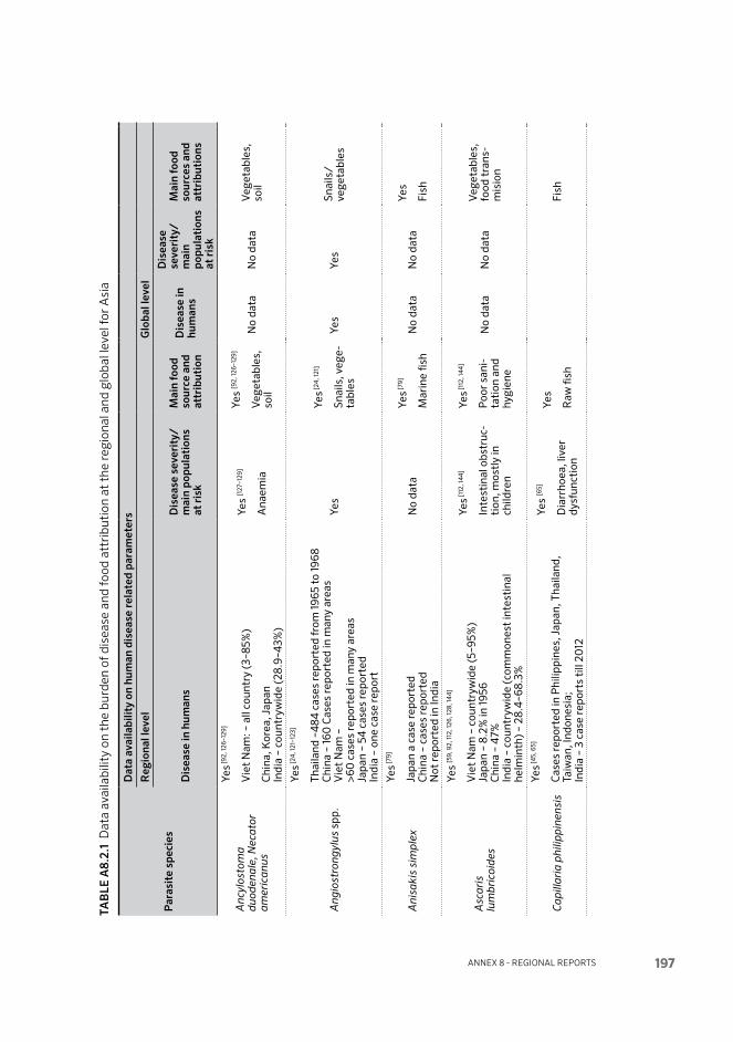
Annex 8 - regionAl reports 197
TAb
lE A
8.2.
1 D
ata
avai
labi
lity
on t
he b
urde
n of
dis
ease
and
food
att
ribu
tion
at
the
regi
onal
and
glo
bal l
evel
for
Asi
a
Par
asit
e sp
ecie
s
Dat
a av
aila
bilit
y on
hum
an d
isea
se re
late
d pa
ram
eter
s
Reg
iona
l lev
elG
loba
l lev
el
Dis
ease
in h
uman
sD
isea
se s
ever
ity/
m
ain
popu
lati
ons
at r
isk
Mai
n fo
od
sour
ce a
nd
attr
ibut
ion
Dis
ease
in
hum
ans
Dis
ease
se
veri
ty/
mai
n po
pula
tion
s at
ris
k
Mai
n fo
od
sour
ces
and
attr
ibut
ions
Anc
ylos
tom
a d
uod
enal
e, N
ecat
or
amer
ican
us
Yes [9
2, 12
6–12
9]
Vie
t N
am: –
all
coun
try
(3–8
5%)
Chi
na, K
orea
, Jap
an
Indi
a –
coun
try w
ide
(28.
9–4
3%)
Yes
[127
–129
]
Ana
emia
Yes
[92,
126–
129]
Veg
e tab
les,
so
ilN
o da
taN
o da
taV
ege t
able
s,
soil
Ang
iost
ron g
ylus
spp
.
Yes [2
4, 1
21–1
23]
Thai
land
–4
84 c
ases
repo
rted
from
1965
to
1968
C
hina
– 16
0 C
ases
repo
rted
in m
any
area
s V
iet
Nam
–
>60
cas
es re
port
ed in
man
y ar
eas
Japa
n –
54 c
ases
repo
rted
In
dia
– on
e ca
se re
port
Yes
Yes [2
4, 1
21]
Snai
ls, v
ege-
tabl
esYe
sYe
sSn
ails
/ve
geta
bles
Ani
saki
s si
mp
lex
Yes [7
9]
Japa
n a
case
repo
rted
C
hina
– c
ases
repo
rted
N
ot re
port
ed in
Indi
a
No
data
Yes [7
9]
Mar
ine
fish
No
data
No
data
Yes
Fish
Asc
aris
lu
mb
ri co
ides
Yes
[59,
92,
112,
126,
128,
144
]
Vie
t N
am –
cou
ntry
wid
e (5
–95%
) Ja
pan
– 8.
2% in
1956
C
hina
– 4
7%
Indi
a –
coun
try w
ide
(com
mon
est
inte
stin
al
helm
inth
) – 2
8.4
–68.
3%
Yes [1
12, 1
44
]
Inte
stin
al o
bstr
uc-
tion
, mos
tly
in
child
ren
Yes [1
12, 1
44
]
Poo
r sa
ni-
tati
on a
nd
hygi
ene
No
data
No
data
Veg
e tab
les,
fo
od t
rans
-m
isio
n
Cap
illar
ia p
hilip
pin
ensi
s
Yes [4
5, 6
5]
Cas
es re
port
ed in
Phi
lippi
nes,
Jap
an, T
haila
nd,
Taiw
an, I
ndon
esia
; In
dia
– 3
case
repo
rts
till
2012
Yes
[65]
Dia
rrho
ea, l
iver
dy
s fun
ctio
n
Yes
Raw
fish
Fish

Multi criteria-based ranking for risk ManageMent of food-borne parasites198
Par
asit
e sp
ecie
s
Dat
a av
aila
bilit
y on
hum
an d
isea
se re
late
d pa
ram
eter
s
Reg
iona
l lev
elG
loba
l lev
el
Dis
ease
in h
uman
sD
isea
se s
ever
ity/
m
ain
popu
lati
ons
at r
isk
Mai
n fo
od
sour
ce a
nd
attr
ibut
ion
Dis
ease
in
hum
ans
Dis
ease
se
veri
ty/
mai
n po
pula
tion
s at
ris
k
Mai
n fo
od
sour
ces
and
attr
ibut
ions
Clo
nor
chis
sin
ensi
s
Yes
[24
, 46,
48–
52]
Chi
na –
15 m
illio
n K
orea
– 2
mill
ion
infe
cted
, pre
vale
nce
of 1.
4–2
1.0
%
Japa
n –
prev
alen
ce o
f 1.0
–54
.2%
(196
0);
10.9
–66%
(1
961)
; now
alm
ost
disa
ppea
red
Vie
t N
am –
pre
vale
nce
of 19
.5%
(0.2
–40
%) i
n 15
/64
pr
ovin
ces
in t
he n
orth
Ta
iwan
– p
reva
lenc
e of
10–2
0%
C
hina
– 15
mill
ion
infe
cted
in 2
7 pr
ovin
ces
Indi
a –
Alm
ost
abse
nt. V
ery
few
cas
e re
port
s
Yes
[46,
48,
51,
52]
601 m
illio
n
Acu
te d
isea
se
Yes
[48,
50
]
Raw
& u
nder
-co
oked
fish
.
Yes
[46,
47]
Yes
[46]
601 m
illio
n
Raw
& u
nder
-co
oked
fish
(f
resh
wat
er)
Cry
pto
spor
idiu
m s
pp
Yes [9
2, 9
5]
Vie
t N
am –
2.8
% a
nd c
ase
repo
rted
(nat
iona
l)
Indi
a –
18.9
% in
chi
ldre
n w
ith
diar
rhoe
a Ja
pan
– ca
se re
port
ed
Chi
na –
1.36
–13.
3%
No
data
[95]
Yes [9
2–94
]
Veg
e tab
les,
w
ater
ra
w m
eat
(yuk
ke)
No
data
[91]
No
data
[91]
Yes [9
1]
Wat
er, v
ege-
tabl
es
HIV
-rel
ated
Dip
hyllo
bot
h riu
m s
pp.
Yes
[81–
87]
Cas
es re
port
ed o
f D. n
ihon
kaie
nse,
D. l
atum
, D
. pac
ificu
m, D
. cam
eron
i, D
. yon
agoe
nse
in J
apan
Fe
w c
ases
of D
. lat
um re
port
ed fr
om s
outh
Indi
a
Yes [8
1, 8
2]Ye
s [8
1–87
]N
o da
taN
o da
taN
o da
ta
Dip
log
ono p
orus
bal
aen
opte
rae
Yes [6
6, 8
9]
Cas
e re
port
ed in
Jap
an
Not
repo
rted
in In
dia,
Vie
t N
am o
r Th
aila
ndYe
s [66,
89]
Yes
[89]
No
data
No
data
No
data
Ech
ino c
occu
s sp
p.
Yes [5
9, 13
5–14
0]
Cas
es re
port
ed in
Jap
an, C
hina
(380
00
0 c
ases
),
Kor
ea, M
ongo
lia, T
haila
nd, B
angl
ades
h, N
epal
In
dia
– pr
eval
ence
not
cle
arly
kno
wn;
end
emic
in
both
rur
al a
nd u
rban
are
as o
f sou
ther
n an
d ce
ntra
l st
ates
.
Yes
Yes
ve
ge ta
bles
No
data
No
data
Veg
e tab
les
Ech
ino s
tom
a sp
p
Yes
[73–
75]
Japa
n –
22.4
%
Thai
land
– 0
.04
–55.
3%
Chi
na –
1.5–
20.1
%
Vie
t N
am –
a c
ase
repo
rted
In
dia
– ve
ry r
are;
ver
y fe
w c
ase
repo
rts
Yes
Yes
[73]
Raw
sna
il &
fis
hN
o da
taN
o da
taYe
s
Snai
l; fis
h

Annex 8 - regionAl reports 199
Par
asit
e sp
ecie
s
Dat
a av
aila
bilit
y on
hum
an d
isea
se re
late
d pa
ram
eter
s
Reg
iona
l lev
elG
loba
l lev
el
Dis
ease
in h
uman
sD
isea
se s
ever
ity/
m
ain
popu
lati
ons
at r
isk
Mai
n fo
od
sour
ce a
nd
attr
ibut
ion
Dis
ease
in
hum
ans
Dis
ease
se
veri
ty/
mai
n po
pula
tion
s at
ris
k
Mai
n fo
od
sour
ces
and
attr
ibut
ions
Ent
amoe
ba
hist
olyt
ica
Yes
[92,
148–
152]
Vie
t N
am –
2–6
% in
chi
ldre
n In
dia
– in
test
inal
am
oebi
asis
wit
h E
. his
toly
tica
or
E. d
ispa
r (1
–58%
); in
test
inal
am
oeb i
asis
wit
h pr
oven
E
. his
toly
tica
(34
.6%
of a
ll sa
mpl
es fo
und
+ve
E. h
isto
lyti
ca o
r E
. dis
par)
; ext
ra-i
ntes
tina
l am
oeb-
iasi
s –
amoe
bic
liver
abs
cess
(3–9
% o
f all
the
case
s of
in
test
inal
am
oeb i
asis
)
Yes
No
data
No
data
No
data
Veg
e tab
les,
w
ater
, foo
d tr
ansm
issi
on
Ent
erob
ius
verm
icul
aris
Yes
[92,
111,
112,
157]
Vie
t N
am –
29–
43%
in t
he n
orth
; 7.5
–50
% in
the
ce
ntre
; 16–
47%
in t
he s
outh
; 51.
2% in
chi
ldre
n 1–
5 ye
ars
old
Indi
a –
coun
try w
ide
in c
hild
ren
(0.5
–12.
6%);
mor
e co
mm
on in
rur
al t
han
urba
n ar
eas
Yes
Yes
[111
, 112
]
Ove
r-cr
owdi
ngN
o da
taN
o da
ta
Fasc
iola
spp
Yes
[79,
92.
96,
99]
Vie
t N
am –
>20
00
0 c
ases
from
52/
64 p
rovi
nces
C
ases
repo
rted
in C
hina
(14
8 ca
ses)
, Tha
iland
, Kor
ea,
Iran
, Jap
an, M
alay
sia,
Sin
gapo
re, L
aos,
Cam
bodi
a &
P
hilip
pine
s In
dia
– A
few
cas
e re
port
s
No
data
Yes [7
9, 9
2, 9
6]
Wat
er, r
aw
vege
tabl
esN
o da
taN
o da
taYe
s [7
9]
wat
er, v
ege-
tabl
es
Fasc
iolo
psi
s b
uski
Yes [7
3, 9
2, 10
2, 10
3]
Vie
t N
am –
0.5
–3.8
% in
16/6
4 p
rovi
nces
C
hina
– 10
.2–9
2.9%
in s
ome
area
s Th
aila
nd –
10%
in c
hild
ren
wit
h in
tes t
inal
par
asit
es
Cas
es re
port
ed in
Tai
wan
, Cam
bodi
a, L
aos
PD
R,
Mal
aysi
a, In
done
sia
& M
yanm
ar
Indi
a –
ende
mic
in E
& N
E s
tate
s –
prev
alen
ce o
f 0
–22.
4%
Yes
[10
2]
Abd
omin
al p
ain,
di
arrh
oea,
inte
stin
al
obst
ruc t
ion
Yes
[73,
92,
102]
Wat
er, r
aw
vege
tabl
esN
o da
taN
o da
taW
ater
, raw
ve
ge ta
bles

Multi criteria-based ranking for risk ManageMent of food-borne parasites200
Par
asit
e sp
ecie
s
Dat
a av
aila
bilit
y on
hum
an d
isea
se re
late
d pa
ram
eter
s
Reg
iona
l lev
elG
loba
l lev
el
Dis
ease
in h
uman
sD
isea
se s
ever
ity/
m
ain
popu
lati
ons
at r
isk
Mai
n fo
od
sour
ce a
nd
attr
ibut
ion
Dis
ease
in
hum
ans
Dis
ease
se
veri
ty/
mai
n po
pula
tion
s at
ris
k
Mai
n fo
od
sour
ces
and
attr
ibut
ions
Gia
rdia
duo
den
alis
(sy
n.
G. l
amb
lia, G
. int
esti
nal
is)
Yes
[92,
110
, 154
]
Vie
t N
am –
1–10
%
Chi
na –
infe
ctio
n ra
te r
ange
d fr
om 8
.67%
–9.0
7%,
foun
d in
13 a
reas
of 3
5 ci
ties
at
the
prov
inci
al le
vel
Indi
a –
coun
try w
ide
dist
ribu
tion
(8.4
–53.
8%)
Yes
[110
, 154
]N
o da
taN
o da
taN
o da
ta
Veg
e tab
les,
w
ater
, foo
d tr
ans m
issi
on
Gna
tho s
tom
a sp
p
Yes
[70
–72,
90
]
Cas
es re
port
ed in
Jap
an w
ith
3 22
5 ca
ses,
incl
udin
g 86
from
Chi
na a
nd 3
4 fr
om o
ther
Asi
an s
ourc
es.
Cas
es re
port
ed in
Chi
na, T
haila
nd, V
iet
Nam
, Ind
ia,
Laos
PD
R, M
yanm
ar, C
ambo
dia,
Ban
glad
esh,
M
alay
sia,
Indo
nesi
a an
d P
hilip
pine
s.
Indi
a –
14 c
ases
repo
rted
unt
il 20
12
Yes
[71,
72]
Ocu
lar
and
cere
bral
m
anif
es ta
tion
s
Yes [7
0]
Raw
fish
, am
phib
ian
rept
ile
Fish
, am
phib
ian
rept
ile
Het
ero p
hyid
s
Yes [5
3–55
]
Thai
land
– 0
.3–7
.8%
V
iet
Nam
– 0
.5–6
4.4
% in
>18
pro
vinc
es
Chi
na –
1–2%
Ja
pan
– 11
%
Indi
a –
Not
yet
repo
rted
Yes [5
5]
Yes
[53,
55]
Raw
fish
di
shes
Yes
Yes
Raw
Fis
h
Kud
oa s
epte
m p
unct
ata
No
data
Not
repo
rted
in In
dia
Yes
Acu
te a
nd s
elf-
limit
ing
Yes [8
8]
Raw
flat
fish
(P
ara l
ich t
hys
oliv
aceu
s)
100
% fo
od-
born
e tr
ans-
mis
sion
No
data
No
data
Yes
Fish
; foo
d-bo
rne
tran
s-m
issi
on
Met
agon
imus
spp
.
Yes
[57,
76,
78]
Man
y ca
ses
repo
rted
in K
orea
& C
hina
In
dia
– ve
ry r
are.
Ver
y fe
w c
ase
repo
rts
of
Met
agon
imus
yok
ogaw
ai
Yes [7
6, 7
8]
Acu
te d
iarr
hoea
Yes
[76,
78]
Und
er co
oked
fr
esh w
ater
fis
h
No
data
No
data
Yes
Fish

Annex 8 - regionAl reports 201
Par
asit
e sp
ecie
s
Dat
a av
aila
bilit
y on
hum
an d
isea
se re
late
d pa
ram
eter
s
Reg
iona
l lev
elG
loba
l lev
el
Dis
ease
in h
uman
sD
isea
se s
ever
ity/
m
ain
popu
lati
ons
at r
isk
Mai
n fo
od
sour
ce a
nd
attr
ibut
ion
Dis
ease
in
hum
ans
Dis
ease
se
veri
ty/
mai
n po
pula
tion
s at
ris
k
Mai
n fo
od
sour
ces
and
attr
ibut
ions
Op
isth
orch
is v
iver
rini
Yes [4
2, 4
4, 4
5]
Thai
land
– 15
.7%
La
o P
DR
– 3
7–86
%
Cam
bodi
a –
som
e ca
ses
Vie
t N
am –
1.4
–37.
9% in
9/6
4 p
rovi
nces
in t
he s
outh
. M
alay
sia
– a
case
repo
rted
In
dia
– N
o ca
ses
yet
repo
rted
.
Yes [4
2, 4
4]
67 m
illio
n
Yes
[42,
44
]
Raw
fish
dis
h
Yes
[43]
10 m
illio
n
Yes [4
3]
67 m
illio
n
Yes
[42]
Koi
pla
; Lap
pl
a; P
la s
om;
Raw
-fish
Par
a gon
imus
spp
.
Yes
[56–
62]
Thai
land
– c
ases
repo
rted
in 2
3/68
pro
vinc
es
Vie
t N
am –
0.5
–15%
in 10
/64
pro
vinc
es
Japa
n –
case
repo
rts
wit
h >2
00
cas
es
Phi
lippi
nes
– 27
.2–4
0%
in s
ome
area
s C
hina
– 4
.1–5
.1%
in 2
4 p
rovi
nces
In
dia
– en
dem
ic t
o N
E s
tate
s (M
anip
ur, N
agal
and
and
Aru
nach
al P
rade
sh);
Par
agon
imus
het
erot
rem
us
is t
he c
omm
on s
peci
es; u
p to
50
% s
ero p
rev a
lenc
e in
th
ese
regi
ons.
Yes
[60
–62]
Cou
gh, d
yspn
oea,
re
curr
ent
haem
-op
tysi
s
Yes
[56,
60
–62]
Raw
cra
b;
fres
h wat
er
crab
, wild
bo
ar m
eat
in
Japa
n
Yes
Yes
Fres
h wat
er
crab
; w
ild b
oar
mea
t [5
6]
Pse
udo t
erra
nov
a d
ecip
iens
e
Yes
[79]
Cas
e re
port
ed in
Jap
an
Not
repo
rted
in In
dia
No
data
Yes
[79]
Mar
ine
fish
No
data
No
data
Yes
Fish
Sar
cocy
stis
faye
riYe
s
Cas
e re
port
ed in
Jap
an [1
58]
Yes
[158
]
Acu
te a
nd s
elf-
limit
ed
Yes
[158
]
Raw
hor
se
mea
t 10
0%
food
-bo
rne
Sar
cocy
stis
spp
.
Yes
[1, 2
, 4, 6
]
Thai
land
(1.5
%)
Indi
a –
11 c
ase
repo
rts
from
1990
to
200
4. A
few
ea
rlie
r re
port
s.
Chi
na (2
9.7%
) 4
6 ca
ses
repo
rted
by
1990
in A
sia,
incl
udin
g C
hina
, M
alay
sia
and
Indi
a.
In M
alay
sia,
19.7
% o
f 24
3 pe
rson
s ha
d an
tibo
dies
to
Sarc
ocys
tis
No
data
Yes
[2,–
4, 6
]
Mea
t (p
ork
and
beef
)
Raw
por
k m
uscl
e an
d off
al
No
data
No
data
Yes
Mea
t (p
ork
and
beef
)
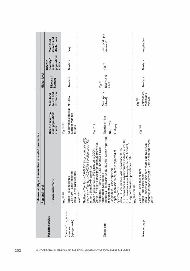
Multi criteria-based ranking for risk ManageMent of food-borne parasites202
Par
asit
e sp
ecie
s
Dat
a av
aila
bilit
y on
hum
an d
isea
se re
late
d pa
ram
eter
s
Reg
iona
l lev
elG
loba
l lev
el
Dis
ease
in h
uman
sD
isea
se s
ever
ity/
m
ain
popu
lati
ons
at r
isk
Mai
n fo
od
sour
ce a
nd
attr
ibut
ion
Dis
ease
in
hum
ans
Dis
ease
se
veri
ty/
mai
n po
pula
tion
s at
ris
k
Mai
n fo
od
sour
ces
and
attr
ibut
ions
Sp
irom
etra
eri
nace
ieu
rop
aei
(spa
rgan
osis
)
Yes
[66–
69]
Japa
n –
case
repo
rted
V
iet
Nam
– c
ase
repo
rted
In
dia
– a
few
cas
e re
port
s
Yes
[66–
69]
Abd
omin
al, c
ereb
ral
& o
cula
r m
anif
es-
tati
ons
No
data
No
data
No
data
Frog
Taen
ia s
pp.
Yes [8
, 10
–19]
Thai
land
– T
aeni
asis
(0.6
–5.9
%) &
cys
tice
rcos
is (4
%).
V
iet
Nam
– T
aeni
asis
(0.5
–12%
) & c
ysti
cerc
osis
(7%
) in
mor
e th
an 5
0/6
4 p
rovi
nces
. Ja
pan
– cy
stic
erco
sis
44
6 ca
ses
up t
o 20
04
. C
hina
– 7
mill
ion
peop
le in
fect
ed in
29
prov
ince
s.
Phi
lippi
nes
– Ta
enia
sis
(0.5
6–10
.26%
) & c
ase
repo
rted
of c
ysti
cerc
osis
. In
done
sia
– Ta
enia
sis
(0.5
6–10
.26%
) & c
ase
repo
rted
of
cys
tice
rcos
is.
Ban
glad
esh
– ca
se re
port
ed.
Nep
al –
Tae
nias
is (4
3%) &
cas
e re
port
ed o
f cy
stic
erco
sis.
In
dia
– T.
sol
ium
Tae
nias
is p
reva
lenc
e 18
.6%
; N
CC
pre
vale
nce
in a
sym
ptom
atic
indi
vidu
als
15.1
%;
NC
C p
reva
lenc
e in
act
ive
epile
ptic
s 26
.3–5
6.8%
; T.
sag
inat
a Ta
enia
sis
prev
alen
ce 5
.3%
.
Yes [1
3, 17
]
Taen
iasi
s –
No
NC
C –
Yes
Epi
leps
y
Mea
t (p
ork
& b
eef)
Yes [9
]
DA
LY: 2
–5
×10
6Ye
s [9]
Bee
f, p
ork,
pig
vi
scer
a [9
]
Toxo
cara
spp
.
Yes
[92,
126.
132,
133]
Vie
t N
am –
one
cas
e re
port
Ja
pan
– on
e ca
se re
port
In
dia
– en
dem
ic in
nor
ther
n st
ates
; up
to 3
3% in
K
ashm
ir; s
ero p
osit
ivit
y of
6–2
3% in
oth
er n
orth
ern
stat
es
Yes
[133
]Ye
s [133
]
Veg
e tab
les,
fo
od t
rans
-m
issi
on
No
data
No
data
Veg
e tab
les

Annex 8 - regionAl reports 203
Par
asit
e sp
ecie
s
Dat
a av
aila
bilit
y on
hum
an d
isea
se re
late
d pa
ram
eter
s
Reg
iona
l lev
elG
loba
l lev
el
Dis
ease
in h
uman
sD
isea
se s
ever
ity/
m
ain
popu
lati
ons
at r
isk
Mai
n fo
od
sour
ce a
nd
attr
ibut
ion
Dis
ease
in
hum
ans
Dis
ease
se
veri
ty/
mai
n po
pula
tion
s at
ris
k
Mai
n fo
od
sour
ces
and
attr
ibut
ions
Toxo
pla
sma
gon
dii
Yes
[30
, 31,
33–
41]
Thai
land
– 2
.6%
C
hina
– 12
-45%
and
12.7
–15.
1%
Vie
t N
am –
som
e ca
ses
repo
rted
Sr
i Lan
ka –
27.
5%
Japa
n –
1.8–
5.6%
M
alay
sia
– 10
–50
%
Nep
al –
45.
6%
Indi
a –
sero
posi
tivi
ty fo
r Ig
G in
gen
eral
pop
ulat
ion
– 10
.8–5
1.8%
and
for
IgM
– 2
–5%
; in
fem
ales
wit
h ba
d ob
stet
ric
hist
ory
– Ig
G w
as 4
9.5%
; in
HIV
-inf
ecte
d ca
ses
sero
posi
tivi
ty fo
r Ig
G 7
0%
.
Yes
[33,
36–
38]
CN
S di
seas
e in
HIV
in
fect
ed
Raw
mea
t,
pork
, ch
icke
n,
frui
t, v
ege-
tabl
es [3
0]
Yes [3
2]Ye
s [32]
Yes
[30
, 32]
Bee
f, p
ork,
go
at, h
orse
, sh
eep,
ch
icke
n;
cont
ami n
ated
fr
uit
& v
ege-
tabl
es; r
aw
mus
sels
, cla
ms
& o
yste
rs
Tric
hine
lla s
pir
alis
Yes
[20
–28]
Thai
land
– 0
.9–9
%
Vie
t N
am –
5 o
ut br
eaks
in n
orth
mou
ntai
nous
pr
ovin
ces,
wit
h >1
00
pat
ient
s an
d 8
deat
hs u
p to
20
12.
Chi
na –
>50
0 o
utbr
eaks
in 12
/34
pro
vinc
es, w
ith
25 6
85 p
erso
ns a
ffec
ted
(24
1 dea
ths)
. Ja
pan
– 1 c
ase
repo
rted
. In
dia
– ve
ry fe
w c
ase
repo
rts.
Rec
entl
y a
poin
t so
urce
ou
tbre
ak o
f 42
case
s.
Yes
[25,
27]
Hig
h m
orta
lity
Yes [2
1, 2
6, 2
7, 2
9]
Mea
t;
cons
ump-
tion
of
inad
e-qu
atel
y co
oked
wild
bo
ar m
eat
Yes
[21,
26,
29]
Yes
[21,
26,
29]
Yes [2
1, 2
6, 2
9]
Dom
esti
c pi
gs,
wild
boa
r, r
aw
hors
e m
eat
100
% fo
od-
born
e tr
ansm
is si
on
Tric
huri
s tr
ichi
ura
Yes
[10
5, 10
8–11
2]
Thai
land
– 7
0%
La
os P
DR
– 4
1.5%
V
iet
Nam
– 7
0–8
0%
in t
he n
orth
and
5–1
0%
in t
he
sout
h In
dia
– ad
ults
2–6
.6%
; chi
ldre
n 8–
26.4
%
Yes
[10
6–10
9]
Rec
tal p
rola
pse
Yes
Veg
e tab
le,
pers
onal
hy
gien
e
Yes
Yes [1
07]
For
2010
, gl
obal
po
pula
tion
at
ris
k:
5023
.3
(mill
ions
)
Food
born
e

Multi criteria-based ranking for risk ManageMent of food-borne parasites204
Sources for Table A8.2.1
001. Nichpanit, S., Nakai, W., Wongsaroj, t. & Nithikathkul, C. 2010. First large scale of human Sarcocystis hominis in Thailand. Trends Research in Science and Technology, 2(1): 1–5.
002. Fayer, R. 2004. Sarcocystis spp. in human infections. Clinical Microbiology Reviews, 17(4): 894-+
003. Banerjee, p.S., Bhatia, B.B. & pandit, B.A. 1994. Sarcocystis suihominis infection in human beings in India. Journal of Veterinary Parasitology, 8(1): 57–58.
004. von Sonnenburg, F., Cramer, J.p., Freedman, D.O., plier, D.A., Esposito, D.H., Sotir, M.J. & lankau, E.W. 2012. Notes from the field: acute muscular sarcocystosis among returning travelers – Tioman Island, Malaysia, 2011 (Reprinted from MMWR, 61:37–38). Journal of the American Medical Association, 307(12): 1247–1247.
006. li, J.H., lin, Z., Qin, y.X. & Du, J. 2004. Sarcocystis suihominis infection in human and pig population in Guangxi [In Chinese; no abstract available]. Zhongguo ji sheng chong xue yu ji sheng chong bing za zhi [Chinese Journal of Parasitology & Parasitic Diseases], 22: 82.
008. Anantaphruti, M.t., yamasaki, H., Nakao, M., Waikagul, J., Watthanakulpanich, D., Nuamtanong, S., Maipanich, W., pubampen, S., Sanguankiat, S., Muennoo, C., Nakaya, k., Sato, M.O., Sako, y., Okamoto, M. & Ito, A. 2007. Sympatric occurrence of Taenia solium, T. saginata, and T. asiatica, Thailand. Emerging Infectious Diseases, 13(9): 1413–1416.
009. torgerson, p.R. & Macpherson, C.N.l. 2011. The socioeconomic burden of parasitic zoonoses: Global trends. Veterinary Parasitology, 182(1 – Special Issue): 79–95.
010. De, N.V. 2004. Taenia and Cysticercosis in Vietnam. In: [Abstracts of the ] Joint International Tropical Medicine Meeting (JITMM 2004), 29 November – 1 December 2004Bangkok, Thailand. Mahidol University, Faculty of Tropical Medicine. 284 p.
011. Ito, A., Craig, p.S. & Schantz, p.M. (Editors). 2006. Taeniasis/cysticercosis and echinococcosis with focus on Asia and the Pacific. Proceedings of the 5th International Symposium on the Cestode Zoonoses, Asahikawa, Japan, 2006. Parasitology International, 55 (Suppl.): S1–S312.
012. li, t., Ito, A., Craig, p.S., Chen, X., Qiu, D., Zhou, X., Xiao, N. & Qiu, J. 2007. Taeniasis/cysticercosis in China. Southeast Asian Journal of Tropical Medicine and Public Health, 38(Suppl.1): 131–139.
013. parija, S.C. & Raman, G.A. 2100. Anti-Taenia solium larval stage IgG antibodies in patients with epileptic seizures. Tropical Parasitology, 1(1): 20–25.
014. parija, S.C. & Gireesh, A. 2011. Cysticercus cellulosae antigens in the serodiagnosis of neurocysticercosis. Tropical Parasitology, 1(2): 64–72.
015. parija, S.C. & Sahu, p.S. A serological study of human cysticercosis in Pondicherry, South India. Journal of Communicable Diseases, 35(4): 283–289.
016. parija, S.C., Balamurungan, N., Sahu, p.S. & Subbaiah, S.p. 2005. Cysticercus antibodies and antigens in serum from blood donors from Pondicherry, India. Revista do Instituto de Medicina Tropical de São Paulo, 47(4): 227–230.
017. prasad, k.N., prasad, A., Verma, A. & Singh, A.k. 2008. Human cysticercosis and Indian scenario: a review. Journal of Biosciences, 33(4): 571–582.
018. prasad, k.N., prasad, A., Gupta, R.k., pandey, C.M. & Singh, u. 2007. Prevalence and associated risk factors of Taenia solium Taeniasis in a rural pig farming community of north India. Transactions of the Royal Society of Tropical Medicine and Hygiene, 101(12): 1241–1247.
019. Wani, S.A., Ahmad, F., Zargar, S.A., Amin, A., Dar, Z.A. & Dar, p.A. 2010. Intestinal helminthiasis in children of Gurez valley of Jammu and Kashmir State, India. Journal of Global Infectious Diseases, 2(2): 91–94.
020. liu, M.y. & Boireau, p. 2002. Trichinellosis in China: epidemiology and control. Trends in Parasitology, 18(12): 553–556.
021. Murrell, k.D. & pozio, E. 2011. Worldwide occurrence and impact of human trichinellosis 1986–2009. Emerging Infectious Diseases, 17(12): 2194–2202.

Annex 8 - regionAl reports 205
022. De, N.V., Dorny, p. & Waikagul, J. 2006. Trichinelliasis in Vietnam. pp. 37–42, in: [Proceedings of the ] Seminar on Food- and Water- borne Parasitic Zoonoses (5thFBPZ), 28–30 November 2006.
023. De, N.V., trung, N.V., Ha, N.H., Nga, V.t., Ha, N.M., thuy, p.t., Duyet, l.V. & Chai, J.y. 2012. An outbreak of trichinosis with molecular identification of Trichinella sp. in Vietnam. Korean Journal of Parasitology, 50(4): 339–343.
024. Arizono, N. 2005. Food-borne helminthiasis in Asia. Asian Parasitology Monographs Series, vol. 1. Federation of Asian Parasitologists, Chiba, Japan. 318 p.
025. yamasaki, H. 2013. Current status and perspectives of cysticercosis and Taeniasis in Japan. Paper presented at Symposium on Asian Taenia, October 2011, Osong, Republic of Korea. Korean Journal of Parasitology, 51(1): 19–29.
026. Orlandi, p.A., Chu, D.-M.t., Bier, J.W. & Jackson, G.J. 2002. Parasites and the food supply. Food Technology, 56(4): 72–81.
027. Sethi, B., Butola, k.S., kumar, y. & Mishra, J.p. 2012. Multiple outbreaks of trichinellosis with high mortality rate. Tropical Doctor, 42(4): 243–243.
028. pebam, S., Goni, V., patel, S. kumar, V., Rawall, S. & Bali, k. 2012. Case Report: A 12-year-old child with trichinellosis, pyomyositis and secondary osteomyelitis. Journal of Global Infectious Diseases, 4(1): 84–88.
029. Slifko, t.R., Smith, H.V. & Rose, J.B. 2000. Emerging parasite zoonoses associated with water and food. International Journal for Parasitology, 30(12-13): 1379–1393.
030. Sakae, C., Natphopsuk, S., Settheetham-Ishida, W. & Ishida, t. 2013. Low prevalence of Toxoplasma gondii infection among women in north-eastern Thailand. Journal of Parasitology, 99(1): 172–173.
031. Zhou, p., Chen, Z., li, H.-l., Zheng, H., He, S., lin, R.-Q. & Zhu, X.-Q. 2011. Toxoplasma gondii infection in humans in China. Parasites & Vectors, 4: 165
032. Jones, J.l. & Dubey, J.p. 2012. Foodborne toxoplasmosis. Clinical Infectious Diseases, 55(6): 845–851.
033. Akao, N. & Ohta, N. 2007. Toxocariasis in Japan. Parasitology International, 56(2): 87–93.
036. parija, S.C. 2005. Parasitic infections of the central nervous system. Journal of Parasitic Diseases, 29(2): 85–96.
037. Shivaprakash. M.R., parija, S.C. & Sujatha, S. 2001. Seroprevalence of toxoplasmosis in HIV infected patients in Pondicherry. Journal of Communicable Diseases, 33(3): 221–223.
038. Dhumne, M., Sengupta, C., kadival, G., Rathinaswamy, A. & Velumani, A. 2007. National seroprevalence of Toxoplasma gondii in India. Journal of Parasitology, 93(6): 1520–1521.
039. Sucilathangam, G., palaniappan, N., Sreekumar, C. & Anna, t. 2010. IgG – Indirect fluorescent antibody technique to detect seroprevalence of Toxoplasma gondii in immunocompetent and immunodeficient patients in southern districts of Tamil Nadu. Indian Journal of Medical Microbiology, 28(4): 354–357.
040. Elhence, p., Agarwal, p., prasad, k.N. & Chaudhary, R.k. 2010. Seroprevalence of Toxoplasma gondii antibodies in North Indian blood donors: Implications for transfusion transmissible toxoplasmosis. Transfusion and Apheresis Science, 43(1): 37-40.
041. Dubey, J.p. 1986. A review of toxoplasmosis in pigs. Veterinary Parasitology, 19(3-4): 181–223.
042. Sithithaworn, p., Andrews, R.H., Nguyen, V.D., Wongsaroj, t., Sinuon, M., Odermatt, p., Nawa, y., liang, S., Brindley, p.J. & Sripa, B. 2012. The current status of opisthorchiasis and clonorchiasis in the Mekong Basin. Parasitology International, 61(1, Special Issue): 10–16.
043. Sripa, B., Bethony, J.M., Sithithaworn, p., kaewkes, S., Mairiang, E., loukas, A., Mulvenna, J., laha, t., Hotez, p.J. & Brindley, p.J. 2011. Opisthorchiasis and opisthorchis-associated cholangiocarcinoma in Thailand and Laos. Acta Tropica, 120(Special Issue – Suppl. 1): S158–S168.

Multi criteria-based ranking for risk ManageMent of food-borne parasites206
044. De, N.V. 2004. Fish-borne trematodes in Viet Nam. Southeast Asian Journal of Tropical Medicine and Public Health, 35(Suppl. 1): 299–301.
045. Miyazaki, I. 1991. Helminthic zoonoses. SEAMIC publication, no. 62. International Medical Foundation of Japan, Tokyo. 494 p.
046 keiser, J. & utzinger, J. 2005. Emerging foodborne trematodiasis. Perspective. Emerging Infectious Diseases [Online] http://dx.doi.org/10.3201/eid1110.050614
047. Qian, M.-B., Chen, y.-D., Fang, y.-y., Xu, l.-Q., Zhu, t.-J., tan, t., Zhou, C.-H., Wang, G.-F., Jia, t.-W., yang, G.-J. & Zhou, X.-N. 2011. Disability weight of Clonorchis sinensis infection: captured from community study and model simulation. PLOS Neglected Tropical Diseases, 5(12): Art. no. e1377. [Online]
048. De, N.V. & le, t.H. 2011. Human infections of fish-borne trematodes in Vietnam: Prevalence and molecular specific identification at an endemic commune in Nam Dinh province. Experimental Parasitology, 129(4): 355–361.
050. yoshida, y. 2012. Clonorchiasis – A historical review of contributions of Japanese parasitologists. Parasitology International, 61(1 – Special Issue): 5–9.
051. Zhou, p., Chen, N., Zhang, R.l., lin, R.Q. & Zhu, X.Q. 2008. Food-borne parasitic zoonoses in China: perspective for control. Trends in Parasitology, 24(4): 190–196.
052. Mirdha, B.R., Gulati, S., Sarkar, t. & Samantray, J.C. 1998 Acute clonorchiasis in a child. Indian Journal of Gastroenterology, 17(4): 155.
053. Arizono, N. 2005. Food-borne helminthiasis in Asia. Asian Parasitology Monographs Series, vol. 1. Federation of Asian Parasitologists, Chiba, Japan. 318 p.
054. Dung, D.t., Van De, N., Waikagul, J., Dalsgaard, A., Chai, J.y., Sohn, W.M. & Murrell, k.D. 2007. Fish-borne zoonotic intestinal trematodes, Vietnam. Emerging Infectious Diseases, 13(12): 1828–1833.
055. kino, H., Oishi, H., Ohno, y. & Ishiguro, M. 2002. An endemic human infection with Heterophyes nocens Onji et Nishio 1916 at Mikkabi-cho, Shizuoka, Japan. Japanese Journal of Tropical Medicine and Hygiene, 30: 301–304.
056. De, N.V. 2004. Epidemiology, pathology and treatment of paragonimiasis in Viet Nam. Southeast Asian Journal of Tropical Medicine and Public Health, 35(Suppl. 1): 331–336.
057. WHO. 2004. Report of the Joint WHO/FAO Workshop on Foodborne Trematode Infections in Asia. Ha Noi, Viet Nam, 26–28 November 2002. 62 p. Available at http://whqlibdoc.who.int/wpro/2004/RS_2002_GE_40(VTN).pdf
058. kawashima, k. 2003. Biology of Paragonimus. pp. 165–182, in: M. Otsuru, S. Kamegai and S. Hayashi (editors). Progress of Medical Parasitology in Japan, Vol. 8. Meguro Parasitological Museum, Tokyo, Japan.
059. Xu, l., Chen, y., Sun, F. Cai, l., Fang, y. & Wang, l. 2005. A national survey on current status of the important parasitic diseases in the human population. Coordinating Office of the National Survey on the Important Human Parasitic Diseases. Chinese Journal of Parasitology and Parasitic Diseases, 23: 332–340.
060. liu, Q., Wei, F., liu, W., yang, S. & Zhang, X. 2008. Paragonimiasis: an important food-borne zoonosis in China. Trends in Parasitology, 24: 318–323.
061. Singh, t.S., Sugiyama, H., umehara, A., Hiese, S. & khalo, k. 2009. Paragonimus heterotremus infection in Nagaland: A new focus of paragonimiasis in India. Indian Journal of Medical Microbiology, 27(2): 123–127.
062. Mahajan, R.C. 2005. Paragonimiasis: an emerging public health problem in India. Indian Journal of Medical Research, 121(6): 716–718.
065. Vasantha, p.l., Girish, N, & Sai leela, k. 2012. Human intestinal capillariasis: A rare case report from non-endemic area (Andhra Pradesh, India). Indian Journal of Medical Microbiology, 30(2) 236–239.
066. kamo, H. 2003. Cestodes – General view. pp. 235–236, in: M. Otsuru, S. Kamegai and S. Hayashi (editors). Progress of Medical Parasitology in Japan, Vol. 8. Meguro Parasitological Museum, Tokyo, Japan.
067. Duggal, S., Mahajan, R.k., Duggal, N. & Hans, C. 2011. Case of sparganosis: A diagnostic dilemma. Case report. Indian Journal of Medical Microbiology, 29(2): 183–186.
Annex 8 - regionAl reports 207
068. Sundaram, C., prasad, V.S.S.V. & Reddy, J.J.M. 2003. Cerebral sparganosis. Journal of the Association of Physicians of India, 51(11): 1107–1109.
069. Sen, D.k., Muller, R., Gupta, V.p. & Chilana, J.S. 1989. Cestode larva (sparganum) in the anterior-chamber of the eye. Tropical and Geographical Medicine, 41(3): 270–273.
070. Arizono, N. 2005. Food-borne helminthiasis in Asia. Asian Parasitology Monographs Series, vol. 1. Federation of Asian Parasitologists, Chiba, Japan. 318 p.
071. Rao, A.V., pravin, t. & parija, S.C. 1999. Intracameral gnathostomiasis: A first case report from Pondicherry. Journal of Communicable Diseases, 31(3): 197–198.
072. pillai, G.S., kumar, A., Radhakrishnan, N., Maniyelil, J., Shafi, t., Dinesh, k.R. & karim, S. 2102. Case Report: Intraocular gnathostomiasis: report of a case and review of literature. American Journal of Tropical Medicine and Hygiene, 86(4):620–623.
073. Arizono, N. 2005. Food-borne helminthiasis in Asia. Asian Parasitology Monographs Series, vol. 1. Federation of Asian Parasitologists, Chiba, Japan. 318 p.
074. Maji, A.k., Bera, D.k., Manna, B., Nandy, A., Addy, M. & Bandyopadhyay, A.k. 1993. First record of human infection with Echinostoma malayanum in India. Transactions of the Royal Society of Tropical Medicine and Hygiene, 87(6): 673–673.
075. Grover, M., Dutta, R., kumar, R., Aneja, S. & Mehta, G. 1998. Echinostoma iliocanum infection. Case report. Indian Pediatrics, 35 (June): 549–552.
076. Saito, S. 2003. Metagonimus – Research done after 1960. pp. 219–231, in: M. Otsuru, S. Kamegai and S. Hayashi (editors). Progress of Medical Parasitology in Japan, Vol. 8. Meguro Parasitological Museum, Tokyo, Japan.
078. uppal, B. & Wadhwa, V. 2005. Rare case of Metagonimus yokogawai. Indian Journal of Medical Microbiology, 23(1): 61–62.
079. Ishikura, H. 2003. Anisakiasis (2) Clinical Pathology and Epidemiology. pp. 451–473, in: M. Otsuru, S. Kamegai and S. Hayashi (editors). Progress of Medical Parasitology in Japan, Vol. 8. Meguro Parasitological Museum, Tokyo, Japan.
081. yamane, y. & Shiwaku, k. 2003. Diphyllobothrium nihonkaiense and other marine-origin cestodes. pp. 245–259, in: M. Otsuru, S. Kamegai and S. Hayashi (editors). Progress of Medical Parasitology in Japan, Vol. 8. Meguro Parasitological Museum, Tokyo, Japan.
082. yamasaki, H. 2013. Current status and perspectives of cysticercosis and Taeniasis in Japan. Paper presented at Symposium on Asian Taenia, October 2011, Osong, Republic of Korea. Korean Journal of Parasitology, 51(1): 19–29.
083. Ando, k., Ishikura, k., Nakakugi, t., Shimono, y., tamai, t., Sugawa, M., limviroj, W. & Chinzei, y. 2001. Five cases of Diphyllobothrium nihonkaiense infection with discovery of plerocercoids from an infective source, Oncorhynchus masou ishikawae. Journal of Parasitology, 87(1): 96–100.
084. Suzuki, J., Murata, R., Sadamatsu, k. & Araki, J. 2010. Detection and identification of Diphyllobothrium nihonkaiense plerocercoids from wild Pacific salmon (Oncorhynchus spp.) in Japan. Journal of Helminthology, 84: 434–440.
085. pancharatnam, S., Jacob, E. & kang, G. 1998. Human diphyllobothriasis: first report from India. Transactions of the Royal Society of Tropical Medicine and Hygiene, 92(2): 179–180.
086. Sheela Devi, C., Shashikala, Srinivasan, S., Murmu, u.C., Barman, p. & kanungo, R. 2007. A rare case of diphyllobothriasis from Pondicherry, South India. Indian Journal of Medical Microbiology, 25(2): 152–154.
087. Ramana, k.V., Rao, S., Vinaykumar, M., krishnappa, M., Reddy, R., Sarfaraz, M., kondle, V., Ratnamani, M.S. & Rao, R. 2011. Diphyllobothriasis in a nine-year-old child in India: a case report. Journal of Medical Case Reports, 5: 332.
088. kawai, t., Sekizuka, t., yahata, y., kuroda, M., kumeda, y., Iijima, y., kamata, y., Sugita-konishi, y. & Ohnishi, t. 2012. Identification of Kudoa septempunctata as the causative agent of novel food poisoning outbreaks in Japan by consumption of Paralichthys olivaceus in raw fish. Clinical Infectious Diseases, 54(8): 1046–1052.
089. kino, H., Hori, W., kobayashi, H., Nakamura, N. & Nagasawa, k. 2002. A mass occurrence of human infection with Diplogonoporus grandis (Cestoda: Diphyllobothriidae) in Shizuoka Prefecture, central Japan. Parasitology International, 51(1): 73–79.

Multi criteria-based ranking for risk ManageMent of food-borne parasites208
090. Ando, k. 2005. Gnathostomiasis in Japan. pp. 231–239, in: N. Arizono, J.-Y. Chai, Y. Nawan and Y. Takahashi (editors). Asian Parasitology, Vol. 1. The Federation of Asian Parasitologists Journal Ltd., Chiba, Japan.
091. putignani, l. & Menichella, D. 2010. Global distribution, public health and clinical impact of the protozoan pathogen Cryptosporidium. Interdisciplinary Perspectives on Infectious Diseases, 2010: Art, ID 753512. 39 p. [Online]
092. De, N.V. & khue. 2009. Zoonotic parasites in humans. [In Vietnamese]. Scientific Book Education Publishing House, Ha Noi. 260 p.
093. yoshida, H., Matsuo, M., Miyoshi, t., uchino, k., Nakaguchi, H., Fukumoto, t., teranaka, y. & tanaka, t. 2007. An outbreak of cryptosporidiosis suspected to be related to contaminated food, October 2006, Sakai City, Japan. Japanese Journal of Infectious Diseases, 60(6): 405–407.
094. takagi, M., toriumi, H., Endo, t., yamamotom, N. & kuroki, t. 2008 [An outbreak of cryptosporidiosis associated with swimming pools]. Kansenshogaku Zasshi, 82(1): 14–19. [In Japanese]
095. Wang. 2008. Advances in epidemiology and treatment of cryptosporidiosis. [In Chinese]. Journal of Pathogen Biology, 3: 953–957.
096. De, N.V. 2012. Fascioliasis in Viet Nam. Pers. comm. in response to request for data.
099. Ramachandran, J., Ajjampur, S.S.R., Chandramohan, A. & Varghese, G.M. 2012. Cases of human fascioliasis in India: Tip of the iceberg. Journal Of Postgraduate Medicine, 58(2): 150–152.
102. kumari. N., kumar, M., Rai, A. & Acharya, A. 2006. Intestinal trematode infection in North Bihar. Journal of The Nepal Medical Association, 45(161): 204–206.
103. Muralidhar, S., Srivastava, l., Aggarwal, p., Jain, N. & Sharma D.k. 2000. Fasciolopsiasis--a persisting problem in eastern U.P. – a case report. Indian Journal of Pathology and Microbiology, 43(1) 69–71.
105. Areekul, p., putaporntip, C., pattanawong, u., Sitthicharoenchai, p. & Jongwutiwes, S. 2010. Trichuris vulpis and T. trichiura infections among schoolchildren of a rural community in north-western Thailand: the possible role of dogs in disease transmission. Asian Biomedicine, 4(1): 49–60.
106. Conlan, J.V., khamlome, B., Vongxay, k., Elliot, A., pallant, l., Sripa, B., Blacksell, S.D., Fenwick, S. & thompson, R.C.A. 2012. Soil-transmitted helminthiasis in Laos: a community-wide cross-sectional study of humans and dogs in a mass drug administration environment. American Journal of Tropical Medicine and Hygiene, 86(4): 624–634.
107. pullan, R.l. & Brooker, S.J. 2012. The global limits and population at risk of soil-transmitted helminth infections in 2010. Parasites & Vectors, 5: Art. no. 81 [Online]
108. van der Hoek, W., De, N.V., konradsen, F., Cam, p.D., Hoa, N.t., toan, N.D. & Cong, le D. 2003. Current status of soil-transmitted helminths in Vietnam. Southeast Asian Journal of Tropical Medicine and Public Health, 34(Suppl. 1): 1–12.
109. Rayan, p., Verghese, S. & McDonnell, p.A. 2010. Geographical location and age affects the incidence of parasitic infestations in school children. Indian Journal of Pathology and Microbiology, 53(3): 498–502.
110. kang, G., Mathew, M.S., Rajan, D.p., Daniel, J.D., Mathan, M.M. & Muliyil, J.p. 1998. Prevalence of intestinal parasites in rural Southern Indians. Tropical Medicine & International Health, 3(1): 70–75.
111. Fernandez, M.C., Verghese, S., Bhuvaneswari, R., Elizabeth, S.J., Mathew, t., Anitha, A. & Chitra, A.k. 2002. A comparative study of the intestinal parasites prevalent among children living in rural and urban settings in and around Chennai. Journal of Communicable Diseases, 34(1): 35–39.
112. Wani, S.A., Ahmad, E., Zargar, S.A., Dar, p.A., Dar, Z.A. & Jan, t.R. 2008. Intestinal helminths in a population of children from the Kashmir valley, India. Journal of Helminthology, 82(4): 313–317.
122. Chen, X.G., li, H. & lun, Z.R. 2005. Angiostrongyliasis, mainland China. Emerging Infectious Diseases, 11(10): 1645–1647.

Annex 8 - regionAl reports 209
123. Malhotra, S., Mehta, D.k., Arora, R., Chauhan, D., Ray, S. & Jain, M. 2006. Ocular angio strongy liasis in a child – First case report from India. Journal of Tropical Pediatrics, 52(3): 223–225.
126. Miyazaki, I. 1991. Helminthic zoonoses. SEAMIC publication, no. 62. International Medical Foundation of Japan, Tokyo. 494 p.
127. parija, S.C., Malini, G. & Rao, R.S. 1992. Prevalence of hookworm species in Pondicherry, India. Tropical and Geographical Medicine, 44(4): 378–380.
128. Ragunathan, l., kalivaradhan, S.k., Ramadass, S., Nagaraj, M. & Ramesh, k. 2010. Helminthic infections in school children in Puducherry, south India. Journal of Microbiology Immunology and Infection, 43(3): 228–232.
129. traub, R.J., Robertson, I.D., Irwin, p., Mencke, N. & thompson, R.C.A. 2004. The prevalence, intensities and risk factors associated with geohelminth infection in tea-growing communities of Assam, India. Tropical Medicine & International Health, 9(6): 688–701.
132. Fomda, B.A., Ahmad, Z., khan, N.N., tanveer, S. & Wani, S.A. 2007. Ocular toxocariasis in a child: A case report from Kashmir, north India. Indian Journal of Medical Microbiology, 25(4): 411–412.
133. Malla, N., Aggarwal, A.k. & Mahajan, R.C. 2002. A serological study of human toxocariasis in north India. National Medical Journal of India, 15(3): 145–147.
137. parija, S.C., Rao, R.S., Badrinath, S. & Sengupta, D.N. 1983. Hydatid disease in Pondicherry. Journal of Tropical Medicine and Hygiene, 86(3): 113–115.
138. parija, S.C., Sasikala, A. & Rao, R.S. 1987. Serological survey of hydatid disease in Pondicherry, India. Transactions of the Royal Society of Tropical Medicine and Hygiene, 81(5): 802–803.
139. parija, S.C. 1991. Recent trends in serodiagnosis of hydatid disease. Review. Southeast Asian Journal of Tropical Medicine and Public Health, 22(suppl.): 371–376.
140. Rao, S.S., Mehra, B., & Narang, R. 2012. The spectrum of hydatid disease in rural central India: An 11-year experience. Annals of Tropical Medicine and Public Health, 5(3): 225–230.
144. Wani, S.A., Ahmad, F., Zargar, S.A., Ahmad, Z., Ahmad, p. & tak, H. 2007. Prevalence of intestinal parasites and associated risk factors among schoolchildren in Srinagar City, Kashmir, India. Journal of Parasitology, 93(6): 1541–1543.
148. parija, S.C. & Rao, S. 1992. Serological survey of amoebiasis in Pondicherry. Indian Journal of Parasitology, 16(1): 69–72.
149 parija S.C. 2002. Epidemiology, clinical features and laboratory diagnosis of amoebiasis in India. Journal of Parasitic Diseases, 26(1): 1–8.
150. parija, S.C. & khairnar, k. 2005. Entamoeba moshkovskii and Entamoeba dispar-associated infections in Pondicherry, India. Journal of Health Population and Nutrition, 23(3): 292–295.
151. khairnar, k.S. & parija, S.C. 2007. A novel nested multiplex polymerase chain reaction (PCR) assay for differential detection of Entamoeba histolytica, E. moshkovskii and E. dispar DNA in stool samples. BMC Microbiology, 7: Art. No. 47 [Online]
152. parija, S.C. & khairnar, k. 2008. Mutation detection analysis of a region of 16S-like ribosomal RNA gene of Entamoeba histolytica, Entamoeba dispar and Entamoeba moshkovskii. BMC Infectious Diseases, 8: Art. no. 131. [Online]
154. Mirdha, B.R. & Samantray, J.C. 2002. Hymenolepis nana: A common cause of paediatric diarrhoea in urban slum dwellers in India. Journal of Tropical Pediatrics, 48(6): 331–334.
157. parija, S.C., Basile, A.l. & Nalni, p. 1999. Enterobius vermicularis infection in children in Pondicherry. Biomedicine, 19(2): 103–105.
158. NIID (National Institute of Infectious Diseases, Japan). 2012. Kudoa and Sarcocystis food poisoning in Japan. IASR – Infectious Agents Surveillance Report, 33(6): 147–148. (IASR No. 388).

Multi criteria-based ranking for risk ManageMent of food-borne parasites210
TAblE A8.2.2 Data availability for parasite prevalence or concentration in the main food categories for AsiaNOTE: Please read table in close association with Table A8.2.1. Game could include fish as sources are unclear.
Ancylo stoma duodenale, necator ameri canusGame Yes. Viet Nam – country-wide (3–85%); China, Korea, Japan.
Vegetables Yes
Other Yes [22] Vege tables, soils contact, walking barefoot on soil.
Angio strongylus spp.Game Yes. Thailand – 484 cases from 1965 to 1968; China – cases reported in
many areas; Viet Nam – >50 cases in many areas; Japan – event reported
with 54 cases.
Other Yes [13] Snails, vege tables, raw frogs.
Anisakis simplexBeef Yes [17]
Pork Yes [17]
Game Yes. Case reported.
Other Yes. Marine fish.
Ascaris lumbri coidesGame Yes. Viet Nam – country-wide (5–95%); Japan – 8.2% in 1956.
Vegetables Yes. Vege tables, improper ly washed vege tables.
Other Yes [24, 25] Vege tables, food trans mision.
capillaria philip pinensisGame Yes. Cases reported in Philippines, Japan, Thailand, Taiwan, Indonesia.
Other Fish [14] Under cooked fresh water fish.
clonorchis sinensisGame Yes. China – 35 million; Korea – 2 million infected, prev alence of
1.4–21.0%; Japan – prev alence of 1.0–54.2% (1960) & 10.9–66% (1961);
Viet Nam – prev alence of 19.5% (0.2–40%) in 15/64 prov inces in the
north; Taiwan – prev alence of 10–20%.
Other Yes [3, 4] Fish.
crypto sporidium spp.
Pork Yes.
Game Yes. Viet Nam – 2.8% prevalence and case reported (national); India –
18.9% found in children with; Japan – case reported.
Fruits Yes [8]
Vegetables Yes [8] Grown in contact with soil
Other Yes [8] Water; vetge tables; HIV-related. Water contaminated with human
& animal excreta.
echinococcus spp.Game Yes. Cases reported in Japan, China, Korea, Mongolia, Thailand,
Bangladesh, Nepal, India
Vegetables Yes. Vege tables, but very little data.
Other Yes [28] Vege tables, water and food contam inated with infected dog
faeces, fingers as fomites on contact with dogs.

Annex 8 - regionAl reports 211
echino stoma spp.Game Yes. Japan – 22.4%; Thailand – 0.04–55.3%; China – 1.5–20.1%; Japan –
22.4%.
Other Yes [18] Under cooked snails & fresh water fish.
Ent amoeba histolytica
Game Yes. Viet Nam – 2-6% in children.
Vegetables Yes [26] Improper ly washed vege tables.
Other Yes [26] Food trans mision, food contam inated with human faeces.
Fasciola spp.Pork Yes. Water, raw vege tables.
Game Yes. Viet Nam – >20 000 cases from 52/64 provinces; Cases reported in
China, Thailand, Korea, Iran, Japan, Malaysia, Singapore, Laos, Cambodia,
Philippines.
Vegetables Yes. Vege tables.
Other Yes [20] Water, vege tables, aquatic plants, watercress.
Fasciolopsis buskiDairy Yes
Game Viet Nam – 0.5–3.8% in 16/64 provinces; China – 10.2–92.9% in some
areas; Thailand: 10% in children with intestinal parasites; Cases reported
in Taiwan, Cambodia, Laos, Malaysia, Indonesia, Myanmar, India.
Vegetables Yes. vege tables; vege tables from aquatic plants.
Other Yes [21] Water, vege tables, aquatic vege tation.
giardia duodenalis (syn. g. lamblia, g. intestinalis)Game Yes. Viet Nam – 1–10%.
Vegetables Yes [27] Vege tables, improp erly washed vege tables.
Other Yes [27] Vege tables, food trans mision, food contam inated with human
faeces.
gnathostoma spp.Game Yes. Case reports from Japan (3225 cases including 86 in China and
34 in other Asian areas); Cases reported in China, Thailand, Viet Nam,
India, Laos PDR, Myanmar, Cambodia, Bangladesh, Malaysia, Indonesia,
Philippines.
Other Fish & amphib ian reptiles. [16] Raw or under cooked fresh water fish,
amphib ians, birds and mammals.
hetero phyidsGame Yes. Thailand – 0.3–7.8%; Viet Nam – 0.5–64.4% in >18 provinces; China –
1–2%; Japan – 11%.
Other Yes. Fish.
metagonimus spp.Game Cases reported in Korea and Japan.
Other Yes [19] Fish, under cooked fresh water fish.
opisth orchis viverriniGame Thailand – 15.7%; Lao PDR – 37–86%; Cambodia – some cases; Viet Nam
– 1.4–37.9% in 9/64 provinces in south.
Other Yes. Fish.
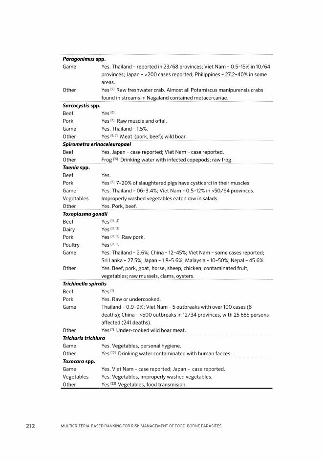
Multi criteria-based ranking for risk ManageMent of food-borne parasites212
para gonimus spp.Game Yes. Thailand – reported in 23/68 provinces; Viet Nam – 0.5–15% in 10/64
provinces; Japan – >200 cases reported; Philippines – 27.2–40% in some
areas.
Other Yes [9] Raw fresh water crab. Almost all Pota miscus mani purensis crabs
found in streams in Nagaland con tained meta cercariae.
sarco cystis spp.Beef Yes [6]
Pork Yes [7] Raw muscle and offal.
Game Yes. Thailand – 1.5%.
Other Yes [6, 7] Meat (pork, beef); wild boar.
spirometra erinacei europaeiBeef Yes. Japan – case repor ted; Viet Nam – case repor ted.
Other Frog [15] Drinking water with infected cope pods; raw frog.
taenia spp.Beef Yes.
Pork Yes [5] 7–20% of slaughtered pigs have cysticerci in their muscles.
Game Yes. Thailand – 06–3.4%; Viet Nam – 0.5–12% in >50/64 provinces.
Vegetables Improper ly washed vege tables eaten raw in salads.
Other Yes. Pork, beef.
toxoplasma gondiiBeef Yes [11, 12]
Dairy Yes [11, 12]
Pork Yes [11, 12] Raw pork.
Poultry Yes [11, 12]
Game Yes. Thailand – 2.6%; China – 12–45%; Viet Nam – some cases reported;
Sri Lanka – 27.5%; Japan – 1.8–5.6%; Malaysia – 10–50%; Nepal – 45.6%.
Other Yes. Beef, pork, goat, horse, sheep, chicken; contaminated fruit,
vegetables; raw mussels, clams, oysters.
trichinella spiralisBeef Yes [1]
Pork Yes. Raw or under cooked.
Game Thailand – 0.9–9%; Viet Nam – 5 outbreaks with over 100 cases (8
deaths); China – >500 outbreaks in 12/34 provinces, with 25 685 persons
affected (241 deaths).
Other Yes [2] Under-cooked wild boar meat.
trichuris trichiuraGame Yes. Vegetables, personal hygiene.
Other Yes [10] Drinking water contaminated with human faeces.
toxocara spp.Game Yes. Viet Nam – case reported; Japan – case reported.
Vegetables Yes. Vege tables, improper ly washed vege tables.
Other Yes [23] Vege tables, food trans mision.

Annex 8 - regionAl reports 213
Sources used for Table A8.2.2, but read in conjunction with references cited in the main text and in Table A8.2.1
01. pebam, S., Goni, V., patel, S. kumar, V., Rawall, S. & Bali, k. 2012. Case Report: A 12-year-old child with trichinellosis, pyomyositis and secondary osteomyelitis. Journal of Global Infectious Diseases, 4(1): 84–88.
02. Sethi, B., Butola, k.S., kumar, y. & Mishra, J.p. 2012. Multiple outbreaks of trichinellosis with high mortality rate. Tropical Doctor, 42(4): 243–243.
03. De, N.V., Murrell, k.D., Cong, le D., Cam, p.D., Chau, le V., toan, N.D. & Dalsgaard, A. 2003. The foodborne trematode zoonoses of Viet Nam. Southeast Asian Journal of Tropical Medicine and Public Health, 34(Suppl.1): 12–34.
04. Mirdha, B.R., Gulati, S., Sarkar, t. & Samantray, J.C. 1998 Acute clonorchiasis in a child. Indian Journal of Gastroenterology, 17(4): 155.
05. prasad, k.N., prasad, A., Verma, A. & Singh, A.k. 2008. Human cysticercosis and Indian scenario: a review. Journal of Biosciences, 33(4): 571–582.
06. Nichpanit, S., Nakai, W., Wongsaroj, t. & Nithikathkul, C. 2010. First large scale of human Sarcocystis hominis in Thailand. Trends Research in Science and Technology, 2(1): 1–5.
07. Banerjee, p.S., Bhatia, B.B. & pandit, B.A. 1994. Sarcocystis suihominis infection in human beings in India. Journal of Veterinary Parasitology, 8(1): 57–58.
08. parija, S.C. 2000. Rare intestinal protozoal infections in India. Journal of International Medical Sciences Academy, 13(1): 49–54.
09. Singh, t.S., Sugiyama, H., umehara, A., Hiese, S. & khalo, k. 2009. Paragonimus heterotremus infection in Nagaland: A new focus of paragonimiasis in India. Indian Journal of Medical Microbiology, 27(2): 123–127.
10. Narain, k., Rajguru, S.k. & Mahanta, J. 2000. Prevalence of Trichuris trichiura in relation to socio-economic & behavioural determinants of exposure to infection in rural Assam. Indian Journal of Medical Research, 112: 140–146.
11. Shivaprakash. M.R., parija, S.C. & Sujatha, S. 2001. Seroprevalence of toxoplasmosis in HIV infected patients in Pondicherry. Journal of Communicable Diseases, 33(3): 221–223.
12. Borkakoty, B.J., Borthakur, A.k. & Gohain, M. 2007. Prevalence of Toxoplasma gondii infection amongst pregnant women in Assam, India. Indian Journal of Medical Microbiology, 25(4): 431–432.
13. Malhotra, S., Mehta, D.k., Arora, R., Chauhan, D., Ray, S. & Jain, M. 2006. Ocular angio strongy liasis in a child – First case report from India. Journal of Tropical Pediatrics, 52(3): 223–225.
14. Vasantha, p.l., Girish, N, & Sai leela, k. 2012. Human intestinal capillariasis: A rare case report from non-endemic area (Andhra Pradesh, India). Indian Journal of Medical Microbiology, 30(2) 236–239.
15. Sundaram, C., prasad, V.S.S.V. & Reddy, J.J.M. 2003. Cerebral sparganosis. Journal of the Association of Physicians of India, 51(11): 1107–1109.
16. pillai, G.S., kumar, A., Radhakrishnan, N., Maniyelil, J., Shafi, t., Dinesh, k.R. & karim, S. 2102. Case Report: Intraocular gnathostomiasis: report of a case and review of literature. American Journal of Tropical Medicine and Hygiene, 86(4):620–623.
17. Ishikura, H. 2003. Anisakiasis (2) Clinical Pathology and Epidemiology. pp. 451–473, in: M. Otsuru, S. Kamegai and S. Hayashi (editors). Progress of Medical Parasitology in Japan, Vol. 8. Meguro Parasitological Museum, Tokyo, Japan.
18. Grover, M., Dutta, R., kumar, R., Aneja, S. & Mehta, G. 1998. Echinostoma iliocanum infection. Case report. Indian Pediatrics, 35 (June): 549–552.
19. uppal, B. & Wadhwa, V. 2005. Rare case of Metagonimus yokogawai. Indian Journal of Medical Microbiology, 23(1): 61–62.
20. Ramachandran, J., Ajjampur, S.S.R., Chandramohan, A. & Varghese, G.M. 2012. Cases of human fascioliasis in India: Tip of the iceberg. Journal Of Postgraduate Medicine, 58(2): 150–152.

Multi criteria-based ranking for risk ManageMent of food-borne parasites214
21. Muralidhar, S., Srivastava, l., Aggarwal, p., Jain, N. & Sharma D.k. 2000. Fasciolopsiasis--a persisting problem in eastern U.P. – a case report. Indian Journal of Pathology and Microbiology, 43(1) 69–71.
22. parija, S.C., Malini, G. & Rao, R.S. 1992. Prevalence of hookworm species in Pondicherry, India. Tropical and Geographical Medicine, 44(4): 378–380.
23. Malla, N., Aggarwal, A.k. & Mahajan, R.C. 2002. A serological study of human toxocariasis in north India. National Medical Journal of India, 15(3): 145–147.
24. Wani, S.A., Ahmad, E., Zargar, S.A., Dar, p.A., Dar, Z.A. & Jan, t.R. 2008. Intestinal helminths in a population of children from the Kashmir valley, India. Journal of Helminthology, 82(4): 313–317.
25. Ragunathan, l., kalivaradhan, S.k., Ramadass, S., Nagaraj, M. & Ramesh, k. 2010. Helminthic infections in school children in Puducherry, south India. Journal of Microbiology Immunology and Infection, 43(3): 228–232.
26. parija S.C. 2002. Epidemiology, clinical features and laboratory diagnosis of amoebiasis in India. Journal of Parasitic Diseases, 26(1): 1–8.
27. parija, S.C. & Rao, R.S. 1987. Prevalence of parasitic infections in Pondicherry. Indian Journal of Parasitology,11: 63–65.
28. parija, S.C., Rao, R.S., Badrinath, S. & Sengupta, D.N. 1983. Hydatid disease in Pondicherry. Journal of Tropical Medicine and Hygiene, 86(3): 113–115.
TAblE A8.2.3 Data availability for risk management options for each parasite-com-modity combination in the Asia context.NOTE: The authors were asked to consider all combinations of the particular parasite and the main food categories, namely Beef, Dairy, Pork, Poultry, Game, Seafood, Fruit, Vegetables and Other.
Angio strongylus spp.Other Yes [18] Proper cooking of frogs and snails
Ancylostoma duo denale, necator ameri canusOther Yes [28] Hook worm larvae were in areas 2.1–5.2% on vege tables. Vege tables &
food trans mission, so use sanitary disposal of human excreta; avoid walking
barefoot.
Anisakis simplexAll No substantive data found.
Ascaris lumbri coidesVegetables Yes. Ascaris eggs were in areas reported as 2.1–2.7% in vege tables.
Other Yes [30] Vege tables & food trans mission; hand washing; washing of
vegetables before con sumption; proper sanitation.
Capillaria philip pinensis
Other Yes [19] Proper cooking of freshwater fish.
clonorchis sinensisDairy Very little data [4]
Game Yes [4] Viet Nam – 44.4–92.9% fresh water fish infected by Clon orchis
sinensis larvae.
Other Yes. Proper cooking of freshwater fish.
crypto spor idium spp.Beef Yes [11]
Other Yes. Vege tables, food, water trans mission; hand washing, boiling or
filtration of drinking water
echino coccus spp. [34]

Annex 8 - regionAl reports 215
Vegetables Yes.
Other Yes [34] Proper care of pet dogs; avoid close contact with stray dogs; hand
washing; thorough washing of vegetables before con sumption.
echino stoma spp. [23]
Other Proper cooking of freshwater snails.
Fasciola spp. Other Yes [25, 26] Fasciola larvae in areas reportedly 0.4% in vege tables. Avoid eating
uncleaned aquatic plants and vegetables.
Fascio lopsis buskiOther Yes [27] Avoid eating uncleaned aquatic plants and vegetables.
en tamoeba histolyticaGame Yes.
Vegetables Yes. E. histo lytica cysts in areas reportedly 1.8–6.7% in vege tables.
Other Yes [31] Vege tables & food; water trans mission; hand washing; thorough
washing of vegetables before con sumption; proper sanitation.
giardia duo denalis (syn. g. lamblia, g. intestinalis)Game Yes.
Vegetables Yes. Giardia cysts were in areas reportedly 2.7–13.9% in vege tables.
Other Yes [32, 33] Vege tables & food; water trans mission; hand washing; thorough
washing of vegetables before con sumption; proper sanitation.
gnatho stoma spp.Game Yes. Gnatho stoma larvae were in areas reportedly 6.7–11.4% in eels.
Other Yes [22] Fish, eel, amphibians. Proper cooking of fresh water fish & frogs.
Hetero phyidsGame Yes [12] Heterophyid larvae 7.4–62.8% in various fish species.
Other Yes. Fish.
meta gonimus spp.Other Yes [24] Proper cooking of fresh water fish.
opisth orchis viverriniGame Yes [4] Viet Nam – 10–29% fresh water fish infected by O. viverrini.
Other Yes. Fish.
paragon imus spp. [13]
Pork Yes. Wild boar meat.
Game Viet Nam – rate of Paragonimus larvae was 9.7% to 98.1% in Potamicus
crab.
Other Yes [14] Fresh water crab, wild boar meat – proper cooking of crabs.
sarcocystis spp.Pork Yes [9] Proper cook ing.
Other Yes. Proper cooking of wild boar meat.
spiro metra erinacei europaeiGame Yes. 8–10% frogs reportedlt infected by S. erinacei europaei larvae.
Other Yes [21] Frogs & amphibians; boiling or filtration of drinking water; Yes [17]
Proper cook ing. Proper cooking of frogs
taenia spp. [6]
Beef Yes [7, 8] Dis card infec ted meat in abattoir; proper cook ing.
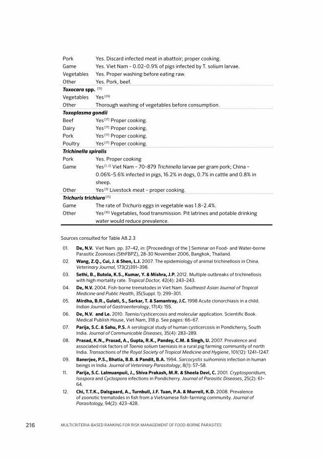
Multi criteria-based ranking for risk ManageMent of food-borne parasites216
Pork Yes. Dis card infec ted meat in abattoir; proper cook ing.
Game Yes. Viet Nam – 0.02–0.9% of pigs infected by T. solium larvae.
Vegetables Yes. Proper washing before eating raw.
Other Yes. Pork, beef.
toxocara spp. [15]
Vegetables Yes [29]
Other Thorough washing of vegetables before consumption.
toxo plasma gondiiBeef Yes [17] Proper cook ing.
Dairy Yes [17] Proper cook ing.
Pork Yes [17] Proper cook ing.
Poultry Yes [17] Proper cook ing.
trichinella spiralisPork Yes. Proper cooking
Game Yes [1, 2] Viet Nam – 70–879 Trichinella larvae per gram pork; China –
0.06%–5.6% infected in pigs, 16.2% in dogs, 0.7% in cattle and 0.8% in
sheep.
Other Yes [3] Livestock meat – proper cooking.
trichuris trichiura [15]
Game The rate of Trichuris eggs in vegetable was 1.8–2.4%.
Other Yes [16] Vegetables, food transmission. Pit latrines and potable drinking
water would reduce prevalence.
Sources consulted for Table A8.2.3
01. De, N.V. Viet Nam. pp. 37–42, in: [Proceedings of the ] Seminar on Food- and Water-borne Parasitic Zoonoses (5thFBPZ), 28-30 November 2006, Bangkok, Thailand.
02. Wang, Z.Q., Cui, J. & Shen, l.J. 2007. The epidemiology of animal trichinellosis in China. Veterinary Journal, 173(2)391–398.
03. Sethi, B., Butola, k.S., kumar, y. & Mishra, J.p. 2012. Multiple outbreaks of trichinellosis with high mortality rate. Tropical Doctor, 42(4): 243–243.
04. De, N.V. 2004. Fish-borne trematodes in Viet Nam. Southeast Asian Journal of Tropical Medicine and Public Health, 35(Suppl. 1): 299–301.
05. Mirdha, B.R., Gulati, S., Sarkar, t. & Samantray, J.C. 1998 Acute clonorchiasis in a child. Indian Journal of Gastroenterology, 17(4): 155.
06. De, N.V. and le. 2010. Taenia/cysticercosis and molecular application. Scientific Book. Medical Publish House, Viet Nam, 318 p. See pages: 66–67.
07. parija, S.C. & Sahu, p.S. A serological study of human cysticercosis in Pondicherry, South India. Journal of Communicable Diseases, 35(4): 283–289.
08. prasad, k.N., prasad, A., Gupta, R.k., pandey, C.M. & Singh, u. 2007. Prevalence and associated risk factors of Taenia solium taeniasis in a rural pig farming community of north India. Transactions of the Royal Society of Tropical Medicine and Hygiene, 101(12): 1241–1247.
09. Banerjee, p.S., Bhatia, B.B. & pandit, B.A. 1994. Sarcocystis suihominis infection in human beings in India. Journal of Veterinary Parasitology, 8(1): 57–58.
11. parija, S.C. lalmuanpuii, J., Shiva prakash, M.R. & Sheela Devi, C. 2001. Cryptosporidium, Isospora and Cyclospora infections in Pondicherry. Journal of Parasitic Diseases, 25(2): 61–64.
12. Chi, t.t.k., Dalsgaard, A., turnbull, J.F. tuan, p.A. & Murrell, k.D. 2008. Prevalence of zoonotic trematodes in fish from a Vietnamese fish-farming community. Journal of Parasitology, 94(2): 423–428.

Annex 8 - regionAl reports 217
13. De, N.V. 2004. Epidemiology, pathology and treatment of paragonimiasis in Viet Nam. Southeast Asian Journal of Tropical Medicine and Public Health, 35(Suppl. 1): 331–336.
14. Mahajan, R.C. 2005. Paragonimiasis: an emerging public health problem in India. Indian Journal of Medical Research, 121(6): 716–718.
15. phuong et al. 2011. Parasitic infection in wastewater-irrigated vegetables in rural and urban areas in Nam Dinh province. Journal of Military Medicine, 9 Nov. 2009: 33-37
16. Narain, k., Rajguru, S.k. & Mahanta, J. 2000. Prevalence of Trichuris trichiura in relation to socio-economic & behavioural determinants of exposure to infection in rural Assam. Indian Journal of Medical Research, 112: 140–146.
17. Borkakoty, B.J., Borthakur, A.k. & Gohain, M. 2007. Prevalence of Toxoplasma gondii infection amongst pregnant women in Assam, India. Indian Journal of Medical Microbiology, 25(4): 431–432.
18. Malhotra, S., Mehta, D.k., Arora, R., Chauhan, D., Ray, S. & Jain, M. 2006. Ocular angio strongy liasis in a child – First case report from India. Journal of Tropical Pediatrics, 52(3): 223–225.
19. Vasantha, p.l., Girish, N, & Sai leela, k. 2012. Human intestinal capillariasis: A rare case report from non-endemic area (Andhra Pradesh, India). Indian Journal of Medical Microbiology, 30(2) 236–239.
21. Sundaram, C., prasad, V.S.S.V. & Reddy, J.J.M. 2003. Cerebral sparganosis. Journal of the Association of Physicians of India, 51(11): 1107–1109.
22. pillai, G.S., kumar, A., Radhakrishnan, N., Maniyelil, J., Shafi, t., Dinesh, k.R. & karim, S. 2102. Case Report: Intraocular gnathostomiasis: report of a case and review of literature. American Journal of Tropical Medicine and Hygiene, 86(4):620–623.
23. Grover, M., Dutta, R., kumar, R., Aneja, S. & Mehta, G. 1998. Echinostoma iliocanum infection. Case report. Indian Pediatrics, 35 (June): 549–552.
24. uppal, B. & Wadhwa, V. 2005. Rare case of Metagonimus yokogawai. Indian Journal of Medical Microbiology, 23(1): 61–62.
25. De, N.V. 2012. Fascioliasis in Viet Nam. Pers. comm. in response to request for data.
26. Ramachandran, J., Ajjampur, S.S.R., Chandramohan, A. & Varghese, G.M. 2012. Cases of human fascioliasis in India: Tip of the iceberg. Journal Of Postgraduate Medicine, 58(2): 150–152.
27. Muralidhar, S., Srivastava, l., Aggarwal, p., Jain, N. & Sharma D.k. 2000. Fasciolopsiasis--a persisting problem in eastern U.P. – a case report. Indian Journal of Pathology and Microbiology, 43(1) 69–71.
28. parija, S.C., Malini, G. & Rao, R.S. 1992. Prevalence of hookworm species in Pondicherry, India. Tropical and Geographical Medicine, 44(4): 378–380.
29. Malla, N., Aggarwal, A.k. & Mahajan, R.C. 2002. A serological study of human toxocariasis in north India. National Medical Journal of India, 15(3): 145–147.
30. Wani, S.A., Ahmad, F., Zargar, S.A., Ahmad, Z., Ahmad, p. & tak, H. 2007. Prevalence of intestinal parasites and associated risk factors among schoolchildren in Srinagar City, Kashmir, India. Journal of Parasitology, 93(6): 1541–1543.
32. parija, S.C. & Rao, R.S. 1987. Prevalence of parasitic infections in Pondicherry. Indian Journal of Parasitology,11: 63–65.
33. kang, G., Mathew, M.S., Rajan, D.p., Daniel, J.D., Mathan, M.M. & Muliyil, J.p. 1998. Prevalence of intestinal parasites in rural Southern Indians. Tropical Medicine & International Health, 3(1): 70–75.
34. parija, S.C., Rao, R.S., Badrinath, S. & Sengupta, D.N. 1983. Hydatid disease in Pondicherry. Journal of Tropical Medicine and Hygiene, 86(3): 113–115.

Multi criteria-based ranking for risk ManageMent of food-borne parasites218
ANNEX 8.3 – AuStRAlIA
A8.3.1 PreparationThe information for Australia was compiled by Dr Rebecca Traub, Senior Lecturer in Veterinary Public Health, School of Veterinary Sciences, The University of Queensland, Gatton. In developing this section of the report, Dr Traub used lit-erature searches using PubMed (search terms used = “Parasite Name” + Australia) together with personal communications with experts in academia and the Depart-ment of Agriculture, Fisheries and Forestry (DAFF), Queensland Health, and Food Standards Australia.
A8.3.2 Data availability in humans and food attribution Surveillance systems in place include the National Animal Health Information System (NAHIS) and National Notifiable Diseases Surveillance System (NNDSS), which collect, collate, analyse and report on data on animal and human health status. In general, information with regard to the incidence or burden of food-borne parasites in humans and animals in Australia is lacking, but is assumed to be negligible. Although limited, most data is generated from research-based surveys conducted by academic institutes, together with published hospital case reports. Surveillance (end product testing for food borne parasites) is usually not consid-ered necessary due to the low perceived risks to public health, based on:
• Highstandardsoffoodsafetyandinspectionpracticesthatutilizea‘whole-of-chain’ approach, which includes implementation of risk-based hazard analysis and HACCP. In addition to this, all exported food must comply with the Export Control (Prescribed Goods - General) Orders 2005, and the Export Control (Plants and Plant Products) Orders, 2005. Exporters must meet both the requirements of relevant export legislation and of any importing country requirements for the Australian Quarantine and In-spection Service (AQIS) to provide the necessary documentation to enable products to be exported.
• Thedietary habits ofmostAustralians, namely eatingmedium- towell-cooked meats.
• TheabsenceofmanyofthefoodborneparasitesofpublichealthconcerninAustralia (exotic pathogens).
Except for Cryptosporidium, no other food borne parasites are listed in the human notifiable diseases list. For example, cystic hydatid disease in humans is no longer notifiable on a state or national level, despite its enzootic nature in rural settings. Many of the fish- and plant-borne parasites (e.g. Anisakiasis, plant- or vector-borne protozoa and helminth infections) may be missed unless an ‘obvious outbreak’ has been detected and reported to the State Public Health Unit. Primary

Annex 8 - regionAl reports 219
means of surveillance of food borne parasites are performed through abattoirs due to export certification requirements, such as data on the incidence of suspect Cysticercus bovis lesions in beef and Trichinella in game meat, and exports would be well documented.

Multi criteria-based ranking for risk ManageMent of food-borne parasites220
TAb
lE A
8.3.
1 Dat
a av
aila
bilit
y on
the
bur
den
of d
isea
se a
nd fo
od a
ttri
buti
on a
t th
e re
gion
al a
nd g
loba
l lev
el fo
r A
ustr
alia
Par
asit
e sp
ecie
s
Dat
a av
aila
bilit
y on
hum
an-d
isea
se-r
elat
ed p
aram
eter
sR
egio
nal l
evel
G
loba
l lev
el
Dis
ease
in
hum
ans?
Dis
ease
sev
erit
y an
d m
ain
popu
lati
on(s
) at
ris
k
Mai
n fo
od s
ourc
e an
d at
trib
utio
nD
isea
se in
hu
man
s?
Dis
ease
se
veri
ty
and
mai
n po
pula
tion
s at
ris
k
Mai
n fo
od
sour
ces
and
attr
ibu t
ions
Ang
iost
rong
ylus
can
ton
ensi
sYe
s –
qual
itat
ive
–
case
repo
rts
[21]
Yes
– qu
alit
ativ
e –
case
repo
rts
[21]
Yes
– an
ecdo
tal f
rom
cas
e
hist
ory
[21]
Ani
saki
s sp
p.; C
ontr
acae
cum
spp.
Yes
[12]
No
data
Yes
[13]
Yello
w e
ye m
ulle
t, t
iger
flath
ead,
sea
mul
let,
Kin
g
Geo
rge
whi
ting
, bre
am, s
and
flath
ead,
pilc
hard
Cry
pto
spor
idiu
m s
pp.
(inc
ludi
ng C
. hom
inis
and
C. p
arvu
m)
Yes
[22]
Yes
[22]
Yes.
Mos
t ou
tbre
aks
wat
er-b
orne
recr
eati
onal
swim
min
g. O
ther
spo
radi
c
outb
reak
s ‘U
nkno
wn’
sou
rce
[22]
Ech
inoc
occu
s g
ranu
losu
sYe
s –
retr
ospe
ctiv
e
hosp
ital
cas
es.
Not
not
ifiab
le
sinc
e 20
00
[19]
Yes
Yes
– qu
anti
tati
ve d
ata
on
prev
alen
ce in
wild
and
farm
dogs
[20
]
No
attr
ibut
ion
to fo
od.
Ent
eric
pro
tozo
a
Gia
rdia
, Cyc
losp
ora,
Bla
stoc
ysti
s, D
ient
amoe
ba
frag
ilis,
Isos
por
a b
elli
No
data
Yes
Indi
geno
us
Aus
tral
ians
[23]
Imm
uno-
supp
ress
ed
HIV
/AID
s pa
tien
ts [2
4]
No
data
Ent
eric
hel
min
ths;
Asc
aris
;
Tric
huri
s; h
ookw
orm
s
No
data
Yes
Indi
geno
us
Aus
tral
ians
[23]
No
data
Sp
irom
etra
or
spar
gano
sis
No
data
No
data
Wild
boa
r, s
nake
Taen
ia s
agin
ata
No
data
No
data
Bee
f – 10
0%
mea
t-bo
rne
[11]
Cat
tle
– 10
0%
food
born
e

Annex 8 - regionAl reports 221
TAb
lE A
8.3.
1 Dat
a av
aila
bilit
y on
the
bur
den
of d
isea
se a
nd fo
od a
ttri
buti
on a
t th
e re
gion
al a
nd g
loba
l lev
el fo
r A
ustr
alia
Par
asit
e sp
ecie
s
Dat
a av
aila
bilit
y on
hum
an-d
isea
se-r
elat
ed p
aram
eter
sR
egio
nal l
evel
G
loba
l lev
el
Dis
ease
in
hum
ans?
Dis
ease
sev
erit
y an
d m
ain
popu
lati
on(s
) at
ris
k
Mai
n fo
od s
ourc
e an
d at
trib
utio
nD
isea
se in
hu
man
s?
Dis
ease
se
veri
ty
and
mai
n po
pula
tion
s at
ris
k
Mai
n fo
od
sour
ces
and
attr
ibu t
ions
Ang
iost
rong
ylus
can
ton
ensi
sYe
s –
qual
itat
ive
–
case
repo
rts
[21]
Yes
– qu
alit
ativ
e –
case
repo
rts
[21]
Yes
– an
ecdo
tal f
rom
cas
e
hist
ory
[21]
Ani
saki
s sp
p.; C
ontr
acae
cum
spp.
Yes
[12]
No
data
Yes
[13]
Yello
w e
ye m
ulle
t, t
iger
flath
ead,
sea
mul
let,
Kin
g
Geo
rge
whi
ting
, bre
am, s
and
flath
ead,
pilc
hard
Cry
pto
spor
idiu
m s
pp.
(inc
ludi
ng C
. hom
inis
and
C. p
arvu
m)
Yes
[22]
Yes
[22]
Yes.
Mos
t ou
tbre
aks
wat
er-b
orne
recr
eati
onal
swim
min
g. O
ther
spo
radi
c
outb
reak
s ‘U
nkno
wn’
sou
rce
[22]
Ech
inoc
occu
s g
ranu
losu
sYe
s –
retr
ospe
ctiv
e
hosp
ital
cas
es.
Not
not
ifiab
le
sinc
e 20
00
[19]
Yes
Yes
– qu
anti
tati
ve d
ata
on
prev
alen
ce in
wild
and
farm
dogs
[20
]
No
attr
ibut
ion
to fo
od.
Ent
eric
pro
tozo
a
Gia
rdia
, Cyc
losp
ora,
Bla
stoc
ysti
s, D
ient
amoe
ba
frag
ilis,
Isos
por
a b
elli
No
data
Yes
Indi
geno
us
Aus
tral
ians
[23]
Imm
uno-
supp
ress
ed
HIV
/AID
s pa
tien
ts [2
4]
No
data
Ent
eric
hel
min
ths;
Asc
aris
;
Tric
huri
s; h
ookw
orm
s
No
data
Yes
Indi
geno
us
Aus
tral
ians
[23]
No
data
Sp
irom
etra
or
spar
gano
sis
No
data
No
data
Wild
boa
r, s
nake
Taen
ia s
agin
ata
No
data
No
data
Bee
f – 10
0%
mea
t-bo
rne
[11]
Cat
tle
– 10
0%
food
born
e
Par
asit
e sp
ecie
s
Dat
a av
aila
bilit
y on
hum
an-d
isea
se-r
elat
ed p
aram
eter
sR
egio
nal l
evel
G
loba
l lev
el
Dis
ease
in
hum
ans?
Dis
ease
sev
erit
y an
d m
ain
popu
lati
on(s
) at
ris
k
Mai
n fo
od s
ourc
e an
d at
trib
utio
nD
isea
se in
hu
man
s?
Dis
ease
se
veri
ty
and
mai
n po
pula
tion
s at
ris
k
Mai
n fo
od
sour
ces
and
attr
ibu t
ions
Toxo
pla
sma
gon
dii
0.6
% p
oste
rior
uvei
tis
–
Abo
rigi
nal
Aus
tral
ians
[14
]
3.5%
of
ence
phal
itis
hosp
ital
izat
ions
;
dow
n si
nce
1990
s
(HIV
pea
k) [1
5]
2 re
port
ed
outb
reak
s
(raw
lam
b an
d
kang
aroo
) [16]
No
data
Yes
– qu
anti
tati
ve s
erol
ogic
al
data
[17]
Kan
garo
o m
eat;
lam
b (s
heep
);
pigs
[16,
18]
Tric
hine
lla p
apua
e in
Tor
res
Stra
it Is
land
s
Yes
– qu
alit
ativ
e
anec
dota
l
repo
rts[1
, 2]
No
data
Wild
boa
r (S
us s
crof
a) [3
]
Impo
rted
cro
codi
le m
eat
from
PN
G 4
]
Yes
[5, 6
]Ye
s [5
, 7]
Dom
esti
c an
d
wild
boa
r [5
]
Cro
codi
le
mea
t, t
urtl
e
mea
t [8
]
100
%
food
born
e
Tric
hine
lla p
seud
o sp
iral
is –
Tasm
ania
No
data
No
data
Das
yuri
ds (q
uolls
; Tas
man
ian
devi
l,) a
nd c
arri
on-f
eedi
ng
bird
s (m
arsh
har
rier
, mas
ked
owl)
[9]
Pur
ely
sylv
atic
cyc
le
Yes
[6, 1
0]
Yes
[7, 1
0]
Dom
esti
c an
d
wild
boa
r [9
]
100
%
food
born
e
Multi criteria-based ranking for risk ManageMent of food-borne parasites222
Sources used in Table A8.3.1
01. Spratt, D.M., Beveridge, I., Andrews, J.R.H. & Dennett, X. 1999. Haycocknema perplexum n.g., n.sp. (Nematoda: Robertdollfusidae): an intramyofibre parasite in man. Systematic Parasitology, 43(2): 123–131.
02. Department of primary Industries (DpI) report. No date. Three human clinical cases in Adelaide (archived, pers. comm. Jack van Wick).
03. Cuttell, l., Cookson, B., Jackson, l.A., Gray, C. & traub, R.J. 2012. First report of a Trichinella papuae infection in a wild pig (Sus scrofa) from an Australian island in the Torres Strait region. Veterinary Parasitology, 185(2-4): 343–345.
04. louise Jackson, DAFF, pers. comm.
05. kusolsuk, t., kamonrattanakun, S., Wesanonthawech, A. and 11 others. 2010. The second outbreak of trichinellosis caused by Trichinella papuae in Thailand. Transactions of the Royal Society of Tropical Medicine and Hygiene, 104(6): 433–437.
06. WHO. Various dates (a). Total cases and incidence of Trichinella spp. infections, by World Health Organization region and country, 1986–2009.
07. WHO. Various dates (b). Clinically confirmed cases of trichinellosis in humans documented in World Health Organization regions, 1986–2009.
08. lo, y.-C., Hung, C.-C., lai, C.-S., Wu, Z., Nagano, I., Maeda, t., takahashi, y., Chiu, C.H. & Jiang, D.D.-S. 2009. Human trichinosis after consumption of soft-shelled turtles, Taiwan. Emerging Infectious Diseases, 15(12): 2056–2058.
09. Obendorf, D.l. & Clarke, k.p. 1992. Trichinella pseudospiralis infections in free-living Tasmanian birds. Journal of the Helminthological Society of Washington, 59(1): 144–147; Obendorf, D.l., Handlinger, J.H., Mason, R.W., Clarke, k.p., Forman, A.J., Hooper, p.t., Smith, S.J. & Holdsworth, M. 1990. Trichinella pseudospiralis infection in Tasmanian wildlife. Australian Veterinary Journal, 67(3): 108–110.
10. Murrell, k.D. & pozio, E. 2011. Worldwide occurrence and impact of human trichinellosis 1986–2009. Emerging Infectious Diseases, 17(12): 2194–2202.
11. Jenkins et al., under review. Cysticercosis in beef cattle in New South Wales. Submitted to Australian Veterinary Journal. Under review; pearse, B.H.G., traub, R.J., Davis, A., Cobbold, R. & Vanderlinde, p.B. 2010. Prevalence of Cysticercus bovis in Australian cattle. Australian Veterinary Journal, 88(7): 260–262; Brown, G., Dennis, M.M., Slapeta, J. & thompson, A.R. 2010. Prevalence of Cysticercus bovis (beef measles) in Australian cattle. [Comment on Pearse et al., 2010, q.v... Australian Veterinary Journal, 88(12): 463–464.
12. Shamsi, S. & Butcher, A.R. 2011. First report of human anisakidosis in Australia. Medical Journal of Australia, 194(4): 199–200.
13. Shamsi, S., Eisenbarth, A., Saptarshi, S., Beveridge, I., Gasser, R.B. & lopata, A.l. 2011. Occurrence and abundance of anisakid nematode larvae in five species of fish from southern Australian waters. Parasitology Research, 108(4): 927–934; lymbery, A.J., Doupe, R.G., Munshi, M.A. & Wong, t. 2002. Larvae of Contracaecum sp. among inshore fish species of south-western Australia. Diseases of Aquatic Organisms, 51(2): 157–159.
14. Chang, J.H., landers, J., Henderson, t.R.M. & Craig, J.E. 2012. Prevalence of uveitis in indigenous populations presenting to remote clinics of central Australia: The Central Australian Ocular Health Study. Clinical And Experimental Ophthalmology, 40(5): 448–453.
15. Huppatz, C., Durrheim, D.N., levi, C., Dalton, C., Williams, D., Clements, M.S. & kelly, p.M. 2009. Etiology of encephalitis in Australia, 1990–2007. Emerging Infectious Diseases, 15(9): 1359–1365.
16. parameswaran, N., O’Handley, R.M., Grigg, M.E., Fenwick, S.G. & thompson, R.C.A. 2009. Seroprevalence of Toxoplasma gondii in wild kangaroos using an ELISA. Parasitology International, 58(2): 161–165.

Annex 8 - regionAl reports 223
17. plant, J.W., Freeman, p. & Saunders, E. 1982. Serological survey of the prevalence of Toxoplasma gondii antibodies in rams in sheep flocks in New South Wales. Australian Veterinary Journal, 59(3): 87–89.
18. Munday, B.l. 1975. Prevalence of toxoplasmosis in Tasmanian meat animals. Australian Veterinary Journal, 51(6): 315–316.
19. Jenkins, D.J. & power, k. 1996 Human hydatidosis in New South Wales and the Australian Capital Territory, 1987–1992. Medical Journal of Australia, 164(1): 18–21.
20. Jenkins, D.J., Allen, l. & Goullet, M. 2008. Encroachment of Echinococcus granulosus into urban areas in eastern Queensland, Australia. Australian Veterinary Journal, 86(8): 294–300; Jenkins, D.J., Mckinlay, A., He, D.l., Bradshaw, H. & Craig, p.S. 2006. Detection of Echinococcus granulosus coproantigens in faeces from naturally infected rural domestic dogs in south-eastern Australia. Australian Veterinary Journal, 84(1-2): 12–16.
21. prociv, p. & Carlisle, M.S. 2001. The spread of Angiostrongylus cantonensis in Australia. Southeast Asian Journal of Tropical Medical Public Health, 32(Suppl. 2): 126–128.
22. OzFoodNet. Various dates. Communicable Diseases Australia quarterly reports.
23. Holt, D.C., McCarthy, J.S. & Carapetis, J.R. 2010. Parasitic diseases of remote indigenous communities in Australia. International Journal for Parasitology, 40(10; Special issue): 1119–1126.
24. Stark, D., Barratt, J.l.N., van Hal, S., Marriott, D., Harkness, J., & Ellis, J.t. 2009. Clinical significance of enteric protozoa in immunosuppressed human populations. Clinical Microbiology Reviews, 22(4): 634–640.
25. B. Cookson, Biosecurity Officer, Northern Australian Quarantine Strategy, Department of Agriculture, Fisheries & Forestry, Australian Government. Pers. comm., 2011.
TAblE A8.3.2 Data availability for Australia for parasite prevalence or concentration in the main food categories
Angiostrongylus cantonensis
Other No direct data. Some snail surveys. Anecdotal evidence of increase in
incidence in domestic pets and wildlife in urban areas of Brisbane and
Sydney. Human case reported from ingesting slugs while intoxicated [1]
Anisakis spp., contracaecum spp.
Seafood Little data. Prevalence in Yellow eye mullet, flathead, sea mullet, King
George Whiting, Bream, sand flathead, pilchard described and endemic off
coastal Australia. Most larvae in viscera post-harvest and not muscle [2, 3].
Other Yes. Wild caught and commercial fish at post-harvest level.
cryptosporidium spp. (including c. hominis and c. parvum)
Beef Yes
Dairy Yes
Pork No data
Game No data
Seafood Yes [4]. Reported Cryptosporidium oocyst contamination of some high-risk
foods.

Multi criteria-based ranking for risk ManageMent of food-borne parasites224
Fruits Yes [4]. Reported Cryptosporidium oocyst contamination of some high-risk
foods.
Vegetables Yes [4]. Reported Cryptosporidium oocyst contamination of some high-risk
foods.
Other Goat meat (India)
echinococcus granulosus
Vegetables No data
Other No direct data. Abattoir reports suggest hydatid disease is still highly
endemic among livestock. Prevalence studies in dogs, esp. in rural areas,
also demonstrate high prevalence among farm dogs [5, 6]
Enteric helminths: Ascaris, trichuris, Hookworms
Fruits No data
Vegetables No data
Enteric protozoa: giardia, cyclospora, Blastocystis, dientamoeba fragilis, isospora belli
Fruits No data
Vegetables No data
spirometra erinacei or sparganosis Quantitative data may be obtained from Biosecurity Queensland records. Endemic in
tropics, esp. wild boar, native frogs, feral cats, dogs.
Pork No data
Game No data
Other Wild boar Northern Australian Quarantine Strategy (NAQS) surveys
taenia saginata or bovine cysticercosis
Beef Incidence estimated to be less than 1 in 500 000 head according to a recent
national survey [7]. However sporadic cases or outbreaks, although rare, are
known to occur and have subsequently been confirmed as C. bovis [8, 9].
toxoplasma gondii
Beef Yes [10]. Most cited references in report from 1975 and 1980s.
Dairy Yes [10]. Most cited references in report from 1975 and 1980s.
Pork Yes [10]. Most cited references in report from 1975 and 1980s.
Poultry Yes [10]. Most cited references in report from 1975 and 1980s.
Game Yes [10]. Most cited references in report from 1975 and 1980s.
Fruits No data
Vegetables No data
Other Pademelons, wallabies, lamb. 15.5% kangaroos from WA positive by
ELISA [11].
trichinella papuae, t. pseudo spiralis
Pork No data

Annex 8 - regionAl reports 225
Poultry No data
Game Yes. All horse, croco dile and wild pig expor ted to EU from mainland
Australia undergoes pooled Artificial Digestion technique (AD). To date no
larvae detected on mainland.
Recent research-based survey using AD and polymerase chain reaction
(PCR) detected 2 pigs positive for larvae/DNA of T. papuae on a remote
island of the Torres Strait [15]. Moreover, recent serological evidence for
Trichinella on mainland [12, 14].
Other [14] Foxes and wild dogs on main land. Tas manian quolls, pos sums, raptoral
birds [19, 20].
Limited indicator species testing on mainland (200 rats; 60 foxes, 31 wild
dogs, 9 cats, 27 quolls) tested by AD, all negative [16–18].
Sources used for Table A8.3.2
01. Richard Mailk & McCarthy, pers. comm., 2012.
02. Shamsi, S., Eisenbarth, A., Saptarshi, S., Beveridge, I., Gasser, R.B. & lopata, A.l. 2011. Occurrence and abundance of anisakid nematode larvae in five species of fish from southern Australian waters. Parasitology Research, 108(4): 927–934;
03. lymbery, A.J., Doupe, R.G., Munshi, M.A. & Wong, t. 2002. Larvae of Contracaecum sp. among inshore fish species of south-western Australia. Diseases of Aquatic Organisms, 51(2): 157–159.
04. Smith, H.V. & Nichols, R.A.B. 2010. Cryptosporidium: Detection in water and food. Experimental Parasitology, 124(1; Special Issue): 61–79.
05. Jenkins, D.J., Mckinlay, A., He, D.l., Bradshaw, H. & Craig, p.S. 2006. Detection of Echinococcus granulosus coproantigens in faeces from naturally infected rural domestic dogs in south eastern Australia. Australian Veterinary Journal, 84(1-2): 12–16.
06. Jenkins, D.J., Allen, l. & Goullet, M. 2008. Encroachment of Echinococcus granulosus into urban areas in eastern Queensland, Australia. Australian Veterinary Journal, 86(8): 294–300.
07. pearse, B.H.G., traub, R.J., Davis, A., Cobbold, R. & Vanderlinde, p.B. 2010. Prevalence of Cysticercus bovis in Australian cattle. Australian Veterinary Journal, 88(7): 260–262;
08. Brown, G., Dennis, M.M., Slapeta, J. & thompson, A.R. 2010. Prevalence of Cysticercus bovis (beef measles) in Australian cattle. [Comment on Pearse et al., 2010, q.v.]. Australian Veterinary Journal, 88(12): 463–464.
09. Jenkins, D.J., Brown, G.k. & traub, R.J. 2012. Cysticercosis “storm” in cattle in feedlot cattle in north-west New South Wales. Australian Veterinary Journal 91(3): 89–93.
10. lake, R., Hudson, A. & Cressey, p. 2002. Risk profile: Toxoplasma gondii in red meat and meat products. Prepared as part of a New Zealand Food Safety Authority contract for scientific services. 36 p. See: http://www.foodsafety.govt.nz/elibrary/industry/Risk_Profile_Toxoplasma-Science_Research.pdf
11. parameswaran, N., O’Handley, R.M., Grigg, M.E., Fenwick, S.G. & thompson, R.C.A. 2009. Seroprevalence of Toxoplasma gondii in wild kangaroos using an ELISA. Parasitology International, 58(2): 161–165.

Multi criteria-based ranking for risk ManageMent of food-borne parasites226
12. Cuttell, l., Gomez-Morales, M.A., Cookson, B., Adams, p.J., Reid, S., Vanderlinde, p., Jackson, l.A. & traub, R.J. [2013]. Evaluation of ELISA coupled with confirmatory testing as a surveillance tool for Trichinella infection in wild boar. In preparation.
14. Cuttell, l., Corley, S.W., Gray, C.p., Vanderlinde, p.B., Jackson, l.A. & traub, R.J. 2012. Real-time PCR as a surveillance tool for the detection of Trichinella infection in muscle samples from wildlife. Veterinary Parasitology, 188(3-4): 285–293.
15. Cuttell, l., Cookson, B., Jackson, l.A., Gray, C. & traub, R.J. 2012. First report of a Trichinella papuae infection in a wild pig (Sus scrofa) from an Australian island in the Torres Strait region. Veterinary Parasitology, 185(2-4): 343–345.
16. Jenkins, D.J. & pozio, E. unpublished data, cited in Pozio, E. & Murrell, K.D. 2006. Systematics and epidemiology of Trichinella. Advances in Parasitology, 63: 367–439.
17. Waddell, A.H. 1969. The search for Trichinella spiralis in Australia. Australian Veterinary Journal, 45(4): 207.
18. Oakwood, M. & Spratt, D.M. 2000, Parasites of the northern quoll, Dasyurus hallucatus (Marsupialia: Dasyuridae) in tropical savanna, Northern Territory. Australian Journal of Zoology, 48: 79–90.
19. Obendorf, D.l. & Clarke, k.p. 1992. Trichinella pseudospiralis infections in free-living Tasmanian birds. Journal of the Helminthological Society of Washington, 59(1): 144–147.
20. Obendorf, D.l., Handlinger, J.H., Mason, R.W., Clarke, k.p., Forman, A.J., Hooper, p.t., Smith, S.J. & Holdsworth, M. 1990. Trichinella pseudospiralis infection in Tasmanian wildlife. Australian Veterinary Journal, 67(3): 108–110.
A8.3.3 Agri-food tradeFood borne parasites that currently have implications for the meat industry in terms of extra costs associated with inspection and testing are primarily restricted to C. bovis in the domestic and exported beef industry, and Trichinella testing of game meat (farmed crocodile, wild boar and horses) destined for the EU. However, for example, were anisakidosis to become an emerging public health problem in Australia, the fish industry would have to bear the costs of additional end-product inspection (e.g. candling, pooled PCR) to certify high-risk marine species safe for consumption.
Bovine cysticercosisAlthough rare, sporadic cases of C. bovis continue to be reported in the beef industry. Carcasses heavily positive for suspect C. bovis lesions are condemned, whereas those with low levels of infection are excluded from the export chain. Any visible lesions are trimmed at boning under veterinary supervision, freeze certified and sold on the domestic market. At present the abattoirs (industry) bear the cost of positively inspected carcasses. There is a move to reform this and place more cost and responsibility on the farmer by bringing in on-plant monitoring schemes that feed back to the farm level, providing incentive for modification or improve-ments in farm management. Given the low incidence of bovine cysticercosis and poor positive predictive value of organoleptic inspection, a risk-based system for inspecting C. bovis is being proposed and reviewed (Webber et al., 2012).
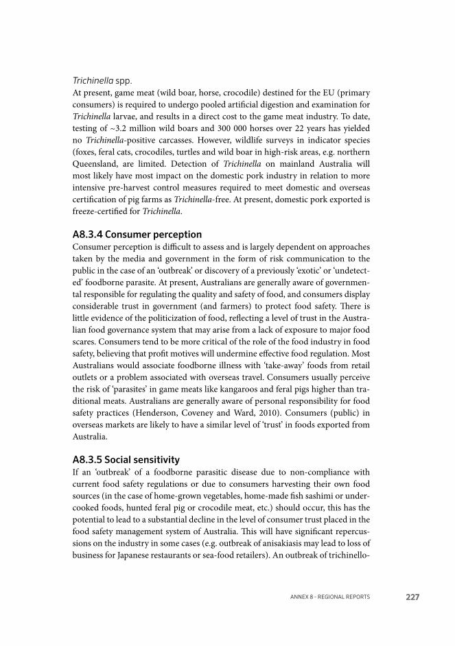
Annex 8 - regionAl reports 227
Trichinella spp. At present, game meat (wild boar, horse, crocodile) destined for the EU (primary consumers) is required to undergo pooled artificial digestion and examination for Trichinella larvae, and results in a direct cost to the game meat industry. To date, testing of ~3.2 million wild boars and 300 000 horses over 22 years has yielded no Trichinellapositive carcasses. However, wildlife surveys in indicator species (foxes, feral cats, crocodiles, turtles and wild boar in high-risk areas, e.g. northern Queensland, are limited. Detection of Trichinella on mainland Australia will most likely have most impact on the domestic pork industry in relation to more intensive pre-harvest control measures required to meet domestic and overseas certification of pig farms as Trichinella-free. At present, domestic pork exported is freeze-certified for Trichinella.
A8.3.4 Consumer perceptionConsumer perception is difficult to assess and is largely dependent on approaches taken by the media and government in the form of risk communication to the public in the case of an ‘outbreak’ or discovery of a previously ‘exotic’ or ‘undetect-ed’ food borne parasite. At present, Australians are generally aware of governmen-tal responsible for regulating the quality and safety of food, and consumers display considerable trust in government (and farmers) to protect food safety. There is little evidence of the politicization of food, reflecting a level of trust in the Austra-lian food governance system that may arise from a lack of exposure to major food scares. Consumers tend to be more critical of the role of the food industry in food safety, believing that profit motives will undermine effective food regulation. Most Australians would associate food borne illness with ‘take-away’ foods from retail outlets or a problem associated with overseas travel. Consumers usually perceive the risk of ‘parasites’ in game meats like kangaroos and feral pigs higher than tra-ditional meats. Australians are generally aware of personal responsibility for food safety practices (Henderson, Coveney and Ward, 2010). Consumers (public) in overseas markets are likely to have a similar level of ‘trust’ in foods exported from Australia.
A8.3.5 Social sensitivity If an ‘outbreak’ of a food borne parasitic disease due to non-compliance with current food safety regulations or due to consumers harvesting their own food sources (in the case of home-grown vegetables, home-made fish sashimi or under-cooked foods, hunted feral pig or crocodile meat, etc.) should occur, this has the potential to lead to a substantial decline in the level of consumer trust placed in the food safety management system of Australia. This will have significant repercus-sions on the industry in some cases (e.g. outbreak of anisakiasis may lead to loss of business for Japanese restaurants or sea-food retailers). An outbreak of trichinello-
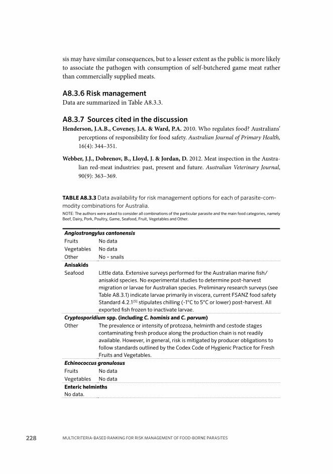
Multi criteria-based ranking for risk ManageMent of food-borne parasites228
sis may have similar consequences, but to a lesser extent as the public is more likely to associate the pathogen with consumption of self-butchered game meat rather than commercially supplied meats.
A8.3.6 Risk managementData are summarized in Table A8.3.3.
A8.3.7 Sources cited in the discussionHenderson, J.A.B., Coveney, J.A. & Ward, P.A. 2010. Who regulates food? Australians’
perceptions of responsibility for food safety. Australian Journal of Primary Health, 16(4): 344–351.
Webber, J.J., Dobrenov, B., Lloyd, J. & Jordan, D. 2012. Meat inspection in the Austra-lian red-meat industries: past, present and future. Australian Veterinary Journal, 90(9): 363–369.
tABlE A8.3.3 Data availability for risk management options for each of parasite-com-modity combinations for Australia.NOTE: The authors were asked to consider all combinations of the particular parasite and the main food categories, namely Beef, Dairy, Pork, Poultry, Game, Seafood, Fruit, Vegetables and Other.
Angio strongylus cantonensisFruits No data
Vegetables No data
Other No – snails
AnisakidsSeafood Little data. Extensive surveys performed for the Australian marine fish/
anisakid species. No experimental studies to determine post-harvest migration or larvae for Australian species. Preliminary research surveys (see Table A8.3.1) indicate larvae primarily in viscera, current FSANZ food safety Standard 4.2.1 [5] stipulates chilling (-1°C to 5°C or lower) post-harvest. All exported fish frozen to inactivate larvae.
crypto sporidium spp. (including c. hominis and c. parvum)Other The prevalence or intensity of protozoa, helminth and cestode stages
contaminating fresh produce along the production chain is not readily available. However, in general, risk is mitigated by producer obligations to follow standards outlined by the Codex Code of Hygienic Practice for Fresh Fruits and Vegetables.
echino coccus granulosusFruits No data
Vegetables No data
Enteric helminths No data.

Annex 8 - regionAl reports 229
Enteric protozoa, including giardia, cyclospora, Blastocystis, dientamoeba fragilis and isospora belliNo data.
taenia saginata or bovine cysticer cosisBeef Yes [1, 2]. See also Table A8.3.1
Other Low incidence and poor PPV of inspection of predilection sites – more emphasis on farm-level management to reduce risks.
toxoplasma gondiiRegionally, a risk profile [3] was conducted by New Zealand Food Safety Authority, based on outdated prevalence data for retail raw meats.Globally, the requirements for inactivation of tissue cysts in meat have been well studied and documented.
Beef Yes qualitative. Quantitative lacking on % positive retail meats with viable tissue cysts lacking.
Dairy Food Standards Australia New Zealand (FSANZ) Standard 4.2.4 – Primary Production and Processing Standard for Dairy Products
Pork Yes qualitative. Quantitative lacking on % positive retail meats with viable tissue cysts lacking
Poultry Yes qualitative. Quantitative lacking on % positive retail meats with viable tissue cysts lacking
Game Yes qualitative. Quantitative lacking on % positive retail meats with viable tissue cysts lacking
Fruits No data
Vegetables No data
Trichinella papuae, T. pseudospiralis
Pork No data
Game No extensive surveys in ‘hot-spots’ Experimental studies in pigs for T. papuae lacking to assess survivability, freeze tolerance
Other No surveys on the mainland. A small-scale survey in feral pigs by Cuttell [4] (see Table A8.3.1) allowed a qualitative risk assessment to be attempted.
Sources cited in Table A8.3.3
01. pearse, B.H.G., traub, R.J., Davis, A., Cobbold, R. & Vanderlinde, p.B. 2010. Prevalence of Cysticercus bovis in Australian cattle. Australian Veterinary Journal, 88(7): 260–262;
02. Webber, J.J., Dobrenov, B., lloyd, J. & Jordan, D. 2012. Meat inspection in the Australian red-meat industries: past, present and future. Australian Veterinary Journal, 90(9): 363–369.
03. lake, R., Hudson, A. & Cressey, p. 2002. Risk profile: Toxoplasma gondii in red meat and meat products. Prepared as part of a New Zealand Food Safety Authority contract for scientific services. 36 p. See: http://www.foodsafety.govt.nz/elibrary/industry/Risk_Profile_Toxoplasma-Science_Research.pdf
04. Cuttell, l., Cookson, B., Jackson, l.A., Gray, C. & traub, R.J. 2012. First report of a Trichinella papuae infection in a wild pig (Sus scrofa) from an Australian island in the Torres Strait region. Veterinary Parasitology, 185(2-4): 343–345.
05. FSANZ (Food Safety Australia New Zealand). 2012. Australia New Zealand Food Standards Code - Standard 4.2.1 - Primary Production and Processing Standard for Seafood (Australia Only). Current. See: http://www.comlaw.gov.au/Series/F2012L00291

Multi criteria-based ranking for risk ManageMent of food-borne parasites230
ANNEX 8.4 – EuROpE
A8.4.1 PreparationThe information was compiled by a group comprising Lucy Robertson, Norway (Group leader-cum-coordinator); Pascal Boireau, France; Joke van der Giessen, The Netherlands; Malcolm Kennedy, Scotland, UK; and Patrizia Rossi, Italy, although Patrizia was away during report writing, and thus contributed only in-formation.
Communication among the group members was largely by e-mail, and the final version (following inclusion of comments on and modifications to the draft) was submitted on 20 August.
The different group members used their own approaches to data gathering, accessing:
• publishedscientificpapers (identifiedusing theirknowledgeandexperi-ence of the topic, coupled with appropriate database searches;
• nationalreportsorriskassessments,orboth;• theEuropeanUnionReferenceLaboratoryforParasites(EURLP)inRome
(http://www.iss.it/crlp/),• scientificreportssubmittedtoEuropeanFoodSafetyAuthority(EFSA);• reportsfromWorldOrganisationforAnimalHealth(OIE),WorldHealth
Organization (WHO), and European Centre for Disease Prevention and Control (ECDC);
• reportsfromEUresearchgroups(e.g.MedVetNet,Echinorisk);• CodexAlimentariusCommissionreports;and• booksandbookchapters.
A8.4.2 Data availability in humans and food attributionData from Europe is patchy. EFSA has had initiatives in which data concerned with meat-borne parasites (Sarcocystis spp., Toxoplasma spp., Trichinella spp., Taenia solium and Taenia saginata), Echinococcus spp. and fish-borne parasites (particu-larly Anisakis simplex) have been reported, including proposals for harmonized monitoring tools.
These reports give a relatively good overview of the data available in EU member states.
Although Sarcocystis spp. is probably widespread, the low public and zoonotic health impact means that Sarcocystis spp. should not be considered a parasite of major concern in Europe.

Annex 8 - regionAl reports 231
Data on distribution regarding Taenia spp. is patchy, but taeniosis and/or cysticer-cosis caused by T. solium in humans in Europe is considered rare.
Data on Toxoplasma gondii and Trichinella spp. is relatively good, particularly for the latter parasite and for Toxoplasma spp. if mandatory screening is in place; both, particularly T. gondii in high risk groups, are considered of importance.
Data on Echinococcus (E. granulosus sensu lato and E. multilocularis) are patchy; this is largely due to lack of notifications, diagnostic difficulties and uneven distri-bution of infection around Europe, although evidence suggests that E. multilocularis infection is spreading in foxes in Europe, resulting in greater infection risk in humans.
Data on infection with intestinal protozoa such as Cryptosporidium, Giardia and Cyclospora are patchy, being, relatively good in some places (e.g. United Kingdom), but poor in others, but whether sporadic infections are food borne or not is usually impossible to ascertain; some outbreaks have been shown to be food borne.
Data on infection with trematodes and nematodes is patchy unless associated with specific outbreaks.
It can be assumed that many food borne parasitic infections in Europe are not diagnosed, due to non-specific symptoms, or, if they are diagnosed, food borne transmission is not recognized, particularly for sporadic cases not associated with outbreaks.
A8.4. 3 Data on the burden of disease and food attribution See Table A8.4.1.
A8.4.4 Data on parasite prevalence, incidence and concentration in the main food categoriesThe data are summarized in Table A8.4.2.
A8.4.5 Agri-food tradeAgri-food trade is affected when parasite contamination is found in imported products, particularly if: a) the imported parasite does not exist in the region pre-viously and/or b) an outbreak of infection results.
MeatMeat on the market must be free of Trichinella larvae, and metacestodes of Taenia and Echinococcus. Some countries also regulate against Sarcocystis, such as the

Multi criteria-based ranking for risk ManageMent of food-borne parasites232
Italian requirement from 1992 for restaurants to freeze fishes to be served raw or undercooked. Meat inspection is used for metacestodes (and sarcocysts). For Trichinella, a specific lab analysis (digestion) is used. For the EU there are definitive regulations (EU legislation on the hygiene of foodstuffs). There is a meat inspection on target hosts. Only Trichinella needs a specific laboratory analysis. The cost and reliability of this analysis is important to take into account in the balance of cost vs benefit. Some one-third of European countries impose individual carcass control for Trichinella as a requirement for exportation. Trichinella “negligible risk” areas were defined in Europe to reduce the cost of individual carcass control. However, for trade in pig meat, countries ask for individual carcass control even from negli-gible risk areas. Currently, harmonized risk-based control strategies are evaluated by OIE, Codex and EU bodies. Exotic meats are also subject to these controls (e.g. crocodile meat and T. zimbabwensis).
Although Toxoplasma is a high risk in meat, it is so widespread that it does not affect the agri-food business at present. Currently, no specific regulations are in place to identify Toxoplasma in meat.
SeafoodThere has been a significant increase in fish or seafood borne parasitic diseases in Europe, caused either by infection following ingestion of viable parasites or as an allergenic reaction against parasite antigens (hypersensitivity). Due to modern cooking habits, there is a need to harmonize control measures for fish to decrease the risk of fish-borne parasites. In the EU, raw fish meant for human consump-tion (herring, salmon) should be frozen before consumption, but more and more raw or under cooked fish (which is unregulated for parasites) is being consumed nowadays.
In Europe, the most important fish parasites causing illness in humans are from the Anisakidae family, with 24 genera, although the species most commonly as-sociated with human infection is Anisakis simplex, followed by Pseudo terra nova decipiens. Nevertheless A. simplex is the primary instigator of the different forms of allergy, triggered by infection by live larvae. However, it is not known if this increase in parasitic disease has affected agri-food trade. Human outbreaks of Opisthorchis infection have been identified (e.g. Italy, the Netherlands), but it is not clear whether this has affected agri-food trade.
Fresh produceOutbreaks associated with contaminated fresh produce may affect agri-food trade (as exemplified by the raspberries/Cyclospora impact on import to North America from Guatemala). Although there have been outbreaks of parasitic infection as-
Annex 8 - regionAl reports 233
sociated with imported fresh produce in Europe (e.g. in Sweden, Cyclospora on sugarsnap peas from Guatemala; Cryptosporidium on parsley from Italy), there do not appear to have been long-term effects on agri-food trade. Similarly, it is unclear whether outbreaks associated with local fresh-produce (e.g. Fasciola on water cress in France) have affected agri-food trade exports.
ConclusionImportant parasite-commodity combinations that could affect agri-food trade in Europe include: meat+Trichinella; meat+Taenia; meat+Echincoccus; fish+all parasites, including flukes; fresh produce+Cryptosporidium; fresh produce+Cyclospora; fresh produce+Giardia; and fresh produce+Fasciola. Current concern seems to be more for public health and food import than for trade and export. However, this situation is likely to change should an extensive outbreak be associated with an exported European food product.
A8.4.6 Consumer perceptionThere have been no specific surveys at EU level to analyse consumer percep-tions regarding the risks generated by parasites in food, although there have been various general surveys performed in Europe or specific countries. One of the most relevant is from 2005 (Anon. 2006), in which the then 25 countries of the EU (without Romania and Bulgaria, which joined the EU in 2007) were interviewed to assess consumer risk perceptions, particularly regarding food safety. Although food borne parasitic infections were not specifically addressed, the survey dem-onstrated that the major concerns regarding food safety in the EU were directed towards pesticide residues in fruit, vegetables and cereals; residues such as antibiot-ics and hormones in meats; unhygienic conditions in food processing plants, shops or restaurants’ contamination with bacteria (food poisoning); pollutants such as mercury or dioxins; genetically modified products; and additives such as colours, preservatives or flavourings. According to the survey results, on a country basis, the Greeks, Italians and Cypriots were the biggest worriers about food contami-nation issues, but respondents in Sweden and Finland worried the least. Nordic countries also appeared to have greater confidence in public authorities concern-ing provision of information on specific food safety issues, with Finland demon-strating the greatest confidence in the authorities.
Globally, consumers tend to have more trust in vegetables and fruits than meat products when it comes to safety (Anon., 2004). Fewer respondents thought that food was become worse with regard to safety. A locally or regionally adapted food-risk communication is preferred by EU consumers, and may be more efficient that a pan-European communication, due to different culinary habits across Europe and differing parasite densities transmitted in different regions.

Multi criteria-based ranking for risk ManageMent of food-borne parasites234
However, harmonized communication on the risks for food borne parasites, e.g. Echinococcus multilocularis, is needed, as risk communication is currently scattered and inconsistent.
Outbreaks obviously affect consumer perceptions considerably (perhaps out of proportion to the risk), and this again means that communication is important.
A8.4.7 Social sensitivity The presence of Anisakis in fish meat is not acceptable. Even if the meat is frozen there is a risk of allergenicity. This secondary danger is not well known by consumers. In addition, the finding of parasites in fish food during consumption (visual) engenders emotional reactions, and even if the public health risk is low, it is unacceptable to European consumers.
Consumers expect meat to be parasite-free. For parasites in meat that are not visible to the naked eye (Sarcocystis, Toxoplasma), restrictions are important if local culinary habits (raw or under cooked meat consumption) increase infection risk. The detection and reporting of a parasite risk in meat (or probably any other food commodity) induces an immediate drop in consumption. This was partic-ularly well studied during trichinellosis in France (1986–1998) after infection of consumers eating horse meat.
The increased risk of parasitic infections when organic meat is consumed is not well understood by the public. In general, people believe that organic products are more healthy, which is usually not the case regarding parasitic risks. Some ‘organic products’ may, however, be perfectly acceptable produce, but it should also be rec-ognized that specialist producers are most likely to be affected should an outbreak reported in the media be specifically associated with their specialization, regardless of their own standards, epitomized by the recent furore over alfalfa sprouts and bacterial infection.
Food sovereignty is an important concept regarding developing countries exporting produce (meat, fish, fresh produce) to wealthier countries that may ultimately reject on micro biological grounds or for other reasons. This concept is being explored in some research projects (e.g. Veg-i-Trade, see: www.vegitrade.com).
The parasite-commodity combinations that are probably most relevant from a European perspective are: meat+Trichinella; meat+Taenia; meat+Echincoccus; fish+all parasites, including flukes; fresh produce+Cryptosporidium; fresh produce+Cyclospora; and fresh produce+Giardia. Concern is more for public health and food imports than for trade and exports.

Chapter 1 -IntroduCtIon 235
A8.4.8 Risk managementThe data are summarized in Table A8.4.3.
A8.4.9 Sources cited in the text of the Europe section discussionAnon[ymous]. 2004. Consumer Trust in Food. A European Study of the Social and Institutional Conditions for the Production of Trust. The TRUSTINFOOD project (2002-2004) is supported by the European Commission, Quality of Life and Man-agement of Living Resources Programme (QoL), Key Action 1 Food, Nutrition and Health (contract no. QLK1-CT-2001-00291). For further information sSee: http://www.academia.edu/307738/Trust_and_Food._A_Theoretical_Discussion
Anon. 2006. Risk Issues. Special Eurobarometer 238 / Wave 64.1. prepared by TNS Opinion & Social for Directorate-General Health and Consumer Protection as well as the European Food Safety Authority and coordinated by Directorate-General. See: http://ec.europa.eu/food/food/resources/special-eurobarometer_riskissues20060206_en.pdf

Multi criteria-based ranking for risk ManageMent of food-borne parasites236
TAb
lE A
8.4
.1 D
ata
avai
labi
lity
on t
he b
urde
n of
dis
ease
and
food
att
ribu
tion
at
the
regi
onal
(Eur
opea
n) a
nd g
loba
l lev
els
Par
asit
e sp
ecie
s
Dat
a av
aila
bilit
y on
hum
an d
isea
se re
late
d pa
ram
eter
s
Reg
iona
l lev
elG
loba
l lev
el
Dis
ease
in
hum
ans
Dis
ease
sev
erit
y/m
ain
popu
lati
ons
at r
isk
Mai
n fo
od s
ourc
e an
d at
trib
utio
nD
isea
se in
hu
man
s
Dis
ease
sev
erit
y/m
ain
popu
lati
ons
at r
isk
Man
food
sou
rces
and
at
trib
utio
ns
Ala
ria
alat
aYe
s -v
ery
rare
. H
uman
s ca
n be
pa
rate
nic
host
s.
noYe
s [26,
27]
Con
sum
ptio
n of
w
ild b
oar
mea
t co
ntam
inat
ed w
ith
larv
ae. C
onsu
mpt
ion
of
frog
is q
uest
iona
ble.
nono
Ani
saki
s sp
p. (a
nd
othe
r an
isak
ids)
Yes [3
2, 3
3]Ye
s [32,
33]
Hyp
er se
nsit
ivit
y ri
sk
and
infe
ctio
n ri
sk
Yes [3
2, 3
3]
Infe
ctio
n ri
sk:
unco
oked
, lig
htly
sa
lted
/sm
oked
/pic
k led
fis
hH
yper
sens
itiv
ity
risk
: as
abov
e, b
ut a
lso
cook
ed
or fr
ozen
fish
Yes [3
2, 3
3]
Hyp
er se
nsit
ivit
y ri
sk a
nd
infe
ctio
n ri
sk
No
data
Yes [3
2, 3
3]
Infe
ctio
n ri
sk: u
ncoo
ked,
lig
htly
sal
ted/
smok
ed/
pick
led
fish
Hyp
erse
nsit
ivit
y ri
sk: a
s ab
ove,
but
als
o co
oked
or
froz
en fi
sh
Asc
aris
lum
bri
coid
es,
Toxo
cara
spp
. and
ot
her
STH
Yes
Yes
Toxo
cara
infe
ctio
n m
ay re
sult
in
ocul
ar p
robl
ems;
as
soci
atio
n w
ith
alle
rgy
Yes
Fres
h pr
oduc
e –
frui
t,
vege
tabl
es
Yes
Yes
May
resu
lt in
ocu
lar
prob
lem
s. M
ay
exac
erba
te o
ther
pr
ob le
ms
such
as
mal
nutr
itio
n
Yes
Fres
h pr
oduc
e –
frui
t,
vege
tabl
es
Cry
pto
spor
idiu
m
par
vum
, Cry
pto
sp
orid
ium
hom
inis
&
oth
er C
ryp
to
spor
idiu
m s
pp.
Not
e A
.
Yes [3
2, 3
8–4
2]Ye
s [32,
38–
42]
Imm
uno-
com
prom
ised
, yo
ung
Yes [3
2, 3
8–4
2]
Wat
er a
nd fo
od
(fre
sh p
rodu
ce, m
ilk)
cont
amin
ated
by
oocy
sts
Yes [4
2]Ye
s [42]
Imm
uno-
com
prom
ised
, yo
ung
Yes [4
2]
Wat
er a
nd fo
od (f
resh
pr
oduc
e, m
ilk, a
pple
juic
e,
raw
mea
t) c
onta
min
ated
by
ooc
ysts

Annex 8 - regionAl reports 237
Par
asit
e sp
ecie
s
Dat
a av
aila
bilit
y on
hum
an d
isea
se re
late
d pa
ram
eter
s
Reg
iona
l lev
elG
loba
l lev
el
Dis
ease
in
hum
ans
Dis
ease
sev
erit
y/m
ain
popu
lati
ons
at r
isk
Mai
n fo
od s
ourc
e an
d at
trib
utio
nD
isea
se in
hu
man
s
Dis
ease
sev
erit
y/m
ain
popu
lati
ons
at r
isk
Man
food
sou
rces
and
at
trib
utio
ns
Cyc
losp
ora
caye
tan e
nsis
Yes [4
7]Ye
s [47]
Yes [4
7]
Fres
h pr
oduc
e (s
alad
, m
ange
tou
t)
Yes [4
7]Ye
s [47]
Yes [4
7]
Fres
h pr
oduc
e (s
alad
, ra
spbe
rrie
s)
Dip
hyllo
bot
hriu
m
latu
m a
nd o
ther
D
iphy
llo b
othr
ium
sp
p.
Yes [3
4–3
7]Ye
s [34
–37]
No
data
(vi
tam
in B
12
defic
ienc
y an
aem
ia
poss
ible
)
Yes [3
4–3
7]
Und
er co
oked
fr
eshw
ater
fish
Yes [3
6, 3
7]Ye
s [36,
37]
No
data
(vi
tam
in
B12
defi
cien
cy
anae
mia
pos
sibl
e)
Yes [3
6, 3
7]
Und
er co
oked
fres
h wat
er
fish
Ech
ino c
occu
s g
ranu
losu
sYe
s [17,
25]
Yes [1
7, 2
5]Ye
s [17,
25]
Food
con
tam
inat
ed
by e
ggs
excr
eted
by
infe
cted
dog
s (n
ote
that
co
ntro
l can
aim
at
mea
t in
the
tra
nsm
issi
on
cycl
e –
not
feed
ing
visc
era
to d
ogs
– re
duci
ng p
reva
lenc
e in
in
term
edia
te h
osts
)
Yes
Yes
Food
con
tam
inat
ed b
y eg
gs e
xcre
ted
by in
fect
ed
dogs
(not
e th
at c
ontr
ol
can
aim
at
mea
t in
the
tr
ansm
issi
on c
ycle
– n
ot
feed
ing
visc
era
to d
ogs
– re
duci
ng p
reva
lenc
e in
in
term
edia
te h
osts
)
Ech
ino c
occu
s m
ulti
lo
cula
ris
Yes [1
7–23
]Ye
s [17,
19–2
2]Ye
s [17,
19–2
2]
Food
(par
ticu
larl
y be
rrie
s) c
on ta
min
ated
by
egg
s ex
cret
ed b
y in
fect
ed c
anid
s (d
ogs,
fo
xes
and
racc
oon
dogs
)
Yes [1
9]Ye
s [19]
Yes [1
9]
Food
(par
ticu
larl
y be
rrie
s gr
owin
g at
gro
und
leve
l)
cont
amin
ated
by
eggs
ex
cret
ed b
y in
fect
ed
cani
ds (d
ogs,
foxe
s an
d ra
ccoo
n do
gs)
Ech
ino c
cocu
s (n
on-E
urop
ean
spec
ies)
No
data
Ye
sYe
sYe
s [17]
Food
con
tam
inat
ed
by e
ggs
excr
eted
by
carn
ivor
ous

Multi criteria-based ranking for risk ManageMent of food-borne parasites238
Par
asit
e sp
ecie
s
Dat
a av
aila
bilit
y on
hum
an d
isea
se re
late
d pa
ram
eter
s
Reg
iona
l lev
elG
loba
l lev
el
Dis
ease
in
hum
ans
Dis
ease
sev
erit
y/m
ain
popu
lati
ons
at r
isk
Mai
n fo
od s
ourc
e an
d at
trib
utio
nD
isea
se in
hu
man
s
Dis
ease
sev
erit
y/m
ain
popu
lati
ons
at r
isk
Man
food
sou
rces
and
at
trib
utio
ns
Fasc
iola
hep
atic
a an
d ot
her
Fasc
iola
sp
p.
Yes
Yes
Peo
ple
eati
ng
wat
er cr
ess
Yes
Wat
er cr
ess
Yes [5
3, 5
4]
Yes [5
3, 5
4]
Yes [5
3, 5
4]
Lett
uce,
wat
er cr
ess
Gia
rdia
duo
den
alis
Yes [4
5, 4
6]Ye
s [44
–46]
Rat
e of
5.6
per
10
0 0
00
in 2
00
9.
Age
gro
up 0
–4 y
ears
Yes [4
3, 4
5, 4
6]
Wat
er, f
resh
pro
duce
Yes [4
6]Ye
s [46]
Yes [4
6]
Wat
er, f
resh
pro
duce
Ling
uatu
la s
erra
taN
o da
taN
o da
taN
o da
taYe
s [30
, 31]
Yes [3
0, 3
1]
Sar
co cy
stis
spp
.Ye
s [28]
Yes [2
8]Ye
s [28]
Por
k or
bee
f, d
epen
ding
on
spe
cies
Yes [2
9]Ye
s [29]
Yes [2
9]
Dep
ends
on
spec
ies
Taen
ia s
agin
ata
(syn
. Tae
nia r
hync
hus
sag
inat
a)
Yes [7
]Ye
s [7]
Taen
iosi
s no
t se
vere
bu
t co
nsid
ered
un
acce
ptab
le.
Yes [7
]
Bov
ine
mea
tYe
s [8]
Yes [8
]
Taen
iosi
s no
t se
vere
bu
t co
nsid
ered
un
acce
ptab
le.
Yes [8
]
Bov
ine
mea
t
Taen
ia s
oliu
mYe
s [7, 1
0]
(E. E
urop
e on
ly?)
Yes [7
]
Taen
iosi
s no
t se
vere
bu
t co
nsid
ered
un
acce
ptab
le.
Cyst
icer
cosi
s is
se
vere
.
Yes [7
]
Por
k (a
dult
T. s
oliu
m
infe
ctio
n)C
onta
min
ated
ve
geta
bles
, etc
. (c
ysti
cerc
osis
)
Yes [8
, 9]
Yes [8
]
neur
ocys
ti ce
rcos
isYe
s [8]
Por
k (a
dult
T. s
oliu
m
infe
ctio
n)C
onta
min
ated
ve
geta
bles
, etc
. (c
ysti
cerc
osis
)
Toxo
pla
sma
gon
dii
Yes [1
1, 13
–15]
Yes [1
1, 13
–15]
Pre
gnan
t w
omen
, im
mun
o-co
mpr
omis
ed)
Yes [1
1–13
–15]
Mea
t (e
sp. l
amb)
, ve
geta
bles
co
ntam
inat
ed w
ith
oocy
sts
(foo
d hy
gien
e)
Yes [1
6]Ye
s [16]
Pre
gnan
t w
omen
, im
mun
o-co
mpr
omis
ed)
Yes [1
6]
Mea
t fr
om d
omes
tic
anim
al a
nd w
ild m
amm
als
(por
k, m
utto
n, v
enis
on);
m
ilk; f
ood
cont
amin
ated
by
ooc
ysts

Annex 8 - regionAl reports 239
Par
asit
e sp
ecie
s
Dat
a av
aila
bilit
y on
hum
an d
isea
se re
late
d pa
ram
eter
s
Reg
iona
l lev
elG
loba
l lev
el
Dis
ease
in
hum
ans
Dis
ease
sev
erit
y/m
ain
popu
lati
ons
at r
isk
Mai
n fo
od s
ourc
e an
d at
trib
utio
nD
isea
se in
hu
man
s
Dis
ease
sev
erit
y/m
ain
popu
lati
ons
at r
isk
Man
food
sou
rces
and
at
trib
utio
ns
Tric
hine
lla s
pir
alis
Yes
Yes
Yes [1
, 3]
100
% fo
od bo
rne
tran
smis
sion
.M
eat
(inf
ecte
d m
uscl
es
from
mam
mal
s (r
arel
y bi
rds)
)
Yes [1
, 2, 4
, 5]
Yes [1
, 2, 4
, 5]
Yes [1
–6]
100
% fo
od bo
rne
tran
smis
sion
Bac
kyar
d do
mes
tic
pork
, hor
se
mea
t, w
ild c
arni
vore
s/om
nivo
res
(boa
r, b
ear,
w
alru
s, e
tc.)
.
Oth
er T
rich
inel
la s
pp.
Yes
Yes
Yes
Yes
Yes
Yes [6
]
Wild
car
nivo
rous
mea
t.
Hor
se m
eat
poss
ible
(i
nfre
quen
t <1
/30
0 0
00
).
Sea-
mam
mal
s (w
alru
s)
in A
rcti
c ar
eas
(fre
eze-
resi
stan
t T.
nat
iva)
Tryp
ano s
oma
cruz
iYe
s [49–
52]
Litt
le d
ata
Yes [4
9–52
]
Impo
rted
cas
es
in E
urop
e (u
nder
-di
agno
sed)
–
part
icul
arly
co
untr
ies
wit
h la
rge
Lati
n A
mer
ican
co
mm
unit
ies
Yes [4
9–52
]Ye
s [48,
49]
Yes [4
8. 4
9]Ye
s [48]
Fres
h ju
ices
NO
TES:
A. R
epor
tabl
e in
fect
ion
in s
ome
Eur
opea
n co
untr
ies
(inc
l. U
K &
Nor
way
)

Multi criteria-based ranking for risk ManageMent of food-borne parasites240
Sources used in Table A8.4.1
01. Murrell, k.D. & pozio, E. 2011. Worldwide occurrence and impact of human trichinellosis, 1986–2009. Emerging Infectious Diseases, 17(12): 2194–2202.
02. Dupouy-Camet, J. & Murrell, k.D. (editors). 2007. FAO/WHO/OIE Guidelines for the Surveillance, Management, Prevention and Control of Trichinellosis. World Organisatiion for Animal Health (OIE), Paris, France. See: http://www.trichinellosis.org/uploads/FAO-WHO-OIE_Guidelines.pdf
03. pozio, E., Alban, l., Boes, J. and 19 others. 2010. Development of harmonised schemes for the monitoring and reporting of Trichinella in animals and foodstuffs in the European Union. Scientific Report submitted to EFSA. 47 p. Available at http://www.efsa.europa.eu/en/supporting/doc/35e.pdf
04. Orlandi, p.A., Chu, D.-M.t., Bier, J.W. & Jackson, G.J. 2002. Parasites and the food supply. Food Technology, 56(4): 72–81.
05. Slifko,t.R., Smith, H.V. & Rose, J.B. 2000. Emerging parasite zoonoses associated with water and food. International Journal for Parasitology, 30: 1379–1393.
06. Alban. l., pozio, E., Boes, J., and 19 others. 2011. Towards a standardised surveillance for Trichinella in the European Union. Preventive Veterinary Medicine, 99(2-4): 148–160.
07. Dorny. p., Vallée, I. Alban, l., and 18 others. 2010. Development of harmonised schemes for the monitoring and reporting of Cysticercus in animals and foodstuffs in the European Union. Scientific Report submitted to EFSA. 30 p. Available at: http://www.efsa.europa.eu/en/supporting/doc/34e.pdf
08. k.D. Murrell, p. Dorny, A. Flisser, S. Geerts, N.C. kyvsgaard, D.p. McManus, t.E. Nash & Z.S. pawłowski (editors). 2005. WHO/FAO/OIE Guidelines for the Surveillance, Prevention and Control of Taeniosis/Cysticercosis. OIE (World Organisation for Animal Health), Paris, France. See: ftp://ftp.fao.org/docrep/fao/011/aj005e/aj005e.pdf
09. torgerson, p.R. & Macpherson, C.N. 2011. The socio-economic burden of parasitic zoonoses: global trends. Veterinary Parasitology, 182(1): 79–95.
10. lovadina, J. 2012. La cysticercose: parasitose négligée mais véritable enjeu de santé publique dans les pays en développement. These du Docteur en Pharmacie. Faculte de Pharmacie de Grenoble, France.
11. Halos, l. thebault, A., Aubert, D., and 10 others. 2012. An innovative survey underlining the significant level of contamination by Toxoplasma gondii of ovine meat consumed in France. International Journal of Parasitology, 40: 193–200.
12. Villena, I., Durand, B., Aubert, D., and 9 others. 2012. New strategy for the survey of Toxoplasma gondii in meat for human consumption. Veterinary Parasitology, 183: 203–208.
13. Havelaar, A.H., Haagsma, J.A., Mangen, M.J., and 8 others. 2012. Disease burden of foodborne pathogens in the Netherlands, 2009. International Journal of Food Microbiology, 156(3): 231–238.
14. Opsteegh, M., prickaerts, S., Frankena, k. & Evers, E.G. 2011. A quantitative microbial risk assessment for meat-borne Toxoplasma gondii infection in The Netherlands. International Journal of Food Microbiology, 150: 103–114.
15. EFSA. 2012. Food-borne outbreaks: Parasites. pp. 270–277, in: The European Union Summary Report on Trends and Sources of Zoonoses, Zoonotic Agents and Foodborne Outbreaks in 2010. Scientific Report of EFSA (European Food Safety Authority) and ECDC (European Centre for Disease Prevention and Control). Available at: http://www.efsa.europa.eu/en/search/doc/2597.pdf
16. Guy, E.C., Dubey, J.p. & Hill, D.E. 2012. Toxoplasma gondii. pp. 167–188 (Chapter 6), in: L.J. Robertson and H.V. Smith (editors). Food borne Protozoan Parasites. Nova Science Publishers.

Annex 8 - regionAl reports 241
17. Boué, F., Boes, J., Boireau, p., and 18 others. 2010. Development of Harmonised Schemes for the Monitoring and Reporting of Echinococcus in Animals and Foodstuffs in the European Union. Scientific Report submitted to EFSA. 41 p. Available at: http://www.efsa.europa.eu/en/supporting/doc/36e.pdf
Note: for Echino ccocus (non-European species), see Table C, pp. 35–36.
18. takumi, k., Hegglin, D., Deplazes, p., Gottstein, B., teunis, p. & van der Giessen, J. 2011. Mapping the increasing risk of human alveolar echinococcosis in Limburg, The Netherlands. Epidemiology and Infection, 7: 1–5.
19. Davidson, R.k., Romig, t., Jenkins, E., tryland, M. & Robertson, l.J. 2012. The impact of globalization on distribution of Echinococcus multilocularis. Trends in Parasitology, 28(6): 239–247.
20. VkM (Vitenskapskomiteen for mattrygghet). 2012. Assessment of risk of introduction of Echinococcus multilocularis to mainland Norway. Opinion of the Panel on biological hazards of the Norwegian Scientific Committee for Food Safety. Prepared by L. Robertson, J. Lassen, M. Tryland and R.K. Davidson. Doc. 11-106-final. See: http://www.vkm.no/dav/d35674e4f0.pdf
21. kern, p., Ammon, A., kron, M., Sinn, G., Sander, S., petersen, l.R., Gaus, W. & kern, p. 2004. Risk factors for alveolar echinococcosis in humans. Emerging Infectious Diseases, 10(12): 2088–2093.
22. Romig, t., Dinkel, A. & Mackenstedt, u. 2006. The present situation of echinococcosis in Europe. Parasitology International, 55 (Suppl): S187–S191.
23. Schweiger, A., Ammann, R.W., Candinas, D., and 11 others. 2007. Human alveolar echinococcosis after fox population increase, Switzerland. Emerging Infectious Diseases, 13(6): 878–882.
25. Eckert, J. & Deplazes, p. 2004. Biological, epidemiological, and clinical aspects of echinococcosis, a zoonosis of increasing concern. Clinical Microbiology Reviews, 17(1): 107–135.
26. portier, J., Jouet, D., Ferte, H., Gibout, O., Heckmann, A., Boireau, p. & Vallee, I. 2012. New data in France on the trematode Alaria alata (Goeze, 1792) obtained during Trichinella inspections. ParasiteJournal de la Societe Francaise de Parasitologie, 18(3): 271–275.
27. portier, J., Jouet, D., Vallee, I. & Ferte, H. 2012. Detection of Planorbis planorbis and Anisus vortex as first intermediate hosts of Alaria alata (Goeze, 1792) in natural conditions in France: Molecular evidence. Veterinary Parasitology, 190(1-2): 151–158.
28. taylor, M.A., Boes, J., Boireau, p., and 17 others. 2012. Development of harmonised schemes for the monitoring and reporting of Sarcocystis in animals and foodstuffs in the European Union. Scientific Report submitted to EFSA.
29. Rosenthal, B., yang, Z. & yuan, l.k. 2012. Sarcocystis spp. pp. 151–166 (Chapter 5), in: L.J. Robertson and H.V. Smith (editors). Food borne Protozoan Parasites. Nova Science Publishers.
30. Gardiner, C.H., Dyke, J.W. & Shirley, S.F. 1984. Hepatic granuloma due to a nymph of Linguatula serrata in a woman from Michigan: a case report and review of the literature. American Journal of Tropical Medicine and Hygiene, 33(1): 187–189.
31. Buslau, M., kühne, u. & Marsch, W.C. 1990. Dermatological signs of nasopharyngeal linguatulosis (halzoun, Marrara syndrome) – the possible role of major basic protein. Dermatologica, 181(4): 327.
32. EFSA. 2012. Food-borne outbreaks: Parasites. pp. 357–358, in: The European Union Summary Report on Trends and Sources of Zoonoses, Zoonotic Agents and Foodborne Outbreaks in 2010. Scientific Report of EFSA (European Food Safety Authority) and ECDC (European Centre for Disease Prevention and Control). Available at: http://www.efsa.europa.eu/en/search/doc/2597.pdf

Multi criteria-based ranking for risk ManageMent of food-borne parasites242
33. EC (European Commission). 1998. Opinion of the Scientific Committee on Veterinary Measures Relating to Public Health - Allergic reactions to ingested Anisakis simplex antigens and evaluation of the possible risk to human health. pp. 1–5. The European Commission, Health and Consumer Protection. See http://ec.europa.eu/food/fs/sc/scv/out05_en.html
34. EFSA (European Food Safety Authority). 2011. Scientific Opinion on assessment of epidemiological data in relation to the health risks resulting from the presence of parasites in wild caught fish from fishing grounds in the Baltic Sea. EFSA Journal, 9(7): 2320 [40 p.].
35. Jackson, y., pastore, R., Sudre, p., loutan, l. & Chappuis, F. 2007. Diphyllobothrium latum outbreak from marinated raw perch, Lake Geneva, Switzerland. Emerging Infectious Diseases, 13(12): 1957–1958.
36. Arizono, N., yamada, M., Nakamura-uchiyama, F. & Ohnishi, k. 2009. Diphyllobothriasis associated with eating raw pacific salmon. Emerging Infectious Diseases, 15(6): 866–870.
37. Scholz, t., Garcia, H.H., kuchta, R. & Wicht, B. 2009. Update on the human broad tapeworm (genus Giphyllobothrium), including clinical relevance. Clinical Microbiology Reviews, 22(1): 146–160.
38. Wielinga, p.R., de Vries, A., van der Goot, t.H., Mank, t., Mars, M.H., kortbeek, l.M. & van der Giessen, J.W. 2008. Molecular epidemiology of Cryptosporidium in humans and cattle in The Netherlands. International Journal of Parasitology, 38(7): 809–817.
39. de Wit, M.A., koopmans, M.p., kortbeek, l.M., van leeuwen, N.J., Bartelds, A.I. & van Duynhoven, y.t. 2001. Gastroenteritis in sentinel general practices, The Netherlands. Emerging Infectious Diseases, 7(1): 82–91.
40. de Wit, M.A., koopmans, M.p., kortbeek, l.M., Wannet, W.J., Vinje, J., van leusden, F., Bartelds, A.I. & van Duynhoven, y.t. 2001. Sensor, a population-based cohort study on gastroenteritis in the Netherlands: incidence and etiology. American Journal of Epidemiology, 154: 666–674.
41. Elwin, k., Hadfield, S.J., Robinson, G. & Chalmers, R.M. 2012. The epidemiology of sporadic human infections with unusual cryptosporidia detected during routine typing in England and Wales, 2000–2008. Epidemiology and Infection, 140(4): 673–683.
42. Robertson, l.J. & Fayer, R. 2012. Cryptosporidium spp. pp. 33–64 (Chapter 2), in: L.J. Robertson and H.V. Smith (editors). Food borne Protozoan Parasites. Nova Science Publishers.
43. Vijgen, S.M.C., Mangen, M.J.M., kortbeek, l.M., van Duijnhoven, y.t.H.p. & Havelaar, A.H. 2007. Disease burden and related costs of cryptosporidiosis and giardiasis in the Netherlands. National Institute for Public Health and the Environment (RIVM) report 330081001/2007.
44. Anon[ymous]. 2005. A European network for the detection and control of Cryptosporidium. MVN News, October 2005: 4.
45. ECDC (European Centre for Disease prevention and Control). 2011. Giardiasis. pp. 87–89, in: Annual epidemiological report. Reporting on 2009 Surveillance Data and 2010 Epidemic Intelligence Data. European Centre for Disease Prevention and Control (ECDC), Sweden.
46. Cook, N. & lim, y.A.l. 2012. Giardia duodenalis. pp. 107–132 (Chapter 4), in: L.J. Robertson and H.V. Smith (editors). Food borne Protozoan Parasites. Nova Science Publishers.
47. Shields, J. & Ortega, y.R. 2012. Cyclospora cayetanensis. pp. 65–104 (Chapter 3), in: L.J. Robertson and H.V. Smith (editors). Food borne Protozoan Parasites. Nova Science Publishers.
Annex 8 - regionAl reports 243
48. pereira, k.S., Barbosa, R.l., passos, l.A.C., de Aguiar, F.S., Rogez, H., Alarcón de Noya, B. & González, O.N. 2012. Trypanosoma cruzi. pp. 189–216 (Chapter 7), in: L.J. Robertson and H.V. Smith (editors). Food borne Protozoan Parasites. Nova Science Publishers.
49 Schmunis, G.A. & yadon, Z.E. 2010. Chagas disease: a Latin American health problem becoming a world health problem. Acta Tropica, 115(1-2): 14–21.
50. llenas-García, J., Hernando, A., Fiorante, S., Maseda, D., Matarranz, M., Salto, E., Rubio, R. & pulido, F. 2012. Chagas disease screening among HIV-positive Latin American immigrants: an emerging problem. European Journal of Clinical Microbiology and Infectious Disease, 31(8): 1991–1997.
51. pérez-Molina, J.A., Norman, F. & lópez-Vélez, R. 2012. Chagas disease in non-endemic countries: epidemiology, clinical presentation and treatment. Current Infectious Disease Reports, 14(3): 263–274.
52. Sandahl, k., Botero-kleiven, S. & Hellgren, u. 2011. [Chagas’ disease in Sweden – great need of guidelines for testing. Probably hundreds of seropositive cases, only a few known] [In Swedish]. Lakartidningen. 108(46): 2368–2371.
53. keiser, J. & utzinger, J. 2005. Emerging food borne trematodiasis. Emerging Infectious Disease, 11(10): 1507–1514.
54. Rojas Rivero, l., Vazquez, A., Domenech, I. & Robertson, l.J. 2010. Fascioliasis: can Cuba conquer this emerging parasitosis? Trends in Parasitology, 26: 26–34
55. Zukiewicz, M., kaczmarski, M., topczewska, M., Sidor, k. & tomaszewska, B.M. 2011. Epidemiological and clinical picture of parasitic infections in the group of children and adolescents from north-east region of Poland. Wiad Parazytol., 57(3): 179–187
56. pinelli, E., Herremans, t., Harms, M.G., Hoek, D. & kortbeek, l.M. 2011. Toxocara and Ascaris seropositivity among patients suspected of visceral and ocular larva migrans in the Netherlands: trends from 1998 to 2009. European Journal of Clinical Microbiology and Infectious Disease, 30(7): 873–879.
57. Albajar-Vinas, p. & Jannin, J. 2011. The hidden Chagas disease burden in Europe. EuroSurveillance, 16(38). pii: 19975.
TAblE A8.4.2 Data availability for Europe for parasite prevalence or concentration in the main food categories
Anisakis spp.Seafood Yes if unfrozen
Ascaris [12]
Fruits Yes
Vegetables Yes
crypto sporidium parvum (zoo notic); crypto sporidium hominis (mainly anthropo notic), other crypto sporidium spp.Dairy Milk has been associated with outbreaks – no survey data
Seafood Yes [5, 10, 11] Shellfish.
Fruits Yes [5, 12–15]
Vegetables Yes [5, 12–15]
Other Water – consid erable data available from a range of sources
cyclo spora cayetan ensis [12, 18]
Fruits Yes
Vegetables Yes

Multi criteria-based ranking for risk ManageMent of food-borne parasites244
diphyllo bothrium latum and diphyllo bothrium spp. [8, 9]
Seafood Yes
echino coccus granu losus (E. granulosus is transmitted to humans via eggs in dog faeces. However the occurrence of E. granulosus in slaughter animals is an important part of the transmission cycle, and thus the prevalence of E. granulosus infection in these food animals is of relevance.)
Beef No data as a food vehicle
Pork No data as a food vehicle
Game No data as a food vehicle
Fruits Yes – little data available
Vegetables Yes – little data available
Other Water – little data available
echino coccus multilocu larisFruits Yes – little data available
Vegetables Yes – little data available
Other Water – little data available
giardia duo denalis (syn. g. lamblia, g. intestinalis)Seafood Yes [5, 11, 16] Shellfish.
Fruits Yes [12–14, 17]
Vegetables Yes [12–14, 17]
Other Water – considerable data available from a range of sources.
sarcocystis bovi hominis [6,7]
Beef Yes
sarcocystis suihominis [6,7]
Pork Yes
taenia saginataBeef Yes
taenia soliumPork Yes. Eastern Euro pean coun tries or illegally import ed meat.
toxo plasma gondiiBeef Yes [1]
Dairy Yes [1] In meat of dairy cattle, but not in milk.
Pork Yes. Mainly out door pigs.
Poultry Yes, but not relevant.
Game Yes [1] Region al in wild boar.
Seafood Possible. Sea mammals can be infected too. Data limited. [5]
Fruits Yes [2]
Vegetables Yes [2]
Other Lamb and mutton; water [1–3]
trichinella spiralisPork Yes. Out door pigs, pig breed ing in area of high endem icity.
Game Yes (wild boar and other game).
Other Horse meat <1/300 000 carcas ses.
Annex 8 - regionAl reports 245
Other trichinella species T. nativa – freeze resistant, important for game and sea mammals. T. britovi and T. murelli also important.
Pork Yes Out door pigs, pig breed ing in area of high endem city.
Game Yes (wild boar, bears).
Seafood Sea mammals (seals, walrus).
Other Horse meat <1/300 000 carcas ses.
Main Sources used in Table A8.4.2
01. Opsteegh, M., teunis, p., Mensink, M., Züchner, l., titilincu, A., langelaar, M. & van der Giessen, J. 2010. Evaluation of ELISA test characteristics and estimation of Toxoplasma gondii seroprevalence in Dutch sheep using mixture models. Preventive Veterinary Medicine, 96(3-4): 232–240.
Regional
02. Guy, E.C., Dubey, J.p. & Hill, D.E. 2012. Toxoplasma gondii. pp 167–188, in: L.J. Robertson and H.V. Smith (editors). Food borne Protozoan Parasites. Nova Science Publishers.
Global with regional foci
03. Halos, l., thebault, A., Aubert, D. and 10 others. 2010. An innovative survey underlining the significant level of contamination by Toxoplasma gondii of ovine meat consumed in France. International Journal for Parasitology, 40(2): 193–200.
Regional
05. Robertson, l.J. 2007. The potential for marine bivalve shellfish to act as transmission vehicles for outbreaks of protozoan infections in humans: A review. International Journal of Food Microbiology, 120(3): 201–216.
Global problem
06. taylor, M.A., Boes, J., Boireau, p. and 17 others. 2012. Development of harmonised schemes for the monitoring and reporting of Sarcocystis in animals and foodstuffs in the European Union. Scientific Report submitted to EFSA. 28 p. Available at http://www.efsa.europa.eu/en/supporting/doc/33e.pdf
Regional problem
07. Rosenthal, B., yang, Z. & yuan, l.k. 2012. Sarcocystis spp. pp. 151–166, in: L.J. Robertson and H.V. Smith (editors). Food borne Protozoan Parasites. Nova Science Publishers.
Global problem
08. Scholz, t., Garcia, H.H., kuchta, R. & Wicht, B. 2009. Update on the human broad tapeworm (genus Diphyllobothrium), including clinical relevance. Clinical Microbiology Reviews, 22(1): 146–160.
Global with regional foci
09. EFSA [European Food Safety Authority]. 2011. Scientific Opinion on assessment of epidemiological data in relation to the health risks resulting from the presence of parasites in wild caught fish from fishing grounds in the Baltic Sea. Prepared by EFSA Panel on Biological Hazards (BIOHAZ). EFSA Journal, 9(7): Art. No. 2320 [40 p.].
Regional
10. Robertson, l.J. & Fayer, R. 2012. Cryptosporidium spp. pp. 33–64, in: L.J. Robertson and H.V. Smith (editors). Food borne Protozoan Parasites. Nova Science Publishers.
Global with regional foci
11. Robertson, l.J. & Gjerde, B. 2008. Development and use of a pepsin digestion method for analysis of shellfish for Cryptosporidium oocysts and Giardia cysts. Journal of Food Protection, 71(5): 959–966.
Regional
12. Robertson, l.J. & Gjerde, B. 2001. Occurrence of parasites on fruits and vegetables in Norway. Journal of Food Protection, 64(11): 1793–1798.
Regional
13. Amorós, I., Alonso, J.l. & Cuesta, G. 2010. Cryptosporidium oocysts and Giardia cysts on salad products irrigated with contaminated water. Journal of Food Protection, 73(6): 1138–1140.
Regional

Multi criteria-based ranking for risk ManageMent of food-borne parasites246
14. Di Benedetto, M.A., Cannova, l., Di piazza, F., Amodio, E., Bono, F., Cerame, G. & Romano, N. 2007. Hygienic-sanitary quality of ready-to-eat salad vegetables on sale in the city of Palermo (Sicily). Igiene e Sanita Pubblica, 63(6): 659–670.
Regional
15. Rzezutka, A., Nichols, R.A., Connelly, l., kaupke, A., kozyra, I., Cook, N., Birrell, S. & Smith, H.V. 2010. Cryptosporidium oocysts on fresh produce from areas of high livestock production in Poland. International Journal of Food Microbiology, 139(1-2): 96–101.
Regional
16. Gomez-Couso, H. & Ares-Mazas, E. 2012. Giardia duodenalis. Contamination of bivalve molluscs. pp. 133–150, in: L.J. Robertson and H.V. Smith (editors). Food borne Protozoan Parasites. Nova Science Publishers.
Global with regional foci
17. Cook, N. & lim, y.A.l. 2012. Giardia duodenalis. pp. 107–132, in: L.J. Robertson and H.V. Smith (editors). Food borne Protozoan Parasites. Nova Science Publishers.
Global with regional foci
TAblE A8.4.3 Data availability for risk management options for each of parasite-com-modity combinations NOTE: The authors were asked to consider all combinations of the particular parasite and the main food categories, namely Beef, Dairy, Pork, Poultry, Game, Seafood, Fruit, Vegetables and Other.
Ascaris suumPork Yes – quali tative assess ment [5]
crypto spor idium Vegetables Yes – quan titative & semi-quan titative [15–17]
Other Water [11–14]
cyclospora cayetanensis – No data
Fish parasites (Anisakis) Seafood Yes [18]
Quali tative risk analysis
giardia duodenalis Vegetables Yes [16, 17]
Quantitative & semi-quantitative
Other Water [14]
taenia solium, taenia saginataGame Qualitative risk analysis for the pork freezing [4]
toxoplasma spp. [5, 8–10]
Beef yes
Dairy yes
Pork yes
Game yes
trichinella spiralis [1–6]
Pork No data, GIS mapping in the USA. [1] Qualitative risk analysis for pork
freezing [4]

Annex 8 - regionAl reports 247
Sources cited in Table A8.4.3
01. Burke, R., Masuoka, p. & Murrel, D. 2008. Swine Trichinella infection and geographic information system tools. Emerging Infectious Diseases, 14: 1109–1111.
Regional
02. takumi, k., teunis, p., Fonville, M., Vallee, I., Boireau, p., Nöckler, k. & van der Giessen, J. 2009. Transmission risk of human trichinellosis. Veterinary Parasitology, 159(3-4): 324–327.
Regional
03. teunis, p.F., koningstein, M., takumi, k. & van der Giessen, J.W. 2011. Human beings are highly susceptible to low doses of Trichinella spp. Epidemiology and Infection, 14: 1–9.
Regional
04. EFSA [European Food Safety Authority]. 2004. Opinion of the Scientific Panel on Biological Hazards on “the suitability and details of freezing methods to allow human consumption of meat infected with Trichinella or Cysticercus”. EFSA Journal, 142: 1–50. Online; doi:10.2903/j.efsa.2005.142.
Regional
05. EFSA [European Food Safety Authority]. 2011. Scientific Opinion on assessment of epidemiological data in relation to the health risks resulting from the presence of parasites in wild caught fish from fishing grounds in the Baltic Sea. Prepared by EFSA Panel on Biological Hazards (BIOHAZ). EFSA Journal, 9(7): Art. No. 2320 [40 p.].
Regional
06. pozio, E., Alban, l., Boes, J. and 19 others. 2010 Development of harmonised schemes for the monitoring and reporting of Trichinella in animals and foodstuffs in the European Union. Scientific Report submitted to EFSA. 47 p. Available at http://www.efsa.europa.eu/en/supporting/doc/35e.pdf
Regional
07. Opsteegh, M., langelaar, M., Sprong, H., den Hartog, l., De Craeye, S., Bokken, G., Ajzenberg, D., kijlstra, A. & van der Giessen, J. 2010. Direct detection and genotyping of Toxoplasma gondii in meat samples using magnetic capture and PCR. International Journal of Food Microbiology, 139(3): 193–201.
Regional
08. Opsteegh, M., prickaerts, S., Frankena, k. & Evers, E.G. 2011. A quantitative microbial risk assessment for meat-borne Toxoplasma gondii infection in The Netherlands. International Journal of Food Microbiology, 150(2-3): 103–114.
Regional
10. Bayarri, S. Gracia, M.J., lázaro, R., pérez-Arquillué, C. & Herrera, A. 2012. Toxoplasma gondii in Meat and Food Safety Implications - A Review. Chapter 13, in: J. Lorenzo-Morales (editor). Zoonosis. Online publ. by InTech. See: http://www.intechopen.com/books/zoonosis
Global
11. pouillot, R., Beaudeau, p., Denis, J.B. & Derouin, F. 2004. A quantitative risk assessment of waterborne cryptosporidiosis in France using second-order Monte Carlo simulation. Risk Analysis, 24: 1–17.
Regional
12. WHO. 2009. Risk assessment of Cryptosporidium in drinking-water. WHO doc. no. WHO/HSE/WSH/09.04 See: http://www.who.int/water_sanitation_health/publications/cryptoRA/en/
Regional
13. Cummins, E., kennedy, R., Cormican, M. 2010. Quantitative risk assessment of Cryptosporidium in tap water in Ireland. Science of The Total Environment, 408(4): 740–753.
Regional
14. Hunter, p.R., de Sylor, M.A., Risebro, H.l., Nichols, G.l., kay, D. & Hartemann, p. 2011. Quantitative microbial risk assessment of cryptosporidiosis and giardiasis from very small private water supplies.Risk Analysis, 31(2): 228–236.
Regional
15. Grace, D., Monda, J., karanja, N., Randolph, tF. & kang’ethe, E.k. 2012. Participatory probabilistic assessment of the risk to human health associated with cryptosporidiosis from urban dairying in Dagoretti, Nairobi, Kenya. Tropical Animal Health and Production, 44(Suppl.1): S33–S40. doi: 10.1007/s11250-012-0204-3.
Global
Multi criteria-based ranking for risk ManageMent of food-borne parasites248
16. Mota, A., Mena, k.D., Soto-Beltran, M., tarwater, p.M. & Cháidez, C. 2009. Risk assessment of Crypto spor idium and Giardia in water irrigating fresh produce in Mexico. Journal of Food Protection, 72(10): 2184–2188.
Global
17. Robertson, l.J., Greig, J.D., Gjerde, B. & Fazil, A. 2005. The potential for acquiring cryptosporidiosis or giardiosis from consumption of mung bean sprouts in Norway: a preliminary step-wise risk assessment. International Journal of Food Microbiology, 98(3): 291–300.
Regional
18. EFSA [European Food Safety Authority]. 2010. Panel on Biological Hazards (BIOHAZ), 2010. Scientific Opinion on risk assessment of parasites in fishery products. EFSA Journal, 8: 91 [33 p]
Regional
Annex 8 - regionAl reports 249
ANNEX 8.5 – NEAR EASt
A8.5.1 Compilation of data availability on food borne parasites relevant to the Near EastThe group comprised Mohammad B. Rokni, Islamic Republic of Iran; Said Shalaby, Egypt; and Darwin Murrell, Denmark (who acted as group leader).
The region was subdivided into three groups of countries and each assigned to the group member with the greatest experience with the particular area. After review of the literature covering all those parasites that had been reported from the area, the group selected those parasites for which a reasonable amount of data was available, and these are listed below and in the three tables. After compiling the tables, the group then wrote this report that summarizes the information displayed in the tables, and offers opinion on research gaps and relative importance of the parasites listed.
The group selected the following parasites to be given priority: • Ascarisspp• Cryptosporidiumparvum• Echinococcusgranulosus& E. multilocularis• Entamoebahistolytica• Fasciolaspp.• Giardiaduodenalis (syn. G. lamblia, G. intestinalis)• Haplorchispumilio• Heterophyesheterophyes• Taeniasaginata• Toxoplasmagondii• Trichuristrichiura
The prevalence of water- and food borne parasites in the area (Enatamoeba, Cryptosporidium, Giardia and Toxoplasma) is generally high, as evident from the numerous prevalence surveys that have been conducted (Tables A8.5.1 and A8.5.2). These parasites are also of global importance. Overall, the availability and quality of these reports is good. However, studies on disease burden (e.g. morbidity/mortality or sequelae) are generally lacking, which makes estimating very difficult. Similarly, quantitative epidemiology studies are also limited, although a few recent studies on risk factors have appeared for all of these species.
For helminths, the availability of reports on prevalence is good for certain of the parasites ( e.g.,, E. granulosus—see Torgerson et al., 2010). Although data are available from hospital records, it may not be sufficient to permit reliable estimates of disease burden.
Multi criteria-based ranking for risk ManageMent of food-borne parasites250
Fasciola spp. are second only to Schistosoma spp. as the most common trematode infection, especially in Egypt, Yemen and Iran, and account for more than one-third of the world’s cases of fascioliasis. As a proportion of the global burden of disease, fascioliasis, ascariasis and trichuriasis in Near East countries account for 36%, 3% and 1%, respectively, of the total global prevalence (Hotez, Savioli and Fenwick, 2012).
In contrast, the prevalence of human Taenia saginata (taeniosis) is infrequently reported, as is bovine cysticercosis from meat inspection. This is in contrast to some countries in Africa. Meat inspection data, which would be valuable in esti-mating risk from this parasite, are not readily available, although such data might be obtained from the grey literature.
Reports on the fish-borne intestinal flukes (e.g. Haplorchis pumilio and Heterophyes heterophyes) indicate that these parasites are common in the Nile Delta region of Egypt, where the food habit of eating improperly cooked fish is well established.
There are numerous prevalence and epidemiology reports on the soil-borne parasites, Ascaris spp. and Trichuris spp. These parasites are very common through-out the region.
Overall, the specific food sources for many of the zoonotic parasites are poorly documented.
A8.5.2 Agri-food tradeNormally, due to low income and geographical situation, many countries of the region are not exporters of meat and meat products; the greatest income in most of the countries is from oil. The same may also be true for exports of high-risk fruits and vegetables, but this needs further inquiry. A potential future obstacle for some countries in attempting to export beef and lamb could be bovine cysticerco-sis and echinococcosis. In some countries of Africa, income from export of beef is negatively affected by bovine cysticercosis. One study on the economic impact of echinococcosis relevant to area was found (see Table A8.5.2).
A8.5.3 Consumer perception and social sensitivityThere is limited information on knowledge, attitude and behaviour of consumers related to food borne parasites in region. In Iran there are several studies on echi-nococcosis, and the results show that general perception is not good.
This topic has not been widely discussed in the region compared with other regions, judging by the global literature. Local awareness of parasitic disease is poor, although there are limited studies in Iran. Continued turmoil in many
Annex 8 - regionAl reports 251
countries of the region prevents governments from raising social sensitivity on this topic, and accordingly people are not interested or aware.
A8.5.4 Risk managementThe strategies for the reducing the risk from food borne parasites vary consider-ably, reflecting the diversity of these parasite’s life histories and their epidemiol-ogy, and so are not easily generalized. Although regional reports are few, there are numerous recommendations in the global literature, as noted in Table A8.5.3.
A8.5.5 Sources cited in the discussionHotez, P.J., Savioli, L. & Fenwick, A. 2012. Neglected tropical diseases of the Middle
East and North Africa: Review of their prevalence, distribution, and opportunities for control. PLoS Neglected Tropical Diseases, 6(2): Art. no. e1475 [Online; DOI: 10.1371/journal.pntd.0001475].
Torgerson, P.R., Keller, K., Magnotta, M. & Ragland, N. 2010. The global burden of alveolar echinococcosis. PLoS Neglected Tropical Diseases, 4(6): Art. no. e722 [On-line; DOI: 10.1371/journal.pntd.0000722].

Multi criteria-based ranking for risk ManageMent of food-borne parasites252
TAb
lE A
8.5.
1 Dat
a av
aila
bilit
y on
food
bor
ne p
aras
ites
rele
vant
to
the
Nea
r E
ast
Par
asit
e sp
ecie
s
Dat
a av
aila
bilit
y on
hum
an d
isea
se re
late
d pa
ram
eter
sR
egio
nal l
evel
Glo
bal l
evel
Dis
ease
in h
uman
sD
isea
se s
ever
ity/
mai
n po
pula
tion
at r
isk
Mai
n fo
od s
ourc
e an
d at
trib
utio
nD
isea
se in
hu
man
s
Dis
ease
se
veri
ty/ m
ain
popu
lati
on a
t ri
sk
Mai
n fo
od s
ourc
es
and
attr
ibut
ions
Asc
aris
lu
mb
ri co
ides
Yes [1
0, 4
4–5
5]
Saud
i Ara
bia
– 0
.4–2
2.2%
; Ir
an –
0.5
7–1.
5%;
Qat
ar –
0.6
%;
UA
E –
6.6
%;
Egy
pt –
23%
; Li
bya
– 0
.1%
; Su
dan
– 4
7.7%
Yes [1
0, 4
4–5
2]
Nea
rly
all s
tudi
es h
ave
data
on
this
top
ic in
the
re
gion
No
Con
tam
inat
ed s
alad
Ye
sYe
s [53,
54
]
Poo
r ch
ildre
n in
tro
pica
l co
untr
ies,
wit
h ov
er cr
owde
d sl
ums
and
inad
e qua
te
sani
tati
on
Yes
Cry
pto
spor
idiu
m
par
vum
Yes [8
, 14
,19,
35–
43]
Rep
orte
d hu
man
pre
vale
nce:
G
aza
- 16
.3%
; Jo
rdan
- 8
.3%
; Ye
men
- 2
4–4
4%
; Sa
udi A
rabi
a –
2.3–
16%
; Ir
aq –
5–9
.7%
; K
uwai
t –
3.4
%;
Egy
pt –
9%
Li
bya
– 2.
5%
Suda
n –
16%
.
Yes [8
, 14
,19,
35–
43]
Yes
Wat
er, s
ome
tran
s mis
sion
by
cont
amin
ated
pla
nt
mat
eria
l.
Yes
Con
side
rabl
e da
ta fr
om
hosp
ital
and
cl
inic
reco
rds
Yes
Veg
etab
les
was
hed
wit
h co
ntam
inat
ed
wat
er
Ech
ino c
occu
s g
ranu
losu
s.Ye
s [58]
Iraq
– 2
/10
0 0
00
; Ir
an –
0.6
-1.2
/10
0 0
00
and
pr
eval
ence
1.2–
24%
; Ye
men
– 2
6–14
0 c
ases
/yea
r du
ring
20
01–
200
8;
Egy
pt –
10%
; Li
bya
– 1.
4%
; Su
dan
– 0
.5%
.
Yes [5
8]
shep
herd
s fo
llow
ed b
y fa
rmer
s
No
data
Mul
tipl
e so
urce
s.
Yes [2
5, 5
9–70
]Ye
s [25,
59–
70]
Hyd
atid
cys
ts
are
very
se
riou
s an
d di
fficu
lt t
o tr
eat.
Yes
Man
is a
n in
term
edia
te h
ost,
an
d lik
ely
is in
fect
ed
by in
gest
ion
of e
ggs
in c
on ta
m in
ated
soi
l an
d w
ater
.
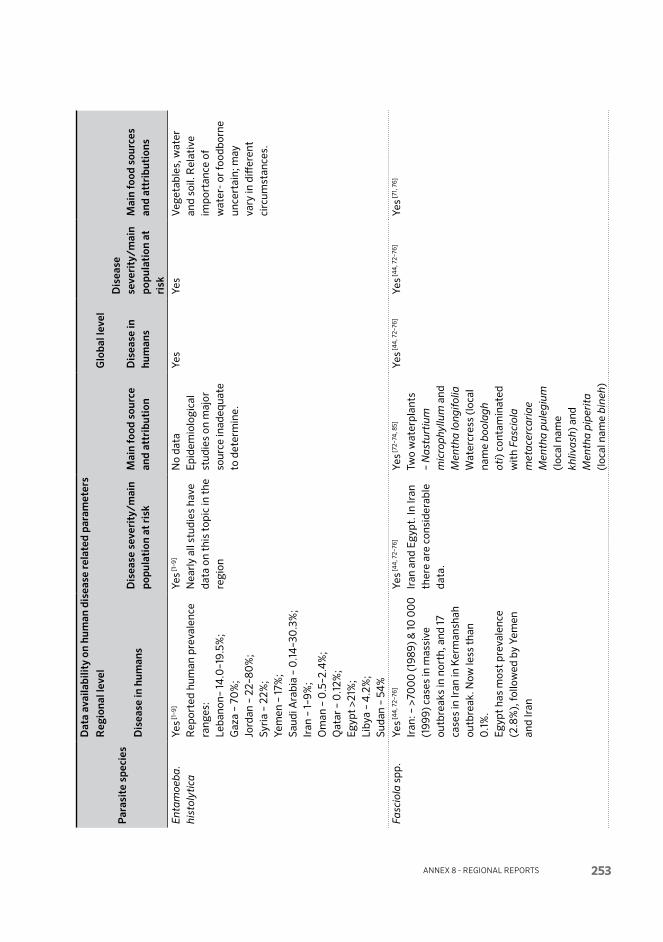
Annex 8 - regionAl reports 253
TAb
lE A
8.5.
1 Dat
a av
aila
bilit
y on
food
bor
ne p
aras
ites
rele
vant
to
the
Nea
r E
ast
Par
asit
e sp
ecie
s
Dat
a av
aila
bilit
y on
hum
an d
isea
se re
late
d pa
ram
eter
sR
egio
nal l
evel
Glo
bal l
evel
Dis
ease
in h
uman
sD
isea
se s
ever
ity/
mai
n po
pula
tion
at r
isk
Mai
n fo
od s
ourc
e an
d at
trib
utio
nD
isea
se in
hu
man
s
Dis
ease
se
veri
ty/ m
ain
popu
lati
on a
t ri
sk
Mai
n fo
od s
ourc
es
and
attr
ibut
ions
Asc
aris
lu
mb
ri co
ides
Yes [1
0, 4
4–5
5]
Saud
i Ara
bia
– 0
.4–2
2.2%
; Ir
an –
0.5
7–1.
5%;
Qat
ar –
0.6
%;
UA
E –
6.6
%;
Egy
pt –
23%
; Li
bya
– 0
.1%
; Su
dan
– 4
7.7%
Yes [1
0, 4
4–5
2]
Nea
rly
all s
tudi
es h
ave
data
on
this
top
ic in
the
re
gion
No
Con
tam
inat
ed s
alad
Ye
sYe
s [53,
54
]
Poo
r ch
ildre
n in
tro
pica
l co
untr
ies,
wit
h ov
er cr
owde
d sl
ums
and
inad
e qua
te
sani
tati
on
Yes
Cry
pto
spor
idiu
m
par
vum
Yes [8
, 14
,19,
35–
43]
Rep
orte
d hu
man
pre
vale
nce:
G
aza
- 16
.3%
; Jo
rdan
- 8
.3%
; Ye
men
- 2
4–4
4%
; Sa
udi A
rabi
a –
2.3–
16%
; Ir
aq –
5–9
.7%
; K
uwai
t –
3.4
%;
Egy
pt –
9%
Li
bya
– 2.
5%
Suda
n –
16%
.
Yes [8
, 14
,19,
35–
43]
Yes
Wat
er, s
ome
tran
s mis
sion
by
cont
amin
ated
pla
nt
mat
eria
l.
Yes
Con
side
rabl
e da
ta fr
om
hosp
ital
and
cl
inic
reco
rds
Yes
Veg
etab
les
was
hed
wit
h co
ntam
inat
ed
wat
er
Ech
ino c
occu
s g
ranu
losu
s.Ye
s [58]
Iraq
– 2
/10
0 0
00
; Ir
an –
0.6
-1.2
/10
0 0
00
and
pr
eval
ence
1.2–
24%
; Ye
men
– 2
6–14
0 c
ases
/yea
r du
ring
20
01–
200
8;
Egy
pt –
10%
; Li
bya
– 1.
4%
; Su
dan
– 0
.5%
.
Yes [5
8]
shep
herd
s fo
llow
ed b
y fa
rmer
s
No
data
Mul
tipl
e so
urce
s.
Yes [2
5, 5
9–70
]Ye
s [25,
59–
70]
Hyd
atid
cys
ts
are
very
se
riou
s an
d di
fficu
lt t
o tr
eat.
Yes
Man
is a
n in
term
edia
te h
ost,
an
d lik
ely
is in
fect
ed
by in
gest
ion
of e
ggs
in c
on ta
m in
ated
soi
l an
d w
ater
.
Par
asit
e sp
ecie
s
Dat
a av
aila
bilit
y on
hum
an d
isea
se re
late
d pa
ram
eter
sR
egio
nal l
evel
Glo
bal l
evel
Dis
ease
in h
uman
sD
isea
se s
ever
ity/
mai
n po
pula
tion
at r
isk
Mai
n fo
od s
ourc
e an
d at
trib
utio
nD
isea
se in
hu
man
s
Dis
ease
se
veri
ty/ m
ain
popu
lati
on a
t ri
sk
Mai
n fo
od s
ourc
es
and
attr
ibut
ions
Ent
amoe
ba.
hi
stol
ytic
aYe
s [1–9
]
Rep
orte
d hu
man
pre
vale
nce
rang
es:
Leba
non–
14.0
–19.
5%;
Gaz
a –
70%
; Jo
rdan
– 2
2–80
%;
Syri
a –
22%
; Ye
men
– 17
%;
Saud
i Ara
bia
– 0
.14
–30
.3%
; Ir
an –
1–9%
; O
man
– 0
.5–2
.4%
; Q
atar
– 0
.12%
; E
gypt
>21
%;
Liby
a –
4.2
%;
Suda
n –
54%
Yes [1
–9]
Nea
rly
all s
tudi
es h
ave
data
on
this
top
ic in
the
re
gion
No
data
Epi
dem
io lo
gica
l st
udie
s on
maj
or
sour
ce in
adeq
uate
to
det
erm
ine.
Yes
Yes
Veg
etab
les,
wat
er
and
soil.
Rel
ativ
e im
port
ance
of
wat
er-
or fo
od bo
rne
unce
rtai
n; m
ay
vary
in d
iffer
ent
circ
umst
ance
s.
Fasc
iola
spp
.Ye
s [44
, 72–
76]
Iran
: – >
700
0 (1
989)
& 10
00
0
(199
9) c
ases
in m
assi
ve
out b
reak
s in
nor
th, a
nd 17
ca
ses
in Ir
an in
Ker
man
shah
ou
tbre
ak. N
ow le
ss t
han
0.1
%.
Egy
pt h
as m
ost
prev
alen
ce
(2.8
%),
follo
wed
by
Yem
en
and
Iran
Yes [4
4, 7
2–76
]
Iran
and
Egy
pt. I
n Ir
an
ther
e ar
e co
nsid
erab
le
data
.
Yes [7
2–74
, 85]
Two
wat
er pl
ants
–
Nas
turt
ium
m
icro
phy
llum
and
M
enth
a lo
ngi f
olia
Wat
ercr
ess
(loc
al
nam
e b
oola
gh
oti)
con
tam
inat
ed
wit
h Fa
scio
la
met
a cer
cari
ae
Men
tha
pul
egiu
m
(loc
al n
ame
khliv
ash)
and
M
enth
a p
iper
ita
(loc
al n
ame
bin
eh)
Yes [4
4, 7
2–76
]Ye
s [44
, 72–
76]
Yes [7
1, 7
6]

Multi criteria-based ranking for risk ManageMent of food-borne parasites254
Par
asit
e sp
ecie
s
Dat
a av
aila
bilit
y on
hum
an d
isea
se re
late
d pa
ram
eter
sR
egio
nal l
evel
Glo
bal l
evel
Dis
ease
in h
uman
sD
isea
se s
ever
ity/
mai
n po
pula
tion
at r
isk
Mai
n fo
od s
ourc
e an
d at
trib
utio
nD
isea
se in
hu
man
s
Dis
ease
se
veri
ty/ m
ain
popu
lati
on a
t ri
sk
Mai
n fo
od s
ourc
es
and
attr
ibut
ions
Gia
rdia
d
uo d
enal
is
(syn
. G. l
amb
lia,
G. i
ntes
tin a
lis)
Yes [1
, 3, 6
, 8, 1
0–2
4]
Rep
orte
d hu
man
pre
vale
nce
rang
es:
Jord
an –
3.9
–42.
6%;
Gaz
a –
10.3
–62.
2%;
Leba
non
– 20
.7%
; W
est
Ban
k –
9,7%
; Sy
ria
– 14
.0–3
1.0
%;
Yem
en –
19.
7%;
Saud
i Ara
bia
– 0
.1–3
7.7%
; Ir
an –
3.7
–14
.5%
; O
man
– 3
.4–1
0.5
%;
Qat
ar –
1.6%
; B
ahra
in –
4%
; Ir
aq –
38.
5%;
Egy
pt –
42%
; Li
bya
– 1.
7%;
Suda
n –
12.3
%.
Yes [1
, 3, 6
, 8, 1
0–2
4]
Bak
ery
wor
kers
[82]
N
earl
y al
l stu
dies
hav
e da
ta o
n th
is t
opic
in t
he
regi
on
Yes [2
4]
Pro
babl
y m
ostl
y w
ater
born
e.Fi
sh
Yes
Yes
Chi
ldre
n ha
ve
espe
c ial
ly h
igh
prev
a len
ce
Yes
Inte
stin
al
fish b
orne
tr
emat
odes
H
eter
o phy
es
hete
rop
hyes
H
. noc
ens
Hap
lorc
his
pum
ilio
Yes [4
5, 5
7, 5
8]
Hyp
er en
dem
ic, e
spec
ially
in
delt
a re
gion
of t
he N
ile
Egy
pt –
33.
8%;
Iran
– 0
.24
%;
Suda
n –
11%
.
Yes [4
5, 5
7, 5
8]
Com
mon
in fi
sher
s,
and
othe
rs c
on su
min
g im
prop
erly
coo
ked
fish.
Yes
Fish
mea
t (e
sp.
un co
oked
)
Yes
Yes
Impr
oper
ly c
ooke
d fis
h

Annex 8 - regionAl reports 255
Par
asit
e sp
ecie
s
Dat
a av
aila
bilit
y on
hum
an d
isea
se re
late
d pa
ram
eter
sR
egio
nal l
evel
Glo
bal l
evel
Dis
ease
in h
uman
sD
isea
se s
ever
ity/
mai
n po
pula
tion
at r
isk
Mai
n fo
od s
ourc
e an
d at
trib
utio
nD
isea
se in
hu
man
s
Dis
ease
se
veri
ty/ m
ain
popu
lati
on a
t ri
sk
Mai
n fo
od s
ourc
es
and
attr
ibut
ions
Taen
ia
sag
inat
a [1
2, 16
,
77–8
1]
Yes
Hum
an p
reva
lenc
e in
the
re
gion
0.4
and
6.0
% in
the
fe
w s
tudi
es re
port
ed.
Saud
i Ara
bia
– 0
.01–
0.2
%;
Iran
– 0
.1%
; Q
atar
– 0
.4%
; E
gypt
– 0
.6%
; Li
bya
– 2%
; Su
dan
– 0
.9%
.
No
data
Yes,
bee
f.N
o da
taYe
sbe
ef
Toxo
pla
sma
gon
dii
Yes [2
6–34
]
Hum
an p
reva
lenc
e:
Leba
non
– 62
%;
Saud
i Ara
bia
– 3.
78–4
.9%
and
4
2.3%
; Q
atar
– 2
9.8%
; B
ahra
in –
4%
; E
gypt
– 11
.7%
; Li
bya
– 4
3.4
%;
Suda
n –
44
.4%
.
Yes [2
6–34
]
Nea
rly
all s
tudi
es h
ave
data
on
this
top
ic in
the
re
gion
Yes [2
6–34
]
How
ever
, dir
ect
inge
stio
n of
ooc
ysts
di
ssem
inat
ed b
y in
fect
ed c
ats
are
also
impo
rtan
t ri
sk [8
3, 8
4]
Toxo
pla
sma
gon
dii
DN
A w
as d
etec
ted
in 2
5% o
f sal
ami,
20.3
% o
f sau
sage
, 21
.8%
of h
am bu
rger
an
d 32
.8%
of
keba
b [29]
Yes [2
5]Ye
s [25]
Yes
Por
k, la
mb.
Tric
huri
s tr
ichi
ura
Yes [1
, 10
, 46,
55–
57]
Saud
i Ara
bia
– 0
.36–
28.8
%;
Qat
ar –
26.
3%;
UA
E –
6.2
%;
Egy
pt –
49.
7%
Suda
n –
46%
.
Yes [1
, 10
, 46,
56,
57]
Nea
rly
all s
tudi
es h
ave
data
on
this
top
ic in
the
re
gion
No
data
Yes
Yes
Yes
Multi criteria-based ranking for risk ManageMent of food-borne parasites256
Sources used for Table A8.5.1
01. Abu-Madi, M.A., Behnke, J.M. & Doiphode, S.H. 2010. Changing trends in intestinal parasitic infections among long-term residents and settled immigrants in Qatar. Parasites & Vectors, 3: Art. No. 98. [Online; DOI: 10.1186/1756-3305-3-98]
02. Rionda, Z.l. & Clements, A. 2005. The burden of disease in West Bank and Gaza: An assessment Report. 27 p. Report sumitted to the US AID, MEDS Project Contract No. HRN-I-00-99-00002-00. MEDS Publications, Washington D.C., USA.
03. Araj, G.F., Musharrafieh, u.M., Haydar, A., Ghawi, A., Itani, R. & Saliba, R. 2011. Trends and prevalence of intestinal parasites at a tertiary care center in Lebanon over a decade. Lebanese Medical Journal, 59(3): 143–148.
04. El kichaoi, A.y., Abdel Fattah, N., Abd Rabou, Fadel A. Sharif & Husni M. El-Amssi. 2004. Changing trends in frequency of intestinal parasites of Gaza, 1995–2000. Journal of the Islamic University of Gaza (Natural Sciences), 12(2): 121–129.
06. Alyousefi, N.A., Mahdy, M.A.k., Mahmud, R. & lim, y.A.l. 2011. Factors associated with high prevalence of intestinal protozoan infections among patients in Sana’a City, Yemen. PLoS ONE, 6(7): Art. no. e22044. [Online; DOI: 10.1371/journal.pone.0022044]
07. Stauffer, W., Abd-Alla, M. & Ravdin, J.I. 2006. Prevalence and incidence of Entamoeba histolytica infection in South Africa and Egypt. Archives of Medical Research, 37(2): 266–269.
08. Gusbi, M.M. 2007. Aetiology of acute diarrhoea in hospitalized children, Tripoli, Libya. PhD thesis, University of Salford, UK.
09. Saeed, A., Abd, H., Evengard, B. Sandstrom, G. 2011. Epidemiology of entamoeba infection in Sudan. African Journal of Microbiology Research, 5(22): 3702–3705.
10. Abu-Madi, M.A., Behnke, J.M. & Ismail, A. 2008. Patterns of infection with intestinal parasites in Qatar among food handlers and housemaids from different geographical regions of origin. Acta Tropica, 106(3): 213–220.
12. Al-Haddad, A.M. & Baswaid, S.H. 2010. Frequency of intestinal parasitic infection among children in Hadhramout governorate (Yemen). Journal of the Egyptian Society of Parasitology, 40(2): 479–499.
13. Ammoura, A.M. 2010. Impact of hygienic level on parasite infection. Asian Pacific Journal of Tropical Medicine, 3(2): 148–149.
14. Nimri, l.F. & Meqdam, M. 2004. Enteropathogens associated with cases of gastro enteritis in a rural population in Jordan. Clinical Microbiology and Infection, 10(7): 634–639.
15. yassin, M.M., Shubair, M.E., al-Hindi, A.I. & Jadallah, S.y. 1999. Prevalence of intestinal parasites among school children in Gaza City, Gaza Strip. Journal of the Egyptian Society of Parasitology, 29(2): 365–373.
16. Araj, G.F., Abdul-Baki, N.y., Hamze, M.M., Alami, S.y., Nassif, R.E. & Naboulsi, M.S. 1996. Prevalence and etiology of intestinal parasites in Lebanon. Lebanese Medical Journal, 44(3): 129–133.
17. Hussein, A.S. 2011. Prevalence of intestinal parasites among school children in northern districts of West Bank-Palestine. Tropical Medicine & International Health, 16(2): 240–244.
18. Almerie, M.Q., Azzouz, M.S., Abdessamad, M.A., Mouchh, M.A., Sakbani, M.W., Alsibai, M.S., Alkafri, A. & Ismail, M.t. 2008. Prevalence and risk factors for giardiasis among primary school children in Damascus, Syria. Saudi Medical Journal, 29(2): 234–240.
19. Al-Shibani, l.A., Azazy, A.A., El-taweel, H.A. 2009. Cryptosporidiosis and other intestinal parasites in 3 Yemeni orphanages: prevalence, risk, and morbidity. Journal of the Egyptian Society of Parasitology, 39(1): 327–337.
20. Al-Saeed, A.t. & Issa, S.H. 2006. Frequency of Giardia lamblia among children in Dohuk, northern Iraq. East Mediterranean Health Journal, 12(5): 555–561.
21. Mukhtar, A. 1994. Giardiasis amongst in- and out-patients in Salmaniya Medical Centre in the state of Bahrain. Indian Journal of Medical Science, 48(6): 135–138.
22. Ghoneim, N.H., Abdel-Moein, k.A. & Saeed, H. 2012. Fish as a possible reservoir for zoonotic Giardia duodenalis assemblages. Parasitology Research, 110(6): 2193–2196.

Annex 8 - regionAl reports 257
23. Sullivan, p.S., Dupont, H.l., Arafat, R.R., thornton, S.A., Selwyn, B.J., Elalamy, M.A. & Zaki, A.M. 1988. Illness and reservoirs associated with Giardia lamblia infection in rural Egypt – the case against treatment in developing world environments of high endemicity. American Journal of Epidemiology, 127(6): 1272–1281.
24. Mamoun, M., Abubakr, I. & El-Muntasir, t. 2009. Frequency of intestinal parasitic infections among displaced children in Kassala town. Khartoum Medical Journal, 2(1): 175–177.
25. torgerson, p.R. & Macpherson, C.N.l. 2011. The socioeconomic burden of parasitic zoonoses: Global trends. Veterinary Parasitology, 182(1; Special issue): 79–95.
26. Abu-Madi, M.A., Al-Molawi, N. & Behnke, J.M. 2008. Seroprevalence and epidemiological correlates of Toxoplasma gondii infections among patients referred for hospital-based serological testing in Doha, Qatar. Parasites & Vectors, 1: Art. No. 39. [Online; DOI: 10.1186/1756-3305-1-39]
27. Abu-Madi, M.A., Behnke, J.M. & Dabritz, H.A. 2010. Toxoplasma gondii seropositivity and co-infection with TORCH pathogens in high-risk patients from Qatar. American Journal of Tropical Medicine and Hygiene, 82(4): 626–633.
28. Bouhamdan, S.F., Bitar, l.k., Saghir, H.J., Bayan, A. & Araj, G.F. 2010. Seroprevalence of toxoplasma antibodies among individuals tested at hospitals and private laboratories in Beirut. Lebanese Medical Journal, 58(1): 8–11.
29. Fallah, E., Hajizadeh, M., Farajnia, S. & khanmohammadi, M. 2011. Prevalence of Toxoplasma gondii in food products in north west of Iran in 2010. Australian Journal of Basic and Applied Sciences, 5(6): 1482–1485.
30. Al-Mohammad, H.I., Amin, t.t., Balaha, M.H. & Al-Moghannum, M.S. 2010. Toxoplasmosis among the pregnant women attending a Saudi maternity hospital: seroprevalence and possible risk factors. Annals of Tropical Medicine and Parasitology, 104(6): 493–504.
31. Al-Qurashi, A.R. 2004. Sero epidemiological study of toxoplasmosis in rural areas in the eastern region of Saudi Arabia. Journal of the Egyptian Society of Parasitology, 34(1): 23–34.
32. tabbara, k.S. & Saleh, F. 2005. Serodiagnosis of toxoplasmosis in Bahrain. Saudi Medical Journal, 26(9): 1383–1387.
33. khadre, M.A. & Elnageh, M.M. 1987. Serological survey for toxoplasmosis in Tripoli, Splaj (Libya). Transactions of the Royal Society of Tropical Medicine and Hygiene, 81(5): 761–763.
34. Abdin Babikir Siddig. 2010. Study on toxoplasmosis in humans and cats in the Red Sea State, Sudan. PhD thesis. Veterinary Medicine College, Sudan University of Science & Technology.
35. Iqbal, J., khalid, N. & Hina, p.R. 2011. Cryptosporidiosis in Kuwaiti children: association of clinical characteristics with Cryptosporidium species and subtypes. Journal of Medical Microbiology, 60(5): 647–652.
36. Iqbal, J., Hira, p.R., Al-Ali, F. & philip, R. 2001. Cryptosporidiosis in Kuwaiti children: seasonality and endemicity. Clinical Microbiology and Infection, 7(5): 261–266.
37. Nazemalhosseini-Mojarad, E., Feng, y. & Xiao, l. 2012. The importance of subtype analysis of Cryptosporidium spp. in epidemiological investigations of human cryptosporidiosis in Iran and other Mideast countries. Gastroenterology and Hepatology from Bed to Bench, 5(2): 67–70.
38. Al-Shamiri, A., Al-Zubairy, A. & Al-Mamari, R. 2010. The prevalence of Cryptosporidium spp. in children, Taiz District, Yemen. Iranian Journal of Parasitology, 5(2): 26–32.
39. Mahdi, N.k. & Ali, N.H. 2002. Cryptosporidiosis among animal handlers and their livestock in Basrah, Iraq. East African Medical Journal, 79(10): 550–553.
40. Mahdi, N.k. & Ali, N.H. 2004. Cryptosporidiosis and other intestinal parasitic infections in patients with chronic diarrhea. Saudi Medical Journal, 25(9): 1204–1207.
41. Ranjbar-Bahadori, S., Sangsefidi, H., Shemshadi, B. & kashefinejad, M. 2011. Cryptosporidiosis and its potential risk factors in children and calves in Babol, north of Iran. Tropical Biomedicine, 28(1): 125–131.
42. youssef, F.G., Adib, I., Riddle, M.S. & Schlett, C.D. 2008. A review of cryptosporidiosis in Egypt. Journal of the Egyptian Society of Parasitology, 38(1): 9–28.
43. Adam, A.A., Hassan, H.S., Shears, p. & Elshibly, E. 1994. Cryptosporidium in Khartoum, Sudan. East African Medical Journal, 71(11): 745–746.

Multi criteria-based ranking for risk ManageMent of food-borne parasites258
45. Chai, J.y., park, J.H., Han, E.t., Shin, E.H., kim, J.l., Guk, S.M., Hong, k.S., lee, S.H. & Rim, H.J. 2004. Prevalence of Heterophyes nocens and Pygydiopsis summa infections among residents of the western and southern coastal islands of the Republic of Korea. American Journal of Tropical Medicine and Hygiene, 71(5): 617–622.
46. Hotez, p.J. 2009. The neglected tropical diseases and their devastating health and economic impact on the member nations of the Organisation of the Islamic Conference. PLoS Neglected Tropical Diseases, 3(10): Art. no. e539. {Online; doi:10.1371/journal.pntd.0000539]
47. Al-Binali, A.M., Bello, C.S., El-Shewy, k. & Abdulla, S.E. 2006. The prevalence of parasites in commonly used leafy vegetables in south-western Saudi Arabia. Saudi Medical Journal, 27(5): 613–616.
48. Al-Madani, A.A. & Mahfouz, A.A. 1995. Prevalence of intestinal parasitic infections among Asian female house-keepers in Abha District, Saudi Arabia. Southeast Asian Journal of Tropical Medicine and Public Health, 26(1): 135–137.
49. Sayyari, A.A., Imanzadeh, F., Bagheri yazdi, S.A., karami, H. & yaghoobi, M. 2005. Prevalence of intestinal parasitic infections in the Islamic Republic of Iran. Eastern Mediterranean Health Journal, 11(3): 377–383.
50. Ali, S.I., Jamal, k. & Qadri, S.M. 1992. Prevalence of intestinal parasites among food handlers in Al-Medinah. Annals of Saudi Medicine, 12(1): 63–66.
51. Ibrahim, O.M.G., Bener, A. & Shalabi, A. 1993. Prevalence of intestinal parasites among expatriate workers in Al-Ain, United Arab Emirates. Annals of Saudi Medicine, 13(2): 126–129.
52. Al-Braiken, F.A. 2008. Is intestinal parasitic infection still a public health concern among Saudi children? Saudi Medical Journal, 29(11): 1630–1635.
53. Anon. Ascariasis. Chapter 10, in: Palmer, P.E.S. and Reeder, M.M. (Editors). Imaging of Tropical Diseases, with Epidemiological, Pathological and Clinical Correlation. Available online at http://tmcr.usuhs.edu/tmcr/chapter10/intro.htm
54. Sadaga, G. & kassem, H. 2007. Prevalence of intestinal parasites among primary school children in Derna district, Libya. Journal of the Egyptian Society of Parasitology, 37(1): 205–214.
55. Salem, G., van de Velden, l., laloé, F., Maire, B., ponton, A., traissac, p. & prost, A. 1994. [Intestinal parasitic diseases and environment in Sahelo-Sudanese towns: the case of Pikine (Senegal)] [Article in French]. Revue d’epidemiologie et sante publique, 42(4): 322–333. [PMID: 8085049]
56. Hotez, p.J., Savioli, l. & Fenwick, A. 2012. Neglected tropical diseases of the Middle East and North Africa: Review of their prevalence, distribution, and opportunities for control. PLoS Neglected Tropical Diseases, 6(2): Art. no. e1475. [Online; DOI: 10.1371/journal.pntd.0001475]
57. Abou-Basha, l.M., Abdel-Fattah, M., Orecchia, p., Di Cave, D. & Zaki, A. 2000. Epidemiological study of heterophyiasis among humans in an area of Egypt. East Mediterranean Health Journal, 6(5-6): 932–938.
58. Crompton, D.W.t. 1999. How much human helminthiasis is there in the world? Journal of Parasitology 85: 379–403. See also http://curezone.com/diseases/parasites/
58. Sadjjadi, S.M. 2006. Present situation of echinococcosis in the Middle East and Arabic North Africa. Parasitology International, 55(Suppl.): S197–S202.
59. torgerson, p.R., keller, k., Magnotta, M. & Ragland, N. 2010. The global burden of alveolar echinococcosis. PLoS Neglected Tropical Diseases, 4(6): Art. no. e722. [Online; DOI: 10.1371/journal.pntd.0000722]
60. Budke, C.M., Deplazes, p. & torgerson, p.R. 2006. Global socioeconomic impact of cystic echinococcosis. Emerging Infectious Diseases, 12(2): 296–303.
61. Grosso, G., Gruttadauria, S., Biondi, A., Marventano, S. & Mistretta, A. 2012. Worldwide epidemiology of liver hydatidosis including the Mediterranean area. World Journal of Gastroenterology, 18(13): 1425–1437.
62. Alghoury, A., El-Hamshary, E., Azazy, A., Hussein, E. & Rayan, H.Z. 2010. Hydatid disease in Yemeni Patients attending public and private hospitals in Sana’a City, Yemen. Oman Medical Journal, 25(2): 88–90.
63. Al-Shibani, l.A., Al-Eryani, S.M., Azazy, A.A. & Al-Mekhlafi, A.M. 2012. Cases of hydatidosis in patients referred to Governmental hospitals for cyst removal in Sana’a City, Republic of Yemen. Tropical Biomedicine, 29(1): 18–23.

Annex 8 - regionAl reports 259
64. Rokni, M.B. 2009. Echinococcosis/hydatidosis in Iran. Iranian Journal of Parasitology, 4(2): 1–16.
65. Saeed, I., kapel, C., Saida, l.A., Willingham, l. & Nansen, p. 2000. Epidemiology of Echinococcus granulosus in Arbil province, northern Iraq, 1990–1998. Journal of Helminthology, 74(1): 83–88.
66. Molan, A.l. & Saida, l.A. 1989. Echinococcosis in Iraq: Prevalence of Echinococcus granulosus in stray dogs in Arbil province. Japanese Journal of Medical Science and Biology, 42(4): 137–141.
67. El-Shazly, A.M., Awad, S.E., Hegazy, M.A., Mohammad, k.A. & Morsy, t.A. 2007. Echinococcosis granulosis/hydatosis – an endemic zoonotic disease in Egypt. Journal of the Egyptian Society of Parasitology, 37(2): 609–622.
68. Dyab, k.A., Hassanein, R., Hussein, A.A., Metwally, S.E. & Gaad, H.M. 2005. Hydatidosis among man and animals in Assiut and Aswan Governorates. Journal of the Egyptian Society of Parasitology, 35(1): 157–166.
69. Shambesh, M.k., Macpherson, C.N.l., Beesley, W.N., Gusbi, A. & Elsonosi, t. 1992. Prevalence of human hydatid-disease in north-western Libya - a cross-sectional ultrasound study. Annals of Tropical Medicine and Parasitology, 86(4): 381–386.
70. Magambo, J.k., Hall, C., Zeyle, E. & Wachira, t.M. 1996. Prevalence of human hydatid disease in southern Sudan. African Journal of Health Science, 3(4): 154–156.
71. Mas-Coma, S., Valero, M.A. & Bargues, M.D. 2009. Fasciola, lymnaeids and human fascioliasis, with a global overview on disease transmission, epidemiology, evolutionary genetics, molecular epidemiology and control. Advances in Parasitology, 69: 41–146.
72. Ashrafi, k., Valero, M.A., Forghan-parast, k., Rezaeian, M., Shahtaheri, S.J., Hadiani, M.R., Bargues, M.D. & Mas-Coma, S. 2006. Potential transmission of human fascioliasis through traditional local foods, in northern Iran. Iranian Journal of Public Health, 35(2): 57–63.
72. Rokni, M.B. 2008. The present status of human helminthic diseases in Iran. Annals of Tropical Medicine and Parasitology, 102(4): 283–295.
73. Ashrafi, k., Valero, M.A., Massoud, J., Sobhani, A., Solaymani-Mohammadi, S., Conde, p., khoubbane, M., Bargues, M.D. & Mas-Coma, S. 2006. Plant-borne human contamination by fascioliasis. American Journal of Tropical Medicine and Hygiene, 75(2): 295–302.
75. Esteban, J.G., Gonzalez, C., Curtale, F., Muñoz-Antoli, C., Valero, M.A., Bargues, M.D., el-Sayed, M., el-Wakeel, A.A., Abdel-Wahab, y., Montresor, A., Engels, D., Savioli, l. & Mas-Coma, S. 2003. Hyper-endemic fascioliasis associated with schistosomiasis in villages in the Nile Delta of Egypt. American Journal of Tropical Medicine and Hygiene, 69(4): 429–437.
76. Shalaby, S.I. 1997. Does fascioliasis in buffaloes constitute a zoonotic health problem in major Cairo, Egypt? pp. 578–581, in: A. Borghese, S. Failla and V.L. Barile (Editors). [Proceedings of the] 5th World Buffalo Congress, Caserta, Italy, 13–16 October 1997. Istituto Sperimentale per la Zootecnia, Monterotondo, Italy.
77. Hamze, M., Dabboussi, F., Al-Ali, k. & Ourabi, l. 2004. [Prevalence of infection by intestinal parasites in north Lebanon: 1997–2001]. [Article in French] East Mediterranean Health Journal, 10(3): 343–348.
78. Al-lahham, A.B., Abu-Saud, M. & Shehabi, A.A. 1990. Prevalence of Salmonella, Shigella and intestinal parasites in food handlers in Irbid, Jordan. Journal of Diarrhoeal Disease Research, 8(4): 160–162.
79. Basem, R.N.A., Sayed, A.S.M., Hussein, A.A.A. & Arafa, M.I. 2009. Occurrence of cysticercosis in cattle and buffaloes and Taenia saginata in man in Assiut Governorate of Egypt. Veterinary World, 2(5): 173–176. [Online; doi: 10.5455/vetworld.2009.173-176]
80. Ben Musa, N.A. 2007. Intestinal parasites in school aged children and the first case report on amoebiasis in urinary bladder in Tripoli, Libya. Journal of the Egyptian Society of Parasitology, 37(3): 775–784.
81. Dada, B.J. 1980. Taeniasis, cysticercosis and echinococcosis/hydatidosis in Nigeria: I –prevalence of human Taeniasis, cysticercosis and hydatidosis based on a retrospective analysis of hospital records. Journal of Helminthology, 54(4): 281–286.

Multi criteria-based ranking for risk ManageMent of food-borne parasites260
82. kheirandish, F., tarahi, M.J., Haghighi, A., Nazemalhosseini-Mojarad, E. & kheirandish, M. 2011. Prevalence of intestinal parasites in bakery workers in Khorramabad, Lorestan Iran. Iranian Journal of Parasitology, 6(4): 76–83.
83. Sarkari, B., Ghobakhloo, N., Moshfea, A.A. & Eilami, O. 2012. Seroprevalence of human fasciolosis in a new-emerging focus of fasciolosis in Yasuj District, south-west Iran. Iranian Journal of Parasitology, 7(2): 15–20.
84. Morsy, t.A., Michael, S.A. & El Disi, A.M. 1985. Cats as reservoir hosts of human parasites in Amman, Jordan. Journal of the Egyptian Society of Parasitology, 10: 5–18.
85. Deeb, B.J., Sufan, M. & Di Giacomi, R.F. 1985. Toxoplasma gondii infection of cats in Beirut, Lebanaon. Journal of Tropical Medicine and Hygiene, 88: 301–306.
TAblE A8.5.2 Data availability for parasite prevalence or concentration in the main food categories in the Near East
Ascaris lumbricoides
Fruits Via munici pal re cycling of waste-water in farming.
Vegetables Yes [3, 34] Via munici pal re-cycling of waste-water in crops.
Other Accidental ingestion of mature Ascaris eggs through con tamin ated food or
water.
crypto spor idium parvum [6,22]
Fruits Yes. If con tam inated with water con taing oocysts
Vegetables Yes. As for fruit.
Other Mostly water borne.
echino coccus gran ulosus [30]
Fruits Yes [32]
Vegetables Yes [31, 33]
Other The eggs may be eaten in foods (e.g. vege tables, fruits or herbs, or drunk in
con tam inated water
entamoeba hystolytica
Fruits Yes 2, 4]
Vegetables Yes [1–3]
Other Water [1-2]
Fasciola spp. [23–25]
Vegetables Yes [3, 26}
Other Primarily leafy greens raised in water (ponds, streams). Some infective
stages (meta cercariae) may be transmitted by water.
Fish-borne trematodes
Seafood Ingest ion of improp erly cooked fresh and brack ish-water fish
giardia duo denalis (syn. g. lamblia, g. intestin alis)
Fruits Yes [10]
Vegetables Yes [1, 5–7]
Other Yes [1, 5–9] Water, fish.

Annex 8 - regionAl reports 261
taenia saginata [27–28]
Beef Yes
toxoplasma gondii
Beef Yes [17]
Dairy Yes [15–16]
Pork Yes [11–14]
Poultry Yes [19]
Game Yes
Fruits Yes [20]
Vegetables Yes [11–14] When cat faeces deposited in vege table farm.
Other Yes [11–14, 18] Soil and water; pork and oocyst contamin ated water, fruits and
vege tables have been implicated; T. gondii DNA was detected in 25%
of salami, 20.3% of sausage, 21.8% of hamburger and 32.8% of kebab
samples. [18]
Sources cited for Table A8.5.2
01. Baldursson, S. & karanis, p. 2011. Waterborne transmission of protozoan parasites: Review of worldwide outbreaks – An update 2004–2010. Water Research, 45(20): 6603–6614.
Global
02. Ximenez, C., Moran, p., Rojas, l., Valadez, A. & Gomez, A. 2009. Reassessment of the epidemiology of amebiasis: State of the art. Infection Genetics and Evolution, 9(6):1023–1032.
Global
03. Daryani, A., Ettehad, G.H., Sharif, M., Ghorbani, l. & Ziaei, H. 2008. Prevalence of intestinal parasites in vegetables consumed in Ardabil, Iran. Food Control, 19(8): 790–794.
Global with regional foci
04. Information from http://www.medic8.com/healthguide/food-poisoning/entamoeba-histolytica.html
Global
05. Rionda, Z.l. & Clements, A. 2005. The burden of disease in West Bank and Gaza: An assessment Report. 27 p. Report sumitted to the US AID, MEDS Project Contract No. HRN-I-00-99-00002-00. MEDS Publications, Washington D.C., USA.
Global
06. Hunter, p.R. & thompson, R.C.A. 2005. The zoonotic transmission of Giardia and Cryptosporidium. International Journal for Parasitology, 35(11-12): 1181–1190.
Global
07. Alyousefi, N.A., Mahdy, M.A.k., Mahmud, R. & lim, y.A.l. 2011. Factors associated with high prevalence of intestinal protozoan infections among patients in Sana’a City, Yemen. PLoS ONE, 6(7): Art. no. e22044. [Online; DOI: 10.1371/journal.pone.0022044]
Global
08. Ghoneim, N.H., Abdel-Moein, k.A. & Saeed, H. 2012. Fish as a possible reservoir for zoonotic Giardia duodenalis assemblages. Parasitology Research, 110(6): 2193–2196.
Global
09. Al-Binali, A.M., Bello, C.S., El-Shewy, k. & Abdulla, S.E. 2006. The prevalence of parasites in commonly used leafy vegetables in south-western Saudi Arabia. Saudi Medical Journal, 27(5): 613–616.
Regional
10. Anon. No date. [Giardiasis fact sheet] Online. See: http://medicalcenter.osu.edu/patientcare/healthcare_services/infectious_diseases/giardiasis/Pages/index.aspx
Global

Multi criteria-based ranking for risk ManageMent of food-borne parasites262
11. torgerson, p.R. & Macpherson, C.N.l. 2011. The socioeconomic burden of parasitic zoonoses: Global trends. Veterinary Parasitology, 182(1; Special issue): 79–95.
Global
12. Dubey, J.p. 2004. Toxoplasmosis – a waterborne zoonosis. Veterinary Parasitology, 126(1-2; Special Issue): 57–72.
Global
13. Alvarado-Esquivel, C., Estrada-Martinez, . & liesenfeld, O. 2011. Toxoplasma gondii infection in workers occupationally exposed to unwashed raw fruits and vegetables: a case control seroprevalence study. Parasites & Vectors, 4: Art. No. 235. [Online; DOI: 10.1186/1756-3305-4-235]
Global
14. lass, A., pietkiewicz, H., Szostakowska, B. & Myjak, p. 2012. The first detection of DNA in environmental fruits and vegetables samples. European Journal of Clinical Microbiology & Infectious Diseases, 31(6): 1101–1108.
Global
15. tzanidakis, N., Maksimov, p., Conraths, F.J., kiossis, E., Brozos, C., Sotiraki, S. & Schares, G. 2012. Toxoplasma gondii in sheep and goats: Seroprevalence and potential risk factors under dairy husbandry practices. Veterinary Parasitology, 190(3-4): 340–348.
Global
16. Asgari, Q., Mehrabani, D., Motazedian, M.H., kalantari, M., Nouroozi, J. & Adnani Sadati, S.J. 2011. The viability and infectivity of Toxoplasma gondii tachyzoites in dairy products undergoing food processing. Asian Journal of Animal Sciences, 5(3): 202–207.
Regional
17. kijlstra, A. & Jongert, E. 2008. Control of the risk of human toxoplasmosis transmitted by meat. International Journal for Parasitology, 38(12): 1359–1370.
Regional
18. Fallah, E., Hajizadeh, M., Farajnia, S. & khanmohammadi, M. 2011. Prevalence of Toxoplasma gondii in food products in north west of Iran in 2010. Australian Journal of Basic and Applied Sciences, 5(6): 1482–1485.
Regional
19. Dubey, J.p. 2010. Toxoplasma gondii infections in chickens (Gallus domesticus): prevalence, clinical disease, diagnosis and public health significance. Zoonoses and Public Health, 57(1): 60–73.
Global
20. pereira, k.S., Franco, R.M. & leal, DA. 2010. Transmission of toxoplasmosis (Toxoplasma gondii) by foods. Advances in Food and Nutrition Research, 60: 1–19.
Global
22. Fayer, R. 2004. Cryptosporidium: a water-borne zoonotic parasite. Veterinary Parasitology, 126(1-2): 37–56.
Global
23. Ashrafi, k., Valero, M.A., Massoud, J., Sobhani, A., Solaymani-Mohammadi, S., Conde, p., khoubbane, M., Bargues, M.D. & Mas-Coma, S. 2006. Plant-borne human contamination by fascioliasis. American Journal of Tropical Medicine and Hygiene, 75(2): 295–302.
Global
24. Mas-Coma, S., Valero, M.A. & Bargues, M.D. 2009. Fasciola, lymnaeids and human fascioliasis, with a global overview on disease transmission, epidemiology, evolutionary genetics, molecular epidemiology and control. Advances in Parasitology, 69: 41–146.
25. Ashrafi, k., Valero, M.A., Forghan-parast, k., Rezaeian, M., Shahtaheri, S.J., Hadiani, M.R., Bargues, M.D. & Mas-Coma, S. 2006. Potential transmission of human fascioliasis through traditional local foods, in northern Iran. Iranian Journal of Public Health, 35(2): 57–63.
Global
26. Sarkari, B., Ghobakhloo, N., Moshfea, A. & Eilami, O. 2012. Seroprevalence of human fasciolosis in a new-emerging focus of fasciolosis in Yasuj district, southwest of Iran. Iran Journal of Parasitology, 7(2): 15–20.
Regional
27. Murrell, k.D. 2005. Epidemiology of taeniosis and cysticercosis. pp. 27–43 (Chapter 3), in: K.D. Murrell, P. Dorny, A. Flisser, S. Geerts, N.C. Kyvsgaard, D.P. McManus, T.E. Nash and Z.S. Pawłowski (editors). WHO/FAO/OIE Guidelines for the Surveillance, Prevention and Control of Taeniosis/Cysticercosis. OIE (World Organisation for Animal Health), Paris, France.
28. khaniki, G.R., Raei, M., kia, E.B., Haghi, A.M. & Selseleh, M. 2010. Prevalence of bovine cysticercosis in slaughtered cattle in Iran. Tropical Animal Health and Production, 42(2): 141–143.
Global

Annex 8 - regionAl reports 263
29. Robinson, M.W. & Dalton, J.p. 2009. Zoonotic helminth infections with particular emphasis on fasciolosis and other trematodiases. Philosophical Transactions of the Royal Society, Series B: Biological Sciences, 364(1530): 2763–2776.
Global
30. Harandi, M.F., Moazezi, S.S., Saba, M., Grimm, F., kamyabi, H., Sheikhzadeh, F., Sharifi, I. & Deplazes, p. 2011. Sonographical and serological survey of human cystic echinococcosis and analysis of risk factors associated with seroconversion in rural communities of Kerman, Iran. Zoonoses and Public Health, 58(8): 582–588.
Global
31. Rokni, M.B. 2009. Echinococcosis/hydatidosis in Iran. Iranian Journal of Parasitology, 4(2): 1–16.
Global
32. Anon. 2011. Echinococcosis (Echinococciasis, Hydatidosis, Hydatid Disease) [Online Factsheet] Center for Food Security and Public Health, College of Veterinary Medicine, Iowa State University, USA. See: http://www.cfsph.iastate.edu/Factsheets/pdfs/echinococcosis.pdf.
Global
33. Bourée, p. 2001. Hydatidosis: dynamics of transmission. World Journal of Surgery, 25(1): 4–9.
Global
34. Shahnazi, M. & Jafari-Sabet, M. 2010. Prevalence of parasitic contamination of raw vegetables in villages of Qazvin Province, Iran. Foodborne Pathogens and Disease, 7(9): 1025–1030.
Regional
table A8-5-3 Data availability for risk management options for each of parasite-com-modity combinations in the Near East
Read in close conjunction with Tables A8.5.1 and A8.5.2 NOTE: The authors were asked to consider all combinations of the particular parasite and the main food categories, namely Beef, Dairy, Pork, Poultry, Game, Seafood, Fruit, Vegetables and Other.
Ascaris lumbricoides, trichuris trichiura
The main source of infection is vegetables, so the best management would be to prevent
use of night soil on vegetable farms, and improving composting techniques for manure.
Fruits Yes
Vege tables Yes [27]
echino coccus granu losus
Control of dog populations in endemic areas is necessary. There are possible implication
for fruits and vegetables. See references 15–22.
Beef Yes
Pork Yes
Game Yes
Fruits Yes
Vege tables Yes
entamoeba hystolytica, giardia duo denalis (syn. g. lamblia, g. intestinalis) and cryptosporidium parvum
The parasites Entamoeba, Giardia and Cryptosporidium can be grouped as primarily
water borne. Risk reduction involves the supply of potable, safe water for not only drinking,
but also for food preparation and washing mouth and hands. Relevant publications linked
to these parasites are in Table A8.5.2.

Multi criteria-based ranking for risk ManageMent of food-borne parasites264
Fruits Yes [1–10]
Vege tables Yes [1–10]
Other Yes. All are water-borne.
Fishborne trematodes: heterophyes heterophyes, haplorchis pumilio, procerovum spp.
Only fish and humans are important in the control of this zoonosis. Dogs may play a role
as alternative definitive host in endemic areas. Major risk factors include the use of fish
from infested regions and improper cooking, or even eating fish raw with spices. Strict
fish muscle examination is needed, especially for imported fish. Deep freezing on board
is mandatory for imported fish. Examination by veterinary and health authorities and
application of national standards is obligatory.
Sea food Yes
Fasciola spp.
Risk management programmes are described in numerous publications, specific for
humans or for livestock. See references 23–24.
Beef Yes
Dairy Yes
Pork Yes
Game Yes
Vege tables Yes
taenia saginata
Because of strict host specificity, only cattle and humans are important in the epidemiology
and control of this zoonosis. Major risk factors include exposure of animals to faecal waste.
For humans, exposure to inadequate meat inspection, inadequate cooking temperatures.
Considerable information on this available in the WHO/FAO/OIE Guidelines[28].
For mitigation of risk in cattle and consumer meats, see reference 25.
For control in humans and egg contamination of the environment (by cattle), see reference
26.
Beef Yes
Dairy Yes?
toxoplasma gondii
Recent evidence that Toxoplasma could be transmitted by contaminated fruits and
vegetables is covered in references 11–14, q.v. See Table A8.5.2 for references on meat and
water transmission. Risk management will require improvement in water safety as well as
prevention of exposure of livestock to infected cats.
Beef Yes [11–14]
Dairy Yes [11–14]
Pork Yes [11–14]
Poultry Yes [11–14]

Annex 8 - regionAl reports 265
Fruits Yes [11–14]
Vege tables Yes [11–14]
Other Yes [11–14]
References cited in Table A8.5.3
01. Rionda, Z.l. & Clements, A. 2005. The burden of disease in West Bank and Gaza: An assessment Report. 27 p. Report sumitted to the US AID, MEDS Project Contract No. HRN-I-00-99-00002-00. MEDS Publications, Washington D.C., USA.
02. Sha’ar, A, kelly, p. & kleinau, E. 2003. USAID village water and sanitation program, West Bank of Palestine: Environmental health assessment—Phase II. Report, Environmental Health Project, Contract HRN-I-00-99-0011-00, Office of Health, Infectious diseases and Nutrition, Bureau for Global Health, US Agerncy for International Development, Washington D.C., USA.
03. Erickson, M.C. & Ortega, y.R. 2006. Inactivation of protozoan parasites in food, water, and environmental systems. Journal of Food Protection, 69(11): 2786–2808.
04 Ranjbar-Bahadori, S., Sangsefidi, H., Shemshadi, B. & kashefinejad, M. 2011. Cryptosporidiosis and its potential risk factors in children and calves in Babol, north of Iran. Tropical Biomedicine, 28(1): 125–131.
05. Stauffer, W., Abd-Alla, M. & Ravdin, J.I. 2006. Prevalence and incidence of Entamoeba histolytica infection in South Africa and Egypt. Archives of Medical Research, 37(2): 266–269.
06. Gusbi, M.M. 2007. Aetiology of acute diarrhoea in hospitalized children, Tripoli, Libya. PhD thesis, University of Salford, UK.
07. Saeed, A., Abd, H., Evengard, B. Sandstrom, G. 2011. Epidemiology of entamoeba infection in Sudan. African Journal of Microbiology Research, 5(22): 3702–3705.
09. Sullivan, p.S., Dupont, H.l., Arafat, R.R., thornton, S.A., Selwyn, B.J., Elalamy, M.A. & Zaki, A.M. 1988. Illness and reservoirs associated with Giardia lamblia infection in rural Egypt – the case against treatment in developing world environments of high endemicity. American Journal of Epidemiology, 127(6): 1272–1281.
09. youssef, F.G., Adib, I., Riddle, M.S. & Schlett, C.D. 2008. A review of cryptosporidiosis in Egypt. Journal of the Egyptian Society of Parasitology, 38(1): 9–28.
10. Adam, A.A., Hassan, H.S., Shears, p. & Elshibly, E. 1994. Cryptosporidium in Khartoum, Sudan. East African Medical Journal, 71(11): 745–746
11. Alvarado-Esquivel, C., Estrada-Martinez, . & liesenfeld, O. 2011. Toxoplasma gondii infection in workers occupationally exposed to unwashed raw fruits and vegetables: a case control seroprevalence study. Parasites & Vectors, 4: Art. No. 235. [Online; DOI: 10.1186/1756-3305-4-235]
12. lass, A., pietkiewicz, H., Szostakowska, B. & Myjak, p. 2012. The first detection of DNA in environmental fruits and vegetables samples. European Journal of Clinical Microbiology & Infectious Diseases, 31(6): 1101–1108.
13. kijlstra, A. & Jongert, E. 2008. Control of the risk of human toxoplasmosis transmitted by meat. International Journal for Parasitology, 38(12): 1359–1370.
14. Asgari, Q., Mehrabani, D., Motazedian, M.H., kalantari, M., Nouroozi, J. & Adnani Sadati, S.J. 2011. The viability and infectivity of Toxoplasma gondii tachyzoites in dairy products undergoing food processing. Asian Journal of Animal Sciences, 5(3): 202–207.
15. Harandi, M.F., Moazezi, S.S., Saba, M., Grimm, F., kamyabi, H., Sheikhzadeh, F., Sharifi, I. & Deplazes, p. 2011. Sonographical and serological survey of human cystic echinococcosis and analysis of risk factors associated with seroconversion in rural communities of Kerman, Iran. Zoonoses and Public Health, 58(8): 582–588.
16. Rokni, M.B. 2009. Echinococcosis/hydatidosis in Iran. Iranian Journal of Parasitology, 4(2): 1–16.
18. El-Shazly, A.M., Awad, S.E., Hegazy, M.A., Mohammad, k.A. & Morsy, t.A. 2007. Echinococcosis granulosis/hydatosis an endemic zoonotic disease in Egypt. Journal of the Egyptian Society of Parasitology, 37(2): 609–622.
Multi criteria-based ranking for risk ManageMent of food-borne parasites266
19. Dyab, k.A., Hassanein, R., Hussein, A.A., Metwally, S.E. & Gaad, H.M. 2005. Hydatidosis among man and animals in Assiut and Aswan Governorates. Journal of the Egyptian Society of Parasitology, 35(1): 157–166.
20. Shambesh, M.k., Macpherson, C.N.l., Beesley, W.N., Gusbi, A. & Elsonosi, t. 1992. Prevalence of human hydatid-disease in north-western Libya - a cross-sectional ultrasound study. Annals of Tropical Medicine and Parasitology, 86(4): 381–386.
21. Magambo, J.k., Hall, C., Zeyle, E. & Wachira, t.M. 1996. Prevalence of human hydatid disease in southern Sudan. African Journal of Health Science, 3(4): 154–156.
22. Bourée, p. 2001. Hydatidosis: dynamics of transmission. World Journal of Surgery, 25(1): 4–9.
23. Mas-Coma, S., Bargues, M.D. & Valero, M.A. 2005. Fascioliasis and other plant-borne trematode zoonoses. International Journal of Parasitology, 35(11-12): 1255–1278.
24. Fürst, t., Sayasone, S., Odermatt, p., keiser, J. & utzinger, J. 2012. Manifestation, diagnosis, and management of food borne trematodiasis. British Medical Journal, 344(7863): Art. no. e4093. [Online; doi: 10.1136/bmj.e4093]
25. kyvsgaard, N. & Murrell, k.D. 2005. Prevention of taeniosis and cysticercosis. pp. 57–72, in: K.D. Murrell, P. Dorny, A. Flisser, S. Geerts, N.C. Kyvsgaard, D.P. McManus, T.E. Nash and Z.S. Pawłowski (editors). WHO/FAO/OIE Guidelines for the Surveillance, Prevention and Control of Taeniosis/Cysticercosis. OIE (World Organisation for Animal Health), Paris, France.
26. pawlowski, Z.S., Allan, J.C. & Meinardi, H. 2005. Control measures for taeniosis and cysticercosis. pp. 73–99 (Chapter 6), in: K.D. Murrell, P. Dorny, A. Flisser, S. Geerts, N.C. Kyvsgaard, D.P. McManus, T.E. Nash and Z.S. Pawłowski (editors). WHO/FAO/OIE Guidelines for the Surveillance, Prevention and Control of Taeniosis/Cysticercosis. OIE (World Organisation for Animal Health), Paris, France.
27. Moro, p.l., Cavero, C.A., tambini, M., Briceno, y., Jimenez, R. & Cabrera, l. 2008. Identification of risk factors for cystic echinococcosis in a peri-urban population of Peru. Transactions of the Royal Society of Tropical Medicine and Hygiene, 102(1): 75–78.
27. Shahnazi, M. & Jafari-Sabet, M. 2010. Prevalence of parasitic contamination of raw vegetables in villages of Qazvin Province, Iran. Foodborne Pathogens and Disease, 7(9): 1025–1030.
28. WHO/FAO/OIE. 2005. WHO/FAO/OIE Guidelines for the Surveillance, Prevention and Control of Taeniosis/Cysticercosis. Edited by K.D. Murrell, P. Dorny, A. Flisser, S. Geerts, N.C. Kyvsgaard, D.P. McManus, T.E. Nash and Z.S. Pawłowski. OIE (World Organisation for Animal Health), Paris, France. See: ftp://ftp.fao.org/docrep/fao/011/aj005e/aj005e.pdf

Annex 8 - regionAl reports 267
ANNEX 8.6 – NORtH AMERICA WItH NOtES ON CENtRAl AMERICA
A8.6.1 Report preparationThis summary of data availability on food borne parasites relevant to the North American region was compiled by Ronald Fayer, USA; Brent Dixon, Canada; and Ynes Ortega, USA, who acted as leader. Communication via e-mail and telephone served to compile information and complete the tables used to draft this report.
A8.6.2 Data availability on human occurrences and food attributionFour parasite genera are of most importance in North America: Toxoplasma, Cryptosporidium, Cyclospora and Giardia. Data relevant to these parasites are available in published reports on cases, outbreaks, surveys and public records. There is in-sufficient or lack of data from exporting countries (fresh produce, meats, fish and shellfish) and where parasites are endemic. There was limited information on good agricultural practices, water quality, and worker hygiene (sanitation). Informa-tion that could be used for trace-back investigations is not readily available, which delays outbreak investigations.
In addition, an attempt was made to identify appropriate sources from Central America. The meagre information available is summarized in Table A8.6.4.
A8.6.3 Data on the burden of disease and food attributionThe data accessible has been summarized in Table A8.6.1.
The estimated costs (in US dollars) of illness caused by the four pathogens of concern in North America are: Cryptosporidium ($47 million), Cyclospora ($2 million) and Toxoplasma ($2.973 billion). Toxoplasma is considered the fourth leading cause of hospitalizations (n=4428) and the second cause of deaths (n=327) associated with food borne illnesses in the USA. Annually the estimated number of illnesses attributed to Cyclospora in the USA is 11 407, 57 616 for Cryptosporidium, 86 686 for Toxoplasma and 76 840 for Giardia.
Data on parasite prevalence, incidence and concentration in the main food catego-ries are summarized in Table A8.6.2
A8.6.4 Agri-food tradeFrom the high-profile Cyclospora outbreaks in North America, consumers are aware of the risks of eating fresh produce from developing countries, especially raspberries, mesclun lettuce and basil. The economic impact on producers if their food item is implicated in a food borne outbreak is significant. That was the case
Multi criteria-based ranking for risk ManageMent of food-borne parasites268
of Cyclospora in 1995, where the outbreak was incorrectly attributed to California strawberries. This resulted in a $20 million loss to that industry. Outbreaks associ-ated with Cyclospora in 1996 and 1997 caused illness in more than 2000 individu-als in North America. Contaminated raspberries from Guatemala were identified. As result of these outbreaks, imports of Guatemalan raspberries to the USA and Canada were restricted, resulting in significant losses to the berry industry.
A8.6.5 Consumer perceptionAs result of widely publicized food borne outbreaks in North America, consumers are aware of risks associated with eating fresh produce, especially from developing countries. Washing fresh produce is common practice and thoroughly cooking or freezing of meats is common practice. Consumers expect government inspection to keep food safe, but pre- and post-harvest points of contamination for fruits and vegetables consumed raw has largely been the responsibility of the food industry. Wildlife and other uncontrollable sources of parasites make treatment of wash water and drinking water essential. Parasites are highly resistant to chlorination and many disinfectants. Cryptosporidium is susceptible to UV, ozone, drying and extreme temperatures. Limited information is available with other parasites, par-ticularly Cyclospora.
It should be a priority for the food industry to address pre- and post-harvest points of contamination for fruits and vegetables that are intended to be consumed raw.
A8.7.6 Social sensitivityAs a result of high-profile outbreaks involving fresh produce from developing countries, consumers are concerned about working conditions for food handlers and their access to sanitation facilities.
Seafood borne trematode infections, not yet a major problem, are associated with immigrants from SE Asia; likewise for fascioliasis and hydatidosis there is concern concerning food contamination from immigrant food handlers from Central and South America. Toxoplasmosis and trichinellosis from poorly cooked game meats (bear, wild boar, marine mammals, etc.) are primarily associated with social groups like hunters and native peoples (such as Inuit), who often consume raw or dried meats. Toxoplasmosis is a recognized concern of physicians for women during pregnancy, but emphasis for prevention is placed on potential contamination from cats rather than from food borne infection.
A8.6.7 Risk managementRisk management is summarized in Table A8.6.3.
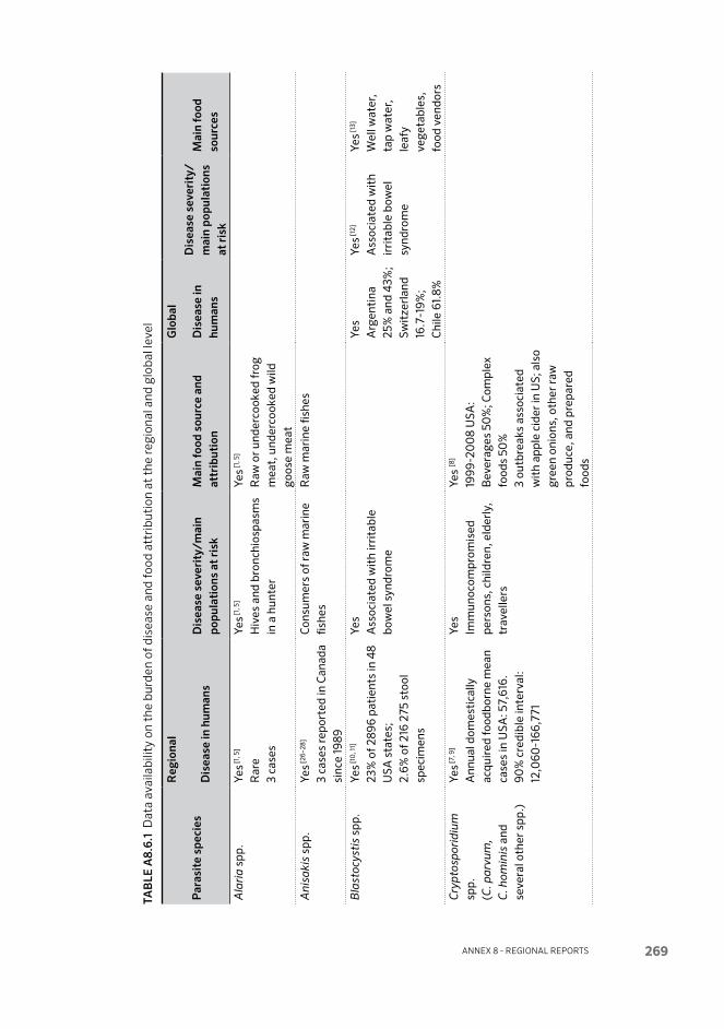
Annex 8 - regionAl reports 269
TAb
lE A
8.6.
1 D
ata
avai
labi
lity
on t
he b
urde
n of
dis
ease
and
food
att
ribu
tion
at
the
regi
onal
and
glo
bal l
evel
Par
asit
e sp
ecie
s
Reg
iona
lG
loba
l
Dis
ease
in h
uman
sD
isea
se s
ever
ity/
mai
n po
pula
tion
s at
ris
k M
ain
food
sou
rce
and
attr
ibut
ion
Dis
ease
in
hum
ans
Dis
ease
sev
erit
y/
mai
n po
pu la
tion
s at
ris
k
Mai
n fo
od
sour
ces
Ala
ria
spp.
Yes [1
, 5]
Rar
e3
case
s
Yes [1
, 5]
Hiv
es a
nd b
ronc
hio s
pasm
s in
a h
unte
r
Yes [1
, 5]
Raw
or
unde
r coo
ked
frog
m
eat,
und
er co
oked
wild
go
ose
mea
t
Ani
saki
s sp
p.Ye
s [26–
28]
3 ca
ses
repo
rted
in C
anad
a si
nce
1989
Con
sum
ers
of r
aw m
arin
e fis
hes
Raw
mar
ine
fishe
s
Bla
stoc
ysti
s sp
p.Ye
s [10
, 11]
23%
of 2
896
pati
ents
in 4
8 U
SA s
tate
s;
2.6%
of 2
16 2
75 s
tool
sp
ecim
ens
Yes
Ass
ocia
ted
wit
h ir
rita
ble
bow
el s
yndr
ome
Yes
Arg
enti
na
25%
and
43%
; Sw
itze
rlan
d 16
.7-1
9%;
Chi
le 6
1.8%
Yes [1
2]
Ass
ocia
ted
wit
h ir
rita
ble
bow
el
synd
rom
e
Yes [1
3]
Wel
l wat
er,
tap
wat
er,
leaf
y ve
ge ta
bles
, fo
od v
endo
rs
Cry
pto
spor
idiu
m
spp.
(C. p
arvu
m,
C. h
omin
is a
nd
seve
ral o
ther
spp
.)
Yes [7
, 9]
Ann
ual d
omes
tica
lly
acqu
ired
food
born
e m
ean
case
s in
USA
: 57,
616.
90
% c
redi
ble
inte
rval
: 12
,060
-166
,771
Yes
Imm
uno c
ompr
omis
ed
pers
ons,
chi
ldre
n, e
lder
ly,
trav
elle
rs
Yes
[8]
1999
-20
08
USA
:B
ever
ages
50
%; C
ompl
ex
food
s 50
%3
outb
reak
s as
soci
ated
w
ith
appl
e ci
der
in U
S; a
lso
gree
n on
ions
, oth
er r
aw
prod
uce,
and
pre
pare
d fo
ods

Multi criteria-based ranking for risk ManageMent of food-borne parasites270
Par
asit
e sp
ecie
s
Reg
iona
lG
loba
l
Dis
ease
in h
uman
sD
isea
se s
ever
ity/
mai
n po
pula
tion
s at
ris
k M
ain
food
sou
rce
and
attr
ibut
ion
Dis
ease
in
hum
ans
Dis
ease
sev
erit
y/
mai
n po
pu la
tion
s at
ris
k
Mai
n fo
od
sour
ces
Cyc
losp
ora
caye
tane
nsis
Yes [7
, 9]
Ann
ual d
omes
tica
lly
acqu
ired
food
born
e m
ean
case
s in
USA
: 11 4
07
(90
%
cred
ible
inte
rval
: 137
–
37 6
73)
Food
born
e ou
tbre
aks
in
Nor
th A
mer
ica
year
ly s
ince
19
95 (s
prin
g/su
mm
er)
Yes [8
]
Imm
uno c
ompr
o mis
ed
pers
ons,
tra
velle
rs
Yes [8
, 9
1999
–20
08
USA
: Com
plex
fo
ods
21.4
%; P
rodu
ce
78.6
%Im
port
ed fr
esh
rasp
berr
ies,
m
escl
un le
ttuc
e, b
asil
Dip
hyllo
bot
hriu
m
spp.
[14
–16]
(D. d
end
riti
c um
, D
. lat
um, D
. urs
i, D
. nih
onka
ien
se)
Yes
Up
to 8
0%
pre
vale
nce
of
D. d
end
riti
cum
in s
ome
Inui
t co
mm
unit
ies
in
Can
ada.
C
ase
of in
fect
ion
wit
h D
. urs
i rep
orte
d in
Bri
tish
C
olum
bia,
Can
ada
in 19
73
One
cas
e of
infe
ctio
n w
ith
D. n
ihon
kaie
nse
in C
anad
a
Yes,
Con
sum
ers
of r
aw
fres
h wat
er a
nd
ana d
rom
ous
fishe
s
Yes,
Raw
fres
hwat
er a
nd
anad
rom
ous
fishe
s
Ech
inoc
occu
s g
ranu
losu
s [17]
No
data
Num
ber
of c
ases
sp
ecifi
cally
ass
ocia
ted
wit
h co
nsum
ptio
n of
co
ntam
inat
ed fo
ods
is
unkn
own
Res
iden
ts o
f Arc
tic
Can
ada;
clo
se a
ssoc
iati
on
wit
h do
gs
No
data
,C
onta
min
atio
n of
food
s w
ith
eggs
from
faec
es o
f do
gs, w
olve
s, c
oyot
es
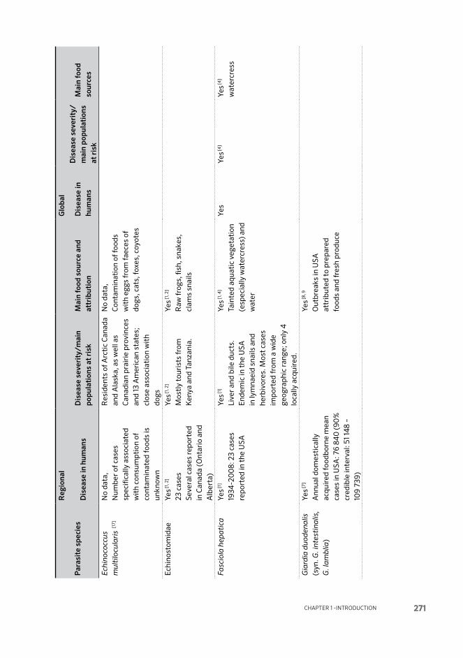
Chapter 1 -IntroduCtIon 271
Par
asit
e sp
ecie
s
Reg
iona
lG
loba
l
Dis
ease
in h
uman
sD
isea
se s
ever
ity/
mai
n po
pula
tion
s at
ris
k M
ain
food
sou
rce
and
attr
ibut
ion
Dis
ease
in
hum
ans
Dis
ease
sev
erit
y/
mai
n po
pu la
tion
s at
ris
k
Mai
n fo
od
sour
ces
Ech
inoc
occu
s m
ulti
locu
lari
s [1
7]
No
data
,N
umbe
r of
cas
es
spec
ifica
lly a
ssoc
iate
d w
ith
cons
umpt
ion
of
cont
amin
ated
food
s is
un
know
n
Res
iden
ts o
f Arc
tic
Can
ada
and
Ala
ska,
as
wel
l as
Can
adia
n pr
airi
e pr
ovin
ces
and
13 A
mer
ican
sta
tes;
cl
ose
asso
ciat
ion
wit
h do
gs
No
data
,C
onta
min
atio
n of
food
s w
ith
eggs
from
faec
es o
f do
gs, c
ats,
foxe
s, c
oyot
es
Ech
ino s
tom
idae
Yes [1
, 2]
23 c
ases
Seve
ral c
ases
repo
rted
in
Can
ada
(Ont
ario
and
A
lber
ta)
Yes [1
, 2]
Mos
tly
tour
ists
from
K
enya
and
Tan
zani
a.
Yes [1
, 2]
Raw
frog
s, fi
sh, s
nake
s,
clam
s sn
ails
Fasc
iola
hep
atic
aYe
s [1]
1934
-20
08:
23
case
s re
port
ed in
the
USA
Yes [1
]
Live
r an
d bi
le d
ucts
.E
ndem
ic in
the
USA
in
lym
naei
d sn
ails
and
he
rbiv
ores
. Mos
t ca
ses
impo
rted
from
a w
ide
geog
raph
ic r
ange
; onl
y 4
lo
cally
acq
uire
d.
Yes [1
, 4]
Tain
ted
aqua
tic
vege
tati
on
(esp
ecia
lly w
ater
cres
s) a
nd
wat
er
Yes
Yes [4
]Ye
s [4]
wat
er cr
ess
Gia
rdia
duo
den
alis
(s
yn. G
. int
esti
n alis
, G
. lam
blia
)
Yes [7
]
Ann
ual d
omes
tica
lly
acqu
ired
food
born
e m
ean
case
s in
USA
: 76
840
(90
%
cred
ible
inte
rval
: 51 1
48
– 10
9 73
9)
Yes [8
, 9
Out
brea
ks in
USA
at
trib
uted
to
prep
ared
fo
ods
and
fres
h pr
oduc
e

Multi criteria-based ranking for risk ManageMent of food-borne parasites272
Par
asit
e sp
ecie
s
Reg
iona
lG
loba
l
Dis
ease
in h
uman
sD
isea
se s
ever
ity/
mai
n po
pula
tion
s at
ris
k M
ain
food
sou
rce
and
attr
ibut
ion
Dis
ease
in
hum
ans
Dis
ease
sev
erit
y/
mai
n po
pu la
tion
s at
ris
k
Mai
n fo
od
sour
ces
Het
erop
hyes
he
tero
phy
esYe
s [1, 2
]
43
case
s (4
1 in
Haw
aii)
A fe
w c
ases
repo
rted
in
rece
nt im
mig
rant
s to
M
anit
oba
and
Alb
erta
(C
anad
a)
Yes [1
, 2]
One
USA
cas
e fr
om s
ushi
pr
epar
ed fr
om fi
shes
im
port
ed fr
om S
E A
sia
Met
a gon
imu
s sp
p.Ye
s [1, 2
]
10 c
ases
2
case
s re
port
ed in
rece
nt
imm
igra
nts
to A
lber
ta
(Can
ada)
Yes [1
, 2]
Rec
urre
nt d
iarr
hoea
re
port
ed in
1 ca
se in
the
US
Yes [1
, 2]
Fish
(sus
hi)
1 cas
e re
port
sug
gest
ed
sala
d co
n tam
inat
ed w
ith
met
a cer
cari
ae fr
om a
cu
ttin
g su
rfac
e
Met
orch
is
conj
unct
usYe
s [2, 6
]
Out
brea
k am
ong
a gr
oup
of K
orea
n na
tion
als
who
co
nsum
ed r
aw w
hite
su
cker
fres
hly
caug
ht
in r
iver
nea
r M
ontr
eal,
Can
ada;
17 o
f 19
indi
vidu
als
beca
me
sym
ptom
atic
Yes [1
]
Abd
omin
al p
ain,
feve
r,
head
ache
, ano
rexi
a,
diar
rhea
, nau
sea,
ba
ckac
he.
Yes [1
]
Raw
whi
te s
ucke
rN
o da
taN
o da
ta
Nan
ophy
etes
spp
.Ye
s [1, 2
]
21 c
ases
, mos
tly
Nor
thw
este
rn U
S
Yes [1
, 2]
Yes [1
, 2]
Raw
sal
mon
, ste
elhe
ad
trou
t, t
rout
egg
s

Annex 8 - regionAl reports 273
Par
asit
e sp
ecie
s
Reg
iona
lG
loba
l
Dis
ease
in h
uman
sD
isea
se s
ever
ity/
mai
n po
pula
tion
s at
ris
k M
ain
food
sou
rce
and
attr
ibut
ion
Dis
ease
in
hum
ans
Dis
ease
sev
erit
y/
mai
n po
pu la
tion
s at
ris
k
Mai
n fo
od
sour
ces
Het
erop
hyes
he
tero
phy
esYe
s [1, 2
]
43
case
s (4
1 in
Haw
aii)
A fe
w c
ases
repo
rted
in
rece
nt im
mig
rant
s to
M
anit
oba
and
Alb
erta
(C
anad
a)
Yes [1
, 2]
One
USA
cas
e fr
om s
ushi
pr
epar
ed fr
om fi
shes
im
port
ed fr
om S
E A
sia
Met
a gon
imu
s sp
p.Ye
s [1, 2
]
10 c
ases
2
case
s re
port
ed in
rece
nt
imm
igra
nts
to A
lber
ta
(Can
ada)
Yes [1
, 2]
Rec
urre
nt d
iarr
hoea
re
port
ed in
1 ca
se in
the
US
Yes [1
, 2]
Fish
(sus
hi)
1 cas
e re
port
sug
gest
ed
sala
d co
n tam
inat
ed w
ith
met
a cer
cari
ae fr
om a
cu
ttin
g su
rfac
e
Met
orch
is
conj
unct
usYe
s [2, 6
]
Out
brea
k am
ong
a gr
oup
of K
orea
n na
tion
als
who
co
nsum
ed r
aw w
hite
su
cker
fres
hly
caug
ht
in r
iver
nea
r M
ontr
eal,
Can
ada;
17 o
f 19
indi
vidu
als
beca
me
sym
ptom
atic
Yes [1
]
Abd
omin
al p
ain,
feve
r,
head
ache
, ano
rexi
a,
diar
rhea
, nau
sea,
ba
ckac
he.
Yes [1
]
Raw
whi
te s
ucke
rN
o da
taN
o da
ta
Nan
ophy
etes
spp
.Ye
s [1, 2
]
21 c
ases
, mos
tly
Nor
thw
este
rn U
S
Yes [1
, 2]
Yes [1
, 2]
Raw
sal
mon
, ste
elhe
ad
trou
t, t
rout
egg
s
Par
asit
e sp
ecie
s
Reg
iona
lG
loba
l
Dis
ease
in h
uman
sD
isea
se s
ever
ity/
mai
n po
pula
tion
s at
ris
k M
ain
food
sou
rce
and
attr
ibut
ion
Dis
ease
in
hum
ans
Dis
ease
sev
erit
y/
mai
n po
pu la
tion
s at
ris
k
Mai
n fo
od
sour
ces
Op
isth
orch
is v
iver
rini
an
d C
lon
orch
is
sine
nsis
Yes [1
, 2]
ca. 1
270
cas
es 18
90–2
00
9,
mos
tly
indi
vidu
al c
ase
repo
rts
Com
mon
ly re
port
ed
amon
g im
mig
rant
s fr
om
SE A
sia
to C
anad
a
Yes [1
, 2]
Gro
up 1
carc
inog
ens;
live
r an
d bi
le d
uct
canc
er.
Infe
ctio
ns m
ainl
y im
port
ed
into
the
USA
. Thr
ough
th
e 19
70s
mos
t w
ere
impo
rted
in C
hine
se,
Japa
nese
and
Kor
ean
imm
igra
nts
or C
auca
sian
s w
ho h
ad re
side
d in
Chi
na.
Beg
inni
ng in
1979
, SE
A
sian
s w
ere
a m
ajor
so
urce
of i
mpo
rted
cas
es,
espe
cial
ly t
hose
from
Tha
i re
fuge
e ca
mps
.
Yes [1
, 2]
Raw
or
unde
r-co
oked
fr
eshw
ater
fish
Yes
Yes
Yes [1
, 2]
Fres
h wat
er
fish,
esp
ecia
lly
cypr
inid
s
Par
agon
imus
spp
.P.
wes
term
ani
P. k
ellic
otti
P. m
exic
anus
Yes [1
–3]
71 c
ases
repo
rted
19
10–2
00
9.P.
kel
licot
ti: 7
cas
es
1968
–20
08,
14 c
ases
20
09–
2010
. A
few
cas
es re
port
ed in
im
mig
rant
s to
Can
ada
from
Ital
y, M
alay
sia,
P
hilip
pine
s.
One
dom
esti
c ca
se in
Q
uebe
c, w
ho s
old
live
snai
ls a
nd c
rust
acea
ns
from
exo
tic
food
sec
tion
of
depa
rtm
ent
stor
e.
Yes [1
, 3]
Mos
t ca
ses
in 19
70s
and
1980
s im
port
ed b
y SE
A
sian
refu
gees
from
Tha
i ca
mps
, im
mig
rant
s fr
om
Kor
ea a
nd P
hilip
pine
s;
som
e co
-inf
ecti
ons
wit
h ot
her
helm
inth
s.
Yes [1
, 3]
Raw
or
unde
r coo
ked
cray
fish
and
crab
s
Yes
Yes
Yes
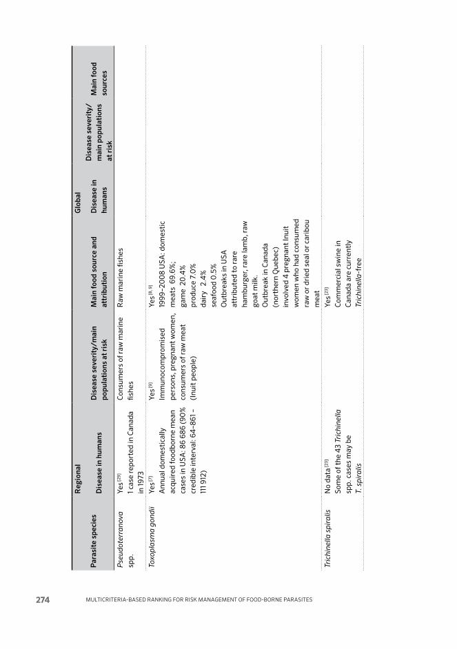
Multi criteria-based ranking for risk ManageMent of food-borne parasites274
Par
asit
e sp
ecie
s
Reg
iona
lG
loba
l
Dis
ease
in h
uman
sD
isea
se s
ever
ity/
mai
n po
pula
tion
s at
ris
k M
ain
food
sou
rce
and
attr
ibut
ion
Dis
ease
in
hum
ans
Dis
ease
sev
erit
y/
mai
n po
pu la
tion
s at
ris
k
Mai
n fo
od
sour
ces
Pse
udo t
erra
nov
a sp
p.Ye
s [29]
1 cas
e re
port
ed in
Can
ada
in 19
73
Con
sum
ers
of r
aw m
arin
e fis
hes
Raw
mar
ine
fishe
s
Toxo
pla
sma
gon
dii
Yes [7
]
Ann
ual d
omes
tica
lly
acqu
ired
food
born
e m
ean
case
s in
USA
: 86
686
(90
%
cred
ible
inte
rval
: 64
–861
–
111 9
12)
Yes [9
]
Imm
uno c
ompr
o mis
ed
pers
ons,
pre
gnan
t w
omen
, co
nsum
ers
of r
aw m
eat
(Inu
it p
eopl
e)
Yes [8
, 9]
1999
–20
08
USA
: dom
esti
c m
eats
69.
6%;
gam
e 2
0.4
%
prod
uce
7.0
%
dair
y 2
.4%
se
afoo
d 0
.5%
Out
brea
ks in
USA
at
trib
uted
to
rare
ha
mbu
rger
, rar
e la
mb,
raw
go
at m
ilk.
Out
brea
k in
Can
ada
(nor
ther
n Q
uebe
c)
invo
lved
4 p
regn
ant
Inui
t w
omen
who
had
con
sum
ed
raw
or
drie
d se
al o
r ca
ribo
u m
eat
Tric
hine
lla s
pir
alis
No
data
[23]
Som
e of
the
43
Tric
hine
lla
spp.
cas
es m
ay b
e T.
sp
iral
is
Yes [2
3]
Com
mer
cial
sw
ine
in
Can
ada
are
curr
entl
y Tr
ichi
nella
-fre
e
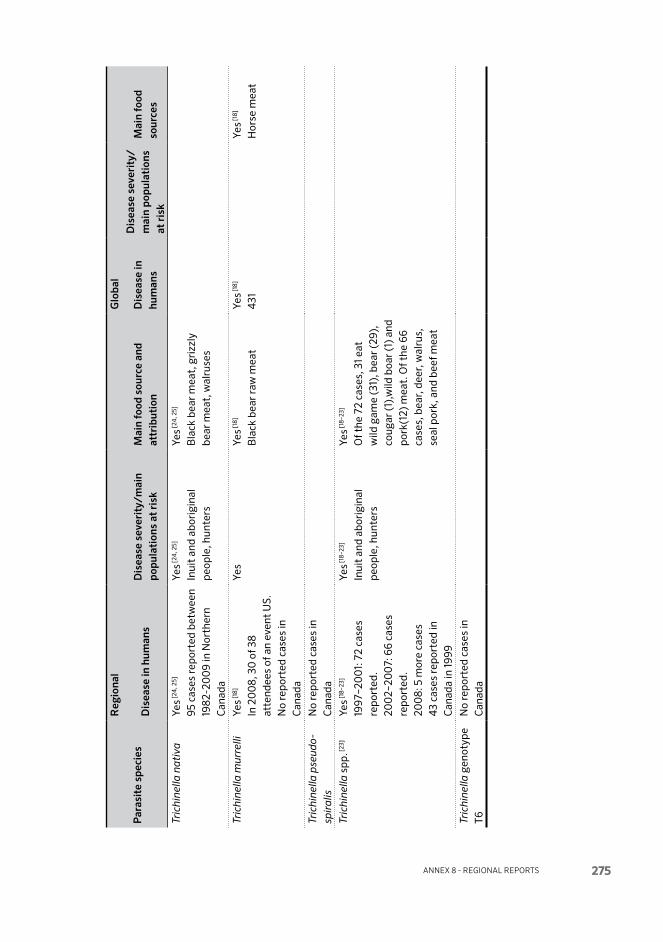
Annex 8 - regionAl reports 275
Par
asit
e sp
ecie
s
Reg
iona
lG
loba
l
Dis
ease
in h
uman
sD
isea
se s
ever
ity/
mai
n po
pula
tion
s at
ris
k M
ain
food
sou
rce
and
attr
ibut
ion
Dis
ease
in
hum
ans
Dis
ease
sev
erit
y/
mai
n po
pu la
tion
s at
ris
k
Mai
n fo
od
sour
ces
Tric
hine
lla n
ativ
aYe
s [24
, 25]
95 c
ases
repo
rted
bet
wee
n 19
82-2
00
9 in
Nor
ther
n C
anad
a
Yes [2
4, 2
5]
Inui
t an
d ab
orig
inal
pe
ople
, hun
ters
Yes [2
4, 2
5]
Bla
ck b
ear
mea
t, g
rizz
ly
bear
mea
t, w
alru
ses
Tric
hine
lla m
urre
lliYe
s [18]
In 2
00
8, 3
0 o
f 38
atte
ndee
s of
an
even
t U
S.
No
repo
rted
cas
es in
C
anad
a
Yes
Yes [1
8]
Bla
ck b
ear
raw
mea
tYe
s [18]
431
Yes [1
8]
Hor
se m
eat
Tric
hine
lla p
seud
osp
iral
isN
o re
port
ed c
ases
in
Can
ada
Tric
hine
lla s
pp. [2
3]Ye
s [18–
23]
1997
–20
01:
72
case
s re
port
ed.
200
2–20
07:
66
case
s re
port
ed.
200
8: 5
mor
e ca
ses
43
case
s re
port
ed in
C
anad
a in
1999
Yes [1
8–23
]
Inui
t an
d ab
orig
inal
pe
ople
, hun
ters
Yes [1
8–23
]
Of t
he 7
2 ca
ses,
31 e
at
wild
gam
e (3
1), b
ear
(29)
, co
ugar
(1),
wild
boa
r (1
) and
po
rk(1
2) m
eat.
Of t
he 6
6 ca
ses,
bea
r, d
eer,
wal
rus,
se
al p
ork,
and
bee
f mea
t
Tric
hine
lla g
enot
ype
T6N
o re
port
ed c
ases
in
Can
ada
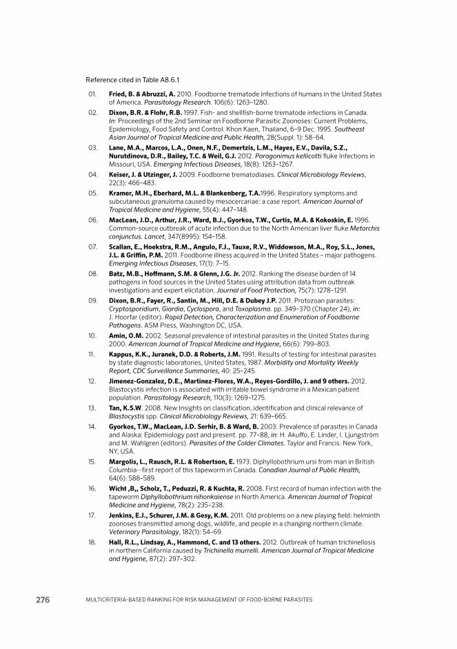
Multi criteria-based ranking for risk ManageMent of food-borne parasites276
Reference cited in Table A8.6.1
01. Fried, B. & Abruzzi, A. 2010. Food borne trematode infections of humans in the United States of America. Parasitology Research. 106(6): 1263–1280.
02. Dixon, B.R. & Flohr, R.B. 1997. Fish- and shellfish-borne trematode infections in Canada. In: Proceedings of the 2nd Seminar on Food borne Parasitic Zoonoses: Current Problems, Epidemiology, Food Safety and Control. khon kaen, Thailand, 6–9 Dec. 1995. Southeast Asian Journal of Tropical Medicine and Public Health, 28(Suppl. 1): 58–64.
03. lane, M.A., Marcos, l.A., Onen, N.F., Demertzis, l.M., Hayes, E.V., Davila, S.Z., Nurutdinova, D.R., Bailey, t.C. & Weil, G.J. 2012. Paragonimus kellicotti fluke Infections in Missouri, USA. Emerging Infectious Diseases, 18(8): 1263–1267.
04. keiser, J. & utzinger, J. 2009. Food borne trematodiases. Clinical Microbiology Reviews, 22(3): 466–483.
05. kramer, M.H., Eberhard, M.l. & Blankenberg, t.A.1996. Respiratory symptoms and subcutaneous granuloma caused by mesocercariae: a case report. American Journal of Tropical Medicine and Hygiene, 55(4): 447–148.
06. Maclean, J.D., Arthur, J.R., Ward, B.J., Gyorkos, t.W., Curtis, M.A. & kokoskin, E. 1996. Common-source outbreak of acute infection due to the North American liver fluke Metorchis conjunctus. Lancet, 347(8995): 154–158.
07. Scallan, E., Hoekstra, R.M., Angulo, F.J., tauxe, R.V., Widdowson, M.A., Roy, S.l., Jones, J.l. & Griffin, p.M. 2011. Foodborne illness acquired in the United States – major pathogens. Emerging Infectious Diseases, 17(1): 7–15.
08. Batz, M.B., Hoffmann, S.M. & Glenn, J.G. Jr. 2012. Ranking the disease burden of 14 pathogens in food sources in the United States using attribution data from outbreak investigations and expert elicitation. Journal of Food Protection, 75(7): 1278–1291.
09. Dixon, B.R., Fayer, R., Santin, M., Hill, D.E. & Dubey J.p. 2011. Protozoan parasites: Cryptosporidium, Giardia, Cyclospora, and Toxoplasma. pp. 349–370 (Chapter 24), in: J. Hoorfar (editor). Rapid Detection, Characterization and Enumeration of Food borne Pathogens. ASM Press, Washington DC, USA.
10. Amin, O.M. 2002. Seasonal prevalence of intestinal parasites in the United States during 2000. American Journal of Tropical Medicine and Hygiene, 66(6): 799–803.
11. kappus, k.k., Juranek, D.D. & Roberts, J.M. 1991. Results of testing for intestinal parasites by state diagnostic laboratories, United States, 1987. Morbidity and Mortality Weekly Report, CDC Surveillance Summaries, 40: 25–245.
12. Jimenez-Gonzalez, D.E., Martinez-Flores, W.A., Reyes-Gordillo, J. and 9 others. 2012. Blastocystis infection is associated with irritable bowel syndrome in a Mexican patient population. Parasitology Research, 110(3): 1269–1275.
13. tan, k.S.W. 2008. New Insights on classification, identification and clinical relevance of Blastocystis spp. Clinical Microbiology Reviews, 21: 639–665.
14. Gyorkos, t.W., Maclean, J.D. Serhir, B. & Ward, B. 2003: Prevalence of parasites in Canada and Alaska: Epidemiology past and present. pp. 77–88, in: H. Akuffo, E. Linder, I. Ljungström and M. Wahlgren (editors). Parasites of the Colder Climates. Taylor and Francis. New York, NY, USA.
15. Margolis, l., Rausch, R.l. & Robertson, E. 1973. Diphyllobothrium ursi from man in British Columbia--first report of this tapeworm in Canada. Canadian Journal of Public Health, 64(6): 588–589.
16. Wicht ,B,, Scholz, t., peduzzi, R. & kuchta, R. 2008. First record of human infection with the tapeworm Diphyllobothrium nihonkaiense in North America. American Journal of Tropical Medicine and Hygiene, 78(2): 235–238.
17. Jenkins, E.J., Schurer, J.M. & Gesy, k.M. 2011. Old problems on a new playing field: helminth zoonoses transmitted among dogs, wildlife, and people in a changing northern climate. Veterinary Parasitology, 182(1): 54–69.
18. Hall, R.l., lindsay, A., Hammond, C. and 13 others. 2012. Outbreak of human trichinellosis in northern California caused by Trichinella murrelli. American Journal of Tropical Medicine and Hygiene, 87(2): 297–302.

Annex 8 - regionAl reports 277
19. Roy, S.l., lopez, A.S. & Schantz, p.M. 2003. Trichinellosis surveillance – United States, 1997–2001. Morbidity and Mortality Weekly Report, CDC Surveillance Summaries, 52(6): 1–8.
20. kennedy, E.D., Hall, R.l., Montgomery, S.p., pyburn, D.G., Jones, J.l. & Centers for Disease Control and prevention. 2009. Trichinellosis surveillance - United States, 2002–2007. Morbidity and Mortality Weekly Report, CDC Surveillance Summaries, 58(9): 1–7.
21. Anon. Various dates. National Notifiable Diseases Database, Public Health Agency of Canada.
22. Appleyard, G.D. & Gajadhar, A.A. 2000. A review of trichinellosis in people and wildlife in Canada. Canadian Journal of Public HealthRevue Canadienne de Sante Publique, 91(4): 293–297.
23. Gajadhar, A.A., Bisaillon, J.R. & Appleyard, G.D. 1997. Status of Trichinella spiralis in domestic swine and wild boar in Canada. Canadian Journal of Veterinary ResearchRevue Canadienne de Recherche Veterinaire, 61(4): 256–259.
24, proulx, Jean-François. 2011. Department of Public Health, Nunavik Regional Board of Health and Social Services. Pers. comm. in response to request for data.
25. Forbes, lorry. 2011. Centre for Food borne and Animal Parasitology, Canadian Food Inspection Agency. Pers. comm. in response to request for data.
26. kowalewska-Grochowska, k., Quinn, J., perry, I. & Sherbaniuk, R. 1989. A case of anisakiasis – Alberta. Canadian Disease Weekly Reports, 15(44): 221–223.
27. Couture, C., Measures, l., Gagnon, J. & Desbiens, C. 2003. Human intestinal anisakiosis due to consumption of raw salmon. American Journal of Surgical Pathology, 27(8): 1167–1172.
28. Bhat, M. & Cleland, p. 2010. Gastric anisakiasis. Clinical Gastroenterology and Hepatology, 8(8): A20.
29. kates, S., Wright, k.A. & Wright, R. 1973. A case of human infection with the cod nematode Phocanema sp. American Journal of Tropical Medicine and Hygiene, 22(5): 606–608.
TAblE A8.6.2 Data availability for parasite prevalence or concentration in the main food categories
Alaria spp.
Seafood Yes [1] Frogs.
Anisakis spp.
Seafood Yes [13–15] Marine fish, squid.
Blastocystis spp.
Vegetables Yes [9] Leafy vegetables.
Other Water.
cryptosporidium spp. [7]
Dairy Yes [8] Un-pasteurized milk.
Seafood Molluscan shellfish.
Fruits Yes. Apple cider.
Vegetables Yes. Green onions; produce.
Other Water; prepared foods.
cyclospora spp. [7]
Fruits Yes. Raspberry.
Vegetables Yes. Lettuce, basil, snow peas, watercress.
Other Water.

Multi criteria-based ranking for risk ManageMent of food-borne parasites278
diphyllo bothrium spp.
Seafood Yes [10–12] Raw freshwater and anadromous fish.
Echino stomidae
Seafood Yes [1. 2] Frogs, snakes, fish, clams, snails.
Fasciola hepatica
Vegetables Yes [1] Aquatic vegetation: watercress.
giardia spp. [7]
Seafood Molluscan shellfish.
Vegetables Yes; fresh produce.
Other Water; prepared foods.
heterophyes heterophyes
Seafood Yes [2] Sushi prepared from fish imported from SE Asia.
metagonimus spp.
Seafood Yes [2] Sushi; possibly salad contaminated with meta cercariae.
nanophyetes spp.
Seafood Yes [1, 2] Raw salmon, steelhead trout, trout eggs.
opisthorchis viverrini and clonorchis sinensis
Seafood Yes [1. 2] Numerous species of freshwater fish.
paragonimus kellicotti
Seafood Yes [3] Freshwater crustaceans (100%).
paragonimus mexicanus
Seafood Freshwater crustaceans (100%).
paragonimus westermani
Seafood Yes [1. 2] Freshwater crustaceans (100%): crayfish, crabs.
pseudoterra nova spp.
Seafood Yes [16] Marine fish.
trichinella spp. [4–6]
Pork Yes. Pork meat
Game Yes. Bear, walrus, wild boar, cougar.
toxoplasma spp. [7]
Beef Yes
Dairy Yes
Pork Yes
Poultry Yes
Game Yes; caribou, seal
Seafood Molluscan shellfish

Annex 8 - regionAl reports 279
Fruits No data, but oocyst contamination is feasible
Vegetables No data, but oocyst contamination is feasible
Other Water
trypano soma cruzi Very little substantive data available.
References cited in Table A8.6.2
01. Fried, B. & Abruzzi, A. 2010. Food borne trematode infections of humans in the United States of America. Parasitology Research. 106(6): 1263–1280.
02. Dixon, B.R. & Flohr, R.B. 1997. Fish- and shellfish-borne trematode infections in Canada. In: Proceedings of the 2nd Seminar on Food borne Parasitic Zoonoses: Current Problems, Epidemiology, Food Safety and Control. khon kaen, Thailand, 6–9 Dec. 1995. Southeast Asian Journal of Tropical Medicine and Public Health, 28(Suppl. 1): 58–64.
03. lane, M.A., Marcos, l.A., Onen, N.F., Demertzis, l.M., Hayes, E.V., Davila, S.Z., Nurutdinova, D.R., Bailey, t.C. & Weil, G.J. 2012. Paragonimus kellicotti fluke Infections in Missouri, USA. Emerging Infectious Diseases, 18(8): 1263–1267.
05. Appleyard, G.D. & Gajadhar, A.A. 2000. A review of trichinellosis in people and wildlife in Canada. Canadian Journal of Public HealthRevue Canadienne de Sante Publique, 91(4): 293–297.
05. kennedy, E.D., Hall, R.l., Montgomery, S.p., pyburn, D.G., Jones, J.l. & Centers for Disease Control and prevention. 2009. Trichinellosis surveillance - United States, 2002–2007. Morbidity and Mortality Weekly Report, CDC Surveillance Summaries, 58(9): 1–7.
06. Roy, S.l., lopez, A.S. & Schantz, p.M. 2003. Trichinellosis surveillance – United States, 1997–2001. Morbidity and Mortality Weekly Report, CDC Surveillance Summaries, 52(6): 1–8.
07. Dixon, B.R., Fayer, R., Santin, M., Hill, D.E. & Dubey J.p. 2011. Protozoan parasites: Cryptosporidium, Giardia, Cyclospora, and Toxoplasma. pp. 349–370 (Chapter 24), in: J. Hoorfar (editor). Rapid Detection, Characterization and Enumeration of Food borne Pathogens. ASM Press, Washington DC, USA.
08. Harper, C.M., Cowell, N.A., Adams, B.C., langley, A.J. & Wohlsen, t.D. 2002. Outbreak of Cryptosporidium linked to drinking unpasteurised milk. Communicable Diseases Intelligence Quarterly Report, 26(3): 449–450.
09. leber, A.l. 1999. Intestinal amebae. Clinical Laboratory Medicine, 19(3): 601–619, vii. [Review].
10. Gyorkos, t.W., J.D. Maclean, B. Serhir & Ward, B. 2003: Prevalence of parasites in Canada and Alaska: Epidemiology past and present. pp. 77–88, in: H. Akuffo, E. Linder, I. Ljungström and M. Wahlgren (editors). Parasites of the Colder Climates. Taylor and Francis. New York, NY, USA.
11. Margolis, l., Rausch, R.l. & Robertson, E. 1973. Diphyllobothrium ursi from man in British Columbia--first report of this tapeworm in Canada. Canadian Journal of Public Health, 64(6): 588–589.
12. Wicht ,B,, Scholz, t., peduzzi, R. & kuchta, R. 2008. First record of human infection with the tapeworm Diphyllobothrium nihonkaiense in North America. American Journal of Tropical Medicine and Hygiene, 78(2): 235–238.
13. kowalewska-Grochowska, k., Quinn, J., perry, I. & Sherbaniuk, R. 1989. A case of anisakiasis – Alberta. Canadian Disease Weekly Reports, 15(44): 221–223.
14. Couture, C., Measures, l., Gagnon, J. & Desbiens, C. 2003. Human intestinal anisakiosis due to consumption of raw salmon. American Journal of Surgical Pathology, 27(8): 1167–1172.
15. Bhat, M. & Cleland, p. 2010. Gastric anisakiasis. Clinical Gastroenterology and Hepatology, 8(8): A20.
16. kates, S., Wright, k.A. & Wright, R. 1973. A case of human infection with the cod nematode Phocanema sp. American Journal of Tropical Medicine and Hygiene, 22(5): 606–608.

Multi criteria-based ranking for risk ManageMent of food-borne parasites280
TAblE A8.6.3 Data availability for risk management options in North America for each parasite-commodity combinationNOTE: The authors were asked to consider all combinations of the particular parasite and the main food categories, namely Beef, Dairy, Pork, Poultry, Game, Seafood, Fruit, Vegetables and Other.
Alaria spp. [1]
Sea food Rare
Anisakis spp., pseudo terranova spp.
Sea food Yes [3] Fish surveillance studies; candling of fillets at processing plants; use
of pre-frozen fish for sushi; consumer education
Blastocystis spp.
Fruit Good agricultural practices, e.g., water quality, worker hygiene, compost
requirements; consumer and food handler education regarding fresh
produce
Vege tables Good agricultural practices; consumer and food handler education
regarding fresh produce
crypto sporidium spp.
Beef Yes [1] Heat and freezing
Dairy Yes [1] Pasteurization
Sea food Yes [5] Shellfish sanitation programs
Fruit Yes [5] Good agricultural practices, e.g., water quality, worker hygiene,
compost requirements; consumer and food handler education regarding
fresh produce; surveillance studies; disinfectants
Vege tables Yes [5] Good agricultural practices; consumer and food handler education
regarding fresh produce; surveillance studies; disinfectants
Other Yes [1] Fruit juice pasteurization
cyclospora cayetanensis
Fruit Yes [5] Good agricultural practices; import restrictions; surveillance studies;
former “Model Plan of Excellence” program in Guatemala; disinfectants
Vege tables Yes [5] Good agricultural practices; import restrictions; surveillance studies;
disinfectants
diphyllo bothrium spp.
Sea food Yes [5] Fish surveillance studies; wild-caught vs aquaculture (pelleted feed);
consumer education
trichinella spp.
Pork Routine surveillance of commercial swine
Game Surveillance of wildlife harvested for food; trichinellosis program in Arctic
Canada (walrus testing); hunter education
Echino stomidae, heterophyes heterophyes
Sea food Yes [1–3] Consumer education (cooking/freezing); good sanitation; import
restrictions

Annex 8 - regionAl reports 281
Fasciola hepatica [1]
Vege tables Rare
Other Rare
giardia duo denalis (syn. g. lamblia, g. intestinalis)
Sea food Yes [5] Shellfish sanitation programmes
Fruit Yes [5] Good agricultural practices; surveillance studies
Vege tables Yes [5] Good agricultural practices; surveillance studies
metagonimus spp.
Sea food Yes [2, 3] Consumer education (cooking/freezing); good sanitation; import
restrictions
nanophyetes spp.
Sea food Yes [1–3] Consumer education (cooking/freezing); good sanitation; import
restrictions
opisthorchis viverrini, clonorchis sinensis and paragonimus westermani
Sea food Yes [1–3] Consumer education (cooking/freezing); good sanitation; import
restrictions
paragonimus kellicotti
Sea food Yes [6] Consumer education: avoid eating raw crayfish
paragonimus mexicanus
Sea food Rare
toxoplasma gondii
Beef Yes [5, 6] Meat surveillance studies; consumer and food handler education
(cooking and freezing)
Dairy Yes [5, 6] Milk pasteurization requirements
Pork Yes [5, 6] Meat surveillance studies; consumer and food handler edu cation
(cooking and freezing)
Poultry Yes [5, 6] Meat surveillance studies; consumer and food handler education
(cooking and freezing)
Game Yes [5, 6] Meat surveillance studies; seroprevalence studies on wildlife;
consumer and food handler education (cooking and freezing)
Sea food Yes [5, 6] Shellfish sanitation programs
References cited in Table A8.6.3
1. Fried, B. & Abruzzi, A. 2010. Food borne trematode infections of humans in the United States of America. Parasitology Research. 106(6): 1263–1280.
2. Dixon, B.R. & Flohr, R.B. 1997. Fish- and shellfish-borne trematode infections in Canada. In: Proceedings of the 2nd Seminar on Food borne Parasitic Zoonoses: Current Problems, Epidemiology, Food Safety and Control. khon kaen, Thailand, 6–9 Dec. 1995. Southeast Asian Journal of Tropical Medicine and Public Health, 28(Suppl. 1): 58–64.
3. Adams, A.M., Murrell, k.D. & Cross, J.H. 1997. Parasites of fish and risks to public health. Revue technique et scientifique OIE, 16(2): 652–660.

Multi criteria-based ranking for risk ManageMent of food-borne parasites282
4. Robertson, l.J. & Fayer, R. 2012. Cryptosporidium spp. pp. 33–64, in: L.J. Robertson and H.V. Smith (editors). Food borne Protozoan Parasites. Nova Science Publishers.
5. Dixon, B.R., Fayer, R., Santin, M., Hill, D.E. & Dubey J.p. 2011. Protozoan parasites: Cryptosporidium, Giardia, Cyclospora, and Toxoplasma. pp. 349–370 (Chapter 24), in: J. Hoorfar (editor). Rapid Detection, Characterization and Enumeration of Food borne Pathogens. ASM Press, Washington DC, USA.
6. lane, M.A., Marcos, l.A., Onen, N.F., Demertzis, l.M., Hayes, E.V., Davila, S.Z., Nurutdinova, D.R., Bailey, t.C. & Weil, G.J. 2012. Paragonimus kellicotti fluke Infections in Missouri, USA. Emerging Infectious Diseases, 18(8): 1263–1267.
7. lindsay, D.S., Dubey, J.p., Santin, M. & Fayer, R. 2012. Coccidia and other protozoa. pp. 895-907 (Chapter 66), in: J.J. Zimmerman, L.A. karriker, A. Ramirez, k.J. Schwartz and G.W. Stevenson (editors). Diseases of Swine. 10th Edition. Wiley-Blackwell, Ames, Iowa, USA.
TAblE A8.6.4 Data availability for Central America
Parasite species Disease in humans
Disease severity/main populations at risk
Main food source and attribution
Alaria spp.
Anisakis spp.
Blastocystis spp. Yes [8, 9]
30% of 456 children in
day care centres in Cuba;
39% of local populations
in Cuba
Cyclospora cayetanensis Yes [1–5]
Endemic in Guatemala
(2.3% prevalence).
Among 182 raspberry
farm workers and family
members examined in
Guatemala, 3.3% had
Cyclospora infection;
Another study failed to
detect oocysts among
raspberry farm workers in
Guatemala
Yes [6, 16]
Outbreak in Quebec,
Canada, in 2005
associated with fresh
basil from Mexico.
Reported in lettuce
from local markets in
Costa Rica
Crypto sporidium spp.
Diphyllo bothrium
spp. (D. dendriticum,
D. latum, D. ursi,
D. nihonkaiense)

Annex 8 - regionAl reports 283
Parasite species Disease in humans
Disease severity/main populations at risk
Main food source and attribution
Echinococcus
granul osus,
E. multilocularis
Echino stomidae
Entamoeba histolytica Yes [11]
In Mexico, Entamoeba
histolytica antibodies
found in 4.49%
Fasciola hepatica
Giardia duodenalis
(syn. G. intestinalis,
G. lamblia)
Yes [7–9]
Nail biting and eating
un washed vegetables
raw were significantly
associated with infection
in hospitalized children
in Cuba; 54.6% of 456
children in day cares
in Cuba; 25% of local
populations in Cuba.
Heterophyes
heterophyes
Metagonimus spp.
Nanophyetes spp.
Opisthorchis viverrini
and Clonorchis sinensis
Paragonimus spp.
Pseudo terra nova spp.
Taenia solium Yes [12–15]
4.9–10.8% tested
positive for cysticercosis
in villages in Mexico;
T. solium taeniasis and
cysticercosis are endemic
in Guatemala; Clinical
incidence of neuro-
cysticer cosis can reach 7%
in Mexico.
Honduras: cysticercosis
annual incidence ca. 30.

Multi criteria-based ranking for risk ManageMent of food-borne parasites284
Parasite species Disease in humans
Disease severity/main populations at risk
Main food source and attribution
Toxoplasma gondii Yes [10]
Estimated incidence in
Honduras 36 000/yr
Trichinella spp.
References cited in Table A8.6.4
01. Cama, V. 2006. Coccidian parasites. pp. 33–55, in: Y.R. Ortega (editor). Food borne Parasites. Springer, New York, NY, USA.
02. Ortega, y.R. & Sanchez, R. 2010. Update on Cyclospora cayetanensis, a food borne and water borne parasite. Clinical Microbiology Reviews, 23(1): 218–234.
03. Bern, C., Hernandez, B., lopez, M.B., Arrowood, M.J., de Mejia, M.A., de Merida, A.M., Hightower, A.W., Venczel, l., Herwaldt, B.l. & klein. R.E. 1999. Epidemiologic studies of Cyclospora cayetanensis in Guatemala. Emerging Infectious Diseases, 5: 766–774.
04. Bern, C., Hernandez, B., lopez, M.B., Arrowood, M.J., De Merida, A.M. & klein, R.E. 2000. The contrasting epidemiology of Cyclospora and Cryptosporidium among outpatients in Guatemala. American Journal of Tropical Medicine and Hygiene, 63(5-6): 231–235.
05. pratdesaba, R.A., González, M., piedrasanta, E., Mérida, C., Contreras, Vela, C., Culajay, F., Flores, l. & torres, O. 2001. Cyclospora cayetanensis in three populations at risk in Guatemala. Journal of Clinical Microbiology, 39: 2951–2953.
06. Calvo, M., Carazo, M., Arias, M.l., Chaves, C., Monge, R. & Chinchilla, M. 2004. [Prevalence of Cyclospora spp., Cryptosporidium spp., microsporidia and fecal coliform determination in fresh fruit and vegetables consumed in Costa Rica.] [In Spanish] Archivos Latinamericos de Nutricion, 54(4): 428–432.
07. Bello, J., Núñez, F.A., González, O.M., Fernández, R., Almirall, p. & Escobedo, A.A. 2011. Risk factors for Giardia infection among hospitalized children in Cuba. Annals of Tropical Medicine and Parasitology, 105(1): 57–64.
08. Mendoza, D., Núñez, F.A., Escobedo, A., pelayo, l., Fernández, M., torres, D. & Cordoví, R.A. 2001. [Intestinal parasitic infections in 4 child day-care centers located in San Miguel del Padrón municipality, Havana City, 1998]. [Article in Spanish] Revista Cubana de Medicina Tropical, 53(3): 189–193.
09. Escobedo, A.A., Cañete, R. & Núñez, F.A. 2007. Intestinal protozoan and helminth infections in the Municipality San Juan y Martínez, Pinar del Río, Cuba. Tropical Doctor, 37(4): 236–238.
10. Dominguez, W. 2009. Estudio de caso – Enfermedades Transmitidas por alimetos en Honduras. pp. 139–157, in: Enfermedades transmitidas por alimentos y su impacto socioeconómico. Estudios de caso en Costa Rica, El Salvador, Guatemala, Honduras y Nicaragua. [FAO] Informe Técnico Sobre Ingeniería Agrícola Y Alimentaria. No.6. See: www.fao.org/docrep/011/i0480s/i0480s00.htm
11. Gonzalez, C.R., Isibasi, A., Ortiznavarrette, V., paniagua, J., Garcia, J.A., Ramirez, A., Salvatierra, B., tapia, R., Sepulveda, J., Gutierrez, G. & kumate, J. 1995. Prevalence of antibodies against Entamoeba histolytica in Mexico measured by ELISA. Epidemiology and Infection, 115(3): 535–543.
12. Sarti, E., Schantz, p. M., plancarte, A., Wilson, M., Gutierrez, I. O., lopez, A.S., Roberts, J. & Flisser, A. 1992. Prevalence and risk factors for Taenia solium Taeniasis and cysticercosis in humans and pigs in a village in Morelos, Mexico. American Journal of Tropical Medicine and Hygiene, 46: 677–685.

Annex 8 - regionAl reports 285
13. Sarti, E., Schantz, p.M., plancarte, A., Wilson, M., Gutierrez, I., Aguilera, J., Roberts, J. & Flisser, A. 1994. Epidemiological investigation of Taenia solium Taeniasis and cysticercosis in a rural village of Michoacan State, Mexico. Transactions of the Royal Society of Tropical Medicine and Hygiene, 88: 49–52.
14. Garcia-Noval, J., Allan, J.C., Fletes, C., Moreno, E., Demata, F., torresalvarez, R., Dealfaro, H.S., yurrita, p., Higuerosmorales, H., Mencos, F. & Craig, p.S. 1996. Epidemiology of Taenia solium Taeniasis and cysticercosis in two rural Guatemalan communities. American Journal of Tropical Medicine and Hygiene, 55: 282–289.
15. Flisser, A. 1988. Neurocysticercosis in Mexico. Parasitology Today, 4: 131–137.
16. Milord, F., lampron-Goulet, E., St-Amour, M., levac, E. & Ramsay, D. 2012. Cyclospora cayetanensis: a description of clinical aspects of an outbreak in Quebec, Canada. Epidemiology and Infection, 140(4): 626–632.
Multi criteria-based ranking for risk ManageMent of food-borne parasites286
ANNEX 8.7 – SOutH AMERICA
A8.7.1 Report preparationThe Group members were: Jorge Enrique Bolpe, Argentina, and Jorge Enrique Gómez-Marín, Colombia. Their sources included systematic literature reviews, involving bibliographic database searches (Pubmed, Scopus, Scielo) and own data from unpublished reports (Dr Gómez-Marin). Additionally, literature reviews of bulletins, national reports, book articles and technical guidelines were used (Dr Bolpe).
A8.7.2 Data availability in humans and food attributionGood evidence exists about Toxoplasma presence in meat for human consump-tion, although some countries have only limited data. Good quality reports exist of food borne Trypanosoma infection. There is good information on the quantity and quality of regional data concerning trichinellosis and cystic echinococcosis in humans in Argentina and other countries in southern of South America, probably because these diseases are included in the national epidemiological surveillance systems in the affected countries. There is also valuable information regarding the identification of food infected with Trichinella, with the identification of the specific species (T. spiralis).
Data on the burden of disease and food attribution are summarized in Table A8.7.1, and data on parasite prevalence, incidence and concentration in the main food cat-egories are covered in Table A8.7.2.
A8.7.3 Agri-food tradeAll the countries in South America export fruits to many continents. Notably, during the last decade, Colombian fruit exports doubled to a total US$ 800 million and more than 1800 ton (Proexport data). Brazil and Argentina export significant volumes of horse and beef meat, while pork meat exportation is less important. At present there are no data indicating the presence of parasites in horses. However, in Argentina, because of trichinellosis endemicity, all horse and pork meat for ex-portation must be certified with a negative test of peptic digestion performed by the National Animal Health Service.
A8.7.4 Consumer perceptionThe recent free-trade agreement with the United States of America has raised important questions concerning sanitary security. In Colombia, for example, wide public consumer debates have developed regarding the origin and security of chicken imports from United States of America. A recent urban outbreak of food-borne trypanosomiasis in a school in Caracas, Venezuela, portends a new epide-miological situation for this disease in Brazil, Colombia and Venezuela. For trichi-

Annex 8 - regionAl reports 287
nellosis consumer perception in Argentina, some parts of the population show a consumer willingness to accept risk in food consumption without sanitary control. In Argentina, many people are regular consumers of pork in the form of stuffed products, such as sausages produced by local butchers, and avoid foods processed under industrial conditions with sanitary control. This is enhanced by current cultural trends. In many family outbreaks, the consumers who have bred pigs using poor husbandry produced food without the detection of Trichinella infection in pig carcasses. Cystic echinococcosis from the ingestion of green vegetables contami-nated with oncospheres is possible in rural areas the parasite is endemic, where cultural practice encourage the parasitic cycle through the slaughter of domestic sheep and the feeding of dogs with raw viscera.
A8.7.5 Social sensitivityThere have been increased food borne outbreaks in most of countries in the region, reflecting cultural changes and increases in the frequency of eating outside the home. Theses outbreaks have been widely publicized, and public pressure developed to reinforce health authority controls.
TrichinellosisThe economic impact of trichinellosis is apparent in the control system for detecting this infection in potential Trichinella carriers, mainly in slaughterhouses, and the occurrence of the disease in human and animals. The economic loss due to the destruction of infected carcases is a significant economic loss in Argentina. The cost for human treatment has been estimated at US$ 6000 in the United Staes of America, and at US$ 3000 in Europe.
Cystic echinococcosisIn a Regional Socio-economic Impact of Cystic Echinococcosis (CE) in Argentina, Brazil, Chile and Uruguay, DALYs calculated for the region as a measure of damage caused by CE were 1551.83 due to premature death and 1766.93 due to different degrees of disability, both values adjusted for reported cases. The overall monetary cost of CE in the countries—collating human cases, the lost income due to relapse and morbidity, and livestock losses associated with the condemnation of the liver, reduced carcass weight, loss of milk production, decreased fertility and wool yield—was estimated in the range of at least US$ 75 million to a maximum of US$ 97 million (See ref. [83] in Table A8.7.1).
For Global Socio-economic Impact, when no under reporting is assumed, the estimated human burden of disease is 285 407 DALYs or an annual loss of US$ 193 530 000.
A8.7.6 Risk managementData are summarized in Table A8.7.3.
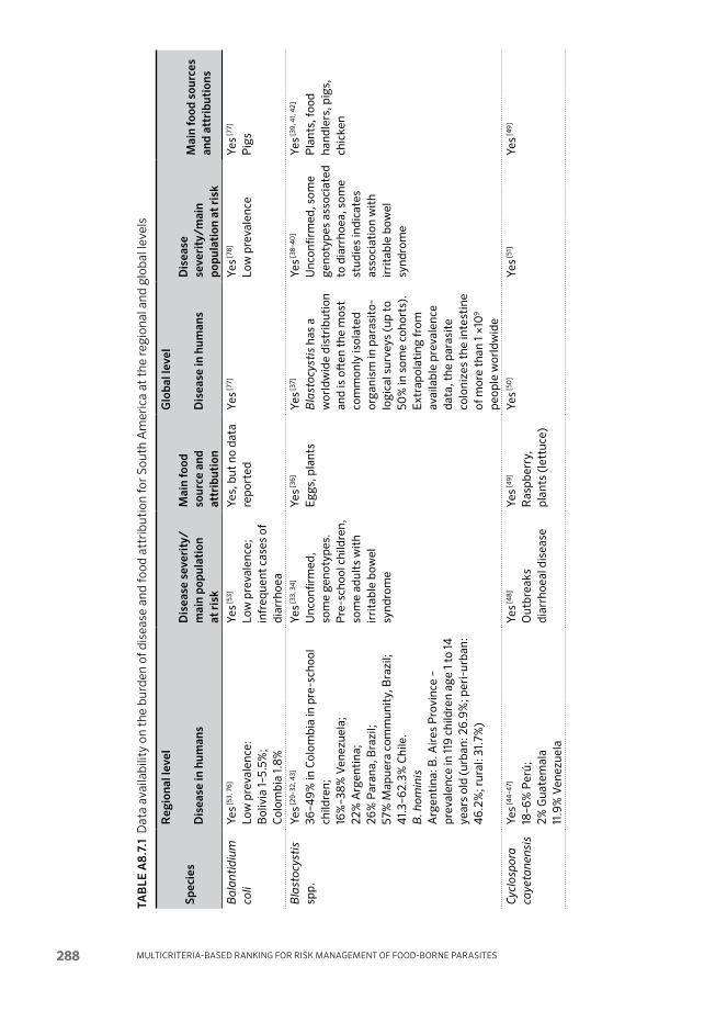
Multi criteria-based ranking for risk ManageMent of food-borne parasites288
TAb
lE A
8.7.
1 D
ata
avai
labi
lity
on t
he b
urde
n of
dis
ease
and
food
att
ribu
tion
for
Sout
h A
mer
ica
at t
he re
gion
al a
nd g
loba
l lev
els
Spec
ies
Reg
iona
l lev
elG
loba
l lev
el
Dis
ease
in h
uman
sD
isea
se s
ever
ity/
m
ain
popu
lati
on
at r
isk
Mai
n fo
od
sour
ce a
nd
attr
ibut
ion
Dis
ease
in h
uman
sD
isea
se
seve
rity
/ mai
n po
pula
tion
at r
isk
Mai
n fo
od s
ourc
es
and
attr
ibut
ions
Bal
an ti
diu
m
coli
Yes [5
3, 7
6]
Low
pre
vale
nce:
B
oliv
ia 1–
5.5%
; C
olom
bia
1.8%
Yes [5
3]
Low
pre
va le
nce;
in
freq
uent
cas
es o
f di
arrh
oea
Yes,
but
no
data
re
port
edYe
s [77]
Yes [7
8]
Low
pre
v ale
nce
Yes [7
7]
Pig
s
Bla
sto c
ysti
s sp
p.Ye
s [29–
32, 4
3]
36–4
9% in
Col
ombi
a in
pre
-sch
ool
child
ren;
16
%–3
8% V
enez
uela
; 22
% A
rgen
tina
; 26
% P
aran
a, B
razi
l; 57
% M
apue
ra c
omm
unit
y, B
razi
l; 4
1.3–
62.3
% C
hile
.B
. hom
inis
A
rgen
tina
: B. A
ires
Pro
vinc
e –
prev
alen
ce in
119
child
ren
age
1 to
14
year
s ol
d (u
rban
: 26.
9%; p
eri-
urba
n:
46.
2%; r
ural
: 31.
7%)
Yes [3
3, 3
4]
Unc
onfir
med
, so
me
geno
type
s.
Pre
-sch
ool c
hild
ren,
so
me
adul
ts w
ith
irri
tabl
e bo
wel
sy
ndro
me
Yes [3
6]
Egg
s, p
lant
sYe
s [37]
Bla
stoc
ysti
s ha
s a
wor
ldw
ide
dist
ribu
tion
an
d is
oft
en t
he m
ost
com
mon
ly is
olat
ed
orga
nism
in p
aras
ito-
logi
cal s
urve
ys (u
p to
50
% in
som
e co
hort
s).
Ext
rap o
lati
ng fr
om
avai
labl
e pr
eval
ence
da
ta, t
he p
aras
ite
colo
nize
s th
e in
test
ine
of m
ore
than
1 ×1
09
peop
le w
orld
wid
e
Yes [3
8–4
0]
Unc
onfir
med
, som
e ge
noty
pes
asso
ciat
ed
to d
iarr
hoea
, som
e st
udie
s in
dica
tes
asso
ciat
ion
wit
h ir
rita
ble
bow
el
synd
rom
e
Yes [3
9, 4
1, 4
2]
Pla
nts,
food
ha
ndle
rs, p
igs,
ch
icke
n
Cyc
lo sp
ora
caye
tan e
nsis
Yes [4
4–4
7]
18–6
% P
erú;
2%
Gua
tem
ala
11.9
% V
enez
uela
Yes [4
8]
Out
brea
ks
diar
rhoe
al d
isea
se
Yes [4
9]
Ras
pber
ry,
plan
ts (l
ettu
ce)
Yes [5
0]
Yes [5
1]Ye
s [49]

Annex 8 - regionAl reports 289
TAb
lE A
8.7.
1 D
ata
avai
labi
lity
on t
he b
urde
n of
dis
ease
and
food
att
ribu
tion
for
Sout
h A
mer
ica
at t
he re
gion
al a
nd g
loba
l lev
els
Spec
ies
Reg
iona
l lev
elG
loba
l lev
el
Dis
ease
in h
uman
sD
isea
se s
ever
ity/
m
ain
popu
lati
on
at r
isk
Mai
n fo
od
sour
ce a
nd
attr
ibut
ion
Dis
ease
in h
uman
sD
isea
se
seve
rity
/ mai
n po
pula
tion
at r
isk
Mai
n fo
od s
ourc
es
and
attr
ibut
ions
Bal
an ti
diu
m
coli
Yes [5
3, 7
6]
Low
pre
vale
nce:
B
oliv
ia 1–
5.5%
; C
olom
bia
1.8%
Yes [5
3]
Low
pre
va le
nce;
in
freq
uent
cas
es o
f di
arrh
oea
Yes,
but
no
data
re
port
edYe
s [77]
Yes [7
8]
Low
pre
v ale
nce
Yes [7
7]
Pig
s
Bla
sto c
ysti
s sp
p.Ye
s [29–
32, 4
3]
36–4
9% in
Col
ombi
a in
pre
-sch
ool
child
ren;
16
%–3
8% V
enez
uela
; 22
% A
rgen
tina
; 26
% P
aran
a, B
razi
l; 57
% M
apue
ra c
omm
unit
y, B
razi
l; 4
1.3–
62.3
% C
hile
.B
. hom
inis
A
rgen
tina
: B. A
ires
Pro
vinc
e –
prev
alen
ce in
119
child
ren
age
1 to
14
year
s ol
d (u
rban
: 26.
9%; p
eri-
urba
n:
46.
2%; r
ural
: 31.
7%)
Yes [3
3, 3
4]
Unc
onfir
med
, so
me
geno
type
s.
Pre
-sch
ool c
hild
ren,
so
me
adul
ts w
ith
irri
tabl
e bo
wel
sy
ndro
me
Yes [3
6]
Egg
s, p
lant
sYe
s [37]
Bla
stoc
ysti
s ha
s a
wor
ldw
ide
dist
ribu
tion
an
d is
oft
en t
he m
ost
com
mon
ly is
olat
ed
orga
nism
in p
aras
ito-
logi
cal s
urve
ys (u
p to
50
% in
som
e co
hort
s).
Ext
rap o
lati
ng fr
om
avai
labl
e pr
eval
ence
da
ta, t
he p
aras
ite
colo
nize
s th
e in
test
ine
of m
ore
than
1 ×1
09
peop
le w
orld
wid
e
Yes [3
8–4
0]
Unc
onfir
med
, som
e ge
noty
pes
asso
ciat
ed
to d
iarr
hoea
, som
e st
udie
s in
dica
tes
asso
ciat
ion
wit
h ir
rita
ble
bow
el
synd
rom
e
Yes [3
9, 4
1, 4
2]
Pla
nts,
food
ha
ndle
rs, p
igs,
ch
icke
n
Cyc
lo sp
ora
caye
tan e
nsis
Yes [4
4–4
7]
18–6
% P
erú;
2%
Gua
tem
ala
11.9
% V
enez
uela
Yes [4
8]
Out
brea
ks
diar
rhoe
al d
isea
se
Yes [4
9]
Ras
pber
ry,
plan
ts (l
ettu
ce)
Yes [5
0]
Yes [5
1]Ye
s [49]
Spec
ies
Reg
iona
l lev
elG
loba
l lev
el
Dis
ease
in h
uman
sD
isea
se s
ever
ity/
m
ain
popu
lati
on
at r
isk
Mai
n fo
od
sour
ce a
nd
attr
ibut
ion
Dis
ease
in h
uman
sD
isea
se
seve
rity
/ mai
n po
pula
tion
at r
isk
Mai
n fo
od s
ourc
es
and
attr
ibut
ions
Ech
ino c
occu
s g
ranu
losu
sYe
s [12,
79–
85]
Ove
r 20
00
new
hum
an c
ases
are
re
port
ed e
very
yea
r in
the
regi
on o
f So
uth
Am
eric
a.
Inci
denc
e fr
om 4
1 per
100
00
0 in
th
e P
atag
onia
n re
gion
in s
outh
ern
Arg
enti
na, 8
0 p
er 10
0 0
00
in t
he
XI R
egio
n of
Chi
le; u
p to
100
per
10
0 0
00
in t
he F
lore
s D
epar
tmen
t of
U
rugu
ay.
Infe
ctio
n ra
tes
of 5
.5%
in 19
86 in
B
lack
Riv
er, A
rgen
tina
; 14
.2%
in 19
88 in
Lon
copu
é, N
euqu
én,
Tacu
arem
bo, U
rugu
ay;
1.6%
in 19
97 in
Flo
rida
, Uru
guay
; 3.
6% in
1998
in P
each
, Uru
guay
; 5.
1% in
1999
in V
icha
ycoc
ha, P
eru.
14
18 c
ases
hav
e al
so b
een
repo
rted
by
ult
raso
und
scre
enin
g on
as
ympt
omat
ic h
uman
pop
ulat
ion.
Arg
enti
na: M
orbi
dity
in 19
87–1
996:
52
48
hum
an c
ases
, 19
97–2
00
5: 4
079
hum
an c
ases
Arg
enti
na: b
etw
een
200
6 an
d 20
10
1883
sus
pect
ed H
ydat
idos
is c
ases
w
ere
repo
rted
. A
rgen
tina
, Bra
zil,
Chi
le a
nd U
rugu
ay:
DA
LYs
1551
.83
to 17
66.9
3 ad
just
ed
for
repo
rted
cas
es.
Chi
le: 2
00
4 e
stim
ates
an
inci
denc
e of
10 p
er 10
0 0
00
, wit
h m
orta
lity
0.3
–0.4
per
100
00
0.
Bra
zil:
see
[85]
for
data
on
Rio
G
rand
e do
Sul
Yes [1
2, 7
9–85
]
3.8
×10
6 rur
al
inha
bita
nts
live
in
risk
are
a.
Yes [1
2, 7
9–85
]
Inge
stio
n of
ve
ge ta
bles
or
pollu
ted
wat
er
wit
h in
fect
ed
cani
ne fa
eces
.
Yes [1
2, 7
9–85
]
The
mos
t co
nser
v ati
ve
esti
mat
e of
glo
bal
DA
LYs
lost
is 2
85,4
07,
w
ith
no c
onsi
dera
tion
fo
r di
seas
e un
der-
repo
rtin
g.

Multi criteria-based ranking for risk ManageMent of food-borne parasites290
Spec
ies
Reg
iona
l lev
elG
loba
l lev
el
Dis
ease
in h
uman
sD
isea
se s
ever
ity/
m
ain
popu
lati
on
at r
isk
Mai
n fo
od
sour
ce a
nd
attr
ibut
ion
Dis
ease
in h
uman
sD
isea
se
seve
rity
/ mai
n po
pula
tion
at r
isk
Mai
n fo
od s
ourc
es
and
attr
ibut
ions
Ent
amoe
ba
hist
o lyt
ica
Yes [6
9, 7
0]
Col
ombi
a: 0
.6–1
.4%
Yes [7
1]
Low
pre
v ale
nce
whe
n st
udie
s di
ffer
enti
ated
pa
thog
enic
from
no
n-pa
thog
enic
.
Yes,
but
no
data
re
port
ed.
Yes [7
2]Ye
s [73]
Yes [7
4, 7
5] b
ut li
ttle
da
ta
Onl
y on
e re
port
di
ffer
en ti
ated
pa
tho g
enic
from
no
n-pa
tho g
enic
. O
nly
E. d
isp
ar
was
foun
d in
food
ha
ndle
rs
Fasc
iola
he
pat
ica
Yes [1
–3, 8
6]
Arg
enti
na –
619
aut
ocht
hono
us c
ases
fr
om 13
Pro
vinc
es, i
n 58
repo
rts
of
diff
eren
t ki
nds
anal
ysed
up
to 2
010
.
Yes [1
–3, 8
6]
Cas
e sp
read
, by
gend
er, p
rovi
nce,
di
agno
stic
met
hod,
tr
eat m
ent,
etc
.
Yes [1
–3, 8
6]
Inge
stio
n of
fr
eshw
ater
pl
ants
car
ryin
g in
fect
ive
met
a cer
cari
a,w
ater
cres
s
Gia
rdia
spp
.Ye
s [34
, 43,
52,
53]
Mex
ico
50%
; C
olom
bia
15,0
%
Arg
enti
na: p
reva
lenc
e of
Gia
rdia
d
uo d
enal
is (s
yn. G
. lam
blia
, G
. int
esti
nalis
) in
119
child
ren
age
1 to
14 y
ears
old
– u
rban
9.6
%; p
eri-
urba
n 34
.6%
; rur
al 7
.3%
Arg
enti
na: i
n m
ulti
cent
re s
tudi
es
usin
g da
ta fr
om d
iffer
ent
prov
ince
s –
pre
val e
nce
of G
iard
ia d
uo d
enal
is
(syn
. G. l
amb
lia, G
. int
es ti
nalis
) (1
3.17
%) i
n 1 t
o 3
year
-old
chi
ldre
n in
th
e co
untr
y 20
04
–05.
Yes [5
4]
Ret
ard
in c
ogni
tion
de
velo
pmen
t
Yes [5
5,56
]Ye
s [57]
In d
evel
opin
g co
untr
ies
arou
nd 2
0%
(4–4
3%)
and
in d
evel
oped
co
untr
ies
5% (3
–7%
)
Yes [5
4]
Yes [5
6]
Can
ned
salm
on,
sand
wic
hes,
noo
dle
sala
d, fr
uit
sala
d,
raw
veg
e tab
les,
ice
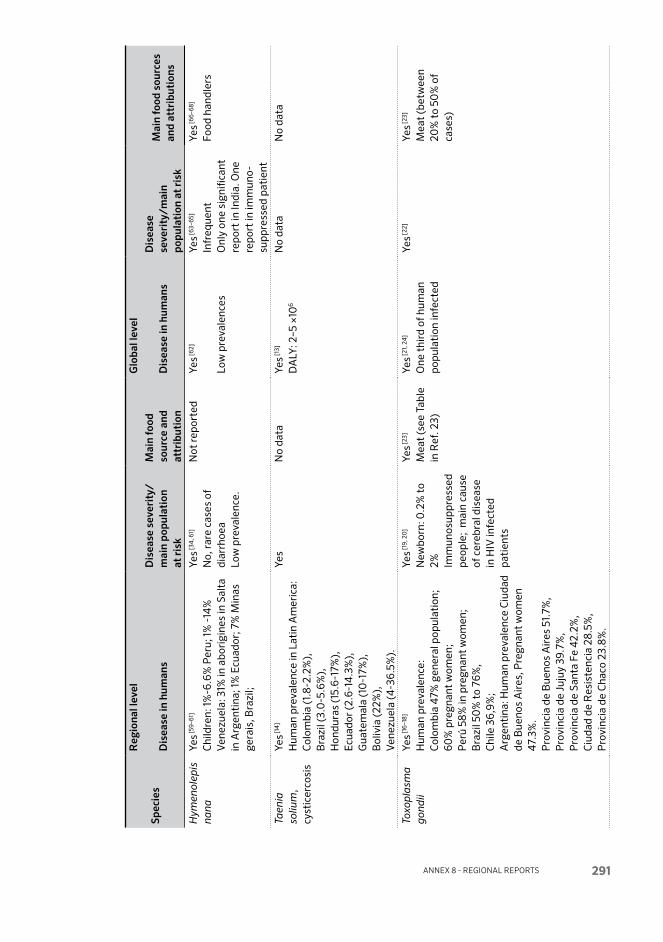
Annex 8 - regionAl reports 291
Spec
ies
Reg
iona
l lev
elG
loba
l lev
el
Dis
ease
in h
uman
sD
isea
se s
ever
ity/
m
ain
popu
lati
on
at r
isk
Mai
n fo
od
sour
ce a
nd
attr
ibut
ion
Dis
ease
in h
uman
sD
isea
se
seve
rity
/ mai
n po
pula
tion
at r
isk
Mai
n fo
od s
ourc
es
and
attr
ibut
ions
Hym
enol
epis
na
naYe
s [59–
61]
Chi
ldre
n: 1%
-6.6
% P
eru;
1% -
14%
V
enez
uela
: 31%
in a
bori
gine
s in
Sal
ta
in A
rgen
tina
; 1%
Ecu
ador
; 7%
Min
as
gera
is, B
razi
l;
Yes [3
4, 6
1]
No,
rar
e ca
ses
of
diar
rhoe
aLo
w p
reva
lenc
e.
Not
repo
rted
Yes [6
2]
Low
pre
vale
nces
Yes [6
3–65
]
Infr
eque
nt
Onl
y on
e si
gnifi
cant
re
port
in In
dia.
One
re
port
in im
mun
o-su
ppre
s sed
pat
ient
Yes [6
6–68
]
Food
han
dler
s
Taen
ia
soliu
m,
cyst
icer
cosi
s
Yes [1
4]
Hum
an p
reva
lenc
e in
Lat
in A
mer
ica:
C
olom
bia
(1.8
-2.2
%),
B
razi
l (3.
0-5
.6%
),
Hon
dura
s (1
5.6-
17%
),
Ecu
ador
(2.6
-14
.3%
),
Gua
tem
ala
(10
-17%
),
Bol
ivia
(22%
),
Ven
ezue
la (4
-36.
5%).
Yes
No
data
Yes [1
3]
DA
LY: 2
–5 ×
106
No
data
No
data
Toxo
pla
sma
gon
dii
Yes [1
6–18
]
Hum
an p
reva
lenc
e:
Col
ombi
a 4
7% g
ener
al p
opul
atio
n;
60%
pre
gnan
t w
omen
; P
erú
58%
in p
regn
ant
wom
en;
Bra
zil 5
0%
to
76%
, C
hile
36,
9%;
Arg
enti
na: H
uman
pre
vale
nce
Ciu
dad
de B
ueno
s A
ires
, Pre
gnan
t w
omen
4
7.3%
. P
rovi
ncia
de
Bue
nos
Air
es 5
1.7%
, P
rovi
ncia
de
Juju
y 39
.7%
, P
rovi
ncia
de
Sant
a Fe
42.
2%,
Ciu
dad
de R
esis
tenc
ia 2
8.5%
, P
rovi
ncia
de
Cha
co 2
3.8%
.
Yes [1
9, 2
0]
New
born
: 0.2
% t
o 2% Im
mun
o sup
pres
sed
peop
le;
mai
n ca
use
of c
ereb
ral d
isea
se
in H
IV in
fect
ed
pati
ents
Yes [2
3]
Mea
t (s
ee T
able
in
Ref
. 23)
Yes [2
1, 2
4]
One
thi
rd o
f hum
an
popu
lati
on in
fect
ed
Yes [2
2]Ye
s [23]
Mea
t (b
etw
een
20%
to
50%
of
case
s)

Multi criteria-based ranking for risk ManageMent of food-borne parasites292
Spec
ies
Reg
iona
l lev
elG
loba
l lev
el
Dis
ease
in h
uman
sD
isea
se s
ever
ity/
m
ain
popu
lati
on
at r
isk
Mai
n fo
od
sour
ce a
nd
attr
ibut
ion
Dis
ease
in h
uman
sD
isea
se
seve
rity
/ mai
n po
pula
tion
at r
isk
Mai
n fo
od s
ourc
es
and
attr
ibut
ions
Tric
hin e
lla
spir
alis
Yes [4
–11]
Arg
enti
na, B
ueno
s A
ires
Pro
vinc
e:
Tota
l hum
an c
ases
and
65
outb
reak
s re
port
ed fr
om 0
1/20
00
to
09/
200
4
wer
e an
a lys
ed. N
o. o
f hum
an c
ases
in
crea
sed
from
90
8 be
twee
n 19
71
and
1981
, to
6919
bet
wee
n 19
90 a
nd
200
2C
hile
: Tot
al c
ases
20
12 a
nd p
revi
ous
inci
denc
e de
scri
ptio
nA
rgen
tina
: Ser
olog
ical
sur
vey
in
bloo
d do
nors
pre
vale
nce
8.0
%
Yes [4
]
Des
crip
tion
of
clin
ical
sym
ptom
s an
d/or
sig
ns,
epid
emio
logi
cal
data
and
labo
rato
ry
aspe
cts
of h
uman
tr
ichi
nello
sis
Yes [1
–5, 7
, 9]
Mea
tE
atin
g po
rk a
nd
pork
pro
duct
s no
t sa
nita
ry
insp
ecte
d an
d no
t pr
oper
ly
cook
ed.
Yes [1
–4, 1
2]
DA
LYs:
The
mos
t co
nser
v ati
ve e
stim
ate
of n
umbe
r of
glo
bal
DA
LYs
lost
is 2
85 4
07,
w
ith
no c
onsi
der a
tion
fo
r di
seas
e un
der-
repo
rtin
g.
Yes [1
–3]
Yes [1
–3]
Dom
esti
c pi
gs, w
ild
boar
s, r
aw h
orse
m
eat
100
% fo
od bo
rne
tran
smis
sion
Tric
huri
s tr
ichi
ura
No
data
Yes [1
5]N
o da
taN
o da
taYe
s [15]
For
2010
, glo
bal
pop u
la ti
on a
t ri
sk:
5.0
23 b
illio
n.
No
data
Tryp
ano s
oma
cruz
iYe
s [25]
Mor
e th
an 7
×10
6 peo
ple
infe
cted
in
the
Am
eric
as
Yes [2
5, 2
6]
10%
mor
talit
y in
ac
ute
case
s;
41 2
00
new
cas
es
per
year
– r
ate
of
7.7
per
100
00
0
inha
bita
nts
–14
385
ca
ses
of c
onge
nita
l C
haga
s
Yes [2
7, 2
8]
Aça
i pal
m ju
ice
Gua
va ju
ice
No
data
No
data
No
data

Annex 8 - regionAl reports 293
References for Table A8.7.1
01. Murrell, k.D. & pozio, E. 2011. Worldwide occurrence and impact of human trichinellosis, 1986–2009. Emerging Infectious Diseases, 17(12): 2194–2202.
02. Orlandi, p.A., Chu, D.-M.t., Bier, J.W. & Jackson, G.J. 2002. Parasites and the food supply. Food Technology, 56(4): 72–81.
03. Slifko, t.R., Smith, H.V. & Rose, J.B. 2000. Emerging parasite zoonoses associated with water and food. International Journal for Parasitology, 30: 1379–1393.
04. Bolpe, J., ledesma, M., Benitez, M., Cechini, D. & Gonzales Ayala, S. 2005. Human trichinellosis in Argentina. Clinical Microbiology and Infection, 11 (Suppl. 2): 46–47 [Abstract only].
05. Ortega-pierres, M.G., Arriaga, C. & yépez-Mulia, l. 2000. Epidemiology of trichinellosis in Mexico, Central and South America. A Review. Veterinary Parasitology, 93(3-4): 201–225.
06. Bolpe, J.E. 2011. Triquinosis aspectos epidemiológicos, de diagnostico y control de una zoonosis endémica en la Republica Argentina. pp. 347–354, in: Chapter 39 of Temas de Zoonosis V. Asociación Argentina de Zoonosis.
07. Ribicich, M., Gamble, H.R., Rosa, A., Bolpe, J. & Franco, A. 2005. Trichinellosis in Argentina: an historical review. Veterinary Parasitology, 132(1-2): 137–142.
08. Ministerio de Salud de la Nación – Dirección de Epidemiología. 2011. Gastroentéricas. pp. 11–12 (Ch. 2), in: Boletín Epidemiológico Anual 2010. Buenos Aires, Argentina. See: http://msal.gov.ar/htm/site/sala_situacion/PANELES/bep-anual-2010/BEPANUAL_2010.pdf
09. Caracostantogolo, J., Steffan, p., Dillon, J., De la Sota, M., Belgrano, D., Veneroni, R., Ruiz, M., Schapiro, J., Castaño, R., Martinez, M., Morici, G., Balbiani, G., Castro, M. & Eddi, C. 2007. ANEXO II Focos de Trichinellosis detectados en el país durante los años 2005–2006 (SENASA), in: Mejoramiento del control de la trichinellosis en Argentina. Proyecto TCP ARG 3003 entre la FAO y el Gobierno Argentino. See: http://cnia.inta.gov.ar/helminto/pub%20triquinosis/Mejorcontroltrichinella.pdf
10. Oficina de Vigilancia, Dpto. de Epidemiología DIPLAS/MINSAL, Chile. Resumen ejecutivo informe situacion epidemiológica de triquinosis. Semana Epidemiológica 1 a 52. 3 p. [National report] Available at http://epi.minsal.cl/epi/html/bolets/reportes/Triquinosis/Triqui_SE522012.pdf
11. Barlasina, M.S., pedevilla, C., kade, p., Costantino, S.N., taus, M.R. & Venturiello, S.M. 2009. Serología para triquinosis en dadores de sangre en área no endémica de la Argentina. [Trichinellosis serology in blood-donors from a non-endemic area of Argentina] [Article in Spanish]. Medicina (B. Aires), 69(3): 297–301.
12. Budke, C.M., Deplazes, p. & torgerson, p.R. 2006. Global socio economic impact of cystic echinococcosis. Emerging Infectious Diseases 12(2): 296–303.
13. torgerson, p.R. & Macpherson, C.N. 2011. The socioeconomic burden of parasitic zoonoses: global trends. Veterinary Parasitology, 182(1): 79–95.
14. lovadina, J. 2012. La cysticercose: parasitose négligée mais véritable enjeu de santé publique dans les pays en développement. These du Docteur en Pharmacie. Faculte de Pharmacie de Grenoble. France.
15. pullan, R.l. & Brooker, S.J. 2012. The global limits and population at risk of soil-transmitted helminths infections in 2010. Parasites & Vectors, 5: Art. no. 81 [Online: doi:10.1186/1756-3305-5-81]
16. Gómez, J.E., Ruiz, B., Silva, p., Beltrán, S., Cortés, J., Montoya, J. & Agudelo, A. 2007. Guía de práctica clínica para toxoplasmosis durante el embarazo y toxoplasmosis congénita en Colombia. Infectio, 11(3): 129–141.
17. Cubillas, R., Maguiña, C., Saona, p., Chinga, E. & llanos, F. 2000. Prevalencia de anticuerpos anti-toxoplasma gondii en gestantes del Hospital Cayetano Heredia (Lima). Revista de la Sociedad Peruana de Medicina Interna, 13(3): 124–130.
18. Contreras, M., Schenone, H., Salinas, p., Sandoval, l., Rojas, A., Villarroel, F. & Solis, F. 1996. Sero epidemiology of human toxoplasmosis in Chile. Revista Instituto Medicina Tropical, Sao Paulo, 38(6): 431–435.

Multi criteria-based ranking for risk ManageMent of food-borne parasites294
19. Gómez-Marin, J.E., de la torre, A., Angel-Muller, E. and 30 others. 2011. First Colombian multicentric newborn screening for congenital toxoplasmosis. PLoS Neglected Tropical Diseases, 5(5): e1195 [Online; doi: 10.1371/journal.pntd.0001195].
20. Gómez-Marin, J.E. 2010. Congenital toxoplasmosis in South American children. Scientia Medica (Porto Alegre), 1: 18.
21. tenter, A.M., Heckeroth, A.R. & Weiss, l.M. 2000. Toxoplasma gondii: from animals to humans. International Journal of Parasitology, 30(12-13): 1217–1258.
22. pappas, G., Roussos, N. & Falagas, M.E. 2009. Toxoplasmosis snapshots: global status of Toxoplasma gondii seroprevalence and implications for pregnancy and congenital toxoplasmosis. International Journal of Parasitology, 39(12): 1385–1394.
23. kijlstra, A. & Jongert, E. 2008. Control of the risk of human toxoplasmosis transmitted by meat. International Journal for Parasitology, 38(12): 1359–1370.
24. Durlach, R., kaufer, F., Carral, l. and 16 others. 2008. Argentine consensus of congenital toxoplasmosis. Medicina (Buenos Aires), 68(1): 75–87.
25. Guhl, F. & lazdins-Helds, J.k. (editors). 2007. Reporte del grupo de trabajo científico sobre la enfermedad de Chagas, 17 a 20 de abril de 2005, Buenos Aires, Argentina. WHO Doc TDR/GTC/09. WHO, Geneva, Switzerland. 96 p. See: http://whqlibdoc.who.int/hq/2007/TDR_SWG_09_spa.pdf
26. Nicholls, R.S., Cucunubá, Z.M., knudson, A., Flórez, A.C., Montilla, M., puerta, C.J. & pavía, p.X. 2007. [Acute Chagas disease in Colombia: a rarely suspected disease. Report of 10 cases presented during the 2002-2005 period] [Article in Spanish]. Biomedica, 27(Suppl. 1): 8–17.
27. Nóbrega, A.A., Garcia, M.H., tatto, E., Obara, M.t., Costa, E., Sobel, J. & Araujo, W.N. 2009. Oral transmission of Chagas disease by consumption of açaí palm fruit, Brazil. Emerging Infectious Diseases, 15(4): 653–655.
28. de Noya, B.A., Diaz-Bello, Z., Colmenares, C. and 24 others. 2010. Large urban outbreak of orally acquired acute Chagas disease at a school in Caracas, Venezuela. Journal of Infectious Diseases, 201(9): 1308–1315.
29. Boeke, C.E., Mora-plazas, M., Forero, y. & Villamor, E. 2010. Intestinal protozoan infections in relation to nutritional status and gastrointestinal morbidity in Colombian school children. Journal of Tropical Paediatrics, 56(5): 299–306.
30. londoño, A.l., Mejía, S. & Gómez-Marín, J.E. 2009. [Prevalence and risk factors associated with intestinal parasitism in preschool children from the urban area of Calarcá, Colombia] [Article in Spanish]. Revista de Salud Publica (Bogota), 11(1): 72–81.
31. Mercado, R. & Schenone, H. 2004 [Blastocystis. The most frequent intestinal parasitosis in Chile]. Revista Medica de Chile, 132(8): 1015–1016.
32. Requena, I., Hernández, y., Ramsay, M., Salazar, C. & Devera, R. 2003. [Prevalence of Blastocystis hominis among food handlers from Caroni municipality, Bolivar State, Venezuela] [Article in Spanish]. Cadernos Saude Publica, 19(6): 1721–1727.
33. Malheiros, A.F., Stensvold, C.R., Clark, C.G., Braga, G.B. & Shaw, J.J. 2011. Short report: Molecular characterization of Blastocystis obtained from members of the indigenous Tapirapé ethnic group from the Brazilian Amazon region, Brazil. American Journal of Tropical Medicine and Hygiene, 85(6): 1050–1053.
34. Giraldo-Gómez, J.M., lora, F., Henao, l.H., Mejía, S. & Gómez-Marín, J.E. 2005. Prevalencia de giardiasis y parásitos intestinales en preescolares atendidos en un programa estatal en Armenia, Colombia [Prevalence of giardiasis and intestinal parasites in pre-school children from homes being attended as part of a state programme in Armenia, Colombia] [Article in Spanish]. Revista de Salud Publica (Bogota). 7(3): 327–338.
36. londoño, A.l., lora, F., loaiza, J., Rivera, R. & Gomez, J.E. 2010. Blastocystis sp. en fuentes ambientales y relación con infección sintomática en población infantil, Calarcá, Quindío. Poster presentation kO-25 in: Memorias VII Encuentro Nacional de Investigación en Enfermedades Infecciosas. Asociación Colombiana De Infectología, Revista Infectio, 14(Suppl. 1): 57. Available at: http://www.scielo.org.co/pdf/inf/v14s1/v14s1n3.pdf
37. Stensvold, C.R. 2012. Thinking Blastocystis out of the box. Trends in Parasitology, 28(8): 305.

Annex 8 - regionAl reports 295
38. Coyle, C.M., Varughese, J., Weiss, l.M. & tanowitz, H.B. 2012. Blastocystis: to treat or not to treat. Clinical Infectious Disease, 54(1): 105–110.
39. tan, k.S. 2008. New insights on classification, identification, and clinical relevance of Blastocystis spp. Clinical Microbiology Reviews, 21(4): 639–665.
40. poirier, p., Wawrzyniak, I., Vivarès, C.p., Delbac, F. & El Alaoui, H. 2012. New insights into Blastocystis spp.: a potential link with irritable bowel syndrome. PLoS Pathogens, 8(3): Art. no. e1002545 [Online: doi: 10.1371/journal.ppat.1002545]
41. Cruz licea, V., plancarte, C.A., Morán, A.C., Valencia, R.S., Rodríguez, S.G. Vega, F.l. 2003. Blastocystis hominis among food vendors in Xochimilco markets. Revista Latinoamericana del Microbiologica,. 45(1-2): 12–15.
42. Navarro, C., Domínguez-Márquez, M.V., Garijo-toledo, M.M., Vega-García, S., Fernández-Barredo, S., pérez-Gracia, M.t., García, A., Borrás, R. & Gómez-Muñoz, M.t. 2008. High prevalence of Blastocystis spp. in pigs reared under intensive growing systems: frequency of ribotypes and associated risk factors. Veterinary Parasitology, 153(3-4): 347–358.
43. Zonta, M.l., Navone, G.t. & Oyhenart, E.E. 2007. Intestinal parasites in pre-school and school-age children: current situation in urban, peri-urban and rural populations in Brandsen, Buenos Aires, Argentina. Parasitologia Latinoamericana, 62: 54–60.
44. pratdesaba, R.A., González, M., piedrasanta, E., Mérida, C., Contreras, k., Vela, C., Culajay, F., Flores, l. & torres, O. 2001. Cyclospora cayetanensis in three populations at risk in Guatemala. Journal of Clinical Microbiology, 39(8): 2951–953.
45. Devera, R., Blanco, y. & Cabello, E. 2005. [High prevalence of Cyclospora cayetanensis among indigenous people in Bolivar State, Venezuela] [Article in Spanish]. Cadernas de Saude Publica, 21(6): 1776–1778.
46. Escobedo, A.A. & Núñez, F.A. 1999. Prevalence of intestinal parasites in Cuban acquired immunodeficiency syndrome (AIDS) patients. Acta Tropica, 72(1): 125–130.
47. Mendoza, D., Núñez, F.A., Escobedo, A., pelayo, l., Fernández, M., torres, D. & Cordoví, R.A. 2001. Parasitosis intestinales en 4 círculos infantiles de San Miguel del Padrón, Ciudad de La Habana, 1998. [Intestinal parasitic infections in 4 child day-care centers located in San Miguel del Padrón municipality, Havana City, 1998] [Article in Spanish]. Revista Cubana de Medicina Tropical, 53(3): 189–193.
48. Botero-Garcés. J., Montoya-palacio, M.N., Barguil, J.I. & Castaño-González, A. 2006. [An outbreak of Cyclospora cayetanensis in Medellín, Colombia] [Article in Spanish]. Revista Salud Publica (Bogota), 8(3): 258–268.
49. Mansfield, l.S. & Gajadhar, A.A. 2004. Cyclospora cayetanensis, a food- and waterborne coccidian parasite. Veterinary Parasitology, 126: 73–90.
50. Chacín-Bonilla, l. 2010. Epidemiology of Cyclospora cayetanensis: A review focusing in endemic areas. Acta Tropica, 115(3): 181–193.
51. Ortega, y.R. & Sanchez, R. 2010. Update on Cyclospora cayetanensis, a food borne and water borne parasite. Clinical Microbiology Reviews, 23(1): 218–234.
52. Corredor, A. & Arciniegas, E. 2000. Parasitismo Intestinal. pp. 13–73. Imprenta Instituto Nacional de Salud, Bogota.
53. lora-Suarez, F., Marin-Vasquez, C., loango, N., Gallego, M., torres, E., Gonzalez, M.M., Castaño-Osorio, J.C. & Gómez-Marín, J.E. 2002. Giardiasis in children living in post-earthquake camps from Armenia (Colombia). BMC Public Health, 2: Art. no. 5 [Online; DOI: 10.1186/1471-2458-2-5]
54. Berkman, D.S., lescano, A.G., Gilman, R.H., lopez, S.l. & Black, M.M. 2002. Effects of stunting, diarrhoeal disease, and parasitic infection during infancy on cognition in late childhood: a follow-up study. Lancet, 359(9306): 564–571.
55. Freites, A., Colmenares, D., pérez, M., García, M. & Díaz de Suárez, O. 2009. [Cryptosporidium spp. infections and other intestinal parasites in food handlers from Zulia state, Venezuela] [Article in Spanish]. Investigacione Clinica, 50(1): 13–21.
56. Smith, H. & Nichols, R.A. 2006. Zoonotic protozoa – food for thought. Parasitologia, 48(1-2): 101–104.

Multi criteria-based ranking for risk ManageMent of food-borne parasites296
57. Cacciò, S.M., thompson, R.C., Mclauchlin, J. & Smith, H.V. 2005. Unravelling Cryptosporidium and Giardia epidemiology. Trends in Parasitology, 21(9): 430–437.
58. kozubsky, l.E. 2008. Zoonosis parasitarias en poblaciones infantiles. pp. 401–407 in Chapter 45, in: Libro Temas de Zoonosis IV. Buenos Aires Asociación Argentina de Zoonosis. See: http://www.veterinariargentina.com/revista/2009/11/zoonosis-parasitarias-en-poblaciones-infantiles/
59. Gonçalves, A.l., Belizário, t.l., pimentel, J. de B., penatti, M.p. & pedroso, R. dos S. 2011. Prevalence of intestinal parasites in preschool children in the region of Uberlândia, State of Minas Gerais, Brazil. Revista Sciedade Brasiliera de Medicina Tropicale, 44(2): 191–193.
60. Vidal, S., toloza, l. & Cancino, B. 2010. [Evolution of the prevalence of enteroparasitoses in Talca-Chile] [Article in Spanish]. Revista Chilena de Infectologia, 27(4): 336-340.
61. Miller, S.A., Rosario, C.l., Rojas, E. & Scorza, J.V. 2003. Intestinal parasitic infection and associated symptoms in children attending day care centres in Trujillo, Venezuela. Tropical Medicine and International Health, 8(4): 342–347.
62. Al-Haddad, A.M. & Baswaid, S.H. 2010. Frequency of intestinal parasitic infection among children in Hadhramout governorate (Yemen). Journal of the Egyptian Society of Parasitology, 40(2): 479–488.
63. Mirdha, B.R. & Samantray, J.C. 2002 .Hymenolepis nana: A common cause of paediatric diarrhoea in urban slum dwellers in India. Journal of Tropical Pediatrics, 48(6): 331–334.
64. Al-Megrin, W.A. 2010. Intestinal parasites infection among immuno compromised patients in Riyadh, Saudi Arabia. Pakistan Journal of Biological Sciences, 13(8): 390–394.
65. Gupta, A., upadhay, B.k., khaira, A., Bhowmik, D. & tiwari, S.C. 2009. Chronic diarrhea caused by Hymenolepis nana in a renal transplant recipient. Clinical and Experimental Nephrology, 13(2): 185–186.
66. kheirandish, F., tarahi, M., Haghighi, A., Nazemalhosseini-Mojarad, E. & kheirandish, M. 2011. Prevalence of intestinal parasites in bakery workers in khorramabad, Lorestan, Iran. Iranian Journal of Parasitology, 6(4): 76–83.
67. Babiker, M.A., Ali, M.S. & Ahmed, E.S. 2009. Frequency of intestinal parasites among food-handlers in khartoum, Sudan. East Mediterranean Health Journal, 15(5): 1098–1104.
68. Sithithaworn, p., Sukavat, k., Vannachone, B., Sophonphong, k., Ben-Embarek, p., petney, t. & Andrews, R. 2006. Epidemiology of food-borne trematodes and other parasite infections in a fishing community on the Nam Ngum reservoir, Lao PDR. Southeast Asian Journal of Tropical Medicine and Public Health, 37(6): 1083–1090.
69. Gallego, M.l., Gómez Marín, J.E., torres, E. & lora, F. 2003. Prevalencia de Entamoeba histolytica en asentamiento temporales post-terremotos en la ciudad de Armenia. Infectio – Revista de la Asociación Colombiana de Infectología, 7(4): 190–194.
70. Guzmán, C.E, lópez, M.C., Reyes, p., Gómez, J.E., Corredor, A. & Agudelo, C. 2001. Diferenciación de Entamoeba histolytica y Entamoeba dispar en muestras de materia fecal por detección de adhesina de E. histolytica mediante ELlSA. Comunicación breve. Biomedica,; 21(2): 167–171.
71. pinilla, A.E., lópez, M.C., Castillo, B., Murcia, M.I., Nicholls, R.S., Duque, S. & Orozco, l.C. 2003. Enfoque clínico y diagnóstico del absceso hepático. [A diagnostic approach to hepatic abscess] [Article in Spanish]. Revista Medica de Chile, 131(12): 1411–1420.
72. Ximénez, C., Morán, p., Rojas, l., Valadez, A. & Gómez, A. 2009. Reassessment of the epidemiology of amebiasis: state of the art. Infection, Genetics and Evolution, 96(6): 1023–1032.
73. tengku, S.A. & Norhayati, M. 2011. Public health and clinical importance of amoebiasis in Malaysia: a review. Tropical Biomedicine, 28(2): 194–222.
74. Blessmann, J., Van linh, p., Nu, p.A., thi, H.D., Muller-Myhsok, B., Buss, H. & tannich, E. 2002. Epidemiology of amebiasis in a region of high incidence of amebic liver abscess in central Vietnam. American Journal of Tropical Medicine and Hygiene, 66(5): 578–583.

Annex 8 - regionAl reports 297
75. Ben Ayed, S., Ben, A.R., Mousli, M., Aoun, k., thellier, M. & Bouratbine, A. 2008. Molecular differentiation of Entamoeba histolytica and Entamoeba dispar from Tunisian food handlers with amoeba infection initially diagnosed by microscopy. Parasite, 15(1): 65–68.
76. Esteban, J.G., Aguirre, C., Angles, R., Ash, l.R. & Mas-Coma, S. 1998. Balantidiasis in Aymara children from the northern Bolivian Altiplano. American Journal of Tropical Medicine and Hygiene, 59(6): 922–927.
77. Schuster, F.l. & Ramirez-Avila, l. 2008. Current world status of Balantidium coli. Clinical Microbiology Reviews, 21(4): 626–638.
78. Sharma, S. & Harding, G. 2003. Necrotizing lung infection caused by the protozoan Balantidium coli. Canadian Journal of Infectious Disease, 14: 163–166.
79. Eckert, J., Gemmell, M.A., Meslin, F.-X. & pawlowski, Z.S. (Editors). 2001. WHO/OIE manual on echinococcosis in humans and animals: a public health problem of global concern. World Health Organization (WHO), Geneva, Switzerland, and World Organisation for Animal Health (OIE), Paris, France. 265 p.
80. larrieu, E., Belloto, A., Arambulo III, p. & tamayo, H. 2009. Echinococcosis quística: epidemiología y control en América del Sur. Parasitologia Latinoamericana, 59: 82–89.
81. Guarnera, E.A. 2009. Hidatidosis en Argentina: carga de enfermedad. 1a ed. Organización Panamericana de la Salud - OPS. Buenos Aires, Argentina. 87 p. Available at http://publicaciones.ops.org.ar/publicaciones/otras%20pub/pubhidatidosis.pdf
82. Ministerio De Salud De la Nacion.. 2012. Secretaria De Promoción Y Programas Sanitarios National Report. Boletín Integrado de Vigilancia, 102 - SE 51: 7–13.
83. Irabedra, p. & Roig, C. 2007. Estimación del impacto económico de la equinococosis quistica en el Cono Sur (Argentina, Brasil, Chile y Uruguay). Organizacion de las Naciones Unidas para la Alimentacion y la Agricultura, Oficina Regional para America Latina y el Caribe. Documento FAO/RLC Junio 2007.
84. pavletic, C. 2004. Situacion de la Hidatidosis en Chile. pp. 34–38, in: Informe del Proyecto Subregional Cono Sur de Control y Vigilancia de la Hidatidosis – Argentina, Brasil, Chile y Uruguay. Primera Reunión Constitutiva. Montevideo, Uruguay, 7 al 9 de julio de 2004. OPS Doc. OPS/DPC/VP/PANAFTOSA/URU-QH.01- 04 See: http://www.bvsops.org.uy/pdf/equinoc.pdf
85. paz, F.A.Z. 2004. Situação da hidatidose no Rio Grande do Sul, Brasil. pp. 45–50, in: Informe del Proyecto Subregional Cono Sur de Control y Vigilancia de la Hidatidosis – Argentina, Brasil, Chile y Uruguay. Primera Reunión Constitutiva. Montevideo, Uruguay, 7 al 9 de julio de 2004. OPS Doc. OPS/DPC/VP/PANAFTOSA/URU-QH.01- 04 See: http://www.bvsops.org.uy/pdf/equinoc.pdf
86. Mera y Sierra, R., Agramunt, V.H., Cuervo, p. & Mas-Coma, S. 2011. Human fascioliasis in Argentina: retrospective overview, critical analysis and baseline for future research. A review. Parasites & Vectors, 4: Art. no. 104 [Online; DOI: 10.1186/1756-3305-4-104]
TAblE A8.7.2 Data availability for parasite prevalence or concentration in the main food categories for South America
Balantidium coli No substantive data.
Blastocystis spp.
Vegetables Yes [22] Colombia: 44% tomatoes; 37% carrot; 28% cabbage; 25% onion.
Other Yes [22] Colombia: 34% of eggs.
cyclospora cayetanensis
Fruits Yes [23–25]
Vegetables Yes [23–25]

Multi criteria-based ranking for risk ManageMent of food-borne parasites298
entamoeba histolytica No substantive data.
giardia spp. No substantive data.
hymenolepis nana No substantive data.
toxoplasma gondii [9–21]
Beef Colombia: 48% by PCR
Colombia: seroprevalence 35%
Brazil: 49.4% seropositive (38/77) in cattle in Rio Janeiro;
For comparison: 0% by bioassay in USA
Pork Colombia: 29–70% by PCR
Erechim, Brazil: 17/50 (34%) samples from the diaphragm and 33/50 (66%)
samples from the tongue demonstrated a positive PCR reaction.
Colombia: seroprevalence 9–15%.
Rio Janeiro, Brazil: seroprevalence 7.64% (31/406) in pigs; 11.5% (7/61) in
pigs.
Londrina, Brazil: bioassay in mice, 13 (8.7%) sausage samples were
positive, in one of them T. gondii was isolated and in the other 12 the mice
seroconverted)
1% USA; USA 24–92% by bioassay
Poultry 40% by PCR (Colombia); Seroprevalence 16% (Colombia); 40%
seroprevalence in free range chicken in Espirito Santo, Brazil.
Game Deer: 21%-27% by bioassay (USA)
Other Sheep: 4-77% (bioassay, USA); Brasil seroprevalence 1980–2011: 18.6% São
Paulo to 61% Minas Gerais
trichinella spiralis [1–8]
Pork Argentina: in 11.7% of 1128 human cases the suspected food was pork meat
and derivatives 1–150 larvae per gram.
Argentina: pigs in Buenos Aires Province studied by DAR had 2.07%
prevalence, with worm burdens 8.4–105.6 larvae per gram of muscle.
ELISA serology prevalence 20–21%
Argentina: Muscle larvae of Trichinella from infected animals were identified
at the species level by PCR in 38 of 56 pork products.
Argentina: 300 pigs slaughtered in Rio Negro province 2000–2002 had
prevalence (DAR) of 4.8–7.3%.
ELISA serology prevalence in 181 animals 19.9%.
Game Argentina: Trichinella spp. from a sylvatic cycle caused human outbreaks due
to eating meat from puma, armadillo and wild boar.
Chile: human trichinosis from eating roast wild boar (Sus scrofa)
trypanosoma cruzi
Fruits Yes [7, 8] Experimental infection. In outbreak oral transmission by juice fruits
considered the most important origin.

Annex 8 - regionAl reports 299
References for Table A8.7. 2
01. Bolpe, J., ledesma, M., Benitez, M., Cechini, D. & Gonzales Ayala, S. 2005. Human trichinellosis in Argentina. Clinical Microbiology and Infection, 11 (Suppl. 2): 46–47 [Abstract only].
02. Ribicich, M., Gamble, H.R., Rosa, A., Bolpe, J. & Franco, A. 2005. Trichinellosis in Argentina: an historical review. Veterinary Parasitology, 132(1-2): 137–142.
03. Ribicich, M., Gamble, H.R., Bolpe, J., Scialfa, E., Mundo, S., pasqualetti, M., Cardillo, N., San Martin, C., Vizio, E., Borrás, p., Fariña, F. & Rosa, A. 2011. Diagnosis of trichinellosis by elisa test with three types of antigens of Trichinella spiralis in pigs raised under different conditions of confinement. The Pig Journal, 66: 55–58.
04. krivokapich, S.J., Molina, V., Bergagna, H.F. & Guarnera, E.A. 2006. Epidemiological survey of Trichinella infection in domestic, synanthropic and sylvatic animals from Argentina. Journal of Helminthology, 80(3): 267–269.
05. García, E., Mora, l., torres, p., Jercic, M.I. & Mercado, R. 2005. First record of human trichinosis in Chile associated with consumption of wild boar (Sus scrofa). Memorias do Instituto Oswaldo Cruz, 100(1): 17–18.
06. larrieu, E., Molina, V., Albarracín, S., Mancini, S., Bigatti, R., ledesma, l., Chiosso, C., krivokapich, S., Herrero, E. & Guarnera, E. 2004. Porcine and rodent infection with Trichinella, in the Sierra Grande area of Rio Negro province, Argentina. Annals of Tropical Medicine and Parasitology, 98(7): 725–731.
07. Cardoso, A.V., lescano, S.A., Amato Neto, V., Gakiya, E. & Santos, S.V. 2006. Survival of Trypanosoma cruzi in sugar cane used to prepare juice. Revistra do Instituto de Medicina Tropical de Sao Paulo,; 48: 287–289
08. Nóbrega, A.A., Garcia, M.H., tatto, E., Obara, M.t., Costa, E., Sobel, J. & Araujo, W.N. 2009. Oral transmission of Chagas disease by consumption of açaí palm fruit, Brazil. Emerging Infectious Diseases, 15(4): 653–655.
09. lora, F., Aricada, H., pérez, J.E., Arias, l.E., Idarraga, S.E., Mier, D. & Gómez Marín, J.E. 2007. Detección de Toxoplasma gondii en carnes de consumo humano por la técnica de reacción en cadena de la polimerasa (PCR) en tres ciudades del eje cafetero. Infectio, 11(3): 117–123.
10. Riddell, p., Daguer, H., trigueiro, R., da Costa, t., lustoza, A., Gatti, l. & Reis, M.R. 2004. Soroprevalência de anticorpos anti-Toxoplasma gondii em bovinos e funcionários de matadouros da microrregião de Pato Branco, Paraná, Brasil. Ciência Rural, 34: 1133–1137. Available at: http://www.scielo.br/pdf/cr/v37n1/a50v37n1.pdf
11. pérez, J.E., Aricapa, H.J., Candelo, S.M., Guevara, l.A., Meza, J.A. & Correa, R.A. 2006. Prevalencia de anticuerpo anti-Toxoplasma gondii en cuatro especies de consumo humano en Caldas - Colombia. Biosalud, 5: 33–42.
12. luciano, D.M., Menezes, R.C., Ferreira, l.C., Nicolau, J.l., das Neves, l.B., luciano, R.M., Dahroug, M.A. & Amendoeira, M.R. 2011. Occurrence of anti-Toxoplasma gondii antibodies in cattle and pigs slaughtered, State of Rio de Janeiro. Revista Brasiliera de Parasitologia Vetinaria, 20(4): 351–353.
13. Frazão-teixeira, E. & de Oliveira, F.C. 2011. Anti-Toxoplasma gondii antibodies in cattle and pigs in a highly endemic area for human toxoplasmosis in Brazil. Journal of Parasitology, 97(1): 44–47.
14. Jones, J.l. & Dubey, J.p. 2012. Foodborne toxoplasmosis. Clinical Infectious Diseases, 55(6): 845–851.
15. Belfort-Neto, R., Nussenblatt, V., Rizzo, l., Muccioli, C., Silveira, C., Nussenblatt, R., khan, A., Sibley, l.D. & Belfort, R. Jr. 2007. High prevalence of unusual genotypes of Toxoplasma gondii infection in pork meat samples from Erechim, Southern Brazil. Anais da Academia Brasiliera de Ciencas, 79(1): 111–114.
16. Dias, R.A., Navarro, I.t., Ruffolo, B.B., Bugni, F.M., Castro, M.V. & Freire, R.l. 2005. Toxoplasma gondii in fresh pork sausage and seroprevalence in butchers from factories in Londrina, Paraná State, Brazil. Revista do Instituto de Medicina Tropical de Sao Paulo, 47(4): 185–189.

Multi criteria-based ranking for risk ManageMent of food-borne parasites300
17. Fialho, C. & pacheco, F.A. 2003. Detecção de anticorpos para Toxoplasma gondii em soro de suínos criados e abatidos em frigoríficos da região da grande Porto Alegre-RS, Brasil. [Detection of antibodies against Toxoplasma gondii in sera from swine bred and slaughtered in the greater Porto Alegre-RS abbattoirs, Brazil. In Brazilian] Ciencia Rural, 33: 893–897. See: http://www.scielo.br/pdf/cr/v33n5/17136.pdf
18. Dubey, J.p., Gomez-Marin, J.E., Bedoya, A., lora, F., Vianna, M.C., Hill, D., kwok, O.C., Shen, S.k., Marcet, p.l. & lehmann, t. 2005. Genetic and biologic characteristics of Toxoplasma gondii isolates in free-range chickens from Colombia, South America. Veterinary Parasitology, 134(1-2): 67–72.
19. Beltrame, M.A., pena, H.F., ton, N.C., lino, A.J., Gennari, S.M., Dubey, J.p. & pereira, F.E. 2012. Seroprevalence and isolation of Toxoplasma gondii from free-range chickens from Espírito Santo state, southeastern Brazil. Veterinary Parasitology, 188(3-4): 225–230.
20. Chun-Hsuan Wang, C.-H., kliebenstein, J., Hallam, A., and 9 others. 2001. Levels of Toxoplasma gondii in swine operations. Iowa State University, USA, Health/Food Safety report. Available at: http://www.ipic.iastate.edu/reports/00swinereports/asl-693.pdf [Accessed 2013-04-09]
21. Andrade, M. 2012. Prevalência da toxoplasmose em ovinos e caracterização molecular de isolados de Toxoplasma gondii (Nicolle & Manceaux, 1909) obtidos de animais de produção no Estado do Rio Grande do Norte. Tese, Departamento de Parasitologia do Instituto de Ciências Biológicas da Universidade Federal de Minas Gerais, Brazil. Available at: http://www.bibliotecadigital.ufmg.br/dspace/bitstream/handle/1843/BUOS-8VVkDT/1_tese_11_5_12_vers_o_final_.pdf?sequence=1
22. londoño, A.l., lora, F., loaiza, J., Rivera, R. & Gomez, J.E. 2010. Blastocystis sp. en fuentes ambientales y relación con infección sintomática en población infantil, Calarcá, Quindío. Poster presentation kO-25 in: Memorias VII Encuentro Nacional de Investigación en Enfermedades Infecciosas. Asociación Colombiana De Infectología, Revista Infectio, 14(Suppl. 1): 57. Available at: http://www.scielo.org.co/pdf/inf/v14s1/v14s1n3.pdf
TAblE A8.7.3 Data availability for risk management options for each parasite-com-modity combination for South AmericaNOTE: The authors were asked to consider all combinations of the particular parasite and the main food categories, namely Beef, Dairy, Pork, Poultry, Game, Seafood, Fruit, Vegetables and Other.
echinococcus granulosus (See refs 13–14)
Beef In endemic areas of Argentina, Chile, Uruguay, Brazil and Peru: Control programmes with systematic de-worming of rural dogs, improvement of family slaughter of sheep and pigs for human consumption, avoiding feeding dogs with raw viscera, and health education of rural inhabitants.
Pork In endemic areas of Argentina, Chile, Uruguay, Brazil and Peru: Control programmes with systematic de-worming of rural dogs, improvement of family slaughter of sheep and pigs for human consumption, avoiding feeding dogs with raw viscera, and health education of rural inhabitants.
Vegetables In endemic areas of Argentina, Chile, Uruguay, Brazil and Peru: Control programmes with systematic de-worming of rural dogs, improvement of family slaughter of sheep and pigs for human consumption, avoiding feeding dogs with raw viscera, and health education of rural inhabitants.

Annex 8 - regionAl reports 301
toxoplasma gondii (See refs 10–12)
Beef Pre harvest: remove cat from farm; reduce or prevent oocyst shedding contamination; sterilize feed and bedding; no outdoor access; reduce exposure to oocysts. Toxoplasma-infected rodents: rodent control programme; reduce transmission of Toxoplasma to omnivorous meat animals. Tissue cysts in meat: post-harverst. Irradiation at 0.4–0.7 kGy or high-pressure processing at 300–400 MPa can inactivate T. gondii tissue cysts in meat. However, the effects of irradiation on colour and of high pressure treatment on colour and texture have limited consumer acceptance. Freezing meat to an internal temperature of -12°C kills T. gondii tissue cysts. Salting, curing, smoking, and the addition of solutions to meat to enhance colour and taste can reduce the viability of T. gondii in meat. However, there is too much variability in these procedures to make a safety recommendation.
Pork Pre harvest: remove cat from farm; reduce or prevent oocyst shedding contamination; sterilize feed and bedding; no outdoor access; reduce exposure to oocysts. Toxoplasma-infected rodents: rodent control programme; reduce transmission of Toxoplasma to omnivorous meat animals. Tissue cysts in meat: post-harverst. Irradiation at 0.4–0.7 kGy or high-pressure processing at 300–400 MPa can inactivate T. gondii tissue cysts in meat. However, the effects of irradiation on colour and of high pressure treatment on colour and texture have limited consumer acceptance. Freezing meat to an internal temperature of -12°C kills T. gondii tissue cysts. Salting, curing, smoking, and the addition of solutions to meat to enhance colour and taste can reduce the viability of T. gondii in meat. However, there is too much variability in these procedures to make a safety recommendation.
Poultry Pre harvest: remove cat from farm; reduce or prevent oocyst shedding contamination; sterilize feed and bedding; no outdoor access; reduce exposure to oocysts. Toxoplasma-infected rodents: rodent control programme; reduce transmission of Toxoplasma to omnivorous meat animals. Tissue cysts in meat: post-harverst. Irradiation at 0.4–0.7 kGy or high-pressure processing at 300–400 MPa can inactivate T. gondii tissue cysts in meat. However, the effects of irradiation on colour and of high pressure treatment on colour and texture have limited consumer acceptance. Freezing meat to an internal temperature of -12°C kills T. gondii tissue cysts. Salting, curing, smoking, and the addition of solutions to meat to enhance colour and taste can reduce the viability of T. gondii in meat. However, there is too much variability in these procedures to make a safety recommendation.

Multi criteria-based ranking for risk ManageMent of food-borne parasites302
trichinella spiralis (See refs 1–9)
Pork Recommended methods for monitoring Trichinella in domestic and wild animals for human consumption Trichinella control at all levels (farm, slaughterhouse and processed meats) Breeding improvement
Game Recommended methods for monitoring Trichinella in domestic and wild animals for human consumption Trichinella control at all levels (farm, slaughterhouse and processed meats) Breeding improvement
Sources used for Table A8.7.3
01. Gamble, H.R., Bessonov, A.S., Cuperlovic, k., Gajadhar, A.A., van knapen, F., Noeckler, k., Schenone, H. & Zhu, X. 2000. International Commission on Trichinellosis: recommendations on methods for the control of Trichinella in domestic and wild animals intended for human consumption. Veterinary Parasitology, 93(3–4): 393–408.
02. Gamble, H.R., pozio, E., Bruschi, F., Nöckler, k., kapel, C.M. & Gajadhar, A.A. 2004. International Commission on Trichinellosis: recommendations on the use of serological tests for the detection of Trichinella infection in animals and man. Parasite, 11(1): 3–13.
03. Ribicich, M., Gamble, H.R., Bolpe, J., Sommerfelt, I., Cardillo, N., Scialfa, E., Gimenez, R., pasqualetti, M., pascual, G., Franco, A. & Rosa, A. 2009. Evaluation of the risk of transmission of Trichinella in pork production systems in Argentina. Veterinary Parasitology, 159(3-4): 350–353.
04. Ribicich, M., Miguez, M., Franco,A., Basso, N., Gamble, R.H., Santillan, S., Molina,V. & Guanera, E. 2000. Evaluation of ELISA test for the diagnosis of porcine trichinellosis. The Pig Journal, 46: 24–34.
05. OIE (World Organisation for Animal Health). 2012. Manual of Diagnostic Tests and Vaccines for Terrestrial Animals. Chapter 2.1.6. Trichinellosis. [Version adopted by the World Assembly of Delegates of the OIE in May 2012]. 9 p. See: http://www.oie.int/fileadmin/Home/eng/Health_standards/tahm/2.01.16_TRICHINELLOSIS.pdf
06. Guarnera, E.A., Molina, V.E. & krivokapich, S.J. 2007. Vigilancia epidemiológica in vivo de la trichinellosis porcina en cerdos expuestos naturalmente a la enfermedad. pp. 112–116, in: Seminario final del proyecto TCP 3003 – Mejoramiento del Control de la Trichinellosis. 5 de octubre de 2006, Instituto de Patobiología, INTA Castelar, Argentina. FAO, Rome, Italy.
07. Murrell, k.D. 2007. Trichinellosis. The Global Situation and Prospects for Complete Control. pp. 110–111, in: Seminario final del proyecto TCP 3003– Mejoramiento del Control de la Trichinellosis. 5 de octubre de 2006, Instituto de Patobiología, INTA Castelar, Argentina. FAO. Rome, Italy.
08. van knapen, F. 2000. Control of trichinellosis by inspection and farm management practices. Veterinary Parasitology, 93(3-4): 385–392.
09. Nöckler, k., pozio, E., Voigt, W.p. & Heidrich, J. 2000. Detection of Trichinella infection in food animals. Veterinary Parasitology, 93(3-4): 335–350.
10. kijlstra, A. & Jongert, E. 2009. Toxoplasma-safe meat: close to reality? Trends in Parasitology, 25(1): 18–22.
11. kijlstra, A. & Jongert, E. 2008. Control of the risk of human toxoplasmosis transmitted by meat. International Journal for Parasitology, 38(12): 1359–1370
12. Jones, J.l. & Dubey, J.p. 2012. Foodborne toxoplasmosis. Clinical Infectious Diseases, 55(6): 845–851.
13. Eckert, J., Gemmell, M.A., Meslin, F.-X. & pawlowski, Z.S. (Editors). 2001. WHO/OIE manual on echinococcosis in humans and animals: a public health problem of global concern. World Health Organization (WHO), Geneva, Switzerland, and World Organisation for Animal Health (OIE), Paris, France. 265 p.
14. Ministerio de Salud de la Nacion. 2009. Norma técnica y manual de procedimientos para el control de la hidatidosis en la República Argentina.

Multicriteria-based ranking for risk management of food-borne parasites
M I C R O B I O L O G I C A L R I S K A S S E S S M E N T S E R I E S
23
ISSN 1726-5274
I3649E/1/02.14
ISBN 978-92-5-108199-0
9 7 8 9 2 5 1 0 8 1 9 9 0
Infectious diseases caused by food-borne parasites have not received the same level of attention as other food-borne biological and chemical hazards. Nevertheless, they cause a high burden of disease in humans, may have prolonged, severe, and sometimes fatal outcomes, and result in considerable hardship in terms of food safety, security, quality of life, and negative impacts on livelihoods. The transmission routes for food-borne parasites are diverse. They can be transmitted by ingesting fresh or processed foods that have been contaminated via the environment, by animals or people. Additionally, notification to public health authorities is not compulsory for most parasitic diseases, so official reports do not capture the true prevalence or incidence of the diseases, as much underreporting occurs.
This report presents the results of a global ranking of food-borne parasites from a food safety perspective. It also provides an overview of the current status of knowledge of the ranked parasites in food and their public health and trade impact, and provides advice and guidance on the parasite-commodity combinations of particular concern, the issues that need to be addressed by risk managers, and the risk man-agement options available to them. It documents the ranking process used to facilitate its adoption at regional, national, or local levels.
This volume and others in this Microbiological Risk Assessment Series contain information that is useful to both risk assessors and risk managers, the Codex Alimentarius Commission, governments and regulatory agencies, food producers and processers and other institutions or individuals with an interest in foodborne parasites and their impact on food safety, public health and livelihoods.