Characterization and Distribution of Colonic Dendritic Cells in Crohn’s Disease

Muc2 Protects against Lethal Infectious Colitis byDisassociating Pathogenic and Commensal Bacteria fromthe Colonic MucosaKirk S. B. Bergstrom1, Vanessa Kissoon-Singh2, Deanna L. Gibson3, Caixia Ma1, Marinieve Montero1, Ho
Pan Sham1, Natasha Ryz1, Tina Huang1, Anna Velcich4, B. Brett Finlay5, Kris Chadee2*, Bruce A.
Vallance1*
1 Department of Pediatrics, Division of Gastroenterology, Child and Family Research Institute, Vancouver, British Columbia, Canada, 2 Department of Microbiology and
Infectious Diseases, University of Calgary, Calgary, Alberta, Canada, 3 Department of Biology and Physical Geography, Irving K. Barber School of Arts and Sciences,
University of British Columbia-Okanagan, Kelowna, British Columbia, Canada, 4 Department of Oncology, Albert Einstein Cancer Center/Montefiore Medical Center, Bronx,
New York, United States of America, 5 Michael Smith Laboratories and Department of Microbiology and Immunology, University of British Columbia, Vancouver, British
Columbia, Canada
Abstract
Despite recent advances in our understanding of the pathogenesis of attaching and effacing (A/E) Escherichia coli infections,the mechanisms by which the host defends against these microbes are unclear. The goal of this study was to determine therole of goblet cell-derived Muc2, the major intestinal secretory mucin and primary component of the mucus layer, in hostprotection against A/E pathogens. To assess the role of Muc2 during A/E bacterial infections, we inoculated Muc2 deficient(Muc22/2) mice with Citrobacter rodentium, a murine A/E pathogen related to diarrheagenic A/E E. coli. Unlike wildtype (WT)mice, infected Muc22/2 mice exhibited rapid weight loss and suffered up to 90% mortality. Stool plating demonstrated 10–100 fold greater C. rodentium burdens in Muc22/2 vs. WT mice, most of which were found to be loosely adherent to thecolonic mucosa. Histology of Muc22/2 mice revealed ulceration in the colon amid focal bacterial microcolonies. Metaboliclabeling of secreted mucins in the large intestine demonstrated that mucin secretion was markedly increased in WT miceduring infection compared to uninfected controls, suggesting that the host uses increased mucin release to flush pathogensfrom the mucosal surface. Muc2 also impacted host-commensal interactions during infection, as FISH analysis revealed C.rodentium microcolonies contained numerous commensal microbes, which was not observed in WT mice. Orallyadministered FITC-Dextran and FISH staining showed significantly worsened intestinal barrier disruption in Muc22/2 vs. WTmice, with overt pathogen and commensal translocation into the Muc22/2 colonic mucosa. Interestingly, commensaldepletion enhanced C. rodentium colonization of Muc22/2 mice, although colonic pathology was not significantly altered. Inconclusion, Muc2 production is critical for host protection during A/E bacterial infections, by limiting overall pathogen andcommensal numbers associated with the colonic mucosal surface. Such actions limit tissue damage and translocation ofpathogenic and commensal bacteria across the epithelium.
Citation: Bergstrom KSB, Kissoon-Singh V, Gibson DL, Ma C, Montero M, et al. (2010) Muc2 Protects against Lethal Infectious Colitis by Disassociating Pathogenicand Commensal Bacteria from the Colonic Mucosa. PLoS Pathog 6(5): e1000902. doi:10.1371/journal.ppat.1000902
Editor: Craig R. Roy, Yale University School of Medicine, United States of America
Received October 21, 2009; Accepted April 8, 2010; Published May 13, 2010
Copyright: � 2010 Bergstrom et al. This is an open-access article distributed under the terms of the Creative Commons Attribution License, which permitsunrestricted use, distribution, and reproduction in any medium, provided the original author and source are credited.
Funding: Funding for these studies was provided by a Grant in Aid awarded by the Crohn’s and Colitis Foundation of Canada (CCFC) (http://www.ccfc.ca/English/index.html) to B.A.V. and K.C. Additional laboratory funding to B.A.V. was provided by the Children with Intestinal and Liver Disorders (CHILD) Foundation(http://www.child.ca/). K.S.B.B. was supported by a Canadian Institutes of Health Research (CIHR) Canada Graduate Scholarships Doctoral Research Award, and aMichael Smith Foundation for Health Research senior graduate studentship (http://www.msfhr.org/). B.A.V. is the CHILD Foundation Research Scholar, a MSFHRResearch Scholar, and the Canada Research Chair in Pediatric Gastroenterology. K.C. is a Tier 1 Canada Research Chair in Gastrointestinal Inflammation. Thefunders had no role in study design, data collection and analysis, decision to publish, or preparation of the manuscript.
Competing Interests: The authors have declared that no competing interests exist.
* E-mail: [email protected] (KC); [email protected] (BAV)
Introduction
The attaching and effacing (A/E) bacteria Enteropathogenic
Escherichia coli (EPEC) and Enterohemorrhagic E. coli (EHEC) are
major contributors to the global disease burden caused by enteric
bacterial pathogens [1]. EPEC infects the small bowel causing
acute watery diarrhea, fever and nausea [1,2] and is an important
cause of infant diarrheal disease in developing countries. EPEC
infections lead to the deaths of hundreds of thousands of infants
annually from dehydration and other complications [1,3]. In
contrast, EHEC (O157:H7) infection is associated with sporadic
outbreaks across industrialized countries, due to consumption of
contaminated beef or water supplies [1,4]. EHEC colonizes the
large bowel and secretes the highly cytotoxic Shiga Toxin (Stx),
which can lead to severe hemorrhagic colitis and bloody diarrhea
in people of all ages [5]. Children are at an additional risk of
EHEC-induced Hemolytic Uremic Syndrome, a potentially fatal
complication caused by Stx-mediated acute renal failure [6]. Both
EPEC and EHEC are minimally invasive, as they intimately
attach to the apical plasma membrane of intestinal epithelial cells
via a Type 3 Secretion System (T3SS). Infection causes localized
destruction (effacement) of the epithelial microvilli to form the
PLoS Pathogens | www.plospathogens.org 1 May 2010 | Volume 6 | Issue 5 | e1000902

unique A/E lesion [7]. Significant advances have been made in
delineating the mechanisms of A/E lesion formation and their
requirement for disease [8]; however, the factors involved in host
susceptibility to and defense against A/E pathogens remain ill
defined.
As EPEC and EHEC are human-specific and do not cause
relevant disease in animal models [7], our understanding of innate
and adaptive immunity against these pathogens has come from
studying related A/E bacteria that infect other mammals.
Citrobacter rodentium is a natural A/E pathogen of mice that infects
epithelial cells lining the cecum, descending colon and rectum of
the murine large bowel [7,9]. C. rodentium infection leads to an
acute colitis, mucosal hyperplasia, barrier disruption, and loose
stools, but is resolved in 3–4 weeks in C57BL/6 mice [10]. Since
C. rodentium uses similar virulence strategies to those employed by
EPEC and EHEC to infect cells, including T3SS-mediated
intimate attachment and A/E lesion formation, it is widely used
as an in vivo model of A/E bacterial infection [10]. The C. rodentium
model also allows for identification of the cells and mediators
utilized by the host to control infections by A/E pathogens. While
a robust adaptive immune response involving CD4+ T cells and B
cells (via immunoglobulin G (IgG) secretion) is required for
pathogen clearance [11,12], studies have shown epithelial cells to
be important in limiting C. rodentium colonization [13,14]. In this
regard, mounting evidence suggests epithelial-derived mucin
production is an additional defense mechanism to manage enteric
bacterial infections [15,16]. Mucins are high molecular weight
glycoproteins characterized by extended serine, threonine, and
proline-rich domains in the protein core, which are sites of
extensive O-linked glycosylation with oligosaccharides [17]. The
mucin gene family contains 16 known members in humans that
can be broadly divided into membrane bound or secretory forms
[15]. The membrane-bound Muc1, which is produced by all
intestinal epithelial cells, has been shown to play a role in host
defense against Campylobacter jejuni in vivo, limiting disease and
systemic spread [18]. Muc1 is also upregulated in C. rodentium
infection [19], although its role in this infection is not known.
However, membrane-bound MUC3 has been associated with
decreased colonization of EPEC in vitro [20]. Collectively, these
studies suggest that mucins may play a role in limiting the
pathogenesis of A/E infections.
MUC2 (mouse, Muc2) is the major colonic secretory mucin in
humans and mice [21,22]. In contrast to other epithelial mucins in
the gut, MUC2 is synthesized specifically by goblet cells of the
small and large intestine [22]. These cells constitutively produce
MUC2 polymers, which are densely packaged into numerous
apically-stored granules, and released into the intestinal lumen to
form the structural basis of the mucus–gel layer [21,23]. This layer
is a biochemically complex medium, rich in carbohydrates,
antimicrobial peptides and other proteins, as well as lipids and
electrolytes [23,24]. The depth of the mucus layer varies with the
region of the intestinal tract, but is thickest in the colon and
rectum, reaching over 800 mm in rodents [25]. Studies have
revealed that Muc2-mediated mucus formation in the mammalian
colon leads to 2 distinct sublayers; an inner layer that is firmly
adherent to the intestinal mucosa, and an outer layer that can be
washed off with minimal rinsing [26,27]. Interestingly, commensal
bacteria heavily colonize the outer of these two layers, whereas the
inner layer is virtually sterile [27]. The mechanisms underlying the
formation and function of these sublayers is still under investiga-
tion; however, studies in animal models have indicated that Muc2-
dependent mucus production profoundly impacts intestinal
physiology, as demonstrated in vivo with the generation of Muc2
deficient (Muc22/2) mice [28], which lack a mucus layer [27].
Depending on their genetic background, aged Muc22/2 mice may
develop colorectal cancer [28] and/or spontaneous colitis [29].
Although the exact mechanisms that lead to these intestinal
disorders are still elusive, deficiency in mucus production appears
to alter the normal localization of commensal microbiota within
the colon [27] as well as disrupt the mechanisms that govern
epithelial [28,30,31] and immune homeostasis [29,32].
Despite the role of Muc2 in regulating commensal and gut
homeostasis, its role in host defense against epithelial-adherent
pathogens such as A/E bacteria is not clear. In vitro studies have
implicated MUC2 in limiting colonization of epithelial cells by
EPEC [20], however the biological significance of this in vivo is
undetermined. Indeed, considering that A/E pathogens colonize
the mucosal surface and should therefore be constantly in contact
with secreted Muc2, there is surprisingly little known about how
these pathogens interact with Muc2 and the mucus layer in vivo.
This is a critical question since the Muc2-dependent mucus layer is
one of the first anatomical features bacteria such as A/E pathogens
must encounter before reaching the intestinal epithelium [33].
Such early interactions could therefore profoundly influence the
course of infection. The aim of our study was to use the C. rodentium
model of A/E bacterial infection in Muc2-sufficient (wildtype)
mice and Muc2-deficient (Muc22/2) mice to understand how A/E
bacteria interact with Muc2 and the mucus layer in vivo, and for
the first time to assess the role of these interactions in host defense
against this important class of bacterial pathogens. Our studies
reveal novel yet fundamental insights into how Muc2 is used by
the host to control infection by an A/E bacterial pathogen.
Results
C. rodentium penetrates the mucus layer during infectionWhile C. rodentium is known to infect the colonic mucosal surface
by directly attaching to epithelial cells, its location with respect to
the colonic mucus layer has not been previously assessed in situ. To
study this, we infected C57BL/6 mice with a green-fluorescent
Author Summary
Enteropathogenic E. coli (EPEC) and Enterohemorrhagic E.coli (EHEC) are important causes of diarrheal disease andother serious complications worldwide. Despite manystudies addressing the pathogenic strategies used bythese microbes, how the host protects itself from thesepathogens is poorly understood. A critical question weaddress here is whether the thick mucus layer that overliesthe intestinal surface plays a role in host protection. SinceEPEC and EHEC do not infect mice efficiently, we used arelated mouse pathogen called Citrobacter rodentium toinfect and compare responses between wildtype mice andMuc2-deficient mice, which are defective in mucusproduction. We show that Muc2-deficient mice areextremely susceptible to C. rodentium infection-inducedmortality and disease. Muc2-deficient mice were alsocolonized faster and had higher pathogen burdensthroughout the experiment. Resident (non-pathogenic)bacteria were found to interact with C. rodentium and hosttissues in Muc2-deficient mice, indicating Muc2 regulatesall forms of intestinal microbiota at the gut surface.Deficiency in mucus production also contributed toincreased leakiness of the gut, which allowed microbesto enter mucosal tissues. Our study shows that Muc2-dependent mucus production is critical for effectivemanagement of both pathogenic and non-pathogenicbacteria during infection by an EPEC/EHEC-like pathogen.
Muc2 Is Protective during A/E Bacterial Infection
PLoS Pathogens | www.plospathogens.org 2 May 2010 | Volume 6 | Issue 5 | e1000902

protein (GFP)-expressing C. rodentium, and at 6 days post-infection
(DPI) we euthanized mice and fixed large intestinal tissues in
Carnoy’s fixative, which preserves the mucus layer [34]. To
maximize our ability to visualize the bacteria, we conducted dual
immunostaining for GFP to label C. rodentium, and murine Muc2 to
label the inner and outer mucus layer. In uninfected tissues, no
GFP-staining was observed, confirming the specificity of the GFP
antibody (Figure 1, top panels). However, during infection, we
found GFP-C. rodentium widely spread in the outer mucus layer, as
well as interspersed throughout the normally sterile inner mucus
layer often in proximity to infected epithelial cells (Figure 1,
bottom panels). These are the first studies to definitively show C.
rodentium within and ultimately crossing both colonic mucus layers
in situ. Since C. rodentium is able to circumvent the mucus barrier,
we sought to more clearly define whether this Muc2-rich layer
actually protects the host, by infecting mice genetically deficient in
Muc2.
Muc2-deficient mice exhibit heightened susceptibility toC. rodentium infection
We first infected C57BL/6, Muc2+/+ mice and Muc22/2 mice
with C. rodentium and monitored body weights and survival over the
first 2 weeks of infection. Since we did not detect any significant
phenotypic differences between C57BL/6 and Muc2+/+ mice
following infection, we will subsequently refer to these mice as
wildtype (WT) mice. As shown in Figure 2A, infected WT mice
displayed a slight drop in weight at 2 DPI, followed by recovery
and a progressive weight gain over the following week. In contrast,
Muc22/2 mice steadily lost weight as their infection progressed. By
6 to 10 DPI Muc22/2 mice had lost on average over 15% of their
initial body mass (Figure 2A). This was associated with several
clinical signs of morbidity, including hunched posture, bloody
diarrhea, and inactivity, to the point where they became moribund
and had to be euthanized. Ultimately, depending on the infection,
80–100% of Muc22/2 mice required euthanization, compared to
only 0–20% of WT mice (Figure 2B).
We hypothesized that Muc2 secretion and mucus layer
formation would limit C. rodentium colonization. Therefore, to
address whether the mortality suffered by Muc22/2 mice was
associated with increased C. rodentium burdens, we monitored
bacterial levels first via bioluminescent imaging of live mice using a
luciferase-expressing C. rodentium [35]. Significantly stronger
overall signals (3 to 11 fold) were observed emanating from the
abdomens of Muc22/2 mice at 4 DPI. (Figure 2C). To verify this
by another method, we conducted colony counts on stool samples
from mice following oral infection with a streptomycin-resistant
strain of C. rodentium. Our results showed significantly increased
levels of C. rodentium in the stools of infected Muc22/2 mice, at
levels 10 to 100 fold those found in WT mice starting at 2 DPI,
and this significance was maintained at 4 and 6 DPI (Figure 2D).
Thus, Muc22/2 mice were colonized at a faster rate and to a
greater extent than WT mice.
Muc22/2 mice exhibit worsened mucosal damage andmicrocolony formation on their mucosal surface
Concomitant with the increased bacterial burdens were overt
signs of worsened macroscopic damage to the large intestines of
infected Muc22/2 mice. This was characterized macroscopically
by a shrunken cecum, which in approximately (<) 60% of mice
exhibited focal ulcerations (Figure 3A, arrow, right panel). There
was thickening of the descending colon and rectum (colorectal
tissue) of infected Muc22/2 mice (Figure 3A left panels), and in
<40% of mice ulcers were also observed in these regions.
Histological analysis of H&E stained sections confirmed the
exaggerated damage in the infected Muc22/2 mice: In the cecum
there was marked submucosal edema, extensive regions of mucosal
hyperplasia, and increased cellular infiltrate throughout the cecal
wall (Figure 3B, upper right panel). Similar features were seen in
Figure 1. Citrobacter rodentium penetrates the colonic mucus layer in vivo. Staining for GFP-expressing C. rodentium using an antibody thatrecognizes GFP (green), and murine Muc2 (red), with DAPI (blue) as a counterstain. No GFP-labeled C. rodentium can be seen in the mucus layers ofuninfected mice (upper panels), but in infected mice, C rodentium is observed within the outer and inner mucus layer in regions where the underlyingepithelium is infected (bottom panels). Right panels ‘‘a’’ and ‘‘b’’ are expanded images of corresponding boxed regions in left panels. o = outermucus layer; i = inner mucus layer; Cr = C. rodentium; GC = goblet cell. Original magnification = 2006. Scale bar = 50 mm.doi:10.1371/journal.ppat.1000902.g001
Muc2 Is Protective during A/E Bacterial Infection
PLoS Pathogens | www.plospathogens.org 3 May 2010 | Volume 6 | Issue 5 | e1000902

Muc2 Is Protective during A/E Bacterial Infection
PLoS Pathogens | www.plospathogens.org 4 May 2010 | Volume 6 | Issue 5 | e1000902

the descending colon and rectum; however, although edema was
less overt, there was diffuse damage to the surface mucosa,
including ulceration in this region (Figure 3C). The inflammatory
cell infiltrate consisted primarily of neutrophils and macrophages
as assessed by myeloperoxidase (MPO) and F4/80 staining,
respectively (Figure S1A). In contrast, only minimal pathology
and reduced inflammatory cell recruitment was observed in
infected WT mice (Figure 3A–C; Figure S1A).
The increased damage in infected Muc22/2 mice correlated
with enhanced expression of genes encoding inflammatory
markers including keratinocyte-derived cytokine (KC), monocyte
chemoattractant protein-1 (MCP-1), interferon-gamma (IFN-c)
and tumor necrosis factor-alpha (TNF-a) primarily in the cecum
(Figure 3D), and in the colon (Figure S1B). We also assessed the
expression of genes encoding colitis-associated cytokines that
influence susceptibility to C. rodentium infection, including IL-17A
and IL17F [36], IL-22 [37], and IL-23 [38]. The levels of these
cytokines were upregulated to a similar degree in infected WT and
Muc22/2 compared to uninfected WT mice (Figure S1C).
Additionally, the IL-22-regulated lectin regenerating islet-derived
III-gamma (RegIII-c) which can prevent C. rodentium-induced
mortality in susceptible mice [37], was also highly upregulated in
both strains during infection, and elevated at baseline in
uninfected Muc22/2 mice (Figure S1C). Although the large
intestinal inflammatory tone (i.e. inflammatory gene expression)
of Muc22/2 mice was elevated at baseline relative to uninfected
WT mice (Figure 3D, Figure S1B and C), this did not translate to
any overt inflammatory cell infiltrate or mucosal damage as
determined by histopathological scoring (Figure 3E); however it
was accompanied by increased colonic crypt lengths compared to
WT mice, as was previously reported [28] (Figure 3C upper left vs.
lower left panel), giving rise to the higher score in uninfected
Muc22/2 vs. WT mice (Figure 3E). Overall, following infection,
histological damage scores were significantly higher in Muc22/2
mice compared to all other groups (Figure 3E).
During our histological examinations, we also noticed focal
aggregation of C. rodentium on the mucosal surface of colorectal
tissues in Muc22/2 mice, giving rise to bacterial microcolonies,
similar to those described by Bry and Brenner [39]. These C.
rodentium microcolonies were frequently seen overlying ulcerated
mucosal regions (Figure 3C, upper right panel), which were highly
populated with neutrophils in direct contact with the microcolonies
(Figure S1D). The ulcers also contained macrophages and necrotic
epithelial cells (not shown). These microcolonies and ulcers were not
observed in infected WT mice (Figure 3C, bottom right panel).
Muc2 deficiency renders mice more susceptible toattenuated C. rodentium strains, although susceptibility isT3SS dependent
We next asked whether the mucosal injury occurred through
previously described virulence mechanisms. C. rodentium, as well as
other A/E pathogens, is known to cause epithelial injury and
apoptosis primarily through the actions of the translocated effector
EspF [40,41]. This effector plays a critical role in causing
ulcerations in other susceptible mouse strains [42], so we infected
both WT and Muc22/2 mice with wildtype (wt) or DespF C.
rodentium. As expected, the wt and DespF mutant caused minimal
morbidity to WT mice as assessed by measurement of weight loss
(Figure 4A). In contrast, there was significant weight loss in the
Muc22/2 mice infected with DespF C. rodentium that was associated
with 60% mortality rate, although there was a delay in the onset of
these phenotypes compared to wt C. rodentium infection (Figure 4B).
Moreover, consistent with these results, there were higher fecal
DespF C. rodentium burdens in Muc22/2 mice compared to WT
mice (Figure 4C). Interestingly, histology revealed that the DespF C.
rodentium strain also formed the same microcolonies as wt C.
rodentium, in concert with focal mucosal ulcerations underlying
these overgrowths (Figure 4D). These data indicate that these
microcolony-associated ulcerations develop independently of the
translocated effector EspF.
To further test the degree of susceptibility of these mice, we
infected them with a C. rodentium strain, DescN, which is unable to
form a functional T3SS and is therefore severely impaired in
virulence [43,44]. In contrast to the DespF mutant, DescN C.
rodentium failed to induce weight loss in Muc22/2 mice, or colonize
it to any significant degree (Figure 4E&F). Collectively these results
show that Muc2-deficiency renders mice more susceptible to even
attenuated A/E bacterial pathogens; however the susceptibility
does not extend to strains lacking a functional T3SS.
Muc2 limits initial colonization of the mucosal epithelia,but ultimately controls the levels of luminal bacterialoosely associated with the mucosal tissue
While our histological stains confirmed that C. rodentium crosses
the mucus layer to infect the underlying epithelium, the analysis of
fecal burdens suggested that Muc2 limits C. rodentium colonization
of large bowel epithelium. Consistent with this idea, in vitro studies
have shown that rabbit mucins can inhibit EPEC attachment to
epithelial cells in culture [45]. These data collectively suggest
mucus may play a role in innate host defense by acting as a
physical barrier to limit pathogen access to the epithelium. We
tested this using an in vivo colonization assay. This was performed
through cecal loop surgery in WT and Muc22/2 mice, where the
ascending colon was tied off with sutures and 16108 C. rodentium
were injected into the cecum (see also Materials and Methods).
Ten hrs later, when the mice were euthanized and the ceca were
removed, thoroughly washed of their contents, homogenized and
plated, we found significantly greater numbers of adherent
bacteria attached to the ceca of Muc22/2 mice compared to
WT mice (Figure 5A). These counts were supported by
immunostaining for the C. rodentium-derived infection marker
translocated intimin receptor (Tir) [46], where a greater mucosal
surface area was positive for Tir in the Muc22/2 ceca, compared
to WT ceca that exhibited only patchy Tir staining (Figure 5B).
These results demonstrate that Muc2 production limits the rate of
intestinal epithelial colonization by this A/E pathogen in vivo.
Figure 2. Muc22/2 mice exhibit dramatic susceptibility to C. rodentium-induced morbidity and mortality. A. Body weights following C.rodentium infection of WT (n = 10) and Muc22/2 (n = 10) mice. Muc22/2 mice rapidly lose weight following C. rodentium infection. Results arerepresentative of 2 independent experiments. B. Survival curve of WT mice (n = 10) and Muc22/2 mice (n = 10) following C. rodentium infection.Results are representative of 3 independent infections, each with 5–10 mice per group. C. Bioluminescent imaging showing WT and Muc22/2 mice at4 DPI with a luciferase-expressing C. rodentium. The color bar is displayed on the left where red corresponds to the highest signal intensity and bluecorresponds to the lowest signal intensity, with corresponding logarithmic units of light measurement (photons s21 cm22 seradian21). Overall signalwas significantly greater by 3–10 fold in the Muc22/2 mice vs. WT mice (*P = 0.039, students t-test, 3 mice per group). D. Enumeration of C. rodentiumin stool at various times post-infection. Each data point represents one animal. Results are pooled from two separate infections. (2 DPI, *P = 0.013; 4DPI, ***P,0.0001; 6 DPI, ***P = 0.0004, Mann-Whitney test).doi:10.1371/journal.ppat.1000902.g002
Muc2 Is Protective during A/E Bacterial Infection
PLoS Pathogens | www.plospathogens.org 5 May 2010 | Volume 6 | Issue 5 | e1000902

Figure 3. Heightened mucosal damage in Muc22/2 mice is associated with increased pathogen burdens and mucosa-associated bacterialovergrowths. A. Resected large intestines of uninfected and infected WT and Muc22/2 mice at 6 DPI. Note the shrunken, inflamed cecum of Muc22/2
mice compared to uninfected Muc22/2 mice, as well as the focal ulcers (arrow, right panel). B. H&E stained cecal sections from uninfected and infected WT
Muc2 Is Protective during A/E Bacterial Infection
PLoS Pathogens | www.plospathogens.org 6 May 2010 | Volume 6 | Issue 5 | e1000902

Despite these findings, it was unclear if a doubling in the
colonization rate, as seen in the cecal loop model could explain the
10–100 fold increase in total pathogen burdens found in the orally
infected Muc22/2 mice. We therefore quantified intimately
adherent (i.e. directly infecting epithelial cells) versus luminal
(non-infecting) C. rodentium in the cecal and colorectal tissues of
orally infected WT and Muc22/2 mice, focusing on 4 and 7 DPI,
prior to when Muc22/2 mice become moribund. Unexpectedly,
we found no significant difference at either time point in the
number of intimately adherent C. rodentium in the large bowel of
Muc22/2 mice compared to WT mice (Figure 5C). However there
was a significant and dramatic 10-fold increase in the numbers of
luminal C. rodentium recovered from Muc22/2 mice compared to
WT mice (Figure 5C).
To clarify what these burdens meant with respect to how C.
rodentium interacted with the mucosa in situ, we stained for C.
rodentium lipopolysaccharide (LPS) as well as the infection marker
Tir. Immunostaining at 4 DPI showed that in both strains, C.
rodentium primarily infected the mucosal surface (Tir-positive), but
did not invade the crypts (Figure 5D). Interestingly, while there was
significantly more LPS staining in Muc22/2 tissues, most of the
staining was focused in patches where large numbers of C. rodentium
accumulated on the mucosal surface, although only a small fraction
of these bacteria expressed Tir and were thus directly attached to
and infecting the epithelium (Figure 5D, bottom panels). These
results indicate that Muc2 deficiency does not significantly impact
the total number of bacteria that ultimately infect the tissue, but
predisposes the large bowel to greater numbers of loosely (i.e. non-
epithelial) adherent bacteria on the mucosal surface, giving rise to
the increased overall luminal burdens. As the infection progressed to
6 DPI, when mice started to become moribund, it appeared that the
microcolonies were more invasive, as they penetrated deeper into
the crypts and were more frequently associated with ulcerated
regions (not shown, and Figure 3). Thus the propensity to
accumulate bacteria on the surface of a Muc2-deficient mucosa is
likely a key contributory factor to the ulcer development that occurs
in these mice during infection.
The increased luminal C. rodentium burdens in Muc22/2
mice are not due to intrinsic defects in antimicrobialactivity at their mucosal surface
We have shown that the mucus layer provides a structural barrier
that limits initial C. rodentium attachment in vivo; however, this barrier
effect does not readily explain the accumulation of loosely adherent
bacteria and microcolony formation at the mucosal surface of
Muc22/2 mice. One plausible explanation for these overgrowths is
an overall reduction in antimicrobial activity at the epithelial
surface. To assay antimicrobial production in Muc22/2 mice, we
first looked at the gene expression levels for epithelial-derived
murine cathelicidin-related antimicrobial peptide (mCRAMP) and
inducible nitric oxide synthase (iNOS), which have been shown to
play a role in controlling C. rodentium levels in vivo [13,47]. We did not
see any significant differences in the expression of cnlp (mCRAMP),
between mouse strains however, and the expression of inos was
higher in Muc22/2 mice (Figure 6A). These data were supported at
the protein level by immunostaining (not shown), indicating that the
loss of Muc2 does not result in overt defects in the expression or
production of innate factors known to control this pathogen.
An alternative explanation could be that Muc2 is essential for
controlling pathogen numbers on the colonic surface by mediating
direct antibacterial activity as shown for gastric mucus against
Helicobacter pylori [44], and/or indirect activity by acting as a matrix
to strategically position host defense peptides, as recently shown
for small bowel mucus [43]. To address this in the large bowel, we
tested the antimicrobial activity of crude mucus isolated from the
colorectal tissues of WT uninfected mice, in a manner similar to
that described by Meyer-Hoffart et al. [48]. Interestingly, we found
no evidence that the crude colonic mucus had any antimicrobial
activity against C. rodentium; instead, the addition of the mucus
actually led to increased C. rodentium growth, likely by acting as a
nutrient source (Figure 6B).
Mucus secretion is increased in response to C. rodentiuminfection
In the absence of antimicrobial activity by the mucus layer,
another mechanism by which Muc2 could limit luminal numbers
of C. rodentium is by binding to and mechanically flushing C.
rodentium out of the colon. It has already been shown that intestinal
mucus binds with high affinity to pathogens [49] including C.
rodentium [19], and that bacterial products [50] as well as host
factors stimulate mucin release both in vitro and in vivo [51].
Therefore, we hypothesized that enhanced mucus secretion could
be key to the rapid removal of loosely adherent C. rodentium from
the mucosal surface. To determine if we could see evidence of this
histologically, we first conducted periodic acid-Schiff (PAS)
staining on Carnoy’s-fixed colorectal sections from uninfected
and C. rodentium-infected mice at 6 DPI. As shown in Figure 7A,
infected WT mice showed evidence of increased luminal mucus
staining compared to uninfected mice.
To quantify this increased mucus production, we conducted
pulse-chase experiments using [3H]-glucosamine injections in mice
to metabolically label glycoproteins such as mucins in uninfected
and infected mice. Mucin secretion was analyzed at 6 DPI when
bacteria exhibit uniform colonization of the distal colorectal
mucosa. At 3.5 hrs post-injection of [3H]-glucosamine, we
extracted total secretions from the entire colon of control and
infected mice, and quantified the secretions via scintillation
counting. We observed <30% higher total counts per minute
(CPM) in secretions from infected vs. uninfected mice (Figure 7B).
To determine how this related to mucin vs. non-mucin
production, we subjected the [3H]-labeled secretions to fraction-
ation on a Sepharose 4B column calibrated with blue dextran
(fractions 17–22), and ovalbumin (fractions 30–35) where mucins
are eluted in the void volumes (Vo) and non-mucin glycoproteins
are eluted in later fractions (Vt) [52]. Graphical analysis of the
fractions (Fraction # vs. CPM), revealed a higher amplitude and
larger breadth of the peak of the Vo fractions (#13–21) of D6-
infected mice compared to uninfected controls (Figure 7C). This
and Muc22/2 mice at 6 DPI. Inflammation is found throughout the mucosa and submucosa of Muc22/2 mice (top right panel). Original magnification =1006. Scale bar = 100 mm. C. H&E stained sections of descending colons from uninfected and infected WT and Muc22/2 mice at 6 DPI. Diffuse damage isassociated with the mucosa of infected Muc22/2 mice. C. rodentium microcolonies can be seen associated with the mucosa in regions of ulceration(arrowhead, top right panel). Original magnification = 1006. Scale bar = 100 mm. D. Quantitative PCR results of pro-inflammatory chemokine and cytokinegene expression analysis in the ceca of uninfected or infected mice. Results represent the mean of the averages from 3 independent infections, each with2–4 mice per group. Error bars = SEM. E. Cumulative histologic damage scores from colorectal tissues of WT vs Muc22/2 mice under uninfected andinfected conditions. Scores were determined by two independent observers under blinded conditions. Results represent the means of 3–9 experimentswith 2–4 mice per group. Error bars = SEM (*P,0.05, **P,0.005, *** P,0.0001, Students t test).doi:10.1371/journal.ppat.1000902.g003
Muc2 Is Protective during A/E Bacterial Infection
PLoS Pathogens | www.plospathogens.org 7 May 2010 | Volume 6 | Issue 5 | e1000902

translated to an average 40610% increase in [3H]-labeled mucin
in the pooled high molecular weight Vo fractions in infected mice
(Figure 7D).
To visualize how mucus secretion specifically impacts host-
pathogen interactions, we conducted dual epifluorescent staining
for C. rodentium LPS and Ulex europaeus agglutinin UEA-1, which
Figure 4. Muc2 deficiency renders mice more susceptible to attenuated strains, but susceptibility is T3SS dependent. A. Bodyweights following infection of WT and Muc22/2 mice with wt or DespF C. rodentium. n = 5 mice per group. Error bars = SEM. B. Survival curve of wt orDespF C. rodentium infected WT (n = 5) and Muc22/2 mice (n = 5). DespF C. rodentium infection results in comparable mortality to that of wt C.rodentium in Muc22/2 mice. C. Assessment of fecal burden of wt or DespF C. rodentium. Each data point represents the value from one individual.Error bars = SEM (Muc22/2 + DespF Cr vs. WT + DespF Cr, *P = 0.0286; Muc22/2 + DespF Cr vs. WT + wt Cr, *P = 0.0286, Mann-Whitney test). D.Representative H&E staining of colorectal section from DespF C. rodentium-infected Muc22/2 mice. Arrow points to DespF C. rodentium microcolonyon an ulcerated mucosal surface. Original magnification = 2006. Scale bar = 50 mm. E. Analysis of body weights of wt or DescN C. rodentium infectedMuc22/2 mice. Results are representative of 2 independent infections, with 2–3 mice per group. F. Assessment of fecal burdens of mice in E. Resultsare pooled from 2 individual experiments with 2–3 mice per group (**P = 0.005, Mann-Whitney test).doi:10.1371/journal.ppat.1000902.g004
Muc2 Is Protective during A/E Bacterial Infection
PLoS Pathogens | www.plospathogens.org 8 May 2010 | Volume 6 | Issue 5 | e1000902

Muc2 Is Protective during A/E Bacterial Infection
PLoS Pathogens | www.plospathogens.org 9 May 2010 | Volume 6 | Issue 5 | e1000902

binds to fucosylated residues abundant in mucus. Staining was
performed on colorectal tissues at 6 DPI in WT mice in heavily
infected regions where Muc2/mucus responses were underway.
Supporting and extending the findings of previous reports [19,44]
we identified a single layer of C. rodentium infecting the epithelium,
with no signs of microcolony formation. Instead numerous
individual C. rodentium were seen intermixed within the luminal
mucus directly above but not in contact with intimately adherent
bacteria (Figure 7E, left panel and inset). In stark contrast, when
we conducted UEA-1/LPS staining in Muc22/2 mice (6 DPI) we
found that, although there were UEA-1 positive hypotrophic
goblet cells, the crypt lumens were devoid of mucus as expected,
and the absent mucus was replaced by a C. rodentium microcolony
on the surface epithelium (Figure 7E, right panel). These results
strongly suggest that secretion of mucus is important for removing
loosely associated bacteria from the mucosal surface.
Although Muc2 is the major secreted mucin in human and
mouse colon under baseline and inflammatory conditions
[27,53,54], other intestinally expressed mucins may also contribute
to the secreted mucin pool. We assessed the gene expression of
several mucins that have been implicated in C. rodentium infection,
and/or that are up-regulated in colitis, including the cell-surface
mucins Muc1 and Muc3/17, and Muc13 [19], and the secreted
non-gel forming mucin Muc4 that can be expressed by goblet cells
[19,55]; we also looked mucins that have gel-forming capacity,
including the secreted gel-forming salivary and gastric mucins
Figure 5. Muc2 limits initial colonization of the mucosal epithelia, but ultimately controls levels of luminal pathogen burdens. A.Fold differences of intimately adherent C. rodentium numbers present in the ceca of WT vs Muc22/2 mice 10 hours post-injection of 1.56108 CFU intocecal lumen in a cecal loop surgery experiment (see Material & Methods). Results are of data from a total of 5 mice per group pooled from 2 individualexperiments. Error bars = SEM (*P = 0.0109, Mann-Whitney test). B. Representative immunostaining for the C. rodentium-specific effector Tir in cecaacquired from cecal loop surgery, 10 hrs post-injection. C. rodentium is found on the surface of Muc22/2 cecal mucosa in a continuous fashioncompared to WT mice, where Tir staining is patchy amid long stretches of uncolonized surface epithelium (white arrows). Original magnification,1006. Scale bar = 100 mm. C. Quantification of luminal C. rodentium vs. intimately adherent C. rodentium attached to the cecal and colonic mucosa inWT vs. Muc22/2 mice at 4 and 7 DPI. Results represent the mean value pooled from 2 independent infections containing 3–4 mice per group. Errorbars = SEM (*P = 0.0140; **P = 0.005, Mann-Whitney test). D. Visualization of C. rodentium infection by staining for LPS (green) and Tir (red; redarrowhead), with nuclei specific DAPI (blue).as a counterstain. Tir staining is localized to the surface epithelium in both WT and Muc22/2 miceindicating direct infection, but the majority of LPS-positive cells in Muc22/2 mice are not infecting (Tir-negative), yet are accumulating on the surfaceof the mucosa. Original magnification, 2006. Scale bar = 50 mm.doi:10.1371/journal.ppat.1000902.g005
Figure 6. Evidence that Muc22/2 mice do not have intrinsic defects in anti-microbial activity at their mucosal surface. A. QuantitativePCR analysis of cnlp (encodes mCRAMP) and inos expression in the cecum and rectal tissues of WT and Muc22/2 mice. Results represent the meansfrom 3 independent infections, each with 2–3 animals per group. Error bars = SEM. B. Titration curve from a microtitre assay showing crude mucinisolated from colorectal tissues of WT mice contains dose-dependent growth activity on C. rodentium. Assay was performed in duplicate for eachdilution. Error bars = SEM. Results are representative of 2 independent experiments.doi:10.1371/journal.ppat.1000902.g006
Muc2 Is Protective during A/E Bacterial Infection
PLoS Pathogens | www.plospathogens.org 10 May 2010 | Volume 6 | Issue 5 | e1000902

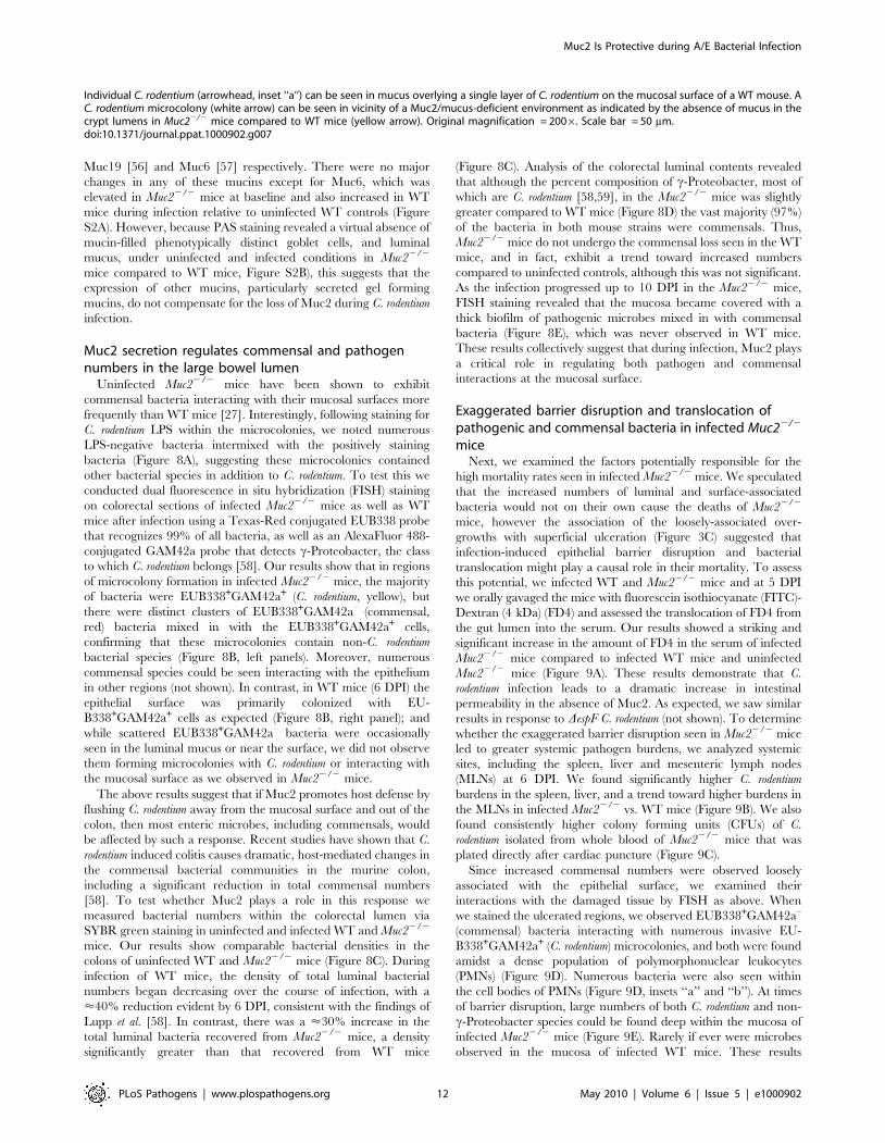
Figure 7. C. rodentium infection results in increased mucin secretion during infection. A. Representative PAS/Haematoxylin staining ofCarnoy’s fixed rectal sections from uninfected (left panel) and C. rodentium-infected mice (right panel). Arrow points to luminal mucus. Originalmagnification = 1006. Scale bar = 100 mm. B. Total counts per minute (CPM) of [3H]-glucosamine labeled glycoproteins found in colorectalsecretions 3.5 hrs post-injection from uninfected and infected (6 DPI) WT mice. Results are representative of 2 independent infections containing 5mice per group. C. Plot of liquid scintillation counts of fractions containing [3H] activity after total secretions were subjected to gel filtration on aSepharose 4B chromatography column. This graph is representative of 2 independent infections with 5 mice per group. D. Graph of total CPMs ofvoid volumes of S4B-fractionated mucins as described in D. Data represents the mean of the average of 2 independent experiments, each with 5 miceper group. Error bars = SEM. E. Combined epifluorescent staining for mucus using the lectin UEA-1 (red), and C. rodentium LPS (green), and cellularDNA (blue) using DAPI as a counterstain in heavily infected (6 DPI) regions of the colorectal mucosa from WT and Muc22/2 mice, as indicated.
Muc2 Is Protective during A/E Bacterial Infection
PLoS Pathogens | www.plospathogens.org 11 May 2010 | Volume 6 | Issue 5 | e1000902
Muc19 [56] and Muc6 [57] respectively. There were no major
changes in any of these mucins except for Muc6, which was
elevated in Muc22/2 mice at baseline and also increased in WT
mice during infection relative to uninfected WT controls (Figure
S2A). However, because PAS staining revealed a virtual absence of
mucin-filled phenotypically distinct goblet cells, and luminal
mucus, under uninfected and infected conditions in Muc22/2
mice compared to WT mice, Figure S2B), this suggests that the
expression of other mucins, particularly secreted gel forming
mucins, do not compensate for the loss of Muc2 during C. rodentium
infection.
Muc2 secretion regulates commensal and pathogennumbers in the large bowel lumen
Uninfected Muc22/2 mice have been shown to exhibit
commensal bacteria interacting with their mucosal surfaces more
frequently than WT mice [27]. Interestingly, following staining for
C. rodentium LPS within the microcolonies, we noted numerous
LPS-negative bacteria intermixed with the positively staining
bacteria (Figure 8A), suggesting these microcolonies contained
other bacterial species in addition to C. rodentium. To test this we
conducted dual fluorescence in situ hybridization (FISH) staining
on colorectal sections of infected Muc22/2 mice as well as WT
mice after infection using a Texas-Red conjugated EUB338 probe
that recognizes 99% of all bacteria, as well as an AlexaFluor 488-
conjugated GAM42a probe that detects c-Proteobacter, the class
to which C. rodentium belongs [58]. Our results show that in regions
of microcolony formation in infected Muc22/2 mice, the majority
of bacteria were EUB338+GAM42a+ (C. rodentium, yellow), but
there were distinct clusters of EUB338+GAM42a– (commensal,
red) bacteria mixed in with the EUB338+GAM42a+ cells,
confirming that these microcolonies contain non-C. rodentium
bacterial species (Figure 8B, left panels). Moreover, numerous
commensal species could be seen interacting with the epithelium
in other regions (not shown). In contrast, in WT mice (6 DPI) the
epithelial surface was primarily colonized with EU-
B338+GAM42a+ cells as expected (Figure 8B, right panel); and
while scattered EUB338+GAM42a– bacteria were occasionally
seen in the luminal mucus or near the surface, we did not observe
them forming microcolonies with C. rodentium or interacting with
the mucosal surface as we observed in Muc22/2 mice.
The above results suggest that if Muc2 promotes host defense by
flushing C. rodentium away from the mucosal surface and out of the
colon, then most enteric microbes, including commensals, would
be affected by such a response. Recent studies have shown that C.
rodentium induced colitis causes dramatic, host-mediated changes in
the commensal bacterial communities in the murine colon,
including a significant reduction in total commensal numbers
[58]. To test whether Muc2 plays a role in this response we
measured bacterial numbers within the colorectal lumen via
SYBR green staining in uninfected and infected WT and Muc22/2
mice. Our results show comparable bacterial densities in the
colons of uninfected WT and Muc22/2 mice (Figure 8C). During
infection of WT mice, the density of total luminal bacterial
numbers began decreasing over the course of infection, with a
<40% reduction evident by 6 DPI, consistent with the findings of
Lupp et al. [58]. In contrast, there was a <30% increase in the
total luminal bacteria recovered from Muc22/2 mice, a density
significantly greater than that recovered from WT mice
(Figure 8C). Analysis of the colorectal luminal contents revealed
that although the percent composition of c-Proteobacter, most of
which are C. rodentium [58,59], in the Muc22/2 mice was slightly
greater compared to WT mice (Figure 8D) the vast majority (97%)
of the bacteria in both mouse strains were commensals. Thus,
Muc22/2 mice do not undergo the commensal loss seen in the WT
mice, and in fact, exhibit a trend toward increased numbers
compared to uninfected controls, although this was not significant.
As the infection progressed up to 10 DPI in the Muc22/2 mice,
FISH staining revealed that the mucosa became covered with a
thick biofilm of pathogenic microbes mixed in with commensal
bacteria (Figure 8E), which was never observed in WT mice.
These results collectively suggest that during infection, Muc2 plays
a critical role in regulating both pathogen and commensal
interactions at the mucosal surface.
Exaggerated barrier disruption and translocation ofpathogenic and commensal bacteria in infected Muc22/2
miceNext, we examined the factors potentially responsible for the
high mortality rates seen in infected Muc22/2 mice. We speculated
that the increased numbers of luminal and surface-associated
bacteria would not on their own cause the deaths of Muc22/2
mice, however the association of the loosely-associated over-
growths with superficial ulceration (Figure 3C) suggested that
infection-induced epithelial barrier disruption and bacterial
translocation might play a causal role in their mortality. To assess
this potential, we infected WT and Muc22/2 mice and at 5 DPI
we orally gavaged the mice with fluorescein isothiocyanate (FITC)-
Dextran (4 kDa) (FD4) and assessed the translocation of FD4 from
the gut lumen into the serum. Our results showed a striking and
significant increase in the amount of FD4 in the serum of infected
Muc22/2 mice compared to infected WT mice and uninfected
Muc22/2 mice (Figure 9A). These results demonstrate that C.
rodentium infection leads to a dramatic increase in intestinal
permeability in the absence of Muc2. As expected, we saw similar
results in response to DespF C. rodentium (not shown). To determine
whether the exaggerated barrier disruption seen in Muc22/2 mice
led to greater systemic pathogen burdens, we analyzed systemic
sites, including the spleen, liver and mesenteric lymph nodes
(MLNs) at 6 DPI. We found significantly higher C. rodentium
burdens in the spleen, liver, and a trend toward higher burdens in
the MLNs in infected Muc22/2 vs. WT mice (Figure 9B). We also
found consistently higher colony forming units (CFUs) of C.
rodentium isolated from whole blood of Muc22/2 mice that was
plated directly after cardiac puncture (Figure 9C).
Since increased commensal numbers were observed loosely
associated with the epithelial surface, we examined their
interactions with the damaged tissue by FISH as above. When
we stained the ulcerated regions, we observed EUB338+GAM42a–
(commensal) bacteria interacting with numerous invasive EU-
B338+GAM42a+ (C. rodentium) microcolonies, and both were found
amidst a dense population of polymorphonuclear leukocytes
(PMNs) (Figure 9D). Numerous bacteria were also seen within
the cell bodies of PMNs (Figure 9D, insets ‘‘a’’ and ‘‘b’’). At times
of barrier disruption, large numbers of both C. rodentium and non-
c-Proteobacter species could be found deep within the mucosa of
infected Muc22/2 mice (Figure 9E). Rarely if ever were microbes
observed in the mucosa of infected WT mice. These results
Individual C. rodentium (arrowhead, inset ‘‘a’’) can be seen in mucus overlying a single layer of C. rodentium on the mucosal surface of a WT mouse. AC. rodentium microcolony (white arrow) can be seen in vicinity of a Muc2/mucus-deficient environment as indicated by the absence of mucus in thecrypt lumens in Muc22/2 mice compared to WT mice (yellow arrow). Original magnification = 2006. Scale bar = 50 mm.doi:10.1371/journal.ppat.1000902.g007
Muc2 Is Protective during A/E Bacterial Infection
PLoS Pathogens | www.plospathogens.org 12 May 2010 | Volume 6 | Issue 5 | e1000902

Figure 8. Increased luminal load of both pathogenic and non-pathogenic bacteria in Muc22/2 mice during infection. A.Immunofluorescence staining for C. rodentium LPS and DAPI in Muc22/2 at 4 DPI Notice DAPI-stained bacteria that are negative for LPS in the C.rodentium microcolonies (arrow). Original magnification = 2006. B. Dual FISH staining using DNA probes that label virtually all true bacteria (EUB338,red) and the c-Proteobacter class to which C. rodentium belongs (GAM42a, green). Pathogenic bacteria (i.e. EUB338+/GAM42a+ cells) are yellow, andcommensal bacteria (EUB338+/GAM422) cells are red. Note the non-ulcer associated bacterial microcolony containing commensal bacteria (red)mixed in with pathogenic bacteria (yellow) in Muc22/2 mice (left panels). Such mixed microcolonies were not seen in WT mice, which showpredominantly pathogenic bacteria intimately adherent to the mucosa (right panel). Tissues were fixed in Carnoy’s fixative prior to processing.Original magnification = 2006. Scale bar = 100 mm. C. SYBR green quantification of total bacterial burden per gram of colorectal lumen contents ofWT vs. Muc22/2 mice before infection and at 6 DPI. Results are presented as the means of a total of 5–7 mice per group pooled from 2 independentexperiments. Error bars = SEM (**P = 0.0082, Mann-Whitney test). D. Graph illustrating the percent composition of c-Proteobacter (EUB338+/GAM42a+ cells), which is primarily represented by C. rodentium, in colorectal luminal content from uninfected or infected WT vs. Muc22/2 mice.
Muc2 Is Protective during A/E Bacterial Infection
PLoS Pathogens | www.plospathogens.org 13 May 2010 | Volume 6 | Issue 5 | e1000902

strongly suggest that both pathogenic and commensal bacteria
contribute to the disease and mortality suffered by Muc22/2 mice,
since A/E bacterial infection-induced disruption of the epithelial
barrier allows massive translocation of both pathogenic and
commensal bacteria out of the intestinal lumen and into mucosal
tissues, and pathogens into systemic compartments, leading to
bacteremia.
Evidence that Muc2-deficiency reduces host-mediatedpathogen clearance when commensal-dependent hostcolonization resistance is compromised
The above data show commensal and pathogenic bacteria
occupying intestinal niches in Muc22/2 mice that are not
colonized in WT mice during infection. To attempt to elucidate
the precise role of commensal bacteria during C. rodentium infection
in Muc22/2 mice, we administered a high dose of the antibiotic
streptomycin (20 mg/mouse) by oral gavage to reduce the
numbers of total commensals prior to infection. Stool was
collected immediately prior to treatment and again 24 hrs later,
and then stool bacteria was quantified as above to confirm
commensal depletion. Streptomycin (strep) treatment resulted in a
significant (average 10–20 fold) reduction in commensal bacterial
numbers in both WT and Muc22/2 mice, while vehicle treatment
did not cause any significant changes (Figure 10A). Neither
treatment led to any inflammation or pathology on its own when
assessed 7 days later (not shown). 24 hrs after treatment, strep- and
vehicle-treated WT and Muc22/2 mice were also gavaged with
DespF C. rodentiumStr (strep-resistant), which was chosen instead of
wt C. rodentium because it is less virulent. Colonization was assessed
by plating stool contents every second day. The results show that
at 2 DPI, strep-treated WT and Muc22/2 mice carried 10–50 fold
higher bacterial burdens compared to infected vehicle-treated WT
and Muc22/2 mice (Figure 10B). However by 4 and 6 DPI, while
DespF C. rodentiumStr burdens began to decline in infected strep-
treated WT mice ultimately to levels similar to infected vehicle-
treated WT mice (6 DPI), bacterial burdens in infected strep-
treated Muc22/2 mice continued to increase to levels significantly
higher than all other groups (Figure 10B). Moreover, burdens in
infected vehicle-treated Muc22/2 mice also increased to levels that
were higher than infected strep-treated WT mice at 6 DPI.
Although weight loss varied among mice both Muc22/2 groups,
only WT mice tended to gain weight during infection (Figure 10C).
At 6 DPI, both cecal and colonic tissues were resected and
assessed by histology. As shown by H&E (Figure 10D, bottom
panels), strep-treatment led to increased edema and inflammation
in WT ceca compared to vehicle-treated WT mice during
infection; however in infected Muc22/2 tissues, there were no
obvious differences in cecal and colonic inflammation between
strep-and vehicle-treated groups (Figure 10D, top panels). Overt
ulceration was seen in the ceca of vehicle-treated Muc22/2 mice
(Figure 10E), while ulcers were observed in the colons of strep-
treated Muc22/2 mice (Figure 10F) Interestingly, FISH staining of
cecal sections from infected vehicle-treated Muc22/2 mice showed
large numbers of commensals (EUB338+GAM42a–, red) directly
interacting with PMNs in ulcerated regions (Figure 10E, left
panel). These interactions were seen at the mucosal surface of
ulcers where there was little evidence of DespF C. rodentium;
however DespF C. rodentiumStr could still be seen within the PMNs
(Figure 10E, right panel, inset). In contrast, large invasive DespF C.
rodentiumStr microcolonies (EUB338+GAM42a+, yellow) could be
seen associated with the ulcers in the colons of infected strep-
treated Muc22/2 mice (Figure 10F, right panel). Such pathology
was never observed in uninfected mice or in any of the infected
WT groups. Collectively, these results indicate that (i) Muc2
promotes host-mediated colonization resistance when commensals
are depleted; and (ii) commensal bacteria, although initially
important in promoting colonization resistance in both strains,
ultimately come into direct contact with large numbers of PMNs
following the infection-induced ulceration that occurs in a Muc2-
deficient environment. Thus Muc2 is critical for managing
commensal and pathogenic bacteria within the GI tract,
particularly at mucosal surfaces during an enteric infection.
Discussion
The Muc2-rich mucus layer is the first host-defense barrier that
noxious luminal agents contact in the intestine [33], and as such, it
functions as the main interface between the host and its luminal
microbiota. To our knowledge, this is the first study to formally
demonstrate the importance of the major mucus glycoprotein
Muc2 in host defense against an A/E bacterial pathogen in vivo.
We show that the presence of Muc2 and hence the mucus layer is
necessary to protect against severe mucosal damage and barrier
dysfunction during infection. This was in part due to Muc2
functioning as a structural barrier to limit the rate of pathogen
colonization of epithelial cells in the large bowel. However, Muc2
plays an additional role in host defense by controlling the
pathogen burden that resides within the colonic lumen, primarily
by removing loosely adherent bacteria and preventing bacterial
accumulation and microcolony formation on the colorectal
surface. The inability to effect this removal likely contributes to
the severe barrier dysfunction seen in Muc22/2 mice. We provide
evidence that the ability of Muc2 to control luminal bacteria is
most likely attributable to increased Muc2/mucus secretion during
infection, which was demonstrated through metabolic labeling of
mucin glycoproteins in WT mice. Moreover, we demonstrate that
the ability of Muc2 to control luminal pathogens also impacts the
resident commensal microbiota, as the microcolonies seen
overlying the mucosa of infected Muc22/2 mice contained both
C. rodentium as well as commensal microbes, and both types of
bacteria were seen translocating across the colonic epithelium and
into the lamina propria. These results ultimately reveal Muc2
production as a critical mechanism by which the host controls
exposure to both pathogenic and commensal bacteria in vivo.
While we assumed that A/E pathogens such as C. rodentium
would have to interact with the mucus layer during the course of
infection, we demonstrate and characterize this interaction for the
first time in situ. We show that C. rodentium colonizes the outer
mucus layer in high numbers, and can also be found traversing the
normally bacteria-free inner mucus layer to gain access to the
underlying epithelial cells. These results raise the question of how
A/E pathogens manage to circumvent the mucus layer. C.
rodentium lacks a functional flagellum and is thus non-motile [60],
and therefore likely utilizes specific mucinases or glycosidases to
digest mucin in order to overcome the mucus barrier, although
this has yet to be formerly demonstrated. Notably, EHEC has
Results are the mean percentages from a total of 5–7 mice per group pooled from 2 independent experiments. ND, none detected. Error bars = SEM.E. FISH staining as described above, showing a thick biofilm of mostly pathogenic but also commensal bacteria on the mucosal surface in a colonicsection from a moribund Muc22/2 mouse at 10 DPI (inset). Such phenotypes were not observed in WT mice. Original magnification = 2006. Scalebar = 50 mm.doi:10.1371/journal.ppat.1000902.g008
Muc2 Is Protective during A/E Bacterial Infection
PLoS Pathogens | www.plospathogens.org 14 May 2010 | Volume 6 | Issue 5 | e1000902

Figure 9. Susceptibility of Muc22/2 mice to C. rodentium is associated with severe defects in intestinal barrier function and increasedtranslocation of commensal and pathogenic bacteria. Muc22/2 mice display increased FITC-dextran flux across the intestinal mucosa during C.rodentium infection. Uninfected or C. rodentium infected (5 DPI) WT and Muc22/2 mice were gavaged with FITC-dextran (4 kDa) and serum was takenby cardiac puncture 4 hrs later, as described in Materials and Methods. A. Quantity of FD4 in serum from WT and Muc22/2 mice. Bars represent theaverage value of a total of 5–7 mice per group pooled from 2 individual experiments. Error bars = SEM (**P = 0.0051; ***P = 0.0006, Mann-Whitneytest). B. Quantification of viable C. rodentium found in the spleens, liver, and MLNs of WT and Muc22/2 mice at 7 DPI. Each data point represents oneanimal. Bars represent the means of 9 WT and 12 Muc22/2 mice pooled from 3 independent experiments. Error bars = SEM (**P = 0.0031, Mann-
Muc2 Is Protective during A/E Bacterial Infection
PLoS Pathogens | www.plospathogens.org 15 May 2010 | Volume 6 | Issue 5 | e1000902

recently been shown to secrete the metalloprotease StcE that has
apparent mucinase activity [61] suggesting A/E pathogens do
employ this strategy. In contrast, despite their diversity and
extreme density in mammalian colon, commensal bacteria do not
penetrate the inner mucus layer to any significant degree, probably
because they are more adapted to the nutrient-rich luminal
environment [62]. Ultimately, this suggests that colonizing the
outer and inner mucus layer is a key step for the pathogenesis of
A/E bacteria, therefore, the bacterial factors involved in crossing
the mucus layer are likely critical for virulence.
Our studies reveal an unexpected insight into how Muc2
mediates protection. Muc2 is widely presumed to act as a
physicochemical barrier to limit access to epithelial tissues by
luminal pathogens [17], including pathogens such as A/E
bacteria. Several lines of evidence support this, such as the
demonstration of mucins inhibiting EPEC adherence in vitro [20]
and our in vivo cecal loop colonization assay described in this
report. However, since the total numbers of bacteria that
ultimately infected (i.e. became intimately adherent to) the tissue
was not significantly different in a Muc2 deficient environment,
the role of Muc2 as a defense barrier may be of only transient
importance. Rather the major function played by Muc2, at least in
response to A/E bacteria, appears to be to limit luminal burdens,
mainly by preventing the accumulation of pathogens that are
loosely associated with the tissue. These bacteria probably arise
from replication of intimately-bound pathogens, as the T3SS
mutant (DescN C. rodentium) failed to efficiently colonize. This
massive increase in the overall pathogen burden at the mucosal
surface has important implications for downstream host responses.
EPEC and EHEC both disrupt epithelial permeability in vitro [63],
as does C. rodentium in vivo [64,65]. While intimately-adherent
bacteria are firmly bound to the epithelia, the non-infecting, but
loosely adherent bacteria are more likely to translocate into the
mucosa, particularly when faced with the mechanical pressures of
dietary flow. Indeed, at times of severe barrier disruption we saw
much higher systemic levels of C. rodentium in the Muc22/2 mice.
Although Muc2 deficiency did not ultimately impact on the
numbers of intimately-adherent C. rodentium, there was a striking
increase in intestinal permeability in Muc22/2 compared to WT
mice. The susceptibility to ulcer formation in the Muc22/2 mice is
probably a major contributor to the barrier dysfunction and
morbidity seen in these mice since it was associated with greater
systemic pathogen burdens. While the mechanisms are unclear, we
suggest the accumulation of bacteria and microcolony formation
on the epithelial surface in a Muc2-deficient environment is linked
to either the development and/or maintenance of the ulceration,
since most ulcers were associated with the microcolonies. It has
been proposed that serum proteins released at ulcerated sites
contribute to ulcer-associated C. rodentium overgrowth [66];
however the fact we saw microcolony formation also in non-
ulcerated sites argues against this always being the case.
Interestingly, past studies have shown that the A/E pathogen
translocated effector EspF has been linked to epithelial barrier
disruption [41,67] and ulcer-associated damage [42]. However,
since ulcers, microcolony formation, and barrier disruption were
also seen in mice infected with the DespF strain, these data indicate
that barrier disruption occurs through non-canonical pathways.
We speculate that bacterial accumulation and microcolony
formation at the surface adversely affects epithelial survival either
directly, by producing a high local concentration of toxic
metabolites; or indirectly, by causing the recruitment of large
numbers of PMNs to the site of infection, where epithelial cell
death is the result of collateral damage caused by neutrophils
releasing cytotoxic mediators to control the infection. In fact, one
can envision these microcolonies to be an overwhelming burden to
recruited phagocytes, perpetuating a vicious inflammatory cycle
(Figure 10). Whatever the specific role of these invasive
microcolonies, they likely exacerbate the focal damage and
associated barrier defects, and thus have a severe impact on
morbidity in the Muc22/2 mice.
Although we attribute the majority of the pathological
phenotypes in infected Muc22/2 mice to result from C. rodentium,
one of the striking features during the course of infection was the
maintenance of commensal bacteria at the mucosal surface of the
Muc22/2 mice. While we also found scattered commensal bacteria
overlying the epithelium before infection [27], C. rodentium was
clearly unable to totally displace them. This led to some intriguing
phenotypes, including direct intimate interactions between
commensal bacteria and the pathogen, where commensals were
found intermixed with C. rodentium clusters to create multispecies
microcolonies. Critically, commensal species could also be found
translocating across the mucosal surface and into the lamina
propria, where they were in direct contact with PMNs at sites of
microcolony-associated ulceration, even forming microcolonies of
their own. We explored whether these commensal bacteria
contribute to the resulting colitis by transiently depleting them
using the antibiotic streptomycin. While the depletion was
successful, it also led to an exaggerated pathogen burden,
confirming that commensal bacteria play an important host
defense role by providing colonization resistance against C.
rodentium. Although we did not identify overt differences in the
resulting pathology in Muc22/2 mice following antibiotic
pretreatment, we were unable to conclude to what degree
commensal translocation might play in the resulting colitis,
considering the loss of commensals occurred concomitantly with
increased pathogen burdens. However, the fact that infection-
induced cecal ulceration in Muc22/2 mice led to large numbers of
commensals that were directly interacting with PMNs points to a
pathologic host-commensal interaction during infection. There-
fore, while commensals are beneficial early during an infection by
enhancing colonization resistance, their continued presence as the
infection progresses likely plays a pathologic role. These studies are
particularly interesting in light of the study by Lupp et al. [58] who
described an overall reduction of commensal bacterial numbers
after an established C. rodentium infection. It has been suggested this
is a pathogenic strategy where pathogens exploit inflammation to
suppress commensal growth and thereby reduce colonization
resistance [68]. However, our findings strongly suggest that
clearance of commensal microbes from the colon after an
established C. rodentium infection may also benefit the host, by
decreasing the total bacterial burden faced by the host at a time
when its intestinal barriers are compromised.
Whitney test). C. Enumeration of live bacterial burdens cultured from the serum of Muc22/2 and WT mice at 6 DPI. Results represent the average of8 WT and 12 Muc22/2 mice pooled from 3 independent experiments. Error bars = SEM. D. FISH staining showing invasive microcolonies within anulcerated region in the descending colon of an infected Muc22/2 mouse. Pathogenic bacteria can be seen engulfed by PMNs that are attacking themicrocolony (inset ‘‘a’’, arrowheads). A commensal bacterial microcolony (red) can also be seen amongst the C. rodentium microcolonies and incontact with PMNs (inset ‘‘b’’, arrow). Original magnification = 2006. Scale bar = 100 mm. E. Numerous c-Proteobacter (C. rodentium, yellow; yellowarrowhead in inset) and non-c-Proteobacter (red; white arrowhead in inset) can be seen invading the lamina propria of infected Muc22/2 mice (6DPI). Lu = gut lumen. LP = lamina propria; Original Magnification, 2006. Results are representative of 3 separate experiments.doi:10.1371/journal.ppat.1000902.g009
Muc2 Is Protective during A/E Bacterial Infection
PLoS Pathogens | www.plospathogens.org 16 May 2010 | Volume 6 | Issue 5 | e1000902

Figure 10. Antibiotic induced commensal depletion enhances pathogen colonization but does not alter host pathology in Muc22/2
mice. A. Quantification of DAPI stained bacteria from stools of WT and Muc22/2 mice 24 hours following oral treatment with Streptomycin (20 mg)or Vehicle (dH20). Streptomycin (strep) led to significantly reduced numbers of total bacteria within mouse stool. Results represent the means of 3–4mice per group. Error bars = SEM (***P,0.001, unpaired t test). B. Enumeration of DespF C. rodentiumStr (strep-resistant) in stool of strep-or vehicle-treated mice as indicated, at various times post-infection. Results represent the means of 3–4 mice per group. Error bars = SEM (*P#0.05, Mann-Whitney test, one-tailed). C. Body weights following infection of strep or vehicle treated WT and Muc22/2 mice with DespF C. rodentiumStr. n = 3–4mice per group. Error bars = SEM. D. Representative histological sections of ceca from uninfected or infected (6 DPI) strep- or vehicle-treated WT andMuc22/2 mice. Original magnification = 1006. Scale bar = 100 mm. E. H&E (Left panel) and FISH analysis (right panel) of an ulcer from DespF C.rodentiumStr infected vehicle-treated Muc22/2 mouse cecum (6 DPI). Numerous commensals (EUB338+/GAM422 cells, red) can be seen overlying theulcer in direct contact with PMNs (arrow), and both pathogen (EUB338+/GAM42a+ cells, yellow) and commensal (red) can be seen within the PMNs(arrow heads, inset). Original magnification = 2006. Scale bars = 100 mm. F. H&E and FISH analysis of an ulcer in the descending colon from an DespFC. rodentiumStr infected strep-treated Muc22/2 mouse (6 DPI). Large pathogenic microcolonies (yellow) are associated with the ulcer (arrows), whilecommensals (red) can be seen in the lumen. Original magnification = 2006. Scale bar = 100 mm.doi:10.1371/journal.ppat.1000902.g010
Muc2 Is Protective during A/E Bacterial Infection
PLoS Pathogens | www.plospathogens.org 17 May 2010 | Volume 6 | Issue 5 | e1000902

We hypothesize that mucin secretion is the key mechanism by
which Muc2 controls the levels of A/E bacteria, and commensal
bacteria at the surface. Recent studies have suggested there is
enhanced mucin secretion in the colon during C. rodentium infection
[19]. We extend these findings through metabolic labeling to show
at least a 40% increase in mucus secretion in response to infection,
and specifically indentified C. rodentium within luminal mucus. This
increase in mucin secretion is likely a gross underestimate of the
local increase in mucin release, since in order to have sufficient
quantities for analysis, we extracted mucus from the whole colon,
and the increase in secretion is expected to be focused in the
descending colon and rectum where the infection occurs [69]. Due
to the lack of antimicrobial activity we saw within the crude
mucus, and the fact that it was recently shown by the McGuckin
laboratory that C. rodentium directly binds to Muc2/mucus in vitro
[19], we hypothesize that induced mucin secretion is an effective
means for the host to bind and remove non-infecting, loosely
adherent A/E bacteria that would otherwise accumulate on the
surface and exacerbate disease (Figure 11). Although beyond the
scope of the present study, an outstanding issue yet to be addressed
is deciphering the precise molecules responsible for the induced
Muc2 secretion in vivo. There are a plethora of candidates,
including bacterial products, such as LPS [51,70], or host derived
cytokines such as TNFa [71], neuromodulators including
vasoactive intestinal peptide [72], or neutrophils via elaboration
of secretagogues such as neutrophil elastase [73], all of which have
been shown to cause enhanced mucin release from goblet cells in
tissue culture, and are present during C. rodentium infection
[74,75,76]. Based on the data presented in our report, the
elucidation of the specific host and/or microbial factors and
molecular pathways that regulate mucus production during enteric
bacterial infection constitutes a fertile area of research.
Importantly, while we ascribe the ability of intestinal mucus to
flush away luminal bacteria from the mucosal surface to primarily
reflect the actions of Muc2, there are likely other mucins,
(potentially found in total secreted mucus) that may also contribute
to the protective actions of the mucus. These include Muc1, a cell
surface mucin that is upregulated in bacterial induced colitis [19]
and potentially cleaved to release its a-subunit containing the
extracellular mucin domain into the intestinal lumen, as seen
during H. pylori infection [77]; Muc4 which can be up-regulated
during DSS-induced colitis [78]and be expressed by colonic goblet
cells [55]; and the secreted gel-forming mucins Muc19 and Muc6,
the latter being produced in Muc22/2 mice during colitis [29].
Even so, we maintain that Muc2 is the major protective mucin.
This is in part based upon the phenotype of Muc22/2 mice
(confirmed by our studies), where Muc2-deficiency leads to a
virtual loss of mucin-filled phenotypically mature goblet cells
within the large intestine, and a corresponding loss of both the
inner and outer colonic mucus layers [24] and other forms of
luminal mucus. Moreover, Muc2 is by far the major secretory
mucin under both baseline (in mice and humans) [24,53] and
inflammatory conditions in the colon [54]. However, we did see a
modest up-regulation of Muc6 mRNA expression during infection
of WT mice, and the impact of this expression is currently under
investigation.
During the course of this article review, it was demonstrated by
Hasnain et al. [79] that Muc2-deficiency renders mice more
susceptible to intestinal nematode infections, suggesting Muc2 and
mucus production can protect against diverse enteric pathogens.
Muc2 production is clearly protective during A/E bacterial
infection, but whether this is true for other enteric bacterial
pathogens of the gut remains to be shown. Importantly, since
bacteria and other enteric pathogens have co-evolved with their
hosts, many exhibit multiple strategies to subvert and exploit host
defenses including mucus to promote colonization [80]. A well
known motility factor is flagella, which is commonly utilized by
pathogenic bacteria such Vibrio cholerae to migrate through mucus
(reviewed in [33]). In addition, Salmonella appears to anchor itself
to mucus via specific adhesins [81] to promote colonization [82],
and exhibits resistance to small bowel mucus antimicrobial activity
[48]. Yersinia enterocolitica has been shown to utilize polysaccharides
present in mucins like Muc2 to harvest energy and promote
growth [83]. A similar observation has been shown for Salmonella
Typhimurium, for which it has been proposed as a strategy to
outcompete the commensal microbiota within an inflammatory
niche [84]. Moreover, parasites such as Entamoeba histolytica
stimulate mucin release to deplete the mucus layer [85], as well
as proteolytically break down the polymeric structure of secreted
Muc2 to facilitate access to the underlying epithelium [86,87].
Thus, whether Muc2 has evolved primarily to regulate interactions
with normal microbiota and other luminal contents, or to provide
adequate host defense against enteric pathogens has yet to be
determined. However, because the commensal microbiota is a
major variable in any enteric infection, particularly in the colon, it
is likely that the presence of Muc2 allows for effective
immunological management of the infectious agent by limiting
commensal burdens at mucosal surfaces.
In conclusion, our studies have shown that Muc2 and the mucus
layer are critical for host defense against an A/E bacterial
pathogen. However, it is important to note that Muc2 can
potentially be modulated in several ways either during infection,
such as at the level of gene expression, post-translational
modification, or even at the level of secretion into the intestinal
lumen. Each regulatory step may influence the biological function
of Muc2, which in turn will influence how the host responds to
enteric pathogens. Since Muc2 is an integral part of the colonic
ecosystem, future studies are warranted to unravel precisely how
intestinal mucus impacts the course of infectious disease.
Materials and Methods
MiceSix to eleven-week-old C57BL/6, Muc2+/+ and Muc22/2 mice
(on C57BL/6 background) were purchased from the National
Cancer Institute (NCI) or bred in our animal facility. Mice were
kept in sterilized, filter-topped cages, handled in tissue culture
hoods and fed autoclaved food and water under specific pathogen
free (SPF) conditions. Sentinel animals were routinely tested for
common pathogens. The protocols employed were approved by
the University of British Columbia’s Animal Care Committee and
in direct accordance with guidelines drafted by the Canadian
Council on the Use of Laboratory Animals.
Bacterial strains and infection of miceMice were infected by oral gavage with 0.1 ml of an overnight
culture of LB containing approximately 2.56108 cfu of wt C.
rodentium (formerly C. freundii biotype 4280, strain DBS100, the
EspF mutant DespF C.rodentium, or the T3SS mutant DescN C.
rodentium [88]. Bioluminescent strains of C. rodentium were
constructed by introducing plasmid pT7 (E. A. Meighen,
Department of Biochemistry, McGill University) carrying the
entire lux operon from Photorhabdus luminescens. For bacterial
enumeration studies, a streptomycin-resistant derivative of C.
rodentium DBS100 was utilized. For some studies a streptomycin-
resistant DespF C.rodentium was utilized, and was constructed in our
laboratory by routine procedures. GFP-C. rodentium was construct-
ed within our laboratory by chromosomal insertion of gfp into C.
Muc2 Is Protective during A/E Bacterial Infection
PLoS Pathogens | www.plospathogens.org 18 May 2010 | Volume 6 | Issue 5 | e1000902

Muc2 Is Protective during A/E Bacterial Infection
PLoS Pathogens | www.plospathogens.org 19 May 2010 | Volume 6 | Issue 5 | e1000902

rodentium DBS-100 via Red/ET Recombination, using a Quick &
Easy E. coli Gene Deletion Kit (Gene Bridges) as per manufac-
turers instructions. The virulence of the GFP-C. rodentium was
confirmed in preliminary studies. For commensal depletion
studies, mice were pre-treated with 0.1 ml of 200 mg/ml
(20 mg) streptomycin (or H20) 24 hrs prior to infection.
Tissue collectionUninfected or infected mice were anesthetized with Halothane,
killed by cervical dislocation, and their large intestines were
resected and divided into cecum, ascending colon, descending
colon, and rectum for further analysis. Tissues were immediately
placed in 10% neutral buffered formalin (Fisher) (48 hrs, 4uC) or
ice cold fresh Carnoy’s Fixative (2 hrs, 4uC) or 4% paraformal-
dehyde (1 hr, room temp) for histological studies, or placed in
RNAlater (Qiagen) and stored at 286uC for subsequent RNA
extraction.
Bacterial countsFor enumeration of bacteria within the tissue and luminal
compartments, whole mouse ceca or colons were sliced open
longitudinally, and their luminal contents were collected in a
preweighed 2.0 ml microtube containing 1.0 ml of phosphobuf-
fered saline (PBS) and a 5.0 mm steel bead (Qiagen). Cecal and
colonic tissues were washed vigorously in PBS (pH 7.4), cut into
several pieces, and also placed in a tube as above. Tissue and
lumen contents were weighed, and then homogenized in a
MixerMill 301 bead miller (Retche) for a total of 6 mins at
30 Hz at room temperature. Tissue homogenates were serially
diluted in PBS and plated onto luria broth (LB) agar plates
containing 100 mg/ml streptomycin, incubated overnight at
37uC, and bacterial colonies were enumerated the following day,
normalizing them to the tissue or stool weight (per gram). For fecal
bacterial burden analysis, stool was collected from live mice at
various times post-infection (described in text) and processed as
described for luminal contents. For some studies with non-
antibiotic resistant C. rodentium, plating was performed on
MacConkey Agar (Difco), C. rodentium colonies were clearly
identified by their unique characteristic of being round with red
centre and a thin white rim. Colonies were confirmed to be C.
rodentium by PCR for the C. rodentium T3SS translocator gene escN.
Histological stainingBriefly, 5 mm paraffin sections were deparaffinized by heating at
55–65uC for 10 min, cleared with xylene, rehydrated through an
ethanol gradient to water. Sections were blocked using the
appropriate blocking buffer (either 2% Goat or Donkey Serum
in PBS containing 1% bovine serum albumin (BSA), 0.1% Triton-
X100 (Sigma), and 0.05% Tween 20, and 0.05% sodium azide.
For detection of biotinylated targets, blocking of endogenous
biotin was carried out prior to blocking with serum, using the
Endogenous Biotin Blocking kit (Molecular Probes). Primary
antibodies or lectins were diluted in PBS containing 1% BSA,
0.1% Triton-X100 (Sigma), and 0.05% Tween 20, and 0.05%
sodium azide. The antibodies used were rat anti-F4/80 (1:8000;
Serotec), rabbit anti-MPO (1:100; NeoMarkers), rat antisera
generated against C. rodentium specific Tir (1:5K; gift from W.
Deng), rabbit anti-E.coli Poly 8 LPS (1:500; Biotec Laboratories),
biotinylated goat anti-GFP (1:100: GeneTex), polyclonal antisera
that recognized the murine colonic mucin Muc2 (1:50; a gift from
Jan Dekker). Staining for fucosylated mucins was carried out using
biotinylated-Ulex europaeus agglutinin-1 (2 ug/ml; Vector Labs).
Antigen retrieval was used for F4/80 and MPO staining, and was
performed prior to blocking and staining by placing deparaffi-
nized, rehydrated slides in 10 mM citric acid pH 6.0 at 90–100uCfor 20 min, followed by cooling to room temperature. Preparation
and staining of PFA-fixed frozen sections was performed as
described previously [44]. For dual LPS/Tir staining, no
detergents (TritonX-100 or Tween-20) were used in the dilution
buffers, to avoid Tir staining within bacteria. Epifluorescent
labeling for all stains was carried out with the appropriate
secondary antibody using AlexaFluor 488-conjugated goat (or
donkey) anti-rabbit IgG, AlexaFluor 568-conjugated goat anti-
rabbit IgG, AlexaFluor 568-conjugated goat anti-rat IgG (all
1:2000), or AlexaFluor 568-conjugated Streptavidin (1:1000)
(Molecular Probes/Invitrogen). Tissues were mounted using
ProLong Gold Antifade reagent (Molecular Probes/Invitrogen)
that contains 49,69-diamidino-2-phenylindole (DAPI) for DNA
staining. Sections were viewed at 350, 488, and 594 nm on a Zeiss
AxioImager microscope. Images were obtained using a Zeiss
AxioImager microscope equipped with an AxioCam HRm
camera operating through AxioVision software (Version 4.4).
RNA extraction and quantitative RT-PCRColon tissues stored in RNAlater (Qiagen) at 286uC were
thawed, weighed, and total RNA extracted using the Qiagen
RNeasy kit following the manufacturer’s instructions. Tissues were
homogenized in a 2.0 ml microtube containing 0.6 ml of Buffer
RLT (supplied in Qiagen RNeasy kit) and a 5.0 mm steel bead
(Qiagen), and homogenized in a MixerMill 301 bead miller
(Retche) for 4 minutes at 30 Hz at room temperature. Total RNA
was quantified using a NanoDrop Spectrophotometer (ND1000).
1–2 ug of RNA was reverse-transcribed using a Qiagen Omnis-
cript RT kit (Qiagen), according to manufacturer’s instructions.
For quantitative PCR, cDNA was diluted 1:5 in RNase/DNase
free H2O and 5 ml was added to 15 ml PCR reaction mix. The
final reaction volume was 20 uL, containing BioRad Supermix
used at a 1:2 dilution, and primers at a final concentration of
0.6 uM each. qPCR was carried out using a BioRad Miniopticon
or Opticon2. Melting point analysis confirmed the specificity for
each of the PCR reactions. Quantitation was performed using
Figure 11. Proposed model of the role of Muc2 in the disassociation of A/E pathogen and commensal bacteria from the largeintestinal mucosa. A. In a Muc2-sufficient intestine, A/E bacteria such as C. rodentium (yellow) need to first traverse the outer and inner mucuslayers to access the underlying epithelium. Following infection of epithelial cells, there is an enhancement in mucin secretion probably due tosynergistic actions between bacterial products and host derived cytokines after innate recognition by pattern recognition receptors, and recruitmentof inflammatory cells such as PMNs. In addition, there is moderate epithelial barrier dysfunction as a result of host and pathogen induced alteration oftight junctions. As the A/E pathogen replicates following intimate attachment, the secreted Muc2 binds newly reproduced bacteria and flushes themaway from the surface to prevent microcolony formation on the surface and their translocation into the mucosa. B. In a state of Muc2-deficiency thelack of mucus causes a more rapid infection and an accumulation of pathogens that are loosely associated with the mucosa, forming microcolonies.Commensal bacteria (red) can also be caught up in these pathogenic microcolonies, further increasing total burden at the surface and likelihood ofdirect and/or indirect epithelial damage. Following infection, severe barrier dysfunction occurs, mostly by altered tight junctions as well as overtepithelial cell death. As a result both the loosely-adherent pathogens and commensals leak across the epithelia and into the mucosa, overwhelmingthe phagocytes and perpetuating a vicious inflammatory cycle.doi:10.1371/journal.ppat.1000902.g011
Muc2 Is Protective during A/E Bacterial Infection
PLoS Pathogens | www.plospathogens.org 20 May 2010 | Volume 6 | Issue 5 | e1000902
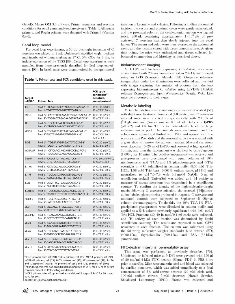
GeneEx Macro OM 3.0 software. Primer sequences and reaction
conditions for or all genes analyzed are given in Table 1. All mucin
primers, and Reg3g primers were designed with Primer3 (Version
0.4.0).
Cecal loop modelFor cecal loop experiments, a 50 uL overnight inoculum of C.
rodentium was placed in 3 mL Dulbecco’s modified eagle medium
and incubated without shaking at 37uC, 5% CO2 for 3 hrs, to
induce expression of the T3SS [89]. Cecal loop experiments were
modified from those previously described for ileal loop experi-
ments [90]. In brief, mice were anaesthetized by intraperitoneal
injection of ketamine and xylazine. Following a midline abdominal
incision, the cecum and proximal colon were gently exteriorized,
and the proximal colon at the cecal-colonic junction was ligated
twice. 300 uL containing approximately 16108 cfu of pre-
activated C. rodentium was then slowly injected into the cecal
lumen. The cecum and colon were then returned to the abdominal
cavity and the incision closed with discontinuous sutures. At given
time points, the mice were euthanized and tissues collected for
bacterial enumeration and histology as described above.
Bioluminescent imagingAt 4 DPI with luciferase expressing C. rodentium, mice were
anaesthetized with 2% isofluorane carried in 2% O2 and imaged
using an IVIS (Xenogen; Almeda, CA). Greyscale reference
images taken under low illumination were collected and overlaid
with images capturing the emission of photons from the lux-
expressing bioluminescent C. rodentium using LIVING IMAGE
software (Xenogen) and Igor (Wavemetrics; Seattle, WA). Live
mice were returned to their cages.
Metabolic labelingMetabolic labeling was carried out as previously described [52]
with slight modifications. Uninfected (LB treated) and C. rodentium-
infected mice were injected intraperitoneally with 20 mCi of
[3H]glucosamine (Amersham) in 0.3 ml of Dulbeccos(D)-PBS
(pH 7.2) and left for 3.5 hrs to metabolically label the large
intestinal mucin pool. The animals were euthanized, and the
colons were excised and flushed with PBS, and opened with fine
scissors into a Petri dish and the mucosal surface was scraped with
a glass slide to remove the adherent mucus. Mucosal secretions
were placed in 15–20 ml of D-PBS and vortexed at high speed for
10 min, and then the supernatant was clarified by centrifugation
(1,000 g for 10 min). The cell-free supernatant was reserved and
glycoproteins were precipitated with equal volumes of 10%
trichloroacetic acid (TCA) and 1% phosphotungstic acid (PTA)
overnight at 4uC, solubilized in column buffer (8.06 mM Tris-
HCL, 1.98 mM Tris- base, 0.001% sodium azide, pH 8.0) and
neutralized to pH 7.0–7.4 with 0.1 mol/l NaOH. 5 ml of
scintillation cocktail (UniverSol) was added, and 3H activity (a
measure of mucus secretion) was determined in a scintillation
counter. To confirm the identity of the high-molecular-weight
mucin following C. rodentium infection, the secreted [3H]glucos-
amine-labeled glycoproteins produced in response C. rodentium and
untreated controls were subjected to Sepharose-4B (Sigma)
column chromatography. To do this, the 10% TCA-1% PTA-
precipitated glycoproteins were dissolved in column buffer and
applied to a S4B column previously equilibrated with 0.01 mol/l
Tris HCl. Fractions (30–40 in total/0.4 ml each) were collected,
and 3H activity of each fraction was determined by liquid
scintillation counting. The results are expressed as total CPM
recovered in each fraction. The column was calibrated using
the following molecular weights standards: blue dextran (BD;
2,000 kDa), thyroglobulin (669 kDa) and BSA (67 kDa)
(Amersham).
FITC-dextran intestinal permeability assayThis assay was performed as previously described [75].
Uninfected or infected mice at 5 DPI were gavaged with 150 ml
of 80 mg/ml 4 kDa FITC-dextran (Sigma; FD4) in PBS 4 hrs
prior to sacrifice. Mice were anaesthetized and blood was collected
by cardiac punctures, which was added immediately to a final
concentration of 3% acid-citrate dextrose (20 mM citric acid,
100 nM sodium citrate, 5 mM dextrose) (Harald Schulze,
Shivdasani Laboratory, DFCI). Plasma was collected and
Table 1. Primer sets and PCR conditions used in this study.
TargetmRNAa Primer Sets
PCR cycleconditionsb
denature/anneal/extend
IFN-c Fwd: 59- TCAAGTGGCATAGATGTGGAAGAA -39
Rev: 59-TGGCTCTGCAGGATTTTCATG -39
95uC, 30 s/60uC,30 s/72uC, 30 s
TNF-a Fwd: 59- CATCTTCTCAAAATTCGAGTGACAA -39
Rev: 59- TGGGAGTAGACAAGGTACAACCC-39
94uC, 30 s/55uC,30 s/72uC, 45 s
KC Fwd: 59- TGCACCCAAACCGAAGTCAT-39
Rev: 59- TTGTCAGAAGCCAGCGTTCAC-39
94uC, 30 s/57uC,30 s/72uC, 45 s
MCP-1c Fwd: 59-TGCTACTCATTAACCAGCAAGAT -39
Rev: 59-TGCTTGAGGTGGTTGTGGAA -39
94uC, 30 s/59uC,15 s/72uC, 90 s+78uC, 5 s
iNOS Fwd: 59- TGGGAATGGAGACTGTCCCAG-39
Rev: 59- GGGATCTGAATGTGATGTTTG-39
94uC, 30 s/60uC,30 s/72uC, 30 s
mCRAMP Fwd: 59- CTTCAACCAGCAGTCCCTAGACA-39
Rev: 59- TCCAGGTCCAGGAGACGGTA-39
94uC, 30 s/55uC,30 s/72uC, 30 s
b-actin Fwd: 59-CAGCTTCTTTGCAGCTCCTT-39
Rev: 59-CTTCTCCATGTCGTCCCAGT-39
94uC, 30 s/55–60uC,30 s/72uC, 30 s
IL-17A Fwd: 59-GCTCCAGAAGGCCCTCAGA-39
Rev: 59-CTTTCCCTCCGCATTGACA-39
94uC, 30 s/60uC,30 s/72uC, 30 s
IL-17F Fwd: 59-TGCTACTGTTGATGTTGGGAC-39
Rev: 59-AATGCCCTGGTTTTGGTTGAA-39
94uC, 30 s/55uC,30 s/72uC, 45 s
IL-22 Fwd: 59-ACCTTTCCTGACCAAACTCA-39
Rev: 59-AGCTTCTTCTCGCTCAGACG-39
94uC, 30 s/58uC,30 s/72uC, 30 s
IL-23p19 Fwd: 59-TGGCTGTGCCTAGGAGTAGCA -39
Rev: 59-TTCATCCTCTTCTTCTCTTAGTAGATT -39
94uC, 30 s/60uC,30 s/72uC, 30 s
RegIII-c Fwd: 59-TGCCTATGGCTCCTATTGCT-39
Rev: 59-CACTCCCATCCACCTCTGTT-39
94uC, 30 s/58uC,30 s/72uC, 30 s
Muc1 Fwd: 59-AGGAGGTTTCGGCAGGTAAT-39
Rev: 59-TCCTTCTGAGAGCCACCACT-39
94uC, 30 s/55uC,30 s/72uC, 45 s
Muc3/17 Fwd: 59-TGAGCAAAGGCAGTATCGTG-39
Rev: 59-GCCTCCTTCTTGCATGTCTC-39
94uC, 30 s/55uC,30 s/72uC, 45 s
Muc4 Fwd: 59-GAAAAGCGTGTTGCCTCTTC-39
Rev: 59-AGAGGGAAATGCCCTGATCT-39
94uC, 30 s/55uC,30 s/72uC, 45 s
Muc6 Fwd: 59-TGCATGCTCAATGGTATGGT-39
Rev: 59-TGTGGGCTCTGGAGAAGAGT-39
94uC, 30 s/55uC,30 s/72uC, 45 s
Muc13 Fwd: 59-TCTGGACTCTGGCCACTCTT-39
Rev: 59-GAGGACAGAGCCAGTCCAAG-39
94uC, 30 s/55uC,30 s/72uC, 45 s
Muc19 Fwd: 59-ACTGGAACCACAGCCAAATC-39
Rev: 59-CTACGGCCTGTTTTTCGGTA-39
94uC, 30 s/55uC,30 s/72uC, 45 s
aIFN-c primers from ref. [94]; TNF-a primers, ref. [95]; MCP-1 primers, ref. [96];mCRAMP primers, ref. [13]; iNOS primers, ref. [97]; KC primers, ref. [98]; IL-17Aand IL-23p19 ref. [99]; IL-17F primers ref. [36]; and IL-22 primers ref. [100].bAll PCR experiments had an initial denaturing step of 95uC for 3–5 mins beforecommencement of PCR cycling conditions.cMCP1 primers after 40 cycles had an additional 2 steps of 94uC for 30 s, and50uC for 30 s.doi:10.1371/journal.ppat.1000902.t001
Muc2 Is Protective during A/E Bacterial Infection
PLoS Pathogens | www.plospathogens.org 21 May 2010 | Volume 6 | Issue 5 | e1000902

fluorescence was quantified using a Wallace Victor (Perkin-Elmer
Life Sciences, Boston, MA) at excitation 485 nm, emission 530 nm
for 0.1 s.
Fluorescence in-situ hybridizationFormalin-fixed paraffin-embedded sections were deparaffinized
and rehydrated as described above. Sections were incubated
overnight at 37uC in the dark with Texas red-conjugated EUB338
general bacterial probe (59-GCT GCC TCC CGT AGG AGT-39)
and an AlexaFluor 488 conjugated GAM42a probe (59-GCC
TTC CCA CAT CGT TT-39) that recognizes bacteria that belong
to the c-Proteobacter class [58,91] diluted to a final concentration
of 2.5 ng/ul each in hybridization solution (0.9 M NaCL, 0.1 M
TRIS pH 7.2, 30% Formamide, 0.1% SDS). Sections were then
washed once in the dark with hybridization solution for 15 minutes
with gentle shaking. This step was repeated once with wash buffer
(0.9 M NaCL, 0.1 M TRIS pH 7.2), and sections were placed in
dH2O, and then mounted using ProLong Gold Antifade reagent
with DAPI (Molecular Probes) and imaged as described above. For
quantification studies, the methods were carried as previously
described [58].
SYBR green DNA stainingLarge intestines were collected and prepared as described above
for bacterial counts, except the lumen contents from the cecum
and colon were separated. After homogenization, samples were
diluted 1:10 in PBS, then 450 ul of the 1:10 dilution was placed in
50 ul 10% Neutral Buffered Formalin, vortexed briefly, and stored
at 4¡C. 2–5 ul of the 1:10 diluted sample stored in formalin was
diluted in 1 ml PBS and filtered onto Anodisc 25 filters (Whatman
International Ltd) with a pore size of 0.2 mM and 2.5 cm
diameter. The samples were allowed to thoroughly dry, and then
were stained with 0.25 ml SYBR green (Invitrogen) in 100 ml PBS
for 15 min in the dark. Alternatively, samples were filtered onto
Nucleopore Track-Etch membranes (Whatman) for DAPI staining
only. The filters were air dried (in the dark for SYBR staining) and
mounted on glass slides with ProLong Gold Antifade reagent with
DAPI (Molecular Probes) and viewed as above. The mean number
of cells counted in 3 to 6 randomly chosen fields per disc was
determined.
Antimicrobial assayCrude mucus was isolated from colorectal tissues in the same
manner as described for the small intestine by by Meyer-Hoffert
et al. [48]. Resected colons from WT mice were flushed gently with
PBS using a pippette fitted to a syringe. Colons were then opened
up longitudinally and placed in a Petri dish, mucosa side up. The
round edge of forceps was then used to gently scrape off the inner
colonic mucus layer with minimal damage to the epithelial surface.
The mucus globule was placed in a tube, diluted 1:1 with PBS, and
mixed well by vortexing and pipetting up-and-down, and then
immediately placed on ice. For the antimicrobial assay we
conducted assays described by Turner et al. [92] with slight
modifications. An overnight culture of streptomycin-resistant C.
rodentium grown in LB was diluted 1:1000 in Tryptic Soy Broth
(TSB) and grown to mid log phase (OD620 0.6–1.0). The bacteria
was washed by centrifugation (3000 rpm, 4uC, 10 mins) and
removing the supernatant, and resuspending the pellet in ice cold
10 mM sodium phosphate buffer (SPB) (pH 7.4). This step was
repeated once. The washed sample was diluted to a final OD620 of
0.7, diluted 10006, and 5 uL of this dilution (containing <16104
bacteria) were added to 25 ul 10 mM SPB with 0.03% TSB
containing 50 ug/ml streptomycin +/220 ul of various dilutions
of crude mucin as described in the text. For negative controls, only
SPB + streptomycin was added. The total reaction volume was
50 ul. Cultures were left for 3 hrs at room temp, then serially
diluted and plated on LB plates containing 50 ug/ml streptomy-
cin, and incubated overnight at 37uC incubator. Colonies were
counted the next day.
Histopathological scoringTo assess tissue pathology, we used a scoring system adapted from
previously described scoring systems [88,93]. In brief, paraffin-
embedded colonic tissue sections (5 mm) that had been stained with
haematoxylin and eosin were examined by two blinded observers.
Tissue sections were assessed for submucosal edema (0 = no change;
1 = mild; 2 = moderate; 3 = profound), epithelial hyperplasia
(scored based on percentage above the height of the control where
0 = no change; 1 = 1–50%; 2 = 51–100%; 3 = .100%), epithelial
integrity (0 = no change; 1 = ,10 epithelial cells shedding per lesion;
2 = 11–20 epithelial cells shedding per lesion; 3 = epithelial
ulceration; 4 = epithelial ulceration with severe crypt destruction);
neutrophil and mononuclear cell infiltration (0 = none; 1 = mild; 2 =
moderate; 3 = severe). The maximum score that could result from
this scoring was 15.
Statistical analysisStatistical significance was calculated by using either a two-
tailed Student’s t-test or the Mann-Whitney test unless otherwise
indicated, with assistance from GraphPad Prism Software Version
4.00 (GraphPad Software, San Diego California USA, www.
graphpad.com). A P value of #0.05 was considered significant.
The results are expressed as the mean value with standard error of
the mean (SEM).
Gene accession numbersThe following are the GeneIDs (Database: Entrez Gene) for
each gene analyzed in this manuscript, given as gene name (official
symbol GeneID #): TNF-a (Tnf GeneID: 21926); IL-23p19
(Il23a GeneID: 83430); IFN-c (Ifng GeneID: 15978); IL-17A
(Il17a GeneID: 16171), IL-17F (Il17f GeneID: 257630); IL-22
(Il22 GeneID: 50929); MCP-1 (Ccl2 GeneID: 20296); KC
(Cxcl1 GeneID: 14825); iNOS (Nos2 GeneID: 18126)
mCRAMP (Camp GeneID: 12796); Muc1 (Muc1 GeneID:17829), Muc2 (Muc2 GeneID: 17831); Muc3/17 (Muc3
GeneID: 666339); Muc4 (Muc4 GeneID: 140474); Muc6
(GeneID: 353328); Muc13 (Muc13 GeneID: 17063); and
Muc19 (Muc19 GeneID: 239611).
Supporting Information
Figure S1 Characterization of the inflammatory cell infiltrate
within the colons of C. rodentium-infected WT and Muc22/2 mice.
A. Immunostaining for infiltrating macrophages via F4/80
staining (top panels) and neutrophils via MPO staining (bottom
panels) in descending colons of WT and Muc22/2 and mice.
Original magnification = 2006. Scale Bar = 50 mm. B. Quanti-
tative PCR analysis of pro-inflammatory chemokines and
cytokines in the descending colons of WT and Muc22/2 mice at
6 DPI compared to their respective uninfected controls. Results
averaged from 3 independent infections, with n = 2–4 mice per
group. Error bars = SEM. C. Quantitative PCR analysis of genes
that are associated with host-susceptibility to C. rodentium in the
colons of WT and Muc22/2 mice at 6 DPI. Results are averaged
from 4–5 mice per group, pooled from 2 independent infections.
Error Bars = SEM. D. MPO staining as above in an ulcerated
region of an infected Muc22/2 mouse, showing a dense population
of neutrophils in direct contact with a large microcolony of C.
Muc2 Is Protective during A/E Bacterial Infection
PLoS Pathogens | www.plospathogens.org 22 May 2010 | Volume 6 | Issue 5 | e1000902

rodentium (white asterisk, C. rodentium aggregate; arrowhead, MPO
positive cell in indirect contact with the microcolony). Original
magnification = 2006. Scale Bar = 50 mm.
Found at: doi:10.1371/journal.ppat.1000902.s001 (8.58 MB TIF)
Figure S2 Analysis of Muc family gene expression and overall
mucin content in colorectal tissues of uninfected or C. rodentium-
infected WT and Muc22/2 mice. A. Quantitative PCR analysis of
expression of genes encoding various Muc family members in the
rectal tissues of WT and Muc22/2 mice under uninfected or
infected (6 DPI) conditions. Results are presented as the average of
4–5 mice per group pooled from 2 independent infections. B. PAS
staining of Carnoy’s-fixed colorectal tissues of WT and Muc22/2
under uninfected or C. rodentium-infected (6 DPI) conditions. Very
little mucin staining (magenta, arrows) can be seen in the
epithelium or lumens of uninfected or infected Muc22/2 prior to
or during infection. Results are representative of at least 3
independent infections with 2–3 mice per group. Original
magnification = 1006. Scale bar = 100 mm.
Found at: doi:10.1371/journal.ppat.1000902.s002 (4.68 MB TIF)
Acknowledgments
We are grateful to Dr. Laura Sly and Dr. Lisa Mansson for their critical
reading of this manuscript. We also gratefully acknowledge Dr. Jan Dekker
for providing the anti-murine Muc2 antisera, and Dr. Wanyin Deng for
providing the anti-C. rodentium Tir antisera, and Dr. Lisa Mansson for her
help with the culturing experiments.
Author Contributions
Conceived and designed the experiments: KSBB BBF KC BAV.
Performed the experiments: KSBB VKS CM MM HPS NR TH BAV.
Analyzed the data: KSBB VKS KC BAV. Contributed reagents/
materials/analysis tools: DLG AV BBF KC. Wrote the paper: KSBB BAV.
References
1. Nataro JP, Kaper JB (1998) Diarrheagenic Escherichia coli. Clin Microbiol Rev
11: 142–201.
2. Fagundes-Neto U, Andrade JABd (1999) Acute Diarrhea and Malnutrition:
Lethality Risk in Hospitalized Infants. J Am Coll Nutr 18: 303–308.
3. Vilchez S, Reyes D, Paniagua M, Bucardo F, Mollby R, et al. (2009) Prevalence
of diarrhoeagenic Escherichia coli in children from Leon, Nicaragua. J Med
Microbiol 58: 630–637.
4. Gonzalez Garcia EA (2002) Animal health and foodborne pathogens:
enterohaemorrhagic O157:H7 strains and other pathogenic Escherichia coli
virotypes (EPEC, ETEC, EIEC, EHEC). Pol J Vet Sci 5: 103–115.
5. Karch H, Tarr PI, Bielaszewska M (2005) Enterohaemorrhagic Escherichia coli
in human medicine. International Journal of Medical Microbiology 295:
405–418.
6. Yoon JW, Hovde CJ (2008) All blood, no stool: enterohemorrhagic Escherichia
coli O157:H7 infection. J Vet Sci 9: 219–231.
7. Wales AD, Woodward MJ, Pearson GR (2005) Attaching-effacing bacteria in
animals. J Comp Pathol 132: 1–26.
8. Vallance BA, Finlay BB (2000) Exploitation of host cells by enteropathogenic
Escherichia coli. Proc Natl Acad Sci U S A 97: 8799–8806.
9. Luperchio SA, Schauer DB (2001) Molecular pathogenesis of Citrobacter
rodentium and transmissible murine colonic hyperplasia. Microbes Infect 3:
333–340.
10. Mundy R, MacDonald TT, Dougan G, Frankel G, Wiles S (2005) Citrobacter
rodentium of mice and man. Cell Microbiol 7: 1697–1706.
11. Bry L, Brenner MB (2004) Critical Role of T Cell-Dependent Serum Antibody,
but Not the Gut-Associated Lymphoid Tissue, for Surviving Acute Mucosal
Infection with Citrobacter rodentium, an Attaching and Effacing Pathogen.
J Immunol 172: 433–441.
12. Maaser C, Housley MP, Iimura M, Smith JR, Vallance BA, et al. (2004)
Clearance of Citrobacter rodentium Requires B Cells but Not Secretory
Immunoglobulin A (IgA) or IgM Antibodies. Infect Immun 72: 3315–3324.
13. Iimura M, Gallo RL, Hase K, Miyamoto Y, Eckmann L, et al. (2005)
Cathelicidin mediates innate intestinal defense against colonization with
epithelial adherent bacterial pathogens. J Immunol 174: 4901–4907.
14. Vallance BA, Dijkstra G, Qiu B, van der Waaij LA, van Goor H, et al. (2004)
Relative contributions of NOS isoforms during experimental colitis: endothelial-
derived NOS maintains mucosal integrity. Am J Physiol Gastrointest Liver
Physiol 287: G865–874.
15. Linden SK, Sutton P, Karlsson NG, Korolik V, McGuckin MA (2008) Mucins
in the mucosal barrier to infection. Mucosal Immunol 1: 183–197.
16. Dharmani P, Srivastava V, Kissoon-Singh V, Chadee K (2009) Role of Intestinal
Mucins in Innate Host Defense Mechanisms against Pathogens. Journal of
Innate Immunity 1: 123–135.
17. Lievin-Le Moal V, Servin AL (2006) The Front Line of Enteric Host Defense
against Unwelcome Intrusion of Harmful Microorganisms: Mucins, Antimicro-
bial Peptides, and Microbiota. Clin Microbiol Rev 19: 315–337.
18. McAuley JL, Linden SK, Png CW, King RM, Pennington HL, et al. (2007)
MUC1 cell surface mucin is a critical element of the mucosal barrier to infection.
J Clin Invest 117: 2313–2324.
19. Linden SK, Florin TH, McGuckin MA (2008) Mucin dynamics in intestinal
bacterial infection. PLoS One 3: e3952.
20. Mack DR, Michail S, Wei S, McDougall L, Hollingsworth MA (1999) Probiotics
inhibit enteropathogenic E.{coli adherence in vitro by inducing intestinal mucin
gene expression. Am J Physiol Gastrointest Liver Physiol 276: G941–950.
21. Allen A, Hutton DA, Pearson JP (1998) The MUC2 gene product: a human
intestinal mucin. The International Journal of Biochemistry & Cell Biology 30:
797–801.
22. Van Klinken BJ-W, Einerhand AWC, Duits LA, Makkink MK, Tytgat KMAJ,
et al. (1999) Gastrointestinal expression and partial cDNA cloning of murine
Muc2. Am J Physiol Gastrointest Liver Physiol 276: G115–124.
23. Specian RD, Oliver MG (1991) Functional biology of intestinal goblet cells.
Am J Physiol 260: C183–193.
24. Johansson MEV, Thomsson KA, Hansson GC (2009) Proteomic Analyses of the
Two Mucus Layers of the Colon Barrier Reveal That Their Main Component,
the Muc2 Mucin, Is Strongly Bound to the Fcgbp Protein. Journal of Proteome
Research 8: 3549–3557.
25. Atuma C, Strugala V, Allen A, Holm L (2001) The adherent gastrointestinal
mucus gel layer: thickness and physical state in vivo. Am J Physiol Gastrointest
Liver Physiol 280: G922–929.
26. Matsuo K, Ota H, Akamatsu T, Sugiyama A, Katsuyama T (1997)
Histochemistry of the surface mucous gel layer of the human colon. Gut 40:
782–789.
27. Johansson ME, Phillipson M, Petersson J, Velcich A, Holm L, et al. (2008) The
inner of the two Muc2 mucin-dependent mucus layers in colon is devoid of
bacteria. Proc Natl Acad Sci U S A 105: 15064–15069.
28. Velcich A, Yang W, Heyer J, Fragale A, Nicholas C, et al. (2002) Colorectal
cancer in mice genetically deficient in the mucin Muc2. Science 295:
1726–1729.
29. Van der Sluis M, De Koning BA, De Bruijn AC, Velcich A, Meijerink JP, et al.
(2006) Muc2-deficient mice spontaneously develop colitis, indicating that MUC2
is critical for colonic protection. Gastroenterology 131: 117–129.
30. Yang K, Popova NV, Yang WC, Lozonschi I, Tadesse S, et al. (2008)
Interaction of Muc2 and Apc on Wnt signaling and in intestinal tumorigenesis:
potential role of chronic inflammation. Cancer Res 68: 7313–7322.
31. Yang W, Velcich A, Lozonschi I, Liang J, Nicholas C, et al. (2005) Inactivation
of p21WAF1/cip1 enhances intestinal tumor formation in Muc22/2 mice.
Am J Pathol 166: 1239–1246.
32. Heazlewood CK, Cook MC, Eri R, Price GR, Tauro SB, et al. (2008) Aberrant
mucin assembly in mice causes endoplasmic reticulum stress and spontaneous
inflammation resembling ulcerative colitis. PLoS Med 5: e54.
33. Deplancke B, Gaskins HR (2001) Microbial modulation of innate defense: goblet
cells and the intestinal mucus layer. Am J Clin Nutr 73: 1131S–1141S.
34. Swidsinski A, Loening-Baucke V, Theissig F, Engelhardt H, Bengmark S, et al.
(2007) Comparative study of the intestinal mucus barrier in normal and inflamed
colon. Gut 56: 343–350.
35. Wiles S, Pickard KM, Peng K, MacDonald TT, Frankel G (2006) In Vivo
Bioluminescence Imaging of the Murine Pathogen Citrobacter rodentium. Infect
Immun 74: 5391–5396.
36. Ishigame H, Kakuta S, Nagai T, Kadoki M, Nambu A, et al. (2009) Differential
Roles of Interleukin-17A and -17F in Host Defense against Mucoepithelial
Bacterial Infection and Allergic Responses. Immunity 30: 108–119.
37. Zheng Y, Valdez PA, Danilenko DM, Hu Y, Sa SM, et al. (2008) Interleukin-22
mediates early host defense against attaching and effacing bacterial pathogens.
Nat Med 14: 282–289.
38. Mangan PR, Harrington LE, O’Quinn DB, Helms WS, Bullard DC, et al.
(2006) Transforming growth factor-[beta] induces development of the TH17
lineage. Nature 441: 231–234.
39. Bry L, Brigl M, Brenner MB (2006) CD4+-T-Cell Effector Functions and
Costimulatory Requirements Essential for Surviving Mucosal Infection with
Citrobacter rodentium. Infect Immun 74: 673–681.
40. Nagai T, Abe A, Sasakawa C (2005) Targeting of Enteropathogenic Escherichia
coli EspF to Host Mitochondria Is Essential for Bacterial Pathogenesis:
CRITICAL ROLE OF THE 16TH LEUCINE RESIDUE IN EspF. J Biol
Chem 280: 2998–3011.
Muc2 Is Protective during A/E Bacterial Infection
PLoS Pathogens | www.plospathogens.org 23 May 2010 | Volume 6 | Issue 5 | e1000902

41. Torchinsky MB, Garaude J, Martin AP, Blander JM (2009) Innate immune
recognition of infected apoptotic cells directs TH17 cell differentiation. Nature458: 78–82.
42. Gibson DL, Ma C, Rosenberger CM, Bergstrom KS, Valdez Y, et al. (2008)
Toll-like receptor 2 plays a critical role in maintaining mucosal integrity duringCitrobacter rodentium-induced colitis. Cell Microbiol 10: 388–403.
43. Deng W, Puente JL, Gruenheid S, Li Y, Vallance BA, et al. (2004) Dissecting
virulence: systematic and functional analyses of a pathogenicity island. Proc Natl
Acad Sci U S A 101: 3597–3602.
44. Bergstrom KS, Guttman JA, Rumi M, Ma C, Bouzari S, et al. (2008)Modulation of intestinal goblet cell function during infection by an attaching and
effacing bacterial pathogen. Infect Immun 76: 796–811.
45. Smith CJ, Kaper JB, Mack DR (1995) Intestinal mucin inhibits adhesion ofhuman enteropathogenic Escherichia coli to HEp-2 cells. J Pediatr Gastroenterol
Nutr 21: 269–276.
46. Deng W, Vallance BA, Li Y, Puente JL, Finlay BB (2003) Citrobacter rodentium
translocated intimin receptor (Tir) is an essential virulence factor needed foractin condensation, intestinal colonization and colonic hyperplasia in mice. Mol
Microbiol 48: 95–115.
47. Vallance BA, Deng W, De Grado M, Chan C, Jacobson K, et al. (2002)Modulation of inducible nitric oxide synthase expression by the attaching and
effacing bacterial pathogen citrobacter rodentium in infected mice. InfectImmun 70: 6424–6435.
48. Meyer-Hoffert U, Hornef MW, Henriques-Normark B, Axelsson LG,Midtvedt T, et al. (2008) Secreted enteric antimicrobial activity localises to
the mucus surface layer. Gut 57: 764–771.
49. Mantle M, Husar SD (1993) Adhesion of Yersinia enterocolitica to purifiedrabbit and human intestinal mucin. Infect Immun 61: 2340–2346.
50. Lievin-Le Moal V, Servin AL, Coconnier-Polter MH (2005) The increase in
mucin exocytosis and the upregulation of MUC genes encoding for membrane-bound mucins induced by the thiol-activated exotoxin listeriolysin O is a host cell
defence response that inhibits the cell-entry of Listeria monocytogenes. Cell
Microbiol 7: 1035–1048.
51. Enss ML, Muller H, Schmidt-Wittig U, Kownatzki R, Coenen M, et al. (1996)Effects of perorally applied endotoxin on colonic mucins of germfree rats.
Scand J Gastroenterol 31: 868–874.
52. Caballero-Franco C, Keller K, De Simone C, Chadee K (2007) The VSL#3probiotic formula induces mucin gene expression and secretion in colonic
epithelial cells. Am J Physiol Gastrointest Liver Physiol 292: G315–322.
53. Tytgat KM, Buller HA, Opdam FJ, Kim YS, Einerhand AW, et al. (1994)
Biosynthesis of human colonic mucin: Muc2 is the prominent secretory mucin.Gastroenterology 107: 1352–1363.
54. Tytgat KM, Opdam FJ, Einerhand AW, Buller HA, Dekker J (1996) MUC2 is
the prominent colonic mucin expressed in ulcerative colitis. Gut 38: 554–563.
55. Rong M, Rossi EA, Zhang J, McNeer RR, van den Brande JM, et al. (2005)Expression and localization of Muc4/sialomucin complex (SMC) in the adult
and developing rat intestine: implications for Muc4/SMC function. J Cell
Physiol 202: 275–284.
56. Das B, Cash MN, Hand AR, Shivazad A, Grieshaber SS, et al. TissueDistibution of Murine Muc19/Smgc Gene Products. J Histochem Cytochem 58:
141–156.
57. Escande F, Porchet N, Bernigaud A, Petitprez D, Aubert J-P, et al. (2004) Themouse secreted gel-forming mucin gene cluster. Biochimica et Biophysica Acta
(BBA) - Gene Structure and Expression 1676: 240–250.
58. Lupp C, Robertson ML, Wickham ME, Sekirov I, Champion OL, et al. (2007)
Host-mediated inflammation disrupts the intestinal microbiota and promotes theovergrowth of Enterobacteriaceae. Cell Host Microbe 2: 204.
59. Hoffmann C, Hill DA, Minkah N, Kirn T, Troy A, et al. (2009) Community-
Wide Response of the Gut Microbiota to Enteropathogenic Citrobacterrodentium Infection Revealed by Deep Sequencing. Infect Immun 77:
4668–4678.
60. Khan MA, Bouzari S, Ma C, Rosenberger CM, Bergstrom KS, et al. (2008)
Flagellin-dependent and -independent inflammatory responses following infec-tion by enteropathogenic Escherichia coli and Citrobacter rodentium. Infect
Immun 76: 1410–1422.
61. Grys TE, Walters LL, Welch RA (2006) Characterization of the StcE proteaseactivity of Escherichia coli O157:H7. J Bacteriol 188: 4646–4653.
62. Hooper LV (2009) Do symbiotic bacteria subvert host immunity? Nat Rev
Microbiol 7: 367–374.
63. Viswanathan VK, Koutsouris A, Lukic S, Pilkinton M, Simonovic I, et al. (2004)
Comparative Analysis of EspF from Enteropathogenic and EnterohemorrhagicEscherichia coli in Alteration of Epithelial Barrier Function. Infect Immun 72:
3218–3227.
64. Gunning RF, Wales AD, Pearson GR, Done E, Cookson AL, et al. (2001)Attaching and effacing lesions in the intestines of two calves associated with
natural infection with Escherichia coli O26:H11. Vet Rec 148: 780–782.
65. Ma C, Wickham ME, Guttman JA, Deng W, Walker J, et al. (2006) Citrobacter
rodentium infection causes both mitochondrial dysfunction and intestinal epithelialbarrier disruption in vivo: role of mitochondrial associated protein (Map). Cellular
Microbiology 8: 1669–1686.
66. Dann SM, Spehlmann ME, Hammond DC, Iimura M, Hase K, et al. (2008) IL-6-Dependent Mucosal Protection Prevents Establishment of a Microbial Niche
for Attaching/Effacing Lesion-Forming Enteric Bacterial Pathogens. J Immunol
180: 6816–6826.
67. Guttman JA, Li Y, Wickham ME, Deng W, Vogl AW, et al. (2006) Attaching
and effacing pathogen-induced tight junction disruption in vivo. Cellular
Microbiology 8: 634–645.
68. Stecher Br, Robbiani R, Walker AW, Westendorf AM, Barthel M, et al. (2007)
Salmonella enterica Serovar Typhimurium Exploits Inflammation to Compete
with the Intestinal Microbiota. PLoS Biol 5: e244.
69. Wiles S, Pickard KM, Peng K, MacDonald TT, Frankel G (2006) In vivo
bioluminescence imaging of the murine pathogen Citrobacter rodentium. Infect
Immun 74: 5391–5396.
70. Smirnova MG, Guo L, Birchall JP, Pearson JP (2003) LPS up-regulates mucin
and cytokine mRNA expression and stimulates mucin and cytokine secretion in
goblet cells. Cell Immunol 221: 42–49.
71. Iwashita J, Sato Y, Sugaya H, Takahashi N, Sasaki H, et al. (2003) mRNA of
MUC2 is stimulated by IL-4, IL-13 or TNF-[alpha] through a mitogen-activated
protein kinase pathway in human colon cancer cells. Immunol Cell Biol 81:
275–282.
72. Plaisancie P, Barcelo A, Moro F, Claustre J, Chayvialle J-A, et al. (1998) Effects
of neurotransmitters, gut hormones, and inflammatory mediators on mucus
discharge in rat colon. Am J Physiol Gastrointest Liver Physiol 275:
G1073–1084.
73. Kim S, Nadel JA (2004) Role of neutrophils in mucus hypersecretion in COPD
and implications for therapy. Treat Respir Med 3: 147–159.
74. Lebeis SL, Bommarius B, Parkos CA, Sherman MA, Kalman D (2007) TLR
signaling mediated by MyD88 is required for a protective innate immune
response by neutrophils to Citrobacter rodentium. J Immunol 179: 566–577.
75. Gibson DL, Ma C, Bergstrom KS, Huang JT, Man C, et al. (2008) MyD88
signalling plays a critical role in host defence by controlling pathogen burden
and promoting epithelial cell homeostasis during Citrobacter rodentium-induced
colitis. Cell Microbiol 10: 618–631.
76. Conlin VS, Wu X, Nguyen C, Dai C, Vallance BA, et al. (2009) Vasoactive
Intestinal Peptide Ameliorates Intestinal Barrier Disruption Associated with
Citrobacter rodentium-induced Colitis. Am J Physiol Gastrointest Liver Physiol.
77. Lind!�n SK, Sheng YH, Every AL, Miles KM, Skoog EC, et al. (2009) MUC1
Limits Helicobacter pylori Infection both by Steric Hindrance and by Acting as a
Releasable Decoy. PLoS Pathog 5: e1000617.
78. Hoebler C, Gaudier E, De Coppet P, Rival M, Cherbut C (2006) MUC Genes
Are Differently Expressed During Onset and Maintenance of Inflammation in
Dextran Sodium Sulfate-Treated Mice. Digestive Diseases and Sciences 51:
381–389.
79. Hasnain SZ, Wang H, Ghia J-E, Haq N, Deng Y, et al. Mucin Gene Deficiency
in Mice Impairs Host Resistance to an Enteric Parasitic Infection. Gastroen-
terology In Press, Accepted Manuscript.
80. Shames SR, Auweter SD, Finlay BB (2009) Co-evolution and exploitation of
host cell signaling pathways by bacterial pathogens. The International Journal of
Biochemistry & Cell Biology 41: 380–389.
81. Chessa D, Winter MG, Jakomin M, B%umler AJ (2009) Salmonella enterica
serotype Typhimurium Std fimbriae bind terminal a-03B1;(1,2)fucose residues
in the cecal mucosa. Molecular Microbiology 71: 864–875.
82. Vimal DB, Khullar M, Gupta S, Ganguly NK (2000) Intestinal mucins: the
binding sites for Salmonella typhimurium. Mol Cell Biochem 204: 107–117.
83. Mantle M, Rombough C (1993) Growth in and breakdown of purified rabbit
small intestinal mucin by Yersinia enterocolitica. Infect Immun 61: 4131–4138.
84. Stecher Br, Barthel M, Schlumberger MC, Haberli L, Rabsch W, et al. (2008)
Motility allows S. Typhimurium to benefit from the mucosal defence. Cellular
Microbiology 10: 1166–1180.
85. Chadee K, Keller K, Forstner J, Innes DJ, Ravdin JI (1991) Mucin and
nonmucin secretagogue activity of Entamoeba histolytica and cholera toxin in
rat colon. Gastroenterology 100: 986–997.
86. Lidell ME, Moncada DM, Chadee K, Hansson GC (2006) Entamoeba
histolytica cysteine proteases cleave the MUC2 mucin in its C-terminal domain
and dissolve the protective colonic mucus gel. Proc Natl Acad Sci U S A 103:
9298–9303.
87. Moncada D, Keller K, Chadee K (2003) Entamoeba histolytica cysteine
proteinases disrupt the polymeric structure of colonic mucin and alter its
protective function. Infect Immun 71: 838–844.
88. Khan MA, Ma C, Knodler LA, Valdez Y, Rosenberger CM, et al. (2006) Toll-
like receptor 4 contributes to colitis development but not to host defense during
Citrobacter rodentium infection in mice. Infect Immun 74: 2522–2536.
89. Dean P, Maresca M, Schuller S, Phillips AD, Kenny B (2006) Potent
diarrheagenic mechanism mediated by the cooperative action of three
enteropathogenic Escherichia coli-injected effector proteins. Proc Natl Acad
Sci U S A 103: 1876–1881.
90. Brown NF, Vallance BA, Coombes BK, Valdez Y, Coburn BA, et al. (2005)
Salmonella Pathogenicity Island 2 Is Expressed Prior to Penetrating the Intestine.
PLoS Pathog 1: e32.
91. Amann RI, Krumholz L, Stahl DA (1990) Fluorescent-oligonucleotide probing
of whole cells for determinative, phylogenetic, and environmental studies in
microbiology. J Bacteriol 172: 762–770.
92. Turner J, Cho Y, Dinh NN, Waring AJ, Lehrer RI (1998) Activities of LL-37, a
cathelin-associated antimicrobial peptide of human neutrophils. Antimicrob
Agents Chemother 42: 2206–2214.
93. Barthel M, Hapfelmeier S, Quintanilla-Martinez L, Kremer M, Rohde M, et al.
(2003) Pretreatment of mice with streptomycin provides a Salmonella enterica
Muc2 Is Protective during A/E Bacterial Infection
PLoS Pathogens | www.plospathogens.org 24 May 2010 | Volume 6 | Issue 5 | e1000902

serovar Typhimurium colitis model that allows analysis of both pathogen and
host. Infect Immun 71: 2839–2858.
94. Rhee SJ, Walker WA, Cherayil BJ (2005) Developmentally Regulated Intestinal
Expression of IFN-{gamma} and Its Target Genes and the Age-Specific
Response to Enteric Salmonella Infection. J Immunol 175: 1127–1136.
95. Nenci A, Becker C, Wullaert A, Gareus R, van Loo G, et al. (2007) Epithelial
NEMO links innate immunity to chronic intestinal inflammation. Nature 446:
557–561.
96. Marriott I, Gray DL, Rati DM, Fowler VG, Jr., Stryjewski ME, et al. (2005)
Osteoblasts produce monocyte chemoattractant protein-1 in a murine model of
Staphylococcus aureus osteomyelitis and infected human bone tissue. Bone 37:
504–512.
97. Sugawara I, Yamada H, Li C, Mizuno S, Takeuchi O, et al. (2003)
Mycobacterial Infection in TLR2 and TLR6 Knockout Mice. MICROBIOL-OGY and IMMUNOLOGY 47: 327–336.
98. Godinez I, Haneda T, Raffatellu M, George MD, Paixao TA, et al. (2008) T
Cells Help To Amplify Inflammatory Responses Induced by Salmonella entericaSerotype Typhimurium in the Intestinal Mucosa. Infect Immun 76: 2008–2017.
99. Happel KI, Lockhart EA, Mason CM, Porretta E, Keoshkerian E, et al. (2005)Pulmonary Interleukin-23 Gene Delivery Increases Local T-Cell Immunity and
Controls Growth of Mycobacterium tuberculosis in the Lungs. Infect Immun 73:
5782–5788.100. Brand S, Beigel F, Olszak T, Zitzmann K, Eichhorst ST, et al. (2006) IL-22 is
increased in active Crohn’s disease and promotes proinflammatory geneexpression and intestinal epithelial cell migration. Am J Physiol Gastrointest
Liver Physiol 290: G827–838.
Muc2 Is Protective during A/E Bacterial Infection
PLoS Pathogens | www.plospathogens.org 25 May 2010 | Volume 6 | Issue 5 | e1000902
Copyright © 2022 FDOKUMEN