Clinical Features and Colonic Motor Disturbances in Chronic Megacolon in Adults
10
ORIGINAL ARTICLE Clinical Features and Colonic Motor Disturbances in Chronic Megacolon in Adults Ralph Hurley O’Dwyer 1 • Andre ´s Acosta 1 • Michael Camilleri 1 • Duane Burton 1 • Irene Busciglio 1 • Adil E. Bharucha 1 Received: 5 December 2014 / Accepted: 26 March 2015 Ó Springer Science+Business Media New York 2015 Abstract Background Chronic megacolon is a rare disease of the colonic motor function characterized by a permanent in- crease in colonic diameter. Methods We reviewed electronic medical records of all patients diagnosed with chronic megacolon from 1999 to 2014 at Mayo Clinic. Our aim was to summarize clinical and motility features, including colonic compliance and tone measured by colonic barostat-controlled 10-cm-long infinitely compliant balloon. Colonic compliance curves were compared to healthy control (40) and disease (47) control groups. Results Among 24 identified patients, the mean maximal colonic diameter on abdominal radiograph was 12.7 ± 0.8 cm. The cause of megacolon was idiopathic in 16 of 24 and secondary in 8 of 24. A relatively high prevalence (10/24) of comorbid pelvic floor dyssynergia was identified. At the time of this report, 16 patients had undergone colectomy. In general, megacolon presented high fasting colonic volume at relatively low pressures (16–20 mmHg), suggesting high colonic compliance; similarly, volumes at operating pressures that ensured apposition of the balloon to the colonic wall suggested low colonic tone. Median balloon volume at 44 mmHg distension was 584 mL (IQR 556.5–600) in pa- tients with megacolon compared to 251 mL (212–281) in healthy, 240 mL (207–286) in functional constipation, and 241 mL (210.8–277.5) in diarrhea-predominant irritable bowel syndrome controls. Colon’s tonic response to feeding was generally intact, and there was frequently maintained phasic contractile response to feeding. Conclusions Chronic megacolon is a severe colonic dysmotility, manifesting radiologically with increased colonic diameter; it can be proven by measuring colonic compliance and typically requires colectomy because of failed medical therapy. Keywords Idiopathic Á Compliance Á Tone Á Manometry Á Constipation Á Barostat Introduction Chronic megacolon is characterized by a permanently en- larged diameter of the colon due to a chronic process. This abnormal diameter reflects colonic distension and manifests clinically as a chronic disorder of colonic motility, often with intractable constipation, abdominal pain, and bloating. Chronic megacolon is caused by diseases of intestinal smooth muscle cells and the enteric and extrinsic nervous systems. However, the connective tissue component of the & Michael Camilleri [email protected] Ralph Hurley O’Dwyer [email protected] Andre ´s Acosta [email protected] Duane Burton [email protected] Irene Busciglio [email protected] Adil E. Bharucha [email protected] 1 Clinical Enteric Neuroscience Translational and Epidemiological Research (C.E.N.T.E.R.), Division of Gastroenterology and Hepatology, Mayo Clinic, Charlton Bldg., Rm. 8-110, 200 First St. S.W., Rochester, MN 55905, USA 123 Dig Dis Sci DOI 10.1007/s10620-015-3645-5
Transcript of Clinical Features and Colonic Motor Disturbances in Chronic Megacolon in Adults

ORIGINAL ARTICLE
Clinical Features and Colonic Motor Disturbances in ChronicMegacolon in Adults
Ralph Hurley O’Dwyer1• Andres Acosta1
• Michael Camilleri1 • Duane Burton1•
Irene Busciglio1• Adil E. Bharucha1
Received: 5 December 2014 / Accepted: 26 March 2015
� Springer Science+Business Media New York 2015
Abstract
Background Chronic megacolon is a rare disease of the
colonic motor function characterized by a permanent in-
crease in colonic diameter.
Methods We reviewed electronic medical records of all
patients diagnosed with chronic megacolon from 1999 to
2014 at Mayo Clinic. Our aim was to summarize clinical
and motility features, including colonic compliance and
tone measured by colonic barostat-controlled 10-cm-long
infinitely compliant balloon. Colonic compliance curves
were compared to healthy control (40) and disease (47)
control groups.
Results Among 24 identified patients, the mean maximal
colonic diameter on abdominal radiograph was 12.7 ±
0.8 cm. The cause of megacolon was idiopathic in 16 of 24
and secondary in 8 of 24. A relatively high prevalence (10/24)
of comorbid pelvic floor dyssynergia was identified. At the
time of this report, 16 patients had undergone colectomy. In
general, megacolon presented high fasting colonic volume at
relatively low pressures (16–20 mmHg), suggesting high
colonic compliance; similarly, volumes at operating pressures
that ensured apposition of the balloon to the colonic wall
suggested low colonic tone. Median balloon volume at
44 mmHg distension was 584 mL (IQR 556.5–600) in pa-
tients with megacolon compared to 251 mL (212–281) in
healthy, 240 mL (207–286) in functional constipation, and
241 mL (210.8–277.5) in diarrhea-predominant irritable
bowel syndrome controls. Colon’s tonic response to feeding
was generally intact, and there was frequently maintained
phasic contractile response to feeding.
Conclusions Chronic megacolon is a severe colonic
dysmotility, manifesting radiologically with increased
colonic diameter; it can be proven by measuring colonic
compliance and typically requires colectomy because of
failed medical therapy.
Keywords Idiopathic � Compliance � Tone �Manometry �Constipation � Barostat
Introduction
Chronic megacolon is characterized by a permanently en-
larged diameter of the colon due to a chronic process. This
abnormal diameter reflects colonic distension and manifests
clinically as a chronic disorder of colonic motility, often with
intractable constipation, abdominal pain, and bloating.
Chronic megacolon is caused by diseases of intestinal
smooth muscle cells and the enteric and extrinsic nervous
systems. However, the connective tissue component of the
& Michael Camilleri
Ralph Hurley O’Dwyer
Andres Acosta
Duane Burton
Irene Busciglio
Adil E. Bharucha
1 Clinical Enteric Neuroscience Translational and
Epidemiological Research (C.E.N.T.E.R.), Division of
Gastroenterology and Hepatology, Mayo Clinic, Charlton
Bldg., Rm. 8-110, 200 First St. S.W., Rochester, MN 55905,
USA
123
Dig Dis Sci
DOI 10.1007/s10620-015-3645-5

colonic wall may also be relevant in the context of alterations
of tone and increased colonic diameter. Malfunctions of any
of these components may thus result in megacolon.
The pathogenesis of chronic megacolon has been at-
tributed to an ‘‘idiopathic’’ disorder or a heterogeneous
group of secondary causes. Examples of myopathic disor-
ders include Duchenne’s and myotonic muscular dystrophy
[1, 2], visceral myopathy, as well as degenerative
leiomyopathy, a rare condition described mainly in African
children [3]. Disorders of the enteric nervous system
causing megacolon may result from deficient migration of
neural crest cells to the developing colon, as in congenital
disorders such as Hirschsprung’s disease [4], hypogan-
glionosis and intestinal ganglioneuromatosis [5, 6], spinal
cord dysraphism [possibly causing extrinsic denervation 7].
Other acquired disorders causing megacolon include a re-
duction in the number of interstitial cells of Cajal [ICCs 8,
9] or reduction in enteric nerves, which may be idiopathic
[9] or related to Chagas disease [10]. Some diseases, such
as Parkinson’s disease, may cause either extrinsic dener-
vation or intrinsic neuropathy, resulting in megacolon [11].
There have been studies of colonic motility in patients with
chronic colonic pseudo-obstruction [12], though it is unclear
whether the reports included patients with chronically dilated
colon. Those reports document, in those with intestinal
pseudo-obstruction with colonic involvement and constipa-
tion, discrete motor abnormalities, such as the absence of
high-amplitude colonic contractions, and failure to increase
phasic contractions in postprandial period (considered con-
sistent with neuropathy) or complete absence of contractions
(suggestive of myopathy). However, no systematic analyses
of colonic compliance or tone are available to date.
The aims of this study were to appraise the demo-
graphics and clinical features, associated medical condi-
tions, and colonic tone, and compliance among a cohort of
adult patients with chronic megacolon diagnosed at Mayo
Clinic in Rochester, Minnesota, over a period of 15 years.
Materials and Methods
Search Strategy for Identifying Adults with Chronic
Megacolon
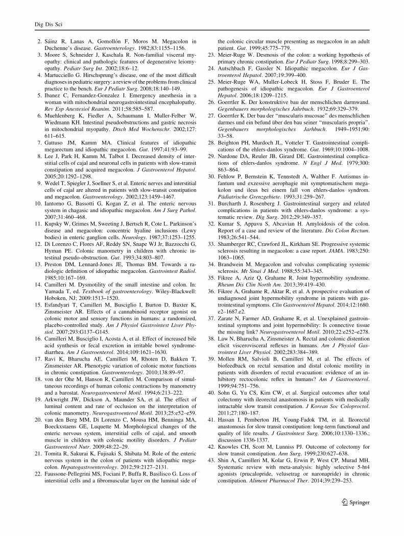
The proprietary Medical Clinical Notes Search Tool avail-
able at Mayo Clinic was used to identify adult patients with
chronic megacolon through their electronic medical records
(Fig. 1). A total of 2,800 patient records were identified with
the word ‘‘megacolon’’ in their clinical records, with 959
identified with both ‘‘megacolon’’ and ‘‘constipation’’ men-
tioned in their records. Patients who were under 18 years of
age, those who underwent surgery for chronic megacolon
prior to referral to Mayo Clinic, or those with ‘‘toxic
megacolon,’’ ‘‘acute megacolon,’’ or ‘‘Ogilvie syndrome
(also defined in the medical records as acute colonic pseudo-
obstruction)’’ were excluded; this resulted in identification
of 24 patients with chronic megacolon.
Mayo Clinic Institutional Review Board approved this
study for patients who had consented to the use of their
medical records for research.
Data Extraction
The data extracted for this study included demographics,
clinical features, family history, colon imaging, colonic
barostat-manometry data, anorectal manometry data,
treatment for megacolon, and follow-up information.
Diagnostic Criteria
All patients had at least one criterion for chronic mega-
colon based on imaging studies or intraluminal assessment
of colonic motor function. Criteria for diagnosing mega-
colon on imaging studies were: colonic diameter greater
than 6.5 cm at the pelvic brim as proposed previously [13],
greater than 8 cm in the ascending colon, or greater than
12 cm in the cecum [14]. Among seven patients in whom
imaging studies were unavailable, a fasting volume of a
10-cm colonic segment [400 mL at 44 mmHg distension
was included as megacolon. This cut-off value was deter-
mined by prior work conducted by the same research team,
based on studies of 47 patients with diarrhea-predominant
irritable bowel syndrome (IBS-D) and 40 healthy volun-
teers [15, 16]; a volume of 400 mL at 44 mmHg distension
had never been observed in these control groups.
Participants
In the study period between January 1, 1999, and June 30,
2014, we identified 24 patients diagnosed with chronic
megacolon by one of the two (MC and AEB) experienced
gastroenterologists with expertise in motility disorders.
Family history was recorded as part of the evaluation by
the staff gastroenterologists, rather than geneticists.
Measurement of Colonic Motility and Tone
Colonic motility and tone were analyzed in the ten patients
with suspected megacolon in whom there was no ra-
diological documentation of megacolon in order to ap-
praise the utility of these measurements. Colonic motor
functions were measured using intracolonic 6-lumen
manometry (5 cm apart) and a 10-cm-long infinitely
compliant balloon (Hefty Baggies, Mobil Chemical, Pitts-
ford, NY) in which infused air was maintained at constant
pressures by means of an electronic rigid piston barostat
Dig Dis Sci
123

(Engineering Department, Mayo Clinic, Rochester, MN).
This was performed as described in detail previously [17].
Before the colonic motility study, all medications with
potential effects on colonic motility were discontinued for at
least 48 h. The colon was cleansed with 2–5 L of polyethylene
glycol 3350 and electrolyte solution (Golytely�, Abbott
Laboratories, Chicago, IL). Patients fasted overnight. The next
morning, a colonic manometric-barostat assembly was posi-
tioned in the left colon by colonoscopy without sedation [17].
The study commenced after a 30-min equilibration pe-
riod. Colonic motor activity was measured as described
previously (30). After a conditioning distension, the bal-
loon was inflated from 0 to 44 mmHg in 4-mmHg steps at
30-s intervals to measure colonic compliance. Thereafter,
we measured, in order, contractile responses to a meal
(1000 kcal; 35 % carbohydrate, 53 % fat, and 12 % pro-
tein) and to neostigmine (1 mg intravenously) with the
polyethylene balloon clamped at baseline pressure and
colonic compliance with 0–44 mmHg distension as per-
formed during the fasting, baseline period.
Colonic motor activity was quantified using established
approaches. Only data recorded by the barostat are
presented in this paper because phasic pressure activity
recorded by manometry is less reliable when the colonic
diameter exceeds 5.6 cm [18], although there is evidence
that high-resolution fiber-optic manometry is capable of
recording non-occluding reductions in diameter, provided
that they occurred with sufficiently viscous content in silico
or in the rabbit colon [19]. The phasic contraction mea-
surements in our studies were not conducted in the pres-
ence of viscous colonic content, since we cleansed the
colon of stool with the bowel preparation and removed any
residual fluid at the time of colonic tube placement. Hence,
we assessed the phasic pressure activity qualitatively, not
with the same quantitative goals used in measurements of
colonic compliance and tone. This approach was also re-
inforced by the experience of other groups that were unable
to classify specific manometric findings as reflective of
myopathic or neuropathic abnormalities in patients with
colonic motility disorders [20]. The phasic contractile ac-
tivity on manometric recordings served mostly to evaluate
phasic volume events measured by barostat.
Data from 47 IBS-D patients, 46 functional constipa-
tion/constipation-predominant IBS (IBS-C) patients, and
25 patients with chronic megacolon
All patients with megacolon identifiable on barostat included
Patients with documented megacolon and constipation: all seen and diagnosed by two experienced motility consultants in Mayo Clinic
“Megacolon” referenced in note: 2,800 patients identified
Megacolon AND constipation: 959 patients identified
Radiological diagnosis
N=6 Criteria: colonic diameter >6.5cm for the
rectosigmoid and descending colon; >8cm for
the ascending colon; or >12cm for the cecum
Barostat diagnosis
N=10
Criteria: Fasting barostat volume >400mL at
44mmHg
Barostat and radiological diagnosis
N=8
Patients excluded:
- Toxic megacolon: 231- Acute megacolon: 2- Ogilvie Syndrome: 22- No megacolon(20), no evidence of
megacolon(41), no sign of megacolon(1), excluding megacolon(9)
- Patients <18 on 30 June 2014 : 92- Remaining patients manually excluded if diagnosed and treated outside of Mayo Clinic and if not reviewed in GI consultation
Mayo Clinic Rochester medical clinical notes databasesearched Jan. 1, 1999 – June 21, 2014
Fig. 1 Search strategy to identify adults with chronic megacolon
Dig Dis Sci
123

40 healthy volunteers [15, 16] acquired by the same
method in our laboratories were used as controls in order to
appraise the colonic motility measurements in the patients
with chronic megacolon.
Statistical Analysis
Data are generally presented as median (interquartile
range, IQR). Analysis of variance on ranks (Kruskal–
Wallis test) was used to compare measurements of colonic
tone and compliance in the patients with megacolon and in
controls with IBS-D and functional constipation/IBS-C,
and healthy controls.
Results
Demographics
Figure 1 shows the search strategy and identification of
patients with chronic megacolon, based on the electronic
medical record. Fifteen patients (62.5 %) were women.
Patients reported that their symptoms began in childhood
(46 %) or in adulthood (54 %). The age at diagnosis of
megacolon was 43.1 ± 4.5 (SEM) years. In patients in
whom the symptoms began in adulthood, the age at diag-
nosis was 45.8 ± 5 years. Forty-two percent of patients
had a first-generation family member with chronic consti-
pation. Among the patients with megacolon, 54.5 % re-
ported that other members of their family had reported
chronic constipation since childhood, whereas 30.8 % of
the patients reported other family members with constipa-
tion presenting in adulthood.
Associated Conditions
Eight of 24 patients (33.3 %) had another condition known
to be associated with megacolon. Five patients had a
condition associated with intrinsic or extrinsic neural in-
volvement, namely extrinsic denervation, chemotherapy-
induced neuropathy, congenital hindgut dysgenesis, and
multiple endocrine neoplasia type IIB with ganglioneuro-
matosis. Three patients had a systemic disorder affecting
connective tissue, one each with scleroderma, Ehlers–
Danlos syndrome, and transthyretin amyloidosis. No pa-
tients had a skeletal muscle myopathy.
Symptoms and Plain Abdominal Radiology
The principal symptoms reported by patients were ab-
dominal pain (83 %), bloating (96 %), and abdominal
distention (96 %) (Table 1). The median number of bowel
movements passed per week was 1.5 (IQR 1–2.5).
The largest colonic diameter in any segment of the colon
in each of the 15 individuals who had abdominal radio-
graphs on record was 12.7 ± 0.8 cm (mean ± SEM).
Anorectal Testing
Twelve patients underwent anorectal testing, including
anorectal manometry and a balloon expulsion test, because
a digital rectal examination suggested a rectal evacuation
disorder. The balloon expulsion test (which was regarded
as the primary evidence of pelvic floor dyssynergia) was
abnormal ([200 g required to expel the balloon) in eight
patients, confirming the concomitant diagnosis of pelvic
floor dyssynergia. In the other patients, there was also high
resting anal sphincter pressure ([90 mmHg).
Colonic Motility Testing
Fasting Colonic Tone and Compliance
The characteristic feature of chronic megacolon is an ex-
cessively high fasting volume (suggesting low tone) of the
infinitely compliant intracolonic balloon at operating
pressures (defined as pressures at which the infinitely
compliant balloon is able to identify respiratory variation,
typically 8–16 mmHg) (Fig. 2).
Table 1 Symptoms and signs at time of presentation to Mayo Clinic
[number (percentage) unless otherwise stated]
Clinical and radiological features
Bowel movements (BM)
Average number BM/week 1.5 (range 1–2.5)
Straining [25 % of the time 5/24 (21 %)
Sense of incomplete evacuation 5/24 (21 %)
Digital maneuvers to evacuate BM 1/24 (4 %)
Abdominal pain (%) 20/24 (84 %)
Bloating 23/24 (96 %)
Abdominal distension 23/24 (96 %)
Evidence of rectal evacuation disorder
Balloon expulsion requiring [200 g weight 8/24 (33 %)
Abnormal resting anal sphincter pressure 12/24 (50 %)
Treatments received prior to evaluation
Fiber supplementation 3/24 (13 %)
Osmotic laxative 12/24 (50 %)
Prokinetic 10/24 (42 %)
Enemas 8/24 (33 %)
Maximum diameter (cm) of colon on radiological examination
Mean 12.74 ± 0.79 (SEM)
Median 12 (IQR 10.8–13)
Evidence of rectal evacuation disorder was based on the abnormal
balloon expulsion and/or abnormal anal manometry
Dig Dis Sci
123

In addition, colonic compliance is markedly increased in
megacolon (Fig. 3). The colonic pressure–volume relation-
ships are characterized by a marked increase in volume with
a relatively small increase in the intraballoon pressure (e.g.,
from 12 to 20 mmHg) (Fig. 3). Even at 8 mmHg, the
colonic balloon volume was significantly higher in patients
with chronic megacolon (165 mL, IQR 85–301) than in
healthy controls (30 mL, IQR 28–32), IBS-D (34 mL, IQR
32–36), and functional constipation/IBS-C (28.5 mL, IQR
10.8–51) (p = 0.001 overall by Kruskal–Wallis test and
p \ 0.05 for megacolon compared to the three control
groups, corrected for multiple comparisons) (Figs. 3, 4).
A colonic balloon volume greater than 300 mL at a
pressure of 20 mmHg is virtually diagnostic of chronic
megacolon (Fig. 4). At 44 mmHg, the median balloon
volume was 584 mL (IQR 556.5–600) in the patients with
megacolon compared to 251 mL (IQR 212–281) in healthy
controls, 241 mL (210.8–277.5) in IBS-D controls, and
240 mL (IQR 207–286) in IBS-C controls (p \ 0.0001
overall by Kruskal–Wallis test and p \ 0.05 for megacolon
compared to the three control groups, corrected for multi-
ple comparisons).
Postprandial Colonic Motor Activity
The postprandial response in patients with megacolon was
associated with frequent and profound phasic contractile
responses, based on the phasic volume contractions of
[100 mL, as observed in 7 of 10 patients (70 %).
The average postprandial balloon volume measured at
constant pressure (Fig. 2) during the first postprandial hour
was expressed as a percent reduction of barostat balloon
volume from preprandial volume. The average postprandial
volume change was not significantly lower in patients with
megacolon [27.6 % (14.2–39.1)] compared to healthy
controls [36.5 % (23.8–50.5)] and patients with IBS-D
[29.8 % (19.4–40.8)]. However, it is important to note that
the balloon volume measured postprandially is sig-
nificantly impacted by the large phasic volume reductions
observed regularly in the postprandial period, as shown by
the example in Fig. 2; these short-duration volume reduc-
tions are consistent with phasic colonic contractions.
Effect of Neostigmine on Colonic Motility
Eight patients with megacolon received neostigmine (1 mg
intravenously), and the compliance curve was repeated.
At 16 mmHg distension, the balloon volume was
169.8 mL (median IQR 81–273.5) after neostigmine,
compared to the balloon volume recorded at the same
pressure prior to administration of the meal or medication
[290 mL (228–390), p = 0.06].
At 44 mmHg distension, the corresponding balloon
volumes were 499.5 mL (median IQR 343–591) after
neostigmine, compared to 577 mL (500–598) prior to the
meal or medication (p = 0.10). This suggests that there is
still a tonic response to neostigmine, but the drug does not
restore normal colonic tone.
Fig. 2 Phasic and tonic contractile activity measured under constant
pressure conditions in the colon (operating pressure 6 mmHg) of
patients with a slow transit constipation and b chronic megacolon.
Note the large colonic volume (indicating low tone) during fasting
and the persistence of phasic contractile activity despite the low
colonic tone after the ingestion of a 1000-kcal liquid nutrient meal
Table 2 Management of megacolon after evaluation
Management Number (%)
Pyridostigmine 4/24 (17)
Progression to colectomy postpyridostigmine trial 3/4 (75)
Colectomy 16/24 (7)
With ileorectal anastomosis 15/16 (94)
With ileostomy 1/16 (6)
Outcome of colectomy
Alleviation of symptoms 7/16 (44.4)
Symptoms not alleviated 3/16 (19.9)
Unknown 6/16 (38.8)
Dig Dis Sci
123

Management (Table 2)
Four of the 24 patients were initially treated with oral pyri-
dostigmine at a typical dose of 60 mg three times daily.
Three of these four patients eventually underwent a colec-
tomy. Fifteen of 24 patients (62.5 %) had a subtotal colec-
tomy with ileorectal anastomosis. One patient underwent
colectomy with terminal ileostomy and closure of rectal
stump. The medical records documented the outcome of
colectomy with ileorectal anastomosis for 10 patients as
follows: in seven patients, symptoms resolved after colec-
tomy, except for single documented episodes of subacute
intestinal obstruction that was attributed to adhesions and
resolved spontaneously in two patients; the remaining three
patients continued to experience symptoms of constipation
after surgery. Two of these three patients had pelvic floor
dysfunction causing an evacuation disorder and underwent
pelvic floor retraining with biofeedback.
Six of the 16 patients underwent surgery at other insti-
tutions, and no detailed outcome data are available.
Discussion
This paper assessed the clinical features and colonic motor
functions in patients with chronic megacolon. Only 24
patients with documented chronic megacolon were identi-
fied in our tertiary referral practice, which suggests that the
condition is rare. The salient features of dysfunctions of the
tone of the colon include higher colonic compliance, re-
duced colonic tone, and large phasic reductions in colonic
volume after a meal. Moreover, seven of ten patients in
whom outcome data were available reported that their
symptoms of constipation had resolved after colectomy.
Chronic megacolon can be diagnosed by imaging studies
using the criteria of colonic diameter greater than 6.5 cm at
the pelvic brim [13], greater than 8 cm in the ascending
colon, or greater than 12 cm in the cecum [14]. However,
the sensitivity of these measurements has never been for-
mally tested. Here, we describe that, in patients with clinical
suspicion of chronic megacolon or when the imaging studies
are equivocal, colonic motility testing should be considered
to confirm the reduced colonic compliance and tone. Hence,
chronic megacolon can be diagnosed by recording volume in
a 10-cm-long balloon of over 300 mL at a pressure of
20 mmHg and over 400 mL at a pressure of 44 mmHg.
Additionally, our study suggests that intracolonic motility
testing may be beneficial to study response to treatment (i.e.,
intravenous neostigmine as a strong colonic prokinetic) and
to select patients for treatment with anticholinesterase
treatment (with oral pyridostigmine). Drug treatment can be
useful before resorting to surgery.
The following disturbances have been documented in
the literature in idiopathic chronic megacolon: a relative
increase in non-adrenergic, non-cholinergic (NANC) in-
hibitory neuronal input to smooth muscle with a con-
comitant relative decrease in excitatory cholinergic and
NANC nerves [21] or a reduction in ICCs [22]. Connective
Fig. 3 Colonic compliance in
a healthy, b functional
constipation/constipation-
predominant irritable bowel
syndrome, and c diarrhea-
predominant irritable bowel
syndrome (IBS-D) control
groups; and d patients with
chronic megacolon. Note the
markedly increased volume of
the intracolonic balloon (10 cm
long) in patients with
megacolon compared to
controls. Note also the marked
increase in intracolonic balloon
volume ([300 mL) at
16 mmHg distension in all
except one patient with
megacolon, which is observed
in only one healthy control and
in none of the IBS-D patients
Dig Dis Sci
123

tissue disorders predisposing to megacolon have been less
well characterized. Desmosis coli, characterized by atrophy
in the tendinous fibrous net supporting the muscularis pro-
pria, has been postulated to cause idiopathic megacolon and
was documented in a large proportion of patients with idio-
pathic megacolon and chronic constipation [23–25]. Thus, it
has been postulated that disruption of the connective tissue
network in the muscularis propria results in impairment of
the coordinated movements of the longitudinal and circular
muscle layers [26, 27]. Indeed, megacolon has been observed
in patients with certain systemic connective tissue disorders,
including Ehlers–Danlos syndrome [EDS 28–31], amyloi-
dosis [32], and scleroderma [33, 34].
One-third of patients had a condition that is known to be
associated with reduced colonic tone. Consistent with a
previous study [16], there was no clinically identifiable ex-
planation for extrinsic or enteric nerve dysfunction in a
majority of patients, suggesting that the primary abnormality
may be a loss of enteric nerves and/or ICCs [8, 9]. We did not
systematically evaluate patients for joint hypermobility
syndrome, which is associated with functional gastrointesti-
nal disorders, particularly constipation [35–37]. This
association suggests that a defect in collagen synthesis may
predispose to both conditions. This hypothesis is supported
by the observation that chronic megacolon is observed in
many patients with Ehlers–Danlos syndrome. Indeed, some
forms of joint hypermobility syndrome are now regarded as a
mild phenotype of Ehlers–Danlos syndrome [35]. In the fu-
ture, we believe that patients presenting with chronic
megacolon should be screened for joint hypermobility syn-
drome and Ehlers–Danlos syndrome. Unfortunately, no
definitive biomarker or genetic test for these conditions is
currently available, and therefore, these conditions are usu-
ally identified by clinical phenotype.
Ten of 24 patients (42 %) with megacolon also had
evidence of pelvic floor dysfunction. This raises the ques-
tion as to whether megacolon is an additional manifestation
of a systemic neuromuscular disorder, an acquired response
to colonic atony, or is partly a response to the outlet ob-
struction. Rectal distension, as may occur in pelvic floor
dysfunction, inhibits colonic tone through a negative
feedback mechanism mediated by a viscerovisceral reflex
[38]. Patients with constipation due to pelvic floor dys-
function also have an impaired colonic contractile response
Fig. 4 Volume in 10-cm-long colonic barostat balloon at 8, 16, 20, and 44 mHg distension pressures (overall p value\0.001 for each pressure
by Kruskal–Wallis test, and megacolon is significantly different when compared to each group, p \ 0.05 by Dunn’s method)
Dig Dis Sci
123

to a meal; this response improves after biofeedback therapy
[39]. The presence of pelvic floor dysfunction is relevant in
selecting the sequence of treatments for megacolon.
Without correcting pelvic floor dysfunction, subtotal
colectomy with ileorectal anastomosis is less likely to re-
lieve symptoms of slow transit constipation [40–42]. This
appeared to be the case in two patients in this cohort, both
of whom required pelvic floor therapy with biofeedback
after colectomy in order to relieve the constipation. Hence,
if there are clinical features of rectal evacuation disorder, it
is recommended that pelvic floor function should be in-
vestigated and followed with biofeedback training, if nec-
essary, in order to restore normal rectal evacuation before
elective colectomy.
In general, treatment of chronic megacolon is similar to
that of intractable chronic constipation. In the tertiary care
setting, patients generally present after failure of standard
therapy for constipation, including osmotic agents and
gastrointestinal stimulants (e.g., bisacodyl, senna alka-
loids). While there are no controlled studies in chronic
megacolon, prokinetic agents such as prucalopride should
be considered, based on their efficacy in chronic consti-
pation [43]. A therapeutic trial of the acetylcholinesterase
inhibitor, pyridostigmine [44], taken 30 min preprandially,
may be a reasonable alternative, based on the prior expe-
rience with constipation associated with autonomic neu-
ropathy [45] and diabetes mellitus [46]. Moreover, an
assessment of the colonic contractile response to neostig-
mine with intracolonic measurement is useful because it
predicted the pyridostigmine-induced acceleration of
colonic transit [45]. If pyridostigmine is ineffective, as
occurred in four patients in this series, elective colectomy
should be considered in patients without contraindications
for surgery. There is no other literature report documenting
patient satisfaction following elective colectomy as a
treatment for chronic megacolon.
By inhibiting acetylcholinesterase, pyridostigmine in-
creases the level of acetylcholine. The lack of efficacy of
pyridostigmine in many patients may be due to several
factors including reduced acetylcholine release as a result
of loss of enteric nerves, a reduction in other (e.g., non-
adrenergic, non-cholinergic) neurons [47], and/or severe
connective tissue or smooth muscle dysfunction in sclero-
derma or myopathic disorders (e.g., myotonic dystrophy).
Future studies examining the efficacy of pyridostigmine or
other colonic prokinetics (such as 5-HT4 agonists
prucalopride [48] and YKP10811 [49] or the ghrelin ago-
nist relamorelin [50]) in various subtypes of chronic
megacolon are thus needed to better establish their poten-
tial therapeutic values and indications.
Cats, particularly middle-aged male cats, provide an
animal model of chronic megacolon that appears to be due
to colonic smooth muscle dysfunction. Some cats may
become refractory to laxative or prokinetic therapies and
may progress through recurrent constipation to obstipation
and megacolon. These cats eventually require colectomy
and have a generally favorable prognosis for recovery
following colectomy [51].
This is the largest series of adult patients with chronic
megacolon reported to date. It is a retrospective review of
patients referred to a tertiary center. However, it is prob-
able that most patients with symptomatic chronic mega-
colon reach specialized gastroenterology centers, given the
rarity of this condition and the challenges in its clinical
management. Thus, these patients are most likely repre-
sentative of patients with chronic megacolon, although
milder phenotypes may remain undiagnosed. The unifor-
mity of the data regarding colonic compliance reassures us
that the observations are likely generalizable. There are
clearly limitations on the clinical observations in such a
retrospective study of medical records; thus, about half of
the patients had acquired megacolon, whereas the other
half had megacolon since childhood, possibly reflecting
congenital abnormalities.
In summary, our study demonstrates that two-thirds of
patients with megacolon have an ‘‘idiopathic’’ disorder.
Assessments of colonic motor function with intracolonic
measurement of colonic compliance and tone are useful,
especially when the abdominal radiographs have not
identified unequivocal dilatation or the chronic nature, to
confirm the diagnosis of chronic megacolon and also to
assess the response to a meal and/or pharmacological sti-
mulus. Many patients with chronic megacolon appear to do
well after a subtotal colectomy.
Key Messages
• Chronic megacolon is a severe colonic dysmotility and can
be diagnosed by measuring colonic compliance ([300 mL
at 20 mmHg distension or[400 mL at 44 mmHg disten-
sion of a 10-cm-long intracolonic balloon).
• Chronic megacolon typically requires colectomy be-
cause of failed medical therapy.
Acknowledgments We thank Mrs. Cindy Stanislav for excellent
secretarial assistance. Dr. Camilleri is supported by R01-DK92179
grant from National Institutes of Health.
Conflict of interest None.
References
1. Torretta A, Mascagni D, Zeri K, et al. The megacolon in my-
otonic dystrophy: case report and review of the literature. Ann Ital
Chir. 2000;71:729–733.
Dig Dis Sci
123
2. Sainz R, Lanas A, Gomollon F, Moros M. Megacolon in
Duchenne’s disease. Gastroenterology. 1982;83:1155–1156.
3. Moore S, Schneider J, Kaschala R. Non-familial visceral my-
opathy: clinical and pathologic features of degenerative leiomy-
opathy. Pediatr Surg Int. 2002;18:6–12.
4. Martucciello G. Hirschsprung’s disease, one of the most difficult
diagnoses in pediatric surgery: a review of the problems from clinical
practice to the bench. Eur J Pediatr Surg. 2008;18:140–149.
5. Ibanez C, Fernandez-Gonzalez I. Emergency anesthesia in a
woman with mitochondrial neurogastrointestinal encephalopathy.
Rev Esp Anestesiol Reanim. 2011;58:585–587.
6. Muehlenberg K, Fiedler A, Schaumann I, Muller-Felber W,
Wiedmann KH. Intestinal pseudoobstructions and gastric necrosis
in mitochondrial myopathy. Dtsch Med Wochenschr. 2002;127:
611–615.
7. Gattuso JM, Kamm MA. Clinical features of idiopathic
megarectum and idiopathic megacolon. Gut. 1997;41:93–99.
8. Lee J, Park H, Kamm M, Talbot I. Decreased density of inter-
stitial cells of cajal and neuronal cells in patients with slow-transit
constipation and acquired megacolon. J Gastroenterol Hepatol.
2005;20:1292–1298.
9. Wedel T, Spiegler J, Soellner S, et al. Enteric nerves and interstitial
cells of cajal are altered in patients with slow-transit constipation
and megacolon. Gastroenterology. 2002;123:1459–1467.
10. Iantorno G, Bassotti G, Kogan Z, et al. The enteric nervous
system in chagasic and idiopathic megacolon. Am J Surg Pathol.
2007;31:460–468.
11. Kupsky W, Grimes M, Sweeting J, Bertsch R, Cote L. Parkinson’s
disease and megacolon: concentric hyaline inclusions (Lewy
bodies) in enteric ganglion cells. Neurology. 1987;37:1253–1255.
12. Di Lorenzo C, Flores AF, Reddy SN, Snape WJ Jr, Bazzocchi G,
Hyman PE. Colonic manometry in children with chronic in-
testinal pseudo-obstruction. Gut. 1993;34:803–807.
13. Preston DM, Lennard-Jones JE, Thomas BM. Towards a ra-
diologic definition of idiopathic megacolon. Gastrointest Radiol.
1985;10:167–169.
14. Camilleri M. Dysmotility of the small intestine and colon. In:
Yamada T, ed. Textbook of gastroenterology. Wiley-Blackwell:
Hoboken, NJ; 2009:1513–1520.
15. Esfandyari T, Camilleri M, Busciglio I, Burton D, Baxter K,
Zinsmeister AR. Effects of a cannabinoid receptor agonist on
colonic motor and sensory functions in humans: a randomized,
placebo-controlled study. Am J Physiol Gastrointest Liver Phy-
siol. 2007;293:G137–G145.
16. Camilleri M, Busciglio I, Acosta A, et al. Effect of increased bile
acid synthesis or fecal excretion in irritable bowel syndrome-
diarrhea. Am J Gastroenterol. 2014;109:1621–1630.
17. Ravi K, Bharucha AE, Camilleri M, Rhoten D, Bakken T,
Zinsmeister AR. Phenotypic variation of colonic motor functions
in chronic constipation. Gastroenterology. 2010;138:89–97.
18. von der Ohe M, Hanson R, Camilleri M. Comparison of simul-
taneous recordings of human colonic contractions by manometry
and a barostat. Neurogastroenterol Motil. 1994;6:213–222.
19. Arkwright JW, Dickson A, Maunder SA, et al. The effect of
luminal content and rate of occlusion on the interpretation of
colonic manometry. Neurogastroenterol Motil. 2013;25:e52–e59.
20. van den Berg MM, Di Lorenzo C, Mousa HM, Benninga MA,
Boeckxstaens GE, Luquette M. Morphological changes of the
enteric nervous system, interstitial cells of cajal, and smooth
muscle in children with colonic motility disorders. J Pediatr
Gastroenterol Nutr. 2009;48:22–29.
21. Tomita R, Sakurai K, Fujisaki S, Shibata M. Role of the enteric
nervous system in the colon of patients with idiopathic mega-
colon. Hepatogastroenterology. 2012;59:2127–2131.
22. Faussone-Pellegrini MS, Fociani P, Buffa R, Basilisco G. Loss of
interstitial cells and a fibromuscular layer on the luminal side of
the colonic circular muscle presenting as megacolon in an adult
patient. Gut. 1999;45:775–779.
23. Meier-Ruge W. Desmosis of the colon: a working hypothesis of
primary chronic constipation. Eur J Pediatr Surg. 1998;8:299–303.
24. Autschbach F, Gassler N. Idiopathic megacolon. Eur J Gas-
troenterol Hepatol. 2007;19:399–400.
25. Meier-Ruge WA, Muller-Lobeck H, Stoss F, Bruder E. The
pathogenesis of idiopathic megacolon. Eur J Gastroenterol
Hepatol. 2006;18:1209–1215.
26. Goerrtler K. Der konstruktive bau der menschlichen darmwand.
Gegenbauers morphologisches Jahrbuch. 1932;69:329–379.
27. Goerrtler K. Der bau der ‘‘muscularis mucosae’’ des menschlichen
darmes und ein befund uber den bau seiner ‘‘muscularis propria’’.
Gegenbauers morphologisches Jarhbuch. 1949–1951;90:
33–58.
28. Beighton PH, Murdoch JL, Votteler T. Gastrointestinal compli-
cations of the ehlers-danlos syndrome. Gut. 1969;10:1004–1008.
29. Nardone DA, Reuler JB, Girard DE. Gastrointestinal complica-
tions of ehlers-danlos syndrome. N Engl J Med. 1979;300:
863–864.
30. Fehlow P, Bernstein K, Tennstedt A, Walther F. Autismus in-
fantum und exzessive aerophagie mit symptomatischem mega-
kolon und ileus bei einem fall von ehlers-danlos syndrom.
Padiatrische Grenzgebiete. 1993;31:259–267.
31. Burcharth J, Rosenberg J. Gastrointestinal surgery and related
complications in patients with ehlers-danlos syndrome: a sys-
tematic review. Dig Surg. 2012;29:349–357.
32. Kumar S, Appavu S, Abcavian H. Amyloidosis of the colon.
Report of a case and review of the literature. Dis Colon Rectum.
1983;26:541–544.
33. Shamberger RC, Crawford JL, Kirkham SE. Progressive systemic
sclerosis resulting in megacolon: a case report. JAMA. 1983;250:
1063–1065.
34. Brandwein M. Megacolon and volvulus complicating systemic
sclerosis. Mt Sinai J Med. 1988;55:343–345.
35. Fikree A, Aziz Q, Grahame R. Joint hypermobility syndrome.
Rheum Dis Clin North Am. 2013;39:419–430.
36. Fikree A, Grahame R, Aktar R, et al. A prospective evaluation of
undiagnosed joint hypermobility syndrome in patients with gas-
trointestinal symptoms. Clin Gastroenterol Hepatol. 2014;12:1680.
e2–1687.e2.
37. Zarate N, Farmer AD, Grahame R, et al. Unexplained gastroin-
testinal symptoms and joint hypermobility: Is connective tissue
the missing link? Neurogastroenterol Motil. 2010;22:e252–e278.
38. Law N, Bharucha A, Zinsmeister A. Rectal and colonic distention
elicit viscerovisceral reflexes in humans. Am J Physiol Gas-
trointest Liver Physiol. 2002;283:384–389.
39. Mollen RM, Salvioli B, Camilleri M, et al. The effects of
biofeedback on rectal sensation and distal colonic motility in
patients with disorders of rectal evacuation: evidence of an in-
hibitory rectocolonic reflex in humans? Am J Gastroenterol.
1999;94:751–756.
40. Sohn G, Yu CS, Kim CW, et al. Surgical outcomes after total
colectomy with ileorectal anastomosis in patients with medically
intractable slow transit constipation. J Korean Soc Coloproctol.
2011;27:180–187.
41. Hassan I, Pemberton JH, Young-Fadok TM, et al. Ileorectal
anastomosis for slow transit constipation: long-term functional and
quality of life results. J Gastrointest Surg. 2006;10:1330–1336.;
discussion 1336-1337.
42. Knowles CH, Scott M, Lunniss PJ. Outcome of colectomy for
slow transit constipation. Ann Surg. 1999;230:627–638.
43. Shin A, Camilleri M, Kolar G, Erwin P, West CP, Murad MH.
Systematic review with meta-analysis: highly selective 5-ht4
agonists (prucalopride, velusetrag or naronapride) in chronic
constipation. Aliment Pharmacol Ther. 2014;39:239–253.
Dig Dis Sci
123

44. O’Dea CJ, Brookes JH, Wattchow DA. The efficacy of treatment
of patients with severe constipation or recurrent pseudo-ob-
struction with pyridostigmine. Colorectal Dis. 2010;12:540–548.
45. Bharucha AE, Low PA, Camilleri M, Burton D, Gehrking TL, Zins-
meister AR. Pilot study of pyridostigmine in constipated patients with
autonomic neuropathy. Clin Auton Res. 2008;18:194–202.
46. Bharucha AE, Low P, Camilleri M, et al. A randomised con-
trolled study of the effect of cholinesterase inhibition on colon
function in patients with diabetes mellitus and constipation. Gut.
2013;62:708–715.
47. Tomita R, Sakurai K, Fujisaki S, Shibata M. Role of the enteric
nervous system in the colon of patients with idiopathic mega-
colon. Hepatogastroenterology. 2012;59:2127–2131.
48. Bouras E, Camilleri M, Burton D, Thomforde G, McKinzie S,
Zinsmeister A. Prucalopride accelerates gastrointestinal and
colonic transit in patients with constipation without a rectal
evacuation disorder. Gastroenterology. 2001;120:354–360.
49. Shin A, Acosta A, Camilleri M, et al. The serotonin receptor
5-HT4 agonist YKP10811 accelerates intestinal transit and im-
proves bowel functions in patients with functional constipation.
Clin Gastroenterol Hepatol. 2014. doi:10.1016/j.cgh.2014.08.
012.
50. Acosta A, Kolar G, Iturrino J, et al. A phase ii, single-center,
randomized, double-blind, placebo-controlled, multiple-dose,
2-period, parallel-group study to evaluate the efficacy, safety, and
pharmacodynamics of rm 131 administered to patients with
chronic constipation. Gastroenterology. 2014;146:S364.
51. Washabau RJ, Holt D. Pathogenesis, diagnosis, and therapy of
feline idiopathic megacolon. Vet Clin N Am Small Anim Pract.
1999;29:589–603.
Dig Dis Sci
123