Motion corrected compressed sensing for free-breathing dynamic cardiac MRI
13
FULL PAPER Motion Corrected Compressed Sensing for Free-Breathing Dynamic Cardiac MRI Muhammad Usman, 1 * David Atkinson, 2 Freddy Odille, 3 Christoph Kolbitsch, 1 Ghislain Vaillant, 1 Tobias Schaeffter, 1 Philip G. Batchelor, 1 and Claudia Prieto 1,4 Compressed sensing (CS) has been demonstrated to acceler- ate MRI acquisitions by reconstructing sparse images of good quality from highly undersampled data. Motion during MR scans can cause inconsistencies in k-space data, result- ing in strong motion artifacts in the reconstructed images. For CS to be useful in these applications, motion correction techniques need to be combined with the undersampled reconstruction. Recently, joint motion correction and CS approaches have been proposed to partially correct for effects of motion. However, the main limitation of these approaches is that they can only correct for affine deforma- tions. In this work, we propose a novel motion corrected CS framework for free-breathing dynamic cardiac MRI that incor- porates a general motion correction formulation directly into the CS reconstruction. This framework can correct for arbi- trary affine or nonrigid motion in the CS reconstructed car- diac images, while simultaneously benefiting from highly accelerated MR acquisition. The application of this approach is demonstrated both in simulations and in vivo data for 2D respiratory self-gated free-breathing cardiac CINE MRI, using a golden angle radial acquisition. Results show that this approach allows for the reconstruction of respiratory motion corrected cardiac CINE images with similar quality to breath-held acquisitions. Magn Reson Med 000:000–000, 2012. V C 2012 Wiley Periodicals, Inc. Key words: compressed sensing; undersampling; motion correction; nonrigid motion; dynamic cardiac MRI INTRODUCTION Compressed sensing (CS) has been recently proposed and applied to speed up the acquisition of MR images (1–3). Its use has been successfully demonstrated in sev- eral MR applications where the images are sparse in themselves or in some transform representation. Applica- tions include brain (3), cardiac (4), coronary (5), and pe- diatric MR imaging (6), among others. In all these appli- cations, motion artifacts such as blurring and ghosting can be introduced in the MR image reconstruction due to unwanted or involuntary movement during acquisi- tion. In free-breathing cardiac gated MR acquisitions, dif- ferent k-space profiles belonging to a specific cardiac phase are acquired at distinctive breathing positions or ‘‘motion states.’’ The combination of profiles from the same cardiac phase but different respiratory motion states can result in inconsistencies in k-space, leading to motion artifacts in the reconstructed images. In addition, this unwanted motion can also reduce the sparsity level of MR images in the sparse representation, thus reducing the acceleration factor achievable with CS reconstruction (7). Hence, to benefit from the high acceleration available from CS methods in these applications, additional flexi- bility is required to combine motion correction with the CS reconstruction. Some approaches to combine CS reconstruction with motion correction techniques have been recently pro- posed (7,8). Jung et al. proposed a CS technique ‘‘k-t FOCal Underdetermined System Solver (FOCUSS)’’ (9–11) that incorporated a motion estimation procedure to predict different cardiac phases from a fully sampled ref- erence cardiac frame. The knowledge of motion between cardiac phases was mainly used to enhance the sparsity in the sparse representation for better CS reconstruction. This framework was demonstrated for breath-hold CINE, where all data were acquired in one respiratory motion state (i.e., end expiration) and hence, there was no issue of motion corruption due to inconsistencies among the acquired k-space data. For free-breathing 2D cardiac MRI where data are acquired in different respiratory motion states, Otazo et al. proposed a combination of CS and par- allel imaging with 1D translational respiratory motion cor- rection (7). For 3D static coronary imaging, Doneva et al. (8) proposed a method that performed CS reconstruction from data acquired in each motion state and afterward averaged the CS reconstructed images following image based affine registration. However, in general, the motion can be arbitrary affine or nonrigid. Hence, a CS-based motion correction framework is needed that can correct for arbitrary nonrigid motion in the CS reconstruction using data acquired at multiple motion states. In 2005, Batchelor et al. (12) introduced a generalized motion correction framework that can correct for general (affine or nonrigid) motion in the image reconstruction. This framework modeled the transformation from the motion free image to the acquired motion corrupted k-space samples at different motion states via a matrix- vector equation. Provided, the motion (affine or nonrigid) itself is known, standard numerical matrix inversion 1 King’s College London, Division of Imaging Sciences and Biomedical Engineering, British Heart Foundation (BHF) Centre of Excellence, Medical Engineering Centre of Research Excellence, London, UK. 2 University College London, Centre for Medical Imaging, London, UK. 3 IADI, Inserm U974, Nancy University Hospital, Nancy, France. 4 Pontificia Universidad Cat olica de Chile, Escuela de Ingenierı´a, Santiago, Chile. Grant sponsor: EPSRC; Grant number: EP/H046410/1; Grant sponsor: FONDECYT; Grant number: 11110053. *Correspondence to: Muhammad Usman, Ph.D., Division of Imaging Sciences and Biomedical Engineering, The Rayne Institute, 4th Floor, Lambeth Wing, St. Thomas’ Hospital, London SE1 7EH, UK. E-mail: muhammad. [email protected] Received 11 June 2012; revised 10 July 2012; accepted 24 July 2012. DOI 10.1002/mrm.24463 Published online in Wiley Online Library (wileyonlinelibrary.com). Magnetic Resonance in Medicine 000:000–000 (2012) V C 2012 Wiley Periodicals, Inc. 1
-
Upload
independent -
Category
Documents
-
view
0 -
download
0
Transcript of Motion corrected compressed sensing for free-breathing dynamic cardiac MRI
FULL PAPER
Motion Corrected Compressed Sensing forFree-Breathing Dynamic Cardiac MRI
Muhammad Usman,1* David Atkinson,2 Freddy Odille,3 Christoph Kolbitsch,1
Ghislain Vaillant,1 Tobias Schaeffter,1 Philip G. Batchelor,1 and Claudia Prieto1,4
Compressed sensing (CS) has been demonstrated to acceler-ate MRI acquisitions by reconstructing sparse images ofgood quality from highly undersampled data. Motion duringMR scans can cause inconsistencies in k-space data, result-ing in strong motion artifacts in the reconstructed images.For CS to be useful in these applications, motion correctiontechniques need to be combined with the undersampledreconstruction. Recently, joint motion correction and CSapproaches have been proposed to partially correct foreffects of motion. However, the main limitation of theseapproaches is that they can only correct for affine deforma-tions. In this work, we propose a novel motion corrected CSframework for free-breathing dynamic cardiac MRI that incor-porates a general motion correction formulation directly intothe CS reconstruction. This framework can correct for arbi-trary affine or nonrigid motion in the CS reconstructed car-diac images, while simultaneously benefiting from highlyaccelerated MR acquisition. The application of this approachis demonstrated both in simulations and in vivo data for 2Drespiratory self-gated free-breathing cardiac CINE MRI, usinga golden angle radial acquisition. Results show that thisapproach allows for the reconstruction of respiratory motioncorrected cardiac CINE images with similar quality tobreath-held acquisitions. Magn Reson Med 000:000–000,2012. VC 2012 Wiley Periodicals, Inc.
Key words: compressed sensing; undersampling; motioncorrection; nonrigid motion; dynamic cardiac MRI
INTRODUCTION
Compressed sensing (CS) has been recently proposedand applied to speed up the acquisition of MR images(1–3). Its use has been successfully demonstrated in sev-eral MR applications where the images are sparse inthemselves or in some transform representation. Applica-tions include brain (3), cardiac (4), coronary (5), and pe-diatric MR imaging (6), among others. In all these appli-cations, motion artifacts such as blurring and ghosting
can be introduced in the MR image reconstruction dueto unwanted or involuntary movement during acquisi-tion. In free-breathing cardiac gated MR acquisitions, dif-ferent k-space profiles belonging to a specific cardiacphase are acquired at distinctive breathing positions or‘‘motion states.’’ The combination of profiles from thesame cardiac phase but different respiratory motionstates can result in inconsistencies in k-space, leading tomotion artifacts in the reconstructed images. In addition,this unwanted motion can also reduce the sparsity levelof MR images in the sparse representation, thus reducingthe acceleration factor achievable with CS reconstruction(7). Hence, to benefit from the high acceleration availablefrom CS methods in these applications, additional flexi-bility is required to combine motion correction with theCS reconstruction.
Some approaches to combine CS reconstruction withmotion correction techniques have been recently pro-posed (7,8). Jung et al. proposed a CS technique ‘‘k-tFOCal Underdetermined System Solver (FOCUSS)’’(9–11) that incorporated a motion estimation procedure topredict different cardiac phases from a fully sampled ref-erence cardiac frame. The knowledge of motion betweencardiac phases was mainly used to enhance the sparsity inthe sparse representation for better CS reconstruction.This framework was demonstrated for breath-hold CINE,where all data were acquired in one respiratory motionstate (i.e., end expiration) and hence, there was no issue ofmotion corruption due to inconsistencies among theacquired k-space data. For free-breathing 2D cardiac MRIwhere data are acquired in different respiratory motionstates, Otazo et al. proposed a combination of CS and par-allel imaging with 1D translational respiratory motion cor-rection (7). For 3D static coronary imaging, Doneva et al.(8) proposed a method that performed CS reconstructionfrom data acquired in each motion state and afterwardaveraged the CS reconstructed images following imagebased affine registration. However, in general, the motioncan be arbitrary affine or nonrigid. Hence, a CS-basedmotion correction framework is needed that can correctfor arbitrary nonrigid motion in the CS reconstructionusing data acquired at multiple motion states.
In 2005, Batchelor et al. (12) introduced a generalizedmotion correction framework that can correct for general(affine or nonrigid) motion in the image reconstruction.This framework modeled the transformation from themotion free image to the acquired motion corruptedk-space samples at different motion states via a matrix-vector equation. Provided, the motion (affine or nonrigid)itself is known, standard numerical matrix inversion
1King’s College London, Division of Imaging Sciences and BiomedicalEngineering, British Heart Foundation (BHF) Centre of Excellence, MedicalEngineering Centre of Research Excellence, London, UK.2University College London, Centre for Medical Imaging, London, UK.3IADI, Inserm U974, Nancy University Hospital, Nancy, France.4Pontificia Universidad Cat�olica de Chile, Escuela de Ingenierı́a, Santiago,Chile.
Grant sponsor: EPSRC; Grant number: EP/H046410/1; Grant sponsor:FONDECYT; Grant number: 11110053.
*Correspondence to: Muhammad Usman, Ph.D., Division of ImagingSciences and Biomedical Engineering, The Rayne Institute, 4th Floor, LambethWing, St. Thomas’ Hospital, London SE1 7EH, UK. E-mail: [email protected]
Received 11 June 2012; revised 10 July 2012; accepted 24 July 2012.
DOI 10.1002/mrm.24463Published online in Wiley Online Library (wileyonlinelibrary.com).
Magnetic Resonance in Medicine 000:000–000 (2012)
VC 2012 Wiley Periodicals, Inc. 1
algorithms can be used to reconstruct a motion correctedimage. Using information from multiple coils in parallelMRI, the application of this framework has been recentlydemonstrated in brain imaging (12), coronary MRI (13),cardiac CINE (14–16), and liver MRI (17) to correct fornonrigid motion.
In this work, we propose a novel motion corrected-compressed sensing (MC-CS) framework for free-breath-ing dynamic cardiac MRI, which incorporates a general-ized motion correction formulation directly into the CSreconstruction. This framework can correct for general(affine or nonrigid) motion in the cardiac images recon-structed from CS undersampled respiratory motion-cor-rupted k-space data. To separate parallel imaging effectsfrom CS, the acquired data from a single element of amulticoil array are considered in this work. The numberof acquired k-space samples in each motion state isbelow the Nyquist rate. The framework was tested usinga respiratory self-gated golden angle radial acquisition(18). This acquisition allows for the reconstruction ofimages with arbitrary temporal resolution, a propertythat can be exploited for self-gating and motion estima-tion. The golden radial acquisition also satisfies thepseudorandom sampling required by CS reconstruction(19). The usefulness of MC-CS framework is demon-strated both in simulations and in vivo free-breathing 2Dcardiac CINE MRI.
THEORY
Batchelor et al. (12) proposed an exact formulation forthe effect of any motion during acquisition of k-spacethat is described as follows: let s be the motion-freeimage to be reconstructed, y the motion corrupted k-space data and assume D possible motion states (d ¼ 1,2, 3, . . ., D) of the underlying object. The motion cor-rupted k-space data y consists of the sum of the k-spacesamples acquired over all the motion states (12):
y ¼X
dAdF
sUds ½1�
where, Ud is a motion matrix that warps the pixels inimage s (reference) to the position at the dth motionstate, Fs is the 2D Fourier encoding matrix that trans-forms the warped image to the k-space, and Ad is theundersampling operator that selects the k-space samplesacquired at motion state d. For non-Cartesian acquisi-tions, Ad also includes the gridding operation.
Considering a free-breathing CINE acquisition with Nretrospectively assigned cardiac phases and D respiratorypositions, similar to Eq. 1, the motion corrupted under-sampled k-space data (yn) for each cardiac phase n ¼ 1,2, . . ., N correspond to:
yn ¼X
dAd;nF
sUd;nsn ¼ Ensn ½2�
where sn is the motion-corrected image for cardiac phase‘‘n,’’ Ud,n is the matrix describing the general (affine ornonrigid) motion of cardiac phase n (n ¼ 1, 2, . . ., N)from a reference respiratory position (usually at end ex-piration) to the dth respiratory position (d ¼ 1, 2, . . ., D),Fs is the 2D spatial Fourier encoding matrix, Ad,n is the
pseudorandom sampling pattern at the dth respiratoryposition for the cardiac phase n and includes the gridd-ing operation for non-Cartesian acquisitions, and En isthe encoding operator given by En ¼
PdAd,nF
sUd,n. For aspecific cardiac phase n and a particular respiratoryposition d, the sampling given by Ad,n does not satisfythe Nyquist criterion. The corresponding undersamplingfactor varies for each respiratory position d according tothe breathing cycle, being lower for the most probablebreathing states (i.e., end-expiration) and higher for othermotion states.
The MR dynamic cardiac images are sparse in the 3Dspatio-temporal domain x–y–f space (x,y: spatial posi-tion, f: temporal frequency; Refs. 4 and 20) due to thequasiperiodic motion of the heart. Exploiting sparsity inthe x–y–f space, the proposed MC-CS formulation torecover the respiratory motion-corrected cardiac phasesis given as:
minsjjFtsjj1 s:t: jjy� Esjj2 � s ½3�
where y ¼
y1y2
..
.
yN
26664
37775, E ¼
E1
E2
. ..
EN
26664
37775, and
s ¼
s1s2...
sN
26664
37775 include all N cardiac phases, Ft is the tempo-
ral Fourier operator that transforms the signal s from thex–y–t space to the sparse x–y–f space by applying theFourier transform along the temporal dimension, ||.||1
denotes the l1 norm given as the sum of absolute valuesof all elements in the sparse representation, ||.||2
denotes the l2 norm, r is a parameter that controls the fi-delity of the reconstruction to the measured data and isusually set below the expected noise level. The differ-ence between this formulation and the standard CSreconstruction is that the encoding operator E includesthe motion information Ud,n and uses data from all themotion states.
Using the motion information embedded in Ud,n, theMC-CS formulation given in Eq. 3 finds the sparsest so-lution in the x–y–f space. Here, for simplicity, we haveassumed independence of motion between cardiacphases in any R–R interval as has been previouslyassumed in the literature (14,15,21,22). The above formu-lation is independent of the acquisition trajectory, pro-vided the motion information Ud,n is available and apseudorandom undersampled acquisition is performed.
METHODS
We propose a new framework for respiratory motion cor-rected reconstruction in 2D dynamic cardiac CINE MRI.The basic strategy is to acquire free-breathing cardiacdata for a number of cardiac cycles and reconstruct a car-diac CINE free of respiratory motion. To apply MC-CSformulation (as defined in Eq. 3) for general (affine ornonrigid) motion correction, the k-space profiles in theacquired free-breathing cardiac MR data have to be
2 Usman et al.
associated/binned into different motion states d ¼ 1, 2,. . ., D such that respiratory motion within each motionstate can be assumed small enough to not cause motion-artifacts in images reconstructed from the data. Thus,binning of data resolves inconsistencies among k-spacedata in each motion state. The motion between differentmotion states can be estimated by first reconstructing theimages at each motion state and then registering theseimages to get the motion information U.
An image based respiratory signal (obtained, e.g., fromexternal navigator on diaphragm or from acquired dataitself as self-gated signal) acts as a surrogate of respira-tory motion in the head–feet (H–F) direction and hencecan be used for data binning (22). Within each motionstate, separate CS reconstructions are performed. Themost common respiratory motion state is chosen as thereference, and all the CS reconstructions are nonrigidlyregistered to this reference. The registrations provide anestimate of motion and the MC-CS formulation in Eq. 3can be used to reconstruct images free of respiratorymotion. The golden angle radial based acquisition (18) isused, because it ensures the k-space sample locations fordifferent k-space profiles for a particular cardiac phase nand a respiratory position ‘‘d’’ are mutually exclusive(except for the center point in k-space) in subsequent R–R intervals, thus allowing flexibility in the reconstruc-tion. Moreover, the reconstruction flexibility of this tra-jectory allows estimation of respiratory surrogate fromthe same acquired data.
To summarize, the proposed MC-CS framework forfree-breathing dynamic cardiac MRI can be divided intofive different steps: (a) a respiratory signal for data bin-ning is estimated by rigid registration of low resolutionvirtual 2D navigator (vNAV) images reconstructed fromthe acquired data; (b) the acquired k-space profiles arebinned into different motion states (or bins) based onvNAV position and a selected reference bin; (c) prelimi-nary CS reconstructions without motion correction areperformed from binned k-space data for each motionstate; (d) preliminary CS reconstructed CINE images forthe reference bin are registered to preliminary CS recon-structed CINE images in each bin via nonrigid registra-
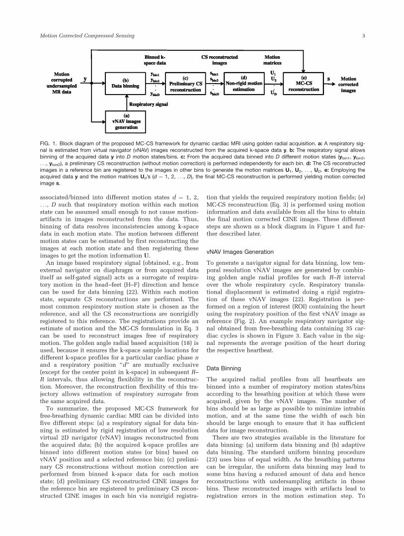
tion that yields the required respiratory motion fields; (e)MC-CS reconstruction (Eq. 3) is performed using motioninformation and data available from all the bins to obtainthe final motion corrected CINE images. These differentsteps are shown as a block diagram in Figure 1 and fur-ther described later.
vNAV Images Generation
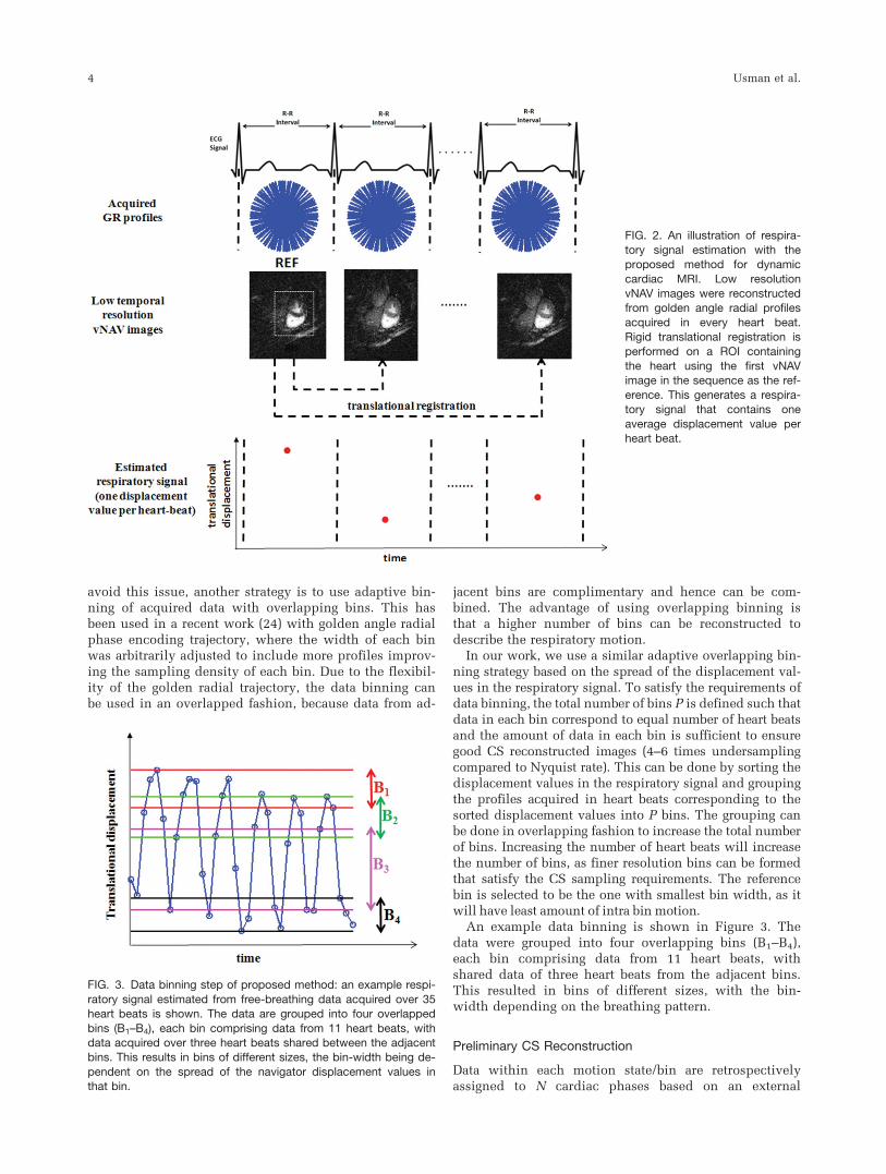
To generate a navigator signal for data binning, low tem-poral resolution vNAV images are generated by combin-ing golden angle radial profiles for each R–R intervalover the whole respiratory cycle. Respiratory transla-tional displacement is estimated doing a rigid registra-tion of these vNAV images (22). Registration is per-formed on a region of interest (ROI) containing the heartusing the respiratory position of the first vNAV image asreference (Fig. 2). An example respiratory navigator sig-nal obtained from free-breathing data containing 35 car-diac cycles is shown in Figure 3. Each value in the sig-nal represents the average position of the heart duringthe respective heartbeat.
Data Binning
The acquired radial profiles from all heartbeats arebinned into a number of respiratory motion states/binsaccording to the breathing position at which these wereacquired, given by the vNAV images. The number ofbins should be as large as possible to minimize intrabinmotion, and at the same time the width of each binshould be large enough to ensure that it has sufficientdata for image reconstruction.
There are two strategies available in the literature fordata binning: (a) uniform data binning and (b) adaptivedata binning. The standard uniform binning procedure(23) uses bins of equal width. As the breathing patternscan be irregular, the uniform data binning may lead tosome bins having a reduced amount of data and hencereconstructions with undersampling artifacts in thosebins. These reconstructed images with artifacts lead toregistration errors in the motion estimation step. To
FIG. 1. Block diagram of the proposed MC-CS framework for dynamic cardiac MRI using golden radial acquisition. a: A respiratory sig-
nal is estimated from virtual navigator (vNAV) images reconstructed from the acquired k-space data y. b: The respiratory signal allowsbinning of the acquired data y into D motion states/bins. c: From the acquired data binned into D different motion states (ybin1, ybin2,. . ., ybinD), a preliminary CS reconstruction (without motion correction) is performed independently for each bin. d: The CS reconstructed
images in a reference bin are registered to the images in other bins to generate the motion matrices U1, U2, . . ., UD. e: Employing theacquired data y and the motion matrices Ud’s (d ¼ 1, 2, . . ., D), the final MC-CS reconstruction is performed yielding motion corrected
image s.
Motion Corrected Compressed Sensing 3
avoid this issue, another strategy is to use adaptive bin-ning of acquired data with overlapping bins. This hasbeen used in a recent work (24) with golden angle radialphase encoding trajectory, where the width of each binwas arbitrarily adjusted to include more profiles improv-ing the sampling density of each bin. Due to the flexibil-ity of the golden radial trajectory, the data binning canbe used in an overlapped fashion, because data from ad-
jacent bins are complimentary and hence can be com-bined. The advantage of using overlapping binning isthat a higher number of bins can be reconstructed todescribe the respiratory motion.
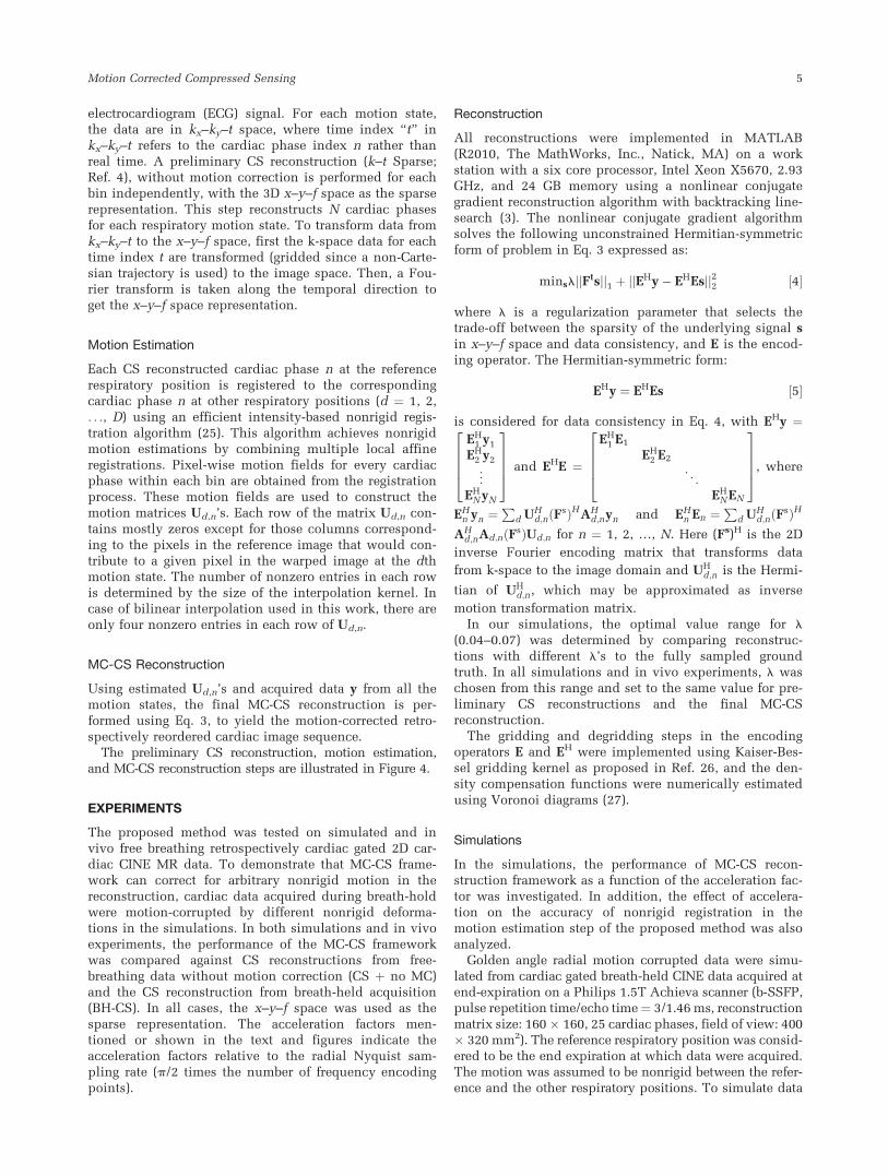
In our work, we use a similar adaptive overlapping bin-ning strategy based on the spread of the displacement val-ues in the respiratory signal. To satisfy the requirements ofdata binning, the total number of bins P is defined such thatdata in each bin correspond to equal number of heart beatsand the amount of data in each bin is sufficient to ensuregood CS reconstructed images (4–6 times undersamplingcompared to Nyquist rate). This can be done by sorting thedisplacement values in the respiratory signal and groupingthe profiles acquired in heart beats corresponding to thesorted displacement values into P bins. The grouping canbe done in overlapping fashion to increase the total numberof bins. Increasing the number of heart beats will increasethe number of bins, as finer resolution bins can be formedthat satisfy the CS sampling requirements. The referencebin is selected to be the one with smallest bin width, as itwill have least amount of intra bin motion.
An example data binning is shown in Figure 3. Thedata were grouped into four overlapping bins (B1–B4),each bin comprising data from 11 heart beats, withshared data of three heart beats from the adjacent bins.This resulted in bins of different sizes, with the bin-width depending on the breathing pattern.
Preliminary CS Reconstruction
Data within each motion state/bin are retrospectivelyassigned to N cardiac phases based on an external
FIG. 2. An illustration of respira-
tory signal estimation with theproposed method for dynamic
cardiac MRI. Low resolutionvNAV images were reconstructedfrom golden angle radial profiles
acquired in every heart beat.Rigid translational registration is
performed on a ROI containingthe heart using the first vNAVimage in the sequence as the ref-
erence. This generates a respira-tory signal that contains one
average displacement value perheart beat.
FIG. 3. Data binning step of proposed method: an example respi-
ratory signal estimated from free-breathing data acquired over 35heart beats is shown. The data are grouped into four overlapped
bins (B1–B4), each bin comprising data from 11 heart beats, withdata acquired over three heart beats shared between the adjacentbins. This results in bins of different sizes, the bin-width being de-
pendent on the spread of the navigator displacement values inthat bin.
4 Usman et al.
electrocardiogram (ECG) signal. For each motion state,the data are in kx–ky–t space, where time index ‘‘t’’ inkx–ky–t refers to the cardiac phase index n rather thanreal time. A preliminary CS reconstruction (k–t Sparse;Ref. 4), without motion correction is performed for eachbin independently, with the 3D x–y–f space as the sparserepresentation. This step reconstructs N cardiac phasesfor each respiratory motion state. To transform data fromkx–ky–t to the x–y–f space, first the k-space data for eachtime index t are transformed (gridded since a non-Carte-sian trajectory is used) to the image space. Then, a Fou-rier transform is taken along the temporal direction toget the x–y–f space representation.
Motion Estimation
Each CS reconstructed cardiac phase n at the referencerespiratory position is registered to the correspondingcardiac phase n at other respiratory positions (d ¼ 1, 2,. . ., D) using an efficient intensity-based nonrigid regis-tration algorithm (25). This algorithm achieves nonrigidmotion estimations by combining multiple local affineregistrations. Pixel-wise motion fields for every cardiacphase within each bin are obtained from the registrationprocess. These motion fields are used to construct themotion matrices Ud,n’s. Each row of the matrix Ud,n con-tains mostly zeros except for those columns correspond-ing to the pixels in the reference image that would con-tribute to a given pixel in the warped image at the dthmotion state. The number of nonzero entries in each rowis determined by the size of the interpolation kernel. Incase of bilinear interpolation used in this work, there areonly four nonzero entries in each row of Ud,n.
MC-CS Reconstruction
Using estimated Ud,n’s and acquired data y from all themotion states, the final MC-CS reconstruction is per-formed using Eq. 3, to yield the motion-corrected retro-spectively reordered cardiac image sequence.
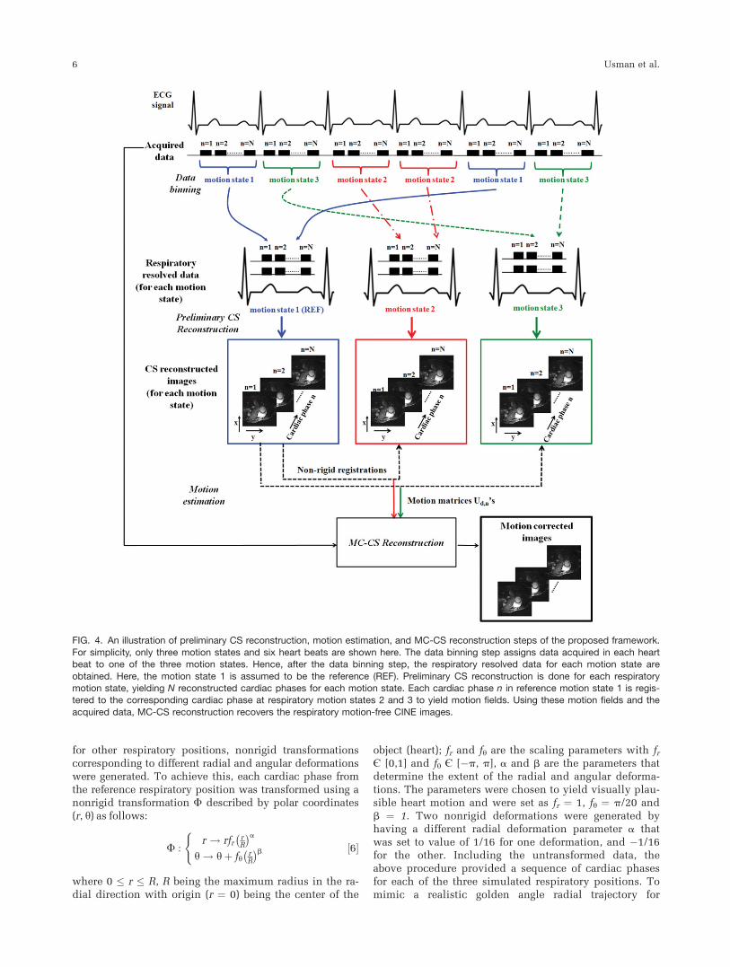
The preliminary CS reconstruction, motion estimation,and MC-CS reconstruction steps are illustrated in Figure 4.
EXPERIMENTS
The proposed method was tested on simulated and invivo free breathing retrospectively cardiac gated 2D car-diac CINE MR data. To demonstrate that MC-CS frame-work can correct for arbitrary nonrigid motion in thereconstruction, cardiac data acquired during breath-holdwere motion-corrupted by different nonrigid deforma-tions in the simulations. In both simulations and in vivoexperiments, the performance of the MC-CS frameworkwas compared against CS reconstructions from free-breathing data without motion correction (CS þ no MC)and the CS reconstruction from breath-held acquisition(BH-CS). In all cases, the x–y–f space was used as thesparse representation. The acceleration factors men-tioned or shown in the text and figures indicate theacceleration factors relative to the radial Nyquist sam-pling rate (p/2 times the number of frequency encodingpoints).
Reconstruction
All reconstructions were implemented in MATLAB(R2010, The MathWorks, Inc., Natick, MA) on a workstation with a six core processor, Intel Xeon X5670, 2.93GHz, and 24 GB memory using a nonlinear conjugategradient reconstruction algorithm with backtracking line-search (3). The nonlinear conjugate gradient algorithmsolves the following unconstrained Hermitian-symmetricform of problem in Eq. 3 expressed as:
minsljjFtsjj1 þ jjEHy� EHEsjj22 ½4�
where l is a regularization parameter that selects thetrade-off between the sparsity of the underlying signal sin x–y–f space and data consistency, and E is the encod-ing operator. The Hermitian-symmetric form:
EHy ¼ EHEs ½5�
is considered for data consistency in Eq. 4, with EHy ¼EH1 y1
EH2 y2...
EHNyN
26664
37775 and EHE ¼
EH1 E1
EH2 E2
. ..
EHNEN
26664
37775, where
EHn yn ¼
Pd U
Hd;n Fsð ÞHAH
d;nyn and EHn En ¼
Pd U
Hd;n Fsð ÞH
AHd;nAd;n Fsð ÞUd;n for n ¼ 1, 2, …, N. Here (Fs)H is the 2D
inverse Fourier encoding matrix that transforms data
from k-space to the image domain and UHd;n is the Hermi-
tian of UHd;n, which may be approximated as inverse
motion transformation matrix.In our simulations, the optimal value range for l
(0.04–0.07) was determined by comparing reconstruc-tions with different l’s to the fully sampled groundtruth. In all simulations and in vivo experiments, l waschosen from this range and set to the same value for pre-liminary CS reconstructions and the final MC-CSreconstruction.
The gridding and degridding steps in the encodingoperators E and EH were implemented using Kaiser-Bes-sel gridding kernel as proposed in Ref. 26, and the den-sity compensation functions were numerically estimatedusing Voronoi diagrams (27).
Simulations
In the simulations, the performance of MC-CS recon-struction framework as a function of the acceleration fac-tor was investigated. In addition, the effect of accelera-tion on the accuracy of nonrigid registration in themotion estimation step of the proposed method was alsoanalyzed.
Golden angle radial motion corrupted data were simu-lated from cardiac gated breath-held CINE data acquired atend-expiration on a Philips 1.5T Achieva scanner (b-SSFP,pulse repetition time/echo time¼ 3/1.46ms, reconstructionmatrix size: 160 � 160, 25 cardiac phases, field of view: 400� 320 mm2). The reference respiratory position was consid-ered to be the end expiration at which data were acquired.The motion was assumed to be nonrigid between the refer-ence and the other respiratory positions. To simulate data
Motion Corrected Compressed Sensing 5
for other respiratory positions, nonrigid transformationscorresponding to different radial and angular deformationswere generated. To achieve this, each cardiac phase fromthe reference respiratory position was transformed using anonrigid transformation F described by polar coordinates(r, y) as follows:
F :r ! rfr
rR
� �ah ! hþ fh
rR
� �b(
½6�
where 0 � r � R, R being the maximum radius in the ra-dial direction with origin (r ¼ 0) being the center of the
object (heart); fr and fy are the scaling parameters with frfi [0,1] and fy fi [�p, p], a and b are the parameters thatdetermine the extent of the radial and angular deforma-tions. The parameters were chosen to yield visually plau-sible heart motion and were set as fr ¼ 1, fy ¼ p/20 andb ¼ 1. Two nonrigid deformations were generated byhaving a different radial deformation parameter a thatwas set to value of 1/16 for one deformation, and �1/16for the other. Including the untransformed data, theabove procedure provided a sequence of cardiac phasesfor each of the three simulated respiratory positions. Tomimic a realistic golden angle radial trajectory for
FIG. 4. An illustration of preliminary CS reconstruction, motion estimation, and MC-CS reconstruction steps of the proposed framework.
For simplicity, only three motion states and six heart beats are shown here. The data binning step assigns data acquired in each heartbeat to one of the three motion states. Hence, after the data binning step, the respiratory resolved data for each motion state are
obtained. Here, the motion state 1 is assumed to be the reference (REF). Preliminary CS reconstruction is done for each respiratorymotion state, yielding N reconstructed cardiac phases for each motion state. Each cardiac phase n in reference motion state 1 is regis-tered to the corresponding cardiac phase at respiratory motion states 2 and 3 to yield motion fields. Using these motion fields and the
acquired data, MC-CS reconstruction recovers the respiratory motion-free CINE images.
6 Usman et al.
sampling in k-space, a respiratory signal was obtainedfrom a prolonged in vivo free-breathing cardiac scancomprising several heartbeats. Given the respiratory sig-nal and the number of bins set to 3, every heartbeat inthe free-breathing scan was associated with one of thethree respiratory positions using the binning proceduredescribed in the ‘‘Methods’’ section. The data acquisitionwas simulated by sampling the deformed cardiac phasesequence in each respiratory position on the goldenangle radial trajectory defined by the respiratory signal.To simulate CS undersampling, the number of heartbeatsfrom the respiratory signal was decreased such that thenumber of radial profiles for each cardiac phase at a spe-cific respiratory position was reduced by accelerationfactor of 3.5–20.
For each reconstruction (CS þ no MC, MC-CS, andBH-CS), the overall relative root mean square (RMS)reconstruction error (Recon RMSE) for the reconstructedsignal x (of length K) was calculated as:
RelativeRMSError ¼
ffiffiffiffiffiffiffiffiffiffiffiffiffiffiffiffiffiffiffiffiffiffiffiffiffiffiffiXKi¼1
xi � x̂ij jxij j2
2
vuut ½7�
where x̂ denote the reconstructed signal, and x is thegold standard signal reconstructed using the number ofprojections corresponding to the radial Nyquist rate.
To analyze the accuracy of the motion estimation step,for different acceleration factors, the motion fields esti-mated via nonrigid registrations were used to deform theuntransformed images in motion state 1. The registrationerror (Registration RMSE) was computed using Eq. 7,where xi now represents the images transformed withgold standard deformations in Eq. 6 and x̂i representsthe images transformed with estimated deformations.Both Recon RMSE and Registration RMSE were com-puted in a ROI containing the heart.
To analyze the effect of selection of regularization pa-rameter l on the MC-CS reconstruction quality, MC-CS
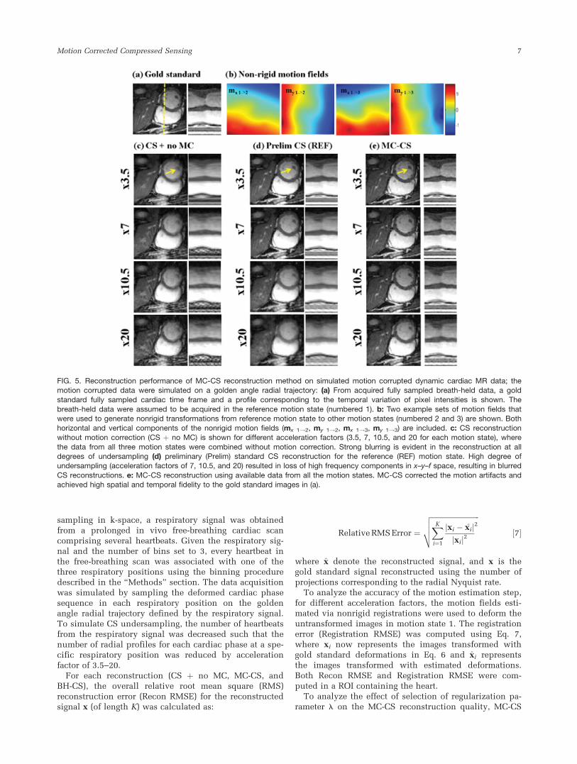
FIG. 5. Reconstruction performance of MC-CS reconstruction method on simulated motion corrupted dynamic cardiac MR data; themotion corrupted data were simulated on a golden angle radial trajectory: (a) From acquired fully sampled breath-held data, a gold
standard fully sampled cardiac time frame and a profile corresponding to the temporal variation of pixel intensities is shown. Thebreath-held data were assumed to be acquired in the reference motion state (numbered 1). b: Two example sets of motion fields thatwere used to generate nonrigid transformations from reference motion state to other motion states (numbered 2 and 3) are shown. Both
horizontal and vertical components of the nonrigid motion fields (mx 1!2, my 1!2, mx 1!3, my 1!3) are included. c: CS reconstructionwithout motion correction (CS þ no MC) is shown for different acceleration factors (3.5, 7, 10.5, and 20 for each motion state), where
the data from all three motion states were combined without motion correction. Strong blurring is evident in the reconstruction at alldegrees of undersampling (d) preliminary (Prelim) standard CS reconstruction for the reference (REF) motion state. High degree ofundersampling (acceleration factors of 7, 10.5, and 20) resulted in loss of high frequency components in x–y–f space, resulting in blurred
CS reconstructions. e: MC-CS reconstruction using available data from all the motion states. MC-CS corrected the motion artifacts andachieved high spatial and temporal fidelity to the gold standard images in (a).
Motion Corrected Compressed Sensing 7
reconstructions were done using varying values of l inthe range of 0.0001–0.5. The Recon RMSE was computedfor each case.
As there can be a number of degrees of freedom in thenonrigid motion that might occur, a number of differentmotion realizations were generated by varying the set ofparameters fr, fy, a, and b in Eq. 6. The parameters werevaried randomly within 20% from their values (fr ¼ 1, fy¼ p/20, b ¼ 1, a ¼ 61/16) in previous simulation suchthat the corresponding image transformations looked vis-ually realistic. MC-CS reconstructions were done in eachcase.
In Vivo Experiments
ECG-gated free-breathing golden angle radial acquisitionswere performed on Philips 1.5T in five healthy volun-teers. Scan parameters include b-SSFP, pulse repetitiontime ¼ 2.98 ms, echo time ¼ 1.49 ms, field of view: 320� 320 mm2, spatial resolution: 1.5–2.0 mm2, number offrequency encoding points: 160–212, 180–200 goldenangle radial profiles per heartbeat, heart rate: 65–85 beatsper minute (bpm), number of cardiac cycles ¼ 25–33,scan duration ¼ 19–25 s. Scans were performed using a5-channel coil but a single channel (with high sensitivityover the heart) was selected for MC-CS reconstruction.The acquired free-breathing data were binned into 6–7respiratory positions, depending on the number of heartbeats, using the described adaptive binning procedure.The CS acceleration factor in each bin was set in therange of 4–6. Twenty cardiac phases were retrospectively
reconstructed resulting in temporal resolution of 30–40ms. For comparison, the breath-held data were acquiredat the end-expiration state in all the volunteers with thesame imaging parameters, except that the scan durationwas reduced (number of cardiac cycles ¼ 13–29, acquisi-tion time ¼ 10–20 s).
RESULTS
Simulations
The gold standard fully sampled reconstruction for oneof the cardiac time frames is shown in Figure 5a. A goldstandard fully sampled temporal profile correspondingto the temporal variation of pixel intensities along thedotted line is also shown. Two example sets of motionfields corresponding to the nonrigid transformationsfrom breath-hold reference motion state 1 to motionstates 2 and 3 are shown in Figure 5b. Both horizontaland vertical components of the motion fields (mx 1!2,my 1!2, mx 1!3, my 1!3) are shown. Relative to theNyquist sampling rate (160 � p/2 � 251 profiles)required for each cardiac phase, acceleration factors of3.5, 7, 10.5, 15, and 20 were simulated for k-space datain each of the three respiratory positions. The CS recon-structed cardiac frames and temporal profiles are shownin Figure 5c–e. Strong blurring artifacts due to nonrigidmotion were evident in the CS reconstruction withoutmotion correction (CS þ no MC) for all acceleration fac-tors (Fig. 5c). The preliminary CS reconstruction for thereference breath-hold position is shown in Figure 5d.The preliminary CS reconstructions at other respiratory
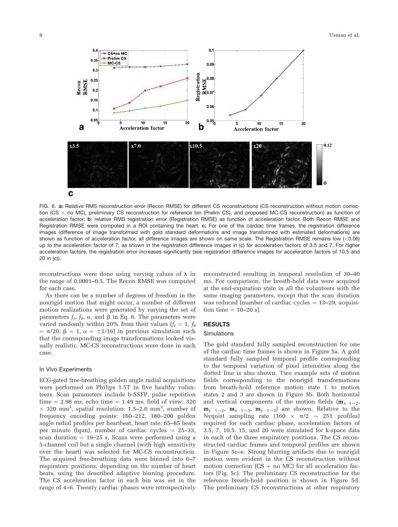
FIG. 6. a: Relative RMS reconstruction error (Recon RMSE) for different CS reconstructions (CS reconstruction without motion correc-
tion (CS þ no MC), preliminary CS reconstruction for reference bin (Prelim CS), and proposed MC-CS reconstruction) as function ofacceleration factor; b: relative RMS registration error (Registration RMSE) as function of acceleration factor. Both Recon RMSE and
Registration RMSE were computed in a ROI containing the heart. c: For one of the cardiac time frames, the registration differenceimages (difference of image transformed with gold standard deformations and image transformed with estimated deformations) areshown as function of acceleration factor, all difference images are shown on same scale. The Registration RMSE remains low (<0.06)
up to the acceleration factor of 7, as shown in the registration difference images in (c) for acceleration factors of 3.5 and 7. For higheracceleration factors, the registration error increases significantly (see registration difference images for acceleration factors of 10.5 and
20 in (c)).
8 Usman et al.
positions (not shown here) were of similar or lower qual-ity. At an acceleration factor of 3.5 (72 golden angle ra-dial profiles per cardiac phase), all preliminary CSreconstructions had good quality, and the expected var-iations in the temporal profile were observed. However,at higher acceleration factors of 7, 10.5, and 20, the pre-liminary CS reconstructions tend to become blurrier.MC-CS reconstruction (Fig. 5e) visually corrected forblurriness in the combined motion corrected images andhad high spatial and temporal fidelity to the gold stand-ard images in Figure 5a.
A plot of relative RMS reconstruction error (ReconRMSE) in the ROI containing the heart for different CSreconstructions as a function of acceleration factor isshown in Figure 6a. Although not reported in the manu-script, the reconstruction error was stronger in the regionoutside the ROI than inside, as both registration and CSreconstruction favor high contrast components for align-ment and reconstruction, and region outside the ROIcontains mainly the low contrast components.
The RMS registration error (Registration RMSE) as afunction of acceleration factor is shown in Figure 6b. Fordifferent acceleration factors, the registration error differ-ence images (difference of an image transformed with
gold standard deformation and image transformed withestimated deformation) for one of the cardiac time framesare also shown in Figure 6c. The registration errorremains low (Registration RMSE < 0.06) up to the accel-eration factor of 7. For higher acceleration factors, theregistration error increases significantly (see registrationdifference images for acceleration factors of 10.5 and 20in Fig. 6c).
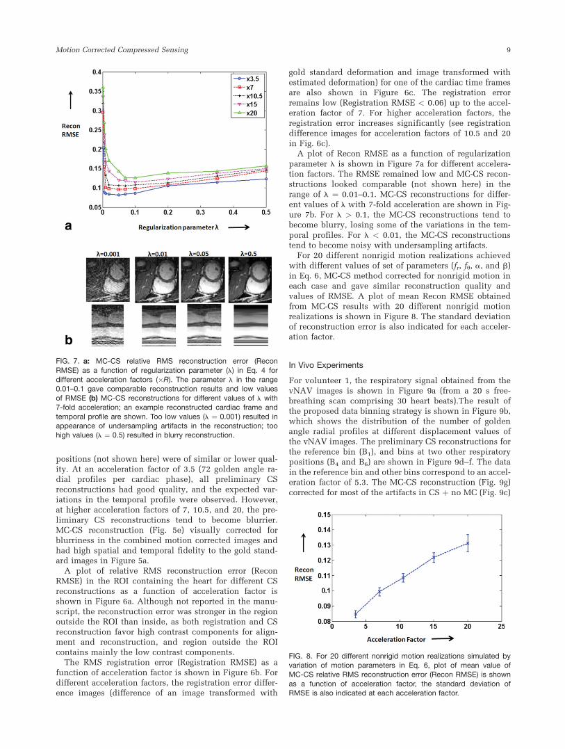
A plot of Recon RMSE as a function of regularizationparameter l is shown in Figure 7a for different accelera-tion factors. The RMSE remained low and MC-CS recon-structions looked comparable (not shown here) in therange of l ¼ 0.01–0.1. MC-CS reconstructions for differ-ent values of l with 7-fold acceleration are shown in Fig-ure 7b. For l > 0.1, the MC-CS reconstructions tend tobecome blurry, losing some of the variations in the tem-poral profiles. For l < 0.01, the MC-CS reconstructionstend to become noisy with undersampling artifacts.
For 20 different nonrigid motion realizations achievedwith different values of set of parameters (fr, fy, a, and b)in Eq. 6, MC-CS method corrected for nonrigid motion ineach case and gave similar reconstruction quality andvalues of RMSE. A plot of mean Recon RMSE obtainedfrom MC-CS results with 20 different nonrigid motionrealizations is shown in Figure 8. The standard deviationof reconstruction error is also indicated for each acceler-ation factor.
In Vivo Experiments
For volunteer 1, the respiratory signal obtained from thevNAV images is shown in Figure 9a (from a 20 s free-breathing scan comprising 30 heart beats).The result ofthe proposed data binning strategy is shown in Figure 9b,which shows the distribution of the number of goldenangle radial profiles at different displacement values ofthe vNAV images. The preliminary CS reconstructions forthe reference bin (B1), and bins at two other respiratorypositions (B4 and B6) are shown in Figure 9d–f. The datain the reference bin and other bins correspond to an accel-eration factor of 5.3. The MC-CS reconstruction (Fig. 9g)corrected for most of the artifacts in CS þ no MC (Fig. 9c)
FIG. 7. a: MC-CS relative RMS reconstruction error (Recon
RMSE) as a function of regularization parameter (l) in Eq. 4 fordifferent acceleration factors (�R). The parameter l in the range0.01–0.1 gave comparable reconstruction results and low values
of RMSE (b) MC-CS reconstructions for different values of l with7-fold acceleration; an example reconstructed cardiac frame and
temporal profile are shown. Too low values (l ¼ 0.001) resulted inappearance of undersampling artifacts in the reconstruction; toohigh values (l ¼ 0.5) resulted in blurry reconstruction.
FIG. 8. For 20 different nonrigid motion realizations simulated byvariation of motion parameters in Eq. 6, plot of mean value ofMC-CS relative RMS reconstruction error (Recon RMSE) is shown
as a function of acceleration factor, the standard deviation ofRMSE is also indicated at each acceleration factor.
Motion Corrected Compressed Sensing 9
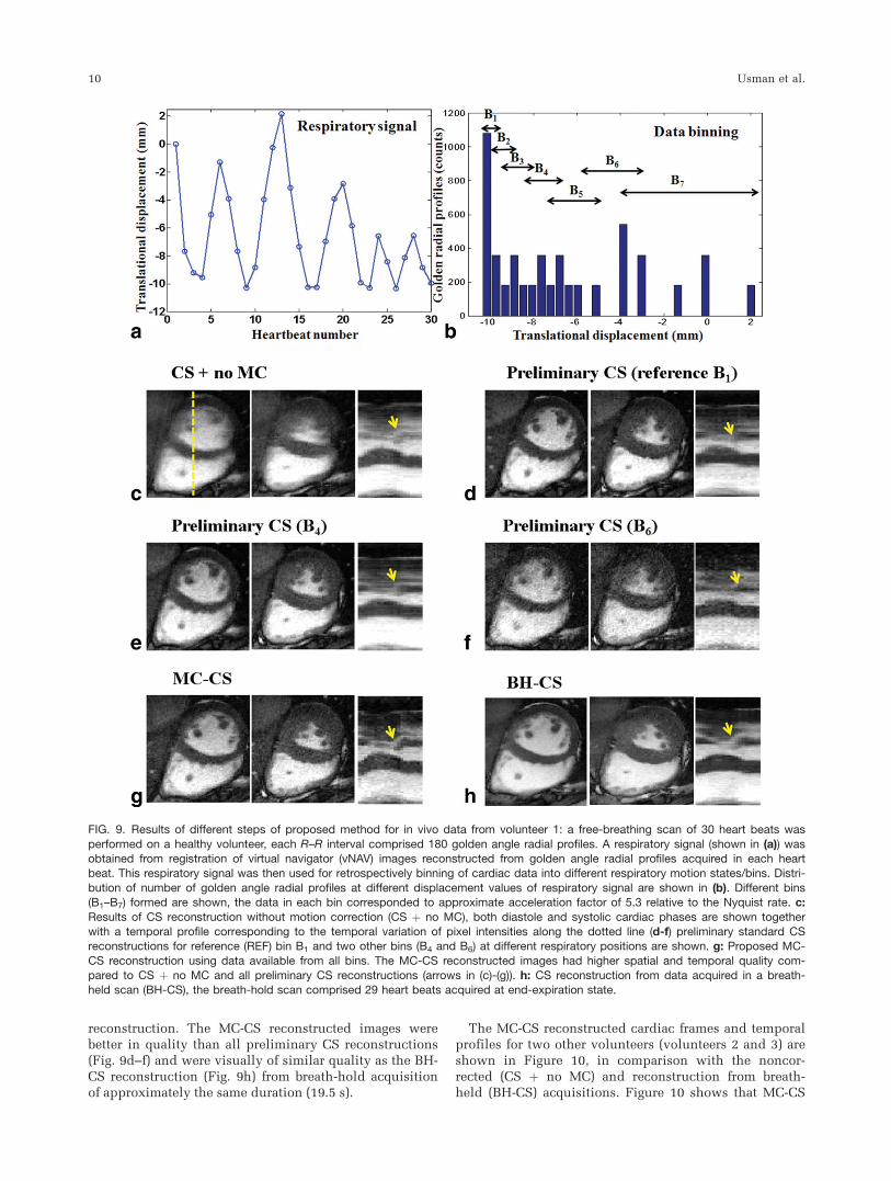
reconstruction. The MC-CS reconstructed images werebetter in quality than all preliminary CS reconstructions(Fig. 9d–f) and were visually of similar quality as the BH-CS reconstruction (Fig. 9h) from breath-hold acquisitionof approximately the same duration (19.5 s).
The MC-CS reconstructed cardiac frames and temporalprofiles for two other volunteers (volunteers 2 and 3) areshown in Figure 10, in comparison with the noncor-rected (CS þ no MC) and reconstruction from breath-held (BH-CS) acquisitions. Figure 10 shows that MC-CS
FIG. 9. Results of different steps of proposed method for in vivo data from volunteer 1: a free-breathing scan of 30 heart beats was
performed on a healthy volunteer, each R–R interval comprised 180 golden angle radial profiles. A respiratory signal (shown in (a)) wasobtained from registration of virtual navigator (vNAV) images reconstructed from golden angle radial profiles acquired in each heartbeat. This respiratory signal was then used for retrospectively binning of cardiac data into different respiratory motion states/bins. Distri-
bution of number of golden angle radial profiles at different displacement values of respiratory signal are shown in (b). Different bins(B1–B7) formed are shown, the data in each bin corresponded to approximate acceleration factor of 5.3 relative to the Nyquist rate. c:Results of CS reconstruction without motion correction (CS þ no MC), both diastole and systolic cardiac phases are shown togetherwith a temporal profile corresponding to the temporal variation of pixel intensities along the dotted line (d-f) preliminary standard CSreconstructions for reference (REF) bin B1 and two other bins (B4 and B6) at different respiratory positions are shown. g: Proposed MC-
CS reconstruction using data available from all bins. The MC-CS reconstructed images had higher spatial and temporal quality com-pared to CS þ no MC and all preliminary CS reconstructions (arrows in (c)-(g)). h: CS reconstruction from data acquired in a breath-held scan (BH-CS), the breath-hold scan comprised 29 heart beats acquired at end-expiration state.
10 Usman et al.

reconstruction achieved similar quality to the breath-hold images.
DISCUSSION
This work proposes a CS motion corrected reconstruc-tion from undersampled general (affine or nonrigid)motion corrupted dynamic cardiac MR data. The pro-posed method is a combination of CS (4) and the motioncorrection technique by Batchelor et al. (12) and can cor-rect for arbitrary motion in the CS reconstruction. Theuse of the proposed method is demonstrated in 2D gatedcardiac CINE MRI, where 20 respiratory-motion free car-diac phases were reconstructed with high spatial andtemporal fidelity to the breath-held images, resulting in atemporal resolution of 30–40 ms. The flexibility of thegolden angle radial acquisition was used for respiratoryself-gating and adaptive binning in free-breathingdynamic cardiac MRI. The proposed approach permitsfree-breathing acquisitions, and further studies mayexplore its application to 3D dynamic cardiac MRI.
While motion correction methods have been well inte-grated into parallel imaging (14,15,21,22), CS-basedreconstruction methods integrated with motion correc-tion have been only shown for rigid motion. Comparedto parallel imaging methods that reconstruct each cardiacphase/frame independently, the proposed MC-CS frame-
work for dynamic cardiac MRI exploits redundancyalong the temporal dimension by finding the sparsest so-lution in the spatio-temporal x–y–f space.
At high acceleration factors (undersampling factor of 7in each cardiac phase in Fig. 5d), due to insufficientnumber of measurements in each bin, the preliminaryCS reconstructions are unable to reconstruct high fre-quency temporal components in the x–y–f space andlead to slightly blurred reconstructions. However, theseslightly blurred reconstructions are still good enough toestimate precise nonrigid motion between differentmotion states (RMS registration error < 0.06 for 7-foldacceleration, Fig. 6b,c). Using the estimated motionfields, the MC-CS is able to correct the nonrigid motionin the final reconstruction with high spatial and tempo-ral fidelity compared to the gold standard reconstructionin Figure 5a. At very high acceleration factors (accelera-tion factor of 20 per cardiac phase Fig. 5d), the prelimi-nary CS reconstructions suffer from strong undersam-pling artifacts, and the image quality is not good enoughto get precise motion between different respiratory posi-tions (Registration RMSE � 0.10, see registration differ-ence images in Fig. 6c), hence the final MC-CS recon-struction still suffers from some blurring artifacts.
We used an adaptive binning procedure for dataacquired in 19–25 s free-breathing scans, comprising25–33 cardiac cycles. The number of bins was in the
FIG. 10. Performance comparison of different CS reconstructions for in vivo data from volunteers 2 and 3; the free-breathing scans forvolunteers 2 and 3 comprised 25 and 33 heart beats, respectively. Data were binned into six and seven respiratory positions for volun-
teers 2 and 3, respectively. CS reconstructed frames during both systole and diastole are shown together with the temporal variation ofpixel intensities along the dotted lines. CS reconstruction without motion correction (CS þ no MC), proposed MC-CS reconstruction
and CS reconstruction from breath-hold acquisition (BH-CS) are shown. The breath-hold scans were of 10- and 15-s duration, and com-prised 13 and 20 heart beats for volunteers 2 and 3, respectively. Proposed MC-CS reconstruction corrected for most of the blurring inCS þ no MC reconstruction and had similar spatial and temporal quality than the BH-CS reconstruction (see the region pointed by
arrows).
Motion Corrected Compressed Sensing 11
range of 6–7 depending on the number of acquired heart-beats. The size of each bin was adjusted to guaranteeundersampling factors of 4–6 for each bin for prelimi-nary CS reconstruction. This resulted in preliminary CSreconstructions of sufficient quality to achieve accurateregistrations and good quality subsequent MC-CS recon-structions. The number of bins was set to be higher thanfive overlapping windows to get an average bin width of2 mm. If too few bins are used (<3), this will lead tostrong intrabin motion and motion artifacts remaining inthe reconstruction despite having more than sufficientamount of data in each bin for the CS reconstruction. Iftoo many bins are used, preliminary CS reconstructedimages may not have enough quality to ensure accuratenonrigid motion estimation. In the experiments, we haveused data available from all bins in the final MC-CSreconstruction. To further improve the reconstruction,the data corresponding to bins having larger widths(greater than 4 mm, for example) could be rejected forMC-CS reconstruction to avoid high intrabin motion.
The proposed approach corrects for respiratory motionduring free-breathing cardiac acquisition, but correctionfor heart-rate variability is not considered in this work.Hence, further modifications will be needed to apply theproposed method in arrhythmic patients. In our work,we have assumed that all cardiac phases are in the samerespiratory motion state. However, the golden radial tra-jectory offers the option of reconstructing multiple navi-gator images (vNAV) for different cardiac phases, whichwill be investigated in future work.
The MC-CS framework is computationally more com-plex compared to the standard CS reconstruction, as pre-liminary CS reconstructions have to be performed foreach bin and additionally for the final MC-CS step. Witha nonoptimized MATLAB based implementation, the av-erage time for reconstruction of motion corrected imagesfrom in vivo free-breathing data was in the range of 2–2.5 h, whereas for breath-held data it was in the range of15–30 min. However, several steps of the proposedframework are highly parallelizable and the reconstruc-tion time might be reduced using parallel computingtechniques.
The proposed method has preliminary CS reconstruc-tion, motion estimation, and final MC-CS reconstructionas sequential stages. One of the limitations of sucharchitecture is that errors in any stage are propagated tothe next one. A possible way around this would be tocombine all steps into one coupled problem, using anapproach similar to generalized reconstruction byinversion of coupled systems (GRICS) (14), where theimage reconstruction and the motion estimation stepsare combined into one coupled optimization problem,leading to an autocalibrated motion model.
In the motion estimation step of the proposed method,we have used intensity-based nonrigid registration algo-rithm (25) to estimate nonrigid motion between differentrespiratory positions. Instead of using registrations, blockbased dense motion field estimation as done in k-tFOCUSS (9–11) might be used for motion estimation.
The use of MC-CS framework may be extended to al-ternative applications where the respiratory signal canbe used as a motion surrogate signal. An example of this
is motion corrected liver MRI. In other applicationswhere a motion surrogate signal is not available for databinning, further novel motion correction techniquesneed to be developed.
CONCLUSIONS
A novel reconstruction framework was presented fordynamic cardiac MRI that benefits from the high acceler-ation available with CS and correction for arbitrary (affineor nonrigid) motion in the CS reconstruction. The use ofthis framework was demonstrated in 2D cardiac gatedMRI using a golden radial acquisition, where respiratorymotion-free cardiac phases were reconstructed fromundersampled self-gated free-breathing k-space data.
ACKNOWLEDGMENTS
The authors dedicate this article to the loving memory ofour wonderful colleague, Philip G. Batchelor. C. Prietoacknowledges financial support from FONDECYT. Theauthors thank Michael Lustig for making the nonlinearconjugate gradient MATLAB code available online.
REFERENCES
1. Donoho DL. Compressed sensing. IEEE Trans Inform Theory 2006;52:
1289–1306.
2. Candes E. Compressive Sampling. In: Proceedings of International
Congress of Mathematicians, Vol. 3, Madrid, Spain, 2006. pp 1433–
1452.
3. Lustig M, Donoho D, Pauly JM. Sparse MRI: the application of com-
pressed sensing for rapid MR imaging. Magn Res Med 2007;58:
1182–1195.
4. Lustig M, Santos JM, Donoho DL, Pauly JM. k–t SPARSE: High Frame
Rate Dynamic MRI Exploiting Spatio-Temporal Sparsity. In Proceed-
ings of the 13th Annual Meeting of ISMRM, Seatle, Washington, USA,
2006. p. 2420.
5. Akcakaya M, Nam S, Hu P, Moghari MH, Ngo LH, Tarokh V, Man-
ning WJ, Nezafat R. Compressed sensing with wavelet domain
dependencies for coronary MRI: a retrospective study. IEEE Trans
Med Imaging 2011;30:1090–1099.
6. Vasanawala SS, Alley MT, Hargreaves BA, Barth RA, Pauly JM, Lus-
tig M. Improved pediatric MR imaging with compressed sensing. Ra-
diology 2010;256:607–616.
7. Otazo R, Kim D, Axel L, Sodickson DK. Combination of Compressed
Sensing and Parallel Imaging with Respiratory Motion Correction for
Highly-Accelerated First-Pass Cardiac Perfusion MRI. In Proceedings of
the 18th Annual Meeting of ISMRM, Montreal, Canada, 2011. p. 63.
8. Doneva M, Stehning C, Nehrke K, B€ornert P. Improving Scan Effi-
ciency of Respiratory Gated Imaging Using Compressed Sensing with
3D Cartesian Golden Angle Sampling. In Proceedings of the 19th
Annual Meeting of ISMRM, Montreal, Canada, 2011. p. 624.
9. Jung H, Sung K, Nayak KS, Kim EY, Ye JC. k-t FOCUSS: a general
compressed sensing framework for high resolution dynamic MRI.
Magn Res Med 2009;61:103–116.
10. Jung H, Park J, Yoo J, Ye JC. Radial k-t FOCUSS for highresolution
cardiac cine MRI. Magn Res Med 2010;63:68–78.
11. Jung H, Ye JC. Motion estimated and compensated compressed sens-
ing dynamic magnetic resonance imaging: what we can learn from
video compression techniques. Int J Imaging Syst Technol 2010;20:
81–98.
12. Batchelor PG, Atkinson D, Irarrazaval P, Hill DLG, Hajnal J, Larkman
D. Matrix description of general motion correction applied to multi-
shot images. Magn Res Med 2005;54:1273–1280.
13. Schmidt JFM, Buehrer M, Boesiger P, Kozerke S. Nonrigid retrospec-
tive respiratory motion correction in whole-heart coronary MRA.
Magn Res Med 2011;66:1541–1549.
14. Odille F, Vuissoz PA, Marie PY, Felblinger J. Generalized reconstruc-
tion by inversion of coupled systems (GRICS) applied to free-breath-
ing MRI. Magn Res Med 2008;60:146–157.
12 Usman et al.
15. Hansen M, Sorensen T, Arai A, Kellman P. Retrospective reconstruc-
tion of high temporal resolution cine images from real-time MRI
using iterative motion correction. Magn Reson Med 2011. doi:
10.1002/mrm.23284.
16. Odille F, Cindea N, Mandry D, Pasquier C, Vuissoz PA, Felblinger J.
Generalized MRI reconstruction including elastic physiological
motion and coil, sensitivity encoding. Magn Res Med 2008;59:
1401–1411.
17. White MJ, Hawkes DJ, Melbourne A, Collins DJ, Coolens C, Hawkins
M, Leach MO, Atkinson D. Motion artifact correction in free-breath-
ing abdominal MRI using overlapping partial samples to recover
image deformations. Magn Res Med 2009;62:440–449.
18. Winkelmann S, Schaeffter T, Koehler T, Eggers H, Doessel O. An
optimal radial profile order based on the golden ratio for time-
resolved MRI. IEEE Trans Med Imaging 2007;26:68–76.
19. Chan RW, Ramsay EA, Cheung EY, Plewes DB. The influence of ra-
dial undersampling schemes on compressed sensing reconstruction
in breast MRI. Magn Res Med 2012;67:363–377.
20. Jung H, Park J, Yoo J, Ye JC. Radial k-t FOCUSS for high-resolution
cardiac cine MRI. Magn Res Med 2010;63:68–78.
21. Kellman P, Chefd’hotel C, Lorenz CH, Mancini C, Arai AE,
McVeigh ER. High spatial and temporal resolution cardiac cine
MRI from retrospective reconstruction of data acquired in real time
using motion correction and resorting. Magn Res Med 2009;62:
1557–1564.
22. Kellman P, Chefd’hotel C, Lorenz CH, Mancini C, Arai AE, McVeigh
ER. Fully automatic, retrospective enhancement of real-time acquired
cardiac cine MR images using image-based navigators and respiratory
motion-corrected averaging. Magn Res Med 2008;59:771–778.
23. Jhooti P, Gatehouse PD, Keegan J, Bunce NH, Taylor AM, Firmin DN.
Phase ordering with automatic window selection (PAWS): a novel
motion-resistant technique for 3D coronary imaging. Magn Res Med
2000;43:470–480.
24. Buerger C, King AP, Schaeffter T, Prieto C. 3D non-rigid motion mod-
elling of the liver from undersampled Golden-Radial Phase encoding
(G-RPE) acquisitions. In Proceedings of the 18th Annual Meeting of
ISMRM, Montreal, Canada, 2011. p. 643.
25. Buerger C, Schaeffter T, King AP. Hierarchical adaptive local affine
registration for fast and robust respiratory motion estimation. Med
Image Anal 2011;15:551–564.
26. Jackson JI, Meyer CH, Nishimura DG, Macovski A. Selection of a con-
volution function for fourier inversion using gridding. IEEE Trans
Med Imaging 1991;10:473–478.
27. Rasche V, Proksa R, Sinkus R, Bornert P, Eggers H. Resampling of
data between arbitrary grids using convolution interpolation. IEEE
Trans Med Imaging 1999;18:385–392.
Motion Corrected Compressed Sensing 13