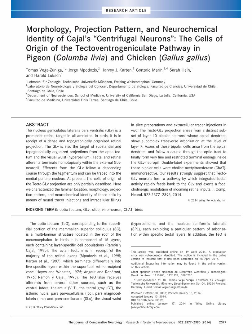
Morphology, projection pattern, and neurochemical identity of Cajal's “centrifugal neurons”: The...
20
Morphology, Projection Pattern, and Neurochemical Identity of Cajal’s “Centrifugal Neurons”: The Cells of Origin of the Tectoventrogeniculate Pathway in Pigeon (Columba livia) and Chicken (Gallus gallus) Tomas Vega-Zuniga, 1 * Jorge Mpodozis, 2 Harvey J. Karten, 3 Gonzalo Mar ın, 2,4 Sarah Hain, 1 and Harald Luksch 1 1 Lehrstuhl f€ ur Zoologie, Technische Universit€ at M€ unchen, Freising-Weihenstephan, Germany 2 Laboratorio de Neurobiolog ıa y Biolog ıa del Conocer, Departamento de Biolog ıa, Facultad de Ciencias, Universidad de Chile, Santiago de Chile, Chile 3 Department of Neurosciences, School of Medicine, University of California San Diego, La Jolla, California, USA 4 Facultad de Medicina, Universidad Finis Terrae, Santiago de Chile, Chile ABSTRACT The nucleus geniculatus lateralis pars ventralis (GLv) is a prominent retinal target in all amniotes. In birds, it is in receipt of a dense and topographically organized retinal projection. The GLv is also the target of substantial and topographically organized projections from the optic tec- tum and the visual wulst (hyperpallium). Tectal and retinal afferents terminate homotopically within the external GLv- neuropil. Efferents from the GLv follow a descending course through the tegmentum and can be traced into the medial pontine nucleus. At present, the cells of origin of the Tecto-GLv projection are only partially described. Here we characterized the laminar location, morphology, projec- tion pattern, and neurochemical identity of these cells by means of neural tracer injections and intracellular fillings in slice preparations and extracellular tracer injections in vivo. The Tecto-GLv projection arises from a distinct sub- set of layer 10 bipolar neurons, whose apical dendrites show a complex transverse arborization at the level of layer 7. Axons of these bipolar cells arise from the apical dendrites and follow a course through the optic tract to finally form very fine and restricted terminal endings inside the GLv-neuropil. Double-label experiments showed that these bipolar cells were choline acetyltransferase (ChAT)- immunoreactive. Our results strongly suggest that Tecto- GLv neurons form a pathway by which integrated tectal activity rapidly feeds back to the GLv and exerts a focal cholinergic modulation of incoming retinal inputs. J. Comp. Neurol. 522:2377–2396, 2014. V C 2014 Wiley Periodicals, Inc. INDEXING TERMS: optic tectum; GLv; slice; vine-neuron; ChAT; birds The optic tectum (TeO), corresponding to the superfi- cial portion of the mammalian superior colliculus (SC), is a multi-laminar structure located in the roof of the mesencephalon. In birds it is composed of 15 layers, each containing layer-specific cell populations (Ram on y Cajal, 1995). The avian tectum is in receipt of the majority of the retinal axons (Mpodozis et al., 1995; Karten et al., 1997), which terminate differentially into five specific layers within the superficial retino-recipient zone (Hayes and Webster, 1975; Angaut and Rep erant, 1976; Ram on y Cajal, 1995). The TeO also receives afferents from several other sources, such as the ventral lateral thalamus (VLT), the tectal gray (GT), the isthmic nuclei pars parvocellularis (Ipc), pars magnocel- lularis (Imc) and pars semilunaris (SLu), the visual wulst (hyperpallium), and the nucleus spiriformis lateralis (SPL), each exhibiting a particular pattern of arboriza- tion within specific tectal layers. In addition, the TeO is This article was published online on 19 April 2014. A production error was subsequently identified. This notice is included in the online version to indicate that it has been corrected on 26 April 2014. Additional Supporting Information may be found in the online version of this article. Grant sponsor: Fondo Nacional de Desarrollo Cient ıfico y Tecnol ogico; Grant numbers: 1110281, 1120124, 1080220. *Correspondence to: Dr. Tomas Vega-Zuniga, Lehrstuhl f€ ur Zoologie, Technische Universit€ at M€ unchen, Liesel-Beckmann Str. 04, 85354 Freising, Germany. E-mail: [email protected] Received October 28, 2013; Revised January 13, 2014; Accepted January 13, 2014. DOI 10.1002/cne.23539 Published online January 17, 2014 in Wiley Online Library (wileyonlinelibrary.com) V C 2014 Wiley Periodicals, Inc. The Journal of Comparative Neurology | Research in Systems Neuroscience 522:2377–2396 (2014) 2377 RESEARCH ARTICLE
Transcript of Morphology, projection pattern, and neurochemical identity of Cajal's “centrifugal neurons”: The...
Morphology, Projection Pattern, and NeurochemicalIdentity of Cajal’s “Centrifugal Neurons”: The Cells ofOrigin of the Tectoventrogeniculate Pathway inPigeon (Columba livia) and Chicken (Gallus gallus)
Tomas Vega-Zuniga,1* Jorge Mpodozis,2 Harvey J. Karten,3 Gonzalo Mar�ın,2,4 Sarah Hain,1
and Harald Luksch1
1Lehrstuhl f€ur Zoologie, Technische Universit€at M€unchen, Freising-Weihenstephan, Germany2Laboratorio de Neurobiolog�ıa y Biolog�ıa del Conocer, Departamento de Biolog�ıa, Facultad de Ciencias, Universidad de Chile,
Santiago de Chile, Chile3Department of Neurosciences, School of Medicine, University of California San Diego, La Jolla, California, USA4Facultad de Medicina, Universidad Finis Terrae, Santiago de Chile, Chile
ABSTRACTThe nucleus geniculatus lateralis pars ventralis (GLv) is a
prominent retinal target in all amniotes. In birds, it is in
receipt of a dense and topographically organized retinal
projection. The GLv is also the target of substantial and
topographically organized projections from the optic tec-
tum and the visual wulst (hyperpallium). Tectal and retinal
afferents terminate homotopically within the external GLv-
neuropil. Efferents from the GLv follow a descending
course through the tegmentum and can be traced into the
medial pontine nucleus. At present, the cells of origin of
the Tecto-GLv projection are only partially described. Here
we characterized the laminar location, morphology, projec-
tion pattern, and neurochemical identity of these cells by
means of neural tracer injections and intracellular fillings
in slice preparations and extracellular tracer injections in
vivo. The Tecto-GLv projection arises from a distinct sub-
set of layer 10 bipolar neurons, whose apical dendrites
show a complex transverse arborization at the level of
layer 7. Axons of these bipolar cells arise from the apical
dendrites and follow a course through the optic tract to
finally form very fine and restricted terminal endings inside
the GLv-neuropil. Double-label experiments showed that
these bipolar cells were choline acetyltransferase (ChAT)-
immunoreactive. Our results strongly suggest that Tecto-
GLv neurons form a pathway by which integrated tectal
activity rapidly feeds back to the GLv and exerts a focal
cholinergic modulation of incoming retinal inputs. J. Comp.
Neurol. 522:2377–2396, 2014.
VC 2014 Wiley Periodicals, Inc.
INDEXING TERMS: optic tectum; GLv; slice; vine-neuron; ChAT; birds
The optic tectum (TeO), corresponding to the superfi-
cial portion of the mammalian superior colliculus (SC),
is a multi-laminar structure located in the roof of the
mesencephalon. In birds it is composed of 15 layers,
each containing layer-specific cell populations (Ram�on y
Cajal, 1995). The avian tectum is in receipt of the
majority of the retinal axons (Mpodozis et al., 1995;
Karten et al., 1997), which terminate differentially into
five specific layers within the superficial retino-recipient
zone (Hayes and Webster, 1975; Angaut and Rep�erant,
1976; Ram�on y Cajal, 1995). The TeO also receives
afferents from several other sources, such as the
ventral lateral thalamus (VLT), the tectal gray (GT), the
isthmic nuclei pars parvocellularis (Ipc), pars magnocel-
lularis (Imc) and pars semilunaris (SLu), the visual wulst
(hyperpallium), and the nucleus spiriformis lateralis
(SPL), each exhibiting a particular pattern of arboriza-
tion within specific tectal layers. In addition, the TeO is
This article was published online on 19 April 2014. A productionerror was subsequently identified. This notice is included in the onlineversion to indicate that it has been corrected on 26 April 2014.
Additional Supporting Information may be found in the online versionof this article.
Grant sponsor: Fondo Nacional de Desarrollo Cient�ıfico y Tecnol�ogico;Grant numbers: 1110281, 1120124, 1080220.
*Correspondence to: Dr. Tomas Vega-Zuniga, Lehrstuhl f€ur Zoologie,Technische Universit€at M€unchen, Liesel-Beckmann Str. 04, 85354 Freising,Germany. E-mail: [email protected]
Received October 28, 2013; Revised January 13, 2014;Accepted January 13, 2014.DOI 10.1002/cne.23539Published online January 17, 2014 in Wiley Online Library(wileyonlinelibrary.com)VC 2014 Wiley Periodicals, Inc.
The Journal of Comparative Neurology | Research in Systems Neuroscience 522:2377–2396 (2014) 2377
RESEARCH ARTICLE
the locus of origin of several efferent ascending and
descending visual pathways that arise from different
layer-specific cell populations (Vanegas, 1984; Wylie
et al., 2009). The tectal ganglion cells located in layer
13 are the source of the ascending tectofugal pathway
(TeO ! nucleus Rotundus ! Entopallium), which is a
main visual pathway in amniotes, including mammals
(Benowitz and Karten, 1976; Reiner, 1994; Luksch
et al., 1998; Major et al., 2000; D�avila et al., 2002).
Deep and intermediate tectal layers are also the origin
of two major descending projections to the hindbrain:
the crossed tectobulbar pathway (CTB) and the ipsilat-
eral tectoreticular-tectopontine pathway (ITP) (Reiner
and Karten, 1982; Hellmann et al., 2004). Other signifi-
cant efferent projections arise from the intermediate
layers and proceed to several thalamic and tegmental
targets, such as the isthmic nuclei pars magnocellularis
(Imc), parvocellularis (Ipc), and semilunaris (SLu), the
isthmo optic nucleus (ION), the nucleus lentiformis mes-
encephali (LM), the tectal gray (GT), the thalamic OPT
complex (homolog of the mammalian dorsal geniculate),
the VLT, and the nucleus geniculatus lateralis pars ven-
tralis (GLv) (Hunt and K€unzle, 1976a; Crossland and
Hughes, 1978; Crossland and Uchwat, 1979; Vanegas,
1984; Gamlin and Cohen, 1988; Wylie et al., 2009).
The GLv is one of the most prominent thalamic tar-
gets of retinal projections in all amniotes. In birds, the
GLv is a distinctive structure nested in the optic tract
in the ventrolateral diencephalon (Crossland and Uch-
wat, 1979). The avian GLv exhibits a characteristic
cytoarchitecture composed of two main layers (as seen
in Fig. 1): the lamina interna (GLv-li), which contains
tightly packed somata of the projection cells, and the
neuropil (GLv-ne) (Crossland and Uchwat, 1979; Guiloff
et al., 1987; Tombol et al., 2004). The optic tectum is
the main nonretinal source of GLv afferents. Retinal
and tectal inputs end topographically and in homotopic
correspondence inside the GLv-ne (Crossland and Uch-
wat, 1979). Other significant afferents originate from
the visual wulst (homologous to the mammalian primary
visual cortex) which produces a dense terminal field in
the outer half of the GLv-ne, the tectal gray (GT), the
nucleus lentiformis mesencephali pars magnocellularis
(LMmc), and the ventrolateral thalamus (VLT) (Karten
et al., 1973; Crossland and Uchwat, 1979; Gamlin and
Cohen, 1988). Projections from the GLv follow a
descending course through the pretectum and tegmen-
tum (Hu et al., 2004), to finally target the nucleus pon-
tis medialis (Pm) (Mar�ın et al., 2001).
The phylogenetic conservation of the GLv, together
with its complex pattern of connections with the main
structures of the visual system, suggests that this
nucleus plays a fundamental role within the neural
structures subserving vision. However, in spite of sev-
eral suggestions found in the literature such as chro-
matic discrimination (Maturana and Varela, 1982),
circadian rhythm (Harrington, 1997), optokinetic reflex
(Gioanni et al., 1991), and visuomotor responses (Pater-
omichelakis, 1979; Guiloff, 1991), the role of the GLv in
vision remains unclear. In this context, a detailed sur-
vey of the TeO-GLv projection is of interest, as it may
clarify which of the many visual operations taking place
in the optic tectum are functionally linked to the GLv.
Previous work (Hunt and K€unzle, 1976a) has shown
that the tectal cells projecting to the GLv are located in
layer 10, and correspond to the cell type previously
described by Ram�on y Cajal (1995) as "centrifugal neu-
rons." In addition, Medina and Reiner (1994) suggested
that these cells could correspond to the choline acetyl-
transferase (ChAT)-immunoreactive neurons located in
tectal layer 10. To delineate the functional contribution
of this neuronal circuit to vision, the neuronal morphol-
ogy, projection pattern, and neurochemical identity of
these cells require further assessment.
In order to clarify these essential issues, we per-
formed in vivo experiments in pigeons and in vitro
experiments in chicken brain slices (containing the TeO-
GLv connection). We show that the TeO-GLv projection
is indeed topographic, and originates from a population
of radially oriented bipolar neurons with fusiform peri-
karya located in tectal layer 10. Conspicuous morpho-
logical characteristics of these cells are dense dendritic
ramifications in layer 7 and an axon ascending in a
vine-like fashion towards the ventral thalamus. In addi-
tion, we found that these cells are indeed ChAT-
positive, and therefore the tectal influence over GLv
might be cholinergic in nature.
Abbreviations
A ArcopalliumGLv Nucleus geniculatus lateralis pars ventralisGLv-li Nucleus geniculatus lateralis pars ventralis – lamina internaGLv-ne Nucleus geniculatus lateralis pars ventralis – neuropilGP Globus pallidusGT Tectal grayH HyperpalliumHL Nucleus habenularis lateralisHM Nucleus habenularis medialisHp HippocampusION Isthmo optic nucleusIpc Nucleus isthmi pars parvo cellularisL Field LLM Nucleus lentiformis mesencephaliLSt Lateral striatumM MesopalliumNCC Nidopallium caudocentraleNCL Nidopallium caudolateraleNI Nidopallium intermediumot Optic tractOV Nucleus ovoidalisRt Nucleus rotundusSO Stratum opticumSPL Nucleus spiriformis lateralisTeO Optic tectum
T. Vega-Zuniga et al.
2378 The Journal of Comparative Neurology |Research in Systems Neuroscience
MATERIALS AND METHODS
AnimalsTwelve adult feral pigeons (Columba livia) of either
sex, obtained from an authorized local dealer, were
used in the in vivo experiments. All surgical procedures
used on these animals were approved by the University
of Chile’s Ethics Committee and conformed to National
Institutes of Health guidelines on the ethical use of the
animals.
In addition, 23 White Leghorn chick hatchlings (Gal-
lus gallus; postnatal (P-1 until P-3) were used in the in
vitro experiments. Fertilized eggs were obtained from
local breeders (Hatchery Hoelzl, Moosburg, Germany)
and incubated at 37�C and 70% humidity. All in vitro
procedures were approved by the Munich Veterinary
Animal Care Committee and conformed to National
Institutes of Health guidelines on the ethical use of the
animals. All efforts were made to minimize both the
suffering and the number of animals used in these
experiments.
In vivo extracellular injectionsInjections of 3–10 nL of a 1% solution of cholera
toxin subunit b (CTb) (List Labs, Campbell, CA) were
performed in 10 pigeons. The targets of these injec-
tions were the eye chamber (two cases), the GLv (five
cases), and the TeO (three cases). In two additional
pigeons, an injection of 200 nL of a 3% solution of
kainic acid (Sigma, St. Louis, MO) was placed unilater-
ally into the nucleus Ipc for immunohistochemical
experiments (see ChAT Immunohistochemistry section).
Pigeons were anesthetized with a combination of
ketamine (40 mg/kg) and xylazine (12 mg/kg) injected
intramuscularly. A single dose usually proved satisfac-
tory for the duration of the surgical procedure. If neces-
sary, a supplementary dose of anesthetic was
administered (ketamine 10 mg/kg and xylazine 3 mg/
kg every 2 hours). Injections into the eye chamber
were performed manually under a dissecting micro-
scope using an insulin syringe (30G needle). Injections
into the GLv, the Ipc, and the TeO were achieved by
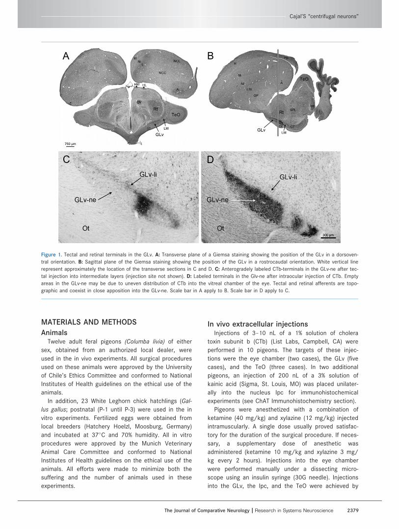
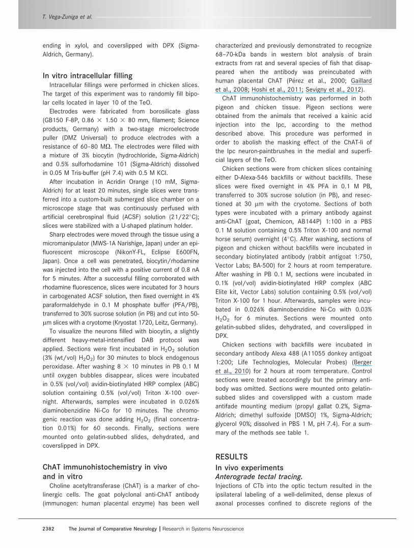
Figure 1. Tectal and retinal terminals in the GLv. A: Transverse plane of a Giemsa staining showing the position of the GLv in a dorsoven-
tral orientation. B: Sagittal plane of the Giemsa staining showing the position of the GLv in a rostrocaudal orientation. White vertical line
represent approximately the location of the transverse sections in C and D. C: Anterogradely labeled CTb-terminals in the GLv-ne after tec-
tal injection into intermediate layers (injection site not shown). D: Labeled terminals in the Glv-ne after intraocular injection of CTb. Empty
areas in the GLv-ne may be due to uneven distribution of CTb into the vitreal chamber of the eye. Tectal and retinal afferents are topo-
graphic and coexist in close apposition into the GLv-ne. Scale bar in A apply to B. Scale bar in D apply to C.
Cajal’S “centrifugal neurons”
The Journal of Comparative Neurology | Research in Systems Neuroscience 2379
stereotaxically (Karten and Hodos, 1967) lowering a
micropipette filled with the CTb solution into the
desired area. Once the target was reached, the tracer
was injected applying pressure pulses using a picosprit-
zer (Picospritzer II, General Valve, Fairfield, NJ). In order
to enhance the accuracy of the injections, electrophysi-
ological responses were monitored during the stereo-
taxic penetration. For a detailed review of these
procedures, see Mpodozis et al. (1996).
After the injections the wounds were covered, the
skin sutured, and treated with topical antibiotics. During
the experiments the heart rate of the animals was con-
tinuously monitored and the body temperature was
held at 40–42C� by means of a thermoregulated elec-
tric blanket. During surgery and recovery all wounds
and pressure points were treated with a commercial
ointment of 5% lidocaine.
After 5–9 days of survival, and 30 days in the cases
with the kainic acid injection, the animals were deeply
anesthetized with an overdose of a mixture of ketamine
and xylazine and perfused via the aorta with 500–800
ml of 0.75% saline, followed by 1,000 ml of an ice-cold
solution of 4% paraformaldehyde in 0.1 M phosphate
buffer (PB, pH 7.4). After the perfusion, brains were
excised, postfixed overnight in the paraformaldehyde
solution, and then transferred for 2–3 days to a 30%
sucrose solution (in 0.1 M PB) for cryoprotection. The
brains were then mounted in the stereotaxic plane over
the stage of a frozen sliding microtome and 30 lm sec-
tions were cut in the transverse plane.
In the cases with the CTb injection, sections were
washed 30 minutes in 0.1 M phosphate-buffered saline
(PBS, pH 7.4) and incubated in goat anti-CTb (1:15,000,
List Labs) overnight at 4�C. The tissue was then proc-
essed using the avidin-biotin-peroxidase method (ABC
Elite kit, Vector Labs, Burlingame, CA) in 0.3% Triton X-
100 in PB for 1 hour. Sections were then washed in
PBS and reacted in 0.025% diaminobenzidine (DAB,
Sigma) and 0.3% hydrogen peroxide in PB for 10
minutes. The tissue was mounted on gelatinized slides,
dehydrated, cleared, and coverslipped using Permount
(Fisher Scientific, Fair Lawn, NJ). Previous control
experiments using the same protocol but without CTb
injection in the brain were performed. The results
showed no labeling in the brain of pigeons (data not
shown).
In vitro extracellular injectionsInjections targeting the GLv and the TeO in the
chicken were made using 12 nL of a 10% solution of
dextran biotin (BDA 10,000 MW, Molecular Probes) or
dextran Alexa Fluor 546 10% (10,000 MW, Invitrogen,
Molecular Probes) dissolved in PB 0.1 M.
Chick hatchlings were deeply anesthetized with a
mixture (3:1) of ketamine (50 mg/ml; Inresa Arzneimit-
tel) and Rompun (2%; Bayer, Leverkusen, Germany) at
37.5 and 5 mg/kg, respectively, and subsequently
decapitated. The skull was opened at the midsagittal
line and the dorsal surface of the brain exposed. After
removal, the brain was transferred into ice-cooled (4�C)
sucrose-substituted Krebs solution (210 mM sucrose, 3
mM KCl (Sigma), 3 mM MgCl2�6H2O, 23 mM NaHCO3,
1.2 mM NaH2PO4�6H2O (Laborbedarf-Vertrieb, Ger-
many), 11 mM D1-glucose).
The forebrain and cerebellum were removed with
cuts through the junction with the diencephalon and
the cerebellar peduncles. The remaining parts of the
brain were cut midsagittally into two hemispheres. The
brain tissue was then embedded in agar at 48�C and
rapidly cooled (1.5% agar in HEPES solution: 290 mM
sucrose, 3 mM KCl, 3 mM MgCl2, and 5 mM HEPES;
Sigma). The optic tectum of each hemisphere was
aligned in an oblique transverse plane. The hemispheres
were cut into slices between 500–900 lm with a vibra-
tome (VF 200 Microtome, Precisionary Instruments, CA,
USA). Slices containing the TeO-GLv were placed in
standard ACSF solution (120 mM NaCl, 3 mM KCl, 1
mM MgCl2�6H2O, 23 mM NaHCO3, 1.2 mM NaH2-
PO4�1H2O, 11 mM D1-glucose) and kept submerged in
a chamber that was bubbled continuously with carbo-
gen (95% oxygen, 5% CO2) at room temperature for at
least 30 minutes.
Pipettes were fabricated from borosilicate glass
(GB100-8P, 0.58 3 1.00 3 80 mm; Science Products,
Germany) with a one-stage microelectrode puller (Sutter
Instrument, USA) to produce a tip opening �30 lm.
The electrodes were filled with mineral oil and attached
to a Nanoliter 2000 injector (World Precision Instru-
ments, USA). Electrodes were filled with dextran biotin
10% (BDA 10,000 MW, Molecular Probes) or dextran
Alexa Fluor 546 10% (10,000 MW; Invitrogen, Molecular
Probes) dissolved in PB 0.1 M.
Single slices were submerged in a chamber with car-
bogenated ACSF solution. Under microscopic control
and using a microdrive (Maerzhaeuser, West Germany),
injections of 13.8 nL tracer solution in three different
positions along the GLv-ne were performed (medio-lat-
eral axis).
After injecting, slices were incubated 4 hours in car-
bogenated ACSF solution to allow transport of the
tracer.
Slices containing BDA backfills were fixed overnight
in 4% paraformaldehyde in 0.1 M phosphate buffer
(PFA/PB), transferred to 30% sucrose solution (in PB)
for 2 hours, and cut to 60-lm sections with a cryotome
(Kryostat 1720, Leitz, Germany). To visualize the filled
T. Vega-Zuniga et al.
2380 The Journal of Comparative Neurology |Research in Systems Neuroscience
neurons, a heavy-metal-intensified DAB protocol was
applied. Sections were first incubated in H2O2 solution
(3% (wt/vol) H2O2) for 30 minutes to block endogenous
peroxidase. After washing eight times (8 3 10 minutes)
in PB 0.1 M until oxygen bubbles disappeared, sections
were incubated in 0.1% (vol/vol) avidin-biotinylated
horseradish peroxidase (HRP) complex (ABC Elite kit,
Vector Labs, Burlingame, CA) solution containing 0.5%
(vol/vol) Triton X-100 for 2 hours. Afterwards, samples
were incubated in 0.026% diaminobenzidine Ni-Co with
0.03% H2O2 for 6 minutes. Sections were mounted onto
gelatin-subbed slides, dehydrated in an ethanol series
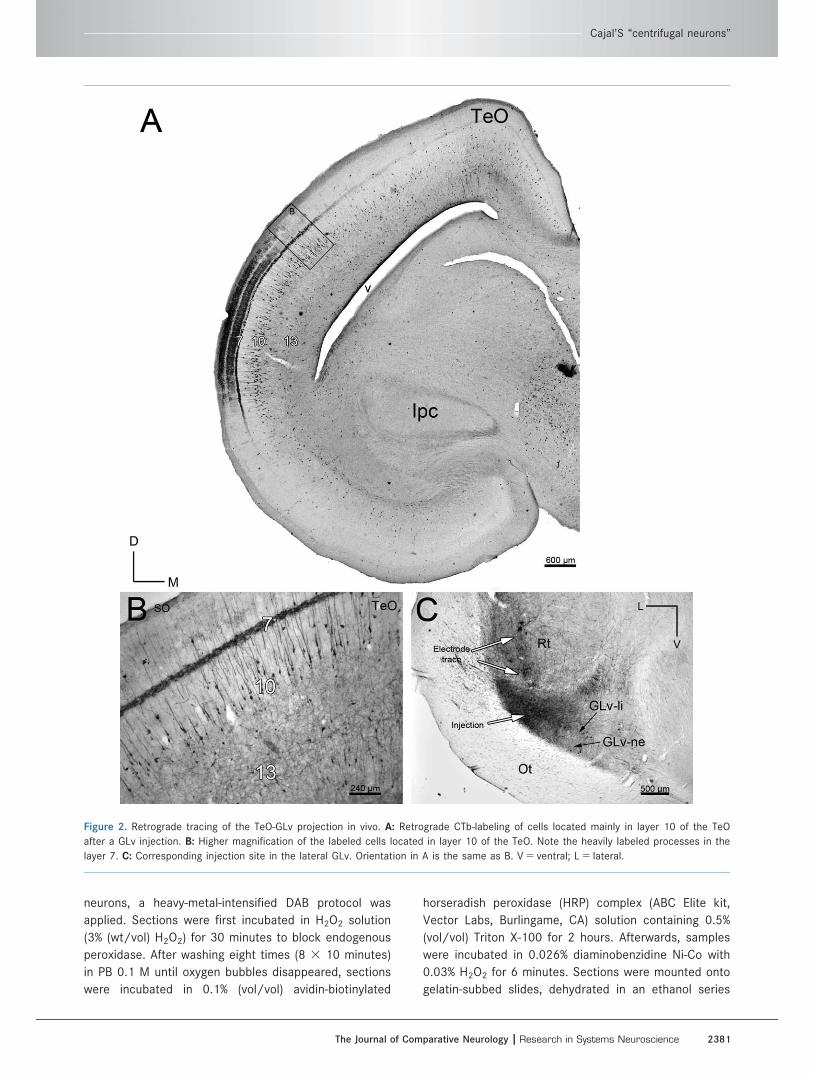
Figure 2. Retrograde tracing of the TeO-GLv projection in vivo. A: Retrograde CTb-labeling of cells located mainly in layer 10 of the TeO
after a GLv injection. B: Higher magnification of the labeled cells located in layer 10 of the TeO. Note the heavily labeled processes in the
layer 7. C: Corresponding injection site in the lateral GLv. Orientation in A is the same as B. V 5 ventral; L 5 lateral.
Cajal’S “centrifugal neurons”
The Journal of Comparative Neurology | Research in Systems Neuroscience 2381
ending in xylol, and coverslipped with DPX (Sigma-
Aldrich, Germany).
In vitro intracellular fillingIntracellular fillings were performed in chicken slices.
The target of this experiment was to randomly fill bipo-
lar cells located in layer 10 of the TeO.
Electrodes were fabricated from borosilicate glass
(GB150 F-8P, 0.86 3 1.50 3 80 mm, filament; Science
products, Germany) with a two-stage microelectrode
puller (DMZ Universal) to produce electrodes with a
resistance of 60–80 MX. The electrodes were filled with
a mixture of 3% biocytin (hydrochloride, Sigma-Aldrich)
and 0.5% sulforhodamine 101 (Sigma-Aldrich) dissolved
in 0.05 M Tris-buffer (pH 7.4) with 0.5 M KCl.
After incubation in Acridin Orange (10 mM, Sigma-
Aldrich) for at least 20 minutes, single slices were trans-
ferred into a custom-built submerged slice chamber on a
microscope stage that was continuously perfused with
artificial cerebrospinal fluid (ACSF) solution (21/22�C);
slices were stabilized with a U-shaped platinum holder.
Sharp electrodes were moved through the tissue using a
micromanipulator (MWS-1A Narishige, Japan) under an epi-
fluorescent microscope (NikonY-FL, Eclipse E600FN,
Japan). Once a cell was penetrated, biocytin/rhodamine
was injected into the cell with a positive current of 0.8 nA
for 5 minutes. After a successful filling corroborated with
rhodamine fluorescence, slices were incubated for 3 hours
in carbogenated ACSF solution, then fixed overnight in 4%
paraformaldehyde in 0.1 M phosphate buffer (PFA/PB),
transferred to 30% sucrose solution (in PB) and cut into 50-
lm slices with a cryotome (Kryostat 1720, Leitz, Germany).
To visualize the neurons filled with biocytin, a slightly
different heavy-metal-intensified DAB protocol was
applied. Sections were first incubated in H2O2 solution
(3% (wt/vol) H2O2) for 30 minutes to block endogenous
peroxidase. After washing 8 3 10 minutes in PB 0.1 M
until oxygen bubbles disappear, slices were incubated
in 0.5% (vol/vol) avidin-biotinylated HRP complex (ABC)
solution containing 0.5% (vol/vol) Triton X-100 over-
night. Afterwards, samples were incubated in 0.026%
diaminobenzidine Ni-Co for 10 minutes. The chromo-
genic reaction was done adding H2O2 (final concentra-
tion 0.01%) for 60 seconds. Finally, sections were
mounted onto gelatin-subbed slides, dehydrated, and
coverslipped in DPX.
ChAT immunohistochemistry in vivoand in vitro
Choline acetyltransferase (ChAT) is a marker of cho-
linergic cells. The goat polyclonal anti-ChAT antibody
(immunogen: human placental enzyme) has been well
characterized and previously demonstrated to recognize
68–70-kDa bands in western blot analysis of brain
extracts from rat and several species of fish that disap-
peared when the antibody was preincubated with
human placental ChAT (P�erez et al., 2000; Gaillard
et al., 2008; Hoshi et al., 2011; Sevigny et al., 2012).
ChAT immunohistochemistry was performed in both
pigeon and chicken tissue. Pigeon sections were
obtained from the animals that received a kainic acid
injection into the Ipc, according to the method
described above. This procedure was performed in
order to abolish the masking effect of the ChAT-li of
the Ipc neuron-paintbrushes in the medial and superfi-
cial layers of the TeO.
Chicken sections were from chicken slices containing
either D-Alexa-546 backfills or without backfills. These
slices were fixed overnight in 4% PFA in 0.1 M PB,
transferred to 30% sucrose solution (in PB), and resec-
tioned at 30 lm with the cryotome. Sections of both
types were incubated with a primary antibody against
anti-ChAT (goat, Chemicon, AB144P) 1:100 in a PBS
0.1 M solution containing 0.5% Triton X-100 and normal
horse serum) overnight (4�C). After washing, sections of
pigeon and chicken without backfills were incubated in
secondary biotinylated antibody (rabbit antigoat 1:750,
Vector Labs; BA-500) for 2 hours at room temperature.
After washing in PB 0.1 M, sections were incubated in
0.1% (vol/vol) avidin-biotinylated HRP complex (ABC
Elite kit, Vector Labs) solution containing 0.5% (vol/vol)
Triton X-100 for 1 hour. Afterwards, samples were incu-
bated in 0.026% diaminobenzidine Ni-Co with 0.03%
H2O2 for 6 minutes. Sections were mounted onto
gelatin-subbed slides, dehydrated, and coverslipped in
DPX.
Chicken sections with backfills were incubated in
secondary antibody Alexa 488 (A11055 donkey antigoat
1:200; Life Technologies, Molecular Probes) (Berger
et al., 2010) for 2 hours at room temperature. Control
sections were treated accordingly but the primary anti-
body was omitted. Sections were mounted onto gelatin-
subbed slides and coverslipped with a custom made
antifade mounting medium (propyl gallat 0.2%, Sigma-
Aldrich; dimethyl sulfoxide [DMSO] 1%, Sigma-Aldrich;
glycerol 90%; dissolved in PBS 1 M, pH 7.4). For a sum-
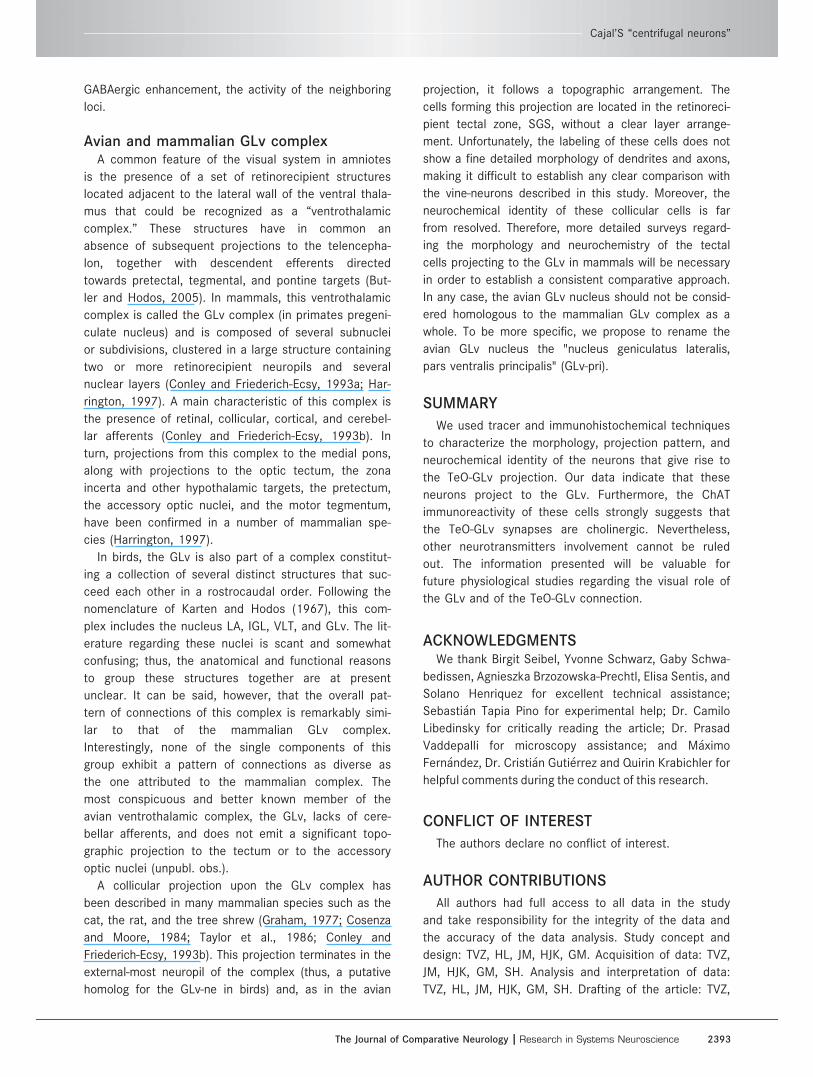
mary of the methods see table 1.
RESULTS
In vivo experimentsAnterograde tectal tracing.Injections of CTb into the optic tectum resulted in the
ipsilateral labeling of a well-delimited, dense plexus of
axonal processes confined to discrete regions of the
T. Vega-Zuniga et al.
2382 The Journal of Comparative Neurology |Research in Systems Neuroscience
GLv-ne (Fig. 1A). These tectal projections were topo-
graphically organized, as different injection sites into
the optic tectum resulted in the labeling of fine axonal
terminals in different locations within the GLv (data not
shown). Tectal axonal terminals were of very fine cali-
ber and spanned the whole dorsoventral extension of
the GLv-ne, without noticeable invasion of the GLv-li.
The distribution of the tectal terminals overlap fully with
that of the more coarse retinal terminals, which also
span densely through the whole extent of the GLv-ne
until the limit with the GLv-li (Fig. 1B).
Injections of CTb into the optic tectum also resulted
in the ipsilateral retrograde labeling of many cells within
the IGL, the LMmc, and the VLT (data not shown). We
were unable to find any indication of a similar substan-
tial projection from the GLv upon the optic tectum.
Only very rarely were retrograde CTb-labeled cells found
in the GLv, even in cases with large tectal injections.
These rare cells were located mostly in the GLv-li, with
occasional ones appearing in the GLv-ne.
Retrograde GLv tracing.Due to the peculiar shape of the GLv, we were unable to
obtain restricted tracer injections either confined to the
limits of the nucleus or resulting in a complete injection
of the whole extent of the nucleus. The most consistent
results (three cases) were achieved by injecting the cen-
tral portion of the GLv, at the level of the central Rt (Fig.
2C). In these cases, only minor leakage to the Rt, the
medially adjacent VSOD, the dorsally adjacent VLT, and
the immediately ventral optic tract was observed. These
injections consistently resulted in the ipsilateral retro-
grade labeling of a characteristic population of bipolar
cells located in tectal layer 10 (Fig. 2A). These labeled
cells had a very distinct morphology, featuring small fusi-
form somata and single apical and basal dendrites
extending radially in opposite directions. The apical den-
drite extended towards layer 2, and formed a conspicu-
ous dense, transversely oriented specialization within the
limits of layer 7 (Fig. 2B). The basal dendrite, of finer cali-
ber, extended towards the deeper layers, and lacked evi-
dent specializations. In some instances it was possible to
trace the axon of these cells arising from the apical den-
drite and following an ascending course towards layer 1.
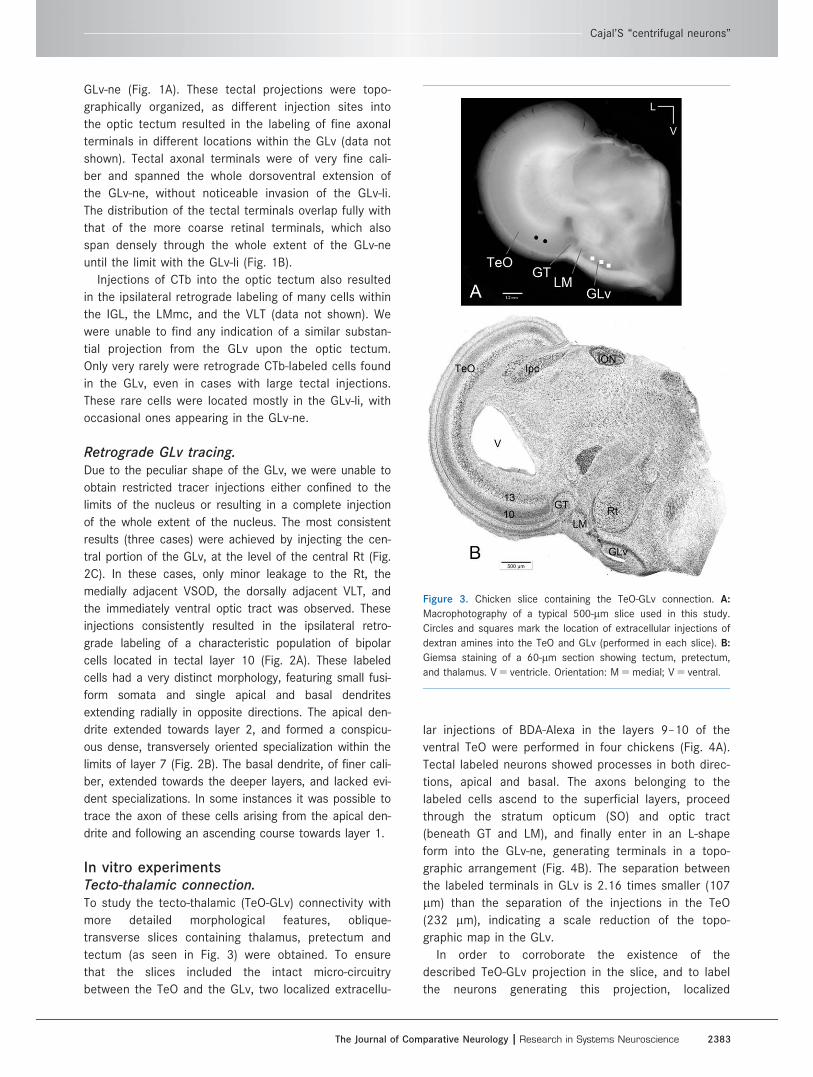
In vitro experimentsTecto-thalamic connection.To study the tecto-thalamic (TeO-GLv) connectivity with
more detailed morphological features, oblique-
transverse slices containing thalamus, pretectum and
tectum (as seen in Fig. 3) were obtained. To ensure
that the slices included the intact micro-circuitry
between the TeO and the GLv, two localized extracellu-
lar injections of BDA-Alexa in the layers 9–10 of the
ventral TeO were performed in four chickens (Fig. 4A).
Tectal labeled neurons showed processes in both direc-
tions, apical and basal. The axons belonging to the
labeled cells ascend to the superficial layers, proceed
through the stratum opticum (SO) and optic tract
(beneath GT and LM), and finally enter in an L-shape
form into the GLv-ne, generating terminals in a topo-
graphic arrangement (Fig. 4B). The separation between
the labeled terminals in GLv is 2.16 times smaller (107
lm) than the separation of the injections in the TeO
(232 lm), indicating a scale reduction of the topo-
graphic map in the GLv.
In order to corroborate the existence of the
described TeO-GLv projection in the slice, and to label
the neurons generating this projection, localized
Figure 3. Chicken slice containing the TeO-GLv connection. A:
Macrophotography of a typical 500-lm slice used in this study.
Circles and squares mark the location of extracellular injections of
dextran amines into the TeO and GLv (performed in each slice). B:
Giemsa staining of a 60-lm section showing tectum, pretectum,
and thalamus. V 5 ventricle. Orientation: M 5 medial; V 5 ventral.
Cajal’S “centrifugal neurons”
The Journal of Comparative Neurology | Research in Systems Neuroscience 2383
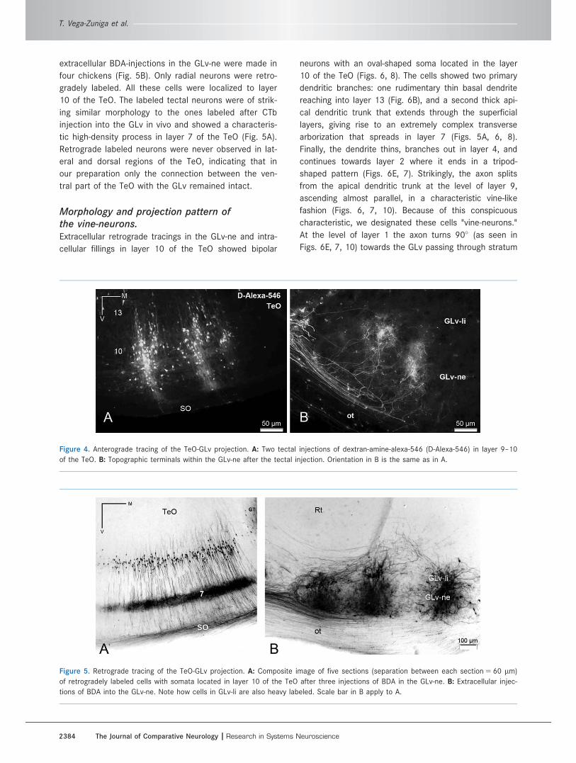
extracellular BDA-injections in the GLv-ne were made in
four chickens (Fig. 5B). Only radial neurons were retro-
gradely labeled. All these cells were localized to layer
10 of the TeO. The labeled tectal neurons were of strik-
ing similar morphology to the ones labeled after CTb
injection into the GLv in vivo and showed a characteris-
tic high-density process in layer 7 of the TeO (Fig. 5A).
Retrograde labeled neurons were never observed in lat-
eral and dorsal regions of the TeO, indicating that in
our preparation only the connection between the ven-
tral part of the TeO with the GLv remained intact.
Morphology and projection pattern ofthe vine-neurons.Extracellular retrograde tracings in the GLv-ne and intra-
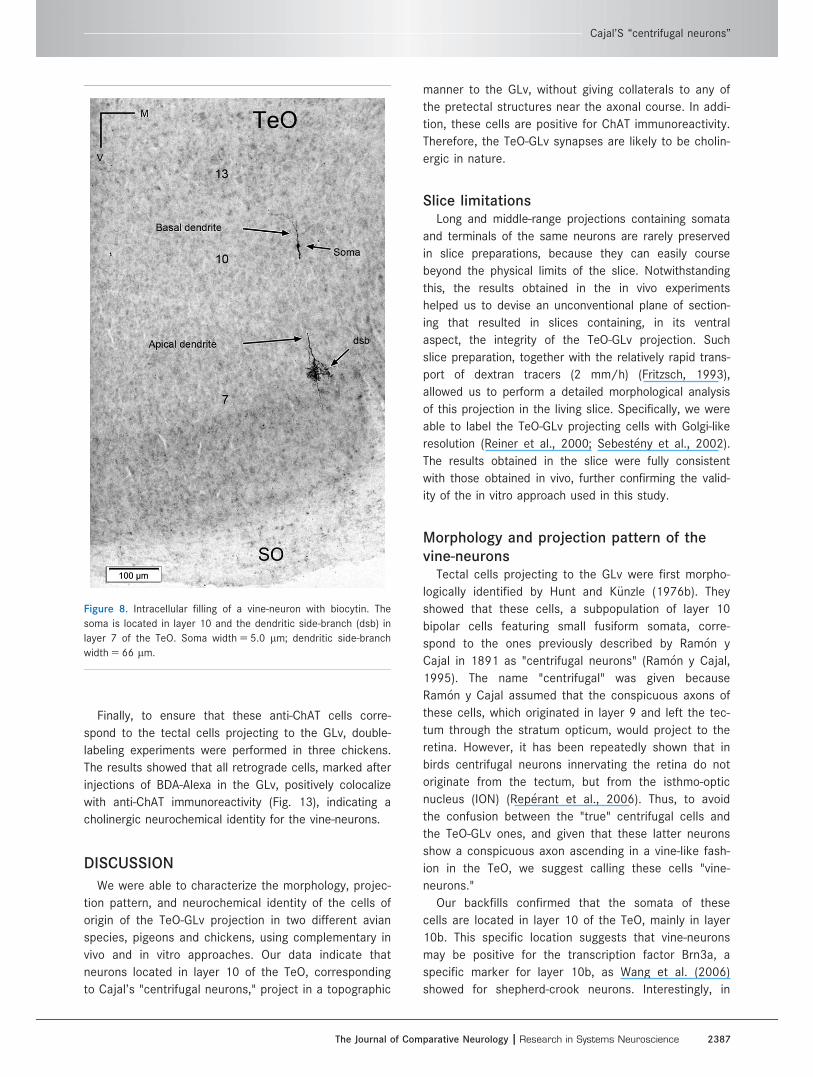
cellular fillings in layer 10 of the TeO showed bipolar
neurons with an oval-shaped soma located in the layer
10 of the TeO (Figs. 6, 8). The cells showed two primary
dendritic branches: one rudimentary thin basal dendrite
reaching into layer 13 (Fig. 6B), and a second thick api-
cal dendritic trunk that extends through the superficial
layers, giving rise to an extremely complex transverse
arborization that spreads in layer 7 (Figs. 5A, 6, 8).
Finally, the dendrite thins, branches out in layer 4, and
continues towards layer 2 where it ends in a tripod-
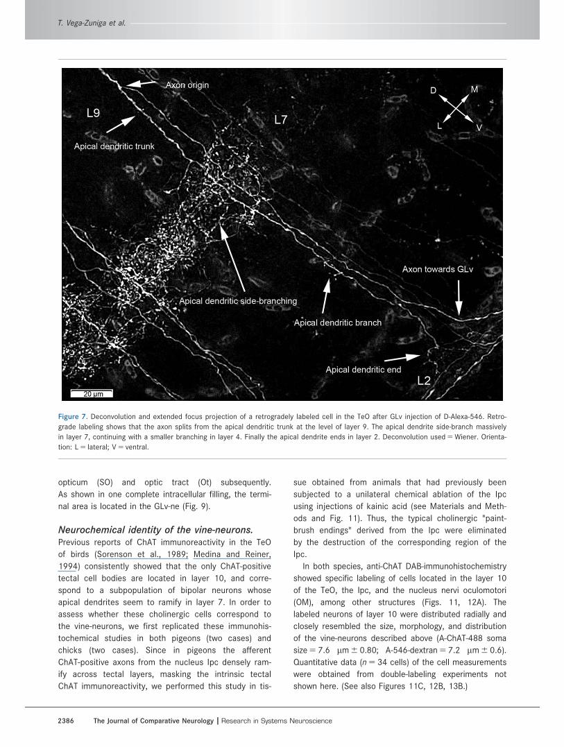
shaped pattern (Figs. 6E, 7). Strikingly, the axon splits
from the apical dendritic trunk at the level of layer 9,
ascending almost parallel, in a characteristic vine-like
fashion (Figs. 6, 7, 10). Because of this conspicuous
characteristic, we designated these cells "vine-neurons."
At the level of layer 1 the axon turns 90� (as seen in
Figs. 6E, 7, 10) towards the GLv passing through stratum
Figure 4. Anterograde tracing of the TeO-GLv projection. A: Two tectal injections of dextran-amine-alexa-546 (D-Alexa-546) in layer 9–10
of the TeO. B: Topographic terminals within the GLv-ne after the tectal injection. Orientation in B is the same as in A.
Figure 5. Retrograde tracing of the TeO-GLv projection. A: Composite image of five sections (separation between each section 5 60 lm)
of retrogradely labeled cells with somata located in layer 10 of the TeO after three injections of BDA in the GLv-ne. B: Extracellular injec-
tions of BDA into the GLv-ne. Note how cells in GLv-li are also heavy labeled. Scale bar in B apply to A.
T. Vega-Zuniga et al.
2384 The Journal of Comparative Neurology |Research in Systems Neuroscience
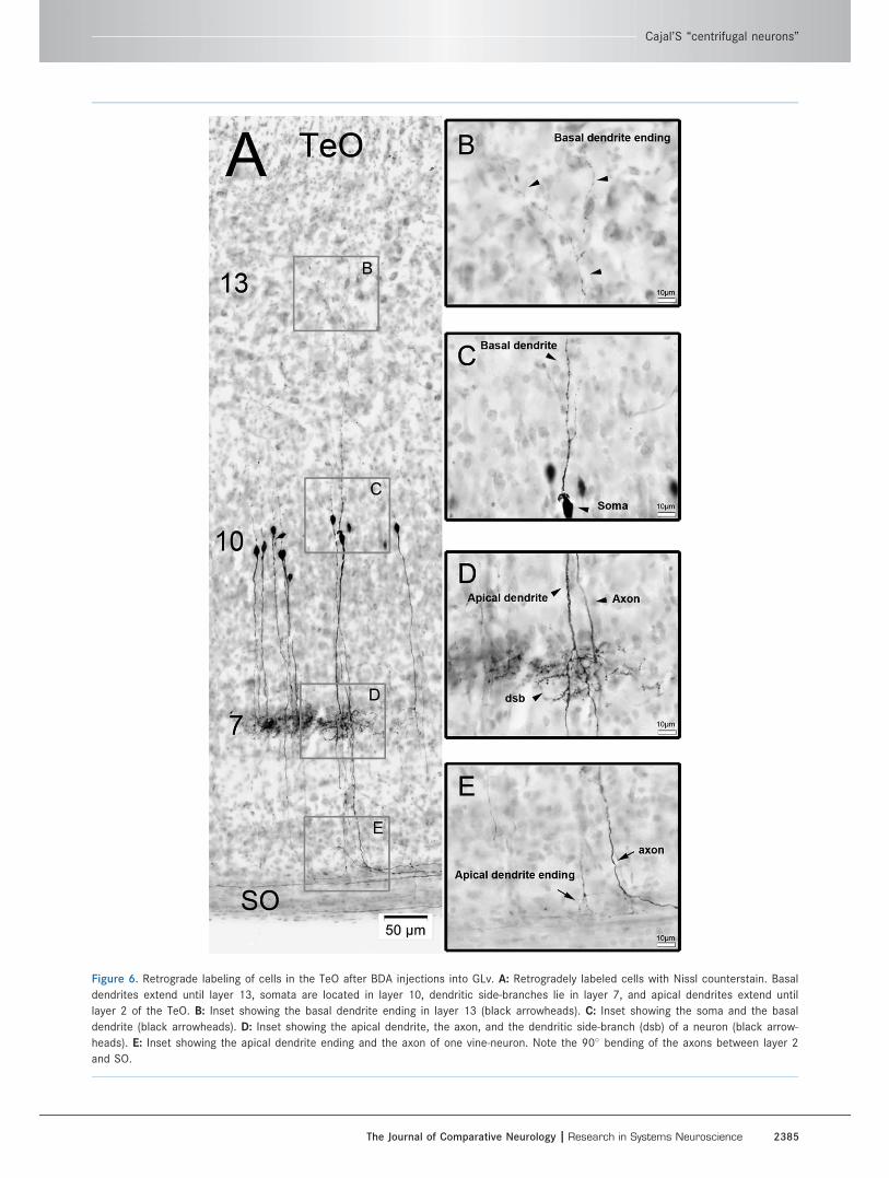
Figure 6. Retrograde labeling of cells in the TeO after BDA injections into GLv. A: Retrogradely labeled cells with Nissl counterstain. Basal
dendrites extend until layer 13, somata are located in layer 10, dendritic side-branches lie in layer 7, and apical dendrites extend until
layer 2 of the TeO. B: Inset showing the basal dendrite ending in layer 13 (black arrowheads). C: Inset showing the soma and the basal
dendrite (black arrowheads). D: Inset showing the apical dendrite, the axon, and the dendritic side-branch (dsb) of a neuron (black arrow-
heads). E: Inset showing the apical dendrite ending and the axon of one vine-neuron. Note the 90� bending of the axons between layer 2
and SO.
Cajal’S “centrifugal neurons”
The Journal of Comparative Neurology | Research in Systems Neuroscience 2385
opticum (SO) and optic tract (Ot) subsequently.
As shown in one complete intracellular filling, the termi-
nal area is located in the GLv-ne (Fig. 9).
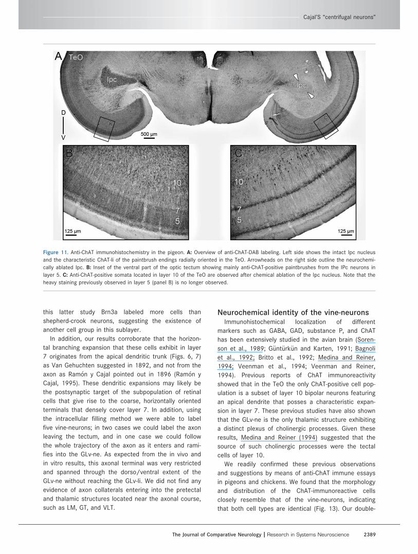
Neurochemical identity of the vine-neurons.Previous reports of ChAT immunoreactivity in the TeO
of birds (Sorenson et al., 1989; Medina and Reiner,
1994) consistently showed that the only ChAT-positive
tectal cell bodies are located in layer 10, and corre-
spond to a subpopulation of bipolar neurons whose
apical dendrites seem to ramify in layer 7. In order to
assess whether these cholinergic cells correspond to
the vine-neurons, we first replicated these immunohis-
tochemical studies in both pigeons (two cases) and
chicks (two cases). Since in pigeons the afferent
ChAT-positive axons from the nucleus Ipc densely ram-
ify across tectal layers, masking the intrinsic tectal
ChAT immunoreactivity, we performed this study in tis-
sue obtained from animals that had previously been
subjected to a unilateral chemical ablation of the Ipc
using injections of kainic acid (see Materials and Meth-
ods and Fig. 11). Thus, the typical cholinergic "paint-
brush endings" derived from the Ipc were eliminated
by the destruction of the corresponding region of the
Ipc.
In both species, anti-ChAT DAB-immunohistochemistry
showed specific labeling of cells located in the layer 10
of the TeO, the Ipc, and the nucleus nervi oculomotori
(OM), among other structures (Figs. 11, 12A). The
labeled neurons of layer 10 were distributed radially and
closely resembled the size, morphology, and distribution
of the vine-neurons described above (A-ChAT-488 soma
size 5 7.6 lm 6 0.80; A-546-dextran 5 7.2 lm 6 0.6).
Quantitative data (n 5 34 cells) of the cell measurements
were obtained from double-labeling experiments not
shown here. (See also Figures 11C, 12B, 13B.)
Figure 7. Deconvolution and extended focus projection of a retrogradely labeled cell in the TeO after GLv injection of D-Alexa-546. Retro-
grade labeling shows that the axon splits from the apical dendritic trunk at the level of layer 9. The apical dendrite side-branch massively
in layer 7, continuing with a smaller branching in layer 4. Finally the apical dendrite ends in layer 2. Deconvolution used 5 Wiener. Orienta-
tion: L 5 lateral; V 5 ventral.
T. Vega-Zuniga et al.
2386 The Journal of Comparative Neurology |Research in Systems Neuroscience
Finally, to ensure that these anti-ChAT cells corre-
spond to the tectal cells projecting to the GLv, double-
labeling experiments were performed in three chickens.
The results showed that all retrograde cells, marked after
injections of BDA-Alexa in the GLv, positively colocalize
with anti-ChAT immunoreactivity (Fig. 13), indicating a
cholinergic neurochemical identity for the vine-neurons.
DISCUSSION
We were able to characterize the morphology, projec-
tion pattern, and neurochemical identity of the cells of
origin of the TeO-GLv projection in two different avian
species, pigeons and chickens, using complementary in
vivo and in vitro approaches. Our data indicate that
neurons located in layer 10 of the TeO, corresponding
to Cajal’s "centrifugal neurons," project in a topographic
manner to the GLv, without giving collaterals to any of
the pretectal structures near the axonal course. In addi-
tion, these cells are positive for ChAT immunoreactivity.
Therefore, the TeO-GLv synapses are likely to be cholin-
ergic in nature.
Slice limitationsLong and middle-range projections containing somata
and terminals of the same neurons are rarely preserved
in slice preparations, because they can easily course
beyond the physical limits of the slice. Notwithstanding
this, the results obtained in the in vivo experiments
helped us to devise an unconventional plane of section-
ing that resulted in slices containing, in its ventral
aspect, the integrity of the TeO-GLv projection. Such
slice preparation, together with the relatively rapid trans-
port of dextran tracers (2 mm/h) (Fritzsch, 1993),
allowed us to perform a detailed morphological analysis
of this projection in the living slice. Specifically, we were
able to label the TeO-GLv projecting cells with Golgi-like
resolution (Reiner et al., 2000; Sebest�eny et al., 2002).
The results obtained in the slice were fully consistent
with those obtained in vivo, further confirming the valid-
ity of the in vitro approach used in this study.
Morphology and projection pattern of thevine-neurons
Tectal cells projecting to the GLv were first morpho-
logically identified by Hunt and K€unzle (1976b). They
showed that these cells, a subpopulation of layer 10
bipolar cells featuring small fusiform somata, corre-
spond to the ones previously described by Ram�on y
Cajal in 1891 as "centrifugal neurons" (Ram�on y Cajal,
1995). The name "centrifugal" was given because
Ram�on y Cajal assumed that the conspicuous axons of
these cells, which originated in layer 9 and left the tec-
tum through the stratum opticum, would project to the
retina. However, it has been repeatedly shown that in
birds centrifugal neurons innervating the retina do not
originate from the tectum, but from the isthmo-optic
nucleus (ION) (Rep�erant et al., 2006). Thus, to avoid
the confusion between the "true" centrifugal cells and
the TeO-GLv ones, and given that these latter neurons
show a conspicuous axon ascending in a vine-like fash-
ion in the TeO, we suggest calling these cells "vine-
neurons."
Our backfills confirmed that the somata of these
cells are located in layer 10 of the TeO, mainly in layer
10b. This specific location suggests that vine-neurons
may be positive for the transcription factor Brn3a, a
specific marker for layer 10b, as Wang et al. (2006)
showed for shepherd-crook neurons. Interestingly, in
Figure 8. Intracellular filling of a vine-neuron with biocytin. The
soma is located in layer 10 and the dendritic side-branch (dsb) in
layer 7 of the TeO. Soma width 5 5.0 lm; dendritic side-branch
width 5 66 lm.
Cajal’S “centrifugal neurons”
The Journal of Comparative Neurology | Research in Systems Neuroscience 2387
Figure 9. Intracellular filling showing a tectal terminal in the GLv-ne. A: Composite image of two contiguous 60-lm sections of the termi-
nal of a vine-neuron in the lateral part of the GLv-ne. B,C: Inset showing the two sections where the terminal was observed. Scale bar in
B apply to C.
Figure 10. Reconstruction of an intracellularly filled vine-neuron with terminals in the GLv. Figure shows a complete filled neuron with bio-
cytin. Basal dendrite does not reach layer 13 as previously shown probably because it extend beyond the limits of the slice. The axon
runs through the upper limit of the SO and the Ot before entering the GLv. Note the L-shape form of the axon entrance into the GLv and
the butterfly-shape of the terminal in GLv-ne. 10, 9, 7 5 layers of the TeO.
T. Vega-Zuniga et al.
2388 The Journal of Comparative Neurology |Research in Systems Neuroscience
this latter study Brn3a labeled more cells than
shepherd-crook neurons, suggesting the existence of
another cell group in this sublayer.
In addition, our results corroborate that the horizon-
tal branching expansion that these cells exhibit in layer
7 originates from the apical dendritic trunk (Figs. 6, 7)
as Van Gehuchten suggested in 1892, and not from the
axon as Ram�on y Cajal pointed out in 1896 (Ram�on y
Cajal, 1995). These dendritic expansions may likely be
the postsynaptic target of the subpopulation of retinal
cells that give rise to the coarse, horizontally oriented
terminals that densely cover layer 7. In addition, using
the intracellular filling method we were able to label
five vine-neurons; in two cases we could label the axon
leaving the tectum, and in one case we could follow
the whole trajectory of the axon as it enters and rami-
fies into the GLv-ne. As expected from the in vivo and
in vitro results, this axonal terminal was very restricted
and spanned through the dorso/ventral extent of the
GLv-ne without reaching the GLv-li. We did not find any
evidence of axon collaterals entering into the pretectal
and thalamic structures located near the axonal course,
such as LM, GT, and VLT.
Neurochemical identity of the vine-neuronsImmunohistochemical localization of different
markers such as GABA, GAD, substance P, and ChAT
has been extensively studied in the avian brain (Soren-
son et al., 1989; G€unt€urk€un and Karten, 1991; Bagnoli
et al., 1992; Britto et al., 1992; Medina and Reiner,
1994; Veenman et al., 1994; Veenman and Reiner,
1994). Previous reports of ChAT immunoreactivity
showed that in the TeO the only ChAT-positive cell pop-
ulation is a subset of layer 10 bipolar neurons featuring
an apical dendrite that posses a characteristic expan-
sion in layer 7. These previous studies have also shown
that the GLv-ne is the only thalamic structure exhibiting
a distinct plexus of cholinergic processes. Given these
results, Medina and Reiner (1994) suggested that the
source of such cholinergic processes were the tectal
cells of layer 10.
We readily confirmed these previous observations
and suggestions by means of anti-ChAT immune essays
in pigeons and chickens. We found that the morphology
and distribution of the ChAT-immunoreactive cells
closely resemble that of the vine-neurons, indicating
that both cell types are identical (Fig. 13). Our double-
Figure 11. Anti-ChAT immunohistochemistry in the pigeon. A: Overview of anti-ChAT-DAB labeling. Left side shows the intact Ipc nucleus
and the characteristic ChAT-li of the paintbrush endings radially oriented in the TeO. Arrowheads on the right side outline the neurochemi-
cally ablated Ipc. B: Inset of the ventral part of the optic tectum showing mainly anti-ChAT-positive paintbrushes from the IPc neurons in
layer 5. C: Anti-ChAT-positive somata located in layer 10 of the TeO are observed after chemical ablation of the Ipc nucleus. Note that the
heavy staining previously observed in layer 5 (panel B) is no longer observed.
Cajal’S “centrifugal neurons”
The Journal of Comparative Neurology | Research in Systems Neuroscience 2389
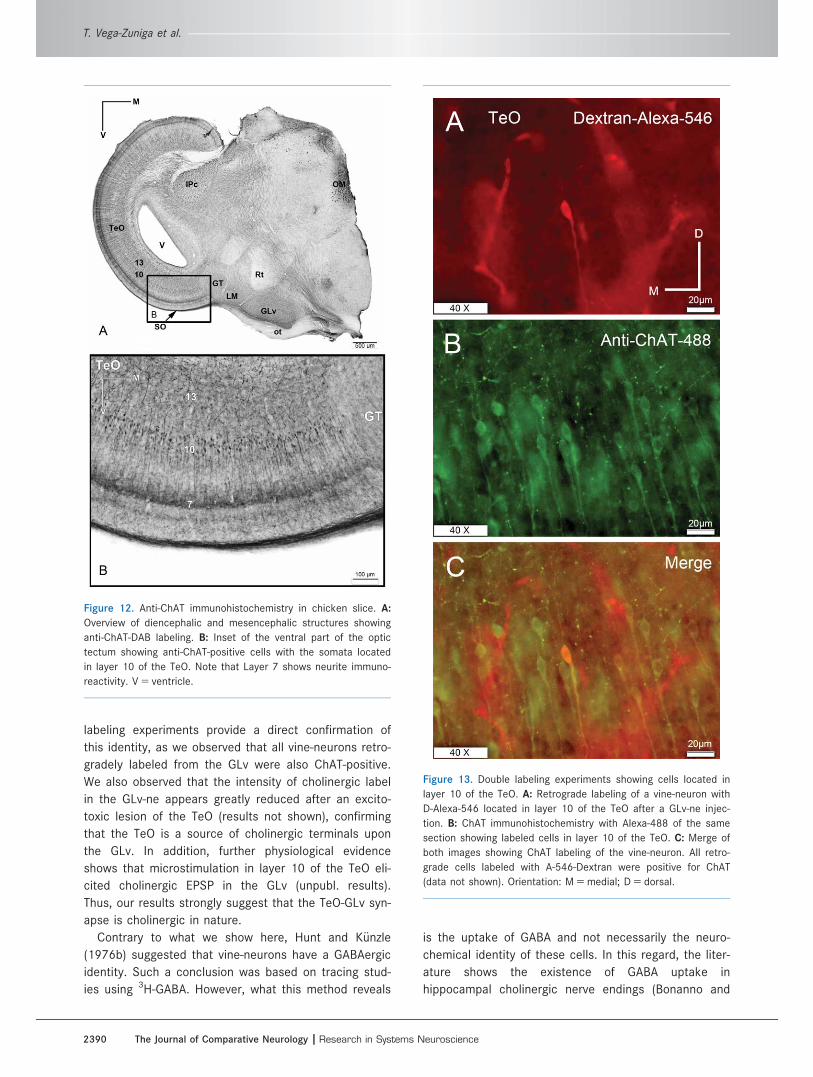
labeling experiments provide a direct confirmation of
this identity, as we observed that all vine-neurons retro-
gradely labeled from the GLv were also ChAT-positive.
We also observed that the intensity of cholinergic label
in the GLv-ne appears greatly reduced after an excito-
toxic lesion of the TeO (results not shown), confirming
that the TeO is a source of cholinergic terminals upon
the GLv. In addition, further physiological evidence
shows that microstimulation in layer 10 of the TeO eli-
cited cholinergic EPSP in the GLv (unpubl. results).
Thus, our results strongly suggest that the TeO-GLv syn-
apse is cholinergic in nature.
Contrary to what we show here, Hunt and K€unzle
(1976b) suggested that vine-neurons have a GABAergic
identity. Such a conclusion was based on tracing stud-
ies using 3H-GABA. However, what this method reveals
is the uptake of GABA and not necessarily the neuro-
chemical identity of these cells. In this regard, the liter-
ature shows the existence of GABA uptake in
hippocampal cholinergic nerve endings (Bonanno and
Figure 12. Anti-ChAT immunohistochemistry in chicken slice. A:
Overview of diencephalic and mesencephalic structures showing
anti-ChAT-DAB labeling. B: Inset of the ventral part of the optic
tectum showing anti-ChAT-positive cells with the somata located
in layer 10 of the TeO. Note that Layer 7 shows neurite immuno-
reactivity. V 5 ventricle.
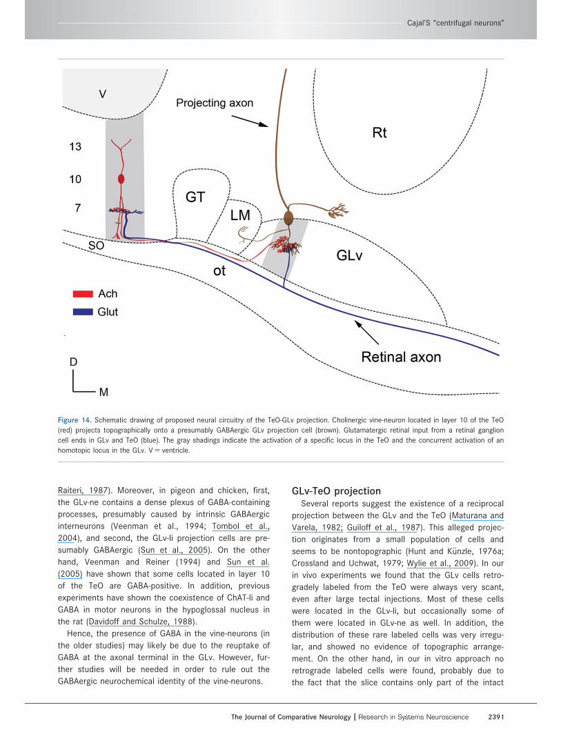
Figure 13. Double labeling experiments showing cells located in
layer 10 of the TeO. A: Retrograde labeling of a vine-neuron with
D-Alexa-546 located in layer 10 of the TeO after a GLv-ne injec-
tion. B: ChAT immunohistochemistry with Alexa-488 of the same
section showing labeled cells in layer 10 of the TeO. C: Merge of
both images showing ChAT labeling of the vine-neuron. All retro-
grade cells labeled with A-546-Dextran were positive for ChAT
(data not shown). Orientation: M 5 medial; D 5 dorsal.
T. Vega-Zuniga et al.
2390 The Journal of Comparative Neurology |Research in Systems Neuroscience
Raiteri, 1987). Moreover, in pigeon and chicken, first,
the GLv-ne contains a dense plexus of GABA-containing
processes, presumably caused by intrinsic GABAergic
interneurons (Veenman et al., 1994; Tombol et al.,
2004), and second, the GLv-li projection cells are pre-
sumably GABAergic (Sun et al., 2005). On the other
hand, Veenman and Reiner (1994) and Sun et al.
(2005) have shown that some cells located in layer 10
of the TeO are GABA-positive. In addition, previous
experiments have shown the coexistence of ChAT-li and
GABA in motor neurons in the hypoglossal nucleus in
the rat (Davidoff and Schulze, 1988).
Hence, the presence of GABA in the vine-neurons (in
the older studies) may likely be due to the reuptake of
GABA at the axonal terminal in the GLv. However, fur-
ther studies will be needed in order to rule out the
GABAergic neurochemical identity of the vine-neurons.
GLv-TeO projectionSeveral reports suggest the existence of a reciprocal
projection between the GLv and the TeO (Maturana and
Varela, 1982; Guiloff et al., 1987). This alleged projec-
tion originates from a small population of cells and
seems to be nontopographic (Hunt and K€unzle, 1976a;
Crossland and Uchwat, 1979; Wylie et al., 2009). In our
in vivo experiments we found that the GLv cells retro-
gradely labeled from the TeO were always very scant,
even after large tectal injections. Most of these cells
were located in the GLv-li, but occasionally some of
them were located in GLv-ne as well. In addition, the
distribution of these rare labeled cells was very irregu-
lar, and showed no evidence of topographic arrange-
ment. On the other hand, in our in vitro approach no
retrograde labeled cells were found, probably due to
the fact that the slice contains only part of the intact
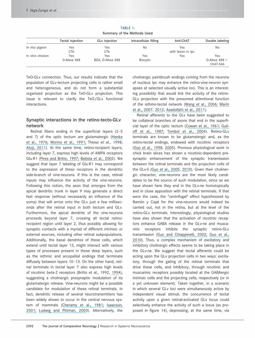
Figure 14. Schematic drawing of proposed neural circuitry of the TeO-GLv projection. Cholinergic vine-neuron located in layer 10 of the TeO
(red) projects topographically onto a presumably GABAergic GLv projection cell (brown). Glutamatergic retinal input from a retinal ganglion
cell ends in GLv and TeO (blue). The gray shadings indicate the activation of a specific locus in the TeO and the concurrent activation of an
homotopic locus in the GLv. V 5 ventricle.
Cajal’S “centrifugal neurons”
The Journal of Comparative Neurology | Research in Systems Neuroscience 2391
TeO-GLv connection. Thus, our results indicate that the
population of GLv-tectum projecting cells is rather small
and heterogeneous, and do not form a substantial
organized projection as the TeO-GLv projection. This
issue is relevant to clarify the TeO/GLv functional
interactions.
Synaptic interactions in the retino-tecto-GLvnetwork
Retinal fibers ending in the superficial layers (2–5
and 7) of the optic tectum are glutamatergic (Henke
et al., 1976; Morino et al., 1991; Theiss et al., 1998;
Atoji, 2011). At the same time, retino-recipient layers,
including layer 7, express high levels of AMPA receptors
Glu-R1 (Pires and Britto, 1997; Batista et al., 2002). We
suggest that layer 7 labeling of Glu-R1 may correspond
to the expression of these receptors in the dendritic
side-branch of vine-neurons. If this is the case, retinal
inputs may influence the activity of the vine-neurons.
Following this notion, the axon that emerges from the
apical dendritic trunk in layer 9 may generate a direct
fast response (without necessarily an influence of the
soma) that will arrive onto the GLv just a few millisec-
onds after the retinal input in both tectum and GLv.
Furthermore, the apical dendrite of the vine-neurons
proceeds beyond layer 7, crossing all tectal retino-
recipient region until layer 2, thus possibly allowing for
synaptic contacts with a myriad of different intrinsic or
external sources, including other retinal subpopulations.
Additionally, the basal dendrites of these cells, which
extend until tectal layer 13, might interact with various
types of processes present in these deep layers, such
as the isthmic and arcopallial endings that terminate
diffusely between layers 10–13. On the other hand, reti-
nal terminals in tectal layer 7 also express high levels
of nicotinic beta-2 receptors (Britto et al., 1992, 1994),
suggesting a cholinergic presynaptic modulation of its
glutamatergic release. Vine-neurons might be a possible
candidate for modulation of these retinal terminals. In
fact, dendritic release of several neurotransmitters has
been widely shown to occur in the central nervous sys-
tem of mammals (Cheramy et al., 1981; Isaacson,
2001; Ludwig and Pittman, 2003). Alternatively, the
cholinergic paintbrush endings coming from the neurons
of nucleus Ipc may enhance the retino-vine-neuron syn-
apses at selected visually active loci. This is an interest-
ing possibility that would link the activity of the retino-
GLv projection with the presumed attentional function
of the isthmo-tectal network (Wang et al., 2006; Mar�ın
et al., 2007, 2012; Asadollahi et al., 2011).
Retinal afferents to the GLv have been suggested to
be collateral branches of axons that end in the superfi-
cial layer of the optic tectum (Cowan et al., 1961; Guil-
off et al., 1987; Tombol et al., 2004). Retino-GLv
terminals are known to be glutamatergic and, as the
retino-tectal endings, endowed with nicotinic receptors
(Guo et al., 1998, 2005). Previous physiological work in
chick brain slices has shown a nicotinic-dependent pre-
synaptic enhancement of the synaptic transmission
between the retinal terminals and the projection cells of
the GLv-li (Guo et al., 2005, 2010). Given their choliner-
gic character, vine-neurons are the most likely candi-
dates to be the source of such modulation, since as we
have shown here they end in the GLv-ne homotopically
and in close apposition with the retinal terminals. If that
were the case, the "centrifugal" effect hypothesized by
Ram�on y Cajal for the vine-neurons would indeed be
carried out, not in the retina, but at the level of the
retino-GLv terminals. Interestingly, physiological studies
have also shown that the activation of nicotinic recep-
tors enhance GABA release in the GLv-ne and musca-
rinic receptors inhibits the synaptic retino-GLv
transmission (Guo and Chiappinelli, 2002; Guo et al.,
2010). Thus, a complex mechanism of excitatory and
inhibitory cholinergic effects seems to be taking place in
the GLv-ne. We suggest that tectal afferents could be
acting upon the GLv projection cells in two ways: excita-
tory, through the gating of the retinal terminals that
drive these cells, and inhibitory, through nicotinic and
muscarinic receptors possibly located at the GABAergic
intrinsic cells and the projecting cells, respectively (or in
a yet unknown element). Taken together, in a scenario
in which several GLv loci were simultaneously active by
independent visual stimuli, the concurrence of tectal
activity upon a given retinal-activated GLv locus could
selectively enhance the activity of such a locus (as pro-
posed in figure 14), depressing, at the same time, via
TABLE 1.
Summary of the Methods Used
Tectal injection GLv injection Intracellular filling Anti-ChAT Double labeling
In vivo pigeon Yes Yes No Yes NoCTb CTb with lesion in Ipc
In vitro chicken Yes Yes Yes Yes YesD-Alexa 488 BDA, D-Alexa 488 Biocytin D-Alexa 488 1
ChAT-546
T. Vega-Zuniga et al.
2392 The Journal of Comparative Neurology |Research in Systems Neuroscience
GABAergic enhancement, the activity of the neighboring
loci.
Avian and mammalian GLv complexA common feature of the visual system in amniotes
is the presence of a set of retinorecipient structures
located adjacent to the lateral wall of the ventral thala-
mus that could be recognized as a “ventrothalamic
complex.” These structures have in common an
absence of subsequent projections to the telencepha-
lon, together with descendent efferents directed
towards pretectal, tegmental, and pontine targets (But-
ler and Hodos, 2005). In mammals, this ventrothalamic
complex is called the GLv complex (in primates pregeni-
culate nucleus) and is composed of several subnuclei
or subdivisions, clustered in a large structure containing
two or more retinorecipient neuropils and several
nuclear layers (Conley and Friederich-Ecsy, 1993a; Har-
rington, 1997). A main characteristic of this complex is
the presence of retinal, collicular, cortical, and cerebel-
lar afferents (Conley and Friederich-Ecsy, 1993b). In
turn, projections from this complex to the medial pons,
along with projections to the optic tectum, the zona
incerta and other hypothalamic targets, the pretectum,
the accessory optic nuclei, and the motor tegmentum,
have been confirmed in a number of mammalian spe-
cies (Harrington, 1997).
In birds, the GLv is also part of a complex constitut-
ing a collection of several distinct structures that suc-
ceed each other in a rostrocaudal order. Following the
nomenclature of Karten and Hodos (1967), this com-
plex includes the nucleus LA, IGL, VLT, and GLv. The lit-
erature regarding these nuclei is scant and somewhat
confusing; thus, the anatomical and functional reasons
to group these structures together are at present
unclear. It can be said, however, that the overall pat-
tern of connections of this complex is remarkably simi-
lar to that of the mammalian GLv complex.
Interestingly, none of the single components of this
group exhibit a pattern of connections as diverse as
the one attributed to the mammalian complex. The
most conspicuous and better known member of the
avian ventrothalamic complex, the GLv, lacks of cere-
bellar afferents, and does not emit a significant topo-
graphic projection to the tectum or to the accessory
optic nuclei (unpubl. obs.).
A collicular projection upon the GLv complex has
been described in many mammalian species such as the
cat, the rat, and the tree shrew (Graham, 1977; Cosenza
and Moore, 1984; Taylor et al., 1986; Conley and
Friederich-Ecsy, 1993b). This projection terminates in the
external-most neuropil of the complex (thus, a putative
homolog for the GLv-ne in birds) and, as in the avian
projection, it follows a topographic arrangement. The
cells forming this projection are located in the retinoreci-
pient tectal zone, SGS, without a clear layer arrange-
ment. Unfortunately, the labeling of these cells does not
show a fine detailed morphology of dendrites and axons,
making it difficult to establish any clear comparison with
the vine-neurons described in this study. Moreover, the
neurochemical identity of these collicular cells is far
from resolved. Therefore, more detailed surveys regard-
ing the morphology and neurochemistry of the tectal
cells projecting to the GLv in mammals will be necessary
in order to establish a consistent comparative approach.
In any case, the avian GLv nucleus should not be consid-
ered homologous to the mammalian GLv complex as a
whole. To be more specific, we propose to rename the
avian GLv nucleus the "nucleus geniculatus lateralis,
pars ventralis principalis" (GLv-pri).
SUMMARY
We used tracer and immunohistochemical techniques
to characterize the morphology, projection pattern, and
neurochemical identity of the neurons that give rise to
the TeO-GLv projection. Our data indicate that these
neurons project to the GLv. Furthermore, the ChAT
immunoreactivity of these cells strongly suggests that
the TeO-GLv synapses are cholinergic. Nevertheless,
other neurotransmitters involvement cannot be ruled
out. The information presented will be valuable for
future physiological studies regarding the visual role of
the GLv and of the TeO-GLv connection.
ACKNOWLEDGMENTSWe thank Birgit Seibel, Yvonne Schwarz, Gaby Schwa-
bedissen, Agnieszka Brzozowska-Prechtl, Elisa Sentis, and
Solano Henriquez for excellent technical assistance;
Sebasti�an Tapia Pino for experimental help; Dr. Camilo
Libedinsky for critically reading the article; Dr. Prasad
Vaddepalli for microscopy assistance; and M�aximo
Fern�andez, Dr. Cristi�an Guti�errez and Quirin Krabichler for
helpful comments during the conduct of this research.
CONFLICT OF INTEREST
The authors declare no conflict of interest.
AUTHOR CONTRIBUTIONS
All authors had full access to all data in the study
and take responsibility for the integrity of the data and
the accuracy of the data analysis. Study concept and
design: TVZ, HL, JM, HJK, GM. Acquisition of data: TVZ,
JM, HJK, GM, SH. Analysis and interpretation of data:
TVZ, HL, JM, HJK, GM, SH. Drafting of the article: TVZ,
Cajal’S “centrifugal neurons”
The Journal of Comparative Neurology | Research in Systems Neuroscience 2393
HL, JM, HJK, GM. Critical revision of the article for
important intellectual content: HL, JM, HJK, GM. Statisti-
cal analysis: TVZ. Administrative, technical, and material
support: HL, JM, HJK, GM.
LITERATURE CITEDAngaut P, Rep�erant J. 1976. Fine structure of the optic fibre
termination layers in the pigeon optic tectum: a Golgiand electron microscope study. Neuroscience 1:93–105.
Asadollahi A, Mysore SP, Knudsen EI. 2011. Rules of competi-tive stimulus selection in a cholinergic isthmic nucleus ofthe owl midbrain. J Neurosci 31:6088–6097.
Atoji Y. 2011. Immunohistochemical localization of vesicularglutamate transporter 2 (vGluT2) in the central nervoussystem of the pigeon (Columba livia). J Comp Neurol519:2887–2905.
Bagnoli P, Fontanesi G, Alesci R, Erichsen JT. 1992. Distribu-tion of neuropeptide Y, substance P, and Choline acetyl-transferase in the developing visual system of the pigeonand effects of unilateral retina removal. J Comp Neurol318:392–414.
Batista SS, Pires RS, Britto LR. 2002. Differential expressionof AMPA-type glutamate receptor subunits during devel-opment of the chick optic tectum. Braz J Med Biol Res35:973–978.
Benowitz LI, Karten HJ. 1976. Organization of the tectofugalvisual pathway in the pigeon: a retrograde transportstudy. J Comp Neurol 167:503–520.
Berger SB, Romero X, Ma C, Wang G, Faubion WA, Liao G,Compeer E, Keszei M, Rameh L, Wang N, Boes M,Regueiro JR, Reinecker H-C, Terhorst C. 2010. SLAM is amicrobial sensor that regulates bacterial phagosomefunctions in macrophages. Nat Immunol 11:920–927.
Bonanno G, Raiteri M. 1987. Presence of a gamma-aminobutyric acid (GABA) uptake system on cholinergicterminals of rat hippocampus: evidence for neuronalcoexistence of acetylcholine and GABA? J Pharmacol ExpTher 240:294–297.
Britto LRG, Keyser KT, Lindstrom JM, Karten HJ. 1992. Immu-nohistochemical localization of nicotinic acetylcholinereceptor subunits in the mesencephalon and diencepha-lon of the chick (Gallus gallus). J Comp Neurol 317:325–340.
Britto LRG, Torrao AS, Hamassaki-Britto DE, Mpodozis J,Keyser KT, Lindstrom JM, Karten HJ. 1994. Effects of ret-inal lesions upon the distribution of nicotinic acetylcho-line receptor subunits in the chick visual system. J CompNeurol 350:473–484.
Butler AB, Hodos W. 2005. Comparative vertebrate neuroanat-omy: evolution and adaptation. Hoboken, NJ: John Wiley& Sons.
Cheramy A, Leviel V, Glowinski J. 1981. Dendritic release ofdopamine in the substantia nigra. Nature 289:537–543.
Conley M, Friederich-Ecsy B. 1993a. Functional organizationof the ventral lateral geniculate complex of the treeshrew (Tupaia belangeri). I. Nuclear subdivisions and reti-nal projections. J Comp Neurol 328:1–20.
Conley M, Friederich-Ecsy B. 1993b. Functional organizationof the ventral lateral geniculate complex of the treeshrew (Tupaia belangeri). II. Connections with the cortex,thalamus, and brainstem. J Comp Neurol 328:21–42.
Cosenza RM, Moore RY. 1984. Afferent connections of theventral lateral geniculate nucleus in the rat: an HRPstudy. Brain Res 310:367–370.
Cowan WM, Adamson L, Powell TP. 1961. An experimentalstudy of the avian visual system. J Anat 95:545–563.
Crossland WJ, Hughes CP. 1978. Observations on the afferentand efferent connections of the avian isthmo-opticnucleus. Brain Res 145:239–256.
Crossland WJ, Uchwat CJ. 1979. Topographic projections ofthe retina and optic tectum upon the ventral lateralgeniculate nucleus in the chick. J Comp Neurol 185:87–106.
Davidoff MS, Schulze W. 1988. Coexistence of GABA-andcholine acetyltransferase (ChAT)-like immunoreactivity inthe hypoglossal nucleus of the rat. Histochemistry 89:25–33.
D�avila JC, Andreu MJ, Real MA, Puelles L, Guirado S. 2002.Mesencephalic and diencephalic afferent connections tothe thalamic nucleus rotundus in the lizard, Psammodro-mus algirus. Eur J Neurosci 16:267–282.
Fritzsch B. 1993. Fast axonal diffusion of 3000 molecularweight dextran amines. J Neurosci Methods 50:95–103.
Gaillard F, Bonfield S, Gilmour GS, Kuny S, Mema SC, MartinBT, Smale L, Crowder N, Stell WK, Sauv�e Y. 2008. Reti-nal anatomy and visual performance in a diurnal cone-rich laboratory rodent, the Nile grass rat (Arvicanthisniloticus). J Comp Neurol 510:525–538.
Gamlin PDR, Cohen DH. 1988. Projections of the retinoreci-pient pretectal nuclei in the pigeon (Columba livia). JComp Neurol 269:18–46.
Gioanni H, Palacios A, Sansonetti A, Varela F. 1991. Role ofthe nucleus geniculatus lateralis ventralis (GLv) in theoptokinetic reflex: a lesion study in the pigeon. Exp BrainRes 86:601–607.
Graham J. 1977. An autoradiographic study of the efferentconnections of the superior colliculus in the cat. J CompNeurol 173:629–654.
Guiloff GD. 1991. Ultrastructural study of the avian ventral lat-eral geniculate nucleus. Vis Neurosci 6:119–134.
Guiloff GD, Maturana HR, Varela FJ. 1987. Cytoarchitecture ofthe avian ventral lateral geniculate nucleus. J Comp Neu-rol 264:509–526.
G€unt€urk€un O, Karten HJ. 1991. An immunocytochemical analy-sis of the lateral geniculate complex in the pigeon(Columba livia). J Comp Neurol 314:721–749.
Guo JZ, Chiappinelli VA. 2002. A novel choline-sensitive nico-tinic receptor subtype that mediates enhanced GABArelease in the chick ventral lateral geniculate nucleus.Neuroscience 110:505–513.
Guo JZ, Tredway TL, Chiappinelli VA. 1998. Glutamate andGABA release are enhanced by different subtypes of pre-synaptic nicotinic receptors in the lateral geniculatenucleus. J Neurosci 18:1963–1969.
Guo JZ, Liu Y, Sorenson EM, Chiappinelli VA. 2005. Synapti-cally released and exogenous ACh activates different nic-otinic receptors to enhance evoked glutamatergictransmission in the lateral geniculate nucleus. J Neuro-physiol 94:2549–2560.
Guo JZ, Sorenson EM, Chiappinelli VA. 2010. Cholinergic mod-ulation of non-N-methyl-d-aspartic acid glutamatergictransmission in the chick ventral lateral geniculatenucleus. Neuroscience 166:604–614.
Harrington ME. 1997. The ventral lateral geniculate nucleusand the intergeniculate leaflet: interrelated structures inthe visual and circadian systems. Neurosci Biobehav Rev21:705–727.
Hayes BP, Webster KE. 1975. An electron microscope studyof the retino-receptive layers of the pigeon optic tectum.J Comp Neurol 162:447–465.
Hellmann B, G€unt€urk€un O, Manns M. 2004. Tectal mosaic:organization of the descending tectal projections in com-parison to the ascending tectofugal pathway in thepigeon. J Comp Neurol 472:395–410.
T. Vega-Zuniga et al.
2394 The Journal of Comparative Neurology |Research in Systems Neuroscience
Henke H, Schenker T, Cu�enod M. 1976. Effects of retinalablation on uptake of glutamate, glycine, GABA, prolineand choline in pigeon tectum. J Neurochem 26:131–139.
Hoshi H, Tian L-M, Massey SC, Mills SL. 2011. Two distincttypes of ON directionally selective ganglion cells in therabbit retina. J Comp Neurol 519:2509–2521.
Hu M, Naito J, Chen Y, Ohmori Y, Fukuta K. 2004. Afferentand efferent connections of the nucleus geniculatus lat-eralis ventralis demonstrated by WGA-HRP in the chick.Anat Histol Embryol 33:192–195.
Hunt SP, K€unzle H. 1976a. Observations on the projectionsand intrinsic organization of the pigeon optic tectum: anautoradiographic study based on anterograde and retro-grade, axonal and dendritic flow. J Comp Neurol 170:153–172.
Hunt SP, K€unzle H. 1976b. Selective uptake and transport oflabel within three identified neuronal systems after injec-tion of 3H-GABA into the pigeon optic tectum: an autora-diographic and Golgi study. J Comp Neurol 170:173–189.
Isaacson JS. 2001. Mechanisms governing dendritic ~a-aminobutyric acid (GABA) release in the rat olfactorybulb. Proc Natl Acad Sci U S A 98:337–342.
Karten HJ, Hodos W. 1967. A stereotaxic atlas of the brain ofthe pigeon (Columba livia). Baltimore: Johns HopkinsPress.
Karten HJ, Hodos W, Nauta WJH, Revzin AM. 1973. Neuralconnections of the "visual wulst" of the avian telenceph-alon. Experimental studies in the pigeon (Columba livia)and owl (Speotyto cunicularia). J Comp Neurol 150:253–277.
Karten HJ, Cox K, Mpodozis J. 1997. Two distinct populationsof tectal neurons have unique connections within the ret-inotectorotundal pathway of the pigeon (Columba livia). JComp Neurol 387:449–465.
Ludwig M, Pittman QJ. 2003. Talking back: dendritic neuro-transmitter release. Trends Neurosci 26:255–261.
Luksch H, Cox K, Karten HJ. 1998. Bottlebrush dendritic end-ings and large dendritic fields: motion-detecting neuronsin the tectofugal pathway. J Comp Neurol 396:399–414.
Major DE, Luksch H, Karten HJ. 2000. Bottlebrush dendriticendings and large dendritic fields: motion-detecting neu-rons in the mammalian tectum. J Comp Neurol 423:243–260.
Mar�ın G, Henny P, Letelier JC, Sentis E, Karten H, Mrosko B,Mpodozis J. 2001. A simple method to microinject solidneural tracers into deep structures of the brain. J Neuro-sci Methods 106:121–129.
Mar�ın G, Salas C, Sentis E, Rojas X, Letelier JC, Mpodozis J.2007. A cholinergic gating mechanism controlled bycompetitive interactions in the optic tectum of thepigeon. J Neurosci 27:8112–8121.
Mar�ın G, Dur�an E, Morales C, Gonz�alez-Cabrera C, Sentis E,Mpodozis J, Letelier JC. 2012. Attentional capture?Synchronized feedback signals from the isthmi boost ret-inal signals to higher visual areas. J Neurosci 32:1110–1122.
Maturana HR, Varela FJ. 1982. Color-opponent responses inthe avian lateral geniculate: a study in the quail (Coturnixcoturnix japonica). Brain Res 247:227–241.
Medina L, Reiner A. 1994. Distribution of choline acetyltrans-ferase immunoreactivity in the pigeon brain. J CompNeurol 342:497–537.
Morino P, Bahro M, Cu�enod M, Streit P. 1991. Glutamate-likeimmunoreactivity in the pigeon optic tectum and effectsof retinal ablation. Eur J Neurosci 3:366–378.
Mpodozis J, Letelier J-C, Concha ML, Maturana H. 1995. Con-duction velocity groups in the retino-tectal and retino-
thalamic visual pathways of the pigeon (Columba livia).Int J Neurosci 81:123–136.
Mpodozis J, Cox K, Shimizu T, Bischof H-J, Woodson W,Karten HJ. 1996. GABAergic inputs to the nucleus rotun-dus (pulvinar inferior) of the pigeon (Columba livia). JComp Neurol 374:204–222.
Pateromichelakis S. 1979. Response properties of units in thelateral geniculate nucleus of the domestic chick (Gallusdomesticus). Brain Res 167:281–296.
P�erez SE, Y�a~nez J, Mar�ın O, Anad�on R, Gonz�alez A,Rodr�ıguez-Moldes I. 2000. Distribution of choline acetyl-transferase (ChAT) immunoreactivity in the brain of theadult trout and tract-tracing observations on the con-nections of the nuclei of the isthmus. J Comp Neurol428:450–474.
Pires RS, Britto LR. 1997. Distribution of AMPA-type gluta-mate receptor subunits in the chick visual system. Braz JMed Biol Res 30:73–77.
Ram�on y Cajal S. 1995. Histology of the nervous system.Oxford, UK: Oxford University Press.
Reiner A. 1994. Laminar distribution of the cells of origin ofascending and descending tectofugal pathways in turtles:implications for the evolution of tectal lamination. BrainBehav Evol 43:274–283.
Reiner A, Karten HJ. 1982. Laminar distribution of thecells of origin of the descending tectofugal pathwaysin the pigeon (Columba livia). J Comp Neurol 204:165–187.
Reiner A, Veenman CL, Medina L, Jiao Y, Del Mar N, HonigMG. 2000. Pathway tracing using biotinylated dextranamines. J Neurosci Methods 103:23–37.
Rep�erant J, Ward R, Miceli D, Rio J, M�edina M, Kenigfest N,Vesselkin N. 2006. The centrifugal visual system of ver-tebrates: a comparative analysis of its functional anatom-ical organization. Brain Res Rev 52:1–57.
Sebest�eny T, Davies DC, Zayats N, N�emeth A, T€omb€ol T.2002. The ramification and connections of retinal fibresin layer 7 of the domestic chick optic tectum: a Golgiimpregnation, anterograde tracer and GABA-immunogoldstudy. J Anat 200:169–183.
Sevigny CP, Bassi J, Williams DA, Anderson CR, Thomas WG,Allen AM. 2012. Efferent projections of C3 adrenergicneurons in the rat central nervous system. J Comp Neu-rol 520:2352–2368.
Sorenson EM, Parkinson D, Dahl JL, Chiappinelli VA. 1989.Immunohistochemical localization of choline acetyltrans-ferase in the chicken mesencephalon. J Comp Neurol281:641–657.
Sun Z, Wang H, Laverghetta A, Yamamoto K, Reiner A. 2005.The distribution and cellular localization of glutamic aciddecarboxylase-65 (GAD65) mRNA in the forebrain andmidbrain of domestic chick. J Chem Neuroanat 29:265–281.
Taylor AM, Jeffery G, Lieberman AR. 1986. Subcortical affer-ent and efferent connections of the superior colliculus inthe rat and comparisons between albino and pigmentedstrains. Exp Brain Res 62:131–142.
Theiss C, Hellmann B, G€unt€urk€un O. 1998. The differential dis-tribution of AMPA-receptor subunits in the tectofugalsystem of the pigeon. Brain Res 785:114–128.
Tombol T, Eyre M, Zayats N, Nemeth A. 2004. The internalstructure of the nucleus geniculatus lateralis ventralis inthe avian brain: a Golgi study and electron microscopicinvestigation. Cells Tissues Organs 177:237–256.
Vanegas H. 1984. Comparative neurology of the optic tectum.New York: Plenum Press.
Veenman CL, Reiner A. 1994. The distribution of GABA-containing perikarya, fibers, and terminals in the
Cajal’S “centrifugal neurons”
The Journal of Comparative Neurology | Research in Systems Neuroscience 2395
forebrain and midbrain of pigeons, with particular refer-ence to the basal ganglia and its projection targets. JComp Neurol 339:209–250.
Veenman CL, Albin RL, Richfield EK, Reiner A. 1994. Distribu-tions of GABAA, GABAB, and benzodiazepine receptors inthe forebrain and midbrain of pigeons. J Comp Neurol344:161–189.
Wang Y, Luksch H, Brecha NC, Karten HJ. 2006. Columnar pro-jections from the cholinergic nucleus isthmi to the optictectum in chicks (Gallus gallus). A possible substrate forsynchronizing tectal channels. J Comp Neurol 494:7–35.
Wylie DR, Gutierrez-Ibanez C, Pakan JM, Iwaniuk AN. 2009. Theoptic tectum of birds: mapping our way to understandingvisual processing. Can J Exp Psychol 63:328–338.
T. Vega-Zuniga et al.
2396 The Journal of Comparative Neurology |Research in Systems Neuroscience