Discrimination and emission of different key-peck durations in the pigeon
Upload
independentCategory
view
3download
0
Journal of Virological Methods 131 (2006) 115–121
Identification and subgrouping of pigeon type Newcastle diseasevirus strains by restriction enzyme cleavage site analysis
Dorina Ujvaria,∗, Eniko Wehmanna, Jozsef Herczegb, Bela Lomniczia
a Veterinary Medical Research Institute of the Hungarian Academy of Sciences, H-1143 Budapest, Hungaria Krt. 21, Hungaryb CEVA-Phylaxia Veterinary Biologicals Co. Ltd., H-1107 Budapest, Szallas Utca 5, Hungary
Received 25 April 2005; received in revised form 19 July 2005; accepted 20 July 2005Available online 15 September 2005
Abstract
A host variant of Newcastle disease virus (NDV, genusAvulavirus, family Paramyxoviridae) is responsible for an autonomous diseasein pigeons. It emerged in the late 1970s in the Mediterranean region. Despite great genetic diversity the vast majority of strains belong toa monophyletic group (sublineage VIb) within genotype VI of NDV strains that were indigenous in the region at that time. To date only amonoclonal antibody assay is available for the specific identification of pigeon type strains. A specific genetic assay is described suitable fort restrictione revealedn tant pigeonv d reliablem©
K
1
isftNpldgeLe
anong;otthe
gelisiddle
eblyDV.anywild
st al.,jv01f the
d in
0d
he identification of pigeon isolates. Cleavage site analysis of a 1349 bp amplicon of the fusion protein gene was carried out usingnzymes (RE)HinfI, BstOI andRsaI. RE analysis of over 100 strains isolated between 1978 and 2002 deriving from 16 countries hasine RE-patterns, which were progressive site variants of the parental (group VI) genotype. In spite of substantial site variation, exiruses lacked aBstOI cleavage site at nucleotide 1601 shared by other NDV strains of chicken origin. RE analysis is a simple anethod both for the identification and subgrouping of pigeon type viruses.2005 Elsevier B.V. All rights reserved.
eywords: PPMV1; Restriction site analysis; F gene
. Introduction
Newcastle disease (ND) is highly contagious and econom-cally one of the most important diseases of poultry. Theymptoms of NDV infection in domestic avian species varyrom inapparent to severe, the latter is associated with respira-ory, enteric and neurological signs (Alexander et al., 2003).D is spread widely in developing countries and strains dis-lay a great genetic diversity. Using phylogenetic analysis at
east eight genotypes (with several sub-types) of NDV wereescribed whose distribution shows host, temporal and/oreographical restrictions (Aldous et al., 2003; Czegledit al., 2002; Herczeg et al., 1999, 2001; Huang et al., 2004;ee et al., 2004; Liu et al., 2003; Lomniczi et al., 1998; Maset al., 2002; Tsai et al., 2004; Wehmann et al., 2003a,b; Wise
∗ Corresponding author. Fax: +36 1 252 1069.E-mail address: [email protected] (D. Ujvari).
et al., 2004; Yu et al., 2001). In 1981 in Italy and the Sudan,epizootic form of an ND-like disease was diagnosed amracing, show and food pigeons (Biancifiori and Fioroni, 1983Eisa and Omer, 1984) and within a few years it spread nonly through Europe but appeared in North America andFar East (Alexander et al., 1985; Kaleta, 1992a,b; Vindevoand Duchatel, 1988; Wilson, 1986). Retrospective analysrevealed that the disease was already present in the MEast in the late 1970s (Kaleta et al., 1985). The causativagent, a host variant of ND-virus (NDV), was called variapigeon paramyxovirus type 1 (PPMV1) or pigeon type NDespite vaccination, pigeon type ND is still enzootic in mcountries and affects not only domesticated but feral andpigeons and zoo birds as well (Abolnik et al., 2004; Aldouet al., 2004; Alexander et al., 1985, 1997; Kommers e2001; Meulemans et al., 2002; Terregino et al., 2003; Uariet al., 2003, 2004; Werner et al., 1999; Zanetti et al., 20).
In a previous study based on sequence analysis oF gene, the vast majority of PPMV1 strains clustere
166-0934/$ – see front matter © 2005 Elsevier B.V. All rights reserved.oi:10.1016/j.jviromet.2005.07.012
116 D. Ujvari et al. / Journal of Virological Methods 131 (2006) 115–121
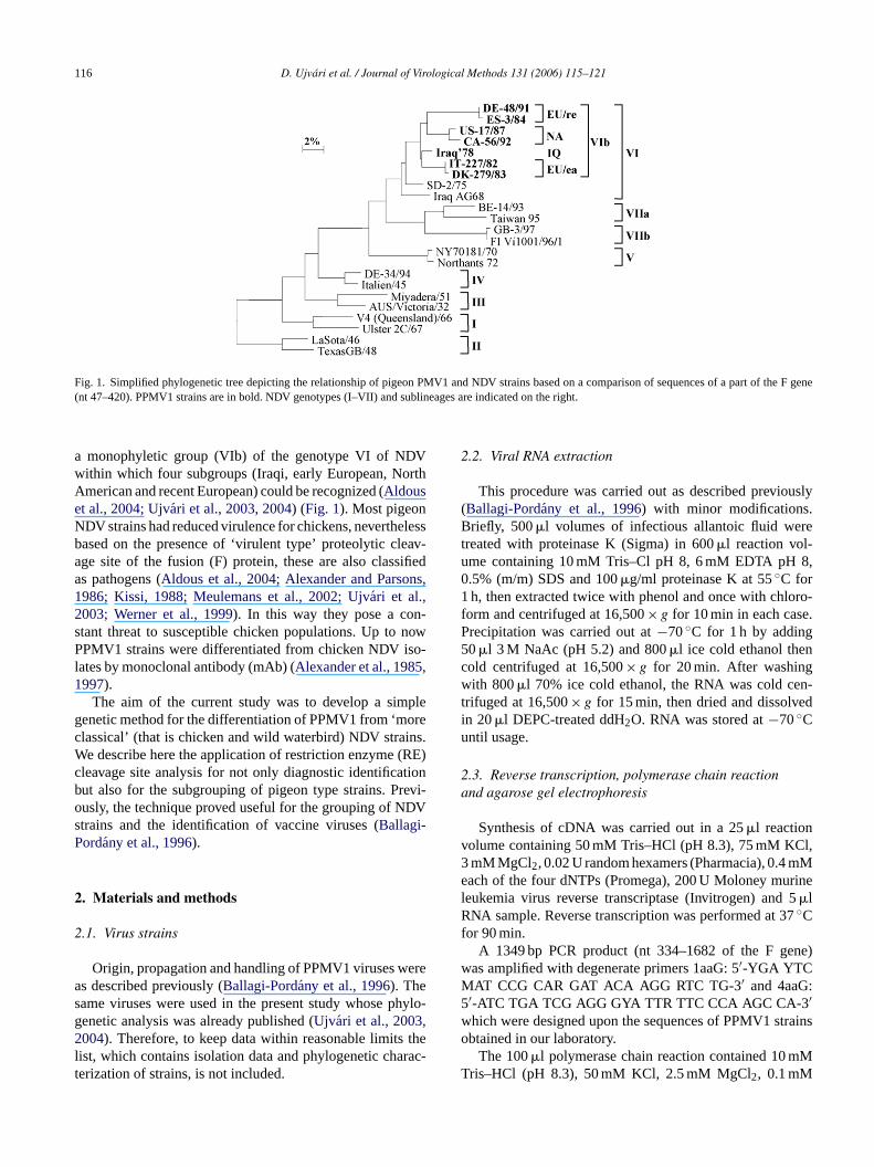
Fig. 1. Simplified phylogenetic tree depicting the relationship of pigeon PMV1 and NDV strains based on a comparison of sequences of a part of the F gene(nt 47–420). PPMV1 strains are in bold. NDV genotypes (I–VII) and sublineages are indicated on the right.
a monophyletic group (VIb) of the genotype VI of NDVwithin which four subgroups (Iraqi, early European, NorthAmerican and recent European) could be recognized (Aldouset al., 2004; Ujvari et al., 2003, 2004) (Fig. 1). Most pigeonNDV strains had reduced virulence for chickens, neverthelessbased on the presence of ‘virulent type’ proteolytic cleav-age site of the fusion (F) protein, these are also classifiedas pathogens (Aldous et al., 2004; Alexander and Parsons,1986; Kissi, 1988; Meulemans et al., 2002; Ujvari et al.,2003; Werner et al., 1999). In this way they pose a con-stant threat to susceptible chicken populations. Up to nowPPMV1 strains were differentiated from chicken NDV iso-lates by monoclonal antibody (mAb) (Alexander et al., 1985,1997).
The aim of the current study was to develop a simplegenetic method for the differentiation of PPMV1 from ‘moreclassical’ (that is chicken and wild waterbird) NDV strains.We describe here the application of restriction enzyme (RE)cleavage site analysis for not only diagnostic identificationbut also for the subgrouping of pigeon type strains. Previ-ously, the technique proved useful for the grouping of NDVstrains and the identification of vaccine viruses (Ballagi-Pordany et al., 1996).
2
2
ereas hylo-g ,2 thel rac-t
2.2. Viral RNA extraction
This procedure was carried out as described previously(Ballagi-Pordany et al., 1996) with minor modifications.Briefly, 500�l volumes of infectious allantoic fluid weretreated with proteinase K (Sigma) in 600�l reaction vol-ume containing 10 mM Tris–Cl pH 8, 6 mM EDTA pH 8,0.5% (m/m) SDS and 100�g/ml proteinase K at 55◦C for1 h, then extracted twice with phenol and once with chloro-form and centrifuged at 16,500× g for 10 min in each case.Precipitation was carried out at−70◦C for 1 h by adding50�l 3 M NaAc (pH 5.2) and 800�l ice cold ethanol thencold centrifuged at 16,500× g for 20 min. After washingwith 800�l 70% ice cold ethanol, the RNA was cold cen-trifuged at 16,500× g for 15 min, then dried and dissolvedin 20�l DEPC-treated ddH2O. RNA was stored at−70◦Cuntil usage.
2.3. Reverse transcription, polymerase chain reactionand agarose gel electrophoresis
Synthesis of cDNA was carried out in a 25�l reactionvolume containing 50 mM Tris–HCl (pH 8.3), 75 mM KCl,3 mM MgCl2, 0.02 U random hexamers (Pharmacia), 0.4 mMeach of the four dNTPs (Promega), 200 U Moloney murinel 5R 7f
ne)wM :5w trainso
MT
. Materials and methods
.1. Virus strains
Origin, propagation and handling of PPMV1 viruses ws described previously (Ballagi-Pordany et al., 1996). Theame viruses were used in the present study whose penetic analysis was already published (Ujvari et al., 2003004). Therefore, to keep data within reasonable limits
ist, which contains isolation data and phylogenetic chaerization of strains, is not included.
eukemia virus reverse transcriptase (Invitrogen) and�lNA sample. Reverse transcription was performed at 3◦C
or 90 min.A 1349 bp PCR product (nt 334–1682 of the F ge
as amplified with degenerate primers 1aaG: 5′-YGA YTCAT CCG CAR GAT ACA AGG RTC TG-3′ and 4aaG
′-ATC TGA TCG AGG GYA TTR TTC CCA AGC CA-3′hich were designed upon the sequences of PPMV1 sbtained in our laboratory.
The 100�l polymerase chain reaction contained 10 mris–HCl (pH 8.3), 50 mM KCl, 2.5 mM MgCl2, 0.1 mM
D. Ujvari et al. / Journal of Virological Methods 131 (2006) 115–121 117
each of the four dNTPs (Promega), 30 pmol each of theprimers, 2 U Taq polymerase (Perkin-Elmer) and 5�l cDNA.Amplification was performed in a PTC-200 Peltier ThermalCycler. After an initial denaturation step at 94◦C for 3 min,the samples were subjected to 35 cycles of 45 s at 94◦C, 1 minat 53◦C and 2 min at 72◦C and kept at 72◦C for 10 min.
An amount of 8�l of the PCR products were subjectedto electrophoresis in 0.5× TBE buffer in 2.5% agarose(Promega) gel and stained with ethidium bromide to visu-alise the DNA.
2.4. Restriction enzyme (RE) site analysis
PCR products were digested withHinfI, BstOI andRsaIrestriction endonucleases, respectively. Briefly, the ampli-cons were precipitated using 1 mln-buthanol, cold cen-trifuged at 16,500× g for 10 min and dried. The pellet wasdissolved in 18�l sterile ddH2O and divided into three parts.Digestion was carried out in a 20�l reaction volume con-taining 1× respective restriction enzyme buffer, 6�l PCRproducts and 8–12 U ofHinfI, BstOI or RsaI restrictionendonucleases (Promega). The digestion of the PCR prod-ucts was carried out at 37◦C for at least 2.5 h. Separation ofthe fragments was carried out in 0.5× TBE buffer in 2.5%agarose gel which was prepared from 2:1 ratio of MetaPhoragarose (Cambrex Bio Science Rockland Inc.) and Analyt-i ne at2 erec
3
nal-y Ther udedi eons( ltry,o wereo triesi iledd vid-uI MV1s gesb I ofc ofPR ngeda
pe VIN geonv EastA y thep phys-i oups
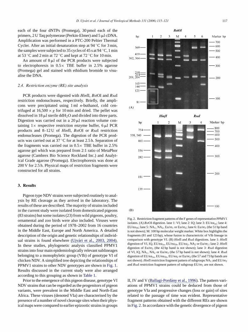
Fig. 2. Restriction fragment patterns of the F genes of representative PPMV1isolates. (A)BstOI digestion. lane 1: VI; lane 2: IQ; lane 3: EU/ea1a; lane 4:EU/ea1b; lane 5: NA1, NA2, Eu/re1 or Eu/re2; lane 6: Eu/re3 (the 51 bp bandis not shown); M: 100 bp molecular weight marker. White box highlights thefragments (81 and 123 bp), whose fusion is characterictic of VIb lineage incomparison with genotype VI. (B)HinfI and RsaI digestions. lane 1:HinfIdigestion of VI, IQ, EU/ea1a, EU/ea1b, EU/ea2, NA2 or Eu/re1; lane 2:HinfIdigestion of Eu/re3 (the 42 bp band is not shown); lane 3:RsaI digestionof VI, IQ, NA1, NA2 or Eu/re3 (the 57 bp band is not shown); lane 4:RsaIdigestion of EU/ea1a, EU/ea1b, EU/ea2 or Eu/re2 (the 57 and 73 bp bands arenot shown).HinfI restriction fragment pattern of subgroups NA1 and EU/re2andRsaI restriction fragment pattern of subgroup EU/re1 are not shown.
II, IV and V (Ballagi-Pordany et al., 1996). The pattern vari-ations of PPMV1 strains could be deduced from those ofgenotype VIa and progressive changes (loss or gain) of sitesrelated to the passage of time was evident. Representativefragment patterns obtained with the different REs are shownin Fig. 2. In accordance with the genetic divergence of pigeon
cal Grade agarose (Promega). Electrophoresis was do00 V for 2.5 h. Physical maps of restriction fragments wonstructed for all strains.
. Results
Pigeon type NDV strains were subjected routinely to asis by RE cleavage as they arrived in the laboratory.esults of these are described. The majority of strains incln the current study were isolated from domesticated pig83 strains) but some isolates (23) from wild pigeons, pournamental and zoo birds were also included. Virusesbtained during the period of 1978–2002 from 16 coun
n the Middle East, Europe and North America. A detaescription of the origin and genetic relationships of indial strains is found elsewhere (Ujvari et al., 2003, 2004).
n these studies, phylogenetic analysis classified PPtrains into four main epidemiologically relevant sublineaelonging to a monophyletic group (VIb) of genotype Vhicken NDV. A simplified tree depicting the relationshipsPMV1 strains to other NDV genotypes are shown inFig. 1.esults discussed in the current study were also arraccording to this grouping as shown inTable 1.
Prior to the emergence of this pigeon disease, genotyDV strains that can be regarded as the progenitors of piariants, were prevalent in the Middle East and North-frica. These viruses (denoted VIa) are characterised bresence of a number of novel cleavage sites when their
cal maps were compared to earlier epizootic strains in gr

118 D. Ujvari et al. / Journal of Virological Methods 131 (2006) 115–121
Table 1Data of pigeon type NDV strains used for restriction analysis
Lineage VI and sublineage within VIba Period Number of isolates BstOI site RsaI site
1601 1260 752 902 1160
Genotype VI (chicken) 1968–2002 >100 + + + − −Iraq 1978 1 − + + − −EU/early 1982–1995 56 − − + − +NA 1983–1998 22 − − − + −EU/recent 1987–2002 27 − − − + + (−)
−, Absence of the corresponding cleavage site; +, presence of the corresponding cleavage site.a Based on phylogenetic analysis of partial F gene sequences (Ujvari et al., 2003, 2004).
type NDV strains (Ujvari et al., 2003), nine groups were iden-tified based on the cleavage site pattern (Fig. 3).
The earliest known PPMV1 strain, Iraq’78 (Kaleta et al.,1985), differed only in a single site from the prototype patternof subgroup VIa due to the loss of aBstOI site at nucleotide1601 yielding a fusion fragment (123 + 81 bp) of 204 bp size.This fragment was not seen in any of the several hundredepizootic NDV strains (in genotypes II–VIII) examined inthe past (Ballagi-Pordany et al., 1996; Czegledi et al., 2003;Herczeg et al., 1999, 2001; Lomniczi et al., 1998; Wehmann
et al., 2003b) and the lack of this site turned out to be charac-teristic for all PPMV1 subgroups examined so far (Table 1).
The largest portion of the pigeon type isolates (56) wasavailable from theearly European (EU/ea) group mainlyfrom the first half of the 1980s. Changes at two additionalsites distinguish these from strain Iraq’78. One is the loss ofa BstOI site at nucleotide 1260 resulting in another fusionfragment, 362 bp (from 144 + 218 bp), a feature present in allsubsequent patterns. The other, the gain of anRsaI site at posi-tion 1160 has generated subfragments 465 and 73 bp from
Fdi
ig. 3. Physical maps of the restriction fragments and cleavage sites of PCesignate the last nucleotide (nt) of a fragment except nt 334 that is the starti
n base-pairs (bp).
R products of F gene of representative strains. Normal numerals above the lineng position of the amplified region. Italics below the line mark the fragment length

D. Ujvari et al. / Journal of Virological Methods 131 (2006) 115–121 119
the 538 bp fragment. The presence of two diagnostic frag-ments of 204 and 362 bp with the concomitant loss of bands123 + 81 and 144 + 218 bp, respectively, makes the recogni-tion of pigeon variants unambiguous. It is interesting thatalready in the first half of the 1980s, variants with and withoutaBstOI site at 953 (designated EU/ea1a and EU/ea1b, respec-tively), not distinguished by phylogenetic analysis, circulatedsimultaneously in 11 European countries on the continent aswell as in Malta and Egypt (Ujvari et al., 2003). The soleexception is EU/ea2 with its 566 bp band due to a third fusionevent, this time the loss ofBstOI site 1478 between the twodiagnostic fragments (Fig. 3). This group (EU/ea2) comprisesphylogenetically slightly diverged isolates obtained in thelater phase (between 1986 and 1996) of the primary wave ofthe disease in Hungary and Italy (Ujvari et al., 2003).
Members of the North American (NA) and recent Euro-pean (EU/re) subgroups shared two novel changes: aBstOIsite at position 902 emerged with the concomitant loss oftheBstOI site at nucleotide 752. As a result, theBstOI pat-terns of both subgroups became identical. Yet, NA strainscan be distinguished from the majority of those of EU/reby the absence of theRsaI site at nucleotide 1160. NA wasnamed so because, although it contained strains from Europe(Germany, Hungary and Sweden) as well, viruses from theUnited States (1), Canada (10) and Mexico (1) occurred onlyin this subgroup. The ‘true’ American variants, NA, are dis-t ithc ani gt
mer-g thataa( .E cter-i ofts
4
d ins emi-o
allP 1w s ofd tiono arec( , thefi ). Itcb in
due to the presence of two adjacent fusion fragments of 204and 362 bp. It is to be noted that among the currently circu-lating chicken viruses those of genotype VIIa (Lomniczi etal., 1998) and VIII (Herczeg et al., 1999) also display the lossof theBstOI site 1601 but with these viruses a conspicuous422 bp fragment was seen on the gel caused by double fusionevents (218 + 123 + 81 bp) therefore this should not posea diagnostic problem. The underlying nucleotide changes,however, are dissimilar: CC↓TGG→ CTTGG with all pat-tern groups of PPMV1 strains (unpublished data) whereasTCTGG in group VIIa (Herczeg et al., 1999). This also con-firms the monophyletic descent of VIb viruses (Ujvari et al.,2003). At the same time, no genuine chicken NDV strain hasbeen seen with the simultaneous presence of 204 and 362 bpBstOI fragments (Ballagi-Pordany et al., 1996; Czegledi etal., 2003; Herczeg et al., 1999; Lomniczi et al., 1998).
After emergence of the disease, monoclonal antibodies(mAbs) for specific identification of pigeon type NDV strainswere developed (Alexander et al., 1985). These mAbs havebeen used in hemagglutination inhibition or ELISA and todate these have been the only diagnostic tools available. TheRE assay described now is a simple and reliable geneticmethod that can be carried out anywhere where PCR is avail-able without the reliance of standardized mAb reagents. Inaddition, it is suitable not only for differentiation of pigeonand other type of NDV strains but for the subgrouping of thei thec om-p servea iso-l
sitec sitesr ingf ps.R lativet nifi-c nsi forf eo typesb force nti-cs I or IIN ot bet
andr t andr else-w -g es ase ctedt enu n the
1inguished by aHinfI cleavage site at nucleotide 1350 (whange 336 bp→ 286 + 50 bp) from a number of Europesolates (6), denoted NA2, phylogenetically also belongino the NA subgroup (Fig. 3) (Ujvari et al., 2003).
Recent European (EU/re) isolates revealed the eence of a number of novel but mainly intragroup sitesllowed further subdivision: namely the gain of anRsaI sitet nucleotide 1591 (EU/re1), a HinfI site at position 958EU/re2) and the reappearance of theRsaI site 1160 in bothU/re3 pattern seen in isolates only after 2000 is chara
zed by the reoccurrence of aBstOI site at 953 and the lossheRsaI site at 1160, and the occasional presence of aHinfIite at nucleotide 925 (Fig. 3).
. Discussion
RE analysis of pigeon type NDV strains has resulteeveral important findings relevant to diagnosis and epidlogy of these viruses.
Firstly, the salient finding of the current work is thatPMV1 isolates lacked theBstOI site at nucleotide 160hose occurrence is a shared property of NDV strainiverse origin. In addition to this marker, with the excepf sublineage IQ, all other currently circulating subgroupsharacterised by the additional loss of aBstOI site at 1260sublineage IQ that is represented only by strain Iraq’78rst and only isolate from the region, is probably extinctan be concluded that tests with even a single RE,BstOI, cane sufficient to identify the pigeon origin (VIb) of any stra
solates into epidemiologically meaningful group. Sinceollection of strains examined in the present study encasses a wide range of isolates, the patterns shown mays references for finer grouping of any unknown PPMV1
ates.Secondly, PPMV1 strains have a number of restriction
hanges including the sequential emergence or loss ofesulting in characteristic combinations as well as allowurther subdivision to epidemiologically defined subgrouestriction site states (presence or absence of sites) re
o those of the basic genotype VI patterns differed in sigance: those listed inTable 1are decisive in classifying strainto any of the four subgroups while others are suitableurther subdivision within subgroups (Fig. 2). The occurrencf some of these sites has been reported in other genout in an entirely distinct combination therefore their uselassifying VIb viruses is not diminished (Ballagi-Pordanyt al., 1996). As a matter of fact, the occurrence of ideal sites (e.g. aBstOI site 953 orRsaI site 1160) in widelyeparated genotypes such as in lineage VIb and groupDV strains should be regarded homoplasious and cann
reated as inherited similarity (Ballagi-Pordany et al., 1996).Thirdly, groups or lineages created by phylogenetic
estriction site analysis appear to be largely congrueneflect epidemiological associations discussed in detailhere (Ujvari et al., 2003, 2004). Interestingly, while phyloenetic analysis failed to show any segregation of isolatarly as in 1982–1983, restriction analysis already dete
wo site variants, EU/ea1a and EU/ea1b. RE assay has besed in other areas of NDV diagnostics: a test based o

120 D. Ujvari et al. / Journal of Virological Methods 131 (2006) 115–121
matrix (M) gene was developed for the positive identificationof all avirulent live vaccine strains currently in use. It was usedsuccessfully for unravelling the persistence and distributionof vaccine strains in chicken flocks and discovering homolo-gous contamination or strain replacement in vaccine batches(Farsang et al., 2003; Wehmann et al., 1997).
In conclusion, a simple RE cleavage site assay has beendeveloped that proved useful both for the identification andsubgrouping of pigeon-adapted NDV strains. Shared derivedfeatures such as the occurrence of twoBstOI fusion fragments(204 and 362 bp), relative to the ancestral NDV group VI,can be utilised as diagnostic markers. As a consequence ofongoing evolution in the pigeon host a considerable intra-lineage (VIb) divergence resulted in the emergence of at leastnine restriction site variants. The pattern variants shown inFig. 3may serve as references for future studies.
Acknowledgement
Parts of this work were supported by grant from theNational Science Fund (OTKA, No. T 038254), Hungary.
References
A riza-s in
A ecu-stleotide.
A lec-vian8 to
A louh,tet,J.,, B.,va,yx-
ative
A chal-1984
A .S.,itiesvirus)8.
A iruses,ultry,
B ,trainsirol.
B aseomp.
Czegledi, A., Herczeg, J., Hadjiev, G., Doumanova, L., Wehmann, E.,Lomniczi, B., 1995. The occurrence of five major Newcastle diseasevirus genotypes (II, IV, V, VI and VIIb) in Bulgaria between 1959and 1995. Epidemiol. Infect. 129, 679–688.
Czegledi, A., Wehmann, E., Lomniczi, B., 2003. On the origins and rela-tionships of Newcastle disease virus vaccine strains Hertfordshire andMukteswar, and virulent strain Herts’33. Avian Pathol. 32, 271–276.
Eisa, M., Omer, E.A., 1984. A natural outbreak of Newcastle disease inpigeons in the Sudan. Vet. Rec. 114, 297.
Farsang, A., Wehmann, E., Soos, T., Lomniczi, B., 2003. Positive iden-tification of Newcastle disease virus vaccine strains and detection ofcontamination in vaccine batches by restriction site analysis of thematrix protein gene. J. Vet. Med. B: Infect. Dis. Vet. Public Health50, 311–315.
Herczeg, J., Pascucci, S., Massi, P., Luini, M., Selli, L., Capua, I., Lom-niczi, B., 2000. A longitudinal study of velogenic Newcastle diseasevirus genotypes isolated in Italy between 1960 and 2000. AvianPathol. 30, 163–168.
Herczeg, J., Wehmann, E., Bragg, R.R., Travassos Dias, P.M., Hadjiev, G.,Werner, O., Lomniczi, B., 1999. Two novel genetic groups (VIIb andVIII) responsible for recent Newcastle disease outbreaks in SouthernAfrica, one (VIIb) of which reached Southern Europe. Arch. Virol.144, 2087–2099.
Huang, Y., Wan, H.Q., Liu, H.Q., Wu, Y.T., Liu, X.F., 2004. Genomicsequence of an isolate of Newcastle disease virus isolated from anoutbreak in geese: a novel six nucleotide insertion in the non-codingregion of the nucleoprotein gene. Arch. Virol. 149, 1445–1457.
Kaleta, E.F., Alexander, D.J., Russell, P.H., 1985. The first isolation of theavian PMV1 virus responsible for the current panzootic in pigeons?Avian Pathol. 14, 553–557.
Kaleta, E.F., 1992a. Unique features of the pigeon PMV1 infection. In:ork-
, July
K s—Aeed-usen,
K rus1iso-
eggs
K e ofkens.
L cularoreaphy-
L o-dis-
some3.
L ,A.P.,
seasean old
M , T.,ew-
obiol.
M s, M.,vian
T a, I.,tained
bolnik, C., Horner, R.F., Maharaj, R., Viljoen, G.J., 2004. Charactetion of a pigeon paramyxovirus (PPMV1) isolated from chickenSouth Africa. Onderstepoort J. Vet. Res. 71, 157–160.
ldous, E.W., Mynn, J.K., Banks, J., Alexander, D.J., 2003. A mollar epidemiological study of avian paramyxovirus type 1 (Newcadisease virus) isolates by phylogenetic analysis of a partial nuclesequence of the fusion protein gene. Avian Pathol. 32, 239–257
ldous, E.W., Fuller, C.M., Mynn, J.K., Alexander, D.J., 2004. A moular epidemiological investigation of isolates of the variant aparamyxovirus type 1 virus (PPMV1) responsible for the 197present panzootic in pigeons. Avian Pathol. 33, 258–269.
lexander, D.J., Russell, P.H., Parsons, G., Abu Elzein, E.M., BalA., Cernik, K., Engstrom, B., Fevereiro, M., Fleury, H.J.A., GuitM., Kaleta, E.F., Kihm, U., Kosters, J., Lomniczi, B., Meister,Meulemans, G., Nerome, K., Petek, M., Pokomunski, S., PoltenPrip, M., Richter, R., Saghy, E., Samberg, Y., Spanoghe, L., TumoB., 1985. Antigenic and biological characterisation of avian paramovirus type 1 isolates from pigeons—an international collaborstudy. Avian Pathol. 14, 365–376.
lexander, D.J., Parsons, G., 1986. Protection of chickens againstlenge with the variant virus responsible for Newcastle disease inby conventional vaccination. Vet. Rec. 118, 176–177.
lexander, D.J., Manvell, R.J., Lowings, J.P., Frost, K.M., Collins, MRussell, P.H., Smith, J.E., 1997. Antigenic diversity and similardetected in avian paramyxovirus type 1 (Newcastle diseaseisolates using monoclonal antibodies. Avian Pathol. 26, 399–41
lexander, D.J., 2003. Newcastle disease, other avian paramyxovand pneumovirus infections. In: Saif, J.M. (Ed.), Diseases of Po11th ed. Iowa State University Press, Ames, Iowa, pp. 63–99.
allagi-Pordany, A., Wehmann, E., Herczeg, J., Belak, S., Lomniczi, B.1996. Identification and grouping of Newcastle disease virus sby restriction site analysis of a region from the F gene. Arch. V141, 243–261.
iancifiori, F., Fioroni, A., 1983. An occurrence of Newcastle disein pigeons: virological and serological studies on the isolates. CImmunol. Microbiol. Infect. Dis. 6, 247–252.
Kaleta, E.F., Heffels-Redmann, U. (Eds.), Proceedings of the Wshop on Avian Paramyxoviruses. Rauischholzhausen, Germany27–29, pp. 250–261.
aleta, E.F., 1992b. Paramyxoviruses in free-living and captive birdbrief account. In: Kaleta, E.F., Heffels-Redmann, U. (Eds.), Procings of the Workshop on Avian Paramyxoviruses. RauischholzhaGermany, July 27–29, pp. 262–271.
issi, B., 1988. Studies on the virulence of pigeon paramyxovi(PMV1). I. Changes in the virulence of pigeon PMV1 strainslated in Hungary upon passage in chickens, embryonated hen’sand pigeons. Acta Vet. Hung. 36, 283–292.
ommers, G.D., King, D.J., Seal, B.S., Brown, C.C., 2001. Virulencpigeon-origin Newcastle disease virus isolates for domestic chicAvian Dis. 45, 906–921.
ee, Y.J., Sung, H.W., Choi, J.G., Kim, J.H., Song, C.S., 2004. Moleepidemiology of Newcastle disease viruses isolated in South Kusing sequencing of the fusion protein cleavage site region andlogenetic relationships. Avian Pathol. 33, 482–491.
iu, X.F., Wan, H.Q., Ni, X.X., Wu, Y.T., Liu, W.B., 1985–2001. Pathtypical and genotypical characterization of strains of Newcastleaese virus isolated from outbreaks in chicken and goose flocks inregions of China during 1985–2001. Arch. Virol. 148, 1387–140
omniczi, B., Wehmann, E., Herczeg, J., Ballagi-Pordany, A., KaletaE.F., Werner, O., Meulemans, G., Jorgensen, P.H., Mante,Gielkens, A.L.J., Capua, I., Damoser, J., 1998. Newcastle dioutbreaks in recent years in Western Europe were caused by(VI) and a novel genotype (VII). Arch. Virol. 143, 49–64.
ase, M., Imai, K., Sanada, Y., Sanada, N., Yuasa, N., ImadaTsukamoto, K., Yamaguchi, S., 2002. Phylogenetic analysis of Ncastle disease virus genotypes isolated in Japan. J. Clin. Micr40, 3826–3830.
eulemans, G., Van den Berg, T.P., Decaesstecker, M., Boschman2002. Evolution of pigeon Newcastle disease virus strains. APathol. 31, 515–519.
erregino, C., Cattoli, G., Grossele, B., Bertoli, E., Tisato, E., Capu2003. Characterization of Newcastle disease virus isolates ob
D. Ujvari et al. / Journal of Virological Methods 131 (2006) 115–121 121
from Eurasian collared doves (Streptopelia decaocto) in Italy. AvianPathol. 32, 63–68.
Tsai, H.J., Chang, K.H., Tseng, C.H., Frost, K.M., Manvell, R.J., Alexan-der, D.J., 1996. Antigenic and genotypical characterization of New-castle disease virus isolated in Taiwan between 1969 and 1996. Vet.Microbiol. 104, 19–30.
Ujvari, D., Wehmann, E., Kaleta, E.F., Werner, O., Savic, V., Nagy,E., Czifra, G., Lomniczi, B., 2003. Phylogenetic analysis revealsextensive evolution of avian paramyxovirus type 1 strains of pigeons(Columba livia) and suggests multiple species transmission. Virus Res.96, 63–73.
Ujvari, D., Skare, J., Fehervari, T., Palfi, V., Wehmann, E., Lomniczi, B.,2004. Genetic analysis of pigeon paramyxovirus1 strains isolated inHungary. Magy. Allatorv. Lapja 126, 39–47.
Vindevogel, H., Duchatel, J.P., 1988. Panzootic Newcastle disease virusin pigeons. In: Alexander, D.J. (Ed.), Newcastle Disease. Kluwer Aca-demic Publishers, Dordrecht, the Netherlands, pp. 184–196.
Wehmann, E., Herczeg, J., Ballagi-Pordany, A., Lomniczi, B., 1997. Rapididentification of Newcastle disease virus vaccine strains LaSota andB-1 by restriction site analysis of their matrix gene. Vaccine 15,1430–1433.
Wehmann, E., Czegledi, A., Werner, O., Kaleta, E.F., Lomniczi, B., 2003a.Occurrence of genotypes IV, V, VI and VIIa in Newcastle diseaseoutbreaks in Germany between 1939 and 1995. Avian Pathol. 32,157–163.
Wehmann, E., Ujvari, D., Mazija, H., Velhner, M., Ciglar-Grozdanic, I.,Savic, V., Jermolenko, G.,Cac, Z., Prukner-Radovcic, E., Lomniczi,B., 2002. Genetic analysis of Newcastle disease virus strains isolatedin Bosnia-Herzegovina, Croatia, Slovenia and Yugoslavia reveals thepresence of only a single genotype V, between 1979 and 2002. Vet.Microbiol. 94, 269–281.
Werner, O., Romer-Oberdorfer, A., Kollner, B., Manvell, R., Alexan-der, D.J., 1999. Characterisation of avian paramyxovirus type 1strains isolated in Germany during 1992–1996. Avian Pathol. 28, 79–88.
Wilson, G.W., 1986. Newcastle disease and paramyxovirus 1 of pigeonsin the European Community. World Poult. Sci. J. 42, 143–153.
Wise, M.G., Sellers, H.S., Alvarez, R., Seal, B.S., 2004. RNA-dependentRNA polymerase gene analysis of worldwide Newcastle disease virusisolates representing different virulence types and their phylogeneticrelationship with other members of the paramyxoviridae. Virus Res.104, 71–80.
Yu, L., Wang, Z., Jiang, Y., Chang, L., Kwang, J., 2001. Characterizationof newly emerging Newcastle disease virus isolates from the Peo-ple’s Republic of China and Taiwan. J. Clin. Microbiol. 39, 3512–3519.
Zanetti, F., Mattiello, R., Garbino, C., Kaloghlian, A., Terrera, M.V.,Boviez, J., Palma, E., Carillo, E., Berinstein, A., 2001. Biological andmolecular characterization of a pigeon paramyxovirus type 1 isolatefound in Argentina. Avian Dis. 45, 567–571.
Copyright © 2022 FDOKUMEN