Abstracts Newcastle upon Tyne, UK September 15–18, 2018
122
Basel • Freiburg • Paris • London • New York • Chennai • New Delhi • Bangkok • Beijing • Shanghai • Tokyo • Kuala Lumpur • Singapore • Sydney 41st Annual Meeting of the European Thyroid Association Abstracts Newcastle upon Tyne, UK September 15–18, 2018 Guest Editors Pilar Santisteban, Madrid, Spain Simon Pearce, Newcastle upon Tyne, UK Abstracts containing errors in spelling, grammar, or scientific content have been published as typed by the author and have not been corrected by the Publisher
-
Upload
khangminh22 -
Category
Documents
-
view
1 -
download
0
Transcript of Abstracts Newcastle upon Tyne, UK September 15–18, 2018

Basel • Freiburg • Paris • London • New York • Chennai • New Delhi •
Bangkok • Beijing • Shanghai • Tokyo • Kuala Lumpur • Singapore • Sydney
41st Annual Meetingof the EuropeanThyroid Association
Abstracts
Newcastle upon Tyne, UKSeptember 15–18, 2018
Guest Editors
Pilar Santisteban, Madrid, SpainSimon Pearce, Newcastle upon Tyne, UK
Abstracts containing errors in spelling, grammar, or scientific contenthave been published as typed by the author and have not been correctedby the Publisher

S. KargerMedical and Scientifi c PublishersBasel • Freiburg • Paris • London • New York • Chennai • New Delhi • Bangkok • Beijing • Shanghai • Tokyo • Kuala Lumpur • Singapore • Sydney
DisclaimerTh e statements, opinions and data contained in this publica-tion are solely those of the individual authors and contributors and not of the publisher and the editor(s). Th e appearance of advertisements in the journal is not a warranty, endorsement, or approval of the products or services advertised or of their eff ectiveness, quality or safety. Th e publisher and the editor(s) disclaim responsibility for any injury to persons or property resulting from any ideas, methods, instructions or products referred to in the content or advertisements.
Drug DosageTh e authors and the publisher have exerted every eff ort to en-sure that drug selection and dosage set forth in this text are in accord with current recommendations and practice at the time of publication. However, in view of ongoing research, changes in government regulations, and the constant fl ow of informa-tion relating to drug therapy and drug reactions, the reader is urged to check the package insert for each drug for any change in indications and dosage and for added warnings and precau-tions. Th is is particularly important when the recommended agent is a new and/or infrequently employed drug.
All rights reserved.No part of this publication may be translated into other languages, reproduced or utilized in any form or by any means, electronic or mechanical, including photocopying, recording, microcopying, or by any information storage and retrieval system, without permission in writing from the publisher or, in the case of photocopying, direct payment of a specifi ed fee to the Copyright Clearance Center (see ‘General Information’).
© Copyright 2018 European Th yroid AssociationPublished by S. Karger AG, BaselP.O. Box, CH–4009 Basel (Switzerland)e-ISBN 978–3–318–06414–8
E-Mail [email protected]/etj

Vol. 7, Suppl. 1, 2018
Contents
Oral Presentations
Saturday, 15th September 20181 Oral Session 1: Topic Highlights
Sunday, 16th September 20183 Oral Session 2: Pregnancy and Inherited Thyroid Disease
7 Oral Session 3: Targets of Thyroid Hormone Action
9 Oral Session 4: Thyroid Dysfunction
13 Oral Session 5: Thyroid Cancer Basic
Monday, 17th September 201816 Oral Session 6: Update in Treatment of Thyroid Cancer
19 Oral Session 7: Thyroid Hormone Metabolism and Transport
21 Oral Session 8: Autoimmunity, Graves’ Orbitopathy, Genetics
24 Oral Session 9: Thyroid Cancer Translational
Tuesday, 18th September 201828 Oral Session 10: Young Investigators / Clinical and Translational
30 Oral Session 11: Young Investigators / Basic
33 Oral Session 12: Clinical Features and Diagnostic Approach of Thyroid
Cancer
36 Oral Session 13: The Thyroid and Thyroid Hormone
Poster Presentations
Poster Session 1 – Saturday, September 15th, 201840 Autoimmunity 1
44 Case Report 1
47 Diagnosis 1
51 Epidemiology and Clinical Features
54 Hypothyroidism 1
58 Nodules 1
62 Thyroid Cancer
E-Mail [email protected]/etj
© 2018 European Th yroid AssociationPublished by S. Karger AG, Basel

IV Contents
Poster Session 2 – Sunday, September 16th, 201865 Diagnosis 2
68 Graves’ Orbitopathy
72 Hypothyroidism 2
75 Nodules 2
78 Thyroid Surgery and Radioiodine
82 Transport and Targets
85 Treatment
Poster Session 3 – Monday, September 17th, 201888 Autoimmunity 2
92 Basic and Translational
94 Case Reports and Clinical Features
97 Diagnosis and Treatment
101 Nodules 3
104 Pregnancy, Iodine and Rare Diseases
108 Thyroid Disease
113 Author Index
jects who responded during the study, which were significantly correlated with changes in free thyroid hormones. Moreover, 5/6 subjects who had a decrease in free thyroid hormones also showed falls in the level of TSHR stimulat-ing antibodies by bioassay (mean reduction 30%, range 16–62%). Of the data presented, 6 of 9 subjects (67%) could be considered to show a signal of the efficacy of ATX-GD-59. Other than mild injection site tenderness, redness and swelling, there were no consistent adverse events reported.
There have been no new treatments for Graves’ hyperthyroidism in 60 years. This study shows a first signal for the efficacy of ATX-GD-59 in patients with untreated Graves’ hyperthyroidism.
OP-01-02
DEVELOPMENT OF A RECOMBINANT FRET-BASED BIOSENSOR TO ASSESS COMPARTMENT-SPECIFIC T3 LEVELS IN LIVE CELLSDorottya Németh1, Péter Egri1, Dóra Fazekas1, Balazs Gereben1
1Institute of Experimental Medicine, Hungarian Academy of Sciences, Lab Molecular Cell Metabolism, Budapest, Hungary
Despite relatively stable circulating thyroid hormone (TH) levels, intracel-lular TH availability undergoes rapid changes that allows tissue-specific regu-lation of TH-mediated biological functions. TH action is mutually regulated by intracellular T3 availability and the T3 binding capacity/affinity of thyroid hormone receptors (TR) of the given cell. Assessment of subcellular T3 avail-ability is gaining further importance by accumulating data on TR-mediated signalling in the nonnuclear compartment.
Therefore we aimed to generate a recombinant biosensor undergoing T3-evoked conformational changes that is manifested in increased energy transfer that can be measured by fluorescent resonance energy transfer (FRET) in live cells.
The sensor is consisted of the human TRβ ligand binding domain (LBD) inserted between well characterized FRET pair, mTurquoise2 and YPet. To increase the efficiency of T3 induction, bait-peptides from TRs interaction partners KAT5 and SRC2 were applied between the T3-sensing core domain and YPet FRET acceptor combined with a set of flexible linkers incorpo-rated into the N- and C-terminal of TRβ LBD. These proteins were expressed in E. coli followed by His-tag affinity purification and in HEK293 cells for characterization in solution and in live cells, respectively an tested using T3 in a nanomolar to micromolar range. Studies on HEK293 cells revealed that in the presence of T3, the incorporation of a KAT5 coactivator derived LXXLL-peptide resulted in faster increase up to ~60% of the FRET signal. Furthermore, insertion of flexible linkers could decrease the relatively high basal signal. These improved candidates showed faster and ~2.2 fold higher T3 responsiveness. Using a time-lapse live cell imaging screen all of the con-structs showed superior responsiveness to T3 over T4 (200% vs. 10%). The biosensor with KAT5 bait-peptide and N-terminal linker showed the highest T3 sensitivity thus we used this candidate for biochemical characterization in in vitro experiments and for nuclear targeting using a C-terminal c-Myc nuclear localization signal. Under these conditions, an increased FRET signal was observed selectively in the nucleus after 10 minutes followed by the T3 addition. The efficiency and the kinetics of the induction were not affected by the introduction of the localization signal.
Our results indicate that the developed FRET-based T3-biosensor can assess compartment-specific T3 availability in live cells and will be especially useful for studies in polarized cells, e.g. neurons.
Saturd ay, 15th September 2018
Oral S ession 1: Topic Highlights
OP-01-01
ANTIGEN-SPECIFIC IMMUNE-MODULATION USING TSH RECEPTOR PEPTIDES (ATX-GD-59) FOR GRAVES’ HYPERTHYROIDISM: RESULTS OF A ‘FIRST-IN-HUMAN’ STUDYSimon Pearce1, Petros Perros2, Salman Razvi3, Kristien Boelaert4, Jackie Gilbert5, Florian Wernig6, Ilaria Muller7, Colin Dayan8, Claire Higham9, Robert Murray10, Bijay Vaidya11, George J. Kahaly12, Kevin Barrell13, Christina Carnegie14, Lotta Jansson15, Keith Martin13
1Newcastle University, Newcastle Upon Tyne, UK; 2Department of Endocrinology, Royal Victoria Infirmary, Newcastle Upon Tyne, UK; 3Newcastle University, Institute of Genetic Medicine, Newcastle Upon Tyne, UK; 4College of Medical and Dental Science, University of Birmingham, Birmingham, UK; 5King’s College Hospital NHS Foundation Trust, London, UK; 6Imperial College of Medicine, London, UK; 7Thyroid Research Group, Division of Infection & Immunity, School of Medicine, Cardiff University, Cardiff, UK; 8Thyroid Research Group, School of Medicine, Cardiff University, Cardiff, UK; 9Christie Hospital, Manchester, UK; 10Department of Endocrinology, Leeds Teaching Hospitals NHS Trust, St James’s University Hospital, Leeds, UK; 11Department of Endocrinology, Royal Devon & Exeter Hospital, Endocrinology, Exeter, UK; 12Johannes Gutenberg University Medical Center, Department of Medicine I, Mainz, Germany; 13Apitope Technology Ltd, Chepstow, UK; 14Carnegie Consultancy, London, UK; 15Apitope International NV, Hasselt, Belgium
Graves’ hyperthyroidism is characterized by an immune response against the extracellular domain of the TSH receptor (TSHR). Two immunodominant TSHR peptides (ATX-GD-59) that ameliorated both the humoral and cell-mediated immune response against TSHR in a murine HLADR3 transgenic model induced by TSHR A-subunit inoculation have now been clinically evaluated.
In this study, 11 females and 1 male with mild untreated Graves’ hyper-thyroidism were treated for 20 weeks once each fortnight with 5 intradermal injections of ATX-GD-59 in a gradually escalating dose to a maximum of 800 μg followed by 5 further injections of 800 μg. Inclusion criteria included FT4 <35 pmol/l, FT3 <15 pmol/l, detectable TSHR antibodies and presence of an HLA-DR3, -DR4 or DR15 allele. Two subjects did not complete the 10 injections (lost to follow up after 1 and 6 injections) and a further subject com-pleted the study, but did not receive the full dose of ATX-GD-59.
In the 9 subjects receiving the full treatment regimen, mean pre-treatment FT4 was 25.1 pmol/l with a range 15.6–32.1 (upper limit of normal 22.7); mean FT3 was 9.5 pmol/l, range 6.2–13.1 (ULN 6.5) and mean TRAb con-centration was 6.55 IU/l (ULN 1.75). Following 10 doses of ATX-GD-59, six of the 9 subjects (67%) had made a full or partial response, with 4 having both FT3 and FT4 within reference range (means 5.1 pmol/l and 15.8 pmol/l, respectively; full response). Two subjects improved their free thyroid hormone concentrations during the treatment period but still had FT3 above reference range following the 10 doses (partial response). No subjects had to be rescued with carbimazole during the 20 weeks of dosing, but 3 subjects had higher FT3 or FT4 concentrations at the end of the treatment than at baseline. There were significant falls in TRAb (mean reduction 29%, range 14–64%) in the sub-
Oral Presentations
Presenting authors are underlined
E-Mail [email protected]/etj
© 2018 European Thyroid Association. Published by S. Karger AG, Basel
Eur Thyroid J 2018;7(suppl 1):1–118DOI: 10.1159/000491542
Published online: August 16, 2018

Eur Thyroid J 2018;7(suppl 1):1–118DOI: 10.1159/000491542
2 41st Annual Meeting of the ETA
fate, to either a neuronal or an oligodendroglial progenitor, is modified by TH availability.
We hypothesized that in the murine adult SVZ (Subventricular Zone), neuron-glia lineage fate decisions involve a tight regulation of TH availability. To this aim, we (i) characterized THTs expression in the adult mouse SVZ, (ii) determined whether modulation of TH availability affects NSC differentiation (iii) analyzed how a strong reduction in TH availability impacts adult neuro-genesis in the mouse SVZ.
Fist, we analysed THTs expression using immunohistochemistry and RT-qPCR performed on NSCs and their progeny isolated by flow cytom-etry. We showed that THTs, deiodinases and TH receptors are differentially expressed in NSC/progenitors cells and neuroblasts: MCT8 and OATP1C1 are highly expressed in NSCs and neuronal progenitors (NPCs), but not in oligo-dendrocyte progenitors (OPCs). In contrast, OPCs, but not NPCs, express high levels of the T3-inactivating deiodinase, D3, thus protecting OPCs from the neuralizing effects of T3.
Next, we addressed the effects of modulations of TH availability on cell fate. To this end, we treated neurosphere cultures with T3, or thyroid receptor antagonist NH3. The addition of T3 increases NPC-genesis at the expense of OPCs. Conversely, NH3 reduced NPCs numbers counteracting the neuralizing effects of T3.
Finally, we analysed the phenotype of the Mct8/Oatp1c1 double KO mice (DKO), where brain uptake of THs is strongly reduced, thus affecting deio-dinase activities and TH target gene expression. In the adult DKO SVZ, we observed a dramatic reduction in the numbers of NSCs and NPCs, while OPCs number was unaffected.
Taken together, these data show that TH signalling is required for NSC commitment toward NPCs and hence a neuronal phenotype in the adult mouse SVZ. In NPCs, high intracellular TH availability is favoured by the expression of THTs and effects of TH by the presence of TRα1. In contrast, in OPCs, D3 expression reduces T3 availability thereby promoting glial determination. The absence of THTs induces a strong reduction of SVZ-derived NSCs and NPCs.
Our work could have numerous applications in stem cell research for neu-rodegenerative diseases, by providing a better understanding of the mecha-nisms underlying TH availability in the control of glia-neuron cell-fate choice.
OP-01-05
WHOLE-GENOME METHYLATION PROFILING OF MEDULLARY THYROID CANCERMalgorzata Oczko-Wojciechowska1, Jolanta Krajewska1, Marta Cieslicka1, Jadwiga Zebracka-Gala1, Aleksandra Pfeifer1, Agnieszka Pawlaczek1, Malgorzata Kowalska1, Dagmara Rusinek1, Tomasz Tyszkiewicz1, Ewa Chmielik1, Tomasz Gawlik1, Agnieszka Czarniecka1, Barbara Jarzab1
1Maria Sklodowska-Curie Institute – Oncology Center, Gliwice Branch, Gliwice, Poland
Objectives: Activating RET proto-oncogene mutations cause hereditary form of medullary thyroid cancer (MTC), whereas somatic RET and RAS genes mutations are frequently detected in sporadic MTCs. Although these genetic events are well characterized, epigenetic basis of this disease is still poorly understood.
The aim of the study was a description of MTC methylome and its rela-tionship with genotype and phenotype of the tumor (MTC clinical course and outcome).
Methods: A genome-wide methylation profiling study was performed on 60 postoperative fresh frozen MTC tissue samples (30 RET positive, 18 RAS positive, 3 with co-existence of RET and RAS mutation and 9 with no detect-able RET and RAS mutations) using Human Methylation 450 K BeadChips. More than 450,000 methylation sites across the whole genome were analyzed. Samples were analyzed by the use of Bioconductor. All samples met quality criteria and were subjected to quantile normalization. All CpG sites with poor quality, originated from X/Y chromosome, cross-reactive or these including common SNPs were filtered out. Beta values and M-values were calculated using minfi package. CpG sites showing significant differences in methyla-tion status between the groups (FDR <0.05 and ∆β value >0.2), were detected using limma package.
Results: The most distinct differences in methylation pattern were observed among samples carrying mutations in RAS genes and samples with no detect-able somatic RAS and RET mutations (2442 probes demonstrated different methylation status) and between samples with H and K-RAS mutations (754 probes showed different methylation status). The differences in methylation
OP-01-03
USE OF NANOSTRING TECHNOLOGY TO DEFINE THE IMMUNE PROFILE OF THYROID CARCINOMASonia Moretti1, Riccardo Giannini2, Elisabetta Macerola2, Clara Ugolini2, Elisa Menicali1, Nicole Nucci1, Silvia Morelli1, Renato Colella3, Fulvio Basolo2, Efisio Puxeddu1
1University of Perugia, Department of Medicine, Perugia, Italy; 2University of Pisa, Department of Surgical, Medical, Molecular Pathology and Critical Area, Pisa, Italy; 3University of Perugia, Department of Experimental Medicine, Perugia, Italy
Objectives: The understanding of the mechanisms underlying thyroid cancer immune escape can lead to the identification of new molecular targets and/or efficacy biomarkers possibly translatable to other cancer models. For this purpose, we performed immune expression profiling in thyroid cancers to obtain a comprehensive view on immune mechanisms activated in the micro-environment of the tumors during cancer progression.
Methods: The study was conducted retrospectively in 25 papillary thy-roid carcinomas (PTC), 14 poorly differentiated thyroid carcinomas (PDTC), 13 anaplastic thyroid carcinomas (ATC) and 7 normal thyroid tissue samples (NT). DNA and RNA were purified from FFPE tissues. An extensive genotyp-ing was carried out by NGS on DNA samples. Conversely, gene expression profiling was obtained on the RNA samples using the Nanostring platform and its nCounter PanCancer Immune Profiling Panel.
Results: Unsupervised hierarchical clustering of the normalized expres-sion data indicated a very strong separation according to the histotype descrip-tors. Conversely, no association could be detected with the genetic descriptors (WT, BRAFV600E, RAS mutation, TERT mutation, RET/PTC rearrange-ments, BRAF+TERT, RAS+TERT). Gene expression comparison of ATC, PTC and PDTC vs NT showed high number of up- and down-regulated genes in the cancer samples. In detail, adhesion, B-Cell functions, chemokines, cytokines, interleukins, leukocyte functions, macrophage functions, NK cell functions, T-cell functions, TLR, TNF superfamily gene sets were significantly modu-lated (ATC > PTC >> PDTC). Interestingly, using the Kyoto Encyclopedia of Genes and Genomes (KEGG) pathway enrichment analysis, ATC, and to a lower extent PTC, showed a significant enrichment of up-regulated genes in the cell adhesion (including PD-L1, PD-L2, LAG-3, TIGIT and their recep-tors) and cytokine-cytokine receptor pathways. Evaluation of immune cells’ abundance showed a strong association between the detected immune expres-sion profiles and the density of tumor-infiltrating leukocytes (TIL). Moreover, ATC showed the highest macrophage infiltration. Conversely, ATC showed the lowest T-cell and B-cell infiltration. Finally, ATC and PDTC showed the highest levels of exhausted CD8+ T-cells.
Conclusions: ATC, PTC and PDTC showed each a peculiar and spe-cific pattern of interaction with the immune system compared to normal tissue; in this regard, PDTCs appear to have only a modest deregulation of immune-related pathways; several genes that resulted deregulated have already been described or even tested as therapeutic targets, thus represent-ing easily targetable molecules; the most affected signaling pathways were the cytokine/cytokine receptor interactions and the cell adhesion molecules pathways, thus confirming the importance of these cell processes in tumor progression.
OP-01-04
LOCAL CONTROL OF THYROID HORMONE AVAILABILITY DETERMINES CELL FATE DECISIONS IN THE ADULT NEURAL STEM NICHECristina Luongo1, Sylvie Remaud1, Anthony Sebillot1, Jean-David Gothié1, Karine Le Blay1, Lucile Butruille1, Heike Heuer2, Barbara Demeneix3
1Muséum National D’histoire Naturelle, Paris, France; 2Universitätsklinikum Essen (Aör), Klinik für Endokrinologie, Essen, Germany; 3Muséum National D’histoire Naturelle/Cnrs, Paris, France
Adult neurogenesis occurs throughout life in specific regions of the brain and is tightly regulated by intrinsic and extrinsic factors. Thyroid hormones (THs) are well known to control neural stem cell (NSC) homeostasis in the mammalian neurogenic niches. Thus, a major question arising is how NSC

Eur Thyroid J 2018;7(suppl 1):1–118DOI: 10.1159/000491542
3Oral Presentations
tissues. The neuro(psycho)logical phenotype is assessed before and after 1 year of Triac treatment.
Results: Currently, all patients (age: 1–66 yr) have been enrolled of which 35 completed 1 year of follow-up. Triac treatment effectively reduced serum TSH concentrations (mean ± SD: 2.9 ± 1.6 to 1.0 ± 1.0 mU/L; p < 0.001), resulting in a strong reduction of T3 concentrations (5.2 ± 1.4 to 1.8 ± 0.8 nmol/L; p < 0.001), when comparing baseline and end-study measurements in these 35 patients. Importantly, the age-specific SD scores for BW (–3.1 ± 1.9 to –2.7 ± 1.8, p < 0.05) and BMI (–2.8 ± 2.6 to –2.2 ± 2.6, p < 0.05) sig-nificantly increased, whereas basal HR (102 ± 13 to 93 ± 8 bpm, p < 0.01) significantly decreased. Moreover, serum markers that reflect tissue thyroid state improved such as SHBG (222 ± 88 to 186 ± 76 nmol/L, p < 0.005) and Creatinine (31.5 ± 10.3 to 36.1 ± 13.0 μmol/L, p < 0.005). The youngest patients had some improvement in neuropsychological markers. No (severe) adverse reactions to Triac occurred.
Conclusions: This interim analysis indicates that Triac treatment effec-tively normalizes serum T3 concentrations in patients with MCT8 deficiency. Both clinical outcomes (BW, BMI and HR) and biochemical markers repre-senting thyroid state in different tissues improved on Triac treatment. Future studies should aim to evaluate the effect of Triac on the neurocognitive pheno-type once treatment is installed early after birth.
Sunday, 16th Septem ber 2018
Oral Session 2: Pregnancy, and Inhe rited Thyroid Disease
OP-02-07
PREGNANCY WEEK SPECIFIC REFERENCE RANGES FOR TSH AND FREE T4 DURING EARLY PREGNANCY IN A COHORT OF 10,438 ANTI-TPO AND ANTI-TG NEGATIVE DANISH PREGNANT WOMENStine Linding Andersen1, Stig Andersen2, Allan Carlé3, Peter Astrup Christensen1, Aase Handberg1, Jesper Karmisholt3, Søren Risom Kristensen1, Simon Lykkeboe1, Inge Bülow Pedersen3, Peter Vestergaard3
1Department of Clinical Biochemistry, Aalborg University Hospital, Aalborg, Denmark; 2Department of Geriatrics, Aalborg University Hospital, Aalborg, Denmark; 3Department of Endocrinology, Aalborg University Hospital, Aalborg, Denmark
Objectives: Physiological changes in maternal thyroid function during pregnancy necessitate the use of pregnancy specific reference ranges. Much emphasis has been on the use of trimester specific intervals, but more evidence is needed to clarify the week specific changes in TSH and free T4 (fT4).
profile between RET positive samples and samples with no detectable somatic RAS and RET mutations were also significant and 422 probes demonstrated dis-tinct methylation status. No significant changes were found during the com-parison of methylation profile between samples with mutation in codons 634 and 918 of the RET gene. Regarding MTC clinical outcome significant differ-ences in tumor methylation pattern were noticed between the group of patients, who achieved complete remission and those with persistent biochemical or structural disease. We were able to distinguish more than 3000 differentiating methylation sites. However, no differences in MTC methylome were observed comparing samples from patients with and without recurrent disease.
Conclusions: Significant differences in MTC methylome were noticed between the samples carrying RET and RAS mutations and with reference to the clinical outcome of the disease. The obtained results suggest that MTC methylation status may be considered as a prognostic factor of MTC outcome.
This research was supported by NCBiR [MILESTONE]: STRATEGMED2/267398/4/NCBR/2015.
OP-01-06
THYROID HORMONE ANALOG THERAPY IN PATIENTS WITH MCT8 DEFICIENCY: THE TRIAC TRIALStefan Groeneweg1, René de Coo2, Ingrid van Beynum3, Marieke van der Knoop2, Femke Aarsen2, Yolanda de Rijke4, Robin Peeters5, Theo Visser6, W. Edward Visser7
1Erasmus Medical Center; Academic Center for Thyroid Diseases, Department of Internal Medicine, Erasmus Medical Center, Rotterdam, Netherlands; 2Erasmus Medical Center, Department of Pediatric Neurology, Rotterdam, Netherlands; 3Erasmus Medical Centre Rotterdam, Sophia Kinderziekenhuis, Department of Pediatrics, Division of Cardiology, Rotterdam, Netherlands; 4Erasmus Medical Center, Department of Clinical Chemistry, Rotterdam, Netherlands; 5Academic Center for Thyroid Diseases, Department of Endocrinology, Erasmus, Academic Center for Thyroid Diseases, Department of Internal Medicine, Erasmus Medical Center, Rotterdam, Netherlands; 6Erasmus University Medical Center, Department Internal Medicine, Rotterdam, Netherlands; 7Erasmus Medical Center, Academic Center for Thyroid Diseases, Department of Internal Medicine, Erasmus Medical Center, Rotterdam, Netherlands
Introduction: Mutations in the thyroid hormone (TH) transporter MCT8 result in MCT8 deficiency, which is characterized by severe intellectual and motor disability and high serum T3 concentrations inducing thyrotoxicity in peripheral tissues. At present, no effective treatment is available, although pre-clinical studies suggest that the T3 analog Triac is a promising candidate to 1) normalize serum T3 levels and thus alleviate the thyrotoxicosis and 2) restore TH signaling in the brain.
Objective: To study the effect of Triac on serum T3 concentrations and signs of thyrotoxicosis in patients with MCT8 deficiency.
Methods: We conduct a world-wide prospective interventional trial in which 46 patients with MCT8 deficiency receive Triac treatment for 1 year. The primary end-point is the reduction of serum T3 concentrations, and sec-ondary end-points include normalization of heart rate (HR), improvement of body weight (BW) and serum parameters that reflect TH action in peripheral
Table 1. (for Abstract OP-02-07)
Week n TSH (mIU/l) Free T4 (pmol/l)
2.5 p 95% CI 97.5 p 95% CI 2.5 p 95% CI 97.5 p 95% CI
4–6 175 0.62 0.28–0.66 3.6 3.1–3.8 12.4 12.2–13.0 20.0 19.2–21.77 259 0.46 0.36–0.59 3.2 3.0–3.5 12.3 11.9–12.6 19.9 19.2–21.28 764 0.28 0.23–0.36 3.1 3.0–3.7 12.7 12.4–12.9 19.9 19.6–20.59 2,728 0.15 0.12–0.17 2.9 2.8–3.0 13.1 13.0–13.1 20.5 20.3–20.810 3,523 0.12 0.10–0.14 2.7 2.6–2.8 13.1 12.9–13.1 20.6 20.4–20.811 1,746 0.12 0.10–0.14 2.9 2.7–3.0 13.0 12.8–13.1 20.3 20.0–20.712 613 0.03 0.02–0.04 2.7 2.5–3.0 13.1 12.8–13.4 19.8 19.6–20.613 339 0.01 0.007–0.06 3.2 2.6–3.5 12.7 12.2–13.1 19.9 19.3–21.1

Eur Thyroid J 2018;7(suppl 1):1–118DOI: 10.1159/000491542
4 41st Annual Meeting of the ETA
OP-02-09
SIMILARITIES AND DIFFERENCES OF DIETARY AND OTHER DETERMINANTS OF IODINE STATUS IN PREGNANT WOMEN FROM THREE EUROPEAN BIRTH COHORTS Mariana Dineva1, Margaret P. Rayman1, Robin P. Peeters2, Mònica Guxens3, Tim I.M. Korevaar4, Deborah Levie4, Sarah C. Bath1
1Department of Nutritional Sciences, Faculty of Health and Medical Sciences, University of Surrey, Guildford, UK; 2Department of Internal Medicine, Academic Centre For Thyroid Diseases, Erasmus University Medical Centre, Rotterdam, Netherlands; 3Isglobal, Centre for Research in Environmental Epidemiology (CREAL), Barcelona, Spain; 4Department of Internal Medicine, Academic Centre For Thyroid Diseases, Erasmus University Medical Centre, The Generation R Study Group, Erasmus University Medical Centre, Rotterdam, Netherlands
Objectives: Iodine is an integral component of the thyroid hormones which are important for optimal fetal and early postnatal neurodevelopment. Pregnant women have higher iodine requirements than the general population, putting them at greater risk of deficiency. Although worldwide, many preg-nant women are still iodine deficient, data are lacking on factors associated with iodine status in pregnancy. Determining the main iodine food sources in pregnancy is essential so that information on how to achieve adequate iodine nutrition can be provided to pregnant women. This study aimed to explore the determinants of iodine status during early pregnancy in three European popu-lations of differing iodine status.
Methods: Data on 6,566 pregnant women from three prospective popula-tion-based birth cohorts were used: 2,852 from ALSPAC (United Kingdom), 2,254 from Generation R (The Netherlands), and 1,460 from INMA (Spain). Urinary iodine-to-creatinine ratio (UI/Creat, μg/g) measured in a spot-urine sample collected at ≤18 weeks’ gestation [median (25th-75th percentiles): 11.0 (8.0–15.0) weeks in ALSPAC, 13.1 (12.1–14.6) weeks in Generation R, and 13.0 (12.4–13.9) weeks in INMA] was used as a measure of individual iodine status. Maternal dietary intake of food groups (g/day) was estimated from food frequency questionnaires (FFQs) administered during pregnancy in each cohort. Multiple linear regression models were used with dietary variables (adjusted for daily energy intake) and maternal characteristics as predictors.
Results: Gestational week at urine sampling, maternal age, body mass index (BMI, kg/m2) and intake of milk and dairy products were significant predictors of UI/Creat in all three cohorts. Maternal age was significantly positively associated with UI/Creat (p < 0.001), whilst there was a statistically significant negative association with BMI (p < 0.01). Intake of milk and dairy products (per 100 g/day) was the only food group positively associated with UI/Creat across all cohorts [ALSPAC (Beta = 3.77, p < 0.0001); Generation R (Beta = 4.55, p = 0.001); INMA (Beta = 6.18, p = 0.002)]. Intake of fish and shellfish was significantly positively associated with UI/Creat in pregnant women in Spain (INMA) (p = 0.029) and the UK (ALSPAC) (p = 0.017). Cohort-specific determinants were also identified, e.g. family adversity index, marital status, intake of fruit, and cakes and confectionary in ALSPAC; smoking, ethnicity, intake of cereals, eggs, added fats, and nuts and seeds in Generation R; and salt and meat intake in INMA.
Conclusions: Various maternal characteristics and dietary habits were associated with UI/Creat during pregnancy, some of which were population-specific. Public-health interventions focusing on improving the dietary iodine intake of pregnant women therefore need to be country-specific.
Methods: We consecutively collected sera from all pregnant women in the North Denmark Region who had a blood sample drawn in early pregnancy as part of the screening program for chromosomal abnormalities, 2011–2015. TSH, fT4, TPO- and Tg-antibodies were measured in sera from 14,323 preg-nant women on an ADVIA Centaur (Siemens Healthcare Diagnostics) immu-noassay. Pregnancy week specific reference ranges were established (2.5/97.5 percentiles) after exclusion of multiple pregnancies, women who were positive for TPO- and/or Tg-antibodies, had thyroid disease, other autoimmune dis-eases, or used thyroid interfering medication (n = 3,885). Box-Cox transfor-mation and Tukey’s fences were used for detection and exclusion of outliers.
Results: Altogether 10,438 pregnant women were included, and 92% ter-minated with live birth. Reference ranges for TSH were dynamic, especially the lower limit, which showed a gradual decrease during the first trimester (Table) that continued in the beginning of second trimester (week 13), and then started to rise again (week 14 (n = 149): 0.06–2.9 mU/l, week 15–20 (n = 142): 0.30–2.9 mU/l). Reference ranges for fT4 showed much less varia-tion (Table), also during second trimester (week 14: 12.4–19.6 pmol/l; week 15–20: 12.8–19.0 pmol/l).
Conclusions: Establishment of pregnancy week specific reference ranges in a large cohort of anti-TPO and anti-Tg negative Danish pregnant women corroborates the dynamics of TSH in early pregnancy. The use of a uniform TSH reference range in the first trimester of pregnancy may be too simple.
OP-02-08
EFFECTS OF IODINE NUTRITION OF PREGNANT WOMEN ON OBSETRIC COMPLICATION DURING PREGNANCY AND INTELLECTUAL DEVELOPMENT OF OFFSPRINGZhongyan Shan1, Chenyan Li1, Weiping Teng1
1Department of Endocrinology and Metabolism, Endocrine Institute, The First Hospital of China Medical University, Shenyang, China
Objectives: The present study aimed at exploring the effects of iodine nutrition of pregnant women on obstetric complication and intellectual devel-opment of offspring in a prospective cohort of pregnant women in China.
Methods: 700 pregnant women at 4 to 12 weeks of gestation were reg-istered in three iodine adequate cities of China. Subjects who had thyroid dysfunction and miscarriage were excluded. Finally, 471 mother and children pair participated in the present study. The mothers were followed at first, sec-ond and third trimester and children were followed at one year of age. Serum TSH, FT4, TPOAb, TgAb, urinary iodine concentrations and creatinine were measured in the pregnancy. The Bayley Scale of Infant Development (BSID II), which including Mental Development Index (MDI) and Psychomotor Development Index (PDI), was used for children evaluation. Median urinary iodine (MUI) for assessing iodine intake in pregnant women were as follows: mild to moderate iodine deficiency, below 99 μg/L; marginal iodine deficiency 100–149 μg/L; adequate, 150–249 μg/L; more than adequate, 250 μg/L or higher.
Results: Comparing the results of MDI and PDI in offspring at the age of 1 year from different groups, we found: 1) There was no difference in MDI and PDI scores between offspring of different iodine nutrition groups during first and second trimester; 2) During the third trimester, the level of PDI in the iodine more than adequate group (PDI:110.5 ± 14.8) was significantly lower than that of iodine adequate group (PDI:117.9 ± 16.3) (p = 0.005), but negative results for MDI in every group comparison.
During the late pregnancy period, the prevalence of gestational hyperten-sion in iodine more than adequate group was 5.3%, significantly higher than 0 in iodine adequate group (p = 0.017). The prevalence of gestational diabetes in marginal iodine deficiency group (16.5%) was much higher than that of iodine adequate group (4.7%) (p = 0.008). The prevalence of preterm labour in iodine more than adequate group (8.8%) was significantly higher than iodine ade-quate group (0.9%) (p = 0.011). There was no significant difference between the iodine nutrition groups in every trimester about low birth weight infants, macrosomia and breech presentation.
Conclusion: Iodine deficiency and more than adequate during the third trimester of pregnancy might predict a greater risk for obstetric complication of pregnancy and intellectual development of offspring.

Eur Thyroid J 2018;7(suppl 1):1–118DOI: 10.1159/000491542
5Oral Presentations
OP-02-11
REFERENCE RANGES AND DETERMINANTS OF THYROID FUNCTION DURING PREGNANCY: THE SELMA STUDYArash Derakhshan1, Huan Shu2, Ralph A. de Poortere3, Sverre Wikström4, Maarten A. Broeren3, Robin P. Peeters5, Barbara Demeneix6, Carl-Gustaf Bornehag7, Tim I.M. Korevaar5
1Erasmus MC, Academic Center for Thyroid Diseases, Department of Internal Medicine, Rotterdam, Netherlands; 2Department of Environmental Science and Analytical Chemistry, Stockholm University, Stockholm, Sweden; 3Laboratory of Clinical Chemistry and Haematology, Máxima Medical Centre, Veldhoven, Netherlands; 4School of Medical Sciences, Örebro University, Örebro, Sweden; 5Academic Center for Thyroid Diseases, Erasmus MC, Department of Internal Medicine, Erasmus MC, Rotterdam, Netherlands; 6Laboratoire D’evolution des Régulations Endocriniennes, Muséum D’histoire Naturelle, Paris, France; 7Division of Public Health Sciences, Karlstad University, Icahn School of Medicine at Mount Sinai (New York City, USA), Karlstad, Sweden
Background and Objective: Establishing reference ranges as well as identifying and quantifying the determinants of thyroid function during preg-nancy is important for proper clinical interpretation and optimizing research efforts. However, such data is sparse, specifically for (F)T3 measurements and most studies do not take into account thyroid antibodies or hCG. We aim to determine reference ranges and identify/quantify determinants of TSH, FT4, FT3, TT4 and TT3.
Methods: This study was embedded in the Swedish Environmental Longitudinal, Mother and child, Asthma and allergy study (SELMA), a pop-ulation-based prospective cohort study of mother–child pairs recruited and followed from the 10th week of pregnancy. Serum samples were collected at enrollment and analyzed for different biomarkers. Reference ranges were cal-culated by 2.5–97.5th percentiles after excluding TPOAb and/or TgAb positive women. We used multivariable linear regression analyses, utilizing restricted cubic splines with three knots to assess non-linearity, to investigate the asso-ciation of determinants with maternal serum concentrations of TSH, FT4, FT3, TT4 and TT3. All analyses were adjusted for maternal age, BMI, parity, ethnicity, education level, smoking status (based on questionnaire or serum cotinine), TPOAb, TgAb, hCG, child sex and gestational age.
Results: After exclusion of TPOAb positive women, reference range were: TSH: 0.11–3.48 mU/L, FT4: 11.6–19.4 pmol/L, FT3: 3.72–5.92 pg/mL, TT4: 82.4–166.2 pmol/L and TT3: 1.28–2.92 nmol/L. Additional exclusion of TgAb positive women did not change the reference ranges substantially.
Compared to non-smokers, women who were categorized as both active and passive smokers based on the questionnaire had a significantly lower TSH and higher FT3 and TT3. These findings were supported by serum cotinine which was also associated with lower TSH but a higher FT3 and TT3 concen-tration. There was no association of serum cotinine or questionnaire-defined smoking status with FT4 or TT4. A lower BMI was associated with a lower TSH and FT4 while a higher BMI was associated with higher TT4, FT3 and TT3. A higher gestational age was associated with a lower FT4 and a lower FT3, but a higher TT4 and a higher TT3.
Conclusions: We show that the exclusion of TgAb positive women on top of excluding TPOAb positive women hardly affects clinical reference ranges. We identified various relevant clinical determinants of TSH, FT4, FT3, TT4 and TT3 which could reflect endocrine disrupting effects and/or effects on thyroid hormone transport or deiodination.
OP-02-10
CONTROLLED ANTENATAL THYROID SCREENING (CATS) II: LONG-TERM CARDIOMETABOLIC EFFECTS OF TREATING MATERNAL SUB-OPTIMAL THYROID FUNCTION Ilaria Muller1, Peter Taylor2, Anna Scholz3, Xiaochen Yin3, Toby Candler4, Hiu Kwan Carolyn Tang5, Charlotte Hales6, Mohd Shazli Draman7, Sue Channon8, Colin Dayan7, John Gregory9, Onyebuchi Okosieme6, John Lazarus10, Aled Rees1, Marian Ludgate11
1Thyroid Research Group, Division of Infection & Immunity, School of Medicine, Cardiff University, Cardiff, UK; 2Cardiff University, School of Medicine, Cardiff, UK; 3Cardiff University, Cardiff, UK; 4Mrc The Gambia at the London School of Hygiene and Tropical Medicine, London, UK; 5Bristol, UK; 6Cardiff, UK; 7Thyroid Research Group, School of Medicine, Cardiff University, Cardiff, UK; 8South East Wales Trials Unit, Centre for Trials Research, Cardiff University, Cardiff, UK; 9Division of Population Medicine, School of Medicine, Cardiff University, Cardiff, UK; 10Cardiff University, Cardiff School of Medicine, Cardiff, UK; 11Division of Infection & Immunity, School of Medicine, Cardiff University, Cardiff, UK
Objectives: The Controlled Antenatal Thyroid Screening (CATS) study II is a follow-up of a randomized trial investigating the effects of levothyrox-ine treatment for suboptimal gestational thyroid function (SGTF). We have previously reported cognitive and behavioural outcomes. Here we present the effects of treatment on anthropometric and cardiometabolic outcomes.
Methods: 336 mothers were evaluated 7–10 years after pregnancy: 203 with normal gestational thyroid function (NGTF), 56 with untreated SGTF (SGTF untreated) and 77 SGTF who received levothyroxine (150 μg daily) during pregnancy (SGTF treated); 334 paired children were also evaluated. Subsets (a) provided blood (294 mothers, 83 children) for measurement of thyroid stimulating hormone (TSH), free-thyroxine (FT4), free-triiodothy-ronine (FT3), autoantibodies to thyroid peroxidase (TPOAb), lipids, insulin and adiponectin, and (b) underwent Vicorder® analysis (190 mothers, 195 children) of vascular function. ANOVA was used to analyse the difference between means of the 3 groups.
Results: The mean (±SD) age and BMI at evaluation was 41.2 ± 5.3 years, 27.3 ± 5.8 Kg/m2 for mothers and 9.34 ± 0.9 years, 17.9 ± 3.0 Kg/m2 for chil-dren. Untreated SGTF mothers had significantly higher BMI (29.0 ± 6.2 Kg/m2) compared with NGTF (26.9 ± 5.4 Kg/m2) and SGTF treated (27.2 ± 6.3 Kg/m2; p = 0.019). Multiple comparison analysis, including thyroid function, revealed that the increased BMI was mainly explained by higher TSH levels in untreated SGTF mothers (2.6 ± 1.6 mU/L) compared with SGTF treated (2.1 ± 1.9 mU/L) and NGTF (1.7 ± 0.9 mU/L; p = 0.001), since a high percentage of SGTF untreated women (64%) had never received levothyroxine treatment. Insulin (p = 0.20), lipid (cholesterol p = 0.57 triglycerides p = 0.20 HDL p = 0.12) and adiponectin (p = 0.96) levels were similar in the 3 groups; adiponec-tin levels correlated inversely with BMI, and hence were lowest in untreated SGTF women. There were no differences in systolic (p = 0.25) and diastolic (p = 0.36) blood pressure, aortic blood pressure (p = 0.11), aortic pulse wave velocity (p = 0.88), peripheral vascular resistance (p = 0.57) and pulse pres-sure (p = 0.51) between groups.
In children, there were no significant differences between groups in BMI (p = 0.06), adiponectin (p = 0.98), insulin (p = 0.15), lipids (cholesterol p = 0.43, triglycerides p = 0.13, HDL p = 0.10), systolic (p = 0.50) and diastolic (p = 0.49) blood pressure, heart rate (p = 0.29), aortic pulse wave velocity (p = 0.94), peripheral vascular resistance (p = 0.09) and pulse pressure (p = 0.39).
Conclusions: Thyroxine supplementation of women with SGTF during pregnancy did not benefit children’s BMI or other cardiometabolic param-eters. However, screening for SGTF during pregnancy identified women that would benefit from levothyroxine replacement: absence of such treatment resulted in sustained long-term BMI increase.

Eur Thyroid J 2018;7(suppl 1):1–118DOI: 10.1159/000491542
6 41st Annual Meeting of the ETA
Objective: We aimed to investigate whether preconceptional TSH level above 2.5 mIU/L predicts delivery rates in euthyroid healthy women undergo-ing first IVF.
Methods: In a retrospective study 623 women with first referral for fer-tility treatment at Aarhus University Hospital, Denmark, between January 1st 2012 until March 31th 2014 were included. Exclusion criteria were chromo-somal abnormalities, comorbidity, prior levothyroxine (LT4) treatment, and lack of TSH measurement. TSH and anti-thyroperoxidase-antibodies (TPOab) were measured as part of infertility work-up. Impact on success of first IVF cycle was assessed using multiple logistic regression adjusted for BMI, smok-ing, age, fertility status, and initiation of LT4.
Results: Overall live birth rate was 27.0% (n = 168). 30.3% (73) of women conceiving miscarried, 80.8% (59) of these occurred before week 8 of gestation. 18.3% (114) had TSH above 2.5 mIU/L. Baseline demographics according to grouping of TSH showed no differences in age, BMI, history of smoking, fertility status or infertility diagnosis. However, women with TSH above 2.5 mIU/L had lower change of clinical pregnancy (21.1% vs 31.0%, p = 0.035) and delivery (18.4% vs 28.9%, p = 0.023), despite comparable chance of conception (22.2% vs 39.9%, p = 0.19). These associations were confirmed in the adjusted analysis with odds ratio (OR) for clinical pregnancy 0.49 (95% CI: 0.27–0.90, p = 0.022) and OR for delivery 0.54 (95% CI: 0.29–1.00, p = 0.049), comparing TSH above vs below 2.5 mIU/L. In crude analysis odds for any pregnancy loss was significantly higher among women with TSH above2.5 mIU/L, but not in the adjusted analysis.
TPOab was only measured in a subcohort. In subanalysis adding TPOab to the model, the adverse impact of TSH on success of IVF remained regarding clinical pregnancy. In line with this finding, odds for early pregnancy loss was higher in women with TSH above 2.5 mIU/L (OR 3.43 (95% CI:1.03–11.40, p = 0.044).
Conclusion: Our data, though retrospective, suggests worse IVF out-comes with preconceptional TSH above 2.5 mIU/L. This adverse effect occurs in very early pregnancy. Potentially challenging the recommendation on opti-mal TSH level to initiate treatment, these findings need replication.
OP-02-14
REVISITING THE DIAGNOSIS OF CONGENITAL HYPOTHYROIDISM: HIGH INCIDENCE OF THYROID DYSFUNCTION ASSOCIATED WITH MUTATIONS IN DUOX2 AND DUOXA2 IN INFANTS UNDETECTED BY CURRENT UK NATIONAL NEWBORN SCREENING CUT POINTSCatherine Peters1, Adeline K. Nicholas2, Greta Lyons2, Shirley Langham3, Eva G. Serra4, Erik Schoenmakers2, Marina Muzza5, Laura Fugazzola6, Nadia Schoenmakers7
1Department of Endocrinology, Great Ormond Street Hospital for Children, London, UK; 2University of Cambridge Metabolic Research Laboratories, Wellcome Trust-Medical Research Council Institute of Metabolic Science, Addenbrooke’s Hospital, Cambridge, UK; 3Department of Endocrinology, Great Ormond Street Hospital for Children, London, UK; 4Department of Human Genetics, The Wellcome Trust Sanger Institute, Hinxton, Cambridge, UK; 5Department of Pathophysiology and Transplantation, University of Milan, Milan, Italy; 6Department of Pathophysiology and Transplantation, University of Milan, Division of Endocrinology and Metabolism, IRCCS Istituto Auxologico Italiano, Milan, Italy; 7University of Cambridge Metabolic Research Laboratories, Wellcome Trust-Medical Research Council Institute of Metabolic Science, Addenbrooke’s Hospital, Cambridge, UK
Objective: The aetiology, trajectory and optimal management of bor-derline congenital hypothyroidism (CH) are poorly defined, leading to con-troversy regarding the optimal bloodspot screening TSH (bsTSH) cut off. Dual oxidase 2 (DUOX2) mutations may cause borderline bsTSH elevation which evolves to significant hypothyroidism on venous confirmatory testing, unequivocally requiring levothyroxine treatment. In this cross sectional study, we investigated the frequency and associated biochemical characteristics of mutations in DUOX2 and its accessory protein DUOXA2 in patients with bor-derline CH. Novel DUOXA2 mutations were characterized in vitro.
Methods: We studied patients with borderline CH, referred over a 3 year period, at Great Ormond Street Hospital, a large UK Paediatric tertiary centre. DUOX2 was sequenced in 52 patients with bsTSH 6–19.9 mU/L, venous TSH (vTSH) >25 mU/L and eutopic thyroid gland-in-situ. DUOXA2 was sequenced
OP-02-12
THE FEATURES OF CYTOKINE AND STEROID ENDOMETRIUM EXPRESSION IN WOMEN WITH AUTOIMMUNE THYROID PATHOLOGY AND REPRODUCTIVE FAILURESKsenia Fris1, Elena Andreeva1, Iya Voronkova2, Ekaterina Troshina1, Nuriya Platonova1, Anastasiya Rybakova1
1Endocrinology Research Centre, Moscow, Russian Federation; 2Endocrinology Research Centre, Morphology Department, Moscow, Russian Federation
Autoimmune thyroid imbalance can be a reason of serious complications during pregnancy and can be combined with generalized autoimmune disor-der, in particular, in endometrium. The pathology in regulation of immuno-competent cells activity (cytokines) and steroid receptivity in endometrium tissue can lead for failed implantation or disruption of embryo development with future consequences.
Objective: To evaluate immunohistochemical (IHC) endometrium expression of autoantibodies against cytokine receptors IL-1 alpha (IL-1α), IL-2 (CD 25), TNF-alpha (TNFα), interferon-gamma (INFγ) and steroid receptors (ERα, ERβ, PR) in women with autoimmune thyroid diseases and spontaneous misscariages.
Materials and Methods: 84 women, considering pregnancy, divided into 4 groups: I- women with reproductive losses and primary hypothyroidism, n = 21, II- TPOAb+ positive women with a history of reproductive losses, n = 21, III- women with reproductive failures without autoimmune thyroid disease, n = 21, IV- healthy women of the control group, n = 21.
IHC endometrium study was performed using mouse/monoclonal antibod-ies. The IHC reactions (IL-2 receptor, IL-1α, TNFα, INF-γ) were assessed by a quantitative score method for immunointensity, for steroid receptors (ERβ, ERα, PR)- AllRed Score evaluation method. For reliability of the differences was used Spearman’s nonparametric rank correlation method. Statistically sig-nificant differences were considered for p < 0.05.
Results: In the research was found the expression decrease of ERα in stroma and glandular compartment in I and II groups compared with III (p < 0.001 and p = 0.002, respectively) and IV group (p < 0.001); in the I group was shown lower rate of ERβ expression vs III and IV (p = 0.04). The results of PR expression indicated their decrease in the I group in relation to the remain-ing cohorts (PR stroma: p < 0.001, PR of the gland: p = 0.03). In comparative analysis in all 4 groups was negative expression of IL-1 alpha and IL-2/CD 25 (p > 0.05). Significant differences in the expression level of IFN gamma were observed in glands in the group with hypothyroidism and TPOAb+ positive women (p = 0.06 and p = 0.001 respectively) than in control group.
Conclusions: Women with autoimmune thyroid pathology have a decrease expression of ERα, ERβ and PR in endometrium tissues, that indi-cates that this group of women are at risk of miscarriage and determines the expediency of pregravity survey.
Expression of INFγ is helpful to assess the risk of a possible complication of pregnancy in women with autoimmune thyroid disease, but further research is required.
Supported by Grant RSF № 17-75-30035.
OP-02-13
PRECONCEPTIONAL THYROTROPIN LEVEL AND REPRODUCTIVE OUTCOME OF IN VITRO FERTILISATION IN HEALTHY EUTHYROID WOMENEva Ebbehoj1, Diana Grove-Laugesen2, Camilla Aaskov3, Ulla Breth Knudsen4
1Aarhus University Hospital, Department of Endocrinology Mea, Aarhus C, Denmark; 2Aarhus University Hospital, Århus C, Denmark; 3Aarhus University, Aarhus C, Denmark; 4Regionalshospitalet Horsens, The Fertility Clinic, Aarhus University, Horsens, Denmark
Thyroid hormones are vital for achieving and maintaining pregnancy. Elevated thyrotropin (TSH) in early pregnancy is associated with increased risk of miscarriage. Current guidelines recommend levothyroxine supplemen-tation in women with elevated TSH undergoing IVF procedures, aiming at TSH 2.5 mIU/L. However, data on impact of preconceptional TSH above 2.5 mIU/L on success of IVF in euthyroid women is sparse.

Eur Thyroid J 2018;7(suppl 1):1–118DOI: 10.1159/000491542
7Oral Presentations
OP-03-16
NON-GENOMIC EFFECTS OF THYROID HORMONES ON MESENCHYMAL STEM CELLS IN TUMOUR ANGIOGENESISKathrin Schmohl1, Sarah Urnauer1, Nathalie Schwenk1, Peter Bartenstein2, Peter Nelson1, Christine Spitzweg1
1Department of Internal Medicine IV, LMU Munich, Munich, Germany; 2Department of Nuclear Medicine, LMU Munich, Munich, Germany
Tumour stroma formation is associated with angiogenesis and requires interactions of various different cell types, including endothelial cells and mesenchymal stem cells (MSCs), which are actively recruited into growing tumour stroma. Thyroid hormones T3 and T4 act as non-classical proangio-genic modulators mediated by non-genomic mechanisms via cell surface receptor integrin αvβ3. The deaminated T4 derivative tetrac is a specific inhib-itor of thyroid hormone action at the integrin site. The aim of this study was to evaluate the effects of T3 and T4 versus tetrac on MSCs in the context of angiogenesis.
Treatment of primary human MSCs with T3 or T4 in the presence of hepatocellular carcinoma (HCC) cell-conditioned medium resulted in stimu-lation of expression of genes associated with angiogenesis as determined by qPCR. Additional treatment with tetrac reversed these effects. Primary human umbilical vein endothelial cells (HUVECs) were seeded on Matrigel and tube formation was analysed microscopically. Compared to untreated HUVECs, treatment with thyroid hormones and MSC-conditioned medium stimulated tube formation, while tetrac reduced tube formation. As the vascular endothe-lial growth factor (VEGF) is a critical angiogenesis mediator, we established a reporter gene system by placing the sodium iodide symporter (NIS) gene under control of the VEGF promoter. MSCs transfected with this construct (VEGF-NIS-MSCs) showed enhanced perchlorate-sensitive NIS-mediated iodide uptake activity in vitro after stimulation with HCC cell-conditioned medium and either T3 or T4 that was blocked by tetrac. T3 effects were additionally blocked both by the PI3K pathway inhibitor LY294002 and the ERK1/2 pathway inhibitor RAF265, while T4 effects were only blocked upon RAF265 treatment, supporting integrin αvβ3-dependency. Effects of thyroid hormone on VEGF-driven NIS expression in MSCs in vivo were evaluated by iodide-124 PET in an orthotopic HCC xenograft mouse model. Tumoural radioiodide uptake demonstrated successful tumoural recruitment of VEGF-NIS-MSCs after systemic application followed by VEGF promoter-driven NIS expression. In hyperthyroid animals, a strongly enhanced radioiodide signal was detected in orthoptopic HCC tumours compared to euthyroid and hypothyroid mice, while treatment with tetrac resulted in a markedly reduced signal. These data confirm the in vitro data suggesting significant thyroid hor-mone-mediated stimulation of VEGF in HCC tumours that was inhibited by tetrac.
Our data suggest that thyroid hormones T3 and T4 influence angiogenic signalling in MSCs in an integrin-dependent fashion, providing further evi-dence of the critical role of thyroid hormones in the regulation of angiogen-esis and the anti-angiogenic activity of tetrac in the context of tumour stroma formation.
OP-03-17
EPIGENETIC CHANGES DURING HUMAN THYROID CELL DIFFERENTIATIONTerry Davies1, Risheng Ma1, Morshed Syed1, Rauf Latif11Icahn School of Medicine at Mount Sinai, New York, NY, USA
The transcriptional co-activator known as TAZ (transcriptional co-acti-vator with PDZ-binding motif) regulates the activity of several transcription factors including PAX8 and NKX2-1 and plays a central role in tissue-specific transcription. It has been shown that TAZ, together with PAX8 and NKX2-1 are co-expressed in the nucleus of thyroid cells and that TAZ interacts directly with both PAX8 and NKX2-1. This interaction leads to significant enhance-ment of the transcriptional activity of PAX8 and NKX2-1 on the thyroglobu-lin (TG) gene promoter hinting at a potential role for TAZ in the control of genes involved in thyroid differentiation. We previously reported that a small molecule called ethacridine, identified as a TAZ activator, was able to induce thyroid specific transcription in endodermal cells differentiated from human embryonic stem (hES) cells. Since epigenetic regulation of stem cell differen-tiation has been reported across an increasing number of cell types, we studied the epigenetic changes in methylation and acetylation in the promoter region
in mutation negative cases. Frequency and associated biochemical characteris-tics of DUOX2 and DUOXA2 mutations were evaluated and novel DUOXA2 mutations were characterized in vitro.
Results: 26 cases (50%) harboured likely pathogenic mutations in either DUOX2 (n = 20, 38%) or DUOXA2 (n = 6, 12%). We detected novel patho-genic mutations in DUOX2 (n = 3) and DUOXA2 (n = 7); two recurrent pathogenic DUOX2 mutations (p.Q570L, p.F966Sfs*29) occurred frequently in indiviuals of specific ethnicities in population databases (MAF ≥0.01). Confirmatory venous hormone levels in mutation-positive cases demon-strated moderate CH (mean fT4 9.6, range <3.9–15.8 pmol/L) despite bsTSH <10 mU/L in 46%.
Conclusion: Recommended TSH screening cut offs fail to detect individ-uals with true dyshormonogenesis who develop at least moderate CH, despite borderline bsTSH concentrations. Targetted sequencing of DUOXA2 and DUOX2 in such cases will have a high diagnostic yield, facilitating prompt diagnosis in familial cases.
Oral Session 3: Targets of Th yroid Hormone Action
OP-03-15
EXOGENOUS HYPERTHYROIDISM INDUCES OSTEOCYTIC OSTEOLYSIS IN MALE MICEElena Tsourdi1, Katharina Jaehn2, Franziska Lademann1, Eva M. Wölfel2, Björn Busse2, Stephane Blouin3, Paul Roschger3, Lorenz C. Hofbauer1, Martina Rauner1
1Uniklinikum Dresden, Medizinische Klinik III, Abteilung Endokrinologie, Dresden, Germany; 2Department for Osteology and Biomechanics, University Medical Center Hamburg-Eppendorf, Hamburg, Germany, Hamburg, Germany; 3Ludwig Boltzmann Institute of Osteology at Hanusch-Hospital of WGKK & Trauma Center Meidling of AUVA, Vienna, Austria
Objective: Osteocytic osteolysis, a process where osteocytes remodel their perilacunar matrix, has been demonstrated during lactation. Our previ-ous studies show that hyperthyroidism in mice results in a rapid bone turnover and a larger percentage of low mineralized bone. Here, we tested the hypoth-esis that hyperthyroidism may contribute to low bone mineralization through osteocytic osteolysis.
Methods: Twelve-week-old C57BL/6 male mice were rendered hyperthy-roid by adding L-thyroxine (T4) to drinking water (1.2 μg/ml) for 4 weeks. Osteocyte lacunae were quantified using a silver precipitation stain, and osteo-cytic expression of the osteoclast marker tartrate-resistant acid phosphatase (TRAP) was investigated using a TRAP stain. Furthermore, the osteocyte-like cell line MLO-Y4 was exposed to 3,5,3’-triiodo-L-thyronine (T3) for 24–72 hours at different concentrations (0.1–10 μM) and expression of osteoclast markers was assessed by real-time PCR.
Results: Hyperthyroid mice displayed a larger osteocyte lacunar area in tibial cortical bone (+11%, p < 0.01) and trabecular bone (+14%, p < 0.05) as assessed by 2D histology. In vertebral cortical bone of hyperthyroid mice, we found less canaliculi/osteocyte (–15%, p < 0.01) and a larger percentage of empty osteocyte lacunae (+48%, p < 0.05) as compared to euthyroid con-trols. Furthermore, hyperthyroid mice showed a higher percentage of osteo-cytes with TRAP activity in trabecular bone in comparison to controls (34.5 ± 15.5% vs. 15.4 ± 9.1%, p < 0.01). Similar results were obtained for osteocytes in cortical bone. Treatment of MLO-Y4 cells with T3 increased the expression of typical osteoclast markers in a time- and dose dependent manner, reaching a maximum at 10 μM T3 and 72 hours. At these conditions, T3 enhanced mRNA levels of cathepsin K by 4-fold (p < 0.01), MMP1 3 by 6-fold (p < 0.01) and N FATc1 by 2.5-fold (p < 0.01).
Conclusion: These results suggest that hyperthyroidism induces osteo-cytic osteolysis, which may contribute to low bone mineralization in hyper-thyroid mice.

Eur Thyroid J 2018;7(suppl 1):1–118DOI: 10.1159/000491542
8 41st Annual Meeting of the ETA
OP-03-19
BENEFICIAL EFFECTS OF HYPOTHYROIDISM ON CHRONIC PRESSURE OVERLOAD MODEL IN MALE MICEHelena Rakov1, Georg Sebastian Hönes1, Judith Hönes1, Janina Gassen1, Denise Zwanziger1, Lars Christian Moeller2, Kristina Lorenz3, Dagmar Führer1
1Department of Endocrinology, Diabetes and Metabolism, University Hospital Essen, University Duisburg-Essen, Essen, Germany; 2Department of Endocrinology, Diabetes and Metabolism, University Hospital Essen, Essen, Germany; 3West German Heart and Vascular Medicine Center Essen, University Hospital Essen, Essen, Germany, Leibniz-Institut für Analytische Wissenschaften – Isas – e.V., Dortmund, Germany
Background: The cardiovascular system is a prime target of thyroid hor-mones (TH) and thyroid dysfunction is associated with increased cardiovas-cular morbidity and mortality. Here we assessed the impact of TH excess and deprivation on heart disease using a chronic pressure overload mouse model to induce cardiac dysfunction.
Methods: To induce chronic left ventricular pressure overload, 8 weeks and 12 months old C57BL/6J male mice were subjected to transverse aortic constriction using a 27 or 25 gauge needle. Mice were followed up by echocar-diography at 1, 3 and 5 weeks after surgery. TH excess was induced by 1 μg/ml thyroxine and TH deprivation by 0.5% perchlorate and 0.04% methimazole in drinking water, starting one week after surgery. Hearts were investigated for changes in functional parameters, signal transduction, cardiomyocyte mor-phology and fibrosis.
Results: Hypothyroidism prevented progression of pressure induced path-ological hypertrophy with a reduced thickness of the intraventricular septum and left ventricular posterior wall, reduced heart/tibia ratio and cardiomyocyte size. Improved cardiac function was indicated by increased fractional short-ening (FS) and reduced fibrosis. Furthermore, hypothyroid hearts exhibited decreased mTOR signaling. In contrast, hyperthyroidism amplified cardiac hypertrophy and decreased FS. All effects were more pronounced in younger compared to older male mice.
Conclusions: In mice, hypothyroidism was associated with a protective effect on pressure overload induced chronic heart disease, in contrast to an adverse influence of hyperthyroidism.
OP-03-20
A NOVEL THRA M256T MUTATION LACKING T3/T4 DISCRIMINATION FOUND IN A RESISTANCE TO THYROID HORMONE ALPHA PATIENTKarn Wejaphikul1, Stefan Groeneweg1, Robin P. Peeters1, Theo J. Visser1, Marcel E. Meima1, W. Edward Visser1
1Department of Internal Medicine, Academic Center for Thyroid Diseases, Erasmus Medical Center, Rotterdam, Netherlands
Introduction: Resistance to thyroid hormone alpha (RTHα) is caused by mutations in the thyroid hormone receptor alpha (TRα) isoform. The clinical phenotypes include growth retardation, macrocephaly, intellectual disability and a high serum T3/T4 ratio. Both T3 and T4 can bind to TRs with different affinities. Previous crystallographic studies have predicted the Met256 residue to be discriminatory in T3 vs. T4 recognition.
Objective: To present the clinical phenotype and molecular characteris-tics of a novel mutation, TRα1-M256T, identified in a 19-year-old male with RTHα.
Methods: The impact of the M256T mutation on the affinity for T3 and T4 was determined using a competitive binding assay. The in vitro T3- and T4-induced transcriptional activity and ligand-dependent receptor-cofactor interactions were evaluated in JEG3 cells. In silico molecular modeling stud-ies were carried out using YASARA Structure.
Results: Our patient presented with disproportionate ischial leg length, mild neurodevelopmental delay, macrocephaly and a high T3/T4 ratio with normal TSH concentrations. A de novo heterozygous missense mutation in the THRA (c.767T>C; p.M256T) was found by exome sequencing and con-firmed by Sanger sequencing. In silico modeling of the M256T mutation into the T3- and T4-bound wild-type (WT) TRα1 crystal structures showed that Met256 has a stronger bonding with the phenolic ring of T3 than T4 and
of selected thyroid transcriptional factors and thyroid specific genes in hES cells treated with ethacridine. There was a low amount (<10%) of methylation found in the NKX2-1 promoter and no methylation in PAX8 and TAZ promot-ers using methylation-specific PCRs and sequencing. In contrast, the promoter activity of NKX2-1, PAX8 and TG was highly induc07ed in ethacridine and Activin A treated hES cells as measured by acetly-histone H4 immunopre-cipitation (ChIP) assay (64 fold, 4 fold, and 6 fold respectively). These results indicated that acetly-histone H4 is involved in the differentiation of thyroid follicular cells from hES cells. The epigenome may be a valuable resource for defining the genetic changes leading to thyroid development.
OP-03-18
HYPOTHYROIDISM SEX-DEPENDENTLY REVERSES MURINE GALLSTONE FORMATIONIrina Kube1, Helena Rakov2, Maik Pietzner3, Ann-Kristin Henning4, Sebastian Hoenes2, Sarah Synoracki5, Kurt Werner Schmid6, Lars Christian Moeller7, Dagmar Führer-Sakel8, Denise Zwanziger9
1Uniklinik Essen, Department of Endocrinology, Diabetes and Metabolism, Essen, Germany; 2Department of Endocrinology, Diabetes and Metabolism, University Hospital Essen, University Duisburg-Essen, Essen, Germany; 3Institute of Clinical Chemistry and Laboratory Medicine, University Medicine Greifswald, Greifswald, Germany; 4Institute of Clinical Chemistry and Laboratory Medicine, Greifswald; 5Universitätsklinikum Essen, Institut für Pathologie, Essen, Germany; 6Institute of Pathology, University Duisburg-Essen, Essen, Germany, Institute of Pathology, Essen, Germany; 7Department of Endocrinology, Diabetes and Metabolism, University Hospital Essen, Essen, Germany; 8Department of Endocrinology and Metabolism and Division of Laboratory Research, University Hospital Essen, University Duisburg-Essen, Essen, Germany; 9University Hospital Essen, Department of Endocrinology, Diabetes and Metabolism, Essen, Germany
Objectives: Hepatic cholesterol and lipid metabolism are influenced by thyroid hormones (TH). Epidemiological data show a correlation between low TH and cholelithiasis (Völzke et al., 2005). However, this only applies for men, whereas under euthyroidism women have a higher risk to develop gallstones (Hoogendorn et al., 2006). This suggests a possible gender-specific association of hypothyroidism and gallstone formation. Using a gallstone sus-ceptible mouse model, we asked whether TH and/or sex influences gallstone prevalence and whether changes in hepatic lipid and bile acid regulation are involved.
Methods: Female and male gallstone susceptible C57L/J mice received a lithogenic chow consisting of high cholesterol, high fat and cholate to induce gallstones either under euthyroid or hypothyroid (low-iodine chow, drinking water containing 0.02% methimazole and 0.5% perchlorate) state for four to eight weeks. Gallstone prevalence, liver and gallbladder histology were deter-mined. Cholesterol and bile acid concentration in serum and liver tissue were investigated by ELISA and LC-MS. Hepatic expression of nuclear receptors, cholesterol and bile acid transporters were determined by qRT-PCR.
Results: Gallstone prevalence was higher in female than male mice under euthyroidism, whereas males showed higher gallstone prevalence under low TH condition. Gallbladder inflammation correlated with the sex-dependent gallstone prevalence. Under euthyroid conditions hepatic cholesterol, hepatic lipid accumulation, serum cholesterol and serum bile acid concentrations were higher in female compared to male mice. This reversed under low TH status. Furthermore expression of the nuclear receptor Fxr was increased in female but decreased in male livers by low TH. In hypothyroid males, diminished expression of the bile acid transporter Bsep is regulated by Fxr and correlated with the elevated serum bile acid concentration.
Conclusions: Our mouse data suggest that hypothyroidism increases gallstone prevalence in males but could be protective in female mice. This involves sex- and TH dependent changes in lipid and bile acid metabolism and secretion and is in agreement with data from epidemiological studies.

Eur Thyroid J 2018;7(suppl 1):1–118DOI: 10.1159/000491542
9Oral Presentations
OP-03-22
THE METABOLIC AND THERMOREGULATORY PHENOTYPE OF MICE LACKING THE THYROID HORMONE RECEPTOR BETALisbeth Harder1, Rebecca Oelkrug1, Kornelia Johann1, Sogol Gachkar1, Jens Mittag2
1Center of Brain, Behavior and Metabolism, Molecular Endocrinology, Universität zu Lübeck, Lübeck, Germany; 2Universität Lübeck, CBBM / Medi, CBBM, Lübeck, Germany
Thyroid hormones (THs) are engaged in numerous physiological pro-cesses. The majority of these are governed by the nuclear TH receptors TRα1, TRβ1 and TRβ2, which regulate gene expression dependent on the availability of 3,3’,5-triiodothyronine (T3). Inactivating mutations for both receptors have been discovered in humans, leading to resistance to TH (RTH). In the case of RTHβ, patients present unsuppressed thyroid stimulating hormone concentra-tions despite elevated TH levels. TRβ knockout mice replicate the impairments in the feedback loop of the hypothalamic-pituitary-thyroid axis including elevated TH levels. Interestingly, however, no increase in body temperature was observed in these animals despite their increase in serum TH. Here we characterize the metabolic and thermoregulatory phenotype of TRβ knockout mice using infrared thermography, indirect calorymetry and nuclear magnetic resonance measurements. We found an increase in basal metabolic rate nor-malized to body weight in TRβ knockout mice compared to control litter-mates. In contrast, no changes in body composition were detectable between both groups and no increased heat dissipation via the tail was monitored in the TRβ knockout animals. Further analysis of the interscapular brown adipose tissue revealed no increased heat production measured by infrared thermog-raphy, which was in line with normal amounts of UCP1 in the tissue detected by western blot in both groups. The data suggest that the elevated obligatory thermogenesis can be compensated in TRβ knockout mice, causing no obvious elevation of oxygen consumption or adaptations in facultative thermogenesis.
Oral Session 4: Thyroid Dysfunction
OP-04-23
THE INFLUENCE OF METFORMIN AND HYPOCALORIC DIETING ON THYROID IODIDE UPTAKE IN HEALTHY VOLUNTEERS: A PILOT STUDYYvette Sloot 1, Marcel Janssen2, Antonius Van Herwaarden3, Robin Peeters4, Romana Teodora Netea-Maier5, Johannes W.A. Smit61Department of Internal Medicine, Endocrinology, Nijmegen, Netherlands; 2Radboud University Medical Centre, Nijmegen, Netherlands; 3Radboud University Medical Centre Nijmegen, Nijmegen, Netherlands; 4Academic Center for Thyroid Diseases, Department of Endocrinology, Erasmus, Academic Center for Thyroid Diseases, Department of Internal Medicine, Erasmus Medical Center, Rotterdam, Netherlands; 5Radboud University Medical Centre, Department of Endocrinology, Nijmegen, Netherlands; 6Radboud University Nijmegen Medical Centre, 463 Internal Medicine, Nijmegen, Netherlands
Background: Sufficient thyroid iodide uptake is needed to ensure effec-tive RAI treatment, which is mediated by the expression of the sodium-iodide symporter (NIS). Previous research has shown that activation of AMP-activated-protein-kinase (AMPK) leads to decreased NIS expression and iodide uptake in in vitro studies and animal models. Clinically relevant conditions that lead to AMPK activation include metformin use, which is an important pharmacological modulator of AMPK, and hypocaloric conditions.
Aim of the Study: Here, we aim to assess the physiological effects of metformin and hypocaloric diet as modulators of AMPK activity, on thyroid iodide uptake in healthy volunteers.
Patients and Methods: We included 19 healthy male volunteers. Baseline measurements, including thyroid I-123 uptake and serum TSH, fT4, T3 and rT3 levels were performed. Subjects were randomized into 3 groups: Group
concomitantly that the M256T-mutation resulted in a more pronounced reori-entation of T3 than T4. In agreement with the model, TRα1-M256T reduced the affinity for T3 more than for T4 and had a larger effect on T3- than on T4-dependent transcriptional activation. Interestingly, the Kd and EC50 for T4 and T4-dependent transcriptional activation were higher than that for T3 for WT but comparable for TRα1-M256T (Kd: WT 6-fold vs. M256T 0.3-fold, p < 0.01; EC50: WT 29-fold vs. M256T 1.2-fold, p < 0.001), indicating an impaired T3/T4 discrimination of TRα1-M256T. The T3- vs. T4-induced interaction with NCoR1 corepressor (IC50: WT 19-fold vs. M256T 1.4-fold, p < 0.01) and SRC1 coactivator (EC50: WT 15-fold vs. M256T 0.9-fold, p < 0.001) also showed a similar trend.
Conclusions: We report a novel TRα1-M256T mutation as a cause of RTHα. In vitro studies confirmed functional impairment of this mutant. Interestingly, this mutation abolishes the ability to discriminate between T3 and T4. This is the first naturally occurring mutation that confirms the impor-tance of the Met256 residue for ligand binding and T3 vs. T4 selectivity of TRα1.
OP-03-21
PROTECTIVE AND DETRIMENTAL EFFECTS OF EXPERIMENTAL HYPO- AND HYPERTHYROIDISM ON MYOCARDIAL INFARCT SIZE AND FUNCTIONAL RECOVERY IN MOUSE HEART Janina Gassen1, Helena Rakov2, Helmut Lieder 3, Nilgün Gedik3, Denise Zwanziger4, Gerd Heusch3, Lars Christian Moeller5, Petra Kleinbongard3, Dagmar Führer-Sakel61Department of Endocrinology, Diabetes and Metabolism, University Hospital Essen, University Duisburg-Essen, Essen, Germany; 2Department of Endocrinology, Diabetes and Metabolism, University Hospital Essen, University Duisburg-Essen, Essen, Germany; 3Institute for Pathophysiology, West German Heart and Vascular Medicine Center Essen, University of Essen Medical School, Essen, Germany; 4University Hospital Essen, Department of Endocrinology, Diabetes and Metabolism, Essen, Germany; 5Department of Endocrinology, Diabetes and Metabolism, University Hospital Essen, Essen, Germany; 6Department of Endocrinology and Metabolism and Division of Laboratory Research, University Hospital Essen, University Duisburg-Essen, Essen, Germany
Background: Thyroid dysfunction is associated with increased cardiovas-cular morbidity and mortality whereby hyper- and hypothyroidism may have distinct impact. Both, the thyroid and cardiac system are sensitive to aging with increasing morbidity in advanced age. Here we studied the influence of thyroid hormone (TH) excess and deprivation on myocardial ischemia/reper-fusion (I/R) injury in young and old mouse heart.
Methods: Chronic hyper- or hypothyroidism was induced over 3 weeks in young (3 months) and old (20 months) male mice by T4 or methimazole/perchlorate addition to drinking water and confirmed by serum TH analysis. Control mice received sham treatment. Hearts were isolated and pressure-constant-perfused. Spontaneous heart rate was determined within first 10 min-utes, hearts were subsequently paced to 500 beats per minute and subjected to 30/120 min of global I/R. Left ventricular developed pressure (LVDP), coronary flow (CF) and infarct size (by triphenyltetrazoliumchloride-staining) were determined. Mitochondria were isolated under baseline conditions for functional measurements. Cardiac tissue samples after I/R were used for pro-tein analysis.
Results: Spontaneous heart rate was elevated in hyperthyroid and decreased in hypothyroid hearts compared to controls. Chronic hyperthyroid-ism resulted in significantly increased infarct size and decreased functional recovery of LVDP but no changes in CF. In contrast, chronic hypothyroidism was associated with decreased infarct size and preserved functional recov-ery of LVDP and CF after ischemia. Furthermore, decrease in mitochondrial apoptosis signaling, ATP production and respiration were associated with TH deprivation. Of note, effects on infarct size were irrespective of age, however impaired functional recovery of old compared to young hearts was noted.
Conclusion: Chronic hypothyroidism reduced myocardial infarct size whereas chronic hyperthyroidism was detrimental and increased infarct size. This was independent of heart rate, as all hearts were equally paced. Thus, lack of TH may be protective rather than harmful in ischemic heart disease and influences mitochondrial function as an important relais in cardioprotection.
Eur Thyroid J 2018;7(suppl 1):1–118DOI: 10.1159/000491542
10 41st Annual Meeting of the ETA
OP-04-25
HYPERTHYROIDISM INCREASES THE RISK OF DEMENTIA, WHICH PARTIALLY RELATES TO PRE-EXISTING MORBIDITY – A REGISTER BASED COHORT STUDY OF TWO LARGE COHORTSLars Folkestad1, Frans Brandt Kristensen2, Mads Lillevang-Johansen3, Thomas Brix4, Laszlo Hegedüs5
1Department of Endocrinology and Metabolism, Odense University Hospital, Odense C, Denmark; 2Hospital of Southern Jutland, Department of Internal Medicine, Sønderborg, Denmark; 3Department of Endocrinology, Odense University Hospital, University of Southern Denmark, Odense C, Denmark; 4Department of Endocrinology, Odense University Hospital, Odense University Hospital, Odense C, Denmark; 5Department Endocrinology, Odense University Hospital, Odense University, Odense, Denmark
Objectives: Thyroid hormones within a narrow range are essential for normal brain function. Whether hyperthyroidism causes dementia is unclear. We hypothesized that patients with hyperthyroidism have increased risk of developing dementia.
Methods: Register-based cohort study of two groups of hyperthyroid patients. Cohort A comprises all patients registered with a hyperthyroid diagnosis (ICD-10) in the Danish National Patient Register (DNPR) from 1995–2014. Each patient was matched according to age and sex with four ref-erence individuals, without known thyroid disease. Cohort B comprises all individuals who in the period 1995–2011 had at least one TSH measurement from general practices or hospitals on Funen, Denmark. Here, all individuals with a normal TSH were included in the reference population, whereas hyper-thyroidism was defined as having a minimum of two decreased TSH values within a period of six months and at least 14 days apart. Pre-existing morbidity was calculated on an individual level, based on data extracted from DNPR. Dementia was identified with a relevant ICD-10 diagnosis in DNPR or treat-ment with relevant drugs according to the ATC-classification in The National Prescription Register (DNPrR).
Using a Cox proportional hazards model, we calculated hazard-ratio (HR) for dementia comparing the two hyperthyroid patient cohorts with their respective reference population in a crude – and an adjusted for age, sex and pre-existing morbidity – model. We calculated the cumulative hazard of low TSH (<0.3 IU/L), in 6 month intervals, for the risk of developing dementia.
Results: Cohort A: 56,624 patients with a hyperthyroid diagnosis (median followup 7.3 yr), of whom 2,120 (3.7%) were subsequently diagnosed with dementia. In the reference population 7,547 (3.3%) out of 226.496 (median followup 8.1 yr) were diagnosed with dementia during followup. HR for dementia was 1.17 [95% confidence interval (CI): 1.12–1.23], but was non-significant when correcting for pre-existing morbidity HR 0.99 [95% CI: 0.94–1.04].
Cohort B: 234,218 patients; 2,772 with hyperthyroidism (median fol-lowup 7.2 yr) and 231,446 reference individuals (median followup 8.6 yr). 192 hyperthyroid patients (7.1%) and 6,431 reference individuals (2.8%) were diagnosed with dementia HR 1.21 [95% CI:1.05–1.40], which persisted after correcting for pre-existing morbidity HR 1.21 [95% CI: 1.04–1.40]. The cumulative HR for dementia, per 6 months of low TSH, was 1.17 [95% CI: 1.13–1.21].
Conclusions: Employing two hyperthyroid cohorts, an approximately 20% increased risk of being diagnosed with dementia is demonstrated. This is partially explained by pre-existing nonthyroid morbidity. Further evaluation of the association between different phenotypes of dementia and hyperthyroid-ism is warranted.
1 (n = 8) received metformin treatment; Group 2 (n = 7) followed a hypoca-loric diet (1500 kcal/day); Group 3 (n = 4): no intervention (control). After two weeks, thyroid function and I-123 uptake measurements were repeated. During the trial, all subjects followed a moderarely iodine restricted diet.
Results: Baseline characteristics were similar between all groups. Subjects were compliant with the study protocol. Levels of TSH and fT4 were similar before and after each intervention. T3 decreased significantly within the nor-mal range after hypocaloric diet and metformin use (–0.2 ± 0.19 nmol/L resp. –0.12 ± 0.13 nmol/L; p < 0.05). The T3/rT3 ratio also significantly decreased after hypocaloric diet from 5.7 ± 1.5 to 4.5 ± 0.8 and after metformin use from 5.0 ± 1.2 to 4.5 ± 1.3. There was no significant difference in thyroid I-123 uptake after each intervention.
Conclusion: Both metformin treatment and hypocaloric diet resulted in a significant decrease in T3 levels and increased T3/rT3 ratios in healthy volun-teers, without significant effects on thyroid iodide uptake. Based on this study, we did not find indications that metformin use or hypocaloric diets will have clinically relevant effects on RAI uptake in healthy volunteers. However, addi-tional studies including a larger number of patients with benign and malignant thyroid pathology are needed in order to elucidate the role of AMPK modula-tors in the regulation of thyroid iodide uptake in thyroid disease.
OP-04-24
TRENDS, DETERMINANTS AND ASSOCIATIONS OF TREATED HYPOTHYROIDISM IN THE UNITED KINGDOM, 2005–2014Salman Razvi1, Tim Korevaar2, Peter Taylor3
1Newcastle University, Institute of Genetic Medicine, Newcastle Upon Tyne, UK; 2Erasmus MC, Rotterdam, The Netherlands, Endocrinology, Rotterdam, Netherlands; 3Cardiff University, School of Medicine, Cardiff, UK
Context: Recent reports suggest that prescriptions for thyroid hormones have increased.
Objective: To analyse recent trends in and determinants of prevalence of treated hypothyroidism across the United Kingdom (UK).
Design and Setting: UK-wide data held by the National Health Service and the Office of National Statistics were examined.
Main Outcome Measures: Trends in prevalence of treated hypothyroid-ism between 2005 till 2014 were analysed. Furthermore, determinants of vari-ation of treated hypothyroidism prevalence across health areas in the UK (n = 237) in 2014 and its association with other health conditions were explored by multivariate linear regression analyses.
Results: The prevalence of treated hypothyroidism increased from 2.3% (1.4 million) to 3.5% (2.2 million) of the total UK population between the years 2005–2014 and is projected to rise further to 4.2% (2.9 million) by 2025. There was large geographical variation of treated hypothyroidism across the UK with London having the lowest (1.4%) and the Western Isles of Scotland having the highest (6.3%) prevalence. Prevalence of treated hypothyroidism was independently related to health areas with higher proportion of individuals who were female, White, obese, and negatively associated with prevalent ciga-rette smokers. Prevalence of treated hypothyroidism was significantly associ-ated with frequency of prevalent atrial fibrillation but not with other major health conditions including ischaemic heart disease and osteoporosis.
Conclusions: Between 2005 and 2014, prevalence of treated hypothyroid-ism increased across the UK, has wide geographical variation, and is likely to increase further for the foreseeable future. Clinical effects and cost-effec-tiveness of the trend in increasing treatment of hypothyroidism remains to be evaluated.

Eur Thyroid J 2018;7(suppl 1):1–118DOI: 10.1159/000491542
11Oral Presentations
OP-04-27
DOES MENTAL VULNERABILITY DERTERMINE THYROID DISEASE OR VICE VERSA?Lena Bjergved1, Nils Knudsen2, Inge Bülow Pedersen3, Allan Carlé3, Torben Jørgensen4, Anne Krejbjerg5, Lars Ovesen6, Lone B. Rasmussen7, Betina Heinsbaek Thuesen8, Hans Perrild2
1Department of Endocrinology, Bispebjerg University Hospital, Copenhagen, Denmark, Center for Clinical Research and Prevention, Bispebjerg and Frederiksberg Hospital, The Capital Region, Copenhagen, Denmark, Department of Internal Medicine, Herlev University Hospital, Herlev, Denmark, Copenhagen, Denmark; 2Department of Endocrinology, Bispebjerg University Hospital, Copenhagen, Denmark, Copenhagen, Denmark; 3Department of Endocrinology and Medicine, Aalborg University Hospital, Aalborg, Denmark, Department of Clinical Institute, Aalborg University, Aalborg, Denmark, Aalborg, Denmark; 4Center for Clinical Research and Prevention, Bispebjerg and Frederiksberg Hospital, The Capital Region, Copenhagen, Denmark, Department of Public Health, Faculty of Health and Medical Sciences, University of Copenhagen, Denmark, Faculty of Medicine, University of Aalborg, Aalborg, Denmark, Copenhagen, Denmark; 5Department of Oncology, Aalborg University Hospital, Aalborg, Denmark, Aalborg, Denmark; 6Department of Gastroenterology, Slagelse Hospital, Slagelse, Denmark, Slagelse, Denmark; 7National Food Institute, Technical University of Denmark, Copenhagen, Denmark, Copenhagen, Denmark; 8Center for Clinical Research and Prevention, Bispebjerg and Frederiksberg Hospital, The Capital Region, Copenhagen, Denmark, Copenhagen, Denmark
Background: In observational studies mental vulnerability (i.e. a ten-dency to experience psychosomatic symptoms, interpersonal problems or mental symptoms) is positively associated with all-cause mortality and car-diovascular disease. Whether mental vulnerability determines thyroid disease or thyroid disease determines mental vulnerability is unclear. Our aim was to investigate the longitudinal relationship between thyroid disease and mental vulnerability.
Subjects/Methods: Thyroid hormones were measured in two previous Danish population-based studies, conducted in 1997–1998 and 2004–2005, including 8,219 adults between 18–75 years of age. After 11-years in 2008, 2,465 participants from the 1997 cohort were re-examined. The mental vul-nerability of participants was recorded using a validated 12-item mental vul-nerability questionnaire developed in Denmark. Persons with known thyroid disease and thyroid hormones within the treatment goals were classified as optimally treated.
Results: Logistic regression analysis in cross-sectional data showed a significantly higher odds ratio (OR) of high mental vulnerability in women with known, optimally treated overt hyperthyroidism (OR 2.2 (CI 95% 1.3 to 3.8)) as well as optimally treated hypothyroidism (OR 2.4 (CI 95% 1.4 to 3.9)) compared with women without thyroid disease. Persons with unknown overt thyroid disease and those with subclinical thyroid disease did not differ in their Mental Vulnerability Score from persons without disease. In a multiple linear regression model there was no significant association between baseline thy-roid peroxidase antibody (TPO Ab) status and change in Mental Vulnerability over time (P = 0.77). Baseline Mental Vulnerability was not associated with an 11-year change in serum thyroxin (TSH) (P = 0.06) in a model adjusted for age, iodine intake and cohort.
Conclusion: We found no evidence of a longitudinal association between Mental Vulnerability and TSH change. TPO Ab did not predict a significant change in Mental Vulnerability. Women with known optimally treated hypo-thyroidism or hyperthyroidism, but not persons unaware of their disease, had a significantly higher Mental Vulnerability Score than persons without thyroid disease which may be explained by disease labeling.
OP-04-26
THYROID HORMONE LEVELS ARE ASSOCIATED WITH LEFT VENTRICULAR STRUCTURE DERIVED FROM MRI IN TWO POPULATION-BASED STUDIESTill Ittermann1, Robin Bülow2, Henry Völzke3, Markus Marcello Ricardo Paulista4, Marcus Dörr2
1University Medicine Greifswald, Institut für Community Medicine – SHIP/Klinisch-Epidemiologische Forschung, Greifswald, Germany; 2Greifswald, Germany; 3Ernst-Moritz-Arndt Universität Greifswald, Institut Für Community Medicine, SHIP/Klinisch-Epidemiologische Forschung, Greifswald, Germany; 4Institute for Community Medicine, Greifswald, Germany
Objectives: Previous studies associating thyroid hormones with left ventricular cardiac function and structure, derived from echocardiography, revealed conflicting results. So far no studies investigated associations of thy-roid function with parameters of left ventricular structure and function based on MRI examinations. Thus, our aim was to associate serum levels of TSH, fT3, and fT4 with lleft-ventricular myocardial mass (MM), endsystolic vol-ume (ESV), enddiastolic volume (EDV), stroke volume (SV), and ejection fraction (EF) derived from MRI in two population-based studies.
Methods: We used data from 1,127 individuals free of previously diag-nosed thyroid disease or intake of thyroid medication, who received contrast-enhanced cardiac MRI in one of the two cohorts of the “Study of Health in Pomerania”. MM, ESV, EDV, SV, and EF were determined by short-axis steady-state free-precession sequences and standardized to height2.7 (except EF). Serum levels of TSH, fT3, and fT4 were associated with the cardiac MRI parameters by linear regression models adjusted for age, sex, smoking status, and body weight.
Results: Serum TSH levels were inversely associated with EDV (β = –0.68; 95%-CI = –1.14 to –0.22; p = 0.003) and SV (β = –0.46; 95%-confi-dence interval (CI) = –0.80 to –0.11; p = 0.010), but not with MM, ESV and EF. Serum fT3 levels were inversely associated with EDV (β = –1.18; 95%-CI = –1.93 to –0.42; p = 0.002) and SV (β = –0.83; 95%-CI = –1.32 to –0.34; p = 0.001), but not with MM, ESV and EF. Serum fT4 levels were inversely associated with MM (β = –0.28; 95%-CI = –0.44 to –0.12; p = 0.001), ESV (β = –0.14; 95%-CI = –0.28 to –0.01; p = 0.045), but not with EDV, SV and EF.
Conclusions: Our results demonstrate that thyroid hormones levels influ-ence left ventricular structure but not function. Associations of TSH und fT3 with left ventricular (enddiastolic) volumes were inverse, whereas fT4 was associated with MM and lower ESV suggesting that both hypo- and hyper-thyroidism may affect left ventricular structure by different pathways. Further studies are needed to verify our findings.

Eur Thyroid J 2018;7(suppl 1):1–118DOI: 10.1159/000491542
12 41st Annual Meeting of the ETA
OP-04-29
DUAL EFFECTS OF THYROID-STIMULATING HORMONE ON METABOLIC SYNDROME AND ITS COMPONENTS IN EUTHYROID ADULTS: A POPULATION-BASED THYROID STUDYHaoyu Wang1, Yongze Li1, Weiping Teng2, Zhongyan Shan1
1Department of Endocrinology and Metabolism, Institute of Endocrinology, The First Affiliated Hospital, China Medical University, Liaoning Provincial Key Laboratory of Endocrine Diseases, Shenyang, China; 2Department of Endocrinology and Metabolism, Endocrine Institute, The First Hospital of China Medical University, Shenyang, China
Objective: The association of thyroid-stimulating hormone (TSH) with risk of metabolic syndrome (MetS) and its components described previously is controversial. The present study aimed to investigate the relationship between thyroid function and metabolic disease risk in euthyroid subjects by a cross-sectional study.
Methods: A total of 48355 adults (25981 males and 22374 female) with-out thyroid disease were recruited and receive a comprehensive health exami-nation including height, weight, waist circumference, blood pressure, and lipid profile. Furthermore, all participants underwent 75 g oral glucose tolerance test. MetS was identified using the 2009 International Diabetes Federation cri-teria. All participants were divided into three groups based on the tertiles of TSH levels.
Results: Generally, 30.5% males and 20.5% females were suffered from MetS in euthyroid population. Prevalence of central obesity, hypertension, and dyslipidemia increased significantly according to TSH tertiles, while that of hyperglycemia decreased. There was no significant difference between TSH tertiles in prevalence of MetS. Moreover, in partial correlation analysis, TSH was positively associated with body mass index (r = 0.027, P < 0.001), waist circumference (r = 0.019, P < 0.001), systolic blood pressure (r = 0.020, P < 0.001), diastolic blood pressure (r = 0.031, P < 0.001), triglyceride (r = 0.040, P < 0.001), and the number of MetS components (r = 0.036, P < 0.001), while negatively with fasting plasma glucose (r = –0.025, P < 0.001), two hours plasma glucose after glucose loading (r = –0.032, P < 0.001), and high-density lipoprotein cholesterol (r = –0.036, P < 0.001). After multivariable adjust-ment, there was a positive correlation between TSH and MetS risk (OR = 1.13[1.03–1.25], P = 0.014 for males; OR = 1.39[1.24–1.57], P < 0.001 for females). Similarly, TSH was positively associated with risks of central obe-sity, hypertension, and dyslipidemia. However, a negative correlation was observed between TSH and hyperglycemia in both males and females (OR = 0.871[0.79–0.96], P = 0.006 for males; OR = 0.91[0.82–1.00], P = 0.049 for females).
Conclusions: Within normal range, TSH decreased risk of hyperglycemia, but increased risk of the other components of metabolic syndrome, suggesting a dual role of TSH in metabolic regulation. This is a possible explanation of the inconsistency between previous studies.
OP-04-28
REDUCED SENSITIVITY TO THYROID HORMONE AS A TRANSGENERATIONAL EPIGENETIC PHENOMENON TRANSMITTED ALONG THE MALE LINEJoao Anselmo1, Neal H. Scherberg 2, Alexandra M. Dumitrescu2, Samuel Refetoff31Serviço de Endocrinologia, Hospital Divino Espírito Santo, Ponta Delgada, Portugal; 2Department of Medicine, University of Chicago, Chicago, IL, USA; 3The University of Chicago, Department of Medicine (Mc3090), Departments of Medicine, Pediatrics and Genetics, Chicago, IL, USA
Adult humans and mice exposed to high maternal thyroid hormones (TH) levels during fetal life, exhibit reduced sensitivity to TH (RSTH) which, based on mouse studies, appears to be caused by an increased expression of pituitary, but not hypothalamic deiodinase type 3 (D3). The latter accelerates TH inac-tivation which decreases the suppression of TSH resulting in RSTH. As the gene encoding for D3 is imprinted, we wondered if the effect is transmitted to subsequent generations without exposure to high TH levels. Therefore, we studied progeny of WT adults (second and third generations), descendent of grandmothers and great grandmothers, with RTHβ due to R243Q mutation, who gave birth the first WT generation exposed to high TH levels during fetal life. Each subject received intravenously 200 μg of TRH after 3 days treatment with 25 μg L-T3 taken orally twice daily. Blood was collected at time inter-vals up to 180 min after TRH administration and analyzed for TSH, prolac-tin (PRL) sex hormone–binding globulin, ferritin, cholesterol, and creatinine kinase. Post L-T3 treatment, TSH ranged from 0.1–0.5 mU/L and serum T3 levels were not different among all groups.
No differences were observed in the PRL responses to TRH or the periph-eral markers of TH action listed above. These findings indicate that fetal expo-sure to high TH levels causes permanent RSTH, which is transmitted by males but not females with RSTH to two subsequent generations of individuals of both gender, not exposed to high TH levels. This epigenetic effect transmitted along a male line is in agreement with a D3 mediated mechanism of RSTH, as DIO3 locus is imprinted, with complex parent of origin gene expression. It remains to be determined whether RSTH is present in other tissues and whether RSTH is attenuated in subsequent generations.
Table 1. Peak TSH response to TRH (pTSH) in the 3 generations studied, all WT for THRB (for Abstract OP-04-28)
Generation Parental status pTSH (mU/L) p values
Progeny of females with RSTH
Progeny of males with RSTH
First RTHβ 6.8±1.0* 1.6±0.5** <0.003Second WT, RSTH 1.6±0.4 6.4±0.6 <0.001Third WT, RSTH 1.3±0.2 4.6±0.6 <0.005
WT: Wild Type; RSTH: Reduced Sensitivity Thyroid Hormone; RTH Resistance to thyroid hormone. * Only this progeny was exposed to high TH levels during fetal life as mothers had RTHβ. ** Controls whose fathers had RTHβ and mothers were WT.

Eur Thyroid J 2018;7(suppl 1):1–118DOI: 10.1159/000491542
13Oral Presentations
Oral Session 5: Thyroi d Cancer Basic
OP-05-31
UNSATURATED FATTY ACID SYNTHESIS IS A METABOLIC FEATURE OF THYROID CANCER-ASSOCIATED MACROPHAGESKatrin Rabold1, Yvette Sloot 2, Christoph Thiele3, Joachim Schultze3, Johannes W.A. Smit4, Gosse Adema5, Mihai Netea5, Romana Teodora Netea-Maier6
1Nijmegen, Netherlands; 2Department of Internal Medicine, Endocrinology, Nijmegen, Netherlands; 3Bonn; 4Radboud University Nijmegen Medical Centre, 463 Internal Medicine, Nijmegen, Netherlands; 5Nijmegen; 6Radboud University Medical Centre, Department of Endocrinology, Nijmegen, Netherlands
Introduction: Tumor associated macrophages (TAMs) are the most abundant innate immune cells in non-medullary thyroid carcinoma (TC) and have been associated with poor prognosis. An important feature of macro-phages is their high plasticity, which enables them to adapt to environmental changes by adjusting their cellular metabolism and immunological phenotype. Therapeutic approaches to reprogram pro-tumoral TAMs towards an anti-tumoral phenotype by targeting cell metabolism may represent an important therapeutic target, yet little is known about the metabolic reprogramming in TC-associated macrophages.
Aim: To study the metabolic and functional changes in TC-induced macrophages.
Methods: Transcriptomics, qPCR, immunofluorescent staining of neutral lipids and mass spectrometry were performed on monocytes co-cultured with TC cell lines in a transwell model. The impact of fatty acid (FA) uptake and biosynthesis, cholesterol biosynthesis and the transcription factor SREBP was assessed using pharmacological inhibitors.
Results: Transcriptome analysis in TC-induced macrophages identified increased inflammatory characteristics and rewiring of cell metabolism as key functional changes. Next to an increase in aerobic glycolysis, FA synthesis and desaturation were upregulated. Furthermore, the intracellular concentrations of the FA precursor Acetyl-CoA were increased, and the upregulation of enzymes involved in FA synthesis was validated by qPCR. Immunofluorescent stain-ing confirmed an increase of neutral intracellular lipids in TC-induced macro-phages. Whereas inhibition of FA uptake and the transcription factor SREBP did not affect the inflammatory characteristics, inhibition of FA synthesis led to a decrease of the inflammatory response. The concept of an important change of FAs in TC-associated macrophages was supported by validation through mass spectrometry of the increase in unsaturated fatty acids in TC-induced TAMs.
Conclusions: Fatty acid synthesis of unsaturated FAs is upregulated in TC-induced macrophages. Furthermore, FA synthesis contributes to the inflammatory characteristics of TAMs.
OP-05-32
CXCL9 AND CXCL11 CHEMOKINES MODULATION BY IFN-GAMMA, TNF-ALPHA AND PPAR-GAMMA IN PAPILLATY THYROID CANCERSilvia Martina Ferrari1, Poupak Fallahi1, Simona Piaggi2, Giusy Elia1, Ilaria Ruffilli1, Alessandro Antonelli11University of Pisa, Department of Clinical and Experimental Medicine, Pisa, Italy; 2University of Pisa, Department of Translational Research and of New Technologies in Medicine and Surgery, Pisa, Italy
Objectives: Chemokines have a pivotal role in tumor progression, angio-genesis and metastasis. To date no findings are reported in literature about the regulation by cytokines (IFN-gamma and TNF-alpha) of chemokines (C-X-C motif) ligand 9 and 11 (CXCL9 and CXCL11) in primary cell cultures of papillary thyroid carcinoma (PTC), as well as about the effect of peroxi-some proliferator-activated receptor-gamma (PPAR-gamma) activators on these chemokines, or regarding the effect of the stated chemokines on PTC proliferation.
We aimed to test: 1) the effect of IFN-gamma and TNF-alpha stimula-tion on CXCL9 and CXCL11 secretion in primary cell cultures obtained from
OP-04-30
IMPAIRED QUALITY OF LIFE AFTER RADIOIODINE COMPARED WITH ANTI-THYROID OR SURGERY TREATMENT FÖR GRAVES’ HYPERTHYROIDISM. A LONG-TERM FOLLOW-UP WITH THYPRO AND SF-36Ove Torring1, Torquil Watt2, Gabriel Sjölin3, Kristina Byström4, Mirna Abraham Nordling5, Jan Calissendorff6, Helena Filipsson Nyström7, Bengt Hallengren8, Mats Holmberg9, Selwan Khamisi10, Mikael Lantz11, Göran Wallin3
1Department of Clinical Science and Education, Karolinska Institutet, Division of Endocrinology, Department of Internal Medicine, Södersjukhuset, Stockholm, Södersjukhuset, Stockholm, Sweden; 2Department of Endocrinology, Copenhagen University Hospital Rigshospitalet, Denmark, Department of Internal Medicine F, Gentofte Hospital, Department of Endocrinology, Herlev Hospital, University of Copenhagen, Copenhagen, Denmark; 3Department of Surgery, Faculty of Medicine and Health, Örebro University, Örebro, Örebro, Sweden; 4Department of Medicine, University Hospital and Örebro University, Örebro, Sweden; 5Department of Molecular Medicine and Surgery, Karolinska Institutet, Stockholm, Stockholm, Sweden; 6Karolinska Institutet, Department of Endocrinology, Stockholm, Sweden; 7Department of Endocrinology, Inst of Medicine, Sahlgrenska Academ, University of Gothenburg, Göteborg, Sweden; 8Department of Endocrinology and Ophthalmology, Skåne University Hospital, Department of Clinical Sciences, Lund University, Malmö, Sweden; 9Sahlgrenska Academy, Gothenburg, Dept of Endoc, Sahlgrenska, Gothenburg, Sweden; 10Uppsala University Hospital, Metabolmottagningen, Uppsala, Sweden; 11Department of Endocrinology, Skåne University Hospital, Malmö, Sweden
Objectives: To describe thyroid-specific and generic quality of life (QoL) 7–9 years after treatment of Graves’ disease (GD) with antithyroid drugs (ATD), radioiodine (RAI) or thyroidectomy.
Methods: Of 2244 patients with for hyperthyroidism due to GD who were registered 2003–05, 1143 volunteered to participate in a QoL follow-up study. The patients were treated in clinical routine at the seven participating centers. In 1143 patients valid clinical data from medical records were obtained and 975 patients returned the thyroid specific ThyPRO and/or the generic SF-36 questionnaire (n = 964) at follow-up. Scores from ThyPRO were compared with scores from a general population sample (n = 712), using multiple linear regression adjusting for age and gender as well as multiple testing. Similarly, differences in specific and generic QoL among patients treated with antithy-roid drug (ATD) alone, RAI (and possibly ATD but not thyroidectomy) or thyroidectomy (regardless of previous treatment) were compared. Sensitivity analyses among patients who had only been treated with one modality as well as adjustment for number of treatments received, sex, age and for co-morbid-ity were performed.
Results: Graves’ patients previously treated with RAI had worse thyroid-related QoL 7–9 years after diagnosis than the general population, regardless of treatment modality. Patients treated with RAI had worse thyroid-related and generic quality of life than patients treated with anti-thyroid drugs or thyroid-ectomy on the majority of QoL-scales. Sensitivity analyses supported these findings of worse QoL in patients treated with radioiodine.
Conclusions: Patients treated for Graves’ hyperthyroidism have worse QoL after 7–9 years compared with the general population. Treatment with RAI implies an overall negative impact on both thyroid specific and generic QoL compared with ATD and surgery. Long-term impact of RAI on QoL needs to be considered when the doctor and patients chose treatment for GD. At present, give and take the pro-et-cons from this study, the findings need further substantiation since it may potentially have great clinical consequence.
Eur Thyroid J 2018;7(suppl 1):1–118DOI: 10.1159/000491542
14 41st Annual Meeting of the ETA
OP-05-34
VAV3 ACTS AS A TUMOR SUPPRESSOR IN THYROID CANCERMiguel Zaballos1, Laura Cobos-Figueroa2, Xose Bustelo3, Pilar Santisteban4
1Instituto de Investigaciones Biomédicas Alberto Sols, Ciberonc, Madrid, Spain; 2Madrid, Spain; 3Salamanca, Spain; 4Instituto de Investigaciones Biomédicas, Universidad Autónoma de Madrid, Ciberonc, Instituto de Salud Carlos III, Madrid, Spain
Introduction: The VAV family of proteins is a highly conserved group of tyrosine phosphorylation-regulated signaling molecules that function as GTPase exchange factors of the RhoA family of proteins. Furthermore their ability to interact with many different partner proteins and to generate mul-tiple second messenger molecules point them as central regulators of signaling events arising from both transmembrane and intracellular tyrosine kinase pro-teins. VAV mutations have not been reported in human cancer however their role in the progression of different types of cancer has been clearly stablished. A possible involvement of VAV proteins in thyroid cancer progression has not been investigated but interestingly an association between VAV3 SNPs (Single nucleotide polymorphisms), hypothyroidism and PTC susceptibility has been reported (Nat Commun. 2017, 13; 8:15966).
Objective: Our purpose in this project was to analyze the role of VAV3 in the progression of thyroid cancer and in the control of thyroid differentiation.
Methods: Analysis of Thyroid Cancer Genome Atlas (TGCA) database. Human thyroid tumor-derived cell lines and differentiated rat-derived cell lines expressing different versions of VAV3 protein of human origin or with impaired VAV3 expression. Immunoblot and immunofluorescence, RT-qPCR, cell viability, migration and invasion assays.
Results: By TCGA database analysis we found that VAV3 mRNA is highly downregulated in mutant BRAF vs. mutant RAS or RET/PTC human PTCs. Furthermore high VAV3 levels correlate with a higher differentia-tion state and increased expression of TG, DUOX1, DUOX2 and FOXO1. Decreased VAV3 expression is associated with a higher degree of extrathyroi-dal extension and recurrence of the disease. Overexpression of VAV3 modifies cytoskeleton architecture and regulates migration, invasion and proliferation of ATC-derived human cell lines. In addition VAV3 controls the differentiation state of rat-derived cell lines by increasing the expression of thyroid differen-tiation markers.
Conclusion: VAV3 is a novel tumor suppressor in thyroid cancer and it is involved in the control of the differentiated state of thyroid cells. VAV3 could be a marker of good prognosis in the progression of thyroid cancer.
OP-05-35
EMBRYONIC THYROID CELLS RESIST BRAFV600E-INDUCED DEDIFFERENTIATION Shawn Liang1, Carmen Moccia1, Henrik Fagman2, Mikael Nilsson1
1Sahlgrenska Cancer Center, Institute of Biomedicine, University of Gothenburg, Gothenburg, Sweden; 2Department of Clinical Pathology and Genetics, Sahlgrenska University Hospital, Gothenburg, Sweden
BRAFV600E mutation is the most common oncogenic driver in papillary thyroid cancer (PTC). Besides inducing neoplastic growth constitutive activa-tion (CA) of the MAPK pathway by mutant Braf confers loss of thyroid func-tion by repressing the expression of the sodium-iodide symporter (NIS), which explains resistance of BRAFV600E positive thyroid tumors to radioactive iodine (RAI) treatment. Raf kinase inhibitors restore radioiodine uptake in mutant cells but this effect is transient due to development of drug resistance.
Recent studies in animal models indicate that proper organ and tissue development during embryogenesis is remarkably robust to perturbations of MAPK signaling, buffered by yet poorly characterized compensatory mech-anisms. In this study, searching for novel means to antagonize oncogene-induced thyroid dedifferentiation, we addressed the possibility that embryonic thyroid cells might respond differently to BrafCA. than adult thyroid cells.
Objectives: To investigate the effect of mutant Braf on developmental thyroid growth and differentiation in mouse embryos.
Methods: Mice carrying BrafV600E (Braffl/fl) were recombined with Nkx2-1Cre (constitutive) or TgCreER(T2) (inducible with tamoxifen) mice to gen-erate embryos in which BrafCA is conditionally expressed in, respectively, Nkx2-1+ thyroid progenitors from embryonic day E9.5 and differentiated fol-licle cells expressing thyroglobulin (Tg) as Cre driver from E15.5 onwards.
PTC, in comparison with non-neoplastic thyroid cells (TFC); 2) to estimate the effect of PPAR-gamma activation on CXCL9 and CXCL11 secretion and proliferation in these cell types; 3) to evaluate the effect of CXCL9 and CXCL11 on proliferation and migration of PTC cells.
Methods: Firstly we measured CXCL9 and CXCL11 basally, then after 24 h of IFN-gamma and/or TNF-alpha stimulation in presence/absence of thia-zolidinediones. We performed also a PPAR-gamma knocking down by RNA interference technique in PTC. Proliferation and migration were assessed in PTC cells after PPAR-gamma agonists, or CXCL9 or CXCL11 treatment.
Results: We observed that: 1) CXCL9 and CXCL11 were basally absent in TFC and in PTC cells; 2) IFN-gamma induced CXCL9 and CXCL11 secre-tion in both cell types in a dose-dependently way; 3) TNF-alpha alone induced a significant chemokines secretion only in PTC cells; 4) IFN-gamma plus TNF-alpha induced a synergistic chemokines release in PTC, while at a lower level in TFC; 5) thiazolidinediones suppressed dose-dependently IFN-gamma plus TNF-alpha induced chemokines release in TFC, while stimulated it in PTC; 6) PPAR-gamma knocking down abolished the effect of PPAR-gamma agonists on chemokines release; 7) PPAR-gamma agonists reduced prolifera-tion, and CXCL9 or CXCL11 (100 and 500 pg/mL) reduced proliferation and migration (P < 0.01) in PTC.
Conclusion: To sum up we have shown that the cytokines (IFN-gamma+TNF-alpha) induced a significative release of CXCL9 and CXCL11 in PTC cells. We have observed that PPAR-gamma agonists stimulated che-mokines secretion, while inhibited proliferation in PTC cells. Furthermore PTC cells proliferation and migration were inibited by these chemokines.
OP-05-33
EXPRESSION OF TERT IN PAPILLARY THYROID CANCER AND BIOLOGICAL EFFECTS OF HTERT SILENCING IN HUMAN PAPILLARY THYROID CANCER CELLSMarilena Celano1, Marialuisa Sponziello2, Valentina Maggisano1, Francesca Rosignolo2, Saverio Massimo Lepore1, Valeria Pecce2, Giuseppe Damante3, Sebastiano Filetti2, Stefania Bulotta1, Cosimo Durante2, Diego Russo1
1Department of Health Sciences, “Magna Graecia” University of Catanzaro, Catanzaro, Italy; 2Department of Internal Medicine and Medical Specialties, “Sapienza” University of Rome, Rome, Italy; 3Department of Medical Area, University of Udine, Udine, Italy
TERT promoter mutations carry out an important role in the tumorigen-esis of less differentiated human thyroid cancers, in which are present with high prevalence making TERT an eligible molecular target for the treatment of these neoplasms. Less clear is the role of TERT in the papillary thyroid cancer (PTC). Recently, a significant association of the co-presence of TERT pro-moter mutations and BRAF V600E mutation with several aggressive clinico-pathological characteristics of PTC has been described.
Aim of this study was to analyze the expression levels of TERT in a series of PTC tissues and its pathogenic role by evaluating the effects of hTERT silencing on the growth and migration properties of two human PTC cells, both carrying the BRAF V600E mutation. In addition, we investigated the molecular mechanism underlying hTERT silencing action.
Expression levels of TERT transcript were measured in 48 PTC tissues collected from patients subjected to total thyroidectomy by real time RT-PCR. The effects of RNA-mediated silencing of hTERT were analyzed in BCPAP and K1 cells, by MTT and migration assays, while western blot analysis was performed to detect the expression levels of phospho-AKT, phospho-ERK and β-catenin.
mRNA hTERT expression was detected in 22/48 (45.8%) PTC tissues, including tumors either positive (n = 6) or negative (n = 16) for the presence of hTERT promoter mutations. Three cases carried also a BRAF V600E muta-tion. A relationship between mRNA hTERT expression levels and an aggres-sive phenotype was present (according to ATA risk of recurrence).
In BCPAP and K1 cell lines, hTERT silencing determined a significant reduction of the growth and migration properties (about 70% vs controls). Such an effect was associated with a reduction of AKT phosphorylation and β-catenin expression levels.
Our findings demonstrate that in subgroups of aggressive PTCs, TERT expression can be detected even in the absence of gene promoter mutations. Moreover, the anti-proliferative and anti-migration effects on PTC cells sug-gest that hTERT may represent an optimal candidate to be targeted also in selected PTCs.
Eur Thyroid J 2018;7(suppl 1):1–118DOI: 10.1159/000491542
15Oral Presentations
eventually developed. Notably, this was accompanied by altered numbers of Ki67+ cells, reduced after 3 mo but increased after 6 mo in comparison to age-matched TgCreBrafCA mice. Thyroid genes (TSHR, Tg, NIS, TPO) were repressed in TgCreBrafCADicer1fl/fl but not in TgCreDicer1fl/fl mice. Hemizygous deletion of Dicer1 did not reduce tumorigenesis by mutant Braf.
Conclusions: This study provides proof of concept that tumor initiation and early development of thyroid cancer in vivo require the normal machinery for miRNA biogenesis. Nonetheless, impaired Dicer1 function may promote growth of tumor clones progressing to PTC.
OP-05-37
PROTEOMIC CHANGES IN BENIGN FOLLICULAR ADENOMA VERSUS MALIGNANT FOLLICULAR VARIANT OF PAPILLARY THYROID CARCINOMACorin Badiu1, Elena Uyy2, Viorel Suica3, Dumitru Ioachim4, Felicia Antohe5
1”C.I.Parhon” National Institute of Endocrinology, “C.Davila” University of Medicine, “C. Davila” University of Medicine and Pharmacy, Bucharest, Romania; 2Institute of Cellular Biology and Pathology “Nicolae Simionescu”, Bucharest, Romania; 3Institute of Cellular Biology and Pathology “Nicolae Simionescu”, Bucharest, Romania; 4”C.I.Parhon” National Institute of Endocrinology, Bucharest, Romania; 5Institute of Cellular Biology and Pathology “Nicolae Simionescu”, Bucharest, Romania
The tumor and adjacent nontumor thyroid tissue fragments from the resected thyroid glands were collected and analyzed from 18 females (17−66 years old), divided into two groups, one with benign FTA (n = 9, average age 52 ± 9 years) and the other with FVPTC (n = 9, average age 45 ± 16 years). Control thyroid tissue (CFTA and CFVPTC) was collected by the hospital pathologist and defined as adjacent to the site of lesion with no histologi-cal markers of abnormal pathology. Tumor and nontumor adjacent samples were analyzed by liquid nanochromatography mass spectrometry, and protein abundance was evaluated by label-free quantification. Western blotting and quantitative real-time polymerase chain reaction were used to validate and complement the mass spectrometry data. The differentially expressed proteins in FTA/CFTA and FVPTC/CFVPTC were merged and retained for further comparative benign versus malign analysis. Thus, 604 proteins were found to be uniquely and differentially altered in FTAs compared with their refer-ence thyroid tissue (FTA/CFTA), while 318 proteins presented a significantly altered abundance only in the follicular variant of papillary thyroid carci-noma (FVPTC/CFVPTC). 1120 proteins were differentially expressed in both groups. To be noted is that the majority of proteins were found to be up-reg-ulated in both FTA and FVPTC groups when compared with their respective controls. Six proteins involved in this signaling pathway repetitively presented a significant alteration of spectral abundance in benign follicular adenoma or FVPTC versus adjacent control thyroid tissue. The results demonstrated deregulated expression of four endoplasmic reticulum chaperones (78 kDa 19 glucose-regulated protein, endoplasmin, calnexin, protein disul-20 fide-isom-erase A4) of glutathione peroxidase 3 and thyroglobulin, all of them involved in thyroid hormone synthesis pathway. The altered tissue abundance of endo-plasmic reticulum chaperones in thyroid cancer was correlated with serum expression levels. The identified proteins significantly discriminate between adenoma and carcinoma in both thyroid tissue and corresponding sera. Data are available via ProteomeXchange with identifier PXD004322.
OP-05-38
THE SIX1 HOMEOPROTEIN REGULATES THE SUPPRESIVE AND ONCOGENIC EFFECT OF TGFB PATHWAYAdrían Acuña1, Garcilaso Riesco-Eizaguirre2, Pilar Santisteban3
1Instituto de Investigaciones Biomédicas, Ciberonc, Instituto de Salud Carlos III, Madrid, Spain; 2Hospital Universitario de Móstoles, Iib “Alberto Sols”, Ciberonc, Móstoles, Spain; 3Instituto de Investigaciones Biomédicas, Universidad Autónoma de Madrid, Ciberonc, Instituto de Salud Carlos III, Madrid, Spain
The SIX1 homeodomain protein is a developmental transcription factor that has been related to tumour initiation and progression. A low Six1 expres-sion has been described in normal epithelial adult tissues, whereas in some
Lineage tracing using a double fluorescent (mTmG) reporter mouse confirmed thyroid-specific transgene expression. Thyroids in embryonic (E12.5, E18.5), postnatal (P0, P10, P30) and adult (3–12 mo) mutant and wildtype mice were investigated by immunofluorescence. Thyroid gene expression was analyzed with qRT-PCR.
Results: Nkx2-1+ progenitors expressing mutant Braf showed acceler-ated proliferation leading to a 4-fold increased size of the thyroid primordium. BrafCA did not disturb the normal morphogenetic program resulting in a giant thyroid gland that had a normal anatomical shape and position at birth (P0). Remarkably, Braf mutant cells synchronously expressed Tg and formed fol-licles corresponding to the natural process of de novo thyroid differentiation. Moreover, wildtype and Braf mutant thyroid cells showed equal transcript lev-els of Pax8, Nis, Tpo ad Tg at P0. Nkx2-1CreBrafCA mice died neonatally due to defective lung development. In TgCreBrafCA mice mutant Braf repressed all thyroid differentiation genes except TSHR already at P10, and eventually generated metastatic PTC tumors.
Conclusions: Mouse thyroid progenitors expressing mutant Braf differ-entiate normally to follicle cells. Thyroid genes including NIS are not down-regulated although constitutive MAPK signaling drives proliferation of these cells. This uncovers novel features of thyroid differentiation mastered by developmental cues that might be exploited to counteract RAI-refractoriness in thyroid cancer.
OP-05-36
DEPENDENCE OF THYROID TUMORIGENESIS ON MIRNA PROCESSING BY DICER1 IN VIVOElin Schoultz1, Carmen Moccia2, Therese Carlsson3, Henriette Undeutsch4, Henrik Fagman5, Jukka Kero6, Mikael Nilsson7
1Sahlgrenska Cancer Center, Institute of Biomedicine, University of Gothenburg, Gothenburg, Sweden; 2Sahlgrenska Cancer Center, Institute of Biomedicine, University of Gothenburg, Gothenburg, Sweden; 3Institute of Biomedicine, Gothenburg, Sweden; 4University of Turku, Department of Physiology, Institute for Biomedicine, Turku, Finland; 5Department of Clinical Pathology and Genetics, Sahlgrenska University Hospital, Gothenburg, Sweden; 6Biomedicine Institute, Turku Center for Disease Modeling, University of Turku, Physiology, Turku, Finland; 7Sahlgrenska Cancer Center, Institute of Biomedicine, Uinversity of Gothenburg, Gothenburg, Sweden
Perturbed microRNA (miRNA) regulation accompanies thyroid cancer suggesting a possibility that altered miRNA expression might impact on tumor development and progression. Indeed, germline mutations of DICER1 encod-ing an endoribonuclease of crucial importance for pre-miRNA processing con-fer familiar multinodular goiter with an increased risk of developing papillary thyroid cancer (PTC). It is also known that BRAFV600E, the most common thy-roid oncogenic mutation, induces distinct changes in the miRNA expression profile that promote growth of cultured thyroid cells. However, functional in vivo studies on the putative impact of Dicer1 and miRNAs on thyroid tumori-genesis are lacking.
In murine models, constitutive inactivation of Dicer1 in thyroid progeni-tors or embryonic follicle cells does not influence thyroid development but progressively destroys thyroid morphology and impairs function leading to overt hypothyroidism postnatally. In contrast, deletion of Dicer1 in adult mouse thyroid cells by inducible Cre, expressed by the thyroglobulin pro-moter, generates more subtle glandular changes suitable for functional stud-ies. Recently, Dicer1 deficiency was found to strongly inhibit thyroid growth in response to goitrogen treatment, but whether neoplastic thyroid growth is Dicer1-dependent is unknown.
Objectives: To investigate the contribution of miRNA in BRAFV600E-induced thyroid tumorigenesis.
Methods: Dicer1 was conditionally deleted (ko) simultaneously with acti-vation of mutant Braf in TgCreBrafCA and TgCreBrafCADicer1fl/fl mice, either globally by tamoxifen injection (tam+) at 4 weeks of age or multifocally by spontaneous Cre activation (tam–) occurring in a minority of cells. Thyroids excised after 3, 6 and 12 months (mo) were processed for HE-staining and immunohistochemistry on serial sections and quantitative RT-PCR analysis of gene expression.
Results: Mutant Braf stimulated follicle cell proliferation and generated CK19+ tumors with a papillary histotype after 6–12 mo most notably in the sporadic (tam–) tumor model. Dicer1 ko inhibited sporadic tumor growth at all time points with preserved follicular architecture among few pre-malig-nant lesions. Dicer1 ko retarded tam+ global thyroid growth although tumors
Eur Thyroid J 2018;7(suppl 1):1–118DOI: 10.1159/000491542
16 41st Annual Meeting of the ETA
PTCs in US were 5.8 ± 1.7 mm and 79.0 ± 68.1 mm3. During median 32.0 months of follow-up, 83 patients (22.5%) experienced tumor size increase and 13 patients (3.5%) had maximum diameter increase over 3 mm. The cumula-tive incidence of tumor size increase gradually increased overtime (6.0 at 2 years, 16.4% at 3 years, 28.2% at 4 years, 36.3% at 5 years, and 47.7% at 6 years). The risk for tumor size increase in patients younger than 45 years was two times higher than in older patients (p = 0.003). There was no significant difference in tumor size changes according to sex, hashimoto’s thyroiditis or thyroxine replacement during active surveillance. During the follow-up, 58 patients (15.7%) underwent delayed thyroid surgery due to anxiety (37.9%), tumor size increase (32.8%), appeared new cervical lymph node (LN) metasta-sis (8.6%), and location of the tumor (6.9%). After delayed surgery, 29.3% of patients had LN metastasis on the final pathology.
Conclusions: A significant number of PTCs might grow during active surveillance and tumor volume change was a more sensitive index to evaluate the size change of tumor. Active surveillance should be carefully applied in selected patients and may not be appropriate for younger patients.
OP-06-40
HYPOTHALAMIC-PITUITARY-ADRENAL AXIS DYSFUNCTION IN PATIENTS RECEIVING TYROSINE-KINASE INHIBITORS FOR ADVANCED THYROID CANCERCarla Colombo1, Marta Di Stefano2, Simone De Leo2, Guia Vannucchi3, Luca Persani4, Laura Fugazzola5
1Division of Endocrine and Metabolic Diseases, Istituto Auxologico Italiano IRCCS, Milan, Italy, Department of Pathophysiology and Transplantation, University of Milan, Milan, Italy; 2Milan, Italy; 3Department of Endocrine and Metabolic Diseases, IRCCS Istituto Auxologico Italiano, Milan, Italy; 4University of Milan, IRCCS Istituto Auxologico Italiano, Ospedale San Luca, Milan, Italy; 5Department of Pathophysiology and Transpl, Istituto Auxologico Italiano IRCCS, University of Milan, Milan, Italy
Background: Fatigue is a major and frequent side effect occurring dur-ing treatment with two tyrosine-kinase inhibitors (TKIs), Lenvatinib and Vandetanib, widely used for advanced radioiodine refractory differentiated (RAI-R-DTC) and medullary thyroid (MTC) carcinomas. To relief patients from this side effect, which strongly affects quality of life, an increase in the daily physical activity has been suggested, but in most cases the reduction of the drug dosage is needed. A possible effect on adrenal function has been reported for some TKIs, but not for Lenvatinib and Vandetanib
Methods: We evaluated 12 patients (7 females/5 males, mean age 57 yrs) with advanced RAI-R DTC and MTC during treatment with Lenvatinib (n = 7) and Vandetanib (n = 5) and followed up at a single Institution. All patients were submitted to a basal evaluation of adrenal function and were adminis-tered with a questionnaire (CTCAE Version 4.0.) aimed to evaluate the degree of fatigue. Moreover, in 10 of them the serum cortisol response to 250 μg ACTH test was tested, too.
Results: After an average of 8.8 ± 1.09 months of treatment, 8/12 (67%) patients showed a progressive increase in ACTH levels with cortisol basal lev-els within normal limits: 2/5 patients were on Vandetanib and 6/7 patients on Lenvatinib.
Moreover, 5/11 patients (45%) showed an impaired response of cortisol upon ACTH infusion, thus allowing to confirm the diagnosis of primitive adrenal insufficiency.
All patients underwent abdominal ultrasound and the presence of adrenal gland lesions was ruled out.
Patients with an established diagnosis of adrenal insufficiency were given cortisone acetate replacement therapy at the standard dosage, leading to a sig-nificant improvement in the degree of fatigue (from Grade 2 to Grade 1 in all cases). Nevertheless, the 6 patients receiving cortisone acetate therapy (25–35 mg/d) showed a persistence of elevated ACTH values.
Conclusions: Our data indicate for the first time the occurrence of primary adrenal insufficiency during Lenvatinib and Vandetanib treatment, which is likely responsible for the fatigue documented in the majority of patients. The administration of cortisone acetate is significantly associated with the relief of symptoms. Therefore, it seems crucial to evaluate the hypothalamic-pituitary-adrenal axis and, upon diagnosis of primary adrenal insufficiency, to start a replacement treatment.
epithelial cancer types is increased. SIX1 acts as a transcriptional repressor or activator depending on protein interactions. Some of the Six1 targets are ZEB1, CDH1, CCND1, CCNA1, and MYC, which are related with cell pro-liferation and epithelial to mesenchymal transition (EMT). In addition, TGFβ pathway has been described as an activator of SIX1 expression in cancer samples.
Our objectives were to study the role that SIX1 plays in the thyroid cancer progression and its connection with the TGFβ pathway as a possible EMT regulator.
We observed that normal thyroid differentiated rat cell line PCCl3 and human control cell line Nthy-ori 3-1 expressed lower SIX1 mRNA and pro-tein levels than human thyroid cancer cell lines. TGFβ decreased SIX1 mRNA and protein levels in normal PCCl3 cells, Nthyori 3.1 and some of the cancer cell lines whereas in other cancer cell lines TGFβ increased SIX1 levels. In these last cell lines, we found that ZEB1 binds to the SIX1 promoter by elec-trophoretic mobility shift assay (EMSA) and chromatin immunoprecipitation (ChIP) and ZEB1 overexpression increased SIX1 mRNA expression. We next generated stable SIX1 cell lines by retroviral infection and observed that SIX1 increased cell proliferation and cell migration.
These findings reveal a hypothetical loop in which ZEB1 is able to upreg-ulate or downregulate the levels of SIX1 depending on TGFβ context and con-sequently regulating the expression itself. Therefore, SIX1 has an important role in how TGFβ affects the follicular thyroid cell function.
Monday, 17th Sep tember 2018
Oral Session 6:Update in Treatm ent of Thyroid Cancer
OP-06-39
ACTIVE SURVEILLANCE IN LOW RISK PAPILLARY THYROID CARCINOMA: A MULTICENTER COHORT STUDY IN KOREAHye-Seon Oh1, Jeonghoon Ha2, Hye In Kim3, Won Gu Kim1, Dong Jun Lim2, Tae Yong Kim4, Sun Wook Kim5, Won Bae Kim6, YoungKee Shong7, Jae Hoon Chung8, Jung Hwan Baek9
1Asan Medical Center, Seoul, Korea, Rep. of South; 2College of Medicine, The Catholic University of Korea, Seoul, Korea, Rep. of South; 3Samsung Changwon Hospital, Sungkyunkwan University School of Medicine, Changwon-Si, Korea, Rep. of South; 4University of Ulsan College of Medicine, Asan Medical Center, Asan Medical Center, Seoul, Korea, Rep. of South; 5Samsung Medical Center, Sungkyunkwan University School of Medicine, Seoul, Korea, Rep. of South; 6Asan Medical Center, University of Ulsan College of Medicine, Asan Medical Center, University of Ulsan College of Medicine, Seoul, Korea, Rep. of South; 7Asan Medical Center, University of Ulsan, Endocrinology, Seoul, Korea, Rep. of South; 8Division of Endocrinology and Metabolism, Department of Medicine, Samsung Medical Center, Sungkyunkwan University School of Medicine, Seoul, Korea, Seoul, Korea, Rep. of South; 9Department of Radiology and Research Institute of Radiology, University of Ulsan College of Medicine, Asan Medical Center, Seoul, Korea, Rep. of South
Context: Recently, the concept of active surveillance has been introduced as a management option for low-risk papillary thyroid carcinoma (PTC) due to its indolent nature.
Methods: This is a multi-center cohort study including 369 low-risk PTC patients who underwent active surveillance rather than immediate surgery. Significant tumor size increase on ultrasonography (US) was defined as maxi-mium diameter increase over 3 mm or significant volume increase over 50% from the baselin e.
Results: The mean age of patients at diagnosis was 51.1 years and 284 patients (77.0%) were female. The initial maximum diameter and volume of

Eur Thyroid J 2018;7(suppl 1):1–118DOI: 10.1159/000491542
17Oral Presentations
OP-06-42
TAILORING LENVATINIB TREATMENT TO INCREASE RESPONSE AND TO LOWER SIDE EFFECTSSimone de Leo1, Carla Colombo2, Laura Fugazzola3
1Division of Endocrine and Metabolic Diseases, Istituto Auxologico Italiano IRCCS, Milan, Italy, Milan, Italy; 2Division of Endocrine and Metabolic Diseases, Istituto Auxologico Italiano IRCCS, Milan, Italy, Department of Pathophysiology and Transplantation, University of Milan, Milan, Italy; 3Department of Pathophysiology and Transpl, Istituto Auxologico Italiano IRCCS, University of Milan, Milan, Italy
Objectives: Lenvatinib has been approved for the treatment of progres-sive radioiodine refractory differentiated thyroid cancer. However, patients often present adverse events requiring a dose reduction.
Methods: From July 2015 to March 2018, seven patients treated with Lenvatinib were followed-up for a mean period of 12.6 months.
Results: The seven patients treated (four women, median age at Lenvatinib initiation: 67 years) had papillary (three patients), follicular (two patients), and insular (two patients) thyroid cancer. All patients had ECOG performance sta-tus ≤ 1. Lenvatinib treatment was started on average 27.6 months (range 1–83) after the appearance of the lesion/s for which we decided to start the treatment. Due to the possible risk of fistulization, two patients with tracheal infiltration started Lenvatinib at a low dose (10 mg/day). The other five patients started Lenvatinib at a 20 mg/day dose. Two of them reduced the dosage: one patient from 20 to 10 mg, after a two-week period of drug interruption, because of reversible posterior leukoencephalopathy; the other patient from 20 to 14 mg due to uncontrolled adverse events.
The most frequently observed side effect was hypertension (six patients). It was controlled in all patients by a combination treatment with ACE-inhibitor and calcium antagonist drugs and in two patients with the addition of diuretics. Diarrhea was recorded in five patients and controlled by loperamide, accom-panied by appropriate diet and fluid and electrolyte replacement. Fatigue was also frequent (five patients) and was associated to primary hypocortisolism. It improved after the introduction of cortisone acetate. Stomatitis (three patients) was treated by cytoprotectors (such as hyaluronic acid gel) and sodium bicarbon-ate mouthwashes. Weight loss (three patients) required nutritional changes and was managed by a nutritionist. Two patients complained of hemorrhoids, in one case (grade 3) a hemorrhoidectomy was required. Palmar-plantar erythrodyses-tesia syndrome (two patients) was treated with sensitive skin moisturizer. Skin ulceration, nausea, arthralgia, and hoarseness were less frequent or occasional.
Mean progression free survival was 12.6 months; no patients had progres-sive disease. Tumor response, according to RECIST criteria, was a partial response in 42.9% and a stable disease in 57.1% of patients. Thyroglobulin levels paralleled radiologic imaging and were extremely useful to indicate the effectiveness of treatment.
Conclusions: Lenvatinib treatment is affected by the occurrence of adverse events. A close monitoring and management of these side effects is crucial to continue the treatment and to limit the need of dose reduction, which may affect the treatment response.
OP-06-43
EFFICACY OF TYROSINE-KINASE INHIBITORS (TKI) AS SECOND-LINE TREATMENT IN ADVANCED THYROID CANCERLucia Brilli1, Filomena Barbato1, Raffaella Forleo1, Fabio Maino1, Noemi Fralassi1, Marco Capezzone1, Furio Pacini1, Maria Grazia Castagna1
1Department of Medical, Surgical and Neurological Sciences, University of Siena, Siena, Italy
Introduction: Tyrosine kinase inhibitors (TKI) represent treatment of choice in patients with advanced metastatic thyroid cancer for more than ten years. In phase II/III clinical trials treatment with TKI was able to determine an objective response [stable disease (SD) and/or partial response (PR)] in 60–70% of cases. However, an important limit of these therapies is the development of drug resistance. In clinical practice a second TKI is usually employed in case of progressive disease (PD) occurred after 1st TKI treat-ment, but so far few data are available on the TKI’s efficacy when used as second-line drugs.
OP-06-41
RECOMBINANT HUMAN THYROTROPIN VS THYROID HORMONE WITHDRAWAL IN RADIOACTIVE IODINE THERAPY OF THYROID CANCER PATIENTS WITH NODAL METASTATIC DISEASE: A LARGE MULTICENTER RETROSPECTIVE MATCHED COHORT STUDYLaurence Leenhardt1, Sophie Leboulleux2, Bournaud Claire3, Slimane Zerdoud4, Claire Schvartz5, Renaud Ciappucini6, Antony Kelly7, Olivier Morel8, Michel Toubeau9, Daniela Rusu10, Cecile Chougnet11, Georges Lion12, Marie Claude Eberlé-Pouzette13, Bogdan Nicolascu-Catargi14, Eliane Le Peillet Feuillet15, David Taieb16
1Thyroid and Endocrine Tumors Unit, Institute of Endocrinology E3m, Pitié Hospital, University Paris VI, Paris, France; 2Gustave Roussy, Université Paris-Sud, Villejuif, France, Villejuif, France; 3Hospices Civils de Lyon, Centre de Médecine Nucleaire, Bron, France; 4Institut Universitaire du Cancer Toulouse Oncopole, Nuclear Medicine, Toulouse, France; 5Clcc Jean Godinot, Reims, France; 6Centre François Baclesse, Caen, France; 7Centre Jean Perrin, Clermont Ferrand, France; 8Ico Paul Papin, Anger, France; 9Centre G. F. Leclrec, Dijon, France; 10Centre René Gauducheau, Saint Herblain, France; 11Saint Louis, Paris, France; 12Centre Hospitalier Régional Universitaire de Lille, Lille, France; 13ICM, Institut Régional du Cancer de Montpellier, Montpellier, France; 14Groupe Hospitalier St André, Bordeaux, France; 15Sanofi Genzyme, Saint Germain En Laye, France; 16Hopital La Timone, Marseille, France
Context: Recombinant human thyrotropin (rhTSH) has been shown to be as effective as thyroid hormone withdrawal (THW) in the preparation for radioactive iodine (RAI) therapy in differentiated thyroid cancer patients (DTC) without distant metastasis, including patients with nodal metastatic dis-ease (N+ DTC). However, there is lack of consensus regarding the degree of nodal involvement which is optimal for rhTSH stimulation.
Objectives: The primary objective of the study was to demonstrate non-inferiority of rhTSH vs. THW in terms of disease-free status (basal ultrasensi-tive Tg <0.2 ng/mL and/or stimulated-Tg ≤1 ng/mL, absence of TgAb and normal neck US) at the first follow-up control performed at 6–18 months post RAI therapy in the real-life setting in N+ DTC. Other objectives included identification of predictors (age, tumor size: pT1/T2/pT3, number of N+: ≤5/>5, size of the largest N+: <1/≥1 cm, N1a/N1b…) of disease status using a logistic regression model, and description of clinical and biological medium-term outcomes.
Design, Patients: This was a French multicenter retrospective, matched cohort study. Groups were matched in each participating center according to the age (<45 vs. ≥45 years), number of metastatic lymph nodes (≤5 vs. >5 nodes) and stage of tumour (pT1-T2 vs. pT3 according to pTNM 2010).
Results: The cohort consisted of 404 pT1-T3 thyroid cancer patients with lymph node metastases and no evidence of distant metastasis at the time of RAI therapy, prepared with either rhTSH stimulation (n = 205) or THW (n = 199). Patients and tumors characteristics and initial administrated radioio-dine activities (3.27 ± 1.00 GBq) were similar between the two groups. Most patients had 5 N+ or less (90.7% and 91.9%, in rhTSH and THW respec-tively). On the initial post-therapy scan, 4.9% (rhTSH group) and 6.6% (THW group) had persistant iodine-avid lymph nodes and a single patient (THW group) had distant metastasis. At the first follow-up control, disease-free patients rate was not inferior in the rhTSH group (75.1% [95% CI: 68.6; 80.9]) compared to the THW group (71.9% [95% CI: 65.1; 78.0]). At the last follow-up control (29.7 ± 20.7 in rhTSH group and 36.7 ± 23.8 months in the THW group), 83.5% (rhTSH group) and 81.5% (THW group) of patients achieved a complete remission status. None of these prognostic factors were found to affect the difference between rhTSH and THW on patient outcomes after RAI therapy.
Conclusions: rhTSH was non-inferior to THW for RAI therapy in our series of DTC patients staged pT1-T3/N1/M0.
Eur Thyroid J 2018;7(suppl 1):1–118DOI: 10.1159/000491542
18 41st Annual Meeting of the ETA
OP-06-45
PAPILLARY MICROCARCINOMAS (PMC): ACTIVE SURVEILLANCE AND ITS IMPACT ON THE QUALITY OF LIFEMaria Cristina Campopiano1, Letizia Pieruzzi1, Antonio Matrone1, Laura Agate1, Valeria Bottici1, Claudio Marcocci1, Paolo Vitti1, Rossella Elisei1, Eleonora Molinaro1
1Unit of Endocrinology, Department of Clinical and Experimental Medicine, University of Pisa, Pisa, Italy
Objective: The incidence of thyroid cancer is doubled in the past 30 years. Almost 50% is PMC, that probably was undiagnosed before ultrasound intro-duction. In fact the mortality is stable, despite the increased incidence. So an active surveillance can be considered as an alternative to immediate surgery to avoid over-treatment in PMC, as suggested by American Thyroid Association Guidelines.
Patients and Methods: After the approval of our Ethical Committee on November 2014 we started the “active surveillance” in PMC patients. The inclusion criteria were the presence of a single thyroid nodule <1.3 cm in the biggest diameter at neck ultrasound (nUS) with a Thy4 or Thy5 cytology on FNA, and no evidence of metastatic laterocervical lymphnodes at nUS. FNA specimens were also collected for genetic analysis. We enrolled 87 patients with thyroid nodules with the above mentioned features. They were 66/87 (76%) females. The mean age was 45 ± 14 yrs (21–81). Cytology on FNA was Thy4 in 53/87 (61%) and Thy5 in 34/87 (39%) nodules. Patients were fol-lowed–up every six months with nUS and thyroid function tests.
Results: To date only 2/87 (2.3%) patients showed a clinical progres-sion, so we indicated a surgical treatment and now these patients are cured. Instead 19/87 (22%) patients (15/19, 79% females; mean age 45 ± 16 yrs; range 24–81) withdrew the observations for personal reasons and opted for surgery without evidence of clinical progression, after a mean follow-up of 7.1 ± 5.3 months (range 1–19).
Conclusion: Many patients with PMC (19/87, 22%), enrolled for active surveillance, dropped out clinical observation for personal reasons and decided to undergo surgery, despite clinical stationarity, probably due to the negative impact that the diagnosis of carcinoma can have on the quality of life. In this series only 2.9% patients showed a clinical progression. Delayed surgery did non impact on final outcome.
OP-06-46
PAPILLARY THYROID (CT1AN0) MICRO-CARCINOMAS MANAGED WITH ULTRASOUND-GUIDED PERCUTANEOUS ETHANOL ABLATION (UPEA): AN EFFECTIVE ALTERNATIVE TO NECK SURGERY OR OBSERVATIONIan Hay1, John Morris2, Marius Stan3, Vahab Fatourechi3, M. Regina Castro3, Robert Lee3
1Division Endocrinol & Internal Medicine, Mayo Clinic, Rochester, MN, USA; 2Mayo Clinic, Division of Endocrinology, Rochester, MN, USA; 3Mayo Clinic, Rochester, MN, USA
Introduction: Papillary thyroid microcarcinoma (PTM) is now globally the commonest endocrine malignancy. Accepted management options vary from lobectomy or total thyroidectomy to “active surveillance”. An alternative novel approach is ultrasound-guided percutaneous ethanol ablation (UPEA). Here we present our experience since 2010 of treating with UPEA sixteen biopsy-proven tumor foci in fourteen PTM patients.
Subjects/Methods: Study patients were aged, at time of UPEA, 36–86 years (median 47 years). The tumors ranged from 4–10 mm diameter; vol-umes ranged from 25–375 cu mm (mean 137). UPEA technique (local anes-thetic, outpatient) was as previously described (Surgery 154: 1448, 2013). The first patient had under ultrasound-guidance an injection of 0.2 cc of ethanol directly into his tumor focus. Subsequently, patients 2–14 had two injections on consecutive days; total ethanol volume injected ranged from 0.45–1.45 cc. Five tumors (31%) had a 3rd injection months later. All patients were followed with neck ultrasound, with measurements at each visit of tumor volume and tumor-associated Doppler flow.
Results: Follow-up ranged from 10–97 months (mean 47 months). No patient developed after UPEA hoarseness or hypocalcemia. All tumor foci have shrunk and Doppler flow eliminated. Mean tumor volume reduction in
Objectives: Aim of the study was to evaluate the efficacy (“best response” -BR) and the length of treatment with TKIs used as salvage therapy after the therapeutic failure of a 1st TKI. The study group consisted of patients with dif-ferentiated or medullary advanced thyroid cancer treated with more than one TKI (n = 17 – Group-1) or with only one TKI (n = 39 – Group-2).
Results: In Group-1 the “BR” with the 1st TKI was evaluable in all patients [PR: 11.7%, SD: 70.6% and PD: 17.7%]. The median length of treat-ment was 28.1 months. Treatment was discontinued in 13/17 (76.4%) for PD and in 4/17 (23.6%) patients for adverse events. In Group-1, the “BR” with the 2nd TKI was evaluable in 13/17 patients [PR: 23.1%, SD: 69.2% and PD: 7.7%]. “BR” was not different between 1st TKI and 2nd TKI in Group-1, while with the 2nd TKI the length of treatment was lower respect to that observed with the 1st TKI (median 14.2 months). In Group-2 the “BR” was evaluable in 34/39 patients [PR: 29.4%, SD: 50.0% and PD: 20.6%]. “BR” was not different between Group-1 and Group-2. Last follow-up was available in 48/51 (94.1%) patients. The mortality rate in the total group was 66.6%; the median survival, calculated as the time between the start of therapy with TKI and patient death, was significantly higher in Group-1 [38.5 versus 13.5 months (p = 0.01)].
Conclusion: Second-line treatment with TKIs as salvage therapy allows to obtain a similar, but shorter objective response compared to that obtained during the 1st treatment. Therefore, patients who experienced PD during 1st TKI treatment should always be treated with other TKI, if available.
OP-06-44
RADIOIDINE TREATMENT AFTER THYROID HORMONE WITHDRAWAL OR RHTSH STIMULATION – A SINGLE CENTRE RETROSPECTIVE STUDY IN DISSEMINATED THYROID CANCER IN PAEDIATRIC PATIENTSAleksandra Kropinska1, Aleksandra Ledwon1, Ewa Paliczka-Cieślik1, Józef Roskosz1, Zbigniew Puch1, Dorota Kula1, Barbara Jarzab1, Daria Handkiewicz-Junak1
1M.Sklodowska Curie Memorial Institute – Cancer Center Gliwice Branch, Gliwice, Poland
Distant metastases are diagnosed in about 20% of children with DTC. Radioiodine is the treatment of choice, however there are limited data on optional preparation for it.
The aim of our retrospective study was to evaluate effectiveness of radio-iodine treatment after thyroid hormone withdrawal (THW) and rhTSH stimu-lation in metastatic paediatric DTC.
Patients: From 501 children diagnosed with DTC during the years 1988–2018, 72 (14.4%) had distant metastases (lungs – 66, bones – 2, both– 4). All 72 children were treated with radioiodine after THW (group A:46 patients) or combination of rhTSH and THW cycles (group B: 26 patients). Median cumu-lated radioiodine activity was 16.8 GBq.
Results: Median time of observation in the whole group of patients was 11.5 years and was longer in group A (13 vs. 5 years, p < 0.05) During the last radioiodine treatment complete scintigraphic response was achieved in 63% and biochemical CR (Tg <2 ng/ml) in 24% (p < 0.05). Complete scintigraphic and biochemical response increased respectively to 86% and 40% during last follow-up on TSH stimulation. During last follow-up supressed Tg decreased below 1 ng/ml in 70% of children.
When we compared last radioiodine treatment in group A and B there was no statistically significant difference in scintigraphic (58% vs. 72%) or bio-chemical (25% vs. 18.5%) complete response. However, during last follow up on TSH suppression complete biochemical response was higher in group A (84% vs. 46%, p < 0.05). In 6 patients treated under rhTSH stimulation only complete biochemical and scintigraphic response was achieved respectively in 1/6 (17%) and 5/6 (83%) patients.
No lung fibrosis nor secondary malignancies were diagnosed during follow-up
Conclusions: Our study confirms that radioidine treatment of dissemi-nated DTC in children/adolescents is safe and effective. To confirm com-plete remission long follow-up is necessary since response is extended in time. rhTSH seem not to decrease response rate to radioiodine treatment and observed difference between groups are probably related to shorter follow-up time after rh-TSH.

Eur Thyroid J 2018;7(suppl 1):1–118DOI: 10.1159/000491542
19Oral Presentations
OP-07-48
THE UPREGULATION OF ENOS AND NOX2, TWO TARGET PROTEINS OF CAVEOLIN-1, AND OF DEIODINASE 3 IS INVOLVED IN THE OXIDATIVE STRESS OF GRAVE’S ORBITAL ADIPOCYTESVan Regemorter Elliott 1, Joris Virginie2, Chantal Daumerie3, Baldeschi Lelio4, Antonella Boschi4, Marie-Christine Many5, Julie Craps5
1UCL, Brussels, Belgium; 2UCL-IREC-FATH, Brussels, Belgium; 3Cliniques Universitaires Saint-Luc, Université Catholique de Louvain, Endocrinologie, Brussels, Belgium; 4Cliniques Universitaires Saint-Luc, Ophtalmologie, Brussels, Belgium; 5SS/MEDE/IREC/UCL, Bruxelles, Belgium
Context: Thanks to it structural scaffolding domain, Caveolin-1 (Cav1) regulates the activity of a large number of target proteins like Glut-4, eNOS or NOX2. Deiodinase 3 (D3), expressed in adipocytes inactivates T4 and T3 and creates a local low T3 syndrome. We hypothesized that Cav1 and D3 are two interesting candidates to explain the oxidative stress (OS) in Graves’ orbi-topathy (GO).
Methods: Adipose tissues were obtained from GO patients (n = 5) and control patients operated for blepharoplasty (n = 5). Cav1, GLUT4, phosphor-ylation of the activation site of eNOS (S1177), NOX2, HIF-1α and D3 were analyzed by Western blots (WB) and immunohistochemistry (IHC).
Results: The expression of Cav 1 is decreased in Graves’ adipocytes as compared to controls. As shown by WB and IHC, the expression of the glu-cose transporter, Glut-4, which is colocalized with Cav 1, was significantly decreased in Graves’ adipocytes whereas S1177 phosphorylation of eNOS and NOX-2 protein were significantly increased. HIF-1α and D3 were also upreg-ulated in Graves’ adipocytes as compared to controls.
Conclusion: The oxidative stress in GO adipocytes is partly due to Cav 1 downregulation inducing a reduction of glucose cell supply. D3 upregulation, probably resulting, as shown in other models, of HIF-1α activation, induces of local T3 deprivation, still aggravating the OS. Moreover, these results suggest that eNOS activation and NOX2 upregulation could induce NO and superox-ide production respectively. NO and superoxide could then react to form the damaging reactive nitrogen species peroxynitrite.
OP-07-49
AN EPIGENETIC MECHANISM IS INVOLVED IN THE EFFECT OF HIGH IODINE TREATMENT DURING THYROID DIFFERENTIATION IN DEVELOPMENTJesús Morillo1, Caroline Serrano2, Maria Tereza Nunes3, Pilar Santisteban4
1Instituto de Investigaciones Biomédicas Csic-Uam, Ciberonc, Instituto de Salud Carlos III, Madrid, Spain; 2Instituto Israelita de Ensino e Pesquisa Albert Einstein, Hospital Albert Einstein, Institute of Biomedical Sciences, University of São Paulo, Sao Paulo, Brazil; 3Institute of Biomedical Sciences, University of São Paulo, São Paulo, Sao Paulo, Brazil; 4Instituto de Investigaciones Biomédicas, Universidad Autónoma de Madrid, Ciberonc, Instituto de Salud Carlos III, Madrid, Spain
Thyroid hormones synthesis is essential for development, growth and metabolism control in vertebrates and depends on sufficient iodine intake. Thus, iodine is essential for thyroid hormones biosynthesis and thyroid regu-lation. Importantly, it has been shown that iodine excess (IE) disrupts thy-roid function, by an inhibitory effect on thyroid gene expression. However, the effects of IE on thyroid development have not been reported yet. Indeed, since the in vivo approaches to embryos development are complex, it becomes necessary to stablish models to study the role of iodine treatment on thyroid gland development.
In order to analyze the effects of IE treatment on the embryonic devel-opment and differentiation of endoderm and thyroid, we used two different strategies: Firstly, mouse embryonic stem cells (mESC) were differentiated into definitive endoderm and then into thyrocytes, through the transient trans-fection with Pax8 and Nkx2.1 expressing vectors (in vitro model). During the differentiation processes, the cells were exposed or not to IE treatment (10–6 M) and gene expression was evaluated. In the second approach, endoderm and
the 16 tumor foci was 79% (range 22–100%); 6 tumors (37%) were no longer identifiable by sonography.
Conclusions: UPEA for PTM was well tolerated and was substantially cheaper than conventional surgery. Our results suggest that, for PTM patients who do not wish neck surgery and are uncomfortable with “active surveil-lance”, UPEA likely represents an attractive and “minimally invasive” defini-tive management option.
Oral Session 7:Thyroid Hormone Metabolism and Tr ansport
OP-07-47
CORTICOSTERONE AFFECTS THYROID HORMONES INDUCED MODIFICATION OF THE METHYLOMEJonchere Vincent1, Buisine Nicolas1, Blugeon Corinne2, Laurent Sachs1
1Muséum National D’histoire Naturelle, CNRS, Paris, France; 2ENS Genomic Platform, Paris, France
Objectives: Acting on DNA methylation is an important means of regu-lating the expression of gene clusters and a central mechanism for coordinat-ing developmental transitions. However, DNA methylation is dynamic and also integrate signals from the constantly changing environment. Thus, certain variation of DNA methylation at these developmental transitions can confer on the genome a risk situation that can have lifelong consequences (mental and behavioral illness, cardiovascular and metabolism diseases, cancer or decreased quality of life). In this context, thyroid hormones are both master regulator of developmental transitions and regulate the expression of genes coding for DNA methylation modifying enzymes. Furthermore, the diversity of thyroid hormone effects may be due to the integration of other physiologi-cal signals. One of them is the glucocorticoids, mediators of respond to stress and essential for the survival of an organism and its adaptation in case of developmental transition and changes in the environments. Our objectives is to locate thyroid hormones and glucocorticoids effects on DNA methylation profiles to characterize potential determinants for certain pathologies and / or adaptation to a variation of the environment.
Methods: We have initiated the mapping of DNA methylation following treatment with T3 and corticosterone independently or in combination, using a method for capturing methylated DNA (MethylCap) combined with high-throughput sequencing (MethylCap-Seq). Our model of developmental tran-sition is the amphibian metamorphosis, a post-embryonic process regulated by thyroid hormones and intertwined with glucocorticoids mediated stress. Indeed, metamorphosis can be either delayed or accelerated, making gluco-corticoids an important interface between environmental cues and thyroid hormone controlled developmental process. Additionally, stress during meta-morphosis might also induce potential phenotype propagation to adult forms, highlighting its importance in driving adaptation to fluctuating environments. We have also compared hormones induced methylation profiles in the hind limb and caudal epidermal, two organs with contrasted fate (respectively mor-phogenesis and cell death).
Results: The presence of the two hormones does not have the effect of adding the effects of these hormones alone. A strong tendency for DNA meth-ylation markers to be removed following treatment with T3 and corticosterone is observed. The majority of differential methylated regions (DMR) are tis-sue specific. However, some DMRs are found in both tissues and show either identical or opposite variations. The DMRs are generally not localize near genes regulated or not by the two hormones.
Conclusions: Several tissue specific variations of the methylome and cross-effects of the T3 and corticosterone were observed.

Eur Thyroid J 2018;7(suppl 1):1–118DOI: 10.1159/000491542
20 41st Annual Meeting of the ETA
OP-07-51
3-IODOTHYRONAMINE AMELIORATES ISCHEMIA-INDUCED SYNAPTIC DYSFUNCTION IN MOUSE ENTORHINAL CORTEXFrancesca Tozzi1, Grazia Rutigliano2, Riccardo Zucchi3, Nicola Origlia1
1Cnr Institute of Neuroscience, Pisa, Italy; 2Scuola Superiore S.Anna Institute of Life Sciences, Pisa, Italy; 3University of Pisa, Department of Pathology, Pisa, Italy
Objectives: Abnormalities in thyroid hormone (TH) availability and/or metabolism have been hypothesized to contribute to Alzheimer’s disease (AD) and to be a risk factor for stroke. Recently, 3-iodothyronamine (T1AM), an endogenous amine putatively derived from TH metabolism, gained interest for its ability to promote learning and memory in the mouse. In the present work we investigated the effect of T1AM on ischemia-induced synaptic dysfunc-tion in the entorhinal cortex, a brain area crucially involved in learning and memory and early affected during AD.
Methods and Results: Field excitatory post-synaptic potential (fEPSP) were recorded in EC/hippocampal horizontal slices obtained either from WT mice (C57bl) or mice overexpressing a human mutant form of amyloid precur-sor protein (mhAPP). Slices were exposed to an oxygen-glucose deprivation protocol (OGD) for 10 min and then recorded for 50 min after reperfusion. T1AM was perfused to slices at the concentration of 5 μM for 10 min dur-ing the application of OGD. The effect of T1AM was compared to that of RO5166017 (250 nM), a specific agonist of trace amine-associated receptor 1 (TAAR1), which is considered as the chief molecular target of T1AM. A long-lasting synaptic depression was induced by OGD in WT slices. As pre-viously reported, OGD effect was enhanced in EC slices form mhAPP mice (mean fEPSP amplitude in the last 10 min of recording was of 59 ± 4% of baseline n = 6 slices, 5 mice). However, T1AM perfusion was capable of pre-venting the long lasting synaptic depression after OGD either in WT slices or mhAPP slices (mean fEPSP ampl. in mhAPP+T1AM was 104 ± 2% of base-line; p < 0.001 vs mhAPP untreated slices; n = 4, 3 mice). A similar protective effect was achieved by the perfusion with RO5166017 (mean fEPSP ampl. was 108%±3% of baseline in mhAPP+RO5166017, p < 0.001 vs. mhAPP untreated slices; n = 4, 3 mice).
Conclusions of the Study: T1AM ameliorates ischemia-induced synaptic dysfunction in the EC. This effect was confirmed in an amyloid enriched envi-ronment. RO5166017 demonstrated a similar efficacy, suggesting the involve-ment of TAAR1 in T1AM-mediated neuroprotection.
OP-07-52
DEIODINASE TYPE 2 KNOCKOUT ZEBRAFISH DISPLAY HYPERGLYCEMIA AND SIGNS OF INSULIN RESISTANCE, ASSOCIATED WITH DIABETES MELLITUS TYPE 2Anne M. Houbrechts1, Veerle Darras1
1Laboratory of Comparative Endocrinology, Biology Department, Ku Leuven, Leuven, Belgium
Objectives: Thyroid hormones (THs) are crucial mediators of metabo-lism and type 2 deiodinase (DIO2) is an important enzyme involved in periph-eral TH activation. (Partial) inactivation of this enzyme is therefore likely to lead to metabolic diseases. Indeed, polymorphisms in human DIO2, as for instance the Thr92Ala missense mutation, have recently been associated with an increased risk for diabetes mellitus type 2 and increased insulin resistance. To understand the molecular pathways underlying this association, experimen-tal models are needed. Zebrafish are one of the established animal models in diabetes research; their energy metabolism is very similar to that in humans and, as in all fish, Dio2 is the key regulator of peripheral T3 production. We therefore investigated their potential as a model to study the link between Dio2 deficiency and type 2 diabetes.
Methods and Results: We generated a Dio2-knockout (Dio2KO) zebraf-ish line characterized by a nonsense mutation at the start of the open reading frame. This mutation results in a clear decrease in body T3 levels as shown in a variety of tissues in adult fish. Male Dio2KO fish and wild-type (WT) siblings were used to investigate the impact of permanent Dio2 deficiency on insulin-dependent glucose metabolism. When studying growth, we found that male Dio2KO fish were smaller than WT. However, no obvious body weight differences were observed between the age of 4 and 12 months, resulting in a
thyroid explants from CD1 mice embryos were collected at embryonic day E8.5 and E14.5, respectively (ex vivo model). Thereafter the explants were exposed or not to IE treatment (10–6 M) for 24 h or 48 h. Gene and protein expression were evaluated by RT-qPCR and Western Blotting, respectively. Furthermore, the involvements of epigenetic mechanisms were also analyzed.
Our results showed that mESC were successfully differentiated to thy-rocytes expressing the specific thyroid markers Tshr, Slc5a5, Tpo and Tg. Moreover, IE treatment reduced the mRNA levels of the main endoderm mark-ers Afp, Crcx4, Foxa1 and Sox17 in both mESC-derived endoderm cells and embryonic explants. Interestingly, the main thyroid markers were also reduced in mESC-derived thyrocytes and in thyroid explants. Finally, we demonstrated an increase of DNA methyltransferases expression as well as hypermethyl-ation and hypoacetylation of histone H3 after IE exposure which suggest that an epigenetic mechanism are involved in the gene repression triggered by IE treatment in the ex vivo and in vitro models. In conclusion, these data report that the IE treatment is deleterious for embryonic endoderm differentiation, and reinforce that IE programs endoderm and thyroid gene expression through epigenetic mechanisms during embryonic life.
OP-07-50
THYROMIMETIC POTENTIAL OF NOVEL TH ANALOGS IN PRIMARY NEURONS AND IN TH TRANSPORTER DEFICIENT MICEEva Salveridou1, Meredith Hartley2, Thomas Scanlan2, Dagmar Führer1, Heike Heuer1
1Universitätsklinikum Essen, Klinik für Endokrinologie und Stoffwechselerkrankungen, Essen, Germany; 2Department Physiology & Pharmacology, Oregon Health & Science University (Us), Portland, Oregon, USA
Patients with an inactive thyroid hormone (TH) transporter MCT8 man-ifest a severe form of psychomotor retardation in combination with abnor-mal TH concentrations in the circulation (so-called Allan-Herndon-Dudley Syndrome (AHDS)). The neurological symptoms are most likely due to an impaired TH transport in the CNS, and, consequently, a disturbed differen-tiation and maturation of neural cells. Passage of TH across the blood-brain barrier is also impeded in mice lacking the TH-transporters Mct8 and Oatp1c1. These so-called M/O dko mice display a delayed cerebellar development, diminished myelination and a disturbed maturation of GABAergic interneu-rons thereby replicating the abnormalities found in MCT8 patients.
Treatment of patients with TH analogs that replace TH in the brain but are not dependent on MCT8 have been suggested as a therapeutic approach. Indeed, the analogs Triac and Ditpa have both been tested in animal models as well as in patients and were both able to normalize the peripheral thyrotoxic state. Moreover, when applied to M/O dko mice both substances were able to partially restore normal brain development with Triac being more efficient than Ditpa.
Here, we tested the efficacy of four novel TH-analogs for the treatment of AHDS. These compounds are different prodrugs of sobetirome (GC-1) that target the CNS. Most importantly, these substances increase the distribution of sobetirome to the CNS from a systemic dose, and decrease the sobetirome concentration in peripheral tissues.
In a first approach, we tested these compounds in murine primary cerebel-lar cultures and analyzed their TH-like effects. For this task, we monitored cerebellar Purkinje cell outgrowth and quantified the number of Parvalbumin positive neurons as both parameters are strongly dependent on the presence of T3. While 10 nM Triac and 10 nM sobetirome were as effective as 1 nM T3 in promoting neuronal differentiation, significantly lower doses (0.1 nM) were needed for two of the tested prodrugs indicating a strong, CNS cell-spe-cific thyromimetic action. Currently, we are testing these compounds in M/O dko mice to study their impact on neuronal differentiation, myelination and T3-regulated gene expression. Our first results clearly indicate pronounced TH-like effects in the CNS at significantly lower doses compared to Triac. Ongoing studies will reveal whether these compounds can be considered as a treatment option for AHDS.
(Supported by the Sherman Foundation).
Eur Thyroid J 2018;7(suppl 1):1–118DOI: 10.1159/000491542
21Oral Presentations
LAT1 and LAT2, and the organic amino acid transporting peptides OATP1A2 and OATP4A1 are expressed in human placenta. In this study, we used phar-macological inhibitors to identify the major TH transporters in a human pla-cental cell model (BeWo cells).
Methods: mRNA expression levels of the known TH transporters in BeWo cells were measured by quantitative PCR. To measure TH uptake, cells were incubated with 1 nM 125I-T3 or 125I-T4 and the amount of radioactivity in the cell lysates was measured in a gamma counter. To determine the efficacy and specificity of the pharmacological inhibitors, we first overexpressed TH transporters in COS1 cells and determined TH uptake and cellular toxicity at different inhibitor concentrations. We then tested TH uptake in BeWo cells in the presence or absence of the optimal inhibitor concentrations.
Results: Quantitative PCR analysis indicates that all tested TH transport-ers except MCT8 are expressed in BeWo cells. In COS1 cells overexpressing these TH transporters, 2-Amino-2-norbornanecarboxylic acid (BCH) specifi-cally inhibited LAT1 and LAT2 while L-tryptophan inhibited LATs and at the highest dose (10 mM) also MCT10. Verapamil inhibited both MCTs and OATP1A2 but not LATs. Both rifampicin and naringin inhibited OATP1A2. Furthermore, silychristin strongly inhibited MCT8, but not MCT10, at a low dose (100 nM) and OATP1A2 at the highest dose (10 μM). In BeWo cells, verapamil, BCH and 1 mM L-tryptophan reduced T3 uptake by 27%, 30% and 42% respectively. The combination of BCH and verapamil or BCH and 10 mM L-tryptophan further decreased T3 uptake by 56% and 59% respec-tively, suggesting a major role for MCT10 and LATs in T3 uptake. The role of MCT10 was confirmed by transfecting BeWo cells with MCT10-specific siRNA, which resulted in a 19% reduction in T3 uptake. Verapamil also decreased T4 uptake by 28%, indicating part of the T4 uptake is facilitated by verapamil sensitive transporters.
Conclusions: TH uptake assays with the inhibitors indicate that MCT10 and LATs play a major role in T3 uptake in BeWo cells. T4 is partially depen-dent on verapamil sensitive transporters, however, the majority of T4 transport is still unaccounted for.
Oral Session 8:A utoimmunity, Graves’ Orbitopathy, Genetics
OP-08-55
T AND B CELLS INFILTRATING ORBITAL TISSUES IN GRAVES’ ORBITOPATHY (GO) AND THEIR RELATION WITH GO ACTIVITY. A POSSIBLE EXPLANATION FOR GO REPONSE TO IMUNNOSUPPRESSIVE TREATMENTSMichele Marino1, Giovanna Rotondo Dottore2, Liborio Torregrossa3, Ilaria Ionni4, Angela Sframeli5, Stefano Sellari Franceschini5, Patrizio Caturegli6, Elena Sabini7, Francesca Menconi8, Paolo Piaggi9, Marco Nardi10, Francesco Latrofa5, Paolo Vitti11, Fulvio Basolo12, Claudio Marcocci13
1University of Pisa, Endocrinology Unit, Pisa, Italy; 2Endocrinology Unit, University of Pisa, Pisa, Italy; 3Department of Surgical, Medical, Molecular Pathology and Critical Area, University of Pisa, Pisa, Italy; 4Department Of Clinical And Experimental Medicine, Endocrinology, University of Pisa, Pisa, Italy; 5Univesrity of Pisa, Pisa, Italy; 6The Johns Hopkins School of Medicine, Baltimore, USA; 7Department of Clinical and Experimental Medicine, Endocrinology Units, University of Pisa and University Hospital of Pisa, Pisa, Italy; 8Endocrinology Unit, University of Pisa, Pisa; 9National Institute of Diabetes and Digestive and Kidney Disease, Phoenix, USA; 10University of Pisa, Lucca, Italy; 11Endocrinology, Pisa, Italy; 12University of Pisa, Department of Surgical, Medical, Molecular Pathology and Critical Area, Pisa, Italy; 13Department of Clinical and Experiment, University of Pisa, Pisa, Italy
Graves’ orbitopathy (GO) responds to immunosuppressive treatments when active but poorly when inactive. In other autoimmune diseases, response has been ascribed to a reduction in lymphocytes infiltrating the target organ. To investigate whether this occurs also in GO, we designed an observational, cohort study, aimed at assessing the extent and immunohistochemical pheno-
somewhat higher body mass index. The endocrine function of the adult pan-creas was assessed by real-time PCR at 7 months, showing that both insulin and glucagon mRNA expression was drastically upregulated in Dio2KO fish. Moreover, immunohistochemical staining of pancreas cryosections for insu-lin showed a strong increase in signal intensity, indicative of elevated protein content, as well as a higher number of insulin-producing β cells. Measurement of insulin and glucagon receptor expression in different tissues is ongoing. We also measured blood glucose at 8 and 11 months of age and found significantly higher levels in fasted as well as fed Dio2KO fish.
Conclusion: These combined results indicate that hypothyroid Dio2KO zebrafish are hyperglycemic and show signs of insulin resistance, two phe-notypical aspects associated with diabetes type 2. This indicates the potential use of our Dio2KO zebrafish line as a disease model to study the mecha-nisms underlying the link between adverse DIO2 polymorphisms and sugar metabolism.
OP-07-53
THE THYROID HORMONE RECEPTOR B DEPLETES THE BREAST CANCER CELL POPULATION IN ESTROGEN-DEPENDENT MCF-7 CELLSIrene López-Mateo1, Elvira Alonso Merino2, Ana Aranda3
1Instituto de Investigaciones Biomédicas “Alberto Sols” Csic-Uam, Madrid, Spain; 2Iib “Albero Sols”, Csic, Madrid, Spain; 3Inst. Investigaciones Biomédicas, Csic, Fac Medicina Uam, Madrid, Spain
More than 70% of breast cancers express high levels of the estrogen receptor (ERα), and require estrogen for sustained growth and progression. A subpopula-tion of highly tumorigenic cancer cells that display stem cell properties (cancer stem cells or CSCs) drive initiation, progression and relapse of breast tumors. CSCs have the capacity for self-renewal, can form mammospheres under non-adherent conditions and can be identified by a CD44+/CD24- and ALDH+ phe-notype. Since expression of the thyroid hormone receptor β (TRβ) in the ERα+ luminal MCF-7 cell line decreases tumor growth in immunodeficient mice (Park et al., 2013. Am.J.Cancer.Res. 3:302–11), we have analyzed the possibility that the receptor could affect the CSC population. Treatment of TRβ-expressing MCF-7 cells (MCF7-TRβ cells) with T3 decreased significantly the popula-tion of CD44+/CD24- and ALDH+ cells, indicating a reduction in the number of CSCs. Accordingly, T3 reduced the efficiency of mammosphere formation, showing that the hormone decreases the number of CSCs with self-renewal capacity. T3 also reduced the expression of the pluripotency factors Sox2, Nanog or ALDH in the mammospheres, as well as the expression of ERα. In contrast TRβ expression was higher in the mammospheres than in the adherent cultures, suggesting that CSCs could be highly responsive to the hormone. T3 also inhibited activation of NF-κB and SMAD signaling pathways essential for breast CSC self-renewal and tumorigenesis and decreased migration and inva-sion of MCF7-TRβ cells, a hallmark of CSCs. Furthermore, TRβ-expressing cells showed strongly reduced tumor initiating capacity when injected at limiting numbers into the fat mammary pads of immunodeficient mice. Transcriptome analysis of mammospheres confirmed downregulation of ER-responsive genes upon T3 treatment. Furthermore, among the repressed genes in response to T3 there was an enrichment in genes containing binding sites for other transcription factors such as FOXA1, FOXM1, GATA 3 or ZNF217 that are key determinants of luminal-type breast cancers and are required for ER binding to chromatin. These results indicate a novel role of TRβ in the biology of CSCs that may be related to its action as a tumor suppressor in ERα+ breast cancer tumors.
OP-07-54
IDENTIFICATION OF THYROID HORMONE TRANSPORTERS IN A HUMAN PLACENTAL CELL MODELZhongli Chen1, Stefan Groeneweg 1, Elise van de Sman1, Theo Visser1, Robin Peeters1, Marcel Meima 11Department of Internal Medicine, Academic Center For Thyroid Diseases, Erasmus MC Rotterdam, Rotterdam
Introduction: Fetal development in the first half of pregnancy is depen-dent on maternal thyroid hormone (TH). This highlights the importance of trans-placental TH transport. Of the known TH transporters, the monocar-boxylate transporters MCT8 and MCT10, the L-type amino acid transporters
Eur Thyroid J 2018;7(suppl 1):1–118DOI: 10.1159/000491542
22 41st Annual Meeting of the ETA
OP-08-57
BLOCKING THE TSH RECEPTOR WITH THE HUMAN MONOCLONAL AUTOANTIBODY K1-70(TM) IMPROVES GRAVES’ OPHTHALMOPATHY AND AIDS CONTROL OF ADVANCED FOLLICULAR THYROID CARCINOMA – RESULTS OF LONG-TERM TREATMENT UNDER THE FIRST IN HUMAN SINGLE PATIENT EXPANDED USE THERAPYJadwiga Furmaniak1, Mabel M. Ryder2, M. Regina Castro2, Mark Wentworth2, Alicia Algeciras2, John C. Morris2, James Garrity2, Jane Wilmot1, Katarzyna Kabelis1, Jane Sanders1, Bernard Rees Smith1
1Firs Laboratories, RSR Ltd, Cardiff, UK; 2Mayo Clinic, Rochester, USA
Introduction: The human monoclonal autoantibody to the TSH receptor (TSHR) K1-70TM, inhibits cyclic AMP mediated TSHR signaling by TSH or stimulating TSHR autoantibodies (TRAbs). Expression of the TSHR in orbital fibroblasts may be linked to the pathogenesis of Graves’ ophthalmopathy (GO). Therefore K1-70TM has potential therapeutic use to control the TSHR activity in Graves’ disease (GD) and GO and to block TSHR signaling in advanced well differentiated thyroid cancers. The results of the first in human, single patient expanded use therapy with K1-70TM in a patient with advanced, well differentiated follicular thyroid carcinoma (FTC) complicated by high levels of stimulating TRAbs and severe GO are reported.
Clinical Case: A 54 year old female smoker presented with GD, GO and locally advanced and distant metastatic well-differentiated FTC. Her disease progressed rapidly despite multiple neck operations, thoracotomies and high dose I-131 therapies. She had severe GO with diplopia, clinical activity score (CAS) 6 (of 7) and exophthalmometry of 21 bilaterally with chemosis, injec-tion, lid swelling, pain with eye movement, spontaneous pain, and lid ery-thema. TRAb levels were 80 U/L with thyroid stimulating immunoglobulin (TSI) levels of 11 (reference <1.3 index).
There was a partial structural response of disease following 4 months of lenvatinib (24 mg) with an initial decrease followed by a slow increase in serum thyroglobulin. However GO continued to progress with proptosis at 97 remaining 21 bilaterally. Quality of life (QoL) was 6/10.
The patient was initiated on 3-weekly intramuscular injections of K1-70TM in combination with 20 mg lenvatinib on an FDA-authorized single patient expanded use therapy. TSI activity decreased to <1.0, CAS improved to 0–1 and exophthalmometry improved to 19 mm bilaterally despite ongoing smok-ing. Bilateral recessions of medial rectus muscle resolved the diplopia. After 11 months of K1-70 TM therapy no grade 1 or higher adverse events occurred. Lenvatinib in combination with K1-70TM exerted ongoing structural control of her FTC and decrease of serum thyroglobulin. On K1-70TM monotherapy for 3 months there was an overall mixed response. As compared to pre-lenvatinib therapy, the rate of tumor progression was attenuated on K1-70 alone and her QoL score improved to 9/10.
Conclusions: These observations indicate that blocking TSHR activity with K1-70™ may be an effective strategy to control GO. Furthermore, in patients with Graves’ disease and advanced FTC, K1-70 TM may have an addi-tional suppressive effect on tumor progression, either alone or in combination with other anti-neoplastic therapies.
type of orbital lymphocytes and relate it with the ophthalmological features of GO, especially its clinical activity score (CAS). The population included 8 men and 12 women, all Caucasians (age: 46 ± 13 yr), who underwent orbital decompression surgery. Orbital tissues samples were collected and subjected to histology and immunohistochemistry. Having established a cut-off value of 300 lymphoid cells per sample, lymphocytes above this value were found in 9/20 patients (45%), often organized into distinct foci. They comprised a mix-ture of T (CD3-positive) and B (CD20-positive) cells, suggesting a mature, polyclonal autoimmune response. In a simple linear regression model, the total number of lymphocytes, as well as the number of CD3- and CD20-positive subsets, correlated with CAS (R: 0.63, 95% CI: 0.27–0.84, P = 0.003; R: 0.59, 95% CI: 0.20–0.82, P = 0.006; and R: 0.65, 95% CI: 0.30–0.85, P = 0.002, respectively). In a multiple linear regression model, lymphocytes maintained their effect on CAS when adjusted for smoking and GO duration, two addi-tional variables that were also correlated with CAS, highlighting even more the important role that orbital lymphocytes play in affecting CAS (total num-ber: R: 0.58, 95% CI: 0.18–0.82, P = 0.01; CD3-positive: R: 0.58, 95% CI: 0.17–0.82, P = 0.01; CD20-positive: R: 0.59, 95% CI = 0.19–0.83, P = 0.01). This study shows a correlation between T and B lymphocytes infiltrating orbital tissues and the activity of GO, possibly enhancing our understanding of the relation between GO immunological features and clinical expression.
OP-08-56
DILUTION ANALYSIS OF THYROID STIMULATING ANTIBODIES DIFFERENTIATES BETWEEN GRAVES’ THYROIDAL AND ORBITAL DISEASETanja Diana1, Christian Wüster2, Michael Kanitz3, Paul Olivo4, George J. Kahaly3
1Johannes Gutenberg University, Medical Center, Mainz, Germany; 2Endocrine Practice, Mainz, Germany; 3Johannes Gutenberg University Medical Center, Mainz, Germany; 4St. Louis, Mo
Objective: Serum levels of TSH receptor (TSH-R) stimulating antibodies (TSAb) correlate with clinical activity and severity of Graves’ thyroidal dis-ease (GD) and associated orbitopathy (GO). We hypothesized a highly signifi-cant differentiation between patients with GD versus GD+GO by analyzing TSAb levels in serially diluted serum samples.
Methods: Twenty well-characterized patients with GD without GO (median, 25th-75th percentile, 38.1 years, 31.5–50.3, 15 female), and 20 well-defined patients with GD+GO (55.7 years, 43.7–65.8, 18 female) were investi-gated. Serial 1:3 dilutions were performed on each patient sample (200 μl) into TSAb-negative control serum (400 μl) up to a final dilution of 1:6561. Results from the TSAb bioassay (Thyretain, Quidel, cut-off SRR% 140) were com-pared with five TSH-R-Ab binding immunoassays: Kronus ELISA (1 IU/L), Dynex ELISA (2 U/L), Cobas (Roche, 1.75 IU/L), Immulite (Siemens, 0.55 IU/L), and Kryptor (Thermofisher, 1.8 IU/L).
Results: All undiluted samples of hyperthyroid patients with GD only were positive in the TSAb bioassay (median SRR% 237, range 217–336) and became negative at dilution 1:27. In contrast, all undiluted samples of hyperthy-roid patients with GD+GO were positive in the TSAb bioassay (627, 576–752) and all remained positive at dilutions 1:3 (626, 528–871), 1:9 (590, 520–874), 1:27 (525, 443–627), and 1:81 (347, 292–442), all p < 0.001. The positivity rate of TSAb at dilution step 1:3 between patients with GD versus GD+GO was markedly different (p = 0.004). At very high dilutions, 1:243 (259, 195–314), 1:729 (159, 150–249), 1:2187 (162, 162–162), the rate of TSAb-positivity for GD+GO patients was 75%, 35% and 5%, respectively (all p < 0.001). All of the GD+GO samples became negative at a dilution of 1:6561. TSH-R-Ab positiv-ity with the Cobas binding assay was 85% (5.9 IU/L, 3.3–7.8) in undiluted GD samples only and 50% (2.6, 2.3–3.8) at dilution 1:3 whereas TSH-R-Ab positivity of undiluted GD+GO samples and at dilutions 1:3–1:243 was 100%, 85%, 85%, 75%, 15% and 5%, respectively. Even after methimazole treatment, all GD+GO samples were still positive at the very high dilution of 1:729. The five binding ELISA and/or automated immunoassays confirmed this marked difference of anti-TSH-R-Ab detection between GD versus GD+GO observed with the TSAb bioassay; however the Kronus, Dynex, Cobas, Immulite, and Kryptor binding assays were all negative in GD only samples at low dilutions of 1:27, 1:9, 1: 9, 1:9, and 1:9, respectively.
Conclusions: This novel TSAb dilution analysis significantly differenti-ates between GD and GD+GO. It also emphasizes the higher sensitivity of anti-TSH-R-Ab detection in the TSAb bioassay versus all ELISA and auto-mated binding assays.
Eur Thyroid J 2018;7(suppl 1):1–118DOI: 10.1159/000491542
23Oral Presentations
negative impact on quality of life connected with an increased psychiatric vul-nerability raises the question of whether HT patients could have an increased risk of unnatural manners of death. However, little is known about risk and distribution of unnatural manners of death in HT. This study investigated the risk of death by accidents, suicide, violence/homicide, and unknown causes in patients with HT, compared to a matched control population.
Methods: Cohort study covering all adult Danes (≥18 years) diagnosed with HT during 1995–2012. Utilizing the Danish National Patient Registry, 111.565 cases with HT were identified and matched for age and sex with four subjects from the background population. The manner of death was identified by linking the study population with the Danish Register of Causes of Death. The hazard ratios (HR) for mortality – due to death by accidents, suicide, vio-lence/homicide, and unknown causes – were calculated using Cox regression analyses, adjusted for pre-existing somatic and psychiatric morbidity. Median follow-up time was 5.9 years (range 0–17.5 years).
Results: Compared to controls, HT patients had an increased frequency of death caused by suicide (0.10% vs 0.07%, p < 0.001) and unknown causes (0.05% vs 0.02%, p < 0.001). There were no significant differences between controls and HT in risk of death by accidents (0.36% vs 0.37%, p = 0.384) or homicide (0.004% vs 0.005%, p = 0.749). After adjustment for pre-existing somatic and psychiatric morbidity HT patients still had an increased risk of suicide [HR = 1.31, 95% confidence interval (CI) 1.04–1.65] and death by unknown causes [HR = 1.65 (1.17–2.34)] whereas risk of death caused by accidents was reduced by 32%, [HR = 0.68 (0.61–0.77)].
Conclusions: Mortality due to suicide and unknown causes, but not acci-dents and homicide, was increased in HT. These findings indicate that HT may have a significant role in the pathophysiological mechanisms of suicidal behavior. Beyond independent confirmation, the high dissatisfaction rate and reduced quality of life in HT patients need to be explored in order to introduce preventive measures.
OP-08-60
COMPARISON OF THE TREATMENT EFFECT BETWEEN THIAMAZOLE AND POTASSIUM IODIDE FOR NEWLY DIAGNOSED GRAVES’ DISEASENami Suzuki1, Jaeduk Yoshimura Noh1, Marino Hiruma1, Chihiro Sekiyama1, Rie Tadokoro1, Hidemi Ohye1, Masako Matsumoto1, Miho Suzuki1, Yo Kunii1, Kenji Iwaku1, Ai Yoshihara1, Natsuko Watanabe1, Kiminori Sugino1, Koichi Ito1
1Ito Hospital, Tokyo, Japan
Objectives: The efficasy of potassium iodide (KI) therapy for Graves’ disease has already been reported, although only a few clinical reports have compared the treatment effect between thiamazole (MMI) and KI.
Methods: This prospective clinical study enrolled 368 patients newly diagnosed with mild to moderate Graves’ disease, defined as FT4< 5.0 ng/dL, between July 2014 and June 2016. Patients were randomly divided into two groups, the MMI and KI groups, and written consent was obtained from all patients in the KI group. Medication was started with a dose of 15 mg of MMI or 50 mg of KI respectively, and if FT4 values did not decrease after the initia-tion of treatment, the doses were increased by 5 mg in the MMI group and 50 mg in the KI group. Patients whose thyroid hormone levels could not be con-trolled with 30 mg of MMI or 100 mg of KI were regarded as non-responders.
Results: In total, 200 patients were included in the MMI group and 168 patients in the KI group. Seventy-one patients in the MMI group and 53 patients in the KI group were excluded for various reasons, and 129 patients in the MMI group and 115 in the KI group were analyzed. Comparison of base-line parameters at the initial visit in the two groups showed that FT3, FT4 and TRAb values were significantly higher in the MMI group. All patients in the MMI group responded well, such that 61/129 (47.3%) patients could stop the medication, and 68/129 (52.7%) patients were controlled with MMI. Nineteen of the 129 patients stopped taking MMI because of side effects. On the other hand, in the KI group, 72/115 (62.6%) patients were controlled with KI, 25 of whom could stop the medication and 47 of whom remained on maintenance KI therapy. The remaining 43/115 (37.4%) patients were non-responders. Multiple logistic regression analysis performed on the parameters measured at the initial visit indicated that FT4 (OR 0.44, 95% CI 0.25–0.75) and TRAb (OR 1.07, 95% CI 1.00–1.15) were significant factors related to KI respon-siveness. ROC curve analysis of the relationship between FT4 value and KI responsiveness indicated a cut off FT4 value of 3.16 ng/dL.
OP-08-58
THE OPTIMAL IODINE INTAKE IS THE PROTECTIVE FACTOR FOR THE POSITIVE THYROID AUTOANTIBODIES: AN EPIDEMIOLOGICAL SURVEY OF 31 PROVINCES IN CHINAWeiping Teng1, Di Teng2, Yongze Li3, Zhongyan Shan1
1Department of Endocrinology and Metabolism, Endocrine Institute, The First Hospital of China Medical University, Shenyang, China; 2Department of Endocrinology and Metabolism, Endocrine Institute, The First Hospital of China Medical University, Shenyang, China; 3Department of Endocrinology and Metabolism, Institute of Endocrinology, The First Affiliated Hospital, China Medical University, Liaoning Provincial Key Laboratory of Endocrine Diseases, Shenyang, China
Objective: To investigate the changes of thyroid antibody in China after 20 years of universal salt iodization
Methods: A stratified cluster sampling method was adopted to investi-gate 71,229 cases of Chinese people over 18 years old, covering 31 provinces in China from 2015–2017.Serum TPOAb, TgAb, TSH were measured using electrochemi-luminescence immunoassays on a Cobas 601 analyzer (Roche Diagnostic, Switzerland); Urine iodine concentration, were measured on Inductively coupled plasma mass spectrometry (ICP-MS) (Agilent 7700x, Agilent Technologies, USA)
Results: The median of urine iodine in school-age children (MUI) is 197.8 μg/ L. The MUI of the general survey population was 182.7 μg/L. 15 provinces were classified as adequate iodine intake, 9 provinces were more than adequate iodine intake and 4 provinces were excessive iodine intake. Now China is an area with adequate iodine intake. The positive rate of TPOAb was 10.78% and the positive rate of TgAb was 12.11% in whole cohort. The TPOAb positive rate of iodine deficiency (<100 μg/L), iodine adequacy (<100–199 μg/L), more than adequate (<200–299 μg/L) and iodine excess (>300 μg/L) group was 13.47%, 10.84%, 9.54% and 9.84%, respectively. The positive rate of TgAb was 14.96%, 12.01%, 10.59% and 11.69% respectively. (X2 trend p value <0.001). Multivariate regression analysis found that female, female, UIC <100 μg/L, age increased by 10 years, family history of thyroid disease were risk factors for TPOAb positive; UIC >200 μg/L, BMI <18.5, eating iodized salt and smoking were the protective factors for TPOAb posi-tive. Female, age 10, family history of thyroid disease, UIC <100 μg/L were the risk factors for TgAb positive. While BMI <18.5, UIC >200 μg/L, eating iodized salt and smoking were the protective factors for TgAb positive.
Conclusion: After implementation of USI for 20 years in China the positive rate of thyroid antibodies did not increase significantly. Iodine defi-ciency is a risk factor for TPOAb and TgAb positive. As the intake of iodine increased, the positive rate of the two antibodies decreased, not increased. The results suggested that an optimal iodine intake inhibited the production of thy-roid antibodies. An iodine intake of 100 to 300 μg/L is safe for positive thyroid antibodies.
OP-08-59
DEATH BY SUICIDE IS INCREASED IN PATIENTS WITH HASHIMOTO’S THYROIDITIS. A NATIONWIDE REGISTER-BASED STUDYMarianne Thvilum1, Charlotte Ferløv-Scwensen2, Thomas Brix3, Laszlo Hegedüs4
1Odense University Hospital, Department of Endocrinology and Metabolism, Odense C, Denmark; 2Department of Endocrinology; Odense University Hospital, Odense C, Denmark; 3Department of Endocrinology, Odense University Hospital, Odense University Hospital, Odense C, Denmark; 4Department Endocrinology, Odense University Hospital, Odense University, Odense, Denmark
Background: Hashimoto’s thyroiditis (HT) is associated with excess psy-chiatric and somatic comorbidity, with possible profound effects on mental health and quality of life. In fact, the majority of HT patients experience some psychiatric symptoms such as, sadness, poor concentration and sometimes even altered personality. Lately, much focus has been on the hitherto largely unexplained many complaints, including cognitive dysfunction, despite being biochemically euthyroid on a number of thyroid hormone combinations. The

Eur Thyroid J 2018;7(suppl 1):1–118DOI: 10.1159/000491542
24 41st Annual Meeting of the ETA
be modified using probiotics, which contain live micro-organisms beneficial to health.
Aims: A double-blind randomized controlled trial, at the University Hospital of Milan (Italy) approved by the local Ethics Committee, assessed whether administration of a well-characterized probiotic would modify the microbiota composition in GD/GO patients, improve their immunologic status and prevent relapse and/or GO progression.
Methods: The probiotic consortia LAB4 comprises 25 billion col-ony-forming-units/capsule of two Lactobacillus acidophilus strains plus Bidifobacterium bifidum and Bidifobacterium animalis var. lactis. Thirty-one GD patients, recruited when untreated or within 4 weeks of initiating anti-thyroid drugs (ATD), were randomized to receive either LAB4 (n = 15, 11 hyperthyroid and 4 euthyroid) or placebo (n = 16, 15 hyperthyroid, 1 euthy-roid) for 6 months per os, 2 capsule BID, while managed on ATD. Blood for biochemical analyses and fecal samples for 16S rRNA gene sequencing were collected at baseline, when euthyroid (EU) and at the end of treatments (more than 6 months, EFU).
Results: At EFU, 6 and 5 patients on placebo versus 3 and 8 on probiot-ics were hyperthyroid and euthyroid, respectively (Chi-square, P = 0.1932). Serum concentrations of FT3 in the placebo group were higher at EFU than the probiotic group (P < 0.04), who remained euthyroid at EU and through EFU. Severity or progression of GO were not different whether patients received placebo or probiotics.
The microbiota composition was more stable in probiotic receiving patients. We also observed a significant reduction in counts of the Firmicutes phylum in the probiotics group compared to placebo (P = 0.033) at EU.
Conclusions: Although our data are preliminary, results indicate that those on probiotic have reduced risk of hyperthyroid relapse, compared with those on placebo, after 6 months of ATD therapy. Additional analyses will investigate possible associations between microbiota composition and smok-ing, thyrotropin receptor autoantibodies and thyroid function.
Oral Session 9:Thyroid Cancer Translational
OP-09- 63
TERT PROMOTER MUTATIONS AND THEIR ASSOCIATION WITH FACTORS OF POOR PROGNOSIS IN PAPILLARY THYROID CARCINOMA- BASED ON POLISH PATIENTSDagmara Rusinek1, Jolanta Krajewska1, Aleksandra Pfeifer1, Malgorzata Oczko-Wojciechowska1, Agnieszka Czarniecka1, Daria Handkiewicz-Junak1, Agnieszka Pawlaczek1, Jadwiga Zebracka-Gala1, Malgorzata Kowalska1, Renata Cyplinska1, Ewa Zembala-Nozynska1, Mykola Chekan1, Roman Lamch1, Barbara Jarzab1
1Maria Sklodowska-Curie Institute – Oncology Center, Gliwice Branch, Gliwice, Poland
Introduction: The frequency of TERT promoter (TERTp) mutations, C228T and C250T in papillary thyroid carcinomas (PTC) ranges between 7.5% and 25.5% and depends on the type of histopathological variant. The data, published so far, indicate the association between TERTp mutations, the presence of BRAF V600E, and poorer outcome in PTC patients. The aim of the study was a retrospective analysis of a group of Polish patients, diag-nosed with PTC with known BRAF 600 codon status, to evaluate the presence of TERTp alterations, their association with factors of poor prognosis and a potential prognostic value.
Material and Methods: The analysis was performed in 189 PTC samples of which 89 were positive for the BRAF V600E mutation. Detection of TERTp mutations was carried out with direct Sanger sequencing method (3130xl Genome Analyzer, Life Technologies). Statistical analyses were performed in order to find out potential associations of studied alterations with poorer clini-copathological outcomes of PTCs (Fisher test, U Mann-Whitney test).
Results and Conclusions: TERTp mutations were detected in 16 out of 189 PTCs (8.5%), The most frequent one was the C228T mutation, present in 13/16 cases (81.25%). In addition one PTC sample harbored known TERTp polymorphism (rs2735943), whereas in 5 patients three new alterations, not
Conclusion: Compared to MMI, KI therapy had a lower withdrawal rate with no side effects, indicating that it is effective therapy for patients with mild Graves’ disease.
OP-08-61
THYROID DYSFUNCTION DURING PD-1 BLOCKADE TREATMENT HAVE IMPORTANT ROLE IN PREDICTING THE EFFICACY OF PD-1 BLOCKADE IN ADDITION TO PD-L1 EXPRESSIONHye In Kim1, So Young Park 2, Tae Hyuk Kim2, Sun Wook Kim2, Jae Hoon Chung2
1Samsung Changwon Hospital, Sungkyunkwan University School of Medicine, Changwon-Si, Korea, Rep. of South; 2Samsung Medical Center, Sungkyunkwan University School of Medicine, Seoul, Korea, Rep. of South
Background: A growing number of patients with non-small cell lung can-cer (NSCLC) are treated with programmed cell-death protein 1 (PD-1) block-ade. Although programmed cell death-ligand 1 (PD-L1) expression has been a standard biomarker to predict clinical response, there are many limitations. Thyroid dysfunction, the most common immune-related adverse event, which is closely related to antitumor mechanism, was recently reported as a prog-nostic factor of PD-1 blockade treatment. We hypothesized that incorporat-ing thyroid dysfunction into PD-L1 expression would better predict clinical outcomes.
Methods: A total of 73 patients with NSCLC treated with PD-1 block-ade who measured PD-L1 expression and regular thyroid function test were enrolled. Patients were categorized according to thyroid function (thyroid dysfunction vs. euthyroid group) and PD-L1 expression (PD-L1 positive vs. PD-L1 negative group). The primary outcome was progression-free survival (PFS). Patients, tumor, and medication factors were adjusted using Cox pro-portional hazard modeling.
Results: Thyroid dysfunction developed in 15 patients (20.5%) with the median 40.2 days to development of thyroid dysfunction. Both thyroid dys-function [adjusted HR = 0.21 (0.07–0.67); p = 0.008] and PD-L1 expression [adjusted HR = 0.18 (0.07–0.46); p < 0.001] were independent protective fac-tor for disease progression. After re-categorization of patients into 4 groups according to PD-L1 expression and thyroid dysfunction, the adjusted HRs for disease progression in PD-L1 positive/euthyroid, PD-L1 negative/thyroid dysfunction, and PD-L1 negative/euthyroid group were 6.26 (1.28–30.55), 10.44 (1.07–101.61), and 32.49 (5.27–200.13), respectively, compared with the PD-L1 positive/thyroid dysfunction group.
Conclusions: Thyroid dysfunction during PD-1 blockade independently predicted treatment response and improved prediction efficacy of PD-L1 expression in early course of PD-1 blockade treatment in patients with NSCLC.
OP-08-62
INVESTIGATION OF NOVEL BIOMARKERS AND DEFINITION OF ROLE OF THE MICROBIOME IN GRAVES’ ORBITOPATHY (INDIGO): THE PROBIOTIC TRIALMario Salvi1, Giuseppe Colucci2, Giulia Masetti3, Danila Covelli4, Hedda Koehling5, Iveta Garaiova6, Daryn Michael6, Lei Zhang7, Marian Ludgate8, Julian Marchesi3, Filippo Biscarini9, Sue Plummer10
1Dipartimento Scienze Mediche, Endocrinologia, Endocrine Unit, Fondazione IRCCS Cà Granda, Milano, Italy; 2Graves’ Orbitopathy Centre, University of Milan, Milan; 3Cardiff; 4Graves’ Orbitopathy Centre, Endocrine Unit, Fondazione IRCCS Cà Granda, Milano, Italy; 5Essen; 6Port Talbot, UK; 7Thyroid Research Group, Division of Infection & Immunity, School of Medicine, Cardiff University, Cardiff, UK; 8Division of Infection & Immunity, School of Medicine, Cardiff University, Cardiff, UK; 9School of Medicine, Cardiff University, Cardiff, Italy; 10Port Talbot
Background: Graves’ disease (GD) and Graves’ orbitopathy (GO) are autoimmune conditions. Loss of tolerance is poorly understood but a role for the gut microbiota has been proposed. Composition of the gut microbiota can

Eur Thyroid J 2018;7(suppl 1):1–118DOI: 10.1159/000491542
25Oral Presentations
OP-09-65
HYPERTHYROIDISM AND PAPILLARY THYROID CARCINOMA IN THYROID STIMULATING HORMONE RECEPTOR D633H MUTANT MICEHolger Jäschke1, Henriette Undeutsch2, Konrad Patyra3, Christoffer Löf3, Markus Eszlinger4, Moosa Khalil5, Meeri Jännäri6, Jorma Toppari7, Fu-ping Zhang6, Matti Poutanen3, Ralf Paschke8, Jukka Kero9
1Universitätsklinikum Essen (Aör), Klinik für Endokrinologie, Diabetologie und Stoffwechsel, Department of Endocrinology, Diabetes and Metabolism, University Hospital Essen, University of Duisburg-Essen, Essen, Germany, Essen, Germany; 2Division of Endocrinology, Diabetes and Metabolism, Beth Israel Deaconess Medical Center and Harvard Medical School, Boston, USA; 3Research Centre for Integrative Physiology and Pharmacology, Turku Center for Disease Modeling, Institute of Biomedicine, University of Turku, Turku, Finland; 4Department of Oncology and Arnie Charbonneau Cancer Institute, Cumming School of Medicine, Calgary, Canada; 5Department of Pathology & Laboratory Medicine, University of Calgary, Calgary, Canada; 6Research Centre for Integrative Physiology and Pharmacology, Institute of Biomedicine, University of Turku, Turku, Finland; 7Research Centre for Integrative Physiology and Pharmacology, Institute of Biomedicine, University of Turku, Department of Pediatrics, University Hospital, Turku, Finland; 8Department of Oncology, Cumming School of Medicine, Arnie Charbonneau Cancer Research Institute, University of Calgary, Calgary, Canada; 9Biomedicine Institute, Turku Center for Disease Modeling, University of Turku, Physiology, Turku, Finland
Objectives: An overactive thyroid in the course of non-autoimmune hyperthyroidism may affect virtually any organ system, can lead to excess comorbidity and mortality, and is potentially lethal, if not treated. Most com-monly, it is caused by constitutively activating mutations of the thyroid stimu-lating hormone receptor (TSHR). All known TSHR activating mutations have been shown to increase basal cAMP production via the G protein Gs and few mutations can additionally mobilize the release of intracellular calcium via Gq/11. Thus far, the functionality of these mutations has been exclusively tested in vitro, but appropriate in vivo models are lacking.
Methods: To understand the pathophysiology and the role of different sig-naling cascades of activating TSHR mutations in vivo we generated a knock-in mouse model carrying TSHR variant D633H by homologous recombination. The development and progression of non-autoimmune hyperthyroidism was monitored over a one year period.
Results: In this model we observed both subclinical and overt hyperthy-roidism depending on age and sex. At 2 months of age homozygous (HOM) female and male mice developed overt hyperthyroidism, indicated by low TSH and high thyroid hormone serum concentrations, while heterozygous (HET) females showed subclinical hyperthyroidism. Hyperthyroidism in HOM mice is transient as a normalization of serum thyroid hormone concentration was observed at the age of 6 months. Histological changes in mice of both sexes at 2 months of age were marginal. At 6 months of age all mice, regardless of sex and genotype, developed colloid goiter associated with flattened thyro-cyte epithelium and increased size of the follicular lumen. At 12 months of age nearly all HOM mice presented large papillary thyroid carcinomas (PTC) associated with an overactive thyroid in HOM females. These PTC areas were well differentiated and showed increased numbers of proliferating thyrocytes as indicated by Nkx2.1 and Ki67 stainings. Besides the introduced activating TSHR mutation additional alterations in common PTC oncogenes (Braf, Nras and Kras) were not found.
Conclusions: Taken together, our results from the TSHR D633H knock-in mouse model show that non-autoimmune hyperthyroidism is not as stable as expected but rather a dynamic condition involving age-, sex- and geno-type-dependent compensatory mechanisms. Furthermore, our data strongly suggests that a permanently active TSHR can lead to the transformation of thyrocytes into cancer cells.
previously described, were found. TERTp hotspot mutations, C228T and C250T, were significantly more prevalent in BRAF V600E-positive cases. The co-existence of studied BRAF and TERTp alterations seems to be associated with more aggressive PTC outcome and higher risk of recurrence. Because of the small number of BRAF(-)TERTp(+) PTCs we were not able to analyze the clinical impact of TERTp mutations alone. However, patients harboring only TERTp mutations displayed significantly more advanced age at the time of PTC diagnosis. Our results are in line with previous studies and suggest that BRAF-TERTp duo may have prognostic value. However, more patients with TERTp mutations only need to be studied in order to give the answer whether TERTp alterations alone may be responsible for the aggressive course of the disease. The contribution of three new TERTp alterations in PTC progression and their potential association with poorer outcome need further evaluation.
This work was supported by the National Centre for Research and Development project under the program “Prevention practices and treat-ment of civilization diseases” STRATEGMED (STRATEGMED2/267398 /4/NCBR/2015).
OP-09-64
MOLECULAR PROFILING OF THYROID NODULE FINE-NEEDLE ASPIRATION CYTOLOGY THROUGH PTC MASS ARRAY GENOTYPING PLATFORMCarla Colombo1, Maria Proverbio2, Guia Vannucchi3, Sonia Palazzo4, Giacomo Gazzano4, Stefano Borgato4, Leonardo Vicentini4, Laura Fugazzola5
1Division of Endocrine and Metabolic Diseases, Istituto Auxologico Italiano IRCCS, Milan, Italy, Department of Pathophysiology and Transplantation, University of Milan, Milan, Italy; 2Department of Pathophysiology and Transplantation, University of Milan, Milan, Italy, Milan, Italy; 3Department of Endocrine and Metabolic Diseases, IRCCS Istituto Auxologico Italiano, Milan, Irccs Istituto Auxologico Italiano, Milano, Italy; 4Milan; 5Department of Pathophysiology and Transpl., Istituto Auxologico Italiano IRCCS, University of Milan, Milan, Italy
Background: Thyroid fine-needle aspiration (FNA) cytology has the inherent limitation of yielding 20–30% unconclusive results (either inadequate or undeterminate classes), and thyroid surgery might be needed for a definitive diagnosis. Nonetheless, accumulating evidence suggests that this limitation can be overcome by molecular diagnostic approaches that definitely allow to better classify the nodular lesion. Aim of the present study was to characterize the molecular profile of thyroid FNAs by a Papillary Thyroid Cancer Mass Array platform (PTC-MA), recently developed by our group on thyroid tissues (Pesenti et al, 2018 Endocrine), which allows the simultaneous detection of 13 hotspot mutations and 6 recurrent fusion genes typical of PTC in a time and cost-effective manner.
Patients: We included 23 cases who were simultaneously submitted to the cytological examination and to the sampling for genetic analysis.
Results: Three inadequate cases (Bethesda class I) were wild type (WT) at PTC-MA profile, and 1/3 was submitted to surgery with the diagnosis of Riedel’s sclerosing thyroiditis. Among the 11 cytologically benign nodules (Bethesda II), 10 were negative for genetic alterations while 1 sample was NRASQ61K mutated and the patient will be submitted to surgery. Four cases had an undeterminate cytology (Bethesda III and IV): 3 of them were WT at the PTC-MA, whereas one sample was HRASQ61K mutated. To date, only 1 WT patient was submitted to surgery, with the diagnosis of oxyphilic cell adenoma. Five samples were suspicious for malignancy (4 Bethesda V and VI, and 1 suspicious lymph node) and harbored at least one genetic alteration: 3 samples were BRAFV600E positive (with allele frequency of 15%, 40% and 40% respectively) and 2 were positive for both BRAFV600E and TERTc.-124C>T mutations (with allele frequency of about 50% for both). They were submitted to thyroidectomy and/or lymphadenectomy, and the histological examination confirmed the presence of a papillary thyroid cancer (conventional variant). Interestingly, the PTC-MA analysis was carried out also at the neoplastic thy-roid tissue level and a tight correspondence was found between the genetic data obtained at the FNA and tissue levels.
Conclusions: Our data indicate that the PTC-MA panel represents a useful approach to identify malignant thyroid nodules. The benefit of these method is the low cost (around 300 euros/samples) and the short time lapse needed (2 days form the sampling to the final result). Data on the sensitivity and specificity of the method will be obtained upon increase of the cytological and surgical series.

Eur Thyroid J 2018;7(suppl 1):1–118DOI: 10.1159/000491542
26 41st Annual Meeting of the ETA
OP-09-67
TARGETING CLAUDIN-1 OVEREXPRESSING PAPILLARY THYROID CARCINOMA BY MODIFIED CLOSTRIDIUM PERFRINGENS ENTEROTOXINAnna Piontek1, Denise Zwanziger2, Miriam Eichner3, Wolfgang Walther4, Jonas Protze1, Sarah Synoracki5, Kurt Werner Schmid6, Dagmar Führer-Sakel7, Jörg Piontek3, Gerd Krause1
1Leibniz-Forschungsinstitut für Molekulare Pharmakologie, Berlin, Germany; 2University Hospital Essen, Department of Endocrinology, Diabetes and Metabolism, Essen, Germany; 3Institute of Clinical Physiology, Charite – Universitätsmedizin Berlin, Berlin, Germany; 4Experimental and Clinical Research Center, Charité – Universitätsmedizin Berlin and Max-Delbrück-Center for Molecular Medicine, Translational Oncology of Solid Cancer, Berlin, Germany; 5Universitätsklinikum Essen, Institut für Pathologie, Essen, Germany; 6Institute of Pathology, University Duisburg-Essen, Essen, Germany, Institute of Pathology, Essen, Germany; 7Department of Endocrinology and Metabolism and Division of Laboratory Research, University Hospital Essen, University Duisburg-Essen, Essen, Germany
Objectives: Papillary thyroid cancer (PTC) is the most common endo-crine malignancy, which is treated by surgery with or without radioiodine ther-apy and normally associated with a good prognosis. However, some patients develop recurrence and/or show distant metastases with absence of response to conventional radioiodine therapy. To overcome their poor prognosis, novel treatment options are needed. Clostridium perfringens enterotoxin (CPE) is a suitable biological tool for targeted cancer therapy. CPE can specifically inter-act with certain members of the claudin-family (Cldn3 and Cldn4) that result in rapid cell death. In non-transformed cells, claudins are localized in tight junctions. Structure-guided mutagenesis led to a CPE-variant with high Cldn1 binding affinity. In PTC, Cldn1 is overexpressed with pronounced plasma membrane localization and could therefore be targeted by the CPE-variant for PTC therapy.
Methods: Immunohistochemistry and immunoblot analysis were used to investigate Cldn1 expression in human PTC tissue and human surrounding normal thyroid tissue as well as in the human thyroid cell lines K1 (PTC) and Nthy-ori (thyroid follicular epithelial). The CPE-S231R/S313H variant (high Cldn1 affinity) and CPE-wildtype (wt, low Cldn1 affinity) were generated in E.coli. For in vitro binding affinity and cytotoxicity HEK293 transiently trans-fected with Cldn1, K1 and Nthy-ori cells were used. In a cell derived xeno-transplant (CDX) model K1 cells were subcutaneously injected into female NMRI mice. Eighteen days post injection, mice were intratumorally injected either with vehicle-control, CPE-wt or CPE-S231R/S313H once a day for 10 days. Tumor growth and tumor histology were investigated.
Results: In human PTC tissue and K1 cells high Cldn1 expression was observed, whereas human normal thyroid tissue and Nthy-ori cells revealed weak Cldn1 expression. The CPE-S231R/S313H variant showed high Cldn1 binding and cytotoxicity in Cldn1-transfected HEK293 cells. Furthermore, CPE-S231R/S313H treatment resulted in a strong cytotoxic effect with elimi-nation of approx. 80% of K1 cells, whereas cell viability of Nthy-ori was not affected. Both CPE-wt and vehicle-control treatment of CDX mice did not suppress tumor growth leading to 5-fold increase of tumor volume within 10 days. In contrast, CPE-S231R/S313H application attenuated tumor growth down to 25% those of CPE-wt and vehicle-control. Tumor histology showed necrosis in CPE-S231R/S313H, but neither in CPE-wt nor vehicle-control treated tumors.
Conclusions: Our data suggest that the novel CPE-variant with increased Cldn1-binding is a suitable tool to target selectively Cldn1-overexpressing PTC as potential therapeutic approach.
OP-09-66
MUTATIONAL PROFILE OF A LARGE SERIES OF SPORADIC MEDULLARY THYROID CARCINOMAS BY NEXT GENERATION TARGETED SEQUENCINGRaffaele Ciampi1, Cristina Romei1, Alessia Tacito1, Teresa Ramone1, David Viola1, Valeria Bottici1, Laura Agate1, Eleonora Molinaro1, Liborio Torregrossa2, Clara Ugolini2, Fulvio Basolo2, Rossella Elisei11Endocrine Unit, Department of Clinical and Experimental Medicine, Pisa, Italy; 2Department of Surgical, Medical, Molecular Pathology and Critical Area, Pisa, Italy
Objectives: About 60% of sporadic Medullary Thyroid Carcinomas (sMTC) harbour mutually exclusive somatic mutations in the RET and RAS genes. A few recent studies performed in limited series and using next genera-tion sequencing (NGS) methods found very rare novel mutations, including gene fusions in RET and ALK genes. Taking into account the results of these studies, up to 30–40% of sMTC remain orphan of a genetic driver mutation. The aim of this project was to characterize the mutational landscape of a large series of sMTC by targeted NGS sequencing.
Methods: Genomic DNA obtained from a total of 166 sMTC tissues was targeted sequenced in a Ion Torrent S5 Platform using a NGS custom panel able to sequence the whole RET, H-, K- and N-RAS genes gene and hotspot exons in the TP53, GNAS, PPM1D, PTEN, MET, BRAF, EIFA1X, AKT1, CHEK2, CTNNB1, STK11, PIK3CA, TSHR genes. C228 and C225 hotspots mutations in the promoter of the Telomerase Reverse Transcriptase (TERT) gene were evaluated by Sanger sequencing.
Results: Sequencing with our custom panel we found 137/166 (82.5%) cases that were positive for somatic mutations. Eighty-eight/137 (64.2%) pre-sented a single RET mutation: 48 M918T, 15 in codon C634, 3 D898_E901del, 2 C620R, 2 A883F, 1 C630R, 1L790F,1 S891A, 1 L629_D631delinsH, 1 T636_V637insCRT, 1 E632_L633del. Another 37/166 (22.3%) presented mutations in the RAS genes: 25/166 (15.1%) in HRAS (21 in Q61 and 4 in G13); 11/166 (6.6%) in KRAS (8 in G12, 2 in Q61, 1 in A146) and 1/166 (0.6%) NRAS (Q61K). Additional 5/166 (3%) presented mutations in other genes: CHK2 W114*, GNAS R844H, PPM1D R458*, PTEN V119fs, TSHR I630L. Simultaneous presence of 2 or more mutations was pres-ent in 18/166 (10.8%) cases: 3 RET+RET, 4 RET+RAS, 3 RET+PPM1D, 1 RET+PTEN, 1 RET+TP53, 1 RET+MET, 1 RET+RAS+PTEN, 2 RAS+MET, 1 RAS+PPM1D, 1 RAS+RET+TSHR. Finally, 29/166 (17.5%) remained neg-ative for all mutations studied. Mutations in the TERT gene promoter were absent in all cases studied.
Conclusions: Applying NGS targeted sequencing we confirmed RET and RAS somatic mutations as principal drivers in sporadic MTC and the rate of negative cases is limited to 20%. The prevalence of RAS mutations appears to be higher with respect to previously reported. Rare mutations found in other genes will be further investigated in order to assess their role in sMTC patho-genesis. Finally, TERT promoter mutations do not have a role in sMTC.

Eur Thyroid J 2018;7(suppl 1):1–118DOI: 10.1159/000491542
27Oral Presentations
lescence and early adult. Average penetrance seems low to 15% except for MNG recently described as 15 to 75% at 40 years.
Objective: To investigate whether MNG could be a pointer for familial DICER1 mutation screening
Methods: We report a series of 9 families whose diagnosis for Dicer1 variants was done on childhood MNG or in index patient or in siblings pre-senting benign (15) and malignant (9) tumours. We screened DNA sample from probands and families’ members (25) for DICER1 variants using Next Generation Sequencing tools. For 3 families the unique manifestation over generations was related to MNG. Patients’ and family history, clinical exami-nation, thyroid ultrasonography, thyroid function and autoimmunity were evaluated.
Results: In all cases but one, the DICER1 pathogenic variants associated to MNG have been already described in the literature or located in the enzy-matic site of the enzyme. In one family, infant history of pulmonary cystic adenomatoid malformation in the context of MNG at 11 for the proband but also father and uncle, led us to explore the DICER1 gene and identified an new heterozygous variant in the exon 20, c.3104C>G, p.Pro1035Arg. Histological sections rereading in view of the familial thyroid history corrected the initial diagnosis in PPB.
Conclusion: GMN is uncommon in children. Its recurrence within a fam-ily or its association with children benign or malignant tumours should make them suspect of anomalies in the DICER1 protein as proposed in recent inter-national recommendations. Early detection of DICER1 variants has important consequences in terms of therapeutic strategy, early tumours screening and genetic counselling.
OP-09-70
THE COMPARISON OF CLINICAL AND GENETIC FEATURES BETWEEN PEDIATRIC AND ADULT PAPILLARY THYROID CARCINOMASVlasta Sykorova1, Sarka Dvorakova1, Eliska Vaclavikova1, Barbora Pekova1, Josef Vcelak1, Rami Katra2, Daniela Kodetova3, Petr Lastuvka4, Jan Plzak4, Petr Bavor5, Jiri Hoch5, Petr Vlcek6, Pavla Sykorova6, Bela Bendlova1
1Department of Molecular Endocrinology, Institute of Endocrinology, Prague, Czech Republic; 2Department of Etn, 2nd Faculty of Medicine, Charles University in Prague and Motol University Hospital, Prague, Czech Republic; 3Department of Pathology and Molecular Medicine, 2nd Faculty of Medicine, Charles University in Prague and Motol University Hospital, Prague, Czech Republic; 4Department of Otorhinolaryngology and Head and Neck Surgery, 1st Faculty of Medicine, Charles University in Prague and Motol University Hospital, Prague, Czech Republic; 5Department of Surgery, 2nd Faculty of Medicine, Charles University in Prague and Motol University Hospital, Prague, Czech Republic; 6Department of Nuclear Medicine and Endocrinology, 2nd Faculty of Medicine, Charles University in Prague and Motol University Hospital, Prague, Czech Republic
Objectives: Papillary thyroid carcinoma (PTC) is the most common type of thyroid cancer. Our aim was to compare the clinical and genetic parameters between our cohorts of pediatric and adult PTC patients.
Methods: Our cohorts contained 73 pediatric (5–18 years) and 460 adult PTC patients. DNA and RNA were extracted from cancer tissue samples. DNA was used for sequencing of TERT promotor C228T and C250T mutations with CEQ8000 (Beckman Coulter) and BRAF and RAS mutations by Nextera XT kit with MiSeq (Illumina). RNA was used for detection of RET/PTC1 and RET/PTC3 rearrangements using Real Time PCR (LC 480, Roche). Clinical and pathological data were compared between both cohorts.
Results: In the pediatric cohort, significantly more often categories T3 and T4 in TNM classification (47% vs. 26.7%, p = 0.001), lymph node metas-tasis (73% vs. 42%, p˂0.001), extrathyroidal invasion (48.3% vs. 29.9%, p = 0.007) and angioinvasion (26.2% vs. 14.0%, p = 0.026) were present in com-parison with adult PTC. Mutations in RAS genes were detected only in two pediatric patients – mutation Q61K in N-RAS gene and Q61R in H-RAS (3% vs. 8% in adults, p = 0.09) and BRAF V600E mutation in 9 pediatric patients (12.8% vs. 37% in adult, p˂0.001). No TERT mutations were found in pediat-ric PTC in contrast to 12% in the adults (p = 0.004). RET/PTC rearrangements were found in 14 patients (20.9% vs. 5% in adults, p˂0.001) – 9 RET/PTC1, 5 RET/PTC3 and one RET/PTC1ex9 were detected. RET/PTC1ex9 in 8 years old boy with aggressive classical variant of PTC (T4N1M1) was created by fusion of exon 1 of CCDC6 with exon 9 of extracellular domain of RET followed by
OP-09-68
GENETIC VARIANTS OF PARP4 GENE IN PATIENTS AFFECTED WITH MULTIPLE PRIMARY TUMORS INCLUDING THYROID CANCERSValentina Cirello1, Carla Colombo2, Maria Carla Proverbio2, Delfina Tosi3, Stefania Rossi4, Luca Persani5, Laura Fugazzola6
1Division of Endocrine and Metabolic Diseases, IRCCS Istituto Auxologico Italiano, Milan, Italy; 2Department of Pathophysiology and Transplantation, Università degli Studi di Milano, Milan, Italy; 3Unit of Pathology, ASST Santi Paolo e Carlo, Department of Health Sciences, Università degli Studi di Milano, Milan, Italy; 4Unit of Pathology, ASST Santi Paolo e Carlo, Milan, Italy; 5University of Milan, IRCCS Istituto Auxologico Italiano, Ospedale San Luca, Milan, Italy; 6Division of Endocrine and Metabolic Diseases, IRCCS Istituto Auxologico Italiano, Department of Pathophysiology and Transplantation, Università degli Studi di Milano, Milan, Italy
Two or more tumors that arise simultaneously or at regular intervals in one patient are defined as primary multiple cancers. Interestingly, individuals affected with a primary thyroid cancer have a high incidence to develop a sec-ond primary cancer, and a possible cause could be a shared genetic pathway. Recently, the PARP4 gene, which codifies for a member of poly (ADP-ribose) polymerases family, has been identified as a possible susceptibility gene of primary thyroid and breast cancers. In particular, two germline PARP4 variants were found significantly associated with the risk of the development of these two cancers: T1170I and G496V (43% of cases vs 0.5% of controls) found in exon 29 and 13, respectively. Moreover, the T899A variant in exon 22 was detected in a patient with a familial history of thyroid and breast cancers. Little is known about the biological function of PARP4, but it is suspected to be involved in the DNA repair pathway and a role as a tumor-suppressor has been suggested. The aim of this study was to investigate the presence of PARP4 variants in a cohort of patients affected with multiple primary cancers includ-ing a thyroid cancer. DNA was extracted from blood samples of 51 patients with multiple primary cancers, 61 patients with papillary thyroid cancer (PTC) alone and 90 healthy donors. Exons 13, 22 and 29 were analyzed by means of PCR amplification and direct sequencing. The previously reported T1170I and G496V variants were absent in our series, whereas T899A variant was frequently detected in both patients and controls. Interestingly, we found a rare variant (c.1481C>A) within exon 13 in one patient affected with PTC, med-ullary thyroid carcinoma, kidney carcinoma and colorectal cancer. This vari-ant was absent either in 57 PTC patients or in 87 healthy controls. Moreover, we detected two rare intronic variants: c.3543+44T>C within the IVS29 and c.2758+9G>A in IVS22. The first variant was found in 2 out of 49 (4%) cases with multiple cancers and in 2 out of 61 (3.3%) patients affected with PTC alone. The other variant was detected in one patient affected with both pri-mary PTC and non-Hodgkin lymphoma, and absent either in 39 patients with PTC alone or in 34 healthy controls. In conclusion, germline PARP4 variants appears to be a risk factor for the development of multiple primary cancers, PTC among them, and further studies on larger series are warranted.
OP-09-69
MULTINODULAR GOITER IN CHILDHOOD: A DIAGNOSTIC GATEWAY FOR SCREENING DICER1 SYNDROMEOliver-Petit Isabelle1, Grunenwald Solange2, Golmard Lisa3, Philippe Caron4, Frederique Savagner5
1Unite d’Endocrinologie, Génétique, Maladies Osseuses, Hôpital des Enfants, Toulouse, France; 2CHU Larrey, Service d’Endocrinologie, Toulouse, France; 3Laboratoire de Génétique Constitutionnelle, Institut Curie, Paris, France; 4CHU Larrey, 7eme Etage/CHU Rangueil, Toulouse, France; 5INSERM UMR1048, Molecular Laboratory CHU, Toulouse, France
Background: DICER1 is a member of the Ribonuclease III family that plays a crucial role in the biogenesis and the maturation of microRNAs. Pathogenic germline DICER1 variants cause a hereditary cancer predisposi-tion syndrome with a variety of manifestations: in addition to first described pleuropulmonary blastoma (PPB) and ovarian sex cord-stromal tumours, indi-viduals may also develop benign (multinodular goiter MNG, cystic nephroma) or malignant tumours as differentiated thyroid carcinoma from infancy to ado-

Eur Thyroid J 2018;7(suppl 1):1–118DOI: 10.1159/000491542
28 41st Annual Meeting of the ETA
Conclusion: Risk of cardiovascular disease is increased in untreated hyperthyroid patients. Duration of decreased TSH is associated with increas-ing risk of cardiovascular outcomes in both treated and untreated hyperthy-roid individuals. Our results point at the importance of initiating treatment and of maintaining biochemical euthyroidism in hyperthyroid patients in order to reduce the risk of cardiovascular disease and death. Confirmation of this hypothesis will require a randomized clinical trial, which is unlikely to be car-ried out.
OP-10-72
DIGOXIN TREATMENT INDUCES TUMOR REDIFFERENTIATION AND AUGMENTS RADIOACTIVE IODIDE UPTAKE IN A MOUSE MODEL OF BRAFV600E-INDUCED THYROID CANCERMarika Tesselaar1, Thomas Crezee1, James Nagarajah2, Ilse van Engen – van Grunsven1, Johannes W.A. Smit3, Romana Teodora Netea-Maier4, Theo Plantinga1
1Radboud University Medical Center, Department of Pathology, Nijmegen, Netherlands; 2Radiology and Nuclear Medicine, Nijmegen, Netherlands; 3Radboud University Nijmegen Medical Centre, 463 Internal Medicine, Nijmegen, Netherlands; 4Radboud University Medical Centre, Department of Endocrinology, Nijmegen, Netherlands
Objectives: Non-medullary thyroid cancer (TC) treatment is based on the ability of thyroid follicular cells to accumulate radioactive iodide (RAI). However, in a subset of TC patients expression of the sodium iodide sym-porter (NIS) is lost due to tumor dedifferentiation, leading to RAI resistance. Currently, for RAI-refractory TC treatment options are limited and not cura-tive. Previously, the cardiac glycoside digoxin has been demonstrated to activate autophagy, to restore NIS expression and to increase iodide uptake capacity in vitro in poorly differentiated TC cell lines, termed redifferentia-tion. However, the in vivo effects of digoxin treatment on TC differentiation remain unclear. In the present study, the in vivo effects of digoxin are inves-tigated in TPO-Cre/LSL-BrafV600E mice that spontaneously develop BrafV600E induced papillary TC.
Methods: Mice with established TC were subjected to 3D ultrasound for monitoring tumor growth and 124I PET/CT for measurement of intratumoral iodide uptake at baseline and 5, 12 and 19 days after start of treatment with either vehicle control, 20 μg digoxin or 60 μg digoxin daily. Post-mortem analyses on tumor tissue comprised gene expression profiling and measure-ment of intratumoral autophagy activity and protein expression of the prolif-eration marker Ki-67.
Results: Tumor growth is inhibited by digoxin treatment which is accom-panied by a reduced protein expression of Ki-67. Furthermore, 124I accumula-tion in the tumor was increased after digoxin treatment, both after 24 hours and 72 hours after 124I injection at all post-treatment time points. Post-mortem analyses revealed that digoxin treatment increased expression of thyroid-specific genes incorporated in the Thyroid Differentiation Score as well as autophagy related genes. In addition, autophagy activity in tumor tissues of digoxin-treated mice was increased compared to vehicle-treated mice.
Conclusion: Digoxin treatment in transgenic mice with BrafV600E induced TC results in inhibited tumor growth and increased iodide uptake capacity by activated autophagy and restored expression of genes involved in iodide metabolism. All together, digoxin treatment emerges as promising adjunc-tive therapeutic strategy for restoring RAI sensitivity in dedifferentiated BRAFV600E-driven TC.
exon 12 of RET. One 17 years old patient with T3N1M0 was carrier of BRAF V600E mutation together with RET/PTC1.
Conclusions: In comparison of pediatric and adult PTC patients, pediat-ric patients had more aggressive features than adults, mainly more frequent advanced T3 and T4 in TNM classification, lymph node metastasis, extra-thyroidal extension and angioinvasion. The genetic analysis revealed sig-nificantly higher prevalence of the RET/PTC fused genes in pediatric PTC compared with adult PTC, in contrast to significantly lower prevalence of the BRAF V600E and TERT mutations.
Supported by AZV 16-32665A and MH CR RVO 00023761.
Tuesday, 18th September 2018
Oral Session 10:You ng Investigators / Clinical and Translational
OP-10-71
DURATION OF HYPERTHYROIDISM AND LACK OF TREATMENT ARE ASSOCIATED WITH INCREASED RISK OF CARDIOVASCULAR DISEASE AND DEATHMads Lillevang-Johansen1, Henrik Løvendahl Jørgensen2, Bo Abrahamsen3, Laszlo Hegedüs4, Thomas Brix5
1Department of Endocrinology, Odense University Hospital, University of Southern Denmark, Odense C, Denmark; 2Department of Clinical Biochemistry, Hvidovre Hospital, Copenhagen, Denmark; 3Holbæk Hospital, Department of Medicine, Open, University of Southern Denmark, Odense, Holbæk, Denmark; 4Department Endocrinology, Odense University Hospital, Odense University, Odense, Denmark; 5Department of Endocrinology, Odense University Hospital, Odense University Hospital, Odense C, Denmark
Objectives: Well accepted, all-cause mortality is increased in hyperthy-roid patients, and cardiovascular disease remains the most prevalent cause of death in hyperthyroidism. However, the effects on cardiovascular risk of vary-ing thyroid status and that of treatment remain unclarified. We investigated the association between hyperthyroidism and the risk of cardiovascular disease and death in treated and untreated hyperthyroid individuals, and the impact of cumulative periods of hyperthyroidism on cardiovascular risk.
Patients and Methods: A case-control study nested within a cohort of 239 768 individuals who had at least one serum-TSH measurement in the period of 1995–2011. Incident cases of cardiovascular disease (myocardial infarction, atrial fibrillation, heart failure, stroke, and cardiovascular death) were matched with three healthy controls from the cohort according to sex and age. Conditional logistic regression analyses were performed to calcu-late odds ratios (OR) for exposure to hyperthyroidism, adjusting for preexist-ing comorbidities, using the Charlson Comorbidity Index. Hyperthyroidism was defined as at least two measurements of decreased serum TSH within 6 months, separated by at least 14 days, in an effort to exclude non-thyroidal ill-ness. Cumulative periods of decreased TSH were included in the analyses as a time-dependent covariate.
Results: 20 651 individuals experienced a cardiovascular event [9.5% incidence rate 13.2/1000 person years, 95% confidence interval (CI) 13.0–13.4]. Conditional logistic regression showed an increased risk of cardio-vascular disease in untreated hyperthyroid patients compared to euthyroid individuals (OR 1.23, 95% CI 1.06–1.48, p = 0.007), but not in treated hyper-thyroid patients [OR 1.04 (95% CI 0.90–1.22, p = 0.57)]. OR for cardiovas-cular disease per 6 months of decreased TSH was 1.09 (95% CI 1.05–1.14, p < 0.001) in treated hyperthyroid individuals, and 1.10 (95% CI 1.05–1.15) in untreated hyperthyroid individuals. After stratification for gender and age ≥65 years and <65 years, the above findings persisted in females and patients older than 65, but not in males and those younger than 65 years, due to lack of power.

Eur Thyroid J 2018;7(suppl 1):1–118DOI: 10.1159/000491542
29Oral Presentations
OP-10-74
REPEATED MOMENTARY MEASUREMENTS OF THYROID-RELATED QUALITY OF LIFE VIA A SMARTPHONE APPLICATIONVictor Brun Boesen1, Ulla Feldt-Rasmussen1, Jakob Bue Bjørner2, Per Cramon1, Mogens Grønvold3, Birte Nygaard4, Åse Krogh Rasmussen1, Tina Vilsbøll5, Torquil Watt61Department of Medical Endocrinology, Copenhagen University Hospital Rigshospitalet, Copenhagen, Denmark; 2Department of Public Health, University of Copenhagen, Optum Patient Insights, Copenhagen; 3Department of Public Health, University of Copenhagen, Department of Palliative Medicine, Copenhagen University Hospital Bispebjerg, Copenhagen; 4Department of Internal Medicine, Copenhagen University Hospital Herlev Gentofte, Herlev, Denmark; 5Clinical Metabolic Physiology, Steno Diabetes Center Copenhagen, University of Copenhagen, Gentofte, Denmark; 6Department of Medical Endocrinology, Copenhagen University Hospital Rigshospitalet, Department of Internal Medicine, Copenhagen University Hospital Herlev Gentofte, Copenhagen, Denmark
Objectives: The impact of thyroid disease on quality of life is an impor-tant disease aspect that is best investigated by patient-reported outcomes. Recent patient-reported outcomes research has raised concern about the valid-ity of traditional retrospective questionnaires. Therefore, repeated momentary assessments of patients’ subjective well-being have been introduced to avoid recall bias, improve contextual validity and enable the studying of processes over time. This study examines the relationship between the retrospective thy-roid-related quality of life patient-reported outcome measure (ThyPRO) and repeated momentary ThyPRO assessments.
Methods: Eighty-three newly diagnosed hyperthyroid patients expected to undergo treatment were included. Participants were equipped with a smart-phone application issuing questions on thyroid-related quality of life in a real-world setting. Momentary versions of twelve ThyPRO items from four multi-item scales (hyperthyroid symptoms, tiredness, anxiety and emotional susceptibility) were developed and issued three times a day during a four-week period. The original retrospective version of ThyPRO was answered on the last day to cover the same assessment period. The mean of all momentary assessments was calculated for each scale and compared with the correspond-ing retrospective rating. Correlations between the two measures were investi-gated, and their agreement was explored using Bland-Altman plots. Finally, it was examined whether retrospective ratings were influenced by recall bias represented by the peak effect and the end effect.
Results: Retrospective and mean momentary ThyPRO ratings were highly correlated (Pearson’s correlations: 0.74–0.88). However, retrospective ratings provided significantly higher scores, i.e. worse quality of life, on all scales with small to moderate effect sizes. Bland-Altman plots showed a skewed dis-tribution, indicating low levels of agreement. Results supported a peak effect for retrospective ratings on tiredness, but not for the remaining scales. Further, results supported end effects for retrospective ratings of emotional susceptibil-ity and anxiety.
Conclusions: Retrospective and mean momentary ThyPRO ratings cor-related strongly, but retrospective ratings were higher, indicating more disease impact. The differences were of magnitudes normally deemed clinically rel-evant. Limited evidence supported peak- and end effect bias for retrospective assessments. The two measurement modalities did not appear congruent and thus cannot be used interchangeably. When designing clinical studies the mea-surement method should be carefully selected depending on the aim of mea-surement. Further prospective analyses are needed to compare any beneficial effects, e.g. in terms of higher precision or sensitivity to clinical change, of momentary assessments.
OP-10-73
MATERNAL IODINE STATUS AND CHILD IQ: A META-ANALYSIS OF INDIVIDUAL-PARTICIPANT DATA FROM THREE POPULATION-BASED BIRTH COHORTSDeborah Levie1, Tim Korevaar2, Sarah Bath3, Mario Murcia4, Mariana Dineva5, Sabrina Llop6, Mercedes Espada7, Jesús Ibarluzea8, Jordi Sunyer9, Henning Tiemeier10, Margaret Rayman11, Mònica Guxens9, Robin Peeters12
1Erasmus University Medical Center, Rotterdam, Netherlands; 2Erasmus MC, Rotterdam, The Netherlands, Endocrinology, Rotterdam, Netherlands; 3University of Surrey, Faculty of Health and Medical Sciences, Department of Nutritional Sciences, Guildford, UK; 4Epidemiology and Environmental Health Joint Research Unit, Fisabio-Universitat Jaume I-Universitat de València, Valencia, Spain; 5Department of Nutritional Sciences, Faculty of Health and Medical Sciences, University of Surrey, Guildford, UK; 6Epidemiology and Environmental Health Joint Research Unit, Fisabio-Universitat Jaume I-Universitat de València, Valencia, Spain; 7Departamento de Sanidad Gobierno Vasco, Laboratorio Normativo de Salud Pública, Bilbao, Spain; 8Departamento de Sanidad Gobierno Vasco, Subdirección de Salud Pública de Guipúzcoa, San Sebastián, Spain; 9Barcelona Institute for Global Health (Isglobal), Barcelona, Spain; 10Erasmus University Medical Center, Rotterdam, The Netherlands, Rotterdam, Netherlands; 11Universary of Surrey, Guildford, UK; 12Academic Center for Thyroid Diseases, Department of Endocrinology, Erasmus, Academic Center for Thyroid Diseases, Department of Internal Medicine, Erasmus Medical Center, Rotterdam, Netherlands
Objective: Optimal iodine levels are essential for the production of thy-roid hormones, which regulate early brain development. Mild maternal iodine deficiency during pregnancy has been shown to impair neurocognitive func-tion of children in some, but not all single-center studies. Associations of excess iodine on offspring neurocognitive development have remained largely unexplored. Therefore, we assessed the association between maternal iodine status during pregnancy and child IQ across three cohorts with different iodine status, and investigated differential effects by gestational age.
Methods: We included data from three large European, population-based birth cohorts: INMA (Spain), Generation R (The Netherlands) and ALSPAC (United Kingdom). The urinary iodine to creatinine ratio (I/Creat) was mea-sured at a median (interquartile range, IQR) gestational age of 13.0 (12.4–14.1) weeks, 13.1 (12.1–14.8) weeks, and 12.0 (8.0–16.0) weeks in INMA, Generation R, and ALSPAC, respectively. Iodine deficiency was defined as an I/Creat below 150 μg/g, iodine sufficiency as an I/Creat between 150 to 500 μg/g, and excessive iodine as an I/Creat above 500 μg/g. Non-verbal and verbal IQ were assessed at 1.5–8 years of age. Cohort specific effects esti-mates were combined using random-effects individual data meta-analysis after adjusting for potential confounding variables.
Results: In total 6,179 mother-child pairs were included. The median I/Creat was 152, 301, and 124 μg/g in INMA, Generation R, and ALSPAC, respectively. There was a linear association between iodine I/Creat and non-verbal IQ (P = 0.01), without evidence for differential effects according to gestational age at thyroid function measurement. In contrast, there was a non-linear association for I/Creat with verbal IQ (P < 0.001). When stratifying the association of iodine status with verbal IQ according to tertiles of gestational age, the association was driven by iodine status measured in early pregnancy (≤14th week of gestation). Iodine deficiency was associated with lower non-verbal IQ [1.1, 95% Confidence Interval (CI) –1.8 to –0.3, P = 0.005], but not with verbal IQ (–1.1, 95% CI –2.5 to 0.3, P = 0.135), as compared to the iodine sufficient reference group. Iodine excess was not associated with child IQ.
Conclusions: Iodine status in pregnancy was associated with child IQ and our results suggest that the fetus may be vulnerable to mild-to-moderate iodine deficiency particularly during early pregnancy for the development of verbal IQ.

Eur Thyroid J 2018;7(suppl 1):1–118DOI: 10.1159/000491542
30 41st Annual Meeting of the ETA
invasive biomarker with the potential to permit personalised treatment plans in thyroid cancer. This proof-of-concept study was a multi-mutational analysis of ctDNA to test this hypothesis in patients with advanced thyroid cancer over multiple time-points.
Methods: Mutational analysis of archival tumour tissue was performed on an NGS platform using a validated gene panel targeted to known cancer hotspots. Custom Taqman assays for discovered variants were designed for plasma ctDNA testing using digital droplet PCR. Concentrations of detected ctDNA were correlated with conventional biomarker concentration and axial imaging status defined by RECIST criteria.
Results: Tumours were obtained from 51 patients, with the following his-tologies: 17 papillary, 15 follicular, 15 medullary, 3 poorly differentiated and 1 anaplastic. Variants were detected in 42 (82%) of the tumour tissue samples. Detected rates of mutation in genes per histological subtype were broadly in line with published data. Plasma was assayed for ctDNA in 190 samples from 42 patients. Circulating tumour DNA was detected in the plasma of 28 of 42 (67%) tested patients. Earlier detection of disease progression was noted in 2 patients with medullary thyroid cancer (MTC). In a further 2 cases conventional biomarkers were not detected due to thyroglobulin antibod-ies and de-differentiated disease, yet ctDNA was detected and also showed increasing levels prior to confirmed disease progression. Changes in ctDNA concentration were noted to occur more rapidly than for conventional mark-ers in response to disease progression in multiple patients receiving targeted therapies.
Conclusion: Detectable levels of ctDNA were found in the plasma of the majority of patients with advanced thyroid cancer. Sub-analysis suggests that ctDNA measurement may offer superiority over conventional markers in several clinically relevant scenarios. These include earlier detection of pro-gression in MTC; use as an alternative biomarker when conventional markers are not available due to auto-antibodies or de-differentiated disease; and more rapid assessment of disease status in response to targeted therapies thereby potentially allowing prompter discontinuation of futile therapies. These early results are promising and support the hypothesis that ctDNA may be a clini-cally useful biomarker in thyroid cancer. A planned multi-centre study will aim to confirm this
Oral Session 11:Young Investigators / Basic
OP-11-77
GESTATIONAL AND EAR LY POSTNATAL HYPOTHYROIDISM ARREST ANGIOGENESIS AND GLYMPHATIC SYSTEM DEVELOPMENT IN THE NEOCORTEX OF RATSFederico Salas-Lucia1, Jesús Pacheco-Torres2, Elisa Alonso-López3, María M. Moreno-Camuñas1, Pere Berbel41University Miguel Hernández; Histology and Anatomy, Sant Joan D\’Alacant (Alicante), Spain; 2Instituto de Neurociencias Universidad Miguel Hernández-Csic, Sant Joan D\’Alacant (Alicante), Spain; 3Servicio de Neurología, Hospital Universitario de la Paz, Madrid, Spain; 4University Miguel Hernández, Histology and Anatomy, Sant Joan D\’Alacant (Alicante), Spain
Thyroid hormones (TH) regulate the expression of key genes involved in the development and maturation of the cerebral cortex. However, data report-ing the role of TH in angiogenesis and glymphatic system development are scarce. We studied early postnatal development of cortical vessels and glym-phatic system, affecting extracellular water and metabolite diffusion in the parietal cortex of control (C) and hypothyroid (H) rats.
Hypothyroidism was induced with 0.02% methimazole and 1% KClO4 from embryonic day 10 until sacrifice at postnatal (P) days ranging from P0-40. Fixed coronal sections of the parietal cortex of C and H pups were used to study: (i) density, length and diameter of vessels and (ii) cell characteriza-tion by immunohistochemistry and in situ hybridization using Col4α, NeuN, GFAP, VEGF, Flk1, AQP1 antibodies and mCol4α and mAQP4 probes. The expression of HIF1α, ARNT, VEGF, Col4α and AQP4 was studied in fresh dissected parietal cortex using ELISA. Furthermore, in vivo water diffusion was studied with MRI at P40.
OP-10-75
LENVATINIB THERAPY IN PROGRESSIVE, RADIOIODINE-REFRACTORY, DIFFERENTIATED THYROID CARCINOMA: ANALYSIS OF 74 CASES FOLLOWED IN A SINGLE CENTRECarlotta Giani1, Laura Valerio1, Letizia Pieruzzi1, Laura Agate1, David Viola1, Valeria Bottici1, Paolo Piaggi2, Loredana Lorusso1, Virginia Cappagli1, Luciana Puleo1, Antonio Matrone1, Salvatore Mazzeo3, Benedetta Pontillo Contillo3, Alessandro Ribechini4, Paolo Vitti1, Eleonora Molinaro1, Rossella Elisei11Endocrinology Unit, Department of Clinical and Experimental Medicine, University of Pisa, Pisa, Italy; 2Phoenix Epidemiology and Clinical Research Branch, National Institute of Diabetes and Digestive and Kidney Diseases, National Institutes of Health, Phoenix, USA; 3Radiodiagnostic Unit, Pisa Hospital, University of Pisa, Pisa, Italy; 4Departiment of Integrated Cardiothoracic and Vascular Activities, Thoracic Endoscopy Section, Pisa Hospital, Pisa, Italy
Objectives: Lenvatinib is an oral multi-thyrosine kinase inhibitor (TKI) approved for the treatment of progressive radioiodine-refractory differentiated thyroid carcinoma (DTC). Primary endpoint was to confirm, in the clinical practice, the efficacy of Lenvatinib therapy in a big series of patients followed in a single centre; secondary endpoint was to evaluate its safety.
Methods: we analyzed the clinical, biochemical and pathological data of 74 patients who, for progressive DTC according to RECIST 1.1, started Lenvatinib therapy.
Results: in our study group no epidemiological, clinical and pathological differences have been found respect the SELECT study (median age at diag-nosis 65.5 vs 64 years; male ratio 51% vs 48%; prior treatment with other TKI in 28% vs 25%; presence of lung metastases in 89% vs 87%). At the moment of first control, after a median of 2 months of Lenvatinib therapy, 100% of patients had e clinical benefit: 34% had a partial response (PR) and 66% had a stable of disease (SD). After a mean of 16 months of follow-up, 30/74 (40.5%) patients were still being treated: 14/30 (47%) patients remained in PR; 12/30 (40%) patients remained in SD, 4/30 (13%) had a progressive disease (PD). We noticed not only a radiological response but also a decreasing in serum thyro-globulin (Tg) value or in the titer of anti-Tg antibodies (TgAbs): at the screen-ing, the median Tg value was 452 ng/ml and the median titer of TgAbs was 193 U/ml vs the median Tg value of 219 ng/ml and the median titer of TgAbs of 54 U/ml at the moment of the best response. Treatment-related adverse events (AE) occurred in 93% of patients. The most frequent AE were fatigue (81%), nausea and anorexia (74%), weight loss (68%), arterial hypertension (67%), dysgeusia (45%), diarrhea (33%) and proteinuria (20%). Regarding the AE, a mean of 84% of patients had grade1/2 AE according to the Common Terminology Criteria for Adverse Events (CTCAE) and only 14% of patients had grade 3/4 (AE). Five/39 deaths, occurred during Lenvatinib therapy, were most likely considered to be drug-related.
Conclusions: these data confirmed that, in the clinical practice, Lenvatinib therapy is effective and associated with a progression-free survival similar to that reported in the phase III SELECT study. After 16 months of follow-up the global response rate was of 40.5%. A high percentage of patients developed AE drug-related but mainly of low-medium grade.
OP-10-76
CIRCULATING TUMOUR DNA AS A POTENTIAL DISEASE PROGRESSION BIOMARKER FOR ADVANCED THYROID CANCERDavid Allin1, Ridwan Shaikh2, Paul Carter2, Khin Thway3, Gonzales de Castro David4, Ben O’Leary1, Isaac Garcia-Murillas1, Mike Hubank2, Kevin Harrington1, Dae Kim5, Kate Newbold6
1Institute of Cancer Research, London, UK; 2Centre for Molecular Pathology, Institute for Cancer Research, London, UK; 3Royal Marsden Hospital, London, UK; 4Queens University, Belfast, UK; 5St George’s Hospital, London, UK; 6Royal Marsden Hospital, London, UK
Introduction: Conventional biomarkers in thyroid cancer (thyroglobulin, calcitonin, CEA) are not disease specific and can fluctuate in advanced disease making interpretation difficult. Cell-free circulating tumour DNA (ctDNA) has been shown to be a useful biomarker in other solid tumours. We hypoth-esize that ctDNA is a potential candidate for a disease specific, minimally

Eur Thyroid J 2018;7(suppl 1):1–118DOI: 10.1159/000491542
31Oral Presentations
Conclusion: T3 and TRα induce vasodilation within minutes by PI3K and eNOS activation in the endothelium of WT and TRαGS mice. Thus, TRα mediates rapid TH effects on vascular physiology independent from gene transcription.
OP-11-79
BROWNING OF WHITE ADIPOSE TISSUE IS MEDIATED BY CANONICAL THYROID HORMONE RECEPTOR BETA SIGNALINGSebastian Hoenes1, Daniela Geist1, Helena Rakov1, Judith M. Hönes1, Dagmar Führer1, Lars Christian Moeller2
1Department of Endocrinology, Diabetes and Metabolism, University Hospital Essen, University Duisburg-Essen, Essen, Germany; 2Department of Endocrinology, Diabetes and Metabolism, University Hospital Essen, Essen, Germany
Introduction: Thyroid hormone (TH) regulates energy metabolism, espe-cially through regulation of white adipose tissue (WAT). TH receptors (TR) α and β can act either as ligand dependent transcription factors, promoting gene expression (canonical, type 1 signaling), or by activation of cellular signaling pathways (noncanonical, type 3 signaling). Aim of this study was to determine which receptor, TRα or TRβ, and which mechanism, canonical or noncanoni-cal, mediates the physiologically important browning of WAT.
Material and Methods: We determined the roles of TRα and TRβ and the underlying mechanism in browning of WAT in several mouse models: WT, TRβKO (global TRβ deficiency to eliminate all TRβ effects), TRβGS (abrogated type 1 but intact type 3 action) and TRβ147F (preserved type 1 but abrogated type 3 signaling). Mice of all genotypes were rendered hypothyroid and half of the groups were treated with T3 (1 μg/ml via drinking water). After 7 days, browning was studied histologically and biochemically by determination of gene expression and western blot analysis in subcutaneous (sc)WAT.
Results: TRα expression in WAT was >5-fold higher than that of TRβ. In WT mice, but not in TRβKO mice, adipocyte cell size was decreased and populations of multilocular brite (brown-in-white) adipocytes were detected, indicating browning of scWAT. These histological changes were accompanied by an increased expression of Ucp1, Pgc1a, Fndc5 and Dio2, genes known to be involved in browning. Staining of Ucp1 as well as western blot analysis confirmed browning of scWAT. As presence of TRα could not compensate for lack of TRβ, these results demonstrate complete dependence of brite adipose tissue activation on TRβ. T3 induced WAT browning in TRβ147F mice to the same extent as in WT mice, but not at all in TRβGS mice. Thus, canonical, type 1 TRβ signaling is the underlying mechanism of TH signaling in WAT.
Conclusion: Browning of WAT by TH is mediated by TRβ, although TRα is more abundant. Therefore, these results demonstrate that organ specific TH effects are not necessarily mediated by the predominantly expressed TR iso-form. Moreover, we identified type 1 TRβ signaling as the underlying mecha-nism of brite adipocyte formation.
OP-11-80
ECTOPIC EXPRESSION OF THE HUMAN TRBETA2 AFFECTS THE COMMITMENT OF L-CONE IDENTITY IN THE ZEBRAFISH, IN AN ISOFORM-SPECIFIC MANNERFederica Marelli1, Luca Persani21Università Degli Studi DI Milano, Dipartimento di Scienze Cliniche e di Comunità, Milan, Italy; 2University of Milan, IRCCS Istituto Auxologico Italiano, Ospedale San Luca, Milan, Italy
In zebrafish, the TH action is mediated by different receptors (TRα1 and TRα2, TRβ1 and TRβ2) encoded by two genes (THRA and THRB), with dif-fering tissue distribution. The proper functioning of sensory organs requires the generation of appropriate numbers and proportions of neuronal subtypes that encodes distinct information. Perception of colour relies on signals from multiple cone photoreceptor types. In zebrafish retina, each cone expresses a single opsin type with peak sensitivity to long (L) (red), medium (M) (green), short (S) (blue) or ultra-short (UV) wavelengths. It has been reported that the zebrafish cone photoreceptors are produced by symmetric division of dedi-cated precursors, in which the activity of TRβ2 is critical. In this work, we micro-injected wild-type and mutant (R243Q, PV) human TRβ2 transcripts
The vessel density in both groups was 35.8% at P0-13 and at P20-40 it increased to 56.6% and 45.1% (P < 0.001) in C and H rats, respectively. The total vessel length (on average, 8,800 ± 400 μm) and the vessel length diam-eter distribution was similar in both groups at P0. However, both parameters differed from P10 onwards. On average, vessel length in H rats at P10-40 was 22,000 ± 5,000 μm vs 31,000 ± 6,000 μm in controls. At P10-20, the total length of vessels with a diameter of <4.5 μm in C rats was 7,300 ± 5,000 μm while in H rats these diameters were not observed. At all ages, C and H rats showed VEGF, mCol4α and mAQP4 expression in neurons and astrocytes, while Flk1 and AQP1 in ependymocytes/tanycytes. Moreover, the expression of HIF1α, ARNT, VEGF, Col4α and AQP4 decreased (P < 0.05; on average, 21.4, 23.3, 15.4, 18.7 and 12.2%, respectively) in H rats respect to controls. In concordance, water diffusivity at P40 also decreased (P < 0.01; on average, 20.1%) in H respect to C rats.
Our data shows that developmental hypothyroidism decreases the expres-sion of pro-angiogenic and transporter molecules that impair cortical angio-genesis and the glymphatic system, affecting water diffusion. These alterations may help to explain the histopathology and physiopathology of neurological and psychiatric diseases comorbid in children suffering gestational and early postnatal TH deficiency. MINECO-SAF2014-58256-R.
OP-11-78
RAPID VASODILATION IS A PHYSIOLOGICAL EFFECT OF NONCANONICAL THYROID HORMONE RECEPTOR ALPHA ACTIONDaniela Geist1, Sebastian Hoenes1, Janina Gassen1, Petra Kleinbongard2, Gerd Heusch2, Dagmar Führer3, Lars Christian Moeller4
1Department of Endocrinology, Diabetes and Metabolism, University Hospital Essen, University Duisburg-Essen, Essen, Germany; 2Institute for Pathophysiology, West German Heart and Vascular Medicine Center Essen, University of Essen Medical School, Essen, Germany; 3Universitätsklinikum Essen, Klinik für Endokrinologie und Stoffwechselerkrankungen, Essen, Germany; 4Department of Endocrinology, Diabetes and Metabolism, University Hospital Essen, Essen, Germany
Introduction: Thyroid hormone (TH) is crucial for physiological homeo-stasis and its cardiovascular effects are mainly mediated by TH receptor (TR) α. TRs can either influence gene transcription (canonical action) or rapidly active cellular signaling pathways (noncanonical action). The physiological relevance of noncanonical TH/TRα action is unknown. As triiodothyronine (T3) had been shown to decrease blood pressure in mice within minutes, we hypothesized that rapid vasodilation could be a physiological effect of nonca-nonical TRα action. Therefore, the aim of our study was to characterize the underlying mechanism of T3 induced vasodilatation.
Material and Methods: Mesenteric arteries were isolated from wild-type (WT) mice, TRα knockout (TRαKO) mice and a knock-in mouse model (TRαGS) with a mutation in the DNA-binding domain which abrogates canon-ical TR action while noncanonical signaling is preserved. In a wire myograph system the isolated vessels were pre-constricted with noradrenalin (10–5 M). The response to T3 (10–5 M) was measured and resulting vasodilation (Δ force [mN]) was normalized to maximum contraction with noradrenalin and expressed as percent. To investigate the underlying mechanism, arteries were pretreated with the endothelial NO-synthase (eNOS) inhibitor N(ω)-nitro-L-arginine methyl ester (L-NAME, 10–4 M) and the phosphatidylinositol 3-kinase (PI3K) inhibitor wortmannin (10–7 M), respectively. To study the role of functional endothelium and determine whether T3 acts in the endo-thelium or in smooth muscle cells, the endothelium was removed from iso-lated rat mesenteric arteries and the T3 effect was compared to that in intact arteries.
Results: T3 treatment induced vasodilation in arteries from WT mice (22 ± 2%) within 5 minutes. This effect was absent in arteries from TRαKO mice (5 ± 1%, TRαKO vs. WT p < 0.0001). Strikingly, T3 mediated vasodila-tion was preserved in TRαGS mice (17 ± 1%, TRαGS vs. WT n.s.), demon-strating that DNA binding of TRα is not required. T3 induced vasodilation was endothelium-dependent (arteries with intact endothelium 37 ± 5% versus arteries without intact endothelium 3 ± 6%; p < 0.0001). In mouse mesenteric arteries the T3 mediated vasodilation was reduced by eNOS inhibition with L-NAME (28 ± 5% vs. 47 ± 5% without L-NAME, p < 0.05). Furthermore, T3 mediated vasodilation was inhibited by pretreatment with the PI3K inhibitor Wortmannin (untreated 47 ± 5% vs. Wortmannin 19 ± 3%, p < 0.05)

Eur Thyroid J 2018;7(suppl 1):1–118DOI: 10.1159/000491542
32 41st Annual Meeting of the ETA
OP-11-82
MCT8 DEFICIENCY DISTURBS EARLY RETINAL DEVELOPMENT AND RESULTS IN A SHIFT TO MORE BLUE LIGHT-SENSITIVE CONES AT THE EXPENSE OF GREEN/RED LIGHT-SENSITIVE CONESPieter Vancamp1, Veerle Darras1
1Laboratory of Comparative Endocrinology, Biology Department, KU Leuven, Leuven, Belgium
Objectives: Patients carrying inactivating mutations in the SLC16A2 gene encoding the thyroid hormone (TH) transporter MCT8 suffer from severe psychomotor retardation. Widespread brain lesions originating during devel-opment are the predominant cause for this phenotype. Some reports also men-tion impaired visual function, but whether or not MCT8 deficiency affects the development of the neuroretina has not yet been studied in detail. Based on the strong expression of MCT8 in chicken retinal precursor cells, we hypothesized that MCT8 may be important for retinal development.
Methods and Results: The retinal cyto-architecture is evolutionary con-served, and comprises 7 cell types organised in distinct layers which arise during consecutive phases in embryonic development. Chicken embryonic development takes 21 days and MCT8 expression in retinal precursor cells starts from embryonic day 4 (E4). We induced MCT8 knockdown by elec-troporating a pRFP-MCT8-RNAi vector into the retinal neuro-epithelium at this stage and used an empty pRFP-RNAi vector as control. Rapid knock-down of MCT8 was confirmed at E5 by showing a clear reduction in MCT8 mRNA transcripts in regions transfected with the pRFP-MCT8-RNAi vector. 5-ethynyl-2’-deoxyuridine (EdU)-pulse labelling at E6 revealed reduced cell proliferation, suggesting that MCT8-dependent TH uptake is necessary for stimulating retinal cell genesis. In addition, immature MCT8-deficient photo-receptor cells displayed delayed migration towards the presumptive photore-ceptor layer, and retinal ganglion cell differentiation was impaired, suggesting that early neurodevelopmental processes are hampered in a cell-specific man-ner. We then examined whether these early defects caused persistent disrup-tion in the structure of the mature retina. A thinner inner nuclear layer and inner plexiform layer together with a reduced number of RFP-positive cells at E18 reflected the reduced expansion of the MCT8-deficient precursor pool at an early stage. This caused a general decrease in the number of all major retinal cell types, as observed using cell-specific markers, but without shifting the ratio between them. However, changes were found within the photorecep-tor population. While the number of rods was unaffected, MCT8 deficiency caused an increased generation of blue light-sensitive cones at the expense of green/red light-sensitive cones, a shift that might interfere with normal colour perception.
Conclusion: MCT8-dependent TH uptake in retinal precursor cells seems pivotal to guide different cell processes during retinal development. We also showed that MCT8 has a role in generating normal cone diversity, providing a first hint on why visual function in MCT8-deficient patients may be impaired.
into zebrafish 1–2 cell embryos. The specification of the different photore-ceptors were assessed by qPCR and in situ hybridization (ISH) of the opsin genes: sws1 (UV-cone); lws1 and lws2 (L-cone); rh2-1, rh2-2, rh2-3 and rh2-4 (M-cone); sws2 (S-cone). The knock-in (KI) of both hTRβ2 mutants severely reduced the number of L-cones and caused a corresponding increase in UV cones. By contrast, the number of S- and M-cones were not affected. Interestingly, the overexpression of hTRβ2 wild-type allele allows the only generation of the L-opsin cones. Our experiments suggest that the cone pre-cursors have the potential to develop as UV-cone and that a correct TRβ2 dos-age appears essential in the commitment to an L-cone identity. To analyse the specificity of TRβ2 controlling photoreceptor specification, we microinjected also the wild-type and mutant (E403X) hTRα1 mRNAs into 1–2 cell stage embryos. Interestingly, both hTRα1-KI embryos did not display any defects during retinal development. Accordingly, the micro-injection of the only hTRβ, but not hTRα, mutants was associated with the typical RTHβ biochemi-cal signature (high T4/T3 with unsuppressed TSH concentrations) indicating the existence of a dominant-negative effect on the pituitary negative feedback. Since the human transcripts should be ubiquitously expressed in the zebraf-ish tissues after micro-injections at 1–2 cell stage embryos, the whole of our findings support the existence of distinct TRα1 or TRβ2 pathways in which specific molecular interactions at the cellular/nuclear level control the access of the different receptors at specific regulatory sites of gene expression. Our KI embryos could represent an useful model to test and identify the domains and specific interactions responsible for the α- or β-dependent action at tissue and molecular level.
OP-11-81
TAZ/WWTR1 AND PAX8 ELICIT A CROSSTALK MECHANISM ON NIS EXPRESSION AND FUNCTIONCelia Fernández Méndez1, Pilar Santisteban2
1Instituto de Investigaciones Biomédicas (CSIC-UAM), Ciberonc, Instituto de Salud Carlos III, Madrid, Spain; 2Instituto de Investigaciones Biomédicas, Universidad Autónoma de Madrid, Ciberonc, Instituto de Salud Carlos III, Madrid, Spain
Despite the highlighted involvement of the Hippo Pathway in the control of cell proliferation, the mechanisms that modulate this pathway in the thy-roid gland remain poorly understood. However, a role for its transcriptional cofactor TAZ has been described as a coactivator of Pax8 on the thyroglobulin promoter. Pax8 is a key driver of thyroid differentiation and it targets a large number of genes crucial for the thyroid function, such as NIS. The aim of this work was to study the role of the Hippo pathway and its mediator TAZ in NIS expression, and hence in thyroid differentiation.
By chromatin immunoprecipitation (ChIP) and luciferase reporter assays, we unexpectedly determined that TAZ is co-repressing Pax8 activation of the NIS promoter by decreasing its binding to the NUE. Furthermore, by Western Blotting and immunoprecipitation we observed that TAZ expression levels are regulated by TSH and TGFβ in an opposite way to the main differentiation markers in thyroid follicular cells. TSH has appeared to be an activator of the Hippo signalling pathway; by stimulation of MST1/2 phosphorylation it pro-motes TAZ translocation to the cytoplasm and its degradation by proteasome. On the contrary, TGFβ induces higher TAZ nuclear levels, thus stabilizing its active form and reducing NIS transcription. In accordance to this, TAZ silenc-ing by RNAi partially impairs TGFβ-induced NIS repression and allows NIS membrane location, improving iodine uptake. Besides, the absence of TAZ is able of increasing Pax8 levels in the nucleus, also reinforcing its interactions with several binding partners as was checked by Proximity Ligation Assays. This last could be the cause of the aforementioned effects of TAZ on NIS expression.
All these data establish a novel role of the Hippo pathway, and particularly the cofactor TAZ, in the regulation of NIS expression in thyroid cells by the crosstalk on this factor with one of the main transcription factors involved in thyroid differentiation.
Eur Thyroid J 2018;7(suppl 1):1–118DOI: 10.1159/000491542
33Oral Presentations
OP-12-84
CALCITONIN DOUBLING TIME (CT-DT) MAY NOT BE INDICATIVE OF DISEASE PROGRESSION AND SURVIVAL IN PATIENTS WITH METASTATIC MEDULLARY THYROID CARCINOMA (MTC)George Simeakis1, Katerina Saltiki1, Sparti Ntai1, Paraskevi Kazakou1, Eva Kassis1, Maria Alevizaki11Endocrine Unit, Department Clinical Therapeutics, Medical School National Kapodistrian University, Athens, Greece
Objectives: A short Ct-DT is considered as an unfavorable prognostic fac-tor for disease progression in MTC patients. However the influence of Ct-DT in disease course of metastatic MTC (meta-MTC) patients undergoing inten-sive locoregional and/or systemic therapy is not well established. We evalu-ated disease course in relation to Ct-DT in MTC patients followed-up in our Unit since 1979.
Methods: Of all MTC patients (n = 280), 49 (17.5%) presented with dis-tant metastases at the time of diagnosis and/or during follow-up. In 41 (14.6%) Ct-DT was available. Patients were classified in 2 groups according to Ct-DT; group1 Ct-DT <12 months:24/41(58.5%), group2 Ct-DT ≥12 months:17/41 (41.5%).
Results: Of 41 meta-MTCs (89.5% sporadic), 28 (68.3%) were men. Age at diagnosis was 5–78 yrs (mean:47.8 ± 17.7). Tumor size:3.3 ± 1.9 cm, cervical lymph node infiltration:100.0%, capsular invasion:84.6%, soft tis-sue invasion:76.5%, multifocality:45.0%. Ct-DT range was 2–36 months (median:8,mean:10.8 ± 8.3). 9/41 (22.0%) presented with distant metastases at diagnosis, while metastases during follow-up occurred within 0.8–25 yrs from diagnosis (median:5 yrs). Patients underwent locoregional therapies (55.3%), Tyrosine Kinase Inhibitors (ΤΚΙs) administration (26.4%, vandetanib 21.1%) or combined therapy (10.5%). The therapeutic strategy did not differ between the two groups. During a follow-up of 0.2–10 yrs (median:2) biochemical (Β) and structural (S) response was as follows: partial response (Β:4.9%,S:9.8%), stabilization (Β:26.8%,S:31.7%), progression (Β:68.3%, S:58.5%). No differ-ences were observed between the two Ct-DT groups. New metastatic lesions occurred in 28/41 patients (68.3%) located mainly in ≥2 loci (46.4%) and lungs (25.0%). The majority of patients received TKIs (65.1%,vandetanib 57.5%) while 23% underwent locoregional therapies. During a follow-up of 0.3–9 yrs (median:2) response was: partial response (Β:22.2%,S:11.1%), stabilization (Β:29.6%,S:51.9%), progression (Β:48.1%,S:37.0%). Again no differences were observed between the two Ct-DT groups. 13/41 patients (31.7%) with new metastatic lesions underwent: further locoregional therapies (24.9%), ΤΚΙs (41.6%, vandetanib 33.3%), combined therapy (16.7%). During a follow-up of 0.1–6 yrs (median:1) response was: stabilization (Β:54.5%,S:54.4%), progression (Β:36.4%, S:45.5%). No difference was found between the two Ct-DT groups. Overall, ΤΚΙs were administrated in 34/41 patients (82.9%), Vandetanib (28/41, 68.3%). Response to vandetanib therapy during a follow-up of 0.3–7 yrs (median:2) was: partial response (25.0%), structural stabiliza-tion (41.7%), progression (33.3%), with no difference between the two Ct-DT groups. Overall response to the various therapeutic interventions during a follow-up of 1–29 yrs (median:10) was: partial response (6/41, 16.7%), stabi-lization (11/41, 30.6%), progression (11/41, 30.6%) while 8/41 (22.2%) died, and did not differ between the two Ct-DT groups; accordingly there was no difference in the survival curve (Kaplan-Meier).
Conclusions: Targeted therapeutic interventions at the right time can restrain disease progression in metastatic MTC patients. Ct-DT may not be used as an index of disease progression and survival when appropriately com-bined local and systemic therapies are used.
Oral Session 12:Clinical Features and Diagnostic Approach of Thyroid Cancer
OP-12-83
IS SERUM TSH A MARKER OF THYROID CARCINOMA IN PATIENTS WITH THYROID NODULAR DISEASE? – A DANISH MULTICENTRE STUDYKristine Zøylner Swan1, Viveque Egsgaard Nielsen2, Christian Godballe3, Jens Faunø Thrane4, Marie Riis Mortensen5, Sten Schytte6, Henrik Baymler Pedersen7, Peer Christiansen8, Steen Joop Bonnema9
1Department of Otorhinolaryngology, and Head and Neck Surgery, Aarhus University Hospital, Institute for Clincal Medicine, Aarhus University, Aarhus C, Denmark; 2Institute for Clinical Medicine, Aarhus University, Aarhus, Denmark; 3Department of ORL – Head & Neck Surgery, Odense University Hospital, Odense, Denmark; 4Department of ORL, Head and Neck Surgery, Aarhus University Hospital, Department of ORL, Head and Neck Surgery, Aalborg University Hospital, Aarhus, Denmark; 5Department of Endocrinology and Metabolism, Odense University Hospital, Odense, Denmark; 6Department of ORL, Head and Neck Surgery, Aarhus University Hospital, Aarhus, Denmark; 7Department of ORL, Head and Neck Surgery, Aalborg University Hospital, Aalborg, Denmark; 8Department of Plastic and Breast Surgery, Aarhus University Hospital, Aarhus, Denmark; 9Department of Endocrinology and Metabolism, Odense University Hospital, Department of Clinical Research, Faculty of Health Sciences, University of Southern Denmark, Odense C, Denmark
Objective: The preoperative level of s-TSH has been reported to be higher in patients with differentiated thyroid cancer (DTC), as compared with patients with benign thyroid nodular disease (TND). Since most previous stud-ies were performed in iodine-sufficient areas, we investigated whether such a relationship between s-TSH and thyroid malignancy exists in a Danish cohort.
Methods: Patients undergoing thyroid surgery for TND were included retrospectively from three tertiary surgical departments. After excluding indi-viduals with overtly coexisting thyroid disorders, patients were compared with the Danish background population, employing previous data from the DanThyr study.
Results: 998 patients [cases/controls: 265/733; female/male: 794/204; age (mean ±sd): 51 ± 15 years] were included. S-TSH was significantly higher in the DTC group [median (IQR): 1.3 (0.9–1.9 IU/l)] compared with the benign TND group [0.9 (0.6–1.5 IU/l)] (p < 0.0001). The median s-TSH in the background population was similar to that found among DTC patients (p = 1.00), but markedly higher than the s-TSH level in the benign TND group (p < 0.0001). There was no association between s-TSH and DTC stage (p = 0.08–0.87).
Conclusion: s-TSH was significantly higher in patients with DTC than among those with benign TND. However, this difference can be explained by an abnormally lower s-TSH level in the latter group, probably caused by subtle nodular functional autonomy. Due to the huge overlap and the small difference in median s-TSH between patients with benign and malignant TND, s-TSH is not suitable as a biomarker of DTC in a clinical setting.

Eur Thyroid J 2018;7(suppl 1):1–118DOI: 10.1159/000491542
34 41st Annual Meeting of the ETA
patients had a higher rate of structural disease and a lower rate of remission than SmPTC (p = 0.007). On the contrary, the final clinical outcome was not differ-ent between the two groups (p = 0.9). We analyzed whether the familial disease was associated with clinical presentation and outcome also in a multivariate model, using a “CHAID tree-building algorithm”. The CHAID algorithm first splitted the patients exclusively according to the incidental/non incidental diagnosis. The rate of intermediate risk patients rose from 33.7% in the whole cohort to 44.3% in patients with non incidental mPTC (p = 0.000). FmPTC was associated with a higher rate of intermediate risk only in non incidental mPTC. The rate of intermediate risk patients rose from 33.7% in the whole cohort to 61.3% in patients with non incidental FmPTC (p = 0.03). Regarding the clini-cal status, evaluated two years after the initial therapy, CHAID algorithm first splitted the patients exclusively according to the presence/absence of lymph node metastases at diagnosis (p = 0.000). In mPTC patients without lymph node metastases, the presence of familial disease significantly increased the rate of structural incomplete response (p = 0.004). In SmPTC patients without lymph node metastases, the non incidental diagnosis increased the rate of struc-tural incomplete response from 1.9% to 2.9% (p = 0.01).
Conclusions: This study suggests that familiarity should be considered as a negative prognostic factor also in mPTC. According to this, in the presence of cytological diagnosis of mPTC, familiarity should be taken into account in the management of mPTC.
OP-12-87
RECURRENCE RATES AFTER LOW DOSE RADIOIODINE ABLATION FOR DIFFERENTIATED THYROID CANCER WITHIN A MULTICENTRE RANDOMIZED TRIALUjjal Mallick1, Hakim Dehbi2, Jonathan Wadsley3, Katie Newbold4, Clive Harmer5, Allan Hackshaw6
1Freeman Hospital, High Heaton, Newcastle Upon Tyne, UK; 2University College London, London, UK; 3Weston Park Hospital, Sheffield, UK; 4The Royal Marsden Hospital, Head & Neck Thyroid Unit, Sutton, UK; 5London, UK; 6CRUK UCL Cancer Trials Centre, London, UK
Background: In 2012, two large non-inferiority randomised trials of patients with well-differentiated thyroid cancer (HiLo and ESTIMABL) showed that 6–9 month post-ablation success rates were similar between a low administered radioactive iodine (RAI) dose (1.1GBq) and the standard high dose (3.7GBq). US and UK guidelines subsequently recommended 1.1GBq in selected low risk patients, but they comment on the lack of long term follow-up data from randomized trials in relation to recurrence rates, which have only come from observational studies (which are affected by potential bias and confounding).
Methods: In the HiLo trial, patients recruited between 2007 and 2010 were randomized to 1.1 or 3.7GBq RAI ablation, each with either Thyrogen or thyroid hormone withdrawal (THW), 1:1:1:1 ratio. Patients (29 UK centers) have had annual clinic visits. Thyroid cancer recurrences were diagnosed and confirmed using national standard methods: serum thyroglobulin, ultrasound, FNA cytology, RAI scans, CT and MRI as applicable.
Results: Median follow-up was 6.5 years in 434 patients. Recurrences were seen in 21 patients (11 with 1.1GBq and 10 with 3.7GBq). Despite including intermediate risk patients, the 3, 5 and 7 year recurrence rates were similar between low dose RAI (1.5, 2.1 and 5.9%), and high dose RAI (2.1, 2.7 and 7.3%); hazard ratio 1.10 (95% CI 0.47–2.59, p-value = 0.83). 1.1GBq was not associated with noticeably more recurrences than 3.7GBq within T- and N-stage groups, even among T3 patients. Only one patient (T3 stage/N0 at baseline) died from thyroid cancer (1.1GBq). We also observed recurrences after 5 years follow-up, in contrast to some reports that further recurrences are unlikely. A confirmed second malignancy occurred in 3 patients (1.1GBq) ver-sus 4 patients (3.7GBq). The recurrence rates at 3, 5 and 7 years were 1.5, 2.1 and 8.3% among patients using Thyrogen, similar to those using THW, 2.1, 2.7, and 5.0% (hazard ratio 1.62, 95% CI 0.67–3.91, p-value = 0.28).
Conclusion: Recurrence has consequences for patients, and studies sug-gest that even though DTC patients have an excellent prognosis, patients can feel fear or anxiety over the potential for recurrence. After more than 6 years follow-up, our multicentre randomized trial show that recurrence rates are similar in patients who had low or high dose RAI ablation (and between those prepared for ablation using Thyrogen or THW). This provides further reliable evidence to assure patients and clinicians about the benefits of using the low administered dose, and can now be used to strengthen guidelines.
OP-12-85
PRESENTATION AND CLINICAL OUTCOME OF FAMILIAL NON-MEDULLARY THYROID CARCINOMA (FNMTC) ACCORDING TO THE NUMBER OF AFFECTED FAMILY MEMBERSNoemi Fralassi1, Chiara Secchi2, Marco Capezzone2, Silvia Cantara2, Lucia Brilli2, Cristina Ciuoli2, Giovanni Di Cairano2, Tania Pilli2, Fabio Maino2, Raffaella Forleo2, Furio Pacini2, Maria Grazia Castagna2
1Department of Medical, Surgical and Neurological Sciences, University of Siena, Siena, Italy; 2Siena, Italy
Introduction : Familial non-medullary thyroid carcinoma (FNMTC) is defined as the presence of the disease in two or more first-degree relatives, without associated genetic syndromes. Concerns exist about the definition of FNMTC. Some authors reported that families with only two affected members may represent only a fortuitous association of this disease and not a FNMTC. On the contrary, there are agreements about the increased aggressiveness of FNMTC when compared with sporadic form of papillary thyroid cancer (PTC). On these bases, we hypothesized that clinical presentation as well as clinical outcome of PTC should not be different between families with two affected first-degree relatives (FNMTC-2) and families with three or more affected first-degree relatives (FNMTC-3), if both FNMTC-2 and FNMTC-3 are familial.
Objective and Methods: To validate our hypothesis, we retrospectively identified 101 patients, 75 females and 26 males, belonging to 40 kindred with FNMTC followed for a mean period of 106.7 months. We analyzed the clini-cal–pathological features of FNMTC patients at diagnosis, including gender, age at diagnosis, tumor diameter, extrathyroidal extension, multicentricity, bilaterality, lymph node metastases and also the clinical outcome at the short and long term follow-up.
Results: There were no significant differences in the clinico-pathological features at diagnosis between the two groups (Low risk: 47.5% in FNMTC-2 and 66.6% in FMTC-3; Intermediate risk 52.5% in FNMTC-2 and 33.3% in FNMTC-3; p = 0.1). The mean diameter of the tumour was slightly greater in the FNMTC-2 patients when compared to FNMTC-3 patients, although the difference was not significant (p = 0.06). The clinical status of the disease at the short term follow-up (12–18 months from initial treatment) and at the long-term follow-up (median follow-up105 months) was not significantly dif-ferent between the two groups (no evidence of disease at the last follow-up: 88.2% in FNMTC-2 and 90.4% in FNMTC-3; p = 0.5).
Conclusions: The similar clinical presentation and outcome in FNMTC-2 and FNMTC-3 support the commonly accepted definition that also PTC patients belonging to family with only two affected first-degree relatives should be considered as affected by FNMTC.
OP-12-86
FAMILIARITY AS A NEW PROGNOSTIC FACTOR IN THE RISK STRATIFICATION OF THYROID MICROCARCINOMAChiara Secchi1, Marco Capezzone1, Noemi Fralassi1, Silvia Cantara1, Lucia Brilli1, Cristina Ciuoli1, Giovanni Di Cairano1, Tania Pilli1, Fabio Maino1, Raffaella Forleo1, Furio Pacini1, Maria Grazia Castagna1
1Department of Medical, Surgery and Neurological Science, University of Siena, Siena, Italy
Introduction: The incidence of papillary thyroid microcarcinoma (mPTC) is constantly increasing. Also familial forms of mPTC (FmPTC) are increasing. It is controversial if the FmPTC has a different clinical behavior than the sporadic form (SmPTC).
Objective: Aim of our study was to address the question if FmPTC has a different clinical presentation and outcome compared to SmPTC and if famil-iarity should be considered a negative prognostic factor also in mPTC.
Methods: We retrospectively analyzed 291 patients with mPTC followed for 110.7 ± 61.3 months.The majority of patients were treated with total thy-roidectomy ± I-131. The FmPTC was defined as the presence of this tumour in two or more first-degree relatives, after excluding hereditary syndromes associated with PTC.
Results: FmPTC patients had more frequently bilateral tumors (32.6%, p = 0.01) and lymph node metastases at diagnosis (30.2%, p = 0.02) than SmPTC (16.5% and 14.9%, respectively). Two years after the initial therapy, FmPTC

Eur Thyroid J 2018;7(suppl 1):1–118DOI: 10.1159/000491542
35Oral Presentations
Results: After a median follow-up of 4.9 yrs,639/4292 (14.9%) patients pre-sented a disease event. Most of them (78%,498/639) showed persistent disease vs. few patients (22%,141/639) with recurrent disease. Average age at diagnosis was significantly higher in the group with persistent disease (46.9 years) than in the disease-free group (45.7 years). The female/male ratio was significantly lower in the group with persistent disease (F/M = 1.9/1) vs the groups with either disease free (F/M = 4.4/1) or recurrent disease (F/M = 4.8/1). Moreover, struc-tured disease was significantly more frequent in the group with persistent disease (65.7%) than in the group with recurrent disease (41.1%). The occurrence of distant metastases was especially different in the two groups (38.4% vs 17.0%). At multivariate analysis, several variables were independently associated with persistent disease: male gender (OR = 1.7), age (OR = 1.02), follicular histotype (OR = 1.5), T-status (T3;OR = 3) and N-status (N1b;OR = 7.7). N-status was the sole variable independently associated with recurrent disease (N1b;OR = 2.5).
Conclusions: In DTC patients, persistent disease is more common than recurrent disease, it is often due to structural disease and distant metastases, and is linked to several independent risk factors. Conversely, recurrent disease is more frequently due to local lymph-nodal spread. Post-operative DTC status is helpful in planning short- and long-term follow-up procedures and in pre-dicting long-term outcome.
OP-12-90
NONINVASIVE FOLLICULAR THYROID NEOPLASM WITH PAPILLARY-LIKE NUCLEAR FEATURES (NIFTP): IMPACT OF THE RECLASSIFICATION FOR THE RISK OF MALIGNANCY IN THE BETHESDA SYSTEM FOR REPORTING THYROID CYTOPATHOLOGYMara Ventura1, Miguel Melo1, Sandra Paiva1, Cristina Ribeiro1, Joana Saraiva1, Graça Fernandes1, Maria João Martins1, Adriana Lages1, Nelson Cunha1, Lúcia Fadiga1, Diana Catarino1, Francisco Carrilho1
1Hospitais da Universidade de Coimbra, Coimbra, Portugal
Objectives: Noninvasive encapsulated follicular variant of papillary thy-roid carcinoma (NEFVPTC) was recently reclassified as noninvasive follicu-lar thyroid neoplasm with papillary-like nuclear features (NIFTP). This new entity is thought to represent a different class with very low risk of adverse outcomes; as so, NIFTP moved out of the malignant category. We aimed to evaluate the rates of malignancy of the Bethesda System for Reporting Thyroid Cytopathology (BSRTC) on fine-needle aspiration (FNA) of a large cohort of samples, before and after the reclassification.
Methods: We analyzed 5.625 consecutive FNA samples performed in 2012–2014 and selected category III [atypia of undetermined significance/follicular lesion of undetermined significance (AUS/FLUS)], IV [follicular neoplasm/suspicious for a follicular neoplasm (FN/SFN)], V [suspicious for malignancy (SFM)] and VI (malignant) of BSRTC. We reviewed the histology of operated patients. The rates of malignancy before/after the introduction of the NIFTP category were compared; the analysis was performed either con-sidering only the group of patients submitted to surgery or the whole sample (patients on surveillance+submitted to surgery).
Results: A total of 772 patients with FNA corresponding to III, IV, V and VI BSRTC categories were identified; 45% of them underwent surgery (n =
OP-12-88
OBESITY AND DIFFERENTIATED THYROID CANCER (DTC): CORRELATION BETWEEN BODY MASS INDEX (BMI) AND HISTOPATHOLOGICAL FEATURESMarianna Beghini1, Antonio Matrone1, Carla Gambale2, Liborio Torregrossa1, Federica Ferrari1, Paolo Vitti1, Giovanni Ceccarini1, Ferruccio Santini1, Rossella Elisei11Operative Unit of Endocrinology- Department of Clinical and Experimental Medicine, University of Pisa, Pisa, Italy; 2Operative Unit of Endocrinology-Department of Endocrinology, Gastroenterology and Surgery, University “Federico II “of Naples, Naples, Operative Unit of Endocrinology- Department of Clinical and Experimental Medicine, University of Pisa, Pisa, Italy
Background: The role of obesity in the incidence of DTC is still debated and it is unclear if higher BMI could be associated with more aggressive pre-sentation of DTC. The aim of this study was to investigate the relationship between BMI and histopathological features of a consecutive series of DTC patients.
Patients and Methods: We retrospectively evaluated the data of 805 con-secutive DTC patients who underwent total thyroidectomy (TTx) and radioio-dine remnant ablation (RRA) in years 2010–2011 at our institution. Patients were divided into three groups accordingly to their BMI [normal weight (Nw), overweight (Ow), obese (Ob)], at the time of the first control after TTx (median 5 months). For each group clinico-pathological and histological features and risk of recurrence (according to 2009 ATA Guidelines) were evaluated.
Results: Out of 805 pts, 360 were Nw (median age 41.5 y, 83.3% females), 285 were Ow (median age 49.0 y, 59.6% females) and 160 were Ob (median age 50.0 y, 64.4% females). Age and BMI were positively associated (p < 0.01). Papillary thyroid cancer, classic variant, was the most frequent histotype observed (48.3% of Nw, 41.5% of Ow, 41.2% of Ob), followed by follicular variant (24.2% of Nw, 29.9% of Ow, 26.2% of Ob). Tumor size was >4 cm in 5.3% of Nw, in 8.9% of Ow, and in 10.1% of Ob. DTC was multifo-cal in 56.6% of Nw, 55.6% of Ow and 59.9% of Ob. Metastatic limphnodes (N1a a/o N1b) were observed in 24.3% of Nw, 23.2% of Ow and in 18.4% of Ob. Accordingly to the ATA 2009, about one half of the pts had an intermedi-ate risk of recurrence (51.4% of Nw, 51.2% of Ow, 50% of Ob) and the other half had low risk (46.9% of Nw, 47.4% of Ow, 48.1% of Ob); as expected, very few were the high risk patients. The post therapeutic whole body scan (ptWBS) showed the presence of metastasis in 3.3% of N, 4.9% of Ow and 1.2% of Ob. The above reported differences among the three groups were not statistically significant.
Conclusions: 1) In our series increased BMI was significantly associated with older age; 2) there were no statistically significant differences in the his-tological presentation of DTC among Nw, Ow and Ob pts; 3) No differences of metastatic disease revealed at ptWBS were observed in the 3 groups.
OP-12-89
PREDICTORS OF PERSISTENT OR RECURRENT DISEASE IN 4,292 PATIENTS WITH DIFFERENTIATED THYROID CANCERGiulia Sapuppo1, Martina Tavarelli1, Giacomo Lucio La Rosa1, Concetto Regalbuto1, Antonino Belfiore1, Gabriella Pellegriti21Endocrinology, Garibaldi Nesima Hospital, University of Catania, Catania, Italy; 2Endocrinology, Garibaldi Nesima Hospital, Catania, Italy
Context: Differentiated thyroid cancer (DTC) is generally associated with an excellent prognosis, however, up to 20% of patients have disease events after surgery, diagnosed “ab initio” or during follow-up. Disease status assess-ment at diagnosis has important clinical implications for planning:1) tailored treatment strategies, and 2) long term follow-up procedures.
Objective: To evaluate predictors of persistent or recurrent disease, according to clinical and pathological data, in a continuous series of DTC patients, followed-up in our Thyroid Clinic.
Design and Patients: We retrospectively reviewed a consecutive series of 4,292 patients undergone thyroidectomy for DTC. According to the time of post-surgical events, patients were subdivided into two groups: with persistent disease (occurrence up to one year from surgery) and with recurrent disease (later than one year).
Table 1. Total events, persistent and recurrent disease in 4,292 DTC patients (for Abstract OP-12-89)
Total events n (%)
Persistent n (%)
Recurrent n. (%)
Total events 639 498 141Biochemical 254/639 (39.7%) 171/498 (34.3%) 83/141 (58.9%)Structured 385/639 (60.3%) 327/498 (65.7%) 58/141 (41.1%)
LN mets 170 136 34Distant mets ± LN mets
215 191 24
Disease free at last visit 151 (23.7%) 71 (14.3%) 80 (56.7%)

Eur Thyroid J 2018;7(suppl 1):1–118DOI: 10.1159/000491542
36 41st Annual Meeting of the ETA
affinity to wild type TSHR260. Furthermore, full-length TSHR containing the TSHR260-JMG55 domain responded to stimulation by TSH or human TSHR monoclonal autoantibodies in a similar manner to full-length wild type TSHR. The crystal structure of unbound TSHR260-JMG55 was solved to 2.83 Å with two molecules in the asymmetric unit and good electron density observed for residues 24–260.
Conclusions: The ligand-free TSHR260-JMG55 structure and the TSHR260 structures in complex with M22™ or K1-70™ are remarkably similar except for small changes in side-chain conformations, with root-mean-square deviations of the Cα atoms of 0.42–0.71 Å for residues 30–256 of TSHR260. The main difference in the structures is at the N-terminus where flexibility in the protein gives rise to dispersed electron density and varying conformations. The similarity in structures suggests that the binding of auto-antibodies does not alter the rigid leucine rich repeat structure of TSHR260. Therefore, stimulation of the TSHR by M22™ and blocking of TSH stimu-lation of the TSHR by K1-70™ must involve changes in other domains of the receptor. This is the first reported structure of a glycoprotein hormone receptor crystallised without a ligand and should be helpful in understanding the interactions of glycoprotein hormones with their receptors. Thermostable TSHR260-JMG55 may also be useful in designing new methods for TSHR autoantibody detection and in developing new drugs for controlling the auto-immune response to the TSHR.
OP-13-92
PRO-INFLAMMATORY EFFECT OF HYALURONAN OLIGOSACCHARIDES (6-MER-HA) IN HUMAN THYROCYTES AND FIBROBLASTS: ROLE OF TOLL-LIKE 2 (TLR-2) AND 4 (TLR-4) RECEPTORSRosaria Ruggeri1, Teresa Manuela Vicchio2, Alfredo Campennì3, Angela Avenoso4, Salvatore Campo5, Giuseppe Maurizio Campo4, Francesco Frasca6, Francesco Trimarchi7, Angela D’Ascola5
1Department of Clinical-Experimental Medicine and Pharmacology, Division of Endocrinology, University of Messina, AOU Policlinico di Messina, Messina, Italy; 2Department of Human Pathology in Adulthood and Childhood, University of Messina, AOU Policlinico di Messina, Messina, Italy; 3Department of Biomedical and Dental Sciences, and Morfo-Functional Images, Nuclear Medicine Unit, University of Messina, AOU Policlinico G. Martino Messina, Messina, Italy; 4Department of Biomedical and Dental Sciences, and Morfo-Functional Images, University of Messina, Messina; 5Department of Biomedical and Dental Sciences, and Morfo-Functional Images, University of Messina, Messina, Italy; 6Universita di Catania, Endocrinologia, Garibaldi Nesima, Catania, Italy; 7University of Messina, Accademia Peloritana Dei Pericolanti, Messina, Italy
Objectives: Lymphocytic infiltration and inflammation in autoimmune thyroid diseases (AID) results in accumulation of hyaluronan (HA) that con-tributes to the pathogenesis of extra-thyroidal manifestations of AIDs, namely ophthalmopathy, pretibial dermopathy and mixedema. Indeed, hyaluronan fragments (originating from native HA during tissue inflammation) are able to up-regulate pro-inflammatory genes by interacting with the Toll-like receptor
348). We found 180 patients with a cytopathologic diagnosis of AUS/FLUS (10 NIFTP),114 with FN/SFN (2 NIFTP),29 with SFM (3 NIFTP) and 25 within BSRTC VI (no NIFTP). The complete results of the series can be found in Table 1. Among the 15 patients with NIFTPs, 93% underwent total thyroid-ectomy and 20% received radioiodine.
Conclusion: Reclassification as a NIFTP resulted in a decrease in overall malignancy rate, and the Bethesda categories most affected were III (AUS/FLUS) and V (SFM). The majority of NIFTP lesions were found on AUS/FLUS category, in which we should take into consideration the reduced risk of malignancy in our approach. Besides reducing the psychological burden associated with a cancer diagnosis, the NIFTP category has the potential for decreasing risks associated with surgery and health costs.
Oral Session 13: The Thyroid and Thyroid Hormone
OP-13-91
CRYSTAL STRUCTURE OF LIGAND-FREE TSH RECEPTORJennifer Miller-Gallacher1, Paul Sanders1, Stuart Young1, Andrew Sullivan1, Stuart Baker1, Samuel Reddington1, Matthew Clue1, Katarzyna Kabelis1, Jill Clark1, Jane Wilmot1, Daniel Thomas1, Monika Chlebowska1, Francesca Cole1, Emily Pearson1, Emma Roberts1, Matthew Holly1, Michele Evans1, Ricardo Núñez Miguel1, Michael Powell1, Jane Sanders1, Jadwiga Furmaniak1, Bernard Rees Smith1
1Firs Laboratories, RSR Ltd, Cardiff, UK
Objective: The crystal structures of the thyroid stimulating hormone receptor (TSHR) leucine-rich domain (amino acids 22–260; TSHR260) have been solved in complex with both a stimulating human monoclonal autoanti-body (M22™) and a blocking human monoclonal autoantibody (K1-70™). However, attempts to purify and crystallise ligand-free TSHR260 have been unsuccessful due to the poor stability of TSHR260. Our aim was to generate a stable TSHR260 construct that could be purified and crystallised without antibodies bound.
Methods: Stable TSHR260-JMG55 was produced by mutagenesis, expressed in insect cells and purified using a combination of ion exchange chromatography, affinity chromatography, nickel-affinity chromatography and size-exclusion chromatography. Purified TSHR260-JMG55 was deglycosyl-ated and crystallised by hanging drop vapour diffusion without autoantibod-ies bound and the data was collected at the European Synchrotron Radiation Facility.
Results: Ligand-free TSHR260-JMG55 remained stable through three rounds of purification, deglycosylation and a further two purification steps. TSHR260-JMG55 was approximately 900 times more thermostable than the wild type TSHR260. Purified TSHR260-JMG55 bound human TSHR mono-clonal autoantibodies and patient serum TSHR autoantibodies with similar
Table 1. Malignancy Rates Before/After Reclassification of NEFVPTC as NIFTP (OP-12-90)
Bethesda Category
Whole sample/ Submitted to surgery (n)
Malignant cases before/after NIFTP (n)
Risk of malignancy, % P value
Whole sample Submitted to surgery
Before NIFTP
After NIFTP
Relative ∆ROM, %
Before NIFTP
After NIFTP
Relative ∆ROM, %
AUS/FLUS 514/180 65/55 12.6 10.7 –15.1 36.1 30.6 –15.2 0.002FN/SFN 182/114 27/25 14.8 13.7 –7.4 23.7 21.9 –7.6 0.500SFM 46/29 21/18 45.7 39.1 –14.4 72.4 62.1 –14.2 0.250Malignant 30/25 21/21 70.0 70.0 0 84.0 84.0 0 NA
ROM, rate of malignancy; NA, not applicable.
Eur Thyroid J 2018;7(suppl 1):1–118DOI: 10.1159/000491542
37Oral Presentations
OP-13-94
COMPLEX CHROMOSOMAL REARRANGEMENT AND SEGMENTAL MATERNAL UPD AT CHROMOSOME 7 IN A PATIENT WITH PENDRED SYNDROME AND SILVER-RUSSELL SYNDROME-LIKE FEATURESLaura Fugazzola1, Chiara Castronovo2, Valentina Giorgini2, Alessandra Sironi3, Silvia Russo2, Palma Finelli3, Susan Marelli4, Luca Persani5, Valentina Cirello6
1Division of Endocrine and Metabolic Diseases, IRCCS Istituto Auxologico Italiano, Department of Pathophysiology and Transplantation, Università degli Studi di Milano, Milan, Italy; 2Laboratory of Medical Cytogenetics and Molecular Genetics, IRCCS Istituto Auxologico Italiano, Milan, Italy; 3Laboratory of Medical Cytogenetics and Molecular Genetics, IRCCS Istituto Auxologico Italiano, Department of Medical Biotechnologies and Translational Medicine, University of Milan, Milan, Italy; 4Neuropsychiatry and Neurorehabilitation Unit, Scientific Institute, IRCCS Eugenio Medea, Bosisio Parini, Lecco; 5University of Milan, IRCCS Istituto Auxologico Italiano, Ospedale San Luca, Milan, Italy; 6Division of Endocrine and Metabolic Diseases, IRCCS Istituto Auxologico Italiano, Milan, Italy
Pendred syndrome (PS) is an autosomal recessive disorder characterized by the association of sensorineural hearing loss, inner ear malformations, and a partial iodide organification defect at the thyroid level, though the phenotype is extremely variable. The clinical features of PS are due to an impaired func-tion of pendrin, a transmembrane protein encoded by the SLC26A4 gene on chr7q22.3. Mutations in SLC26A4 are found in compound heterozygosity or in homozygosity in PS patients. Silver-Russell syndrome (SRS) is a heteroge-neous syndrome with severe intrauterine and postnatal growth retardation, and typical dysmorphic features. A minor epigenetic mechanism leading to SRS is a maternal uniparental disomy either of the whole chromosome 7 (matUPD7) or restricted to 7q (matUPD7q). The clinical features of these cases are less characteristic: the growth is less retarded, the morphological abnormalities are slight, whereas delayed development and speech are more common. Three imprinted loci, GRB10 at 7p12.1, PEG10 al 7q21.3 and MEST at 7q32.2 have been identified, though no imprinted genes have been conclusively impli-cated in SRS. Here, we report the genetic characterization of a female child affected with PS and a slight postnatal growth retardation. Nucleic Acids were extracted from whole blood samples of the child and her parents. Molecular analysis of SLC26A4, cytogenetic analyses, SNPs Array and MS-MLPA were performed. A gross homozygous deletion of SLC26A4 exons 17–20 was found in the child, but the father resulted wild type and the mother heterozygous for the deletion. The sequencing analysis of junction fragments showed the pres-ence of an insertion of about 1 kb of the IVS3 of the CCDC126 gene. Since this gene maps at 7p15.3, we hypothesized the involvement of a complex rear-rangement on chromosome 7. To exclude apparently balanced complex struc-tural chromosome aberrations between chromosome 7p and 7q, which might have mediated the formation of the del/ins rearrangement within SLC26A4, a cytogenetic analysis was performed on both the child and her mother, which did not reveal any anomaly in chromosome 7 homologues. The suspect of a matUPD7 in the child was confirmed by SNPs array, which showed a segmen-tal duplication of almost the long arm of maternal chromosome 7. Moreover, an increased methylation signal of MEST gene was detected in the child by MS-MLPA. In conclusion, we report the first case of a female child affected with PS and SRS-like features harboring a complex chromosomal rearrange-ment and a maternal segmental UPD of chromosome 7.
2 (TLR-2), Toll-like receptor 4 (TLR-4) and CD44. In particular, TLRs activa-tion activates a signaling mediated by adapter molecules, including myeloid differentiation primary response (MyD88) and tumor necrosis factor recep-tor associated factor 6 (TRAF-6), that results in the nuclear factor kappa-B (NF-kB) activation. NF-kB, in turn, modulates the expression of inflamma-tion mediators, as interleukin-1beta (IL-1beta) and inteleukin-6 (IL-6). This study was aimed at investigating the effect of very small HA oligosaccharides (6-mer HA) on human thyrocytes and fibroblasts in vitro.
Methods: Primary thyrocytes and fibroblasts were obtained from patients who thyrodectomized for benign thyroid diseases (solitary thyroid nodule). Cultured cells were treated for 24 h with increasing concentrations of O-6-mer HA (12.5, 25, 50 μg/ml), with and without TLR-2 and TLR-4 blocking antibodies. mRNA and proteins expression for TLR-2, TLR-4, MyD88 and TRAF-6 were evaluated by real-time PCR and Western Blot, respectively. Protein quantification was assessed by densitometry analysis. NF-kB p50/65 activation was determined in nuclear extracts by DNA binding activity assay. IL-1 beta and IL-6 levels were measured by ELISA.
Results: In cultured thyrocytes, 6-mer HA induced the increase in both mRNA and protein of TLR-2, TLR-4, MyD88 and TRAF-6, as well as the activation of NF-kB and, in turn, the increase in IL-1beta and IL-6 levels, at 25 and 50 μg/ml concentrations (p < 0.05 and p < 0.01, respectively). A similar effect of O-6-mer HA was observed in cultured fibroblasts also at the lowest concentration (p < 0.05; p < 0.01; p < 0.001 for HA 12.5, 25 and 50 μg/ml, respectively). Incubation with TLR-2 and/or TLR-4 specific blocking anti-bodies prevented the up-regulation of MyD88 and TRAF-6, and significantly reduced NF-κB activation and pro-inflammatory cytokine production in both cell types (p < 0.05 anti-TLR-2; p < 0.01 anti-TLR-4; p < 0.001 anti-TLR-2 + anti-TLR-4).
Conclusions: HA (6-mer HA) oligosaccharides induce an inflammatory response, via TLR-2 and TLR-4 activation, in both thyrocytes and fibroblasts in vitro. Hence, accumulation of HA oligosaccharides may play an important role in the pathogenesis of extra-thyroidal manifestations of AID, such as orbi-topathy, pretibial dermopathy and myxedema.
OP-13-93
CLASS III PI3K/VPS34 IS CRUCIAL FOR THYROID HORMONOGENESISGrieco Giuseppina1, Wang Tongsong1, Gaide Chevronnay Heloise1, Janssens Virginie1, Liao Xiao2, Samuel Refetoff3, Courtoy Pierre1, Christophe Pierreux4
1De Duve Institute, Brussels, Belgium; 2Chicago, USA; 3Universitiy of Chicago, Medicine/Endocrinology, Chicago, USA; 4De Duve Institute, Université Catholique de Louvain, Bruxelles, Belgium
The production of thyroid hormones (T3, T4) depends on thyroid organi-zation in follicles, lined by a monolayer of thyrocytes with strict apico-basal polarity. Polarization not only supports vectorial transport of thyroglobulin (Tg) for storage into, and recapture from, the colloid but also allows selective dispatching of channels, transporters, pumps and enzymes to their appropri-ate basolateral (NIS and Na+/K+-ATPase) or apical membrane domain (pen-drin, anoctamin, DUOX2, DUOXA2 and TPO). How do these actors of T3/T4 synthesis reach their final destination has been poorly studied. Type III PI3K/Vps34 is now recognized as a main component in the general control of vesicular trafficking and cell homeostasy via autophagy. Here, we selectively inactivated Vps34 in thyrocytes using the Pax8-Cre deleter strain. Vps34 cKO mice were born at the expected Mendelian ratio and showed normal growth until postnatal day 14, then stopped growing and died at around 1 month of age. We thus analyzed thyroid Vps34 cKO at postnatal day 14. We found that loss of Vps34 in thyrocytes causes: (i) disorganization of thyroid paren-chyma with abnormal thyrocyte and follicular shape, (ii) reduced PAS+ col-loidal spaces and impaired luminal iodothyroglobulin/thyroglobulin ratio, (iii) severe hypothyroidism with very low T4 levels (0.75 ± 0.62 μg/dL) and huge plasma TSH (19,311 ± 10,482 mU/L), (iv) intense signal in thyrocytes for the lysosomal membrane marker, LAMP-1, and the autophagy marker, p62. These data suggest that Vps34 is crucial for thyrocyte homeostasis and thyroid hor-monogenesis, by controlling biosynthetic and autophagic routes.

Eur Thyroid J 2018;7(suppl 1):1–118DOI: 10.1159/000491542
38 41st Annual Meeting of the ETA
seems to play a different role depending on the tissue studied. Here we present our results on this matter obtained in thyroid follicular cells.
We performed Sox9 gain and loss of function experiments in PCCl3 cells to analyse its role on several components of the Wnt/β-catenin signaling path-way. The data obtained indicate that TCF4, a transcription factor that exerts its function when the pathway is activated, is the most affected gene by Sox9. Downregulation of Sox9 leads to a reduction in both TCF4 mRNA and protein levels while its overexpression had the reverse effect. Furthermore, protein levels of β-catenin were diminished after silencing of Sox9. In order to test whether TCF4 had an effect on the expression of Sox9, an in silico analysis of its promoter was done identifying several TCF4 binding sites. Protein/DNA binding assays showed functional binding of TCF4 to Sox9 promoter suggest-ing a regulation of its transcriptional activity. Moreover, downregulation by RNA silencing of TCF4 resulted in a reduction of Sox9 protein levels.
In conclusion, we found that Sox9 regulates TCF4, one of the main com-ponents of the Wnt signaling pathway, and this in turn controls Sox9 expres-sion. These findings suggest the existence of a regulation loop controlling the expression of Sox9 and Wnt/Beta-catenin pathway target genes. Future experi-ments will clarify the role of this regulation in thyroid physiology.
OP-13-97
A FEEDBACK LOOP BETWEEN THE TUMOR SUPRESSOR DICER1 AND THYROID DIFFERENTIATION TRANSCRIPTION FACTORS PLAYS AN IMPORTANT ROLE IN THYROID TUMORIGENESISJulia A. Ramirez-Moya1, Garcilaso Riesco-Eizaguirre2, Pilar Santisteban3
1Biomedical Research Institute (CSIC-UAM), Ciberonc From Health Institute Carlos III, Madrid, Spain; 2Hospital Universitario de Móstoles, Iib “Alberto Sols”, Ciberonc, Móstoles, Spain; 3Instituto de Investigaciones Biomédicas, Universidad Autónoma de Madrid, Ciberonc, Instituto de Salud Carlos III, Madrid, Spain
Background: Thyroid cancer is a common endocrine malignancy that has rapidly increased its global incidence in recent decades. It is known that when thyroid cancer progress there is a marked downregulation of differentiation and consequently a decreased expression of thyroid transcription factors. In addition, thyroid tumors loss their response to TSH and to its downstream signaling. In the last years, the role of miRNA in cancer progression has been well established and its functional maturation requires the enzyme DICER1. Since low levels or loss of function mutations in DICER1 have been described in thyroid cancer, the aim of this work was to study the function and implications of DICER1 down-regulation as well as its transcriptional regulation in this pathology.
Methods: DICER1, NKX2.1, PAX8 and CREB1 mRNA levels were eval-uated in PTC patients analysing The Cancer Genome Atlas (TCGA). DICER1 was overexpressed or silenced in tumour thyroid cells and functional assays were performed to assess proliferation, migration and invasion. Transcription factor binding sites within the DICER1 promoter were identified by ECR browser bioinformatic tool. Transcription factors functional activity was anal-ysed by protein/DNA binding assay and luciferase promoter activity
Results: We found that transcription of DICER1 is positively regulated by NKX2.1 and PAX8 two transcription factors involved in thyroid differen-tiation. In accordance, we observed that TSH, the main regulator of thyroid differentiation, increases DICER1 by a mechanism involving CREB1 tran-scription factor. We confirm that NKX2.1, PAX8 and CREB1 are all decreased in thyroid cancer, suggesting that DICER1 downregulation in this tumor type is mediated, at least in part, through the impairment of its transcription.
Our analysis of TCGA revealed an association between low DICER1 expres-sion and thyroid metastasis, high risk, and extrathyroidal extension. Functionally, DICER1 downregulation promoted proliferation, migration, invasion and epithe-lial-mesenchymal transition in thyroid cancer cell lines. However, it suppressed the expression of pro-differentiation transcription factors PAX8 and NKX2.1 supporting the existence of a positive feedback loop. Importantly DICER1 silencing also decreased the expression of the hallmark of thyroid differentiation, NIS, and reduced the cell iodine uptake, essential for thyroid cancer treatment.
Conclusions: Overall, our data establish DICER1 as a new tumor sup-pressor in thyroid cancer being its downregulation associated with worse clini-cal outcome in patients with this cancer. In addition, we show that DICER1 transcription is dependent on NKX2.1, PAX8 and CREB1, three transcription factors known to be markedly decreased in thyroid cancer.
OP-13-95
PARTIAL THYROCYTE-SPECIFIC G PROTEIN ALPHA S DEFICIENCY LEADS TO SEVERE HYPOTHYROIDISM, HYPERPLASIA AND PAPILLARY THYROID CARCINOMA-LIKE LESIONS IN MICEKonrad Patyra1, Holger Jaeschke2, Christoffer Löf1, Meeri Jännäri3, Suvi Ruohonen1, Henriette Undeutsch4, Moosa Khalil5, Andreina Kero6, Matti Poutanen1, Jorma Toppari7, Min Chen8, Lee S. Weinstein8, Ralf Paschke9, Jukka Kero10
1Research Centre for Integrative Physiology and Pharmacology, Turku Center for Disease Modeling, Institute of Biomedicine, University of Turku, Turku, Finland; 2Department of Endocrinology, Diabetes and Metabolism, University Hospital Essen, University of Duisburg-Essen, Essen, Germany; 3Research Centre for Integrative Physiology and Pharmacology, Institute of Biomedicine, University of Turku, Turku, Finland; 4Division of Endocrinology, Diabetes and Metabolism, Beth Israel Deaconess Medical Center and Harvard Medical School, Boston, USA; 5Department of Pathology & Laboratory Medicine, University of Calgary, Calgary, Canada; 6Department of Pediatrics, University Hospital, Turku, Finland; 7Research Centre for Integrative Physiology and Pharmacology, Institute of Biomedicine, University of Turku, Department of Pediatrics, University Hospital, Turku, Finland; 8Metabolic Diseases Branch, National Institute of Diabetes and Digestive and Kidney Diseases (NIDDK), NIH, Bethesda, Maryland, USA; 9Department of Oncology, Cumming School of Medicine, Arnie Charbonneau Cancer Research Institute, University of Calgary, Calgary, Canada; 10Research Centre for Integrative Physiology and Pharmacology, Turku Center for Disease Modeling, Institute of Biomedicine, University of Turku, Department of Pediatrics, University Hospital, Turku, Finland
The thyroid function is controlled by the thyroid-stimulating hormone (TSH), which binds to its G protein-coupled receptor (TSHR) and subse-quently activates the Gs- and Gq/11-mediated signaling cascades. So far, the detailed role of the individual G protein cascades in thyroid pathophysiol-ogy is unknown. In our study, we show that the thyrocyte-specific deletion of Gαs in adult mice (iTGαsKO) quickly impairs thyrocyte function and leads to severe hypothyroidism. Consequently, the iTGαsKO mice show decreased food intake, body temperature and activity. However, the body weight and the amount of white adipose tissue were decreased only in the iTGαsKO males. Surprisingly, at the age of 6 months, neoplastic lesions with increased prolifer-ation and slightly increased phosphorylated extracellular signal-regulated pro-tein kinase 1 and 2 (pERK1/2) staining were found in iTGαsKO mice. These tumors developed from non-recombined thyrocytes still expressing Gαs in the presence of highly elevated serum TSH. In summary, we report that partial thyrocyte–specific Gαs deletion leads to severe hypothyroidism, but can also trigger tumor development in the thyroid of iTGαsKO mice. Thus, these mice are a novel model to elucidate the pathophysiological consequences of hypo-thyroidism and also TSHR/Gs – mediated tumorigenesis.
OP-13-96
SOX9 REGULATES WNT/BETA-CATENIN PATHWAY ACTIVITY THROUGH MODULATION OF TCF4 IN THYROID FOLLICULAR CELLS Carlos Carrasco López1, Arístides López Márquez2, Pilar Santisteban3
1Instituto de Investigaciones Biomédicas (CSIC-UAM), Ciberonc, Instituto de Salud Carlos III, Madrid, Spain, Madrid, Spain; 2Instituto de Investigaciones Biomédicas “Alberto Sols” (CSIC-UAM), Madrid, Spain; 3Instituto de Investigaciones Biomédicas, Universidad Autónoma de Madrid, Ciberonc, Instituto de Salud Carlos III, Madrid, Spain
The sex-determining region Y (SRY)-box (SOX) family of transcription factors are involved in the regulation of many developmental processes such as organogenesis and maintenance of stem cells. Among these factors, Sox9 has been studied in a wide variety of tissues due to its role in cell differentia-tion, proliferation and survival, and it has been shown to have a correlation with the Wnt/β-catenin signaling pathway. However, whether this transcrip-tion factor is an activator or an inhibitor of this pathway is not clear yet, as it
Eur Thyroid J 2018;7(suppl 1):1–118DOI: 10.1159/000491542
39Oral Presentations
In zebrafish, the we observed the expression of glis3 transcript since the early developmental stages, and in particular it well localized in the pharyn-geal endoderm, in the developing pancreas and in the pronephric ducts sug-gesting a direct role of glis3 in the commitment and growth of these organs, but is absent in the differentiated thyrocytes.
Transient knockdown zebrafish embryos, obtained by morpholino micro-injection (called glis3_MOs) revealed a reduced expression of the early thyroid genes nkx2.4 and pax2a, thyroid hypoplasia with low T4 production and high TSH, demonstrating that glis3 is involved in thyroid development. The Sonic hedgehog (Shh) pathway is a critical regulator of embryonic development, which sets off a chain of events in target cells, regulating gene expression by transcription factors of the Gli-family. It has been reported that GLIS3 physi-cally interacts with the Shh-suppressor Sufu, although the link between Shh and GLIS3 is presently unknown. By ISH, we observed that the expression of the Shh was significantly reduced in the pharyngeal endoderm of glis3_MOs. The injection of a morpholino against shh transcripts abolished the expres-sion of glis3 in the endodermal layer, confirming their association during the endocrine cells specification. Furthermore, the treatment with Cyclopamine (Shh-antagonist) resulted in a reduced or absent expression of glis3.
In conclusion, this is the first evidence of Shh-Glis3 possible interactions during the early specification of thyroid primordium. These data suggest a major role of glis3 in regulating the commitment of the thyroid precursor cells and provide novel insights into the molecular mechanisms involved in CH pathogenesis.
OP-13-98
UNRAVELING THE SONIC-HEDGEHOG-GLIS3 PATHWAY INVOLVEMENT IN THE SPECIFICATION OF THE THYROID GLAND IN ZEBRAFISH AND IN THE PATHOGENESIS OF CONGENITAL HYPOTHYROIDISMGiuditta Rurale1, Federica Marelli1, Luca Persani21Università degli Studi di Milano, Dipartimento di Scienze Cliniche e di Comunità, Milan, Italy; 2University of Milan, IRCCS Istituto Auxologico Italiano, Ospedale San Luca, Milan, Italy
GLIS3 (GLI-Similar protein 3) is a member of the Kruppel-like zinc-finger transcription factors that can act as activator or repressor of gene expression. In the last years, GLIS3 has emerged as a new candidate gene for congenital hypothyroidism (CH), since homozygous GLIS3 variants have been associ-ated with NDH syndrome, characterized by neonatal diabetes (type 1 or type 2), CH and polycystic kidney. The NDH patients present low thyroid hormone levels, but variable thyroid defects (thyroid in situ or dysgenesis) and high TSH expression, which is frequently insensitive to thyroxine treatment. In the Glis3-KO mouse model was reported to have a role in the regulation of TSH signalling and thyroid cell proliferation.
Since Glis3 mutations are variably associated with dysgenetic defects, the aim of this study is to gain insight on GLIS3 activity during the early steps of thyroid specification in zebrafish.

E-Mail [email protected]/etj
© 2018 European Thyroid Association. Published by S. Karger AG, Basel
Presenting authors are underlined
Poster Presentations
P1-01-02
REAL TIME OXIDATIVE STRESS BIOMARKERS MEASURED IN PATIENTS WITH HASHIMOTO’S THYROIDITIS – AN ELECTRON PARAMAGNETIC RESONANCE STUDY Joulieta Gerenova1, Galina Nikolova2, Yanka Karamalakova2, Veselina Gadjeva2
1Clinic of Endocrinology, University Hospital, Department of Propeudetics of Internal Diseases, Medical Faculty, Trakia University, Stara Zagora, Bulgaria; 2Department Chemistry and Biochemistry, Medical Faculty, Trakia University, Stara Zagora, Bulgaria
Objective Hashimoto’s thyroiditis (HT) is the most common autoimmune thyroid disorder. It is well known that many HT patients are in euthyroid state for years and others of them with hypothyroidism, achieve euthyroidism due to treatment with levothyroxine. Although the role of presence of thyroid dys-function for overproduction of free radicals has been elucidated, the data about the role of autoimmunity to sustain the presence of oxidative stress despite the normal serum thyroid hormone levels are contradictory. We investigated the changes in oxidative stress markers in HT patients in euthyroid state anд under treatment with levothyroxine compared to healthy controls.
Aim-Methods: The present study aimed by using electron paramagnetic resonance spectroscopy (EPR) methods to elucidate the radical mechanisms included in the pathogenesis of Hashimoto’s thyroiditis by following out the sera levels of some “real time” oxidative stress biomarkers. We investigated 49 with HT: 14 (1 male and 13 female; mean age yr: 41 ± 6) newly diag-nosed patients with euthyroid HT, 35 subjects (2 male and 33 female; mean age yr: 51 ± 3) with HT treated with Levothyroxine. 23 healthy subjects were included as controls. All samples were measured in triplicate and presented as arbitrary units.
Results: We found elevated levels of ascorbate radicals in euthyroid HT patients compared to healthy controls (1.9 ± 0.3, vs 0.7 ± 0.1, р < 0.00). Similar statistically significant increase was observed in HT patients treated with Levothyroxine comparing to controls (1.77 ± 0.2, vs 0.7 ± 0.1, р < 0.00).
Another confirmation about oxidative stress availability in this study were statistically higher levels of reactive oxygen species products found in both studied patients’ groups comparing to the controls (euthyroid HT 1.60 ± 0.22 vs. 0.46 ± 0.08, р = 0.00; HT under treatment 1.69 ± 0.2 vs. 0.46 ± 0.08, р = 0.00, respectively).
Increase in the levels of Nitric oxide (•NO) are observed in euthyroid HT patients compared with the controls (35.24 ± 2.3 vs. 9.65 ± 0.8, р = 0.00). Our study showed also a statistically significant increases in the levels of registered •NO radicals in HT patients under treatment comparing to controls (34.73 ± 1.5 vs. 9.65 ± 0.8, р = 0.00).
Conclusions: Oxidative stress is increased similarly in both euthyroid HT and HT patients under treatment and these data reinforce the idea that this is consequence of autoimmunity per se. Our results determine the addition of antioxidants in the treatment of HT patients regardless of disease activity.
Saturday, September 15th, 2018Poster Session 1
Autoimmunity 1
P1-01-01
PRECLINICAL STUDIES ON THE TOXICOLOGY, PHARMACOKINETICS AND SAFETY OF K1-70(TM), A BLOCKING TYPE HUMAN MONOCLONAL AUTOANTIBODY TO THE TSH RECEPTORJane Sanders1, Jill Clark1, Paul Sanders1, Jane Wilmot1, Katarzyna Kabelis1, Stuart Young1, Jadwiga Furmaniak1, Bernard Rees Smith1
1Firs Laboratories, RSR Ltd, Cardiff, UK
Objectives: The human monoclonal autoantibody K1-70™ to the TSH receptor (TSHR) blocks the stimulation of cyclic AMP production by TSH and thyroid stimulating autoantibodies (TSAb). The toxicity of K1-70™ IgG following weekly intravenous (iv) or intramuscular (im) administration to rats and to cynomolgus monkeys for 29 days was determined. An assess-ment of delayed onset toxicity and/or reversibility of toxicity was made during a 4 week treatment-free period. The pharmacokinetic profile of K1-70 was analysed.
Methods: The study was conducted under the requirements of the Animals (Scientific Procedures) Act 1986 and local ethical approval. Rats were dosed iv with 0, 15, 50 and 150 mg/kg or im with 2 mg/dose. Monkeys were dosed iv with 0, 10, 30 and 100 mg/kg or im with 5 mg/dose. Animals were dosed on days 1, 8, 15, 22 and 29. Delayed onset toxicity and/or reversibility of toxicity was assessed during a subsequent 4 week treatment-free period. Blood samples for pharmacokinetic analysis were taken on days 1 and 22 of the dos-ing phase, pre-dose, 5 min and at 8, 24, 72 and 168 hours post dose.
Results: In rats a direct effect of K1-70™ was a decrease in T3 and T4 with a corresponding increase in TSH and clinical hypothyroidism. Histology showed thyroid follicular cell atrophy and vacuolation in the pituitary consis-tent with hypothyroidism. A decrease in body weight gain, body weight and food consumption with no recovery after 4 weeks off treatment was consid-ered an adverse event.
In monkeys there was a decrease in T3 and T4 with a corresponding increase in TSH. Histology showed an increase in cystic follicles in the thy-roid, vacuolation in the pituitary and involution/atrophy of the thymus consis-tent with hypothyroidism. K1-70™ was well tolerated with no adverse events.
K1-70™ had a long terminal half-life which in monkeys was 155–370 hours (iv) and 247–425 hours (im). Calculated drug accumulation following repeated administration of K1-70™ for both the iv and im routes was 2 to 3 fold in rats and 2.5 fold in monkeys. This was consistent across dose groups, day and sex.
Conclusions: In the studies with monkeys and rats, the toxicological find-ings were attributable to the pharmacology of K1-70 and were consistent with the hypothyroid state. Based on the rat toxicology data the highest maximum dose of K1-70 should be 145 mg for the first in human phase I safety, tolerabil-ity, pharmacokinetic and pharmacodynamics study in subjects with Graves‘ disease.

Eur Thyroid J 2018;7(suppl 1):1–118DOI: 10.1159/000491542
41Poster Presentations
P1-01-04
THYROID PEROXIDASE ANTIBODIES AND ANTI-MÜLLERIAN HORMONE IN 470 WOMEN WITH UNEXPLAINED RECURRENT PREGNANCY LOSSSofie Bliddal1, Henriette Svarre Nielsen2, Linda Hilsted2, Astrid Maria Kolte2, Elisabeth Larsen2, Claus Henrik Nielsen2, Ulla Feldt-Rasmussen3
1Department of Medical Endocrinology, Copenhagen University Hospital (Rigshospitalet), Copenhagen, Denmark; 2Rigshospitalet, Copenhagen; 3Copenhagen University Hospital, Department of Endocrinology, Pe 2132, Copenhagen, Denmark
Objectives: Thyroid autoimmunity is common in women of reproductive age and thyroid peroxidase-antibodies (TPOAbs) have been associated with an increased risk of pregnancy loss. The mechanism is unknown, but TPOAbs have been shown to be present in ovaries of women with pregnancy loss. Anti-Müllerian Hormone (AMH) is currently debated as a marker of egg reserve (and egg quality) and low AMH has been suggested as a risk factor for preg-nancy loss. However, the relationship between TPOAbs and AMH levels in women with recurrent pregnancy loss is unknown. We investigated if TPOAbs were associated with AMH levels in women with unexplained recurrent preg-nancy loss (RPL).
Methods: Cohort study of all women with RPL attending the RPL unit at our tertiary referral center, Copenhagen University Hospital, from 2010–2015. RPL ≥3 consecutive losses. Analyses of AMH by ELISA Generation I (Beckman Coulter, Marseilles, France) and TPOAbs (automated Kryptor immunoflourescent assay; TPOAb-positivity >60 kU/L). Low AMH ≤5 pmol/L. Regression analyses were adjusted for age, previous losses, TPOAbs, and smoking.
Results: A total of 565 women with RPL had attended the unit, of which 470 (83.5%) had measurements of both TPOAbs and AMH. Of these, 72 (15.3%) were TPOAb-positive and 54 (11.5%) had AMH-levels ≤5 pmol/L. There was no difference in median AMH-levels between TPOAb-positive and TPOAb-negative women (17.0 vs. 18.0, Mann-Whitney-U, p = 0.27). Only 4 of 72 (5.6%) TPOAb-positive women had AMH-levels ≤5 pmol/L compared to 50 of 398 (12.6%) TPOAb-negative women (p = 0.11, adjusted Odds Ratio (aOR) 0.4, 95% Confidence Interval (CI):0.1–1.1, p = 0.07). However, smok-ing was a strong predictor of AMH-levels ≤5 pmol/L (aOR 4.2, 95% CI:1.5–11.9, p = 0.008) and, as expected, so was age (aOR 1.2, 95% CI:1.1–1.3, p < 0.001). Among smokers, 21.4% had AMH-levels ≤5 pmol/L compared to 13.3% in non-smokers (p = 0.25). Among TPOAb-positive women, 10.8% had low levels of AMH, 14.8% had normal levels, and 19.8% had high levels if applying method- and age-specific reference ranges. Excluding all women with high AMH-levels, TPOAbs were still not significantly associated with low AMH (aOR 0.4 95% CI:0.1–1.2, p = 0.11).
Conclusion: In 470 women with unexplained RPL, TPOAb-positivity was not associated with AMH levels. Thus, the association between TPOAbs and pregnancy loss is unlikely explained by reduced AMH levels and vice versa. However, women who smoked had a four times higher risk of having a low AMH-level. This should be confirmed in larger studies, but may serve as a clinical warning to women with RPL.
P1-01-05
PATIENTS WITH GRAVES’ DISEASE WITH NON-UNIFORM THYROID RADIO-ISOTOPE UPTAKE ARE OLDER AND HAVE LOWER THYROID HORMONE LEVELSRamesh Vanka1, Ashutosh Kapoor1, Peter Bartholomew1, David Kennedy1, Kilimangalam Narayanan1, Jolanta Weaver2, Vasileios Tsatlidis1, Salman Razvi31Queen Elizabeth Hospital, Gateshead, UK; 2Newcastle University, Queen Elizabeth Hospital, Gateshead, UK; 3Newcastle University, Institute of Genetic Medicine, Newcastle Upon Tyne, UK
Background: Graves’ disease (GD) is an autoimmune condition char-acterised by stimulation of the TSH receptor by autoantibodies leading to hyperthyroidism. Previously, uniform uptake of radio-isotope by the thyroid gland was one of the diagnostic features of GD. However, with the advent of TSH receptor antibody (TRAb) testing a number` of GD patients with non-uniform uptake are being recognized. The prevalence of GD patients with
P1-01-03
SERUM LEVELS OF THE SOLUBLE RECEPTOR FOR ADVANCED GLYCATION END PRODUCTS ARE REDUCED IN CHILDREN SUFFERING FROM HASHIMOTO’S THYROIDITISTommaso Aversa1, Rosaria Ruggeri2, Domenico Corica3, Maria Teresa Cristani4, Silvestro Mirabelli3, Alfredo Campennì5, Teresa Manuela Vicchio6, Francesco Trimarchi7, Filippo De Luca8, Malgorzata Wasniewska9
1Department of Human Pathology in Adulthood and Childhood, University of Messina, Messina, Italy; 2Department of Clinical-Experimental Medicine and Pharmacology, Division of Endocrinology, University of Messina, AOU Policlinico di Messina, Messina, Italy; 3Department of Human Pathology in Adulthood and Childhood, University of Messina, Messina; 4Department of Pharmaceutical Sciences and Health Products, University of Messina, Messina, Italy; 5Department of Biomedical and Dental Sciences, and Morfo-Functional Images, Nuclear Medicine Unit, University of Messina, AOU Policlinico G. Martino Messina, Messina, Italy; 6Department of Human Pathology in Adulthood and Childhood, University of Messina, AOU Policlinico di Messina, Messina, Italy; 7University of Messina, Accademia Peloritana dei Pericolanti, Messina, Italy; 8Department of Human Pathology in Adulthood and Childhood, University of Messina, Messina; 9University of Messina, Messina, Italy
Objectives: Advanced glycation end products (AGEs) increased oxida-tive stress and promote inflammation, resulting in the cellular damage, by interacting with the their receptor (RAGE) on cell membrane. By contrast, the soluble receptor for AGE (sRAGE), that is proteolytically cleaved from cell surface receptor via matrix metalloproteinases, sequester RAGE ligands and act as a cytoprotective and anti-inflammatory agent. AGEs-RAGE/sRAGE interaction is deemed to play a role in the pathogenesis of several disease related to oxidative stress. More other, oxidative stress has been implicated in the pathogenesis of several autoimmune disorders, including thyroid diseases. Mostly, it has been correlated to thyroid dysfunction but recently, increased levels of AGEs, have been reported in adult individuals suffering from euthy-roid Hashimoto’s thyroiditis (HT) (Ruggeri et al. Thyroid 2016). Non data are available on such oxidative stress parameters in pediatric HT patients. The aim of our study was to investigate the changes in oxidative balance in euthyroid HT in pediatric age.
Methods: We enrolled 19 HT pediatric patients (3 M, 16 F; mean age 12.3 ± 2.4 yr) and 18 age- and sex-matched healthy controls (6 M, 12 F; mean age 12.0 ± 2.4 yr). None was on LT-4 therapy. Exclusion criteria: autoimmune, inflammatory and infection comorbidities. Patients did not differ significantly from controls with regard to lipid and glucidic profile neither for anthropo-metric parameters. In sera from each subject, sRAGE levels were measured by ELISA (kit sRAGE ELISA, R&D System, Minneapolis, USA; minimum detectable dose 3 pg/ml). AGEs, compounds formed by the transformation of proteins, were determined on spectrophotometric detection
Results: sRAGE levels were significantly lower in HT patients (median 414.30 pg/ml, range 307.30–850.30) than in controls (558.30, 265.80–1132.30; P = 0.046). These values correlated negatively with BMI (r = –0.365, p = 0.026) and anti-thyroid antibodies positivity (r = –0.364, p = 0.027), irre-spective of TSH values and thyroid functional status. No differences emerged between patients and controls with regard to serum AGEs (124.25 AU/g prot, 71.98–186.72 vs 139.26, 94.06–251.05, p = 0.358).
Conclusion: sRAGE levels were decreased in HT children/adolescents, and autoimmunity per se seem to play an important role in such a reduction of sRAGE, irrespective of any functional alteration. Given the protective effects of sRAGE, children and adolescents suffering from HT may exhibit increased susceptibility to oxidative damage, even when in euthyroid status.
Eur Thyroid J 2018;7(suppl 1):1–118DOI: 10.1159/000491542
42 41st Annual Meeting of the ETA
disease and co-morbidities in patients with toxic nodular goiter were asso-ciated with more impaired sexlife. Overall QoL was lower in patients with thyroid-related sexlife impairment. In the longitudinal sample, 42% of women and 34% of men had impaired sexlife, which improved only in women at six months follow-up. Low education in patients with toxic nodular goiter and high plasma triiodothyronine concentrations in patients with Graves’ dis-ease were predictors of sexual dysfunction. In autoimmune hypothyroidism, younger age was associated with more sexlife impairment
Conclusion: We have found a high frequency of thyroid-related self-reported sexlife impairment in patients with benign thyroid diseases, espe-cially in young women with autoimmune thyroid diseases. Worsening of sexlife persisted in women treated for Graves’ disease, suggesting that normal-ization of thyroid function was not sufficient to restore sexual function
P1-01-07
INVESTIGATION OF NOVEL BIOMARKERS, DEFINITION OF ROLE OF MICROBIOME IN GRAVES’ ORBITOPATHY (GO (INDIGO): MICROBIOTA ANALYSIS OF PATIENTS AT RECRUITMENTFilippo Biscarini1, Giulia Masetti2, Hedda Koehling3, Danila Covelli4, Petros Perros5, Onyebuchi Okosieme6, Chantal Daumerie7, Maria-Cristina Burlacu8, Mario Salvi9, Giuseppe Colucci9, Anja Eckstein10, Sue Plummer11, Julian Marchesi2, Marian Ludgate12
1School of Medicine, Cardiff University, Cardiff, Italy; 2Cardiff, UK; 3Essen, Germany; 4Graves’ Orbitopathy Centre, Endocrine Unit, Fondazione IRCCS Cà Granda, Milano, Italy; 5Freeman Hospital, Endocrine Unit, Newcastle-Upon-Tyne, UK; 6Cardiff, UK; 7Cliniques Universitaires Saint-Luc, Université Catholique de Louvain, Endocrinologie, Brussels, Belgium; 8University Hospital St-Luc, Brussels, Belgium; 9Milan, Italy; 10University-Augenklinik, Essen, Germany; 11Port Talbot; 12Division of Infection & Immunity, School of Medicine, Cardiff University, Cardiff, UK
Background: In Graves’ disease (GD) thyroid stimulating antibod-ies (TSAB) cause hyperthyroidism; about 5% of GD patients also develop GO. Mechanisms underpinning tolerance loss are poorly understood but microorganisms, via molecular mimicry or dysbiosis, may be implicated. Dysbiosis could affect the Th17/Treg balance in the gut-associated lymphoid tissue. We tested the hypothesis that in GD/GO, bacteria inducing tolerance (Treg) are under-represented or those promoting inflammation (Th17) are over-represented.
Methods: Fecal samples were obtained from untreated patients, or within 6 weeks of commencing treatment at recruitment; GD (n = 65) with no or minimal eye signs; GO (n = 56), mild or moderate-severe (as defined by EUGOGO) and healthy controls (n = 42) from four European countries. Total DNA was extracted for microbiota analysis, using V1-V2 region primers of the 16S rRNA gene, to generate 10,000 paired-ends reads per sample (Miseq Illumina). Data were processed using the QIIME bioinformatics pipeline for analysing microbial communities. A subset was also evaluated using tradi-tional microbiology methodology.
Results: The within-sample alpha and between-sample beta diversity were similar in patients at recruitment and controls. However when consid-ering phylum composition, Bacteroidetes were significantly more abundant in controls (38.5%) than in GD (24.2%) and GO (27.3%) patients, while Firmicutes were more abundant in GD (59%) and GO patients (60.5%) than controls (53.2%). Consequently the firmicutes:bacteroidetes ratio was signifi-cantly higher in GD/GO than controls, but similar between GD and GO. In 2 GD patients who developed GO there was a decrease in the genus Bacteroides (BH adjusted p < 0.0001), confirmed using traditional microbiology tech-niques. Furthermore, Enteroccoccus gallinarum counts, a pathobiont reported to be involved in triggering autoimmunity, though low overall were signifi-cantly higher in GD and GO than controls.
Conclusions: Our preliminary data illustrate substantial perturbation of the gut microbiota composition in GD/GO, which may be driven by hyperthy-roidism. Future analyses will explore associations between taxonomic profiles and TSAB, thyroid function and GO disease severity and whether they are affected by treatment.
non-uniform uptake is unclear and their clinical characteristics and signifi-cance is unknown.
Methods: Consecutive patients with newly diagnosed thyrotoxicosis seen in endocrinology out-patient clinics between October 2007 and March 2018 had thyroid radio-isotope uptake scanning performed with Tc99 prior to com-mencement of antithyroid drugs. In addition, TRAb levels were measured by Roche immunoassay. The demographic, clinical and biochemical features of GD patients (diagnosed as TRAb >1.5 U/L) with non-uniform Tc99 uptake was compared to those with uniform uptake.
Results: A total of 257 patients were included in this analysis. The prev-alence of non-uniform uptake of Tc99 was seen in 16 patients (6.2%). The patients with non-uniform uptake were older than those with uniform uptake (60.9 vs 48.2 years, p < 0.001) and tended to have lower thyroid hormone levels at diagnosis (FT4: 34.2 vs 44.7 pmol/L; p = 0.07 and FT3: 12.3 vs 18.3 pmol/L; p = 0.05) despite similar TRAb levels (9.3 vs 10.3; p = 0.35). On multivariate regression analysis, the group with non-uniform uptake had a 8.6 pmol/L lower FT4 level than those with uniform uptake independent of age, gender, smoking status and TRAb level. However, the risk of relapse was similar in both groups after 12 months of antithyroid drug cessation (25% vs 26%, respectively).
Conclusions: Non-uniform uptake of radio-isotope is seen in a small but substantial number of patients with GD and could be misdiagnosed as toxic multinodular goiter if TRAb levels are not evaluated. Furthermore, patients with non-uniform radio-isotope uptake are older, have lower thyroid hormone levels despite similar TRAb concentrations, but have similar risk of relapse as compared to those with uniform uptake.
P1-01-06
SEX LIFE IS IMPAIRED IN PATIENTS WITH BENIGN THYROID DISORDERSNadia Sawicka-Gutaj1, Marek Ruchala2, Ulla Feldt-Rasmussen3, Åse Krogh Rasmussen4, Laszlo Hegedüs5, Steen Joop Bonnema6, Mogens Groenvold7, Jakob Bjorner8, Torquil Watt91Department of Endocrinology, Metabolism and Internal Diseases, Poznan University of Medical Sciences, Poznan, Poland; 2Poznan Univ. of Medical Sciences, Endocrinology, Poznan, Poland; 3Copenhagen University Hospital, Department of Endocrinology, Pe 2132, Copenhagen, Denmark; 4Med. Department Endocrinology Pe, Copenhagen University Hospital, Copenhagen O, Denmark; 5Department Endocrinology, Odense University Hospital, Odense University, Odense, Denmark; 6Department of Endocrinology and Metabolism, Odense University Hospital, Department of Clinical Research, Faculty of Health Sciences, University of Southern Denmark, Odense C, Denmark; 7University of Copenhagen, Copenhagen, Denmark; 8Optum, Lincoln, USA; 9Department of Endocrinology, Copenhagen University Hospital Rigshospitalet, Denmark, Department of Internal Medicine F, Gentofte Hospital, Department of Endocrinology, Herlev Hospital, University of Copenhagen, Copenhagen, Denmark
Background and Aims: Thyroid diseases may impact sexual function. The aims of this study were to describe the frequency of self-reported thy-roid-related impaired sexlife in patients with thyroid diseases, to examine its clinical correlates and relationship with overall quality of life (QoL), and to investigate the effect of treatment
Methods: Two patient samples were investigated: a cross-sectional sam-ple (759 women and 118 men) with benign thyroid diseases treated at two Danish university hospital outpatient clinics, in 2007–2008, and a longitudinal sample (358 women and 74 men) with benign thyroid diseases undergoing treatment at the abovementioned centers, during 2008–2012, evaluated before and 6 months after therapy. The thyroid-specific QoL questionnaire ThyPRO was used to identify sexlife impairment resulting from thyroid disease. Biochemical and clinical variables potentially influencing sexlife were ana-lyzed, i.e. age, education, degree of thyroid dysfunction, co-morbidity, serum thyrotropin, total thyroxine and triiodothyronine, as well as thyroperoxidase and thyrotropin receptor antibody concentrations. Medical Outcomes Studgdy 36-item Short Form (SF-36) was used to analyze the effect of impaired sexlife on overall quality of life
Results: In the cross-sectional sample, 36% of women and 31% of men reported impaired sexlife, attributable to their thyroid disease. Women with autoimmune thyroid diseases reported more impairment than those with non-autoimmune thyroid diseases. Shorter education in patients with Graves’
Eur Thyroid J 2018;7(suppl 1):1–118DOI: 10.1159/000491542
43Poster Presentations
1st group was treated with iron medicines and Levothyroxin (25–37.5 mkg X 1) with duration of 2 months. 2nd group was treated only with iron medicines for a period of 2 months.
Results: Two months later Fe2+ and HGB were identified. All had normal levels. However another two months later following double check of Fe2+ and HGB the following was revealed in Table 1.
Conclusion: 1. If there is anemia of unknown reason which is not sustain-ably treated, availability of Autoimmune thyroiditis should be checked.
2. Although there is autoimmune thyroiditis at the stage of normothyreo-sis (which normally doesn’t require treatment) and anemia conditioned with abnormality in iron absorbing, it is recommended to add Levothyroxin (25–37.5 mkg) to the treatment.
P1-01-10
THE EFFICACY OF SELENIUM IN THE TREATMENT OF THYROID DISEASE IN THE EUTHYROID STATE OR STAGE OF SUBCLINICAL HYPOTHYROIDISMMari Ghasaboghlyan1, Nona Martirosyan2, Edvard Toromanyan3
1Yerevan, Armenia; 2Endocrinology, Internal Medicine, Yerevan, Armenia; 3Yerevan State Medical University, Yerevan, Armenia
Background: Autoimmune thyroid disease (AITD) is a chronic disease and the most common organ specific autoimmune disorder usually resulting in dysfunction (hyper function, hypo function or both) of the thyroid gland.
Selenium is an element that plays great role in thyroid physiology and exerts an antioxidant activity.
Objective: the purpose of this study is to demonstrate the efficacy of the selenium at AITD:
Methods: 632 patients, with mean age 19–60, 41 men and 591 women with autoimmune thyroiditis were enroled in the study: all of them were either in the euthyroid stage or in the stage of subclinical hypothyroidism, with elevated antibodies against thyroglobulin (anti TG Ab) and antibodies against Thyroid Peroxydase (anti TPO Ab). Thyroid ultrasound and labora-tory examinations (TSH, T4 free, anti-Tg, anti-TPO) were done to all patients before treatment.
Selenium with a daily dose of 100 mg per os (which is registered as a bio-logically active supplement in Republic of Armenia) was administrated to 417 patients without levothyroxine. All patients were informed about the potential aim and expectations of the selenium.
Also was enrolled a cohort of 215 patients with autoimmune thyroiditis in the euthyroid stage or in the stage of subclinical hypothyroidism as a control group who did not receive any treatment.
After six months, a laboratory examination was performed-TSH, FT4, Anti TPO Ab, Anti TG and Ultrasound Examination of thyroid gland.
Results: In 380 patients (91.1%) out of 417 were found an absorption of colloid cyts and inflammatory infiltrates by ultrasound examination in com-pare with control group (p < 0.005). In the remaining 37 patients (8.82%) was found increased TSH with unchanged patterns of thyroid glands in ultrasound examination. In the 57 (26.51%) patients of control cohort the infiltrates were changed to nodes (p < 0.05).
Conclusion: From the above mentioned, can be concluded that selenium as an antioxidant biologically active supplement has a positive effect on patients with autoimmune thyroid disease in the euthyroid state or in the stage of subclinical hypothyroidism.
Taking into account the above results, the administration of selenium could be an option in the scheme of treatment of autoimmune thyroid disease in the euthyroid state or in the stage of subclinical hypothyroidism.
P1-01-08
PREVALENCE OF THE TYPE 3 MULTIPLE AUTOIMMUNE SYNDROME (MAS) IN A PROSPECTIVE SERIES OF PATIENTS WITH GRAVES’ DISEASESandro Cardinale1, Tania Pilli1, Silvia Cantara1, Brunetta Porcelli2, Antonella Tabucchi2, Alessandro Pini2, Carlo Scapellato2, Noemi Fralassi1, Cristina Dalmiglio1, Furio Pacini1, Maria Grazia Castagna1
1Department of Medical, Surgical and Neurological Sciences, University of Siena, Siena, Italy; 2Department of Emergency and Diagnostic Services, AOUS, Siena, Italy
Introduction: Autoimmune diseases tend to cluster in the same subject or in the same family. Based on their association four types of MAS can be distinguished: type 1 (at least two of the following diseases: chronic candi-diasis, chronic hypoparathyroidism, Addison disease); type 2 (Addison dis-ease with autoimmune thyroid disease and/or type 1 diabetes mellitus); type 3 (autoimmune thyroid disease with other autoimmune diseases but Addison disease); type 4 (association of diseases not included in the previous MAS). In the natural history of autoimmune diseases there are three different stages: a) potential (presence of circulating autoantibodies), b) subclinical (subclini-cal hypofunction of the target organ) and c) clinical (signs and symptoms of the disease).
Aim: The aims of our study were: 1) to determine the prevalence of the organ-specific autoantibodies [anti-adrenal Ab (ACA), anti-ovary Ab (StCA), anti-pituitary Ab (APA), anti-gastric parietal cells Ab (PCA), anti-transglu-taminase Ab (tTGAb), anti-glutamic acid decarboxylase Ab (GADA), anti-muscle nicotinic receptor Ab (ARAb) in patients with Graves disease and 2) to define the stage of the disease in the patients with one or more positive autoantibodies.
Materials and Methods. One hundred and eight patients [89 F/19 M; aged 46.2 ± 12.8 (m ± SD) years] with Graves disease were prospectively enrolled from 2015 to 2017. ACA, StCA, APA and PCA were measured by indirect immunofluorescence assay, tTGAb/GADA by immunoassay and ARAb by radioimmunoassay.
Results: PCA were positive in 10/108 (9.2%) patients, GADA in 7/108 (6.5%), ACA in 1/108 (0.9%) and StCA in 1/89 (1.1%). APA, tTGAb, ARAb were negative in all subjects. In patients with positive autoantibodies the potential stage was the most common.
Conclusions: As well as in the chronic autoimmune thyroiditis, type 3B is the most prevalent MAS, and particularly the association between Graves disease and chronic atrophic gastritis. The potential stage is the most frequent allowing to schedule an appropriate follow-up in order to make an early diag-nosis and initiate a prompt treatment of the disease.
P1-01-09
ANEMIA AS AN EARLY ESPRESSION OF AUTOIMMUNE THYROIDITISArmine Khroyan1, Avetik Petrosyan2, Edvard Toromanyan3, Kristina Melikyan4, Maria Badalyan3
1Yerevan State Medical University, Endocrinology, Yerevan, Armenia; 2Yerevan, Armenia; 3Yerevan State Medical University, Yerevan, Armenia; 4Vitromed, Yerevan, Armenia
Objectives: The objective of our study is to identify the linkage between anemia and Autoimmune thyroiditis.
Methods: The study had 54 participants (30 women aged 30 ± 5 and 24 men aged 30 ± 5) who had anemia with iron deficiency and were treated with iron medicines. Their HGB level became normal, but decreased after 2–3 months. A consultation with endocrinologist was arranged, TSH, FT4 and anti-TPO were identified:
– TSH = 2.5 ± 1.2 mlU/L (N = 0.4–4.0 mlU/L)– FT4 = 1.3 ± 0.4 mg / dL– anti-TPO >100 ± 40 IU / mL among 50 patients and <35 ± IU/mL (N
<35 IU / mL).Results of ultrasound examination of thyroid gland showed that patients
had diffuse changes and colloid accumulation. These 50 patients who had autoimmune thyroiditis were divided into two
groups with 25 patients in each.
Table 1. (for Abstract P1-01-09)
1st Group 2nd Group
HGB was normal (12.0–16.0 g/dL) among 25 patients.Fe2+ was normal among all patients.
HGB was normal (12.0–16.0 g/dL) among 5 patients, and below the normal level (<9.0–11.8) among 20 patients.Fe2+ was below the normal level among 18 patients.
Eur Thyroid J 2018;7(suppl 1):1–118DOI: 10.1159/000491542
44 41st Annual Meeting of the ETA
A therapy with carbimazole had been tried with 10 mg/d and even 30 mg/d without any effect.
Then the patient was referred to an endocrinologist. A detailed history of the patient’s dietary habits revealed daily consumption of two types of sausage (liver sausage and smoked sausage spread). Thus, contamination of one or both sausages with thyroid hormone was suspected. Analyses of the sausages consumed by the patient and sausage samples obtained as controls in a differ-ent city revealed that indeed the patient’s liver sausage contained excessive amounts of T3, about 30 times that of a control sample (433 pmol/l [patient’s liver sausage] compared to 15 pmol/l [control liver sausage]). The patient was advised to stop consumption of this sausage. He has been euthyroid since then.
Conclusion: Hyperthyroidism in this patient was caused by consump-tion of sausage contaminated with T3, probably because the pig’s thyroid was not removed when cervical tissue was used for sausage production. Although exogenous thyroid hormone intake by accident, due to contaminated food, is known in principle as a cause for thyrotoxicosis, it is probably considered rare and exotic and not a valid differential diagnosis in daily practice. This case demonstrates that if the cause of hyperthyroidism is unclear, such sources of thyroid hormone need to be considered to prevent unnecessary diagnostic pro-cedures and, even more important, unnecessary treatment.
P1-02-14
STEROID-INDUCED THYROTOXIC PERIODIC PARALYSIS, A CASE REPORTThiti Snabboon1
1Chulalongkorn University, Medicine, Medicine, Bangkok, Thailand
Introduction: Thyrotoxic periodic paralysis (TPP) is a disease character-ized by recurrent episodes of muscle weakness due to a shift of potassium into cells in the presence of high levels of thyroid hormone. It occurs more commonly amongst young Asian men. Attacks are precipitated by ingestion of carbohydrate-rich meals, alcohols, or strenuous exercise. In this report, we describe a young man suffering from a TPP attack after receiving dexametha-sone injection.
Case Report: A previously healthy 36-year-old Thai man presented to the emergency department complaining of weakness in both the lower extremities. Three hours prior, his general practitioner prescribed him an 8 mg of dexa-methasone injection to control his tendinitis. Thereafter, he developed myalgia which progressed to paralysis predominantly of both legs. On examination, the patient appeared obese and alert. He was afebrile, normal blood pressure and pulse rate of 85 beats/min. His diffusely enlarged thyroid gland was approxi-mately twice the normal size. No exophthalmos or thyroid bruit was noted. Neurological examinations revealed flaccid paralysis and absent deep tendon reflexes in both lower extremities. Sensory function was normal. Laboratory data revealed a serum potassium of 2.0 mEq/L (3.5–5.1), hypophosphatemia (2.0 mg/dL, 2.7–4.5), and normal serum creatine kinase (300 U/L, 24–195). Electrocardiogram showed normal sinus rhythm and a prolonged QTc inter-val, 0.5 sec. The patient did not have prior hypokalemia, polyuria, diarrhea or excessive perspiration. He denied history of similar weakness as well as his family members. His muscle strength and serum potassium (K+ 3.9 mEq/L) were fully restored within 6 hours after administration of 80 mEq of oral liquid potassium. Graves’ disease was confirmed with elevated free thyroxine (1.97 ng/dL, 0.8–1.8), free triiodothyronine (5.37 pg/mL, 1.6–4.0), low TSH (<0.005 μU/mL, 0.3–4.1), and a positive for TSHR Ab. Methimazole and propanolol were administered. After discharge, subtotal thyroidectomy was pursued and the patient was still in euthyroid state and has not experienced paralysis.
Conclusions: We reported an unusual case of TPP precipitated by the use of high-dose steroid. Clinicians should beware of the attacks when administer steroids in the thyrotoxic patients, especially of Asian descent.
Case Report 1
P1-02-11
WITHDRAWN
P1-02-12
THE CHALLENGES OF TREATING A PATIENT WITH SEVERE RECALCITRANT THYROTOXICOSIS WITH THIONAMIDES-INDUCED AGRANULOCYTOSISWaye Hann Kang1, Nurlela Shukor2, Nor Azmi Kamaruddin2
1University Tunku Abdul Rahman, Kajang, Malaysia; 2Pusat Perubatan Universiti Kebangsaan Malaysia, Universiti Kebangsaan Malaysia, Cheras, Malaysia
Introduction: Thionamides-induced agranulocytosis is a rare and severe complication encountered in the treatment of thyrotoxicosis. Often in this group of patients, other conventional anti-thyroid medications (iodides, lith-ium, steroids) are required to reduce serum thyroid hormone levels. Resistance to these medications is not commonly found, yet it poses a serious problem in managing patients with severe hyperthyroidism or thyroid storm. Stabilizing these patient’s thyrometabolic state and choosing a definitive therapy in such patients can be very challenging.
Case Report: A 20-year-old pharmacy student with Graves’ dis-ease and a prior history of thionamide-induced agranulocytosis with both Carbimazole and Propylthiouracil, presented to our institution with severe thyrotoxicosis. She had a previous history of repeated doses of outpatient Lugol’s iodine as she refused radioactive iodine (RAI) or surgery. Both iodides and steroids therapy failed to improve her abnormal thyrometabolic state, yet there was a dramatic improvement in her serum thyroid levels after 4 days of cholestyramine, with her serum free T4 levels plunging from persistent readings of >64 pmol/L to 18.14 pmol/L. Her T4 rebounded back to >64 pmol/L when Cholestyramine had to be tapered down as it was unavailable in our institution. After being adequately beta-blocked, she underwent RAI 4 weeks later with post-RAI adjuvant lithium. Her T4 levels plummeted to 24.72 pmol/L 4 weeks post-RAI with marked improvement in her thyrotoxic symptoms.
Conclusion: Cross-reactivity is common between both Carbimazole and Propylthiouracil in thionamides-induced agranulocytosis. Repeated doses of Lugol’s iodine may render the subsequent dose ineffective. Cholestyramine is an effective option for thyrotoxicosis, either as an adjuvant therapy to the con-ventional anti-thyroid medications, or as monotherapy. Additional of lithium post-RAI improves the treatment‘s efficacy by prolonging its length of effect. RAI is safe in an adequately beta-blocked thyrotoxic patient with no comor-bidities, even if the T4 levels are >64 pmol/L.
P1-02-13
SAUSAGE THYROTOXICOSISGeorge Vlotides1, G. Sebastian Hönes2, Dagmar Führer2, Lars Christian Moeller2
1Zentrum für Endokrinologie und Stoffwechsel Ingolstadt, Ingolstadt, Germany; 2Department of Endocrinology, Diabetes and Metabolism, University Hospital Essen, Essen, Germany
Introduction: Hyperthyroidism is mostly caused by thyroid hormone excess due to Graves’ disease or hot nodules. The cause is usually easy to determine with ultrasound, TSH receptor autoantibodies and scintigraphy. However, in some cases these tools fail to reveal the underlying cause.
Case: A 75-year-old male patient had been diagnosed with isolated T3 hyperthyroidism (TSH <0.01 mU/l [0.3–3]; FT3 4.97 ng/l [1.7–3.7]; FT4 0.88 ng/dl [0.7–1.5]). There was no history of iodine exposure, amiodarone treat-ment or use of thyroid supplements. The patient’s thyroid was of normal size without nodules or signs of autoimmune thyroid disease. TPO and TSH recep-tor antibodies were negative and on scintigraphy, Tc-uptake was low (0.18%). A whole body 123I scan excluded ectopic thyroid tissue and a pituitary MRI showed no pituitary tumor. Thyroglobulin was in the lower half of the refer-ence range (25.5 ng/ml; 3.5–77), indicating absence of destructive thyroiditis.

Eur Thyroid J 2018;7(suppl 1):1–118DOI: 10.1159/000491542
45Poster Presentations
nostic center on suspicion of malignancy which was rejected by diagnostic imaging. The possibility of polyautoimmunity was explored. A gastroscopy showed signs of gastritis later confirmed by biopsies. The patient subsequently reported of having been unable to get out of bed in the following days, being extremely tired, nauseous and unable to eat or drink at home. Three days later he was brought by his wife to our endocrine out-patient clinic, where a syn-acthen test revealed a critically low 30-minute p-cortisol of 33 nmol/L (refer-ence ≥420). He was immediately admitted to our hospital clinic for Addison’s crisis. Physical examination showed poor general appearance, universal white skin elements, hyperpigmentation in remaining skin areas, and slight activity-related dyspnoea. Blood pressure upon admission 74/69 mm Hg, P-sodium 124 mmol/L (reference 137–44), ACTH 280 pmol/L (2–11), TSH 62 mIU/L (0.4–4.8), total T4 68 nmol/L (70–140), free T4, 8.2 pmol/L (12–22), hae-moglobin 7.1 mmol/L (8.3–10.5), vitamin B12 98 pmol/L (>200), HbA1c 35 mmol/mol (<48). There were high concentrations of autoantibodies towards thyroperoxidase, the adrenal cortex, gastric parietal cells and glutamate decar-boxylase 65. The patient was markedly improved on treatment with hydrocor-tisone, fludrocortisone, hydroxycobalamin, and levothyroxine substitution. He will attend further follow-up in our out-patient clinic.
Conclusion: In this case of severe polyautoimmunity, the patient was diagnosed with autoimmune Hashimoto’s thyroiditis, Addison’s disease, vit-iligo and pernicious anaemia (and high-level glutamate decarboxylase 65 autoantibodies) consistent with autoimmune polyglandular syndrome type 2; likely caused by an underlying breach of immunological tolerance most often due to mutations in the autoimmune regulator or forkhead box protein 3 genes. Clinical awareness of the possibility of polyautoimmunity in patients with autoimmune thyroid disease can be life-saving as seen in the present case.
P1-02-17
AN UNUSUAL CASE OF THYROIDITIS IN A YOUNG ORTHOPAEDIC PATIENTLewis Gray1, Anda Bularga1, Christopher McKenna1, Saket Gupta1
1Victoria Hospital, Kirkcaldy, UK
Introduction: Serotonin syndrome is a serious condition associated with increased serotonergic activity in the central nervous system. It is a clinical diagnosis, and may present with a range of clinical findings including auto-nomic instability, mental state changes and neuromuscular hyperactivity. It is associated with the use of serotonergic agents, in particular the selective sero-tonin reuptake inhibitors. The co-administration of other serotonergic agents, including the analgesic tramadol hydrochloride, increases the risk of develop-ing serotonin toxicity.
The thyroid physiology may have a modulating effect on the brain sero-tonin system. Studies in humans and animals suggest hypothyroidism reduces serotonin levels, which may partly explain mood changes associated with thy-roid dysfunction.
We present a rare case of a young man with serotonin syndrome second-ary to sertraline and tramadol co-prescription who subsequently developed thyroiditis.
Case Report: A 29 year old man was admitted under Orthopaedics fol-lowing surgical fixation of a shoulder fracture dislocation following a fall whilst jogging. His past medical history was notable for anxiety and depres-sion, with fluoxetine switched to 50 mg sertraline 5 days prior to admission by his psychiatrist due to a deterioration in his symptoms. Post-operatively, he received tramadol at a dose of 100 mg qds as analgesia. He became anxious and agitated the following night which appeared to settle by the morning. He was discharged, and readmitted on the same day with a recurrence of agita-tion. On examination, he was tachycardic and pyrexial. Infectious causes were excluded, and a diagnosis of serotonin syndrome was made. Tramadol and sertraline were discontinued. He was subsequently found to be hyperthyroid, with thyroid stimulating hormone (TSH) 0.07 mU/L (0.27–4.2 mU/L) and free thyroxine 44 pmol/L (12–22 pmol/L), with negative thyroid receptor and antiperoxidase antibodies. He was commenced on propranolol for symptom control with hyperthyroidism and behavioural changes resolving over the fol-lowing 2 weeks. Repeat TSH was 1.07 mU/L with free T4 of 20 pmol/L at two months follow-up.
Conclusion: This is the second case in the literature of a patient devel-oping thyroiditis in the context of serotonin syndrome. A possibility of non-thyroidal illness syndrome causing abnormal thyroid function in context of serotonin toxicity cannot be out ruled. This case highlights the importance of measuring thyroid function tests in serotonin syndrome, and supports a link between thyroid hormones and serotonin. Close outpatient follow-up of thy-roid function is mandatory in such cases.
P1-02-15
TREATMENT OF GRAVES’ORBITOPATHY DEVELOPING DURING PREGNANCY: TWO CASE REPORTSLaurence Du Pasquier-Fediaevsky1, Meney Julia2, Berche Michel2, Tazartes Michel2, Héron Emmanuel31Chno des Quinze-Vingts, Médecine Interne, Paris, France; 2Centre Hospitalier National des Quinze Vingts, Paris, France; 3Centre Hospitalier National d’Ophtalmologie des Quinze Vingts, France, France
The development of Graves’orbitopathy (GO) is rarely observed in preg-nant women and the treatment is challenging. We describe two cases of preg-nant women with GO.
Case 1: A 36-year-old woman had new Graves’disease (GD) with major hyperthyroidism, high TrAb and goitre diagnosed at week 23 of pregnancy. Carbimazole led to euthyroid state for the mother. A fœtal goitre was diag-nosed. The patient developed GO with reduced visual acuity (VA) of the left eye (0.7), proptosis, restriction of upgaze. Clinical activity score (CAS) was 4/7. Dysthyroid optic neuropathy (DON) was diagnosed. CT scanning of the orbits demonstrated left apical optic nerve compression. Three consecutive daily doses of 500 mg of methylprednisolone IV were given followed by oral steroids. The improvement was temporary. Within 2 weeks AV deteriorated (0.3) and left eye decompression was performed at week 29 of pregnancy. Left eye VA improved (0.6) but right eye VA deteriorated (0.7) despite oral steroids. At week 31, gestationnal diabetes developed requiring insulin therapy. At week 34 of gestation a caesarean section was performed. The newborn (2250 g) had hyperthyroidism and needed treatment. Orbital decompression of the right eye was performed 2 weeks after delivery. After the slow withdrawal of oral steroids the VA normalized.
Case 2: A 29-year-old woman had new GD diagnosed at week 20 of preg-nancy. Antithyroid drugs were rapidly stopped as euthyroid state was main-tened. The patient developed eye pain and diplopia at week 24 of pregnancy. Examination revealed normal VA, eyelid retraction, proptosis, restriction of upgaze. CAS was 3/7. RMN of the orbits demonstrated unilateral inflamma-tory enlargement of the right ventral rectus. Six weekly doses of 500 mg IV methylprednisolone were given with a reduction of the diplopia from constant to intermittent. During the treatment gestationnal diabetes developed and insu-lin therapy was necessary. At delivery (week 39) the newborn (3450 g) was euthyroid.
The treatment of choice for moderate to severe active GO and for DON is IV pulses methylprednisolone. It is not crontraindicated during pregnancy but there is a risk of diabetes. If a surgical orbital wall decompression is necessary to restore visual acuity, the risk of adverse birth outcomes need to be discussed with a multidisciplinary team and the patient.
A close coordination between endocrinologists, ophthalmologists and obstetricians in required for GO treatment during pregnancy.
P1-02-16
A CASE OF HYPOTHYROIDISM AND SEVERE WEIGHT LOSS – A REMINDER OF POLYAUTOIMMUNITY AS A LIFE-THREATENING PHENOMENONUlla Feldt-Rasmussen1, Sofie Bliddal2, Heydorn Arne1, Anne Elisabeth Jarløv1, Claus Henrik Nielsen3
1Rigshospitalet, Copenhagen, Denmark; 2Department of Medical Endocrinology, Copenhagen University Hospital (Rigshospitalet), Copenhagen, Denmark; 3Copenhagen, Denmark
Introduction: Thyroid autoimmunity is the most common autoimmune disease and often observed in other autoimmune diseases (polyautoimmunity). The presentation of autoimmune thyroid disease with uncharacteristic symp-toms can be a sign of additional underlying severe disease.
Case Report: A 33-year-old previously healthy male was referred to a tertiary referral center due to severe fatigue and 20 kg unintended weight loss during three months. The patient had since infancy suffered from vitiligo, and had in recent years had two children without need of assisted reproductive technology. Due to complaints of increasing tiredness, loss of appetite, diz-ziness, he was diagnosed by his general practitioner with hypothyroidism (TSH of 44 mIU/L). Due to the severe weight loss, he was referred to a diag-

Eur Thyroid J 2018;7(suppl 1):1–118DOI: 10.1159/000491542
46 41st Annual Meeting of the ETA
P1-02-19
HYPERFUNCTIONING METASTATIC THYROID CANCER: CASE REPORTJoana Saraiva1, Paula Soeiro2, Miguel Melo3, Cristina Ribeiro4, Lucia Fadiga5, Dírcea Rodrigues6, José Casanova7, António Bernardes8, Gracinda Costa9, Francisco Carrilho10
1Serviço de Endocrinologia, Diabetes e Metabolismo, Centro Hospitalar e Universitario de Coimbra, Faculdade de Medicina, Universidade de Coimbra, Coimbra, Portugal; 2Serviço de Medicina Nuclear, Centro Hospitalar e Universitario de Coimbra, Coimbra, Portugal; 3Instituto de Investigação e Inovação Em Saúde (I3s), Institute of Pathology and Immunology of the University of Porto (Ipatimup), Department of Endocrinology, Diabetes and Metabolism, Centro Hospitalar e Universitário de Coimbra, Coimbra, Portugal; 4Serviço de Endocrinologia, Diabetes e Metabolismo, Centro Hospitalar e Universitario Coimbra Coimbra, Endocrinology University Hospital, Coimbra, Portugal; 5Serviço de Endocrinologia, Diabetes e Metabolismo, Centro Hospitalar e Universitario Coimbra, Coimbra, Portugal; 6Serviço de Endocrinologia, Diabetes e Metabolismo, Centro Hospitalar e Universitario de Coimbra, Faculdade de Medicina, Universidade de Coimbra, Coimbra; 7Serviço Ortopedia, Centro Hospitalar e Universitario de Coimbra, Faculdade de Medicina, Universidade de Coimbra, Coimbra; 8Serviço de Cirurgia. Centro Hospitalar e Universitario de Coimbra, Faculdade de Medicina, Universidade de Coimbra, Coimbra; 9Serviço de Medicina Nuclear, Centro Hospitalar e Universitario de Coimbra, Faculdade de Medicina, Universidade de Coimbra, Coimbra; 10Serviço de Endocrinologia, Diabetes e Metabolismo, Centro Hospitalar e Universitario de Coimbra, Coimbra, Portugal
Introduction: Thyrotoxicosis due to functional metastases in patients with thyroid cancer is extremely rare. Here we present a case of well-differentiated thyroid cancer with hyperfunctioning bone metastasis causing thyrotoxicosis.
Case Report: 79-year-old woman presented with complaints of persis-tent left hip pain. CT showed a bulky bone mass in the left iliac with 9 cm. She underwent a CT guided biopsy from the mass in December 2016, which revealed metastasis with follicular cells creating a suspicion of metastatic thy-roid cancer. She was referred to our department and submitted to neck ultra-sound-guided fine needle aspiration cytology of right lobe nodule with 12 mm – benign and of left lobe nodule with 10 mm – FLUS. She had no thyrotoxico-sis symptoms. Thyroid profile was: TSH <0.008 uUI/mL (0.4–4), fT4 2.9 ng/dL (0.8–1.9), fT3 7.8 pg/mL (1.8–4.2), Tg 14386 ng/mL, TRAb <0.3 U/L (<1), TgAbs <20 UI/mL (<40). 18F-FDG-PET/CT (February 2017) confirmed large osteolytic lesion on the left iliac wing, conditioning marked bone destruction and an associated soft tissue mass; thyroid was enlarged with calcified images but without significant uptake. The patient underwent total thyroidectomy (10/02/2017). Histopathology revealed widely invasive follicular carcinoma of right lobe with 13 mm. Four weeks later and after withhold levothyrox-ine treatment (125 mcg) during one week she had TSH <0.008 uUI/mL, fT4 2.0 ng/dL and fT3 5.0 pg/mL, Tg 15556 U/L, negative TgAbs and was submit-ted to radiodine treatment (50 mCi, 20/03/2017). The post-therapy whole body scan (WBS) showed iodine concentration in the known lesion. No new lesions were detected. She was under methimazol (MMI) 5 mg id. Second radiodine treatment (164 mCi) was performed (6/11/2017). Metastatic lesion maintained iodine uptake in WBS. Tg decreased to 4599 (TSH <0.008 uUI/mL). As the patient was a poor candidate for surgery she was submitted to embolization (13/12/2017) and local radiotherapy (30 Gy/10 sessions, january 2018). Last analytical evaluation (27/02/2018), under MMI 5 mg, showed: TSH 2.2 uUI/mL, fT4 0.7 ng/dL, fT3 2.2 pg/mL, Tg 7085 U/L, negative TgAbs. At this time antithyroid drug was stopped.
Conclusions: This unusual thyroid carcinoma presentation presents a therapeutic challenge. Both the metastatic cancer and thyrotoxicosis need to be treated. The huge dimension of metastasis precludes surgical intervention. Although metastatic lesion retain radioiodine uptake it also has poor differen-tiation areas with high F18-FDG uptake. We also needed to address hyperthy-roidism treatment to avoid complications due to release of excessive thyroid hormones by the hyperfunctioning metastatic lesion.
P1-02-18
A CASE OF AGRANULOCYTOSIS INDUCED BY SEVERE HYPERTHYROIDISM IN A PATIENT WITH TOXIC MULTINODULAR GOITER Sandro Cardinale1, Tania Pilli1, Noemi Fralassi1, Chiara Secchi1, Alessandro Gozzetti1, Maria Grazia Castagna1
1Department of Medical, Surgical and Neurological Sciences, University of Siena, Siena, Italy
Introduction: A previous study reported a prevalence of neutropenia, defined as a neutrophil count lower than 2000/mmc, in 14.1% of patients with Graves disease, especially not caucasian, and a significant increase of white blood cells (WBC) after anti-thyroid therapy, significantly correlated to FT3 reduction. It is, also, known that anti-thyroid drugs may cause agranulocytosis, although very rarely. In this regard, the recent AmericanThyroid Association guidelines suggest caution in the prescription of anti-thyroid drugs if the neutrophil count is lower than 1000/mmc; this recommendation is, however, weak and with low quality evidences. Moreover, other studies identified single nucleotide polymorphisms (SNPs), in the HLA region of the chromosome 6, associated with agranulocytosis in patients with Graves disease, although the SNPs were different in Asian subjects compared to those from Western Europe.
Case Report: On february 2018, a 68-year old caucasian woman, affected by multinodular goiter, was admitted to the Emergency Room because of symptoms suggestive of thyrotoxicosis. An overt hyperthyroidism (TSH 0.01 mcU/ml, FT3 13.9 pg/ml, FT4 46.2 pg/ml; anti-TSH receptor antibodies were positive) and agranulocytosis [WBC 2.84 thous/mmc, neutrophils (N) 13%] were detected with normal hemoglobin and platelet values. The patient was known to be affected by a mild idiopathic neutropenia and periodically moni-tored at the Hematology Unit without a specific treatment. In the hypothesis of a marked reduction of neutrophils induced by the severe hyperthyroidism, anti-thyroid therapy with methimazole (20 mg/day) was started. Three days after the beginnig of the treatment, both the free thyroid hormones (FT3 7.4 pg/ml, FT4 30.5 pg/ml) and the WBC (2.72 thous/mmc, N 28.7%) improved and a further improvement was observed after 14 days (FT3 4.2 pg/ml, FT4 12.2 pg/ml; WBC 3.89 thous/mmc, N 50.2%) and 35 days (FT3 3 pg/ml, FT4 5.9 pg/ml; WBC 3.47 thous/mmc, N 32.5%). The patient has, recently, been scheduled to be treated with thyrodectomy.
Conclusions: Since a SNP profile, that can differentiate patients with neu-tropenia who will improve after starting anti thyroid therapy from those who will worsen and develop agranulocytosis, is lacking, it is mandatory, in these cases, to use a multidisciplinary approach, involving endocrinologists and hematologists, and to strictly monitor the count blood cells to better evaluate the response to therapy and the natural history of the disease.

Eur Thyroid J 2018;7(suppl 1):1–118DOI: 10.1159/000491542
47Poster Presentations
Sensitivity and specificity of US and US+CT for extrathyroidal extension (T4a) and LN metastasis were also estimated. Pathological results were used as gold standards.
Results: The age was <55 years (younger patients) in 258 patients and => 55 years in 205 patients (older patients). Frequency of lung metastasis in younger patients was 0% in T1-2, N0 patients, 3.1% in T1-3b, N1 patients, 0% in T4a, Any N patients. Frequency of lung metastasis in older patients was 2.8% in T1-2, N0 patients, 6.7% in T1-3b, N1 patients, 33.3% in T4a, any N patients. Sensitivity of US and US+CT for T4a was 11.7% (95% CI: 6.7–19.8) and 20.2% (95% CI: 13.3–29.4), respectively and specificity was 99.7% (95% CI: 98.5–100) and 98.4% (95% CI: 96.6–99.3), respectively. Sensitivity of US and US+CT for N1a was 35.9% (95% CI: 30.9–41.2) and 41.9% (95% CI: 36.7–47.3), respectively and specificity was 98.0% (95% CI: 94.2–99.3) and 94.6% (95% CI: 89.6–97.2), respectively. Sensitivity of US and US+CT for N1b was 84.9% (95% CI: 79.5–89.1) and 87.7% (95% CI: 82.6–91.5), respec-tively and specificity was 94.2% (95% CI: 94.2–99.3) and 90.7% (95% CI: 86.8–93.5), respectively. LN metastasis in the mediastinal region was found by CT in 10 cases that were all N1b.
Conclusion: Frequency of lung metastasis was high in T4a older patients. Sensitivity of US+CT was 8% superior to US for T4a. LN metastasis in the mediastinal region was found by CT only in N1b patients. CT provides addi-tional pre-operative information beyond US in PTC patients that are T4a espe-cially for => 55 years of age or N1b.
P1-03-22
NEW DIAGNOSTIC TOOL FOR THYROID CANCER; ELECTROCHEMICAL IMPEDENCE SPECTROSCOPY (EIS)KiHwan Hong1, YongTae Hong1
1Chonju, Korea, Rep. of South
Purpose: While ultrasonography and Fine needle aspiration (FNA) remains gold standard diagnostic tool in thyroid cancer. Electrochemical impedance spectroscopy (EIS) may be also a tool for investigating the mecha-nisms of electrochemical reactions, for measuring the dielectric and transport properties of tissues and for exploring the properties of porous electrodes and passive surfaces. We developed a new micro-electrical impedance spectros-copy (EIS) sensor on the tip of a hypodermic needle for distinguish between normal and cancer tissues in thyroid gland.
Subjects and Method: The patients of thyroid papillary carcinoma who underwent thyroidectomy for thyroid papillary carcinomas were selected. Ex vivo discrimination between human normal and thyroid cancer tissues was confirmed using lEoN (micro electrical impedance spectroscopy-on-a-nee-dle) by measuring and comparing the electrical impedances in the frequency domain. To quantify the extent of discrimination between dissimilar tissues and to determine the optimal frequency at which the discrimination capability is at a maximum, discrimination index (DI) was employed for both magnitude and phase.
Results: The normal and cancer tissues were clearly discriminated by using EoN (EIS-on-a-needle, EIS: electrical impedance spectroscopy) at the frequency range from 15.9 kHz to 1 MHz. The largest differences between normal and cancer tissues for the magnitude, phase, real, and imaginary part of impedance were observed at 251 kHz, 631 kHz, 251 kHz, and 398 kHz, respectively.
Conclusion: From the experimental results, the impedance values of the majority of the cancer tissues were larger than those of normal tissues, which implies that electric current is more difficult to flow in cancer tissues than in normal tissues. This could be explained by the fact that infinite proliferation in cancer tissues increases cell density; thereby reducing extracellular space where electric current can flow more freely. Thus EoN can be a new diagnostic tool in thyroid cancer.
P1-02-20
AMYLOID GOITER AND FAMILIAL MEDITERANIAN FEVER. A RARE CASE OF HUGE GOITER IN PATIENT UNDER LONG TERM HEMODIALYSIS THERAPYArmen Varzhapetyan1
1Yerevan State Medical University, Astghik Medical Center, Yerevan, Armenia
Introduction: Familial Mediteranian fever is provided by the mutation in a gene located on the chromosome 16 and it is an autosomal recessive dis-order. This pathology is also known as periodic disease, Armenian disease, paroxysmal polyserositis, Jewish family fever. The clinical picture is pre-sented by periodically occurred episodes of exudative inflammation of serous membranes. Amyloidosis in Periodic disease is systemic and mostly affects the kidneys. Renal disease is the most significant clinical sign, which sequen-tially passes through the stages of small asymptomatic proteinuria to nephrotic syndrome and the development of renal failure.
The thyroid gland is affected by amyloidosis in 50%–80%. Amyloid goiter grows rapidly, initially is one-sided, but later the process extends to the whole gland. In amyloid goiter the levels of thyroid hormones usually are normal. In large goiters symptom of adjacent organs compression may occur. Surgical intervention is indicated in such cases.
Case Report: In presented study, the clinical and morphological analysis of the case of huge amyloid goiter in a patient with periodic disease, who is on long-term hemodialysis therapy was performed. The patient, male, born in 1976, Armenian by nationality, was admitted to the surgical department in March 2017. Patient was complaining of cervical discomfort, cough, swallow-ing difficulty. The last 8 years patient gets hemodialysis sessions due to termi-nal stage of renal failure. The patient was born of consanguineous marriages.
In inspection, a big space-occupying lesion determined which involved the whole anterior and lateral surface of the neck covering two thirds of its circumference. After preoperative preparation of patient the thyroidectomy was performed. Thyroid gland clasped larynx like a horseshoe, had coarse-grain, waxy structure with depleted vessels and friable parenchyma. Macro-preparation was presented by a mass of coarse-grained structure with fissile unconsolidated waxy-whitish color layers. The thyroid gland is enlarged, friable, solid consistency, has a greasy appearance. On the cut surface faint detected nodes are noted. A special stain for the detection of amyloid (Congo red) was used. With this staining amyloid is brick-red stained. Postoperative course was normal. Hemodialysis continues on schedule.
Conclusion: Thus, thyroid amyloidosis is a rare disease, the diagnosis is often difficult. The presence of dense rough surface progressing in size goiter, often leads to the assumption of thyroid cancer.
Diagnosis 1
P1-03-21
PRE-OPERATIVE ASSESSMENT OF PAPILLARY THYROID CARCINOMA WITH COMPUTED TOMOGRAPHYYoko Omi1, Takahiro Okamoto1
1Department of Breast and Endocrine Surgery, Tokyo Women’s Medical University, Tokyo, Japan
Ultrasound (US) is the first choice of pre-operative imaging for papillary thyroid carcinoma (PTC). The routine use of other imaging is not recom-mended. In advanced cases, assessment of distant metastasis and lymph node (LN) metastasis in the mediastinum or retro- and para- pharyngeal regions by computed tomography (CT) may provide valuable pre-operative information. The purpose of this study is to detect the population that pre-operative CT provided more information than US.
Materials and Methods: We retrospectively reviewed pre-operative US and neck and/or chest CT findings of 479 PTC patients that performed ini-tial surgery. Patients were clinically classified by US findings according to 8th edition of TNM classification. Frequency of lung and LN metastasis that could only recognized by CT and not by US in each group was calculated.

Eur Thyroid J 2018;7(suppl 1):1–118DOI: 10.1159/000491542
48 41st Annual Meeting of the ETA
92.4%, 75.1%, and 56.6%, respectively; P < 0.001). The proportions of vari-ance explained (PVEs) for the ability of the RPA stage and the eighth edition TNM to predict CSS were 7.1% and 5.7%, respectively. The C-index values were 0.869 (95% CI 0.833–0.905) for the RPA stage and 0.819 (0.789–0.850) for the eighth edition TNM.
Conclusions: This study presents a RPA-based TNM stage groupings that incorporate multiple age cutoffs and essential anatomic information, which can be conveniently used to facilitate the individual prediction of long-term CSS in patients with DTC.
P1-03-25
OUTCOME PREDICTION BY 7TH AND 8TH EDITION OF THE AJCC/TNM SATGING SYSTEM FOR PAPILLARY THYROID CANCER – A 10 YEAR FOLLOW-UP STUDY IN A SINGLE INSTITUTEJin-Woo Park1
1Department of Surgery, College of Medicine Chungbuk National University, Department of Surgery, Chungbuk National University Hospital, Cheongju, Korea, Rep. of South
Purpose: Clinical implication of minimal extrathyroidal extension (mETE) in papaillary thyroid carcinoma (PTC) has been controversial. Recently AJCC TNM classification was revised to 8th edition. Two major changes were made in age grouping and T3b definition. T3b in new edition refers to a tumor of any size with gross ETE invading only strap muscles. Age cutoff changed from 45 years to 55 years. The aim of present study is to evaluate whether 8th AJCC TNM classification is better than previous version in prognostification in PTC.
Patients and Methods: We retrospectively evaluated a total of 239 PTC patients who underwent primary surgery for PTC at our hospital from Jan. 2007 to Dec, 2008. We reviewed medical records and interviewed patients by phone call. We used IBM SPSS Statistics 24 for statistical analysis. Disease free survival (DFS) rate and overall survival (OS) rate were evaluated by Kaplan Meier method.
Results: There were 203 females and 36 males; 235 total thyroidectomy and 4 lobectomy cases; maximum tumor size of 1.3 ± 1.0 cm (131 microscopic PTCs); mutilplicity in 80 patients and bilaterality in 44 patients; ETE(+) in 100 patients; lymph node metastasis (+) in 74 patients and Lateral neck dis-section in 11 patients. Postoperative radioiodine ablation was done in 200 patients. Duration of a median follow-up was 113.4 months (101.9 ± 31.1 months). By 8th edition compared to 7th, TNM stages migrate downward in 81 patients (33.9%); 5 patients from stage II to I; 51 patients from stage III to I; 17 patients from stage III to II; 4 patients from stage IV to I; 1 patients from stage IV to II; 3 patients from stage IV to III. A 10-year disease-free survival (DFS) rate was 97.0% for stage I, 100% for stage II, 97.0% for stage III, and 64.8% for stage IV by 7th edition while that was 96.9% for stage I, 88.9% for stage II, and 66.7% for stage III by 8th edition of the AJCC/TNM staging sys-tem, respectively. There were 3 mortality cases, not related with PTC.
Conclusions: Because of limitations in present study including a small sample size, we could not evaluate mortality rates according to different stag-ing systems. Compared to previous edition, however, 8th edition of the AJCC/TNM staging system differentiated more patients who have a low-risk of recurrence (down-staging).
P1-03-26
DYNAMIC RISK ASSESSMENT IN PATIENTS WITH DIFFERENTIATED THYROID CANCER FOR THE DECISION OF REMNANT ABLATIONErika Abelleira1, Fernando Jerkovich1, David Marin Ossa1, Fernanda Bueno1, Fabián Pitoia1
1Hospital de Clínicas – University of Buenos Aires, Buenos Aires, Argentina
Introduction: The therapeutic approach and the follow-up of patients with differentiated thyroid cancer is currently individualized according to the risk of recurrence (RR)1. The dynamic risk assessment could modify the indi-vidual risk over time.
Objectives: To compare the response to treatment in patients with low and intermediate static RR in whom the decision for radioiodine remnant abla-tion (RRA) was performed immediately after surgery with those non-ablated patients with low dynamic RR. Secondary, to compare the responses to treat-
P1-03-23
PREDICTION OF FOLLICULAR THYROID CARCINOMA ASSOCIATED WITH DISTANT METASTASISSurin Park1, Hankyul Kim2, Jung Hee Shin2, Soo Yeon Hahn2, Young Lyun Oh3, Sun Wook Kim4, Ko Woon Park2
1Department of Radiology, Samsung Medical Center, Seoul, Korea, Rep. of South; 2Samsung Medical Center, Department of Radiology, Seoul, Korea, Rep. of South; 3Samsung Medical Center, Department of Pathology, Seoul, Korea, Rep. of South; 4Samsung Medical Center, Division of Endocrinology and Metabolism, Seoul, Korea, Rep. of South
Background and Purpose: Predicting factors associated with metasta-sis in patients with follicular thyroid carcinoma (FTC) can help establish a risk stratification model. Our aim was to identify predictive factors of distant metastasis in FTC patients.
Materials and Methods: A total of 321 patients who were surgically diag-nosed as having FTC greater than 10 mm from 1994 to 2016 in our institution were included. Preoperative ultrasound (US) images and clinicopathologic features of FTC patients with and without distant metastasis were compared. Associations between distant metastases of FTC and predicting factors were evaluated by using logistic regression analysis in the preoperative and postop-erative models.
Results: Distant metastasis was present in 37 (11.5%) of the 321 FTC patients. Univariate analysis showed that age (≥ 55 years), larger tumor size, widely invasive histology, multiloculated appearance, non-parallel orientation, rim calcification, and hypoechogenicity on US were significant risk factors for distant metastasis of FTC. In the preoperative model, independent predictors of distant metastasis for FTC were age (odds ratio [OR], 3.728; 95% con-fidence interval [CI]: 1.534–9.060), multiloculated appearance (OR, 3.420; 1.419–8.240) and rim calcification (OR, 4.987; 1.798–13.830). In the post-operative model, independent predictors were age (OR, 3.204; 1.285–7.987), rim calcification (OR, 4.582; 1.601–13.115), and widely invasive histology (OR, 4.671; 1.615–13.507). Sensitivities, specificities, and the area under the curves for predicting distant metastasis for FTC were 86.5%, 76.8%, and 0.863 on preoperative status and 91.9%, 71.1%, and 0.885 on postoperative status, respectively.
Conclusion: Age and US features allow preoperative and postoperative prediction of FTC associated with distant metastasis.
P1-03-24
REFINING THE EIGHTH EDITION AJCC/UICC TNM STAGE AND PROGNOSTIC GROUPS FOR DIFFERENTIATED THYROID CARCINOMATae Hyuk Kim1, So Young Park2, Hyein Kim3, Sun Wook Kim4, Jae Hoon Chung5
1Samsung Medical Center, Sungkyunkwan University School of Medicine, Seoul, Korea, Rep. of South; 2Division of Endocrinology and Metabolism, Department of Medicine, Samsung Medical Center, Sungkyunkwan University School of Medicine, Seoul, Korea, Rep. of South; 3Samsung Changwon Hospital, Sungkyunkwan University School of Medicine, Changwon, Korea, Rep. of South; 4Seoul, Korea, Rep. of South; 5Division of Endocrinology and Metabolism, Department of Medicine, Samsung Medical Center, Sungkyunkwan University School of Medicine, Seoul, Korea, Seoul, Korea, Rep. of South
Background: The eighth edition American Joint Committee on Cancer (AJCC)/Union for International Cancer Control (UICC) TNM staging system for differentiated thyroid carcinoma (DTC) was recently developed. However, further refining stage and prognostic groups are warranted to facilitate person-alized probabilistic prediction for individual patient.
Methods: Patients with newly diagnosed DTC treated at two tertiary referral centers from 1994 to 2005 were included. We used recursive partition-ing analysis (RPA) to derive new staging classification. Performance of the RPA stage with respect to prediction of cancer-specific survival (CSS) was assessed against the current eighth edition TNM stage.
Results: The cohort comprised 6342 patients with DTC, with a median follow-up of 11.4 years. Patients in higher RPA groups were at higher risk of death (Stage IA, IB, IIA, IIB, III, and IV 10-year CSS: 99.6%, 98.1%, 93.0%,
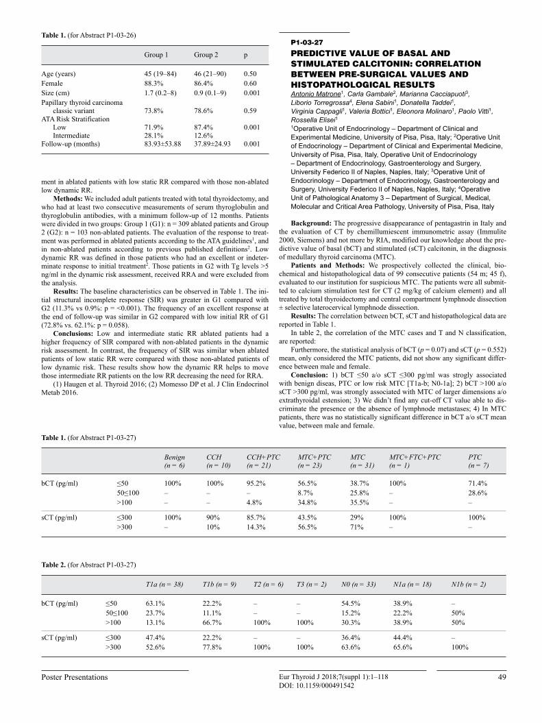
Eur Thyroid J 2018;7(suppl 1):1–118DOI: 10.1159/000491542
49Poster Presentations
P1-03-27
PREDICTIVE VALUE OF BASAL AND STIMULATED CALCITONIN: CORRELATION BETWEEN PRE-SURGICAL VALUES AND HISTOPATHOLOGICAL RESULTSAntonio Matrone1, Carla Gambale2, Marianna Cacciapuoti3, Liborio Torregrossa4, Elena Sabini1, Donatella Taddei1, Virginia Cappagli1, Valeria Bottici1, Eleonora Molinaro1, Paolo Vitti1, Rossella Elisei11Operative Unit of Endocrinology – Department of Clinical and Experimental Medicine, University of Pisa, Pisa, Italy; 2Operative Unit of Endocrinology – Department of Clinical and Experimental Medicine, University of Pisa, Pisa, Italy, Operative Unit of Endocrinology – Department of Endocrinology, Gastroenterology and Surgery, University Federico II of Naples, Naples, Italy; 3Operative Unit of Endocrinology – Department of Endocrinology, Gastroenterology and Surgery, University Federico II of Naples, Naples, Italy; 4Operative Unit of Pathological Anatomy 3 – Department of Surgical, Medical, Molecular and Critical Area Pathology, University of Pisa, Pisa, Italy
Background: The progressive disappearance of pentagastrin in Italy and the evaluation of CT by chemillumiescent immunometric assay (Immulite 2000, Siemens) and not more by RIA, modified our knowledge about the pre-dictive value of basal (bCT) and stimulated (sCT) calcitonin, in the diagnosis of medullary thyroid carcinoma (MTC).
Patients and Methods: We prospectively collected the clinical, bio-chemical and histopathological data of 99 consecutive patients (54 m; 45 f), evaluated to our institution for suspicious MTC. The patients were all submit-ted to calcium stimulation test for CT (2 mg/kg of calcium element) and all treated by total thyroidectomy and central compartment lymphnode dissection ± selective laterocervical lymphnode dissection.
Results: The correlation between bCT, sCT and histopathological data are reported in Table 1.
In table 2, the correlation of the MTC cases and T and N classification, are reported:
Furthermore, the statistical analysis of bCT (p = 0.07) and sCT (p = 0.552) mean, only considered the MTC patients, did not show any significant differ-ence between male and female.
Conclusion: 1) bCT ≤50 a/o sCT ≤300 pg/ml was strogly associated with benign diseas, PTC or low risk MTC [T1a-b; N0-1a]; 2) bCT >100 a/o sCT >300 pg/ml, was strongly associated with MTC of larger dimensions a/o extrathyroidal estension; 3) We didn’t find any cut-off CT value able to dis-criminate the presence or the absence of lymphnode metastases; 4) In MTC patients, there was no statistically significant difference in bCT a/o sCT mean value, between male and female.
ment in ablated patients with low static RR compared with those non-ablated low dynamic RR.
Methods: We included adult patients treated with total thyroidectomy, and who had at least two consecutive measurements of serum thyroglobulin and thyroglobulin antibodies, with a minimum follow-up of 12 months. Patients were divided in two groups: Group 1 (G1): n = 309 ablated patients and Group 2 (G2): n = 103 non-ablated patients. The evaluation of the response to treat-ment was performed in ablated patients according to the ATA guidelines1, and in non-ablated patients according to previous published definitions2. Low dynamic RR was defined in those patients who had an excellent or indeter-minate response to initial treatment2. Those patients in G2 with Tg levels >5 ng/ml in the dynamic risk assessment, received RRA and were excluded from the analysis.
Results: The baseline characteristics can be observed in Table 1. The ini-tial structural incomplete response (SIR) was greater in G1 compared with G2 (11.3% vs 0.9%: p = ˂0.001). The frequency of an excellent response at the end of follow-up was similar in G2 compared with low initial RR of G1 (72.8% vs. 62.1%: p = 0.058).
Conclusions: Low and intermediate static RR ablated patients had a higher frequency of SIR compared with non-ablated patients in the dynamic risk assessment. In contrast, the frequency of SIR was similar when ablated patients of low static RR were compared with those non-ablated patients of low dynamic risk. These results show how the dynamic RR helps to move those intermediate RR patients on the low RR decreasing the need for RRA.
(1) Haugen et al. Thyroid 2016; (2) Momesso DP et al. J Clin Endocrinol Metab 2016.
Table 1. (for Abstract P1-03-26)
Group 1 Group 2 p
Age (years) 45 (19–84) 46 (21–90) 0.50Female 88.3% 86.4% 0.60Size (cm) 1.7 (0.2–8) 0.9 (0.1–9) 0.001Papillary thyroid carcinoma
classic variant 73.8% 78.6% 0.59ATA Risk Stratification
LowIntermediate
71.9%28.1%
87.4%12.6%
0.001
Follow-up (months) 83.93±53.88 37.89±24.93 0.001
Table 1. (for Abstract P1-03-27)
Benign(n = 6)
CCH (n = 10)
CCH+PTC (n = 21)
MTC+PTC (n = 23)
MTC (n = 31)
MTC+FTC+PTC (n = 1)
PTC (n = 7)
bCT (pg/ml) ≤50 100% 100% 95.2% 56.5% 38.7% 100% 71.4%50≤100 – – – 8.7% 25.8% – 28.6%>100 – – 4.8% 34.8% 35.5% – –
sCT (pg/ml) ≤300 100% 90% 85.7% 43.5% 29% 100% 100%>300 – 10% 14.3% 56.5% 71% – –
Table 2. (for Abstract P1-03-27)
T1a (n = 38) T1b (n = 9) T2 (n = 6) T3 (n = 2) N0 (n = 33) N1a (n = 18) N1b (n = 2)
bCT (pg/ml) ≤50 63.1% 22.2% – – 54.5% 38.9% –50≤100 23.7% 11.1% – – 15.2% 22.2% 50%>100 13.1% 66.7% 100% 100% 30.3% 38.9% 50%
sCT (pg/ml) ≤300 47.4% 22.2% – – 36.4% 44.4% –>300 52.6% 77.8% 100% 100% 63.6% 65.6% 100%
Eur Thyroid J 2018;7(suppl 1):1–118DOI: 10.1159/000491542
50 41st Annual Meeting of the ETA
ings previously evaluated by another nuclear physician and a radiologist, and these classifications were compared.
Results: The therapeutic course changed in 20.6% (41/199) of the cases after the analysis of the SPECT/CT images. The staging changed in 22.1% (44/199) of the cases, with an increase in 36.4% (16/44) and a decrease in 63.3% (28/44). In 63.5% (26/41) there was a decrease in the 131I activity administered due to staging decrease, 34.1% (14/41) required greater activity due to staging increase and in 2.4% (1/41) surgical management was indicated due to staging change. Lymph nodal staging was statistically different between the WBS and SPECT/CT methods (41/199, p = 0.019). Positive stimulated thyroglobulin presented a correlation with metastatic SPECT/CT findings, but not the risk classification.
Conclusions: The differentiation between benign cervical structures and affected lymph nodes was the main determining factor for the reestablishment and modification of the conduct. The association of SPECT/CT to pre-RIT WBS contributed to the correct staging of DTC patients (upstaging 36.4% and downstaging 63.5%), mainly regarding lymph node involvement, modifying the therapeutic management of DTC in a significant part of the cases (20.6%), and leading to a global decrease in the 131I administered activity.
P1-03-30
WITHDRAWN
P1-03-31
WITHDRAWN
P1-03-32
SONOGRAPHIC FEATRUES PREDICT THE GROWTH OF PAPILLARY THYROID CARCINOMA DURING ACTIVE SURVEILLANCEHye-Seon Oh1, Hyemi Kwon2, Eyun Song1, Min Ji Jeon2, Tae Yong Kim3, Jeong Hyun Lee4, Won Bae Kim5, YoungKee Shong6, Jung Hwan Baek4, Won Gu Kim1
1Asan Medical Center, Seoul, Korea, Rep. of South; 2Asan Medical Center, University of Ulsan College of Medicine, Seoul, Korea, Rep. of South; 3University of Ulsan College of Medicine, Asan Medical Center, Asan Medical Center, Seoul, Korea, Rep. of South; 4Department of Radiology and Research Institute of Radiology, University of Ulsan College of Medicine, Asan Medical Center, Seoul, Korea, Rep. of South; 5Asan Medical Center, University of Ulsan College of Medicine, Asan Medical Center, University of Ulsan College of Medicine, Seoul, Korea, Rep. of South; 6Asan Medical Center, University of Ulsan, Endocrinology, Seoul, Korea, Rep. of South
Background: Active surveillance has been generally accepted as a man-agement option for low-risk papillary thyroid carcinoma (PTC). However, there are no clear recommendations regarding patients suitable for undergoing active surveillance.
Methods: In this retrospective cohort study from a single tertiary referral center, we evaluated tumor size change between the initial and last ultrasonog-raphy (US). We analyzed 217 PTC patients who underwent active surveillance over 1 year rather than immediate thyroid surgery. Tumor size increase was defined as maximum diameter increase over 3 mm or volume increase over 50% change between the initial and last US findings.
Results: The mean patient age was 51.6 years and 76.0% patients were female. The initial maximum diameter and volume of PTC were 5.8 ± 1.7 mm and 76.8 ± 66.9 mm3, respectively. During the median 38.8-month follow-up, 55 patients (25.3%) experienced tumor size increase. Three features (patients younger than 45 years, taller-than-wide shape on US, and macrocalcification) were associated with increased risk of tumor size increase. A time-dependent increase in the number of these three tumor size increase-related features was significantly associated with higher risk of tumor growth (p < 0.001). The rela-tive risk of tumor size increase with more than two suspicious features was significantly increased compared with that of tumor size increase with one or no suspicious features (hazard ratio = 2.3, p = 0.006).
Conclusions: Some PTC may grow during active surveillance. Young age and the US features of taller-than-wide shape and macrocalcification were associated with tumor size increase. Therefore, active surveillance should be carefully considered in PTC patients with these features.
P1-03-28
IDENTIFYING RISK FACTORS OF LATERAL LYMPH NODE RECURRENCE FOR CLINICALLY NODE-NEGATIVE PAPILLARY THYROID CANCERYoung Jae Ryu1, Shin Jae Kang1, Jin Seong Cho1
1Chonnam National University Hwasun Hospital and Medical School, Gwangju, Korea, Rep. of South
Objectives: There is still debate regarding the role of routine central lymph node (LN) dissection in treating clinically node-negative papillary thy-roid cancer (PTC). The aim of this study was to investigate the risk factors for lateral recurrence after total thyroidectomy and prophylactic bilateral central LN dissection in clinically node-negative PTC patients.
Methods: We retrospectively collected the medical records of 1406 PTC patients who underwent total thyroidectomy and prophylactic bilateral cen-tral LN dissection between January 2004 and December 2008. We used Cox- proportional hazards regression analyses to inspect the predictive factors for recurrence.
Results: During a median follow-up of 107 months (range, 13–164 months), 68 (4.8%) and 37 (2.6%) patients experienced recurrence in any lesion and in lateral neck LN, respectively. Male, main tumor size >1 cm, nodal factors (pathologic N1a, positive delphian LN, lymph node ratio >0.15), lymphovascular invasion, and extrathyroidal extension (ETE) were significantly associated with lateral neck LN recurrence in univariate anal-ysis. Multivariate analysis showed that male (hazard ratio [HR], 2.217; 95% confidence interval [CI], 1.057–4.647; p = 0.035), main tumor size >1 cm (HR, 2.257; 95% CI, 1.138–4.476; p = 0.020), pathologic N1a (HR, 5.957; 95% CI, 2.573–13.789; p < 0.002), minor ETE (vs. no ETE, HR, 3.027; 95% CI, 1.315–6.966; p = 0.009), and gross ETE (vs. no ETE, HR, 4.058; 95% CI, 1.685–9.774; p = 0.002) were independent predictive fac-tors for lateral neck LN recurrence. Among the patients with pathologic N1a, LN ratio of more than 0.55 had worse lateral neck LN recurrence-free survival.
Conclusion: Lateral neck LN recurrence in clinically node-negative PTC patients is predicted by the factors of male sex, main tumor size >1 cm, ETE, and pathologic N1a.
P1-03-29
VALUE OF SPECT-CT WITH IODINE-131 IN STAGING AND THERAPEUTIC MANAGEMENT OF PATIENTS WITH DIFFERENTIATED THYROID CARCINOMAAlinne Verçosa1, Barbara Juarez1, Camila Mosci1, Mariana Lima1, Elba Etchebehere1, Allan O Santos1, Ligia Vera Montalli Assumpcao2, Denise Engelbrecht Zantut Wittmann3, Celso Darío Ramos4
1Nuclear Medicine Division of Radiology Department Faculty of Medical Sciences, University of Campinas, Campinas, Brazil; 2Division of Endocrinology, University of Campinas, Campinas, Sáo Paulo, Brazil; 3Endocrinology Division, Department of Clinical Medicine, Faculty of Medical Sciences, University of Campinas-Unicamp, Campinas, Brazil; 4Nuclear Medicine Division of Radiology Department, Campinas, Brazil
Background: In patients with differentiated thyroid carcinoma (DTC), treatment follow-up and metastasis screening can be performed with whole body scan (WBS), with or without additional SPECT/CT images. The defini-tion of the structures that uptake iodine in the pre-RIT WBS of patients with DTC is of fundamental importance for the therapeutic decision, since it can differentiate physiological sites of uptake from those metastatic. Although the SPECT/CT images aid in the interpretation of the WBS findings, their impact on the therapeutic decision is not fully clarified in the literature.
Aims: To evaluate possible changes in staging and conduct determined by SPECT/CT images performed in complementation with WBS with 131I before radioiodine therapy (RIT) in patients with DTC.
Methods: One hundred and ninety-nine patients with DTC who per-formed SPECT/CT images complementary to pre-RIT WBS were selected, 167 omen and 32 men, mean age 46.3 years, 187 with papillary DTC and 12 follicular DTC, 150 of them performing first WBS and 49 WBS monitoring. The images were taken 48/72 h after oral administration of 5 mCi of 131I. A nuclear physician evaluated the WBS and classified staging, risk classification and therapeutic management, which were compared to the SPECT/CT find-

Eur Thyroid J 2018;7(suppl 1):1–118DOI: 10.1159/000491542
51Poster Presentations
in real life. Therefore, information of the running of these units in the clinical practice in our country is imperative.
Objective: The aim of the present study has been to retrieve real data on the composition, structure, and functioning developed by the multidisciplinary units of thyroid cancer existing in Spain.
Methods: A nationwide survey was carried out through the SEEN web-site. The survey was distributed through the SEEN members and through direct contact with specialists of other disciplines involved in the field of thyroid cancer. The survey consisted of several questions about composition, structure and functioning of multidisciplinary teams. It was available on the SEEN website from November 15, 2017 to February 15, 2018.
Results: Seventy-five multidisciplinary units responded to our survey. Of these, 20% were exclusive of thyroid cancer, while 80% included other endocrine disorders or non-endocrine tumors. The mean (±SD) of members of the teams was 11 ± 4.0. The most frequent medical specialties in the units were: Endocrinology (100%), Surgery (97.4%), Nuclear Medicine (92.2%), Pathology (89.6%), Radiology (84.0%) and Medical Oncology (83.0%). 52% of the units had a coordinator and 61.0% had written operating regula-tions. Periodicity of the meetings was weekly in 14.3%, fortnightly in 23.4% and monthly in 58.4%. Apart from clinical case discussions in the meet-ings, 21.8% of the units included educational activities and 14.1% research subjects. The annual number of cases (median, interquartile range) studied by the teams was 40(20–69). 38.5% of the teams discuss all thyroid cancer patients who come to the hospital, while 70.5% study only cases with special difficulties. Team decisions were recorded in the patient’s medical record in 85.7% of hospitals. 60.5% of the multidisciplinary teams have elaborated clinical protocols for local use, and 22% have developed their own quality indicators.
Conclusion: These results suggest that international recommendations on the multidisciplinary approach to patients with thyroid cancer are followed in Spain. This gives us the opportunity to further studies analyzing the real impact of this high standard of care on patient outcomes.
P1-04-35
ANALYSIS OF THE FACTORS CONDITIONING THE CLINICAL COURSE OF DIFFERENTIATED THYROID CARCINOMA IN CHILDREN AND YOUNG ADULTSLaura Cannavo1, Giuseppina Zirilli2, Francesco Vermiglio2, Maria Antonia Violi2, Alfredo Campennì2, Filippo De Luca2, Malgorzata Wasniewska3
1University of Messina, Department of Human Pathology, Messina, Italy; 2Messna, Italy; 3University of Messina, Messina, Italy
Objectives: aim of this study was to ascertain for the first time whether characteristics of differentiated thyroid carcinoma (DTC) may significantly vary according to age, even within a peculiar study population covering only young patients aged less than 30 years.
Methods: the population was composed by 64 patients (47 females) aged <30.0-yrs at diagnosis of DTC (mean age 22.9 ± 5.5 yrs). The main clini-cal, biochemical and pathologic data at DTC diagnosis were retrospectively recorded in 2 selected cohorts including, respectively, 19 patients aged less than 18 years (Group A) or 45 young adults aged between 20 and 29.8 years (Group B).
Results: The distribution of DTC cases in the different age ranges pro-gressively increased with age. Both groups had a higher proportion of females, indeed the female: male ratio was 2.6:1 and 2.75:1 in Group A and B respec-tively. Group A exhibited at diagnosis a more severe clinical involvement and a higher rate of extra-regional metastases. Moreover, the association with autoimmune thyroid diseases (AITDs) and biochemical thyroid dysfunction was also more common in Group A (p = 0.02 and p = 0.007 respectively). The age at DTC diagnosis correlated with the tumor size at pathology evaluation (r = –0.27, p < 0.05) but not with US nodule diameter, TSH and fT4 serum levels.
Thyroid dysfunction at DTC diagnosis was associated more frequently with multifocality (p = 0.02), metastasis (p = 0.004) and recurrence (p = 0.0005) and needed also more radiotherapy cycles (p = 0.006) if compared with euthyroid DTC patients. Thyroid nodules were smaller in patients with AITDs in comparison to those without AITDs (p = 0.006). Moreover, multifo-cality of DTC required more cycles of therapy (p = 0.02).
During the whole follow-up time, the overall survival rate was 100%, but 11.1% in Group A and 4.5% in Group B were alive with persistent residual TC.
Epidemiology and Clinical Features
P1-04-33
IS ANTI THYROID PEROXIDASE POSITIVITY PROTECTIVE AGAINST THYROID CANCER IN GRAVES DISEASE?Didem Ozdemir1, Sevgul Faki2, Muhammet Cuneyt Bilginer2, Husniye Baser2, Oya Topaloglu2, Reyhan Ersoy2, Bekir Cakir2
1Ankara Yildirim Beyazit University, School of Medicine, Department of Endocrinology and Metabolism, Ankara, Turkey; 2Ankara Yildirim Beyazit University, School of Medicine, Endocrinology and Metabolism Diseases, Ankara, Turkey
Aim: Anti thyroid peroxidase (AntiTPO) and antithyroglobulin (AntiTg) are known to be positive in 75–90% and 35–50% of patients with Graves dis-ease, respectively. Although studies investigating the association between thy-roid cancer and Hashimoto thyroiditis (HT) -in which AntiTPO is an important hallmark of the disease- reported controversial results, studies showing higher risk in HT outweigh. However, the association between AntiTPO and thyroid cancer in patients with Graves disease was not studied. We aimed to investi-gate whether AntiTPO or AntiTg positivity have any effect on malignancy risk in these patients.
Methods: Graves patients operated in our center was recruited for the study retrospectively. The clinical features, operation indications and thyroid autoantibodies (AntiTPO, AntiTg and TSH receptor antibody) were recorded. Patients were grouped as benign and malignant according to histopathological diagnosis
Results: Data of 602 patients were analyzed. There were 410 (%68.1) female and 192 (%31.9) male patients with a mean age of 43.65 ± 12.69. Preoperative ultrasonography revealed no nodule in 286 (%47.5), single nod-ule in 63 (%10.5) and multiple nodules in 253 (%42.0) patients. There were 400 (66.4%) patients with positive AntiTPO and 279 (46.3%) with positive AntiTg. Histopathological diagnosis was benign in 512 (85%) and malignant in 90 (15%) patients. Age, sex and TSH receptor antibody positivity did not differ between benign and malignant patients. 267 (52.1%) patients in benign and 19 (21.1%) patients in malignant group had no nodule in preoperative ultrasonography (p < 0.001). There was significant difference in operation indications between benign and malignant patients (p < 0.001). AntiTg was positive in 48.2% of benign and 35.6% of malignant patients (p = 0.026). AntiTPO was positive in 356 (69.5%) of benign and 44 (48.9%) of malignant patients (p < 0.001). With multiple regression analysis, association between AntiTg positivity and benign histopathology was lost (p = 0.600), while asso-ciation between AntiTPO positivity and benign histopathology remained (p = 0.016).
Conclusion: In accordance with the literature, the presence of nodule in Graves patients increased malignancy risk in our study. Additionaly, for the first time we showed that AntiTPO positivity might play a protective role against thyroid cancer in patients with Graves disease.
P1-04-34
RESULTS OF A NATIONWIDE SURVEY ON MULTIDISCIPLINARY TEAMS OF THYROID CANCER IN SPAINJuan Jose Diez1, Juan Carlos Galofre2, Amelia Oleaga3, Enrique Grande4, Mercedes Mitjavila5, Pablo Moreno6
1Hospital Ramón Y Cajal, Madrid, Spain; 2Clínica Universidad de Navarra, Pamplona, Spain; 3Hospital Universitario Basurto, Bilbao, Spain; 4MD Anderson Cancer Center Madrid, Madrid; 5Hospital Universitario Puerta de Hierro, Madrid, Spain; 6Hospital Universitari de Bellvitge, Barcelona, Spain
Background: The multidisciplinary approach of patients with thyroid cancer is a glaring necessity of healthcare shared by providers and patients. Recently the Sociedad Española de Endocrinología y Nutrición (SEEN, Spanish Society of Endocrinology) has reported a consensus statement in which the composition, requirements, structure and operation plans of mul-tidisciplinary units of thyroid cancer are delineated. However, an important lack of knowledge persists on the functioning of these multidisciplinary units

Eur Thyroid J 2018;7(suppl 1):1–118DOI: 10.1159/000491542
52 41st Annual Meeting of the ETA
P1-04-37
IDENTIFYING RISK FACTORS OF RECURRENCE FOR PAPILLARY THYROID CANCER PATIENTS WHO UNDERWENT MODIFIED RADICAL NECK DISSECTION Yong Min Na1, Young Jae Ryu1, Young Hoe Hur1
1Chonnam National University Hwasun Hospital and Medical School, Gwangju, Korea, Rep. of South
Objectives: The papillary thyroid cancer (PTC) patients with ipsilateral neck metastatic lymph node (LN) and those with contralateral neck metastatic LN belong to N1b. Only a few studies reported the comparison with regard to laterality of metastatic lateral LN. The aim of this study is to evaluate predic-tive factors for contralateral neck LN metastasis and to determine prognostic factors for recurrence in PTC patients with N1b.
Methods: This retrospective study reviewed the medical records of 390 PTC patients who underwent total thyroidectomy and central LN dissection plus ipsilateral or bilateral MRND between January 2004 and December 2012.
Results: During a median follow-up of 81 (range, 6–156) months, 84 patients occurred recurrence in any lesion. Male gender, more than 2 cm of main tumor, number of metastatic central LN, number of harvested and metastatic lateral LN, total LN ratio, multifocality of tumors, bilaterality or tumors, and gross ETE had significance in the patients who underwent bilat-eral MRND. In multivariate analysis, the patients with LN ratio >0.44 in the central compartment (hazard ratio [HR], 1.890; 95% confidence interval [CI], 1.124–3.178; p = 0.015), LN ratio >0.29 in the lateral compartment (HR, 2.351; 95% CI, 1.477–3.743; p < 0.001), and multifocality (HR, 1.583; 95% CI, 1.030–2.431; p = 0.036) were associated with worse RFS. However, bilat-eral MRND had statistically significance only in univariate analysis.
Conclusions: Recurrence in N1b patients is predicted by central neck LN ratio >0.44, lateral neck LN ratio >0.29, and multifocality or tumors. We sug-gest that patients with mentioned factors should receive short-term follow-up and appropriate management.
P1-04-38
FUNCTIONING BONE METASTASES OF FOLLICULAR THYROID CARCINOMA IN TOXIC NODULETalita L. Trevisan1, Cinthia Minatel Riguetto2, Patrícia Sabino de Matos3, Icleia Barreto3, Denise Engelbrecht Zantut Wittmann4, Ligia Vera Montalli Assumpcao5
1Endocrinology Division, Internal Medicine Department, Faculty of Medical Sciences, University of Campinas, Campinas, Brazil; 2Hospital Das Clinicas – Unicamp, Internal Medicine, Endocrinology, Campinas, Brazil; 3Department of Pathology, Faculty of Medical Sciences, University of Campinas, Campinas, Brazil; 4Endocrinology Division, Department of Clinical Medicine, Faculty of Medical Sciences, University of Campinas-Unicamp, Campinas, Brazil; 5Division of Endocrinology, University of Campinas, Campinas, Sáo Paulo, Brazil
Background: Functioning metastasis occur rarely in differentiated thy-roid carcinoma, mainly as a complication of a malign toxic nodule. In this case report, we described a patient with a follicular thyroid carcinoma in a toxic nodule who developed thyrotoxicosis due extensive metastatic disease.
Case Report: A 45 year-old man diagnosed with a Toxic Adenoma (4 cm lesion in the right lobe) in 1997. He presented a severe cutaneous reac-tion with propyltiouracil and was referred to thyroidectomy in the same year. Histopathology revealed a follicular thyroid carcinoma, minimally invasive, 5.5 x 4.0 x 3.5 cm. T4N0Mx. Radioiodine therapy (RIT) is described Table 1.
(2005) Surgery due increasing Tg levels (556 ng/mL): Partial resection of left rib and right femur. After surgery, (2006) Tg was 14.5 ng/mL. Patient maintained good health with high but stable Tg levels. In 2009, he lost weight (90 → 69 kg), concomitant increase in Tg levels (4500 ng/mL). He required levothyroxine withdrawal, developing thyrotoxicosis (FT4 = 7.24 ng/dl, RV = 0.9–1.8) successfully controlled by Methymazole. (2010) He underwent guided surgery for resection of left rib and half of right femur with pros-thesis placement. Histopathology confirmed Follicular Thyroid Carcinoma Metastases. He presented complete resolution of thyrotoxicosis and Tg = 28 ng/mL. Levothyroxine was restarted to keep TSH suppression. (2012) He developed thyrotoxicosis again (FT4 = 5.29 ng/dl) and Tg increase (3420
Conclusions: In a study population younger than 30 years: a) the risk of developing DTC increases with age, achieving its zenith during the 3rd decade of life; b) clinical presentation is more severe in children and adolescents younger than 18 years than in the patients aged between 20 and 30; c) in the cohort of children and adolescents DTC is more often associated with AITDs, which might play some role in conditioning the more aggressive phenotypi-cal presentation of DTC in this patient group. A close US follow-up in chil-dren and adolescents with thyroid dysfunction, ATD and/or thyroid nodules is needed and indicated to earlier diagnosis of a thyroid malignancy.
P1-04-36
DETECTION OF EIF1AX GENE MUTATIONS IN THYROID CARCINOMAS AND BENIGN NODULES IN CZECH COHORTBela Bendlova1, Sarka Dvorakova1, Barbora Pekova1, Vlasta Sykorova1, Gabriela Vacinova1, Eliska Vaclavikova1, Rami Katra2, Petr Vlcek3, Pavla Sykorova3, Jan Plzak4, Petr Bavor5, Daniela Kodetova6, Martin Chovanec7, Josef Vcelak1
1Department of Molecular Endocrinology, Institute of Endocrinology, Prague, Czech Republic; 2Department of Etn, 2nd Faculty of Medicine, Charles University in Prague and Motol University Hospital, Prague, Czech Republic; 3Department of Nuclear Medicine and Endocrinology, 2nd Faculty of Medicine, Charles University in Prague and Motol University Hospital, Prague, Czech Republic; 4Department of Otorhinolaryngology and Head and Neck Surgery, 1st Faculty of Medicine, Charles University in Prague and Motol University Hospital, Prague, Czech Republic; 5Department of Surgery, 2nd Faculty of Medicine, Charles University in Prague and Motol University Hospital, Prague, Czech Republic; 6Department of Pathology and Molecular Medicine, 2nd Faculty of Medicine, Charles University in Prague and Motol University Hospital, Prague, Czech Republic; 7Department of Otorhinolaryngology and Head and Neck Surgery, 3rd Faculty of Medicine, Charles University in Prague, Royal Vinohrady Teaching Hospital, Prague, Czech Republic
Objectives: Thyroid carcinoma is the most often endocrine malignancy. The most frequent somatic point mutations in thyroid cancer are in genes BRAF, H-, K-, N-RAS and RET. Recently, The Cancer Genome Atlas (TCGA) study identified EIF1AX gene as a new causal gene in the thyroid tumour development. This gene is located on chromosome X and encodes the eukary-otic translation initiation factor 1A. The goal of this study was to analyse the EIF1AX gene in different types of thyroid carcinomas and compare the preva-lence of main genes mutations associated with thyroid carcinomas.
Methods: DNAs were isolated from 275 fresh-frozen thyroid tissues of the patients – 222 with papillary thyroid carcinoma (PTC), 5 with anaplas-tic thyroid carcinoma (ATC), 16 with sporadic medullary thyroid carcinoma (MTC) without somatic RET proto-oncogene mutations and 32 with benign nodules (mostly adenomas). Exons 1, 2, 5 and 6 of the EIF1AX gene, together with exon 15 of the BRAF gene, exons 2 and 3 of the H-, K-, N-RAS genes, were prepared using Nextera XT kit and analysed by next generation sequenc-ing on Miseq (Illumina).
Results: In summary, 3 mutations in EIF1AX gene were detected: P2L mutation in the exon 1 in one follicular variant of PTC, G9D mutation in the exon 2 in one benign nodule and A113_splice mutation in exon 6 in one ATC. No other classical thyroid gene mutations were found in these cases. In the PTC cohort, BRAF mutation in 37% and RAS mutations in 13% were detected. In the ATC cohort, one BRAF mutation was detected. In the RET-negative MTC cohort, only RAS mutations in 56% and no EIF1AX mutations were found. In the cohort of thyroid benign nodules, RAS mutations in 22% of cases were found.
Conclusions: Totally, the three different mutations in the EIF1AX in 3 positions of the gene in 3 cases were found – in ATC, PTC and benign nodule. All three mutations were reported in the literature, it seems that each muta-tion has different impact on the function of the protein and aggressiveness of tumour. The most aggressive was A113_splice mutation in accordance with the literature. Mutations in the exon 2 were reported in benign nodules as same as in our case. However, it is necessary to enlarge the studied cohorts, because the detection rate of mutations is low. This work was supported by AZV 16-32665A and MZ ČR-RVO (EÚ, 00023761) grants.

Eur Thyroid J 2018;7(suppl 1):1–118DOI: 10.1159/000491542
53Poster Presentations
P1-04-40
10-YEAR REVIEW OF 683 THYROID CNCER CASES IN AN IRISH TERTIARY REFERRAL CENTRENiall McVeigh1, Conor McVeigh2, Aftab Khattak1, Mary Toner1, Esther O’Regan1, Jennie Cooke1, Marie-Louise Healy1
1St James’s Hospital, Dublin, Ireland; 2School of Medicine, Trinity College, Dublin, Ireland
Objective: The histopathologic classification of differentiated thyroid cancer (DTC) informs staging, risk-stratification, treatment choice. This study set out to set out to review:
– Histological subtypes of DTC recorded over a 10-year period. – Temporal trends, if any of each subtype over the 10-year period. – Treatment choices made based on the application of current guidelines
and risk-stratification at the time of diagnosis– Response to treatment by applying current (American Thyroid
Association-ATA-2015) response to therapy reclassification– Final patient status: alive with active disease, alive and disease free,
dead with disease, dead from another cause.Methods: Patients who underwent thyroidectomy for thyroid cancer
over a 10-year period (2005–2016) at a tertiary hospital were identified. 683 histology reports were reviewed and classified into papillary thyroid cancer (PTC), follicular, medullary and anaplastic subtypes as well as subtypes asso-ciated with higher risk (tall cell, diffuse sclerosing as the WHO classification guidelines.
Patient samples collected with disease recurrence or non-primary tumours were excluded from the dataset. The incidence of each histological sub-type was examined on a year-to-year basis.
All DTC were reviewed on treatment choice and response.Thyroglobulin (TG), and TSH-stimulated TG together with neck imag-
ing were assessed at 1-year and TG and neck imaging at 5-year post-surgery. Patients treated with further surgical intervention and/or RAI were identified over the 10-year period.
Response to RAI was classified as excellent response, biochemical incom-plete response, structural incomplete response and indeterminate response as per the ATA guidelines.
Current patient status was documented as dead from disease, dead from other cause, alive with active disease, alive disease-free.
Results: Of the 683 histology reports reviewed, 87% as PTC, 6.6% fol-licular, 2.2% medullary, 2.5% anaplastic, 1.8% had a poorly differentiated pathology. Absolute case numbers of DTC reports increased from 32 in 2005 to 71 in 2016.
33% of differentiated thyroid cancer cases received RAI as per current guidelines. At year one 20% structural incomplete response (SIR) and 12% achieving a biochemical incomplete response (BIR) and% had an indetermi-nate response. At year 5, 24% a SIR and 14% a BIR. Of those with an incom-plete response, 6.9% required further RAI treatment.
98.1% of those with DTC are alive at the time of analysis.Conclusion: Within our cohort of 683 patient, 639 presented with DTC, of
which 33% underwent RAI. 98.1% of those with DTC are alive at submission.
P1-04-41
PAPILLARY THYROID MICROCARCINOMAS. WHAT MAKE THE DIFFERENCEKalliopi Pazaitou-Panayiotou1, Stylianos Mandanas2, Efterpi Margaritidou2, Eirini Poimenidou2, Constantinos Toulis Pantelitsa Rakitzi2, Alexandra Chrisoulidou2, Maria Boudina2, Kalliopi Pazaitou-Panayiotou2
1Theagenio Anticancer Hospital of Thessaloniki, Thessaloniki, Greece; 2Department of Endocrinology-Endocrine Oncology, Theagenio Cancer Hospital, Thessaloniki, Greece
Papillary thyroid microcarcinomas (PTMs) are the most commonly diagnosed thyroid cancers in individuals who are older than 45 years which often are found incidentally after thyroidectomy or during thyroid ultrasound. Sometimes, PTMs may have a more aggressive behaviour with extrathyroidal extension and cervical lymph node metastases
Aim: Identify factors that may be related to aggressive behaviour of PTMsMaterial and Methods: The medical records of patients with thyroid can-
cer followed at the Theagenio cancer Hospital were retrospectively reviewed.
ng/mL). During this time, the patient did not present clinical conditions for another surgery. Tyrosine Kinase Inhibitors are not available at our hospital. After 14 years from initial diagnosis, still under methymazole, the patient died of respiratory failure secondary to H1N1 infection.
Conclusions: Functioning metastasis are rare in differentiated thyroid car-cinoma, however the thyrotoxic state in these cases have to be promptly recog-nized and treated. Methymazole was an effective drug for such. Unfortunately, the prognosis of these tumors is very poor, and thyrotoxicosis is an aggravat-ing factor that can worsen the general condition of the patient and increase the chances of cardiovascular complications.
P1-04-39
THE CLINICO-PATHOLOGIC FACTORS FOR THE PATTERN OF LYMPHATIC METASTASIS IN PAPILLARY CARCINOMA OF THE THYROID: A PROSPECTIVE STUDYMin Sik Kim1, Dong Huun Lee2, Sang Yun Kim2
1The Catholic University of Korea, Department of Otolaryngology-HNS, Seoul, Korea, Rep. of South; 2The Catholic University of Korea, Seoul, Korea, Rep. of South
Introduction: Papillary thyroid carcinoma (PTC) is the most common form of thyroid gland malignancy with a tendency for lymphatic spread. Lymph node (LN) metastasis in differentiated thyroid carcinomas are related to the high recurrence rate, low disease-specific survival rate, and overall survival rate. The ideal treatment for regional LN metastasis has remained a subject of debate. Determining the extent of neck dissection should be mainly based on the predictable pattern of spread of regional LN metastasis from PTC. If we can identify the predictive clinic-pathologic factors of regional LN metastasis, we can make the proper preoperative plans.
Method: We prospectively evaluated the pattern and predictive factors of LN metastasis of 397 patients with PTC who underwent total thyroidec-tomy and bilateral central LN dissection with or without therapeutic lateral LN dissection.
Result: The incidence of ipsilateral central LN metastasis was (133/397) 33.5% and that of contralateral central LN metastasis was (29/397) 7.3%. Only male sex was statistically significantly related to central LN metastasis (p = 0.03), and ipsilateral central LN metastasis was significantly related to contralateral central LN metastasis (p < 0.01).
About metastasis to lateral LN, the incidence of ipsilateral lateral LN metastasis was (100/397) 25.2% and that of contralateral lateral LN metastasis was (48/397) 12.1%. Extracapsular spread (ECS) were statistically significant (p = 0.01), and central LN metastasis was significantly related to lateral LN metastasis (p = 0.01).
Conclusion: Ipsilateral central neck dissection is recommended for all PTC patients. With ipsilateral central LN metastasis, the possibility of contra-lateral central LN involvement is high. Therefore, complete bilateral central LN dissection would be a proper option. Lateral cervical metastasis frequently occurs with extracapsular invasion and ipsilateral central LN metastasis. More precise pre-operative evaluation should be needed in those patients for lateral LN metastasis.
Table 1. (for Abstract P1-04-38)
Whole Body Scan (year)
(1998)Thyroid bed
(2000)Left rib and right femur
(2002)Left rib and right femur
(2004)Left rib and right femur
Tg (ng/mL) 6.7 74.9 31.7 27.3131I (mCi) 100 300 300 300

Eur Thyroid J 2018;7(suppl 1):1–118DOI: 10.1159/000491542
54 41st Annual Meeting of the ETA
genesis- CoH2), 23 by AH and 22 by CH (13 by idiopathic hypopituitarism- CH1, 9 secondary to pituitary tumors- CH2). Serum fT4 and TSH levels were measured by commercial kits at mean age of 14.9 ± 2.3 yrs.
Results: In AH children, mean L-T4 maintenance euthyroid doses were significantly lower than in the CoH and CH groups (p = 0.02 and p = 0.008 respectively), while no differences were found between CoH and CH groups (p = 0.1) (Table1). Mean L-T4 doses to maintain euthyroidism were similar in patients with athyreosis vs dyshormonogenesis (p = 0.1) and in those with idiopathic and secondary CH (p = 0.4). Moreover, there was no statistically significant correlation between LT4 dosage and serum FT4 levels or chrono-logical age in all forms of permanent hypothyroidism in our study population. In all groups mean FT4 levels were not different, and in AH and CH mean TSH values were similar
Conclusions: In our experience CH children need (weight-based daily) L-T4 dosages similar to CoH ones, while significantly lower doses are suf-ficient to maintain clinical and biochemical euthyroid status in those with AH. These findings are agreeing with the hypothesis that LT-4 replacement dose is inversely correlated with the functionally of thyroid tissue.
P1-05-43
RENAL FUNCTION IN HYPOTHYROID PATIENTSAnna Volkova1, Olga Dygun1, Svetlana Dora1, Lyudmila Belyakova1
1Pavlov First Saint Petersburg State Medical University, Saint Petersburg, Russian Federation
In patients with hypothyroidism increased creatinine level, decreased eGFR, low T3 syndrome are frequently revealed
Objectives: to evaluate renal function in patients with hypothyroidism according with sex and age.
Methods: 466 patients with hypothyroidism (9.9% of manifest hypothy-roidism, 90.1% of subclinical hypothyroidism) were included in the study. The mean age was 61.22 ± 15.06 years. The control group comprised 484 euthyroid patients, whose mean age was 56.35 ± 16.66 years (p = 0.01). Women were significantly more in the hypothyroid group (p = 0.002). TSH level and serum creatinine level were assessed in all patients, free T3 and free T4 were deter-mined in some patients. eGFR was calculated by CKD-EPI formula (2011).
Results: A negative correlation was found between GFR and age (r = –0.46). There wasn’t clinically significant correlation between eGFR and TSH. The patients were divided into 3 groups depending on eGFR: eGFR equal to 0–44 ml/min/1.73 m2, 45–89 ml/min/1.73 m2, ≥90 ml/min/1.73 m2. In the group of euthyroid patients, eGFR equal 0–44 ml/min/1.73 m2 was 2 times less frequent than in hypothyroid group, patients with normal eGFR ( ≥90 ml/min/1.73 m2 ) were 3 times more (p = 0.02).
Tumour size, sex, age, the presence of Hashimoto, intrathyroid invasion and extrathyroid extention were recorded.
Results: 1105 patients with microcarcinomas were isolated, 940 were women. The mean age of patients was 39.63 years and the mean tumor size was 4.83 ± 2.45 mm. 979 of PTMs (88.6%) were intrathyroid, Women more frequently than men had intrathyroid PTMs (89.49%). 568 presented Hashimoto disease. Intrathyroid PTMs were associated with Hashimoto more frequently compared to PTMs with extrathyroid extension
Mean tumour size was significantly higher in the PTMs with extrathy-roid extension compared to the intrathyroid cancers (6.39 ± 2.04 versus 4.63 ± 2.45) Mean age of the patients was not different between itra and extrathyroid PTMs
Conclusion: Tumour size and male sex is a prognostic factor regarding the extrathyroidal extension of PTMs. Hashimoto disease is a protective factor and limits the extent of the disease.
Hypothyroidism 1
P1-05-42
DIFFERENCES IN LEVOTHYROXINE DOSAGES FOR REPLACEMENT IN CHILDREN WITH DISTINCT CAUSES OF PERMANENT HYPOTHYROIDISMMalgorzata Wasniewska1, Laura Cannavo2, Giuseppina Zirilli3, Giorgia Pepe3, Domenico Corica3, Tommaso Aversa3, Filippo De Luca3
1University of Messina, Messina, Italy; 2University of Messina, Department of Human Pathology, Messina, Italy; 3Messina, Italy
Objectives: L-tiroxine (L-T4) is considered the treatment of choice in hypothyroidism, either congenital (CoH), autoimmune (AH) or central (CH). The study objective was to find the mean L-T4 doses for CoH, AH and CH in pediatric age to maintain optimal hormone replacement.
Methods: In this one center, cross-sectional and retrospective study, 67 of 267 evaluated patients, aged <18.0 yrs, with overt and permanent hypo-thyroidism, followed at the Outpatient Pediatric Endocrinology Clinic of University of Messina, were enrolled on the basis of the following inclusion criteria: a) age <14.0 years at hypothyroidism diagnosis; b) follow-up at least 3 yrs under L-T4 therapy; c) normal value of fT4 and/or TSH for at least six months under unchanged L-T4 therapy. Our study population consisted of: 22 children affected by CoH (14 by thyroid dysgenesis- CoH1, 8 by dyshormono-
Table 1. Average L-T4 doses and serum concentrations of TSH and free T4 (mean and SD) at the follow-up visit (for Abstract P1-05-42)
AH CoH CoH1 CoH2 CH CH1 CH2
Mean age (yr) 15±2.1 14.3±2.5 14.6±2.6 13.7±2.3 15.6±2.2 16.1±1.8 14.8±2.5TSH (mIU/L) 1.5±1.2 1.8±1.2 2.0±1.4 1.3±0.7FT4 (pmol/L) 16.7±4.2 17.1±2.4 17.5±2.1 16.5±3.1 15.0±5.1 16.9±4.5 13.7±4.3LT4 (μg/kg/die) 1.4±0.4 1.7±0.4 1.8±0.2 1.5±0.5 1.8±0.7 1.8±0.8 1.9±0.5
Table 1. Distribution of patients with different thyroid function depending on eGFR (for Abstract P1-05-43)
CKD stage eGFR Normal TSH level(TSH 0.5–2.5 mIU/l)n = 484
Subclinical hypothyroidism(TSH 4–10 mIU/l)n = 417
Overt hypothyroidism(TSH >10 mIU/l)n = 49
CKD 3B-5 0–44 ml/min/1.73 m2 7.23% (n = 35) 14.63% (n = 61) 14.29% (n = 7)CKD 2-3A 45–89 ml/min/1.73 m2 47.11% (n = 228) 70.74% (n = 295) 59.18% (n = 29)CKD 1 ≥90 ml/min/1.73 m2 45.66% (n = 221) 14.63% (n = 61) 26.53% (n = 13) 100.00% 100.00% 100.00%
Correlation between eGFR and free T3 (r = 0, 34) and weak relationship between eGFR and free T4 (r = –0.1) were found.

Eur Thyroid J 2018;7(suppl 1):1–118DOI: 10.1159/000491542
55Poster Presentations
sampled during endoscopy to measure gastric pH. These patients switched to softgel T4 preparation, titrated to obtain individual serum TSH values as above.
Results: Mean gastric juice pH in the whole sample was 2.87 and, based on this value, patients were subdivided in two groups: Group A (n = 20) with a mean pH of 1.69 and Group B (n = 8) showing a mean pH of 5.81, that mirrors a defined reduced gastric acid production. The pH values well correlated with the dose of T4 in both groups (p = 0.0329 and 0.0023). Following the switch to softgel, T4 requirement was the same in 19 out of 20 (95%) of patients with normal pH. On the contrary in 7 out of 8 (88%, p < 0.0001; PPV 95%, Likelihood ratio = 7.6) patients with high gastric pH the requirement of T4 in softgel formulation was significantly reduced. The median reduction in these latter patients was from 1.98 to 1.67 μg/kg/day (–19%).
Conclusions: These data indicate that the dose of both tablet and softgel thyroxine correlates with gastric pH and, in hypothyroid patients with dis-orders or conditions impairing gastric acid secretion, softgel T4 preparation should be the preferred therapeutic choice.
P1-05-46
HYPOTHYROID PATIENTS HARBORING POLYMORPHISMS IN THE DIO2 AND MCT10 GENES DID NOT PRESENT HIGHER DISEASE BURDEN AT DISEASE ONSETNils Knudsen1, Hans Perrild2, Lars Ovesen3, Lone B. Rasmussen4, Torben Jørgensen5, Rudi Steffensen6, Jens Faber7, Birte Nygaard8, Inge Bulow Pedersen9
1Bispebjerg University Hospital, Copenhagen, Denmark; 2Department of Endocrinology, Bispebjerg University Hospital, Copenhagen, Denmarkl, Copenhagen, Denmark; 3Department of Internal Medicine, Slagelse Hospital, Slagelse, Denmark, Slagelse, Denmark; 4National Food Institute, Technical University of Denmark, Copenhagen, Denmark, Copenhagen, Denmark; 5Research Centre for Disease Prevention and Health, Copenhagen County, Denmark, Glostrup; 6Department of Clinical Immunology, Aalborg University Hospital, Aalborg, Denmark; 7Herlev University Hospital, Department of Endocrinology, Faculty of Health and Medical Sciences, University of Copenhagen, Herlev, Denmark; 8Department of Endocrinology, Herlev University Hospital, University of Copenhagen, Faculty of Health and Medical Sciences, University of Copenhagen, Copenhagen, Denmark; 9Department of Medicine and Endocrinology, Medicinerhuset, Mølleparkvej Aalborg, Aalborg, Denmark
Objectives: A few studies have shown better treatment effects in patients harbouring specific single nucleotide polymorphisms (SNPs) in genes respon-sible for end target concentrations of T3. However, no study has provided information on how the SNP distribution may affect symptom presentation when hypothyroidism is diagnosed.
Methods: We investigated the clinical presentation of 132 patients newly diagnosed with autoimmune overt hypothyroidism. All patients diagnosed at hospital but also in primary care were included. Thus, patients were recruited without referral bias. We characterized the distribution of SNPs for the fol-lowing: the DIO2 gene (rs225014 = Thr92Ala, rs12885300 = ORFa-Gly3Asp, and rs225015) responsible for the conversion of T4 to T3 within the target cell, and the MCT10 gene (rs17606253) responsible for the thyroid hormone influx. We analyzed whether the SNP presence had any impact on the disease presentation. Primary end-point was symptom score = number of hypothyroid symptoms (range: 0–13). Secondary end-points were: Co-morbidity, well-being, self-judged health, TSH-T3-T4 concentrations, thyroid auto-antibodies, blood pressure, GP and hospital visits, and sick days) when hypothyroidism was diagnosed. We have previously shown that the symptom presentation is highly age-dependant. Thus, analyses were dichotomized by age (young, age <50 y. vs. elderly, age ≥50 y.).
Results: Younger patients harbouring the rs225014 (Thr92Ala) wild type gene (TT) had borderline higher symptom-score (7.4 vs. 5.5, p = 0.01) and longer duration of symptoms (22 vs. 8 months, p = 0.007) compared to SNP patients (CC or CT). This was not reproduced among patients aged 50 years or above. We found no evidence of higher disease burden at disease onset in patients harbouring any of the other SNPs investigated, neither among young or elderly patients. In addition, we could not demonstrate any association between SNP presence and the other patient characteristics studied.
Conclusions: Hypothyroid patients harbouring the DIO2 and MCT SNPs did not present higher disease burden compared to wild type patients at disease
Conclusions: Patients with hypothyroidism were older. There were more women in the group with hypothyroidism compared with euthyroid patients. In hypothyroid patients, severe CKD, low T3 syndrome were often detected in comparison with euthyroid patients. TSH level did not significantly differ in patients with different eGFR values.
P1-05-44
METHODOLOGICAL ASPECTS OF INTERPRETATION OF THYROID-STIMULATING HORMONE REFERENCE INTERVALS IN THE NORTH-WEST REGION MEGAPOLIS HOSPITALAnna Volkova1, Olga Dygun1, Svetlana Dora1, Lyudmila Belyakova1
1Pavlov First Saint Petersburg State Medical University, Saint Petersburg, Russian Federation
The prevalence of subclinical hypothyroidism (SH) is large and according to various studies is from 3 to 21%. There are no major epidemiological stud-ies assessing thyroid function in the Russian Federation.
Objectives: to study TSH level in patients who turned to the clinical units of a large hospital in the North-West region to determine the prevalence of thyroid dysfunction in patients of different sex and age
Methods: 5,303 patients were examined, who applied to the I.P. Pavlov SPbSMU clinics for 2017. In all patients TSH level and free T4 was deter-mined. SH was considered to increase the level of TSH more than 4.0 mIU/l at a normal level of free T4. Patients were divided into groups by age and TSH level in accordance with existing recommendations.
Results: There were 29.26% of men and 70.73% of women. The mean age was 55.08 ± 17.07 years. TSH level had an abnormal distribution and averaged 2.77 mIU/l (TSH median 1.73 mIU/l). The prevalence of SH was 10.1%. Significant differences were found between TSH level and the age of the examined patients (p = 0.003). In young group and in senile group age, the median TSH was 1.64 mIU/l (mean 2.4 mIU/l) and 1.75 mIU/l (mean 2.8 mIU/l), respectively (p = 0.018). Middle age group and senile group also significantly differed in TSH level (median TSH was 1.66 mIU/l (mean 2.7 mIU/l) and 1.75 mIU/l (mean 2.8 mIU/l), respectively (p = 0.009). Elderly patients group significantly differed from young group and middle age group in accordance with TSH level. In women TSH level was significantly higher in all studied groups of patients (p = 0.0001). Among women, there were sig-nificant differences in TSH level between the elderly and young patients (p = 0.015). The prevalence of SH was 7.3% in men and 11.3% in women.
Conclusions: TSH level has abnormal distribution. Increased TSH lev-els were associated with female sex and older age. In the elderly group, the incidence of subclinical hypothyroidism in women was 2 times higher than in men. In most cases (77.4%) in patients with SH, TSH level was in the range of 4.0–6.9 mIU/l.
P1-05-45
TAILORING THYROXINE TREATMENT: USEFULNESS OF SOFTGEL PREPARATION IN PATIENTS WITH IMPAIRED GASTRIC PHCamilla Virili1, Miriam Cellini1, Maria Giulia Santaguida1, Lucilla Gargano2, Marco Centanni11Sapienza, University of Rome, Department of Medico-Surgical Sciences and Biotechnologies, Latina, Italy; 2Ausl Latina, Latina, Italy
Background: Patients with gastric disorders (H.Pylori related gastritis, gastric atrophy, using proton pump inhibitors) require high dose of thyroxine (T4). Gastric pH has been suggested as critical factor for both dissolution and bioavailability of thyroxine. Softgel thyroxine preparation showed a better in vitro dissolution profile at increasing pH as compared to tablet T4 preparation. Clinical studies suggest a better performance of softgel T4 preparation in treat-ing patients with or without T4 malabsorption.
Aim: To analyze whether, in vivo, the better efficacy of softgel preparation may be related to the pH variations of gastric juice.
Methods: We enrolled 28 hypothyroid patients (24F/4M; median age = 48 years) treated, for at least 2 years, with a stable dose of tablet T4 (median = 1.65 μg/kg/day) showing a consistent and stable TSH values (<0.8–2.5> mU/l). All patients warranted to take T4 in fasting conditions waiting at least one hour before eating or drinking. All of these patients underwent endoscopy for either dyspepsia or follow up of gastric disorders. Gastric juice has been
Eur Thyroid J 2018;7(suppl 1):1–118DOI: 10.1159/000491542
56 41st Annual Meeting of the ETA
P1-05-48
ORAL LIQUID L-THYROXINE (VERSUS THE TABLET FORMULATION) IN PATIENTS THYROIDECTOMIZED FOR THYROID CANCER (WITHOUT MALABSORPTION): A PROSPECTIVE STUDYPoupak Fallahi1, Silvia Martina Ferrari1, Gabriele Materazzi2, Francesca Ragusa1, Ilaria Ruffilli1, Armando Patrizio1, Paolo Miccoli2, Alessandro Antonelli11University of Pisa, Department of Clinical and Experimental Medicine, Pisa, Italy; 2University of Pisa, Department of Surgical, Medical, Molecular Pathology and Critical Area, Pisa, Italy
Objectives: Since few data are present in literature regarding the effec-tiveness of Levothyroxine (L-T4) liquid formulation in patients recently undergone to total thyroidectomy (with no malabsorption), we aimed to study the effectiveness of this liquid formulation with respect to L-T4 tablets, in totally thyroidectomized patients for thyroid cancer (without malabsorption or drug interference).
Methods: We have enrolled 114 patients of which 57 received liquid L-T4 formulation, while 57 took L-T4 tablets using the same dosage (1.5 mcg/kg/day). Patients began the treatments the day after surgery, and were admin-istered with the drugs 30 min before breakfast. Serum levels of thyrotropic hormone (TSH), free thyroxine (FT4), and free triiodothyronine (FT3) were assessed in both groups at week 6 (1st control), and then at week 12 (2nd control).
Results: Significantly lower TSH values were observed in the liquid L-T4 group, than in the tablet L-T4 group, at the first (P < 0.05) and at the second control (P < 0.01), meanwhile FT4 and FT3 levels were not significantly dif-ferent. In the L-T4 tablet group there is a higher prevalence of patients in the hypothyroid range (TSH >3.6 mcU/ml).
Conclusions: Our findings suggest a better efficacy of liquid L-T4 with respect to L-T4 tablets in controlling TSH levels in patients previously under-gone to total thyroidectomy for thyroid cancer not having malabsorption, gas-tric disorders, or drug interference.
P1-05-49
DIFFERENT TSH LEVELS DO NOT AFFECT LIPID PROFILES OF PATIENT IN EUTHYROID STATERifat Emral1, Murat Cinel2, Tolga Aydin3, Hasan Emre Kocabay4, Baris Isikoglu5, Demet Çorapçıoğlu3
1Ankara University, Faculty of Medicine, Department of Endocrinology and Metabolic Diseases; Faculty of Medicine, Ankara, Turkey, Endocrinology and Metabolism Department, Ankara, Turkey; 2Ankara University Faculty of Medicine, Endocrinology and Metabolism Department, Ankara, Turkey; 3Ankara University School of Medicine, Department of Endocrinology and Metabolic Diseases; Faculty of Medicine, Ankara, Turkey, Ankara, Turkey; 4Ankara University Faculty of Medicine, Internal Medicine, Ankara, Turkey; 5Ankara University School of Medicine Department of Internal Medicine, Ankara, Turkey
Objectives: Thyroid disfunction and hyperlipidemia are common disor-ders with the prevalance rates 1–10% and 30–39%, respectively. Relationship between thyroid disfunction and dislipidemia is well-establised, especially for hypothyroidism. But effect of thyroid hormones within normal referance ranges to lipid levels in still controversial. In this study we aimed to dem-onstrate any correlation between serum lipid levels and thyroid hormones in euthyroid state.
Material and Method: Serum lipid levels of 606 euthyroid patients who admitted our out-patient clinic were analyzed retrospectively, after excluding patients with familial hyperlipidemia and diabetic mellitus. Patients on antili-pidemic treatment were also excluded. Patients were divided into three groups according to their serum TSH (mIU/mL) levels; Group 1 with TSH levels between 0.3–1.0, Group 2 with TSH levels between 1.0–2.49, and Group 3 with TSH levels between 2.5–4.5.
Results: Basic characteristics of patients are as follows: 466/606 (76.9%) of our patients were male and the rest 23.1% were female with the mean age of 43.1 15.1 years for the whole group. Median body mass index was 27 kg/m2. Mean TSH level was found to be 2.05 ± 1.17 mIU/L. Mean lipid levels were as follows: 202 ± 44.6 mg/dl for total cholesterol, 126.7 ± 37.9 mg/dL
onset. This is somewhat surprising, as some studies have shown worse symp-toms during treatment if the patients harboured the CC or CT variant (vs. wild type TT) of the rs225014 (The92Ala) gene. Based on our findings, analysis of gene polymorphisms seems not to be warranted at the time of diagnosis.
P1-05-47
THE EFFECT OF SAMPLE TIMING ON THE DIAGNOSIS OF SUBCLINICAL THYROID DISEASES IN PATIENTS WITH ACUTE MYOCARDIAL INFARCTION: THE THYRAMI 1 STUDYOwain Leng1, Avais Jabbar2, Peter Carey3, Lorna Ingoe4, Honey Thomas5, Shahid Junejo6, Petra Bijsterveld7, Salman Razvi81Royal Victoria Infirmary, Newcastle Upon Tyne, UK; 2Newcastle University, Newcastle Upon Tyne, UK; 3Sunderland, UK; 4Queen Elizabeth Hospital, Gateshead, UK; 5Northumbria, UK; 6City Hospitals Sunderland, Sunderland, UK; 7Leeds, UK; 8Institute of Genetic Medicine, Newcastle Upon Tyne, UK
Background: Observational studies have reported an association between subclinical thyroid disease (SCTD) with adverse cardiovascular outcomes in cardiac patients. In health TSH secretion follows a circadian pattern with peak levels observed between 02:00–04:00 and a nadir between 16:00–20:00. However, it is unknown whether this diurnal variation in TSH is retained in acute myocardial infarction (AMI) and if it impacts on the diagnosis of SCTDs.
Aim: To determine whether a diurnal pattern exists in the serum levels of TSH, fT4 and fT3 levels in the context of AMI, and if present, to evaluate its effect on the diagnosis of SCTD.
Methods: The multicentre THYRoxine in Acute Myocardial Infarction (ThyrAMI) study prospectively recruited patients aged >18 years presenting with AMI (n = 1569). Serum TSH, fT4 and fT3 samples were obtained at admission. Patients with pre-existing thyroid disease or on medications affect-ing thyroid function were excluded. Cosinor and logistic regression analyses were performed to describe the circadian pattern of serum TSH, fT4 and fT3 levels and to analyse predictors of SCTD, respectively.
Results: Serum TSH maintained its diurnal rhythm with a peak in the early hours of the morning (02:40 am) and nadir in the afternoon. Serum fT4 also exhibited a diurnal rhythmicity with a peak approximately 2 ½ hours after TSH (05:09 am). Serum fT3 had no significant diurnal variation (Table 1).
Corresponding to the TSH diurnal rhythm, the prevalence of subclinical hypothyroidism (SCH) was more than double in the period of 00:00–05:59 than during 12:00–17:59 (23.9% vs 10.6%). Conversely, subclinical hyper-thyroidism (SHyper) was more prevalent between 12:00–17:59 than in the 00:00–05:59 period (2.6% vs 1.4%). Female gender, serum creatinine levels and early morning sampling time period were independent predictors of SCH in AMI patients.
Conclusions: Serum TSH levels in patients with AMI follow a diurnal pattern with the timing of acrophase and nadir similar to the published data for healthy individuals. This results in almost one quarter of AMI patients meeting the criteria for SCH in the early hours of the morning compared to a tenth of patients presenting in the afternoon. This study highlights, for the first time t our knowledge, the potential confounding factor of sample timing on the diag-nostic classification of SCTD in patients with AMI.
Table 0. (for Abstract P1-05-47)
Assay MESOR (IU/L)
Amplitude (IU/L)
Acrophase P value
Serum TSH (mU/L) 2.50 0.47 02:40 <0.0001Serum fT4 (pmol/L) 14.46 1.71 05:09 <0.0001Serum fT3 (pmol/L 4.77 0.123 00:47 0.432

Eur Thyroid J 2018;7(suppl 1):1–118DOI: 10.1159/000491542
57Poster Presentations
Results: The mean age of the study population was 42.3 ± 10.5 years, and the mean BMI was 43.93 ± 5.54 kg/m2. The prevalence of hypothyroidism treated with levothyroxine was 9.0%. The mean weight loss in patients without known hypothyroidism was 35.1 ± 15.0 kg and in patients with treated hypo-thyroidism was 34.0 ± 13.7 kg (p = 0.39), corresponding to a BMI decrease of 13.24 ± 5.31 kg/m2 and 13.34 ± 5.35 kg/m2, respectively (p = 0.85). The variation of BMI 1 year after surgery was not significantly different even after adjustment for age, sex, BMI, type of surgery, diabetes and preoperative TSH levels (+0.39 [–1.20 to +0.42] kg/m2 decrease of BMI decrease in the group treated with levothyroxine; p = 0.35). In the propensity score analysis (n = 134 in each group), the variation of BMI 1 year after surgery was not significantly different between patients with hypothyroidism treated with levothyroxine and patients without known hypothyroidism (13.37 ± 5.35 vs 13.66 ± 4.87 kg/m2, p = 0.65).
Conclusions: The weight loss after bariatric surgery is not significantly affected by the presence of hypothyroidism treated with levothyroxine. Patients with hypothyroidism treated with levothyroxine present a benefit from bariatric surgery similar to euthyroid patients.
P1-05-51
FEATURES OF SECONDARY AND MIXED HYPOTHYROIDISM IN A COHORT OF PATIENTS PRESENTING WITH HYPONATREMIARaluca-Alexandra Trifanescu1, Corin Badiu2, Mariana Purice3, Catalina Poiana4
1”Carol Davila” University of Medicine and Pharmacy, “C.I. Parhon” Institute of Endocrinology, Bucharest, Romania; 2”C.I. Parhon” National Institute of Endocrinology, “C. Davila” University of Medicine, “C. Davila” University of Medicine and Pharmacy, Bucharest, Romania; 3C.I. Parhon National Institute of Endocrinology, Bucharest, Romania; 4”Carol Davila” University of Medicine and Pharmacy, “C.I. Parhon” Institute of Endocrinology, Bucharest, Romania
Objectives: To present characteristics of secondary hypothyroidism in a lot of patients presenting with hyponatremia due to pituitary failure.
Patients and Methods: 34 patients (16 M/18 F), aged 60.5 ± 14.2 years, with multiple pituitary insufficiencies, including secondary hypothyroidism, presenting with moderate or severe hyponatremia (serum Na <130 mmoll/L) were retrospectively reviewed. Baseline serum sodium (measured by sodium-selective membrane electrode), potassium, uric acid, glucose levels, serum & urine osmolality were assessed. TSH and FT4 measured by chemilumines-cence were used for diagnosis of secondary hypothyroidism. ECG and cardiac ultrasonography were performed when appropriate.
Results: Serum sodium levels at presentation was 114.6 ± 8.4 mmol/l (range 97–128), while potassium levels were normal (4.2 ± 0.8 mmol/l). All patients but two showed severe hyponatremia (<125 mmol/l). Plasma osmolal-ity was 269.6 ± 26.3 mosm/kg and urine osmolality was 456 ± 145.5 mosm/kg. Acute hyponatremia occurred in 27 patients and chronic hyponatremia in 7 patients. 31 patients showed secondary hypothyroidism (TSH = 1.1 ± 1 mIU/l, FT4 = 8.9 ± 4.9 pmol/l), while 3 patients showed mixed primary and secondary hypothyroidism (TSH 13 ± 2 mIU/L, FT4 = 10.9 ± 8.2 pmol/l). One patient associated chronic autoimmune thyroiditis. All patients showed secondary adrenal insufficiency (median plasma cortisol = 2.56 μg/dl, 25th percentile 1.42 μg/dl, 75th percentile 5.5 μg/dl; median plasma ACTH = 15.2 pg/ml, 25th percentile 4.5 pg/ml, 75th percentile 28.5 pg/ml), gonadotroph insufficiency (median serum FSH = 1.4 mUI/ml, 25th percentile 0.66 mUI/ml, 75th percentile 3.11 mUI/ml; median serum LH = 0.35 mUI/ml, 25th percentile 0.2 mUI/ml, 75th percentile 0.7 mUI/ml) and somatotroph insuf-ficiency (median IGF1 = 20.8 ng/ml, 25th percentile 11.3 ng/ml, 75th per-centile 25 ng/ml). Asthenia was present in 71% of cases, somnolence in 51% of cases, muscle cramps in 25.8% of cases. Edemas are present in 4 patients (11.8%). One patient, aged 45 years, with long standing secondary hypothy-roidism due to Sheehan syndrome showed large amount of pericardial effusion with both clinical and echocardiographic signs of tamponade, who required pericardiocentesis.
Conclusion: In cases of hyponatremia, a screening for secondary hypo-thyroidism and secondary adrenal insufficiency have to be performed. In very rare cases, cardiac tamponade associated with hyponatremia could be the revealing signs.
for LDL-cholesterol, 52.3 ± 14.1 mg/dl for HDL-cholesterol, and 119.9 ± 83.2 mg/dl for triglyceride. Table 1 shows the comparison of lipid profiles accord-ing to their TSH levels.
Neither of the lipid fraction was positively or negatively correlated with TSH levels in euthyroid patients. Lipid profiles of three groups were similar. Multivariate analysis of the groups according to male/female ratio, mean age, and mean body mass index did not make any change in our results.
In conclusion, in euthyroid state, different TSH levels within normal ranges, does not seem to be effective on lipid levels.
P1-05-50
EFFECTS OF HYPOTHYROIDISM TREATED WITH LEVOTHYROXINE ON WEIGHT LOSS AFTER BARIATRIC SURGERYJoao Sergio Neves1, Maria João Ferreira2, Daniela Salazar2, Vanessa Guerreiro2, Jorge Pedro2, Pedro Souteiro2, Sofia Castro Oliveira2, Daniela Magalhães2, Sandra Belo3, Ana Varela3, Paula Freitas4, Davide Carvalho5
1Department of Endocrinology, Diabetes and Metabolism, Centro Hospitalar São João, Department of Surgery and Physiology, Faculty of Medicine of University of Porto, Porto, Portugal; 2Department of Endocrinology, Diabetes and Metabolism, Centro Hospitalar São João, Faculty of Medicine of University of Porto, Porto, Portugal; 3Department of Endocrinology, Diabetes and Metabolism, Centro Hospitalar São João, Faculty of Medicine of University of Porto, Multidisciplinary Group for Surgical Management of Obesity, Centro Hospitalar São João, Porto, Portugal; 4Department of Endocrinology, Diabetes and Metabolism, Centro Hospitalar São João, Instituto de Investigação e Inovação Em Saúde, University of Porto, Multidisciplinary Group for Surgical Management of Obesity, Centro Hospitalar São João, Porto, Portugal; 5Department of Endocrinology, Diabetes and Metabolism, Centro Hospitalar São João, Faculty of Medicine of University of Porto, Instituto de Investigação e Inovação Em Saúde, University of Porto, Porto, Portugal
Introduction: Thyroid hormones have a central role in the regulation of energy expenditure and body weight. Bariatric surgery is currently the most effective strategy to treat morbid obesity. These surgeries may interfere with levothyroxine absorption. Whether the weight loss after bariatric surgery is different in patients with hypothyroidism treated with levothyroxine remains unknown. Therefore, our aim was to compare the weight loss in patients with morbid obesity and hypothyroidism treated with levothyroxine, with euthyroid patients with morbid obesity.
Methods: We evaluated 1547 patients (85.8% women) with morbid obe-sity who underwent bariatric surgery. We compared the weight loss (assessed by the variation of BMI 1 year after surgery) between patients with hypothy-roidism treated with levothyroxine and euthyroid patients, using t-test and multiple linear regression model adjusted for potential confounders. We also performed a propensity score matched analysis comparing patients with hypo-thyroidism treated with levothyroxine, with patients without known hypothy-roidism. Patients were matched for age, sex, TSH, BMI, diagnosis of diabetes and type of surgery (adjustable gastric band, roux-en-Y gastric bypass or sleeve gastrectomy). The weight loss of the matched groups was compared using stu-dent’s t-test. A P-value of <0.05 was considered statistically significant.
Table 1. Comparison of lipid profiles of patients in different groups (mean±SD) (for Abstract P1-05-49)
Group 1TSH: 0.3–0.99(mIU/L)N = 92
Group 2TSH: 1.0–2.49(mIU/L)N = 328
Group 3TSH: 2.5–4.5(mIU/L)N = 186
p
Total cholesterol (mg/dL) 204.3±47.2 199.7±43.6 202.3±45.7 0.75LDL (mg/dL) 128.2±40.2 125.6±36.3 126.7±39.6 0.72HDL (mg/dL) 51.8±13.5 52.6±15.7 52.3±15.0 0.86Triglyceride (mg/dL) 117.7±68.0 111.2±62.1 136.6±115.0 0.096

Eur Thyroid J 2018;7(suppl 1):1–118DOI: 10.1159/000491542
58 41st Annual Meeting of the ETA
Nodules 1
P1-06-53
WITHDRAWN
P1-06-54
ANALYSIS OF INCIDENTALLY DETECTED THYROID LESIONS ON SPINE MRIJung Kyu Ryu1
1Kyung Hee University Hospital at Gangdong, Seoul, Korea, Rep. of South
Objectives: To evaluate the results following US- guided fine needle aspi-ration (FNA) of incidental thyroid nodules detected on spine MRI
Methods: From January 2007 to May 2017, a total of 32,293 patients underwent spine MRI in our hospital. Among these, 162 patients had inciden-tally detected thyroid lesions on spine MRI and were recommended to perform thyroid US according to radiology reports. Only 44 patients who had under-gone thyroid US were retrospectively evaluated and included in this study. Spine MRIs were variously examined as follows; C-spine (n = 20), L-S spine MR (n = 21), and T/L-spine MRI (n = 1), T-spine MRI (n = 1) and whole spine MR (n = 1). These spine MRIs routinely included whole spine T2 sagittal MRI which extended from brainstem to coccyx, covering near entire thyroid gland. Of 44 patients, 2 patients with diffuse thyroid enlargement, 3 patients with mul-tiple spongiform nodules and 3 patients with cysts were excluded. Among 36 patients, 23 had a single dominant nodule, 10 patients had two solid nodules and 3 patients had more than three nodules. Finally, 36 patients had 53 solid nodules. Among these 53 thyroid nodules, FNAs were performed for 44 thyroid nod-ules whereas 9 nodules were excluded (3 nodules were coexisting with malig-nant nodules and 6 nodules were lost during follow up).
Results: In FNA result of 44 thyroid nodules, 2 nodules were papil-lary carcinoma, 2 nodules were suspicious for papillary carcinoma, 1 nod-ule had both papillary carcinoma and follicular neoplasm. 27 nodules were benign and 12 were non-diagnostic on FNA. On surgery, 3 nodules were proven as papillary carcinoma and 2 nodules were follicular carcinoma.The pathology of 27 benign nodules on FNA were benign follicular nodule (adenomatoid nodule) (n = 19) and cystic lesion (n = 8).
Conclusions: Our results suggest that the spine MRI has limited value for the detection of thyroid lesions and the presence of such lesions cannot be excluded based only on MR imaging of the spine. However, asymptomatic thyroid lesions, including thyroid cancer, can be detected on spine MRI, we recommend reporting all thyroid nodules detected on MR, considering pos-sibility of further work-up with thyroid ultrasound for the patient.
P1-05-52
EFFECTS OF LEVOTHYROXINE TREATMENT ON INSULIN RESISTANCE MARKERS IN PATIENTS WITH AUTOIMMUNE THYROIDITISCelestino Neves1, João Sérgio Neves1, Sofia Castro Oliveira1, Oksana Sokhatska2, Miguel Pereira1, José Luiz Medina3, Luís Delgado2, Davide Carvalho4
1Department of Endocrinology, Diabetes and Metabolism, Centro Hospitalar São João; Faculty of Medicine of University of Porto, Porto, Portugal; 2Service and Laboratory of Immunology, Centro Hospitalar São João, Porto, Portugal; 3Faculty of Medicine of University of Porto, Porto, Portugal; 4Department of Endocrinology, Diabetes and Metabolism, Centro Hospitalar São João; Faculty of Medicine of University of Porto, Instituto de Investigação e Inovação Em Saúde, University of Porto, Porto, Portugal
Introduction: Autoimmune thyroiditis (AIT) and subclinical hypothy-roidism (SCH) are associated with insulin resistance. Our aim was to evaluate the effects of L-thyroxine therapy in markers of insulin resistance.
Methods: One hundred and twenty patients caused by AIT, without any prior treatment for thyroid or cardiovascular were selected. Patients followed up in our ambulatory department were diagnosed with autoimmune thyroid-itis if anti-peroxidase (anti-TPO) serum antibodies were positive and a typi-cal ultrasound pattern was present. Patients only entered the study if they had maintained a stable state of SCH (as demonstrated by two thyroid hor-mone profiles). Patients were evaluated regarding insulin resistance markers including HOMA-IR (Homeostasis Model Assessment for insulin resistance), QUICKI (Quantitative Insulin Sensitivity Check Index), HISI (Hepatic Insulin Sensitivity Index), WBISI (Whole-Body Insulin Sensitivity Index), and IGI (Insulinogenic Index), before and after treatment with levothyroxine. The correlation between markers of insulin resistance and thyroid function tests [(TSH, free T3 (fT3) and free T4 (fT4)] and levels of antithyroid antibod-ies (anti-TPO and anti-Tg) were also evaluated. Statistical analysis was per-formed with Mann-Whitney test and spearman correlations. Patients gave their inform consent to participate and the study was approved by the Ethical Committee of our institution.
Results: The mean age of the study population was 45.7 ± 12.2 years, 80 patients (66.7%) were women and the mean BMI was 28.4 ± 0.8 Kg/m2. After treatment of SCH patients with L-thyroxine, TSH significantly decreased from 6.43 ± 0.53 mUI/mL to 1.23 ± 0.42 UI/mL (p = 0.03). Treatment with levo-thyroxine significantly reduced insulin levels (67.6 ± 41.0 vs 51.1 ± 24.0 mU/ml, p = 0.01), C-peptide (9.4 ± 3.1 vs 8.2 ± 2.4 ng/ml, p = 0.02), HOMA-IR (0.24 ± 0.17 vs 0.17 ± 0.10, p = 0.03), and HISI (334.9 ± 703.8 vs 92.9 ± 97.1, p = 0.02). Before treatment with levothyroxine, TSH levels were posi-tively correlated with IGI levels (r = 0.217, P = 0.05). After treatment, there was no significant correlation between thyroid function and insulin resistance markers.
Conclusions: We have found a significantly decrease in insulin levels, C-peptide and insulin resistance markers after treatment with levothyroxine. Our results suggest that treatment of SCH with levothyroxine significantly improves hyperinsulinism and insulin resistance.

Eur Thyroid J 2018;7(suppl 1):1–118DOI: 10.1159/000491542
59Poster Presentations
subjects with benign thyroid pathology. We found no significant differences between patients with DTC compared to patients in the control group regard-ing: weight (p = 0.647), body mass index (p = 0.890), body surface area (p = 0.560), glycemia (p = 0.429) and the lipid profile: total cholesterol (p = 0.293), triglycerides (p = 0.542), HDL-cholesterol (p = 0.684), LDL-cholesterol (p = 0.300), obesity (p = 0.882) and metabolic syndrome prevalence (p = 0.665). Systolic blood pressure was significantly higher in DTC group compared to the control group (median = 130, IQR = 20 vs median = 125, IQR = 30 mm Hg, p = 0.025). In obese patients, the prevalence of diabetes was significantly lower in patients with DTC compared to controls (3.3% vs 34.8% p = 0.004). Weight (68.57 ± 18.23 vs. 79.64 ± 15.05 kg, p = 0.022) and BMI (25.51 ± 5.15 vs. 29.43 ± 5.90 kg/m2, p = 0.035) were significantly lower in DTC patients with vascular invasion than in those without. T1 stage was more common in hypertensive patients (48.3% vs 14.8%, p = 0.004), while T2 stage was more common among patients with normal blood pressure levels (25.9% vs 6.9%, p = 0.004). Capsular invasion was more frequent in normotensive patients (59.3% vs 36.2%, p = 0.046) and vascular invasion was more common among those without metabolic syndrome compared to those with this syndrome pres-ent (32.4% vs 7.1%, p = 0.046).
Conclusion: Our data showed that the prevalence of obesity and meta-bolic syndrome were not different in patients with DTC compared to patients with benign thyroid disorders. Furthermore, aggressive DTC seemed to be associated to nonobese patients without metabolic syndrome.
P1-06-57
COMPARISON OF ULTRASONOGRAPHY (US) AND COMPUTED TOMOGRAPHY (CT) FEATURES OF CALCIFIED THYROID NODULES (CTNS): HISTOPATHOLOGIC CORRELATIONYooJin Lee1, Dong Wook Kim1
1Department of Radiology, Busan Paik Hospital, Inje University College of Medicine, Busan, Korea, Rep. of South
Objectives: This study aimed to compare the patterns and types of CTNs examined by preoperative neck US and CT.
Methods: From January to June 2011, 224 patients who underwent neck US and CT before thyroid surgery were included. Of the 224 patients, 165 had a CTN showing a clear match on US and CT. The CTN patterns were classi-fied as follows: peripheral, central, and combined. The CTN types were clas-sified as follows: micro-, nodular, eggshell, curvilinear, pure, and mixed. The patterns and types of CTNs examined by preoperative neck US and CT were compared and correlated with histopathologic findings.
Results: Of the 165 CTNs in 165 patients, 143 were papillary thyroid car-cinomas, 2 follicular thyroid carcinomas, 7 follicular adenomas, and 13 nodu-lar hyperplasias. The most common CTN pattern on US and CT was combined and central, respectively, and a statistical difference was observed in the CTN patterns between US and CT (p < 0.0001). In the type of CTNs, the most common type on US was microcalcification (64.2%, 106/165), whereas the prevalence rate of punctate calcification on CT was only 9.1% (15/165). A statistical difference was observed in the type of CTNs between US and CT (p < 0.0001). In addition, eggshell calcification on US and CT showed a low malignancy rate (37.5% and 28.6%), whereas nodular and pure calcifications on CT showed a high malignancy rate (77.6% and 87.5%).
Conclusions: US is superior to CT in the evaluation of microcalcifica-tions, whereas macrocalcifications showed the different features between US and CT. Recognizing the different features of CTNs on US and CT may be helpful in the evaluation of thyroid nodules.
P1-06-55
DIAGNOSTIC PERFORMANCE OF ULTRASOUND-BASED RISK STRATIFICATION SYSTEMS FOR THYROID NODULES: COMPARISON OF THE 2015 ATA GUIDELINES WITH THE 2016 KTA/KSTHR AND 2017 ACR GUIDELINESEun Ju Ha1, Donggyu Na2
1Ajou University School of Medicine, Department of Radiology, Suwon, Korea, Rep. of South; 2Gangneung Asan Hospital, Gangneung-Si, Korea, Rep. of South
Purpose: To compare the diagnostic performance of ultrasound (US)-based risk stratification systems for thyroid nodules in the 2015 ATA guide-lines with those of the 2016 KTA/KSThR and 2017 ACR guidelines
Materials and Methods: From June 2013 to May 2015, a total of 902 consecutive thyroid nodules were enrolled in four institutions and their US features retrospectively reviewed and classified using the categories defined by the three guidelines. We calculated the malignancy risk of each category, as defined by all three risk-stratification systems, and compared the diagnostic performance of the fine-needle aspiration (FNA) indications of the ATA guide-lines with those of the KTA/KSThR and ACR guidelines.
Results: Of all nodules, 636 (70.5%) were benign and 266 (29.5%) malig-nant. The calculated malignancy risks for ATA categories 5, 4, 3, 2, and 1 nodule(s) were 71.9, 21.3, 2.6, 3.9, and 0%. Of all nodules, 5.0% (45/902) did not meet the ATA pattern criteria but the malignancy risk was calculated to be 13.3% (6/45). The ATA guidelines afforded significantly higher diagnostic sensitivity (93.1%) than the ACR guidelines (74.3%), but a lower specific-ity (44.1 vs. 77.1%) (both p < 0.001). On the other hand, the ATA guidelines exhibited significantly lower diagnostic sensitivity than the KTA/KSThR guidelines (100.0%), but a higher specificity (28.2%) (both p < 0.001). The unnecessary FNA rate was the lowest when the ACR guidelines were used (18.9%), followed by the ATA (46.2%), and KTA/KSThR (59.4%) guidelines.
Conclusion: The 2015 ATA guidelines afford relatively moderate sensitiv-ity and unnecessary FNA rate for thyroid cancer detection, compared to the 2016 KTA/KSThR and 2017 ACR guidelines. US practitioners require a deep understanding of the benefits and risks of the US-based FNA criteria of dif-ferent guidelines.
P1-06-56
MODIFIABLE RISK FACTORS ASSOCIATED WITH DIFFERENTIATED THYROID CANCERCarmen Sorina Martin1, Andreea Grigore2, Oana Ion3, Oana Enache4, Anca Sirbu5, Carmen Barbu5, Cosmin Giulea6, Adrian Miron6, Florin Andrei7, Simona Fica5
1Carol Davila University of Medicine and Pharmacy, Endocrinology Department, Elias Hospital, Endocrinology Department, Bucharest, Romania; 2Fundeni Clinical Institute, Gastroenterology Department, Bucharest, Romania; 3Fundeni Clinical Institute, Nephrology Department, Bucharest, Romania; 4Elias Hospital, Endocrinology Department, Bucharest, Romania; 5Carol Davila University of Medicine and Pharmacy, Endocrinology Department, Elias Hospital, Endocrinology Department, Bucharest, Romania; 6Elias Hospital, Surgery Department, Carol Davila University of Medicine and Pharmacy, Surgery Department, Bucharest, Romania; 7Elias Hospital, Pathology Department, Bucharest, Romania
Background: Differentiated thyroid cancer (DTC) is the most common endocrine neoplasia and its prevalence has followed an upward trend over the last decades. On the other hand, obesity has also undergone a doubling of its prevalence over the last 30–40 years and the metabolic syndrome affects up to 25% of the adult population. The aim of this study is to analyze the relation-ship between obesity, metabolic syndrome and DTC, regarding cancer presen-tation and aggressiveness at diagnosis.
Methods: We retrospectively analyzed the files of 493 patients who underwent, between January 2012- September 2015, total thyroidectomy or lobectomy in our surgery department. Anthropometric, biologic and imagistic data, indications of thyroid surgery, surgical procedures and pathology results were recorded.
Results: The study population included 86 subjects with DTC (mean age 53.99 ± 13.91 years, 83.7% women) and 66 age and gender matched control
Eur Thyroid J 2018;7(suppl 1):1–118DOI: 10.1159/000491542
60 41st Annual Meeting of the ETA
pared to 55 ± 16 years for non-participants (p = 0.80) Before surgery, median goiter volume was 57 mL (range: 14–642 mL). Median esophageal horizontal dimensions (medial-to-lateral) increased from median 15 mm (range: 10–21 mm) to 17 mm (range: 12–24 mm) [ES = 0.94, p < 0.001] after surgery, while no statistically significant change was observed for the sagittal dimension (anterior-to-posterior), thus reflecting an increasingly ellipsoid esophageal shape. The smallest cross-sectional area of the esophagus (SCAE) increased from median 95 mm2 (47–147 mm2) to 137 mm2 (72–286 mm2) [ES = 1.31, p < 0.001]. Esophageal deviation decreased moderately after surgery from median 4 mm (0–23 mm) to 3 mm (0–10 mm) [ES = 0.54, p = 0.005]. Using multiple regression analyses the preoperative volume of the thyroid and SCAE was significantly correlated, with a reduction in SCAE of 0.35 mm2 for every 10% increase in goiter volume (p = 0.01). However, no correlation was found between changes in goiter volume and changes in SCAE following surgery.
The goiter symptom score improved profoundly from 40 ± 21 points to 10 ± 10 points [ES = 1.5, p < 0.001] after surgery. There were no statisti-cally significant correlations between goiter symptoms and any of the MRI variables.
Conclusions: Six months after thyroid surgery, patients with symptom-atic benign nodular goiter showed large increases in SCAE and esophageal horizontal dimensions, and reduction in esophageal deviation. However, these changes were not significantly correlated with improvements in goiter symp-toms. The evaluation and understanding of esophageal compression therefore continues to rely on patient reported outcomes.
P1-06-60
DIAGNOSTIC EFFICACY OF CORE NEEDLE BIOPSY AS A FIRST LINE DIAGNOSTIC TOOL FOR LOW OR INTERMEDIATE SUSPICION THYROID NODULES: COMPARISON WITH FINE NEEDLE ASPIRATION USING PROPENSITY SCORE ANALYSISHye Shin Ahn1, In Young youn2, Donggyu Na3, Soo Jin Kim1
1Department of Radiology, Chung-Ang University Hospital, Seoul, Korea, Rep. of South; 2Seoul, Korea, Rep. of South; 3Gangneung Asan Hospital, Gangneung-Si, Korea, Rep. of South
Purpose: To retrospectively compare the diagnostic efficacy of fine nee-dle aspiration (FNA) and core-needle biopsy (CNB) as a first-line diagnostic tool for low or intermediate suspicion thyroid nodules
Material and Methods: From January, 2010 to May 2015, consecutive 408 thyroid nodules (≥ 1 cm) with low or intermediate suspicion US patterns were selected from a database of patients who initially underwent CNB at one institution. For the comparison of CNB, another dataset of consecutive 433 thyroid nodules (≥ 1 cm) were included from patients who initially underwent FNA at two institutions. Adjustments for significant differences in patients’ characteristics were facilitated via propensity score matching (PSM). The rate of inconclusive results including nondiagnostic or atypia/follicular lesion of undetermined significance (AUS/FLUS) was compared. Diagnostic values for malignancy and the complication rate of FNA and CNB were evaluated.
Results: A 1:1 matching of 299 patients via PSM yield no significant differences between two groups for any covariate. After PSM, CNB showed lower rates of nondiagnostic result (1.0% vs. 7.0%, P < 0.001), AUS/FLUS (9.7% vs. 29.4%, P < 0.001), and inconclusive result (10.7% vs. 36.5%, P < 0.001) than FNA. A total of 208 FNA and 253 CNB nodules were finally diagnosed. With the criteria of Bethesda category 4, 5, and 6, CNB showed a significantly higher sensitivity (100% vs. 48.0%, P = 0.001) for malignancy than FNA, while the specificities of those were similar (99.2% vs. 98.4%, P = 0.450). There were only several cases of mild hemorrhage in both groups. However, the complication rate showed no statistical difference (CNB: 1.47% vs. FNA: 0.23%, P = 0.065).
Conclusion: CNB may be more effective for the diagnosis of malignancy than FNA as a first-line diagnostic tool in low or intermediate suspicion thy-roid nodules.
P1-06-58
APPLICATION OF ELASTOGRAPHY IN THE ASSESSMENT OF VARIOUS BENIGN LESION IN CHILDREN AND ADOLESCENTSArtur Bossowski1, Hanna Borysewicz-Sańczyk1, Beata Sawicka1, Janusz Dzieciol21Dep.of Pediatric Endocrinology, Diabetology With Cardiology Division. Medical University. Bialystok, Medical University in Białystok, Bialystok, Poland; 2Department of Human Anatomy, Medical University in Bialystok, Poland, Białystok, Poland
Introduction: Elastography is a noninvasive imaging technique based on estimation of the tissue flexibility. There are two methods of elastography: Static Elastography and Shear Wave Elastography. Scale of deformation under pressure is presented as a colourful map – elastogram, where red colour sig-nify soft tissues, green colour signify middle tough tissues and blue colour signify tough tissues. Analysis of elastograms enable us to present results as ROI1/ROI2 index. In adult patients decreased flexibility is characteristic for malignant lesions (except follicular thyroid carcinoma) and benign lesions are flexible in elastography.
Aim of Study: The aim of our study was to evaluate the deformation in elastography of different benign lesions.
Materials and Methods: In a prospective study between February 2013 and December 2017 112 patients with lesions in thyroid were examined. We compared ROI1/ROI2 index with results of fine needle aspiration cytology (FNAC) to determine any correlations. Elastography parameters were acquired with Toshiba Aplio MX SSA-780A system and analyzed while comparing of the stiffness of ROI 1 (of healthy tissue) to ROI 2 (of the nodule).
Results: All 112 patients were benign in cytological examination. In 34 patients with lymphocytic thyroiditis ROI1/ROI2 index was 2.47 with SD 1.42. In 78 patients with nodular goiter, colloid nodular goiter, nodular goiter with oxyphilic metaplasia, partially cystic nodular goiter, lymph node, lym-phatic tissue with single Hodgkin like cells, lesion resembling haemorrhagic cysts ROI1/ROI2 index was 3.55 with SD 2.99 and it was statistically signifi-cant higher than in patients with lymphocytic inflammation (p = 0.048).
Conclusion: Our results suggest that all benign lesions in thyroid in chil-dren were usually soft in elastography and the lymphocytic thyroiditis in chil-dren seems to be more soft than the nodular goiter.
P1-06-59
THE IMPACT OF ESOPHAGEAL COMPRESSION ON GOITER SYMPTOMS IN PATIENTS WITH BENIGN NODULAR GOITER. A PROSPECTIVE COHORT STUDYFilip Alsted Brinch1, Helle Døssing1, Nina Nguyen2, Steen Joop Bonnema3, Laszlo Hegedüs4, Christian Godballe1, Jesper Roed Sørensen1
1Department of ORL Head & Neck Surgery, Odense University Hospital, Odense, Denmark; 2Department of Radiology, Odense University Hospital, Odense, Denmark; 3Department of Endocrinology and Metabolism, Odense University Hospital, Department of Clinical Research, Faculty of Health Sciences, University of Southern Denmark, Odense C, Denmark; 4Department Endocrinology, Odense University Hospital, Odense University, Odense, Denmark
Introduction: Benign nodular goiter is associated with swallow-ing difficulties, but insight into the associated pathophysiology is limited. Uncomplicated thyroidectomy significantly improves both symptoms related to swallowing and quality of life. The aim of this study was to investigate the effect of surgery on the degree of esophageal compression, and its correlation to swallowing difficulties.
Methods: Esophageal compression and deviation were evaluated blindly on magnetic resonance images (MRI) of the neck, prior to and six months after thyroid surgery for symptomatic benign goiter. Goiter symptoms and swallow-ing difficulties were measured by the Goiter Symptom Scale of the Thyroid-specific Patient-Reported Outcome (ThyPRO) questionnaire. Cohen’s d was used for evaluating effect sizes (ES) with ES of 0.2–05 being a small change, 0.5–0.8 a moderate change, and values >0.8 as a large change.
Results: Sixty-four patients completed the study. Eighty-five percent of patients were females, and the mean age of patients was 54 ± 13 years com-

Eur Thyroid J 2018;7(suppl 1):1–118DOI: 10.1159/000491542
61Poster Presentations
P1-06-62
GROWTH RATE AND SIZE OF LARGE THYROID NODULES OF 2 CM OR LARGER: BE USEFUL TO DIFFERENTIATE MALIGNANCY FROM BENIGNITY?So Lyung Jung1, Hee Young Chung2, Song Lee1, Jinhee Jang1, Hyunsuk Choi1, Kook-jin Ahn1, Bum-soo Kim1
1Seoul, Korea, Rep. of South; 2Seoul St.Mary’s Hospital, The Catholic University of Korea, Seoul, Korea, Rep. of South
Purpose: To clarify whether or not the growth rate and size are the use-ful measures for distinguishing malignancy from benignity in large thyroid nodules.
Materials and Methods: From 2012 to 2015, fine needle aspiration (FNA) or core needle biopsy (CNB) was done for 1856 nodules with lon-gest diameter over the 2 cm in our institute. Sensitivity, specificity, accuracy, positive predictive value, and negative predictive value of the FNA and CNB were evaluated in benign and malign tumor confirmed by surgical pathology. Among them, nodules with previous ultrasonography with at least 12 months of time interval and accurate 3 dimension measurement available were identi-fied for calculation of growth rate of nodule. The rate of tumor growth was defined as increase of the longest diameter and volume per follow up period.
Result: 342 nodules (169 benign and 173 malignant) were confirmed by surgical pathology. Sensitivity, specificity, accuracy, positive predictive value, and negative predictive value are 0.77, 0.73, 0.75, 0.77, 0.73 and 0.84, 0.37, 0.60, 0.55, 0.72 in FNA and CNB, respectively. Among them, calculation of growth rate was available in 81 nodules (47 benign, 34 malignant). Growth rates of the largest diameters were 0.2 ± 0.4 mm/month and 0.3 ± 0.6 mm/month in benign and malignant tumors, respectively. Those of the volumes are 0.4 ± 0.6 ml/month and 1.8 ± 7.0 ml/month in benign and malignant tumors, respectively. Difference of growth rates of both largest diameter and volume between benign and malignant tumors are not statistically significant (p = 0.560 and 0.235, respectively). Longest diameter and volume of the nodules are not significantly different between benign and malignant tumors (p = 0.085 and 0.063, respectively).
Conclusion: The growth rate and size of thyroid nodule may not be the useful measures for distinguishing malignancy from benignity in large thyroid nodules.
P1-06-63
CHANGE IN SIZE OF SMALL THYROID NODULES IS CORRELATED TO BASELINE THYROGLOBULIN BUT NOT TSH IN EUTHYROTIC PATIENTS WITHOUT AUTOIMMUNE DISEASEKatica Bajuk Studen1, Katja Zaletel1, Edvard Pirnat1, Simona Gaberscek2
1University Medical Centre Ljubljana, Department of Nuclear Medicine, Ljubljana, Slovenia; 2Department of Nuclear Medicine, University Medical Centre Ljubljana, Faculty of Medicine, University of Ljubljana, Faculty of Medicine, University of Ljubljana, Ljubljana, Slovenia
Objectives: There is still scarce data on natural course of small (<1 cm) thyroid nodules; therefore different guidelines about follow-up of such nod-ules are inconsistent. Our aim was to evaluate if there is any significant change in size of small thyroid nodules in euthyrotic patients without autoimmune disease in an iodine-sufficient area.
Methods: 187 patients evaluated at our outpatient clinic in 2010–2012, that were diagnosed with one or more unsuspicious thyroid nodules and in which autoimmune disease or thyroid dysfunction were excluded, were invited for re-evaluation by mail after five years. 146 (132 female, 14 male) patients with median age 51 years (range, 18–77) responded and thyroid ultrasound was performed. Thyroid nodules were measured in three axes and nodule vol-ume was calculated. TSH (normal value, 0.55–4.78 mU/L) and Tg level (nor-mal value, 2–68 μg/L) at baseline were recorded. For analysis of significance of change in nodule size, Wilcoxon signed-rank test was used. To determine the correlation, Spearman rho test was calculated.
Results: Out of 245 nodules identified at baseline, only nodules with larg-est diameter smaller than 10 mm at baseline (N = 101) and nodules with base-line volume below 0.523 mL (N = 79) were included in the analysis.
P1-06-61
PAPILLARY THYROID CARCINOMA: THE SIZE OF RISKLivia Lamartina1, Giorgio Grani2, Giuseppe Lucisano3, Antonio Nicolucci3, Emanuela Arvat4, Celestino Pio Lombardi5, Alfredo Pontecorvi6, Maria Grazia Castagna7, Massimo Torlontano8, Maria Chiara Zatelli9, Giovanni Tallini10, Alessandro Antonelli11, Laura Fugazzola12, Efisio Puxeddu13, Cosimo Durante14
1University of Rome Sapienza, Rome, Italy; 2Rome, Italy; 3Center for Outcomes Research and Clinical Epidemiology, Pescara, Italy; 4University of Torino – Osp. Molinette, Division of Endocrinology, Torino, Italy; 5Università Cattolica del Sacro Cuore, Roma, Italy; 6Department of Endocrinology and Metabolic Diseases, Catholic University of the Sacred Heart, Rome, Italy; 7Section of Endocrinology, University of Siena, Siena, Italy; 8Uo Endocrinologia, Casa Sollievo della Sofferenza, San Giovanni Rotondo (Foggia), Italy; 9University of Ferrara, Section of Endocrinology, Section of Endocrinology, Ferrara; 10Department Of Medicine, University of Bologna, Anatomic Pathology, Ospedale Bellaria, Bologna, Italy; 11University of Pisa, Department of Clinical and Experimental Medicine, Pisa, Italy; 12Department of Clinical Sciences and Community Health, University of Milan, Milan, Italy, Endocrine Unit, Fondazione Policlinico IRCCS, Milan, Italy; 13University of Perugia, Department of Medicine, Perugia, Italy; 14Department of Internal Medicine and Medical Specialties, “Sapienza” University of Rome, Rome, Italy
Objectives: Papillary thyroid carcinoma ≤1 cm is recognized as a sepa-rate entity by the World Health Organization: papillary microcarcinoma (microPTC). Biological behavior and clinical outcomes are heterogeneous and conflicting findings are reported in the literature. The aim of this study was to describe the disease status after 6–18 months from primary treatment of microPTC patients.
Methods: A prospective cohort of consecutive cases were collected from the Italian Thyroid Cancer Observatory database according to the fol-lowing inclusion criteria: microPTC with histologic confirmation; complete data about initial treatment and histology; a 6–18 months disease assessment including serum thyroglobulin and anti-thyroglobulin antibodies measure-ment, and imaging studies (neck ultrasound and other imaging if clinically indicated). Risk of recurrence was defined as: very low (pT1aN0), low (pT1a multifocal, N0), low-intermediate (pT3 or N1a with <5 metastatic lymph nodes), intermediate high (N1a ≥5 metastatic lymph nodes, N1b, aggressive histology), high (T4, metastatic lymph node of >3 cm, gross incomplete resec-tion of tumor, M1). Response to treatment was classified according to the 2015 American Thyroid Association guidelines.
Results: 826 subjects were enrolled (77% females, median age 49 years). Initial treatment consisted in total thyroidectomy in 807 (98%) and hemithy-roidectomy in 19 (2%) patients. Radioiodine remnant ablation was performed in 358 (43%). The risk of recurrence was very low in 379 (46%), low in 167 (20%), low-intermediate in 155 (19%), intermediate-high in 109 (13%) and high in 16 (2%) cases. Response to treatment at 6–18 months from primary treatment was: excellent in 518 (63%), biochemical incomplete in 55 (7%), indeterminate 219 (27%), and structural incomplete in 34 (7%) patients. The rate of structural persistent disease was 2%, 4%, 5%, 8% e 19% in patients with very low, low, low-intermediate, intermediate-high and high risk of recur-rence respectively.
Conclusion: microPTC is a heterogeneous group of tumors in terms of risk of recurrence and response to treatment. Treatment and surveillance should be adapted to initial and dynamic risk stratification rather than to tumor size.

Eur Thyroid J 2018;7(suppl 1):1–118DOI: 10.1159/000491542
62 41st Annual Meeting of the ETA
P1-07-65
SYNERGISTIC ANTI-CANCER EFFECT OF HISTONE DEACETYLASE INHIBITION AND BLOCKADE OF THE GLYCOLYTIC PATHWAYChang Hojin1, Kim Seok-Mo1, Soo Young Kim1, Jun Hak Hoon2, Kim Bup-Woo1, Lee Yong Sang1, Park Cheong Soo1, Park Ki Cheong3, Chang Hang-Seok1
1Thyroid Cancer Center, Gangnam Severance Hospital, Yonsei University, Seoul, Korea, Rep. of South; 2Department of Surgery, Cha Bundang Medical Center, Cha University, Seoul, Korea, Rep. of South; 3Department of Surgery, Yonsei University, Seoul, Korea, Rep. of South
Background: Advanced cancer has been shown to have a higher per-centage of epigenetic changes are more often events than genetic mutations. Preclinical models have showed that combination of the HNHA (N-hydroxy-7-(2-naphthylthio) heptanomide) and 2DG (2-Deoxy-D-glucose) is a play cru-cial role in ATC (cancer stem-like cell, anaplastic thyroid cancer). The aim of this research is to study that caspase cleavage dependent apoptosis by combi-nation therapy of HNHA and 2DG in ATC.
Methods: ATC cell lines were exposed to HNHA and 2DG alone or com-bined, and cell viability was determined by MTT assay. Synergistic anti-can-cer effects of the combination therapy on cell cycle and intracellular signaling pathways were estimated by flow cytometry and immuno blot analysis. The ATC cell lines xenograft model was used to examine the anti-tumor activity in vivo.
Results: Consequently, our results are suggest that combination therapy of HNHA and 2DG is synergistically decreased cell viability in ATC cell, and also significantly induced apoptotic cell death in this cells, as showed by the cleavage of caspase-3. HNHA and 2DG combination was reduced anti-apop-totic factor in these cells. Thus, combination therapy with HNHA and 2DG most significantly reduced tumor volume in ATC cell xenografts.
Conclusions: The current study suggests that HNHA and 2DG combi-nation treatment was more effective than treatment with the HNHA or 2DG alone. These findings may offer a new therapeutic approach to ATC include the cancer stem-like cells.
P1-07-66
SYNERGISTIC ANTI-CANCER ACTIVITY OF TYROSINE KINASE INHIBITORS AND PACLITAXEL WITH RADIATION ON ANAPLASTIC THYROID CANCER IN VITRO AND IN VIVOSoo Young Kim1, Hojin Chang1, Seok-Mo Kim1, Hak Hoon Jun2, Bup-Woo Kim1, Yong Sang Lee1, Hang-Seok Chang1, Cheong Soo Park1
1Thyroid Cancer Center, Gangnam Severance Hospital, Yonsei University, Seoul, Korea, Rep. of South; 2Department of Surgery, Cha Bundang Medical Center, Cha University, Sungnam, Korea, Rep. of South
Introduction: Anaplastic thyroid carcinoma (ATC) although rare is the most deadly form of thyroid cancer. The goal of this study was to investigate the anti-tumor activities of paclitaxel with radiation and in combination with tyrosine kinase inhibitors (TKI) in anaplastic thyroid cancer cells in vitro and in vivo.
Material and Methods: Three ATC cell lines were exposed to TKI in the presence or absence of paclitaxel with radiation and cell viability was determined by MTT assay. Effects of combined treatment on cell cycle and intracellular signaling pathways were assessed by flow cytometry and western blot analysis. The ATC cell lines xenograft model was used to examine the anti-tumor activity in vivo.
Results: Our data showed that paclitaxel with radiation and TKIs syner-gistically decreased cell viability in ATC cells, and also significantly increased apoptotic cell death in these cells, as proved by the cleavage of caspase-3 and DNA fragmentation. Paclitaxel and TKI with radiation combination reduced anti-apoptotic factor in ATC. Thus, TKI that targeted the vascular endothelial growth factor receptor family (VEGFR-2 and -3) and platelet-derived growth factor receptor family (PDGFR-beta and Kit), which play key roles in tumor progression and angiogenesis. Combination therapy with paclitaxel and TKI with radiation significantly decreased vessel density, and most significantly reduced tumor volume and increased survival in ATC xenografts.
We found a significant increase in largest diameter of the nodules – pre-sented as median (range) – from 6 (2–10) mm to 7 (2–26) mm, p < 0.001, as well as a significant increase in nodule volume from 0.08 (0.00–0.45) mL to 0.11 (0.00–16.55) mL, p < 0.001, respectively.
Median baseline TSH value was 1.50 (range, 0.405–4.657) mU/L; no cor-relation between baseline TSH and nodule diameter change (rho = –0.07/p = 0.4) or between baseline TSH and nodule volume change (rho = –0.07/p = 0.4) was found. Median baseline thyroglobulin value was 14 (range, 2–1910) μg/L; a significant positive correlation between baseline thyroglobulin and nodule diameter change (rho = 0.16, p < 0.05) and nodule volume change (rho = 0.24, p < 0.05) was found.
Conclusions: Our results suggest that even small thyroid nodules increase in size with time; the change in size is correlated to baseline thyroglobulin but not to baseline TSH in euthyrotic patients without autoimmune disease. Further studies should explore the clinical singificance of growth of small thy-roid nodules.
Thyroid Cancer
P1-07-64
THE PROLIFERATION AND RECOVERY EFFECTS OF PHOTOBIOMODULATION ON RADIATION INDUCED IN THE THYROID FOLLICULAR CELLSPil Sang Chung1, Won Sang Yoo2
1Department of Otolaryngology-Head and Neck Surgery, Dankook University Hospital, Beckman Laser Institute Korea, Dankook University, Cheonan, Choongnam, Korea, Rep. of South; 2Department of Endocrinology, Dankook University Hospital, Cheonan, Korea, Rep. of South
Low-level light therapy (LLLT) is a non-thermal phototherapy used in several medical applications for modulating wound, pain reduction and ame-lioration of oral mucositis. Despite of diverse application photobiomodulation (PBM) to medical therapy, there was no evidence of thyroid function involve-ment. Radiation therapy is well known to destroy residual normal thyroid tis-sue due to inhibit the cell cycle and induce apoptosis. The aim of this study is to investigate the cell cycle and signal transduction mechanism of normal thyroid follicular cells damaged by gamma-radiation, and how PBM using 850 nm LLLT recovered the thyroid function in vitro and in vivo.
Human normal thyroid cell line (N-Thy-3.1) was used for the in vitro study. In a clonogenic assay, a lethal dose of N-Thy-3.1 was revealed 6Gy. To find out the PBM recovery effect to gamma-radiated cells, 850 nm light emission diode (LED) array at 10, 30 and 50 J/cm2 was used. In Proliferation assay, PBM at 10 J/cm2 was most effective and cell cycle analysis using flow cytometry supported this result. G2/M accumulation and apoptotic portion by gamma-radiation were reduced after PBM. Enzyme-linked immunosor-bent analysis (ELISA) also revealed that PBM enhanced the cyclic adenosine mono-phosphate (cAMP), not thyroglobulin. Western blot analysis showed that PBM regulated the phosphorylation of p53 and retinoblastoma (Rb) which had been concerned the cell cycle arrest by gamma-radiation. For in vivo study, 30 Gy gamma-radiation and 60 J/cm2 of PBM were used to C57BL6 mice. PBM recovered the expression of cAMP, thyroglobulin, and thyroid function maker such as thyroid stimulating hormone (TSH) and thyroxine (T4). Furthermore, results showed that PBM could restore the proliferation by regulation of Rb and p53 in histological observation of gamma-radiated thyroid follicular tissues.
Taken together, PBM had an effect on the function recovery of gamma-radiation-induced thyroid follicular cell by increasing the proliferation and the expression of cAMP in vitro and in vivo.
In conclusion, PBM is effective for gamma-radiation induced hypothy-roidism by complementing the cell proliferation and cAMP, and is good clini-cal application with novelty.

Eur Thyroid J 2018;7(suppl 1):1–118DOI: 10.1159/000491542
63Poster Presentations
mouse thyroid primordium. The Gm12446 gene is located immediately next to Foxe1 and is transcribed in the opposite direction, making it a putative diver-gent lncRNA regulator of Foxe1, that is involved in the migration of the early thyroid primordium. Whereas Foxe1 is broadly expressed in the foregut, it is differently regulated it in the thyroid placode and in the adjacent endoderm by unknown mechanisms.
Objectives: To define putative divergent lncRNAs located next to thyroid master genes and investigate their expression in normal mouse tissues and in a murine model of thyroid cancer.
Methods: Bioinformatic analysis of genomic regions surrounding Foxe1, Nkx2-1, Pax8 and Hhex. qPCR analysis of normal mouse tissues and thyroid tumors (TgiCre;BrafCA murine model of papillary thyroid cancer).
Results: Gm12446 is located next to Foxe1 and produces two transcripts of 798 and 713 nucleotides that are both expressed in the mouse thyroid. Neither transcript contains a meaningful open reading frame, thus defining them as divergent lncRNAs. The expression of Gm12446 correlates to that of Foxe1 as both genes are expressed in the thyroid, esophagus and skin whereas neither transcript is detected in the lung or kidney. We found that potential divergent lncRNAs also located nearby two other thyroid master genes: Gm13415 close to Pax8 and Gm26973 close to Nkx2.1. Whereas no expres-sion of Gm13415 could be detected, the expression of Gm26973 paralleled that of Nkx2-1. In a mouse model of papillary thyroid cancer, the decreased expression of Foxe1 and Nkx2-1 was accompanied by similar changes of their contiguous lncRNAs.
Conclusions: We identify divergent lncRNAs located next to thyroid mas-ter genes that constitute a new potential regulatory layer of gene expression that might modulate these transcription factors during normal development and tumor progression. Ongoing work aims at further exploring their mode of action by unbiased search for interacting proteins and more detailed character-ization by RNA sequencing.
P1-07-69
NEXT GENERATION SEQUENCING REVEALED RET OR RAS MUTATIONS IN MEDULLARY THYROID CANCER THAT WERE NEGATIVE AT SANGER SEQUENCINGCristina Romei1, Raffaele Ciampi1, Teresa Ramone2, Alessia Tacito1, Virginia Cappagli1, Loredana Lorusso1, Carlotta Giani1, Laura Valerio1, Rossella Elisei11Endocrine Unit, Department of Clinical and Experimental Medicine, University of Pisa, Pisa, Italy; 2Department of Clin. and Exp. Medicine, University of Pisa, Pisa, Italy
We sequenced 114 medullary thyroid cancer samples for RET and RAS mutations by Sanger sequencing (SS). Forty three /114 (37.7%) resulted to be RET/RAS negative. These samples were reanalyzed with a next generation sequencing system (ION S5) using a custom panel. Twenty-nine cases were confirmed to be RET/RAS negative and also negative for any other analysed oncogene. Fourteen cases became either RET or RAS positive. Three cases were positive for H-RAS (2 G12R, 1 Q61R) with a mean allelic frequency (AF) of 34.91%. These mutations were reanalyzed by SS and still considered as negative. Eight cases were positive for RET (4 M918T, 1 C634W, 1 C620R, 1 p.Asp898_Glu901del, 1 S1024F, 1 Q762K) with a mean AF of 13.66%. These mutations were reanalysed by SS that showed the presence of a very low peak for the mutated allele, that would not be considered as positive with-out the NGS information. The 3 other cases were also positive for RET but the discrepancy was due to technical problems: 1 case had a germline M918T RET mutation not revealed by SS for the presence of a single nucleotide polymor-phism (SNP) at the 5’ end of one primer in the mutated allele that caused the amplification of the wild type allele only; 1 case had a mutation L56M in exon 2 that is not usually investigated with the routine SS; 1 case had a somatic M918T mutations revealed by SS only following an increase in the MgCl2 in the amplification mix. We also compared the AF of mutations found by NGS with the ratio between the height of the SS peak of the mutated and wild type allele. With this analysis we demonstrated that the ratio of the SS peaks corre-sponds to the AF at NGS. In conclusion, our data indicate that NGS identifies mutations in cases negative at Sanger due to the low frequency of the mutated allele and/or to the presence of conditions that allow the amplification of the wild type allele only (i.e. SNPs or other modifications) thus it should be pre-ferred for diagnostic purposes. Furthermore, we show that although SS is not a quantitative method the ratio of the peak heights of the two alleles likely indicates the relative amount of the mutated allele.
Conclusions: These results propose that paclitaxel and TKI with radiation has significant anti-cancer activity in preclinical models, potentially suggest-ing a new clinical approach for patients of advanced thyroid cancer type.
P1-07-67
DIFFERENTIAL EXPRESSION LEVELS OF RET9 AND RET51 ISOFORMS IN NORMAL THYROID AND IN MEDULLARY THYROID CARCINOMA TISSUESTeresa Ramone1, Cristina Romei1, Raffaele Ciampi1, Alessia Tacito1, Rossella Elisei11Endocrine Unit, Department of Clinical and Experimental Medicine, University of Pisa, Pisa, Italy
Object: Medullary thyroid cancer (MTC) is a tumor arising from the parafollicular C cells of the thyroid gland. C-cells express RET, however its expression in follicular cell is still controversial. RET gene is alternatively spliced in the 3’ region and encodes for three different isoforms: RET9, RET51 and RET43, with the latter very little expressed. RET isoforms are generally co-expressed but have distinct molecular and functional properties; in particu-lar, several studies have shown that RET51 and RET9 may differently regulate processes such as cell proliferation and cell motility.
Aim: Aim of this study was to investigate the expression levels of RET9 and RET51 isoforms, in normal thyroid and MTC tissues, in order to evalu-ate if a differential expression of these isoforms could play a role in MTC pathogenesis.
Materials and Methods: Total RNA was extracted from 14 normal thyroid samples and from 6 MTC samples. cDNA was prepared using the SuperScript™ IV VILO™ and quantitative Real time PCR was used to ana-lyze the expression levels of RET isoforms using the Syber Green PCR kit (Bio-Rad). Primers were specifically designed to distinguish the two isoforms; G6PDH gene was used as housekeeping and relative quantification of RET expression was obtained with the ∆Ct and 2^-∆∆Ct method. TT cell line, obtained from MTC, was used as positive control while adipose tissue was used as negative control.
Results: RET9 and RET51 isoforms were both and similarly expressed in the TT cell line with a ∆Ct of 0.35 and –0.35 respectively while no RET amplification was detected in the adipose tissue.
Fourteen/14 (100%) normal thyroid samples expressed the RET9 with a mean ∆Ct value of 6.91 while 11/14 (78.6%) expressed the RET51 isoforms with a mean ∆Ct value 9.37; both RET9 and RET51 isoforms were expressed in 6/6 MTC samples with a mean ∆Ct value of 7.29 and 3.96 respectively. No statistically significant difference was observed between RET9 expression in MTC and normal thyroid while RET51 was significantly more expressed in MTC (p = 0.02). The ratio between the RET51 and RET9 isoform expression was 3.23 in MTC and 0.13 in normal thyroid.
Conclusions: Our results indicate that thyroid follicular cells express both RET isoforms with RET9 expression levels similar to those observed in MTC. At variance RET51 expression is higher in MTC than in normal thyroid. Finally, we showed that while in MTC RET51 is expressed more than RET9, the opposite was observed in normal thyroid.
P1-07-68
DIVERGENT LONG NON-CODING RNAS AND THYROID MASTER GENESCarmen Moccia1, Shawn Liang1, Daniel Andersson1, Anders Ståhlberg1, Mikael Nilsson1, Henrik Fagman2
1Sahlgrenska Cancer Center, Institute of Biomedicine, University of Gothenburg, Gothenburg, Sweden; 2Department of Clinical Pathology and Genetics, Sahlgrenska University Hospital, Gothenburg, Sweden
The transcription factors Nkx2-1, Pax8, Hhex and Foxe1 govern thyroid development and differentiation. Few causative mutations in these genes have been found in patients with thyroid dysgenesis, suggesting the involvement of other mechanisms. Non-protein-coding genes for micro-RNAs and long non-coding RNAs (lncRNA) might contribute to the modulation of transcrip-tion factor genes. Divergent lncRNAs are often located next to transcription factor genes and might fine tune their expression. In a global transcriptomic characterization we previously identified the UniGene ID Mm.389547, later replaced by Gm12246, as one of the most enriched transcripts of the early

Eur Thyroid J 2018;7(suppl 1):1–118DOI: 10.1159/000491542
64 41st Annual Meeting of the ETA
tion with the studied concentrations of rhTSH (1, 5, 10, 20, 50,100 and 1000 mU/L), the number of cells was increased in both cell lines. In Nthy-Ori 3.1 cells the increment was similar between the control and the different rh-TSH concentrations whereas in K1 cells it seems that the increment was gradually increased compared to the control and reached a plateau with rh-TSH concen-tration ≥20 mU/L (one-way, ANOVA, ns).
Conclusions: In this preliminary in vitro study, we show for the first time that, the enrichment of cell cultures of normal and papillary cancer thyroid cells with rh-TSH, was not adequate to further increase cell proliferation. We chose several rh-TSH concentrations to approach the serum TSH values observed in routine practice in DTC patients.
P1-07-72
DIFFERENTIAL VEGF AND ANTIOXIDANT ENZYMES EXPRESSION POST-IRRADIATION AND THEIR MODULATION BY IODINE DEFICIENCY IN THYROID AND BREAST CELLSJessica Vanderstraeten1, Ben Said Naziha1, Marie-Christine Many2, Anne-Catherine Gérard3, Hanane Derradji41Université Catholique de Louvain, Bruxelles, Belgium; 2SS/MEDE/IREC/UCL, Bruxelles, Belgium; 3CHR Mons-Hainaut, Clinique Saint-Joseph, Mons, Belgium; 4Radiobiology Unit, Laboratory of Molecular & Cellular Biology, Belgian Nuclear Research Centre, SCK•CEN, Mol, Belgium
Breast and thyroid are two Na+/I- symporter-expressing organs which are sensitive to radiation and iodine deficiency (ID), as their cancer risk was shown to be increased after exposure to radiations for medical purposes, or after nuclear catastrophes or nuclear bombing in the case of thyroid, and as ID was linked to different thyroid and breast pathologies. In addition, radia-tions and ID can induce oxidative stress and VEGF (vascular endothelial growth factor)-dependent microvascular responses in both organs. Therefore, we hypothesized that radiation-induced ROS overproduction, and a possible consequent regulation of antioxidant enzymes, and VEGF up-regulation could be amplified in ID conditions. The effects of radiations and ID combined and separately were thus compared in two breast cell lines (MCF7 and MCF12A) and in one thyroid cell line (PCCl3). ID was induced by medium change dur-ing 2 to 6 hours and cells were then X-irradiated with a low (0.1 Gy) or a high (3 Gy) dose. In MCF12A cells, ID and both radiation doses separately increased VEGF mRNA and an additive effect was observed when a dose of 3 Gy of X-rays was combined to ID. Both ID and 3 Gy of X-rays led to oxidative stress, which was further increased when both factors were com-bined. Mitochondrial reactive oxygen species (ROS) were involved in VEGF up-regulation induced by radiations alone and combined with ID, but not in the effects of ID alone. Moreover, superoxide dismutase (SOD) 2 mRNA was upregulated only when cells were exposed to 0.1 Gy X-rays combined to ID, but not catalase or SOD1. In MCF7, ID alone or combined with both doses of radiation increased VEGF mRNA expression to a similar extent, but not radia-tions alone. The X-ray low dose led to the upregulation of SOD2 which was further enhanced by ID, and led to enhanced SOD1 mRNA expression in ID conditions. In thyrocytes, ID and the high dose of X-rays separately increased VEGF mRNA expression, but no significant additive effect was observed. No effect was observed on antioxidant enzymes mRNA in thyrocytes. In conclu-sion, radiations induce a different regulation of VEGF in thyrocyte and breast cells, which can lead to additive effects with ID in MCF12A cells. Moreover, antioxidant defense seems to be differentially activated according to the radia-tion dose and iodide status. Since VEGF is related to a bad prognosis in can-cers, iodine status should not be ignored before exposure to radiation.
P1-07-70
MITOCHONDRIAL HOMEOSTASIS IN A CELLULAR MODEL OF ONCOCYTIC THYROID TUMOURLe Pennec Soazig1, Dumont Solenne2, Houlgatte Rémi3, Frederique Savagner4
1Inserm Umr 1048, I2mc, Toulouse, France; 2L’institut du Thorax, Inserm, Cnrs, Univ Nantes, Nantes, France; 3Inserm Umr 954, Nancy, France; 4Insermumr1048, Molecular Laboratory Chu, Toulouse, France
Background: The PGC-1 (Peroxisome proliferator-activated receptor Gamma Coactivator-1) family of coactivators (PGC-1α, PGC-1β, and PRC) plays a central role in the transcriptional control of mitochondrial biogenesis and oxidative phosphorylation (OXPHOS) processes. The XTC.UC1 cell line is a mitochondria-rich model of thyroid tumors whose biogenesis is almost exclusively dependent on PRC.
Objective: To propose an integrative view of the mitochondrial homeo-stasis regulated by PRC
Methods: XTC.UC1 cells invalidated for PRC were tested on cDNA and miRNA microarrays. Chromatin-immunoprecipitation of six factors either transcription factors (Estrogen Related Receptor alpha, ERR1; Nuclear-Respiratory Factors, NRF1 and NRF2; cAMP Response Element Binding, CREB; and Ying Yang, YY1) and PRC obtained from XTC.UC1 cells were tested on promoter arrays. Combined bioinformatics analyses were applied to propose the metabolic pathway controlled by PRC.
Results: PRC induces a complex network of cellular functions interacting with at least one to five of the studied transcription factors. We confirmed that ERR1 is a key partner of PRC in the regulation of mitochondrial functions and suggest a potential role of this complex in RNA processing. PRC is also involved in transcriptional regulatory complexes targeting 12 miRNAs, five of which are involved in the control of the OXPHOS process.
Conclusion: Our findings demonstrate that the PRC coactivator can act in complex with several transcription factors and regulate miRNA expression to control the fine regulation of main metabolic functions in the cell. These results are discussed in the context of therapeutic targets for oncocytic thyroid tumours.
P1-07-71
STUDY OF THE MITOGENIC EFFECT OF RECOMBINANT HUMAN THYROID STIMULATING HORMONE (RH-TSH) IN NORMAL AND PAPILLARY CANCER THYROID FOLLICULAR CELLSChristos Aronis1, Dionysios Chartoumpekis2, Kostas Markou2, Panagiotis Katsoris1, Marina Michalaki21Department of Biology, University of Patras, Patras, Greece; 2Division of Endocrinology, University of Patras, Patras, Greece
Objectives: Levothyroxine suppression therapy is recommended in the treatment of patients with advanced differentiated thyroid cancer (DTC). No randomized prospective clinical trials have been done to prove the benefits of this iatrogenic subclinical hyperthyroidism. Older in vitro studies, which examined the effect of bovine TSH on human normal and cancerous thyroid follicular cells replication, had contradicting results. To this end, we sought to carefully investigate for the first time the effect of recombinant human thyroid stimulating hormone (rh-TSH) on human thyroid cell replication using cell lines of normal and papillary cancer thyroid cells.
Methods: The cell lines Nthy-Ori 3.1 and K1 were used as models of nor-mal and papillary thyroid cancer cells respectively. These lines were initially incubated with increasing concentrations of fetal bovine serum (FBS), namely 2.5%, 5% and 10%, so as to determine the minimum required concentration in which cells can normally survive and replicate and use it as control. Using the control FBS concentration cells were incubated with 1, 5, 10, 20, 50, 100 mU/L and a “megadose” of 1000 mU/L rh-TSH for 48 hours and the cell num-ber was evaluated. The experiments were performed three times each and no other growth factors were added in the cell cultures. Data were analyzed by one-way ANOVA with Tukey’s post-hoc test.
Results: A concentration of 2.5% FBS (control) was enough to preserve the cells of both lines in a healthy proliferative state and this concentration was employed in the subsequent experiments with rh-TSH. Two days after incuba-

Eur Thyroid J 2018;7(suppl 1):1–118DOI: 10.1159/000491542
65Poster Presentations
Material and Method: This retrospective study included 138 pre-dominantly cystic masses who had undergone Fine needle aspiration biopsy (FNAB) between Jan 2016 to December 2017 with Color Doppler US. We defined central color flow positive when flow was detected in solid papillary protruding portion and none when the flow was not visualized with in the cystic mass. We measured the diagnostic accuracy of color Doppler study compared with B-mode US accuracy alone. Sensitivity and specificity were calculated among with US features such as papillary protrusion and internal calcifications with and without color Doppler study to diagnose the malig-nancy in predominantly cystic thyroid nodule.
Results: Among 138 thyroid cystic masses mean size was 2.41 cm (range,1.0 cm-5.4 cm)
FNAB confirmed 5 patients malignant and 2 patients AUS. Color flow present specifically within the solid papillary protruding portion showed the highest specificity of 99.2% (95% CI:95.8%–100%) but low sensitivity 42.9% (95% CI:9.9%–81.6%) whereas B-mode US malignant features alone showed specificity of 78.6% (95% CI:70.6%–85.3) with sensitivity of 71.4% (95% CI: 29%–96.3%). US B-mode malignant feature alone shows higher sensitivity compare to US B-mode and central color Doppler flow (57.1% vs 42.9%) but without statistical significance (p = 0.3173) Higher specificity was noted in comparison of these two groups (92.4% vs 99.2%) with statistically significant p value of 0.0027.
Conclusion: Color flow present in center of solid papillary portions is a highly specific finding of malignancy when compared to B-mode US malig-nant features such as papillary protrusion with microcalcifications alone in diagnosing predominantly cystic mass. This specific finding can be used adjunct to malignant US features of cystic mass in deciding candidates of sclerotherapy in thyroid nodule.
P2-01-75
DIAGNOSIS OF MEDULLAR THYROID CARCINOMA THROUGH AN APPENDECTOMYKlara Tucic Nemet1, Svetozar Damjanovic2, Angelina Obradovic3, Bojan Bagi4, Teodora Bagi51General Hospital Subotica, Department of Endocrinology and Diabetes, Subotica, Serbia; 2Clinic for Endocrinology, Diabetes and Diseases of Metabolism, University Clinical Centre, Belgrade, Serbia; 3General Hospital Subotica, Internal Disease Endocrynology, Odžaci, Serbia; 4General Hospital Subotica, Department of Reanimatology and Anesthesia, Subotica, Serbia; 5General Hospital Subotica, Department of Oncology, Subotica, Serbia
Introduction: A case of medullar carcinoma with atypical diagnostic course
Case Report: In December 2014, a female patient aged 44, was sent in to endocrinologist to evaluate a tumorous formation on the right side of the neck. On ultrasound the nodus appeared inhomogenous, 22x10 mm, accompanied by enlarged lymph nodes. TSH, free thyroid hormones, anti TPO antibodies, and inflammatory parameters were in reference range.
Fine needle punction was not informative, so operation was indicated. Ex tempore patohistology reported papillary type of malignancy, but definitive changed to invasive follicular carcinoma. Radiojodine therapy was applicated twice. In October 2015 patient underwent urgent appendectomy. As procal-citonin remained constantly high after surgery, and CT showed nodular for-mations in the lung, revisory investigations related to malignancy has been performed. Octreoscan gave significant accumulation zones in the projection of the thyroid gland and central mediastinum. Calcitonin analysis for the first time! above 2000. Reasonable suspition in previously diagnosed cancer tipe justyfied complete revision of intraoperative patohystology samples. In futher course tumor treated and controlled by NET specialist as infiltrative medullar carcinoma. January 2016 CT and octreoscan visualises pathologic focuses in projection of the thyroid and paratracheally. Thoracotomy was performed, but bleeding from vena asygos made excision of the fixed lymph nodes impos-sible. After a normal 18 FDG PET finding, patient was sent to DOPA-PET /CT to precise the disease expansion. It revealed metastases in one hepatic seg-ment, groups of neck and paratracheal lymph nodes bilaterally, left hilus and lung, and three vertebra. In July 2016 patient receives first therapy with PRRT 90Y-DOTATOC, second 6 months later.
Staging of disease July 2017 concludes stacionary findings. Calcitonin 3994 pg/ml, CEA 65.9 ng/ml. After 6 months MR described a large soft tissue formation 23x17 mm between vena subclavia and arcus of the aorta. Based on signs of disease progression, mTHOR inhibition therapy with Vandetanib has
Sunday, September 16th, 2018 Poster Session 2
Diagnosis 2
P2-01-73
ULTRASOUD CHARACTERISTICS (EU-TIRADS) AS MOST IMPORTANT FACTOR IN THE EVALUATION OF THYROID NODULESKörber-Hafner Nicole1
1Diagnostikum Nuklearmedizin, Nuklearmed. Praxis, Fulda, Germany
Background: The characterisation of thyroid nodules is still an ongo-ing dilemma. Due to new technical tools as elastography, MIBI scintigraphy etc the clinical data grow but the importance of the new diagnostic proce-dures must be ruled out. These findings are summed using reporting systems (EU-TIRADS1). Thus we analysed the features in our group of patients that were send to thyroid surgery in 2016 and 2017 and compared to the histologi-cal findings.
Methods: The patients of our department, that is spread over the region Fulda, Rotenburg, Lauterbach and Gießen (central Hessia), were registered and characterized after histological determination. The parameters sex, age, TNM stage, time to surgery, ultrasound charateristics, EU-TIRADS, scintig-raphy, volume of the total thyroid gland, volume of the nodule, elastographic charaterisation, TSH-level, fT4 level, TPO-antibody, hTG (each BRAHMS), and calcitonine (IBL) were documented and ultrasound and scintigraphic find-ings were analysed by two different experienced investigators.
The statistical analysis of MANOVA was performed using Statistica 10 with a significance level of p < 0.05.
Results: 105 patients were send to surgery, 55 patients suffered from thyroid carcinoma (10 medullary, 39 papillary and 6 follicular thyroid carci-noma). The mean age was 47.4 ± 17.4 years. In the MANOVA evaluation the following p values could be measured sex p < 0.1, age p < 0.39, echogenicity of the nodule p < 0.000, interior perfusion p < 0.009, halo p < 0.006, margins p < 0.01, total thyroid volume p < 0.08, nodule volume p < 0.29, scintigraphic finding as cold nodule p < 0.09, TSH p < 0.009, fT4 p < 0.06, TPO p < 0.06, hTG p < 0.07, calcitonine 0.03. The statistical analysis itself reached signifi-cance levels p < 0.000, R2 0.998. 89% of the nodules histologically proven as malignant were found within 2 months between first investigation and surgery.
Conclusion: The statistical analysis revealed that the ultrasound charac-terisation using EU-TIRADS is not the only but the most relevant evaluation factor to divide between a benign and malignant thyroid nodule in our system.
The new reporting system therefore plays a prominent role in the evalua-tion of thyroid nodules.
Reference1 Russ G, Bonnema SJ, Erdogan MF, Durante C, Ngu R, Leenhardt L:
European Thyroid Association Guidelines for Ultrasound Malignancy Risk Stratification of Thyroid Nodules in Adults: The EU-TIRADS. Eur Thyroid J 2017;6:225–237.
P2-01-74
THE VALUE OF COLOR DOPPLER ULTRASONOGRAPHY WITH B-MODE MALIGNANT ULTRASOUND FEATURES FOR EVALUATION OF CYSTIC THYROID MASSESYoonjung Choi1, Inyoung Youn1, Seonhyeong Choi1, Shinho Kook1, Juhee Moon1
1Seoul, Korea, Rep. of South
Purpose: To investigate the value of color Doppler ultrasonography (US) with B-mode Malignant Ultrasound features for evaluation of predominantly cystic thyroid masses.

Eur Thyroid J 2018;7(suppl 1):1–118DOI: 10.1159/000491542
66 41st Annual Meeting of the ETA
P2-01-77
THYROID NODULES EXAMINATION AND THE GOLD STANDARD?Tereza Grimmichova1, Petra Pačesová2, Libuše Srbová2, Jana Vrbíková2, Martin Hill21Institute of Endocrinology, 2nd Department of Internal Medicine, University Hospital Královské Vinohrady and Third Faculty of Medicine, Charles University, Prague, Czech Republic; 2Institute of Endocrinology, Prague, Czech Republic
Background: Thyroid nodules are common and mostly benign find-ings during ultrasound examination of neck (US). Despite a rapid increase of thyroid cancer incidence, especially of papillary type (PTC), mortality has remained extremely low. Fine-needle aspiration biopsy (FNA) is the gold stan-dard. But 30% of results are inconclusive and the sensitivity can be signifi-cantly decreased in multinodular goiter. Overdiagnosis can account for 37% of unnecessary FNA. Therefore, not all detected nodules require FNA and/or surgery. Aim of our study is the validation of a risk stratification of thyroid nodules on ultrasonography with the American College of Radiology Thyroid imaging reporting and Database System (ACR-TIRADS) and partly in com-parison to American Thyroid Association (ATA) guidelines.
Methods: prospective study in country with high incidence of thyroid car-cinomas and iodine sufficiency, 307 patients underwent US, FNA (n = 331), biochemical tests, TIRADS and ATA evaluation.
Results: FNA Bethesda I; n = 10 (3.02%); II; n = 256 (77.35%); III; n = 38 (11.48%); IV; n = 13 (3.93%); V; n = 7 (2.11%); VI; n = 7 (2.11%). Histological examination was done in 21 patients, 11 findings were malignant (3.58%). The most common type of carcinoma was papillary. FNA sensitivity 81.8%, speci-ficity 94.4%; PPV 33.3%, NPV 99.3%; AUC = 0.927(0.051); p = 0, cut-off value: Bethesda IV. ACR-TIRADS sensitivity 63.6%, specificity 93.4%, PPV 25%, NPV 98.7%, AUC = 0.857(0.055), p = 0; cut-off value: TIRADS 5. ATA (n = 65) sensitivity 85.7%, specificity 82.8%, PPV 37.5%, NPV 98%; AUC = 0.888(0.038); cut-off value: ATA “high suspicion”. 8 nodules (11%; 1 malig-nant) were not possible to fit to proper ATA US pattern group. After statisti-cal analysis using logistic regression the combination of FNA and TIRADS reaches sensitivity 75% and specificity 98.6%. TSH in benign vs. malignant patients 1.56 (1.03–2.41) vs. 1.54 (1.11–1.9) mUI/l; p = 0.694.
Conclusion: FNA is a gold standard for thyroid nodules examination. FNA combination with ultrasound stratification by ACR-TIRADS/ATA can overcome FNA limitations. ACR-TIRADS is well-arranged in a scheme, easy to use and every nodule can be classified to proper group. Individual risk fac-tors must be always taken into account to avoid overdiagnosis and on the other hand overlook of agressive thyroid cancer.
P2-01-78
ULTRASOUND FEATURES IN MEDULLARY THYROID CARCINOMAClaudia Cipri1, Silvia Maria Sciannimanico1, Elda Kara1, Maria Carpentieri1, Silvia Galasso1, Fabio Vescini1, Franco Grimaldi11Udine, Italy
Objective: The diagnosis of medullary thyroid cancer (MTC) represents a diagnostic challenge in clinical practice. Cytological examination on thyroid needle aspiration, which represents the gold-standard procedure to identify benign thyroid lesions, is not always straightforward in these cases. Routine sample of calcitonin (CT) is still debated in the literature. We focused on the neck ultrasound (US) characteristics of MTC aiming to assess their predictive value of malignancy.
Methods: We retrospectively evaluated 33 patients with histological dia-gnosis of MTC followed by our Endocrinology and Metabolism Unit. Pre-surgery neck US was evaluated according to European Thyroid Guidelines for ultrasound malignancy stratification of thyroid nodules in adults (EU-TIRADS). We also analyzed basal values of plasma CT. TNM stage was studied as well.
Results and Conclusions: We consecutively enrolled 18 females and 15 males in the study. The median age was 65.4 ± 12.7 years. The majority of nodules were hypoechoic (82%) and solid (85%). Nodules had irregular mar-gins in 33% of cases and microcalcifications in 34% of cases. Ovoid shape was found in 48.5% of cases while 51.5% presented round shape. Before sur-gery, up to 73% of patients had lymphadenopathies detected at neck US. We did not find any significant difference between men and women regarding age
been granted. Because of possible additive effect on prolongation of QT inter-val, Somatuline autogel used before has now been suspended. Patient actu-ally on medications: Caprelsa 300 mg, L-thyroxine 100 ug and Calcium and Vitamin D supplementation. Subjectively feeling well, and looking forward to new challenges of different diagnoses and therapeutical solutions.
Conclusion: we must follow evidence based guidelines, and sometimes our instincts when setting serious diagnoses. In this case calcitonin analysis should be done at the beginning, and because incoherenty of ex tempore and definitive patohystology, second look needed to be asked earlier.
P2-01-76
ROLE OF FROZEN SECTIONS IN THE SURGICAL MANAGEMENT OF THYROID NODULES WITH INDETERMINATE CYTOLOGYPaolo Piero Limone1, Luca Vespi2, Cecilia Crosetto2, Lorenzo Daniele2, Maria Josefina Ramunni2, Maurilio Deandrea2, Giovanni De Rosa2
1A.O. Ordine Mauriziano di Torino, Endocrinology and Metabolism, Turin, Italy; 2Turin, Italy
The role of frozen sections in the surgical management of thyroid nod-ules with indeterminate cytology is controversial; current guidelines suggest lobectomy as first-line approach, followed by completion thyroidectomy when malignancy is diagnosed on postsurgical histology.
Intraoperative frozen sections has often been used at our Institution; we retrospectively compared intraoperative to definitive histological diagnosis, to assess the usefulness of frozen sections as a guide to a correct surgical approach to indeterminate thyroid nodules (that is, to prevent a second surgical procedure).
From 2000 to 2017, frozen sections were performed in 1721 patients undergoing total thyroidectomy (51.5%) or lobectomy (49.5%). Cytological specimens were available in 1103 nodules.
In the whole series, malignancies were 21.5%. The sensitivity of the intra-operative histology was 68%, the specificity was 99.7%. PPV and NPV were 98.9% and 91%, respectively.
A concordance between intraoperative and definitive histological diag-nosis was observed in 92.5% of cases, the mismatches accounting for 7.5%. Indeed, the Cohen test showed a satisfactory agreement between intraopera-tive and definitive histology (k = 0.76) (p < 0.05).
The discrepancies between intraoperative and definitive histological diag-nosis were mainly due to follicular lesions classified as benign intraopera-tively and malignant at the final histology.
436 nodules out of 1103 for which cytology was available encompassed the indeterminate cytological class TIR 3 (according to the Italian SIAPEC classification). The “TIR 3A” (low-risk) were 105: 2 out of 8 cancers were correctly diagnosed intraoperatively; the sensitivity of frozen sections was 25%, the specificity 100%.
The “TIR 3B” (high-risk) were 321: in 49 cases out of 115 malignant nod-ules at definitive histology, frozen sections identified a carcinoma, addressing the surgeon towards an immediate total thyroidectomy. False positive intraop-erative diagnosis were 2 in out of 205 benign nodules (0.9%).
The sensitivity and specificity of intraoperative exam were 42.6%, and 99% respectively.
In the whole series, the intraoperative histological examination was highly specific, showing only few false positive cases, due to follicular lesions.
Regarding the nodules with indeterminate cytology, the intraoperative test enabled us to diagnose a malignancy in 49 cases, which correctly addressed surgical procedure (total thyroidectomy) and allowed to prevent second sur-geries in 42.3% of the patients harboring cancer, thus reducing the time of treatment and saving money for the National Health System. Thanks to very small number of false positive cases, the risk of overtreatment is almost neg-ligible. In conclusion, frozen section may be a useful tool in surgical manage-ment of thyroid nodules with indeterminate cytology.

Eur Thyroid J 2018;7(suppl 1):1–118DOI: 10.1159/000491542
67Poster Presentations
P2-01-80
BIOCHEMICAL PREDICTORS OF TREATMENT FAILURE AFTER RHTSH AIDED RADIOIDINE THERAPY OF DIFFERENTIATED THYROID CANCERAleksandra Ledwon1, Aleksandra Kropinska1, Ewa Paliczka-Cieslik1, Gawlik Tomasz1, Aneta Kluczewska-Galka1, Zbigniew Puch1, Jozef Roskosz1, Barbara Jarzab1, Daria Handkiewicz-Junak2
1M.Sklodowska Curie Memorial Institute – Cancer Center Gliwice Branch, Gliwice, Poland; 2Gliwice, Poland
Serum thyroglobulin (Tg) level is an useful parameter in monitoring patients with differentiated thyroid cancer (DTC). Tg concentration during adjuvant radioiodine treatment after thyroid hormone withdrawal has proved to be effective factor for predicting treatment failure. However, data on bio-chemical factors after rhTSH stimulation are limited.
The aim of this retrospective study was to compare biochemical factors (Tg, TgAb and TSH) as a prognostic factor during adjuvant radioiodine treat-ment (131-I) after rh-TSH stimulation.
Patients and Methods: 650 patients with DTC treated with total/near total thyroidectomy and adjuvant radioiodine after rhTSH stimulation were evaluated. Only patients without evidence of persistent disease were included.
577 patients (89%) had papillary cancer 149 patients (23%) had T3/T4 disease and 21% had lymph node metastases. Median time of observation was 5 years.
Results: Median Tg concentration on 1st, 3rd and 6th day of rhTSH stimula-tion was 0.3 ng/ml, 0.9 ng/ml and 7.5 ng/ml respectively. During follow-up 60 (9.2%) patients suffered from treatment failure (including 43 with struc-tural recurrence). In this group of patients Tg during 1st, 3rd and day of rhTSH stimulation was significantly higher than in patients without treatment failure. On day 6th the difference was only borderline significant but highly correlated with thyroid remnant volume.
There was no correlation between TgAb and treatment failure. Elevated TgAb dropped below cut-off level in 100 (91%) patients.
In univariate analysis patients with disease failure had higher TSH on 3rd day (165 uIU/ml vs. 180 uIU/ml; p < 0.05) but not on day 1.
In the next step of the study multivariate and ROC analysis are planned.Conclusions: The results confirmed that Tg concentration during adju-
vant radioiodine treatment is good prognostic factor, yet its concentration on day 6 of stimulation is highly dependent on thyroid remnant volume. Higher rh-TSH concentration and increased risk of treatment failure needs further evaluation.
P2-01-81
BRAF GENE MUTATION AND THYROID FINE-NEEDLE ASPIRATIONAnar Aliyev1, Salima Rahimova2, Aygun Huseynova2, Surkhay Babayev2
1Department of Nuclear Medicine, Department of Thyroid Center, Baku, Azerbaijan; 2Azerbaijan Medical University, Baku, Azerbaijan
Background: Fine-needle aspiration (FNA) is a great method for preop-erative diagnosis of thyroid nodules, but 15%–40% nodules are classified as indeterminate. BRAF(V600E) gene mutation is the most specific alteration of papillary thyroid cancer and considered to be a marker of it. The purpose of this research is the assessment of molecular analysis (BRAF gene mutation) in case of FNAB results with AUS, FLUS, follicular neoplasm and suspicious for malignancy.
The mentioned patients are considered in indeterminate category, and for them various therapeutic strategies from repeated FNAB up to total thyroid-ectomy are preferred. In order to get more definitive results we make use of molecular genetic analysis of patients of indeterminate category.
Methods: Prior to make surgical decision repeated FNAB was performed. Molecular tests are done on all of the patients who had previously indetermi-nate FNAB results AUS, FLUS, follicular neoplasm and suspicious for malig-nancy. After certain surgical interventions results are evaluated.
Results: As a result of FNAB, 25 patients are noticed with AUS, FLUS, follicular neoplasm and suspicious for malignancy. BRAF mutation is detected in 36% (9) of them and surgical operation is recommended. 8 patients are operated; PTC in 7 and follicular adenoma in 1 of them are histopathologi-
at diagnosis, size of the nodule and levels of basal CT. A weak correlation was found between basal CT levels and nodule size (P = 0.042). According to EU-TIRADS guidelines, we found that 55% of nodules were in EU-TIRADS 5 class (high risk of malignancy) while 39% were EU-TIRADS 4 (intermediate risk of malignancy). Around 6% were equally distributed within EU-TIRADS 2 (benign) and EU-TIRADS 3 (low risk of malignancy) classes. Tumor size showed tendential positive correlation with nodule size, even though it did not reach statistical significance (P = 0.093). In conclusion, preoperative neck ultrasound showed features of “high and intermediate risk of malignancy” in 94% of patients, with the highest rate of hypoechoic and solid nodules. The US characteristics found in our patients are not exclusively pertaining to MTC, but can be found in other thyroid malignant tumors. It remains to be cleared whether their associations with CT plasma values may be a particular feature of MTC.
P2-01-79
THE INFLUENCE OF THYROGLOBULIN ANTIBODIES TO THE OUTCOME THYROGLOBULIN MONITORING BY HIGHT-SENSITIVITY TESTS AFTER TREATMENT OF WELL-DIFFERENTIATED THYROID CANCERNatalya Makolina1, Nuriya Platonova1, Galina Melnichenko2, Ekaterina Troshina1
1Endocrinology Research Centre, Moscow, Russian Federation; 2National Research Center for Endocrinology, Inst. Clinical Endocrinology, Moscow, Russian Federation
Aim: To optimize the approach to postoperative monitoring of serum thy-roglobulin (Tg) in patients with well-differentiated thyroid cancer (WDTC).
Design: 50 patients with WDTC underwent the combined treatment (a total thyroidectomy and radioiodine therapy) were included in the study dur-ing the 2010–2012. All patients were at low-to-intermediate risk (disease advanced was Т1–3N0M0).
To exclude the persistence of the disease and regional and/or distant metas-tasis, initial examination of patients included: TSH, fT4, basal Tg, TgAb, neck ultrasonography, radiography of the lungs; TSH stimulation test (withdrawal of levothyroxine for a period of 4–5 weeks), at the end of which the stimulated Тg level was determined and a diagnostic whole-body scan with radioiodine (WBS-I131) was carried out.
Serum samplings of Tg were measured by immunometric method with functional sensitivity (FS) 0.9 ng/mL (Hoffmann la Roche, France) and by high-sensitive assays with FS ≤0.2 ng/mL (EIASON GmbH, Germany).
Treatment outcomes were estimated based on recurrence quantity and on number of the lethal outcomes causes by WDTC (2012–2018, the observation period is not less than 5 years).
Results: The research included 50 patients: average age 42.3 ± 7.2 years; women – 88% (n = 44); the papillary carcinoma prevailed in 94% cases.
During suppressive therapy Tg values below 0.5 ng/mL were defined at 72%, between 0.7–0.9 ng/mL – at 28%. Prevalence of TgAb carrier was 36%.
High-sensitivity Tg tests demonstrated low reproducibility for all TgAb-positive patients (in 100% there was a low reliability of results), while in TgAb-negative patients (n = 32, 64%) all results were reliable.
TSH stimulation test detected disease recurrence in 5 patients, all cases were paratracheal lymph node metastases of papillary carcinoma (stimulated Tg in the range of 2.95–4.58 ng/mL).
Retrospective analysis of the treatment outcomes after subsequent com-plex examination demonstrated a disease-free five-year survival in all TgAb-negative patients (n = 32) with absence of both stimulated Тg (Tg <2.0 ng/mL) and basal Tg levels increase for the moment of the initial examination.
Conclusions: Stimulated Tg is a reliable marker of disease recurrence.Presence of TgAb reduces the clinical value of Tg measurements by high-
sensitivity test (FS ≤0.2 ng/mL), therefore can’t replace completely stimulat-ing test.
However, it was justified for low-to-intermediate-risk patients without TgAb; this approach allows tailoring follow-up intensity on an individual basis, considerably reducing the need for detection of stimulated Tg further.
Funding: This research performed under Russian scientific fond grant (RSF) № 17-75-30035.
Eur Thyroid J 2018;7(suppl 1):1–118DOI: 10.1159/000491542
68 41st Annual Meeting of the ETA
and the laboratory tests, regarding N/L, MPV, and Plt and and US-g-FNA cytology (US-g-FNAC) which had been reported according the guidance of The Bethesda System for Reporting Thyroid Cytopathology I (TBSRTC I), of the cases had been collected retrospectively. The cases were divided into two groups, benign nodular thyroid diseases (BNTD), diagnosed by the current TBSRTC I Class II as Group 1 and PTC whose diagnoses also had been veri-fied histopathologically, Group 2. It has been also analyzed if the age, gender, size of the tumor had ben correlated to the inflammatory hematological param-eters regarding N/L, MPV, and Plt.
Results: Some 9 (8.65%) out of 104 cases possessing thyroid nodules (male/female:15/89, 10.41%/61.8%) had PTC, while 95 (91.35%) had BNTD. No any statistically significant difference was stated between PTC and BNTD in terms of age, gender, size of the nodule, N/L, MPV, and Plt (p > 0.05) (Table 1).
Conclusion: The present study had the period involving approximately three and a half years. The preoperative inflammatory hematological param-eters, in terms of N/L, MPV, and Plt, may not be useful as a predictive diag-nostic marker of the thyroid malignancy, PTC.
Graves’ Orbitopathy
P2-02-84
SERUM HIGH CHOLESTEROL IS A NOVEL RISK FACTOR FOR GRAVES’ ORBITOPAHY (GO): RESULTS OF A CROSS-SECTIONAL STUDYElena Sabini1, Barbara Mazzi1, Roberto Rocchi1, Ilaria Ionni1, Francesca Menconi1, Marenza Leo1, Marco Nardi1, Paolo Vitti1, Claudio Marcocci1, Michele Marino2
1Pisa, Italy; 2University of Pisa, Endocrinology Unit, Pisa, Italy
Objectives: Limited data suggest that treatment with statins is associated with a reduced risk of GO in patients with Graves’ disease (GD), attributed to the anti-inflammatory rather than to the hypolipemic effects of these medi-cations. Here we investigated whether there is an association between high cholesterol and GO. The primary outcome was the relation between GO and low-density lipoprotein (LDL)-cholesterol. The secondary outcomes were the relations between severity or activity [the clinical activity score (CAS)] of GO and LDL-cholesterol.
Methods: We conducted a cross-sectional investigation in consecutive patients with GD who came to our observation to undergo radioiodine treat-ment, a stratification aimed at forming two distinct groups of patients under the same conditions. We enrolled 250 patients, 133 with and 117 without GO. Ophthalmological assessments and serum lipids measurements were performed.
Results: In multivariate analyses with correction for the duration of hyperthyroidism, a variable that differed between patients with respect to the presence or absence of GO, a correlation between the presence of GO and both total (P = 0.01) and LDL-cholesterol (P = 0.02) was observed. In patients with hyperthyroidism lasting <44 months, total and LDL-cholesterol were higher (P = 0.01 and P = 0.008, respectively) among GO patients. In this subgroup, based on the presence/absence of GO, we established cut-off values for total (191 mg/dl) and LDL-cholesterol (118.4 mg/dl), above which an increased risk of GO was observed (total cholesterol RR: 1.47; P = 0.03; LDL-cholesterol RR: 1.28; P = 0.03). GO severity and CAS did not correlate with serum lipids. However, CAS was found to be higher (P = 0.02) in patients with high total cholesterol. When the analysis was restricted to untreated GO patients, we found a correlation between CAS and both total (P = 0.04) and LDL-cholesterol (P = 0.03), after adjustment for GO duration.
Conclusions: In patients with a short duration of hyperthyroidism, total and LDL-cholesterol correlate with the presence of GO, suggesting a role of cholesterol in the development of GO. Depending on GO duration, total and LDL-cholesterol correlate with GO activity, suggesting a role of cholesterol in the clinical expression of GO.
cally confirmed. Other only 2 patient of 16 patient were thyroidectomied and histopathology was benign.
Conclusion: BRAF test is approved as determinative method in addition to FNAB.
P2-01-82
DIAGNOSTIC BENEFIT OF REPEATED FINE-NEEDLE ASPIRATION ACCORDING TO ULTRASOUND PATTERNS IN THYROID NODULES INITIALLY DIAGNOSED AS ATYPIA/FOLLICULAR LESION OF UNDETERMINED SIGNIFICANCEDonggyu Na1, Ji-Hoon Kim Kim2
1Gangneung Asan Hospital, Gangneung-Si, Korea, Rep. of South; 2Seoul, Korea, Rep. of South
Purpose: To determine the diagnostic benefit of repeated fine-needle aspi-ration (RFNA), according to the US patterns in thyroid nodules initially diag-nosed as atypia/follicular lesion of undetermined significance (AUS/FLUS).
Materials and Methods: This study included 273 consecutive nodules in which follow-up RFNA was performed among 502 thyroid nodules (≥1 cm) initially diagnosed as AUS/FLUS from January 2010 to December 2014. The diagnostic benefit of obviating unnecessary diagnostic surgery was determined when the RFNA cytology result was benign. We assessed the rate of diagnostic benefit, surgery decision (RFNA result of category 4, 5, 6), and conclusive diagnostic result (RFNA result of category 2, 4, 5, 6) on RFNA according to US patterns of nodules defined by Korean Thyroid Imaging Reporting and Data System (K-TIRADS).
Results: The diagnostic benefit of benign RFNA result was found in 49% in K-TIRADS 3, 37.8% in K-TIRADS 4, and 28% in K-TIRADS 5 nodules, and there was a decreasing trend of the diagnostic benefit rate on RFNA with increasing K-TIRADS score (P = 0.034). The surgery decision was made in 3.4% in K-TIRADS 3, 11.2% in K-TIRADS 4, and 28% in K-TIRADS 5 nod-ules (P < 0.001). There was no difference of conclusive RFNA results among K-TIRADS scores (p = 0.773). The AUS/FLUS subcategory (AUS vs. FLUS) and nodule size was not significantly associated with the diagnostic benefit of RFNA. The false negative rate of benign cytology result of the first RFNA was 1.7%~2.3% according to the criteria of final benign diagnosis.
Conclusion: The diagnostic benefit of RFNA to obviate unnecessary sur-gery was found at least 28% in the initially diagnosed AUS/FLUS nodules. Therefore, repeated biopsy may be helpful to reduce the unnecessary diag-nostic surgery even in AUS/FLUS nodules with high suspicion (K-TIRADS 5) US pattern.
P2-01-83
CAN NEUTROPHIL LYMPHOCYTE RATIO, MEAN PLATELET VOLUME, AND PLATELET COUNT BE DETERMINED AS THE DIAGNOSTIC VARIABLES BETWEEN PAPILLARY THYROID CANCER AND BENIGN NODULAR THYROID DISEASES?Demet Sengul1, Ilker Sengul21Department of Pathology, Giresun University Faculty of Medicine, Giresun, Turkey; 2Division of Endocrine Surgery, Department of General Surgery, Giresun University Faculty Medicine, Giresun, Turkey
Objectives: The shortage of the clically proven markers in the diagno-sis of thyroid cancers like the other head and neck cancers still sustains the adversity in absolute and definitive preoperative diagnosis in the field of Neck-Endocrine Surgery. Neutrophil lymphocyte ratio (N/L) and mean plate-let (Plt) volume (MPV), the markers of systemic inflammatory response, has been investigated in many cancers, but data for head and neck cancers and thy-roid carcinoma are limited. In the present study, it had been purposed to study N/L, MPV, and Plt levels in papillary thyroid carcinoma (PTC) as a diagnostic marker.
Methods: A total of 104 patients, had undergone ultrasonography (US) guided fine needle aspiration (FNA) (US-g-FNA) by one-endocrine surgeon and thyroidectomy, for indicated cases, between April 2010 and August 2013 at the Division of Endocrin Surgery, Department of General Surgery, Giresun University Faculty of Medicine, Giresun, Turkey, were enrolled in the study

Eur Thyroid J 2018;7(suppl 1):1–118DOI: 10.1159/000491542
69Poster Presentations
GD should be informed about the risk of TED and given an early warning card. TEAMeD has developed “GO early warning cards” which can be given to all patients with an established diagnosis of GD to raise awareness of TED and to facilitate earlier diagnosis. The warning card describes early symptoms of TED and smoking advice. In a pilot study of 160 patients issued TEAMeD early warning cards, 6% of patients contacted their endocrine service about new eye symptoms. Nine calls resulted in an additional clinic review and four diagnoses of TED were made. (4) PREVENT: Current smokers should be referred to smoking cessation services. In all patients with GD, euthyroid-ism should be achieved promptly and maintained avoiding periods of hypo-thyroidism. Patients receiving 131I should be closely monitored and treated early with thyroxine to avoid a period of hypothyroidism. In active TED, 131I therapy should either be deferred or steroid cover given. (5) REFER: Patients with mild TED can be managed by an endocrinologist with an interest in TED. Patients with moderate or severe TED, or TED which affects their quality of life, should be referred to a specialist multidisciplinary joint thyroid eye clinic. The detailed recommendations and tools are available at http://www.btf-thy-roid.org/projects/teamed/332-teamed-5.
P2-02-87
CLINICAL MANIFISTATIONS AND ORBITAL MSCT PARAMETERS OF GRAVE’S ORBITOPATHYAlexander Mikheenkov1, Irina M. Belovalova1, Marina Sheremeta1, Elena Bessmertnaya1, Anna A. Chepurina1, Natalia Sviridenko1
1Endocrinology Research Centre, Moscow, Russian Federation
Objectives: To investigate the relationship between clinical manifesta-tions of GO in patients with Graves’ disease (GD) and the multispiral com-puted tomographic (MSCT) parameters of orbital tissues.
Methods: 102 patients (204 eyes / orbits) with GO symptoms were exam-ined. 72 of them were women (70.6%) and 30 men (29.4%), control group included 9 patients (18 eyes / orbits) without eye diseases. All patients under-went the standard ophthalmological examination. The activity and severity of GO were defined with CAS scale and EUGOGO protocol, respectively. All patients were performed a three-dimensional orbital MSCT. The extraocular muscles (EOM) and orbital fat tissue (OFT) density were examined in coronal and axial projections, stepping 1–2 mm from the contours of the muscle. The follow-up period was from 6 to 12 months.
Results: Increase OFT in the absence of enlargement EOM, observed in 20% of patients with GO (group 1), increase EOM without increasing OFT – in 11% (group 2). In 69% of the cases GO had a typical character with involve-ment in the process of both EOM and OFT (group 3). In the group 1 course of GO was more benign. Despite the expressed exophthalmos, EOM movements were preserved in full, there was no optic neuropathy and decreased eyesight. EOM x-ray density evaluation revealed areas corresponding to the density of the fat tissue (from 0 to minus 36 HU). In the group 2 there was a violation of the function of EOM with the development of diplopia, strabismus, but with-out pronounced exophthalmos. Fibrosis developed rapidly – in 4–6 months after first clinical manifestations. The presence of fibrosis was evidenced by an increase in EOM density (plus 50 – plus 98 HU).
Conclusions: The analysis of clinical and tomographic parameters allowed to distinguish 3 variants of GO. The isolation of independent clinical variants of GO with features of clinical symptoms and different tomographic characteristics testifies different pathogenetic mechanisms of GO develop-ment and determines personalized approaches of treatment.
P2-02-85
CONCOMITANT VS SEQUENTIAL GLUCOCORTICOIDS AND RADIATION THERAPY FOR MODERATE-TO-SEVERE GRAVES ORBITOPATHYPaolo Piero Limone1, Marco Mellano2, Fabiana Giannoccaro2, Maurilio Deandrea3, Annalisa Rossi2, Lavinia Bianco2, Francesca Garino2, Maria Grazia Ruo Redda4
1A.O. Ordine Mauriziano di Torino, Endocrinology and Metabolism, Turin, Italy; 2A.O. Ordine Mauriziano di Torino, Turin, Italy; 3A.O. Ordine Mauriziano di Torino, Endocrinologia e Diabetologia, Turin, Italy; 4A.O. Ordine Mauriziano di Torino, University of Turin, Turin
Background: Glucocorticoids (GCs) and external orbital radiotherapy (RT) are used alone or in combination in the treatment of moderate-to-severe forms of Graves’ orbitopathy (GO), but some uncertainties remain about the optimal treatment schedule.
Methods and Materials: 73 patients with moderate to severe GO were treated with a combination of i.v. GCs and RT, according to two different protocols, in which RT was delivered with concomitant schedule (Group A, 53 patients) or subsequently to the first GCs course (Group B, 20 patients), respectively. Clinical outcomes were assessed by Clinical activity score (CAS) and NOSPECS classification. The aim of this retrospective analysis was to check if one schedule was superior to the other in controlling GO.
Results: At baseline, CAS (median 4.0) and the percentage of patients encompassing the various grades of the classes 2, 3 and 4 of the NOSPECS score were similar in both groups. Six months after the end of RT, CAS sig-nificantly improved (p = 0.0003 vs baseline), without significant difference between the two groups (2.0 in both), as well as the extraocular muscle dys-function NOSPECS class 4, (p < 0.0001 vs baseline). The improvement in soft tissue involvement (NOSPECS class 2) at six months was significantly greater in group A than in group B (p = 0.016). Furthermore, the median cumulative dose of GCs was significantly lower in group A than in group B (median 4.500 vs 6000 mg, p < 0.007); the overall length of therapy was shorter in group A than in group B (68 ± 33 days vs 106 ± 49, mean± SD, p < 0.001). Proptosis (NOSPECS class 3) was unchanged in both treatment groups. In the long-term follow-up, CAS and NOSPECS classes 2, 3 and 4 further improved, without significant difference between the two groups.
Conclusion: Our data show a favorable effect of concomitant GCs and RT schedule in moderate to severe GO, thus suggesting that RT should be carried out early during steroid therapy, when clinical symptoms do not improve or deteriorate after the first i.v. administrations of GCs.
P2-02-86
TEAMED-5: A PRACTICAL APPROACH FOR USE BY ENDOCRINOLOGISTS TO IMPROVE OUTCOMES IN THYROID EYE DISEASE Mohd Shazli Draman1, Anna Mitchell2, Colin Dayan1, on Behalf of TEAMeD (Thyroid Eye Disease Amsterdam Declaration Implementation Group UK)3
1Thyroid Research Group, School of Medicine, Cardiff University, Cardiff, UK; 2Institute of Genetic Medicine, Newcastle University, International Centre for Life, Newcastle Upon Tyne, UK; 3London, UK
Thyroid eye disease (TED) is the commonest and most distressing com-plication of Graves’ disease (GD). TED can have a significant and nega-tive impact upon the quality of patients’ lives and visual function. Delays in making a diagnosis of TED and initiating treatment are common. TEAMeD (Thyroid Eye Disease Amsterdam Declaration Implementation Group UK) has developed a process to improve care for people with TED and prevent TED in those at risk by a system of 5 care steps to be used by all endocrinolo-gists managing Graves’ Disease – “TEAMeD-5”. (1) DIAGNOSE: TEAMeD recommends that an accurate diagnosis of the cause is made in all cases of thy-rotoxicosis to identify those at the risk of TED e.g. using TRAb. (2) SCREEN: A systematic assessment should be performed for early signs/ symptoms of TED in all patients with an established diagnosis of GD to allow prompt and timely referral to appropriate ophthalmic care. DiaGO was developed as a clinical assessment tool to screen all patients with GD for signs and symptoms of TED. DiaGO has been tested and was found to be highly sensitive in pick-ing up TED with a low false positive rate (<8%). (3) ALERT: All patients with
Eur Thyroid J 2018;7(suppl 1):1–118DOI: 10.1159/000491542
70 41st Annual Meeting of the ETA
the fat tissue (from 0 to minus 36 HU). In the group 2 there was a violation of the function of EOM with the development of diplopia, strabismus, but with-out pronounced exophthalmos. Fibrosis developed rapidly – in 4–6 months after first clinical manifestations. The presence of fibrosis was evidenced by an increase in EOM density (plus 50 – plus 98 HU).
Conclusions: The analysis of clinical and tomographic parameters allowed to distinguish 3 variants of GO. The isolation of independent clinical variants of GO with features of clinical symptoms and different tomographic characteristics testifies different pathogenetic mechanisms of GO develop-ment and determines personalized approaches of treatment.
P2-02-90
CENTRAL CORNEAL AND CENTRAL RETINAL THICKNESSES, INTRAOCULER PRESSURE AND THICKNESSES OF CORIORETINAL LAYERS IN GRAVES’ DISEASE PATIENTS WITH OR WITHOUT ORBITOPATHYBerna Evranos Ogmen1, Muhammet Cuneyt Bilginer1, Sefika Burcak Polat1, Birgul Genc1, Nagihan Ugurlu2, Bekir Cakir3, Reyhan Ersoy3
1Yildirim Beyazit University, Ataturk Education and Research Hospital, Endocrinology Department, Ankara, Turkey; 2Yildirim Beyazit University Ataturk Education and Research Hospital, Ophthalmology Department, Ankara, Turkey; 3Yildirim Beyazit University, Faculty of Medicine, School of Medicine, Department of Endocrinology and Metabolism, Bilkent, Ankara, Turkey
Objectives: Graves’ disease is an autoimmune disease that may consist of hyperthyroidism, goiter, eye disease (orbitopathy), and occasionally der-mopathy. Graves’ orbitopathy (GO) is an autoimmune disease of the retroocu-lar tissues. At initial presentation, the majority (approximately 75 percent) of patients had no ocular involvement, whereas mild, moderate-to-severe, and sight-threatening orbitopathy were present in approximately 20, 6, and 0.5 percent of patients, respectively. We aimed to compare central corneal (CCT) and central retinal thicknesses (RT), intraocular pressure (IOP) and thicknesses of chorioretinal layers in Graves’ Disease patients with or without Orbitopathy.
Methods: We enrolled 98 patients with Graves’ disease (71 females, 27 males and mean age: 45.04 ± 14.35 years). Sixteen (16.3%) of the patients had GO while 82 (83.7%) of the patients didn’t have. Patients with or with-out GO had similar age and gender. All participants underwent ophthalmo-logical examination including measurement of central corneal thickness (CCT), retinal thickness (RT), intraocular pressure (IOP) and thicknesses of chorioretinal layers. The seven chorioretinal layers were retinal nerve fiber layer (RNFL), ganglion cell layer (GCL), inner plexiform layer (IPL), inner nuclear layer (INL), outer plexiform layer (OPL), outer nuclear layer (ONL), and retinal pigment epithelium (RPE). Additionally, we calculated the mean thickness of two combined layers: inner retinal layer (IRL) and photore-ceptor layer (PL). The results of patients with GO were compared with the patients without GO.
Results: There were no statistically significant differences in median right-left and mean RNFL, GCL, IPL, INL, OPL, ONL, RPE IRL and PL mea-surements between patients with Graves’ disease with or without GO (p > 0.05 for each). Median right-left CCT, mean CCT, median right and mean IOP was higher in patients with GO (p = 0.043, p = 0.011, p = 0.048, p = 0.032, p = 0.02, respectively). Left IOP was similar in two groups (p = 0.062).
Conclusion: To our knowledge, this is the first study that evaluates thick-nesses of chorioretinal layers in patients with Graves’ disease. Thicknesses of chorioretinal layers were similar in patients with or without GO, while CCT and IOP was higher in patients with GO.
P2-02-88
ORBITAL RADIOTHERAPY AS A FIRST-CHOICE TREATMENT IN SELECTED CASES OF THYROID-ASSOCIATED OPHTHALMOPATHYMariya Stoynova1, Roussanka Kovatcheva1, Alexander Shinkov1, Inna Dimitrova1, Gergana Ivanova1, Mashka Angelova1, Ralica Ivanova1, Miroslav Novoselski2, Nadya Nakova2, Zahari Zahariev2, Liliya Gocheva2
1Clinical Center of Endocrinology, Medical University, Sofia, Bulgaria; 2Queen Giovanna Hospital, Medical University, Sofia, Bulgaria
Objectives: To assess the therapeutic potential of orbital radiotherapy (OR) used as a treatment of first choice in patients with moderate-to-severe thyroid-associated ophthalmopathy (TAO) and severe co-morbidities.
Methods: 5 out of 40 patients with untreated moderate-to-severe TAO referred to our clinical center in the last year, had severe co-morbidities. In these cases, the long-term high-dose glucocorticoid pulse therapy seemed to have an unfavorable benefit/risk ratio. Instead, low-dose fractionated OR was performed combined with low-dose oral glucocorticoids.
The patients were examined before OR and at the 2nd week, 3th month and 6th month after the end of the therapy. Changes in ocular symptoms and status, as well as side effects, if any, were recorded. Quality of life was assessed at baseline and at the 6th month follow-up by a disease-specific questionnaire.
Results: OR lead to a significant improvement of subjective symptoms, periorbital congestion, partial or complete resolution of preexisting dip-lopia and motility deficits in 4 out of the 5 patients. One of these patients experienced an improvement of visual acuity as well. The initial effect was observed as early as 2 weeks after OR. During the follow-up there was a gradual decrease in the clinical activity score. The quality of life improved in parallel with the clinical amelioration of TAO. The only patient whose ocular manifestations did not significantly change after OR had the longest history of TAO with the lowest clinical activity score at baseline compared with the others.
As side effects, an acceleration of cataract formation was found in one patient, who was previously diagnosed with incipient cataract. A slight increase in body weight was observed in all patients most likely due to the concomitant intake of oral glucocorticoids.
Conclusions: OR is effective and safe when used as a first-choice treat-ment of TAO in patients who cannot tolerate an optimal-dose intravenous glu-cocorticoid treatment. The early application of OR in the disease course seems to give better results.
P2-02-89
CLINICAL MANIFISTATIONS AND ORBITAL MSCT PARAMETRS OF GRAVE’S ORBITOPATHY (GO)Alexander Mikheenkov1, Irina M. Belovalova1, Marina Sheremeta1, Elena Bessmertnaya1, Anna A. Chepurina1, Natalia Sviridenko1
1Endocrinology Research Centre, Moscow, Russian Federation
Objectives: To investigate the relationship between clinical manifesta-tions of GO in patients with Graves’ disease (GD) and the multispiral com-puted tomographic (MSCT) parameters of orbital tissues.
Methods: 102 patients (204 eyes / orbits) with GO symptoms were exam-ined. 72 of them were women (70.6%) and 30 men (29.4%), control group included 9 patients (18 eyes / orbits) without eye diseases. All patients under-went the standard ophthalmological examination. The activity and severity of GO were defined with CAS scale and EUGOGO protocol, respectively. All patients were performed a three-dimensional orbital MSCT. The extraocular muscles (EOM) and orbital fat tissue (OFT) density were examined in coronal and axial projections, stepping 1–2 mm from the contours of the muscle. The follow-up period was from 6 to 12 months.
Results: Increase OFT in the absence of enlargement EOM, observed in 20% of patients with GO (group 1), increase EOM without increasing OFT – in 11% (group 2). In 69% of the cases GO had a typical character with involve-ment in the process of both EOM and OFT (group 3). In the group 1 course of GO was more benign. Despite the expressed exophthalmos, EOM movements were preserved in full, there was no optic neuropathy and decreased eyesight. EOM x-ray density evaluation revealed areas corresponding to the density of

Eur Thyroid J 2018;7(suppl 1):1–118DOI: 10.1159/000491542
71Poster Presentations
P2-02-92
VERY LOW DOSE RITUXIMAB FOR THE TREATMENT OF ACTIVE MODERATE TO SEVERE GRAVES’ ORBITOPATHYIrene Campi1, Danila Covelli2, Nicola Currò3, Andrea Palomba4, Davide Soranna5, Antonella Zambon5, Claudio Guastella6, Laura Fugazzola7, Mario Salvi81IRCCS Istituto Auxologico Italiano, Division of Endocrine and Metabolic Diseases, Department of Pathophysiology and Transplantation, University of Milan, Milan, Italy; 2Graves’ Orbitopathy Centre, Endocrine Unit, Fondazione IRCCS Cà Granda, Milano, Italy; 3Ophthalmology, Fondazione IRCCS Cà Granda, Milan, Italy; 4Dipartimento di Statistica e Metodi Quantitativi, Università Milano-Bicocca, Milan, Italy; 5Dipartimento di Statistica e Metodi Quantitativi, Università Milano-Bicocca, Istituto Auxologico Italiano, Milan, Italy; 6Otolaryngology, Fondazione IRCCS Cà Granda, Milan, Italy; 7Endocrine and Metabolic Department, Istituto Auxologico Italiano IRCCS, Departments of Clinical Sciences and Community Health, Milan, Italy; 8Dipartimento Scienze Mediche, Endocrinologia, Endocrine Unit, Fondazione IRCCS Cà Granda, Milano, Italy
Previous studies have shown that Rituximab (RTX) is effective as a dis-ease modifying drug at doses of 500 mg or 1000 x2 mg in active GO. We have conducted a pilot study (EUDRACT 2012-001980-53) in which patients with active moderate-severe GO were treated with a a very low dose of RTX (100 mg) in a single administration.
Patients and Methods: Sixteen patients with 0.5–10.1 months disease duration were enrolled and completed the study at 76 weeks. Eight patients were unresponsive to i.v. steroids, eight had newly diagnosed GO. Disease activity was assessed with the clinical activity score (CAS) and severity with NOSPECS score. The primary endpoint was the decrease of the CAS of 2 points or CAS ≤3 at 12 and 24 weeks.
Results: All patients were B cell depleted and some had minor infusion-related reactions. Fifteen out sixteen of them had inactive disease at 12 weeks (ANOVA P = 0.01), whether the disease duration was less or more than median duration of disease (4.2 months) (P = NS). One patient, inactive at 8 weeks, underwent surgical orbital decompression because of signs of conges-tive disease at 11 weeks. At 16 weeks two patients underwent surgical optical decompression because of suspected subclinical optic neuropathy. One patient who had transient disease reactivation at 12 weeks became inactive at 32 weeks, without any further treatment. None of the patients showed relapse of GO through follow-up until 76 weeks. The inactivation incidence cumulative rate showed that 50% of patients was already inactive at 4 weeks with a further increase up to 94% at 12 weeks.
Conclusion: very low dose of RTX leads to a rapid disease inactivation in active moderate to severe GO with no long term relapse of the disease. While we confirm that 100 mg RTX is effective for GO inactivation in most patients, we do not recommend its use in patients with suspected subclinical optic neu-ropathy, also according to the recently published EUGOGO guidelines.
P2-02-93
EYE THYROID CLINIC- RESULTS OF A TERTIARY HOSPITALFrancisco Sobral do Rosário1, Mara Ferreira1, Anabela Martins1, Jorge Oliveira2, António Garrão1, João Cabral11Hospital Da Luz, Lisboa, Portugal; 2Universidade Lusófona, Lisboa, Portugal
Introduction: 20 to 25% of Graves’ Disease (GD) patients with hyperthy-roidism have orbitopathy (GO). Activity assessment (Clinical Activity Score – CAS) is essential for therapeutic decision. Early referral for Ophthalmology clinic avoids the progression of the inflammatory process. Eye-Thyroid Clinic allows to define strategies.
Material and Methods: Retrospective evaluation of GO appointment data from 2007 to March 2018 of a terciary Hospital Ophthalmology Clinic. A Thyroid-Eye Clinic was implemented in 2013 with referral protocols. GO activity was assessed using CAS score. Statistical analysis is performed with SPSS (version 21).
Objectives: Characterize the population and analyze the effect of the implementation of an Eye – Thyroid Clinic.
P2-02-91
THYROID OFTHALMOPATY THERAPY IN GRAVES DISEASE WITH METHYLPREDNISOLONE – EXPERIENCE OF THE EYE CLINIC AT OUR INSTITUTIONAnabela Martins1, Mara Ferreira1, Francisco Sobral do Rosário1, Jorge Oliveira2, António Garrão1, João Cabral11Hospital Da Luz, Lisboa, Portugal; 2Universidade Lusófona, Lisboa, Portugal
Introduction: Graves’ Thyroid Ophthalmopathy (GO) occurs in this dis-ease in about 20 to 25% of cases. The measurement of its activity and severity, through validated scores, is essential for the appropriate treatment. Most cases of GO are classified as mild and self-limited, in which conservative measures are appropriate. In moderate to severe GO, the need for more aggressive ther-apy is mandatory, with the use of methylprednisolone.
The Thyroid Eye clinic establishes a close articulation between Ophthalmology and Endocrinology, using defined protocols for the referral of these patients.
Objectives: To characterize the group of patients with GO who underwent methylprednisolone (PMP) vs group of patients not submitted to methylpred-nisolone (nPMP) and identify statistically significant correlations.
Material and Methods: retrospective evaluation of the patients observed at the Thyroid Ophthalmopathy clinic between 2007 and March 2018. The methylprednisolone group was submitted to 0.5 g methylprednisolone, intra-venously; weekly for 6 weeks, followed by 0.25 g methylprednisolone weekly for 6 weeks.The GO activity was assessed using the Clinical Activity Score (CAS) at the first appointment and subsequent visits. Statistical analysis was performed using the SPSS program (version 21). The threshold of statistical significance (p) considered was 0.05.
Results: From a total of 380 patients, 38 (10%) were submitted to PMP, 30 women and 8 men, the nPMP group consisting of 276 women and 66 men. There were no differences in smoking habits in the groups. In the PMP group the mean age was higher (57.09 years vs 48.60, p < 0.001), as was the initial CAS (median 3/7 vs median 0/7, p < 0.001). The time interval between onset of symptoms and referral to Ophthalmology was 1.09 years on average in PMP patients and 4.24 years on average in nPMP patients, and this difference was statistically significant (p = 0.001). There was a more favorable evolution of CAS over time in PMP vs nPMP patients (p < 0.001). In the PMP group, 20 cases reduced TRAb (54%) after therapy, and the measured activity decreased in 27 (73%).
Conclusion: The PMP group is older and has higher disease activity than the nPMP group. The time interval between the symptoms and the appoint-ment was significantly lower in the PMP group, which meant a faster refer-ence to Ophthalmology. The creation of the specialized Eye-thyroid clinic facilitated the referral processes and consequently of GO therapy, as a multi-disciplinary approach is required.

Eur Thyroid J 2018;7(suppl 1):1–118DOI: 10.1159/000491542
72 41st Annual Meeting of the ETA
P2-03-95
SELENIUM AS A REASON FOR THYROID HYPOFUNCTIONAvetik Petrosyan1, Armine Khroyan2, Edvard Toromanyan3, Maria Badalyan3, Kristina Melikyan4
1Yerevan, Armenia; 2Yerevan State Medical University, Endocrinology, Yerevan, Armenia; 3Yerevan State Medical University, Yerevan, Armenia; 4Vitromed, Yerevan, Armenia
Objectives: The objective of our study is to identify whether Selenium deficit leads to disfunction of thyroid gland.
Methods: The study had 100 female participants aged 35 ± 7, who had Subclinical hypothyroidism, but not as a result of Autoimmune thyroiditis. These patients were divided into 2 groups with 50 patients in each.
THS of all patients was 5–10 mlU/L (N = 0.4–4.0 mlU/L), FT4–0.8–1.3 mg/dl (N = 0.7–1.9 mg/dl); anti- TPO was less than 35 IU / ml.
The 1st group was treated with the Supplement of Iodine (200 mkg).The 2nd group was treated with 200 mkg of the Supplement of Iodine and
100 mg of Selenium (as a preventive measure and with the allowable dosage without checking the level of Selenium as it’s not done in our country).
Results: After 6 months (See Table 1).
Table 1. (for Abstract P2-03-95)
1st Group 2nd Group
TSH30% <5 mlU/L48% – 5–7 mlU/L22% – 8–10 mlU/L
TSH74% < mlU/L18% – 5–7 mlU/L8% – 8–10 mlU/L
As you can see the group that in the group treated with Selenium the com-pensation percent is higher than in the 2nd group for 44%.
Conclusion: 1. Not in all cases thyroid hypofunction is conditioned with Iodine deficit. 2. The treatment of hypothyroidism and Subclinical hypothy-roidism requires adding Selenium to the treatment.
P2-03-96
RELATION OF THYROID FUNCTION WITH TROPONIN T LEVELS IN ACUTE MYOCARDIAL INFARCTION – THE THYRAMI 1 STUDYAvais Jabbar1, Lorna Ingoe2, Honey Thomas3, Peter Carey4, Shahid Junejo4, Azfar Zaman5, Salman Razvi61Newcastle University, Newcastle Upon Tyne, UK; 2Queen Elizabeth Hospital, Gateshead, UK; 3Northumbria Healthcare NHS Foundation Trust, Wansbeck, UK; 4City Hospitals Sunderland, Sunderland, UK; 5Freeman Hospital and Newcastle University, Institute of Cellular Medicine, Newcastle, UK; 6Newcastle University, Institute of Genetic Medicine, Newcastle Upon Tyne, UK
Background: Both troponin levels and thyroid dysfunction at the time of an acute myocardial infarction (AMI) are important adverse predictors of cardiovascular outcomes.
Objective: To analyse the relationship between troponin T levels and thy-roid function in AMI patients.
Methods: The ThyrAMI 1 study is a prospective multicentre study that assessed thyroid function on admission to hospital in consecutive AMI patients (both ST-elevation [STEMI] and non-ST-elevation [NSTEMI]) between January 2015 and December 2016. Multiple linear regression analy-ses were performed to evaluate the relationship of thyroid status and thyroid function with 6-hour troponin T levels after AMI, taking into account relevant demographic and clinical factors.
Results: Of the 1407 patients analysed, the majority of participants were euthyroid (72.1%). The prevalence of subclinical hypothyrioidism (SCH) was 19.5%, subclinical hyperthyroidism was 0.8%, levothyroxine-treated hypo-thyroidism was 6.2% and isolated low T3 state was 1.0%. Individuals with SCH had higher median (interquartile range) troponin T levels than those with euthyroidism: 581 (147–2394) vs 334 (101–1483), p = 0.002, which remained 22.6% (95% confidence interval, 4–51%) higher after adjustment for con-
Results: Of the 380 patients, 306 were female (80.5%) and 31% had smoking habits. Mean age of 49.4 years (SD-13.6 11–91). Since 2013, 302 patients had their first appointment (79.4%). Interval between diagnosis of GD and Ophthalmology appointment was 4.12 years (sd- 6.21; 0–32). Interval between onset of orbital symptoms and appointment was 2.56 years (sd = 5.45). 247 patients were referred from endocrinology (68.9%), and the propor-tion of patients with less than 1 year after diagnosis (76.8%) was higher than that observed in the referral from other specialties (p < 0.01). Initial activity varies between 0/0 and 7/7 (median 0) and is higher in patients with less than 1 year after diagnosis, TSH >5 IU / L and in patients with red eye or eyelid edema. Tobacco is associated with eyelid edema and diplopia, but not with higher activity at first appointment. In subsequent appointment there is asso-ciation between tobacco and increased activity. Surveillance was proposed in 67 patients (19%), conservative therapy (local measures and selenium) in 210 (59.7%) and Methylprednisolone (MP) therapy in 38 (10%). Patients under-going MP therapy are significantly older and have greater activity. Surgery was offered to 6 patients (inactive phase). Tocilizumab was administered in 2 patients in refractory GO. The value of TRAb at the first appointment was positive in 93.3% patients and lowers between the first and second observation (93.5%). 21% of the patients underwent definitive thyroid therapy.
Conclusions: Eye-Thyroid Clinic increased patient number evaluated in Ophthalmology, with a greater number of referrals in the first year of diag-nosis. It allowed a better screening of GO and an earlier detection of active GO. Tobacco and hypothyroidism are relevant. MP therapy was initiated in 38 patients.
Hypothyroidism 2
P2-03-94
THYROID FUNCTION AND CARDIO-ANKLE VASCULAR INDEXMiho Suzuki1, Jaeduk Y. Noh1, Natsuko Watanabe1, Yo Kunii1, Masako Matsumoto1, Hidemi Ohye1, Ai Yoshihara1, Nami Suzuki1, Chihiro Sekiyama1, Rie Tadokoro1, Asako Mizoguchi1, Marino Hiruma1, Kiminori Sugino1, Koichi Ito1
1Ito Hospital, Tokyo, Japan
Background: The cardio-ankle vascular index (CAVI) is independent of blood pressure, and hypothyroidism is known to cause arteriosclerosis. We aimed to determine the relationship between thyroid function and arterioscle-rosis using CAVI.
Patients and Methods: We enrolled 413 patients with untreated thyroid dysfunction who presented at an outpatient clinic between April 2015 and March 2017 and provided informed consent. We excluded patients with hyper-thyroidism and those under treatment for hypercholesterolemia. We divided the patients into groups A, B, and C based on thyroid stimulating hormone (TSH) levels of 0.2–2.5 (n = 235), 2.51–4.5 (n = 67), and >4.5 (n = 39), respectively. Multiple regression analysis included CAVI as the target vari-able and age, body mass index (BMI), C-reactive protein (CRP), low density lipoprotein cholesterol (LDL-C), hemoglobin A1c, free thyroxine (FT4), and TSH as explanatory variables.
Results: CAVI did not significantly differ among the groups (p = 0.1125). The median (range) CAVI in groups A, B, and C were 6.9 (3.7–10.2), 7.2 (4.7–10.5), and 7.1 (5.3–11.3), respectively, and FT4 levels did not significantly correlate with CAVI. Multiple regression analysis selected age, BMI, CRP, and TSH values as being significantly associated with CAVI.
A CAVI of ≥9.0 indicates risk for atherosclerosis. Only 16 patients in the present study had a CAVI ≥9.0, but other parameters were increased except for estimated glomerular filtration rate (eGFR) in patients with CAVI <9.0: age (median 39 vs. 66.5, p = <0.0001), BMI (21.1 vs. 22.5, p = 0.0123), LDL-C (111 vs. 132, p = 0.0260), eGFR (89 vs. 71.45, p = <0.0001), and CRP (0.024 vs. 0.092, p = 0.0002), respectively. Thyroid function did not significantly dif-fer among patients with CAVI <9.0.
Conclusion: Thyroid function has little effect on CAVI.

Eur Thyroid J 2018;7(suppl 1):1–118DOI: 10.1159/000491542
73Poster Presentations
Methods: A total of 6,434 subjects of the Korea National Health and Nutrition Examination Survey VI (2013 to 2015) without known thyroid dis-ease who were examined for thyroid stimulating hormone, free thyroxine, TPO Ab, and urine iodine concentration (UIC) were enrolled. The weighted proportions, demographic variables, and severity of hypothyroidism of immune related and non-immune related hypothyroidism were compared. To analyze the effect of iodine on hypothyroidism in TPO Ab positive or negative populations, the weighted prevalence of hypothyroidism was assessed in each population according to UIC subgroup.
Results: The prevalence of undetected hypothyroidism in Korea was 3.8% (N = 233). Of these, 171 (71.8%) cases were non-immune related. The prevalence of hypothyroidism increased significantly in parallel with median UIC in the TPO Ab negative (non-immune related) population (p for trend <0.001), but not in the TPO Ab positive (immune related) population (p for trend 0.133).
Conclusions: This nationwide study firstly demonstrated that non-immune related hypothyroidism is the most common cause of hypothyroid-ism in iodine-sufficient areas and has a strong association with excess iodine intake. A proper health policy and further study about iodine excess is needed to eradicate this unrecognized but widespread potential health risk.
P2-03-99
THYROID DISEASES IN ELDERLY TURKISH PATIENTSBengur Taskiran1, Guven Baris Cansu2
1Yunus Emre State Hospital, Tepebasi Eskisehir, Turkey; 2Yunus Emre State Hospital, Eskisehir
Objectives: The population of aging people is increasing worldwide. It is supposed that the increase in population over 70 years will double in 2030. Like many other chronic diseases, thyroid diseases increase with age. Changes in thyroid function tests and increased nodularity of thyroid paranchyme are also common in aging people. The region neighbouring capital city of Turkey is characterised with mild to moderate iodine deficiency. We aimed to evalu-ate functional and structural thyroid diseases in people over 70 years of age in Eskisehir,
Methods: We evaluated 730 patients (150 male, 580 femlae) aged between 70–92, who were admitted to the outpatient clinic of endocrinology due to thyroid disease in years between 2013 and 2018. Electronic records regarding anamnestic data, thyroid antibodies, ultrasound reports, and thyroid function tests were reviewed. Four hundred fifty three of them had previously known thyroid disease, while 277 (69 male, 208 female) had new diagnosis.
Results: Mean age was 75.29 ± 3.83. Among the patients with previ-ously known thyroid disease, 239 were using levothyroxine (TSH 4.04 ± 9.34 mIU/L) and 81 were either on propylthiouracil or methimazole therapy (TSH 1.81 ± 8.35 mIU/L). Eight patients had previously established diagnosis of thyroid cancer (one medullary thyroid cancer, 7 differentiated thyroid cancer). Four from each group had established diagnosis of Graves‘ disease.
Conclusions: Our study is the largest study in Turkey evaluating thyroid diseases in elderly patients. Since many patients were referred to endocrinol-
founders. In the euthyroid group, serum free trioiodothyronine (FT3) had an independent quadratic (‘U’-shaped) relationship with troponin T levels [FT3: p < 0.001 and (FT3)2: p = 0.001], which was not observed with free thyroxine or thyrotropin (TSH).
Conclusions: SCH at the time of AMI is related to higher 6-hour tropo-nin T levels. Studies with appropriate doses of thyroid hormone treatment are required to evaluate cardiovascular outcomes in SCH patients with AMI.
P2-03-97
ULCERATIVE COLITIS: A NOVEL CAUSE OF INCREASED NEED FOR THYROXINEMarco Centanni1, Ilaria Stramazzo1, Silvia Capriello1, Nunzia Brusca1, Camilla Virili11Sapienza, University of Rome, Department of Medico-Surgical Sciences and Biotechnologies, Latina, Italy
Ulcerative colitis (UC) is a chronic inflammatory disorder of the large bowel whose incidence is 10 to 200 cases/100000 inhabitants and the preva-lence of about 0.01% in the general population. Data on the association with thyroid disorders are scarce and whether this disease may interfere with thy-roxine (T4) treatment efficacy is not known. The aim of this study has been to analyze the presence of UC and its possible role in the oral thyroxine mal-absorption in a large cohort of consecutively examined patients with thyroid disorders.
The records of 8537 patients were retrospectively analyzed and a total of 43 patients bearing an inflammatory bowel disease were recruited (0.005%). Among them, 32 patients had UC (28F/4M; median age = 59 years), and 15 of them (F/M; median age = 60 years) were in need for T4 treatment. All patients have pledged to take thyroxine in fasting conditions, abstaining from eating or drinking for at least one hour. According with the policy of our Centre, T4 was prescribed in an increasing fashion until the target serum TSH (<0.8–2.5> mU/l) has been attained and maintained in at least 2 controls. To calculate the possible excess of T4 required in our patients, the individual requirement of T4 was compared to the one observed in 115 similarly treated age- and BMI-matched patients, clearly devoid from gastrointestinal and/or pharmacological interference.
The dose required was higher than in the reference group in 13/15 patients (87%) and the median thyroxine dose needed was 1.72 μg/kg/day. A median dose excess of 22% has been observed as compared to the minimal effective dose in control group. Since half of these were senior patients, we divided the sample in two groups: under 60 years (7 patients; median age = 53 years) and over 60 years (8 patients; median age = 73 years). In adult patients a dose excess has been detected in 5 out of 7 patients (median T4 increase=+26%). The median T4 requirement in younger patients was 1.78 μg/kg/day with a significant increase as compared to reference patients (1.31 μg/kg/day; p = 0.003). In elderly patients an increased T4 dose was seen in all 8 patients (median T4 increase=+21%). The median T4 requirement was significantly higher than in the age-matched reference group (p = 0.019). The increased need for thyroxine was therefore similar independently from the age of patients. In conclusion, these data support the hypothesis that ulcerative colitis may represent a novel cause of increased need for thyroxine.
P2-03-98
NON-IMMUNE RELATED HYPOTHYROIDISM AND ITS RELATIONSHIP WITH EXCESS IODINE: A MAJOR CAUSE OF HYPOTHYROIDISM IN IODINE SUFFICIENT AREASHye In Kim1, So Young Park2, Tae Hyuk Kim 2, Sun Wook Kim2, Jae Hoon Chung2
1Samsung Changwon Hospital, Sungkyunkwan University School of Medicine, Changwon-Si, Korea, Rep. of South; 2Samsung Medical Center, Sungkyunkwan University School of Medicine, Seoul, Korea, Rep. of South
Background: Autoimmune hypothyroidism has been regarded as the main cause of hypothyroidism in iodine-sufficient areas and only susceptible subjects such as those with positive thyroid peroxidase antibody (TPO Ab) is related to hypothyroidism induced by excess iodine. However, the proportion of non-immune related hypothyroidism and its relationship with excess iodine have been rarely evaluated.
Table 1. (for Abstract P2-03-99)
Previous diagnosis (n = 453)
New diagnosis (n = 277)
Age 75.1±3.7 (70–91) 75.4±3.9 (70–92) Therapy:LevothyroxineAntithyroid drugs
32023981
–
TSH 3.42±10.17 3.52±12.81 Anti-TPO/thyroglobulin positive 112 46TRAB positive 4 4Hyperthyroidism 111 (24%) 90 (24 overt) (32%)Hypothyroidism 247 (54%) 35 (7 overt) (12%)Amiodarone induced thyroiditis 1 3Subacute thyroiditis 1 3Nodular thyroid disease (solitary/multiple nodules)
229 (50%) 183 (66%)

Eur Thyroid J 2018;7(suppl 1):1–118DOI: 10.1159/000491542
74 41st Annual Meeting of the ETA
P2-03-101
REPORT OF A LARGE SERIES OF PATIENTS WITH AUTOIMMUNE ATROPHIC GASTRITIS WHO OBTAINED A SERUM TSH LEVELS NORMALISATION AFTER SWITCHING FROM ORAL L-T4 IN TABLET FORM TO L-T4 IN LIQUID FORMULATIONPoupak Fallahi1, Silvia Martina Ferrari1, Giusy Elia1, Francesca Ragusa1, Sabrina Rosaria Paparo1, Ilaria Ruffilli1, Alessandro Antonelli11University of Pisa, Department of Clinical and Experimental Medicine, Pisa, Italy
Objectives: Patients with autoimmune atrophic gastritis could report sev-eral issues in L-thyroxine (L-T4) absorption.
Methods: Thirty-two patients with autoimmune atrophic gastritis and showing high serum thyrotropin (TSH) levels (in the hypothyroid range), while following a therapy with L-T4 in tablet formulation, were enrolled. All patients were switched to receive an oral L-T4 liquid formulation maintaining the same dosage.
Results: We have observed a circulating TSH levels normalisation/reduc-tion in all the patients who had been switched from L-T4 in tablet formula-tion to an oral liquid one with the same L-T4 dosage. TSH levels worsened again, reaching levels in the hypothyroid range, in eleven patients who were switched back again to receive L-T4 in tablets maintaining the dosage.
Conclusions: Considering that the change from tablets to oral liquid for-mulation normalised serum TSH levels, and that switching back to tablets caused TSH levels to worsen, it has been hypothesized that absorption of L-T4 is greater with oral liquid formulations in these patients. We can suppose that the oral L-T4 liquid formulation could circumvent the pH alteration resulting from atrophic gastritis.
P2-03-102
IMPACT OF METFORMIN ON THYROID-STIMULATING HORMONE LEVELS IN PATIENTS WITH TYPE 2 DIABETES MELLITUSSnezana Vujosevic1, Sanja Borozan2, Selim Agic3, Sanja Vucetic2, Djordjije Krnjevic2, Sreten Kavaric2, Olivera Boskovic2
1Podgorica – Klinici Centar Crne Gore, Medicinski Fakultet Podgorica, Podgorica, Montenegro; 2Podgorica, Montenegro; 3Bijelo Polje
Objective: Metformin is widely prescribed medication with favorable safety profile in patients with type 2 diabetes mellitus (T2DM). Several stud-ies have previously reported that metformin can affect levels of thyroid-stimu-lating hormone (TSH) in diabetics with hypothyroidism, but levels of TSH in euthyroid population remain unaltered. The aim of the study was to establish a relationship between metformin and TSH levels in hypothyroid and euthyroid patients with T2DM.
Methods: A cross sectional population based study included patients with T2DM that have been examined in Clinical center of Montenegro during one day. Demographic data were collected, along with a type of treatment for T2DM. The patients were divided in two groups: the first with pre-existing hypothyroidism (on treatment with L-thyroxine) and the second with normal thyroid function. Statistical analysis was performed using descriptive statistics and Student t-test.
Results: Study included 89 subjects, 48 were female and 41 were male, mean age 66.78 ± 9.67 years. All of them were treated with metformin, solely or in a combination with insulin or other oral glucose-lowering agents. Out of 89 patients, 26 (29.2%) were hypothyroid, with mean TSH level of 1.3 ± 0.83 mIU/L. Other 63 (70.8%) had TSH levels in reference range (mean 3.9 ± 1.24 mIU/L). Between two groups, we found statistically significant difference (p ˂ 0.01).
Conclusion: Our study demonstrated that metformin have significant influence on TSH levels in patients with T2DM and hypothyroidism.
ogy clinics due to suspicion of thyroid disease or abnormal thyroid function tests, our study does not reflect the actual prevalence of thyroid diseases in elderly people. Nodular thyroid disease takes the lead both in patients with previous diagnosis and new diagnosis. Surgery was a common cause of hypo-thyroidism in our patients. Hyperthyroidism was much more common than hypothyroidism in patients with new diagnosis of thyroid disease. It may be due to increased autonomy of nodules with aging. Another contributing factor to the high prevalence of nodular thyroid diseases in elderly patients may be longer exposure to iodine deficiency before national iodisation programme.
P2-03-100
THYROID DYSFUNCTION IN HEAD AND NECK CARCINOMA AFTER EXTERNAL RADIOTHERAPY: METASTASES AND TYPE 2 DIABETES AS RISK FACTORSAdriana Russo Fiore1, Carmen Passos Lima2, Eduardo Baldon2, Denise Engelbrecht Zantut Wittmann3
1Endocrinology Division, Faculty of Medical Sciences, University of Campinas, Campinas, Campinas; 2Oncology Division, Faculty of Medical Sciences, University of Campinas, Campinas, Brazil; 3Endocrinology Division, Department of Clinical Medicine, Faculty of Medical Sciences, University of Campinas-Unicamp, Campinas, Brazil
Aims: To evaluate the presence of thyroid dysfunction among patients submitted to external radiotherapy for the treatment of head and neck neopla-sia, with serial evaluation of thyroid function. Besides that, the evaluation of variables related to the development of thyroid dysfunction, as well as the time to its occurrence and the association with the final outcome of the patient’s evolution until the end of follow-up, should be highlighted.
Methods: This is a retrospective longitudinal study of the follow-up of thyroid function in patients submitted to external radiotherapy for the treat-ment of head and neck neoplasia. Patients were classified as having overt primary hypothyroidism, subclinical hypothyroidism, central hypothyroidism and subclinical thyrotoxicosis.
Results: 53.8% of 340 patients presented thyroid dysfunction, 45.2% of them maintained persistent dysfunction. The most common dysfunction was subclinical hypothyroidism (n = 125). Of these, 68.8% remained with subclin-ical hypothyroidism, 20.8% evolved to overt hypothyroidism, 0.8% presented central hypothyroidism and 9.6% returned to the euthyroid state. The mean time after radiotherapy for the occurrence of subclinical dysfunction was 16.8 months, whereas for overt evolution it was 23.8 months. Regarding the risk of progression of subclinical hypothyroidism, a direct correlation with TSH level was observed: all patients with TSH ≥7.5 mIU/mL evolved to primary hypo-thyroidism or remained in subclinical hypothyroidism, whereas among those with TSH <7.5 mIU/mL, 19.6% were euthyroid at the end of follow-up. Type 2 Diabetes Mellitus was a risk factor for thyroid dysfunction and a develop-ment at an earlier age as were the existence of distant metastases and patients not undergoing surgery for primary tumor.
Conclusions: Fifty percent of our patients presented some degree of hypothyroidism in 23.8 months on average and 4.8% developed subclinical thyrotoxicosis, in an average time of 3.8 months. These data indicate the need for frequent monitoring of thyroid function in these patients, with early and long-term onset. Special attention should be given to the population at greater risk, such as those with distant metastatic disease, extensive lymph node dis-ease (N3) and type 2 diabetes mellitus.

Eur Thyroid J 2018;7(suppl 1):1–118DOI: 10.1159/000491542
75Poster Presentations
Nodules 2
P2-04-104
COMPUTER-AIDED DIAGNOSIS SYSTEM FOR THYROID NODULES ON ULTRASONOGRAPHY: DIAGNOSTIC PERFORMANCE AND REPRODUCIBILITY BASED ON THE EXPERIENCE LEVEL OF OPERATORSEun Ju Ha1, Eun Young Jeong1, Seon Young Park1, Hye Lin Kim1, Yoon Joo Cho1, Miran Han1
1Ajou University School of Medicine, Department of Radiology, Suwon, Korea, Rep. of South
Purpose: To evaluate the diagnostic performance and reproducibility of a computer-aided diagnosis (CAD) system for thyroid cancer diagnosis using ultrasonography (US) based on the operator’s experience.
Materials and Methods: Between July 2016 and October 2016, 76 con-secutive patients with 100 thyroid nodules (≥ 1.0 cm) were prospectively included. An experienced radiologist performed the US examinations with a real-time CAD system integrated into the US machine, and three operators with different levels of US experience (0–5 years) independently applied the CAD system. We compared the diagnostic performance of the CAD system based on the operators’ experience and calculated the interobserver agreement for cancer diagnosis and in terms of each US descriptor.
Results: The sensitivity, specificity, positive predictive value (PPV), nega-tive predictive value (NPV), and accuracy of the CAD system were 88.6, 83.9, 81.3, 90.4, and 86.0%, respectively. The sensitivity and accuracy of the CAD system were not significantly different from those of the radiologist (P > 0.05); while the specificity was higher for the experienced radiologist (P = 0.016). For the less-experienced operators, the sensitivity was 68.8–73.8%, specificity 74.1–88.5%, PPV 68.9–73.3%, NPV 72.7–80.0%, and accuracy 71.0–75.0%. The less-experienced operators showed lower sensitivity and accuracy than those for the experienced radiologist (all P < 0.05). The interobserver agree-ment was good for the final diagnosis and each US descriptor; however, the margin and composition remained moderate agreement.
Conclusions: The CAD system may have a potential role in the thyroid cancer diagnosis. However, operator dependency still remains and needs improvement.
P2-04-105
UTILITY OF FROZEN SECTION PLUS IMMUNOHISTOCHEMICAL STAINING FOR DETERMINING THE EXTENT OF THYROIDECTOMY IN PATIENTS WITH FOLLICULAR NEOPLASMHak Hoon Jun1, Soo Young Kim2, Seok-Mo Kim2, Hojin Chang2, Bup-Woo Kim2, Yong Sang Lee2, Hang-Seok Chang2, Cheong Soo Park2
1Department of Surgery, Cha Bundang Medical Center, Cha University, Sungnam, Korea, Rep. of South; 2Thyroid Cancer Center, Gangnam Severance Hospital, Yonsei University, Seoul, Korea, Rep. of South
Background: The purpose of this study was to evaluate the role of frozen section plus immunohistochemical (IHC) staining for determining the extent of thyroidectomy in patients with preoperative fine-needle aspiration cytology result of follicular neoplasm.
Materials and Methods: Between January 2010 and December 2015, 194 patients who underwent thyroidectomy for follicular neoplasm were enrolled. All lesions diagnosed follicular neoplasm in preoperative cytology were examined by intraoperative frozen section. According to performing the IHC staining, patients were divided in two groups; Group I, conventional fro-zen section examination without IHC (n = 129) and Group II, frozen section plus IHC staining (n = 65).
Results: Clinicopathologic characteristics between two groups were simi-lar. In Group I, 53 patients (41.1%) were diagnosed as defer in frozen section, and 16 (24.6%) were diagnosed as defer in Group II. Reoperation rate was decreased in Group II (1.5%) rather than Group I (9.3%).
P2-03-103
BODY MASS INDEX AND WAIST CIRCUMFERENCE CORRELATION WITH TSH LEVELS AMONG THE EUTHYROID PATIENTSDemet Corapcioglu1, Tolga Aydin2, Hasan Emre Kocabay2, Barıs Dogukan Isikoglu2, Murat Cinel1, Rifat Emral31Ankara University Faculty of Medicine, Endocrinology and Metabolism Department, Ankara, Turkey; 2Ankara University Faculty of Medicine, Internal Medicine, Ankara, Turkey; 3Ankara University, Faculty of Medicine, Department of Endocrinology and Metabolic Diseases; Faculty of Medicine, Ankara, Turkey, Endocrinology and Metabolism Department, Ankara, Turkey
Introduction: Thyroid disfunction and obesity are common disorders with the prevalence rates 1–10% and 30–60%, in population based studies, respectively. Relationship between thyroid disfunction and obesity is well-established, especially for hypothyroidism. Although weight gain in hypothy-roidism usually modest, serum TSH concentrations within the normal range have also been associated with mild increase in body fat composition.
Aim: In this study we aimed to demonstrate any correlation between body mass index (BMI) and waist circumference (WC) and thyroid hormone levels in euthyroid state.
Material and Method: Serum TSH levels of 487 euthyroid patients who admitted our out-patient clinic were analyzed retrospectively, after excluding patients with diabetes mellitus and other forms of obesity syndromes. Patients were divided into two groups according to their serum TSH (mIU/mL) levels; Group 1 with TSH levels were between 0.3–1.99, Group 2 with TSH levels were between 2.0–2.5
Results: 373 patients were female (76.5%) and 144 (23.5%) were male, mean age was 43.0 ± 15.1, 358 (73.5%) patients had underlying thyroid dis-orders, mean TSH level was 2.0 ± 1.14 mIU/mL and serum free levothroxine level was 11.9 ± 3.63 pmol/L, mean BMI was 27.7 ± 6.0 kg/m2 and WC was 90.4 ± 13.8 cm.
Body mass index / waist circumference and TSH levels does not seem to be correlate in euthyroid patients. Obesity parameters of two groups were sim-ilar. Multivariate analysis of the groups according to male/female ratio, mean age, and underlying thyroid disorders did not make any change in our results.
In conclusion, in euthyroid state, high-normal and low-normal TSH levels within normal ranges, does not seem to be releated to increase in body mass index and waist circumference.
Table 1. Comparison of lipid profiles of patients in different groups (for Abstract P2-03-103)
N = 487 Group 1TSH: 0.3–1.99(mIU/mL)N = 260
Group 2TSH: 2.0–4.5(mIU/mL)N = 227
p
Body mass index (kg/m2) (mean) 27.4±5.7 28.1±6.3 0.34Waist circumference (cm) (mean) 90.1±13.3 90.8±14.3 0.89

Eur Thyroid J 2018;7(suppl 1):1–118DOI: 10.1159/000491542
76 41st Annual Meeting of the ETA
patients received unilateral thyroid lobectomies with or without central neck lymph node dissection, seven patients received bilateral total thyroidectomy, and one patient received unilateral lobectomy followed by completed total thyroidectomy at two weeks after the first operation. The final diagnosis are as follows: Twenty-six patients with benign thyroid nodules (mean size: 3.7 ± 1.8 cm, range,1.4–8.8 cm), four patients with Graves’ disease, four patients with papillary carcinoma (mean size: 1.5 ± 0.8 cm, range 0.8–2.7 cm). The surgical margin of the malignant cases were free. The mean surgical time was 242 ± 58 minutes with or without intraoperative frozen section. The mean hospital stay was 2.5 ± 0.8 days. The VAS pain score were 2.3 ± 1.0 at immediate post-oper-ation, 2.0 ± 0.9 on postoperative day 1, 1.7 ± 0.9 on postoperative day 2, and 1.7 ± 0.7 on postoperative day 3. Two patients reported transient vocal cord palsy. Two patients experienced transient paresthesia of the lower lip less than 0.5 cm width which resolved within 4 weeks. None of the surgical complica-tions including seroma, hematoma, or surgical site infection was reported.
Conclusion: Transoral endoscopic thyroidectomy vestibular approach (TOETVA) is a feasible natural orifice thyroid surgery with few minor compli-cations and excellent cosmetic results.
P2-04-109
ACTIVE SURVEILLANCE: A NEW ALTERNATIVE IN PATIENTS WITH DIFFERENTIATED THYROID CARCINOMAFernanda Bueno1, Anabella Smulever1, Erika Abelleira1, Fabián Pitoia1
1Hospital de Clínicas – University of Buenos Aires, Buenos Aires, Argentina
Introduction: The recent dramatic increase in the incidence of papillary thyroid carcinoma (PTC) is largely due to a rise in the diagnosis of small (<1 cm) PTC, also known as papillary microcarcinomas (PMCs). If never diag-nosed and treated, most (estimated as 50%–90%) of these PTC would not go on to cause symptoms or death. This situation has been characterized as an “epidemic of diagnosis” rather than an epidemic of disease. Recognizing this indolent behavior, most guidelines recommend the active surveillance as an alternative in properly selected patients. The experience with this approach in our country is limited.
Objectives: To describe the clinical characteristics and the outcome of PTC tumor growth during active surveillance.
Methods: The diagnosis of PTC was based on ultrasonography (US)-guided fine-needle aspiration biopsy. When the PTC measurement was 15 mm or less, we offered two management options: observation alone or sur-gical treatment. We included 22 patients with PTC without: (i) US regional lymph-node metastasis or clinical distant metastasis, (ii) US extrathyroidal extension or (iii) tumors located adjacent to the recurrent laryngeal nerve or trachea. They were followed-up with thyroglobulin and thyroglobulin antibod-ies measurements and ultrasound examination twice per year. PTC progres-sion was defined when: 1) tumor’s size increased ≥3 mm, ii) novel appearance of lymph-node metastasis, and iii) duplication of thyroglobulin levels. For patients with these features, surgery was recommended.
Results: The frequency of patients who showed tumor enlargement was 13% after a median of 3-years (range 1–16 y) of follow-up, without any evi-dence of nodal or distant metastases. Additionally, 10% of patients presented tumor diameter decrease. Five out of 22 patients underwent surgical treatment due to: i) patient’s decision or, ii) tumor located adjacent to the trachea with duplication of serum thyroglobulin levels. These 5 patients were rendered with no evidence of disease after a median of 36 months (range 24–72 m) of follow-up.
Conclusions: This is the first experience performed in Argentina consid-ering the active surveillance in patients with PTC. Although surgical treat-ment continues to be the usual choice for most patients, this new alternative seems to be easily applicable in centers with experience in the management of patients with thyroid cancer.
Conclusion: Frozen section plus immunohistochemical staining is of value for determining the extent of thyroidectomy in patients with follicular neoplasm.
P2-04-106
COMPUTOR-AIDED DIAGNOSTIC TECHNIQUE IN FDG POSITIVE THYROID NODULESSzu-Tah Chen1, Yi-Hsuan Lin1
1Chang-Gung Memorial Hospital, Linkou Branch, Taoyuan, Taiwan
Aim: The aim of this study was to apply a computer-aided diagnosis (CAD) technique to assist ultrasonography (US) diagnosis of 18-FDG-avid thyroid incidentalomas.
Patients: A total of 82 18-FDG-avid thyroid nodules found in 74 non-thyroid cancer patients during August 2008 to October 2016 at Chang-Gung Memorial Hospital (CGMH) were retrospectively analyzed by US and the CAD software (AmCAD-UT; AmCad BioMed, Taiwan); which were com-pared to another 38 non-18-FDG-avid nodules found in the same patient group after US examination. The CAD parameters included anechoic area, hyper- and hypo- echogenicity, heterogeneity, margin, taller than wide, eccentric area and the lesion size. Fine needle aspiration cytology (FNAC) was done simulta-neously and 13 of these patients received surgical intervention. Eventually, 46 benign, 5 indeterminate and 19 (8 thyroid originated, 10 metastatic, 1 indeter-minate) malignant lesions were reported in 70 FDG avid nodules in contrast to 16 benign, 1 indeterminate and 1 thyroid cancer in 18 non-FDG-avid nodules.
Result: No significant difference of mean size or individual CAD param-eters was found among benign, indeterminate and malignant groups in FDG-avid nodules. Linear regression showed one-unit elevation of standardized uptake value (SUV) decreased 0.49 units of eccentric area value (p < 0.05) in FDG-avid nodules. A taller than wide CAD character was found significantly different between thyroid originated and metastatic cancers (0.30 vs 0.16, P < 0.05). In patients co-existing FDG-avid and non-avid nodules, higher eccentric area CAD value was found in benign nodules with FDG uptake. Nevertheless, neither a single nor the sum-up (from 1–7) scores of the CAD parameter pre-dicts malignancy of the incidentaloma. However, a discrimination point of 4 with a sensitivity of 75% and a specificity of 79% predicts malignancy of the incidentaloma when combining the CAD parameters and PET/CT (0–4) scores. The area under the ROC curve (AUC) was 0.750 with 95% confidence interval (95% CI) within 0.601–0.890; p = 0.001.
Conclusion: We conclude that benign thyroid nodules with irregular shape of solid component and greater than 15% cystic part may be correlated with FDG uptake. Furthermore, a total of CAD and PET score less than 4 may predict benignity of thyroid incidentaloma.
P2-04-107
WITHDRAWN
P2-04-108
INITIAL EXPERIENCE OF TRANSORAL ENDOSCOPIC THYROIDECTOMY VESTIBULAR APPROACH FOR THYROID NODULE BY SINGLE SURGEONHuang Yang Chen1, Yu Hsien Chen2
1Taipei, Taiwan; 2Taoyuan City, Taiwan
Objectives: Transoral endoscopic thyroidectomy vestibular approach (TOETVA) is a kind of thyroid natural orifice surgery using three port tech-nique through oral vestibular approach. The scarless surgery have excellent cosmetic result. This study reports the surgical outcome of TOETVA surgery by a single surgeon.
Methods: From June 2017 to February 2018, 34 consecutive patients underwent transoral endoscopic thyroidectomy vestibular approach (TOETVA) surgery for benign and malignant thyroid diseases. Three port technique through vestibular approach was used. The main indications are Graves’ disease, symptomatic benign thyroid nodules <10 cm and malignant thyroid nodule <3 cm. The surgical outcomes are retrospectively reviewed.
Results: Thirty-four patients (31 females, 3 males; mean age 48.4 ± 15.8 (range,19–78) years) underwent TOETVA surgery. Among them, twenty-six
Eur Thyroid J 2018;7(suppl 1):1–118DOI: 10.1159/000491542
77Poster Presentations
low up: 17.9 months). A statistically significant difference was found between stable group and tumor growing group regarding the halo (p = 0.003), while no significant differences were observed in the solidity, echogenicity, shape, orientation, calcification, or vascularity of the lesion (p > 0.05).
Conclusion: Ultrasonographic features of benign thyroid nodules were not helpful for predicting tumor growth, although tumor growing group showed a high prevalence of having halo on ultrasonography.
P2-04-112
LONG-TERM EFFICACY OF RADIOFREQUENCY THERMAL ABLATION OF BENIGN THYROID NODULES: 5 YEAR RESULTS AFTER A SINGLE TREATMENTMaurilio Deandrea1, Leovine Lenielle Nguepedja Kwagnwou1, Albeto Mormile2, Francesca Garino2, Federico Ragazzoni2, Paolo Piero Limone3
1A.O. Ordine Mauriziano di Torino, Endocrinologia e Diabetologia, Turin, Italy; 2Department of Endocrinology, Diabetes and Metabolism, Ao Mauriziano, Turin, Italy; 3A.O. Ordine Mauriziano di Torino, Endocrinology and Metabolism, Turin, Italy
Objectives: To evaluate the long-term efficacy, side effects and rate of unsuccess of radiofrequency thermal ablation (RFA) after a single treatment session of compressive benign thyroid nodules.
Patients and Methods: Between 2010 and 2013 we treated 290 con-secutive patients with benign non-functioning thyroid nodules (cytology class THY2) and a solid portion >70%. At follow up, clinical and US evaluation were carried out from 6 months up to 5 years, with compressive and cosmetic score and thyroid function assessment.
Results: Out of 290 patients treated, 215 patients with a median volume of 20.9 mL (range between 3.5 and 310 mL, 33 males, 182 females, 66 yrs median age) participated the follow-up program, of whom 207 (95.4%) had a volume reduction ratio (VRR) after RFA >50% of baseline. The mean VRR of the nodules is shown in the Table 1.
A significant VRR was observed at 6 months (p < 0.001 vs baseline vol-ume), one year (p < 0.001 vs baseline volume p = 0.009 vs 6 mnth), two years after RFA (p = 0.05 vs 1-year control) while no difference was observed at a longer follow-up. Significative differences in the VRR of the nodules were found among the different volume groups. The final VRR respectively were: volume <10 mL (81.81% p = 0.04); volume 10–20 mL (74.50% p = 0.01); volume >20 mL (65.32%). 8 nodules (4.6%) didn’t shrink more than 50% or regrowth within one year. 22 patients (10%) experienced minor side-effects, 16 patients (7%) minor complications, no major complication. Most patients experienced better compressive and aesthetic score after RFA.
Conclusions: This study confirms the efficacy of RFA in reducing the volume of compressive thyroid nodules after a single treatment session, with a significant VRR >50% which progressively increases up to 2 years and then stabilized over time thereafter. VRR is inversely correlated to nodule’s vol-ume. Ineffective treatment or regrowth are rare and can be detected in the first year. Side effects and complications are rare. RFA represents a valid alterna-tive to the surgical treatment of benign thyroid nodules.
P2-04-110
KOREAN THYROID IMAGING REPORTING AND DATA SYSTEM FEATURES OF FOLLICULAR THYROID ADENOMA AND NODULAR HYPERPLASIASang Yu Nam1, Chorong Seo1, Eun Young Yoo1, Min Ji Hong1, Hey-Young Choi11Department of Radiology, Gachon University Gil Medical Center, Gachon University of Medicine and Science, Incheon, Korea, Rep. of South
Objectives: Our objective was to assess to comparison of ultrasonography features of follicular thyroid adenoma (FTA) and nodular hyperplasia (NH) by using the most recently published guidelines for the US-based management of thyroid nodules, the Korean thyroid imaging reporting and data system (K-TIRADS).
Methods: From January 2010 to December 2011, 106 patients who under-went preoperative thyroid ultrasonography and thyroid surgery were included. The US features of each thyroid nodule were retrospectively reviewed accord-ing to the K-TIRADS.
Results: Of the 106 nodules (mean size, 3.8 ± 1.6 cm), 22 were FTAs (mean size, 4.1 ± 1.2 cm) and 84 were NHs (mean size: 3.3 ± 1.6 cm). A sta-tistically significant difference was found between FTA and NH regarding the halo (p = 0.005), while no significant differences were observed in the solidity, echogenicity, shape, orientation, calcification, or vascularity of the lesion (p > 0.05). The FTAs belonged to K-TIRADS categories 3 (n = 15) and 4 (n = 7), while the NHs belonged to K-TIRADS categories 2(n = 1), 3(n = 71), 4 (n = 11), and 5 (n = 1). There was no statistically significant difference in the distri-bution of K-TIRADS categories between FTAs and NHs (p = 0.19).
Conclusion: Ultrasonographic features were not helpful for distinguish-ing FTA from NH, although FTAs showed a high prevalence of having halo on ultrasonography.
P2-04-111
COMPARISON OF ULTRASONOGRAPHY FEATURES OF BENIGN THYROID NODULES ACCORDING TO THE INTERVAL CHANGES BY USING THE KOREAN THYROID IMAGING REPORTING AND DATA SYSTEMSang Yu Nam1, Chorong Seo1, Eun Young Yoo1, Min Ji Hong1, Hey-Young Choi11Department of Radiology, Gachon University Gil Medical Center, Gachon University of Medicine and Science, Incheon, Korea, Rep. of South
Objectives: Our objective was to assess to comparison of ultrasonogra-phy features of benign thyroid nodules according to the interval changes by using the most recently published guidelines for the US-based management of thyroid nodules, the Korean thyroid imaging reporting and data system (K-TIRADS).
Methods: A retrospective study was conducted on patients performed fine needle aspiration biopsy (FNAB) for benign thyroid nodules from March 2014 to February 2016. Among the 488 patients who underwent thyroid FNAB, 218 benign thyroid nodules were found, of which 104 were having follow-up imaging. The US images were analyzed according to the US descriptors of the K-TIRADS.
Results: Of the 104 benign thyroid nodules (mean size, 1.86 ± 1.6 cm), 83 were stable and 21 were increase in size at follow up period (mean fol-
Table 1. (for Abstract P2-04-112)
Time after RFA session 6 month 1 year 2 year 3 year 4 year 5 year
Nodule redution after RFA (median) 56% 63% 66% 66% 66% 67%Standard deviation 15.97% 18.98% 18.88% 24.86% 20.72% 18.84%Present at follow-up 207 191 144 99 85 74

Eur Thyroid J 2018;7(suppl 1):1–118DOI: 10.1159/000491542
78 41st Annual Meeting of the ETA
Thyroid Surgery and Radioiodine
P2-05-115
DEVELOPMENT AND VALIDATION OF THE VOICE HANDICAP INDEX-10 FOR THYROID CANCER (VHI-10T)Younmi Ryu1, Chang Hwan Ryu1, Junsun Ryu1, Yuh-Seog Jung1, Seongae Jo1, SangA Yoo1
1Goyang, Korea, Rep. of South
Objectives: Many patients complain of voice problems after thyroid can-cer surgery. Currently, the most important method to evaluate voice problems of thyroid cancer patients is the Voice Handicap Index-30 (VHI-30), which consists of 30 items. However, since it takes a long time to complete the questionnaire, an easier and short-form of questionnaire reflecting the voice changes after thyroidectomy was needed. Therefore, in this study, we tried to develop the miniature VHI that can be easily performed in clinical practice to track the changes in voice of thyroid cancer patients.
Methods: We analyzed the Voice Handicap Index-30 (VHI-30) in patients who underwent thyroidectomy from January 2010 to December. VHI 30 was taken at the time before surgery and at 1 month, 6 months, and 12 months after thyroidectomy. The items were chosen in the best way to show the differ-ence between before and after surgery. First, based on the clinical consensus, candidates were selected. And statistical analysis that compares the validity of the short-form questionnaire with the VHI-30 was performed in those patients group representing voice problems of thyroid cancer.
Results: When mean scores and mean differences were obtained by col-lecting all 236 viewpoints, 12 items with little difference between before and after surgery were excluded. We selected four candidates and confirmed that they can represent the VHI-30. Candidates presented good changes in speech over time. Therefore, the candidate group considering the domain was finally selected. It showed over 94% of VHI-30 explanatory power (R square = 0.94) and the ratio of VHI-30 was more than 0.33. In addition, it showed the same pattern with VHI-30 in changes of voice problems before and after surgery.
Conclusions: Through the results, we can conclude that VHI-10T can be performed more easily and reflects vocal characteristics in patients with thy-roid cancer more accurately.
P2-05-116
FACTORS PREDICTING THE RECOVERY OF UNILATERAL VOCAL FOLD PARALYSIS AFTER THYROIDECTOMYDong-Il Sun1
1Department of Otolaryngology-Head and Neck Surgery, College of Medicine, The Catholic University of Korea, Seoul St’Mary Hospital, Seoul, Korea, Rep. of South
Background: We used voice analysis and clinicopathological factors to explore the prognosis of unilateral vocal fold paralysis after thyroid surgery.
Methods: The medical records of 63 females who developed unilateral vocal fold paralysis after thyroidectomy were reviewed. All patients were divided into two groups: those who recovered from vocal fold paralysis and those who did not. We analyzed clinical parameters and voice analysis results in a search for correlations with recovery from paralysis.
Results: Of the 63 patients, 37 (58%) recovered from paralysis. A small tumor size, incomplete paralysis, the absence of arytenoid tilting, no compen-satory movement of the normal side, lower postoperative shimmer, a higher postoperative maximum phonation time (MPT), and lower postoperative subglottic pressure correlated significantly with recovery from vocal fold paralysis. Multivariate analysis confirmed that the absence of compensatory movement of the normal side on videostroboscopy was independently prog-nostic. A postoperative MPT of 6.86 appeared to be optimal for prediction of recovery. Most patients recovered within 6 months, but those with incomplete paralysis recovered about 3 months earlier. At the 12-month follow-up, the thyroidectomy-related voice questionnaire scores had returned to preoperative values in only 12 patients (19.0%); 51 patients (81.0%) did not fully recover.
Conclusion: Compensatory movement of the normal side evident on videostroboscopy was a poor prognostic factor. Voice analysis can be help-
P2- 04-113
ARE THE ULTRASONOGRAPHIC CHARACTERISTICS A GOOD PREDICTOR OF THYROID CARCINOMA? – A CROSS-SECTIONAL STUDY OF A CENTRAL HOSPITALCátia Ferrinho1, Ricardo Capitão1, Rute Ferreira1, Maria Clotilde Limbert1, Catarina Saraiva1, Joao Duarte2, Martinha Chorão1, Carlos Costa1, Carlos Vasconcelos1
1Hospital Egas Moniz, Lisboa, Portugal; 2CHLO, Lisboa, Portugal
Introduction: Ultrasonography is the key element in evaluation of thy-roid nodules and ultrasonographic (US) features, such as microcalcifications (mC), taller-than-wide (TtW) and irregular margins (iM), are associated with a higher risk of malignancy (EU-TIRADS 5).
Objective: Evaluate the prevalence and likelihood ratio (LHR) of three suspected US characteristics (mC, TtW and iM), comparing patients with his-tological results of thyroid carcinoma and patients with histological findings of nodular hyperplasia at our institution from June 2015 to December 2017.
Materials and Methods: Cross-sectional study. A total of 133 patients were evaluated, of these 73 had histology of nodular hyperplasia (benign) and 60 had histology of thyroid carcinoma (malign) after partial or total thyroidec-tomy. We recorded the cytological diagnosis, the size of the nodule (the largest diameter), the US suspicion findings (mC, TtW and iM) and the histology of thyroid. The data were analysed descriptively and inductively using SPSS v20.0®.
Results: Of the 133 patients, 80.5% (n = 107) were female, with a mean age of 58.0 ± 15.74 years, the mean nodule size in the group with benign his-tology was 37.03 ± 14.67 mm and in the group with malign histology 26.35 ± 14.99 mm.
From the histology of malignant nodules: 91.7% (n = 55) were papillary carcinomas, 5.0% (n = 3) follicular carcinomas, 1.7% (n = 1) medullary carci-noma and 1.7% (n = 1) anaplastic carcinoma.
In patients with malignant histology, 63.3% (n = 38) had at least one sus-pected US characteristic and 72.6% (n = 53) had none of these characteristics in patients with benign histology.
In papillary carcinoma the US characteristics were distributed as follows: mC 30/55, iM 14/55, TtW 4/55; in follicular carcinoma: mC 1/3, iM 1/3, TtW 1/3; in medullary carcinoma: TtW 1/1; in anaplastic carcinoma: iM 1/1.
The three US features had a statistically significant association with malignancy (mC p = 0.002; TtW p = 0.027; iM p = 0.000).
The TtW characteristic had the highest specificity of 98.6% and positive LHR of 7.14; the iM had a specificity of 97.3% and the highest value of LHR of 9.89 and the mC had a specificity of 73.8% and LHR of 1.99. These three characteristics had low sensitivity (10.0%, 26.7% and 51.7%, respectively).
Conclusion: In our study, the papillary carcinoma was the most prevalent and microcalcifications were the most prevalent US characteristic, but with less specificity.
All three characteristics were associated with the occurrence of malig-nancy, but the irregular margins were the best predictor with a LHR-positive near 10.
P2-04-114
WITHDRAWN
Eur Thyroid J 2018;7(suppl 1):1–118DOI: 10.1159/000491542
79Poster Presentations
Methods: Patients who underwent thyroidectomy for Graves’ disease at Seoul National University Bundang Hospital from 2013 to 2016 were retro-spectively reviewed. The definition of controlled Graves’ disease and intracta-ble Graves’ disease was defined as the case of normalization of free T4 values and continuously high free T4 levels (>1.7) by taking preoperative anti-thy-roid drugs, respectively.
Results: Twenty-seven Graves patients were divided into 12 controlled Graves’ group (CG group) and seventeen intractable Graves’ group (IG group). The mean age of CG and IG were 39.4 years and 37.4 years, respec-tively (p = 0.630). Preoperative free T4 levels were 1.2 ng/dL for CG and 2.6 ng/dL for IG (p < 0.001). Preoperative TSH is 0.6 U/ml for CG and 0.1 U/ml for IG (p = 0.115). The mean highest SBP was 137.1 mm Hg in CG and 132.9 mm Hg in IG (p = 0.413). In SBP, the average number of times higher than 140 was 0.8 in CG and 0.8 in IG (p = 0.899). Mean HR was 85.8 in CG and 96.8 in IG (p = 0.246).
Conclusions: There is no statistically significant difference in the change of vital signs during thyroid surgery between controlled and intractable Graves’ disease.
P2-05-119
SAFETY OF SUTURELESS AND DRAINLESS OPEN THYROIDECTOMY USING ULTRASONIC COAGULATOR: PROSPECTIVE STUDYYoung San Jeon1, Moo Hyun Lee2
1Department of Endocrine Surgery, Goo Hospital, Tae-Gu, Korea, Rep. of South; 2Department of Surgery, Keimyung University School of Medicine, Taegu, Korea, Rep. of South
Objectives: To evaluate the safety of sutureless and non-drain open thyor-oidectomy using ultrasonic coagulator.
Methods: A prospective study was conducted on 1200 consecutive thy-roid surgeries, over a period of 74 months. In case of carcinoma, central lymph node dissection was conducted routinely. Grave’s disease, radical neck dissection and endoscopic thyroidectomy cases were excluded. During surgery, bleeding control, vessel ligation, soft tissue dissection were done by mono-polar coagulator, bipolar coagulator and mainly untrasonic coagulator (Harmonic®). Any suture materials and drainage procedure were not used in this period. All patients were observed for the swelling of operative site, dys-pnea, neck tightness, vital sign on first 1, 3, 9, 24 hours after thyoroidectomy and USG of neck was done on first postoperative day for monitor the fluid collection or hematoma.
Results: The mean age of patients was 48.3 years (ranges, 19–77 years). Numbers of thyroidecotmy of benign tumor was 87 cases (unilateral thyroid-ecotmy: 67 cases, bilateral thyroidecotmy: 20 cases) and number of malignant tumor was 1113 cases (unilateral thyroidecotmy: 617 cases, bilateral thyroid-ecotmy: 496 cases). After surgery, total 24 patients complained symptom. 2 patients complained mild dyspnea, 7 patients complained tightness of opera-tive site, 15 patients was examined neck swelling by surgeon. But, on fol-lowing USG of operative site, only 4 patients were observed significant fluid collection and needed USG-guided aspration. Amount of aspirate fluid was 16cc, 20cc, 22cc, 47cc and natures of aspirates were all serous nature. No patient required re-operation for bleeding or hematoma.
Conclusions: Sutureless and non-drain open thyroidectomy using ultra-sonic coagulator is safe and feasible and has many advantages (minimal skin incision, decreased hospital stay, no problem of suture, granuloma, decreased time of operation, no adverse wound from drainage procedure, no ascending infection from suction drainage).
P2-05-120
CASE SERIES OF TRANSORAL ROBOTIC THYROIDECTOMY VESTIBULAR APPROACH USING DAVINCI XI FOR THYROID NODULE BY SINGLE SURGEONYu Hsien Chen1
1Taoyuan City, Taiwan
Objectives: Transoral thyroid surgery has gained considerable attention in recent years for being regarded as minimally invasive surgery. The flap dissec-tion of transoral surgery remains similar to open thyroidectomy while provid-ing an excellent cosmetic result without any visible scar. Davinci Xi provides
ful in counseling vocal fold paralysis patients after thyroidectomy, and early intervention may be considered in patients who are expected to have a poor prognosis.
P2-05-117
UTILITY OF A CALCULATED RADIOIODINE DOSE METHOD IN PATIENTS WITH HYPERTHYROIDISMDaniel González Duarte1, Milagros Cardoso Rodríguez2, Jesús Melgar Pérez3, Francisco Javier Vilchez López4, Manuel Aguilar Diosdado4 1UGC Endocrinology and Nutrition, Cádiz. Hospital Punta Europa de Algeciras. 2UCG Radiology. Nuclear Medicine Department, Hospital Punta Europa de Algeciras. 3Radiation Physics Department, Hospital Punta Europa de Algeciras. 4UGC Endocrinology and Nutrition, Cádiz, Hospital Puerta del Mar de Cádiz
Objectives: There is still controversy regarding the best method to deter-mine the radioiodine-131(I-131) activity necessary for an optimal treatment of hyperthyroidism. The aim of our study was to compare the clinical outcomes of patients with hyperthyroidism using an individualized dosimetry method versus fixed activity doses of I-131. The second objective was to analyze pos-sible independent predictor factors related to cure.
Material and Methods: 143 hyperthyroid patients treated with an indi-vidualized I-131 dose (ID) and 102 treated with a fixed dose (FD) at Punta Europa Hospital in Algeciras were followed prospectively during a year. In the ID group, biokinetic and morfological parameters were estimated from planar scintigraphic images obtained at 4, 24 and 96 hours after injection of I-123 and I-131 activities were calculated to deliver 120 Gy to the thyroid using Matheoud specific dosimetry method. Both groups were compared through bivariate and multivariate statistical analysis to identify potential independent factors associated with the cure of hyperthyroidism.
Results: 106 patients treated with ID (74%) were euthyroid at the end of the year compared to only 37 of the FD group (26%), p < 0.0005. Regarding hypothyroidism, 33 patients of the group with ID (23%) were hypothyroid versus 45 in the FD (44%), p < 0.001. Median I-131 doses used in Graves’ disease were 5.3 ± 3.6 and 10 ± 2 mCi in patients with ID and FD, respectively (p < 0.0005). There was not significant differences between I-131 doses in multinodular goiter/toxic adenoma with ID or FD. Using an individualized dose protocol was independently and positively associated with euthyroidism (odds ratio [OR] 5.18, 95% confidence interval [CI] 2.84–9.44, p < 0.0005 ). Euthyroidism was also more common in multinodular goiter/ toxic adenoma than Graves’ disease in the multivariate analysis (OR 4.94; 95% CI 1.12–5-88%, p < 0.026)
Conclusions: Treatment of hyperthyroidism with an individualized I-131 algorithm instead of the commonly used fixed dose protocol clearly improves clinical outcomes after a year in patients with hyperthyroidism. In Graves’ dis-ease. Improvement can be achieved using much lower I-131 doses and there-fore optimizing the treatment with less radiation risk
P2-05-118
COMPARISON OF INTRA-OPERATIVE VITAL SIGN CHANGES DURING TOTAL THYROIDECTOMY BETWEEN CONTROLLED AND INTRACTABLE GRAVES’ DISEASEHyeong Won Yu1, Jung Hyun Lee1, Hyun Soo Kim2, Sang Gab Yoon2, Jin Wook Yi2, Su-jin Kim2, Young Jun Chai3, June Young Choi1, Kyu Eun Lee2
1Seoul National University Bundang Hospital, Seongnam-Si, Korea, Rep. of South; 2Seoul National University Hospital, Seoul, Korea, Rep. of South; 3Seoul National University Boramae Medical Center, Seoul, Korea, Rep. of South
Background: Intractable Graves’ disease is known to cause dangerous conditions such as sudden changes in blood pressure and heart rate during thyroid surgery. However, most of the data reported so far are case reports and no clinical studies have been reported. We would like to report a comparison of intra-operative vital signs changes during total thyroidectomy between con-trolled and intractable Graves’ diseases.

Eur Thyroid J 2018;7(suppl 1):1–118DOI: 10.1159/000491542
80 41st Annual Meeting of the ETA
pmol/L. Total thyroidectomy was performed. Median hospital stay was three days. Postoperatively, seven patients had transient hypocalcaemia, one patient developed paroxysmal supraventricular tachycardia and another experienced rupture of sigmoid diverticula. No complications during general anesthesia and no major perioperative bleedings were detected. With respect to histologi-cal diagnosis, the findings were amiodarone-associated changes.
Conclusion: in our experience, thyroidectomy is a safe and definitive pro-cedure with no mortality and no severe peri- and postoperative complications in high risk cardiac patients with AIT. It seems very important to choose the optimal time for thyroidectomy, before the general condition of such patients further deteriorates.
P2-05-122
CLINICOPATHOLOGIC FEATURES AND ONCOLOGIC OUTCOMES IN PATIENTS WITH PAPILLARY THYROID MICROCARCINOMA UNDERWENT MODIFIED RADICAL NECK DISSECTION OR LOBECTOMY THAT WOULD MEET THE CRITERIA FOR ACTIVE SURVEILLANCEJungbum Choi1, Jande Lee2, Young Seok Jo2, Cho Roc Ree2
1Yonsei University College of Medicine, Seoul, Korea, Rep. of South; 2Seoul, Korea, Rep. of South
Introduction: No large-scale studies have been conducted in Korea on oncologic outcomes of patients with PTMC using active surveillance. The present study was designed to evaluate the clinic-pathologic features and oncologic outcomes in patients with PTMC underwent mRND or lobectomy that would meet the criteria for active surveillance retrospectively.
Method: From April 1978 to October 2017, 18,421 PTMC patients underwent thyroidectomy in Yonsei University Hospital. Of those, we iden-tified 7,264 patients underwent lobectomy and selected 4,944 patients who could be candidates for active surveillance (Group A). Likewise, we identi-fied 808 patients underwent total thyroidectomy (TT) with CCND plus mRND and selected 421 patients who could be candidates for active surveillance (Group B). Of the 4,944 patients in group A, 29 patients underwent second-wave completion total thyroidectomy (TT) with mRND because of lateral LN recurrences (Group C). Clinicopathologic features and surgical outcomes were analyzed by retrospective medical chart review. Mean follow up duration was 78.2 ± 52.3 months.
Result In this study, group B showed significantly more aggressive patho-logic findings than group A in terms of multicentricity (46.7% vs. 10.1%; p = 0.000), bilaterality (30.8% vs. 0.2%; p = 0.0000), ETE (57.7% vs. 26.8%; p = 0.0000), central LN metastasis (CLNM) (73.1% vs. 20.7%; p = 0.0000), central LN ratio (0.53 ± 0.22 vs. 0.11 ± 0.03 ; p = 0.013), lateral LN metas-tasis (LLNM) (100% vs. 0%; p = 0.0000), Total LN ratio (0.30 ± 0.12 vs. 0.11 ± 0.03 ; p = 0.031). DFS was lower in the group B than in the group A (p = 0.045). There were no significance differences between group B and C in pathologic findings, but DFS was lower in the group B than in the group C (p = 0.033).
Conclusion: In conclusion, we demonstrated that once the rescue sur-gery was performed during active surveillance, the surgical outcomes could be worse than those after immediate surgery in low-risk PTMC patients. Furthermore, even if mRND was done after lobectomy due to recurrence, recurrence rate and DFS were lower than those of the rescue surgery during active surveillance.
P2-05-123
THYROIDECTOMY IN ELDERLY PATIENTS: HOW OLD IS TOO OLD?Adriana Lages1, Miguel Melo1, Isabel Paiva1, Patricia Oliveira1, Diana Oliveira1, Mara Ventura1, Nelson Cunha1, Diana Catarino1, Lúcia Fadiga1, Manuel Rosete1, João Simões1, Carlos Mesquita1, Maria João Martins1, Francisco Carrilho1
1Coimbra, Portugal
Context: Thyroid diseases are common among elder patients. The improvement on health resources access and extended life expectancy raised the feasibility to perform thyroidectomy for the geriatric population.
magnified 3D surgical views with intuitive motion of instruments, which may result in better peri-neural and lymph node dissections. Davinci Xi improves its design with less instrument collision compared to Davinci Si, giving better instrument movements in the limited space of transoral surgery. This study summarizes the initial surgical outcome of transoral robotic thyroidectomy by single surgeon.
Methods: From September 2017 to February 2018, six patients underwent transoral robotic thyroidectomy for benign and malignant thyroid diseases. Four robotic arm technique with three vestibular ports and one right axil-lary port using Davinci Xi system was used. The surgical indications include Graves’ disease, symptomatic benign thyroid nodules <4 cm, and malignant thyroid nodules <2 cm. The surgical outcomes are retrospectively reviewed.
Results: A total of six patients (five female, one male; mean age 42.3 ± 13.9 years, range 23–63 years) underwent transoral robotic thyroidectomy surgery. Five patients received unilateral lobectomy with or without unilat-eral central neck lymph node dissection. One patient received bilateral total thyroidectomy. The final diagnosis are as follows: two patients with papil-lary microcarcinoma, three patients with benign nodules, and one patient with Graves’ disease. The surgical margins of cancer patients were free. The mean surgical time was 398.5 ± 56.1 minutes with or without intraoperative frozen section. The mean hospital stay was 3.5 ± 1.7 days. The VAS pain score were 2.6 ± 0.5 at immediate post-operation, 1.8 ± 0.9 on postoperative day 1, 1.6 ± 0.8 on postoperative day 2, and 1.6 ± 0.8 on postoperative day 3. One patient experienced transient hypoparathyroidism. One patient had seroma which was resolved after aspiration. None of the surgical complications includ-ing vocal cord palsy, surgical site infection, or paresthesia of lower lip was reported.
Conclusions: Transoral robotic thyroidectomy is a feasible natural orifice thyroid surgery with few minor complications and excellent cosmetic results.
P2-05-121
TREATMENT OF SEVERE AMIODARONE-INDUCED THYROTOXICOSIS WITH TOTAL THYROIDECTOMYNataša Bedernjak Bajuk1, Vid Bajuk2, Katja Zaletel1, Mihael Sok3, Simona Gaberšček4
1University Medical Centre Ljubljana, Department of Nuclear Medicine, Ljubljana, Slovenia; 2University of Ljubljana, Faculty of Medicine, Ljubljana, Slovenia; 3University Medical Centre Ljubljana, Department of Thoracic Surgery, Ljubljana, Slovenia; 4University Medical Centre Ljubljana, Department of Nuclear Medicine, University of Ljubljana, Faculty of Medicine, Ljubljana, Slovenia
Objective: amiodarone-induced thyrotoxicosis (AIT) is a severe side effect of amiodarone treatment. Even with high doses of antithyroid drugs, perchlorate and methylprednisolone, hyperthyroidism may persist and repre-sents an ongoing threat for the worsening of cardiac disease. Less than 150 cases of surgical treatment have been reported in the literature.
Our retrospective study aimed to evaluate the outcome of thyroid surgery in high risk cardiac patients with AIT.
Methods: in this retrospective study we reviewed medical records of all patients with AIT who were treated with total thyroidectomy at the University Medical Centre Ljubljana between January 2007 and December 2016. Basic demographic data, information about cardiac disease and comorbidities were obtained. Thyroid hormone levels at the first examination and at the last pre-operative evaluation as well as information about AIT medicamentous treat-ment were noted. The reference range for fT4 and fT3 was 11.5−22.7 pmol/L and 3.5−6.5 pmol/L, respectively. Peri- and postoperative events and com-plications including duration of postoperative hospital stay were recorded. Patohystological results were obtained.
Results: in the observed period, total thyroidectomy was required in 15 patients with AIT. Among them, there were 9 men and 6 women, who were on average 58 years old. All patients had a history of severe heart disease includ-ing supraventricular tachycardia, ventricular tachycardia, heart failure, dilated cardiomyopathy, coronary artery disease. Some patients were after valve replacement. Three patients had implantable cardioverter defibrillator and one was on heart transplant list. Almost all patients had serious comorbidities. At presentation they were severely hyperthyroid with the median fT4 of 63.2 pmol/L and median fT3 of 15.2 pmol/L. Treatment with high doses of thiam-azole, perchlorate and methylprednisolone was initiated. The median duration of medicamentous treatment was two months. Before surgery, all patients were still hyperthyroid with the median fT4 of 49.4 pmol/L and median fT3 of 7.2
Eur Thyroid J 2018;7(suppl 1):1–118DOI: 10.1159/000491542
81Poster Presentations
predictive value and negative predictive value of the cut-off value for this ratio was 89%, 100%, 100%, 33%, respectively.
Conclusion: Preoperative CT provides essential information on subster-nal goiter with respect to the extent of mediastinal involvement and is helpful to predict the necessity of an extra-cervical approach. The ratio between the diameter of the mediastinal component and the thoracic inlet ≥1.24 was a sig-nificant determining factor for an extra-cervical approach. This information can be obtained by a trained head and neck surgeon.
P2-05-125
SURVEY ON PERCEPTION AND ACCEPTANCE OF TRANS-ORAL THYROIDECTOMYKai Pun Wong1, Fredrick Chi Wai Lui1, Jason Yu Yin Li1, Arnold Wu1, Brian Hung Hin Lang1
1The University of Hong Kong, Hong Kong
Introduction: Trans-oral thyroidectomy (TOT) has showed promising results with excellent cosmetic outcome. However, safety and completeness of operation have been the major concerns for clinicians. It is unclear how general population understand and perceive the benefit of TOT and in what extent of morbidity they accepted. In this study, we evaluated the perception and acceptance of trans-oral thyroidectomy in general population, patients and health care provider.
Method: 765 self-administrated questionnaires were distributed in wait-ing hall, clinic, ward and operation theatre in two hospitals. Questions focused on factors influencing the choice of surgical approach, understandings, per-ceptions & desire on TOT and maximal accepted risk of complications, cost, recovery and oncological outcome.
Result: 713 (93.2%) completed questionnaires were collected. Majority of participants were female (67.0%) with median age of 44. 22.2% of par-ticipants were medical-related staffs. In general, participants perceived TOT was neutral to slightly better than conventional thyroidectomy (CT) in terms of complication, cosmesis, pain, cost and recovery duration. Five hundred and six participants (71.0%) prefer TOT to CT for benign thyroid disease. However, only about 15.6% accepted additional 5% or more risk of complica-tion and 26% agreed to pay extra $10,000 for TOT.
Comparing to others, medical-related staff perceived TOT had a better cosmesis (5.98 vs. 5.57, p < 0.001) but lesser extent of benefits on complica-tions, pain, cost and recovery over CT (p < 0.001). They were also less keen on TOT for benign condition (61.5% vs. 73.7%, p = 0.003). Similarly, TOT for benign condition is less preferred in participants with prior thyroidectomy than those did not (55.1% vs 72.9%, p = 0.001).
If TOT did not cure thyroid cancer well, 26.1% still preferred TOT over CT. They were at lower education level (p = 0.001) and accepted a higher 10-year recurrence risk (≥ 5%) compared to those prefer CT. (18.9% vs. 9.3%, p = 0.006)
Conclusion: TOT were perceived to have a superior outcome over CT in general population. There were discrepancy in preference on TOT between participants with and without understanding of thyroidectomy. Before mak-ing decision on surgical approach, patients should be better explained on the potential over-perceived benefits of TOT.
Case-Report: Male patient, 91 years-old, previously autonomous, was sent to the Accident & Emergency Department due to progressive dyspnea with type 2 respiratory insufficiency in context of a bulky cervical mass.
He had been evaluated in 2015 at an Oncology outpatient clinic in another institution due to an incidentally discovered thyroid nodule of 6.6x5.2 cm with mediastinal extension. Fine needle-aspiration (FNA) results revealed TTF1 positivity, diffuse thyroglobulin (Tg) positivity and follicular cell pattern with cytological atypia– Bethesda IV (suspicious for follicular neoplasm).
Due to the asymptomatic clinical picture, previous history of chronic B lymphocytic leukemia (0 IA), along with elder age, the patient delayed the decision for thyroidectomy.
One year-later, the mass presented significant growth on CT scan – 8x7.2x14.5 cm; extension to hyoid bone and supra aortic vessels, left devia-tion of medium line structures and right deviation of carotid space vascular structures. Instead of an extensive and compressive behavior, the mass had regular contour and no evidence of invasiveness. A 2nd FNA confirmed the previous known diagnose of thyroid origin tumor. Bronchofibroscopy showed significant extrinsic compression of the trachea, with a minimum caliber of 4 mm, and no vegetant endotracheal component.
Thyroid functional tests were normal, with markedly elevated thyroglobu-lin (TSH 1.5 uUI/mL; fT4 1.0 ng/dL; Tg 224520 ng/mL; calcitonin 7.8 pg/mL; negative Tg and TPO antibodies).
The patient was submitted to an urgent near-total thyroidectomy with no relevant complications during the peri-operative period and markedly improvement of the respiratory distress.
Histologically, the diagnosis was Hürthle Cell Carcinoma, extensively invasive of extra-tumor thyroid parenchyma as well as of venous vessels, with multiple necrosis foci – T3aNxMx.
Conclusion: Although we must consider the risk of surgery in geriat-ric patients, the use of updated surgical and anaesthesiologic techniques can reduce operative time and incidence of complications. Due to longer life expectancy, an individualized evaluation beyond chronological age must be performed to minimize potential risks associated with increased clinical aggressiveness related to delaying surgical intervention.
P2-05-124
SUBSTERNAL GOITER: IT IS POSSIBLE TO PREDICT THE NEED FOR AN EXTRA-CERVICAL APPROACH?Ana Torre1, Mariana Leite2, Hugo Louro2, Mariana Santos2, Lilite Barbosa1, Susana Graça2, Antónia Póvoa1, Carlos Soares2, José Vieira2, Jorge Maciel21Centro Hospitalar Vila Nova de Gaia/Espinho, Vila Nova de Gaia, Portugal; 2Vila Nova de Gaia, Portugal
Introduction: Sternotomy for substernal goiter is associated with greater morbidity than a cervical approach to thyroidectomy.
Aims: To investigate the correlation between the dimension of the medi-astinal portion of the thyroid gland and the need for an extra-cervical approach for substernal goiter.
Methods: A retrospective review of a database with patients that under-went thyroidectomy between January 2012 and October 2017 was performed. We identified 123 patients treated for substernal goiter. Of these 123 patients, 7 required an extra-cervical approach and 116 patients required a cervical inci-sion. Medical records and preoperative CT scans were reviewed. It was per-formed the measurement of craniocaudal length and the larger diameter of the mediastinal component of the thyroid gland and the diameter of the thoracic inlet in all patients with substernal goiter. ROC analysis was performed to determine the cut-off value for the craniocaudal length and the diameter of the mediastinal thyroid mass, which significantly predict the need of an extra-cervical approach for substernal goiter.
Results: The ROC analysis of craniocaudal length and the diameter of the mediastinal component identified ≥34.5 mm and 53.5 mm as the cut-off values with maximum accuracy, respectively. The craniocaudal length of the thyroid mass below the thoracic inlet ≥34.5 mm and the diameter of the medi-astinal component ≥53.5 mmm were significantly associated with the need of an extra-cervical approach (p = 0.005 and = 0.015, respectively). We also analyzed the ratio between the diameter of the mediastinal component and the diameter of the thoracic inlet, and the ROC analysis of this ratio identi-fied ≥1.24 as the cut-off value with maximum accuracy. A ratio ≥1.24 was significantly associated with the need of an extra-cervical approach (p = 0.03). For predicting an extra-cervical approach, the sensitivity, specificity, positive

Eur Thyroid J 2018;7(suppl 1):1–118DOI: 10.1159/000491542
82 41st Annual Meeting of the ETA
P2-06-127
IDENTIFICATION OF ZINC TRANSPORTER ZNT8 IN THYROID TISSUES FROM CHILDREN AND ADOLESCENTS WITH THYROID DISEASESArtur Bossowski1, Wieslawa Niklinska2, Marta Gasowska2, Aleksandra Góralczyk1, Dariusz Polnik3, Joanna Reszec4
1Dep.of Pediatric Endocrinology, Diabetology With Cardiology Division. Medical University. Bialystok, Medical University in Białystok, Bialystok, Poland; 2Department of Histology and Embryology, Medical University in Bialystok, Białystok, Poland; 3Department of Children’s Surgery and Transplantology, Children’s Memorial Health Institute, Warsaw, Poland; 4Department of Medical Patomorphology, Medical University in Bialystok, Białystok, Poland
Introduction: Recent studies have revealed the presence of zinc and the expression of zinc transporter (ZnT) family members in most endocrine cell types. It has been demonstrated that the ZnT family plays an important role in the synthesis and secretion of different hormones. Furthermore, ZnT8Ab (zinc transporter-8 autoantibodies) together with GADAb (glutamic acid decarbox-ylase antibodies), IAA (insulin autoantibodies) and IA-2Ab (islet antigen-2 antibodies) are markers of autoimmunity in patients with type 1 diabetes mel-litus (T1DM). We studied the expression of ZnT8 transporter in thyroid tissues from patients with thyroid nodular goiter (TNG).
Material and Methods: The study was performed in the group consist-ing of 17 patients with thyroid nodular hyperplasia (mean age, 17.8 years ± 4 years) and patients with pancreatic tumor as a positive controls. Patients were recruited from Polish endocrine centers. The ZnT8 expression protein was evaluated using immunohistochemistry. The specimens were paraf-fin embedded tissues, derived from the pediatric patients, who had thyroid nodular hyperplasia. The antibody against ZnT8 was goat polyclonal antibody (Santa Cruz Biotechnology USA; sc-98243). The antigen was retrieval was done using high pH (PTLink DAKO) and antibody was incubated in 4°C over-night in 1:50 dilution.
Results: In all of the examined cases we observed the ZnT8 expression in the thyroid follicular cells. He staining was strong and diffuse and observed in almost all thyroid follicular cells. The staining was observed in the cyto-plasm. However in 2 out of 17 cases we observed C cells hyperplasia and ZnT8 expression was identified in those cells, also in the cytoplasm and the perinuclear area of the hyperplastic C cells.
Conclusion: According to our knowledge this is the first investigation which identified ZnT8 transporter in pediatric thyroid tissues. Further studies in thyrocytes covered by an autoimmune process are scheduled to confirm ZnT8 as a new thyroid autoantigen.
P2-06-128
REGIONAL HYPERTHERMIA ENHANCES SELECTIVE MESENCHYMAL STEM CELL MIGRATION TOWARDS THE TUMOR STROMAMariella Tutter1, Christina Schug1, Kathrin Schmohl1, Nathalie Schwenk1, Wouter J.M. Lokerse2, Lars H. Lindner2, Peter J. Nelson1, Christine Spitzweg1
1Medical Policlinic IV, University Hospital of Munich, LMU Munich, Munich, Germany; 2Medical Policlinic III, University Hospital of Munich, LMU Munich, Munich, Germany
The strong tropism of mesenchymal stem cells (MSCs) for tumors provides the basis for a “Trojan Horse”-like therapy approach, in which genetically modified MSCs deliver a therapeutic gene into the critical micro-environment of growing tumors. Due to its dual role as reporter and therapy gene, the sodium iodide symporter (NIS) allows detailed noninvasive imag-ing of transgene expression and, subsequently a highly effective application of therapeutic radionuclides. To enhance the selective migratory properties of MSCs to the tumor stroma and thereby trigger targeted delivery of the NIS gene to the tumor, we are examining the pre-treatment of tumors with regional hyperthermia, as heat induces the secretion of immunomodulatory chemo-kines, cytokines and growth factors, well-known attractants of MSCs.
Human hepatocellular carcinoma cells (HuH7) were heat-treated in a water bath at 41° C for 1 h, followed by incubation at 37° C for 0–48 h. Chemokine mRNA analysis by quantitative real-time PCR indicated a substantial increase in expression levels of chemokines and growth factors after heat exposure,
Transport and Targets
P2-06-126
THE PLEIOTROPIC FUNCTIONALITY OF ENDOGENOUS 3-IODOTHYRONAMINE (T1AM) AND SYNTHETIC THYRONAMINE-LIKE ANALOGS: A POWERFUL TOOL TO TARGET INTERLINKED DISEASES SUCH AS OBESITY AND NEURODEGENARATIONLorenza Bellusci1, Grazia Chiellini2, Martina Sabatini1, Giulia Nesi1, Massimiliano Runfola1, Angelo Mannella1, Stefania Moscato1, Vittoria Carnicelli1, Poala Lenzi1, Gloria Lazzeri1, Federica Fulceri1, Simona Rapposelli1, Francesco Fornai1, Riccardo Zucchi11University of Pisa, Pisa, Italy; 2Department of Pathology, University of Pisa, Pisa, Italy,
Accumulating evidence has suggested the presence of a strong correlation between obesity and neurodegeneration. Neurodegenerative diseases (NDDs) are characterized by a progressive loss of memory and cognition, which ulti-mately can lead to death. This deterioration is mostly due to inflammation trig-gered by aberrant protein deposition, oxidative stress and modification in lipid pathways. Because of these multifactorial aspects, the design of multi-target directed ligand (MTDL) could represent a potential strategy for the treatment of NDDs. In this context, the polypharmacology described in good detail for naturally occurring 3-iodothyronamine (T1AM), and rapidly emerging also for thyronamine-like analogs SG-1 and SG-2, may provide a novel pleiotropic therapeutic approach for the treatment of NDDs.
With the aim to provide a detailed characterization of the pharmacological profile of these new drug candidates, in the present work we evaluated their ability to promote lipolysis in HepG2 cells, as well as, to activate clearing pathways, such as autophagy (ATG) and ubiquitine proteasome (UP) in human glioblastoma cells (U87-MG).
Methods: Cultured HepG2 cells were incubated for 24 h with 10 μM T1AM or SG-2 and Oil-red O staining was used to monitor intracellular lipid accumulation. Cell culture supernatants were also collected and analyzed for free glycerol release.
In another set of experiments, cultured U87-MG cells were treated with 1 μM T1AM, SG-1, SG-2 or vehicle for 30 min, 4, 8 and 24 h and the induc-tion of ATG was monitored morphologically by using transmission electron microscopy (TEM) and immunofluorescence (IF) microscopy. Ultrastructural morphometry, based on the stoichiometric binding of immunogold particles, allowed the quantitative evaluation of ATG and UP component (i.e. LC3 and P20S, respectively) within autophagosomes and autophagoproteasomes. RT-qPCR and Western blot assays were applied to detect the expression of ATG and UP indicators.
Results: A significant decrease in lipid accumulation was observed in HepG2 cells treated with T1AM or SG-2, possibly due to increased lipolytic activity, further confirmed by accumulation of glycerol (an end product of tri-glyceride lipolysis) in the culture media.
Treatment with T1AM, SG-1 or SG-2 induced autophagy in U87-MG cells, by promoting autophagosome formation and up-regulating LC3-II expression and p62 degradation.
Notably, increased 20S proteasome recruitment to autophagosome was also observed, suggesting that these compounds might modulate both ATG and UP protein clearing pathways within the autophagoproteasomes.
Conclusions: Our studies highlight the potential of T1AM and its syn-thetic analogs, SG-1 and SG-2, as novel drugs for the treatment of obesity and NDDs.

Eur Thyroid J 2018;7(suppl 1):1–118DOI: 10.1159/000491542
83Poster Presentations
P2-06-130
SINGLE-NUCLEOTIDE MISSENSE VARIANTS OF TRACE AMINE-ASSOCIATED RECEPTOR 1 IN MENTAL DISORDERSGrazia Rutigliano1, Vittoria Carnicelli2, Claudia Del Grande2, Isabella Masci2, Sergio Merlino2, Alberto Marino2, Liliana Dell’Osso2, Riccardo Zucchi31Scuola Superiore Sant’Anna, National Research Council (Cnr), Institute of Clinical Physiology (Ifc), University of Pisa, Pisa; 2University of Pisa, Pisa; 3University of Pisa, Department of Pathology, Pisa, Italy
Background and Objectives: The G protein-coupled receptor trace amine associated receptor 1 (TAAR1) is stimulated with nanomolar affinity by 3-iodothyronamine (T1AM), an endogenous messenger with close struc-tural similarity to thyroid hormone. The region of the chromosome 6 where the human genes for TAAR are located was associated with neuropsychiatric disorders in linkage studies. We performed screening for TAAR1 single nucle-otide polymorphisms (SNPs) in a heterogeneous group of patients suffering from unselected neuropsychiatric disorders.
Materials and Methods: We recruited 80 patients receiving treatment at the Psychiatry Clinic, University of Pisa, for some form of psychopathology, defined as an acute disturbance of mental functions, leading to a significant decline in (inter)personal functioning with respect to a previously attained level of functioning. These were compared to 75 healthy controls. Genomic DNA was isolated from saliva samples by standard methods and quantified. We screened by Sanger sequencing three partially overlapping amplicons spanning the coding region and the 5’- and 3’-UTR. Three in silico tools avail-able in dbNSFP2.9 were used to assess the functional effect of the detected variants, SNP&GO (http://snps-and-go.biocomp.unibo.it/snps-and-go/index.html), SNAP (https://rostlab.org/services/snap), PhD-SNP (http://snps.biofold.org/phdsnp/phd-snp.html). The residual variation intolerance score (RVIS) (http://chgv.org/GenicIntolerance) was applied, and the evolutionary conser-vation of the detected variants was assessed (Blossum62 matrix).
Results: We detected 12 non-synonimous SNPs. Three of the vari-ants, namely p.Arg23Cys (rs8192618), p.Tyr131Cys (rs41286174), and p.Cys263Arg (rs142169206), were predicted to be damaging by all the in silico tools used. These variants were observed in 3 patients, corresponding to a minor allele frequency (MAF) of 0.006, significantly higher than the MAF reported for the general population in public database (National Center for Biotechnology Information, dbSNP, http://www.ncbi.nlm.nih.gov/SNP/). None of these SNPs were observed in controls. The p.Arg23Cys variant, located at the transition between transmembrane helix 1 and the N-terminal tail at the extracellular side of the receptor, and p.Cys263Arg variant, located in the sixth transmembrane domain, result in sub- or non-functional receptors from functional in vitro characterization. The p.Tyr131Cys variant, located in the second cytoplasmic loop, was associated with the apparently neutral p.Tyr123Cys variant (rs371440762), in the only patient affected by frontotem-poral dementia. All the 3 variants are conserved among TAAR1 orthologous. Finally, from RVIS analysis, TAAR1 resulted among 34.6% of the most intol-erant of human genes.
Conclusions: In this screening of TAAR1 SNPs, we identified 3 heterozy-gous missense variants, which could produce a detrimental functional change in the receptor and have an etiological role in mental disorders.
P2-06-131
DISTRIBUTION OF IMMUNE CELLS IN PERIPHERAL BLOOD OF PATIENTS TREATED WITH EXOGENOUS THYROTROPINZbigniew Adamczewski1, Przemysław Śliwka2, Katarzyna Wieczorek-Szukała3, Mariusz Stasiołek1, Andrzej Lewiński11Medical University of Lodz, Polish Mother’s Memorial Hospital- Research Institute, Lodz; 2Polish Mother’s Memorial Hospital- Research Institute, Łódź; 3Medical University of Lodz, Lodz
Objectives: Antigen presenting cells (APCs) – mainly dendritic cells (DCs) play a major role as regulators of inflammatory events associated with thyroid pathology. The immunoregulatory function of DCs depends strongly on their subtype, as well as maturation and activation status. Numerous hor-monal factors modulate the immune properties of DCs. In our earlier work, we have demonstrated stimulatory influence of thyroid hormones on human
particularly CXCL-8, CXCL-12, VEGF, FGF-2 and PDGF-β, all of which are involved in MSC tumor homing. Preliminary data from protein level quan-tification in the supernatants by ELISA confirmed heat-induced chemokine secretion by HuH7 cells. In addition, in a 3D chemotaxis assay, MSCs showed directed migration towards the supernatant of thermo-stimulated cancer cells. In an in vivo HuH7 subcutaneous xenograft mouse model, we treated mice with mild regional hyperthermia before systemically injecting MSCs trans-fected with NIS driven by the unspecific CMV-promoter (CMV-NIS-MSCs) and assessed tumoral iodide uptake by 123I-scintigraphy. We observed a sig-nificantly increased uptake of 123I in tumors of heat-treated animals (41° C) compared to control animals (37° C), demonstrating a hyperthermia-enhanced stimulation of MSC recruitment to the tumor stroma. Immunohistochemical staining of tumor sections showed strong tumoral NIS-specific immunoreac-tivity in tumors whereas no immunoreaction was detected in control organs confirming tumor-selective MSC migration.
In summary, we have demonstrated a significantly increased, selec-tive MSC migration towards the tumor stroma after tumor pre-treatment with regional hyperthermia. The combination of NIS gene therapy with mild regional hyperthermia opens the exciting prospect of stimulating the tumor selectivity and therapeutic efficacy of MSC-mediated gene therapy. Hyperthermia itself may also have additive or synergistic therapeutic effects based on its own anticancer properties and its well characterized radio- and chemosensitizing qualities.
P2-06-129
TRANSMEMBRANE PROTEINS AS TARGETS OF 3-IODOTHYRONAMINEHeike Biebermann1, Julia Bräunig1, Noushafarin Khajavi11Institut für Experimentelle Pädiatrische Endokrinologie, Charité Universitätsmedizin Berlin, Berlin, Germany
Over the last years many effects of the thyroid hormone derivative 3-iodo-thyronamine (3-T1AM) were observed after application in rodents. This lead to the conclusion that 3-T1AM is a multi-target ligand. The first identified target of 3-T1AM is the trace amine-associated receptor 1 (TAAR1) belong to the class A G-protein-coupled receptors (GPCR). TAAR1 signaling is known to activate Gs/adenylyl cyclase pathway. Application of pharmacological doses of 3-T1AM induced metabolic and anapyrexic effects in rodents, which might be centrally mediated in the hypothalamus. Besides TAAR1 3-T1AM is capable to modify the function of other GPCRs belonging to the aminergic receptors such as the alpha 2 A adrenergic receptor (ADRA2A) or the sero-tonin 1 B receptor (HT1b). In addition, transient receptor potential channels (TRPs) identified as a novel target of 3-T1AM. This superfamily of ion chan-nels are expressed in tissues that influence energy balance and metabolism such as hypothalamus. Previous studies suggested that 3-T1AM acts as a cooling agent to directly affect TRPM8 activation in different cell types. It is well-stablished that TRPM8 is expressed in rodent hypothalamus and plays a pivotal role in thermoregulation. In this study we aim to analyze the functional network of 3-T1AM in three different murine hypothalamic cell lines, GT1-7, N39 and N41. These cell lines express a variety of aminergic GPCRs in addi-tion to different TRPs. Functional characterization was performed in terms of activation of the Gs and Gi/o in addition to Ca2+ imaging and patch clamp recording to obtain a complete picture of activated pathways. The effect of activated GPCRs is rather small, however, application of 3-T1AM in N41 cells induced a significant increase of intracellular Ca2+ concentration and whole-cell currents which was blocked by the specific TRPM8 inhibitor (AMTB). The result of this study suggests that profound pharmacological effects of 3-T1AM on energy metabolism might be partially attributable to TRP channel activation in hypothalamic cells.

Eur Thyroid J 2018;7(suppl 1):1–118DOI: 10.1159/000491542
84 41st Annual Meeting of the ETA
Conclusions: Brain slices take up exogenous T1AM, which is progres-sively released over 45 min either unchanged or after oxidation to TA1. T1AM concentration in the effluent buffer, which can be considered as an estimate of extracellular T1AM concentration, undergoes phasic changes, and it is 20-fold to 100-fold lower than the administered concentration; TA1 concentration in the effluent buffer is on the same order as T1AM concentration.
P2-06-133
CD5L, A NEW PUTATIVE THYROID HORMONE DEPENDENT BIOMARKERSebastian Nock1, Kornelia Johann1, Lisbeth Harder1, Carolin Höfig2, Vanessa Kracke3, Beatrice Engelmann3, Georg Homuth3, Uwe Völker3, Jens Mittag4, Georg Brabant51Center of Brain, Behavior and Metabolism, Molecular Endocrinology, Universität zu Lübeck, Lübeck, Germany; 2Charité-Universitätsmedizin Berlin, Institut für Experimentelle Endokrinologie, Berlin, Germany; 3Interfaculty Institute for Genetics and Functional Genomics, University Medicine Greifswald, Greifswald, Germany; 4Universität Lübeck, Cbbm / Medi, Cbbm, Lübeck, Germany; 5Experimental and Clinical Endocrinology, University of Luebeck, Lübeck, Germany
Measurement of TSH levels in relation to the concentrations of free thyroid hormones, fT4 and fT3, are the basis for the biochemical diagnosis of thyroid dysfunction. As this relation is no longer valid in certain clinical conditions such as resistance to TH receptor (TR) β, TSH secreting pituitary adenomas or central hypothyroidism, new and independent biomarkers of thyroid func-tion are urgently required. In an attempt to identify new TH dependent bio-markers, we here analysed the plasma proteome of otherwise healthy human volunteers (n = 16) under the daily application of 250 μg T4 for 8 weeks and compared this to an experimental T4 thyrotoxicosis (1 mg/L) of 2 weeks in mice (n = 8). In both models, the recovery after stopping exogenous T4 was followed (8 weeks in humans or 2 weeks in mice). We identified 16 concor-dantly expressed genes in both mice and men. 3 of these putative targets were also altered in liver transcriptome of the T4-treated mice. Literature research points out liver, bone and the lymphoid system as tissues with highest target gene expression. To confirm TH dependent gene expression in these tissues, we performed an independent mice study (n = 6–8) with 3 differently treated groups. One group was rendered hypothyroid by administration of methimazol (0.1% w/v) and perchlorate (0.2% w/v) in the drinking water. Another group received T4 (1 mg/L) comparable to the previous study and a third group T3 (0.5 mg/L) in the drinking water. Subsequent gene expression analysis by qPCR revealed changes in 8 genes in the liver, 4 genes in the bone and 1 gene in the spleen. Across the different tissues the target gene CD5L showed robust changes which could be reproduced in residential M1 macrophages. They fur-ther could be confirmed following short term administration of T4 (4 days) or T3 (1 day). These data suggest that CD5L may serve as a biomarker for TH action which is able to rapidly respond to changes in TH. This work was sup-ported by grants from the Deutsche Forschungsgemeinschaft (Mi242/5-1 and BR915/12-1).
P2-06-134
LAT3: A DIRECT EFFLUX OF THYROID HORMONES ACROSS THE MEMBRANE CAN NOT BE OBSERVED Katrin Manuela Hinz1, Franka Thiesen1, Carolin Scheffner1, Gerd Krause1
1Leibniz-Forschungsinstitut für Molekulare Pharmakologie, Berlin, Germany
Thyroid hormones (TH) need to be distributed throughout the body and transported from the blood stream to the cells and between cells. This transport is facilitated by TH transporters among which the L-type amino acid transport-ers (LATs) have recently gained attention. The members of the LAT family are proteins with a topology of 12 transmembrane helices incorporated into the plasma membrane, allowing the transport of neutral amino acids and TH across this membrane. Two members of the LAT family, LAT1 and LAT2 have been found to facilitate the uptake of TH like T3 and 3,3’-T2 while the trans-port features of the members LAT3 and LAT4 remain unclear. Previous inves-tigations by competition studies of LAT2 and LAT3 gave indirect hints on a possible role of LAT3 as an exporting transporter of 3,3’-T2.
peripheral blood DCs both ex vivo and in vitro. However, little is known about effects exerted by the hypothalamus-pituitary-thyroid-axis on APCs. Thyrotropin (TSH) receptor expression was described earlier on murine DCs and TSH stimulation was shown to augment the phagocytic properties and secretion of proinflammatory cytokines of DCs.
The aim of the present study was to analyze the effect of systemically administered TSH on human blood mononuclear cells ex vivo.
Methods and Results: The study was performed in patients thyroidecto-mized because of differentiated thyroid carcinoma and qualified for recom-binant human TSH (rhTSH) administration from standard indications. Blood samples for the cytometric analysis of peripheral blood mononuclear cells (PBMC) were collected from patients at 2 time points: (i) directly before the commencement of TSH administration and (ii) 5 days after first TSH injec-tion. The whole blood quantitative and phenotypic analysis of APCs and other immunology cells was performed by flow cytometry.
Administration of rhTSH increased the percentage of CD16+ cells in lym-phocyte fraction of PBMC but not CD16+ monocyte subpopulations. Analysis revealed also an increased percentage of one of the conventional (c) DC subset – CD141high/BDCA3 cDCs while the percentage of CD1c+/BDCA1 cDC sub-set was not affected by thyrotropin. TSH administration had no effect on the percentage of plasmacytoid (p) DCs in peripheral blood of study participants. TSH administration had no effect on the surface expression of CD86 – one of the major costimulatory molecules – neither in the whole PBMC fraction nor in particular DCs subtypes.
Conclusions: Obtained results revealed an effect of rhTSH administration on particular cellular elements of immunoregulatory system. An increased per-centage of CD16+ cells in lymphocyte but not monocyte fraction of PBMCs indicate an effect of rhTSH on CD16+ NK cells subpopulation with no effect on any of CD16+ monocyte subpopulations. A significant quantitative changes in the CD141high cDCs subpopulation but not CD1c+ cDCs nor pDC show a selective influence of rhTSH on naturally occurring human peripheral blood DCs.
P2-06-132
METABOLISM AND AVAILABILITY OF 3-IODOTHYRONAMINE IN MOUSE BRAIN SLICES Marco Borsò1, Grazia Rutigliano2, Ginevra Sacripanti1, Alessandro Saba1, Nicola Origlia3, Riccardo Zucchi41University of Pisa, Pisa, Italy; 2Scuola Superiore Sant’Anna, Pisa; 3Institute of Neuroscience, National Research Council, Pisa; 4University of Pisa, Department of Pathology, Pisa, Italy
Background and Objectives: Administration of exogenous 3-iodothy-ronamine (T1AM) in isolated mouse brain slices has been reported to rescue long term potentiation from beta amyloid toxicity. This protective effect was elicited after short-term (10 min) exposure to micromolar T1AM, but owing to the complexities of T1AM transport, distribution and metabolism, its active concentration at receptor level is unclear. In the present work, we used this preparation to determine the availability of T1AM in the perfusion buffer after exogenous administration. 3-iodothyroacetic acid (TA1), the major T1AM metabolite, was also assayed.
Materials and Methods: Horizontal brain slices containing the hippo-campus/entorhinal cortex were obtained from wild type mice and continuously perfused with carbogen-bubbled aCSF solution (mM: NaCl, 119; KCl, 2.5; CaCl2, 2; MgSO4, 1.2; NaH2PO4, 1; NaHCO3, 6.2; glucose, 10; HEPES, 10) using a peristaltic pump at a rate of 2 ml/min. The effluent was collected over time intervals of 5–10 min. After equilibration, T1AM was delivered to slices through general perfusion for 10 min at the nominal concentration of 5 μmol/l. The effluent continued to be collected for a washout period of 45 min after-wards. T1AM and its metabolite 3-iodothyroacetic acid (TA1) were assayed in the perfusion buffer by mass spectrometry coupled to liquid chromatography.
Results: During T1AM perfusion, T1AM concentration in the effluent increased from 107 ± 2 to 343 ± 2 nmol/l in the 0–5 and 5–10 min interval, respectively. In the same samples, TA1 was detected at concentration averag-ing 166 ± 56 and 195 ± 32 nmol/l, respectively. In the washout phase, both T1AM and TA1 were detected in the effluent. In the 10–20, 20–30 and 30–45 min samples, T1AM concentration decreased from 255 ± 20 to 44 ± 9 nmol/l, while TA1 concentration decreased from 208 ± 21 to 136 ± 19 nmol/l, respec-tively. In the washout phase, overall T1AM release and TA1 release averaged 4.9 ± 0.4 nmoles and 6.5 ± 0.7 nmoles, respectively, while an additional 2.5 ± 0.6 nmoles of TA1 were released during T1AM infusion.

Eur Thyroid J 2018;7(suppl 1):1–118DOI: 10.1159/000491542
85Poster Presentations
Results: A total of 146 patients (49% male) were enrolled in Europe and valid for analysis. The median duration of observation from study initiation was 9.8 months. At study entry, the median age was 68 years and most patients had an ECOG performance status of 0 or 1 (95%) and distant metastases (90%). The most common histology was papillary (64%) and the median time from initial diagnosis of DTC to study entry was 6 years. The median administered cumulative activity of prior RAI treatment was 11.10 GBq. RAI refractoriness was mainly due to a lack of RAI uptake (72%). In total, 36 (25%) patients were treated with sorafenib and 17 (12%) treated with lenvatinib at any time during the study. Of the 36 sorafenib-treated patients, the median duration of sorafenib treatment was 8.6 months (IQR: 3.2–17.0), and 78% received an ini-tial dose of 800 mg/day. Of these 36 patients, 31 were included in the sorafenib safety analysis, 30/31 (97%) had ≥1 adverse event (AE), and 7/31 (23%) had ≥1 serious AE; hand–foot skin reaction (HFSR) was reported in 15/31 patients (48%), and grade 3 HFSR was reported in 3/31 patients (10%).
Conclusions: The RIFTOS MKI study is the largest non-interventional study in RAI-R DTC. European data on patient characteristics from the second interim analysis are consistent with those reported for the global population. Safety data from sorafenib-treated patients in Europe is consistent with the known safety profile of sorafenib.
Clinical trial registration: NCT02303444.
P2-07-136
CAUSE OF DEATH IN WELL DIFFERENTIATED THYROID CARCINOMA: COMPARISON BETWEEN 1971-1997 AND 2006-2017 AT A SINGLE INSTITUTIONChisato Tomoda1, Kiminori Sugino1, Takayuki Ishigaki1, Tomoaki Tanaka1, Yuna Ogimi1, Chie Masaki1, Junko Akaishi1, Kiyomi Hames Yamada1, Tomonori Yabuta1, Akifumi Suzuki1, Kenichi Matsuzu1, Takashi Uruno1, Keiko Ohkuwa1, Wataru Kitagawa1, Mitsuji Nagahama1, Koichi Ito1
1Ito Hospital, Tokyo, Japan
Objectives: Cause-specific mortality from well differentiated thyroid cancer is rare. The cause of death in the majority of patients with thyroid car-cinoma is distant metastases, often lung metastases causing respiratory insuf-ficiency. Local disease may cause massive hemorrhage from the major vessels of the neck or direct compression of the trachea. Recently, there has been a trend away from local recurrence as the primary cause of death. The aim of this study was to compare in detail patients dying of well differentiated thy-roid carcinoma at initial treatment between 1971–1997 (THEN: N = 99) and 2006–2017 (NOW: N = 111) at a single institution.
Material and Methods: This was a retrospective chart review with fol-low-up. Patients’ data were collected on age, sex, stage, tumor pathological findings, size of tumor, treatment, recurrence, cause of death, and length of survival.
Results: The median ages at initial treatment in THEN and NOW were 58.7 years and 60.8 years, respectively (p = 0.228). The survival times from initial treatment to death were 106.4 months in THEN and 122.4 months in NOW (P = 0.264). At the time of death, 82% and 33% in both groups had pulmonary metastases and bone metastases, respectively. Survival from pul-monary metastasis to death was 30.3 months in THEN and 52.6 months in NOW (P = 0.001). The most common specific cause of death was respiratory insufficiency in both groups, followed by recurrence of local disease (airway obstruction (THEN: 11.6% vs NOW: 10.5%) and hemorrhage from tumor (15.9% vs 9.3%)). Anaplastic change from well differentiated carcinoma was seen in 38.4% of THEN and 29.7% of NOW.
Conclusion: Most patients died due to distant metastases, but death result-ing from local disease still occurred. Survival from distant metastasis to death in NOW was long compared to THEN.
The objective was to examine experimentally whether LAT3 can by itself facilitate the export of TH across the membrane of HEK293 cells. In order to preload the cell with TH, stably transfected HEK293 cells were established, which expressed the importing TH transporter LAT2 together with its essential escort protein 4F2hc. To observe a direct effect of LAT3 on TH efflux tran-sient LAT3 transfection of the stably transfected LAT2/4F2hc HEK293 cells was performed. Using 125I-labelled 3,3’-T2 as substrate, cellular uptake and also efflux in the stably transfected LAT2/4F2hc-HEK293 cells and the same cell line with an additional transient LAT3 transfection were measured and compared.
The efflux assays revealed equally low quantities of 3,3’-T2 released by all investigated cell lines, failing to show direct efflux mediated by LAT3. Additionally, the 3,3’-T2 efflux’s pH dependency was measured, resulting in reduced levels of 3,3’-T2 efflux in a low pH of 5.5. An interaction between LAT2 and LAT3 in the HEK293 cells was suggested and gene expression anal-yses were conducted, using flow cytometry and western blot. The expression analyses revealed no significant reduction of LAT2 expression in the stably transfected LAT2/4F2hc HEK293 cells upon transient transfection with LAT3. In contrast to previous LAT2 mutagenesis, model guided LAT3 mutants could unfortunately not significantly enhance neither import nor export of TH.
Altogether, our findings suggest that LAT3 plays a role in the transport of TH across the cell membrane, but not through direct TH efflux, but rather indirectly by down regulation of the LAT2 induced TH uptake.
Treatment
P2-07-135
SECOND INTERIM ANALYSIS OF RIFTOS MKI, A GLOBAL, NON-INTERVENTIONAL STUDY ASSESSING THE USE OF MULTIKINASE INHIBITORS (MKIS) IN THE TREATMENT OF PATIENTS WITH ASYMPTOMATIC RADIOACTIVE IODINE-REFRACTORY DIFFERENTIATED THYROID CANCER (RAI-R DTC): A SUBGROUP ANALYSIS OF EUROPEAN PATIENTSJohannes W.A. Smit1, Marcia S. Brose2, Fabian Pitoia3, Chia-Chi Lin4, Iwao Sugitani5, Maria Alevizaki6, Yann Godbert7, Javier Aller8, Kalliopi Pazaitou-Panayiotou9, Laurence Leenhardt10, Marc Fellous11, Inga Bayh12, Aruna Mehra11, Martin Schlumberger13
1Radboud University Nijmegen Medical Centre, 463 Internal Medicine, Nijmegen, Netherlands; 2Department of Otorhinolaryngology, Head and Neck Surgery and the Abramson Cancer Center of the University of Pennsylvania, Philadelphia, PA, USA; 3Universidad de Buenos Aires, Buenos Aires, Argentina; 4National Taiwan University Hospital, Taipei, Taiwan; 5Nippon Medical School Graduate School of Medicine, Tokyo, Japan; 6Alexandra Hospital, Kapodistrian University of Athens, Athens, Greece; 7Institut Bergonié, Bordeaux, France; 8Hospital Universitario Puerta de Hierro Majadahonda, Madrid, Spain; 9Theagenio Anticancer Hospital of Thessaloniki, Thessaloniki, Greece; 10Pitié-Salpêtrière Hospital, Aphp, Sorbonne University, Paris, France; 11Bayer Healthcare Pharmaceuticals, Whippany, NJ, USA; 12Bayer AG, Sbu Oncology, Pharmaceuticals, Wuppertal, Germany; 13Gustave Roussy, Villejuif, France
Background: Sorafenib and lenvatinib are oral MKIs approved for the treatment of RAI-R DTC; however, there is no consensus on when patients with asymptomatic RAI-R DTC should start MKI treatment. We have previ-ously reported global data from an interim analysis of RIFTOS MKI (Brose MS et al, JCO, 2017, abstract 6084).
Methods: RIFTOS MKI is an ongoing, global, non-interventional study enrolling patients with asymptomatic RAI-R progressive DTC from Europe, Japan, USA, and the rest of the world. The study is designed to assess the time to symptomatic progression from study entry in the real-life setting; two cohorts were defined by the treating physician’s decision to initiate a MKI at study entry (yes or no). Here we report results for the combined subgroup of 5 European countries (France, Germany, Greece, the Netherlands, and Spain) from the planned second interim analysis. No comparisons between cohorts were made.

Eur Thyroid J 2018;7(suppl 1):1–118DOI: 10.1159/000491542
86 41st Annual Meeting of the ETA
spread scarring, prominent inflammatory changes and fibrosis which were thought to represent tumour response to TKIs.
Conclusions: Locally advanced thyroid cancer management is variable and depends on the extent of invasion of critical structures within the neck, as well as overall disease burden, tumour biology and patient-related factors. To achieve surgical clearance of disease vital structures may need to be sacrificed leading to functional impairment and cosmetic deformity. Historically for patients deemed unsuitable for surgery, limited options were available. In this case, TKI therapy in a locally advanced unresectable tumour reduced tumour size and infiltration to a degree that surgical resection of macroscopic disease was possible, without requiring airway resection. Microscopically there was evidence of dramatic regression. This case raises the possibility that TKIs may have a neoadjuvant role in selected cases of locally advanced DTC to reduce tumour volume and therefore morbidity of subsequent surgical resection.
P2-07-139
SINGLE INSTITUTION EXPERIENCE OF LENVATINIB FOR PATIENTS WITH RADIOIODINE-REFRACTORY DIFFERENTIATED THYROID CARCINOMAChie Masaki1, Kiminori Sugino1, Takayuki Ishigaki1, Tomoaki Tanaka1, Yuna Ogimi1, Junko Akaishi1, Tomonori Yabuta1, Chisato Tomoda1, Akifumi Suzuki1, Kenichi Matsuzu1, Takashi Uruno1, Keiko Ohkuwa1, Wataru Kitagawa1, Mitsuji Nagahama1, Koichi Ito1
1Ito Hospital, Tokyo, Japan
Introduction: Lenvatinib is approved for treating radioiodine-refractory advanced thyroid carcinoma (RRDTC). However, only a few patients are eli-gible for lenvatinib treatment in real-world clinical practice because RRDTC does not always progress rapidly or cause symptoms. Little has been reported regarding experience with lenvatinib therapy in clinical practice. The present study aimed to determine the effects of lenvatinib in real-world patients at a single institution.
Material and Methods: We retrospectively reviewed data from 42 patients (female, 31; median age, 66 [range, 33–83] years; median follow-up, 15.2 [range, 1–34.2] months; median treatment duration, 13.4 months; prior tyrosine kinase therapy, n = 4) between May 2015 and January 2018 at our hospital. Effects were evaluated according to the RECIST criteria. Overall survival (OS), time to treatment failure (TTF), and progression-free survival (PFS) were calculated using the Kaplan-Meier method.
Results: Indications for lenvatinib therapy for 23 (55%) of our patients were the same as those in a phase 3 clinical trial. Median OS, median TTF, and median PFS were not reached (NR) [15.5-NR], 28.9 [13.7-NR], and 13.8 [5.0-NR] months, respectively. The outcomes were partial response, stable disease, and progressive disease in 29 (69%), 8 (19%), and 8 patients (19%), respec-tively. Factors related to OS were poor general status, size of target lesion, and tumor doubling time. Lenvatinib was continued for 13 (31%) of 17 (40%) patients with progressive disease and overall permissible status.
Conclusion: Although the characteristics and indications of the patients differed from those in the clinical trial, lenvatinib proved effective for actual patients with RRDTC.
P2-07-140
THYROID CANCER SURVIVAL IN A SINGLE INSTITUTION IN NORTHERN ENGLANDPetros Perros1, Richard Bliss2, Rachel Brannan3, Casey Dunlop3, Sarah Johnson4, Tom Lennard2, Ujjal Mallick5, Mark Pearce6
1Department of Endocrinology, Royal Victoria Infirmary, Newcastle Upon Tyne, UK; 2Department of Endocrine Surgery, Royal Victoria Infirmary, Newcastle Upon Tyne, UK; 3Office for Data Release, Public Health England, London, UK; 4Department of Histopathology, Royal Victoria Infirmary, Newcastle Upon Tyne, UK; 5Northern Centre for Cancer Care, Newcastle Upon Tyne, UK; 6Institute of Health and Society, Newcastle University, Newcastle Upon Tyne, UK
Background: Thyroid cancer survival was highlighted in EUROCARE-4 as being worse in England than many other European countries. Data on thy-roid cancer survival by cancer stage are not available in England. The aim of the study was to ascertain 1-, 5- and 10-year survival in patients attending the Northern Centre for Cancer Care, a tertiary centre in Northern England.
P2-07-137
SURGICAL MANAGEMENT OF AIRWAY IN LOCALLY ADVANCED THYROID CANCERMin Sik Kim1, Geun Jeon Kim2, Sang Yeon Kim2
1The Catholic University of Korea, Department of Otolaryngology-HNS, Seoul, Korea, Rep. of South; 2The Catholic University of Korea, Seoul, Korea, Rep. of South
Background: Surgery remains the main treatment for locally advanced thyroid cancer invading the upper airway. We attempted to preserve the laryn-geal skeleton by performing vertical hemilarygopharyngectomy (VHLP) in patients requiring total laryngectomy and sternocleidomastoid (SCM) muscle flap for reconstruction defect in thyroid cancer involving the trachea cases.
Methods: From January 2005 to December 2016, thyroid cancer patients hospitalized in Seoul St. Mary Hospital involved in this study. We have applied VHLP in locally advanced thyroid cancer with laryngeal invasion in three cases which are needs to take a total laryngectomy, and describe eight cases of thyroid cancer involving the trachea reconstructed with SCM mus-cle flap. We evaluate oncological and functional results from medical record retrospectively.
Results: Three patients had advanced thyroid cancer with laryngeal skeleton invasion and eight patients had trachea invasion. The first case was laryngeal invasion by spindle cell carcinoma of thyroid with paraglottic space invasion, the second casebys papillary carcinoma with paraglottic space inva-sion, and the third case by anaplastic carcinoma with pyriform sinus invasion. In the case of anaplastic carcinoma, the cancer was recurred locally and the patient died 6 months after surgery and other two cases were NED (no evi-dence of disease) state with 5–10 years follow-up.
The five of the SCM muscle flap cases were recurred tumor with tracheal wall invasion after total thyroidectomy. Other three cases were the primary cases. One patient recurred and operation again using the opposite site of SCM muscle flap. All patients were NED state now.
Conclusion: VHLP is one of the good surgical procedure to preserve the laryngeal function in the surgical treatment of locally advanced head and neck cancer especially deeply invading into the larynx. The SCM muscle flap method is a good substitute to solve this problem especially mid size tracheal invasion cases.
P2-07-138
TKI THERAPY IN LOCALLY ADVANCED THYROID CANCER: A CASE REPORTKirsten Stewart1, Mark Strachan2, Devraj Srinivasan3, Morna MacNeill4, Lucy Wall3, Iain Nixon1
1Department of Head & Neck Surgery, Edinburgh, UK; 2Metabolic Unit, Edinburgh, UK; 3Edinburgh Cancer Centre, Edinburgh, UK; 4Department of Pathology, Edinburgh, UK
Introduction: Tyrosine kinase inhibitor (TKI) therapy has been approved for use in patients with progressive-advanced radioactive iodine (RAI)-refractory differentiated thyroid cancer (DTC). We report a case of TKI use in a patient with unresectable local disease in a DTC at presentation who went on to have sufficient disease response to permit surgery and pathological assess-ment. Few cases of TKI use in this scenario have been reported, and therefore the clinical and pathological response in this setting is poorly understood.
Case Report: A 73-year-old female presenting with haemoptysis and dys-pnoea was found to have a locally advanced left thyroid mass and vocal cord palsy. Tracheoscopy demonstrated upper tracheal invasion and allowed biopsy and airway debulking. Imaging confirmed the extent of local disease and an ipsilateral central neck node with no distant metastases. Histopathological analysis reported a follicular variant of papillary thyroid cancer (PTC). Following discussion, the tumour was deemed unresectable due to local extent and patient comorbidities. 14 months of TKI therapy was initiated. Sorafenib was poorly tolerated with cardiac toxicity. Lenvatinib was then commenced. This caused mild gastrointestinal toxicity consistent with clinical trial reports and was tolerated for ˃12 months with dose-reduction. On serial scanning, a marked reduction in tumour volume from 31x59x32 mm to 17x28x22 mm was noted. This subsequently allowed successful surgical resection with a total thyroidectomy and central neck dissection with no evidence of residual mac-roscopic disease. Histopathology confirmed a well-differentiated PTC, with no significant change in tumour morphology compared with the diagnostic biopsy. There were pathological features of tumour regression including wide-

Eur Thyroid J 2018;7(suppl 1):1–118DOI: 10.1159/000491542
87Poster Presentations
Results: see Table 1.Conclusions: 3. Dynamic risk restratification could begin immediately
after surgery, taking into account the value of Tg, TgAb and the result of neck US. 4. The results of the ptWBS, did not change the further diagnostic and therapeutic strategy in most of the LR and IR patients. 5. The main impact of RRA in LR and IR is the effect on serum Tg levels, which is one of the corner-stones of dynamic risk restratification, even more relevant after 131-I therapy.
P2-07-142
AN ACCURATE CHARACTERIZATION OF THYROID MICROCARCINOMAS DETEREMINES A MORE APPROPRIATE CLINICAL MANAGEMENTPaolo Piero Limone1, Francesca Tallone2, Lorenzo Daniele2, Maurilio Deandrea2, Aurora Grassi2, Giovanni Canale2, Giovanni De Rosa2
1A.O. Ordine Mauriziano di Torino, Endocrinology and Metabolism, Turin, Italy; 2Turin, Italy
In the last few years the incidence of thyroid carcinoma has increased con-siderably. However, the increase in incidence doesn’t seem to have a signifi-cant impact on survival, which remains unchanged. This discrepancy reflects the indolent course of the pathology as well as the use of diagnostic techniques with higher sensitivity in detecting very small tumors. In these cases, the risks of treatment (surgery, I131, L-thyroxine) could outweigh the benefits. However, in early stages, even aggressive tumors can appear as microcarcinomas and, if not treated appropriately, they can progress, with a higher risk of recurrence (and perhaps of mortality).
The aim of the study was to identify reliable prognostic factors in patients with mPTC, in order to stratify the risk in as much detail as possible to choose the best therapeutic strategy. 404 cases of papillary mPTC were treated at our institution between 1996 and 2016, with a follow-up of at least two years. We considered as potential prognostic factors sex, age, histologic variant, TNM, possible multifocality and associated thyroid diseases. Furthermore, the therapy performed and the course of the disease over time were recorded. A comparison between the group of patients treated with I131 and those not treated was also made. Moreover, we riclassified microcarcinomas with mini-mal extrathyroidal extension (ETE) previously classified as pT3 (TNM 2009) according to TNM 2017 which consider such tumors as pT1 (TNM 2017).
Results: a statistically significant association was found between the dis-ease recurrence and the histological variant (mostly sclerosing variant), lymph node metastasis, non-incidental finding. The association is not statistically significant for multifocality (also associated with lymph node metastasis at diagnosis), age ≥50 years, associated thyroiditis. Of the 95 cases classified as pT3 (for TNM 2009), 49.5 would today be considered pT1, for which a less aggressive treatment is recommended. The group of patients not treated with I131 presents more favorable characteristics at diagnosis: smaller diameter, less aggressive histologic variant, unifocality, absence of lymph node metastasis and ETE. In this group the percentage of recurrence is extremely low (0.5%), mortality is zero and the response is “incomplete” in only 2.5%.
Conclusions: despite the thyroid microcarcinoma has an excellent prog-nosis, we showed that some tumors have more aggressive features, more often associated with disease recurrence. For such cases a more aggressive treat-
Method: Data were reviewed for 420 patients identified by searching the histopathology database of our centre for new diagnoses of differenti-ated thyroid cancer between 1996 and 2010. Review of electronic hospital records yielded data on tumour stage (TNM 5th edition) and 1-, 5-, and 10-year survival for 316 patients. Survival status and year of death were provided by Public Health England.
Results: Mean age of patients at diagnosis was 46.4 years (SD = 15.7) and 79% were female. Overall survival at 1, 5 and 10 years was 99% (n = 313), 97% (n = 308) and 88% (n = 170) respectively. Overall survival was greater in women than men (1-, 5 and 10-year 99.6%, 98.4%, 90.4% vs 97%, 94% and 78% respectively). Proportions for each tumour stage were: stage I, 62.7% (n = 198); stage II, 24.3 (n = 77); stage III, 5.4% (n = 17); and stage IV, 7.6% (n = 24). Five-year overall survival for stages I, II, III and IV was 98.5%, 98.7%, 82.4% and 95.8% respectively. Ten-year overall survival for stages I, II, III and IV was 93%, 88.7%, 58.3% and 71.2% respectively.
Conclusion: EUROCARE-5 reported overall 1- and 5-year survival in Europe for people with thyroid cancer diagnosed between 2000–2007 of 90% and 82% respectively. The EUROCARE-5 data for overall 5-year survival in England were 82% for women and 71% for men, while ours were 98.4% and 94% respectively. Our institutional 1- and 5-year overall survival figures appear to be better than both EUROCARE-4 and 5. There are several potential explanations that may relate to methodological and other reasons, worthy of further investigation.
P2-07-141
RADIOIODINE REMNANT ABLATION (RRA) IMPACT ON THE DYNAMIC RISK RESTRATIFICATION IN THE SHORT-TERM FOLLOW-UP OF PATIENTS WITH DIFFERENTIATED THYROID CANCER (DTC)Carla Gambale1, Antonio Matrone2, Liborio Torregrossa3, Laura Valerio2, Carlotta Giani2, Laura Agate2, Eleonora Molinaro2, Francesca Bianchi2, Fulvio Basolo3, Paolo Vitti2, Rossella Elisei21Operative Unit of Endocrinology-Department of Endocrinology, Gastroenterology and Surgery, University “Federico II “of Naples, Naples, Operative Unit of Endocrinology- Department of Clinical and Experimental Medicine, University of Pisa, Pisa, Italy; 2Operative Unit of Endocrinology- Department of Clinical and Experimental Medicine, University of Pisa, Pisa, Italy; 3Operative Unit of Pathological Anatomy 3-Department of Surgical, Medical and Molecular Pathology and Critical Area, University of Pisa, Pisa, Italy
Background: Post-surgical management of the DTC is based on the dynamic stratification of the risk of recurrence, according to the 2015 ATA guidelines [Excellent (ExR), Biochemical Incomplete (BiR), Structural Incomplete (StR) and Indeterminate Response (InR)]. The aim of this study was to evaluate the impact of RRA in the short-term follow-up of the DTC patients.
Matherials and Methods: We retrospectively evaluated the clinico-path-ological data of 807 consecutive DTC patients who were submitted to RRA in our Department. The initial class of risk, defined on the basis of the histologi-cal criteria, has been reclassified: 1) After the first post-operative evaluation. 2) Including the result of post therapeutic Whole Body Scan (ptWBS). 3) At the moment of the first post-RRA evaluation
Table 1. (for Abstract P2-07-141)
Post-operative evaluation (median time from surgery 6 months)
Reclassification based on the result of the ptWBS
First post-RRA evaluation(*)(median time from RRA 7.3 months)
ExR BiR StR InR ExR BiR StR InR ExR BiR StR InR
Low Risk382/807 (47.3%)
37.7 3.1 2.4 56.8 37.2 3.1 3.7 56 74 5.1 2.7 18.3
Intermediate Risk411/807 (50.9%)
27 4.9 10 58.2 26.3 4.2 12.8 56.7 56.3 6.8 12.3 24.8
High Risk 13/807 (1.6%)
_ 15.4 53.8 30.8 _ 7.7 61.5 30.8 15.4 15.4 46.2 23.1
(*) 785/807 patients (97.3%).

Eur Thyroid J 2018;7(suppl 1):1–118DOI: 10.1159/000491542
88 41st Annual Meeting of the ETA
the available data on the impact of delayed RRA on the outcome of IR pts, are conflicting.
Patients and Methods: We retrospectively evaluated the data of 311 con-secutive IR-DTC pts followed at our institution for a median of 5.9 yrs. All patients performed RRA with 30 mCi 131I after recombinant TSH (rhTSH). We divided pts in Group-A (RRA <6 mths from surgery – 101 pts) and Group-B (RRA ≥6 mths from surgery – 210 pts).
Results: The median time elapsed between surgery and RRA was 3.4 mths in Group-A and 7.3 mths in Group-B. The two Groups were similar for gender distribution (female 75.6 vs 67.6% – p = 0.17) and age (median 44.7 vs 47.1 yrs – p = 0.18). Classic variant PTC (43.6 vs 42.4%), follicular variant (10.9 vs 14.8%), FTC (5 vs 4.3%) and aggressive variants PTC (40.6 vs 38.6%) were similarly distributed in Group-A and B (p = 0.821), as well as tumor dimension (median 2.2 vs 2 cm – p = 0.5), multifocality (55.4 vs 53.3% – p = 0.73), lym-phnode metastases at histology (26.7 vs 27.6% – p = 0.87). At the first post-operative evaluation (median 6 mths), no differences in lymphnode metastases at neck US (6.9 vs 5.7% – p = 0.67), were noted. At the first control after RRA (median 7 mths), no difference in Excellent (58.2 vs 62.3%), Biochemical (7.1 vs 5.8%), Structural (6.1 vs 5.8%) and Indeterminate Response (28.6 vs 26.1%), were noted (p = 0.91). At the end of follow-up (median 5.9 yrs), there were no difference in Excellent (74.5 vs 76.9%), Biochemical (2 vs 2.4%), Structural (10.2 vs 7.7%) and Indeterminate Response (13.3 vs 13%) [p = 0.9], as far as in the number of 131I courses performed during the follow-up, only RRA (80.6 vs 82.2%), RRA + one 131I (12.2 vs 8.2%) or RRA + two or more 131I (7.2 vs 9.6%) [p = 0.67].
Conclusions: 1) In IR-DTC pts, RRA performed <6 mths or ≥6 mths showed the same efficacy, both at the first control after ablation (median 7 mths) and at the end of follow-up (median 5.9 yrs); 2) No difference in the total number of 131I courses were noted between the two groups; 3) IR-DTC could critically and safely reassessed in the year following the surgery before deciding to perform RRA.
Monday, September 17th, 2018Poster Session 3
Autoimmunity 2
P3-01-145
THE EFFECT OF RADIOIODINE TREATMENT ON TRAB, ANTI-TPO, AND ANTI-TG IN GRAVES’ DISEASEOla Lindgren1, Pernilla Asp2, Anna Sundlöv2, Jan Tennvall3, Bushra Shahida4, Tereza Planck5, Peter Åsman6, Mikael Lantz7
1Department of Endocrinology, Skåne University Hospital, Lund, Sweden; 2Department of Oncology, Skåne Univeristy Hospital, Lund, Sweden; 3Department of Oncology, Ikvl, Lund University, Lund, Skånes Onkologiska Klinik, Sus, Lund, Sweden; 4Lund University, Department of Clinical Sciences Malmö, Diabetes & Endocrinology, Malmö, Sweden; 5Sus Malmö, Department of Endocrinology, Lund University, Dpt. Clinical Sciences, Malmö, Sweden; 6Lund University, Department Clinical Sciences Malmö Opthalmology, Skåne University Hospital Dpt. of Opthalmology, Malmö, Sweden; 7Department of Endocrinology, Lund University, Dpt. of Clinical Sciences, Skåne University Hospital, Malmö, Sweden
Background: In Graves’ disease (GD) immunocompetent cells infiltrate thyroid tissue with release of TSH-receptor stimulating antibodies (TRAb) resulting in hyperthyroidism. The uptake of iodine in thyroid follicular cells is a prerequisite for the production of thyroid hormones, therefore radioiodine is used as a treatment modality resulting in a local destruction of thyroid tissue. It may seem paradoxical that this treatment elicits an increase of TRAb with a maximum in three months followed by a slow decrease, in some patients for several years. The aim was to study whether all patients respond to radioiodine
ment is advisable. In low-risk patients, we can confirm no indication to radio-metabolic therapy, the probability of relapse being negligible.
P2-07-143
IMPACT OF THE STIMULATION METHOD USED FOR 131I-THERAPY IN PATIENTS WITH DIFFERENTIATED THYROID CANCERElisabetta Addante1, Carlotta Giani1, Francesca Bianchi2, Federica Brozzi2, Pierina Santini2, Laura Agate1, David Viola1, Valeria Bottici1, Paolo Vitti1, Rossella Elisei1, Eleonora Molinaro1
1Endocrine Unit, Department of Clinical and Experimental Medicine, Pisa, Italy; 2Section of Endocrine-Oncology Nuclear-Medicine Therapy, Pisa Hospital, University of Pisa, Pisa, Italy
Objectives: Recombinant human TSH (rhTSH) has been approved for 131I thyroid remnant ablation (RRA) in patients with differentiated thyroid cancer (DTC). Nowadays, 131I-therapy for metatasis is performed after L-thyroxine withdrawal with subsequent hypothyroidism and rhTSH stimulation is reserved to compassionate cases. About 20% of patients treated for RRA need to be re-treated for biochemical evidence of disease (BED) or structural evidence of disease (SED). The aim of this study was to compare the impact of the method used to administer the first 131I-therapy (rhTSH vs Hypothyroidism) in patients with persistent disease, BED or SED, after 131I thyroid RRA.
Methods: 198 DTC consecutive patients with persistence of disease after total thyroidectomy and 131I RRA were analysed: 102 patients were treated in euthyroidism (EU-group) and 96 in hypothyroidism (HYPO-group).
Results: epidemiological and pathological data were similar in the two groups (age, p = 0.45, sex, p = 0.78, 7th AJCC/TNM staging, p = 0.41, ATA risk class, p = 0.62; histology, p = 0.77). The main reason of 131I-therapy was similar in the two groups: for BED in 56/102 (55%) and 50/96 (52%) in EU and HYPO-group, respectively; for SED in 46/102 (45%) and 46/96 (48%) in EU and HYPO-group, respectively, p = 0.47. A similar 131I-activity was administered in the two groups (4259.4 ± 677 mCi in EU-group; 4371.9 ± 627 mCi in HYPO-group, p = 0.16). The whole-body scan (WBS) post-131I-therapy was positive in 76/102 (75%) patients of EU-group and 84/96 (88%) patients of HYPO-group: the positivity was for residual thyroid tissue in 58/76 (76%) and 65/84 (77%), lymph node metastasis in 9/76 (12%) and 13/84 (16%), distant metastasis in 9/76 (12%) and 6/84 (7%) patients in EU and HYPO-group, respectively (p = 0.15). Six months after first 131I-therapy, 11/75 (15%) and 18/84 (21%) patients of EU and HYPO-group respectively were in clinical remission (p = 0.27); 64/75 (85%) and 66/84 (79%) patients of EU and HYPO-group respectively had a persistent disease: 25/64 (39%) and 32/66 (48%) patients of EU and HYPO-group, respectively, had a SED; 39/64 (61%) and 34/66 (52%) of EU and HYPO-group respectively presented a BED (p = 0.28). After 6 years of follow-up there was no significant difference in outcome between the two groups regardless the therapeutic strategies: 53/98 (54%) and 50/96 (52%) patients of EU and HYPO-group respectively were in clinical remission, p = 0.78.
Conclusions: WBS post-131I-therapy, first control and final outcome were similar in the two groups. These data confirmed that there were no differ-ences regarding the method used to administer 131I-therapy (rhTSH vs Hypo).
P2-07-144
DELAYED RADIOIODINE REMNANT ABLATION (RRA) DOES NOT IMPACT ON THE OUTCOME OF INTERMEDIATE RISK FOR RECURRENCE DIFFERENTIATED THYROID CANCER PATIENTS (IR-DTC)Antonio Matrone1, Carla Gambale1, Liborio Torregrossa2, Francesca Bianchi1, Laura Valerio1, David Viola1, Laura Agate1, Eleonora Molinaro1, Fulvio Basolo2, Paolo Vitti1, Rossella Elisei11Operative Unit of Endocrinology – Department of Clinical and Experimental Medicine, University of Pisa, Pisa, Italy; 2Department of Surgical, Medical, Molecular Pathology and Critical Area, University of Pisa, Pisa, Italy
Background: Selective use of 131I was advocated by ATA guidelines for IR-DTC. The post-operative evaluation, 3–4 mths after total thyroidectomy, should be considered in the decision making to perform or not RRA. However,

Eur Thyroid J 2018;7(suppl 1):1–118DOI: 10.1159/000491542
89Poster Presentations
P3-01-147
AUTOIMMUNE THYROID DISEASE PREVALENCE IN AN ITALIAN-COHORT OF PATIENTS WITH PROLACTINOMATania Pilli1, Sandro Cardinale1, Chiara Secchi1, Cristina Dalmiglio1, Noemi Fralassi1, Gabriele Cevenini2, Giovanni Di Cairano1, Furio Pacini1, Maria Grazia Castagna1
1Department of Medical, Surgical and Neurological Sciences, University of Siena, Siena, Italy; 2Department of Medical Biotechnologies, University of Siena, Siena, Italy
Introduction: Prolactin may exert immunological effects and favor the onset of autoimmune diseases. Recently a higher prevalence of autoimmune thyroiditis has been reported in patients with prolactinoma in areas with suf-ficient iodine intake.
Purpose: The aim of our study was to evaluate the prevalence of autoim-mune thyroid diseases in a retrospective cohort of patients with prolactinoma (PRL) compared to an age-and sex-matched control group represented by sub-jects with non-secreting pituitary adenomas (NFPA).
Materials and Methods: We enrolled 130 patients (93F / 37M, age 36.5 ± 13.1 years, m ± SD) with PRL (93 micro / 37 macro) and 88 subjects (66F / 22M; age 39.7 ± 13.9 years, m ± SD) with NFPA (54 micro / 30 macro), with normal prolactin values not upon specific therapy. The diagnosis of autoim-mune thyroid diseases [Graves Disease (GD) and chronic autoimmune thy-roiditis, (AIT)] was based on the presence of at least two of the following criteria: 1) impaired thyroid function (primary hypothyroidism or hyperthy-roidism); 2) positive anti-thyroid antibodies (anti-thyroglobulin antibodies (TgAb) and/or anti-thyroperoxidase (TPOAb) ≥100 U/ml, anti-TSH receptor antibodies (TRAb) > 2U/l); 3) typical thyroid ultrasound pattern (diffuse or focal hypoechogenicity). TPOAb and TgAb were measured by chemilumines-cence and TRAb by an immunoenzymatic assay. Thyroid ultrasound was per-formed using a color doppler apparatus and a 7.5 MHz linear probe. Thyroid volume was calculated using the ellipsoid formula (width × depth × length × 0.524).
Results: Median prolactin was significantly higher in PRL (98.3 ng/ml) than in NFPA (10.8 ng / ml, p = <0.0001), as expected. The prevalence of autoimmune thyroid diseases was 18.5% (24/130) in patients with PRL [2/24 with GD and 22/24 with AIT] compared to 9.1% (8/88) in subjects with NFPA. Although the prevalence is almost double in the PRL group, it does not reach statistical significance (p = 0.078), likely due to the sample size. Estimated thyroid volume (10.8 mL vs 11 mL, p = ns) and the presence of uni- or mul-tinodular goiter (28.7% versus 36.1%, p = ns) did not differ between the two groups.
Conclusions: Our preliminary data suggest a higher prevalence of auto-immune thyroid diseases in patients with prolactinoma, as previously reported in literature, also in an area with mild iodine deficiency.
P3-01-148
THYROGLOBULIN ANTIBODIES ARE ASSOCIATED WITH SYMPTOM BURDEN IN PATIENTS WITH HASHIMOTO’S THYROIDITISAna Barić1, Luka Brčić2, Sanda Gračan1, Veselin Škrabić1, Marko Brekalo1, Marta Šimunac1, Vesela Torlak Lovrić1, Iva Anić2, Maja Barbalić2, Tatijana Zemunik2, Ante Punda1, Vesna Boraska Perica2
1University Hospital Split, Split, Croatia; 2University of Split, School of Medicine, Split, Croatia
Objectives: Hashimoto thyroiditis (HT) is the most common form of autoimmune thyroid disorders, caused by autoantibodies to thyroglobulin (TgAb) and thyroid peroxidase (TPOAb), and today there is rising interest in influence of thyroid antibodies on human health. In our study we have included large cohort of patients with HT, previousley carachterised according to antropometric and clinical parameters, to investigate differences amoung them in regard to levothyroxine (LT4) therapy intake and possible influence of thyroid autoimmunity on symptom burden.
Methods: In the period from 2013 to 2017 we collected data from 491 HT patient that have been admitted on Department for Nuclear Medicine at the University Hospital Split, including 455 females (93%) and 36 males (7%). Patients were diagnosed with HT based on clinical examination, thyroid ultra-
in the same manner and if anti-thyroperoxidase (anti-TPO) and anti-thyroglob-ulin (anti-TG) antibodies also increase.
Method: This is a prospective observational study where all GD patients admitted to the Department of Oncology for treatment with radioiodine during August 2016 until May 2017 were included. 131 patients were registered and thyroid antibodies were measured before and three months after 131- iodine treatment.
Results: After three months a fold change above 1.1 was found in 66% of GD patients while the remaining 34% did not change or decreased in TRAb. This was also demonstrated for anti-TPO and anti-TG where the for-mer showed an increase in 73% and the latter an increase in 52% while 27% and 48% decreased/was unchanged. A significant positive correlation was found for TRAb and anti-TPO but not for anti-TG. In the group where TRAb increased, patients born outside Sweden were overrepresented (30%) com-pared to the group that decreased (18%) but this was not significant, neither were differences in age or smoking. In the group with an increase in TRAb the median fold change was 5.1. The median age was 48 years in the group with fold change >5.1 and 59 years in the group with fold change <5.1. The propor-tion of women below the median age (51.5 years) was significantly higher in the group that increased in TRAb compared to those that decreased/was unchanged, 66% vs 34%.
Conclusion: Treatment with radioiodine elicits an increase in all three thyroid antibodies but not in all GD patients. The proportion of responders varied between antibodies and was affected by age resulting in a stronger immune response with increase of TRAb in the pre-menopausal age. There were no effects of smoking on the immune response neither in responders nor in non-responders.
P3-01-146
TSH RHYTHM IN RESISTANCE TO THYROID HORMONE BETA WITH AUTOIMMUNE THYROID DISEASE AND TYPE 2 DIABETES MELLITUSHaixia Guan1, Di Wu2, Zhongyan Shan3
1Department of Endocrinology and Metabolism, The First Hospital of China Medical University, Shenyang, China; 2Washington DC, USA; 3China, China
In resistance to thyroid hormone β (RTH-β), mutations in the thyroid hor-mone receptor isoform β results in symptoms of both increased and decreased TH action depending on the tissues‘ predominant receptor isoform expression. TSH circadian rhythm under the effect of different types of autoimmune thy-roid disease (AITD) in RTH-β and type 2 diabetes mellitus (T2DM) has not been observed. Here we present an RTH-β family with different accompany-ing AITD in which both genetically-diagnosed members also had concomi-tant T2DM. We observed the index patient (Case 1) through gestation and 2 years postpartum, and assessed the circadian rhythm of circulating TSH, FT4 and FT3 in both subjects. We found that the natural progression of AITD is independent of inherent RTH-β. In Case 1, the presence of RTH-β did not increase disease duration or progression toward permanent hypothyroidism after the development of postpartum thyroiditis (PPT). An analysis of the cir-cadian rhythm of TSH in RTH-β at baseline thyroid hormone levels showed that despite stably elevated FT4 and FT3, nocturnal TSH increase was evident, starting at approximately 7 PM, whereas in RTH-β with Grave’s disease (GD) (Case 2), TSH was suppressed to the low detection limit with no observable TSH surge though pulsatility remains. Glucose metabolism was independent of TFT results, but TSH rhythm may be absent in uncontrolled diabetes. The presence of different AITD in this family and the occurrence of PPT in one subject allowed the observation of glucose changes under variations of thyroid hormone levels. In Case 1, the most dramatic change in TH results was dur-ing the hypothyroid phase of PPT, at which basal glucose and insulin was still comparable to baseline levels. On the other hand, no nocturnal TSH increase was observed for unmanaged diabetes in Case 2. In conclusion, TSH circa-dian rhythm may be preserved in RTH with HT in controlled glycemia, while the TSH nocturnal surge was absent in RTH with GD in uncontrolled diabe-tes. Although it is unknown whether thyroid hormone or glucose level has a greater impact on TSH variation in RTH-β patients, the treatment of hormonal disturbance and hyperglycemia may be synergetic toward normalizing noctur-nal TSH surge.

Eur Thyroid J 2018;7(suppl 1):1–118DOI: 10.1159/000491542
90 41st Annual Meeting of the ETA
P3-01-150
RELATIONSHIP BETWEEN QUALITY OF LIFE AND THYROID STATUS IN GRAVES’ DISEASECinthia Minatel Riguetto1, Arnaldo Moura Neto2, Marcos Antonio Tambascia3, Denise Engelbrecht Zantut Wittmann4
1Hospital Das Clinicas – Unicamp, Internal Medicine, Endocrinology, Campinas, Brazil; 2Unicamp, Endocrinology, Campinas, Brazil; 3Endocrinology Division of Internal Medicine Department, Faculty of Medical Sciences, University of Campinas, Campinas, Brazil; 4Endocrinology Division, Department of Clinical Medicine, Faculty of Medical Sciences, University of Campinas-Unicamp, Campinas, Brazil
Aims: The aim of this study was evaluated the quality of life and cogni-tive function in GD patients in different conditions, with and without ocular disease.
Methods: One hundred and fifth four patients with GD were analyzed, 54 of them had ophthalmopathy. All of them were evaluated with Clinical Activity Score (CAS), Health-related quality of life (HR-QoL) and Mini-Mental State Examination (MMSE). Patients with ophthalmopathy were also evaluated with Graves’ Orbitopathy Quality of Life Questionnaire (GO-QoL).
Results: Patients with hyperthyroidism presented a greater impairment in quality of life when compared to euthyroidism group, especially in physical role functioning (59.62 x 82.81, p = 0.0061) and emotional role functioning (61.54 x 82.81, p = 0.0093). A lower score in physical role functioning was found at both subgroups with active disease, in hyperthyroidism and euthy-roidism using thionamides (p 0.0281). A lower score was also seen in visual function between patients with hyperthyroidism and euthyroidism (88.93 x 95.17, p = 0.0268), but no difference was found in appearance. No significant difference was found in cognitive function, by MMSE punctuation, between patients in euthyroidism and hyperthyroidism, as well as in subgroups. Younger ages at diagnosis, euthyroidism and absence of ophthalmopathy were factors associated with better quality of life, as well as a shorter disease dura-tion was a factor associated with better recall (p < 0.0001), attention and cal-culation (p 0.0193).
Conclusions: A great impairment in quality of life among patients with active GD was evidenced, even in those receiving thionamides and with normal thyroid function. Ophthalmopathy was a factor associated with a poor quality of life and no clear evidence of cognitive impairment was demonstrated.
P3-01-151
THYROID DYSFUNCTION INDUCED BY IMMUNE-CHECK POINT INHIBITORSCatarina Machado1, Patrícia Tavares1, Nuno China Pereira2, Sérgio Campainha2, Ana Barroso2, Sara Monteiro1, Gustavo Rocha1, Maria João Oliveira1
1Endocrinology Department, Centro Hospitalar de Vila Nova de Gaia/Espinho, Vila Nova de Gaia, Portugal; 2Department of Respiratory Medicine, Centro Hospitalar de Vila Nova de Gaia/Espinho, Vila Nova de Gaia, Portugal
Introduction: Immune checkpoint inhibitors have an important role in the treatment of malignant tumors. Nivolumab and Pembrolizumab are anti-programmed death-1 monoclonal antibodies used in the treatment of lung can-cer and may induce immune-related adverse effects, including in endocrine glands. The aim of this study was to evaluate the prevalence of thyroid disor-ders induced by these drugs.
Methods: Retrospective and descriptive study of patients with non-small cells lung cancer (NSCLC) treated with Nivolumab and Pembrolizumab since 2015.
Results: Twenty-nine patients (86.2% male, mean age 61.1 ± 8.1 years) with NSCLC were included; 24 (82.8%) were treated with nivolumab (3 mg/kg every 2 weeks) and 5 (17.2%) were treated with pembrolizumab (2 mg/kg every 3 weeks). Thyroid function was assessed before treatment in the major-ity of patients (96.6%) and all had normal thyroid function. One patient (3.4%) had previous hypothyroidism, compensated with levothyroxine treatment. Thyroid function was monitored during the course of treatment in the major-ity of patients (93.1%) and in about half of the patients (51.7%, n = 15), the first thyroid evaluation occurred following the first cycle of treatment. During treatment, 27.6% (n = 8) developed thyroid dysfunction (5 patients had thyro-toxicosis/ isolated elevated free T4, 1 associated with subsequent hypothyroid-ism; 2 de novo hypothyroidism and 1 worsening of previous hypothyroidism).
sound presentation, thyroid hormones values and positivity to TPOAb and TgAb, according to ETA recommendations and guidelines for Management of Subclinical Hypothyroidism.
Also, we collected detailed information on patient’s medical status includ-ing personal anamnesis, anthropometric and cardiovascular measurements, symptoms load, clinical classification of goitre.
For statistical analysis we used Mann-Whitney test, Kolmogorov-Smirnov test, Spearman rank correlation test, χ2-test.
Results: We have found sigificant association of TgAb levels with the number of symptoms in the group of patients without therapy, and to lesser extent similar association in a case of TPOAb. Among 16 evaluated hypo-thyroidism symptoms, face edema, edema of the eyes, fragile hair and con-stipation were significantly increased in patients with elevated TgAb levels. The two subgroups of HT patients, depending on the LT4 therapy, differ sig-nificantly for several clinical parameters, moreover we observe significantly higher number of symptoms in the group of HT patients on therapy, mostly dry and rough skin, sensitivity to coldness and cold skin. It may be explained by lower biological effectiveness of synthetic LT4 and difference in tissue deiodinase activity. Also, we have found positive correlation between T4 and age in HT patients on therapy suggesting that the introduction of LT4 therapy modifies the influence of aging on thyroid hormone levels.
Conclusions: TgAb have important effect on general health and clinical manifestations of HT, and elevated TgAb level may cause the observed symp-tom burden in HT patients, leading to conclusion that not all HT pateints may be clustered in one gruop. The symptoms in patients with HT should be further differentiated to those that are truly caused by hypothyroidism and those that develop due to autoimmunity per se.
P3-01-149
INCREASED EXPRESSION AND ACTIVITY OF NLRP3 AND NLRC4 INFLAMMASOMES IN PERIPHERAL BLOOD MONONUCLEAR CELLS ARE ASSOCIATED WITH AUTOIMMUNE THYROIDITISQingling Guo1, Zhongyan Shan1, Weiping Teng1
1Department of Endocrinology and Metabolism, Endocrine Institute, The First Hospital of China Medical University, Shenyang, China
Objectives: Inflammasomes are important members of the innate immune system and have been linked to a variety of autoimmune disorders. The present study aimed to investigate whether aberrant expression and activity of inflam-masomes are involved in the pathogenesis of autoimmune thyroiditis (AIT).
Methods: We recruited a cross-sectional population including 50 AIT patients and 50 matched controls. Peripheral blood mononuclear cells (PBMCs) and serum samples were collected. Expression of pro- and active IL-1β and IL-18 and inflammasome components (NLRP1, NLRP3, NLRC4, AIM2, ASC, CASP1) in PBMCs were determined at the mRNA level by real-time PCR and at the protein level by Western-blot. Concentration of IL-18 and IL-1β in serum was assayed with enzyme-linked immunosorbent assay (ELISA).
Results: Express ion of NLRP3, NLRC4, pro IL-1β and pro IL-18 mRNA and protein were significantly elevated in PBMCs from patients with AIT com-pared with those from healthy controls, while no difference was observed in NLRP1, AIM2, ASC and pro caspase-1 expression. PBMCs from AIT patients expressed higher levels of active caspase-1 and active IL-18 than controls, but equal level of active IL-1β. Serum IL-18 level was significantly higher in AIT group than control group, while serum IL-1β level was not different between the two groups.
Conclusions: We have showed for the first time that increased expression of NLRP3, NLRC4, active caspase-1, active IL-18 in PBMCs and elevated IL-18 in serum from patients with autoimmune thyroiditis, which indicating the participation of NLRC4 and NLRP3 inflammasomes in the pathogenesis of AIT. Our work has suggested that NLRP3 and NLRC4 may be potential therapeutic targets and biomarkers for AIT.
Eur Thyroid J 2018;7(suppl 1):1–118DOI: 10.1159/000491542
91Poster Presentations
P3-01-153
GRAVES’ DISEASE: RELAPSE AFTER A SINGLE COURSE OF ATD PLUS PARENTERAL CORTICOSTEROIDS PULSE THERAPYFabrizio Lo Giudice1, Antonio Belfiore1, Francesco Frasca2, Rosario Le Moli31University of Catania, Catania, Italy; 2Universita di Catania, Endocrinologia, Garibaldi Nesima, Catania, Italy; 3Endocrinologia, Clinical and Experimental Medicine, University of Catania, Catania, Italy
Introduction: Treatment options of Graves’ disease (GD) include antithy-roid drugs (ATD), radioiodine therapy, and surgery. The overall remission rate after a course of ATD varies between 30–50%. The contribution of age, sex, smoking, and immunosuppressive drugs plus ATD on GD recurrence differ between studies.
Aim: we investigated whether the addition of parenteral steroids, admin-istered for thyroid related orbitopathy (TAO), to a standard ATD treatment reduces the risk of GD recurrence.
Methods: we retrospectively studied 162 patients who received ATD continuously for almost 18 months from GD diagnosis. Parenteral metilpred-nisolone (MPDS) was administered because of active TAO, with a cumulative dose of 4,500 mg in 12 weeks. Patients’ clinical and biochemical characteris-tics were evaluated every 3 months. The cut-off value of <40 years as putative predictor variable for recurrent GD was obtained by Receiver Operator Curve (ROC) analyses. We investigated the relationship between GD recurrence and several variables by Cox proportional-hazards analysis (HR). Independent predictors were identified by the Wald test (W) with a p-value <0.05.
Results: Patients included 118 females and 44 males with mean age 41.3 ± 1.2 years, and mean body mass index (BMI) 24.5 ± 4.6. Of them, 39.5% (64/162) were smokers, and 47.5% (77/162) presented TAO. ATD therapy duration was 26.4 ± 9.3 months, and was mostly (75.3% of cases) according to the BR scheme. Forty-three patients (26.5%) received MPDS because active TAO. After a mean follow-up of 24 months (1–103) after ATD withdrawn, 59.9% experienced GD recurrence while 40.1% remained euthyroid. BMI, smoking, presence and grade of TAO, type of ATD therapy, thyroid function and TRAb levels were not dif-ferent in patients with or without GD recurrence. The rate of GD recurrence in patients <40 years was significantly higher than in older patients (71.6% vs. 51.6%, respectively p = 0.01). MPDS, age ≥40 and female sex were signifi-cantly associated with a lower GD recurrence rate (HR = 0.55 (0.33–0.92), 0.61 (0.43–0.94), and 0.65 (0.43–0.98), respectively). However, in the subgroup of patients <40 years female sex, HR = 0.51 (0.31–0.91), and MPDS were also associated with a lower GD relapse rate (HR = 0.35 (0.14–0.89).
Conclusions: Age ≥40, female sex, and pulse MPDS are independent protective factors for GD recurrence; the effect of MPDS seems stronger in younger patients.
P3-01-154
DOSE-DEPENDENCY OF METHIMAZOLE-INDUCED AGRANULOCYTOSIS: A RETROSPECTIVE COHORT STUDY INVOLVING 15,054 PATIENTS WITH GRAVES’ DISEASE AT A SINGLE MEDICAL INSTITUTION IN JAPANNatsuko Watanabe1, Jaeduk Yoshimura Noh1, Kenji Iwaku1, Masako Matsumoto1, Miho Suzuki1, Nami Suzuki1, Hidemi Ohye1, Yo Kunii1, Ai Yoshihara1, Rie Tadokoro1, Chihiro Sekiyama1, Asako Mizoguchi1, Marino Hiruma1, Kiminori Sugino1, Koichi Ito1
1Ito Hospital, Tokyo, Japan
Context: Some reports have suggested that higher doses of methimazole may be more likely to cause agranulocytosis.
Objectives: The purpose of this study was to analyze the dose dependency of methimazole-induced agranulocytosis.
Methods: A total of 15,054 patients with new-onset Graves’ disease ini-tially treated with methimazole at Ito Hospital between 2005 and 2016 were retrospectively reviewed.
Main Outcome Measure: Incidence of methimazole-induced agranulocytosis.
Results: The median age of patients was 40 years (range, 4–92 years). The male-to-female ratio was 1:3.6. Initial doses included 5 mg/d (n = 374), 10
All patients who had thyroid dysfunction were treated with nivolumab. One patient, later on, had ACTH deficiency. Most patients were asymptomatic and thyroid evaluation was assessed routinely. Thyroid disorders occurred a median of 56.5 days (IQR 14.3–104.5) after the initiation of nivolumab. Thyroid auto-immunity was evaluated in 3 patients with thyroid dysfunction and was negative in all. In two patients, thyroid function follow-up was not possible due to patients’ death.
Conclusion: Immune checkpoint inhibitors may cause asymptomatic autoimmune thyroid disorders. The prevalence of thyroid dysfunction is vari-able, ranging from 6% to 20% in previous studies. In our study, the prevalence of thyroid dysfunction was higher probably due to screening for this adverse side effect very early in the course of treatment in order to prevent the nega-tive impact of thyroid dysfunction on the quality of life and on the clinical outcome.
P3-01-152
INCREASED 24-H PULSE WAVE VELOCITY IN NEWLY DIAGNOSED PATIENTS WITH GRAVES’ DISEASE COMPARED TO EUTHYROID CONTROLSDiana Grove-Laugesen1, Sofie Malmstrøm1, Klavs Würgler Hansen2, Eva Ebbehøj1, Torquil Watt3, Lars Rejnmark1
1Aarhus University Hospital, Århus C, Denmark; 2Regional Hospital Silkeborg, Silkeborg, Denmark; 3Rigshospitalet, Copenhagen, Denmark
Introduction: Hyperthyroidism including Graves’ Disease (GD) is asso-ciated with increased cardiovascular risk (CVD). There are several possible mechanism but these are not fully understood. Arterial stiffness is a well estab-lished risk marker of CVD. The association between arterial stiffness and GD remains elusive.
Objective: We aimed to investigate whether arterial stiffness differ in patients newly diagnosed with Graves’ Disease compared to euthyroid controls.
Methods: 32 newly diagnosed patients with GD (<14 days of antithy-roid drugs) and 30 euthyroid controls were included. Ambulatory (24-h) blood pressure, pulse wave velocity (PWV), and central augmentation index (AIX) were measured on the non-dominant arm using the Arteriograph24. Office PWV and AIX were measured in the supine position after 10 minutes rest and 8 hours fasting using the SphygmoCor XCEL device. Differences between groups were accessed using the independent student t-test or Wilcoxon rank-sum test when appropriate.
Results: Patients with GD and controls were comparable regarding age (GD vs controls: 38.4 vs 37.8 years, p = 0.85), sex (GD vs controls: 75% vs 73.3% female, p = 0.88), and mean arterial pressure (GD vs controls: 85.9 vs 86.8 mm Hg, p = 0.71). Patients were hyperthyroid with mean free triio-dothyronine level of 12.7 pmol/L (IQR: 8.9–16.9). Among GD patients 24-h PWV was increased (9.1 m/s (95% CI: 8.6–9.6) vs 7.6 m/s (95% CI: 7.2–8.0), p < 0.001). The difference between groups remained significant in a multiple regression analysis adjusted for age, sex, low-density lipoprotein, 24-h heart rate, and 24-h mean arterial pressure (24-h PWV difference GD vs controls: 1.1 m/s (95% CI: 0.6–1.6), p < 0.001). 24h pulse pressure was increased in GD (57 mm Hg (95% CI: 54–60) vs 51 mm Hg (95% CI: 48–53), p < 0.001) and remained in a regression model adjusted for age, sex, low-density lipoprotein, and 24-h heart rate (24-h PP difference GD vs controls: 6.1 mm Hg (95% CI: 1.9–10.4), p = 0.006). Looking isolated at day and night period both PWV and PP were significantly increased in GD. Office PWV (GD vs controls: 6.6 vs 6.5 m/s, p = 0.31) and AIX were comparable.
Conclusion: Arterial stiffness is increased in patients newly diagnosed with Graves’ Disease compared to euthyroid controls. Diurnal measures of arterial stiffness in terms of 24-h pulse wave velocity and 24-h pulse pres-sure were increased whereas office measures in the resting condition where comparable. Our data need replication but may add a piece to the puzzle of understanding excess cardiovascular morbidity in hyperthyroidism, including Graves’ Disease.
Eur Thyroid J 2018;7(suppl 1):1–118DOI: 10.1159/000491542
92 41st Annual Meeting of the ETA
P3-02-156
INHIBITORY EFFECT OF URSOLIC ACID AND ITS ISOMERS OLEANOLIC ACID ON HUMAN PAPILLARY THYROID CANCER CELL TPC-1Hua Jie1, Fanyuan Fan1, Qingchen Qing1, Xiangyunqing Xiang1, Fanyidan Fan1
1Kunming, China
Objective: To compare the inhibitory effect of oleanolic acid and ursolic acid isomers on human papillary thyroid cancer cell TPC-1.
Method: TPC-1 cells were cultured with complete medium containing various concentrations of ursolic acid or oleanolic acid (0, 3, 6, and 12 μM) for 48 h. MTT was performed to examine the ursolic acid or oleanolic acid toxicity and the proliferation ability of TPC-1 cells. The apoptosis rate and cell cycle were assessed through flow cytometry assay.The mRNA (bcl-2,bax,caspase-9) in TPC-1 cells were examined using QRT-PCR.The differen-tial gene and KEGG enrichment were analyzed by RNA-Seq.
Results: The results of the present study revealed the anticancer effect of ursolic acid treatment was able to reduce the growth of TPC-1 cells in a dose-dependent manner, while oleanolic acid had no effect on the growth of TPC-1 cells in this concentration range (P > 0.05). The results of treatment with ursolic acid (3, 6 and 12 μM) significantly increased apoptosis of TPC-1 cells at 48 h in a dose-dependent manner, compared with treatment with 0 μM of ursolic acid.whereas oleanolic acid increased apoptosis of TPC-1 cells only in high concentration (12 μM). The results showed that ursolic acid signifi-cantly increased the number of TPC-1 cells in the S phase in a dose-dependent manner. The results showed that oleanolic acid significantly increased the number of TPC-1 cells in the G0/G1 phase in high concentration (12 μM). Compared with the negative control group and ursolic acid group, the expres-sions of Bcl-2 in ursolic acid group was decreased, while the expressions of Bax, Caspase-9 were observably increased. Compared with the negative con-trol group and oleanolic acid group, the expressions of Bcl-2 in oleanolic acid group was decreased. RNA-Seq results showed 29 identical differential genes for the two compounds, 1006 different differential genes.KEGG enrichment analysis, it was mainly enriched in PI3K/AKT, Focal adhesion, and Bcl-2/Bax signal pathways with treatment ursolic acid.Oleanolic acid was mainly enriched in Oxidative phosphorylation and VEGF signaling pathways.
Conclusion: Ursolic acid and oleanolic acid, both belonging to pentacy-clic triterpene carboxylic acid, are extensively researched, and are isomers yet.The inhibitory effect of ursolic acid on TPC-1 cell was stronger than that of oleanolic acid, but its mechanism of apoptosis is different.
P3-02-157
HUMAN LEUKOCYTE ANTIGEN-G (HLA-G) GENE EXTENDED HAPLOTYPES IN PAPILLARY THYROID CARCINOMABruna Bertol1, Guilherme Debortoli1, Fabrício César Dias1, Jéssica Nayara Góes de Araújo2, Nathalie Figueiredo-Feitosa1, Luiz Carlos Conti de Freitas1, Vivian Nogueira Silbiger2, Celso Mendes-Junior3, Erick da Cruz Castelli4, Léa Maria Zanini Maciel1, Eduardo Donadi11Ribeirão Preto Medical School, University of São Paulo, Ribeirão Preto, Brazil; 2Department of Clinical Analysis and Toxicology, Federal University of Rio Grande Do Norte, Natal, Brazil; 3Faculdade de Filosofia, Ciências e Letras de Ribeirão Preto, Universidade de São Paulo, Ribeirão Preto, Brazil; 4School of Medicine of Botucatu, Universidade Estadual Paulista, Botucatu, Brazil
Introduction: Papillary Thyroid Carcinoma (PTC) is a common endo-crine malignant neoplasia, in which the susceptibility and progression of the disease might be associated with genetic factors. In this context, Human Leukocyte Antigen-G (HLA-G) is a nonclassical class I histocompatibility molecule associated with immune escape of tumor cells since is capable to inhibit T lymphocytes and natural killer cells. The HLA-G gene present sev-eral functional polymorphisms distributed in three different regions (promoter, coding and 3’ untranslated region [3’UTR]), probably influencing its expres-sion and/or function.
Objective: To evaluate the genetic variability of the entire HLA-G gene in PTC patients and the possible association between HLA-G extended haplo-types and histopathological features that influence PTC prognosis.
mg/d (n = 1,148), 12.5 mg/d (n = 21), 15 mg/d (n = 9,598), 17.5 mg/d (n = 1), 20 mg/d (n = 1,144), 22.5 mg/d (n = 1), 25 mg/d (n = 10), 30 mg/d (n = 2,715), and 45 mg/d (n = 4). Of the 15,054 patients, 11 (0.073%) developed agranu-locytosis. The median interval between initiation of methimazole therapy and onset of agranulocytosis was 31 d (range, 21–89 d). Of those 11 patients, 7 were initially treated with 30 mg/d of methimazole (M30 group) and 4 were initially treated with 15 mg/d of methimazole (M15 group). The incidence of methimazole-induced agranulocytosis was significantly higher among patients initially treated with 30 mg/d of methimazole (0.258%, 7/2715) than with 15 mg/d of methimazole (0.042%, 4/9598; p = 0.0036). Minimal neutrophil count at the onset of agranulocytosis was lower in the M30 group (0/μl, range, 0–14/μl) than in the M15 group (23/μl; range, 0–405 μl; p = 0.038), while the median interval between initiation of methimazole therapy and onset of agranulocytosis and recovery period from agranulocytosis did not differ sig-nificantly between groups [39 d (range, 21–89 d) vs. 40 d (range, 28–80 d), p = 0.70; 10 d (range, 3–13 d) vs. 7 d (3–7 d), p = 0.29, respectively].
Conclusions: Agranulocytosis is more frequent and severe in patients with an initial dose of 30 mg/d than with 15 mg/d. From the perspective of adverse events, 15 mg/d of methimazole is preferable to 30 mg/d in the initial treatment of Graves’ disease.
Basic and Translational
P3-02-155
POLYMORPHIMS OF TGFB1 GENE AND ITS RECEPTORS (TGFBR1 AND TGFBR2) MAY IMPACT THYROID TUMOR AGRESSIVENESSKarina Colombera Peres1, Natassia Bufalo1, Laís Helena Pereira Amaral1, Larissa Teodoro1, Elisangela Souza Teixeira1, Icleia Siqueira Barreto2, Ligia Vera Montalli Assumpcao3, Laura Ward1
1Laboratory of Cancer Molecular Genetics, University of Campinas, Campinas, São Paulo, Brazil; 2Clinical Pathology, University of Campinas, Campinas, Sáo Paulo, Brazil; 3Division of Endocrinology, University of Campinas, Campinas, Sáo Paulo, Brazil
Thyroid cancer incidence has presented a massive increase in the last 20 years. According to our National Institute of Cancer/Brazil (INCA) the estima-tive for 2016 was 5870 new cases diagnosed in Brazilian women. TGF-β1 is a multifunctional cytokine that plays a significant role in a number of biological processes and regulation of immune system. Deregulation of TGF-β1 signaling, especially by genetic polymorphisms in the regulatory region, has been associ-ated with the development of several human diseases, such as cancer, fibrosis, and autoimmune diseases. The aim of this study was to evaluate the impact of the inheritance of the different genotypes of polymorphisms in TGFB1 (rs1800469, rs1800472, rs11466321, rs2241716, rs8110090) and its receptors, TGBR1 (rs7850895, rs10512263) and TGFBR2 (rs2228048), genes in the susceptibil-ity to thyroid nodules. Three hundred and thirty nine patients (287 females, 52 males, mean age 46.1 ± 14.4 years) with malignant and benign thyroid nodules and 306 healthy subjects were genotyped by TaqMan® SNP Genotyping tech-nique. There were 159 papillary thyroid carcinomas (PTC), including 88 classic CPT, 13 follicular variant of PTC and 58 PTC microcarcinomas, and 178 benign nodules (120 goiters and 58 follicular adenomas). All the polymorphisms ana-lyzed were in the Hardy–Weinberg equilibrium. None of the analyzed poly-morphisms was able to differentiate malignant from benign nodules, or any of histological types of thyroid cancer. However, two TGFB1 gene polymorphisms were associated with aggressiveness: patients carrying polymorphic or heterozy-gous genotypes (AA or AG) of rs1800469 polymorphism had greater chances of having not encapsulated thyroid tumors (OR = 3.232, 95% CI: 1.366–7.647, p = 0.0097); the same happened to patients with the heterozygous genotype (AG) of rs1800472 polymorphism which have 3 times more chance to present lymph node metastasis at diagnosis when compared to wild type patients (OR = 3.461, 95% CI: 1.078–11.114, p = 0.0469). On the contrary, the inheritance of the altered genotypes of the TGFB1 gene receptor (TGFBR1) rs10512263 polymor-phism provided lower risk of invasion (OR = 0.3715, 95% CI: 0.1698–0.8127, p = 0.0173). In conclusion, these data suggest that polymorphisms in TGFB1 and TGFBR1, genes do not modulate susceptibility to thyroid cancer, but may be related to tumor characteristics of aggressiveness.

Eur Thyroid J 2018;7(suppl 1):1–118DOI: 10.1159/000491542
93Poster Presentations
hand, RAS mutations in benign nodules were detected in 5 cases – 2x H-RAS – G13R, K-RAS – G12D, K-RAS – Q61R and N-RAS – Q61R.
Conclusions: Totally, the genetic cause of PTC in 27 of 66 children and adolescent patients (41%) was found. The most frequent genetic cause in PTC was RET/PTC rearrangements, then BRAF mutation and RAS mutations. On contrary, a high portion of RAS positive cases in benign nodules was detected. After the screening of classical thyroid genes there is still a large part of tissues without detected mutation, thus, our following project focuses on detection of variants in other genes and detection of fused genes in this cohort.
This work was supported by AZV 16-32665A and MZ ČR-RVO (EÚ, 00023761) grants.
P3-02-159
ROLE OF OXIDATIVE STRESS IN RADIO-INDUCED DNA DAMAGE AND IN RET/PTC REARRANGEMENT IN PAPILLARY THYROID CANCERLaura Valerio1, Fabio Hecht2, Ameziane El Hassani Rabii2, Liborio Torregrossa3, Cristina Niccoli3, Fulvio Basolo4, Martin Schlumberger5, Rossella Elisei6, Corinne Dupuy7
1Endocrine Unit; Dept of Clin and Exp Medicine; University Pisa, Pisa, Italy, Institut Gustave Roussy; Umr 8200 Cnrs, Villejuif, France; 2Institut Gustave Roussy; Umr 8200 Cnrs, Villejuif, France; 3Department of Surgical, Medical, Molecular Pathology and Critical Area, University of Pisa, Pisa, Italy; 4University of Pisa, Department of Surgical, Medical, Molecular Pathology and Critical Area, Pisa, Italy; 5Gustave Roussy, Villejuif, France; 6Endocrine Unit, Dept of Clin and Exp Medicine, University Pisa, Pisa, Italy; 7Institut Gustave Roussy, Umr 8200 Cnrs, Villejuif, France
Objective: The thyroid gland is one of the most susceptible organs to the carcinogenic effects of ionizing radiation and about 90% of these cancers are papillary, presenting a RET/PTC chromosomal rearrangement in 70% of cases. The objective of this study was to demonstrate the role of the oxidative stress caused by up-regulation of NADPH oxidase DUOX1 in DNA damage and in RET/PTC rearrangement in thyroid cells at post-irradiation.
Methods: We analyzed, in vitro, the effect of irradiation in NTHY cells in terms of oxidative and replicative stress. Moreover, we analyzed DUOX1 expression in 28 papillary thyroid cancers (PTC) (RET/PTC positive and nega-tive, irradiated and not irradiated) and we correlated this data with the expres-sion of DUOX1 in 28 normal thyroid tissues (NTT).
Results: Preliminary data from human thyroid cells show that chroma-tin loading and activation from day 3 post-irradiation of the kinase ATR, which is crucial for genome integrity, is impaired respectively, by cata-lase, a scavenger of H2O2, and diphenileniodonium (DPI), an inhibitor of NADPH oxidases. Analysis of replication speed by DNA combing shows a decrease of the speed from day 3 post-IR, which is also reversed by DPI. Chromatin Immunoprecipitaton-quantitative PCR (ChIP-QPCR) analysis shows that, while GAPDH, CCDN2 genes, in addiction to RET and CCD6 genes, break 30 min after irradiation due to stochastic damage, there is only an enrichment of γH2AX (a marker of double-strand breaks) in genomic regions mapping between RET and CCDC6 gene at day 4 post-irradiation. Immunohistochemistry performed on thyroid tissues showed that 15/28 (54%) PTC tissues had a DUOX1 moderate-strong staining pattern. At variance, only 3/28 (11%) NTT showed a DUOX1 moderate-strong staining pattern.
Conclusions: These data suggest that a radio-induced oxidative stress may promote a replicative stress involved in DNA breakage in a region between RET and CCDC6. The impact of DUOX1 as a major source of radio-induced H2O2 on the endogenous replicative stress underlying the formation of RET/PTC1 translocation is under investigation. Moreover, the expression of DUOX1 is greater in PTC tissues than in normal thyroid tissue.
Methods: We collected and analyzed DNA from peripheral blood of 185 PTC patients and 154 healthy controls. More than 90 polymorphic sites distributed in the three different HLA-G gene region were characterized by Sanger sequencing. Haplotypes of each HLA-G gene region were first identi-fied to further construct the HLA-G extended haplotypes. HLA-G extended haplotypes were compared between PTC patients and controls, as well as in PTC patients between the different histopathological features, using Fisher exact test, in which odds ratio (OR), confidence interval (CI) and P-values were calculated. P-values below 0.05 were considered to be significant.
Results: We identified 33 different HLA-G extended haplo-types in PTC patients and 41 in controls. Compared to controls, the 0104a(promoter)01:04:01(coding)UTR-3(3’UTR) extended haplotype was underrep-resented in PTC patients, however, the difference did not reach significance (OR = 0.5871, CI 95%=0.3184–1.0827, P = 0.0903). The frequency of other HLA-G extended haplotypes did not differ between PTC patients and controls. Regarding histopathological features of PTC, the 0104a(promoter)01:04:01(coding)UTR-3(3’UTR) extended haplotype was less frequent in PTC patients presenting tumor multicentricity (OR = 0.2842, CI 95%=0.0821–0.9836, P = 0.0362), while the 010102a(promoter)01:01:12(+324G)
(coding)UTR-02(3’UTR) extended hap-lotype was more frequent (OR = 11.2857, CI 95%: 1.3438–94.7784, P = 0.0094). The 010102a(promoter) 01:01:12(+324G)
(coding)UTR-02(3’UTR) extended hap-lotype was also associated with the presence of Hashimoto’s thyroiditis (OR = 6.4851, CI 95%: 1.2383–33.9649, P = 0.0224) and 0103a(promoter)01:03:01:02(coding)UTR-05(3’UTR) extended haplotype was more frequent in PTC patients who presented advanced stage of the disease at diagnosis (TNM staging III and IV) (OR = 0.3541, CI 95%=0.1360–0.9219, P = 0.0370). There were no other significant associations of HLA-G extended haplotypes with tumor size, histological variants of PTC, local invasion, metastasis at diagnosis and extra-thyroidal extension.
Conclusion: Our data suggest a potential influence of HLA-G gene vari-ability in the prognosis of PTC.
P3-02-158
DETECTION OF SOMATIC CHANGES IN THYROID NODULES IN CZECH CHILDREN AND ADOLESCENT PATIENTSSarka Dvorakova1, Vlasta Sykorova 1, Gabriela Vacinova1, Barbora Pekova1, Eliska Vaclavikova1, Rami Katra2, Pavla Sykorova3, Petr Vlcek3, Daniela Kodetova4, Josef Vcelak1, Bela Bendlova1
1Department of Molecular Endocrinology, Institute of Endocrinology, Prague, Czech Republic; 2Department of Ent, 2nd Faculty of Medicine, Charles University in Prague and Motol University Hospital, Prague, Czech Republic; 3Department of Nuclear Medicine and Endocrinology, 2nd Faculty of Medicine, Charles University in Prague and Motol University Hospital, Prague, Czech Republic; 4Department of Pathology and Molecular Medicine, 2nd Faculty of Medicine, Charles University in Prague and Motol University Hospital, Prague, Czech Republic
Objectives: Although thyroid nodules occur less frequently in children and adolescent patients than in the adults, thyroid carcinomas are the eighth most frequent cancer in children and adolescents. Among them, papillary thyroid carcinomas (PTC) are the most common form. Somatic mutations in genes BRAF, RAS, TERT and RET/PTC rearrangements belong to the main studied genetic changes in thyroid nodules. Mutations in BRAF and TERT are associated with a worse prognosis and response to the therapy. RAS mutations are detected mainly in follicular variant of PTC. The aim of this study was the genetic analysis of thyroid nodules in Czech children and adolescent patients.
Methods: DNA and RNA were isolated from 85 fresh-frozen thyroid nod-ules of young patients (6–18 years old) – 66 with PTC and 25 benign nod-ules. Exon 15 of the BRAF gene, exons 2 and 3 of the H-, K-, N-RAS genes were analyzed by NGS using Nextera XT kit (Miseq, Illumina). TERT pro-moter was sequenced by capillary sequencing machine (CEQ8000, Beckman Coulter). RNA was converted by reverse transcription to cDNA and used for detection of RET/PTC1 and RET/PTC3 by Real Time PCR (LC480, Roche).
Results: In BRAF gene the classical mutation V600E in 10 of 66 (15%) patients with PTC was found, but it was not detected in any patient with benign nodule. Surprisingly, one patient with benign nodule has a rare BRAF mutation S607F with unknown effect. TERT mutations were not proven in any case in our cohort. RET/PTC rearrangements in 16 PTC patients (10x RET/PTC1 and 6x RET/PTC3) were found. Mutations in RAS genes in two PTC patients were detected – Q61K in N-RAS and Q61R v H-RAS. On the other

Eur Thyroid J 2018;7(suppl 1):1–118DOI: 10.1159/000491542
94 41st Annual Meeting of the ETA
Results: No significant differenence was observed in the mean age at diagnosis and the prevalence of autoimmune thyroiditis during the three time periods. Based on postoperative pathology, the mean maximal tumor size was 1.04 ± 0.86 cm. Multifocal papillary microcarcinoma (all of the lesions sized <1 cm) was reported in 52.6% patients. Bilateral PTMCs were detected with the same frequency in the 3 periods, while 3 or more foci were reported with a significantly increased frequency in the third period (p = 0.002). In multivari-ate analysis, patients diagnosed in the third period had significantly increased risk for thyroid capsule invasion (OR, 4.4, 95% CI, 1.7–11.2) and extrathyroi-dal extension (OR, 3.5, 95% CI, 1.2–9.9). The risk of lymph node metastasis was significantly increased in both the second and the third period (OR, 4.4, 95% CI, 1.1–19.2 and OR, 4.5, 95% CI, 1.1–18.7, respectively). Although no difference in the response rate was observed between patients diagnosed in the different time periods, multifocal papillary microcarcinoma was significantly associated with excellent response at the last follow up evaluation (OR, 2.5, 95% CI, 1.4–4.8).
Conclusions: In our study, patients with multifocal PTMCs, traditionally treated with total thyroidectomy and RAI ablation did not differ in their clini-cal response, despite the increased risk of aggressive histopathologic charac-teristics in the last 7 years. Whether these favourable outcomes will remain in the future, given that routine RAI ablation for multifocal PTMCs is often not recommended according to recent guidelines, remains to be investigated.
P3-03-162
CASE REPORT OF ADVANCED PAPILLARY THYROID CANCER TREATMENT WITH LENVATINIB WHEN SURGICAL AND RADIOACTIVE-IODINE THERAPY CONTRAINDICATEDRoman Murashko1, Elena Lymar1, Sergey Sharov1
1Krasnodar, Russian Federation
Introduction: According to the subanalysis of the SELECT study, the change in tumor size conferred by lenvatinib was characterized by two phases: an initial, rapid decline (median, –24.7% at 8 week after ramdomization), followed by slower, continuous shrinkage (–1.3% per month)1. Lenvatinib-induced tumor reduction was associated with a decline in Thyroglobulin (Tg) levels, which is often used as a measure of successful tumor treatment1. Here we present one clinical case with lenvatinib, which confirms the results of clinical trial in real clinical practice.
Case Report: Patient N., 1957, female with papillary thyroid carcinoma TxNxM1, IV. Surgery and radioactive-iodine therapy were contraindicated because of massive tumor bulk and concomitant diseases (mellitus diabetes type 2, obesity III). Computed tomography (CT) of Dec, 16: tumor sizes: 4.6x3.5x7.8 cm left lobe, 3.3x2.6x7.8 cm right lobe, tumor activity in lungs (maximal in S9–2.1x1.8 cm), in mediastinal node (2.2 cm), the right lobe is closely attached to the trachea, deforming its lumen at the I-II cartilages level. Tg level was 359.6 ng/ml. Lenvatinib 24 mg was admitted in Apr,17. CT of June, 17 (in 8 weeks after lenvatinib administration): decrease tumor sizes (3.0x1.9x4.8 cm left lobe, 2.5x2.2x4.8 cm right lobe), tumor activity in lungs (S9–1.7x1.2 cm), in mediastinal node (1.7 cm). Partial response (>50%). Tg level was 131.7 ng/ml. CT of Feb, 18 (after 9 courses of lenvatinib): con-tinuous decrease tumor sizes (3.0x1.4x3.4 cm left lobe, 3.6x2.5x4.1 cm right lobe), tumor activity in lungs (S9–1.5x1.3 cm). Stabilization. Tg level was 98.8 ng/ml. The adverse effects (AE) related to the treatment were stomati-tis and arterial hypertension. They were reversible symptomatically and con-trolled by antihypertension therapy. Patient continues treatment.
Conclusion: Our results confirmed that lenvatinib induced expressed early-on-treatment response at 8 week after administration followed by a slower but continuous decrease in tumor size. And tumor reduction was also associated with a decline in Tg level, which was decreased to 72% of the base-line. AE were managed without dose modification of lenvatinib. Thus lenva-tinib is a promising therapy that can significantly improve outcome of patients with DTC.
Reference1 Robinson B, et al: J Clin Endocrinol Metab 2016;101:4103–4109.
Case Reports and Clinical Features
P3-03-160
DIFFERENCE IN PROGNOSTIC FACTORS AND DISEASE FREE SURVIVAL OF THYROID CANCER PATIENTS AFTER THYROIDECTOMY ACCORDING TO THE SOCIOECONOMIC STATUSDonghui Cho1, Jin Yoon2, Kyoungbok Lee1, Hanyoung Lee1, Junki Lee1
1Seoul Medical Center, Seoul, Korea, Rep. of South
Purpose: In Korea, the national health insurance system covers the entire population. Patients with a lower socioeconomic status are supported by the medical aid system. In this study, we examined the association of the medical insurance status of thyroid cancer patients with prognostic factors and their disease free survival outcomes after thyroidectomy.
Methods: We retrospectively reviewed 127 patients who underwent surgi-cal treatment for thyroid papillary cancer at the Seoul Medical Center between 2008 and 2017. Patients were stratified into two groups based on their insur-ance status: the national health insurance registered group (n = 93). And the medical aid covered group (n = 34). The survival rate was calculated by using the Kaplan-Meier method.
Results: Tumor size of the national health insurance registered group was larger than the medical aid covered group (0.83 cm vs 1.30 cm, p = 0.037). The frequency of multifocality was 24% in in the medical aid covered group and 11% in the national health insurance registered group (p = 0.047). Metastasis at level III and IV was more frequent in the medical aid covered group (11% vs 3%, p = 0.029). The frequency of extrathyroidal extension was 17% in in the medical aid covered group and 6% in the national health insurance registered group (p = 0.048). Multivariate analysis determined that age (p = 0.037) and the TNM stage (p = 0.029) were independent prognostic factors of disease free survival. The medical insurance status was a statistically significant prognos-tic factor for thyroid cancer patients (p = 0.047).
Conclusion: The medical insurance status reflects the socioeconomic sta-tus of a patient, and thus, it can influence prognostic factors of thyroid cancer patients.
P3-03-161
CLINICOPATHOLOGIC CHARACTERISTICS OF MULTIFOCAL PAPILLARY THYROID CARCINOMA OVER A 24-YEAR TIME PERIOD IN CRETE, GREECEVasiliki Daraki1, Maria Chrysoulaki1, Paraskevi Floroskoufi1, Theodoros Liarigkovinos1, Theano Roumeliotaki2, Maria Dolapsaki1, Katerina Margetaki2, Evangelia Mamalaki1, George Samonis1, Stathis Papavasiliou1
1Department of Endocrinology, Diabetes and Metabolic Diseases, University Hospital of Crete, Heraklion, Crete, Greece; 2Department of Social Medicine, Faculty of Medicine, University of Crete, Heraklion, Crete, Greece
Objectives: Multifocality is a well-recognized feature of papillary thyroid carcinoma (PTC) and has been associated with recurrence and poor outcomes. We aimed to investigate changes in clinicopathologic characteristics and to evaluate the response to treatment of multifocal PTMCs during the last 24 years in the island of Crete, Greece.
Methods: We retrospectively reviewed the medical records and pathol-ogy reports of 258 subjects with multifocal PTC (21% men, mean age 47.6 ± 14 years) treated at the Department of Endocrinology of University Hospital of Heraklion, Crete, Greece, between 1994 and 2017. All patients underwent total thyroidectomy and radioactive iodine (RAI) ablation. We recorded demographics, history of autoimmune thyroid disease and histo-pathologic features, including tumor size, thyroid and extrathyroidal inva-sion, lymph node metastasis, and distant metastasis. Data were grouped according to year of diagnosis: period 1, 1994–2003; period 2, 2004–2010; and period 3, 2011–2017. The clinical status at the time of the last follow-up evaluation (median 6 years, range 0.5–24 years) was recorded for each patient.

Eur Thyroid J 2018;7(suppl 1):1–118DOI: 10.1159/000491542
95Poster Presentations
Case Report: We present 71-year-old female with FTC, soft tissue metas-tasis and parathyroid adenoma. The patient had a long history of neck mass, ended with partial resection of right thyroid lobe at a low-volume surgical unit. The histological result was microfollicular adenoma suspicious for FTC. She was lost of follow-up for the next 12 years until she presented to our department with a clearly visible, hard and partly mobile mass in the jugular notch. The subject was euthyroid without substitution, but serum calcium and PTH were elevated. The thyroid ultrasound showed 3 suspicious nodules in the right lobe and another suspicious extrathyroid formation in the midline, in front of the anterior neck muscles. Additionally, a well-defined hypoechoic lesion was found behind the lower pole of the left lobe, suggestive for para-thyroid adenoma. Fine needle biopsy of all lesions was performed and the cytology was positive for follicular neoplasm and parathyroid adenoma. The washout from the extrathyroid formation was positive for thyroglobuline, while the washout from the suspected parathyroid adenoma was strongly posi-tive for PTH. We accepted the diagnosis of recurrent FTC with local soft tis-sue metastasis and PHPT with left inferior parathyroid adenoma. The patient was referred for total thyroidectomy and parathyroidectomy with subsequent radioiodine therapy.
Conclusion: Inappropriate extent of surgery, uncertain histology and absence of follow-up are main determinants for the recurrence of FTC. The incidental coexistence of PHPT is a further indication for surgical treatment.
P3-03-165
UNUSUAL DIAGNOSIS PRESENTATION OF DIFFERENTIATED THYROID CARCINOMA: UNEXPECTED FINDSHércules Marques1, Amanda Alves Pereira1, Larisse Vieira Mendes1, Mariana Baldini1, Icleia Barreto2, Denise Engelbrecht Zantut Wittmann3, Ligia Vera Montalli Assumpcao4
1Endocrinology Division, Internal Medicine Department, Faculty of Medical Sciences, University of Campinas, Campinas, Brazil; 2Department of Pathology, Faculty of Medical Sciences, University of Campinas, Campinas, Brazil; 3Endocrinology Division, Department of Clinical Medicine, Faculty of Medical Sciences, University of Campinas-Unicamp, Campinas, Brazil; 4Division of Endocrinology, University of Campinas, Campinas, Sáo Paulo, Brazil
Background: Differentiated thyroid carcinoma (DTC) typically presents as an indolent nodal lesion with a good prognosis, most of which are acci-dentally discovered (by cervical ultrasound, altered palpation/inspection of the patient’s neck). In these present cases reports, both patients were diagnosed with DTC by finding metastases, one of them at a distance, through lung nod-ules, and another in lymph node of a parathyroidectomy product.
Cases Report: First case, a 48-year-old woman with chronic renal disease in dialysis and tertiary hyperparathyroidism, underwent parathyroidectomy for posterior implant in the upper limb. During the histopathological evaluation of the parathyroidectomy product, metastatic papillary carcinoma was observed in the cervical lymph node, suggestive of a primary thyroid site. Subsequently, the patient underwent total thyroidectomy, with left cervical emptying surgery. Anatomopathologic of thyroidectomy product confirms papillary carcinoma, with a classic, follicular and oncocytic variant of 1.1 cm.
Second case, a 76-year-old male patient, a former smoker, started with dyspnea on minimal efforts. Report of total thyroidectomy 40 years ago, for goiter. He underwent chest tomography with evidence of multiple bilateral pulmonary nodules, and it was indicated biopsies of lesions. Histopathological findings revealed moderately differentiated adenocarcinoma with a follicular architectural pattern and immunohistochemistry suggestive of a primary thy-roid site. Patient was then submitted to radioiodine therapy (cumulative dose of 320 mCi), presenting an unsatisfactory biochemical response.
Conclusions: Papillary thyroid carcinoma commonly presents regional lymph node metastases, as in the first case. On the other hand, follicular thy-roid carcinoma usually presents distant metastasis, with bone or pulmonary involvement, as in the second case. However, due to the indolent nature, the metastatic disease will rarely be the initial presentation of DTC. The usual manifestation occurs by nodular goiter or compressive symptoms (dysphagia, dyspnea, hoarseness, local pain), which results in detailed investigation. In addition, many cases are accidentally diagnosed by performing examinations for other causes. Although DTC is commonly investigated through fine needle aspiration (FNA) of suspected thyroid nodules, the clinical cases presented show that unusual forms of diagnosis are possible, but unfortunately occur in more advanced stages of the disease, presenting a more reserved prognosis.
P3-03-163
POORLY DIFFERENTIATED THYROID CANCER – A RARE BUT CHALLENGING DISEASEInna Dimitrova1, Alexander Shinkov1, Radina Ivanova1, Ralica Ivanova1, Mashka Angelova1, Ivan Punchev1, Taner Ahmedov1, Kalin Vidinov1, Roussanka Kovatcheva1
1Clinical Center of Endocrinology, Medical University – Sofia, Sofia, Bulgaria
Introduction: Poorly differentiated thyroid cancer (PDTC) is a follicu-lar cell neoplasm that shows limited evidence of follicular cell differentiation. PDTC is a rare malignancy, accounting for 0.3–6.7% of all thyroid cancers. It is generally considered in the middle of the spectrum between differentiated thyroid cancer (DTC) and anaplastic thyroid cancer. PDTC has more aggres-sive clinical behavior than DTC, often presents at an advanced stage and has a propensity for local recurrence and distant metastases. The ability of PDTC and its metastases to concentrate radioactive iodine (RAI) is crucial for the treatment. Multimodality therapy is usually applied.
Sorafenib is an emerging therapeutic option in the treatment of RAI-refractory advanced or metastatic thyroid cancer, especially with multiple pul-monary metastases.
Case Report: We present a case of a 47-year-old male who underwent thyroidectomy and modified lymph node dissection for insular variant of PDTC in the left lobe. Chest computed tomography (CT) showed pulmonary metastases before the thyroid surgery. The tumor was staged as pT4aN1aM1 according to the 7-th edition of American Joint Committee on Cancer classi-fication. High-dose RAI ablation was performed and the posttreatment whole body scan showed uptake in the right thyroid bed and right upper mediastinum. The pulmonary metastases were not RAI-avid. Three months later the control CT scan demonstrated disease progression according to Response Evaluation Criteria in Solid Tumors (RECIST). An adjuvant external beam radiation ther-apy was performed to the neck and the chest. Therapy with Sorafenib was initiated and 4 months later the disease was classified as stable according to RECIST. After 1 year of targeted therapy, the patient noticed several bilateral neck masses. Fine-needle aspiration cytology proved metastatic lymph nodes of PDTC and the CT scan showed disease progression. Thyroglobulin (Tg) in the needle washout was strongly positive, as well as the serum Tg under thyroid hormone suppression.
Taking the high Tg production into consideration, a lymph node dissection followed by a second high-dose RAI therapy was recommended to achieve locoregional control of PDTC.
Conclusion: Aggressive clinical behavior of PDTC and the difference in RAI avidity of metastases require multimodality therapy and ongoing risk classification.
P3-03-164
UNUSUAL COEXISTENCE OF FOLLICULAR THYROID CARCINOMA AND PRIMARY HYPERPARATHYROIDISMGergana Ivanova1, Inna Dimitrova1, Alexander Shinkov1, Radina Ivanova1, Kalin Vidinov1, Galina Lalkovska1, Ralica Ivanova1, Mashka Angelova1, Roussanka Kovatcheva1
1Clinical Center of Endocrinology, Medical University – Sofia, Sofia, Bulgaria
Introduction: Follicular thyroid carcinoma (FTC) is a well-differentiated thyroid tumor and affects predominantly females. Its incidence has decreased during the decades predominantly due to the implementation of strict histo-logical criteria for vascular and capsular invasion and the definition of the folicullar variant of papillary thyroid carcinoma (PTC) as a separate group. FTC is classified into 2 categories based on the degree of invasion: widely and minimally invasive. The main difference of FTC from PTC is the typical unifocal location and the absence of lymph node metastases. FTC is prone to early hematogenous metastases, usually to the lungs and bone.
Primary hyperparathyroidism (PHPT) is a condition of parathyroid hor-mone (PTH) overproduction resulting from hyperplasia or adenoma of one or more parathyroid glands. It affects predominantly women in postmenopausal age. In the recent decades PHPT has become more often an incidental finding during neck ultrasound for thyroid disorders.
Both FTC and PHPT affect women in the 6th decade of life, but their coex-istence is rare.

Eur Thyroid J 2018;7(suppl 1):1–118DOI: 10.1159/000491542
96 41st Annual Meeting of the ETA
the proportion of variation explained (PVE) and the Harrell concordance index (C-index) were used to evaluate predictive performance.
Results: Under the eighth edition of AJCC TNM staging, only two (1.1%) patients were down staged compared to the seventh edition. The AUC at the 10 year, PVE and C-index were 0.679, 8.7% and 0.744 for the seventh edition, and 0.681, 8.9% and 0.747 for the eighth edition. Under the RPA staging, 104 (57.14%) patients were down staged compared to the eighth edition. The RPA staging had a better prognostic performance with respect to disease-specific survival (AUC at 10 year, 0.750; PVE, 20.9%; C-index, 0.881).
Conclusions: The eighth edition of AJCC TNM staging system has not changed much compared to the seventh edition. The newly proposed RPA staging is superior to the AJCC TNM staging systems. Further studies are needed to modify the current MTC staging systems.
P3-03-168
THE SIGNIFICANCE OF CERVICAL LYMPH NODE INVASION AND/OR CAPSULAR INVASION IN DISEASE PROGRESSION AND CLINICAL COURSE OF DIFFERENTIATED FOLLICULLAR CELL DERIVED THYROID CANCER (DTC)Katerina Saltiki1, George Simeakis1, Evangelia Vogiatzi1, Ioanna Patinioti1, Evangelia Zapanti1, Eleni Anastasiou1, Maria Alevizaki11Endocrine Unit, Department Clinical Therapeutics, Medical School National Kapodistrian University, Athens, Greece
Objectives: According to the recent ATA 2015 guidelines, DTC patients with only capsular invasion (Caps-inv) are considered as low risk and radioio-dine (RAI) administration is not recommended. In our study we investigated the clinical course (disease persistence or progression) in patients with lymph node invasion (LN-inv) and/or Caps-inv.
Methods: 671 DTC patients (190 men, 28.3%) – out of 1285 recorded – presented at diagnosis with LN-inv and/or Caps-inv. They were followed-up for 1–44 yrs (median:3 yrs) and were classified in 3 groups; group1: (+)caps-inv (n = 293, 43.7%), group2: (+)LN-inv (n = 131, 19.5%), group3: (+)caps-inv and (+)LN-inv (n = 247, 36.8%). Clinical, histological and biochemi-cal features were recorded at diagnosis and during follow-up.
Results: Men compared to women had less frequently caps-inv at the time of diagnosis (22.9% vs 77.1%, p = 0.012). Age differed in the 3 groups (44.4 ± 14.7 vs 37.2 ± 14.9 vs 39.7 ± 16.8 yrs, p = 0.001). Tumor size was larger in cases with simultaneous Caps-inv and LN-inv (p = 0.001). History of autoim-munity did not differ. Group2 had more frequently follicular DTC and group3 more aggressive forms of papillary carcinomas (p = 0.001) and distant metas-tases (2.1% vs 8.3% vs 10.3%, p = 0.001). RAI was administrated in: 94.0% vs 96.3% vs 98.7% of patients (p = 0.022). During follow-up disease persis-tence was recorded in, 11.1% vs 26.6% vs 26.9% and progression in 3.4% vs 13.8% vs 16.4% of patients in the 3 groups (p < 0.001). Distant metastases (mediastinum included) occurred in 4.7% vs 14.1% vs 17.3% (p < 0.001), and RAI refractory disease was observed in 2.9% vs 8.9% vs 15.9% of patients (p < 0.001). The 10-year probability of lack of progression of disease differed between groups (96.0% vs 87.5% vs 84.2%, LogRank 21.1, p < 0.001). In Cox proportional hazard analysis when age at diagnosis, gender, tumor size, histo-logical type, lymph node, capsular, soft tissue invasion and distant metastases at diagnosis were taken into account, predictors for disease progression were LN-inv (HR 0.26, 95% CI 0.09–0.79, p = 0.017) and occurrence of distant metastases (p < 0.001).
Conclusions: Cervical LN-inv with or without concurrent Caps-inv appears to be worse prognostic factor for disease persistence or progression compared to only Caps-inv. However, it should be noted that a small per-centage of patients with only Caps-inv may present disease progression. It is recommended that these patients who have not received RAI – according to the recent ATA guidelines – should be closely monitored for possible disease recurrence.
P3-03-166
CHEMOEMBOLIZATION AS A THERAPEUTIC TOOL FOR DIFFERENTIATED THYROID CARCINOMA BONE METASTASISLarisse Vieira Mendes1, Amanda Alves Pereira1, Hércules Marques1, Mariana Baldini1, Icleia Barreto2, Denise Engelbrecht Zantut Wittmann3, Ligia Vera Montalli Assumpcao4
1Endocrinology Division, Internal Medicine Department, Faculty of Medical Sciences, University of Campinas, Campinas, Brazil; 2Department of Pathology, Faculty of Medical Sciences, University of Campinas, Campinas, Brazil; 3Endocrinology Division, Department of Clinical Medicine, Faculty of Medical Sciences, University of Campinas-Unicamp, Campinas, Brazil; 4Division of Endocrinology, University of Campinas, Campinas, Sáo Paulo, Brazil
Background: Papillary and follicular carcinomas are considered differ-entiated thyroid carcinomas and are responsible for at least 94% of thyroid carcinomas. In these cases, distant metastases are rare, representing the most frequent lung and bone sites. In this case report, we described a patient with follicular variant of papillary carcinoma whose onset of illness was lumbar compressive symptoms due bone metastasis.
Case-Report: Patient, 57 years old, female, with diagnosis of metastatic follicular variant of papillary carcinoma (pT1N0M1). Referred lumbar pain for 3 years and muscle weakness in right leg, with CT scan of lumbosacral spine, with evidence of expansive massive, vascularized solid formation in right L5 and sacral wing showing signs of invasion of the vertebral canal, adja-cent muscle planes and right iliac bone, measuring 9.6 x 8x 7.8 cm. Subjected to paravertebral lesion biopsy, with diagnosis of metastatic follicular variant of papillary carcinoma. She performed US of thyroid, with multiple bilateral solid nodules of variable dimensions, the largest calcified in the left lobe, 15 mm. The patient underwent total thyroidectomy with anatomopathologi-cal evidence of papillary carcinoma, multifocal, follicular variant, measuring 1 cm, 0.4 cm and 0.3 cm, without vascular, capsular or perineural invasion. Subsequently, submitted to local external radiotherapy for severe pain, 5 ses-sions of 4 Gy (accumulated dose 20Gy), with partial improvement. During follow-up, chemoembolization of the right internal iliac artery branch and right L4-lumbar artery was performed with hystoacril and lipiodol, with sig-nificant reduction of pain and muscle weakness. After 4 months, MRI was performed with evidence of tumor reduction of approximately 24%, with a lesion of 7.3x7.3x5.8 cm.
Conclusion: Distant metastases of differentiated thyroid carcinoma are rare, but represent an important clinical morbidity factor, especially when it results in compressive symptoms, as strong pain and progressive muscle weakness. Radiotherapy and chemoembolization represent possible therapeu-tic options in cases of unfeasibility to use tyrosine kinase inhibitors or surgical impossibility, such as unresectable lesions or high cardiovascular risk.
P3-03-167
CLINICAL VALIDATION OF THE PROGNOSTIC GROUPINGS OF TNM STAGING SYSTEM FOR MEDULLARY THYROID CARCINOMASo Young Park1, Hyein Kim2, Tae Hyuk Kim3, Sun Wook Kim3, Jae Hoon Chung1
1Division of Endocrinology and Metabolism, Department of Medicine, Samsung Medical Center, Sungkyunkwan University School of Medicine, Seoul, Korea, Rep. of South; 2Samsung Changwon Hospital, Sungkyunkwan University School of Medicine, Changwon, Korea, Rep. of South; 3Samsung Medical Center, Sungkyunkwan University School of Medicine, Seoul, Korea, Rep. of South
Objectives: Despite advances in the thyroid cancer staging systems, con-siderable controversy remains in the current staging system for medullary thyroid carcinoma (MTC). This study aimed to evaluate the prognostic per-formance of the revised eighth edition of AJCC TNM staging system and the newly proposed TNM groupings in JAMA surgery.
Methods: This study retrospectively analyzed 182 MTC patients who treated at a tertiary Korean hospital between 1995 and 2015. Survival analysis was conducted according to the eighth edition of AJCC TNM staging system and the newly proposed TNM groupings by recursive partitioning analysis (RPA staging). The area under receiver operating characteristic curves (AUC),

Eur Thyroid J 2018;7(suppl 1):1–118DOI: 10.1159/000491542
97Poster Presentations
ered as FNA-positive. Criteria 3: “Benign” was considered as FNA-negative; “FN/SFN”, “SUSP”, and “malignancy” were considered as FNA-positive. Criteria 4: “Unsatisfactory”, “Benign”, and “AUS/FLUS” were considered as FNA-negative; “FN/SFN”, “SUSP”, and “malignancy” were considered as FNA-positive.
Background: Conventional smears (CS) of samples obtained by fine-needle aspiration (FNA) have proven useful in thyroid nodules evaluation, but the additional contribution of cell block (CB) has only been marginally inves-tigated. In this study, we aimed to evaluate whether the auxiliary application of CB adds to the diagnostic accuracy of the CS by a College of American Pathologists (CAP) –accredited hospital laboratory.
Methods: All thyroid FNA samples processed with CS only or combined CB and CS in West China Hospital from January 2011 to December 2015 were retrospectively collected. All specimens were classified according to Bethesda System and the distribution of Bethesda categories were compared between CS and combined CS and CB. Further, we compared the diagnostic perfor-mance between these two groups for nodules with follow-up histopathology.
Results: A total of 11011 thyroid nodules from 10,206 patients were included. Of these, 2395 nodules from 2211 patients underwent surgical resec-tion. The unsatisfactory rate decreased significantly from 18.1% to 9.8% in the total group and from 1.7% to 0.8% in the group with surgery after com-bined use of CS and CB. The proportion of AUS/FLUS also declined slightly. Furthermore, all of the sensitivities, specificities, accuracies, and PPVs increased significantly after the combined use of CS and CB for different cal-culation methods.
Conclusions: In conclusion, combined use of CS and CB can significantly decrease the unsatisfactory rate of thyroid FNAs, improve diagnostic efficacy, and thus should be routinely applied in thyroid nodule evaluation if available.
P3-04-171
WITHDRAWN
P3-04-172
COMPARING DISEASE OUTCOME BETWEEN PAPILLARY AND FOLLICULAR THYROID CANCER IN AMERICAN THYROID ASSOCIATION HIGH RISK PATIENTSEvert van Velsen1, Merel Stegenga1, Folkert van Kemenade2, Boen L.R. Kam3, Tessa van Ginhoven4, W. Edward Visser1, Robin Peeters1
1Academic Center for Thyroid Diseases, Department of Internal Medicine, Erasmus Medical Center, Rotterdam, Netherlands; 2Academic Center for Thyroid Diseases, Department of Pathology, Erasmus Medical Center, Rotterdam, Netherlands; 3Academic Center for Thyroid Diseases, Department of Nuclear Medicine, Erasmus Medical Center, Rotterdam, Netherlands; 4Academic Center for Thyroid Diseases, Department of Surgery, Erasmus Medical Center, Rotterdam, Netherlands
Background: The 2015 American Thyroid Association (ATA) risk strati-fication system for differentiated thyroid cancer (DTC) is designed to predict disease recurrence, but earlier studies showed that it is also a predictor of dis-ease specific survival. However, these studies only comprised patients with papillary thyroid cancer (PTC) or made no distinction between PTC and fol-licular thyroid cancer (FTC). Therefore, we aimed to compare survival and
P3-03-169
PAPILLARY THYROID MICROCARCINOMAAnila Rrupulli1, Viktoria Xega2
1Endocrinology, Hospital Xhaferr Kongoli, Elbasan, Albania; 2Berlin-Chemie, Hospital of Lac, Tirana, Albania
Introduction:According to the World Health Organization, papillary thy-roid microcarcinoma (PTmC) is a papillary thyroid cancer (PTC) measuring 10 mm or less in size. There has been a recent worldwide increase in the inci-dence of thyroid cancer, largely attributed to an increase in the incidence of PTC and more precisely to an increase in the incidence of PTmC. The man-agement of PTmC continues to be an area of controversy and has resulted in wide differences in recommended management, ranging from observation to an aggressive approach with total thyroidectomy, central lymph node dissec-tion and radioiodine ablation therapy.
Case Report: We present the case of A.M 29 years of age who underwent dexter lobectomy for a papillary thyroid microcarcinoma.Before the dexter lobectomy we have these results Tsh = 1.1 ft4 = 3.5 ft4 = 13.5 tiroglobulina 63.9, Calcitonin 1.5, Ca 2.36,phosphorous 1.29, magnesium 0.71 Thyroid ultrasonography:Lob dexter with a size of 25×16 mm, a solid nodule with 44 ×14× 31. Near is another nodule 7 mm.Lob sinister with size of 22×16 mm no pathology.Trachea is deviated from the left. No Lymphadenopathy. Fine needle aspiration of dexter nodule. Tipical tireociti, mature lymphocyte and colloidal substance. Negativ Tir 2. It is referred to intervention of dexter lobectomy. The material is exame by anatomopathologist. Macroscopically is a dark neofor-mation about 3 cm.In the distance of 0.7 cm from the first nodule is a second nodule about 0.6 cm. Diagnosis: papillary thyroid microcarcinoma.pT1a (m) Radioidine was not given. After a month we make total thyroidectomy.
Conclusion: The question is: what will you preferred to do:Lobectomy or thyroidectomy? In general, PTmC has an excellent prognosis, but there is a small subset that has aggressive behaviour. To date, there is no prospective, random-ized, controlled trial to examine if the extent of surgery or postoperative admin-istration of RAI leads to better outcomes in PTmC patients. Total or near total thyroidectomy is the preferred procedure in patients with PTmC in order to deal with tumour multifocality and to decrease overall recurrence rate. In the near future, genetic marker testing may move from the bench to bedside to help in assessing tumour behaviour and tailoring targeted therapies for PTmC patients
Diagnosis and Treatment
P3-04-170
CELL BLOCK AS A VALUABLE ADJUNCT TO CONVENTIONAL SMEAR FOR THYROID FINE-NEEDLE ASPIRATION: THE CHINESE EXPERIENCE OF 11011THYROID SPECIMENSJiang Ke1
1Thyroid and Parathyroid Surgery Center, West China Hospital of Sichuan University, Chengdu, China
Criteria 1: “Benign” was considered as FNA-negative; “SUSP” and “malignancy” were considered as FNA-positive. Criteria 2: “AUS/FLUS” were considered as FNA-negative; “SUSP” and “malignancy” were consid-
Table 1. Comparison of diagnostic performances between CS and combined CS and CB (for Abstract P3-04-170)
Indicator Criteria 1 Criteria 2 Criteria 3 Criteria4
CS CS+CB P CS CS+CB P CS CS+CB P CS CS+CB P
Sensitivity 97.26% 99.23% 0.001 94.66% 97.04% 0.01 97.28% 99.23% 0.001 93.22% 96.54% 0.001Specificity 44.12% 73.91% <0.001 52.80% 79.17% <0.001 42.55% 59.03% 0.005 52.35% 66.85% 0.006Accuracy 86.68% 97.33% <0.001 85.34% 95.43% <0.001 86.13% 95.56% <0.001 83.89% 93.33% <0.001PPV 87.50% 97.92% <0.001 87.50% 97.92% <0.001 86.87% 96.01% <0.001 86.87% 96.01% <0.001NPV 80.00% 88.54% 0.123 73.91% 72.61% 0.811 80.00% 88.54% 0.123 69.53% 70.00% 0.93

Eur Thyroid J 2018;7(suppl 1):1–118DOI: 10.1159/000491542
98 41st Annual Meeting of the ETA
(=19 untreated VMs). The mean follow-up for untreated VMs was 2.4 ± 1.2 [1.1–5.9] years. Despite the lack of difference in terms of metastases’ charac-teristics, the rate of VREs was significantly lower for treated VMs compared to untreated VMs: 7.3% (epiduritis, n = 3) versus 36.8% (epidiritis, n = 6 and vertebral fracture, n = 1), p = 0.008, odds ratio = 0.135 [95% CI = 0.030–0.607]. VRE free survival at 2 years was higher in treated VMs compared to the untreated VMs: 92.9 ± 2.9% versus 63.8 ± 5.9% p = 0.003.
Conclusion: A curative thermal-ablation of VMs decreases the rate of VREs in non-medullary thyroid carcinoma patients.
P3-04-174
DIGOXIN TREATMENT FOR HEART DISEASE IS ASSOCIATED WITH A HIGHER TUMOR DIFFERENTIATION STATUS AND FAVORABLE CLINICAL OUTCOME IN NON-MEDULLARY THYROID CANCER PATIENTSThomas Crezee1, Marika Tesselaar1, Wim Corver3, Hans Morreau3, Henk Stunnenberg4, Ilse van Engen – van Grunsven1, Johannes W.A. Smit5, Romana Teodora Netea-Maier6, Theo Plantinga1
1Radboud University Medical Center, Department of Pathology, Nijmegen, Netherlands; 3Leiden University Medical Center, Leiden, Netherlands; 4Radboud University Medical Center, Department of Molecular Biology, Nijmegen, Netherlands; 5Radboud University Nijmegen Medical Centre, 463 Internal Medicine, Nijmegen, Netherlands; 6Radboud University Medical Centre, Department of Endocrinology, Nijmegen, Netherlands
Objectives: Non-medullary thyroid cancer (NMTC) is the most frequent endocrine tumor with in most cases a good prognosis. Unfortunately, 30–40% of patients with metastatic NMTC are unresponsive to 131I radioactive iodide (RAI) treatment as a result of tumor dedifferentiation. Autophagy has emerged as an important mechanism involved in NMTC dedifferentiation. Furthermore, activation of autophagy by cardiac glycosides such as digoxin has been dem-onstrated to induce effective in vitro redifferentiation of poorly differentiated and anaplastic thyroid cancer cell lines, thereby restoring sensitivity to RAI treatment. However, the in vivo effects of digoxin treatment on tumor differen-tiation in NMTC patients remains unclear.
Methods: In the present retrospective clinical study, archived tumor mate-rial obtained from NMTC patients that received digoxin as treatment of heart disease before and after NMTC diagnosis was investigated. By a national PALGA-PHARMO database search, 11 digoxin-treated NMTC patients were included encompassing all major histological NMTC subtypes. In addition, 11 control NMTC patients never treated with digoxin were included that were matched for age, gender, histological tumor type, TNM staging, genetic profile and co-medication. Molecular tumor characteristics were analyzed and clini-cal follow-up data were gathered.
Results: Assessment of autophagy activity by immunofluorescent LC3 staining indicated that tumor material from digoxin-treated NMTC patients exhibited significantly higher autophagy activity as compared to tumor mate-rial of matched control NMTC patients. Whole transcriptomics analysis was performed by RNA sequencing demonstrating profoundly higher expression of thyroid-specific genes in all 11 tumor tissues obtained from digoxin-treated NMTC patients as compared to the matched control NMTC patients. Digoxin-treated NMTC patients also showed favorable clinical outcomes compared to matched control NMTC patients not treated with digoxin.
Conclusions: Treatment of NMTC patients with digoxin before and after NMTC diagnosis is associated with a higher tumor differentiation status as compared to tumor tissue from closely matched NMTC patients not treated with digoxin. These in vivo data confirm our previous in vitro findings and provide accumulating evidence that digoxin could represent a beneficial adjunctive treatment modality to improve RAI sensitivity in patients with RAI-refractory thyroid carcinoma
response to therapy between PTC and FTC in a European population of ATA High Risk (ATA-HR) patients.
Methods: Adult patients diagnosed and/or treated for DTC at the Erasmus MC between January 2002 and December 2015, and fulfilling the ATA-HR 2015 criteria, were included. Demographical, disease, treatment, ATA response to therapy at final follow-up, and mortality characteristics were retrospectively obtained from patient records. Overall survival (OS) and disease specific sur-vival (DSS) were analyzed using the Kaplan-Meier (KM) method. The Cox proportional hazards model was used to compare the effect of PTC and FTC on survival.
Results: We included 237 patients (62% women) with ATA-HR; 143 patients (60%) had PTC, the others FTC. Mean age was 59 years and median follow-up 71 months. During follow-up, 83 patients died of which 62 (26%) due to thyroid cancer. No significant differences between PTC and FTC regarding disease specific mortality were seen.
At final follow-up, 61 patients (26%) had excellent response, while 130 (55%) had persistent structural disease. There was no significant difference between PTC and FTC for these outcomes.
For DTC, 10-year OS and DSS were 59% and 68% respectively. Adjusted for age and sex, no significant differences for both OS and DSS were seen between PTC and FTC (p = 0.95 and p = 0.65 respectively).
Further, for DTC, old patients (age ≥55) had a significant worse DSS than young patients (age <55); 10-year DSS 54% vs. 87%, Hazard Ratio 5.4, p < 0.001). This pattern was also seen for PTC and FTC separately (p < 0.001 and p = 0.039 respectively).
Conclusion: In a European population of ATA High Risk patients, no sig-nificant differences between PTC en FTC regarding both OS and DSS were seen. Additionally, over 50% of both FTC and PTC patients had persistent structural disease at final follow-up. Furthermore, age is a major determinant of DSS in these High Risk patients both for PTC and FTC.
P3-04-173
CURATIVE THERMAL-ABLATION OF BONE METASTASES TO PREVENT VERTEBRAL RELATED EVENTS IN PATIENTS WITH NON-MEDULLARY THYROÏD CARCINOMAMaxime Barat1, Lambros Tselikas1, Thierry De Baere1, Guillaume Gravel1, Alexandre Delpla1, Nicolas Magand1, Guillaume Louvel1, Amandine Berdelou2, Eric Baudin3, Martin Schlumberger4, Sophie Leboulleux3, Frederic Deschamps1
1Cancer Campus Gustave Roussy, Villejuif, France; 2Gustave Roussy, Université Paris-Sud, Curie, Saint Cloud, Villejuif, France; 3Gustave Roussy, Université Paris-Sud, Villejuif, France; 4Gustave Roussy, Villejuif, France
Purpose: Bon e metastases are frequent in patients with metastatic non-medullary thyroid carcinoma and results in high rate of complications, espe-cially in the spine. A preventive curative treatment of vertebral metastases (VMs) could be of interest to decrease the occurrence of Vertebral Related Events (VREs).
Methods: T his single center study retrospectively evaluates a preventive strategy that used thermal-ablation techniques (cryotherapy or radiofrequency-ablation) to treat VMs in patients with metastatic non-medullary thyroid car-cinoma. The inclusion criteria were: asymptomatic VMs and thermal-ablation in a curative intent of all existing VMs. During the follow-up, new VMs were not considered for additional thermal-ablations because local destruction strat-egy was challenged by bone disease progression and systemic approaches were preferred. These untreated metastases were used as a control group in our study. Patients’, tumors’ and treatments’ characteristics were reported. The entire imaging file during the follow-up were reviewed to report the rate of complete local treatment (no residual tumor at MRI and/or PET-CT scan) at 3, 12 and 24 months, the occurrence of new VMs and the rate of VREs (verte-bral fracture, epiduritis or spinal cord compression). We compared the rate of VREs between the complete and incomplete local treatments and between the treated and the untreated VMs.
Results: Between January 2008 and February 2017, 28 patients had ther-mal-ablation to cure all their VMs (n = 41 treated VMs). The mean follow-up for treated VMs was 2.7 ± 1.6 [1.0–6.6] years. The rate of complete local treat-ment at 3, 12 and 24 months were 87.8%, 82.9% and 75.6% respectively. The rate of VREs was significantly lower for metastases that demonstrated a com-plete local treatment at 3 months: 0% vs. 60% (epiduritis, n = 3), p = 0.001, odds ratio = 0.4 [95% CI = 0.137–1.17]. New VMs occurred in 11 patients

Eur Thyroid J 2018;7(suppl 1):1–118DOI: 10.1159/000491542
99Poster Presentations
Results: Blood pressure (BP) should be monitored 1 week after initia-tion of lenvatinib and every 2 weeks for the first 2 months. For patients with systolic BP ≥140 mm Hg to <160 mm Hg or diastolic BP ≥90 mm Hg to <100 mm Hg, lenvatinib therapy should be continued but antihypertensive therapy initiated or intensified. For patients who remain hypertensive, a treatment break can be considered with lenvatinib reinitiated at a reduced dose once the patient’s blood pressure has stabilised for at least 48 hours. For diarrhoea, interventions should be focused on symptomatic management when Grade 1 or 2 diarrhoea first emerges. Initial treatment should be with loperamide. A 1-week treatment interruption should be considered if diarrhoea persists. Weight loss of 10% of baseline body weight or the onset of anorexia should be managed with a 1-week treatment break and patients should be advised to eat little and often. For patients with >2+ proteinuria, 24-hour urinary protein should be considered and if Grade 3, treatment should be interrupted for 1 week or until proteinuria returns to 1+. For >3+ proteinuria, lenvatinib treat-ment should be interrupted until proteinuria returns to 1+. For patients with chronic, resistant proteinuria, lenvatinib treatment should be stopped. Skin toxicities should be managed with moisturisers or emollients and soap substi-tutes. Topical steroids or oral antibiotics may be considered.
Conclusions: These guidelines represent the first UK published guidance on lenvatinib in this setting. Careful management of emergent AEs for patients initiated on lenvatinib is essential to enable patients to remain on the optimal dose regimen. Prophylaxis, regular monitoring and symptomatic management with appropriate short treatment breaks and, for persistent AEs, dose reduc-tions, are recommended.
P3-04-177
THE USING OF THYRO-ID GENETIC PANEL IN THE DETECTION OF SOMATIC MUTATIONS IN THYROID NODULESJitka Moravcová1, Šárka Dvořáková1, Vlasta Sýkorová1, Eliška Václavíková1, Barbora Peková1, Petra Pačesová2, Libuše Srbová2, Jana Vrbíková2, Zdeněk Novák2, Tereza Grimmichová2, Marie Hlaváčková2, Josef Včelák1, Běla Bendlová1
1Department of Molecular Endocrinology, Institute of Endocrinology, Prague, Czech Republic; 2Department of Clinical Endocrinology, Institute of Endocrinology, Prague, Czech Republic
Objectives: The thyroid nodules are preoperatively sonographically examined and a sample (fine needle aspiration biopsy; FNAB) is taken from the suspicious nodules. FNAB is important for a cytological assessment and classified according to the Bethesda system into 6 categories. Molecular genetic analysis could precise diagnosis and solve undistinguished cases. One approach in the genetic analysis is search of mutations in genes using the next generation sequencing (NGS).
Methods: DNA was isolated from 46 FNAB with Bethesda system 3–6. Samples were analyzed by NGS using Thyro-ID kit (4 bases) focused on analysis of 14 genes. Genes KRAS (exon 2, 3 and 4), NRAS (exon 2, 3 and 4), HRAS (exon 2 and 3), BRAF (exon 15), EGFR (exon 18–21), TP53 (exon 4–9), PTEN (exon 5–8), PIK3CA (exon 10 and 21), CDKN2A (exon 1 and 2), NOTCH (exon 26 and 27), CTNNB1 (exon 1), AKT1 (exon 1), TERT (pro-moter) and TSHR (exon 6, 8 and 9) were included in this kit. Targeted exons of genes were amplified by a mixture of specific primers and libraries were sequenced on Miseq (Illumina).
Results: The mutation Q61K with synonymous variant G60G in KRAS gene (exon 3) in 1 of 46 (2%) patients were found. The mutations Q61R (exon 3) in 6 of 46 (13%) patients and Q61K in 1 of 46 (2%) patients in NRAS gene were found. The mutation Q61R in HRAS gene (exon 3) in 2 of 46 (4%) patients was found. The pathogenic mutation V600E in BRAF gene (exon 15) in 7 of 46 (15%) patients was found, of which the variant C228T in TERT gene promoter in 2 patients was found. In other genes known polymorphisms or unknown variants were found that will be confirmed. In total, mutations were found in 4 of 24 (17%) patients with Bethesda category 3, in 5 of 10 (50%) patients with category 4, in 6 of 10 (60%) patients with category 5 and in 2 of 2 (100%) patients with category 6.
Conclusion: This NGS panel is very sensitive and is able to find many mutations from the large spectrum of genes from a small amount of material in patient samples. In summary, the detection rate of mutations was 17 of 46 (37%). The highest mutation detection rate was in Bethesda category 6.
This work was supported AZV 16-32665A and MH CR RVO 00023761.
P3-04-175
METASTATIC PAPILLARY THYROID CARCINOMA TREATED WITH LENVATINIBGustavo Rocha1, Patrícia Tavares1, Catarina Machado1, Carlos Soares1, Antónia Póvoa1, Susana Graça1, Maria João Oliveira1
1Vila Nova de Gaia, Portugal
Introduction: Papillary thyroid carcinoma (PTC) accounts for approxi-mately 85% of thyroid cancers. Most cases are clinically indolent and the 10 year-disease-specific-mortality for differentiated thyroid carcinoma (DTC) is less than 5%. Primary treatment is surgery, sometimes followed by radioiodine ablation (RAIT), with systemic therapies reserved for patients with metastatic disease refractory to surgery and RAIT.
Lenvatinib is an oral tyrosine kinase inhibitor (TKI) and its use in PTC results from the SELECT trial in which it was demonstrated that increases significantly progression-free survival (PFS) in patients with radioiodine-refractory DTC, compared with those on placebo.
We report a case of a patient with PTC treated with lenvatinib.Case Report: A 34-year-old female presented with a cervical mass. Neck
ultrasound (US) showed a solid, hypoechoic nodule with 45 mm and irregu-lar margins in the right lobe of the thyroid with suspicious ipsilateral lymph nodes.
Cytology was compatible with PTC and in January 2013 she was submit to a total thyroidectomy with right modified radical neck dissection.
Histology showed PTC with capsular, lymphatic and vascular invasion (pT4N1b M0 R1) and extra-thyroidal extension. Following surgery, she under-went RAIT with 150 mCi of 131I
In March 2014, because of high thyroglobulin (Tg) levels with a nega-tive US, a PET-scan was performed and showed pulmonary metastases. The patient underwent a second RAIT with 200 mCi.
In June 2015, due to local and lymphatic disease in the cervical US, she was submitted to a new surgery and three months later diagnosed with liver metastases.
She was proposed for treatment with lenvatinib 24 mg/day. There was a decrease in the Tg levels and a reduction in the metastatic lesions. The full 24 mg daily dose was not tolerated due to side effects (gastro-intestinal, hyperten-sion and proteinuria) and was reduced to 14 mg daily.
Nine months after the patient was admitted due to abscesses in lung metastasis and the treatment was discontinued for one month. There was a biochemical and imagiological progression of the disease and lenvatinib was resumed. Despite the maximum dose of lenvatinib there was still progression of the disease and the patient was proposed for sorafenib but died some days after.
Conclusion: This case report alerts to the use of molecular-targeted ther-apy with TKIs in order to control the progression of metastatic, radioiodine-refractory PTC. The negative side effects of this therapy must be addressed specially in the management of toxicity with these drugs.
P3-04-176
MANAGEMENT OF ADVERSE EVENTS DURING TREATMENT WITH LENVATINIB FOR THYROID CANCERNick Reed1, Georgina Gerrard2, Hilary Glen1, James Good3, Mary Lei4, Jonathan Wadsley5, Kate Newbold6
1Beatson West of Scotland Cancer Centre, Glasgow, UK; 2St James Hospital, Leeds, UK; 3Qe Hospital, Birmingham, UK; 4Guy’s and St Thomas’, London, UK; 5Weston Park Hospital, Sheffield, UK; 6Royal Marsden Hospital, London, UK
Background: Lenvatinib (Lenvima®) is an oral multi-kinase inhibitor approved for the treatment of adults with progressive, locally advanced or metastatic, differentiated thyroid carcinoma refractory to radioactive iodine (RR-DTC) which has recently been made available in the UK. A review of the current evidence-based literature was undertaken to inform the development of expert consensus-based guidance for the routine management of adverse events (AEs) associated with lenvatinib for RR-DTC.
Methods: PubMed was searched on October 24th 2017 using the search terms ‘lenvatinib’ and ‘thyroid cancer’. Expert opinion-based recommenda-tions for the clinical management of AEs were developed by UK experts in thyroid cancer and drug development based on the available evidence and their clinical experience at a roundtable meeting.
Eur Thyroid J 2018;7(suppl 1):1–118DOI: 10.1159/000491542
100 41st Annual Meeting of the ETA
to T1 or T2 patients, respectively, according to the 7th edition. These patients were more likely to have biochemical incomplete response (T1 patients: 7% versus 2% p = 0.007; T2 patients: 19% versus 3%, p = 0.001) but there were no differences regarding structural incomplete response or disease specific-mortality. T3 patients downgraded to T1 were also more likely to be treated with radioiodine (13% versus 95%; p = 0.001).
N1 patients who were downstaged to stage II were more prone to have persistent disease compared to stage II patients according to the 7th edition (N1a: 27.3% versus 4.8%; p = 0.003; N1b 37.2% versus 12.5%; p = 0.001). N1a patients were also more likely to have biochemical incomplete response (12.5% versus 1.5%; p = 0.025) and N1b, structural incomplete response (25% versus 7%; p = 0.012). There were no differences regarding recurrence of dis-ease or disease-specific mortality.
There was no significant difference regarding outcome between patients who were downstaged due to the age cut-off and stage I/II patients with <45 years old, namely recurrence rate, persistence of disease or disease-specific mortality.
Discussion: Staging of DTC according to the 8th edition AJCC/UICC staging system results in marked downstaging. Biochemical incomplete response is more common in T3 patients who were downgraded to T1/T2 and N1 patients downstaged to stage II. Structural incomplete response is more common in N1b patients downstaged to stage II. Although this may be relevant for disease management, it does not seem to affect disease-specific mortality.
P3-04-180
EVALUATION OF THE EFFECT ON RADIOIODINE AVIDITY BY PATIENT AGE AND THE INTERVAL FOR RADIOIODINE THERAPY: A RETROSPECTIVE STUDYYasushi Noguchi11Beppu, Japan
Aims: It is well known that younger patients will have more benefits from radio-iodine therapy. On the other hand, from clinical experience, we know that elder patients are likely to have less benefits. Meanwhile, Higashi and col-leagues has reported that the interval between thyroidectomy and radioiodine therapy will also impact the outcome, and radioiodine therapy was recom-mended within 6 month from the initial surgery.
The aim of this study is to retrospectively evaluate the effect of the age and the interval for radioiodine therapy by using radioiodine avidity of the metastatic lesions of papillary thyroid cancer (PTC).
Methods: From January 2007 to December 2016, 136 adult cases of ini-tial radioiodine therapy for distant metastasis of PTC were performed. The total group had 27 male, 109 female, average age of initial surgery was 52.9. The total group was compared by dividing the total group into three groups by age of initial surgery: 21–40, 41–60 and over 61. The total group was also divided by the interval of 24 month for initial radioiodine therapy. Finally, the three groups divided by age was once more divided by 24 month interval for initial radioiodine therapy. Avidity rates of each group was compared.
Results: The total group had only 23.5% in avidity rate. In each age group, the avidity rate was 21–40:46.3%, 41–60:15.8% and over 61:12.3%. The group which had radioiodine therapy within 24 month had avidity rate of 39.6% and was significant compared to those after 24 month (avidity rate 13.3%, p < 0.01). With each age group divided by interval of 24 month, avidity rate were higher when treated earlier (21–40:62.5% vs 36.0%, 41–60:55.6% vs 3.4%, over 61:21.4% vs 3.4%). Significance in avidity rate was only seen in the eldest group compared to the youngest group when radioiodine therapy were given within 24 month (p < 0.01). Only the middle age group had signifi-cance by the interval for initial radioiodine therapy. (p = 0.00001)
Even with the comparison of avidity rate only, patients under 40 at the time of initial surgery had the best avidity rate. Patients 41–60 at the time of initial surgery had an acceptable avidity rate if treated within 24 month.
Conclusions: Radioiodine therapy can have benefits for patients with distant metastasis of PTC, especially if treated within 24 month after initial surgery and younger than 60 when initial surgery was performed.
P3-04-178
CLINICAL VALUE OF A PREABLATION SCAN IN PATIENTS WITH DIFFERENTIATED THYROID CARCINOMATrynke van der Boom1, Claire Dekkers2, Anouk van der Horst-Schrivers3, Liesbeth Jansen2, Schelto Kruijff2, A.H. Brouwers2, Thera Links3
1University Medical Center Groningen, Department of Endocrinology, Hpc Aa31, Groningen, Netherlands; 2University Medical Center Groningen, Groningen, Netherlands; 3University Medical Center Groningen, Department of Endocrinology, Groningen, Netherlands
The standard treatment in patients with differentiated thyroid carcinoma (DTC) consists of thyroidectomy and subsequent I-131 ablation. A preablation scan is a diagnostic I-131 scan before starting ablation, potentially altering later clinical management. However, in the current guidelines, there is no con-sensus regarding the use of a preablation scan nor regarding treatment adjust-ments based on an increased preablation I-131 uptake. Therefore, the objective of this study was to evaluate the value of a preablation scan in the treatment of differentiated thyroid carcinoma.
Methods: We included 262 consecutive DTC patients diagnosed from January 2005 untill July 2015 at the University Medical Center Groningen. We collected data on patient characteristics, preablation scan outcomes, and treat-ment. The primary endpoint was the percentage of patients with an uptake >5%. In addition, we performed binary logistic regression to study risk factors for an uptake >5%. We studied altered clinical management and clinical complaints. Finally, we studied the association between uptake and ablation success.
Results: The uptake at the preablation scan was >5% in 52 of 262 patients (19.8%). Risk factors for an uptake >5% were a two-step surgical procedure (OR 2.7, 95% CI 1.5–5.0), and surgery in non-tertiary hospital (OR 4.1, 95% CI 1.9–8.9). In 32 of 52 patients (61.5%) with an uptake >5% the clinical management was changed by decreasing I-131 dosage (n = 7), performing additional surgery (n = 5) and prescribing anti-inflammatory drugs (to avoid radiation thyroiditis and neck edema) (n = 24). In patients with an uptake >5%, 15 out of 52 patients (28.8%) reported clinical complaints after I-131 ablation. An increased uptake at the preablation scan was not associated with a worse ablation success (OR 1.0, 95% 0.9–1.1).
Conclusion: Based on the current findings we conclude that the preabla-tion scan proves to be of value for clinical management and treatment changes in the setting of a tertiary academic endocrine clinic. One out of five DTC patients had an uptake >5%. The preablation scan results led to alterations of treatment including decreased I-131 dosages, additional performed surgeries and additional prescription of anti-inflammatory drugs.
P3-04-179
PROGNOSTIC VALUE OF TNM CLASSIFICATION SYSTEM FOR DIFFERENTIATED THYROID CANCER (8TH EDITION VERSUS 7TH EDITION)Bernardo Marques1, Raquel Martins2, Joana Couto2, Jacinta Santos2, Teresa Martins2, Fernando Rodrigues2
1Portuguese Institute of Oncology of Coimbra Fg, Epe, Coimbra, Portugal; 2Portuguese Institute of Oncology of Coimbra Fg, Coimbra, Portugal
Introduction: The TNM classification system of the AJCC/UICC is the most used staging system for differentiated thyroid carcinoma (DTC). The 8th edition introduced major changes, including age cut-off, relevance of extrathy-roidal extension and classification of nodal disease.
Our goal was to assess the prognostic value of the 8th edition compared to the 7th edition, in patients with DTC
Methods: Retrospective study of 913 consecutive patients, who under-went surgery for DTC between 2000 and 2012. Response to therapy was assessed using the 2015 ATA classification system. The association between variables was assessed using chi-square test.
Results: Based on the 8th edition, 71% of previously T3 patients (due to extrathyroid extension) were downgraded to T1/T2; 35% were downstaged due to the classification of nodal disease and 23% were downstaged due to the change of the age cut-off from 45 to 55 years old.
Persistent disease was more common in T3 patients who were downgraded to T1 (11% versus 3% p = 0.001) or T2 (37% versus 7% p = 0.005) compared

Eur Thyroid J 2018;7(suppl 1):1–118DOI: 10.1159/000491542
101Poster Presentations
August 1st 2017. Follow-up data was collected using an integrated hospital-community system. Collected data included demographics, sonographic descriptions, cytology and histology.
Results: 141 nodules from 131 patients were included. Of these, 37/141 (26%) nodules were referred for surgery on initial investigation and 12/37 (32%) were found to be malignant. The remaining 104/141 (74%) were referred for follow-up. In the follow-up group, 41 (39%) underwent repeated FNA, 37 (36%) were followed clinically, and 26 (25%) did not comply with follow-up recommendations. Median follow-up was 53.5 months. None of the patients in the cohort developed a regional or distal disease. During the follow-up period, additional 24 nodules from 23 patients underwent thyroid operations and malignancy was found in a single nodule (4%). When compar-ing indications for surgery, 22 nodules (59%) operated initially were due to non-benign cytology, compared with a single nodule (4%) in nodules oper-ated during follow-up. Non-benign cytology was significantly associated with malignancy when compared to other indications such as growth and patients’ wishes (p = 0.01). The false negative (FN) rate of benign aspiration was 6.7% (2/30).
In a univariate analysis, hypoechogenicity, irregular margins and overall TIRADS score were significantly associated with malignancy (p = 0.012; p < 0.001 and p = 0.003, respectively). Microcalcifications demonstrated a border-line significant association with malignancy (p = 0.081).
The mean change in a nodule size during the follow-up was a 7% reduc-tion, with no significant trend of change over time.
Conclusion: Large thyroid nodules should be managed similarly to other thyroid nodules, with selection for surgery based on clinical and sonographic suspicion rather than on size alone.
P3-05-183
ULTRASONOGRAPHIC CHARACTERISTICS OF THE HYPERFUNCTIONING THYROID NODULE AND ASSOCIATED FACTORS FOR SUPPRESSED TSHHoonsung Choi1, Won Sang Yoo2
1Kangwon National University Hospital, Chuncheon-Si, Korea, Rep. of South; 2Department of Endocrinology, Dankook University Hospital, Cheonan, Korea, Rep. of South
Objectives: Thyroid scan is a good tool for diagnosis of hyperfunctioning thyroid nodule (HTN), however it has been limited in use in a primary clini-cal practice, because of its inconvenience and limited accessibility. This study aimed to analyze ultrasonographic (US) characteristics of thyroid nodule to predict hyperfunctioning.
Methods: We included 114 patients who presented with hot spots on thyroid scan from 2008 to 2017 in a tertiary hospital. Among them, we ana-lyzed US characteristics of 73 HTN patients except toxic multi-nodular goi-ter, unclear US images, history of surgery or medication for thyroid disease. The control group consisted of 188 patients with cold lesion on thyroid scan during the same period. Nodule size was analyzed using maximal diameter and 3D-estimated volume. Malignancy risk of each HTN was classified by EU-TIRADS. Suppressed TSH was defined as serum level lower 0.4 uIU/mL. We compared US characteristics of HTNs between two groups with or without TSH suppression.
Results: The mean age of patients with HTN was 46.9 ± 16.3 years, and women was 58 (79.5%). Nodule size was 2.20 ± 1.2 cm and volume was 5.2 ±
Nodules 3
P3-05-181
CHALLENGING CASES DURING THYROID ULTRASOUNDJung Kyu Ryu1
1Kyung Hee University Hospital at Gangdong, Seoul, Korea, Rep. of South
Objective: To visualize troublesome lesions and structures during thyroid ultrasound examination and to improve your ability to reach an accurate diag-nosis in a brief examination time.
Method: During recent 10 years of experience in thyroid ultrasound exam-ination, we have often encountered diverse troublesome lesions and normal variations. We tried to demonstrate and contrast these confusing cases with typical findings.
Results: Pseudo-lesions mimicking thyroid malignancy Extrathyroidal abnormality;1. Pharyngoesophageal diverticulum2. Parathyroid adenoma3. Extrathyroid fat tissue4. Bony structures; the transverse vertebral process of cervical spine or a
cervical rib5. Artifacts caused by SCM muscle interface6. Incomplete ossification of thyroid cartilage7. Filler injectionIntrathyroidal abnormality1. Focal parenchymal disease; subacute thyroiditis2. Vascular structures; confluence of intrathyroidal vessels Thyroid malignancy, easy to be missedDiffuse sclerosing variant Other various cases of malignanciesConclusion: Through this scientific exhibition, you would be acquainted
with relatively common problems that cannot be missed while doing a thyroid ultrasound and also you could be supported to improve your ability to reach an accurate diagnosis in a brief examination time.
P3-05-182
IN-HOSPITAL AND COMMUNITY FOLLOW-UP FOR LARGE THYROID NODULES: RISK STRATIFICATION, PATIENTS’ SELECTION AND OUTCOMEOded Cohen1, Tom Raz-Yarkoni1, Yonatan Lahav1, Taiba Zornitzki1, Doron Schindel1, Doron Halperin1, Moshe Yehuda2
1Kaplan Medical Center, Rehovot, Israel; 2Sourasky Medical Center, Tel Aviv, Israel
Objective: The aim of the study was to assess the outcome of clinical follow-up and risk stratification in large thyroid nodules managed in hospital and within the community.
Methods: All patients with thyroid nodules ≥3 cm who underwent ultra-sound-guided FNA biopsies between 1/2009–1/2013 were followed until
Table 1. (for Abstract P3-05-182)
BSRTC N Benign Malignant n (%) Comments
1 3 3 0 (0%) Single RFNA resulted in BSRTC 22 30* 28 2* (12.5%) 5 RFNA resulted in BSRTC 2
3 RFNA resulted in BSRTC 13 15 13 2 (13%) 2 RFNA resulted in BSRTC 24 6 3 3 (50%) Single RFNA resulted in BSRTC 65 1 0 1 (100%)6 4 0 4 (100%)
Total 61 47 14

Eur Thyroid J 2018;7(suppl 1):1–118DOI: 10.1159/000491542
102 41st Annual Meeting of the ETA
P3-05-185
PREDICTIVE VALUE OF MALIGNANCY IN CYTOLOGICAL SPECIMENS OF FOLLICULAR LESIONS: COMPARISON BETWEEN BETHESDA AND SIAPEC CLASSIFICATION SYSTEMSPaolo Piero Limone1, Cecilia Crosetto2, Lorenzo Daniele2, Maurilio Deandrea2, Luca Vespi2, Giovanni Canale2, Gabriella Magliona2, Giovanni De Rosa2
1A.O. Ordine Mauriziano di Torino, Endocrinology and Metabolism, Turin, Italy; 2Turin, Italy
The predictive value of cytological sub-classification of the follicular pat-terned thyroid nodules is still debated, and most follicular lesions are surgi-cally excised for diagnostic purposes.
We analyzed the 2009–2017 experience at the Mauriziano Hospital of Turin, concerning the differentiation of follicular lesions according to the 2014 Italian classification proposed by SIAPEC. This data was compared with a previous study (2000–2008) carried out using the Bethesda reporting system. The purpose was to determine the malignancy risk associated with each class, the sensitivity, the specificity, the PPV, the NPV of both reporting systems, to ascertain any differences between the two classifications.
From 2009 to 2017, 461 nodules showing risk of malignancy according to SIAPEC (TIR 3A, TIR 3B, TIR 4, TIR 5) were selected; 555 nodules encom-passing similar risk categories were collected between 2000 and 2008. All nodules underwent surgery and histological examination.
In the 2009–2017 series, the malignancy rate was 10% in TIR 3A class, 46% in TIR 3B, 97% in TIR 4 and 100% in TIR5. The rate of malignancy was significantly different between TIR 3A e TIR3B (κ2 = 32, p = 0.000). In the 2000–2008 series the malignancy rate was 5% in the class named by us as THY2a (corresponding to AUS/FLUS of the Bethesda classification), 25% in THY3b class (corresponding to Follicular neoplasia), 78% in Thy4 and 98% in THY5. In 2009–2017 TIR 3 class as a whole significantly increased from 53% to 65% vs 2000–2008 (p = 0.00), while the TIR 4 class significantly decreased from 16% to 9% (p = 0.000); TIR 3B increased from 35% to 49% (p = 0.000), with a higher rate of malignancy (46% vs 24%, p < 0.0001). The overall malignancy rate did not significantly differ in the two periods. Sensitivity and specificity of the high and low risk sub-classification were similar in the two periods. Statistical analysis confirmed a good sensitivity (93% with 95% CI 90.3–96.2%) and a high NPV (90.4% with 95% IC 86.4–93.4).
Both Bethesda and SIAPEC classification systems showed similar power in predicting malignancy in the whole series of thyroid nodules. The higher rate of malignancy in TIR 3b category in 2009–2017 could be due to the recent trend to classify as TIR 3B those specimens with nuclear features of papillary carcinoma too mild or focal to be included in the suspicious category (TIR 4), as proposed by SIAPEC.
P3-05-186
FALSE NEHATIVE RATE OF FINE NEEDLE ASPIRATION IN THYROID NODULES: IMPACT OF NODULE SIZE AND ULTRASOUND PATTERNHye Shin Ahn1, Donggyu Na2, Jung Hwan Baek3, Jin Yong Sung4, Ji-Hoon Kim5
1Department of Radiology, Chung-Ang University Hospital, Seoul, Korea, Rep. of South; 2Gangneung Asan Hospital, Gangneung-Si, Korea, Rep. of South; 3Department of Radiology and Research Institute of Radiology, University of Ulsan College of Medicine, Asan Medical Center, Seoul, Korea, Rep. of South; 4Daerim St. Mary’s Hospital, Seoul, Korea, Rep. of South; 5Seoul National University Hospital, Seoul, Korea, Rep. of South
Background: To retrospectively evaluate the false negative rate (FNR) of ultrasound (US)-guided fine needle aspiration (FNA) according to the nodule size and US pattern.
Methods: From January 2010 to May 2011, 432 consecutive thyroid nod-ules from 384 patients who underwent US-guided FNA with benign results (≥ 1 cm) were included. The FNR in the nodules was assessed according to the nodule size (1–1.9 cm, 2–2.9 cm, and ≥3 cm) and US pattern based on the Korean-Thyroid Imaging Reporting and Data System (K-TIRADS).
Results: The overall FNR was 3.2% (14/432). Malignant nodules included 8 (57.1%) papillary thyroid carcinomas and 6 (42.9%) follicular carcinomas.
9.1 cm3. There were no differences of age, sex, free T4 and total T3 between HTN or control group. Sonographic features of HTN showed more frequently mixed type content (P = 0.003), isoechoic echogenicity (P < 0.001), hypervas-cularity (P < 0.001) and peripheral halo sign (P = 0.007) than control group. The proportion of very low suspicion, low suspicion, intermediate and high suspicious in EU-TIRADS category were 17.8%, 57.5%, 24.7% and 0%. Among patients with HTN, patients with suppressed TSH were 18 (30%) and patients with normal TSH were 42 (70%). Free T4 and total T3 level were significantly higher in suppressed TSH group (1.47 ± 0.37 ng/dL vs. 1.26 ± 0.27 ng/dL; P = 0.019 and 207.41 ± 53.47 ng/dL vs. 137.65 ± 49.37 ng/dL; P = 0.005). Nodule maximal size and volume were significantly larger in sup-pressed TSH group, 2.9 ± 1.2 cm vs. 1.9 ± 1.1 cm; P = 0.002 and 9.28 ± 9.94 vs. 3.25 ± 5.59 cm3; P = 0.004, respectively. In ROC analyses for prediction of suppressed TSH, optimal cutoff size was 2.6 cm and volume was 1.13 cm3. Two patients with malignancy of HTN were only in patients with normal TSH.
Conclusions: Hyperfunctioning thyroid nodules showed more frequently inner mixed type contents, isoechoic echogenicity, hypervascularity, and peripheral halo sign in US finding. Thyroid nodule size and volume were asso-ciated with suppressed TSH level of HTN, and optimal cutoff levels for pre-diction of TSH suppression were 2.6 cm and 1.13 cm3, respectively.
P3-05-184
ROLE OF HISTOGRAM ANALYSIS OF GRAYSCALE SONOGRAMS TO DIFFERENTIATE THYROID NODULES IDENTIFIED BY 18F-FDG PET-CT: PRELIMINARY REPORTKo Woon Park1, Jung Hee Shin1, Soo Yeon Hahn1, Jae-Hun Kim1, Surin Park1
1Department of Radiology, Samsung Medical Center, Seoul, Korea, Rep. of South
Objective: We assessed whether histogram analysis using gray scale sonogram can differentiate benign nodules, primary thyroid malignancies and non-thyroidal metastatic nodules in thyroid nodules detected by 18F-FDG PET-CT.
Methods: From January 2010 to June 2013, 71 thyroid nodules 1 cm or larger from 71 patients identified by 8F-FDG PET-CT that underwent subse-quent gray-scale ultrasound with ultrasound-guided fine-needle aspiration or core needle biopsy were included in this study. Each gray-scale ultrasound feature was retrospectively reviewed and categorized according to Korean thy-roid imaging reporting and data system (K-TIRADS). Histogram parameters (skewness, kurtosis, intensity, uniformity, entropy) were extracted from gray-scale ultrasound. Statistical analysis was performed using chi-square test or Mann-Whitney test.
Results: The 71 nodules comprised 30 (42.3%) benign thyroidal lesions, 30 (42.3%) primary thyroid malignancies and 11 (15.4%) metastatic lesions to thyroid. Tumor size, K-TIRADS, histogram parameters were significantly different between benign and malignant thyroid nodules (p = 0.011, p = 0.000, and p < 0.02). Metastatic thyroid nodules more frequently showed parallel ori-entation and the absence of calcifications than primary thyroid malignancies (p = 0.04, and p < 0.000). However, histogram parameters and K-TIRADS were not significantly different between primary thyroid malignancies and metastasis to thyroid.
Conclusions: Texture analysis of gray scale sonograms contributes to dif-ferentiate between benign and malignant thyroid nodules. However, malig-nant-looking nodules on US cannot avoid cytopathologic confirmation to diagnose metastasis in cancer patients with PET uptake

Eur Thyroid J 2018;7(suppl 1):1–118DOI: 10.1159/000491542
103Poster Presentations
P3-05-188
SONOGRAPHICALLY ESTIMATED RISKS OF MALIGNANCY FOR THYROID NODULES COMPUTED WITH FIVE STANDARD CLASSIFICATION SYSTEMS: CHANGES OVER TIME AND THEIR RELATION TO MALIGNANCYGiorgio Grani1, Livia Lamartina2, Marco Biffoni2, Laura Giacomelli2, Marianna Maranghi2, Rosa Falcone2, Valeria Ramundo2, Vito Cantisani2, Sebastiano Filetti2, Cosimo Durante3
1Rome, Italy; 2University of Rome Sapienza, Rome, Italy; 3Department of Internal Medicine and Medical Specialties, “Sapienza” University of Rome, Rome, Italy
Objective: Over 50% of newly diagnosed thyroid nodules are either cyto-logically benign or presumed to be benign on the basis of low-suspicion sono-graphic findings. The strategies used for their long-term surveillance are based mainly on the estimated residual risk of malignancy calculated with various ultrasonographic classification systems (e.g., Thyroid Image Reporting and Data Systems [TIRADS]). We conducted a longitudinal study to evaluate the temporal stability of the initial risk estimates computed with five widely used systems and to determine whether risk-class increases during follow-up are indeed predictive of malignancy.
Methods: We re-analyzed data prospectively collected at a single aca-demic referral center on 232 patients (age: 54.1 ± 13.7 years) with 432 asymp-tomatic, sonographically or cytologically benign thyroid nodules at baseline (T0) and 122 new nodules that were present five years later (T5). At both time points, the sonographically-estimated risk of malignancy was calculated as recommended by the American Association of Clinical Endocrinologists/American College of Endocrinology/Associazione Medici Endocrinologi, the American College of Radiologists’ TIRADS, the American Thyroid Association’s 2015 practice guidelines, the European Thyroid Association’s TIRADS (EU-TIRADS), and the TIRADS of the Korean Society of Thyroid Radiology (K-TIRADS).
Results: For 57 to 127 (13.2–29.4%) of the original nodules, depending on the system used, the estimated malignancy risk increased over the 5-year interval. Of the nodules whose baseline risk had not warranted cytological assessment, very few (6.3–8.3%) met the criteria for cytology at the 5-year evaluation. Biopsy was indicated for only 4 to 8 (3.3–6.6%) of the new nodules based on T5 risk estimates. Despite these changes, none of the 232 patients was ever diagnosed with a cancer.
Conclusions: Ultrasound-based risk classes of presumably benign thy-roid nodules remain fairly stable over time, and changes warranting biopsy are rare indeed. The appearance of new nodules is a frequent event, but very few (<5%) are classified as high-risk, and only the 3–7% meet the criteria for cytological assessment or re-assessment. Collectively, these findings support the view that patients with presumably benign thyroid nodules can be safely followed with less intensive protocols.
P3-05-189
THYROID INCIDENTALOMAS DETECTED BY 18F FLUORODEOXYGLUCOSE PET/CTJonathan Elers1, Susanna Jakobson Mo2, Maria Sandström2
1Department of Radiation Sciences, Diagnostic Radiology, Umeå, Sweden; 2Department of Radiation Sciences, Umeå, Sweden
Objectives: Each year about 1500 full body 18F Fluorodeoxyglucose (FDG) positron-emission tomography/computer tomography (PET/CT) scans are performed at the University hospital in Umeå. Patients from the whole northern region are referred. Scans are mainly performed during an onco-logical investigation. In the literature 0.1–4.8% of these scans shows an increased focal FDG uptake in the thyroid, i.e a thyroid incidentaloma (TI). The metabolism can be semi-quantitatively assessed by a Standardized Uptake Value (SUV). A high SUV has been found to correlate with increased risk for malignancy. However, an FDG PET/CT cannot determine if a TI is benign or malignant with certainty.
There are currently no known reports regarding the frequency of malig-nantTIs in Swedish material. In this retrospective study the incidence of TI in Northern Sweden during 2012–2017 was assessed. Also, for patients resident in Västerbotten county, frequency of malignant TIs and correlation between the SUV and malignancy was investigated.
Although the FNR did not increase as the nodules enlarged among the overall nodules (p = 0.766), the FNR was higher in nodules with a high suspicion US pattern (K-TIRADS 5) (p < 0.001) and there was a trend towards an increas-ing FNR as the score of K-TIRADS increased (p < 0.001). In low or high suspicion nodules (K-TIRADS 3 and 5), there was no significant difference in FNR according to the nodule size; however, among the intermediate suspicion nodules (K-TIRADS 4), the FNR was significantly higher in large nodules (≥ 3 cm, p = 0.039) with a trend towards an increasing FNR as the nodules enlarged (p = 0.028).
Conclusion: The impact of nodule size on the FNR differed according to the US pattern. A large nodule size (≥ 3 cm) showed a higher FNR than smaller nodules among the intermediate suspicion nodules.
P3-05-187
THE LEVEL OF TSH AS A RISK FACTOR OF MALIGNANCY OF THYROID NODULES Sophia Novosad1, Narine Martirosian2, Inna Novichkova3, Nina Petunina1
1Sechenov University, Moscow, Russian Federation; 2Sechenov First Moscow State Medical University, Moscow, Russian Federation; 3The Polyclinica 2 of Administration Department of President of Russian Federation, Moscow, Russian Federation
TSH is a factor of development, which stimulates proliferation of thyroid nodules. However, its role as malignancy predicator of thyroid nodules is still under discussion.
Aim: to evaluate the level of TSH as a possible malignancy predicator of thyroid nodules.
Methods: The records of all patients with nodules from January 2016 till February 2018 in our clinic were evaluated. Patients with known thyroid can-cer and autoimmune thyroid diseases were excluded. Patients underwent fine-needle aspiration biopsy (FNAB) under ultrasonographic guidance. The level of TSH was investigated in all cases. Patients with nodules Bethesda category IV-V and category II ≥40 mm underwent histologic study to define the final diagnosis.
Results: The FNAB analysis of 138 thyroid nodules was carried out. All the patients were divided according to Bethesda categories. The mean age was 55.3. The mean figures of TSH level were 1.59 mkU/ml. In the category Bethesda I an average level of TSH was 1.6 mkU/ml. The number of patients with TSH more than 2.5 mkU/ml was 14.2%. In the category Bethesda II an average TSH level was 1.2 mkU/ml, the number of patients with TSH more than 2.5 mkU/ml – 13%. In the category Bethesda III an average TSH level was 1.74 mkU/ml, the number of patients with TSH more than 2.5 mkU/ml – 14.8%. In the category Bethesda IV an average TSH level was 1.9 mkU/ml, the number of patients with TSH more than 2.5 mkU/ml – 13%. In the category Bethesda V an average TSH level was 2.3 mkU/ml, the number of patients with TSH more than 2.5 mkU/ml – 38%.
Thus, the TSH level was correlated with Bethesda category (r = 0.313, p = 0.007) and with histopathological diagnosis (r = 0.277, p = 0.016). The TSH level was higher in the category Bethesda V (p = 0.004). The number of patients with TSH ≥2.5 mkU/ml was higher in the category of Bethesda V in comparison with category Bethesda II (p = 0.02). The analysis of preoperative level of patients’ TSH, who experienced FNAB, showed that TSH level was significantly higher in malignancy than in benign nodules: 2.23 mkU/ml vs 1.23 mkU/ml accordingly (p = 0.006)
Conclusions: TSH level could serve as a predictive risk factor of thyroid cancer.

Eur Thyroid J 2018;7(suppl 1):1–118DOI: 10.1159/000491542
104 41st Annual Meeting of the ETA
Pregnancy, Iodine and Rare Diseases
P3-06-191
MATERNAL THYROID PARAMETERS IN PREGNANT WOMEN WITH DIFFERENT ETHNIC BACKGROUNDS: DO ETHNICITY-SPECIFIC REFERENCE RANGES IMPROVE THE DIAGNOSIS OF SUBCLINICAL HYPOTHYROIDISM?Veltri Flora1, Julie Belhomme 2, Pierre Kleynen2, Lidia Grabczan2, Serge Rozenberg1, Pepersack Thierry3, Kris Poppe1
1Chu Saint-Pierre (Université Libre de Bruxelles; Ulb), Brussels, Belgium; 2Centre Hospitalier Universitaire Saint Pierre, Brussels, Belgium; 3Bordet Institute, Belgium
Objective: Guidelines on the management of thyroid dysfunction during pregnancy have recently been updated and, for the diagnosis of subclinical hypothyroidism (SCH), a thyroid-stimulating hormone (TSH) upper refer-ence limit (cut-off) of 4.0 mIU/L has been proposed when no institutional values are available. It is also suggested that serum TSH and thyroid autoim-munity (TAI) may be different according to the ethnic background of the women. We therefore determined the prevalence of TAI and SCH in preg-nant women with different ethnic backgrounds and, to define SCH, we used different first trimester TSH upper reference cut-offs (institutional, ethnicity-specific, 2.5 mIU/L [Endocrine Society] and 4.0 mIU/L [American Thyroid Association]).
Design: Cross-sectional data analysis of 1683 pregnant women nested within an ongoing prospective database of pregnant women.
Method: The study was performed in a single centre in Brussels, Belgium. During the first antenatal visit, thyroid peroxidase antibodies (TPO-abs), TSH and free T4 (FT4) were measured and baseline characteristics recorded. Data from 481 women with sub-Saharan (SaBg; 28.6%), 754 North African (NaBg; 44.8%) and 448 Caucasian (CaBg; 26.6%) backgrounds were analysed. For the calculation of TSH reference ranges, women with TAI, outliers, twin and assisted pregnancies were excluded.
Results: The prevalence of TAI was significantly lower in the SaBg group than in NaBg and CaBg groups (3.3% vs 8.6% and 11.1%; P < 0.001, respec-tively). Median TSH was significantly lower in SaBg and NaBg groups as compared with the CaBg group (1.3 and 1.4 vs 1.5 mIU/L; P = 0.006 and 0.014, respectively). The prevalence of women with SCH was comparable between all groups when 2.5 mIU/L was used as cut-off, but when 4.0 mIU/L or the institutional cut-off (3.74 mIU/L) was used, it was significantly higher in the CaBg group vs the NaBg group (5.4% vs 2.1% and 7.1% vs 3.3%, P = 0.008 and 0.013, respectively). The use of ethnicity-specific cut-offs did not change the prevalence of SCH as compared to the use of institutional cut-offs. However, when these cut-offs were used, the prevalence of SCH reduced by >70% (4.5% instead of 16.7%; P < 0.001) relative to the 2.5 mIU/L cut-off.
Conclusions: Pregnant women with a sub-Saharan African background had a lower prevalence of TAI and TSH levels as compared with women from other backgrounds.
P3-06-192
FAMILIAL GESTATIONAL HYPERTHYROIDISM CAUSED BY VAL597ILE MUTANT OF TSH RECEPTOR GENE WITH HUMAN CHORIONIC GONADOTROPIN HYPERSENSITIVITYPhilippe Caron1, Broussaud Stéphanie2, Susini Marion2, Savagner Frédérique3
1CHU Larrey, 7eme Etage/CHU Rangueil, Toulouse, France; 2Department of Endocrinology and Metabolic Diseases, CHU Larrey, Toulouse, France; 3Laboratory of Biochemistry, CHU Purpan, Toulouse, France
Context: Familial gestational hyperthyroidism caused by mutations of TSH receptor gene, hypersensitive to human chorionic gonadotropin (hCG), is rare. Only two mutations at the same amino acid (lys183Arg, Lys183Asn)
Methods: The radiological information system was used to search for radiological reports of FDG PET/CT examinations describing FDG uptake in the thyroid. Around 880 scans performed between 2012–2017 matched the search criteria. These reports was reviewed to select the patients with a TI. In cases where fine-needle aspiration and surgery of the TI was performed, the Bethesda classification and pathology report was recorded. Descriptive statis-tics and frequency analysis was used to describe collected data. Student’s T-test was used to compare the SUV in benign and malignant lesions. Correlation analysis was used to examine the correlation between SUV and Bethesda.
Results: In the northern region in Sweden, between the years 2012–2017, 4875 individuals was examined with FDG-PET/CT. A total of 7368 investiga-tions was performed. The frequency of TI was 5.6% and the ratio between men and women was 2 (66.9% women and 33.1% men). The median SUV was 5.3. There was no correlation between SUV and malignancy (p = 0.468). Of the 95 cases from Västerbotten county with a TI, 15 underwent hemi- or total thy-roidectomy. In 11 of those cases the TI was malignant and 4 TIs were benign.
Conclusion: The frequency of TI in our material was 5.6%. Our study supports that a focal thyroid uptake found on a FDG-PET/CT should be fur-ther investigated since the SUV cannot exclude a malignant lesion in the thy-roid. The cohort in this study will be further analyzed and more data on this important issue will be available.
P3-05-190
CLINICAL UTILITY OF THE NEW ATA 2015 ESTIMATED RISK OF MALIGNANCY FOR SONOGRAPHIC CATEGORIES FOR THYROID NODULES: A SINGLE CENTER RETROSPECTIVE STUDY OF 641 NODULES FROM 515 PATIENTSTeresa Antón1, María Augusta Guillén1, Garcilaso Riesco-Eizaguirre2
1Hospital Universitario de Móstoles, Móstoles- Madrid, Spain; 2Hospital Universitario de Móstoles, Iib “Alberto Sols”, Ciberonc, Móstoles, Spain
Thyroid nodules are a common and benign clinical problem in most of the cases. Although differentiated thyroid cancer is becoming increasingly prevalent, in many cases these tumors have an indolent behaviour. In 2015, ATA guidelines classified thyroid nodules into 5 categories based on their sonographic appearance. For each category, guided FNA was recommended depending on the estimated risk of malignancy based on sonographic patterns and nodule size.
Objective: To evaluate the clinical utility of sonographic patterns described in the ATA 2015 guidelines.
Methods: We performed a retrospective observational study including 641 nodules from 515 patients, in whom we performed a thyroid ultrasound. Nodules were classified prospectively into one of the 5 ATA categories. Some nodules, which could not be assigned to any ATA category, were defined as “Non ATA”, corresponding to solid, isoecoic/hyperecoic nodules with suspi-cious sonographic features (irregular margins, microcalcifications, taller than wide shape or extrathyroidal extension) as well as those heteroecoic.
Results: 82.3% of the patients were females. Mean age was 52 ± 13 years. According to ATA sonographic categories: 8.6% of the nodules were benign, 19% were very low suspicion, 46.4% low suspicion, 15.2% intermediate sus-picion, 5.5% high suspicion, and 5.3% were “Non ATA”.
FNA was indicated in 278 nodules, 101 surgeries were performed, (41 hemithyroidectomy, 60 total thyroidectomy), and malignancy was found in 19 patients. We defined benign nodules as those with either a benign cytology or histology. Malignancy rates were 0% in benign and very low suspicion nod-ules, 3.2% in low suspicion nodules, 0% in intermediate suspicion nodules, 46.2% in high suspicion nodules and 13.6% in “Non ATA” nodules. When considering indeterminate cytologies (Bethesda III and IV), having an ATA high risk pattern was associated with malignancy in 80% of the cases, whereas “Non ATA,” low suspicion and intermediate suspicion nodules were associ-ated with 16.7%, 21.4% and 0% risk of malignancy.
Conclusions: In our study, intermediate suspicion category had a lower risk of malignancy than expected. There are some non-classifiable nodules (“Non-ATA”) in which the risk of malignancy is similar to the expected for an intermediate sonographic suspicion nodule. The combination of an indeter-minate cytology (Bethesda III and IV) with a high suspicion ATA sonographic pattern was associated to a higher rate of malignancy, favouring surgery in these cases.

Eur Thyroid J 2018;7(suppl 1):1–118DOI: 10.1159/000491542
105Poster Presentations
Conclusion: We report two members of a family with C65G mutation in the TRU-TCA1-1 gene encoding for tRNA[Ser]Sec. The two patients were clini-cally asymptomatic. In the index patient with homozygote mutation, follow-up of the thyroid function revealed euthyroid hyperthyroxinemia suggesting impaired deiodinase activity, and in the two members of the family stress-related selenoproteins (GPX1, SOD) concentrations were decreased implying that this tRNA[Ser]Sec mutation could differentially impact selenoproteins syn-thesis and/or activity.
P3-06-194
EFFECT OF VARIOUS DOSES OF IODINE ON THYROID GLAND IN PREGNANT AND LACTATING WOMEN, ON THE EXAMPLE OF REGIONAL STUDIES IN RUSSIAN FEDERATION (RF)Ekaterina Troshina 1, Alena Sekinaeva1, Fatima Abdulhabirova1, Nadezhda Platonova1, Anastasia Rybakova1, Ksenia Fris1
1 Endocrinology Research Center, Moscow, Russian Federation
Relevance: Question of standards of iodine intake in pregnant and lactat-ing women remains relevant. One of important reasons preventing adequate sufficiency of iodine is iron deficiency in pregnant women.
Aim of Study: to determine status and function of thyroid gland, in preg-nant and lactating women with and without antibodies to thyroid gland, on the background of taking different doses of iodine in 3 different regions of RF with different iodine sufficiency.
Materials and Methods: Our study included 414 women in the t I tri-mester of pregnancy (18–42 y.o.) and 256 newborns. They were divided into 2 groups: 1st group – women, receiving potassium iodide at a dose of 200 mcg/day, newborns breastfed for women, receiving potassium iodide 200 mcg/day; 2nd group – women and newborns, receiving 300 mcg/day, respectively.
The study assessed level of TSH, fT4, Anti-TPO, urinary excretion of iodine, hemoglobin, erythrocyte, hematocrit, serum iron, ferritin, thyroid ultrasound at baseline and after 3 months.
Results: Initially, in all three regions, median iodinuria was below the threshold level (150 μg/l). After 3 months, a significant increase in the level of iodinuria in group 2 (96 μg/l at baseline and 259 μg/L at 3 months) was noted. When comparing level of iodinuria at baseline in pregnant women with anemia and without it (threshold level Hb 110 g/l), statistically significant dif-ferences were found, (median iodinuria was 105 and 145 μg/l, respectively). Against the background of taking different doses, there was no increase of Anti-TPO. When comparing the iodine content of infants who are breastfed, there were no significant differences between the groups. Normal concentra-tion of iodine in the urine was 58.6% in the newborns in the first group and 71% in the second group. The levels of TSH were not statistically different; there were no increase above 5 mU/l.
Conclusions: We can say that in order to achieve optimal iodinuria, level of iodine intake should correspond to at least 250 μg/day. For now, initial uri-nary excretion of iodine does not correspond to normal iodine supply, which indicates the need for preconception treatment in regions with proven iodine deficiency. Decrease of serum ferritin below 15 ng/ml (corresponding to latent iron deficiency) detected in each four pregnant women increases risk of non-effective iodine prevention by 1.5 times, so the optimal iodine maintenance in this group should be at least 300 μg/day.
Study was carried out by RSF grant N17-75-30035 (2017).
in the leucine-rich region of the extracellular N-terminal domain of the TSH receptor have been reported.
Patients: A 38-year-old woman was seen during the first trimester of her second pregnancy for weight loss (5 kgs), nausea and vomiting. Thyroid function test revealed thyrotoxicosis with increased fT3 = 8.3 ng/dl (normal range 2.4–4.1 ng/dl) and freeT4 = 2.3 ng/dl (0.8–1.3 ng/dl) concentrations and low TSH (<0.03 mU/L) levels without anti-TSH receptor antibody. Thyroid ultrasound showed a normal-sized and homogenous thyroid gland with dif-fuse hyper-vascularization. Thyrotoxicosis persisted at 2nd trimester (fT3 = 7.0 ng/dl, fT4 = 1.3 ng/dl) and improved spontaneously during the 3rd trimester (T3 = 3.5 ng/dl, fT4 = 1.3 ng/dl). She gave birth to an euthyroid girl (3300 gr, 48 cm). Interestingly she presented similar symptoms with a loss of 6 kgs during the first trimester of her first pregnancy. Her mother reported similar symptoms during her first pregnancy. At the age of 66 years, she had normal thyroid function (TSH = 0.92 mU/L) and high gonadotropin (LH = 26.8 IU/L, FSH = 85.7 IU/L) levels.
Results: DNA sequencing of this woman and her mother, led to identify a heterozygous variant (c.1789 G>A) changing Valine to Isoleucine residue at codon 597 in the exon 10 of the TSH receptor. Functional studies of this mutant receptor showed low cell surface expression (28% of the wild type receptor), high constitutive activity in regard to the basal level of cAMP and IP3 production (2 to 2.5-fold higher), reduced response to TSH compared to that of wild type receptor (average 50%). This Val597Ile mutant presented a dose-dependent increase in cAMP in response to chorionic gonadotrophin and luteinizing hormone whereas the wild type receptor was insensitive to those hormones except at high concentration of chorionic gonodotrophin.
Conclusion: We describe familial gestational hyperthyroidism due to a new variant in TSH receptor gene with hCG hypersensitivity. This amino-acid, located in the 5th transmenbrane helix of the receptor, is highly con-served among the receptors for TSH and LH in different species. We analyzed clinical and hormonal data related to the increased constitutive activity of the Val507Ile receptor and thyroid hypersentivity to HCG and LH in women of this family.
P3-06-193
FAMILIAL SELENOCYSTEINE TRANSFERT RNA MUTATION: CLINICAL, BIOCHEMICAL AND HORMONAL EVALUATION OF TWO MUTATED PATIENTSGeslot Aurore1, Savagner Frédérique2, Philippe Caron3
1Department of Endocrinology and Metabolic Diseases, CHU Larrey, Toulouse, France; 2Laboratory of Biochemistry, CHU Purpan, Toulouse, France; 3Chu Larrey, 7eme Etage/CHU Rangueil, Toulouse, France
Introduction: Iodothyronine deiodinases are selenoproteins whose selenocysteines (Sec) are introduced on cis-acting Sec-insertion sequence (SECIS) elements by a complex machinery involving tRNA[Ser]Sec. Recently was reported a mutation in the TRU-TCA1-1 gene encoding for tRNA[Ser]Sec that resulted in reduced expression of stress-related selenoproteins. The pro-band presented with multisystem symptoms, abnormal thyroid function test (euthyroid hyperthyroxinemia) and selenium deficiency. Here we describe clinical, biochemical and thyroid evaluation of two new patients of a family harbouring the same tRNA[Ser]Sec mutation.
Patients and Results: A 13-year-old patient was seen for Hashimoto’s disease with raised FT3 (4.6 pg/ml, normal range 2–4.2 pg/ml), normal FT4 and TSH concentrations. He had not clinical complaint. During a 6-year clinical and hormonal follow-up, FT3 decreased (3.9 pg/ml), FT4 increased (20.1 pg/ml) and serum TSH stayed in the normal range (2 mU/L) attesting euthyroid hyperthyroxinemia. No additional clinical symptom was observed in absence of any treatment. Plasma selenium level was low (91 μg/l, nor-mal range 95–125 μg/l), and the expression of stress-related selenoproteins was decreased: glutathione peroxidase 1 (GPX1) was in the low-normal range (168 IU/L, normal range: 150–558 IU/L) and superoxide dismutase (SOD) was low (1.2 U/ml, normal range: 3.5 ± 0.2 U/ml). DNA sequenc-ing of the patient led to identify a homozygote mutation of a single nucleo-tide (C65G) in the TRU-TCA1-1 gene identical to that previously described (Schoenmakers E, J Clin Invest 2016). The same heterozygote tRNA[Ser]Sec mutation was present in his father: this 54-year old man was asymptomatic, had normal thyroid function tests (FT3 = 3.3 pg/ml, FT4 = 15 pg/ml, TSH = 2.2 mU/L), increased plasma selenium level (143 μg/l), but low-normal glu-tathione peroxidase 1 (180 IU/L) and low superoxide dismutase (1.8 U/ml) levels.

Eur Thyroid J 2018;7(suppl 1):1–118DOI: 10.1159/000491542
106 41st Annual Meeting of the ETA
P3-06-196
TREATMENT OF HYPOTHYROIDISM DURING PREGNANCY AND THE POSTPARTUM PERIOD – A RETROSPECTIVE STUDYIoannis Kakoulidis1, Ioannis Ilias1, Aikaterini Michou1, Anastasia Linardi1, Charalampos Milionis1, Fotini Petychaki1, Konstantinos Markou2, Eftychia Koukkou1
1Department of Endocrinology, Diabetes and Metabolism, Elena Venizelou General and Maternity Hospital, Athens, Greece; 2Division of Endocrinology, University of Patras Medical School, Rion – Patras, Greece
Background: Hypothyroidism is the most common thyroid disorder in pregnancy (3–5%) and is associated with adverse long-term outcomes of the newborn (mainly regarding neuropsychological and intellectual development). There is inconsistency in the literature regarding the management in the post-partum period of women diagnosed with hypothyroidism during pregnancy.
Objective: To determine whether continuation or discontinuation of levo-thyroxine (LT4) treatment is essential in the postpartum period.
Methods: We conducted a retrospective case file study of women with new-onset subclinical or overt hypothyroidism during pregnancy. We assessed medical records between 2013 and 2017. Follow-up (FU) was up to 1 year postpartum. We excluded women with thyroid disorders in the past and those that were lost to FU after delivery. The study included 147 women (mean age+SD: 31.3 ± 5.6 years, mean body weight gain of 12.9 ± 5.2 kg during pregnancy). Sixty-seven women (45.5%) had a vaginal birth and 80 (54.5%) underwent a caesarian section (newborn mean weight was 3201.4 ± 494.9 g). Treatment with LT4 began at 18.7 ± 8.6 weeks of gestation. Statistical evalua-tion was done with analysis of covariance.
Results: On treatment initiation, mean TSH value was 4.48 ± 2.58 μIU/mL and a mean LT4 dose of 71.5 ± 20.4 μg/day was given. Twenty-five (17%) women had positive thyroid antibodies, 24 women (16.3%) had positive thy-roid ultrasound findings and 40 women (27.2%) developed gestational dia-betes. Before delivery, the mean TSH value was 1.66 ± 1.15 μIU/mL and the mean LT4 dose was at 78.9 ± 24.2 μg/day. In thirty women, LT4 was dis-continued after delivery. In the remaining 117 women the mean LT4 dose was decreased at 45.1 ± 29.3 μg/day. Mean TSH value on the first postpar-tum visit was 1.84 ± 1.8 μIU/mL. After the first postpartum FU visit, LT4 was discontinued in another 40 women. At the time of the second FU visit (after 6 months), LT4 had been discontinued in 64 (43.5%) women. After one year, in 13/64 patients (20.3%) who stopped LT4 in the postpartum period and were available for FU, treatment was reinstated due to hypothyroidism relapse (mean TSH value of 6.72 μIU/mL). This was unrelated to age, gestational age, body weight gain, initial dosage, thyroid antibodies positivity and presence of gestational diabetes (all p > 0.1).
Conclusions: Since a considerable number of pregnant women who were started levothyroxine treatment during pregnancy were able to discontinue it postpartum, our study underlines the need to reassess thyroid function in the next 6 to 12 months.
P3-06-197
IODINE DEFICIENCY DISORDERS AND ADVERSE PREGNANCY OUTCOME IN HIMALAYAN MOUNTAIN POPULATIONShruti Mishra1, Deeksha Topwal2, Ashish Joshi2, Nikku Yadav2, Atul Kathait3, Anjana Farswan4, Jayanti Semwal5, Asha Chandola2
1Centre for Biosciences and Clinical Research, School of Biosciences, Apeejay Stya University, Gurgaon, India; 2Department of Community Medicine, Himalayan Institute of Medical Sciences, Swami Rama Himalayan University, Dehradun, India; 3Centre for Biosciences and Clinical Research, School of Biosciences, Apeejay Stya University, Gurgaon, India; 4Government Pg College, Department of Zoology, Agastya Muni, Rudraprayag, India; 5Department of Community Medicine, Himalayan Institute of Medical Science, Swami Rama Himalayan University, Jolly Grant, Dehradun, India
Background: The micro-nutrient Iodine is an integral part of the thyroid hormone molecule. Since thyroid hormones are importantly involved in the developmental and reproductive processes, deficiency of iodine in the envi-ronment can produce adverse effects. Himalayan mountains are known to be
P3-06-195
CLINICAL MANIFESTATIONS OF RTH BETA IN THE PEDIATRIC AGEIrene Campi1, Giulia Re Sartò2, Claudia Giavoli3, Alba Pilotta4, Andrea Corrias5, Maria Segni6, Paolo Beck-Peccoz7, Giulia Gelmini8, Anna Grandone9, Daniele Barbaro10, Luca Persani11
1IRCCS Istituto Auxologico Italiano, Division of Endocrine and Metabolic Diseases, Department of Pathophysiology and Transplantation, University of Milan, Milan, Italy; 2IRCCS Istituto Auxologico Italiano, Division of Endocrine and Metabolic Diseases, Milan, Italy; 3Endocrinology and Diabetology Unit, Fondazione IRCCS Ca’ Granda Ospedale Maggiore Policlinico, Department of Clinical Sciences and Community Health, University of Milan, Milan, Italy; 4Department of Pediatrics, Auxoendocrinology Centre, University of Brescia, Spedali Civili, Brescia, Italy; 5Division of Pediatric Endocrinology, Department of Pediatrics, University of Turin, Turin, Italy; 6Department of Pediatrics, Endocrinology Unit, “Sapienza” University of Rome, Rome, Italy; 7Professor Emeritus of the University of Milan, Milan, Italy; 8IRCCS Istituto Auxologico Italiano, Laboratory of Endocrine and Metabolic Research, Milan, Italy; 9Department of Woman, Child and of General and Specialized Surgery, Università degli Studi della Campania “Luigi Vanvitelli”, Naples, Italy; 10Section of Endocrinology, Asl 6 Livorno, Livorno, Italy; 11IRCCS Istituto Auxologico Italiano, Division of Endocrine and Metabolic Diseases & Laboratory of Endocrine and Metabolic Research, Department of Clinical Sciences and Community Health, University of Milan, Milan, Italy
Objectives: The objective of the present study was to revise clinical data of patients affected with resistance to thyroid hormone beta syndrome (RTHβ) diagnosed in the pediatric age.
Methods: 36 patients (16 males and 20 females) with RTHβ were studied; in particular we retrieved genetic, biochemical, thyroid US and anthropomet-ric data, from the medical records at diagnosis (n = 36) and follow-up (n = 11) of these patients.
Results: all patients harbored known mutations of the THRB gene. 17 patients inherited the disease from the mother, 11 from the father and the remaining had de novo mutations.18 familial cases were diagnosed at birth by genetic analysis, in the remaining 10 cases the pediatric patient was the index case. In sporadic and index cases, inappropriate TSH secretion was found dur-ing investigations performed because of failure to thrive (7 cases), tachycardia (1 case), behavioral abnormalities or attention deficit hyperactivity disorder (ADHD) (3 cases) or by routine screening of thyroid function (4 cases). In one patient, RTHβ was diagnosed at 14 years before total thyroidectomy for a papillary thyroid cancer (PTC). Follow up data suggest that failure to thrive is rescued before the age of 4 years, although several patients maintain a low-normal BMI also in adulthood. The majority of the patients were asymp-tomatic, while 3 patients were treated with TRIAC for thyrotoxic features or ADHD, all with a clinical improvement. Two patients with sinus tachycardia were treated with atenolol one by the age of 16 months and one by the age of 16 years. A slight increased thyroid gland was found in 12 patients. None of the patients had thyroid nodules with the exception of the one with PTC.
Conclusions: as previously reported most of the patients with RTHβ are asymptomatic and do not require specific treatments. Since failure to thrive and behavioral or cardiac problems are the most common clinical signs at diagnosis, TSH-reflex based strategy of thyroid function should be carefully interpreted in the pediatric age. In fact, in symptomatic RTHβ children a pre-cocious diagnosis is warranted as they may benefit from the treatment with TRIAC or beta-blockers.

Eur Thyroid J 2018;7(suppl 1):1–118DOI: 10.1159/000491542
107Poster Presentations
(Score 4) and 91% at 48 month (Score 5). No strong correlation and associa-tion were found between body weight & clinical mile stones and age & clini-cal milestones in different age groups. A subset of the population was found to have significantly greater proportion of Strabismus, despite the absence of Goiter.
Conclusion: It can be concluded that majority of young (81–91%) born in this area exhibit normal development despite lack of adequate salt iodisa-tion. Also is interesting the prevalence of strabismus (a neurodevelopemental disorders) in the absence of goiter in a subset population of higher Himalaya.
Financial assistance from Department of Community medicine, Himalayan Institute of Medical Sciences, SRHU is gratefully acknowledged.
P3-06-199
TRIIODOTHYROACETIC ACID IS A SAFE, WELL TOLERATED AND EFFECTIVE TREATMENT FOR SELECTED PATIENTS WITH RESISTANCE TO THYROID HORMONE BETA Katie Tremble1, Greta Lyons1, Anne McGowan1, Laura Watson1, Fiona Ryan2, Nuala Murphy3, Claire Gavin4, Noina Abid5, Mehul T. Dattani6, Krishna Chatterjee1, Carla Moran1
1Metabolic Research Laboratories, Institute of Metabolic Science, Addenbrooke’s Hospital, Cambridge, UK; 2Department of Endocrinology, John Radcliffe Hospital, Oxford, UK; 3Children’s University Hospital, Dublin, Ireland; 4Mater Private Hospital, Dublin, Ireland; 5Royal Belfast Hospital for Sick Children, Belfast, UK; 6Great Ormond Street Hospital for Children, London, UK
Background: Resistance to Thyroid Hormone beta (RTHb) is char-acterised by elevated thyroid hormone (TH) levels due to resistance in the HPT axis, together with variable tissue refractoriness to hormone action. TH receptor (TR) beta-expressing tissues are relatively TH resistant: TRalpha-expressing tissues remain TH sensitive. Although reports of triiodothyroacetic acid (TRIAC, a TH analogue), treatment exist, efficacy has not been system-atically evaluated.
Objectives: To describe clinical, biochemical and metabolic responses to TRIAC in RTHb patients with hyperthyroid features.
Methods: Biochemical indices, sleeping heart rate (SHR), resting energy expenditure (REE), bone mineral density (BMD), and hyperthyroid symp-tom scores (HSS) were recorded in RTHbeta patients Pre and Post TRIAC treatment.
Results: Five children and three adults were treated with TRIAC (dura-tion 1–12 yrs). TSH levels fell (average 3.61 mU/L) except in two, concur-rently carbimazole-treated, cases. Circulating FT4 levels (average reduction 8.99 pmol/L; 6/8 cases) and REE (average fall 0.015 MJ/kg LBM; 6/8 cases) fell. SHR and BMD were unchanged. Patients reported symptomatic improve-ment, with reduced HSS (Pre: mean 16; Post mean 11). Growth improved in childhood. Except for discontinuation due to headache (n = 1), patients expe-rienced no side effects.
Conclusions: TRIAC lowers TSH, FT4, energy expenditure and alleviates symptoms, without adverse effects on heart rate or bone density in selected RTHb patients with hyperthyroid features.
deficient in iodine, and, Universal Salt Iodisation program (USI) of Govt of India was launched in 1986. In this paper attempt was made to assess salt usage pattern, prevalence of iodine deficiency disorders (IDD) and adverse pregnancy outcomes in populations residing in remote Himalayan region with a view to study the impact of Indian salt iodisation program.
Objective: Assessment of prevalence of iodine deficiency disorders (IDD) and adverse pregnancy outcomes in villages of Himalayan Goitre Belt.
Methods: Eight villages (n = 5,785) were surveyed in rural area of dis-trict Dehradun of Uttarakhand State. Periodic Health camps and door to door surveys were conducted in association with Health Directorate. Questionnaire based survey on iodine deficiency disorders (IDD) and adverse pregnancy out-comes were made in normal healthy women after informed consent and due approval of Ethical Committee. Another survey on pregnancy outcome was conducted in subset rural population of Agastyamuni district Rudragprayag of Uttarakhand.
Results: Indicate an overall prevalence of goitre 0.11±, 0.24; Strabismus 0.031%; Spastic diplegia 0.041%; deaf/mute 0.116%; cretins (mentally retarded) 0.087%; stunted growth 0.017% of surveyed. Pregnancy outcome from survey I: Miscarriage: 1.33 ± 0.67%, Still birth: 1.88 ± 0.56% from sur-vey II: Miscarriage: 30%, Still birth: 10%. Actual Iodized salt consumption in the area commenced only in the last 3–4 years before which the granular un-iodised ‘Gara’ salt was used (obtained twice a year through a widely prevalent barter system in exchange with Amaranthus seeds)
Conclusion: The striking finding of the observations in Survey I is the low prevalence of IDD and low occurrence of adverse pregnancy outcome in the absence of proper implementation of USI. Result from survey II on preg-nancy outcome indicated high incidence of adverse pregnancy outcomes in specific pockets which in the absence of goiter may be related to other factors e.g. strenuous mountain life, lack of adequate facilities in higher Himalaya and genes.
Financial assistance from Department of community medicine, Himalayan Institute of Medical Sciences, SRHU is gratefully acknowledged.
P3-06-198
PRELIMINARY STUDY ON NEURODEVELOPMENTAL PARAMETERS AND CLINICAL MILESTONES IN YOUNG CHILDREN (4-48 MONTHS) IN RURAL HIMALAYAN FOOT HILLSShivam Pargai1, Nikku Yadav1, Aarti Saklani1, Sonali Negi1, Ankit Singh1, Jayanti Semwal2, Asha Chandola1
1Department of Community Medicine, Himalayan Institute of Medical Sciences, Swami Rama Himalayan University, Dehradun, India; 2Department of Community Medicine, Himalayan Institute of Medical Science, Swami Rama Himalayan University, Jolly Grant, Dehradun, India
Background: Iodine deficiency in the environment is known to cause goi-ter, impaired growth & development and is the single most common cause of preventable mental retardation and brain damage in the world (WHO ICCIDD 2001). The Himalayan goitre belt is known to be iodine deficient. Surveys have indicated a great majority consuming uniodised salt in the Uttarakhand mountains. The conspicuous lack of scientific studies on neurodevelopemental clinical milestones in these areas prompted us to undertake the present study. We report here preliminary results of monitoring clinical milestones at differ-ent ages in infants from 22 villages of Doiwala.
Objective: To monitor neurodevelopemental process and clinical mile-stone (following WHO protocol) in young children (4–48 month) from rural Himalayan foot hills.
Method: The present study was conducted on young children from 4, 9, 18, 36 and 48 months. Information on body weight, age, was generated. To monitor neurodevelopemental behavioural intellectual disorders, neurodevel-opemental processes clinical milestones (NDCM) of WHO were used. Each milestone was given a score of 1. There were 5, 7, 5, 4 and 5 milestone at 4, 9, 18, 36 and 48 months respectively. IEC approval & informed consent were also taken.
Results: Total 500 young (male 60% female 40%) were monitored in this study in different age groups at 4 (n = 120), 9 (n = 105), 18(n = 102),36(n = 93) and 48(n = 80) months. Body weight: 4.91 ± 1.60, 9.39 ± 1.66, 10.43 ± 1.42, 12.15 ± 2.02, 14.09 ± 1.65 at 4, 9, 18, 36 and 48 months respectively. Majority of youngwere shows maximum score i.e 81% at 4 months (Score 5),89% at 9 month (Score 7),84% at 18 months (Score 5), 87% at 36 months

Eur Thyroid J 2018;7(suppl 1):1–118DOI: 10.1159/000491542
108 41st Annual Meeting of the ETA
Conclusions: Mild iodine deficiency is still prevalent in pregnant women after more than 10 years since the universal salt iodization in Romania. Similarly low median UIC/creatinine ratio was found in early pregnancy and late pregnancy in women from iodine deficient areas. Efforts should be made to increase the use of iodine supplements during pregnancy.
Thyroid Disease
P3-07-201
APPLICATION OF A NOVEL HOMOGENEOUS CYCLIC AMP ASSAY IN A BIOASSAY FOR MEASURING TSH RECEPTOR STIMULATING AUTOANTBODIESNora Lachhein1, Tanja Diana1, Felix Pavenstaedt1, Michael Kanitz1, George J. Kahaly1
1Johannes Gutenberg University Medical Center, Mainz, Germany
Objective: Stimulating TSH receptor (TSHR) autoantibodies (TSAb) are specific for and cause Graves’ disease. We aimed to evaluate the utility of a novel homogeneous, fluorescent cyclic adenosine monophosphate (cAMP) assay for the detection of TSAb.
Methods: Chinese hamster ovary (CHO) cell lines that express a wild-type (wt.) or chimeric (Mc4) TSHR were incubated with the adenyl cyclase acti-vator, forskolin or a human TSAb monoclonal antibody (M22). Intracellular levels of cAMP were measured using a commercially available cAMP assay (Bridge-it® cAMP Designer assay, Mediomics, St Louis, MO, USA) that is based on binding of cAMP to a DNA-binding protein and results were com-pared with a FDA cleared TSAb luciferase bioassay (Thyretain, Quidel, San Diego, CA, USA). The TSAb luciferase bioassay utilizes Chinese hamster ovary cells expressing a chimeric TSHR (Mc4) and cAMP response element (CRE)-dependent firefly luciferase gene.
Results: Mc4 and wt. cells were stimulated in a dose-dependent manner (0.006–200 μM) with forskolin concentrations. The linear range in the Mc4 and wt. cells was 0.8–25 μM and 3.1–50 μM, respectively. Levels of cAMP and luciferase in forskolin-treated Mc4 and wt. cells were positively corre-lated (Spearman’s r = 0.91 and 0.84, both p < 0.001). Incubation of both cell lines with M22 (0.006–50 ng/ml) resulted in dose-dependent cAMP levels with linear ranges for the Mc4 and wt. cells of 0.8–2.5 ng/ml and 0.8–6.3 ng/ml, respectively. Comparison of cAMP and luciferase levels in M22 treated Mc4 and wt. cells also showed a positive correlation (r = 0.88, p < 0.001 and 0.75, p = 0.002). Forskolin and M22 increased cAMP levels in the Mc4 cell
P3-06-200
SIMILARLY LOW MEDIAN URINARY IODINE CONTRATION/CREATININE IN EARLY PREGNANCY AND LATE PREGNANCY IN ROMANIAN WOMEN FROM IODINE DEFICIENT AREASMonica Livia Gheorghiu1, Horea Ursu1, Irina Dumitrascu2, Mihaela Stanciu3, Daniel Popescu3, Ramona Aldea4, Corina Raducanu Lichiardopol5, Mihai Popescu6, Corina Elena Delia7, Geanina Mirela Toma7, Mariana Purice8
1”C.I. Parhon” National Institute of Endocrinology, “C. Davila” University of Medicine and Pharmacy, Bucharest, Romania; 2”Gr. T. Popa” University of Medicine and Pharmacy, Iasi, Romania; 3L. Blaga University, Faculty of Medicine, Sibiu, Romania; 4Campulung Hospital, Arges, Romania; 5Craiova University of Medicine and Pharmacy, Craiova, Romania; 6Ramnicu Sarat Hospital, Buzau, Romania; 7“Alessandrescu Rusescu” National Institute for Mother and Child Care, Bucharest, Romania; 8”C.I. Parhon” National Institute of Endocrinology, Bucharest, Romania
Introduction: Pregnant women are more prone to iodine deficiency due to their higher iodine requirement. Iodine deficiency, especially during first trimester pregnancy, may induce psychoneurological impairment in children.
Objective: To assess the median urinary iodine concentration (UIC) in early pregnancy (up to 14 weeks) and late pregnancy (weeks 20 to 42) in women from iodine deficient areas in Romania, after universal salt iodization was implemented in 2004.
Subjects and Methods: In 2016–2017 median urinary iodine concen-tration (UIC), urinary creatinine and UIC/creatinine ratio were evaluated by spectrophotometry (Sandell – Koltoff method) in the morning urine collected from 631 pregnant women not treated with thyroid hormones. Values from 104 women in early pregnancy were compared with those in other 266 women in late pregnancy from the same iodine deficient counties. The study was approved by the local Ethics Committee.
Results: Median UIC in the whole group of pregnant women was 116.1 mcg/L, reflecting mild iodine deficiency. Median UIC and UIC/creatinine ratio were 87.4 mcg/L and 101.3 mcg/g in women with early pregnancy and 108.6 mcg/L and 102.0 mcg/g (p = NS) in women with late-pregnancy (mostly 3rd trimester). Of note, iodine containing supplements (usually with 150 mcg iodine) were used by only 12.5% of women in early pregnancy, compared to 48.5% in late pregnancy (p < 0.0001). In women not taking iodine-contain-ing supplements, median UIC and UIC/creatinine were 84.4 mcg/L and 93.9 mcg/g in early pregnancy women compared with 109.4 mcg/L and 93.5 mcg/g (p = NS) in late-pregnancy women.
Table 1. (for Abstract P3-06-199)
Gender, Age (yrs)
Mutation Duration on TRIAC (yrs), current dose (mg/day)
TSH (0.4–4.0mU/l)(Pre, Post)
FT4 (9–20 pmol/l)Pre, Post)
SHR bpmPre, Post)
Total bone density Z score(Pre, Post)
REE MJ/kg lean mass(Pre, Post)
HSS /40(Pre, Post)
Comments
F, 2 None 12, 2.8 4.2, 5.64
51, 33.1 *, 75 *, –0.6 *, 0.206 *, 18 Also on Carbimazole 2.5mg bd
M, 32 P453T 4, 2.1 15.3, 2.21 28.8, 25.4 45, 50 0.9, 1.4 0.130, 0.121 6, 13F, 54 P452L 5, 1.4 22.5, 8.44 22.0, 14.46 53, 60 0.0, –0.4 0.162, 0.146 17, 1 Also treated with
thyroxineF, 13 R338W 4, 2.1 3.12, 2.54 36.0, 36.7 77, 66 –1.5, NA 0.216, 0.206 *, 17F, 11 R338W 3, 2.45 1.47, 2.6 54.8, 16.8 77, 89 –0.9, –0.9 0.235, 0.181 18, 13 Also on
Carbimazole 5mg bd
M, 16 A317T 1.2, 2.1 3.51, 1.34 48.9, 44.1 63, 69 –1.2, –1.7 0.190, 0.189 16, 5
M, 9 P453T 2, 1.05 1.85, 1.47 40.9, 47.9 65, 64 –1, –1.4 0.240, 0.247 20, 16M, 6 I353M 1, 1.05 4.36, 3.17 33.9, 26 81, 79 –0.6, –0.5 0.321, 0.311 19, 17
*Not available.

Eur Thyroid J 2018;7(suppl 1):1–118DOI: 10.1159/000491542
109Poster Presentations
P3-07-203
REFERENCE RANGES FOR THYROID FUNCTION TESTS IN THAI PREGNANT WOMEN, A CROSS-SECTIONAL STUDY FROM KING CHULALONGKORN MEMORIAL HOSPITAL, THAILANDThiti Snabboon1
1Chulalongkorn University, Medicine, Medicine, Bangkok, Thailand
Background: To diagnose thyroid dysfunction during pregnancy requires trimester-specific and method-specific reference values for thyroid function tests based on ethnic background, methods of analysis and iodine status.
Objectives: To determine trimester-specific reference ranges for free tri-iodothyronine (FT3), free thyroxine (FT4) and thyroid stimulating hormone (TSH) in Thai pregnant women from King Chulalongkorn Memorial Hospital (KCMH), Bangkok, Thailand.
Design: Cross-sectional descriptive study.Methods: Three hundred and three healthy women with uncomplicated
singleton pregnancies attending antenatal clinic of Department of Obstetrics and Gynecology, KCMH from January 2016 to June 2017 were consecutively recruited. Exclusion criteria included women with a visible goiter, a history of thyroid disease, positive for thyroid autoantibodies (anti-TPO, anti-Tg and TSHR Ab) and urine iodine concentration less than 100 μg/L. Thyroid func-tion tests were analyzed using the electrochemiluminescence immunoassay method, with Roche Diagnostics Cobas e 601 analyzers. Reference intervals in each trimester (<14th week, 14th-28th week and >28th week) were defined as the range between 2.5th and 97.5th percentile values.
Results: Among pregnant women without known thyroid disease in our study, 6% of subjects tested positive for anti-TPO or anti-Tg and 1.6% tested positive for TSHR Ab. Their serum FT3 and FT4 levels decreased with gesta-tional weeks while TSH levels increased. Reference ranges for FT3, FT4 and TSH in the 1st, 2nd, and 3rd trimester corresponding to the 2.5th and 97.5th per-centiles were 2.40–3.65, 2.14–3.38, 1.95–3.04 pg/mL; 0.97–1.49, 0.75–1.37, 0.70–1.21 ng/dL; and 0.18–3.54, 0.34–3.76, 0.40–4.27 μIU/mL, respectively. The analytical intra- and inter-assay coefficient of variation (CV) for FT3, FT4 and TSH were 1.9/2.6%, 1.4/2.7% and 1.5/3.5%, respectively. Analysis of mean values for FT3, FT4 and TSH between pregnant subjects and healthy non-pregnant subjects showed significantly difference in all trimester.
Conclusions: This study provides trimester-specific reference ranges of thyroid function tests in Thai pregnant women that differed from those of non-pregnant women. Our results may help in the interpretation of thyroid function in pregnant women to avoid unnecessary treatments.
P3-07-204
INVESTIGATION OF NOVEL BIOMARKERS, DEFINITION OF ROLE OF MICROBIOME IN GRAVES’ ORBITOPATHY (GO) (INDIGO): CIRCULATING MICRO-RNA AND PROTEINS AS POTENTIAL BIOMARKERSLei Zhang1, Giulia Masetti2, Mario Salvi3, Danila Covelli4, Giuseppe Colucci3, Anja Eckstein5, Mohd Shazli Draman6, Ilaria Muller1, Marian Ludgate7, Filippo Biscarini81Thyroid Research Group, Division of Infection & Immunity, School of Medicine, Cardiff University, Cardiff, UK; 2Cardiff, UK; 3Milan, Italy; 4Graves’ Orbitopathy Centre, Endocrine Unit, Fondazione IRCCS Cà Granda, Milano, Italy; 5University-Augenklinik, Essen, Germany; 6Thyroid Research Group, School of Medicine, Cardiff University, Cardiff, UK; 7Division of Infection & Immunity, School of Medicine, Cardiff University, Cardiff, UK; 8School of Medicine, Cardiff University, Cardiff, UK
Background: Graves’ disease (GD) is due to thyroid-stimulating auto-antibodies (TRAB) causing hyperthyroidism. Most GD patients have some eye signs but ~5% develop inflammatory GO and experience proptosis, and attendant sight problems, following orbital tissue remodelling. Distinguishing GD patients likely to develop GO, and benefit from timely intervention, is difficult. We aimed to analyse circulating microRNA and proteins and iden-tify potential biomarkers for diagnostic and prognostic assessments in patients from three European centres.
line 2.8 and 2.2-fold higher, respectively, compared to the wt. cell line. In precision studies using three concentrations of M22, the intra- and inter-assay %CV ranges were 2% – 4% and 6% – 15%, respectively. Serum samples from twenty investigated well characterized patients with untreated Graves’ hyperthyroidism showed similar positive results in both cAMP and luciferase bioassays.
Conclusion: Measurement of TSAb using a novel cAMP assay provides rapid results comparable to a luciferase-based TSAb bioassay.
P3-07-202
ANALYTICAL PERFORMANCE AND VALIDATION OF A NOVEL THYROID BLOCKING ANTIBODY ASSAYMichael Kanitz1, Tanja Diana1, Paul Olivo2, George J. Kahaly1
1Johannes Gutenberg University Medical Center, Mainz, Germany; 2St. Louis, Mo
Objective: TSH-Receptor blocking autoantibodies (TBAb) are prevalent in patients with autoimmune thyroid disease. The analytical performance and clinical validity of a novel TBAb bioassay was assessed.
Methods: Chinese Hamster Ovary cells expressing a chimeric form of the TSH-receptor (Mc4) were grown in multi-well plates for 16 hours at 37°C, 5% CO2. For the sample preparation, samples were 1:11 diluted in TBAb Working Solution. Patient serum and controls (reference, normal and positive) were measured in duplicate. After three hours incubation, the cells were treated with luciferase substrate / lysis reagent and luciferase expression levels of the cell lysates were measured as relative light units (RLU) in a luminometer. Blocking activity was defined as percentage inhibition of luciferase expression relative to induction with bovine TSH alone. Percent Inhibition (% I) was cal-culated using the formula as follows: % I = (Reference RLU – Sample RLU) / (Reference RLU) x 100.
Results: The analytical performance was determined with 88 serum samples from healthy euthyroid subjects to calculate the limit of blank (LoB). LoB was calculated by the 95th percentile of the blank distribution using the formula: TBAb LoB = results at position [normal blank measurements (p/100) + 0.5)] = [88*0.95+0.5] = 84th position at which the sample produced 13% Inhibition. The limit of detection (LoD) was calculated as LoD = LoB+1.645 standard deviation (low concentration samples) = 13% I+1.645×5.6% I = 22% I. Two serum samples from healthy control subjects were spiked with two dif-ferent concentrations of the human monoclonal K1-70 TBAb (40 and 80 ng/ml) and were utilized for the precision testing. Intra-assay precision for 40 and 80 ng/ml were 45 ± 2.6% I and 71 ± 1.5% I with a very low coefficient of variation (CV %), 5.7% and 2.1%, respectively. The inter-assay precision was 45 ± 7.4% I and 71 ± 45 % I with a low CV%, 16.4% and 6.3% respectively. No cross-reactivity was noted for the follicle-stimulating hormone (FSH), luteinizing hormone (LH) and for human choriongonadotropin (hCG) in the TBAb bioassay. The TBAb results remained positive when testing for interfer-ence of FSH, LH and hCG in the presence of K1-70. The assay cut-off was established with 285 serum samples from healthy control subjects and the 98% probability [mean + 2.05 x SD = 7.47% + (2.05×12.8%)] was 34% I.
Conclusion: The CHO-Mc4 cell line accurately detects TBAb therefore enhancing the clinical validity and utility of this novel TBAb bioassay.

Eur Thyroid J 2018;7(suppl 1):1–118DOI: 10.1159/000491542
110 41st Annual Meeting of the ETA
diversity weighted Unifrac) and a significant positive correlation between Firmicutes and orbital-adipogenesis in these mice.
Conclusions: Differences observed in BALBc mouse microbiota com-position following the same immunization protocol in specific-pathogen free units in different centers support a role for the gut microbiota in modulating the induced response. The gut microbiota might also contribute to the hetero-geneity of induced response, since we identified disease-associated microbial taxonomies and correlation with ocular disease.
Supported by Marie-Sklodowska Curie Industry-Academia Pathways and Partnerships (IAPP) action, GA number 612116 project INDIGO.
P3-07-206
MODULATION OF SPHINGOSINE-1-PHOSPHATE DEPENDENT T-CELL ATTRACTION AS THERAPEUTIC OPTION FOR GRAVES’ ORBITOPATHYSvenja Plöhn1, Xingxuan He2, Bärbel Edelmann-Stephan3, Matthias Hose4, Anke Schlüter5, J Paul Banga6, Erich Gulbins7, Wiebke Hansen4, Edward H Schuchman2, Anja Eckstein8, Utta Berchner-Pfannschmidt11Molecular Ophthalmology, Department of Ophthalmology, University Hospital Essen, Essen, Germany; 2Department of Genetics and Genomic Sciences, Mount Sinai School of Medicine, New York City, USA; 3Department of Hematology and Oncology, University Hospital Magdeburg, Magdeburg, Germany; 4Department of Medical Microbiology, University Hospital Essen, Essen, Germany; 5Department of Oto-Rhino-Laryngology, University Hospital Essen, Essen, Germany; 6King’s College, London, UK; 7Department of Molecular Biology, University Hospital Essen, Essen, Germany; 8Department of Ophthalmology, University Hospital Essen, Essen, Germany
Graves’ orbitopathy (GO) is an orbital complication of autoimmune hyperthyroidism caused by stimulating thyrotropin receptor auto-antibodies. GO is characterized by orbital inflammation, expansion of adipose connec-tive tissue and/or muscle dysfunction. Besides the action of auto-antibodies an infiltration of T cells into the orbital tissue is one main feature of the eye disease. Orbital fibroblasts (OFs) which can be activated by auto-anti-bodies and T-cells are central in orbital inflammation and tissue remodeling. Sphingosine-1-phosphate (S1P) plays an important role in T-cell egress and trafficking. Aim of this study was to elucidate the role of S1P in the devel-opment and progression of GO. We found increased S1P levels in orbital fat tissue of GO patients by immunofluorescence. OFs derived from GO tissues expressed elevated levels of S1P by more than 40% compared to healthy OFs in response to CD40 ligation. Analyses of sphingolipid levels and the activi-ties of involved enzymes by different methods like UPLC or mass spectrom-etry revealed an upregulation of the entire sphingolipid pathway which finally resulted in increased S1P levels. We showed that elevated S1P release upon CD40L stimulation led to an almost twofold enhanced attraction of T-cells towards GO OFs via T-cell migration assays.
This study suggests that increased S1P levels in orbital tissue can con-tribute to T-cell attraction. Therefore S1P may be a therapeutic target for GO. Studies in a GO mouse model on efficacy of the S1P receptor modulator FTY720 are underway.
Methods: We tested sera/plasma from 14 GD, 19 GO and 13 healthy controls using high-throughput proteomics and miRNA sequencing (Illumina’s HiSeq2000 and Agilent-6550 Funnel quadrupole-time-of-flight mass spectrometry). Euclidean distances based on miRNA and protein quan-tification were visualized through multidimensional scaling (MDS). The differential expression (DE) of miRNA and proteins among groups was ana-lysed with multinomial regression models. Additionally, miRNA and pro-teins were used to predict whether individuals belonged to the GD, GO or control groups. Lasso-penalised multinomial regression was used for predic-tions on 150 resampled datasets. This allowed the estimation, along with the accuracy of prediction, of the relative importance of specific miRNA and proteins.
Results: We detected 3025 miRNAs and 1886 proteins. The MDS plot showed good separation of the three groups (GD, GO, controls). Overall pre-diction accuracy was 0.71 or 0.81 with miRNA or protein data alone and 0.86 (±0.18). with miRNA and proteins combined. Comparing the results from DE and prediction analysis identified 5 miRNAs and 20 proteins as potential biomarkers. These include the novel miRNA Novel:19_15038, and the pro-teins Zonulin, Alpha-2 macroglobulin, Beta-2 glycoprotein 1 and Fibronectin. Functional analysis of miRNA targets and proteins identified relevant path-ways, including bacterial invasion of epithelial cells.
Conclusions: Proteomic and miRNA analyses, combined with robust bioinformatics, identified circulating biomarkers useful in early diagnosis and prognosis of GD and to predict GO disease status; a step towards technology-driven personalised medicine.
Supported by Marie-Sklodowska Curie Industry-Academia Pathways and Partnerships (IAPP) action, GA number 612116 project INDIGO.
P3-07-205
INVESTIGATION OF BIOMARKERS, DEFINITION OF ROLE OF MICROBIOME IN GRAVES’ ORBITOPATHY (GO) (INDIGO): CONTRIBUTION OF GUT MICROBIOTA TO IN VIVO MODEL, CORRELATION WITH INDUCED DISEASEGiulia Masetti1, Sajad Moshkelgosha2, Hedda Koehling2, Danila Covelli3, J Paul Banga4, Utta Berchner-Pfannschmidt5, Sue Plummer6, Anja Eckstein7, Marian Ludgate8, Filippo Biscarini9, Julian Marchesi11Cardiff, UK; 2Essen, Germany; 3Graves’ Orbitopathy Centre, Endocrine Unit, Fondazione Irccs Cà Granda, Milano, Italy; 4King’s College London, Diabetes and Endocrinology, London Se5 9nu, UK; 5Molekulare Ophthalmologie, University-Augenklinik, Essen, Germany; 6Port Talbot; 7University-Augenklinik, Essen, Germany; 8Division of Infection & Immunity, School of Medicine, Cardiff University, Cardiff, UK; 9School of Medicine, Cardiff University, Cardiff, UK
Background: In Graves’ disease (GD) thyrotropin receptor (TSHR) autoantibodies cause hyperthyroidism. Many GD patients develop Graves’ orbitopathy (GO) characterized by orbital tissue remodeling including adi-pogenesis. Murine models would help delineate pathogenesis but many lack reproducibility. Immunization of female BALBc mice, using TSHR expres-sion plasmid/electroporation, generated a GD/GO model reproducible in two independent laboratories. Similar orbital disease was induced in both centers, but differences were apparent (e.g. hyperthyroidism only in London). We hypothesized a role for the gut microbiota influencing the outcome and repro-ducibility of induced GO.
Methods: We combined metataxonomics (16S rRNA gene sequencing) and traditional microbial culture to analyze the murine gut microbiota in the two centers. We conducted a comparison using the estimator of richness and diversity (also known as alpha-diversity) of the microbial communities. Beta-diversity, or the change in the community composition of samples, was anal-ysed via Non-Metric Dimensional Scaling (NMDS).
Results: We observed significant differences in alpha, beta-diversity and in the taxonomic profiles, e.g. the genus Lactobacillus was more abun-dant in Essen, Bacteroides and Bifidobacterium counts were more abundant in London where we also observed a negative correlation between the genus Intestinimonas and TSHR autoantibodies (Rho = –0.89; p < 0.05). Traditional microbiology confirmed the metataxonomics data and indicated significantly higher yeast counts in London TSHR-immunized mice. We also compared the gut microbiota in TSHR and βgal/untreated control mice in Essen. We observed a shift of the TSHR immunized mice bacterial communities (beta-

Eur Thyroid J 2018;7(suppl 1):1–118DOI: 10.1159/000491542
111Poster Presentations
cells by TSH or by human monoclonal antibody M22. By ex vivo studies we showed that S37a also inhibits TSHR-activating high TRAK containing sera from GO patients.
Here we present new data.I) We prove the high TSHR selectivity of S37a compared to other pub-
lished TSHR antagonists. With a radioimmunoassay we determined cAMP accumulation in stably transfected HEK cells expressing TSHR or the highly homologous lutropin (LH) or follitropin (FSH) receptors, whereof the lat-ter were not at all affected by S37a, probably due to the rigid bent shape of S37a that is completely different from other known small molecule TSHR inhibitors.
II) In addition to TSAb inhibition, we here provide evidence for a potential future application of S37a in non-autoimmune hyperthyroidism. We observed for S37a a strong reduction of the elevated constitutive (TSH-independent) cAMP accumulation caused by CAM of TSHR. The constitutive activity of the three naturally occurring CAM I486F, I568T or V656F was reduced down to the range of basal wildtype receptor cAMP signalling.
III) S37a also inhibits non-competitively the small molecule agonist C2, which activates TSHR allosterically in the transmembrane domain.
IV) Verifications prior in vivo mouse studies indicate that bTSH-induced activation of mouse TSHR expressed in HEK cells is inhibited with micromo-lar concentrations of S37a. The murine monoclonal activating TSHR antibody KSAb1 is inhibited in a similar manner as human M22.
V) Initial in vivo pharmacokinetic studies in male CD-1 mice revealed a good tolerance and remarkable 53 % bioavailability after oral S37a administration.
In summary we demonstrate new data about a highly TSHR-selective inhibitor, which is applicable in vivo in mice and has a potential for further development.
P3-07-209
MINOR IMPACT OF SEX ON AUTOIMMUNE HYPERTHYROIDISM AND ASSOCIATED ORBITOPATHY IN A THYROTROPIN HORMONE RECEPTOR INDUCED MOUSE MODELUtta Berchner-Pfannschmidt1, Anke Schlüter1, Ulrich Flögel2, J. Paul Banga1, Anja Eckstein1
1Molekulare Ophthalmologie, University-Augenklinik, Essen, Germany; 2Experimentelle Kardiovaskuläre Bildgebung, Heinrich-Heine Universität, Düsseldorf, Germany
Background: Graves’ orbitopathy (GO) is the most common extra thyroi-dal complication of autoimmune hyperthyroidism and occurs predominantly in women but more severe in men. The reason for this effect of gender on GO is unknown. Herein we studied the impact of gender experimentally in an induced mouse model.
Methods: Male and female BALB/c mice were immunized with human TSHR A-subunit encoding plasmid. Mice were daily inspected for eye symptoms. Critical features of GO were evaluated by Magnet Resonance Imaging (MRI) of living mice and/or postmortem by immunohistochem-istry. TSHR antibodies were evaluated by TBII assay and CHO bioas-say. Total T4 values were measured by ELISA. Thyroids and hearts were assessed histologically. Total disease incidence and outcome was analyzed by Z-score method.
Results: Both sexes developed autoimmune hyperthyroidism character-ized by TSHR stimulating autoantibodies, elevated T4 values, hyperplastic thyroids and hearts. Autoimmune mice developed inflammatory eye symp-toms and proptosis although males earlier than females. Serial MRI revealed elevated inflammatory infiltration, increased fat volume and glycosaminogly-can deposition in orbits of both sexes but most significant in female mice. Histologically, infiltration of T-cells, extension of brown fat and overall col-lagen deposition were characteristics of GO in male mice. In contrast, females developed predominately macrophage infiltration in muscle and connective tissue, and muscle hypertrophy. Apart from sex-dependent variabilities in pathogenesis, Z-score analysis and disease classification revealed minor sex-differences in incidence and total outcome.
Conclusion: Gender solely does not predispose for GO. However, addi-tional risk factors linked to gender most likely genetic variabilities, advanced age and smoking could be major determinants for development of female-bias in autoimmune hyperthyroidism and associated orbitopathy. The mouse model can to be useful to dissect the contribution of risk factors to female-bias in GO in future studies.
P3-07-207
EPIGENETIC REGULATION OF OXIDATIVE STRESS AND ANGIOGENESIS BY MIR199A FAMILY IN LOCAL AND SYSTEMIC TISSUES OF GRAVES’ PATIENTSJulie Craps1, Joris Virginie2, Mourad Michel3, Buemi Antoine4, Chantal Daumerie5, Benoît Lengelé6, Behets Catherine7, Baldeschi Lelio8, Antonella Boschi8, Dessy Chantal2, Marie-Christine Many9
1Ss/Mede/Irec/Ucl, Bruxelles, Belgium; 2Ucl-Irec-Fath, Brussels, Belgium; 3Cliniques Universitaires Saint-Luc, Endocrinologie, Brussels, Belgium; 4Cliniques Universitaires Saint-Luc, Brussels, Belgium; 5Cliniques Universitaires Saint-Luc, Université Catholique de Louvain, Endocrinologie, Brussels, Belgium; 6Université Catholique de Louvain, Morphologie Expérimentale, Bruxelles, Belgium; 7Brussels, Belgium; 8Cliniques Universitaires Saint-Luc, Ophtalmologie, Brussels, Belgium; 9Ss/Mede/Irec/Ucl, Bruxelles, Belgium
Introduction: Graves’ disease (GD) is a Th2 autoimmune disease affect-ing thyroid and peripheral tissues such as orbital fat tissue. miR199a-3p (3p) and miR199a-5p (5p) are putative modulator of angiogenesis, endothelial dys-function, oxidative stress (OS) and adipogenesis which are common features of GD pathophysiology. Accordingly, we previously showed that miRs 3p/5p and T4 are redundant regulators of the NOS/NO pathway in endothelial cells. We now aim to investigate the expression of both miR sequences in thyroid and fat tissues as well as in the plasma of GD patients.
Methods: Thyroid and plasma samples were obtained from patients oper-ated for multinodular goiters or for GD, orbital fat samples from blepharo-plasty (controls) or thyroid associated orbitopathy (TAO). miRs expression was evaluated following Maxwell extraction, quantitative real-time PCR or in situ hybridization.
Results: Thyroids from GD patients presented a significant reduction of 3p/5p expression. Interestingly, 3p/5p plasmatic circulation is also decreased in GD patients. GD orbital adipocytes also demonstrated a significant down-regulation of these miRs. In addition, 5p in situ hybridization revealed a decrease in GD thyrocytes and GD adipocytes compare to controls whereas endothelial cells seem to express these miRs in the same way.
Conclusion: We have identified a new cluster of miRs differently regu-lated in the context of GD. A dramatic reduction in miR199a-3p/5p expression in GD-thyroid extracts, GD-plasma and GD-orbital fat are observed in GD tis-sues. Taken together, our results are in agreement with a potential implication of these miRs as regulators of OS, angiogenesis and the systemic manifesta-tions of GD.
P3-07-208
HIGHLY SELECTIVE TSH-RECEPTOR SMALL MOLECULE ANTAGONIST INHIBITS ACTIVATION BY TSH, ANTIBODIES, SMALL AGONIST, SERA FROM GO PATIENTS AND PATHOGENIC ACTIVATING MUTATIONSPatrick Marcinkowski1, Inna Hoyer1, Edgar Specker1, Jens Furkert1, Utta Berchner-Pfannschmidt2, Ralf Schülein1, Anja Eckstein3, Gerd Krause1
1Leibniz-Forschungsinstitut für Molekulare Pharmakologie (Fmp), Berlin, Germany; 2Molekulare Ophthalmologie, University-Augenklinik, Essen, Germany; 3University-Augenklinik, Essen, Germany
The thyrotropin receptor (TSHR) is the key stimulatory protein of the thy-roid gland. TSHR-stimulating autoimmune antibodies (TSAb) bind like TSH to the large ectodomain of TSHR, which in turn activates the receptor via an internal intramolecular agonistic sequence.
Pathological over-activation can be triggered i) by TSAb leading to hyper-thyroidism in the thyroid gland (Graves’ disease, GD) and to exophthalmos of the eye (Graves’ orbitopathy, GO) or ii) by constitutively activating mutations (CAM) of the TSHR causing congenital hyperthyroidism. Severe GO is con-sidered as a dilemma due to a clinically therapeutic gap.
We recently developed a TSHR antagonist by high-throughput screening, subsequent stereo-selective synthesis and chiral separation. Our enantiopure small molecule S37a inhibits not only the TSHR-activation in HEK-TSHR

Eur Thyroid J 2018;7(suppl 1):1–118DOI: 10.1159/000491542
112 41st Annual Meeting of the ETA
Methods: DCFDA was used to quantified ROS production in human pri-mary cultures of thyrocytes incubated with Th1 cytokines (Interleukin-1α and Interferonγ) to mimic HT. The expression of Peroxiredoxin 1 (PRDX1) known to detoxify H202 and of superoxide dismutase 1 (SOD1) known to detoxify superoxide anions was analyzed by western blot (WB), immunohistochem-istry (IHC) and immunofluorescence (IF) in the human primary cultures of thyrocytes and in thyroid samples from HT patients and in paranodular tissue from multinodular goiter patients (controls).
Results: A significant increase of ROS was observed in human thyrocytes incubated with Th1 cytokines. In the meantime, the expression of PRDX1 and SOD1 was dramatically decreased. The decrease of PRDX1 and SOD1 was also observed in thyroids from HT patients. IHC and IF of HT thyroids sec-tions compared to control revealed a main heterogeneity of follicles. In normal type 1 follicles, PRDX1 was decreased while SOD1 was unchanged. In hyper-active type 2 follicles, PRDX1 was increased but SOD1 was barely expressed and inactive type 3 follicles (unable to form T4) did not express those proteins.
Conclusions: The oxidative stress and the thyroid cell destruction pro-voked by TH1 cytokines result from a loss of antioxidant defenses and the intracellular accumulation of ROS. This observation could be of importance in the treatment of HT.
P3-07-210
TH1 CYTOKINES INCREASE ROS PRODUCTION AND REDUCE ANTIOXIDANT DEFENSES IN HUMAN THYROCYTES: A CLUE TO THE PATHOGENESIS OF HASHIMOTO’S THYROIDITISHepp Michael1, Joris Virginie2, Chantal Daumerie3, Mourad Michel4, Marie-Christine Many1, Julie Craps5
1Ss/Mede/Irec/Ucl, Bruxelles, Belgium; 2Ucl-Irec-Fath, Brussels, Belgium; 3Cliniques Universitaires Saint-Luc, Université Catholique de Louvain, Endocrinologie, Brussels, Belgium; 4Cliniques Universitaires Saint-Luc, Endocrinologie, Brussels, Belgium; 5Ss/Mede/Irec/Ucl, Bruxelles, Belgium
Context: Hashimoto’s thyroiditis is characterized by thyroid cell dam-ages due to oxidative stress (OS) driven by Th1 cytokines. OS results from an unbalance between excessive reactive oxygen species (ROS) and reduced antioxidant defenses, both features having been analyzed in this work.

Aarsen, F. OP-01-06Aaskov, C. OP-02-13Abdulhabirova, F. P3-06-194Abelleira, E. P1-03-26, P2-04-109Abid, N. P3-06-199Abrahamsen, B. OP-10-71Acuña, A. OP-05-38Adamczewski, Z. P2-06-131Addante, E. P2-07-143Adema, G. OP-05-31Agate, L. P2-07-141, P2-07-143,
P2-07-144, OP-06-45, OP-09-66, OP-10-75
Agic, S. P2-03-102Ahmedov, T. P3-03-163Ahn, H.S. P1-06-60, P3-05-186Ahn, K.-j. P1-06-62Akaishi, J. P2-07-136, P2-07-139Aldea, R. P3-06-200Alevizaki, M. P2-07-135, P3-03-
168, OP-12-84Algeciras, A. OP-08-57Aliyev, A. P2-01-81Aller, J. P2-07-135Allin, D. OP-10-76Alonso-López, E. OP-11-77Amaral, L.H.P. P3-02-155Anastasiou, E. P3-03-168Andersen, S. OP-02-07Andersen, S.L. OP-02-07Andersson, D. P1-07-68Andreeva, E. OP-02-12Andrei, F. P1-06-56Angelova, M. P2-02-88, P3-03-163,
P3-03-164Anić, I. P3-01-148Anselmo, J. OP-04-28Antohe, F. OP-05-37Antón, T. P3-05-190Antonelli, A. P1-05-48, P1-06-61,
P2-03-101, OP-05-32Aranda, A. OP-07-53Aronis, C. P1-07-71Arvat, E. P1-06-61Åsman, P. P3-01-145Asp, P. P3-01-145Assumpcao, L.V.M. P1-03-29,
P1-04-38, P3-02-155, P3-03-165, P3-03-166
Aurore, G. P3-06-193Avenoso, A. OP-13-92Aversa, T. P1-01-03, P1-05-42Aydin, T. P1-05-49, P2-03-103
Babayev, S. P2-01-81Badalyan, M. P1-01-09, P2-03-95Badiu, C. P1-05-51, OP-05-37Baek, J.H. P1-03-32, P3-05-186,
OP-06-39Bagi, B. P2-01-75Bagi, T. P2-01-75Bajuk, N.B. P2-05-121Bajuk, V. P2-05-121
Baker, S. OP-13-91Baldeschi, L. P3-07-207, OP-07-48Baldini, M. P3-03-165, P3-03-166Baldon, E. P2-03-100Banga, J.P. P3-07-205, P3-07-206,
P3-07-209Barat, M. P3-04-173Barbalić, M. P3-01-148Barbaro, D. P3-06-195Barbato, F. OP-06-43Barbosa, L. P2-05-124Barbu, C. P1-06-56Barić, A. P3-01-148Barrell, K. OP-01-01Barreto, I. P1-04-38, P3-03-165,
P3-03-166Barreto, I.S. P3-02-155Barroso, A. P3-01-151Bartenstein, P. OP-03-16Bartholomew, P. P1-01-05Baser, H. P1-04-33Basolo, F. P2-07-141, P2-07-144,
P3-02-159, OP-01-03, OP-08-55, OP-09-66
Bath, S. OP-10-73Bath, S.C. OP-02-09Baudin, E. P3-04-173Bavor, P. P1-04-36, OP-09-70Bayh, I. P2-07-135Beck-Peccoz, P. P3-06-195Beghini, M. OP-12-88Behets, C. P3-07-207Belfiore, A. P3-01-153, OP-12-89Belhomme, J. P3-06-191Bellusci, L. P2-06-126Belo, S. P1-05-50Belovalova, I.M. P2-02-87,
P2-02-89Belyakova, L. P1-05-43, P1-05-44Bendlova, B. P1-04-36, P3-02-158,
P3-04-177, OP-09-70Berbel, P. OP-11-77Berche, M. P1-02-15Berchner-Pfannschmidt, U. P3-07-
205, P3-07-206, P3-07-208, P3-07-209
Berdelou, A. P3-04-173Bernardes, A. P1-02-19Bertol, B. P3-02-157Bessmertnaya, E. P2-02-87,
P2-02-89Bianchi, F. P2-07-141, P2-07-143,
P2-07-144Bianco, L. P2-02-85Biebermann, H. P2-06-129Biffoni, M. P3-05-188Bijsterveld, P. P1-05-47Bilginer, M.C. P1-04-33, P2-02-90Biscarini, F. P1-01-07, P3-07-204,
P3-07-205, OP-08-62Bjergved, L. OP-04-27Bjorner, J. P1-01-06Bjørner, J.B. OP-10-74
Blay, K.L. OP-01-04Bliddal, S. P1-01-04, P1-02-16Bliss, R. P2-07-140Blouin, S. OP-03-15Blugeon, C. OP-07-47Boelaert, K. OP-01-01Boesen, V.B. OP-10-74Bonnema, S.J. P1-01-06, P1-06-59,
OP-12-83Borgato, S. OP-09-64Bornehag, C.-G. OP-02-11Borozan, S. P2-03-102Borsò, M. P2-06-132Borysewicz-Sańczyk, H. P1-06-58Boschi, A. P3-07-207, OP-07-48Boskovic, O. P2-03-102Bossowski, A. P1-06-58,
P2-06-127Bottici, V. P1-03-27, P2-07-143,
OP-06-45, OP-09-66, OP-10-75Boudina, M. P1-04-41Bournaud, C. OP-06-41Brabant, G. P2-06-133Brannan, R. P2-07-140Bräunig, J. P2-06-129Brekalo, M. P3-01-148Brčić, L. P3-01-148Brilli, L. OP-06-43, OP-12-85,
OP-12-86Brinch, F.A. P1-06-59Brix, T. OP-04-25, OP-08-59,
OP-10-71Broeren, M.A. OP-02-11Brose, M.S. P2-07-135Broussaud, S. P3-06-192Brouwers, A.H. P3-04-178Brozzi, F. P2-07-143Brusca, N. P2-03-97Buemi, A. P3-07-207Bueno, F. P1-03-26, P2-04-109Bufalo, N. P3-02-155Buisine, N. OP-07-47Bularga, A. P1-02-17Bulotta, S. OP-05-33Bülow, R. OP-04-26Burlacu, M.-C. P1-01-07Busse, B. OP-03-15Bustelo, X. OP-05-34Butruille, L. OP-01-04Byström, K. OP-04-30
Cabral, J. P2-02-91, P2-02-93Cacciapuoti, M. P1-03-27Cakir, B. P1-04-33, P2-02-90Calissendorff, J. OP-04-30Campainha, S. P3-01-151Campennì, A. P1-01-03, P1-04-35,
OP-13-92Campi, I. P2-02-92, P3-06-195Campo, G.M. OP-13-92Campo, S. OP-13-92Campopiano, M.C. OP-06-45Canale, G. P2-07-142, P3-05-185
Candler, T. OP-02-10Cannavo, L. P1-04-35, P1-05-42Cansu, G.B. P2-03-99Cantara, S. P1-01-08, OP-12-85,
OP-12-86Cantisani, V. P3-05-188Capezzone, M. OP-06-43,
OP-12-85, OP-12-86Capitão, R. P2-04-113Cappagli, V. P1-03-27, P1-07-69,
OP-10-75Capriello, S. P2-03-97Cardinale, S. P1-01-08, P1-02-18,
P3-01-147Carey, P. P1-05-47, P2-03-96Carlé, A. OP-02-07, OP-04-27Carlsson, T. OP-05-36Carnegie, C. OP-01-01Carnicelli, V. P2-06-126, P2-06-130Caron, P. P3-06-192, P3-06-193,
OP-09-69Carpentieri, M. P2-01-78Carrilho, F. P1-02-19, P2-05-123,
OP-12-90Carter, P. OP-10-76Carvalho, D. P1-05-50, P1-05-52Casanova, J. P1-02-19Castagna, M.G. P1-01-08, P1-02-
18, P1-06-61, P3-01-147, OP-06-43, OP-12-85, OP-12-86
Castro, M.R. OP-06-46, OP-08-57Castronovo, C. OP-13-94Catarino, D. P2-05-123, OP-12-90Caturegli, P. OP-08-55Ceccarini, G. OP-12-88Celano, M. OP-05-33Cellini, M. P1-05-45Centanni, M. P1-05-45, P2-03-97Cevenini, G. P3-01-147Chai, Y.J. P2-05-118Chandola, A. P3-06-197, P3-06-198Chang, H. P1-07-65, P1-07-66,
P2-04-105Chang, H.-S. P1-07-65, P1-07-66,
P2-04-105Channon, S. OP-02-10Chartoumpekis, D. P1-07-71Chatterjee, K. P3-06-199Chekan, M. OP-09-63Chen, H.Y. P2-04-108Chen, M. OP-13-95Chen, S.-T. P2-04-106Chen, Y.H. P2-04-108, P2-05-120Chen, Z. OP-07-54Chepurina, A.A. P2-02-87,
P2-02-89Chiellini, G. P2-06-126Chlebowska, M. OP-13-91Chmielik, E. OP-01-05Cho, D. P3-03-160Cho, J.S. P1-03-28Cho, Y.J. P2-04-104Choi, H. P1-06-62, P3-05-183
Author Index
Numbers refer to abstract numbers
E-Mail [email protected]/etj
© 2018 European Thyroid Association. Published by S. Karger AG, Basel

Eur Thyroid J 2018;7(suppl 1):1–118DOI: 10.1159/000491542
114 41st Annual Meeting of the ETA
Choi, H.-Y. P2-04-110, P2-04-111Choi, J. P2-05-122Choi, J.Y. P2-05-118Choi, S. P2-01-74Choi, Y. P2-01-74Chorão, M. P2-04-113Chougnet, C. OP-06-41Chovanec, M. P1-04-36Chrisoulidou, A. P1-04-41Christensen, P.A. OP-02-07Christiansen, P. OP-12-83Chrysoulaki, M. P3-03-161Chung, H.Y. P1-06-62Chung, J.H. P1-03-24, P2-03-98,
P3-03-167, OP-06-39, OP-08-61Chung, P.S. P1-07-64Ciampi, R. P1-07-67, P1-07-69,
OP-09-66Ciappucini, R. OP-06-41Cieslicka, M. OP-01-05Cinel, M. P1-05-49, P2-03-103Cipri, C. P2-01-78Cirello, V. OP-09-68, OP-13-94Ciuoli, C. OP-12-85, OP-12-86Clark, J. P1-01-01, OP-13-91Clue, M. OP-13-91Cobos-Figueroa, L. OP-05-34Cohen, O. P3-05-182Cole, F. OP-13-91Colella, R. OP-01-03Colombo, C. OP-06-40, OP-06-42,
OP-09-64, OP-09-68Colucci, G. P1-01-07, P3-07-204,
OP-08-62Contillo, B.P. OP-10-75Cooke, J. P1-04-40Corapcioglu, D. P1-05-49,
P2-03-103Corica, D. P1-01-03, P1-05-42Corrias, A. P3-06-195Corver, W. P3-04-174Costa, C. P2-04-113Costa, G. P1-02-19Courtoy, P. OP-13-93Couto, J. P3-04-179Covelli, D. P1-01-07, P2-02-92,
P3-07-204, P3-07-205, OP-08-62
Cramon, P. OP-10-74Craps, J. P3-07-207, P3-07-210,
OP-07-48Crezee, T. P3-04-174, OP-10-72Cristani, M.T. P1-01-03Crosetto, C. P2-01-76, P3-05-185Cunha, N. P2-05-123, OP-12-90Currò, N. P2-02-92Cyplinska, R. OP-09-63Czarniecka, A. OP-01-05, OP-09-63
da Cruz Castelli, E. P3-02-157Dalmiglio, C. P1-01-08, P3-01-147Damante, G. OP-05-33Damjanovic, S. P2-01-75Daniele, L. P2-01-76, P2-07-142,
P3-05-185Daraki, V. P3-03-161Darras, V. OP-07-52, OP-11-82D’Ascola, A. OP-13-92Dattani, M.T. P3-06-199Daumerie, C. P1-01-07, P3-07-207,
P3-07-210, OP-07-48Davies, T. OP-03-17Dayan, C. P2-02-86, OP-01-01,
OP-02-10
de Araújo, J.N.G. P3-02-157De Baere, T. P3-04-173de Castro David, G. OP-10-76de Coo, R. OP-01-06de Freitas, L.C.C. P3-02-157de Leo, S. OP-06-40, OP-06-42De Luca, F. P1-01-03, P1-04-35,
P1-05-42de Matos, P.S. P1-04-38de Poortere, R.A. OP-02-11de Rijke, Y. OP-01-06De Rosa, G. P2-01-76, P2-07-142,
P3-05-185Deandrea, M. P2-01-76, P2-02-85,
P2-04-112, P2-07-142, P3-05-185
Debortoli, G. P3-02-157Dehbi, H. OP-12-87Dekkers, C. P3-04-178Delgado, L. P1-05-52Delia, C.E. P3-06-200Dell’Osso, L. P2-06-130Delpla, A. P3-04-173Demeneix, B. OP-01-04, OP-02-11Derakhshan, A. OP-02-11Derradji, H. P1-07-72Deschamps, F. P3-04-173Dessy, C. P3-07-207Di Cairano, G. P3-01-147, OP-12-
85, OP-12-86Di Stefano, M. OP-06-40Diana, T. P3-07-201, P3-07-202,
OP-08-56Dias, F.C. P3-02-157Diez, J.J. P1-04-34Dimitrova, I. P2-02-88, P3-03-163,
P3-03-164Dineva, M. OP-02-09, OP-10-73Diosdado, M.A. P2-05-117do Rosário, F.S. P2-02-91, P2-02-93Dolapsaki, M. P3-03-161Donadi, E. P3-02-157Dora, S. P1-05-43, P1-05-44Dörr, M. OP-04-26Døssing, H. P1-06-59Dottore, G.R. OP-08-55Draman, M.S. P2-02-86, P3-07-
204, OP-02-10Duarte, D.G. P2-05-117Duarte, J. P2-04-113Dumitrascu, I. P3-06-200Dumitrescu, A.M. OP-04-28Dumont, S. P1-07-70Dunlop, C. P2-07-140Dupuy, C. P3-02-159Durante, C. P1-06-61, P3-05-188,
OP-05-33Dvorakova, S. P1-04-36, P3-02-
158, P3-04-177, OP-09-70Dygun, O. P1-05-43, P1-05-44Dzieciol, J. P1-06-58
Ebbehøj, E. OP-02-13, P3-01-152Eberlé-Pouzette, M.C. OP-06-41Eckstein, A. P1-01-07, P3-07-204,
P3-07-205, P3-07-206, P3-07-208, P3-07-209
Edelmann-Stephan, B. P3-07-206Egri, P. OP-01-02Eichner, M. OP-09-67Elers, J. P3-05-189Elia, G. P2-03-101, OP-05-32Elisei, R. P1-03-27, P1-07-67,
P1-07-69, P2-07-141, P2-07-
143, P2-07-144, P3-02-159, OP-06-45, OP-09-66, OP-10-75, OP-12-88
Emral, R. P1-05-49, P2-03-103Enache, O. P1-06-56Engelmann, B. P2-06-133Ersoy, R. P1-04-33, P2-02-90Espada, M. OP-10-73Eszlinger, M. OP-09-65Etchebehere, E. P1-03-29Evans, M. OP-13-91
Faber, J. P1-05-46Fadiga, L. P1-02-19, P2-05-123,
OP-12-90Fagman, H. P1-07-68, OP-05-35,
OP-05-36Faki, S. P1-04-33Falcone, R. P3-05-188Fallahi, P. P1-05-48, P2-03-101,
OP-05-32Fan, F. P3-02-156Farswan, A. P3-06-197Fatourechi, V. OP-06-46Fazekas, D. OP-01-02Feldt-Rasmussen, U. P1-01-04,
P1-01-06, P1-02-16, OP-10-74Fellous, M. P2-07-135Ferløv-Scwensen, C. OP-08-59Fernandes, G. OP-12-90Ferrari, F. OP-12-88Ferrari, S.M. P1-05-48, P2-03-101,
OP-05-32Ferreira, M. P2-02-91, P2-02-93Ferreira, M.J. P1-05-50Ferreira, R. P2-04-113Ferrinho, C. P2-04-113Feuillet, E.L.P. OP-06-41Fica, S. P1-06-56Figueiredo-Feitosa, N. P3-02-157Filetti, S. P3-05-188, OP-05-33Finelli, P. OP-13-94Fiore, A.R. P2-03-100Flögel, U. P3-07-209Floroskoufi, P. P3-03-161Folkestad, L. OP-04-25Forleo, R. OP-06-43, OP-12-85,
OP-12-86Fornai, F. P2-06-126Fralassi, N. P1-01-08, P1-02-18,
P3-01-147, OP-06-43, OP-12-85, OP-12-86
Franceschini, S.S. OP-08-55Frasca, F. P3-01-153, OP-13-92Freitas, P. P1-05-50Fris, K. P3-06-194, OP-02-12Fugazzola, L. P1-06-61, P2-02-92,
OP-02-14, OP-06-40, OP-06-42, OP-09-64, OP-09-68, OP-13-94
Führer, D. P1-02-13, OP-03-19, OP-07-50, OP-11-78, OP-11-79
Führer-Sakel, D. OP-03-18, OP-03-21, OP-09-67
Fulceri, F. P2-06-126Furkert, J. P3-07-208Furmaniak, J. P1-01-01, OP-08-57,
OP-13-91
Gaberscek, S. P1-06-63, P2-05-121Gachkar, S. OP-03-22Gadjeva, V. P1-01-02Gaide Chevronnay, H. OP-13-93Galasso, S. P2-01-78Galofre, J.C. P1-04-34
Gambale, C. P1-03-27, P2-07-141, P2-07-144, OP-12-88
Garaiova, I. OP-08-62Garcia-Murillas, I. OP-10-76Gargano, L. P1-05-45Garino, F. P2-02-85, P2-04-112Garrão, A. P2-02-91, P2-02-93Garrity, J. OP-08-57Gasowska, M. P2-06-127Gassen, J. OP-03-19, OP-03-21,
OP-11-78Gavin, C. P3-06-199Gawlik, T. OP-01-05Gazzano, G. OP-09-64Gedik, N. OP-03-21Geist, D. OP-11-78, OP-11-79Gelmini, G. P3-06-195Genc, B. P2-02-90Gérard, A.-C. P1-07-72Gereben, B. OP-01-02Gerenova, J. P1-01-02Gerrard, G. P3-04-176Ghasaboghlyan, M. P1-01-10Gheorghiu, M.L. P3-06-200Giacomelli, L. P3-05-188Giani, C. P1-07-69, P2-07-141,
P2-07-143, OP-10-75Giannini, R. OP-01-03Giannoccaro, F. P2-02-85Giavoli, C. P3-06-195Gilbert, J. OP-01-01Giorgini, V. OP-13-94Giudice, F.L. P3-01-153Giulea, C. P1-06-56Glen, H. P3-04-176Gocheva, L. P2-02-88Godballe, C. P1-06-59, OP-12-83Godbert, Y. P2-07-135Golmard, L. OP-09-69Good, J. P3-04-176Góralczyk, A. P2-06-127Gothié, J.-D. OP-01-04Gozzetti, A. P1-02-18Gračan, S. P3-01-148Grabczan, L. P3-06-191Graça, S. P2-05-124, P3-04-175Grande, C.D. P2-06-130Grande, E. P1-04-34Grandone, A. P3-06-195Grani, G. P1-06-61, P3-05-188Grassi, A. P2-07-142Gravel, G. P3-04-173Gray, L. P1-02-17Gregory, J. OP-02-10Grieco, G. OP-13-93Grigore, A. P1-06-56Grimaldi, F. P2-01-78Grimmichová, T. P2-01-77,
P3-04-177Groeneweg, S. OP-01-06,
OP-03-20, OP-07-54Grønvold, M. OP-10-74, P1-01-06Grove-Laugesen, D. P3-01-152,
OP-02-13Grunenwald, S. OP-09-69Guan, H. P3-01-146Guastella, C. P2-02-92Guerreiro, V. P1-05-50Guillén, M.A. P3-05-190Gulbins, E. P3-07-206Guo, Q. P3-01-149Gupta, S. P1-02-17Guxens, M. OP-02-09,
OP-10-73
Eur Thyroid J 2018;7(suppl 1):1–118DOI: 10.1159/000491542
115Author Index
Ha, E.J. P1-06-55, P2-04-104Ha, J. OP-06-39Hackshaw, A. OP-12-87Hahn, S.Y. P1-03-23, P3-05-184Hales, C. OP-02-10Hallengren, B. OP-04-30Halperin, D. P3-05-182Hames Yamada, K. P2-07-136Han, M. P2-04-104Handberg, A. OP-02-07Handkiewicz-Junak, D. P2-01-80,
OP-06-44, OP-09-63Hansen, K.W. P3-01-152Hansen, W. P3-07-206Harder, L. P2-06-133, OP-03-22Harmer, C. OP-12-87Harrington, K. OP-10-76Hartley, M. OP-07-50Hay, I. OP-06-46He, X. P3-07-206Healy, M.-L. P1-04-40Hecht, F. P3-02-159Hegedüs, L. P1-01-06, P1-06-59,
OP-04-25, OP-08-59, OP-10-71Henning, A.-K. OP-03-18Hepp, M. P3-07-210Héron, E. P1-02-15Heuer, H. OP-01-04, OP-07-50Heusch, G. OP-03-21, OP-11-78Heydorn, A. P1-02-16Higham, C. OP-01-01Hill, M. P2-01-77Hilsted, L. P1-01-04Hinz, K.M. P2-06-134Hiruma, M. P2-03-94, P3-01-154,
OP-08-60Hlaváčková, M. P3-04-177Hoch, J. OP-09-70Hoenes, S. OP-03-18, OP-11-78,
OP-11-79Hofbauer, L.C. OP-03-15Höfig, C. P2-06-133Holly, M. OP-13-91Holmberg, M. OP-04-30Homuth, G. P2-06-133Hönes, G.S. P1-02-13, OP-03-19Hönes, J. OP-03-19Hönes, J.M. OP-11-79Hong, K.H. P1-03-22Hong, M.J. P2-04-110, P2-04-111Hong, Y.T. P1-03-22Hose, M. P3-07-206Houbrechts, A.M. OP-07-52Houlgatte, R. P1-07-70Hoyer, I. P3-07-208Hubank, M. OP-10-76Hur, Y.H. P1-04-37Huseynova, A. P2-01-81
Ibarluzea, J. OP-10-73Ilias, I. P3-06-196Ingoe, L. P1-05-47, P2-03-96Ioachim, D. OP-05-37Ion, O. P1-06-56Ionni, I. P2-02-84, OP-08-55Ishigaki, T. P2-07-136, P2-07-139Isikoglu, B. P1-05-49Isikoglu, B.D. P2-03-103Ito, K. P2-03-94, P2-07-136, P2-07-
139, P3-01-154, OP-08-60Ittermann, T. OP-04-26Ivanova, G. P2-02-88, P3-03-164Ivanova, R. P2-02-88, P3-03-163,
P3-03-164
Iwaku, K. P3-01-154, OP-08-60
Jabbar, A. P1-05-47, P2-03-96Jaehn, K. OP-03-15Jaeschke, H. OP-13-95Jakobson Mo, S. P3-05-189Jang, J. P1-06-62Jännäri, M. OP-09-65, OP-13-95Jansen, L. P3-04-178Janssen, M. OP-04-23Janssens, V. OP-13-93Jansson, L. OP-01-01Jarløv, A.E. P1-02-16Jarzab, B. P2-01-80, OP-01-05,
OP-06-44, OP-09-63Jäschke, H. OP-09-65Jeon, M.J. P1-03-32Jeon, Y.S. P2-05-119Jeong, E.Y. P2-04-104Jerkovich, F. P1-03-26Jie, H. P3-02-156Jo, S. P2-05-115Jo, Y.S. P2-05-122João Oliveira, M. P3-04-175Johann, K. P2-06-133, OP-03-22Johnson, S. P2-07-140Jonchere, V. OP-07-47Jørgensen, H.L. OP-10-71Jørgensen, T. P1-05-46, OP-04-27Joris, V. P3-07-207, P3-07-210,
OP-07-48,Joshi, A. P3-06-197Juarez, B. P1-03-29Jun, H.H. P1-07-65, P1-07-66,
P2-04-105Junejo, S. P1-05-47, P2-03-96Jung, K.R. P1-06-54, P3-05-181Jung, S.L. P1-06-62Jung, Y.-S. P2-05-115
Kabelis, K. P1-01-01, OP-08-57, OP-13-91
Kahaly, G.J. P3-07-201, P3-07-202, OP-01-01, OP-08-56
Kakoulidis, I. P3-06-196Kam, B.L.R. P3-04-172Kamaruddin, N.A. P1-02-12Kang, S.J. P1-03-28Kang, W.H. P1-02-12Kanitz, M. P3-07-201, P3-07-202,
OP-08-56Kapoor, A. P1-01-05Kara, E. P2-01-78Karamalakova, Y. P1-01-02Karmisholt, J. OP-02-07Kassis, E. OP-12-84Kathait, A. P3-06-197Katra, R. P1-04-36, P3-02-158,
OP-09-70Katsoris, P. P1-07-71Kavaric, S. P2-03-102Kazakou, P. OP-12-84Ke, J. P3-04-170Kelly, A. OP-06-41Kennedy, D. P1-01-05Kero, A. OP-13-95Kero, J. OP-05-36, OP-09-65,
OP-13-95Khajavi, N. P2-06-129Khalil, M. OP-09-65, OP-13-95Khamisi, S. OP-04-30Khattak, A. P1-04-40Khroyan, A. P1-01-09, P2-03-95Kim, B. P1-06-62
Kim, B.-W. P1-07-65, P1-07-66, P2-04-105
Kim, D. OP-10-76Kim, D.W. P1-06-57Kim, G.J. P2-07-137Kim, H. P1-03-23, P1-03-24,
P3-03-167Kim, H.I. P2-03-98, OP-06-39,
OP-08-61Kim, H.L. P2-04-104Kim, H.S. P2-05-118Kim, J.-H. P3-05-184, P3-05-186Kim, J.-H.K. P2-01-82Kim, M.S. P1-04-39, P2-07-137Kim, S. P2-05-118Kim, S.J. P1-06-60Kim, S.-M. P1-07-65, P1-07-66,
P2-04-105Kim, S.W. P1-03-23, P1-03-24,
P2-03-98, P3-03-167, OP-06-39, OP-08-61
Kim, S.Y. P1-04-39, P1-07-65, P1-07-66, P2-04-105, P2-07-137
Kim, T.H. P1-03-24, P3-03-167, OP-08-61
Kim, T.Y. P1-03-32, OP-06-39Kim, W.B. P1-03-32, OP-06-39Kim, W.G. P1-03-32, OP-06-39Kim, T.H. P2-03-98Kitagawa, W. P2-07-136,
P2-07-139Kleinbongard, P. OP-03-21,
OP-11-78Kleynen, P. P3-06-191Kluczewska-Galka, A. P2-01-80Knudsen, N. P1-05-46, OP-04-27Knudsen, U.B. OP-02-13Kocabay, H.E. P1-05-49, P2-03-103Kodetova, D. P1-04-36, P3-02-158,
OP-09-70Koehling, H. P1-01-07, P3-07-205,
OP-08-62Kolte, A.M. P1-01-04Kook, S. P2-01-74Körber-Hafner, N. P2-01-73Korevaar, T. OP-04-24, OP-10-73Korevaar, T.I.M. OP-02-09,
OP-02-11Koukkou, E. P3-06-196Kovatcheva, R. P2-02-88, P3-03-
163, P3-03-164Kowalska, M. OP-01-05, OP-09-63Kracke, V. P2-06-133Krajewska, J. OP-01-05, OP-09-63Krause, G. P2-06-134, P3-07-208,
OP-09-67Krejbjerg, A. OP-04-27Kristensen, F.B. OP-04-25Kristensen, S.R. OP-02-07Krnjevic, D. P2-03-102Kropinska, A. P2-01-80, OP-06-44Kruijff, S. P3-04-178Kube, I. OP-03-18Kula, D. OP-06-44Kunii, Y. P2-03-94, P3-01-154,
OP-08-60Kwagnwou, L.L.N. P2-04-112Kwon, H. P1-03-32
La Rosa, G.L. OP-12-89Lachhein, N. P3-07-201Lademann, F. OP-03-15Lages, A. P2-05-123, OP-12-90Lahav, Y. P3-05-182
Lalkovska, G. P3-03-164Lamartina, L. P1-06-61, P3-05-188Lamch, R. OP-09-63Lang, B.H.H. P2-05-125Langham, S. OP-02-14Lantz, M. P3-01-145, OP-04-30Larsen, E. P1-01-04Lastuvka, P. OP-09-70Latif, R. OP-03-17Latrofa, F. OP-08-55Lazarus, J. OP-02-10Lazzeri, G. P2-06-126Le Moli, R. P3-01-153Le Pennec, S. P1-07-70Leboulleux, S. P3-04-173,
OP-06-41Ledwon, A. P2-01-80, OP-06-44Lee, D.H. P1-04-39Lee, H. P3-03-160Lee, J. P2-05-122, P3-03-160Lee, J.H. P1-03-32, P2-05-118Lee, K. P3-03-160Lee, K.E. P2-05-118Lee, M.H. P2-05-119Lee, R. OP-06-46Lee, S. P1-06-62Lee, Y.J. P1-06-57Lee, Y.S. P1-07-65, P1-07-66,
P2-04-105Leenhardt, L. P2-07-135, OP-06-41Lei, M. P3-04-176Leite, M. P2-05-124Leng, O. P1-05-47Lengelé, B. P3-07-207Lennard, T. P2-07-140Lenzi, P. P2-06-126Leo, M. P2-02-84Lepore, S.M. OP-05-33Levie, D. OP-02-09, OP-10-73Lewiński, A. P2-06-131Li, C. OP-02-08Li, J.Y.Y. P2-05-125Li, Y. OP-04-29, OP-08-58Liang, S. P1-07-68, OP-05-35Liarigkovinos, T. P3-03-161Lichiardopol, C.R. P3-06-200Lieder, H. OP-03-21Lillevang-Johansen, M. OP-04-25,
OP-10-71Lim, D.J. OP-06-39Lima, C.P. P2-03-100Lima, M. P1-03-29Limbert, M.C. P2-04-113Limone, P.P. P2-01-76, P2-02-85,
P2-04-112, P2-07-142, P3-05-185
Lin, C.-C. P2-07-135Lin, Y.-H. P2-04-106Linardi, A. P3-06-196Lindgren, O. P3-01-145Lindner, L.H. P2-06-128Links, T. P3-04-178Lion, G. OP-06-41Llop, S. OP-10-73Löf, C. OP-09-65, OP-13-95Lokerse, W.J.M. P2-06-128Lombardi, C.P. P1-06-61López, C.C. OP-13-96López, F.J.V. P2-05-117López-Mateo, I. OP-07-53Lorenz, K. OP-03-19Lorusso, L. P1-07-69, OP-10-75Louro, H. P2-05-124Louvel, G. P3-04-173
Eur Thyroid J 2018;7(suppl 1):1–118DOI: 10.1159/000491542
116 41st Annual Meeting of the ETA
Lovrić, V.T. P3-01-148Lucisano, G. P1-06-61Ludgate, M. P1-01-07, P3-07-204,
P3-07-205, OP-02-10, OP-08-62Lui, F.C.W. P2-05-125Luongo, C. OP-01-04Lykkeboe, S. OP-02-07Lymar, E. P3-03-162Lyons, G. P3-06-199, OP-02-14
Ma, R. OP-03-17Macerola, E. OP-01-03Machado, C. P3-01-151, P3-04-175Maciel, J. P2-05-124Maciel, L.M.Z. P3-02-157MacNeill, M. P2-07-138Magalhães, D. P1-05-50Magand, N. P3-04-173Maggisano, V. OP-05-33Magliona, G. P3-05-185Maino, F. OP-06-43, OP-12-85,
OP-12-86Makolina, N. P2-01-79Mallick, U. P2-07-140, OP-12-87Malmstrøm, S. P3-01-152Mamalaki, E. P3-03-161Mandanas, S. P1-04-41Mannella, A. P2-06-126Many, M.-C. P1-07-72, P3-07-207,
P3-07-210, OP-07-48Maranghi, M. P3-05-188Marchesi, J. P1-01-07, P3-07-205,
OP-08-62Marcinkowski, P. P3-07-208Marcocci, C. P2-02-84, OP-06-45,
OP-08-55Marelli, F. OP-11-80, OP-13-98Marelli, S. OP-13-94Margaritidou, E. P1-04-41Margetaki, K. P3-03-161Marino, A. P2-06-130Marino, M. P2-02-84, OP-08-55Markou, K. P1-07-71, P3-06-196Marques, B. P3-04-179Marques, H. P3-03-165, P3-03-166Márquez, A.L. OP-13-96Martin, C.S. P1-06-56Martin, K. OP-01-01Martins, A. P2-02-91, P2-02-93Martins, M.J. P2-05-123, OP-12-90Martins, R. P3-04-179Martins, T. P3-04-179Martirosian, N. P3-05-187Martirosyan, N. P1-01-10Masaki, C. P2-07-136, P2-07-139Masci, I. P2-06-130Masetti, G. P1-01-07, P3-07-204,
P3-07-205, OP-08-62Materazzi, G. P1-05-48Matrone, A. P1-03-27, P2-07-141,
P2-07-144, OP-06-45, OP-10-75, OP-12-88
Matsumoto, M. P2-03-94, P3-01-154, OP-08-60
Matsuzu, K. P2-07-136, P2-07-139Mazzeo, S. OP-10-75Mazzi, B. P2-02-84McGowan, A. P3-06-199McKenna, C. P1-02-17McVeigh, C. P1-04-40McVeigh, N. P1-04-40Medina, J.L. P1-05-52Mehra, A. P2-07-135Meima, M.E. OP-03-20
Meima, M. OP-07-54Melikyan, K. P1-01-09, P2-03-95Mellano, M. P2-02-85Melnichenko, G. P2-01-79Melo, M. P1-02-19, P2-05-123,
OP-12-90Menconi, F. P2-02-84, OP-08-55Mendes, L.V. P3-03-165, P3-03-166Mendes-Junior, C. P3-02-157Méndez, C.F. OP-11-81Meney, J. P1-02-15Menicali, E. OP-01-03Merino, E.A. OP-07-53Merlino, S. P2-06-130Mesquita, C. P2-05-123Miccoli, P. P1-05-48Michael, D. OP-08-62Michalaki, M. P1-07-71Michou, A. P3-06-196Miguel, R.N. OP-13-91Mikheenkov, A. P2-02-87, P2-02-89Milionis, C. P3-06-196Miller-Gallacher, J. OP-13-91Mirabelli, S. P1-01-03Miron, A. P1-06-56Mishra, S. P3-06-197Mitchell, A. P2-02-86Mitjavila, M. P1-04-34Mittag, J. P2-06-133, OP-03-22Mizoguchi, A. P2-03-94, P3-01-154Moccia, C. P1-07-68, OP-05-35,
OP-05-36Moeller, L.C. P1-02-13, OP-03-18,
OP-03-19, OP-03-21, OP-11-78, OP-11-79
Molinaro, E. P1-03-27, P2-07-141, P2-07-143, P2-07-144, OP-06-45, OP-09-66, OP-10-75
Monteiro, S. P3-01-151Moon, J. P2-01-74Moran, C. P3-06-199Moravcová, J. P3-04-177Morel, O. OP-06-41Morelli, S. OP-01-03Moreno, P. P1-04-34Moreno-Camuñas, M.M. OP-11-77Moretti, S. OP-01-03Morillo, J. OP-07-49Mormile, A. P2-04-112Morreau, H. P3-04-174Morris, J. OP-06-46Morris, J.C. OP-08-57Mortensen, M.R. OP-12-83Moscato, S. P2-06-126Mosci, C. P1-03-29Moshkelgosha, S. P3-07-205Mourad, M. P3-07-207, P3-07-210Muller, I. P3-07-204, OP-01-01,
OP-02-10Murashko, R. P3-03-162Murcia, M. OP-10-73Murphy, N. P3-06-199Murray, R. OP-01-01Muzza, M. OP-02-14
Na, D. P1-06-55, P1-06-60, P2-01-82, P3-05-186
Na, Y.M. P1-04-37Nagahama, M. P2-07-136, P2-07-
139Nagarajah, J. OP-10-72Nakova, N. P2-02-88Nam, S.Y. P2-04-110, P2-04-111Narayanan, K. P1-01-05
Nardi, M. P2-02-84, OP-08-55Naziha, B.S. P1-07-72Negi, S. P3-06-198Nelson, P. OP-03-16Nelson, P.J. P2-06-128Nemet, K.T. P2-01-75Németh, D. OP-01-02Nesi, G. P2-06-126Netea, M. OP-05-31Netea-Maier, R.T. P3-04-174,
OP-04-23, OP-05-31, OP-10-72Neto, A.M. P3-01-150Neves, C. P1-05-52Neves, J.S. P1-05-50, P1-05-52Newbold, K. P3-04-176, OP-10-76,
OP-12-87Nguyen, N. P1-06-59Niccoli, C. P3-02-159Nicholas, A.K. OP-02-14Nicolascu-Catargi, B. OP-06-41Nicolucci, A. P1-06-61Nielsen, C.H. P1-01-04, P1-02-16Nielsen, H.S. P1-01-04Nielsen, V.E. OP-12-83Niklinska, W. P2-06-127Nikolova, G. P1-01-02Nilsson, M. P1-07-68, OP-05-35,
OP-05-36Nixon, I. P2-07-138Nock, S. P2-06-133Noguchi, Y. P3-04-180Noh, J. P3-01-154Noh, J.Y. P2-03-94, OP-08-60Nordling, M.A. OP-04-30Novák, Z. P3-04-177Novichkova, I. P3-05-187Novosad, S. P3-05-187Novoselski, M. P2-02-88Ntai, S. OP-12-84Nucci, N. OP-01-03Nunes, M.T. OP-07-49Nygaard, B. P1-05-46, OP-10-74Nyström, H.F. OP-04-30
Obradovic, A. P2-01-75Oczko-Wojciechowska, M.
OP-01-05, OP-09-63Oelkrug, R. OP-03-22Ogimi, Y. P2-07-136, P2-07-139Ogmen, B.E. P2-02-90Oh, H.-S. P1-03-32, OP-06-39Oh, Y.L. P1-03-23Ohkuwa, K. P2-07-136, P2-07-139Ohye, H. P2-03-94, P3-01-154,
OP-08-60Okamoto, T. P1-03-21Okosieme, O. P1-01-07, OP-02-10Oleaga, A. P1-04-34O’Leary, B. OP-10-76Oliveira, D. P2-05-123Oliveira, J. P2-02-91, P2-02-93Oliveira, M.J. P3-01-151Oliveira, P. P2-05-123Oliveira, S.C. P1-05-50, P1-05-52Oliver-Petit, I. OP-09-69Olivo, P. P3-07-202, OP-08-56Omi, Y. P1-03-21O’Regan, E. P1-04-40Origlia, N. P2-06-132, OP-07-51Ossa, D.M. P1-03-26Ovesen, L. P1-05-46, OP-04-27Ozdemir, D. P1-04-33
Pacheco-Torres, J. OP-11-77
Pacini, F. P1-01-08, P3-01-147, OP-06-43, OP-12-85, OP-12-86
Pačesová, P. P2-01-77, P3-04-177Paiva, I. P2-05-123Paiva, S. OP-12-90Palazzo, S. OP-09-64Paliczka-Cieślik, E. P2-01-80,
OP-06-44Palomba, A. P2-02-92Paparo, S.R. P2-03-101Papavasiliou, S. P3-03-161Pargai, S. P3-06-198Park, C.S. P1-07-65, P1-07-66,
P2-04-105Park, J.-W. P1-03-25Park, K.C. P1-07-65Park, K.W. P1-03-23, P3-05-184Park, S. P1-03-23, P3-05-184Park, S.Y. P1-03-24, P2-03-98,
P2-04-104, P3-03-167, OP-08-61
Paschke, R. OP-09-65, OP-13-95Pasquier-Fediaevsky, L.D. P1-02-15Patinioti, I. P3-03-168Patrizio, A. P1-05-48Patyra, K. OP-09-65, OP-13-95Paulista, M.M.R. OP-04-26Pavenstaedt, F. P3-07-201Pawlaczek, A. OP-01-05, OP-09-63Pazaitou-Panayiotou, K. P1-04-41,
P2-07-135Pearce, M. P2-07-140Pearce, S. OP-01-01Pearson, E. OP-13-91Pecce, V. OP-05-33Pedersen, H.B. OP-12-83Pedersen, I.B. P1-05-46, OP-02-07,
OP-04-27Pedro, J. P1-05-50Peeters, R. P3-04-172, OP-01-06,
OP-04-23, OP-07-54, OP-10-73
Peeters, R.P. OP-02-09, OP-02-11, OP-03-20
Pekova, B. P1-04-36, P3-02-158, P3-04-177, OP-09-70
Pellegriti, G. OP-12-89Pepe, G. P1-05-42Pepersack, T. P3-06-191Pereira, A.A. P3-03-165, P3-03-166Pereira, M. P1-05-52Pereira, N.C. P3-01-151Peres, K.C. P3-02-155Pérez, J.M. P2-05-117Perica, V.B. P3-01-148Perrild, H. P1-05-46, OP-04-27Perros, P. P1-01-07, P2-07-140,
OP-01-01Persani, L. P3-06-195, OP-06-40,
OP-09-68, OP-11-80, OP-13-94, OP-13-98
Peters, C. OP-02-14Petrosyan, A. P1-01-09, P2-03-95Petunina, N. P3-05-187Petychaki, F. P3-06-196Pfeifer, A. OP-01-05, OP-09-63Piaggi, P. OP-08-55, OP-10-75Piaggi, S. OP-05-32Pierreux, C. OP-13-93Pieruzzi, L. OP-06-45, OP-10-75Pietzner, M. OP-03-18Pilli, T. P1-01-08, P1-02-18, P3-01-
147, OP-12-85, OP-12-86Pilotta, A. P3-06-195

Eur Thyroid J 2018;7(suppl 1):1–118DOI: 10.1159/000491542
117Author Index
Pini, A. P1-01-08Piontek, A. OP-09-67Piontek, J. OP-09-67Pirnat, E. P1-06-63Pitoia, F. P1-03-26, P2-04-109,
P2-07-135Planck, T. P3-01-145Plantinga, T. P3-04-174, OP-10-72Platonova, N. P2-01-79, P3-06-194,
OP-02-12Plöhn, S. P3-07-206Plummer, S. P1-01-07, P3-07-205,
OP-08-62Plzak, J. P1-04-36, OP-09-70Poiana, C. P1-05-51Poimenidou, E. P1-04-41Polat, S.B. P2-02-90Polnik, D. P2-06-127Pontecorvi, A. P1-06-61Popescu, D. P3-06-200Popescu, M. P3-06-200Poppe, K. P3-06-191Porcelli, B. P1-01-08Poutanen, M. OP-09-65, OP-13-95Póvoa, A. P2-05-124, P3-04-175Powell, M. OP-13-91Protze, J. OP-09-67Proverbio, M. OP-09-64Proverbio, M.C. OP-09-68Puch, Z. P2-01-80, OP-06-44Puleo, L. OP-10-75Punchev, I. P3-03-163Punda, A. P3-01-148Purice, M. P1-05-51, P3-06-200Puxeddu, E. P1-06-61, OP-01-03
Qing, Q. P3-02-156
Rabii, A.E.H. P3-02-159Rabold, K. OP-05-31Ragazzoni, F. P2-04-112Ragusa, F. P1-05-48, P2-03-101Rahimova, S. P2-01-81Rakitzi, C.T.P. P1-04-41Rakov, H. OP-03-18, OP-03-19,
OP-03-21, OP-11-79Ramirez-Moya, J.A. OP-13-97Ramone, T. P1-07-67, P1-07-69,
OP-09-66Ramos, C.D. P1-03-29Ramundo, V. P3-05-188Ramunni, M.J. P2-01-76Rapposelli, S. P2-06-126Rasmussen, Å.K. P1-01-06, OP-10-
74Rasmussen, L.B. P1-05-46, OP-04-
27Rauner, M. OP-03-15Rayman, M. OP-10-73Rayman, M.P. OP-02-09Razvi, S. P1-01-05, P1-05-47,
P2-03-96, OP-01-01, OP-04-24Raz-Yarkoni, T. P3-05-182Re Sartò, G. P3-06-195Redda, M.G.R. P2-02-85Reddington, S. OP-13-91Ree, C.R. P2-05-122Reed, N. P3-04-176Rees, A. OP-02-10Refetoff, S. OP-04-28, OP-13-93Regalbuto, C. OP-12-89Rejnmark, L. P3-01-152Remaud, S. OP-01-04Reszec, J. P2-06-127
Ribechini, A. OP-10-75Ribeiro, C. P1-02-19, OP-12-90Riesco-Eizaguirre, G. P3-05-190,
OP-05-38, OP-13-97Riguetto, C.M. P1-04-38, P3-01-
150Roberts, E. OP-13-91Rocchi, R. P2-02-84Rocha, G. P3-01-151, P3-04-175Rodrigues, D. P1-02-19Rodrigues, F. P3-04-179Rodríguez, M.C. P2-05-117Romei, C. P1-07-67, P1-07-69,
OP-09-66Roschger, P. OP-03-15Rosete, M. P2-05-123Rosignolo, F. OP-05-33Roskosz, J. P2-01-80, OP-06-44Rossi, A. P2-02-85Rossi, S. OP-09-68Roumeliotaki, T. P3-03-161Rozenberg, S. P3-06-191Rrupulli, A. P3-03-169Ruchala, M. P1-01-06Ruffilli, I. P1-05-48, P2-03-101,
OP-05-32Ruggeri, R. P1-01-03, OP-13-92Runfola, M. P2-06-126Ruohonen, S. OP-13-95Rurale, G. OP-13-98Rusinek, D. OP-01-05, OP-09-63Russo, D. OP-05-33Russo, S. OP-13-94Rusu, D. OP-06-41Rutigliano, G. P2-06-130, P2-06-
132, OP-07-51Ryan, F. P3-06-199Rybakova, A. P3-06-194, OP-02-12Ryder, M.M. OP-08-57Ryu, C.H. P2-05-115Ryu, J. P2-05-115Ryu, Y. P2-05-115Ryu, Y.J. P1-03-28, P1-04-37
Saba, A. P2-06-132Sabatini, M. P2-06-126Sabini, E. P1-03-27, P2-02-84,
OP-08-55Sachs, L. OP-07-47Sacripanti, G. P2-06-132Saklani, A. P3-06-198Salas-Lucia, F. OP-11-77Salazar, D. P1-05-50Saltiki, K. P3-03-168, OP-12-84Salveridou, E. OP-07-50Salvi, M. P1-01-07, P2-02-92,
P3-07-204, OP-08-62Samonis, G. P3-03-161Sanders, J. P1-01-01, OP-08-57,
OP-13-91Sanders, P. P1-01-01, OP-13-91Sandström, M. P3-05-189Santaguida, M.G. P1-05-45Santini, F. OP-12-88Santini, P. P2-07-143Santisteban, P. OP-05-34, OP-05-
38, OP-07-49, OP-11-81, OP-13-96, OP-13-97
Santos, A.O. P1-03-29Santos, J. P3-04-179Santos, M. P2-05-124Sapuppo, G. OP-12-89Saraiva, C. P2-04-113Saraiva, J. P1-02-19, OP-12-90
Savagner, F. P1-07-70, P3-06-192, P3-06-193, OP-09-69
Sawicka, B. P1-06-58Sawicka-Gutaj, N. P1-01-06Scanlan, T. OP-07-50Scapellato, C. P1-01-08Scheffner, C. P2-06-134Scherberg, N.H. OP-04-28Schindel, D. P3-05-182Schlumberger, M. P2-07-135,
P3-02-159, P3-04-173Schlüter, A. P3-07-206, P3-07-209Schmid, K.W. OP-03-18, OP-09-67Schmohl, K. P2-06-128, OP-03-16Schoenmakers, E. OP-02-14Schoenmakers, N. OP-02-14Scholz, A. OP-02-10Schoultz, E. OP-05-36Schuchman, E.H. P3-07-206Schug, C. P2-06-128Schülein, R. P3-07-208Schultze, J. OP-05-31Schvartz, C. OP-06-41Schwenk, N. P2-06-128, OP-03-16Schytte, S. OP-12-83Sciannimanico, S.M. P2-01-78Sebillot, A. OP-01-04Secchi, C. P1-02-18, P3-01-147,
OP-12-85, OP-12-86Segni, M. P3-06-195Sekinaeva, A. P3-06-194Sekiyama, C. P2-03-94, P3-01-154,
OP-08-60Semwal, J. P3-06-197, P3-06-198Sengul, D. P2-01-83Sengul, I. P2-01-83Seo, C. P2-04-110, P2-04-111Serra, E.G. OP-02-14Serrano, C. OP-07-49Sframeli, A. OP-08-55Shahida, B. P3-01-145Shaikh, R. OP-10-76Shan, Z. P3-01-146, P3-01-149,
OP-02-08, OP-04-29, OP-08-58Sharov, S. P3-03-162Sheremeta, M. P2-02-87, P2-02-89Shin, J.H. P1-03-23, P3-05-184Shinkov, A. P2-02-88, P3-03-163,
P3-03-164Shong, Y.K. P1-03-32, OP-06-39Shu, H. OP-02-11Shukor, N. P1-02-12Silbiger, V.N. P3-02-157Simeakis, G. P3-03-168, OP-12-84Simões, J. P2-05-123Šimunac, M. P3-01-148Singh, A. P3-06-198Sirbu, A. P1-06-56Sironi, A. OP-13-94Sjölin, G. OP-04-30Škrabić, V. P3-01-148Śliwka, P. P2-06-131Sloot, Y. OP-04-23, OP-05-31Smit, J.W.A. P2-07-135, P3-04-174,
OP-04-23, OP-05-31, OP-10-72Smith, B.R. P1-01-01, OP-08-57,
OP-13-91Smulever, A. P2-04-109Snabboon, T. P1-02-14, P3-07-203Soares, C. P2-05-124, P3-04-175Soeiro, P. P1-02-19Sok, M. P2-05-121Sokhatska, O. P1-05-52Song, E. P1-03-32
Soranna, D. P2-02-92Sørensen, J.R. P1-06-59Souteiro, P. P1-05-50Specker, E. P3-07-208Spitzweg, C. P2-06-128, OP-03-16Sponziello, M. OP-05-33Srbová, L. P2-01-77, P3-04-177Srinivasan, D. P2-07-138Ståhlberg, A. P1-07-68Stan, M. OP-06-46Stanciu, M. P3-06-200Stasiołek, M. P2-06-131Steffensen, R. P1-05-46Stegenga, M. P3-04-172Stewart, K. P2-07-138Stoynova, M. P2-02-88Strachan, M. P2-07-138Stramazzo, I. P2-03-97Studen, K.B. P1-06-63Stunnenberg, H. P3-04-174Sugino, K. P2-03-94, P2-07-136,
P2-07-139, P3-01-154, OP-08-60
Sugitani, I. P2-07-135Suica, V. OP-05-37Sullivan, A. OP-13-91Sun, D.-I. P2-05-116Sundlöv, A. P3-01-145Sung, J.Y. P3-05-186Sunyer, J. OP-10-73Susini, M. P3-06-192Suzuki, A. P2-07-136, P2-07-139Suzuki, M. P2-03-94, P3-01-154,
OP-08-60Suzuki, N. P2-03-94, P3-01-154,
OP-08-60Sviridenko, N. P2-02-87, P2-02-89Swan, K.Z. OP-12-83Syed, M. OP-03-17Sykorova, P. P1-04-36, P3-02-158,
OP-09-70Sykorova, V. P1-04-36, P3-02-158,
P3-04-177, OP-09-70Synoracki, S. OP-03-18, OP-09-67
Tabucchi, A. P1-01-08Tacito, A. P1-07-67, P1-07-69,
OP-09-66Taddei, D. P1-03-27Tadokoro, R. P2-03-94, P3-01-154,
OP-08-60Taieb, D. OP-06-41Tallini, G. P1-06-61Tallone, F. P2-07-142Tambascia, M.A. P3-01-150Tanaka, T. P2-07-136, P2-07-139Tang, H.K.C. OP-02-10Taskiran, B. P2-03-99Tavarelli, M. OP-12-89Tavares, P. P3-01-151, P3-04-175Taylor, P. OP-02-10, OP-04-24Tazartes, M. P1-02-15Teixeira, E.S. P3-02-155Teng, D. OP-08-58Teng, W. P3-01-149, OP-02-08,
OP-04-29, OP-08-58Tennvall, J. P3-01-145Teodoro, L. P3-02-155Tesselaar, M. P3-04-174, OP-10-72Thiele, C. OP-05-31Thiesen, F. P2-06-134Thomas, D. OP-13-91Thomas, H. P1-05-47, P2-03-96Thrane, J.F. OP-12-83

Eur Thyroid J 2018;7(suppl 1):1–118DOI: 10.1159/000491542
118 41st Annual Meeting of the ETA
Thuesen, B.H. OP-04-27Thvilum, M. OP-08-59Thway, K. OP-10-76Tiemeier, H. OP-10-73Toma, G.M. P3-06-200Tomasz, G. P2-01-80Tomoda, C. P2-07-136, P2-07-139Toner, M. P1-04-40Topaloglu, O. P1-04-33Toppari, J. OP-09-65, OP-13-95Topwal, D. P3-06-197Torlontano, M. P1-06-61Toromanyan, E. P1-01-09, P1-01-
10, P2-03-95Torre, A. P2-05-124Torregrossa, L. P1-03-27, P2-07-
141, P2-07-144, P3-02-159, OP-08-55, OP-09-66, OP-12-88
Torring, O. OP-04-30Tosi, D. OP-09-68Toubeau, M. OP-06-41Tozzi, F. OP-07-51Tremble, K. P3-06-199Trevisan, T.L. P1-04-38Trifanescu, R.-A. P1-05-51Trimarchi, F. P1-01-03, OP-13-92Troshina, E. P2-01-79, P3-06-194,
OP-02-12Tsatlidis, V. P1-01-05Tselikas, L. P3-04-173Tsourdi, E. OP-03-15Tutter, M. P2-06-128Tyszkiewicz, T. OP-01-05
Ugolini, C. OP-01-03, OP-09-66Ugurlu, N. P2-02-90Undeutsch, H. OP-05-36, OP-09-
65, OP-13-95Urnauer, S. OP-03-16Ursu, H. P3-06-200Uruno, T. P2-07-136, P2-07-139Uyy, E. OP-05-37
Vacinova, G. P1-04-36, P3-02-158Vaclavikova, E. P1-04-36, P3-02-
158, P3-04-177, OP-09-70
Vaidya, B. OP-01-01Valerio, L. P1-07-69, P2-07-141,
P2-07-144, P3-02-159, OP-10-75
van Beynum, I. OP-01-06van de Sman, E. OP-07-54van der Boom, T. P3-04-178van der Horst-Schrivers, A.
P3-04-178van der Knoop, M. OP-01-06van Engen-van Grunsven, I.
P3-04-174, OP-10-72van Ginhoven, T. P3-04-172Van Herwaarden, A. OP-04-23van Kemenade, F. P3-04-172Van Regemorter, E. OP-07-48van Velsen, E. P3-04-172Vancamp, P. OP-11-82Vanderstraeten, J. P1-07-72Vanka, R. P1-01-05Vannucchi, G. OP-06-40,
OP-09-64Varela, A. P1-05-50Varzhapetyan, A. P1-02-20Vasconcelos, C. P2-04-113Vcelak, J. P1-04-36, P3-02-158,
P3-04-177, OP-09-70Veltri, F. P3-06-191 Ventura, M. P2-05-123, OP-12-90Verçosa, A. P1-03-29Vermiglio, F. P1-04-35Vescini, F. P2-01-78Vespi, L. P2-01-76, P3-05-185Vestergaard, P. OP-02-07Vicchio, T.M. P1-01-03,
OP-13-92Vicentini, L. OP-09-64Vidinov, K. P3-03-163, P3-03-164Vieira, J. P2-05-124Vilsbøll, T. OP-10-74Viola, D. P2-07-143, P2-07-144,
OP-09-66, OP-10-75Violi, M.A. P1-04-35Virili, C. P1-05-45, P2-03-97Visser, T. OP-01-06, OP-07-54Visser, T.J. OP-03-20
Visser, W.E. P3-04-172, OP-01-06, OP-03-20
Vitti, P. P1-03-27, P2-02-84, P2-07-141, P2-07-143, P2-07-144, OP-06-45, OP-08-55, OP-10-75, OP-12-88
Vlcek, P. P1-04-36, P3-02-158, OP-09-70
Vlotides, G. P1-02-13Vogiatzi, E. P3-03-168Völker, U. P2-06-133Volkova, A. P1-05-43, P1-05-44Völzke, H. OP-04-26Voronkova, I. OP-02-12Vrbíková, J. P2-01-77, P3-04-177Vucetic, S. P2-03-102Vujosevic, S. P2-03-102
Wadsley, J. P3-04-176, OP-12-87Wall, L. P2-07-138Wallin, G. OP-04-30Walther, W. OP-09-67Wang, H. OP-04-29Wang, T. OP-13-93Ward, L. P3-02-155Wasniewska, M. P1-01-03,
P1-04-35, P1-05-42Watanabe, N. P2-03-94, P3-01-154,
OP-08-60Watson, L. P3-06-199Watt, T. P1-01-06, P3-01-152,
OP-04-30, OP-10-74Weaver, J. P1-01-05Weinstein, L.S. OP-13-95Wejaphikul, K. OP-03-20Wentworth, M. OP-08-57Wernig, F. OP-01-01Wieczorek-Szukała, K. P2-06-131Wikström, S. OP-02-11Wilmot, J. P1-01-01, OP-08-57,
OP-13-91Wittmann, D.E.Z. P1-03-29, P1-04-
38, P2-03-100, P3-01-150, P3-03-165, P3-03-166
Wölfel, E.M. OP-03-15Wong, K.P. P2-05-125
Wu, A. P2-05-125Wu, D. P3-01-146Wüster, C. OP-08-56
Xega, V. P3-03-169Xiang, X. P3-02-156Xiao, L. OP-13-93
Yabuta, T. P2-07-136, P2-07-139Yadav, N. P3-06-197, P3-06-198Yehuda, M. P3-05-182Yi, J.W. P2-05-118Yin, X. OP-02-10Yoo, E.Y. P2-04-110, P2-04-111Yoo, S.A. P2-05-115Yoo, W.S. P1-07-64, P3-05-183Yoon, J. P3-03-160Yoon, S.G. P2-05-118Yoshihara, A. P2-03-94, P3-01-154,
OP-08-60Youn, I. P2-01-74youn, I.Y. P1-06-60Young, S. P1-01-01, OP-13-91Yu, H.W. P2-05-118
Zaballos, M. OP-05-34Zahariev, Z. P2-02-88Zaletel, K. P1-06-63, P2-05-121Zaman, A. P2-03-96Zambon, A. P2-02-92Zapanti, E. P3-03-168Zatelli, M.C. P1-06-61Zebracka-Gala, J. OP-01-05,
OP-09-63Zembala-Nozynska, E. OP-09-63Zemunik, T. P3-01-148Zerdoud, S. OP-06-41Zhang, F. OP-09-65Zhang, L. P3-07-204, OP-08-62Zirilli, G. P1-04-35, P1-05-42Zornitzki, T. P3-05-182Zucchi, R. P2-06-126, P2-06-130,
P2-06-132, OP-07-51Zwanziger, D. OP-03-18,
OP-03-19, OP-03-21, OP-09-67