Structure of diamond polycrystalline films deposited on silicon substrates
Upload
independentCategory
view
3download
0
MORPHOLOGICAL AND CRYSTALLOGRAPHICPROPERTIES OF RARE EARTH OXIDES COATINGS
DEPOSITED BY DOUBLE DUAL BEAM-PLD
S. KHAMLICH*,†, B. D. NGOM*,†, C. K. KOTSEDI*,†, K. BOUZIANE*,†,E. MANIKANDAN*,†,‡ and M. MAAZA*,†,§,
*UNESCO UNISA Africa Chair in Nanosciences & Nanotechnology,College of Graduate Studies, University of South Africa,Muckleneuk ridge, P. O. Box 392, Pretoria, South Africa
†Nanosciences African Network (NANOAFNET),iThemba LABS-National Research Foundation of South Africa,
1 Old Faure Road, Somerset West 7129,Western Cape, South Africa
‡Nano Research Centre, Physics Department,B. S. Abdur Rahman University, Vandalur,
Chennai 600 048, India§[email protected]
Received 22 March 2013Revised 14 September 2013Accepted 15 September 2013
Published
Various rare earth oxide nanostructures were synthesized for the ¯rst time by dual-double pulsed gasfeeding/pulsed laser deposition. The optically transparent and insulating nanostructures do notexhibit the standard columnar con¯guration of rare earth oxide thin ¯lms but rather dense struc-tures and a signi¯cant chemical stoichiometry. More precisely, they exhibit single crystallographiclow temperature phases with preferential textures, generally similar to that of the bulk used powdertargets. For the cubic °uorine type CeO2 and Ho2O3 ¯lms, an epitaxial growth is observed with aspecial feature noticed in the case of the Eu2O3 nanostructure. For this latter, localized and verylarge oriented crystallites embedded in disordered packed pyramidal crystallites are observed.
Keywords: Double dual beam laser ablation; rare earth oxides; growth process; epitaxial growth.
1. Introduction
In their bulk form, rare earth oxides, characterized bytheir high melting temperature of the order of 2200!C,possess 3 types of polymorphs: type A \hexagonal",typeB\monoclinic" and typeC\centered cubic"whilefor higher temperatures, other crystallographic forms
exist.1 They are attractive laser host materials be-
cause of their superior thermo-mechanical properties,
strong Stark-splitting and low phonon energies.2
Laser operation of sesquioxide crystals doped with
various rare earths has been demonstrated. Especially
sesquioxides doped with trivalent ytterbium have
§Corresponding author.
Surface Review and Letters, Vol. 21, No. 1 (2014) 1450001 (11 pages)°c World Scienti¯c Publishing CompanyDOI: 10.1142/S0218625X14500012
October 19, 2013 11:49:05am WSPC/149-SRL 1450001 ISSN: 0218-625XPage Proof
12345678910111213141516171819202122232425262728293031323334353637383940414243444546474849
12345678910111213141516171819202122232425262728293031323334353637383940414243444546474849
1450001-1

shown very promising results in ¯rst high-power laserexperiments, where their high thermal conductivity(" 2 that of Y3Al5O12) is of great advantage. Theirlow phonon energy ensures large energy storage timesby minimizing nonradiative relaxation processes.Additionally, the broad absorption and emissionbandwidths of Yb-doped sesquioxides allow uncriticaldiode laser pumping and femtosecond pulse genera-tion.2 In their nano-sized form, rare earth oxidestructures have excellent combination of physical-chemical properties that make them functional forradio-frequency applications, and solid electro-chro-mic smart windows as well as luminescent devicesincluding thin ¯lm laser waveguides.3,4 Furthermore,owing to their noteworthy refractory aspect and re-lated properties, rare earth oxide nanostructures haveadditional applications such as antire°ection coat-ings,5 gate insulators6 and protective coatings.7 Be-sides these reciprocal major physical properties, theyare used as bu®er layers to stabilize epitaxial growthof a large number of simple and complex oxidessuch as yttrium stabilized ZrO2 and YBa2Cu3O7#d
nanostructures.8 As comprehensively reported bySchamm et al. and Scarel et al.,9 rare earth oxide thin¯lms are useful materials for microelectronics, and forthe fabrication of good-quality thin-¯lm opticalcoatings.10 Rare earth oxides ¯lms are transparentover a wide spectral range with a refractive index inthe range 1.8–1.92 at 550 nm. Likewise their speci¯-cally attractive dielectric and insulating propertiesclassify them as competitive components for appli-cations in the electronic microcircuits.11 Since 5 years,search for high-permittivity (so called high-!) di-electric materials to replace SiO2 as a gate insulator inmetal-oxide-semiconductor ¯eld e®ect transistors(MOSFETs) has prompted an intense scienti¯c andtechnological activity on oxide compounds.11 Themost studied materials are the metal oxides suchas HfO2, ZrO2 and their silicates and aluminates.12
However, they still have some problems such as in-terfacial layer and/or microcrystal formation duringthe post-deposition annealing process that lead toincrease of equivalent oxide thickness and gate leak-age current.13 Rare earth oxides such as La2O3, CeO2,Pr2O3, Gd2O3 and Lu2O3 are considered among thenext generation of promising candidates either be-cause of their large optical band gap, which willprovide large enough energy barriers for electronsand holes in Si substrate, or because of their large
dielectric constant.9 Moreover, for all rare earth oxi-des, thermodynamic stability on Si is predicted,against both silicide and silicates formation.12 None-theless, this thermodynamic stability is controversialas some of the rare earth oxides, including La2O3,YbO and Lu2O3 have been found to be stable onlyagainst silicide formation.14
However, there are normally two problems en-countered in the elaboration of these rare earth oxidenanostructures as bu®ers. Speci¯cally, one shouldstress on their nominal degree of crystallinity and theirchemical stoichiometry. These properties dependcritically on the speci¯c vacuum methods employed ingrowing these rare earth oxide nanostructures and thesubstrate's temperature involved during the synthe-sis.15,16 Diverse methodologies were used to synthesizerare earth oxide nanostructured coatings such as, forexample,metallo-organic chemical vapor deposition,15
spray inductivity coupled plasma technique,17 pyrosolprocess,18 reactive sputtering19 and molecular beamepitaxy.20 Unfortunately, these techniques require ei-ther the usage of special organo-metallic rare earthprecursors such as b-diketonate and 2,2,6,6-tetra-methyl-3,5-heptanedione or high temperatures/highvacuum to ensure an optimal stoichiometry coupledwith a large degree of crystallinity.
In this regard, due to the ultimate nonther-modynamic equilibrium conditions, pulsed laser de-position (PLD) may be anticipated as a possiblesolution to overcome these growth problems.21 In-deed, as reported in the literature, speci¯cally withinthe area of high-!, PLD is a competitive synthesismethodology.9,22,23 Furthermore, the PLD is con-ceptually and experimentally simple, yet highly ver-satile for nanostructured and multilayered systems.Its advantages for oxides and other chemicallycomplex materials include stoichiometric transfer,growth from an energetic beam, high deposition rate,reactive deposition and inherent simplicity for thegrowth of multilayered nanostructures.24 Likewise, afurther major potential in growth of nanostructureso®ered by the PLD is the possibility of achievingepitaxial growth at lower substrate temperaturesdue to the ultra-short time of laser-target interaction\$ nanosec". In addition to minimizing undesir-able interactions at the substrate-¯lm interface, thePLD also enhances the possibility of producing com-positionally homogeneous multi-component nanos-tructures under conditions that hinder decomposition
S. Khamlich et al.
October 19, 2013 11:49:05am WSPC/149-SRL 1450001 ISSN: 0218-625XPage Proof
12345678910111213141516171819202122232425262728293031323334353637383940414243444546474849
12345678910111213141516171819202122232425262728293031323334353637383940414243444546474849
1450001-2

into thermodynamically favored phases.24,25 Thecurrent contribution reports the crystallographic andsurface morphological investigations of rare earthoxide nanostructures grown by dual double pulsedgas feeding/PLD technique on r-cut optical qualitysapphire substrates. The dual double pulsed gasfeeding/pulsed laser deposition, which is an improvedcon¯guration of the standard PLD, consists of anadditional gas-feeding of O2 at the substrate's surfacein addition to the plume. This approach was consid-ered to avoid the classical columnar growth of rareearth ¯lms as observed in sputtered ones. This hasbeen applied to synthesize the following rare earthoxides, namely: CeO2, Eu2O3, Ho2O3, La2O3, Sm2O3
and Tb2O3. The room temperature morphologicaland crystallographic properties of these dual-beamlaser ablation deposited rare earth oxide nanos-tructures were investigated by Tencor Pro¯lometry,atomic force microscopy (AFM) and X-ray di®raction(XRD). Their refractive index was evaluated at632 nm.
2. Experiments and Results
As mentioned previously, and due to its energeticand shock wave intrinsic properties as well as thepossible implantation assisted e®ect, modi¯ed ver-sion of the standard pulsed laser ablation was con-sidered for the synthesis of the nanostructured rareearth oxides. As illustrated in Fig. 1, the dual doublepulsed gas feeding/PLD consists of a laser beamimpinging the target at about "45! and an O2 gasfeeding beam at the substrate's surface. The O2
feeding °at nozzle impinges the substrate's surface ata grazing incidence of "5! in a pulsed way coveringlocally the full surface. The frequency of the O2
pulses is identical to the laser source repetitionrate which was about 10Hz. This con¯guration isexpected to thermalize less the ablated species withinthe plume and permits a local interaction betweenthe O2 feeding gas pulses and the rare earth clusterspecies. In addition, the pressure in the chamber canbe kept low at a certain extent which indeed mini-mizes the thermalization e®ect. The e®ective pres-sure of the chamber is maintained by a turbo-pumpto ensure identical initial conditions throughoutthe full process of deposition. As mentioned above,the local O2 gas was introduced by a valve onto thesurface of the substrate in order to minimize any
O2 de¯ciency and assist in the growth formationof the statistically more like stable oxide phasesof the rare earth oxide ¯lms. Before each deposition,the chamber is evacuated at a base pressure of10#5 mbar. More precisely, relatively to standardPLD con¯guration, such an approach has the fol-lowing advantages26:
(i) the feeding gas is well localized in the zone ofinteraction between the ejected clusters and thesubstrate minimizing therefore the amount ofinjected gas in the vacuum chamber,
(ii) the atoms, clusters and/or particles originatingfrom the plume will be less thermalized andhence reaching the substrate with high energies(up to 100 eV),
(iii) the substrates are exposed to the gas feedingbeam, which results in an additional energytransfer into the growing layer, and,
(iv) the deposited ¯lms would be dense and compactwith a minimum or without columnar growth.
Fig. 1. Con¯guration of the dual double pulsed gas feed-ing/PLD used for the synthesis of dense and compactnanostructures.
Morphological and Crystallographic Properties of Rare Earth Oxides Coatings
October 19, 2013 11:49:06am WSPC/149-SRL 1450001 ISSN: 0218-625XPage Proof
12345678910111213141516171819202122232425262728293031323334353637383940414243444546474849
12345678910111213141516171819202122232425262728293031323334353637383940414243444546474849
1450001-3

Cylindrical pellets (" " 15mm and about "2mmthick) with a nominal composition RE2O3 aside fromCeO2 were prepared by standard room temperaturesintering method from pure oxide metal powders(Johnsson Mathey, purity 99.9). The targets weremounted in the vacuum system with a base pressureof the order of 2.0 10#6 mbar, and irradiated withKrF excimer laser at normal incidence (LambdaPhysik LPX-305icc, # " 248 nm). A quartz lens wasused to vary the energy density in the range of 0.5–2.7 J/cm2 on the targets. Optical quality sapphiresubstrates \ $rms 2.7 nm" were used. More accurately,the laser deposition were achieved on the r-plane(1102) oriented sapphire faces. They sapphire sub-strates were mounted on a heated stage and kept at750!C% 1!C during the whole period of ablation.This ¯nal heating temperature was measured by athermocouple located onto the substrates surface.The substrates were located at a distance of typically"25mm from the pellets surface, close to the centerof the laser beam focus. The heating temperature ofthe substrates was ¯xed at 750!C taking into accountthe rare-earth element/oxygen phase diagrams. It isexpected that at least with this minimum tempera-ture value coupled with the local O2 °ux excess, oneis able to obtain signi¯cantly crystalline nanos-tructures with a respect of the target chemical com-position. The laser was ¯red at a repetitive rate of10Hz, for a total of 6& 103 shots onto each target at"1:7 J/cm2. The deposition of the nanostructureswas performed in an O2 atmosphere \pressure3& 10#5 mbar" to avoid O2 de¯ciency, if any throughthe described local gas feeding. The laser ablatednanostructures were investigated by surface me-chanical Tencor pro¯lometry, AFM and XRD usingCuk%1 radiation (0.154 nm) on a standard Siemens
di®raction unit while their refractive index was de-duced by Brewster re°ectometry at 632 nm. TheAFM measurements were carried out using a nano-scope III (Park Instrument, CA). The images wereobtained in air subsequent to standard cleaningtreatments, carried out on the nanostructures' sur-face before scanning. All images were obtained indirect contact mode with standard Si tips of nominalradius 10–20 nm. Topography scanning on thenanostructures' surface was measured over 2 scanranges (5& 5&m2 and 0:5& 0:5&m2).
The thickness of the di®erent laser ablated rareearth nanostructures, measured by tencor mechanicalsurface pro¯lmetry was found to be in the rangeof 150 and 250 nm for the ¯xed ablation time of10 min as summarized in Table 1. From electricalpoint of view, as for their corresponding bulk mate-rials, all deposited rare earth oxide nanostructuresexhibited a highly insulating state with a resistance/square larger than 100M! at room temperature.Likewise, the nanostructures displayed a signi¯cantadhesion on sapphire substrates except for La2O3,under the current deposition conditions. This seemsto be correlated to its very large surface roughness,which is of the order of $rms "20 nm. This latterroughness value is higher by a factor of the order of 3relatively to the surface roughness of all other rareearth nanostructures which is, generally, about"8 nm.
Figures 2–5 report the room temperature AFMsurface pro¯les of some of the dual beam-pulsed laserdeposited rare earth nanostructures while Table 1summarizes their corresponding main surface char-acteristics. Generally, all deposited ¯lms consist ofvery compact crystallites ranging from 60–200 nm insize approximately. This dense morphology seems to
Table 1.
Used oxideTargetstructure
Filmstructure
Crystallites'size (nm)
Epitaxialdegree
Deposition rate(nm/min)
Films'roughness (nm)
CeO2 Cubic Cubic 84 (111)// 26.5 8.0Eu2O3 Monoclinic Monoclinic 127, 300–2000 (?)L 23.8 17.0Gd2O3 Monoclinic Monoclinic 275 None 14.6 8.9H2O3 Cubic Cubic 69 (844)// 17.4 6.8La2O3 Monoclinic Monoclinic 307 None 45.8 31.8Nd2O3 Monoclinic Monoclinic 283 None 21.4 3.2Sm2O3 Monoclinic Monoclinic 270 None 18.7 7.3Tb2O3 Monoclinic Monoclinic 290 None 18.2 5.8
S. Khamlich et al.
October 19, 2013 11:49:08am WSPC/149-SRL 1450001 ISSN: 0218-625XPage Proof
12345678910111213141516171819202122232425262728293031323334353637383940414243444546474849
12345678910111213141516171819202122232425262728293031323334353637383940414243444546474849
1450001-4
be a characteristic of dual beams laser deposition as itwas sustainably observed in the case of diamond-likecarbon coatings deposited by the same process.26 Itcan be explained by the fact that the target rare earthoxides are most likely ejected in the form of fairlymassive large molecular clusters as in the standardPLD. In addition, the coalescence of these large mo-lecular clusters on the substrate could be favored bythe plume °ash radiative heating (because the rates ofheating and cooling are fast) as well as the O2 gasfeeding at the surface of the substrate as in the case ofthe ion assisted deposition (IAD) process. Indeed, asin IAD where the substrate is exposed to a low-energyion beam, the local O2 feeding results in an additionalenergy transfer into the growing rare earth layer. Theenergies of the particles and large clusters hit thesubstrate surface can be very high; up to 100 eV. Incombination with the heated substrate and the highpressure O2 feeding, the atoms arriving at the surfacehave enough mobility to form dense, crystallinestructures, while ¯lms made with other depositiontechniques such as electron beam evaporation often
show a porous, columnar growth behavior. Based onsuch a hypothesis, and compared to nanostructuressynthesized by other vacuum processing methods, onecan indeed explain the high surface roughness ob-served on all current laser deposited rare earth oxidenanostructures by the dual beam-PLD (Table 1).
Even if there is a slight di®erence from an oxide toanother, the general surface morphological aspect iscomparatively similar for the following rare earthoxides: Gd2O3, La2O3, Nd2O3, Sm2O3, and Tb2O3.Figure 2 reports the surface morphology of Gd2O3
nanostructure. As it was observed on various parts ofthe ¯lm's surface and highlighted typically in Fig. 2,there is a non-negligible ensemble of droplets every-where on the ¯lm surface even if the focus is on majorones. These droplets are inherent to the laser depo-sition process itself. It is known that these dropletsare due to the laser beam-target interaction and theplume formation-plume extension processes. Thedroplets with size larger than 1&m can be minimizedsigni¯cantly by using optimum conditions including,for example the surface smoothness of the target and
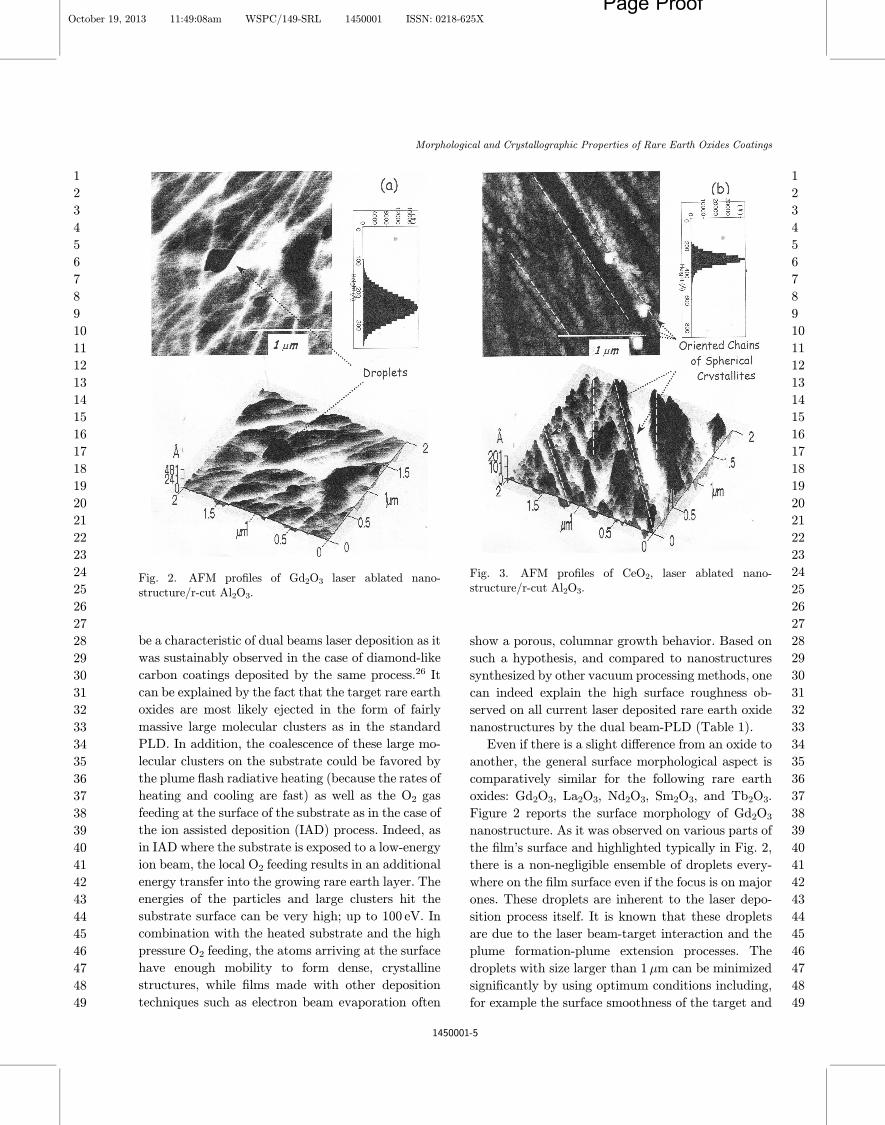
Fig. 3. AFM pro¯les of CeO2, laser ablated nano-structure/r-cut Al2O3.
Fig. 2. AFM pro¯les of Gd2O3 laser ablated nano-structure/r-cut Al2O3.
Morphological and Crystallographic Properties of Rare Earth Oxides Coatings
October 19, 2013 11:49:08am WSPC/149-SRL 1450001 ISSN: 0218-625XPage Proof
12345678910111213141516171819202122232425262728293031323334353637383940414243444546474849
12345678910111213141516171819202122232425262728293031323334353637383940414243444546474849
1450001-5

its rotation as well as the optimization of the target–substrate distance. Besides these surface droplets, noparticular morphological ordering is observed in thissequence in contrast to CeO2 ,Ho2O3 and Eu2O3
nanostructures where there are clearly distinctivesurface features. More speci¯cally, the surface ap-pearance of CeO2 and Ho2O3 ¯lms di®ers consider-ably from the other rare earth oxides. As reported inFigs. 3 and 4, both CeO2 and Ho2O3 nanostructuresexhibit a speci¯c trend. The corresponding crystal-lites are more spherical-like in shape with a mono-disperse size distribution. The average value of theirsize is of < " >"84% 1 nm and " 69% 2 nm forCeO2 and Ho2O3, respectively. In both cases, thesecrystallites seem to grow in a chain-like con¯gurationas well as a de¯ned preferential crystallographic ori-entation relatively to the basal direction of the sap-phire substrate surface. This behavior is morepronounced in the case of CeO2 sample and is un-doubtedly inherent of an epitaxial growth of the CeO2
and Ho2O3 onto sapphire. This epitaxial growth is
likely favored by the small crystalline mismatching ofCeO2 and Ho2O3 with the sapphire as con¯rmedfurther by XRD investigations. The hexagonalstructures of sapphire substrate exhibit lattice con-stants of aAl2O3
"4:759. The lattice matching can beachieved by using di®erent crystallographic orienta-tions for the ¯lm and the substrate, because the cubiclattice of the sesquioxides provides a hexagonal ar-rangement of the ions in the f111g planes of the cubicunit cell. Thus, the relation for lattice matching is3& asubstrate "
p2afilm. The lattice mismatch for
CeO2 on sapphire is < 0:7%. With reference to Eu2O3
nanostructure, the surface morphology is more com-plex as reported in Fig. 5. A special feature that dis-tinguishes the morphology of Eu2O3 from alldeposited oxide samples, is the existence of large co-lumnar planes transversal to the sapphire substrate.More accurately, Eu2O3 exhibits localized very largeoriented crystallites embedded in disordered packedcrystallites of about "127 nm in diameter. The verylarge crystallites seem to grow in a pyramidal mode\basal plane "2&m, transversal height "0:3&m".This preferential orientation perpendicular to the
Fig. 4. AFM pro¯les of Eu2O3, laser ablated nano-structure/r-cut Al2O3.
Fig. 5. AFM pro¯les of Ho2O3, laser ablated nano-structure/r-cut Al2O3.
S. Khamlich et al.
October 19, 2013 11:49:11am WSPC/149-SRL 1450001 ISSN: 0218-625XPage Proof
12345678910111213141516171819202122232425262728293031323334353637383940414243444546474849
12345678910111213141516171819202122232425262728293031323334353637383940414243444546474849
1450001-6

substrate may be inferred to a possible epitaxialgrowth of Eu2O3 on sapphire as well, but partiallycompared to CeO2 and Ho2O3. The triangular (2D) orpyramidal (3D) crystallite structure observed in theEu2O3 ¯lms represents the crystallographic cubic-°uorite structure of its bulk form and can be assignedtherefore to the (111) growth direction. A similarsurface morphology with pyramidally shaped grainswas also observed in the case of Yttria.27–29
To corroborate the observed epitaxial growth ofcertain rare earth oxide nanostructures and the pres-ervation of the bulk stoichiometry, room temperatureXRD measurements were carried out on both laserablated nanostructures and their corresponding bulkpowders used as targets during the dual-beams laserdeposition. Figures 6(a) and 6(b) report the XRDpatterns obtained in a grazing angle di®raction con-¯guration.The incidentX-ray beamwas impinging thesample under a ¯xed angle of the order of 1.5!, thusallowing more interaction (high penetration depth)with each nanostructure while minimizing the inter-action with the sapphire substrate and consequently areduced background di®raction signal from it. For thisreason, only Bragg di®raction lines of the nanos-tructures are observed. For each rare earth oxide, the¯lms exhibit a single identi¯ed crystallographic phasewhich is identical to that of the used pressed powdertarget (Table 1). As can be noticed, there is a netstoichiometry similarity between the bulk rare earthoxide targets and the nanostructured deposits. Asmentioned previously, this expected chemical similar-ity is due to the highly nonequilibrium nature of thepulsed laser–target interaction. During the laserbeam–target interaction, the exchanged energy iscon¯ned in a thin surface layer due to a signi¯cantoptical absorption coe±cient of the UV laser radia-tion.17 Involving the generated shock wave createdduring the laser–matter interaction, this should lead toa massive expulsion of the material, in the order ofhundreds of nanometers per shot, as concluded fromthe AFM (Table 1). For each target, the material ismost likely ejected in a stoichiometric clusters andlarge particles while the local O2 feeding at the sub-strate, the speci¯city of the used dual-beam PLD,would compensate any O de¯ciency. The Scherrerequation was not used as the crystallites of the various¯lms were not spherically shaped except for CeO2.
Besides the degree of chemical stoichiometry rel-atively to the bulk targets, the ¯lms present single
crystallographic low temperature phases, generallysesquioxides (RE2O3 form) except for CeO2 andEu3O4. These could be explained taking into accountthe constitution of the rare earth metal- oxygen sys-tems as well as the used laser source. First, in view ofthe phase diagram, the'3 oxidation state is generallythe stable one for the rare earth elements in the solidstate. In the lighter rare earth elements such as Ceand Pr, where the f-electrons are less tightly bound,and in those with one extra f-electron on top of thehalf-¯lled f shell as in Tb, a large oxygen coordinationnumber is displayed and an oxidation state largerthan 3 is favored.1 For the Ce, Pr and Tb oxides,therefore, both the REO2 and the RE2O3 stoichio-metries can be found. Second, in terms of laser–mat-ter interaction, the used source is an excimer and nota CO2 or Nd-YAG type and consequently no extensiveheat exchanged during the laser–rare earth targets butrather a pure optical absorption phenomenon. All therare earth sesquioxides RE2O3 constitute a series ofstable refractories.9,18–23 In the current laser deposi-tion conditions with the dual-beams PLD, and fromcrystallographic point of view (Table 1), the orientedCeO2 nanostructure is cubic possessing the °uoritestructure while Sm2O3, Gd2O3, Tb2O3, La2O3 andNd2O3 ¯lms are monoclinic corresponding to theB-type polymorph crystallographic structure. Thisstructure is closely related to that of the A-form, andin terms of coordination, the B-type structure hasboth a six- and seven-fold coordinations. For Ho2O3
nanostructure, it crystallizes in the polymorph C-typecubic form closely related to the °uorite structure. Inthis C-type structure, cation lattice is nearly un-changed but one-fourth of the oxygen atoms in the°uorite structure are missing, and the metal ions aresurrounded by only six oxygen atoms. Similar to thetrend observed in the AFM measurements, there is afeature distinguishing the crystallographic structureof the Eu2O3 nanostructure from the other rare oxide¯lms. The di®raction patterns of the nanostructureand the powder ¯t both with the monoclinic phasebut with di®erent textures. The orientations are (402)and (111) for Eu2O3 nanostructure and powder, re-spectively. Strains at the ¯lm–substrate interfacecould possibly reduce the degree of the crystallinemis¯t and thus induce a partial epitaxial growth.
Regardless to the nature of the rare earth oxide,all dual-beams laser ablated nanostructures werefound to be single phased or with preferential textures
Morphological and Crystallographic Properties of Rare Earth Oxides Coatings
October 19, 2013 11:49:14am WSPC/149-SRL 1450001 ISSN: 0218-625XPage Proof
12345678910111213141516171819202122232425262728293031323334353637383940414243444546474849
12345678910111213141516171819202122232425262728293031323334353637383940414243444546474849
1450001-7

Fig. 6. X-Ray di®raction patterns of laser ablated rare earth oxide nanostructures and their corresponding bulk powdermaterials used as original targets.
S. Khamlich et al.
October 19, 2013 11:49:15am WSPC/149-SRL 1450001 ISSN: 0218-625XPage Proof
12345678910111213141516171819202122232425262728293031323334353637383940414243444546474849
12345678910111213141516171819202122232425262728293031323334353637383940414243444546474849
1450001-8

(Table 1). In the case of CeO2 and Ho2O3 nanos-tructures, the presence of only few and very inten-sive Bragg lines indicates an epitaxial growthrelatively to the sapphire surface as observed in theAFM scanning pro¯les. More accurately, the growthdirections are (111) and (841) for CeO2 and Ho2O3:low temperature crystalline phases and share thecubic structure. Due to the low crystalline mis-matching value (Table 1), this latter phase could ef-fectively favor such an epitaxial growth process onthe cubic sapphire substrates. Although not con-¯rmed by direct XRD measurements in the presentstudy but as indicated by the AFM investigation, the
europium oxide Eu2O3 phase is quite likely stable andcan form quite large crystallites perpendicularly tex-tured to the sapphire. Due to the non-negligiblemismatch ("0:7%) and the origin of this partial andparticular epitaxy is not understood. One shouldmention that generally, in all vacuum based processesfor synthesis of simple and multi-oxide thin ¯lmcoatings, there is a lack of stoichiometry between thetarget and the thin ¯lm. This is due to the non-thermodynamic conditions. As highlighted in Sec. 1,the PLD is one of the unique if not the unique thin¯lm deposition which allows stoichiometric transfer ofmatter from the target to the substrate. Likewise, yet
Fig. 6. (Continued)
Morphological and Crystallographic Properties of Rare Earth Oxides Coatings
October 19, 2013 11:49:19am WSPC/149-SRL 1450001 ISSN: 0218-625XPage Proof
12345678910111213141516171819202122232425262728293031323334353637383940414243444546474849
12345678910111213141516171819202122232425262728293031323334353637383940414243444546474849
1450001-9

it was not mentioned previously, it was observed thatcurrent PLD depositions without O2 feeding envi-ronment exhibited multi-phases in general, especiallyfor CeO2 and Ho2O3 as well as Eu2O3. There might beimpurity phases indeed as the oxide metal powdersintered targets are of N3 purity (Johnsson Mathey,purity 99.9). As much as the limit of detection ofXRD, the observed Bragg peaks in all thin ¯lms arerelated to the pure oxide phase for each of the rareearth oxide target that we have used. Yet the ¯lmsare highly transparent in the UV-VIS-NIR spectralrange, the intended application was not optical butrather geared toward high-permittivity (high-!) di-electric materials to replace SiO2 as a gate insulator inMOSFETs. These RE2O3 could be potential re-placement to HfO2 and ZrO2. The future studies onthese ¯lms will be to investigate their optical prop-erties by ellipsometry and IR spectroscopy in view oftheir potential opto-electronic applications. In thefuture studies, it is intended to combine Vanadiumdioxide VO2 with rare earth thin ¯lms based op-toelectronic nano-scaled devices where the uniqueoptical and electronic tuneability of VO2 will beused.30–35
3. Conclusion
Morphological and crystallographic studies of thefollowing rare earth oxide nanostructures \CeO2,Eu2O3, Gd2O3, Ho2O3, La2O3, Nd2O3, Sm2O3 andTb2O3" grown, for the ¯rst time, by dual-beamspulsed laser ablation on sapphire substrates were in-vestigated. Even if the shape of the crystalliteschanges from an oxide to another, all the rare earthoxide nanostructures were found to exhibit consis-tently a very compact morphology with large crys-tallites of 60–200 nm in size approximately. Thereproducible chemical stoichiometry of the obtained¯lms as well as their dense structure suggest that therare earth oxides are most likely ejected in the form offairly massive large molecular clusters during thetarget–laser interaction. This later is supported bythe XRD investigations which showed the automaticsimilarity between the crystalline structures of pow-der targets and the nanostructures. From crystallo-graphic aspect, the AFM and/or XRD studies showthat the °uorine cubic structure CeO2 and Ho2O3
nanostructures exhibit a high epitaxial growth degree
while the monoclinic Eu2O3 nanostructure presents apartial preferential orientation perpendicularly to thesapphire substrate.
Acknowledgments
This research program is conducted within the frame-work of the UNESCO UNISA Africa Chair in Nanos-ciences&Nanotechnology. It was generously supportedby grants from the National Research Foundation ofSouth Africa (NRF), the Abdus Salam ICTP via theNanosciences African Network (NANOAFNET), theOrganization of Women in Science for the DevelopingWorld (OWSDW), the Academy of Science for theDeveloping World (TWAS), the French Embassy inPretoria-South Africa and the African Laser Center towhom we are grateful.
References
1. J. T. Cheung and H. Sankur, CRC Crit. Rev. SolidState Mater. Sci. 15 (1998) 63.
2. L. Laversenne, Y. Guyot, C. Goutaudier, M. T. Cohen-Adad and G. Boulon, Opt. Mater. 16 (2001) 475–483.
3. H. K. Yang, J. W. Chung, B. K. Moon, B. C. Choi, J.H. Jung, S. S. Yi, J. H. Kim and K. H. Kim, Surf. Rev.Lett. 14 (2007) 873–878.
4. C. Grivas, T. C. May-Smith, D. P. Shepherd and R. W.Eason, Appl. Phys. A 79 (2004) 1203–1206.
5. W. Heitmann, Appl. Opt. 12 (1973) 394.6. T. S. Kalkur, R. Y. Kwor and W. D. Foster, Electro-
compon. Sci. Technol. 3 (1976) 51.7. D. R. Sigler, Oxid. Met. 40 (1993) 295.8. M. Schieber, J. Cryst. Growth. 109 (1991) 401.9. S. Shamm, G. Scarel and M. Fanciulli, Top. Appl.
Phys. 106 (2007) 153–177.10. T. Wiktorczyk, Optica Applicata 31 (2001) 153.11. A. R. Chaudhuri, A. Fissel, V. R. Archakam and
J. Osten, Appl. Phys. Lett. 102 (2013) 022904.12. G. D. Wilk, R. M.Wallace and J. M. Anthony, J. Appl.
Phys. 89 (2001) 5243.13. S. Ohmi, M. Takeda, H. Ishiwara and H. Iwai, in ISTC
(2002), pp. 251–261.14. L. Marsella and V. Fiorentini, Phys. Rev. B 69 (2004)
172103.15. G. Bonnet, M. Lachkar, J. C. Colson and J. P. Larpin,
Thin Solid Films 261 (1995) 31.16. K. L. Saegner, Process. Adv. Mater. 3 (1993) 11.17. M. Suzuki, M. Kagawa, Y. Syono and T. Hirai, J.
Cryst. Growth. 112 (1991) 621.18. M. Langlet and R. D. Shannon, Thin Solid Films 186
(1990) 1.19. R. M. Silver and T. Talvacchio, Appl. Phys. Lett. 51
(1990) 2149.
S. Khamlich et al.
October 19, 2013 11:49:20am WSPC/149-SRL 1450001 ISSN: 0218-625XPage Proof
12345678910111213141516171819202122232425262728293031323334353637383940414243444546474849
12345678910111213141516171819202122232425262728293031323334353637383940414243444546474849
1450001-10

20. H. Jorg Osten, E. Bugiel, M. Czernohorsky, Z. Elassar,O. Kirfel and A. Fissel, Topics Appl. Phys. 106 (2007)101–114.
21. C. M. Cotell and S. Grabowski,MRS Bull. 17 (1992) 44.22. J. Schubert, T. Heeg and M. Wagner, Top. Appl. Phys.
106 (2007) 115–126.23. Y. Lebedinskii, A. Zenkevitch, G. Scarel and M. Fan-
ciulli, Top. Appl. Phys. 106 (2007) 127–142.24. D. B. Chrisey and A. Inam, MRS Bull. 17 (1992) 37.25. J. Perriere, L. Ranno, C. Marechal, R. P. Casero, R. G.
S. Roman and D. M. Garcia, SPIE Proc. 2207 (1994)678.
26. S. Sotobe, MSc dissertation, University of WesternCape, 2009.
27. S. Bär, H. Scheife, K. Peterman and G. Huber, Top.Appl. Phys. 106 (2007) 401–422.
28. K. G. Cho, D. Kumar, D. G. Lee, S. L. Jones, P. H.Holloway and R. K. Singh, Appl. Phys. Lett. 71 (1997)3335–3337.
29. M.B.Korzenski, P. Lecoeur, B.Mercey,D.Chippaux, B.Raveau and R. Desfeux, Chem. Mater. 12 (2000) 3139.
30. M. Maaza, C. Sella, B. Baruch-Barak, N. Ouassini andA. C. Beye, Opt. Commun. 254 (2005) 188.
31. M. Maaza, N. Ouassini, C. Sella and A. C. Beye, GoldBull. 38 (2005) 3.
32. J. B. K. Kana, J. M. Ndjaka, P. O. Ateba, B. D. Ngom,N. Manyala, O. Nemraoui, A. C. Beye and M. Maaza,Appl. Surf. Scie. 254 (2008) 3959–3963.
33. J. B. K. Kana, J. M. Ndjaka, B. D. Ngoma, N. Man-yala, O. Nemraouif, A. Y. Fasasi, R. Nemutudi, A.Gibaud, D. Knoesen and M. Maaza, Thin Solid Films518 (2010) 1641–1647.
34. J. B. K. Kana, J. M. Ndjaka, G. Vignaud, A. Gibaudand M. Maaza, Opt. Commun. 284 (2011) 807–812.
35. M. Maaza, D. Hamidi, A. Simo, T. Kerdja, A. K.Chaudhary and J. B. K. Kana, Opt. Commun. 285(2012) 1190–1193.
36. R. S. Roth and S. J. Schneider, J. Res. Natl. Bur.Stand. 64A (1960) 309.
37. M. Foex and J. P. Traverse, Rev. Int. Hautes. Temp.Refract. 3 (1966) 429.
38. G. Brauer and H. Graindringer, Z. Anorg. Chem. 276(1954) 209.
39. W. C. Koehler and E. O. Wollan, Acta Cryst. 6 (1953)741.
40. L. L. Fehrenbacher, L. A. Jacobson and C. T. Lynch,Rare Earth Research III, ed. L. Eyring (SciencePublishers, New York, 1995).
41. J. G. Duh and M. H. Lee, J. Mater. Sci. 24 (1987)4467.
42. W. B. Pearson, in Handbook of Lattice Spacings andStructures of Metals and Alloys, International Series ofMonographs on Metal Physics and Physical Metallur-gy, ed. G. V. Raynor (Pergamon Press, London, 1958).
Morphological and Crystallographic Properties of Rare Earth Oxides Coatings
October 19, 2013 11:49:20am WSPC/149-SRL 1450001 ISSN: 0218-625XPage Proof
12345678910111213141516171819202122232425262728293031323334353637383940414243444546474849
12345678910111213141516171819202122232425262728293031323334353637383940414243444546474849
1450001-11
Copyright © 2022 FDOKUMEN