Molecular typing and drug sensitivity testing of Mycobacterium tuberculosis isolated by a...
7
RESEARCH ARTICLE Open Access Molecular typing and drug sensitivity testing of Mycobacterium tuberculosis isolated by a community-based survey in Ethiopia Muluwork Getahun 1* , Gobena Ameni 2 , Abebaw Kebede 1 , Zelalem Yaregal 1 , Elena Hailu 3 , Grimay Medihn 2 , Daniel Demssie 1 , Feven Girmachew 1 , Yetnebersh Fiseha 1 , Abyot Meaza 1 , Nathneal Dirse 1 , Mulualem Agonafir 1 , Feleke Dana 1 , Fasil Tsegaye 1 , Zeleke Alebachew 1 , Almaz Abebe 1 , Amha Kebede 1 and Eshetu Lemma 1 Abstract Background: The identification of circulating TB strains in the community and drug sensitivity patterns is essential for the tuberculosis control program. This study was undertaken to identify M. tuberculosis strains circulating in selected communities in Ethiopia as well as to evaluate the drug sensitivity pattern of these strains. Method: This study was a continuation of the Ethiopian National TB Prevalence Survey that was conducted between 2010 and 2011. Culture-positive isolates of M. tuberculosis from previous study were typed using region of difference (RD) 9-based polymerase chain reaction (PCR) and spoligotyping. Drug sensitivity testing was conducted using the indirect proportion method on Lowenstein-Jensen media. Result: All 92 isolates were confirmed as M. tuberculosis by RD9-based PCR and spoligotyping of 91 of these isolates leds to the identification of 41 spoligotype patterns. Spoligotype revealed higher diversity (45 %) and among this 65.8 % (27/41) were not previously reported. The strains were grouped into 14 clusters consisting of 2–15 isolates. The dominant strains were SIT53, SIT149 and SIT37 consisting of 15, 11, and 9 isolates, respectively. Our study reveals 70 % (64/91) clustered strains and only 39.1 % (25/64) occurred within the same Kebele. Further assignment of the strains to the lineages showed that 74.7 % (68/91) belonged to Euro-American lineage, 18.6 % (17/91) to East Africa Indian lineage and the remaining 6.5 % (6/91) belonged to Indo-oceanic lineage. Valid drug susceptibility test results were available for 90 of the 92 isolates. Mono-resistance was observed in 27.7 % (25/90) and poly-resistance in 5.5 % (5/90) of the isolates. Moreover, multi-drug resistance (MDR-TB) was detected in 4.4 % of the isolates whilst the rest (60/90) were susceptible to all drugs. The highest level of mono-resistance, 26.6 % (24/90), was observed for streptomycin with majority (91.1 %) of streptomycin mono-resistant strains belonging to the Euro-American lineage. Conclusion: In this study, the strains of M. tuberculosis circulating in selected sites of Ethiopia were identified along with the drug sensitivity patterns. Thus, these findings are useful for the TB Control Program of the country. * Correspondence: [email protected] 1 Ethiopian Public Health Institute, Addis Ababa, Ethiopia Full list of author information is available at the end of the article © 2015 Getahun et al. Open Access This article is distributed under the terms of the Creative Commons Attribution 4.0 International License (http://creativecommons.org/licenses/by/4.0/), which permits unrestricted use, distribution, and reproduction in any medium, provided you give appropriate credit to the original author(s) and the source, provide a link to the Creative Commons license, and indicate if changes were made. The Creative Commons Public Domain Dedication waiver (http://creativecommons.org/publicdomain/zero/1.0/) applies to the data made available in this article, unless otherwise stated. Getahun et al. BMC Public Health (2015) 15:751 DOI 10.1186/s12889-015-2105-7
Transcript of Molecular typing and drug sensitivity testing of Mycobacterium tuberculosis isolated by a...

RESEARCH ARTICLE Open Access
Molecular typing and drug sensitivity testingof Mycobacterium tuberculosis isolated by acommunity-based survey in EthiopiaMuluwork Getahun1*, Gobena Ameni2, Abebaw Kebede1, Zelalem Yaregal1, Elena Hailu3, Grimay Medihn2,Daniel Demssie1, Feven Girmachew1, Yetnebersh Fiseha1, Abyot Meaza1, Nathneal Dirse1, Mulualem Agonafir1,Feleke Dana1, Fasil Tsegaye1, Zeleke Alebachew1, Almaz Abebe1, Amha Kebede1 and Eshetu Lemma1
Abstract
Background: The identification of circulating TB strains in the community and drug sensitivity patterns is essentialfor the tuberculosis control program. This study was undertaken to identify M. tuberculosis strains circulating inselected communities in Ethiopia as well as to evaluate the drug sensitivity pattern of these strains.
Method: This study was a continuation of the Ethiopian National TB Prevalence Survey that was conductedbetween 2010 and 2011. Culture-positive isolates of M. tuberculosis from previous study were typed using region ofdifference (RD) 9-based polymerase chain reaction (PCR) and spoligotyping. Drug sensitivity testing was conductedusing the indirect proportion method on Lowenstein-Jensen media.
Result: All 92 isolates were confirmed as M. tuberculosis by RD9-based PCR and spoligotyping of 91 of theseisolates leds to the identification of 41 spoligotype patterns. Spoligotype revealed higher diversity (45 %) andamong this 65.8 % (27/41) were not previously reported. The strains were grouped into 14 clusters consisting of2–15 isolates. The dominant strains were SIT53, SIT149 and SIT37 consisting of 15, 11, and 9 isolates, respectively.Our study reveals 70 % (64/91) clustered strains and only 39.1 % (25/64) occurred within the same Kebele. Furtherassignment of the strains to the lineages showed that 74.7 % (68/91) belonged to Euro-American lineage, 18.6 %(17/91) to East Africa Indian lineage and the remaining 6.5 % (6/91) belonged to Indo-oceanic lineage. Valid drugsusceptibility test results were available for 90 of the 92 isolates. Mono-resistance was observed in 27.7 % (25/90)and poly-resistance in 5.5 % (5/90) of the isolates. Moreover, multi-drug resistance (MDR-TB) was detected in 4.4 %of the isolates whilst the rest (60/90) were susceptible to all drugs. The highest level of mono-resistance, 26.6 %(24/90), was observed for streptomycin with majority (91.1 %) of streptomycin mono-resistant strains belonging tothe Euro-American lineage.
Conclusion: In this study, the strains of M. tuberculosis circulating in selected sites of Ethiopia were identified alongwith the drug sensitivity patterns. Thus, these findings are useful for the TB Control Program of the country.
* Correspondence: [email protected] Public Health Institute, Addis Ababa, EthiopiaFull list of author information is available at the end of the article
© 2015 Getahun et al. Open Access This article is distributed under the terms of the Creative Commons Attribution 4.0International License (http://creativecommons.org/licenses/by/4.0/), which permits unrestricted use, distribution, andreproduction in any medium, provided you give appropriate credit to the original author(s) and the source, provide alink to the Creative Commons license, and indicate if changes were made. The Creative Commons Public DomainDedication waiver (http://creativecommons.org/publicdomain/zero/1.0/) applies to the data made available in thisarticle, unless otherwise stated.
Getahun et al. BMC Public Health (2015) 15:751 DOI 10.1186/s12889-015-2105-7
BackgroundThere are an estimated 9 million new cases of TB everyyear. TB is the second leading cause of death from aninfectious disease worldwide and is responsible for 1.5million deaths annually. Control of TB is becoming achallenge to the endemic countries due to the emergenceof drug resistance. Recently, the WHO Global TB Reportestimated that total of 480,000 people developed MDR-TBworldwide in 2013. The prevalence of MDR-TB has beenestimated to be 3.5 % in newly diagnosed cases and 20.5 %in previously treated TB cases [1].In Ethiopia, TB is a major public health problem and the
WHO categorizes Ethiopia as a high TB and MDR-TB bur-den country [1]. The first Ethiopian National TB prevalencesurvey was conducted between 2010 and 2011. The preva-lence of smear-positive TB among persons aged ≥ 15 yearswas 108/100,000 (95 % CI 73–143), whereas the prevalenceof bacteriologically confirmed TB in the same age groupwas 277/100,000 (95 % CI 208–347) [2]. According to thenationwide drug resistance survey conducted between 2003and 2005 the overall prevalence of MDR-TB was 2.5 %.The prevalence of MDR-TB was 1.6 % and 11.8 % in newlydiagnosed TB cases and previously treated cases, respect-ively [3].Knowledge on the Mycobacterium tuberculosis (M.
tuberculosis) strains circulating in Ethiopian communitiesand evaluation of the drug sensitivity patterns of thesestrains is useful for the TB control program in the country.Since the study was done on positive cultures isolatedfrom community-based survey, those patients who havelow health seeking behavior were mostly captured. There-fore, this study was undertaken for the identification ofthe M. tuberculosis strains circulating in selected commu-nities in Ethiopia and thereafter to evaluate the drug sensi-tivity pattern of these strains.
MethodsStudy populationThis study was a continuation of the Ethiopian NationalTB Prevalence Survey that was conducted between 2010and 2011. The participants for the prevalence surveycarried out by Kebede AH and colleagues were recruitedfrom 85 Kebeles (communes), selected from 85 Woredas(districts) which were stratified as urban, rural and pas-toralist population. The number of Kebeles covered inrural, urban and pastoralist areas were 63, 14 and 8 re-spectively. Sample size determination for the prevalencesurvey is reported elsewhere [2]. The planned samplesize for prevalence survey was 46,514 adults aged ≥15years. Following this 85 Woredas were selected by multi-stage cluster sampling using probability proportion tosize (PPS) from three strata (urban, rural and pastoral-ist). In each selected districts one Kebele with estimatedeligible adult of 548 was selected using PPS. However
during the recruitment of participants the total numbersof individuals from 85 selected clusters were 51,667.Since cluster sampling was used and to maximize sam-ple size, all eligible individuals were invited to participatein the study. Of the total eligible individuals, 46,697(90 %) participated in the study. The case enrollmentsfor the study were based on symptoms suggestive of TB(cough ≥2 weeks) and/or any abnormality in the lungdetected using chest X-ray. For individuals exempt fromchest X-ray who did not have cough ≥2 weeks, one ofthe following criteria was used: weight loss >3 kg in thelast 1 month, night sweats >2 weeks, fever >2 weeks andcontact with a TB patient in the last year. Using theabove criteria a total of 6080 (13 %) participants wereeligible for sputum examination. Of these, 5868 (97 %)submitted at least one sputum specimen and 5606(92 %) submitted two specimens [2]. The present studywas carried out on culture positive isolates identifiedfrom the prevalence survey.
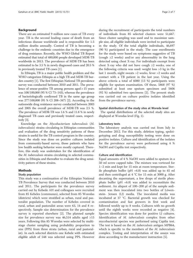
Spatial distribution of the study sites at Woreda levelThe spatial distributions of the selected study sites aredisplayed at Woredas level (Fig. 1).
Laboratory testsThe laboratory analysis was carried out from July toDecember 2012. For this study, deletion typing, spoloi-gotyping and drug susceptibility testing were done onpositive isolates. Culture and identification of the isolatesfor the prevalence survey were performed using 4 %NaOH and Capilia test respectively.
Culture and identificationEqual amounts of 4 % NaOH were added to sputum in a50 ml screw capped tube. The mixture was vortexed for1–2 min and kept for 15 min at room temperature. Ster-ile phosphate buffer (pH =6.8) was added up to 45 mland then centrifuged at 4 °C for 15 min at 3000 g. Afterdecanting the supernatant, a few drops of sterile phos-phate buffer (pH =6.8) was added to reconstitute thesediment. An aliquot of 100–200 μl of the sample sedi-ment was then inoculated into two bottles of Löwen-stein- Jensen (LJ) media. The inoculated media wasincubated at 37 °C. Bacterial growth was checked forcontamination and fast growers in first week andfollowed weekly up to 8 weeks. Cultures with no growthuntil the eighth weeks were recorded as negative [4].Species identification was done for positive LJ cultures.Identification of M. tuberculosis complex from othermycobacterial species was performed using Capilia test.The test is based on the M. tuberculosis antigen MPB64,which is specific to the members of the M. tuberculosiscomplex. Testing and interpretation of the assays wasdone according to the manufacturer instruction [5].
Getahun et al. BMC Public Health (2015) 15:751 Page 2 of 7
Molecular characterization of the isolatesIdentificationIdentification of M. tuberculosis from the other members ofthe M. tuberculosis complex species was done using regionof difference (RD)-9 polymerase chain reaction. RD9-PCRdeletion typing was performed on heat-killed cells toconfirm the presence or absence of RD9 using threeprimers RD9flankF, RD9IntR and RD9flankR [6]. PCR amp-lification was done as indicated by Berg et al. using acommercially available kit (Qiagen, United Kingdom). Inter-pretation of the result was based on differences in molecu-lar weight. A molecular weight of 396bp was considered asM. tuberculosis or M. canettii, while a molecular weight575bp was considered M. bovis orM. africanum [7].
Molecular typingStrain typing of M. tuberculosis was done using spoligo-typing as previously described by Kamerbeek and col-leagues [8]. The PCR amplification of DNA was done bytargeting the direct repeat (DR) locus, the region withthe highest level of polymorphism in the M. tuberculosis
chromosome. The DR region was amplified using primersDRa (GGTTTTGGGTCTGACGAC) and DRb (CCGAGAGGGGACGGAAAC). Spoligotyping was performedwith a commercially available kit according to themanufacturer instructions (Isogen Bioscience BV Maarssen,The Netherlands). Lineage and SIT (Shared InternationalTypes) number assignment was done using SPOLD4 andSPOTCLUST [9].
Drug Susceptibility testingDrug susceptibility testing (DST) was performed forfirst line drugs; isoniazid, streptomycin, rifampicin andethambutol, using indirect proportion method on Low-enstein–Jense media. The critical concentration foreach drug was 0.2μg/ml, 4μg/ml, 40μg/ml and 2μg/mlfor isoniazid, streptomycin, rifampicin and ethambutolrespectively. Result interpretation was done by compar-ing growth on control media and media containingdrug. If more than 1 % of the test population was ob-served on the drug containing media, the result wasinterpreted as resistant to that drug [10]
Fig. 1 Spatial distribution study sites at Woreda level
Getahun et al. BMC Public Health (2015) 15:751 Page 3 of 7

Statistical analysisStatistical analysis was carried out using SPSS softwarepackages. Patient characteristics for differences in pro-portions were compared. The chi square test was usedto assess statistical significant difference. P values of 0.05or less were regarded as significant.
Ethical considerationThe laboratory analysis was done on stored culture posi-tive samples. Specific patient identifiers were not usedfor this study. Ethical clearance was obtained from Sci-entific and Ethics Review Committee at Ethiopian PublicHealth Institute.
ResultBackground characteristicsThe mean age of culture positive participants was 34.4years (95 % CI 31.2–37.7). The overall culture positivityrate was 1.6 % (96/5863). The distribution of culturepositivity across the categories of background character-istics of TB suspects is summarized in Table 1. The cul-ture positivity was significantly associated with age andregion of the study participants. High culture positivityrate (3.2 %) was observed in age group between 15 and24. Among three regions with high culture positivity rate(Addis Ababa, SNNPR and Gambela) the contribution
of younger age categories (15–34) was 44 % (4 out of 9),60 % (18 out of 30) and 80 % (4 out of 5) from the totalpositive culture from those regions.
Molecular characterization of the isolatesIdentificationOut of 96 isolates positive for M. tuberculosis complexbased on Capilia TB immunochromographic assay, 4 sam-ples were excluded from RD9-PCR analysis (2 isolates failedto grow after re-culturing and 2 cultures were not retrieved).The remaining 92 isolates were analyzed based on region ofdifference analysis. RD9 analysis showed that 92 isolatescontained an intact RD9. This indicated that the 92 isolateswereMycobacterium tuberculosis species orM. canettii.
Molecular typingNinety two isolates were characterized using spoligotyp-ing and 91 of them showed good patterns. Spoligotypingidentified 41 different spoligotypes with the overall di-versity of 45 % and among this 65.8 % (27/41) were notpreviously reported. The isolates were grouped into 14clusters consisting of 2–15 isolates each. The dominantstrains of M. tuberculosis were SIT53 (15/91), SIT149(11/91) and SIT37 (9/91). Our study reveals 70 % (64/91) clustered strains of which 53 were registered in theinternational database whereas 11 were newly identified.
Table 1 Culture result Vs sex, age, region and resident type
Culture Positive Culture Negative/NTM/Contaminated Total Culture Positivity Rate P-value
Sex Male 51 3122 3173 1.6 0.844
Female 45 2645 2690 1.7
Age 15–24 32 962 994 3.2 <0.001
25–34 21 1183 1204 1.7
35–44 17 1084 1101 1.5
45–54 12 992 1004 1.2
55–64 10 736 746 1.3
> = 65 4 810 814 0.5
Region Oromiya 28 2250 2278 1.2 <0.001
SNNPR 30 1191 1221 2.5
Amhara 10 948 958 1
Tigray 6 505 511 1.2
Afar 1 143 144 0.7
Gambela 5 83 88 5.7
Somali 7 388 395 1.8
Benshangul 0 25 25 0
Dire Dawa 0 45 45 0
Addis Ababa 9 189 198 4.5
Resident Urban 14 744 758 1.8 0.844
Rural 72 4349 4421 1.6
Pastoral 10 674 684 1.5
Getahun et al. BMC Public Health (2015) 15:751 Page 4 of 7
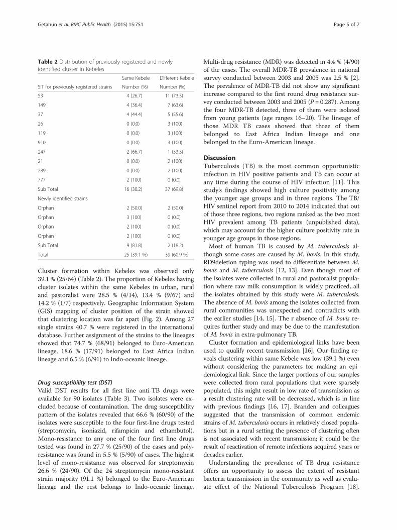
Cluster formation within Kebeles was observed only39.1 % (25/64) (Table 2). The proportion of Kebeles havingcluster isolates within the same Kebeles in urban, ruraland pastoralist were 28.5 % (4/14), 13.4 % (9/67) and14.2 % (1/7) respectively. Geographic Information System(GIS) mapping of cluster position of the strain showedthat clustering location was far apart (Fig. 2). Among 27single strains 40.7 % were registered in the internationaldatabase. Further assignment of the strains to the lineagesshowed that 74.7 % (68/91) belonged to Euro-Americanlineage, 18.6 % (17/91) belonged to East Africa Indianlineage and 6.5 % (6/91) to Indo-oceanic lineage.
Drug susceptibility test (DST)Valid DST results for all first line anti-TB drugs wereavailable for 90 isolates (Table 3). Two isolates were ex-cluded because of contamination. The drug susceptibilitypattern of the isolates revealed that 66.6 % (60/90) of theisolates were susceptible to the four first-line drugs tested(streptomycin, isoniazid, rifampicin and ethambutol).Mono-resistance to any one of the four first line drugstested was found in 27.7 % (25/90) of the cases and poly-resistance was found in 5.5 % (5/90) of cases. The highestlevel of mono-resistance was observed for streptomycin26.6 % (24/90). Of the 24 streptomycin mono-resistantstrain majority (91.1 %) belonged to the Euro-Americanlineage and the rest belongs to Indo-oceanic lineage.
Multi-drug resistance (MDR) was detected in 4.4 % (4/90)of the cases. The overall MDR-TB prevalence in nationalsurvey conducted between 2003 and 2005 was 2.5 % [2].The prevalence of MDR-TB did not show any significantincrease compared to the first round drug resistance sur-vey conducted between 2003 and 2005 (P = 0.287). Amongthe four MDR-TB detected, three of them were isolatedfrom young patients (age ranges 16–20). The lineage ofthose MDR TB cases showed that three of thembelonged to East Africa Indian lineage and onebelonged to the Euro-American lineage.
DiscussionTuberculosis (TB) is the most common opportunisticinfection in HIV positive patients and TB can occur atany time during the course of HIV infection [11]. Thisstudy’s findings showed high culture positivity amongthe younger age groups and in three regions. The TB/HIV sentinel report from 2010 to 2014 indicated that outof those three regions, two regions ranked as the two mostHIV prevalent among TB patients (unpublished data),which may account for the higher culture positivity rate inyounger age groups in those regions.Most of human TB is caused by M. tuberculosis al-
though some cases are caused by M. bovis. In this study,RD9deletion typing was used to differentiate between M.bovis and M. tuberculosis [12, 13]. Even though most ofthe isolates were collected in rural and pastoralist popula-tion where raw milk consumption is widely practiced, allthe isolates obtained by this study were M. tuberculosis.The absence of M. bovis among the isolates collected fromrural communities was unexpected and contradicts withthe earlier studies [14, 15]. The r absence of M. bovis re-quires further study and may be due to the manifestationof M. bovis in extra-pulmonary TB.Cluster formation and epidemiological links have been
used to qualify recent transmission [16]. Our finding re-veals clustering within same Kebele was low (39.1 %) evenwithout considering the parameters for making an epi-demiological link. Since the larger portions of our sampleswere collected from rural populations that were sparselypopulated, this might result in low rate of transmission asa result clustering rate will be decreased, which is in linewith previous findings [16, 17]. Branden and colleaguessuggested that the transmission of common endemicstrains of M. tuberculosis occurs in relatively closed popula-tions but in a rural setting the presence of clustering oftenis not associated with recent transmission; it could be theresult of reactivation of remote infections acquired years ordecades earlier.Understanding the prevalence of TB drug resistance
offers an opportunity to assess the extent of resistantbacteria transmission in the community as well as evalu-ate effect of the National Tuberculosis Program [18].
Table 2 Distribution of previously registered and newlyidentified cluster in Kebeles
Same Kebele Different Kebele
SIT for previously registered strains Number (%) Number (%)
53 4 (26.7) 11 (73.3)
149 4 (36.4) 7 (63.6)
37 4 (44.4) 5 (55.6)
26 0 (0.0) 3 (100)
119 0 (0.0) 3 (100)
910 0 (0.0) 3 (100)
247 2 (66.7) 1 (33.3)
21 0 (0.0) 2 (100)
289 0 (0.0) 2 (100)
777 2 (100) 0 (0.0)
Sub Total 16 (30.2) 37 (69.8)
Newly identified strains
Orphan 2 (50.0) 2 (50.0)
Orphan 3 (100) 0 (0.0)
Orphan 2 (100) 0 (0.0)
Orphan 2 (100) 0 (0.0)
Sub Total 9 (81.8) 2 (18.2)
Total 25 (39.1 %) 39 (60.9 %)
Getahun et al. BMC Public Health (2015) 15:751 Page 5 of 7

The MDR rate did not show any significant incrementcompared to the previous nationwide surveillance re-port even though the study population was different[3]. A study conducted in the Philippines at hospitals inMetro Manila, multi- drug resistant TB prevalencerates in hospital ranged from 14–45 %, whereas thecommunity rate of MDR TB in Metro Manila was6.4 % [19]. This suggested that high rate of MDR TBoccur in health facilities. Thus in Ethiopia, the rate ofMDR TB in the health facilities might be higher thanthe finding of this current study. Out of 4 MDR casesidentified in the present study, three MDR TB cases oc-curred in participants 16–20 years old. As reported byanother study [20] the high prevalence of primaryMDR-TB in a young population group implies trans-mission of drug resistant strains.
Fig. 2 Distribution of strains in their respective collection sites at Woreda level: Both registered and orphan clustered strains were indicated incolored. Orphans were given a number according to their order of identification. Both registered and orphan single isolates is represented in redhallow circle. The numeral in the side of the circle indicates that the number of single strains identified in that specific location
Table 3 Susceptibility pattern of 90 isolate for first line drugs
Number Percentage
Susceptible 60 66.6
Mono-resistance 25 27.7
Resistant to
INH only 1 1.1
SM only 24 26.6
INH + SM 1 1.1
MDR 4 4.4
INH + SM + RIF 1 1.1
INH + SM + RIF + EMB 3 3.3
Getahun et al. BMC Public Health (2015) 15:751 Page 6 of 7
ConclusionsIn this study, the strains of M. tuberculosis circulating inselected sites of Ethiopia were identified along with thedrug sensitivity patterns. More than 83 % of the popula-tion is living in rural areas of Ethiopia. Since decliningincidence of TB is expected with decreased clustering,coupling case detection and treatment alone in thoselarge portions of the rural population will have a hugecontribution to the reduction of TB. Besides, TB andMDR rate in young was high but with such small num-ber of cases the possible relationship is inconclusive itneeds further study.
Competing interestsWe declare that there are no financial, professional or personal competinginterests that might have influenced the performance or presentation of thework described in this manuscript.
Authors’ contributionMG conceived, designed, carried out the study, and wrote the manuscript.GA advised in proposal development, laboratory analysis, data analysis, andmanuscript write up. AK involved in data analysis and manuscripts write up.ZY carried out quality control for all laboratory analysis. EH carried outspoligotyping, GM advised in proposal development, data analysis andmanuscript write up. DD, FG, YF, AM and ND carried out drug susceptibilitytesting. ZA, FT, MA and FD carried out data extraction and data analysis. EL,AK, and AA advise in proposal development, data analysis and manuscriptwrite up. All authors read and approved the final manuscript.
AcknowledgmentWe would like to thank EPHI, ALIPB and AHRI for the financial support. Ouracknowledgment also goes to Girma Urgeacha for doing GIS mapping,Yared Kebede, Blessing Marondera and Justin Taylor for editing themanuscript, Wegene Tamene and Aboma Zewude for their technicalsupport.
Author details1Ethiopian Public Health Institute, Addis Ababa, Ethiopia. 2Aklilu LemaInstitute of Pathobiology, Addis Ababa, Ethiopia. 3Armauer Hansen ResearchInstitute, Addis Ababa, Ethiopia.
Received: 27 December 2014 Accepted: 28 July 2015
References1. World Health Organization Global Tuberculosis Report. Who, Geneva:
Annual Report; 2014.2. Kebede AH, Alebachew Z, Tsegaye F, Lemma E, Abebe A, Agonafir M, et al.
The first population-based national tuberculosis prevalence survey inEthiopia, 2010–2011. Int J Tuberc Lung Dis. 2014;18:635–9.
3. World Health Organization Anti-Tuberculosis Drug Resistance in the World:Fourth Global Report; 2008.
4. Kantor IND, Kim SJ, Frieden T: Laboratory Services in Tuberculosis ControlCulture Part III. World Health Organization 1998, 57–58.
5. Abe C, Hirano K, Tomiyama T. Simple and Rapid Identification of theMycobacterium tuberculosis Complex by Immunochromatographic AssayUsing Anti-MPB64 Monoclonal Antibodies. J Clin Microbiol. 1999;37:3693–7.
6. Brosch R, Gordon SV, Marmiesse MP. A new evolutionary scenario for theMycobacterium tuberculosis complex. Proc Natl Acad Sci. 2002;99(6):3684–9.
7. Nakajima C, Rahim Z, Fukushima Y, Sugawara I, van de Zanden AGM,Tamaru A, et al. Identification of Mycobacterium tuberculosis clinical isolatesin Bangladesh by a species distinguishable multiplex PCR. BioMedCenterInfect Dis. 2010;10(118):1–7.
8. Kamerbeek J, Schouls L, Kolk A, van Agterveld M, van Soolingen D,Kuijper S, et al. Simultaneous detection and strain differentiation ofMycobacterium tuberculosis for diagnosis and epidemiology. J ClinMicrobiol. 1997;35(4):907–14.
9. Molhuizen HOF, Bunschoten AE, Schouls LM, van Embden J. Rapiddetection and simultaneous strain differentiation of Mycobacteriumtuberculosis complex bacteria by spoligotyping. Methods Mol Biol.1998;101:381–94.
10. Kent PT, and Kubica GP, Public health mycobacteriology a guide for thelevel III laboratory. Centers for Disease control 1985: 159–185.
11. Swaminathan S, Ramachandran R, Baskaran G, Paramasivan CN, RamanathanU, Venkatesan P, et al. Risk of development of tuberculosis in HIV- infectedpatients. Int J Tuberc Lung Dis. 2000;4:839–44.
12. Cataldi A, Romano MI. Tuberculosis caused by Other Members of the M.tuberculosis Complex. In: Palomino JC, Leão SC, Ritacco V, editors.Tuberculosis 2007 From basic science to patient care. Belgium: BerndSebastian Kamps and Patricia Bourcillier; 2007. p. 283–314.
13. Rodríguez E, Sánchez LP, Pérez S, Herrera L, Jiménez MS, Samper S, et al.Human tuberculosis due to Mycobacterium bovis and M. caprae in Spain,2004–2007. Int J Tuberc Lung Dis. 2009;13(12):1536–41.
14. Regassa A, Medhin G, Ameni G. Bovine tuberculosis 302 is more prevalentin cattle owned by farmers with active tuberculosis in central Ethiopia.Elsevier. 2007;178(2008):119–25.
15. Shitaye JE, Tsegaye W, Pavlik I. Bovine tuberculosis infection in animal andhuman populations in Ethiopia: a review. Vet Med. 2007;52:317–32. ReviewArticle.
16. Braden CR, Templeton GL, Cave MD, Valway S, Onorato IM, Castro KG, et al.Interpretation of restriction fragment length polymorphism analysis ofMycobacterium tuberculosis isolates from a state with a large ruralpopulation. J Infect Dis. 1997;175:1446–52.
17. Ellis BA, Crawford JT, Braden CR, McNabb SJN, Moore M, Kammerer S, et al.Molecular epidemiology of tuberculosis in a sentinel surveillancepopulation. Emerg Infect Dis. 2002;8:1197–209.
18. Paramasivan CN, Bhaskarair K, Venkataraman P, Chandrasekaran V,Narayanan PR. Surveillance of drug resistance in tuberculosis in the state ofTamil Nadu. Indian J Tuberc. 2000;47:27–33.
19. Mendoza MT, Tan-Torres T, Ang CF, Arciaga R, Elona F, Retuta M, et al.Community-Based surveillance for drug resistance of Mycobacteriumtuberculosis in selected areas in the Philippines. Phil J Microbiol Infect Dis.2002;3:169–75.
20. Merza MA, Farnia P, Tabarsi P, Khazampour M, Masjedi MR, Velayati AA. Antituberculosis drug resistance and associated risk factors in a tertiary level TBcentre in Iran: a retrospective analysis. J Infect Dev Ctries. 2011;5(7):511–9.
Submit your next manuscript to BioMed Centraland take full advantage of:
• Convenient online submission
• Thorough peer review
• No space constraints or color figure charges
• Immediate publication on acceptance
• Inclusion in PubMed, CAS, Scopus and Google Scholar
• Research which is freely available for redistribution
Submit your manuscript at www.biomedcentral.com/submit
Getahun et al. BMC Public Health (2015) 15:751 Page 7 of 7