Molecular identity of the mitochondrial permeability transition pore and its role in...
38
Molecular identity of the mitochondrial permeability transition pore and its role in ischemia-reperfusion injury Giampaolo Morciano, Carlotta Giorgi, Massimo Bonora, Silvia Pun- zetti, Rita Pavasini, Mariusz R. Wieckowski, Gianluca Campo, Paolo Pinton PII: S0022-2828(14)00262-4 DOI: doi: 10.1016/j.yjmcc.2014.08.015 Reference: YJMCC 7875 To appear in: Journal of Molecular and Cellular Cardiology Received date: 15 July 2014 Revised date: 18 August 2014 Accepted date: 19 August 2014 Please cite this article as: Morciano Giampaolo, Giorgi Carlotta, Bonora Massimo, Pun- zetti Silvia, Pavasini Rita, Wieckowski Mariusz R., Campo Gianluca, Pinton Paolo, Molecular identity of the mitochondrial permeability transition pore and its role in ischemia-reperfusion injury, Journal of Molecular and Cellular Cardiology (2014), doi: 10.1016/j.yjmcc.2014.08.015 This is a PDF file of an unedited manuscript that has been accepted for publication. As a service to our customers we are providing this early version of the manuscript. The manuscript will undergo copyediting, typesetting, and review of the resulting proof before it is published in its final form. Please note that during the production process errors may be discovered which could affect the content, and all legal disclaimers that apply to the journal pertain.
-
Upload
independent -
Category
Documents
-
view
3 -
download
0
Transcript of Molecular identity of the mitochondrial permeability transition pore and its role in...

�������� ����� ��
Molecular identity of the mitochondrial permeability transition pore and itsrole in ischemia-reperfusion injury
Giampaolo Morciano, Carlotta Giorgi, Massimo Bonora, Silvia Pun-zetti, Rita Pavasini, Mariusz R. Wieckowski, Gianluca Campo, Paolo Pinton
PII: S0022-2828(14)00262-4DOI: doi: 10.1016/j.yjmcc.2014.08.015Reference: YJMCC 7875
To appear in: Journal of Molecular and Cellular Cardiology
Received date: 15 July 2014Revised date: 18 August 2014Accepted date: 19 August 2014
Please cite this article as: Morciano Giampaolo, Giorgi Carlotta, Bonora Massimo, Pun-zetti Silvia, Pavasini Rita, Wieckowski Mariusz R., Campo Gianluca, Pinton Paolo,Molecular identity of the mitochondrial permeability transition pore and its role inischemia-reperfusion injury, Journal of Molecular and Cellular Cardiology (2014), doi:10.1016/j.yjmcc.2014.08.015
This is a PDF file of an unedited manuscript that has been accepted for publication.As a service to our customers we are providing this early version of the manuscript.The manuscript will undergo copyediting, typesetting, and review of the resulting proofbefore it is published in its final form. Please note that during the production processerrors may be discovered which could affect the content, and all legal disclaimers thatapply to the journal pertain.
ACC
EPTE
D M
ANU
SCR
IPT
ACCEPTED MANUSCRIPT
1
Molecular identity of the mitochondrial permeability transition pore
and its role in ischemia-reperfusion injury
Giampaolo Morciano1, §, Carlotta Giorgi1, §, Massimo Bonora1, Silvia Punzetti2, Rita Pavasini2,
Mariusz R Wieckowski3, Gianluca Campo2 and Paolo Pinton1, *
1 Department of Morphology, Surgery and Experimental Medicine. Section of Pathology, Oncology and
Experimental Biology. Laboratory for Technologies of Advanced Therapies (LTTA). University of Ferrara,
Ferrara, Italy
2Cardiovascular Institute, Azienda Ospedaliero-Universitaria S. Anna and LTTA Center, Ferrara, Italy
3Department of Biochemistry, Nencki Institute of Experimental Biology; Warsaw, Poland
Running title: What is the mPTP, and what role does it play in ischemia-reperfusion?
§These authors contributed equally to this work
*Correspondence:
Paolo Pinton, PhD
Email: [email protected]

ACC
EPTE
D M
ANU
SCR
IPT
ACCEPTED MANUSCRIPT
2
Abstract
The mitochondrial permeability transition is a key event in cell death. Intense research efforts
have been focused on elucidating the molecular components of the mitochondrial permeability
transition pore (mPTP) to improve the understanding and treatment of various pathologies,
including neurodegenerative disorders, cancer and cardiac diseases. Several molecular factors
have been proposed as core components of the mPTP; however, further investigation has
indicated that these factors are among a wide range of regulators. Thus, the scientific
community lacks a clear model of the mPTP. Here, we review the molecular factors involved in
the regulation and formation of the mPTP. Furthermore, we propose that the mitochondrial ATP
synthase, specifically its c subunit, is the central core component of the mPTP complex.
Moreover, we discuss the involvement of the mPTP in ischemia and reperfusion as well as the
results of clinical studies targeting the mPTP to ameliorate ischemia-reperfusion injury.
Keywords:
Mitochondrial permeability transition pore; ATP synthase; c subunit; cell death; apoptosis;
ischemia-reperfusion; myocardial infarction.
Abbreviation list
ADP: adenosine diphosphate; ANT: adenine nucleotide transporter; ATP: adenine triphosphate;
C1QBP: Complement component 1 Q subcomponent-binding protein; Ca2+: calcium; CK: creatine
kinase; CsA: cyclosporine A; CYCLE: CYCLosporinE A in reperfused acute myocardial infarction; ER:
endoplasmic reticulum; ETC: electron transport chain; FADD: Fas-activated with death domain;
FLIP: FLICE-inhibitory protein; GIK: glucose-insulin-potassium; GLP-1: glucagon-like peptide 1;
GSK3-β: glycogen synthase kinase 3 beta; HF: heart failure; HK: hexokinase; Hot-DOG: 3H 2-
deoxyglucose; IHD: ischemic heart disease; IF-1: inhibitor protein F1; IMM: inner mitochondrial
membrane; IMS: intermembrane space; IRI: ischemia-reperfusion injury; K+: potassium; LV: left
ventricular; Mg2+: magnesium; MI: myocardial infarction; MITOCARE: prospective, multicenter,
randomized, double-blind, placebo-controlled, phase IIa study; MPT: mitochondrial permeability
transition; mPTP: mitochondrial permeability transition pore; mtCypD: mitochondrial cyclophilin

ACC
EPTE
D M
ANU
SCR
IPT
ACCEPTED MANUSCRIPT
3
D; MRI: magnetic resonance imaging; mTOR: mammalian target of rapamycin; MVO: microvascular
obstruction; Na+: sodium; NO: nitric oxide; OMM: outer mitochondrial membrane; OSCP:
oligomycin sensitivity conferring protein; OXPHOS: oxidative phosphorylation; PCI: percutaneous
coronary intervention; PEG: polyethylene glycol; Pi: inorganic phosphate; PiC: inorganic phosphate
carrier; PM: plasma membrane; PPIF: peptidylprolyl isomerase f; PK11195: N-butan-2-yl-1-(2-
chlorophenyl)-N-methylisoquinoline-3-carboxamide; PK: protein kinase; RISK: reperfusion injury
survival kinase; RO5-4864: 4'-chlorodiazepam; ROS: reactive oxygen species; SAFE: survivor
activating factor enhancement; SR: sarcoplasmic reticulum; STEMI: ST elevation myocardial
infarction; TIMI: Thrombolysis in MI; TNF: tumor necrosis factor alpha; TNFR1: TNF receptor 1;
TRAIL: TNF-related apoptosis-inducing ligand; TRO40303: 3,5-Seco-4-nor-cholestan-5-one oxime-3-
ol; TSPO: translocator protein; VDAC: voltage-dependent anion channel.
Highlights
The c ring of mitochondrial ATP synthase is a critical component of the mPTP
Mitochondria play a key role in necrosis and apoptosis in myocardial infarction
mPTP is an important player in ischemia-reperfusion injury
Ischemia-reperfusion injury induces dramatic increases in mitochondrial permeability
mPTP represents an important therapeutic target to treat myocardial ischemia-reperfusion injury
ACC
EPTE
D M
ANU
SCR
IPT
ACCEPTED MANUSCRIPT
4
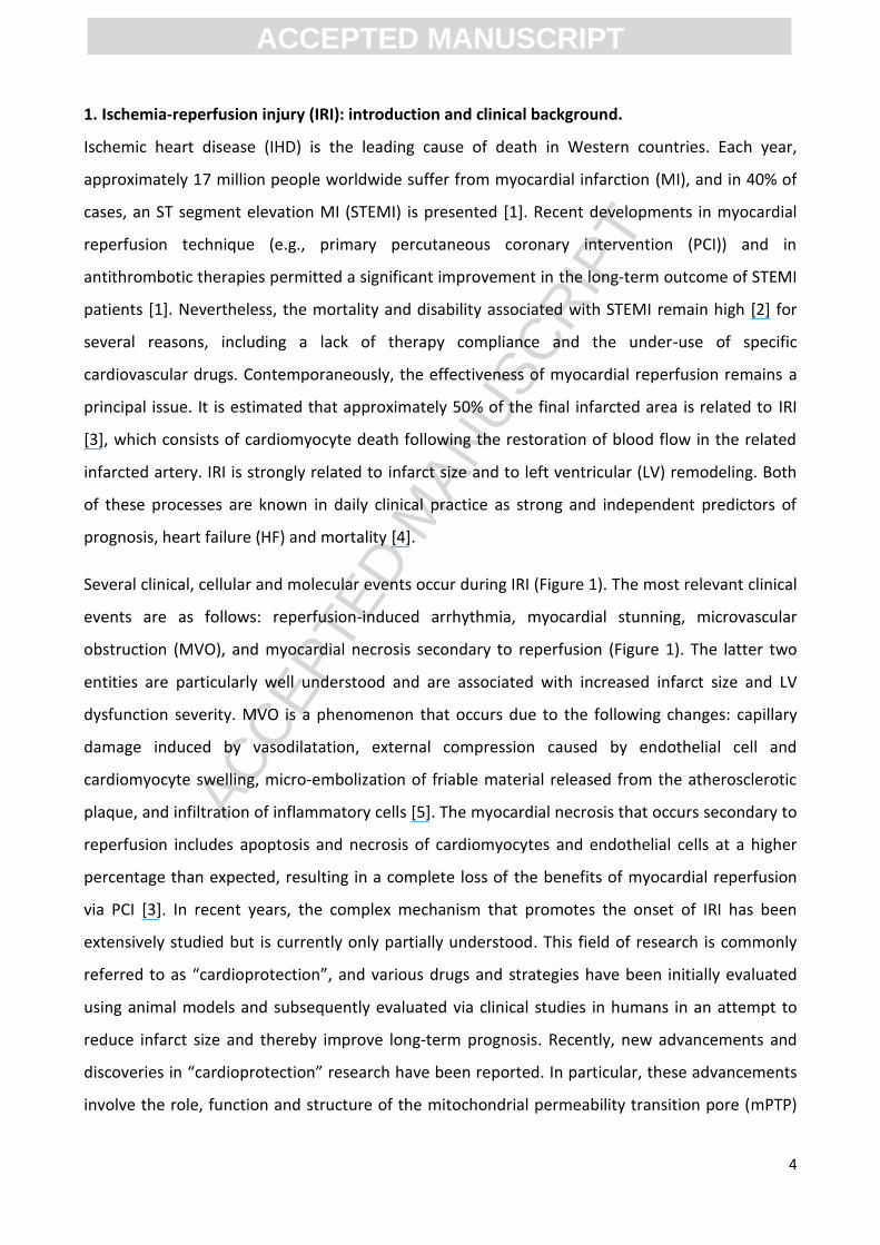
1. Ischemia-reperfusion injury (IRI): introduction and clinical background.
Ischemic heart disease (IHD) is the leading cause of death in Western countries. Each year,
approximately 17 million people worldwide suffer from myocardial infarction (MI), and in 40% of
cases, an ST segment elevation MI (STEMI) is presented [1]. Recent developments in myocardial
reperfusion technique (e.g., primary percutaneous coronary intervention (PCI)) and in
antithrombotic therapies permitted a significant improvement in the long-term outcome of STEMI
patients [1]. Nevertheless, the mortality and disability associated with STEMI remain high [2] for
several reasons, including a lack of therapy compliance and the under-use of specific
cardiovascular drugs. Contemporaneously, the effectiveness of myocardial reperfusion remains a
principal issue. It is estimated that approximately 50% of the final infarcted area is related to IRI
[3], which consists of cardiomyocyte death following the restoration of blood flow in the related
infarcted artery. IRI is strongly related to infarct size and to left ventricular (LV) remodeling. Both
of these processes are known in daily clinical practice as strong and independent predictors of
prognosis, heart failure (HF) and mortality [4].
Several clinical, cellular and molecular events occur during IRI (Figure 1). The most relevant clinical
events are as follows: reperfusion-induced arrhythmia, myocardial stunning, microvascular
obstruction (MVO), and myocardial necrosis secondary to reperfusion (Figure 1). The latter two
entities are particularly well understood and are associated with increased infarct size and LV
dysfunction severity. MVO is a phenomenon that occurs due to the following changes: capillary
damage induced by vasodilatation, external compression caused by endothelial cell and
cardiomyocyte swelling, micro-embolization of friable material released from the atherosclerotic
plaque, and infiltration of inflammatory cells [5]. The myocardial necrosis that occurs secondary to
reperfusion includes apoptosis and necrosis of cardiomyocytes and endothelial cells at a higher
percentage than expected, resulting in a complete loss of the benefits of myocardial reperfusion
via PCI [3]. In recent years, the complex mechanism that promotes the onset of IRI has been
extensively studied but is currently only partially understood. This field of research is commonly
referred to as “cardioprotection”, and various drugs and strategies have been initially evaluated
using animal models and subsequently evaluated via clinical studies in humans in an attempt to
reduce infarct size and thereby improve long-term prognosis. Recently, new advancements and
discoveries in “cardioprotection” research have been reported. In particular, these advancements
involve the role, function and structure of the mitochondrial permeability transition pore (mPTP)

ACC
EPTE
D M
ANU
SCR
IPT
ACCEPTED MANUSCRIPT
5
(Figure 2). Indeed, the mPTP (specifically, its opening) plays a key role in the development of
myocardial necrosis that occurs secondary to reperfusion.
2. MPTP structure and the c subunit of mitochondrial adenosine triphosphate (ATP) synthase
2.1 Core components of the mPTP
It has been widely accepted that the permeability of the mitochondrial inner membrane is
extremely low; thus, the discovery of a non-specific permeability transition with a threshold of 1.5
kDa suggested the existence of a pore (called the mPTP) that was responsible for this transition
[6].
The initial clue for the existence of the mPTP came from the very early studies of Haworth and
Hunter, which suggested that a hydrophilic channel was responsible for the permeability transition
induced by polyethylene glycol (PEG) polymers of size up to 1.5 kDa [7]. This idea was confirmed
by Crompton and Costi in 1988, who showed how, in its opened state, the mPTP channel should
obtain a diameter of 2 - 2.6 nm [8]. Later electrophysiological studies performed by Zoratti’s group
identified the putative mPTP as the giant channel that, at that time, was known as the
mitochondrial megachannel [9,10].
The initial evidence about the sensitivity of the mPTP to ADP and to the adenine nucleotide
transporter (ANT) inhibitor atractyloside suggested a role for the ANT in the regulation of mPTP,
and this finding was further supported by several other studies that involved identifying MPT
sensitivity to other ANT ligands such as bongkrekic acid, palmitoyl-CoA and carboxy- atractyloside
[11,12].
This idea was confirmed by a study of Halestrap and Davidson [13], who clearly displayed the
correlation between ADP, ATP, Bongkrekic acid and carboxy-atractyloside with Ca2+-induced MPT
induction or in combination with cyclosporine A, an already well-known MPT inhibitor at that time
[14–16]. Furthermore, in this initial study, Halestrap and Davidson proposed for the first time a
model for the mPTP structure that involved the conformational state of the ANT and its
interaction with the CsA target mitochondrial cyclophilin D (mtCypD) [13].
This model was supported by observations indicating that reconstituted ANT generates oligomers
with properties analogous to the mPTP in artificial membranes [17,18].

ACC
EPTE
D M
ANU
SCR
IPT
ACCEPTED MANUSCRIPT
6
Shortly thereafter, electrophysiological studies proposed that two molecules of the voltage-
dependent anion channel (VDAC) were components of the mPTP [9]. The involvement of the VDAC
in the mPTP structure suggested that it might not be a common pore but rather a more complex
and highly organized structure that included contact sites between the inner mitochondrial
membrane (IMM, where the MPT actually occurs) and the outer mitochondrial membrane (OMM,
where the VDAC is located).
This concept was demonstrated in 1996 by Brdiczka’s group, who observed the existence of a
protein complex that included the VDAC, ANT, hexokinase I (HK) and creatine kinase (CK, in its
octameric form) and that displayed MPT activities when reconstituted in liposomal vesicles
[19,20].
Due to its pharmacological properties, a protein of particular relevance for solving the molecular
identity of the mPTP was mtCypD [21]. mtCypD can be inhibited by CsA, which has similar but
opposite binding sensitivities to Ca2+ and ADP [22]. Additionally, mtCypD was shown to bind both
the VDAC and the ANT [23]. The generation of transgenic mice lacking the peptidylprolyl
isomerase f (ppif) gene confirmed that mtCypD is the protein element of mPTP that confers
sensitivity to CsA [24,25]. Further, its cysteine 203 appears to have critical importance, especially
regarding the sensitivity of mPTP to reactive oxygen species (ROS) [26]. Nonetheless, mtCypD is a
mitochondrial matrix protein; thus, it is unable to generate a pore, and its depletion does not deny
the existence of an MPT but rather dramatically increases the threshold for Ca2+ induction [27].
For a long time, the mPTP model proposed the VDAC and the ANT, which are located in the OMM
and IMM, respectively, as the core components of the mPTP. These components have been
proposed to be linked via a CK bridge [20]. mtCypD is also included in this complex as a regulatory
element in the mitochondrial matrix [28].
However, two different studies based on knockout animal models challenged this model. The first
study was performed using ANT1 and ANT2 double-knockout mice, and it demonstrated that the
MPT occurs despite the loss of the ANT, even though these mice exhibit a loss of sensitivity to ANT
inhibitors (bongkrekic acid or atractyloside) and a reduction in the Ca2+ threshold for mPTP
opening [29]. The second study performed by Molkentin’s group was based on a triple VDAC
knockout model, which did not display any significant differences in either the Ca2+ threshold for
mPTP induction or in cell death in response to various types of stimuli [30].

ACC
EPTE
D M
ANU
SCR
IPT
ACCEPTED MANUSCRIPT
7
These findings prompted a reconstruction of the structural mPTP model. Assuming that the ANT is
not the pore-forming element on the IMM (it should be mentioned that two additional ANT
isoforms have been identified after the ANT1/2 KO study), the VDAC-CK-HK-ANT complex should
be interpreted only as a functional regulator of mPTP activity. In an early study, Brdiczka’s group
proposed that this complex may be fundamental for channeling adenine nucleotides [31] across
the mitochondrial membrane, thus facilitating faster diffusion [19]. This hypothesis is supported
by the loss of sensitivity of the MPT to ADP in ANT1/2 KO mice [29].
It has long been known that inorganic phosphate sensitizes the MPT pore, suggesting that the
mPTP could possess a Pi-binding site. Inorganic phosphate is transported to the mitochondrial
matrix by the mitochondrial phosphate inorganic carrier (PiC). In support of this concept, Leung
and colleagues determined that the PiC interacts with mtCypD and the ANT [32]. Furthermore, this
interaction is strengthened by MPT-inducing agents, whereas MPT-blocking compounds diminish
this interaction. In the same year, based on a genetic screen, another group determined that PiC
overexpression induces mitochondrial dysfunction and apoptosis [33]. These results, together with
the earlier finding that a nonspecific pore is generated in liposomes by reconstituting the PiC [34],
identified PiC as a strong candidate for the core-forming element of the mPTP.
This idea was well accepted until last year, when the same group performed PiC silencing
experiments and found that knockdown of up to 70% of this carrier does not lead to any
significant alteration in the Ca2+ threshold for the MPT, suggesting that either a small amount of
PiC is required in the mPTP structure or that PiC is not a component of this structure [35].
The same concept was recently confirmed by the Baines and Molkentin groups, who generated
cardiac-specific mouse strains whereby mitochondrial PiC levels could be genetically manipulated
by overexpressing or knocking out/knocking down the slc25a3 gene [36,37]. Both studies indicated
that mPTP activity is not lost during PiC silencing or knock out and showed differences in its
possible role as regulator. In fact, whereas the first study found no alterations in the mitochondrial
Ca2+ retention capacity during either overexpression or silencing, the second showed that slc25a3
deletion results in increased Ca2+ retention capacity (less-sensitive mPTP) and protects cells from
stimuli able to induce MPT. Furthermore, the KO mouse was protected by reperfusion injury
compared to the control, confirming its role as a regulator of the mPTP (a critical player during
heart reperfusion injury, see below). Identifying what variables could generate the differences
ACC
EPTE
D M
ANU
SCR
IPT
ACCEPTED MANUSCRIPT
8
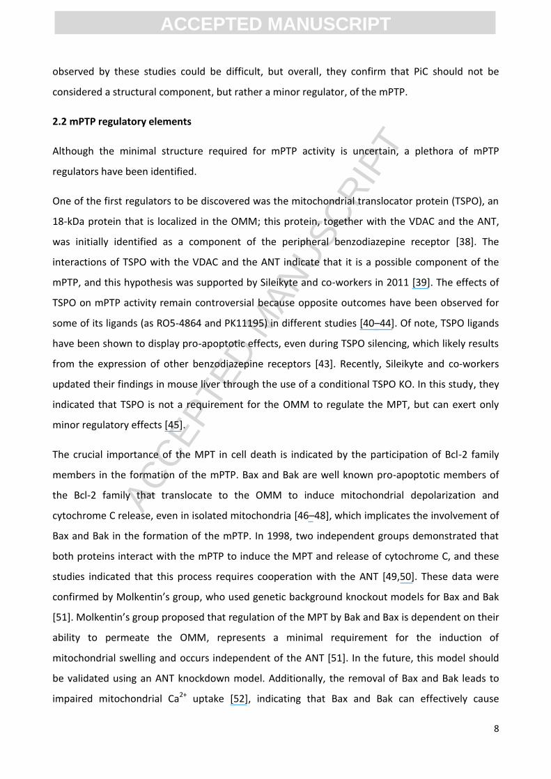
observed by these studies could be difficult, but overall, they confirm that PiC should not be
considered a structural component, but rather a minor regulator, of the mPTP.
2.2 mPTP regulatory elements
Although the minimal structure required for mPTP activity is uncertain, a plethora of mPTP
regulators have been identified.
One of the first regulators to be discovered was the mitochondrial translocator protein (TSPO), an
18-kDa protein that is localized in the OMM; this protein, together with the VDAC and the ANT,
was initially identified as a component of the peripheral benzodiazepine receptor [38]. The
interactions of TSPO with the VDAC and the ANT indicate that it is a possible component of the
mPTP, and this hypothesis was supported by Sileikyte and co-workers in 2011 [39]. The effects of
TSPO on mPTP activity remain controversial because opposite outcomes have been observed for
some of its ligands (as RO5-4864 and PK11195) in different studies [40–44]. Of note, TSPO ligands
have been shown to display pro-apoptotic effects, even during TSPO silencing, which likely results
from the expression of other benzodiazepine receptors [43]. Recently, Sileikyte and co-workers
updated their findings in mouse liver through the use of a conditional TSPO KO. In this study, they
indicated that TSPO is not a requirement for the OMM to regulate the MPT, but can exert only
minor regulatory effects [45].
The crucial importance of the MPT in cell death is indicated by the participation of Bcl-2 family
members in the formation of the mPTP. Bax and Bak are well known pro-apoptotic members of
the Bcl-2 family that translocate to the OMM to induce mitochondrial depolarization and
cytochrome C release, even in isolated mitochondria [46–48], which implicates the involvement of
Bax and Bak in the formation of the mPTP. In 1998, two independent groups demonstrated that
both proteins interact with the mPTP to induce the MPT and release of cytochrome C, and these
studies indicated that this process requires cooperation with the ANT [49,50]. These data were
confirmed by Molkentin’s group, who used genetic background knockout models for Bax and Bak
[51]. Molkentin’s group proposed that regulation of the MPT by Bak and Bax is dependent on their
ability to permeate the OMM, represents a minimal requirement for the induction of
mitochondrial swelling and occurs independent of the ANT [51]. In the future, this model should
be validated using an ANT knockdown model. Additionally, the removal of Bax and Bak leads to
impaired mitochondrial Ca2+ uptake [52], indicating that Bax and Bak can effectively cause

ACC
EPTE
D M
ANU
SCR
IPT
ACCEPTED MANUSCRIPT
9
impaired OMM permeability (assuming that this permeability affects Ca2+ transport across the
OMM). During their stimulation, Bax and Bak can also increase the amount of free Ca2+ in the
mitochondrial matrix (by promoting Ca2+ flux into the mitochondrial matrix) to trigger the MPT.
Furthermore, Bad, which is a pro-apoptotic member of the Bcl-2 family, has been shown to induce
the MPT in isolated mitochondria in a Bax- and Bak-independent manner [53]. In addition to these
findings, anti-apoptotic members of the Bcl-2 family have been shown to modify MPT activity. For
example, Bcl-2 and Bcl-XL have been shown to interact with the ANT and the VDAC, respectively
[54,55].
One mPTP regulator that has attracted particular interest is glycogen synthase kinase 3 beta
(GSK3-β) [56]. This protein contributes to many cellular processes, such as transcription,
metabolism, cell division, adhesion and apoptosis. In 2004, it was proposed that GSK3-β functions
as a convergence point for the inhibition of the MPT via different survival signaling pathways,
including protein kinase A (PKA), protein kinase B (PKB), protein kinase C (PKC) and mammalian
target of rapamycin (mTOR) [57]. This concept was supported by additional studies reporting that
GSK3-β is a therapeutic target for cardioprotection [58–60]. The complete mechanism by which
GSK3-β is involved in mPTP function has yet to be elucidated, and it is especially unclear if its
kinase activity is required. Nonetheless, it has been shown that GSK3-β inhibitors impair adenine
nucleotide transport across the matrix, which is related to a reduction in VDAC2 phosphorylation
[61].
mPTP regulation via pro-survival kinase signaling has also been attributed to PKCε. This particular
isoform has been associated with cardioprotection based on studies using transgenic mice [62].
Interestingly, the same group has shown that PKCε interacts with the VDAC1-HKII-ANT complex,
resulting in VDAC1 phosphorylation and inhibition of mPTP activity. Additionally, PKCε has been
reported to be able to reduce the Ca2+ content in the sarcoplasmic reticulum and decrease the risk
of mPTP opening during reperfusion, thus providing a novel mechanism for preconditioning-
mediated cardioprotection [63].
Several other proteins, such as PKG, p53, and Complement component 1 Q subcomponent-binding
protein (C1QBP), have been proposed in at least one study to directly modulate mPTP activity [64–
66]. Further elucidation of their role in the MPT is required.

ACC
EPTE
D M
ANU
SCR
IPT
ACCEPTED MANUSCRIPT
10
2.3 The critical role of the ATP synthase c-subunit in mPTP function
ATP synthase displays a series of characteristics upstream of its regulation that resemble those of
the mPTP. First, the hydrolytic activity of ATP synthase is strongly inhibited by the concurrent
binding of two mPTP inhibitors, namely ADP and Mg2+, to its catalytic site, the so-called Mg-ADP
block [67], but the mPTP inducer Pi has been proposed to abolish this block. Second, two different
cysteine residues (C294 in the alpha subunit and C103 in the gamma subunit) may be linked by a
disulfide bridge during oxidative stress, thereby impeding ATP synthase activity [68]. Furthermore,
the ATP synthase complex forms a supercomplex with the ANT and PiC, both of which have been
proposed as components of the mPTP, and this complex is referred to as the ATP synthasome
[69,70]. In 2009, mtCypD, another regulatory component of the mPTP, was shown to interact with
the peripheral stalk of ATP synthase, particularly the oligomycin sensitivity conferring protein
(OSCP) and d subunits. These interactions result in reduced catalytic activity (both hydrolase and
synthase) that can be restored by displacing mtCypD with CsA [71]. Finally, anti-apoptotic Bcl-XL, a
known MPT inhibitor, interacts with ATP synthase and promotes its synthetase activity [72].
Physiological studies also suggest a correlation between ATP synthase and the mPTP. The c-ring-
selective inhibitor, oligomycin, prevents both tumor necrosis factor alpha- (TNF) and Bax-induced
MPT and cell death [49,73].
Recently, we identified the c subunit of the mitochondrial ATPase as a fundamental regulator of
mPTP activity [74,75]. Of all of the subunits that compose the FO complex (see above), the a, b and
c subunits are sufficient to facilitate the translocation of protons across lipid bilayers, and these
subunits are highly evolutionarily conserved, as previously mentioned [76,77].
It has recently been shown that Rho0 cells, which lack mitochondrial DNA, are equipped with a
functional mPTP [78]. This finding excludes a role for the a subunit of the mitochondrial ATP
synthase in the mPTP. Furthermore, conductive properties have only been ascribed to the c
subunit [79], and a peptide displaying a consistent degree of similarity to the c subunit has been
proposed as a putative regulator of the mPTP [41,80], thus indicating that the c subunit is the best
candidate for a pore component.
Furthermore, we found that silencing c subunit expression completely blocks MPT induction by
Ca2+ and oxidants, whereas c subunit overexpression dramatically enhances MPT induction.
Silencing the c subunit does not affect ATP synthesis, suggesting that MPT inhibition is not due to

ACC
EPTE
D M
ANU
SCR
IPT
ACCEPTED MANUSCRIPT
11
the accumulation of ADP in the mitochondrial matrix. Furthermore, silencing α subunit expression
does not lead to any significant alteration in MPT activity, suggesting that the c subunit of the
mitochondrial ATP synthase is a central component of the mPTP. In support of our results, it has
been recently reported that the isolated c subunit induces the MPT in isolated mitochondria and
forms ion channels in artificial bilayer membranes. Furthermore, this activity is stimulated by Ca2+,
inhibited by CsA and dependent on the phosphorylation state of the c subunit [81].
Nonetheless, it has yet to be validated that c-rings exist on the outside of ATP synthase, leaving
the c-ring unoccupied by the central stalk and thus available to generate currents in vivo.
Interestingly, a few months after our publication, Bernardi’s group confirmed the regulatory role
of ATP synthase in mPTP function and suggested that only ATP synthase dimers exert mPTP-like
activity when inserted into a lipid bilayer [82]. However, this concept contrasted findings
published by the same group that showed that MPT characteristics are also detected in Rho0 cells
depleted of mitochondrial DNA [78]. Indeed, Wittig et al. demonstrated that Rho0 cells contain
unstable oligomeric (and dimeric) structures of ATP synthase at extremely reduced levels [83].
Dimerization of ATP synthase is favored and stabilized by the inhibitor protein F1 (IF-1), and this
event is associated with increased ATP production and reduced susceptibility to cell death during
ischemia [84]. Reduced dimerization is detected in aging cells, thus favoring cell death.
Furthermore, dimer dissociation is reduced by mtCypD, and CsA impedes the transition from
dimers to monomers, thereby suggesting that the dimer itself is likely not the mPTP but rather
that the transition from dimers to monomers favors mPTP formation [85]. Giorgio et al. showed
that ATP synthase dimers extracted from a native gel prior to insertion into a lipid bilayer could
produce some monomers, most likely due to technical manipulation during complex extraction.
Therefore, it is possible that an unstable monomer generated during this procedure could
rearrange under appropriate conditions to generate the mPTP based on the c rings of ATP
synthase. This notion is supported by a recent publication demonstrating that the c-ring can
generate a non-specific current ascribable to the mPTP and as isolated F1/FO ATP synthase
monomers reconstituted on vesicles generate mPTP-like currents when bound to mtCypD and
exposed to Ca2+ [86]. Moreover, it has been suggested as the Ca2+-induced mitochondrial swelling
can at least partially detach the F1 subunit from the FO subunit, and this detachment can be
reversed by CsA [86].
ACC
EPTE
D M
ANU
SCR
IPT
ACCEPTED MANUSCRIPT
12
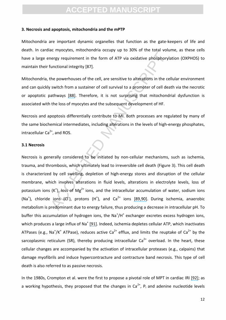
3. Necrosis and apoptosis, mitochondria and the mPTP
Mitochondria are important dynamic organelles that function as the gate-keepers of life and
death. In cardiac myocytes, mitochondria occupy up to 30% of the total volume, as these cells
have a large energy requirement in the form of ATP via oxidative phosphorylation (OXPHOS) to
maintain their functional integrity [87].
Mitochondria, the powerhouses of the cell, are sensitive to alterations in the cellular environment
and can quickly switch from a sustainer of cell survival to a promoter of cell death via the necrotic
or apoptotic pathways [88]. Therefore, it is not surprising that mitochondrial dysfunction is
associated with the loss of myocytes and the subsequent development of HF.
Necrosis and apoptosis differentially contribute to MI. Both processes are regulated by many of
the same biochemical intermediates, including alterations in the levels of high-energy phosphates,
intracellular Ca2+, and ROS.
3.1 Necrosis
Necrosis is generally considered to be initiated by non-cellular mechanisms, such as ischemia,
trauma, and thrombosis, which ultimately lead to irreversible cell death (Figure 3). This cell death
is characterized by cell swelling, depletion of high-energy stores and disruption of the cellular
membrane, which involves alterations in fluid levels, alterations in electrolyte levels, loss of
potassium ions (K+), loss of Mg2+ ions, and the intracellular accumulation of water, sodium ions
(Na+), chloride ions (Cl-), protons (H+), and Ca2+ ions [89,90]. During ischemia, anaerobic
metabolism is predominant due to energy failure, thus producing a decrease in intracellular pH. To
buffer this accumulation of hydrogen ions, the Na+/H+ exchanger excretes excess hydrogen ions,
which produces a large influx of Na+ [91]. Indeed, ischemia depletes cellular ATP, which inactivates
ATPases (e.g., Na+/K+ ATPase), reduces active Ca2+ efflux, and limits the reuptake of Ca2+ by the
sarcoplasmic reticulum (SR), thereby producing intracellular Ca2+ overload. In the heart, these
cellular changes are accompanied by the activation of intracellular proteases (e.g., calpains) that
damage myofibrils and induce hypercontracture and contracture band necrosis. This type of cell
death is also referred to as passive necrosis.
In the 1980s, Crompton et al. were the first to propose a pivotal role of MPT in cardiac IRI [92]; as
a working hypothesis, they proposed that the changes in Ca2+, Pi and adenine nucleotide levels

ACC
EPTE
D M
ANU
SCR
IPT
ACCEPTED MANUSCRIPT
13
during ischemia trigger mPTP opening [14,92,93]. Griffiths and Halestrap subsequently
demonstrated that MPT occurs upon reperfusion of the ischemic heart. In 1995, using the
mitochondrial ‘Hot DOG’ – entrapment technique, they showed that some mitochondria can
undergo mPTP opening and closure in the ischemic-reperfused heart [94]. Their data confirm that
pore opening occurs during reperfusion of the heart after ischemia, but not in the ischemic
priming period. Their experimental procedures tell us that the extent of DOG uptake increases
until the period of ischemia that precedes reperfusion increases to an empirical maximum of 30-
40 min [94].
Opening of the mPTP facilitates the free passage of protons across the IMM, leading to a
dissipation of the mitochondrial membrane potential and pH gradient, which comprise the proton
motive force. Not only does this process prevent ATP generation, but reversal of the ATPase also
occurs, thus causing the breakdown of cytosolic ATP that is generated via glycolysis. Energy
metabolism is further impaired, thereby resulting in a continuous cycle of increasing Ca2+
deregulation and mPTP opening. These changes activate phospholipases, nucleases and proteases.
The importance of the mPTP in the necrotic death of cardiomyocytes under such conditions was
initially detected in experiments using mPTP inhibitors, such as CsA [14,90]. Recently, further
evidence for a critical role of mPTP opening in necrotic cell death has been provided by the use of
mice in which the target of CsA, mtCypD, was knocked out [89,91]. These animals exhibit
substantial protection from IRI-induced damage (infarct size) to the heart. In addition, using these
mice, it has been shown that cardiac failure associated with chronic Ca2+ overload involves the
mPTP-dependent death of cardiomyocytes [95]. At last, in 2014, a study of sixty-one patients that
was directed by Ovize showed that cyclosporine administration at the time of reperfusion protects
against reperfusion injury in patients undergoing aortic valve surgery by reducing the levels of
cardiac troponin I in the cyclosporine group compared with the control group [96]. Most of these
concepts have been widely reviewed [97,98].
Today, MI, bypass surgery and organ transplantation provide dramatic examples of this
mechanism of cardiac failure. This step in cell death involves the mPTP and a complex network of
cellular signals. Because the severity of the insult in most infarction cases in the heart is
heterogeneous, there is often no clear boundary between apoptosis and necrosis. However, if the
stress experienced by the cell is a severe insult, the extent of mPTP opening is catastrophic, and

ACC
EPTE
D M
ANU
SCR
IPT
ACCEPTED MANUSCRIPT
14
necrotic cell death is inevitable, as occurs in the core of vessel obstruction. In this region, most
mitochondria undergo massive matrix swelling and OMM rupture.
3.2 Apoptosis
In addition, reperfusion can lead to an enhancement in apoptosis [95], which is an evolutionarily
conserved mode of cell death that can be initiated via two different pathways in mammals: the
death receptor pathway (extrinsic apoptotic pathway) and the mitochondrial pathway (intrinsic
apoptotic pathway). Furthermore, the apoptosis pathway that is activated depends on the nature
of the death signal (Figure 3). Apoptosis, similarly to necrosis, can be induced by mPTP opening.
For apoptosis, the stress is often a milder insult than that for necrosis, which could explain the
apoptotic ring around the necrotic core of a coronary infarct [99]. mPTP opening might be
transient or maintained in some mitochondria undergoing matrix swelling, where all small-
molecular-mass solutes equilibrate across the IMM, and proteins remain at a high concentration in
the matrix and exert colloidal osmotic pressure that unfolds the IMM cristae and induces OMM
rupture [100,101].
3.2.1 Extrinsic apoptotic pathway
Mitochondrial membrane permeabilization does not play a crucial role in the extrinsic pathway.
Instead, it is most likely activated in response to inflammation that is required for healing and scar
formation in the infarct. Plasma membrane receptors are activated by pro-inflammatory ligands,
including Fas, TNF-α and TNF-related apoptosis-inducing ligand (TRAIL).
Fas and Fas ligands are expressed in the heart and enhanced expression of Fas is associated with
increased apoptosis in experimental models of MI [102,103]. Simulated IRI in a cell culture model
increases the sensitivity of myocytes to Fas-mediated death. Therefore, it has been suggested that
IRI might down-regulate inhibitors of the Fas pathway, such as cellular FLICE-inhibitory protein
(cFLIP). cFLIP is highly expressed in the heart under normal physiological conditions but is
degraded after IRI. Thus, the loss of cFLIP expression may be important for enhancing the
sensitivity of cardiomyocytes to apoptosis after IRI. These results suggest that the Fas-mediated
cell death pathway exists in cardiomyocytes but that under normal conditions, this pathway is

ACC
EPTE
D M
ANU
SCR
IPT
ACCEPTED MANUSCRIPT
15
down-regulated by inhibitors. However, after stress, such as ischemia, cFLIP becomes inactivated,
thus rendering the cells susceptible to death via the Fas pathway.
Recent studies have revealed that TNF plays a role in the progression of myocardial disease.
Increased TNF-α and TNF receptor 1 (TNFR1) expression levels are associated with HF [104]. As
TNF-α induces apoptosis in cardiomyocytes [105], it is thought that at least part of its pathogenic
effect in the heart is due to its induction of cell death.
In contrast, there is also evidence supporting a prosurvival role of TNF in the heart, including the
involvement of TNF in the regulation of adaptive responses to biomechanical stress. Examples of
these adaptive responses include the induction of cellular hypertrophy in response to pressure
overload and the modulation of contractile function following ischemia.
The role of inflammation in MI as a target for cardioprotection has not been completely
addressed. A small number of studies have investigated the effects of reducing the inflammatory
response to myocardial reperfusion injury. Experimental animal studies have reported significant
reductions in MI size with several interventions administered at the time of myocardial
reperfusion, such as the inhibition of neutrophil aggregation and attenuation of leukocyte
infiltration into the infarcted myocardium [106,107]. On the other hand, more recently, clinical
studies targeting the inflammatory components of MI have failed to show a significant
improvement in reperfused–STEMI patients [108]. Further studies on the role of inflammation in
MI are required.
3.2.2 Intrinsic apoptotic pathway
Ca2+ is a critical sensitizing signal for the pro-apoptotic transition of mitochondria that plays a key
role in the regulation of cell death [109]. Mitochondrial Ca2+ overload is a pro-apoptotic inducer of
mitochondrial swelling, and OMM perturbation or OMM rupture leads to mitochondrial apoptotic
factor (cytochrome c, Smac/DIABLO, AIF and Omi/HtrA2) release into the cytosol [110,111].
Cytochrome c-mediated apoptosis is important in cardiomyocytes. Serum and glucose deprivation
induce cytochrome c release in vitro, thereby resulting in the activation of caspase-9, caspase-3
and apoptosis [112]. As serum and glucose deprivation are components of ischemia in vivo, these
results indicate that this pathway may be involved in heart disease-related cell death.

ACC
EPTE
D M
ANU
SCR
IPT
ACCEPTED MANUSCRIPT
16
Most reperfusion-induced apoptotic death of cardiomyocytes occurs during the initial minutes of
reperfusion due to increased ROS production, intracellular Ca2+ overload and mPTP opening [95].
The role of apoptosis in reperfusion injury has recently been addressed using rat and rabbit animal
models in which reperfusion accelerates the occurrence of apoptosis in cardiomyocytes [95,113].
In the infarcted region of the ventricular wall, myocytes containing DNA strand breaks are
detected 2 hours after coronary artery occlusion, and approximately 2.7 million myocytes are
apoptotic at this time point. Moreover, 6.6 million cells are apoptotic at 4.5 hours, indicating that
there is a 2.4-fold increase in the absolute number of apoptotic myocytes in the left ventricular
free wall from 2 to 4.5 hours after coronary artery occlusion. The magnitude of apoptosis
progressively decreases at later time intervals. Necrosis of myocytes also appears 2 hours after
coronary artery occlusion and continuously increases until one day following coronary artery
ligation. These findings demonstrate that myocyte apoptosis and necrosis are independent
variables contributing to infarct size, although apoptosis accounts for 86% of the total loss of
myocytes, and necrosis accounts for only 14% of the total loss [113].
In contrast, findings from other laboratories that support these experimental data indicate that MI
results from a significant increase in necrosis rather than apoptosis, where pro-apoptotic factors
are evident only early during ischemia but do not significantly contribute to infarct size [114,115].
Others have found that apoptosis and necrosis occurred simultaneously in all instances in hearts
from cases of fatal MI [116]. One likely hypothesis that could explain the coexistence of apoptosis
and necrosis after IRI is that damage produced by ischemia is capable of initiating apoptosis, but if
ischemia is prolonged, necrosis ensues (as discussed later).
3.3 Role of ROS and Ca2+ in IRI-induced damage
A wide range of mitochondrial ROS-induced damage has been described, including protein
carbonylation, lipid peroxidation and mitochondrial DNA damage [117]. These modifications are
important factors in the progression of myocardial IRI-induced damage. The re-introduction of
abundant oxygen at the onset of reperfusion evokes a burst of toxic oxygen derivatives within the
first few minutes of reperfusion. Moreover, oxidative stress also reduces the bioavailability of
nitric oxide (NO, a vasodilator) during reperfusion [118].
Cytosolic Ca2+ accumulation plays major roles in the initiation of programmed cell death during
acute MI. A prolonged increase in cytosolic Ca2+ induces mitochondrial Ca2+ overload, which leads

ACC
EPTE
D M
ANU
SCR
IPT
ACCEPTED MANUSCRIPT
17
to mPTP opening and the activation of Ca2+-dependent proteases [119]. Increased cytosolic Ca2+
plays a pivotal role in activating the serine threonine Ca2+/calmodulin-regulated phosphatase,
calcineurin. This phosphatase is a critical transducer of Ca2+ signals in most cell types, particularly
in the heart, due to its specific responsiveness to sustained, low-frequency Ca2+ signals [120].
Biochemical events leading to mPTP opening during ischemia and reperfusion
The effects of ROS and Ca2+ on MPT have been widely reported as key players during ischemia and
reperfusion damage [97,121] (Figure 3). During ischemia (the MPT-priming phase), the
accumulation of factors, including Ca2+, long-chain fatty acids and ROS, progressively increases the
susceptibility to MPT, thus increasing the likelihood that MPT will occur upon reperfusion (the
MPT-activating phase) [122].
Indeed, the conditions that occur during ischemia and reperfusion are identical to those that
induce mPTP opening. During ischemia, increased glycolysis causes the accumulation of lactic acid
and the reduction of pH. To restore the pH, the Na+/H+ antiporter is activated, but it acts
inefficiently because Na+ cannot be pumped out of the cell, as the Na+/K+ ATPase is inhibited by
the absence of intracellular ATP. Consequently, the cytosolic Ca2+ concentration increases because
the activity of the Na+/Ca2+ antiporter is reduced or reversed. In addition, during ischemia, there is
a decrease in the adenine nucleotide concentration, which is associated with an increased
phosphate concentration, thereby sensitizing mPTP opening in response to Ca2+; however, low pH
inhibits mPTP opening. If the period of ischemia is prolonged, the heart becomes irreversibly
damaged due to the activity of degradative enzymes, such as phospholipases and proteases, which
also compromise mitochondrial function [123].
Upon reperfusion, the mitochondria recover their ability to respire and rescue the sustained
mitochondrial membrane potential that is required for ATP synthesis. However, the mitochondrial
membrane potential is the driving force for mitochondrial Ca2+ uptake, thus leading to Ca2+
overload. In addition, rapid and extensive production of ROS occurs when the inhibited respiratory
chain is re-exposed to oxygen. Thus, the following resulting conditions are nearly optimal for mPTP
opening: high Ca2+ levels within the mitochondrial matrix, increased levels of phosphate and
oxidative stress, depletion of adenine nucleotide concentration, and rapid return of the pH to a
physiological value [124,125].

ACC
EPTE
D M
ANU
SCR
IPT
ACCEPTED MANUSCRIPT
18
After ischemia and reperfusion, the fate of the cell is determined by the severity of the damage as
follows: if the damage is minimal, the cell may recover; if the damage is moderate, the cell may
undergo apoptosis; and if the damage is severe, the cell may die from necrosis due to inadequate
energy production. Thus, mitochondria serve as an arbiter of cell fate in response to stress [119].
4. Clinical studies examining pharmacological agents to reduce IRI.
Considering the pivotal role of the mPTP in IRI during STEMI, many studies have focused their
attention on pharmacological agents that modulate mPTP opening. Currently, a limited number of
these agents act directly on mPTP and/or its components. Contrarily, the majority of these agents
are able to influence biological parameters (e.g., ROS, pH and PI signaling pathways) that indirectly
modulate the final stage of mPTP opening. Finally, several strategies of ischemic pre- and post-
conditioning have been developed and studied to reduce IRI during STEMI [126]. Nevertheless,
details of these studies are beyond the aim of this review. Hence, this review will only focus on the
pharmacological approaches to cardioprotection in humans (Table 1).
Agents directly targeting mPTP
One of the most promising results in cardioprotection has been reported by Piot et al. using CsA
[127]. Since the 1990s, it has been known that CsA inhibits mPTP opening by binding to mtCypD, a
mitochondrial isomerase that binds to subunits b, d and O in the lateral stalk of the F1-Fo ATPase.
Studying 58 patients, Piot el al. examined the effect of administration of an intravenous bolus of
2.5 mg/kg CsA to patients experiencing STEMI immediately before undergoing PCI by measuring
the release of myocardial-specific enzymes and performing magnetic resonance imaging (MRI) on
the infarcted heart within the fifth day after reperfusion. The results confirmed the
cardioprotective effect of CsA and showed a significantly reduced overall infarcted area in the
group treated with CsA compared with the control group [127].
The CYCLosporinE A in reperfused acute myocardial infarction (CYCLE) phase III clinical trial is
currently underway and is designed to address the clinical effectiveness of CsA for STEMI during
reperfusion therapy [128]. The role of CsA in cardioprotection has also been tested in cardiac
surgery and after coronary artery bypass graft and after aortic valve surgery. In the first study,
Hunseloy et al. demonstrated that a single intravenous bolus of CsA (2.5 mg/kg) administered

ACC
EPTE
D M
ANU
SCR
IPT
ACCEPTED MANUSCRIPT
19
prior to CABG surgery reduced the extent of perioperative myocardial injury, with a reduced
postoperative cardiac troponin T rise by 0.03 ng/ml for every 10 minutes, when compared with
the control (p=0.049) [129]. Additionally, in the setting of aortic valve surgery, the administration
of CsA demonstrated a beneficial effect in reducing RI that was expressed as a 35% reduction in
the area under the curve of cardiac troponin I compared with the control group (p=0.03) [96]. 3,5-
Seco-4-nor-cholestan-5-one oxime-3-ol (TRO40303) is an mPTP modulator that binds to the
mitochondrial translocator protein at its cholesterol site, which results in reduced release of
apoptosis-inducing factors into the cytosol after ischemia and reperfusion [130]. This new drug is
under evaluation in the MITOCARE trial to test whether its injection reduces infarct size, as
measured via both cardiac biomarker release and MRI within the fifth day after primary PCI [131].
Agents indirectly targeting the mPTP
Many other substances that indirectly target the mPTP have been tested. Among the most
interesting drugs that have been studied are exenatide, atrial natriuretic peptide, and glucose-
insulin-potassium (GIK). Exenatide is an analog of glucagon-like peptide 1 (GLP-1), and a post-hoc
study has demonstrated that this substance reduces the final infarct size by 30% in patients
experiencing STEMI and Thrombolysis in MI (TIMI) flow grades of 0 or 1 based on angiogram [132].
However, this benefit was limited to patients with a rapid symptom-onset-to-balloon time (≤132
minutes). The cardioprotective effect of exenatide is unclear, but it has been recently proposed
that GLP-1 also acts on the mPTP. The beneficial effect of atrial natriuretic peptide has been
evaluated in a small, randomized trial [132]. This study enrolled patients experiencing acute MI
who received PCI, and the atrial natriuretic peptide was administered as an adjunctive treatment
(compared to placebo). The authors showed that the patients receiving atrial natriuretic peptide
exhibited a 14.7% reduction in the infarct size (95% CI 3.0-24.9%) and a significant increase in the
LV ejection fraction after 6-12 months. The effect of atrial natriuretic peptide on mPTP is most
likely due to inactivation of GSK3-β [133]. Despite promising preliminary results (generally using
animal models), randomized studies using other pharmacological agents have failed to
demonstrate a clear benefit in reducing IRI or mortality (Table 1). Yellon et al. described the
cardioprotective role of reperfusion injury survival kinase (RISK) and survivor-activating factor
enhancement (SAFE), which are two pro-survival kinase pathways that converge on the
mitochondria to reduce mPTP opening [134]. Accordingly, some authors have speculated that a
GIK solution exerts a cardioprotective effect by modulating pro-survival kinase pathways via the

ACC
EPTE
D M
ANU
SCR
IPT
ACCEPTED MANUSCRIPT
20
GIK receptor, which is a G protein-coupled receptor [135]. Nevertheless, no clinical benefit has
been observed in a confirmatory randomized clinical trial (Table 1) [136]. Finally, a new substance,
namely Bendavia, is under evaluation. Bendavia is a peptide that interacts with cardiolipin in the
IMM to reduce ROS production and maintain the efficiency of the electron transport chain during
reperfusion [137]. The EMBRACE trial is ongoing to test the potential clinical application and
effectiveness of Bendavia. The principal aim of the EMBRACE trial is to demonstrate that Bendavia
injection will reduce infarct size, as assessed by analyzing cardiac biomarker release and MRI [137].
Overall, the current available data regarding pharmacological agents acting directly or indirectly on
the mPTP and IRI are limited. Few trials have demonstrated a net clinical benefit but have been
limited by a small sample size, the use of surrogate endpoints and extensive exclusion criteria.
As mentioned above, to evaluate the reduction in infarct size, all trials measured the biomarker
levels, and the ejection fraction was determined based on echocardiography and magnetic
resonance imaging. In the majority of cases, all patients underwent two MRI scans as follows: the
first scan was performed within a week after primary PCI, and the second scan was performed at a
follow-up visit. The measured parameters include the area at risk based on T2-weighted images,
the final infarct size based on late-enhancement MRI sequences and the myocardial salvage index
[(area at risk minus infarcted size)/area at risk] [138,139].
5. Conclusions.
IRI induces dramatic increases in mitochondrial permeability, thereby initiating a chain of events
that leads to both apoptosis and necrosis of cardiomyocytes. Thus, the mPTP represents a
therapeutic target to reduce cardiomyocyte mortality and treat myocardial IRI. Unfortunately,
antagonizing the mPTP in the clinical setting has been hampered by the lack of a precise
understanding of its molecular architecture.
Here, we propose a model in which ATP synthase is the central element of the mPTP as follows: (i)
ATP synthase shares several activators and inhibitors; (ii) ATP synthase interacts with various
regulators of the mPTP (including the ANT, PiC and mtCypD); and (iii) the c ring of ATP synthase
(the lone subunit confirmed to display gating capacity) plays a critical role in mPTP activity. Further
studies are required to achieve a complete understanding of the structure and activity of the
mPTP. Finally, the recent discovery of several mPTP components provides novel targets for

ACC
EPTE
D M
ANU
SCR
IPT
ACCEPTED MANUSCRIPT
21
cardioprotection. Currently, the overall molecular identity of the mPTP remains unknown, but this
information may facilitate the development of more specific and potent mPTP inhibitors.
Acknowledgments. This study was supported by: the Italian Association for Cancer Research (AIRC: IG-14442 to Paolo
Pinton and MFAG-13521 to C.G.); local funds from the University of Ferrara to Paolo Pinton and Carlotta Giorgi;
Telethon (GGP11139B); and the Italian Ministry of Education, University and Research (COFIN, FIRB, and Futuro in
Ricerca) to Paolo Pinton. Mariusz R. Wieckowski was supported by the Polish Ministry of Science and Higher Education
(W100/HFSC/2011) and Grant HFSP RGP0027/2011.
ACC
EPTE
D M
ANU
SCR
IPT
ACCEPTED MANUSCRIPT
22
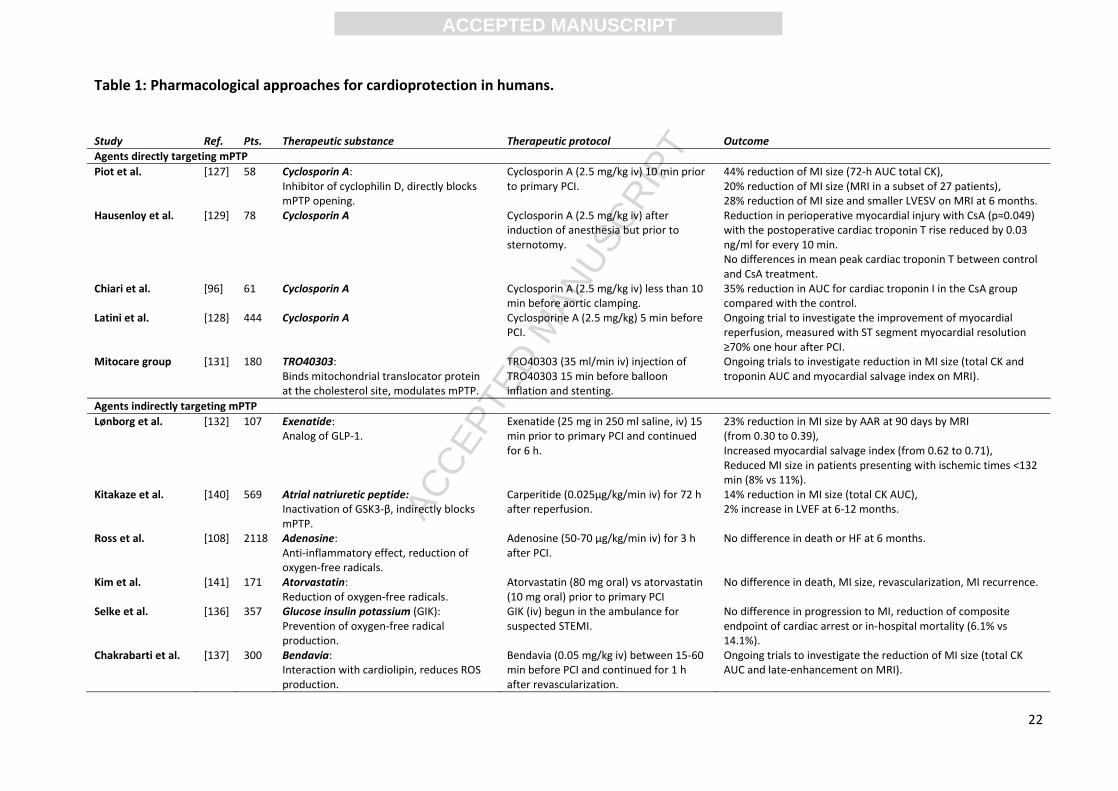
Table 1: Pharmacological approaches for cardioprotection in humans.
Study Ref. Pts. Therapeutic substance Therapeutic protocol Outcome
Agents directly targeting mPTP
Piot et al. [127] 58 Cyclosporin A: Inhibitor of cyclophilin D, directly blocks mPTP opening.
Cyclosporin A (2.5 mg/kg iv) 10 min prior to primary PCI.
44% reduction of MI size (72-h AUC total CK), 20% reduction of MI size (MRI in a subset of 27 patients), 28% reduction of MI size and smaller LVESV on MRI at 6 months.
Hausenloy et al. [129] 78 Cyclosporin A Cyclosporin A (2.5 mg/kg iv) after induction of anesthesia but prior to sternotomy.
Reduction in perioperative myocardial injury with CsA (p=0.049) with the postoperative cardiac troponin T rise reduced by 0.03 ng/ml for every 10 min. No differences in mean peak cardiac troponin T between control and CsA treatment.
Chiari et al. [96] 61 Cyclosporin A Cyclosporin A (2.5 mg/kg iv) less than 10 min before aortic clamping.
35% reduction in AUC for cardiac troponin I in the CsA group compared with the control.
Latini et al. [128] 444 Cyclosporin A Cyclosporine A (2.5 mg/kg) 5 min before PCI.
Ongoing trial to investigate the improvement of myocardial reperfusion, measured with ST segment myocardial resolution ≥70% one hour after PCI.
Mitocare group [131] 180 TRO40303: Binds mitochondrial translocator protein at the cholesterol site, modulates mPTP.
TRO40303 (35 ml/min iv) injection of TRO40303 15 min before balloon inflation and stenting.
Ongoing trials to investigate reduction in MI size (total CK and troponin AUC and myocardial salvage index on MRI).
Agents indirectly targeting mPTP
Lønborg et al. [132] 107 Exenatide: Analog of GLP-1.
Exenatide (25 mg in 250 ml saline, iv) 15 min prior to primary PCI and continued for 6 h.
23% reduction in MI size by AAR at 90 days by MRI (from 0.30 to 0.39), Increased myocardial salvage index (from 0.62 to 0.71), Reduced MI size in patients presenting with ischemic times <132 min (8% vs 11%).
Kitakaze et al. [140] 569 Atrial natriuretic peptide: Inactivation of GSK3-β, indirectly blocks mPTP.
Carperitide (0.025μg/kg/min iv) for 72 h after reperfusion.
14% reduction in MI size (total CK AUC), 2% increase in LVEF at 6-12 months.
Ross et al. [108] 2118 Adenosine: Anti-inflammatory effect, reduction of oxygen-free radicals.
Adenosine (50-70 μg/kg/min iv) for 3 h after PCI.
No difference in death or HF at 6 months.
Kim et al.
[141] 171 Atorvastatin: Reduction of oxygen-free radicals.
Atorvastatin (80 mg oral) vs atorvastatin (10 mg oral) prior to primary PCI
No difference in death, MI size, revascularization, MI recurrence.
Selke et al. [136] 357 Glucose insulin potassium (GIK): Prevention of oxygen-free radical production.
GIK (iv) begun in the ambulance for suspected STEMI.
No difference in progression to MI, reduction of composite endpoint of cardiac arrest or in-hospital mortality (6.1% vs 14.1%).
Chakrabarti et al. [137] 300 Bendavia: Interaction with cardiolipin, reduces ROS production.
Bendavia (0.05 mg/kg iv) between 15-60 min before PCI and continued for 1 h after revascularization.
Ongoing trials to investigate the reduction of MI size (total CK AUC and late-enhancement on MRI).

ACC
EPTE
D M
ANU
SCR
IPT
ACCEPTED MANUSCRIPT
23
Ref: reference. Pts: number of patients. mPTP: mitochondrial permeability transition pore. PCI: percutaneous coronary intervention. MI: myocardial infarction. CK: creatine kinase.
AUC: area under the curve. MRI: magnetic resonance imaging. h: hours. AAR: area at risk. min: minutes. LVEF: left ventricle ejection fraction. HF: heart failure.

ACC
EPTE
D M
ANU
SCR
IPT
ACCEPTED MANUSCRIPT
24
Figure 1. Ischemia-reperfusion injury during acute myocardial infarction.

ACC
EPTE
D M
ANU
SCR
IPT
ACCEPTED MANUSCRIPT
25
Figure 2. Novel model for mPTP structure.
The present model for mPTP is built around F1/FO ATP synthase superstructures (involving the ANT and
PiC) that directly interact with the main mPTP regulator CypD. The c-ring of the ATP synthase acts as the
pore of the mPTP. The model spans from the inner mitochondrial membrane (IMM) to the outer
mitochondrial membrane (OMM) by interactions with the VDAC, Bax and Bak, and CK oligomers in the
intermembrane space (IMS). Finally, the complex is surrounded by regulatory elements, as protein kinase C
epsilon (PKCε), glycogen synthase kinase 3-beta (GSK3-β) and mitochondrial translocator protein (TSPO) are
involved.
ACC
EPTE
D M
ANU
SCR
IPT
ACCEPTED MANUSCRIPT
26
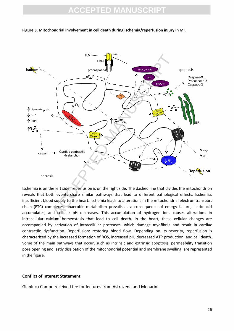
Figure 3. Mitochondrial involvement in cell death during ischemia/reperfusion injury in MI.
Ischemia is on the left side; reperfusion is on the right side. The dashed line that divides the mitochondrion
reveals that both events share similar pathways that lead to different pathological effects. Ischemia:
insufficient blood supply to the heart. Ischemia leads to alterations in the mitochondrial electron transport
chain (ETC) complexes, anaerobic metabolism prevails as a consequence of energy failure, lactic acid
accumulates, and cellular pH decreases. This accumulation of hydrogen ions causes alterations in
intracellular calcium homeostasis that lead to cell death. In the heart, these cellular changes are
accompanied by activation of intracellular proteases, which damage myofibrils and result in cardiac
contractile dysfunction. Reperfusion: restoring blood flow. Depending on its severity, reperfusion is
characterized by the increased formation of ROS, increased pH, decreased ATP production, and cell death.
Some of the main pathways that occur, such as intrinsic and extrinsic apoptosis, permeability transition
pore opening and lastly dissipation of the mitochondrial potential and membrane swelling, are represented
in the figure.
Conflict of Interest Statement
Gianluca Campo received fee for lectures from Astrazena and Menarini.

ACC
EPTE
D M
ANU
SCR
IPT
ACCEPTED MANUSCRIPT
27
References
[1] Laslett LJ, Alagona P, Clark BA, Drozda JP, Saldivar F, Wilson SR, et al. The worldwide environment of cardiovascular disease: prevalence, diagnosis, therapy, and policy issues: a report from the American College of Cardiology. J Am Coll Cardiol 2012;60:S1–49.
[2] Campo G, Saia F, Guastaroba P, Marchesini J, Varani E, Manari A, et al. Prognostic impact of hospital readmissions after primary percutaneous coronary intervention. Arch Intern Med 2011;171:1948–9.
[3] Yetgin T, Manintveld OC, Duncker DJ, van der Giessen WJ. Postconditioning against ischaemia-reperfusion injury: ready for wide application in patients? Neth Heart J 2010;18:389–92.
[4] Yellon DM, Hausenloy DJ. Myocardial reperfusion injury. N Engl J Med 2007;357:1121–35.
[5] Kloner RA, Bolli R, Marban E, Reinlib L, Braunwald E. Medical and cellular implications of stunning, hibernation, and preconditioning: an NHLBI workshop. Circulation 1998;97:1848–67.
[6] Halestrap AP, Connern CP, Griffiths EJ, Kerr PM. Cyclosporin A binding to mitochondrial cyclophilin inhibits the permeability transition pore and protects hearts from ischaemia/reperfusion injury. Mol Cell Biochem 1997;174:167–72.
[7] Haworth RA, Hunter DR. The Ca2+-induced membrane transition in mitochondria. II. Nature of the Ca2+ trigger site. Arch Biochem Biophys 1979;195:460–7.
[8] Crompton M, Costi A. A heart mitochondrial Ca2(+)-dependent pore of possible relevance to re-perfusion-induced injury. Evidence that ADP facilitates pore interconversion between the closed and open states. Biochem J 1990;266:33–9.
[9] Szabó I, Zoratti M. The mitochondrial permeability transition pore may comprise VDAC molecules. I. Binary structure and voltage dependence of the pore. FEBS Lett 1993;330:201–5.
[10] Szabó I, De Pinto V, Zoratti M. The mitochondrial permeability transition pore may comprise VDAC molecules. II. The electrophysiological properties of VDAC are compatible with those of the mitochondrial megachannel. FEBS Lett 1993;330:206–10.
[11] Harris EJ. Modulation of Ca2+ efflux from heart mitochondria. Biochem J 1979;178:673–80.
[12] Lê Quôc K, Lê Quôc D. Involvement of the ADP/ATP carrier in calcium-induced perturbations of the mitochondrial inner membrane permeability: importance of the orientation of the nucleotide binding site. Arch Biochem Biophys 1988;265:249–57.
[13] Halestrap AP, Davidson AM. Inhibition of Ca2(+)-induced large-amplitude swelling of liver and heart mitochondria by cyclosporin is probably caused by the inhibitor binding to

ACC
EPTE
D M
ANU
SCR
IPT
ACCEPTED MANUSCRIPT
28
mitochondrial-matrix peptidyl-prolyl cis-trans isomerase and preventing it interacting with the adenine nuc. Biochem J 1990;268:153–60.
[14] Crompton M, Ellinger H, Costi A. Inhibition by cyclosporin A of a Ca2+-dependent pore in heart mitochondria activated by inorganic phosphate and oxidative stress. Biochem J 1988;255:357–60.
[15] Broekemeier KM, Pfeiffer DR. Cyclosporin A-sensitive and insensitive mechanisms produce the permeability transition in mitochondria. Biochem Biophys Res Commun 1989;163:561–6.
[16] Broekemeier KM, Dempsey ME, Pfeiffer DR. Cyclosporin A is a potent inhibitor of the inner membrane permeability transition in liver mitochondria. J Biol Chem 1989;264:7826–30.
[17] Brustovetsky N, Klingenberg M. Mitochondrial ADP/ATP carrier can be reversibly converted into a large channel by Ca2+. Biochemistry 1996;35:8483–8.
[18] Rück A, Dolder M, Wallimann T, Brdiczka D. Reconstituted adenine nucleotide translocase forms a channel for small molecules comparable to the mitochondrial permeability transition pore. FEBS Lett 1998;426:97–101.
[19] Beutner G, Rück A, Riede B, Welte W, Brdiczka D. Complexes between kinases, mitochondrial porin and adenylate translocator in rat brain resemble the permeability transition pore. FEBS Lett 1996;396:189–95.
[20] Beutner G, Rück A, Riede B, Brdiczka D. Complexes between porin, hexokinase, mitochondrial creatine kinase and adenylate translocator display properties of the permeability transition pore. Implication for regulation of permeability transition by the kinases. Biochim Biophys Acta - Biomembr 1998;1368:7–18.
[21] Elrod JW, Molkentin JD. Physiologic functions of cyclophilin D and the mitochondrial permeability transition pore. Circ J 2013;77:1111–22.
[22] Tanveer A, Virji S, Andreeva L, Totty NF, Hsuan JJ, Ward JM, et al. Involvement of cyclophilin D in the activation of a mitochondrial pore by Ca2+ and oxidant stress. Eur J Biochem 1996;238:166–72.
[23] Crompton M, Virji S, Ward JM. Cyclophilin-D binds strongly to complexes of the voltage-dependent anion channel and the adenine nucleotide translocase to form the permeability transition pore. Eur J Biochem 1998;258:729–35.
[24] Basso E, Fante L, Fowlkes J, Petronilli V, Forte MA, Bernardi P. Properties of the permeability transition pore in mitochondria devoid of Cyclophilin D. J Biol Chem 2005;280:18558–61.
[25] Schinzel AC, Takeuchi O, Huang Z, Fisher JK, Zhou Z, Rubens J, et al. Cyclophilin D is a component of mitochondrial permeability transition and mediates neuronal cell death after focal cerebral ischemia. Proc Natl Acad Sci U S A 2005;102:12005–10.

ACC
EPTE
D M
ANU
SCR
IPT
ACCEPTED MANUSCRIPT
29
[26] Nguyen TT, Stevens M V, Kohr M, Steenbergen C, Sack MN, Murphy E. Cysteine 203 of cyclophilin D is critical for cyclophilin D activation of the mitochondrial permeability transition pore. J Biol Chem 2011;286:40184–92.
[27] Baines CP, Kaiser RA, Purcell NH, Blair NS, Osinska H, Hambleton MA, et al. Loss of cyclophilin D reveals a critical role for mitochondrial permeability transition in cell death. Nature 2005;434:658–62.
[28] Nicolli A, Basso E, Petronilli V, Wenger RM, Bernardi P. Interactions of cyclophilin with the mitochondrial inner membrane and regulation of the permeability transition pore, and cyclosporin A-sensitive channel. J Biol Chem 1996;271:2185–92.
[29] Kokoszka JE, Waymire KG, Levy SE, Sligh JE, Cai J, Jones DP, et al. The ADP/ATP translocator is not essential for the mitochondrial permeability transition pore. Nature 2004;427:461–5.
[30] Baines CP, Kaiser RA, Sheiko T, Craigen WJ, Molkentin JD. Voltage-dependent anion channels are dispensable for mitochondrial-dependent cell death. Nat Cell Biol 2007;9:550–5.
[31] Bonora M, Patergnani S, Rimessi A, De Marchi E, Suski JM, Bononi A, et al. ATP synthesis and storage. Purinergic Signal 2012;8:343–57.
[32] Leung AWC, Varanyuwatana P, Halestrap AP. The mitochondrial phosphate carrier interacts with cyclophilin D and may play a key role in the permeability transition. J Biol Chem 2008;283:26312–23.
[33] Alcalá S, Klee M, Fernández J, Fleischer A, Pimentel-Muiños FX. A high-throughput screening for mammalian cell death effectors identifies the mitochondrial phosphate carrier as a regulator of cytochrome c release. Oncogene 2008;27:44–54.
[34] Schroers A, Krämer R, Wohlrab H. The reversible antiport-uniport conversion of the phosphate carrier from yeast mitochondria depends on the presence of a single cysteine. J Biol Chem 1997;272:10558–64.
[35] Varanyuwatana P, Halestrap AP. The roles of phosphate and the phosphate carrier in the mitochondrial permeability transition pore. Mitochondrion 2012;12:120–5.
[36] Gutiérrez-Aguilar M, Douglas DL, Gibson AK, Domeier TL, Molkentin JD, Baines CP. Genetic manipulation of the cardiac mitochondrial phosphate carrier does not affect permeability transition. J Mol Cell Cardiol 2014;72:316–25.
[37] Kwong JQ, Davis J, Baines CP, Sargent MA, Karch J, Wang X, et al. Genetic deletion of the mitochondrial phosphate carrier desensitizes the mitochondrial permeability transition pore and causes cardiomyopathy. Cell Death Differ 2014;21:1209–17.
[38] McEnery MW, Snowman a M, Trifiletti RR, Snyder SH. Isolation of the mitochondrial benzodiazepine receptor: association with the voltage-dependent anion channel and the adenine nucleotide carrier. Proc Natl Acad Sci U S A 1992;89:3170–4.

ACC
EPTE
D M
ANU
SCR
IPT
ACCEPTED MANUSCRIPT
30
[39] Sileikyte J, Petronilli V, Zulian A, Dabbeni-Sala F, Tognon G, Nikolov P, et al. Regulation of the inner membrane mitochondrial permeability transition by the outer membrane translocator protein (peripheral benzodiazepine receptor). J Biol Chem 2011;286:1046–53.
[40] Li J, Wang J, Zeng Y. Peripheral benzodiazepine receptor ligand, PK11195 induces mitochondria cytochrome c release and dissipation of mitochondria potential via induction of mitochondria permeability transition. Eur J Pharmacol 2007;560:117–22.
[41] Krestinina O V, Grachev DE, Odinokova I V, Reiser G, Evtodienko Y V, Azarashvili TS. Effect of peripheral benzodiazepine receptor (PBR/TSPO) ligands on opening of Ca2+-induced pore and phosphorylation of 3.5-kDa polypeptide in rat brain mitochondria. Biochemistry (Mosc) 2009;74:421–9.
[42] Maaser K, Höpfner M, Jansen A, Weisinger G, Gavish M, Kozikowski AP, et al. Specific ligands of the peripheral benzodiazepine receptor induce apoptosis and cell cycle arrest in human colorectal cancer cells. Br J Cancer 2001;85:1771–80.
[43] Fulda S, Galluzzi L, Kroemer G. Targeting mitochondria for cancer therapy. Nat Rev Drug Discov 2010;9:447–64.
[44] Zunino SJ, Storms DH. Resveratrol-induced apoptosis is enhanced in acute lymphoblastic leukemia cells by modulation of the mitochondrial permeability transition pore. Cancer Lett 2006;240:123–34.
[45] Sileikyte J, Blachly-Dyson E, Sewell R, Carpi A, Menabo R, Di Lisa F, et al. Regulation of the Mitochondrial Permeability Transition Pore by the Outer Membrane does not Involve the Peripheral Benzodiazepine Receptor (TSPO). J Biol Chem 2014;289:13769–81.
[46] Jürgensmeier JM, Xie Z, Deveraux Q, Ellerby L, Bredesen D, Reed JC. Bax directly induces release of cytochrome c from isolated mitochondria. Proc Natl Acad Sci U S A 1998;95:4997–5002.
[47] Wolter KG, Hsu YT, Smith CL, Nechushtan A, Xi XG, Youle RJ. Movement of Bax from the cytosol to mitochondria during apoptosis. J Cell Biol 1997;139:1281–92.
[48] Hsu YT, Wolter KG, Youle RJ. Cytosol-to-membrane redistribution of Bax and Bcl-X(L) during apoptosis. Proc Natl Acad Sci U S A 1997;94:3668–72.
[49] Narita M, Shimizu S, Ito T, Chittenden T, Lutz RJ, Matsuda H, et al. Bax interacts with the permeability transition pore to induce permeability transition and cytochrome c release in isolated mitochondria. Proc Natl Acad Sci 1998;95:14681–6.
[50] Marzo I, Brenner C, Zamzami N, Jürgensmeier JM, Susin SA, Vieira HL, et al. Bax and adenine nucleotide translocator cooperate in the mitochondrial control of apoptosis. Science 1998;281:2027–31.
[51] Karch J, Kwong JQ, Burr AR, Sargent MA, Elrod JW, Peixoto PM, et al. Bax and Bak function as the outer membrane component of the mitochondrial permeability pore in regulating necrotic cell death in mice. Elife 2013;2:e00772.
ACC
EPTE
D M
ANU
SCR
IPT
ACCEPTED MANUSCRIPT
31
[52] Scorrano L, Oakes S a, Opferman JT, Cheng EH, Sorcinelli MD, Pozzan T, et al. BAX and BAK regulation of endoplasmic reticulum Ca2+: a control point for apoptosis. Science 2003;300:135–9.
[53] Roy SS, Madesh M, Davies E, Antonsson B, Danial N, Hajnóczky G. Bad targets the permeability transition pore independent of Bax or Bak to switch between Ca2+-dependent cell survival and death. Mol Cell 2009;33:377–88.
[54] Brenner C, Cadiou H, Vieira HL, Zamzami N, Marzo I, Xie Z, et al. Bcl-2 and Bax regulate the channel activity of the mitochondrial adenine nucleotide translocator. Oncogene 2000;19:329–36.
[55] Arbel N, Ben-Hail D, Shoshan-Barmatz V. Mediation of the antiapoptotic activity of Bcl-xL protein upon interaction with VDAC1 protein. J Biol Chem 2012;287:23152–61.
[56] Murphy E, Steenbergen C. Inhibition of GSK-3beta as a target for cardioprotection: the importance of timing, location, duration and degree of inhibition. Expert Opin Ther Targets 2005;9:447–56.
[57] Juhaszova M, Zorov DB, Kim S-H, Pepe S, Fu Q, Fishbein KW, et al. Glycogen synthase kinase-3beta mediates convergence of protection signaling to inhibit the mitochondrial permeability transition pore. J Clin Invest 2004;113:1535–49.
[58] Park S-S, Zhao H, Jang Y, Mueller RA, Xu Z. N6-(3-iodobenzyl)-adenosine-5’-N-methylcarboxamide confers cardioprotection at reperfusion by inhibiting mitochondrial permeability transition pore opening via glycogen synthase kinase 3 beta. J Pharmacol Exp Ther 2006;318:124–31.
[59] Zhu J, Rebecchi MJ, Glass PSA, Brink PR, Liu L. Cardioprotection of the aged rat heart by GSK-3beta inhibitor is attenuated: age-related changes in mitochondrial permeability transition pore modulation. Am J Physiol Heart Circ Physiol 2011;300:H922–30.
[60] Onishi A, Miyamae M, Kaneda K, Kotani J, Figueredo VM. Direct evidence for inhibition of mitochondrial permeability transition pore opening by sevoflurane preconditioning in cardiomyocytes: comparison with cyclosporine A. Eur J Pharmacol 2012;675:40–6.
[61] Das S, Wong R, Rajapakse N, Murphy E, Steenbergen C. Glycogen synthase kinase 3 inhibition slows mitochondrial adenine nucleotide transport and regulates voltage-dependent anion channel phosphorylation. Circ Res 2008;103:983–91.
[62] Baines CP. Mitochondrial PKCepsilon and MAPK Form Signaling Modules in the Murine Heart: Enhanced Mitochondrial PKCepsilon-MAPK Interactions and Differential MAPK Activation in PKCepsilon-Induced Cardioprotection. Circ Res 2002;90:390–7.
[63] Yamamura K, Steenbergen C, Murphy E. Protein kinase C and preconditioning: role of the sarcoplasmic reticulum. Am J Physiol Heart Circ Physiol 2005;289:H2484–90.

ACC
EPTE
D M
ANU
SCR
IPT
ACCEPTED MANUSCRIPT
32
[64] Takuma K, Phuagphong P, Lee E, Mori K, Baba A, Matsuda T. Anti-apoptotic effect of cGMP in cultured astrocytes: inhibition by cGMP-dependent protein kinase of mitochondrial permeable transition pore. J Biol Chem 2001;276:48093–9.
[65] Vaseva A V, Marchenko ND, Ji K, Tsirka SE, Holzmann S, Moll UM. p53 opens the mitochondrial permeability transition pore to trigger necrosis. Cell 2012;149:1536–48.
[66] McGee AM, Baines CP. Complement 1q-binding protein inhibits the mitochondrial permeability transition pore and protects against oxidative stress-induced death. Biochem J 2011;433:119–25.
[67] Feniouk BA, Yoshida M. Regulatory mechanisms of proton-translocating F(O)F (1)-ATP synthase. Results Probl Cell Differ 2008;45:279–308.
[68] Wang S-B, Murray CI, Chung HS, Van Eyk JE. Redox Regulation of Mitochondrial ATP Synthase. Trends Cardiovasc Med 2013;23:14–8.
[69] Ko YH, Delannoy M, Hullihen J, Chiu W, Pedersen PL. Mitochondrial ATP synthasome. Cristae-enriched membranes and a multiwell detergent screening assay yield dispersed single complexes containing the ATP synthase and carriers for Pi and ADP/ATP. J Biol Chem 2003;278:12305–9.
[70] Wittig I, Schägger H. Structural organization of mitochondrial ATP synthase. Biochim Biophys Acta 2008;1777:592–8.
[71] Giorgio V, Bisetto E, Soriano ME, Dabbeni-Sala F, Basso E, Petronilli V, et al. Cyclophilin D modulates mitochondrial F0F1-ATP synthase by interacting with the lateral stalk of the complex. J Biol Chem 2009;284:33982–8.
[72] Alavian KN, Li H, Collis L, Bonanni L, Zeng L, Lazrove E, et al. Bcl-xL regulates metabolic efficiency of neurons through interaction with the mitochondrial F1 FO ATP synthase 2012;13:1224–33.
[73] Shchepina L a, Pletjushkina OY, Avetisyan A V, Bakeeva LE, Fetisova EK, Izyumov DS, et al. Oligomycin, inhibitor of the F0 part of H+-ATP-synthase, suppresses the TNF-induced apoptosis. Oncogene 2002;21:8149–57.
[74] Bonora M, Bononi A, De Marchi E, Giorgi C, Lebiedzinska M, Marchi S, et al. Role of the c subunit of the FO ATP synthase in mitochondrial permeability transition. Cell Cycle 2013;12:674–83.
[75] De Marchi E, Bonora M, Giorgi C, Pinton P. The mitochondrial permeability transition pore is a dispensable element for mitochondrial calcium efflux. Cell Calcium 2014;56:1–13.
[76] Greie JC, Deckers-Hebestreit G, Altendorf K. Secondary structure composition of reconstituted subunit b of the Escherichia coli ATP synthase. Eur J Biochem 2000;267:3040–8.

ACC
EPTE
D M
ANU
SCR
IPT
ACCEPTED MANUSCRIPT
33
[77] Greie J-C, Heitkamp T, Altendorf K. The transmembrane domain of subunit b of the Escherichia coli F1F(O) ATP synthase is sufficient for H(+)-translocating activity together with subunits a and c. Eur J Biochem 2004;271:3036–42.
[78] Masgras I, Rasola A, Bernardi P. Induction of the permeability transition pore in cells depleted of mitochondrial DNA. Biochim Biophys Acta 2012;1817:1860–6.
[79] McGeoch JE, Guidotti G. A 0.1-700 Hz current through a voltage-clamped pore: candidate protein for initiator of neural oscillations. Brain Res 1997;766:188–94.
[80] Azarashvili TS, Tyynelä J, Odinokova I V, Grigorjev P a, Baumann M, Evtodienko Y V, et al. Phosphorylation of a peptide related to subunit c of the F0F1-ATPase/ATP synthase and relationship to permeability transition pore opening in mitochondria. J Bioenerg Biomembr 2002;34:279–84.
[81] Azarashvili T, Odinokova I, Bakunts A, Ternovsky V, Krestinina O, Tyynelä J, et al. Potential role of subunit c of F0F1-ATPase and subunit c of storage body in the mitochondrial permeability transition. Effect of the phosphorylation status of subunit c on pore opening. Cell Calcium 2014;55:69–77.
[82] Giorgio V, von Stockum S, Antoniel M, Fabbro A, Fogolari F, Forte M, et al. Dimers of mitochondrial ATP synthase form the permeability transition pore. Proc Natl Acad Sci U S A 2013;110:5887–92.
[83] Wittig I, Meyer B, Heide H, Steger M, Bleier L, Wumaier Z, et al. Assembly and oligomerization of human ATP synthase lacking mitochondrial subunits a and A6L. Biochim Biophys Acta n.d.;1797:1004–11.
[84] Campanella M, Casswell E, Chong S, Farah Z, Wieckowski MR, Abramov AY, et al. Regulation of mitochondrial structure and function by the F1Fo-ATPase inhibitor protein, IF1. Cell Metab 2008;8:13–25.
[85] Daum B, Walter A, Horst A, Osiewacz HD, Kühlbrandt W. Age-dependent dissociation of ATP synthase dimers and loss of inner-membrane cristae in mitochondria. Proc Natl Acad Sci U S A 2013;110:15301–6.
[86] Alavian KN, Beutner G, Lazrove E, Sacchetti S, Park H-A, Licznerski P, et al. An uncoupling channel within the c-subunit ring of the F1FO ATP synthase is the mitochondrial permeability transition pore. Proc Natl Acad Sci U S A 2014;111:10580–5.
[87] Schwartz Longacre L, Kloner RA, Arai AE, Baines CP, Bolli R, Braunwald E, et al. New horizons in cardioprotection: recommendations from the 2010 National Heart, Lung, and Blood Institute Workshop. Circulation 2011;124:1172–9.
[88] McBride HM, Neuspiel M, Wasiak S. Mitochondria: more than just a powerhouse. Curr Biol 2006;16:R551–60.
[89] Schaper J, Elsässer A, Kostin S. The role of cell death in heart failure. Circ Res 1999;85:867–9.
ACC
EPTE
D M
ANU
SCR
IPT
ACCEPTED MANUSCRIPT
34
[90] Haunstetter A, Izumo S. Apoptosis: basic mechanisms and implications for cardiovascular disease. Circ Res 1998;82:1111–29.
[91] Sanada S, Komuro I, Kitakaze M. Pathophysiology of myocardial reperfusion injury: preconditioning, postconditioning, and translational aspects of protective measures. Am J Physiol Heart Circ Physiol 2011;301:H1723–41.
[92] Crompton M, Costi A, Hayat L. Evidence for the presence of a reversible Ca2+-dependent pore activated by oxidative stress in heart mitochondria. Biochem J 1987;245:915–8.
[93] CROMPTON M, COSTI A. Kinetic evidence for a heart mitochondrial pore activated by Ca2+, inorganic phosphate and oxidative stress. A potential mechanism for mitochondrial dysfunction during cellular Ca2+ overload. Eur J Biochem 1988;178:489–501.
[94] Griffiths EJ, Halestrap AP. Mitochondrial non-specific pores remain closed during cardiac ischaemia, but open upon reperfusion. Biochem J 1995;307 Pt 1:93–8.
[95] Anversa P, Cheng W, Liu Y, Leri A, Redaelli G, Kajstura J. Apoptosis and myocardial infarction. Basic Res Cardiol 1998;93 Suppl 3:8–12.
[96] Chiari P, Angoulvant D, Mewton N, Desebbe O, Obadia J-F, Robin J, et al. Cyclosporine Protects the Heart during Aortic Valve Surgery. Anesthesiology 2014;121:232–8.
[97] Crompton M. The mitochondrial permeability transition pore and its role in cell death. Biochem J 1999;341 Pt 2:233–49.
[98] Gateau-roesch O, Argaud L, Ovize M. Mitochondrial permeability transition pore and postconditioning 2006;70:264–73.
[99] Saraste A, Pulkki K, Kallajoki M, Henriksen K, Parvinen M, Voipio-Pulkki L-M. Apoptosis in Human Acute Myocardial Infarction. Circulation 1997;95:320–3.
[100] Bonora M, Wieckowski MR, Chinopoulos C, Kepp O, Kroemer G, Galluzzi L, et al. Molecular mechanisms of cell death: central implication of ATP synthase in mitochondrial permeability transition. Oncogene 2014; doi: 10.1038/onc.2014.96. [Epub ahead of print]
[101] Kroemer G, Galluzzi L, Brenner C. Mitochondrial membrane permeabilization in cell death. Physiol Rev 2007;87:99–163.
[102] Kajstura J, Cheng W, Reiss K, Clark WA, Sonnenblick EH, Krajewski S, et al. Apoptotic and necrotic myocyte cell deaths are independent contributing variables of infarct size in rats. Lab Invest 1996;74:86–107.
[103] Jeremias I, Kupatt C, Martin-Villalba A, Habazettl H, Schenkel J, Boekstegers P, et al. Involvement of CD95/Apo1/Fas in cell death after myocardial ischemia. Circulation 2000;102:915–20.
ACC
EPTE
D M
ANU
SCR
IPT
ACCEPTED MANUSCRIPT
35
[104] Torre-Amione G, Kapadia S, Lee J, Durand JB, Bies RD, Young JB, et al. Tumor necrosis factor-alpha and tumor necrosis factor receptors in the failing human heart. Circulation 1996;93:704–11.
[105] Krown KA, Page MT, Nguyen C, Zechner D, Gutierrez V, Comstock KL, et al. Tumor necrosis factor alpha-induced apoptosis in cardiac myocytes. Involvement of the sphingolipid signaling cascade in cardiac cell death. J Clin Invest 1996;98:2854–65.
[106] Bednar M, Smith B, Pinto A, Mullane KM. Nafazatrom-induced salvage of ischemic myocardium in anesthetized dogs is mediated through inhibition of neutrophil function. Circ Res 1985;57:131–41.
[107] Mullane KM, Read N, Salmon JA, Moncada S. Role of leukocytes in acute myocardial infarction in anesthetized dogs: relationship to myocardial salvage by anti-inflammatory drugs. J Pharmacol Exp Ther 1984;228:510–22.
[108] Ross AM, Gibbons RJ, Stone GW, Kloner RA, Alexander RW. A randomized, double-blinded, placebo-controlled multicenter trial of adenosine as an adjunct to reperfusion in the treatment of acute myocardial infarction (AMISTAD-II). J Am Coll Cardiol 2005;45:1775–80.
[109] Kroemer G, Reed JC. Mitochondrial control of cell death. Nat Med 2000;6:513–9.
[110] Giorgi C, Baldassari F, Bononi A, Bonora M, De Marchi E, Marchi S, et al. Mitochondrial Ca(2+) and apoptosis. Cell Calcium 2012;52:36–43.
[111] Pinton P, Ferrari D, Di Virgilio F, Pozzan T, Rizzuto R. Molecular machinery and signaling events in apoptosis. Drug Dev Res 2001;52:558–70.
[112] Bialik S, Cryns VL, Drincic A, Miyata S, Wollowick AL, Srinivasan A, et al. The mitochondrial apoptotic pathway is activated by serum and glucose deprivation in cardiac myocytes. Circ Res 1999;85:403–14.
[113] Gottlieb RA, Burleson KO, Kloner RA, Babior BM, Engler RL. Reperfusion injury induces apoptosis in rabbit cardiomyocytes. J Clin Invest 1994;94:1621–8.
[114] McCully JD, Wakiyama H, Hsieh Y-J, Jones M, Levitsky S. Differential contribution of necrosis and apoptosis in myocardial ischemia-reperfusion injury. Am J Physiol Heart Circ Physiol 2004;286:H1923–35.
[115] Yeap X-Y, Dehn S, Adelman J, Lipsitz J, Thorp EB. Quantitation of acute necrosis after experimental myocardial infarction. Methods Mol Biol 2013;1004:115–33.
[116] James TN. The variable morphological coexistence of apoptosis and necrosis in human myocardial infarction: significance for understanding its pathogenesis, clinical course, diagnosis and prognosis. Coron Artery Dis 1998;9:291–307.
[117] Marchi S, Giorgi C, Suski JM, Agnoletto C, Bononi A, Bonora M, et al. Mitochondria-ros crosstalk in the control of cell death and aging. J Signal Transduct 2012;2012:329635.

ACC
EPTE
D M
ANU
SCR
IPT
ACCEPTED MANUSCRIPT
36
[118] Zweier JL, Talukder MAH. The role of oxidants and free radicals in reperfusion injury. Cardiovasc Res 2006;70:181–90.
[119] Giorgi C, Romagnoli A, Pinton P, Rizzuto R. Ca2+ signaling, mitochondria and cell death. Curr Mol Med 2008;8:119–30.
[120] Saito S, Hiroi Y, Zou Y, Aikawa R, Toko H, Shibasaki F, et al. beta-Adrenergic pathway induces apoptosis through calcineurin activation in cardiac myocytes. J Biol Chem 2000;275:34528–33.
[121] Wong R, Steenbergen C, Murphy E. Mitochondrial permeability transition pore and calcium handling. Methods Mol Biol 2012;810:235–42.
[122] Weiss JN, Korge P, Honda HM, Ping P. Role of the mitochondrial permeability transition in myocardial disease. Circ Res 2003;93:292–301.
[123] Baines CP. The mitochondrial permeability transition pore and ischemia-reperfusion injury. Basic Res Cardiol 2009;104:181–8.
[124] Halestrap AP, Clarke SJ, Javadov SA. Mitochondrial permeability transition pore opening during myocardial reperfusion--a target for cardioprotection. Cardiovasc Res 2004;61:372–85.
[125] Murphy E, Steenbergen C. What makes the mitochondria a killer? Can we condition them to be less destructive? Biochim Biophys Acta 2011;1813:1302–8.
[126] Gerczuk PZ, Kloner RA. An update on cardioprotection: a review of the latest adjunctive therapies to limit myocardial infarction size in clinical trials. J Am Coll Cardiol 2012;59:969–78.
[127] Piot C, Croisille P, Staat P, Thibault H, Rioufol G, Mewton N, et al. Effect of cyclosporine on reperfusion injury in acute myocardial infarction. N Engl J Med 2008;359:473–81.
[128] CYCLosporinE A in Reperfused Acute Myocardial Infarction - Identifier: NCT01650662 - ClinicalTrials.gov n.d.
[129] Hausenloy D, Kunst G, Boston-Griffiths E, Kolvekar S, Chaubey S, John L, et al. The effect of cyclosporin-A on peri-operative myocardial injury in adult patients undergoing coronary artery bypass graft surgery: a randomised controlled clinical trial. Heart 2014;100:544–9.
[130] Schaller S, Paradis S, Ngoh GA, Assaly R, Buisson B, Drouot C, et al. TRO40303, a new cardioprotective compound, inhibits mitochondrial permeability transition. J Pharmacol Exp Ther 2010;333:696–706.
[131] Rationale and design of the “MITOCARE” Study: a phase II, multicenter, randomized, double-blind, placebo-controlled study to assess the safety and efficacy of TRO40303 for the reduction of reperfusion injury in patients undergoing percutaneous coronary in. Cardiology 2012;123:201–7.

ACC
EPTE
D M
ANU
SCR
IPT
ACCEPTED MANUSCRIPT
37
[132] Lønborg J, Kelbæk H, Vejlstrup N, Bøtker HE, Kim WY, Holmvang L, et al. Exenatide reduces final infarct size in patients with ST-segment-elevation myocardial infarction and short-duration of ischemia. Circ Cardiovasc Interv 2012;5:288–95.
[133] Hong L, Xi J, Zhang Y, Tian W, Xu J, Cui X, et al. Atrial natriuretic peptide prevents the mitochondrial permeability transition pore opening by inactivating glycogen synthase kinase 3β via PKG and PI3K in cardiac H9c2 cells. Eur J Pharmacol 2012;695:13–9.
[134] Hausenloy DJ, Lecour S, Yellon DM. Reperfusion injury salvage kinase and survivor activating factor enhancement prosurvival signaling pathways in ischemic postconditioning: two sides of the same coin. Antioxid Redox Signal 2011;14:893–907.
[135] Clarke SJ, McCormick LM, Dutka DP. Optimising cardioprotection during myocardial ischaemia: targeting potential intracellular pathways with glucagon-like peptide-1. Cardiovasc Diabetol 2014;13:12.
[136] Selker HP, Beshansky JR, Sheehan PR, Massaro JM, Griffith JL, D’Agostino RB, et al. Out-of-hospital administration of intravenous glucose-insulin-potassium in patients with suspected acute coronary syndromes: the IMMEDIATE randomized controlled trial. JAMA 2012;307:1925–33.
[137] Chakrabarti AK, Feeney K, Abueg C, Brown DA, Czyz E, Tendera M, et al. Rationale and design of the EMBRACE STEMI study: a phase 2a, randomized, double-blind, placebo-controlled trial to evaluate the safety, tolerability and efficacy of intravenous Bendavia on reperfusion injury in patients treated with standard therapy inclu. Am Heart J 2013;165:509–514.e7.
[138] Friedrich MG, Abdel-Aty H, Taylor A, Schulz-Menger J, Messroghli D, Dietz R. The salvaged area at risk in reperfused acute myocardial infarction as visualized by cardiovascular magnetic resonance. J Am Coll Cardiol 2008;51:1581–7.
[139] Kim RJ, Wu E, Rafael A, Chen EL, Parker MA, Simonetti O, et al. The use of contrast-enhanced magnetic resonance imaging to identify reversible myocardial dysfunction. N Engl J Med 2000;343:1445–53.
[140] Kitakaze M, Asakura M, Kim J, Shintani Y, Asanuma H, Hamasaki T, et al. Human atrial natriuretic peptide and nicorandil as adjuncts to reperfusion treatment for acute myocardial infarction (J-WIND): two randomised trials. Lancet 2007;370:1483–93.
[141] Kim J-S, Kim J, Choi D, Lee CJ, Lee SH, Ko Y-G, et al. Efficacy of high-dose atorvastatin loading before primary percutaneous coronary intervention in ST-segment elevation myocardial infarction: the STATIN STEMI trial. JACC Cardiovasc Interv 2010;3:332–9.