Cloning and Expression of a Human Serotonin 5HT4 Receptor cDNA
Upload
independentCategory
view
0download
0
Plant Cell Physiol. 46(1): 233–244 (2005)
doi:10.1093/pcp/pci021, available online at www.pcp.oupjournals.org
JSPP © 2005
at The Japanese S
ociety of Plant P
hysiologists on Novem
ber 5, 2010pcp.oxfordjournals.org
Dow
nloaded from
Molecular Characterization of a Novel Quinolizidine Alkaloid O-Tigloyltransferase: cDNA Cloning, Catalytic Activity of Recombinant Protein and Expression Analysis in Lupinus Plants
Taketo Okada 1, 3, Masami Yokota Hirai 1, 2, 3, Hideyuki Suzuki 1, 4, Mami Yamazaki 1 and Kazuki Saito 1, 2, 5
1 Department of Molecular Biology and Biotechnology, Graduate School of Pharmaceutical Sciences, Chiba University, Chiba, Japan 2 CREST of Japan Science and Technology Agency (JST), Yayoi-cho 1-33, Inage-ku, Chiba, 263-8522 Japan
;
A novel acyltransferase committed to the final step of
quinolizidine alkaloid biosynthesis, tigloyl-CoA:(–)-13α-
hydroxymultiflorine/(+)-13α-hydroxylupanine O-tigloyl-
transferase, has been purified from Lupinus albus. The
internal amino acid sequences were determined with pro-
tease-digested fragments of 25 and 30 kDa bands, allowing
design of primers for amplification of cDNA fragments by
polymerase chain reaction. Using an amplified fragment as
the probe, a full-length cDNA clone was isolated. Sequence
analysis revealed that the cDNA encodes a protein of 453
amino acids with a molecular mass of 51.2 kDa. Phyloge-
netic analysis of the deduced amino acid sequences indi-
cated that this alkaloid acyltransferase belongs to a unique
subfamily of a plant acyl-CoA-dependent acyltransferase
gene family. The cDNA was expressed in bacterial cells as a
recombinant protein fused to glutathione S-transferase.
The fusion protein was affinity purified and cleaved to yield
the recombinant enzyme for the study of catalytic proper-
ties. The recombinant enzyme catalyzed the acyltransfer
reaction from tigloyl-CoA to (–)-13α-hydroxymultiflorine
and (+)-13α-hydroxylupanine. Benzoyl-CoA could also
serve efficiently as an acyl donor for these hydroxylated
alkaloids. RNA blot analysis suggested that the gene was
expressed in roots and hypocotyls but not in cotyledons and
leaves. These results indicated that this specialized acyl-
transferase, isolated for the first time as tigloyltransferase
from nature, is committed to control the quinolizidine alka-
loid patterns in a tissue-specific manner.
Keywords: Acyltransferase — Alkaloid — HMT/HLT —
Lupinus albus — Tigloyltransferase.
Abbreviations: DEPC, diethylpyrocarbonate; GST, glutathione S-
transferase; HMT/HLT, tigloyl-CoA:(–)-13α-hydroxymultiflorine/ (+)-
13α-hydroxylupanine O-tigloyltransferase; LC-PDA-ESI/MS, liquid
chromatography-photodiode array detection-electrospray ionization-
mass spectrometry; ORF, open reading frame; RACE, rapid amplifica-
tion of cDNA end; RT–PCR, reverse transcription–polymerase chain
reaction.
The nucleotide sequence reported in this paper has been sub-
mitted to GenBank, EMBL, DDBJ under the accession number
AB181292 [tigloyl-CoA:(–)-13α-hydroxymultiflorine/ (+)-13α-
hydroxylupanine O-tigloyltransferase from Lupinus albus].
Introduction
The quinolizidine alkaloids are plant secondary products
which are distributed mainly in the family Leguminosae, espe-
cially in the subfamily Papilionaceae (Ohmiya et al. 1995).
More than 200 structurally related compounds belonging to this
alkaloid group are found naturally. These alkaloids are assumed
to play indispensable roles for the survival of plants producing
these metabolites as defense compounds against pathogenic
organisms or predators and allelopathic metabolites for com-
peting with other plant species (Roberts and Wink 1998). Some
alkaloids are beneficial for mankind as potential sources of
medicines because of their pharmacological activities in ani-
mals. In fact, some plants containing the quinolizidine alka-
loids have been used as traditional herbal medicines, and the
quinolizidine alkaloids have been proven to be the principal
components responsible for the pharmacological activities of
these herbal medicines (Tang and Eisenbrand 1992).
The quinolizidine alkaloids are biosynthesized from L-
lysine via its decarboxylated metabolite cadaverine (Fig. 1).
Three units of cadaverine are subjected to oxidative cycliza-
tion to form tetracyclic alkaloids such as (–)-multiflorine and
(+)-lupanine. These de novo synthesized alkaloids are modi-
fied further by hydroxylation and by subsequent esterification
to yield the ester-type alkaloids, e.g. 13α-tigloyloxymultiflor-
ine. The ester-type quinolizidine alkaloids are widely distrib-
uted in the genera Lupinus, Cytisus, Perasonia, Calpurnia,
Genista and Rothia, as the esters of acetic acid, tiglic acid, p-
coumaric acid, ferulic acid and benzoic acid (Ohmiya et al.
1995). The tigloyl esters of quinolizidine alkaloids are the
major forms found in Lupinus plants (Saito and Murakoshi
1995). These ester-type alkaloids are assumed to be the end-
products of biosynthesis and thus the forms for transport and
storage.
A superfamily of acyl-CoA-dependent acyltransferases
exhibiting a conserved sequence motif has been documented in
3 These authors contributed equally to this work.4 Present address: Kazusa DNA Research Institute, Kisarazu, Japan.5 Corresponding author: E-mail, [email protected]; Fax, +81-43-290-2905.
233

Alkaloid acyltransferase234
at The Japanese S
ociety of Plant P
hysiologists on Novem
ber 5, 2010pcp.oxfordjournals.org
Dow
nloaded from
plants (St-Pierre et al. 1998). This versatile gene family is
assumed to evolve the catalytic ability of transfer of a variety
of acyl groups to a number of different substrates having
diverse functions in plants (St-Pierre and De Luca 2000). How-
ever, no gene and its protein that catalyzes tigloyl transfer have
been characterized in this superfamily up to now. Also, no
report is available on the presence of tigloyl esters and tigloyl-
transferase from other organisms, animals and microorgan-
isms. Therefore, it is intriguing to isolate genes encoding
tigloyltransferase responsible for the biosynthesis of ester-type
quinolizidine alkaloids.
The acyltransferases responsible for the conversion of
hydroxylated alkaloids to their esters have been detected in
several Lupinus species (Wink and Hartmann 1982a, Strack et
al. 1991, Saito et al. 1993). From L. albus (=L. termis), we pre-
viously have purified and characterized tigloyl-CoA:(–)-13α-
hydroxymultiflorine/(+)-13α-hydroxylupanine O-tigloyltrans-
ferase (HMT/HLT) that catalyzes the formation of (–)-13α-
tigloyloxymultiflorine or (+)-13α-tigloyloxylupanine from (–)-
13α-hydroxymultiflorine or (+)-13α-hydroxylupanine in the
presence of tigloyl-CoA (Suzuki et al. 1994). In the continuing
study on the enzymology of this biosynthetic pathway, we
report here the isolation of a HMT/HLT cDNA from L. albus
by improved purification of the enzyme proteins and determi-
nation of amino acid sequences of digested peptide fragments.
The cloned HMT/HLT belongs to a large plant-specific acyl-
transferase superfamily. The recombinant protein produced in
Escherichia coli efficiently catalyzes the acyltransfer reaction
from several acyl-CoAs to the hydroxylated alkaloids 13α-
hydroxymultiflorine and 13α-hydroxylupanine. HMT/HLT is
the key enzyme that determines the alkaloid pattern in a plant,
and it is also the first enzyme whose cDNA is isolated in the
biosynthetic pathway of quinolizidine alkaloids.
Results
Improved purification of HMT/HLT and determination of inter-
nal peptide sequences
An improved method requiring fewer chromatographic
steps than previously described (Suzuki et al. 1994) was used
to purify HMT/HLT from roots and hypocotyls of L. albus. The
purification procedure included a novel butyl FF hydrophobic
Fig. 1 Biosynthetic pathway of the ester-type quinolizidine alka-
loids, (–)-13α-tigloyloxymultiflorine and (+)-13α-tigloyloxylupanine,
found in Lupinus plants. The tetracyclic skeleton of quinolizidine alka-
loids is derived from L-lysine via cadaverine. HMT/HLT is committed
in the final step of the biosynthesis of ester-type alkaloids, and thus
this enzyme is responsible for determination of alkaloid accumulation
patterns of the plants.
Fig. 2 SDS–PAGE analysis and determined internal amino acid
sequences of purified HMT/HLT from L. albus. The proteins from the
final purification step were separated by 12.5% SDS–PAGE. Three
bands of 25, 30 and 60 kDa polypeptides were detected by silver
staining and subjected to determination of the internal amino acid
sequences after in-gel trypsin digestion. The amino acid sequences
determined are indicated in the boxes. The DFGWG in the 25 kDa
band indicates the motif sequence highly conserved among the plant
acyl-CoA-dependent acyltransferase family (St-Pierre and De Luca
2000).

Alkaloid acyltransferase 235
at The Japanese S
ociety of Plant P
hysiologists on Novem
ber 5, 2010pcp.oxfordjournals.org
Dow
nloaded from
Table 1 Summary of purification of HMT/HLT from L. albus
a The numbers in parentheses indicates the data of the 0.125 aliquot of the purified protein solution from SP
Sepharose XL.
Purification stepActivity (pkat)
Protein (mg)
Specific activity (pkat mg–1)
Yield (%)Purification factor (-fold)
30–80% (NH4)2SO
464,700 6,080 10.6 100 1
Butyl FF 23,780 103 231 36 22
Superdex 200 pg 12,870 19 677 20 64
SP Sepharose XL 10,740 3.2 3,356 16 317
Superdex 200 HR 5,201 (655) a 0.79 (0.1) 6,550 8.0 618
RESOURCE S 2,088 (263) 0.29 (0.036) 7,305 3.2 689
Fig. 3 Nucleotide and deduced
amino acid sequences of HMT/
HLT cDNA. The amino acid se-
quences in blue and green indi-
cate the partially determined
amino acid sequences of the 25
and 30 kDa peptides, respec-
tively. The blue and green arrow-
heads indicate the N-terminal sites
of the 25 and 30 kDa polypep-
tides, respectively. The under-
lined amino acid sequence in red
indicates a signature motif se-
quence highly conserved among
the plant acyl-CoA-dependent
acyltransferase family. The nucle-
otide sequence in blue indicates
the 242 bp fragment specifically
amplified by RT–PCR. Dotted ar-
rows show the positions of prim-
ers designed for RT–PCR, P25K-
3F-1, P25K-1R and P25K-2R.
Alkaloid acyltransferase236
at The Japanese S
ociety of Plant P
hysiologists on Novem
ber 5, 2010pcp.oxfordjournals.org
Dow
nloaded from
column step, together with RESOURCE S cation exchange
chromatography and it omitted a few of the chromatographic
separation steps described in Suzuki et al. (1994) to yield a
high recovery of active pure HMT/HLT (Table 1). The specific
activity of the 689-fold purified HMT/HLT was 7,305 pkat mg–1
protein and sufficient amounts of protein were produced to
determine several internal HMT/HLT amino acid sequences.
Analysis of the purified protein by SDS–PAGE and silver
staining revealed three polypeptide bands with Mrs of 25, 30
and 60 kDa (Fig. 2). The N-terminal amino acid sequencing of
trypsin-digested peptide fragments from these three polypep-
tides produced the six sequences shown in Fig. 2. In the
digested peptides of the 25 kDa band, a motif sequence
DFGWG that is highly conserved among the plant acyl-CoA-
dependent acyltransferase family (St-Pierre et al. 1998, St-
Pierre and De Luca 2000) was found, suggesting the authentic-
ity of the 25 kDa polypeptide, at least as a part of functional
HMT/HLT. The remaining four peptide sequences found in the
25 and 30 kDa bands showed no significant homology with the
sequences registered in the protein data banks. The sequence
found in the 60 kDa band exhibited a similarity to pectin este-
rase, implying other contaminating protein still in this fraction.
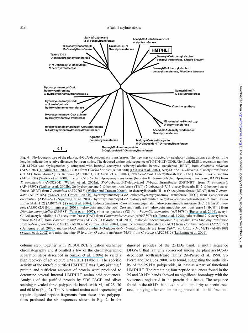
Fig. 4 Phylogenetic tree of the plant acyl-CoA-dependent acyltransferases. The tree was constructed by neighbor-joining distance analysis. Line
lengths indicate the relative distances between nodes. The deduced amino acid sequence of HMT/HLT (DDBJ/GenBank/EMBL accession number
AB181292) was phylogenetically compared with benzoyl coenzyme A:benzyl alcohol benzoyl transferase (BEBT) from Nicotiana tabacum
(AF500202) (D’Auria et al. 2002), BEBT from Clarkia breweri (AF500200) (D’Auria et al. 2002), acetyl-CoA:cis-3-hexen-1-ol acetyl transferase
(CHAT) from Arabidopsis thaliana (AF500201) (D’Auria et al. 2002), taxadien-5α-ol O-acetyltransferase (TAT) from Taxus cuspidata
(AF190130) (Walker et al. 2000c), taxoid C-13 O-phenylpropanoyltransferase (baccatin III:3-amino-3-phenylpropanoyltransferase, BAPT) from
T. canadensis (AY082804) (Walker et al. 2002a), 3′-N-debenzoyl-2′-deoxytaxol N-benzoyltransferase (DBTNBT) from T. canadensis
(AF466397) (Walker et al. 2002b), 2α-hydroxytaxane 2-O-benzoyltransferase (TBT) (2-debenzoyl-7,13-diacetylbaccatin III-2-O-benzoyl trans-
ferase, DBBT) from T. cuspidata (AF297618) (Walker and Croteau 2000a), 10-deacetylbaccatin III-10-O-acetyltransferase (DBAT) from T. cuspi-
data (AF193765) (Walker and Croteau 2000b), hydroxycinnamoyl-CoA quinate:hydroxycynnamoyl transferase (HQT) from Lycopersicon
esculentum (AJ582652) (Niggeweg et al. 2004), hydroxycinnamoyl-CoA:hydroxyanthranilate N-hydroxycinnamoyltransferase 2 from Avena
sativa (AsHHT2) (AB076981) (Yang et al. 2004), hydroxycinnamoyl-CoA:shikimate/quinate hydroxycinnamoyltransferase (HCT) from N. taba-
cum (AJ507825) (Hoffmann et al. 2003), hydroxycinnamoyl/benzoyl-CoA:anthranilate N-hydroxycinnamoyl/benzoyltransferase 1 (HCBT1) from
Dianthus caryophyllus (Z84383) (Yang et al. 1997), vinorine synthase (VS) from Rauvolfia serpentina (AJ556780) (Bayer et al. 2004), acetyl-
CoA:deacetylvindoline-4-O-acetyltransferase (DAT) from Catharanthus roseus (AF053307) (St-Pierre et al. 1998), salutaridinol 7-O-acetyltrans-
ferase (SALAT) from Papaver somniferum (AF339913) (Grothe et al. 2001), malonyl-CoA:anthocyanin 5-glucoside 4′′′-O-malonyltransferase
from Salvia splendens (Ss5MaT2) (AY383734) (Suzuki et al. 2004), agmatine coumaroyltransferase (ACT) from Hordeum vulgare (AY228552)
(Burhenne et al. 2003), malonyl-CoA:anthocyanidin 3-O-glucoside-6′′-O-malonyltransferase from Dahlia variabilis (Dv3MaT) (AF489108)
(Suzuki et al. 2002) and minovincinine 19-hydroxy-O-acetyltransferase (MAT) from C. roseus (AF253415) (Laflamme et al. 2001).

Alkaloid acyltransferase 237
at The Japanese S
ociety of Plant P
hysiologists on Novem
ber 5, 2010pcp.oxfordjournals.org
Dow
nloaded from
Isolation of cDNA encoding HMT/HLT
Since the 25 kDa polypeptide possessed a sequence motif
common for plant acyltransferases, combinations of degener-
ated primers encoding the determined amino acid sequences
from the 25 kDa band were designed for nested reverse tran-
scription–polymerase chain reaction (RT–PCR) amplification.
The overlapping 242 and 89 bp nucleotide fragments were
amplified specifically with combinations of the forward primer
P25K-3F-1 and the reverse primers P25K-1R or P25K-2R,
respectively (Fig. 3). This 242 bp amplified fragment was used
to isolate 14 cDNA clones by screening of the cDNA library
generated from the roots of 10-day-old L. albus, and sequence
analyses indicated that all clones were identical apart from
their different lengths. The full-length HMT/HLT cDNA
sequence was obtained by 5′-rapid amplification of cDNA end
(RACE) to yield a 1,641 bp poly(A) tail-containing clone with
a putative open reading frame (ORF) of 453 amino acids (Fig.
3). The calculated molecular mass of the encoded protein was
51.2 kDa, which was consistent with that of the purified protein
determined by SDS–PAGE in our previous study (Suzuki et al.
1994).
The ORF of the isolated cDNA contained the two peptide
sequences obtained from the 30 kDa band in the purified HMT/
HLT fraction (Fig. 3), although the cDNA was isolated solely
with the probe encoding the partial 25 kDa band. It seemed that
the 51.2 kDa protein might be cleaved to give 25 and 30 kDa
bands. Then we determined the N-terminal amino acid
sequences of these two 25 and 30 kDa bands isolated from the
gel. The N-terminal sequence (APQTQ) of the 30 kDa band
was identical to one of the peptides isolated upon trypsin diges-
tion, and perfectly matched the sequence from the second to the
sixth amino acid of the deduced sequence from the cDNA as
shown in Fig. 3. The N-terminal sequence (FILQH) of the
25 kDa band was identical to the deduced sequence from the
cDNA. From these results, we confirmed that the 25 and
30 kDa peptides were encoded by the single cDNA.
Phylogenetic analyses of HMT/HLT
Phylogenetic analysis of the deduced protein sequences
revealed that HMT/HLT is separated from the subfamily of
other alkaloid acyltransferases such as deacetylvindorine
acetyltransferase (St-Pierre et al. 1998) and salutaridinol 7-O-
acetyltransferase (Grothe et al. 2001) (Fig. 4). The most closely
related sequence to HMT/HLT is benzoyl-CoA:benzylalcohol
benzoyltransferase from Nicotiana tabacum (D’Auria et al.
2002) with 57% identity on an amino acid sequence level (Fig.
4). The sequence identities of HMT/HLT to other members of
this subfamily are 55% to benzoyl-CoA:benzylalcohol ben-
zoyltransferase from Clarkia brewei and 49% to acetyl-CoA:
cis-3-hexen-1-ol acetyltransferase from Arabidopsis thaliana
(D’Auria et al. 2002).
Catalytic activity of recombinant HMT/HLT produced in
Escherichia coli
To confirm that the isolated cDNA encodes the catalyti-
cally active HMT/HLT, the functional analysis was carried out
with the recombinant protein heterogeneously produced in E.
coli. The recombinant HMT/HLT protein was purified by glu-
tathione S-transferase (GST) tag affinity and subsequent cleav-
age of the tag (Fig. 5). By in vitro enzymatic assay using the
protein extracts of E. coli and the purified recombinant pro-
tein, the single product in the reaction using 13α-hydroxymul-
tiflorine and tigloyl-CoA as substrates was detected on high
performance liquid chromatography (HPLC), while no prod-
ucts were formed in negative control reactions using E. coli
extracts of the empty vector (Fig. 6a). This specific product
was identified as 13α-tigloyloxymultiflorine by liquid chroma-
tography-photodiode array detection-electrospray ionization-
mass spectrometry (LC-PDA-ESI/MS) analysis (Fig. 6b). In a
similar way, the activity for 13α-hydroxylupanine was also
confirmed with tigloyl-CoA as the acyl donor. These results
Fig. 5 Purification and SDS–PAGE analysis of recombinant HMT/
HLT produced in E. coli. The crude extracts from E. coli harboring
pGEX-HMT/HLT expressing the cDNA and the purified recombinant
HMT/HLT were separated by 10% SDS–PAGE. Protein bands were
detected by Coomassie brilliant blue staining. The arrowhead indi-
cates the position of the purified HMT/HLT protein by GST tag and
subsequent tag cleavage on a column.

Alkaloid acyltransferase238
at The Japanese S
ociety of Plant P
hysiologists on Novem
ber 5, 2010pcp.oxfordjournals.org
Dow
nloaded from
Fig. 6 Identification of the product of the reaction cat-
alyzed by the recombinant HMT/HLT by LC-PDA-ESI/
MS. (a) HPLC-PDA analysis of the reaction product of
the recombinant HMT/HLT. The reaction with 13α-
hydroxymultiflorine and tigloyl-CoA was carried out
with the protein extract of E. coli harboring pGEX-
HMT/HLT (upper chromatogram) or pGEX empty vec-
tor (lower chromatogram). HPLC analysis was per-
formed on a Mightysil RP-18 GP 150–4.6 column
(Kanto Chemical) with a mobile phase of 15% ace-
tonitrile and 0.1% trifluoroacetic acid at a flow rate of
0.5 ml min–1. The substrate and the product, 13α-tigloy-
loxymultiflorine, were monitored at 318 nm, the maxi-
mum absorbance of these compounds. The arrow
indicates 13α-tigloyloxymultiflorine specifically pro-
duced by the recombinant HMT/HLT. (b) LC-PDA-
ESI/MS analysis of the specific product, 13α-tigloy-
loxymultiflorine, by the recombinant HMT/HLT. The
product was identified by its quasi-molecular ion mass
at m/z 345 ([M+H]+) (upper spectrum) and its frag-
mented ion at m/z 245 ([M-C5H
7O
2]+) by the MS/MS
technique (lower spectrum).

Alkaloid acyltransferase 239
at The Japanese S
ociety of Plant P
hysiologists on Novem
ber 5, 2010pcp.oxfordjournals.org
Dow
nloaded from
indicated that the isolated cDNA actually encoded the func-
tional HMT/HLT.
Substrate specificity and inhibition of recombinant HMT/HLT
To investigate the substrate specificity of HMT/HLT, the
activity of purified recombinant HMT/HLT protein was
assayed with the five different acyl-CoA derivatives with 13α-
hydroxymultiflorine (Fig. 7a) and 13α-hydroxylupanine (Fig.
7b). Interestingly, the activities with benzoyl-CoA as acyl
donor were ∼1.8-fold higher than those with tigloyl-CoA with
two alkaloid aglycons. The activities with acetyl-CoA, propio-
nyl-CoA and 2-butenoyl-CoA were ∼2–20% of those with
tigloyl-CoA. The Km
values for 13α-hydroxymultiflorine, 13α-
hydroxylupanine, tigloyl-CoA and benzoyl-CoA were deter-
mined (Table 2). Regarding alkaloid aglycon, 13α-hydroxy-
multiflorine is a better substrate than 13α-hydroxylupanine,
judged from the Km
values for each alkaloid and for each acyl
donor with these alkaloids as co-substrate. However, no appar-
ent difference in Km
values was found between tigloyl-CoA and
benzoyl-CoA.
Phylogenetic analysis indicated that the sequence of
HMT/HLT was close to that of benzoyl-CoA:benzylalcohol
benzoyltransferase. Thus, to confirm whether HMT/HLT pos-
sesses the activity of benzoyl-CoA:benzylalcohol benzoyltrans-
ferase, we performed the assay for the reaction using benzoyl-
CoA and benzyl alcohol as the substrates with the recombinant
HMT/HLT. Determination of the possible reaction product,
benzylbenzoate, was carried out with gas chromatography–
mass spectrometry. However, no benzylbenzoate formation was
detected with the recombinant HMT/HLT (data not shown),
suggesting that HMT/HLT does not catalyze this reaction,
despite its close sequence similarity to that of benzoyl-CoA:
benzylalcohol benzoyltransferase.
The HMT/HLT activity was inhibited by the pre-treat-
ment of the enzyme protein with p-chloromercuribenzoic acid,
a sulfhydryl blocker, and diethylpyrocarbonate (DEPC), a histi-
dine blocker (Table 3). These results suggested that cysteine
and histidine residues are essential for the catalytic function of
HMT/HLT.
mRNA expression of the HMT/HLT gene
Southern blot analysis indicated that a small number (1–2
copies) of homologous genes are present in the genome of L.
albus (data not shown). Northern blot analysis was also carried
out as shown in Fig. 8. The mRNA expression of the HMT/
HLT gene was the highest in roots followed by that in hypocot-
yls. In contrast, HMT/HLT gene expression in leaves and coty-
ledons was at a low level. No expression was detected in young
developing leaves (data not shown).
Fig. 7 Substrate specificity of recombinant HMT/HLT. The acyl-
transfer reaction was carried out with recombinant HMT/HLT using
13α-hydroxymultiflorine or 13α-hydroxylupanine as acyl acceptors
and various acyl-CoAs as acyl donors. The identification of the reac-
tion products was made by LC-PDA-ESI/MS. The relative activities
were determined by intensities of the [M+H]+ ions of the respective
products. The data are expressed as the values relative to (a) 13α-
tigloyloxymultiflorine or (b) 13α-tigloyloxylupanine. The bar indi-
cates the data of duplicate assays.
Table 2 Summary of Km
values of HMT/HLT
Substrate for Km
Km
Co-substrate
13α-Hydroxymultiflorine 94 µM Tigloyl-CoA (0.3 mM)
13α-Hydroxylupanine 112 µM Tigloyl-CoA (0.3 mM)
Tigloyl-CoA 98 µM 13α-Hydroxymultiflorine (0.3 mM)
359 µM 13α-Hydroxylupanine (0.3 mM)
Benzoyl-CoA 93 µM 13α-Hydroxymultiflorine (0.3 mM)
405 µM 13α-Hydroxylupanine (0.3 mM)

Alkaloid acyltransferase240
at The Japanese S
ociety of Plant P
hysiologists on Novem
ber 5, 2010pcp.oxfordjournals.org
Dow
nloaded from
Discussion
Although the quinolizidine alkaloids form a large family
of plant secondary products, no genes encoding enzymes
involved in their biosynthesis have been cloned, presumably
because of the weak enzymatic activities found in plants. Only
a limited number of reports are available for detection of the
enzymatic activities in the cell-free protein extracts for quino-
lizidine alkaloid biosynthesis (Ohmiya et al. 1995). In the
present study, we have cloned, for the first time, the cDNA
encoding a biosynthetic enzyme for quinolizidine alkaloids.
This study could lead to the molecular investigation of metabo-
lism of this large family of plant secondary products.
The present study is also the first example of the molecu-
lar cloning of a tigloyl transferase from any biological source
including plants. Tigloyl esters are widely distributed in plants
as minor components of secondary products, e.g. alkaloids
(Lounasmaa 1988, Ohmiya et al. 1995, Saito and Murakoshi
1995). However, no tigloyl esters and tigloyl transferase have
been found in microorganisms and animals. The tigloyl moiety
is derived from isoleucine through several steps, as suggested
for tigloyl esters of tropane alkaloids in Datura plants (Leete
1973, McGaw and Woolley 1977). Tigloyl-CoA formed by this
pathway is assumed to be used for the esterification of a vari-
ety of aglycons including hydroxylated alkaloids by specific
transferases such as HMT/HLT.
The evolutionarily related plant acyl-CoA-dependent acyl-
transferases (EC 2.3.1.x) are referred to as the BAHD family
based on the names of the first genes isolated (BEAT AHCT
HCBT1 DAT) exhibiting characteristic sequence motifs (St-
Pierre and De Luca 2000). This family consists of a large
number of members; for example, there are approximately 60
BAHD gene family members in A. thaliana. However, only a
limited numbers of genes have been characterized with their
biochemical functions. Phylogenetically, the deduced amino
acid sequence of HMT/HLT is closely related to benzoyl-CoA:
benzyl alcohol benzoyltransferase (BEBT) from C. breweri and
N. tabacum, which is presumed to be involved in the forma-
tion of volatile esters in flowers (D’Auria et al. 2002). Indeed,
HMT/HLT accepted benzoyl-CoA instead of tigloyl-CoA for
acyltransfer to 13α-hydroxymultiflorine and 13α-hydroxylupa-
nine. However, HMT/HLT could not catalyze the benzoyl-
CoA:benzyl alcohol benzoyltransferase reaction, indicating a
relatively strict specificity of HMT/HLT regarding hydroxy-
Fig. 8 Organ-specific expression of the HMT/HLT gene by northern
blot analysis. Total RNA (40 µg) isolated from roots, hypocotyls, coty-
ledons and leaves of 10-day-old L. albus was electrophoresed on an
agarose gel (1.2%), transferred to a positively charged nylon mem-
brane and then hybridized with a 32P-labeled probe of the ORF region
of HMT/HLT cDNA. The final wash of the membrane was performed
in 0.5×SSPE and 0.1% SDS at 65°C. rRNA bands are shown as the
control of equal amounts of RNA loaded.
Table 3 Inhibition of HMT/HLT by sulfhydryl and histidine blockers
The enzyme protein was pre-treated with the inhibitor for 15 min prior to the standard acyltransferase reac-
tion as described in Materials and Methods. For the p-chloromercuribenzoic acid experiment, DTT was omit-
ted from the standard reaction mixture. For the DEPC experiment, after pre-treatment by DEPC, imidazole
(20 mM) was added to quench the reaction with DEPC.
InhibitorConcentration for 50% inhibition
Substrate
p-Chloromercuribenzoic acid 212 µM 13α-Hydroxymultiflorine and tigloyl-CoA
218 µM 13α-Hydroxylupanine and tigloyl-CoA
DEPC 23 mM 13α-Hydroxymultiflorine and tigloyl-CoA
37 mM 13α-Hydroxylupanine and tigloyl-CoA
Fig. 9 Multiple alignment of conserved motifs in the family of plant
acyltransferases. Arrowheads indicate possible functional residues of
cysteine and histidine that presumably are modified by p-chloromer-
curibenzoic acid and DEPC, respectively. The residues with a black
and gray background indicate those of perfect conservation and >50%
identity, respectively, in the 10 proteins. Abbreviations for the pro-
teins are described in the legend to Fig. 4.

Alkaloid acyltransferase 241
at The Japanese S
ociety of Plant P
hysiologists on Novem
ber 5, 2010pcp.oxfordjournals.org
Dow
nloaded from
lated aglycons. We have shown previously that the purified
HMT/HLT only utilized the 13α(axial)-hydroxylated tetracy-
clic alkaloids as acyl acceptors (Suzuki et al. 1994). Although
HMT/HLT, benzoyl-CoA:benzyl alcohol benzoyltransferase
and acety-CoA:cis-3-hexene-1-ol acetyltransferase from A.
thaliana (D’Auria et al. 2002) belong to the same subfamily of
the BAHD acyltransferases, the substrate specificities of these
enzymes are rather diverse. Similarly, the diverse substrate spe-
cificity in a single subfamily was reported for phylogenetically
closely related acyltransferases from Taxus species (Walker and
Croteau 2001). These results suggest that the substrate specifi-
city of the BAHD acyltransferase could evolve relatively easily.
Since Km
values of HMT/HLT for tigloyl-CoA and ben-
zoyl-CoA are nearly the same (Table 2) and the activity with
benzoyl-CoA is higher than that with tigloyl-CoA (Fig. 7), this
enzyme is more properly referred to as tigloyl/benzoyl-CoA:
(–)-13α-hydroxymultiflorine/(+)-13α-hydroxylupanine O-tigloyl/
benzoyltransferase. However, only a trace amount of benzoy-
loxylupanine was detected (Wink and Witte 1984) and no
apparent accumulation of benzoyloxymultiflorine was reported
in L. albus. This presumably is due to the limited supply of
benzoyl-CoA compared with tigloyl-CoA or different compart-
mentation of each acyl-CoA and HMT/HLT in this plant. The
accumulation of benzoyloxylupanine has been reported in other
taxonomically related Lupinus species, e.g. L. polyphyllus
(Wink et al. 1982b) and L. angustifolius (Strack et al. 1991,
Hirai et al. 2000). HMT/HLT presumably is responsible for
biosynthesis of benzoyloxylupanine in these species.
Previously we purified HMT/HLT as a single protein of
50 kDa (Suzuki et al. 1994). However, we noticed that 30 and
25 kDa bands were associated with the activity and could not
be separated. In the present study, we purified these two bands
to obtain sufficient amounts of enzyme protein for peptide
sequencing. The isolated cDNA encoded a single 50 kDa pro-
tein, which contained the partial peptide sequences from the 30
and 25 kDa bands. These results suggest that the cleavage of
the 50 kDa protein into 30 and 25 kDa bands is an artifact of
the purification procedure. Nevertheless, these fragments still
retained the catalytic activity, presumably remaining physi-
cally associated. A similar situation has been reported for
acetyl-CoA:deacetylvindoline 4-O-acetyltransferase from
Catharanthus roseus (periwinkle) (Power et al. 1990, St-Pierre
et al. 1998) and for hydroxycinnamoyl/benzoyl-CoA:anthrani-
late N-hydroxycinnamoyl/benzoyltransferase from Dianthus
caryophyllus (Yang et al. 1997). This could be a common char-
acter of the proteins belonging to this family.
As shown in Fig. 9, the members of the BAHD acyltrans-
ferase family have a few conserved sequence motifs. In addi-
tion to a highly conserved DFGWG motif as the signature of
this family, cysteine and histidine residues indicated in Fig. 9
are also conserved in all sequences. The histidine residue in the
HXXXD(G) motif has been postulated to function as a general
base in catalysis of the acyltransfer reaction (Shaw and Leslie
1991, St-Pierre et al. 1998). Indeed, the activity of HMT/HLT
was inhibited by DEPC, although the inhibition required a
higher concentration of DEPC than previously reported for
acyltransferases (for example, St-Pierre et al. 1998, St-Pierre
and De Luca 2000, Grothe et al. 2001). The requirement for a
higher concentration of DEPC could be due to the different
protein folding pattern that prevents the access of DEPC to the
catalytic histidine. The HMT/HLT activity was also inhibited
by p-chloromercuribenzoic acid, indicating the involvement of
a cysteine residue in the catalytic function. This could be the
conserved cysteine residue indicated in Fig. 9, that may be a
component of a catalytic triad as suggested previously (Grothe
et al. 2001).
The HMT/HLT gene was expressed specifically in roots
and hypocotyls, and only a limited level of expression has been
detected in cotyledons and leaves. This expression pattern is in
good agreement with the enzymatic activity of HMT/HLT, rep-
resenting ∼84% of the total activity found in roots and hypocot-
yls (Suzuki et al. 1994). These results indicate that roots and
hypocotyls are the main organs for the biosynthesis of quinoliz-
idine alkaloids in plant organs. Previously we also reported that
the distribution of several different acyltransferases in different
plant species is well correlated with the accumulation of
acylated quinolizidine alkaloids (Suzuki et al. 1994), suggest-
ing the importance of the terminal acyltransferase for alkaloid
accumulation patterns in diverse plant species.
Previously we detected HMT/HLT activities dominantly
in the particle fractions associated with the mitochondrial
marker enzymes (Suzuki et al. 1996). In fact, by the prediction
of the iPSORT program (http://psort.nibb.ac.jp/), the N-termi-
nal sequence of the deduced HMT/HLT showed the highest
score for translocation to the mitochondria. However, the
sequence of purified HMT/HLT started from the second amino
acid of the deduced sequence derived from cDNA, indicating
that no processing as an ordinary mitochondrial transit peptide
takes place. Since there are some examples of a lack of a cleav-
able mitochondrial pre-sequence (Sjöling and Glaser 1998), the
N-terminal sequence of HMT/HLT may act as a targeting sig-
nal to mitochondria without extensive processing. Otherwise
HMT/HLT may localize in another particle fraction such as the
peroxisomes, since there is a signal peptide-like sequence
(SHI) for peroxisome translocation in the C-terminus of the
HMT/HLT protein. This sequence shows a close similarity to
the characteristic C-terminal peroxisome targeting signal type 1
(Reumann 2004). Thus, it still remains an open question in
which subcellular compartment HMT/HLT localizes.
In some Lupinus species, the alkaloid-rich ‘bitter’ form
and the alkaloid-poor ‘sweet’ form are available. Recently, we
have investigated the alkaloid accumulation patterns, gene
expression profiles by cDNA-amplified fragment length poly-
morphism and the HMT/HLT activities of the ‘bitter’ and
‘sweet’ forms of L. angustifolius (Hirai et al. 2000). Although
striking differences in alkaloid accumulation were observed
between the two forms, no significant difference in the HMT/
HLT activity was seen in the hypocotyl cell-free extracts of

Alkaloid acyltransferase242
at The Japanese S
ociety of Plant P
hysiologists on Novem
ber 5, 2010pcp.oxfordjournals.org
Dow
nloaded from
these forms. These results suggest that HMT/HLT is regulated
independently from the genetic factor that determines the alka-
loid accumulation in ‘bitter’ and ‘sweet’ forms.
In conclusion, we have cloned and characterized the
cDNA encoding HMT/HLT responsible for determination of
alkaloid accumulation patterns in L. albus. The present findings
can serve as the molecular information for a better understand-
ing and a further metabolic engineering of quinolizidine alka-
loid metabolism in plants.
Materials and Methods
Chemicals
(–)-13α-Hydroxymultiflorine and (+)-13α-hydroxylupanine used
in this study were from our laboratory stock. Tigloyl-, acetyl-, propio-
nyl-, 2-butenoyl- and benzoyl-CoA were purchased from Sigma-
Aldrich (St Louis, MO, U.S.A.). The molecular weight markers used
for SDS–PAGE were purchased from Amersham Biosciences (Piscata-
way, NJ, U.S.A.). Bradford protein dye reagent was purchased from
Bio-Rad (Hercules, CA, U.S.A.). Restriction enzymes were purchased
from TaKaRa (Ohtsu, Japan) or Toyobo (Osaka, Japan). T4 DNA
ligase was purchased from Promega (Madison, WI, U.S.A.) or Nova-
gen EMD Biosciences (Madison, WI, U.S.A.). DNA amplification by
PCR using Pyrobest or Ex Taq DNA polymerase (TaKaRa) was car-
ried out in an iCycler thermocycler (Bio-Rad). Other chemicals and
enzymes were of the highest grade available.
Plant materials
The seeds of L. albus Forsk were germinated in moistened ver-
miculite in daylight in a greenhouse at ∼25°C as described previously
(Suzuki et al. 1994).
Purification of HMT/HLT
All procedures were performed at 4°C. Crude protein was
extracted from roots, hypocotyls and epicotyls (2.47 kg, fresh weight)
of 13-day-old L. albus seedlings as described (Suzuki et al. 1994). The
precipitate appearing between 30 and 80% (NH4)2SO
4 was collected
and dissolved in buffer A (20 mM sodium phosphate, pH 7.0, contain-
ing 10 mM 2-mercaptoethanol and 1.5 M ammonium sulfate). The
proteins were absorbed on butyl FF (Amersham) equilibrated with
buffer A and were eluted at a flow rate of 0.8 ml min–1 with a linear
gradient of 1.5–1.0 M ammonium sulfate. The fractions with the
enzyme activity were collected and concentrated. The proteins were
then separated on Superdex 200 pg (Amersham) equilibrated with
buffer B (20 mM sodium phosphate, pH 6.3, containing 10 mM 2-mer-
captoethanol) at a flow rate of 1.0 ml min–1. The fractions with the
enzyme activity were collected and then applied to SP Sepharose XL
(Amersham). The proteins were eluted at a flow rate of 2.0 ml min–1
with a linear gradient of 0–0.5 M NaCl, and the fractions with the
enzyme activity were collected and concentrated. The portion (0.125
volume) of the concentrated enzyme solution was then separated on
Superdex 200 HR (Amersham) equilibrated with buffer B at a flow
rate of 0.3 ml min–1. The fractions with the enzyme activity were col-
lected and then applied to RESOURCE S (Amersham) equilibrated
with buffer B. Proteins were eluted at a flow rate of 4.0 ml min–1 with
a linear gradient of 0–0.5 M NaCl.
Determination of internal and N-terminal amino acid sequences
After the final step of protein purification by RESOURCE S, the
fraction with the highest enzymatic activity was subjected to 12.5%
SDS–PAGE. Bands were detected by silver staining. The three
observed bands of 25, 30 and 60 kDa proteins were subjected to deter-
mination of the internal amino acid sequences. The respective bands
were digested in-gel by trypsin and then separated by reverse-phase
HPLC. HPLC separation was performed on a TSKgel ODS-80Ts QA
(2.0×250 mm) column (Tosoh, Tokyo, Japan) with a gradient of 0–
90% acetonitrile in 0.1% trifluoroacetic acid (TFA) at a flow rate of
0.2 ml min–1. Separated peptides were monitored at 210 and 280 nm.
Sequencing of the peptides was performed using a Procise 494 cLC
(Applied Biosystems, Foster City, CA, U.S.A.) or Hewlett-Packard
G1005A (Hewlett Packard, Palo Alto, CA, U.S.A.) protein sequenc-
ing system. To determine the N-terminal amino acid sequence of the
25 and 30 kDa bands, the peptides in the gel of SDS–PAGE were
transferred to a PVDF membrane and then analyzed by protein
sequencer, Procise 494 HT (Applied Biosystems).
Molecular cloning of cDNA
Isolation of partial fragments encoding the 25 kDa peptide by
RT–PCR—Total RNA was extracted from roots of 10-day-old L. albus
with the RNeasy Plant Mini Kit (QIAGEN, Hilden, Germany) or TRI-
zol reagents (Invitrogen, Carlsbad, CA, U.S.A.). mRNA was purified
using an mRNA Purification Kit (Amersham). From total RNA
extracted from roots, first-strand cDNA was synthesized using AMV
reverse transcriptase XL (TaKaRa) with the Oligo dT-3sites Adaptor
Primer (TaKaRa). The degenerate primers were designed from the
amino acid sequence of the 25 kDa peptide. The first PCR was per-
formed using the forward primers P25K-1F, GARYTNGAYGAYYT-
NTTYAA (corresponding to the amino acid sequence of ELDDLFK);
P25K-2F, GAYGTNGAYTTYGGNTGGGGNAA (corresponding to
the amino acid sequence of DVDFGWGK); or P25K-3F-1, CCNWS-
NTAYTTYTAYAAYGA (corresponding to the amino acid sequence of
PSYFYND) and the reverse primer of the 3sites Adaptor Primer
(TaKaRa) which hybridizes to a region synthesized by the Oligo dT-
3sites Adaptor Primer. With the forward primer corresponding to these
templates and the other reverse primers, which have complementary
sequences to those of the forward primers, P25K-1R, P25K-2R and
P25K-3R-1 (corresponding to P25K-1F, P25K-2F and P25K-3F-1,
respectively), the nested PCR was performed with all possible combi-
nations of the forward and reverse primers. The amplified fragments
were cloned into pT7Blue T-vector (Novagen) by TA-cloning, and the
nucleotide sequences of the inserts were determined.
Isolation of the full-length clone by cDNA library screening and
5′-RACE—The cDNA library was constructed with the λZAPII vector
(Stratagene, La Jolla, CA, U.S.A.) from mRNA of L. albus, and
2.0×10⁵ plaques of primary cDNA library were screened by the PCR-
amplified fragment. Plaques were blotted onto positively charged
nylon membranes, Hybond N+ (Amersham). Duplicate filters were
hybridized with the 32P-labeled probe prepared by the Random Primer
DNA Labeling Kit Ver.2 (TaKaRa) using the 242 bp nucleotide frag-
ment, which was amplified specifically by RT–PCR with the combina-
tion of P25K-3F-1 and P25K-1R as a template. The final post-
hybridization wash was performed in 0.2×SSPE (Sambrook et al.
1989) and 0.1% SDS at 65°C. For extension of the 5′-terminus of the
cDNA, 5′-RACE was performed using the 5′-Full RACE Core Set
(TaKaRa).
Expression and purification of recombinant HMT/HLT
BamHI sites were created on both ends of the HMT/HLT coding
region by PCR using the two primers: BamHI F, CGCGGATCCAT-
GGCTCCCCAAACTCAATCTCTA; and BamHI R, CGCGGATCCT-
CAAATGTGTGACCTTAATATTTG. The engineered cDNA fragment
was inserted into the BamHI site of pGEX-6P-2 (Amersham) with the
correct orientation confirmed by sequence analysis as designated
pGEX-HMT/HLT, which gives a recombinant gene product with an N-

Alkaloid acyltransferase 243
at The Japanese S
ociety of Plant P
hysiologists on Novem
ber 5, 2010pcp.oxfordjournals.org
Dow
nloaded from
terminal GST protein tag. E. coli BL21 cells harboring pGEX-HMT/
HLT and empty pGEX were grown at 20°C in 2×YT-ampicillin
(100 µg ml–1) liquid medium to an OD600
= 0.7–1.0. The solution of
0.1 mM isopropyl-β-D-thiogalactoside was added immediately for
inducible production of recombinant protein. The cells were suspended
in 1×phosphate-buffered saline [140 mM NaCl, 2.7 mM KCl, 10 mM
Na2HPO
4 and 1.8 mM KH
2PO
4 (pH 7.3)] and disrupted by sonication.
After the addition of Trition X-100 to 1% concentration and following
centrifugation, the supernatant was used as crude protein extracts. For
purification of the recombinant HMT/HLT, a GSTrap column
(Amersham), whose resin has glutathione as ligand resulting in affin-
ity for GST, was used. GST tags were removed from GST fusion
recombinant proteins by in-column digestion using PreScission Pro-
tease (Amersham).
Assay of enzymatic activity
The HMT/HLT activity was determined by detection of the ester
alkaloid formed from alkaloid substrates and acyl-CoA derivatives by
enzymatic reaction. The standard reaction mixture consisted of
100 mM potassium phosphate (pH 8.0), 0.5 mM EDTA, 1 mM dithio-
threitol (DTT), 0.15 mM alkaloid substrate, 0.15 mM acyl-CoA deriv-
ative and protein. After incubation for 10–60 min at 30°C, the reaction
was terminated by addition of a 0.67 volume of methanol. The reaction
mixture was analyzed directly by HPLC. HPLC analysis was per-
formed on a Mightysil RP-18 GP 150–4.6 (5 µm) column (Kanto
Chemical, Tokyo, Japan) with a mobile phase of 15% acetonitrile and
20 mM sodium phosphate (pH 5.5) at a flow rate of 0.5–1.0 ml min–1.
The formation of 13α-hydroxymultiflorine and 13α-hydroxylupanine
derivatives was monitored at 327 and 220 nm, respectively, as
described (Saito et al. 1989). The identification of products was con-
firmed further by LC-ESI/MS.
To elucidate the substrate specificity, the reaction with recom-
binant HMT/HLT protein was carried out with the various acyl-CoA
derivatives and the aglycons. The products were identified with LC-
ESI/MS
LC-PDA-ESI/MS analysis
The reaction mixtures were analyzed by an LC-PDA-ESI/MS
system consisting of a Finnigan LCQ DECA mass spectrometer
(Thermo Quest, San Jose, CA, U.S.A.) and an Agilent HPLC 1100
series (Agilent technologies, Palo Alto, CA, U.S.A.). LC separations
were performed on a Mightysil RP-18 GP 150–4.6 (5 µm) column
(Kanto Chemical) with a mobile phase of 15% acetonitrile and 0.1%
TFA at a flow rate of 0.5 ml min–1 using UV photodiode array detec-
tion. Nitrogen was used as sheath gas for positive ion ESI/MS per-
formed at a capillary temperature and voltage of 320°C and 5.0 kV,
respectively. The tube lens offset was set at 10.0 V. Full scan mass
spectra were acquired from 50 to 1,000 m/z at two scans s–1. Tandem
MS analysis was carried out with helium as collision gas. The normal-
ized collision energy was set to 40%. The specific products were iden-
tified from the theoretical unit mass for [M+H]+ in +ESI: 13-
tigloyloxymultiflorine, 345; 13-acetyloxymultiflorine, 305; 13-propio-
nyloxymultiflorine, 319; 13-(2-butenoyl)oxymultiflorine, 331; 13-ben-
zoyloxymultiflorine, 367; 13-tigloyloxylupanine, 347; 13-
acetyloxylupanine, 307; 13-propionyloxylupanine, 321; 13-(2-buten-
oyl)oxylupanine, 333; and 13-benzoyloxylupanine, 369. Fragmented
ions derived from these quasi-molecular ions were also identified by
the MS/MS technique. The relative activities regarding each acyl-CoA
were calculated by relative integration of ion areas of quasi-molecular
ions compared with the products of the tigloyl transferase reaction.
Northern and Southern blot analyses
For northern blot analysis, 40 µg of total RNA isolated from
leaves, developing (folded) leaves, cotyledons, hypocotyls and roots
was denatured and separated in a formaldehyde agarose (1.2%) gel.
For Southern blot analysis, genomic DNA was extracted from L. albus
using the DNeasy Plant Mini Kit (QIAGEN). Aliquots of 20 µg of
genomic DNA were digested with BamHI or EcoRV and separated on
a 0.8% agarose gel. After transfer to a Hybond N+ membrane, hybridi-
zation was carried out with the 32P-labeled probe prepared from the
ORF of HMT/HLT cDNA using the Random Primer DNA Labeling
Kit Ver.2 (TaKaRa). The final washes on post-hybridization were per-
formed in 0.5× SSPE and 0.1% SDS at 65°C. Hybridization signals
were detected with a STORM 860 image analyzer (Amersham).
Miscellaneous techniques
All recombinant DNA technology principally followed the meth-
ods of Sambrook et al. (1989). DNA sequencing was performed by the
dideoxy chain termination method. Phylogenetic analysis was carried
out by CLUSTAL W packaged in DNASpace Ver. 3.5 (Hitachi Soft-
ware, Tokyo, Japan), and the tree was drawn by DNASpace. SDS–
PAGE and subsequent staining of proteins were performed as
described previously (Suzuki et al. 1994). Gas chromatography–mass
spectrometry for detection of benzylbenzoate was carried out as
described previously (Hirai et al. 2000, D’Auria et al. 2002).
Acknowledgments
This work was supported, in part, by Grants-in-Aid for Scien-
tific Research from the Ministry of Education, Culture, Sports, Sci-
ence and Technology, Japan, by CREST of Japan Science and
Technology Agency (JST) and by Research for the Future Program
(00L01605) ‘Molecular mechanisms on regulation of morphogenesis
and metabolism leading to increased plant productivity’. We thank
CREST-Plant Molecular Science Satellite Laboratory in the Life Sci-
ence Research Support Center of Akita Prefectural University for
DNA sequencing.
References
Bayer, A., Ma, X. and Stöckigt, J. (2004) Acetyltransfer in natural product bio-
synthesis—functional cloning and molecular analysis of vinorine synthase.
Bioorg. Med. Chem. 12: 2787–2795.
Burhenne, K., Kristensen, B.K. and Rasmussen, S.K. (2003) A new class of N-
hydroxycinnamoyltransferases. Purification, cloning, and expression of a bar-
ley agmatine coumaroyltransferase (EC 2.3.1.64). J. Biol. Chem. 278: 13919–
13927.
D’Auria, J.C., Chen, F. and Pichersky, E. (2002) Characterization of an acyl-
transferase capable of synthesizing benzylbenzoate and other volatile esters in
flowers and damaged leaves of Clarkia breweri. Plant Physiol. 130: 466–476.
Grothe, T., Lenz, R. and Kutchan, T.M. (2001) Molecular characterization of the
salutaridinol 7-O-acetyltransferase involved in morphine biosynthesis in
opium poppy Papaver somniferum. J. Biol. Chem. 276: 30717–30723.
Hirai, M.Y., Suzuki, H., Yamazaki, M. and Saito, K. (2000) Biochemical and
partial molecular characterization of bitter and sweet forms of Lupinus angus-
tifolius, an experimental model for study of molecular regulation of quinolizi-
dine alkaloid biosynthesis. Chem. Pharm. Bull. 48: 1458–1461.
Hoffmann, L., Maury, S., Martz, F., Geoffroy, P. and Legrand, M. (2003) Purifi-
cation, cloning, and properties of an acyltransferase controlling shikimate and
quinate ester intermediates in phenylpropanoid metabolism. J. Biol. Chem.
278: 95–103.
Laflamme, P., St-Pierre, B. and De Luca, V. (2001) Molecular and biochemical
analysis of a Madagascar periwinkle root-specific minovincinine-19-
hydroxy-O-acetyltransferase. Plant Physiol. 125: 189–198.
Leete, E. (1973) Biosynthetic conversion of α-methylbutyric acid to tiglic acid
in Datura meteloides. Phytochemistry 12: 2203–2205.

Alkaloid acyltransferase244
at The Japanese S
ociety of Plant P
hysiologists on Novem
ber 5, 2010pcp.oxfordjournals.org
Dow
nloaded from
Lounasmaa, M. (1988) The Alkaloids: The Tropane Alkaloids. Edited by Brossi,
A. Vol. 33. Academic Press, Inc., CA.
McGaw, B.A. and Woolley, J.G. (1977) Non-reversibility of the isoleucine-ace-
tate pathway in Datura. Phytochemistry 16: 1711–1713.
Niggeweg, R., Michael, A.J. and Martin, C. (2004) Engineering plants with
increased levels of the antioxidant chlorogenic acid. Nature Biothechnol. 22:
746–754.
Ohmiya, S., Saito, K. and Murakoshi, I. (1995) The Alkaloids: Lupine Alka-
loids. Edited by Cordell, G.A. Vol. 47. Academic Press, Inc., CA.
Power, R., Kurz W.G. and De Luca, V. (1990) Purification and characterization
of acetylcoenzyme A:deacetylvindoline 4-O-acetyltransferase from Catha-
ranthus roseus. Arch. Biochem. Biophys. 279: 370–376.
Reumann, S. (2004) Specification of the peroxisome targeting signals type 1 and
type 2 of plant peroxisomes by bioinformatics analyses. Plant Physiol. 135:
783–800.
Roberts, M.F. and Wink, M. (1998) Alkaloids Biochemistry, Ecology, and
Medicinal Applications. Plenum Press, New York.
Saito, K., Kobayashi, K., Ohmiya, S., Otomasu, H. and Murakoshi, I. (1989)
Analysis of lupine alkaloids in plants by high-performance liquid chromatog-
raphy. J. Chromatogr. A. 462: 333–340.
Saito, K. and Murakoshi, I. (1995) Studies in Natural Chemistry: Chemistry,
Biochemistry and Chemotaxonomy of Lupine Alkaloids in Leguminosae.
Edited by Rahman, A.-U. Vol. 15. Elsevier Science, Oxford.
Saito, K., Suzuki, H., Takamatsu, S. and Murakoshi, I. (1993) Acyltransferases
for lupin alkaloids in Lupinus hirsutus. Phytochemistry. 32: 87–91.
Sambrook, J., Fritsch, E.F. and Maniatis, T. (1989) Molecular Cloning: A Labo-
ratory Manual, 2nd edn. Cold Spring Harbor Laboratory Press, Cold Spring
Harbor, NY.
Shaw, W.V. and Leslie, A.G. (1991) Chloramphenicol acetyltransferase. Annu.
Rev. Biophys. Biophys. Chem. 20: 363–386.
Sjöling, S. and Glaser, E. (1998) Mitochondrial targeting peptides in plants.
Trends Plant Sci. 3: 136–140.
St-Pierre, B. and De Luca, V. (2000) Recent Advances in Phytochemistry Evolu-
tion of Metabolic Pathways. Edited by John, R.I., Romeo, T., Varin, L. and
De Luca, V. Vol. 34. Elsevier Science, Oxford.
St-Pierre, B., Laflamme, P., Alarco, A.-M. and De Luca, V. (1998) The terminal
O-acetyltransferase involved in vindoline biosynthesis defines a new class of
proteins responsible for coenzyme A-dependent acyl transfer. Plant J. 14:
703–713.
Strack, D., Becher, A., Brall, S. and Witte, L. (1991) Quinolizidine alkaloids and
the enzymatic syntheses of their cinnamic and hydroxycinnamic acid esters in
Lupinus angustifolius and L. luteus. Phytochemistry 30: 1493–1498.
Suzuki, H., Koike, Y., Murakoshi, I. and Saito, K. (1996) Subcellular localiza-
tion of acyltransferases for quinolizidine alkaloid biosynthesis in Lupinus.
Phytochemistry 42: 1557–1562.
Suzuki, H., Murakoshi I. and Saito, K. (1994) A novel O-tigloyltransferase for
alkaloid biosynthesis in plants. Purification, characterization, and distribution
in Lupinus plants. J. Biol. Chem. 269: 15853–15860.
Suzuki, H., Nakayama, T., Yonekura-Sakakibara, K., Fukui, Y., Nakamura, N.,
Yamaguchi, M.A., Tanaka, Y., Kusumi, T. and Nishino, T. (2002) cDNA
cloning, heterologous expressions, and functional characterization of malo-
nyl-coenzyme A:anthocyanidin 3-O-glucoside-6′′-O-malonyltransferase from
dahlia flowers. Plant Physiol. 130: 2142–2151.
Suzuki, H., Sawada, S., Watanabe, K., Nagae, S., Yamaguchi, M.A., Nakayama,
T. and Nishino, T. (2004) Identification and characterization of a novel
anthocyanin malonyltransferase from scarlet sage (Salvia splendens) flowers:
an enzyme that is phylogenetically separated from other anthocyanin acyl-
transferases. Plant J. 38: 994–1003.
Tang, W. and Eisenbrand, G. (1992) Chinese Drugs of Plant Origin, Chemistry,
Pharmacology, and Use in Traditional and Modern Medicine. Springer-Ver-
lag, Berlin.
Walker, K. and Croteau, R. (2001) Taxol biosynthetic genes. Phytochemistry 58:
1–7.
Walker, K. and Croteau, R. (2000a) Taxol biosynthesis: molecular cloning of a
benzoyl-CoA:taxane 2α-O-benzoyltransferase cDNA from Taxus and func-
tional expression in Escherichia coli. Proc. Natl. Acad. Sci. USA 97: 13591–
13596.
Walker, K. and Croteau, R. (2000b) Molecular cloning of a 10-deacetylbaccatin
III-10-O-acetyl transferase cDNA from Taxus and functional expression in
Escherichia coli. Proc. Natl. Acad. Sci. USA 97: 583–587.
Walker, K., Fujisaki, S., Long, R. and Croteau, R. (2002a) Molecular cloning
and heterologous expression of the C-13 phenylpropanoid side chain-CoA
acyltransferase that functions in taxol biosynthesis. Proc. Natl. Acad. Sci.
USA 99: 12715–12720.
Walker, K., Long, R. and Croteau, R. (2002b) The final acylation step in taxol
biosynthesis: cloning of the taxoid C13-side-chain N-benzoyltransferase from
Taxus. Proc. Natl. Acad. Sci. USA 99: 9166–9171.
Walker, K., Schoendorf, A. and Croteau, R. (2000c) Molecular cloning of a
taxa-4(20), 11(12)-dien-5α-ol-O-acetyl transferase cDNA from Taxus and
functional expression in Escherichia coli. Arch. Biochem. Biophys. 374: 371–
380.
Wink, M. and Hartmann, T. (1982a) Enzymatic synthesis of quinolizidine alka-
loid esters: a tigloyl-CoA:13-hydroxylupanine O-tigloyltransferase from
Lupinus albus L. Planta 156: 560–565.
Wink, M., Schiebel, H.M., Witte, L. and Hartmann, T. (1982b) Quinolizidine
alkaloids from plants and their cell suspension culture. Planta Med. 44: 15–
20.
Wink, M. and Witte, L. (1984) Turnover and transport of quinolizidine alka-
loids: diurnal variation of lupanine in the phloem sap, leaves and fruits of
Lupinus albus L. Planta 161: 519–524.
Yang, Q., Reinhard, K., Schiltz, E. and Matern, U. (1997) Characterization and
heterologous expression of hydroxycinnamoyl/benzoyl-CoA:anthranilate N-
hydroxycinnamoyl/benzoyltransferase from elicited cell cultures of carna-
tion, Dianthus caryophyllus L. Plant Mol. Biol. 35: 777–789.
Yang, Q., Trinh, H.X., Imai, S., Ishihara, A., Zhang, L., Nakayashiki, H., Tosa,
Y. and Mayama, S. (2004) Analysis of the involvement of hydroxyanthrani-
late hydroxycinnamoyltransferase and caffeoyl-CoA 3-O-methyltransferase
in phytoalexin biosynthesis in oat. Mol. Plant Microbe Interact. 17: 81–89.
(Received October 12, 2004; Accepted November 10, 2004)
Copyright © 2022 FDOKUMEN