Modulatory Role of Sensory Innervation on Hair Follicle Stem Cell Progeny during Wound Healing of...
17
Modulatory Role of Sensory Innervation on Hair Follicle Stem Cell Progeny during Wound Healing of the Rat Skin EduardoMartı´nez-Martı´nez 1 *, Claudio I. Galva ´ n-Herna ´ ndez 1 , Brenda Toscano-Ma ´ rquez 1 , Gabriel Gutie ´ rrez-Ospina 1,2 * 1 Departamento de Biologı ´a Celular y Fisiologı ´a and Grupo Multidisciplinario de Investigacio ´ n en Ce ´ lulas Troncales IMPULSA 02, Instituto de Investigaciones Biome ´ dicas, Universidad Nacional Auto ´ noma de Me ´ xico, Coyoaca ´n, Distrito Federal, Me ´xico, 2 Coordinacio ´ n de Psicofisiologı ´a, Facultad de Psicologı ´a, Colonia Copilco Universidad, Coyoaca ´n, Distrito Federal, Me ´xico Abstract Background: The bulge region of the hair follicle contains resident epithelial stem cells (SCs) that are activated and mobilized during hair growth and after epidermal wounding. However, little is known about the signals that modulate these processes. Clinical and experimental observations show that a reduced supply of sensory innervation is associated with delayed wound healing. Since axon terminals of sensory neurons are among the components of the bulge SC niche, we investigated whether these neurons are involved in the activation and mobilization of the hair stem cells during wound healing. Methodology/Principal Findings: We used neonatal capsaicin treatment to reduce sensory terminals in the rat skin and performed morphometric analyses using design-based stereological methods. Epithelial proliferation was analyzed by quantifying the number of bromodeoxyuridine-labeled (BrdU + ) nuclei in the epidermis and hair follicles. After wounding, the epidermis of capsaicin-treated rats presented fewer BrdU + nuclei than in control rats. To assess SC progeny migration, we employed a double labeling protocol with iododeoxyuridine and chlorodeoxyuridine (IdU + /CldU + ). The proportion of double-labeled cells was similar in the hair follicles of both groups at 32 h postwounding. IdU + /CldU + cell proportion increased in the epidermis of control rats and decreased in treated rats at 61 h postwounding. The epidermal volume immunostained for keratin 6 was greater in treated rats at 61 h. Confocal microscopy analysis revealed that substance P (SP) and calcitonin gene-related peptide (CGRP) receptor immunoreactivity were both present in CD34 + and BrdU-retaining cells of the hair follicles. Conclusions/Significance: Our results suggest that capsaicin denervation impairs SC progeny egress from the hair follicles, a circumstance associated with a greater epidermal activation. Altogether, these phenomena would explain the longer times for healing in denervated skin. Thus, sensory innervation may play a functional role in the modulation of hair SC physiology during wound healing. Citation: Martı ´nez-Martı ´nez E, Galva ´n-Herna ´ndez CI, Toscano-Ma ´ rquez B, Gutie ´rrez-Ospina G (2012) Modulatory Role of Sensory Innervation on Hair Follicle Stem Cell Progeny during Wound Healing of the Rat Skin. PLoS ONE 7(5): e36421. doi:10.1371/journal.pone.0036421 Editor: Mauricio Rojas, University of Pittsburgh, United States of America Received June 20, 2011; Accepted March 31, 2012; Published May 4, 2012 Copyright: ß 2012 Martı ´nez-Martı ´nez et al. This is an open-access article distributed under the terms of the Creative Commons Attribution License, which permits unrestricted use, distribution, and reproduction in any medium, provided the original author and source are credited. Funding: This work was supported by grants 38615N, 45872M, and 53194 from Consejo Nacional de Ciencia y Tecnologı ´a (CONACYT), grant IN215208 from Direccio ´ n General de Asuntos del Personal Acade ´mico, UNAM (DGAPA), and grant IMPULSA-02 from Coordinacio ´ n de la Investigacio ´ n Cientı ´fica, UNAM (CIC- UNAM) to GGO. EMM and BTM were fellows of CONACYT. EMM also received support from Direccio ´ n General de Estudios de Posgrado-UNAM and IMPULSA-02. CIGH was a fellow of the Fundacio ´ n Lorena Alejandra Gallardo I.A.P. The funders had no role in study design, data collection and analysis, decision to publish, or preparation of the manuscript. Competing Interests: The authors have declared that no competing interests exist. * E-mail: [email protected] (EMM); [email protected] (GGO) Introduction Both clinical and experimental observations indicate that sensory neurons are involved in the process of wound repair. A reduction of the cutaneous sensory innervation is generally associated with longer times for healing [1,2,3,4]. These findings imply that the function of dorsal root ganglion (DRG) or sensory neurons is not restricted to receive and transduce mechanical and noxius stimuli (afferent function). Over the past few years, a growing body of evidence documents that sensory nerves regulate physiological and patho- logical process in the skin and other tissues by activating target cells that express specific receptors for neuromediators (efferent function) [5,6]. With the recent development of antibodies against sensory neuropeptides and other neuronal markers, it has been possible to establish that sensory nerve terminals occupy very precise positions on skin compartments rather than a random distribution. Both epidermis and hair follicles are predominantly innervated by small sensory neurons with unmyelinated C-fibers and poorly myelinated Ad-fibers. The nerve terminals of these neurons are characterized for its ability to release a variety of neuromediators which include neuropeptides such as CGRP and SP. Thus, this anatomical relationship raises the question whether nerve terminals from the DRG may be involved in maintaining epithelial tissue homeostasis. Epithelial homeostasis is a process that depends on the presence of stem cells (SCs). Recent studies have documented that there is a group of SCs residing in the region called the bulge which PLoS ONE | www.plosone.org 1 May 2012 | Volume 7 | Issue 5 | e36421
Transcript of Modulatory Role of Sensory Innervation on Hair Follicle Stem Cell Progeny during Wound Healing of...
Modulatory Role of Sensory Innervation on Hair FollicleStem Cell Progeny during Wound Healing of the Rat SkinEduardo Martınez-Martınez1*, Claudio I. Galvan-Hernandez1, Brenda Toscano-Marquez1,
Gabriel Gutierrez-Ospina1,2*
1Departamento de Biologıa Celular y Fisiologıa and Grupo Multidisciplinario de Investigacion en Celulas Troncales IMPULSA 02, Instituto de Investigaciones Biomedicas,
Universidad Nacional Autonoma de Mexico, Coyoacan, Distrito Federal, Mexico, 2Coordinacion de Psicofisiologıa, Facultad de Psicologıa, Colonia Copilco Universidad,
Coyoacan, Distrito Federal, Mexico
Abstract
Background: The bulge region of the hair follicle contains resident epithelial stem cells (SCs) that are activated andmobilized during hair growth and after epidermal wounding. However, little is known about the signals that modulate theseprocesses. Clinical and experimental observations show that a reduced supply of sensory innervation is associated withdelayed wound healing. Since axon terminals of sensory neurons are among the components of the bulge SC niche, weinvestigated whether these neurons are involved in the activation and mobilization of the hair stem cells during woundhealing.
Methodology/Principal Findings: We used neonatal capsaicin treatment to reduce sensory terminals in the rat skin andperformed morphometric analyses using design-based stereological methods. Epithelial proliferation was analyzed byquantifying the number of bromodeoxyuridine-labeled (BrdU+) nuclei in the epidermis and hair follicles. After wounding,the epidermis of capsaicin-treated rats presented fewer BrdU+ nuclei than in control rats. To assess SC progeny migration,we employed a double labeling protocol with iododeoxyuridine and chlorodeoxyuridine (IdU+/CldU+). The proportion ofdouble-labeled cells was similar in the hair follicles of both groups at 32 h postwounding. IdU+/CldU+ cell proportionincreased in the epidermis of control rats and decreased in treated rats at 61 h postwounding. The epidermal volumeimmunostained for keratin 6 was greater in treated rats at 61 h. Confocal microscopy analysis revealed that substance P (SP)and calcitonin gene-related peptide (CGRP) receptor immunoreactivity were both present in CD34+ and BrdU-retaining cellsof the hair follicles.
Conclusions/Significance: Our results suggest that capsaicin denervation impairs SC progeny egress from the hair follicles,a circumstance associated with a greater epidermal activation. Altogether, these phenomena would explain the longertimes for healing in denervated skin. Thus, sensory innervation may play a functional role in the modulation of hair SCphysiology during wound healing.
Citation: Martınez-Martınez E, Galvan-Hernandez CI, Toscano-Marquez B, Gutierrez-Ospina G (2012) Modulatory Role of Sensory Innervation on Hair Follicle StemCell Progeny during Wound Healing of the Rat Skin. PLoS ONE 7(5): e36421. doi:10.1371/journal.pone.0036421
Editor: Mauricio Rojas, University of Pittsburgh, United States of America
Received June 20, 2011; Accepted March 31, 2012; Published May 4, 2012
Copyright: � 2012 Martınez-Martınez et al. This is an open-access article distributed under the terms of the Creative Commons Attribution License, whichpermits unrestricted use, distribution, and reproduction in any medium, provided the original author and source are credited.
Funding: This work was supported by grants 38615N, 45872M, and 53194 from Consejo Nacional de Ciencia y Tecnologıa (CONACYT), grant IN215208 fromDireccion General de Asuntos del Personal Academico, UNAM (DGAPA), and grant IMPULSA-02 from Coordinacion de la Investigacion Cientıfica, UNAM (CIC-UNAM) to GGO. EMM and BTM were fellows of CONACYT. EMM also received support from Direccion General de Estudios de Posgrado-UNAM and IMPULSA-02.CIGH was a fellow of the Fundacion Lorena Alejandra Gallardo I.A.P. The funders had no role in study design, data collection and analysis, decision to publish, orpreparation of the manuscript.
Competing Interests: The authors have declared that no competing interests exist.
* E-mail: [email protected] (EMM); [email protected] (GGO)
Introduction
Both clinical and experimental observations indicate that sensory
neurons are involved in the process of wound repair. A reduction of
the cutaneous sensory innervation is generally associatedwith longer
times for healing [1,2,3,4]. These findings imply that the function of
dorsal root ganglion (DRG) or sensory neurons is not restricted to
receive and transduce mechanical and noxius stimuli (afferent
function). Over the past few years, a growing body of evidence
documents that sensory nerves regulate physiological and patho-
logical process in the skin and other tissues by activating target cells
that express specific receptors for neuromediators (efferent function)
[5,6]. With the recent development of antibodies against sensory
neuropeptides and other neuronal markers, it has been possible to
establish that sensory nerve terminals occupy very precise positions
on skin compartments rather than a random distribution. Both
epidermis and hair follicles are predominantly innervated by small
sensory neurons with unmyelinated C-fibers and poorly myelinated
Ad-fibers. The nerve terminals of these neurons are characterized
for its ability to release a variety of neuromediators which include
neuropeptides such as CGRP and SP. Thus, this anatomical
relationship raises the question whether nerve terminals from the
DRG may be involved in maintaining epithelial tissue homeostasis.
Epithelial homeostasis is a process that depends on the presence
of stem cells (SCs). Recent studies have documented that there is
a group of SCs residing in the region called the bulge which
PLoS ONE | www.plosone.org 1 May 2012 | Volume 7 | Issue 5 | e36421
comprises the lower end of the permanent portion of the hair
follicle [7,8,9]. These SCs participate in hair growth and
epidermal repair. Epidermal wounds result in bulge SC activation
and in the mobilization of transient-amplifying cells to the
epidermis which ensures the acceleration of reepithelialization
[7,10,11]. Although in recent years great advances have been
made about the intracellular pathways involved in the mainte-
nance of SC quiescence and self-renewal, less is known about the
molecular signals coming from the surrounding tissue that
influence bulge SCs [12]. These signals may come from cellular
elements such as blood vessels, fibroblasts and nerve endings which
altogether form the bulge SC niche. In this regard, it has been
shown that peripheral neurons are involved in the retention of
hematopoietic SCs in the bone endosteal surfaces [13]. As
mechanisms and molecular effectors are conserved among SC
niches, this finding suggests that the peripheral nervous system
may influence the physiology of other SC niches [14]. Neverthe-
less, it has not been investigated whether sensory derived signals
are among the factors that govern SC exit from the bulge and
mobilization to the epidermis.
Neonatal capsaicin treatment, which reduces nerve supply to
the skin by eliminating small DRG neurons, provokes delayed
reepithelialization of full-thickness skin wounds [4,15]. Notewor-
thy, the exogenous administration of CGRP and SP promotes
wound closure, whereas the lack of CGRP or the administration of
a CGRP antagonist results in delayed wound healing
[15,16,17,18]. However, the mechanism by which DRG neurons
beneficiate wound closure remains poorly understood. Sensory
neuropeptides, especially CGRP, can promote epithelial pro-
liferation both in vivo and in vitro [19,20]. Additionally, the
sensory nerve terminals associated with bulge region and
perifollicular epidermis undergo fiber remodeling during hair
growth cycle and wound healing [21]. Overall, these data raise the
possibility that sensory innervation may influence bulge SC and
their progeny. In this study, we used neonatal capsaicin treatment
to explore whether sensory nerves activate bulge SCs after an
epidermal wound and promote cell migration from the follicles to
the epidermis to accelerate reepithelialization. By employing
design-based stereology analysis, we followed the fate of cutaneous
epithelial cells labeled with thymidine analogs under different
protocols. Our results indicate that sensory innervation is involved
in the activation of epidermal progenitors around the wound, but
not follicular progenitors. In addition, sensory nerves facilitate SC
bulge progeny traffic to the epidermis. Finally, we find that bulge
SC niche contains receptors to neuropeptides which suggest that
peptidergic neurons could be involved in the epithelial repair and
hair growth.
Results
Sensory Denervation Reduced the Activation ofEpidermal Cell ProliferationTo determine whether chemical denervation altered the cell
proliferation of the skin epithelium after wounding, we injected
a single pulse of BrdU to control and capsaicin-treated rats. The
BrdU labeled (BrdU+) nuclei were quantified at 31, 47, and 61 h
postwounding. The epidermis proximal to the edge of the wound
was thicker in control rats than in treated rats at 31 h and 47 h
(Fig. 1). A feature common to both groups was that the number of
labeled nuclei decreased with increasing distance from the edge of
the wound. However, the labeled nuclei in the case of control rats
were more concentrated toward the edge of the wound, whereas
the epidermis of the treated rats showed a relatively homogeneous
distribution of labeled nuclei throughout the activated epidermis.
The BrdU+ nuclei were counted in a region spanning 2 mm
around the wound (inner region) and 2 mm from the edge of the
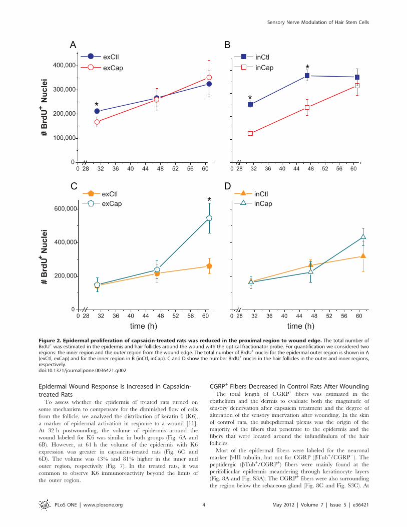
inner region (outer region). The number of BrdU+ nuclei in the
epidermis of the inner region was 50% lower in treated rats at 31 h
postwounding, 36% lower at 47 h, and with no difference at 61 h
(Fig. 2B). In the epidermis of the outer region, the number of
BrdU+ nuclei was decreased in the treated rats by 21% only at
31 h postwounding (Fig. 2A). In the follicles of the inner region,
there were no differences between both groups (Fig. 2D). In
contrast, the number of labeled nuclei in the follicles of the outer
region was 110% higher in the capsaicin-treated rats than in
control rats at 61 h postwounding (Fig. 2C). Interestingly,
wounding induced hair growth in some follicles at apparently
random spots around the wound.
Follicle Cell Migration is Impaired in Capsaicin-treatedRatsTo document possible shifts in the flow of keratinocytes from the
SC niche of the follicle to the epidermis, we used a double labeling
protocol with two thymidine analogs, IdU and CldU. Each of
these analogs can be detected independently and are used to label
and follow cell populations with different dynamics of pro-
liferation. Therefore, the upper follicular cells can be distinguished
because they divide more rapidly than the cells of the epidermis
[7,22]. In the control rats, the double labeled cells (IdU+/CldU+)
were concentrated mainly in the upper portion of the follicle
(infundibulum) and in the basal layer of the perifollicular epidermis
at 41 h postwounding. At 61 h the IdU+/CldU+ cells were also
present in the suprabasal layers and evenly spread throughout the
epidermis (Fig. 3C and 3E). In contrast, capsaicin-treated rats did
not present IdU+/CldU+ cells concentrated in the perifollicular
region at any time point. The few double-labeled cells in treated
rats were distributed throughout the epidermis (Fig. 3B, 3D, and
3F). Noteworthy, the IdU+/CldU+ cells were concentrated at the
upper part of the follicles in the treated rats at 41 h postwounding
(Fig. 3D and Fig. S1).
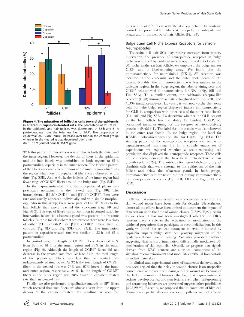
The migration of the follicular cells was analyzed by calculating
the percentage of double labeled cells from the total labeled cells
(IdU+ cells) in both the hair follicles and the epidermis. In control
rats the proportion of IdU+/CldU+ cells increased in the epidermis
from 32 h to 61 h postwounding, while the proportion of these
cells in the follicles tended to decrease (Fig. 4). In contrast, the
proportion of IdU+/CldU+ cells in the epidermis of treated rats
was higher than in control rats at 32 h and this proportion
decreased by 61 h postwounding. In addition, the proportion of
double-labeled cells in the hair follicles significantly decreased
from 32 h to 61 h in the capsaicin-treated rats.
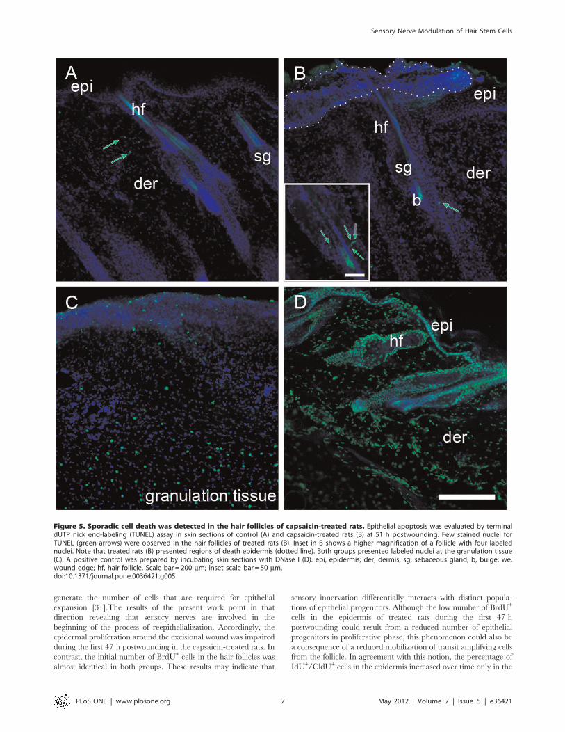
Sporadic Events of Apoptosis Occurred in the HairFollicles of the Capsaicin-treated RatsTo examine if the reduced number of IdU+/CldU+ in the
epidermis of treated rats was due to apoptotic cell death, we
performed a TUNEL assay and active caspase-3 immunostaining
at 51 h and 61 h postwounding. In both groups, the labeling for
TUNEL and caspase-3 were observed in the granulation tissue
and in some dermal cells between follicles (Fig. 5 and Fig. S2). The
capsaicin-treated rats presented only few cells positive for TUNEL
(1–4 per follicle) in the upper part of some hair follicles at 51 h
(Fig. 5B). Occasionally, the capsaicin-treated rats presented small
areas of death epidermis at random positions where it was
notorious the presence of a mass of material that lacked
organization (Fig. 5B and Fig. S2).
Sensory Nerve Modulation of Hair Stem Cells
PLoS ONE | www.plosone.org 2 May 2012 | Volume 7 | Issue 5 | e36421
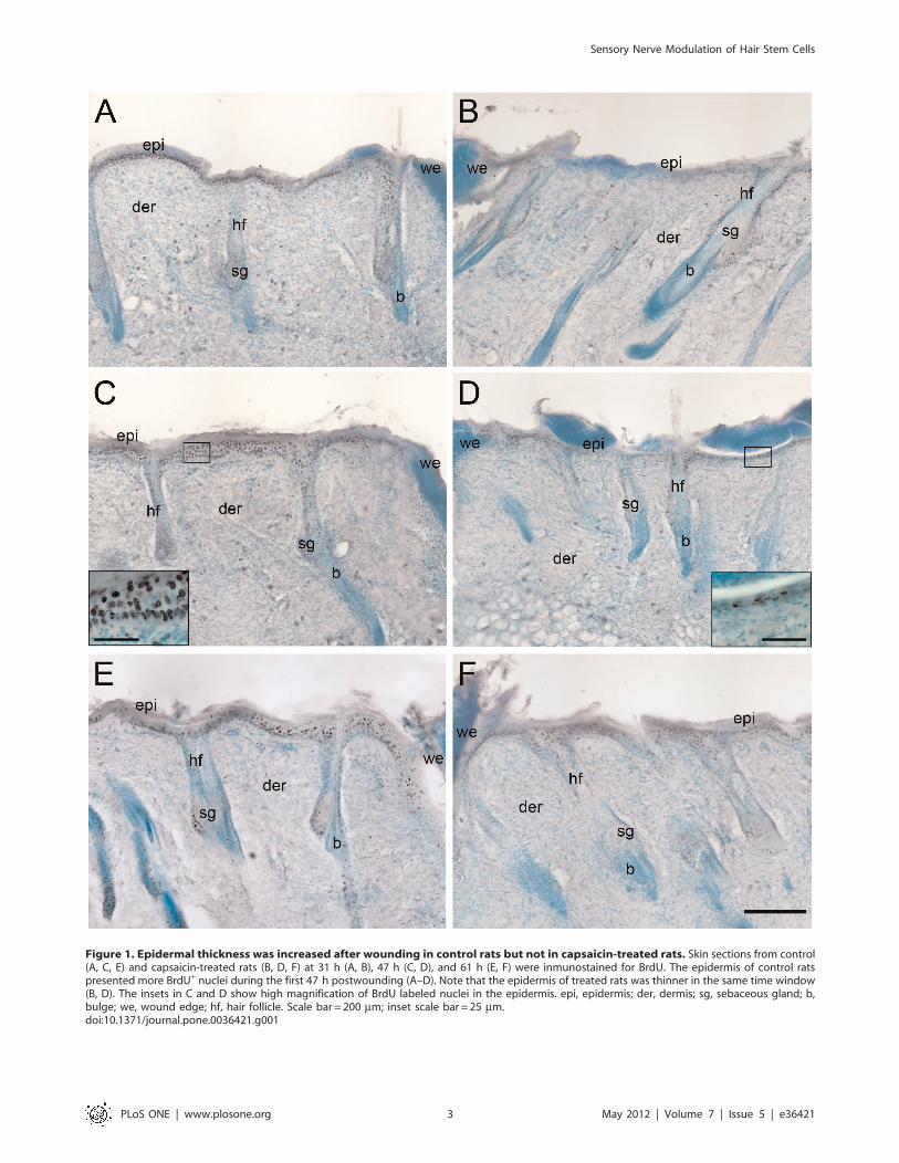
Figure 1. Epidermal thickness was increased after wounding in control rats but not in capsaicin-treated rats. Skin sections from control(A, C, E) and capsaicin-treated rats (B, D, F) at 31 h (A, B), 47 h (C, D), and 61 h (E, F) were inmunostained for BrdU. The epidermis of control ratspresented more BrdU+ nuclei during the first 47 h postwounding (A–D). Note that the epidermis of treated rats was thinner in the same time window(B, D). The insets in C and D show high magnification of BrdU labeled nuclei in the epidermis. epi, epidermis; der, dermis; sg, sebaceous gland; b,bulge; we, wound edge; hf, hair follicle. Scale bar = 200 mm; inset scale bar = 25 mm.doi:10.1371/journal.pone.0036421.g001
Sensory Nerve Modulation of Hair Stem Cells
PLoS ONE | www.plosone.org 3 May 2012 | Volume 7 | Issue 5 | e36421
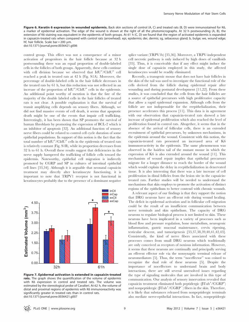
Epidermal Wound Response is Increased in Capsaicin-treated RatsTo assess whether the epidermis of treated rats turned on
some mechanism to compensate for the diminished flow of cells
from the follicle, we analyzed the distribution of keratin 6 (K6),
a marker of epidermal activation in response to a wound [11].
At 32 h postwounding, the volume of epidermis around the
wound labeled for K6 was similar in both groups (Fig. 6A and
6B). However, at 61 h the volume of the epidermis with K6
expression was greater in capsaicin-treated rats (Fig. 6C and
6D). The volume was 43% and 81% higher in the inner and
outer region, respectively (Fig. 7). In the treated rats, it was
common to observe K6 immunoreactivity beyond the limits of
the outer region.
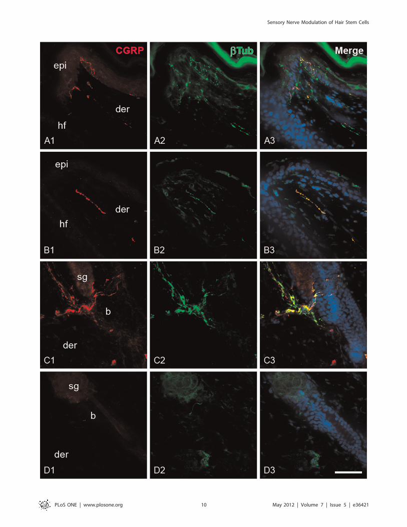
CGRP+ Fibers Decreased in Control Rats After WoundingThe total length of CGRP+ fibers was estimated in the
epithelium and the dermis to evaluate both the magnitude of
sensory denervation after capsaicin treatment and the degree of
alteration of the sensory innervation after wounding. In the skin
of control rats, the subepdiermal plexus was the origin of the
majority of the fibers that penetrate to the epidermis and the
fibers that were located around the infundibulum of the hair
follicles.
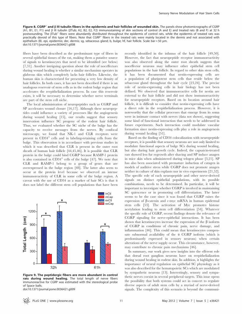
Most of the epidermal fibers were labeled for the neuronal
marker b-III tubulin, but not for CGRP (bTub+/CGRP2). The
peptidergic (bTub+/CGRP+) fibers were mainly found at the
perifollicular epidermis meandering through keratinocyte layers
(Fig. 8A and Fig. S3A). The CGRP+ fibers were also surrounding
the region below the sebaceous gland (Fig. 8C and Fig. S3C). At
Figure 2. Epidermal proliferation of capsaicin-treated rats was reduced in the proximal region to wound edge. The total number ofBrdU+ was estimated in the epidermis and hair follicles around the wound with the optical fractionator probe. For quantification we considered tworegions: the inner region and the outer region from the wound edge. The total number of BrdU+ nuclei for the epidermal outer region is shown in A(exCtl, exCap) and for the inner region in B (inCtl, inCap). C and D show the number BrdU+ nuclei in the hair follicles in the outer and inner regions,respectively.doi:10.1371/journal.pone.0036421.g002
Sensory Nerve Modulation of Hair Stem Cells
PLoS ONE | www.plosone.org 4 May 2012 | Volume 7 | Issue 5 | e36421
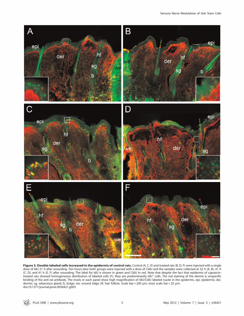
Figure 3. Double-labeled cells increased in the epidermis of control rats. Control (A, C, E) and treated rats (B, D, F) were injected with a singledose of IdU 21 h after wounding. Ten hours later both groups were injected with a dose of CldU and the samples were collected at 32 h (A, B), 41 h(C, D), and 61 h (E, F) after wounding. The label for IdU is shown in green and CldU in red. Note that despite the fact that epidermis of capsaicin-treated rats showed homogeneous distribution of labeled cells (F), they are predominantly IdU+ cells. The red staining of the dermis is unspecificbinding of the anti-rat antibody. The insets in each panel show high magnification of IdU/CldU labeled nuclei in the epidermis. epi, epidermis; der,dermis; sg, sebaceous gland; b, bulge; we, wound edge; hf, hair follicle. Scale bar = 200 mm; inset scale bar = 25 mm.doi:10.1371/journal.pone.0036421.g003
Sensory Nerve Modulation of Hair Stem Cells
PLoS ONE | www.plosone.org 5 May 2012 | Volume 7 | Issue 5 | e36421
32 h this pattern of innervation was similar in both the outer and
the inner region. However, the density of fibers in the epidermis
and the hair follicle was diminished in both regions at 61 h
postwounding, especially in the inner region. The labeling pattern
of the fibers appeared discontinuous at the inner region which was
the region where less intraepidermal fibers were observed at this
time (Fig. S3E). Also at 61 h, the follicles of the inner region had
fewer rings of CGRP+ fibers around the bulge area (Fig. S3F).
In the capsaicin-treated rats, the subepidermal plexus was
practically nonexistent in the treated rats (Fig. 8B). The
intraepidermal bTub+/CGRP2 and bTub+/CGRP+ fibers were
rare and usually appeared individually and with simple morphol-
ogy. Also in this group, there were parallel CGRP+ fibers to the
hair follicle that rarely reached the epidermis (Fig. 8B and
Fig. S3G). This type of fibers was not common in control rats. The
innervation below the sebaceous gland was present in only some
follicles. In those follicles where it was present there were less rings
of either bTub+/CGRP2 or bTub+/CGRP+ fibers than in
controls (Fig. 8D and Fig. S3D and S3H). The innervation
pattern in capsaicin-treated rats was similar at 32 h and 61 h
postwounding.
In control rats, the length of CGRP+ fibers decreased 43%
from 32 h to 61 h in the inner region and 39% in the outer
region (Fig. 9). Although the length of CGRP+ fibers did not
decrease in the treated rats from 32 h to 61 h, the total length
of the peptidergic fibers was less than in control rats
independently of time point. At 32 h the total length of CGRP+
fibers in the treated rats was 73% and 67% lower in the inner
and outer region, respectively. At 61 h, the length of CGRP+
fibers in the outer region was 38% lower in capsaicin-treated
rats than in control rats.
Finally, we also performed a qualitative analysis of SP+ fibers
which revealed that such fibers are almost absent from the upper
dermis of the capsaicin-treated rats, resulting in very few
interactions of SP+ fibers with the skin epithelium. In contrast,
control rats presented SP+ fibers at the epidermis, subepidermal
plexus and in the nearby of hair follicles (Fig. S4).
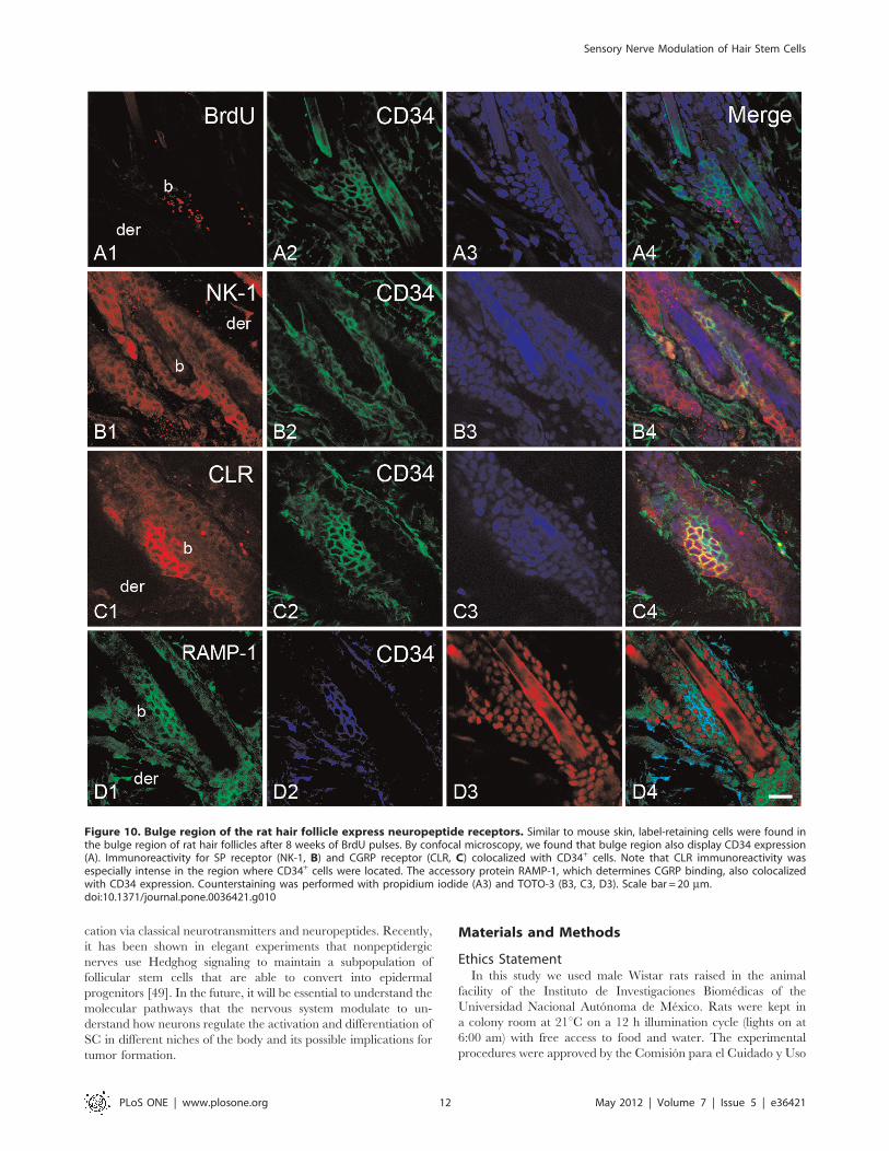
Bulge Stem Cell Niche Express Receptors for SensoryNeuropeptidesTo evaluate if hair SCs may receive messages from sensory
innervation, the presence of neuropeptide receptors in bulge
niche was studied by confocal microscopy. In order to locate the
SC niche in the rat hair follicle, we employed the bulge marker
CD34 and a label-retaining assay. We found that the
immunoreactivity for neurokinin-1 (NK-1), SP receptor, was
localized in the epidermis and the outer root sheath of the
follicle. Notably, the immunoreactivity was less intense in the
follicular region. In the bulge region, the label-retaining cells and
CD34+ cells showed immunoreactivity for NK-1 (Fig. 10B and
Fig. S5A). To a similar extent, the calcitonin receptor-like
receptor (CLR) immunoreactivity colocalized with the BrdU and
CD34 immunoreactivity. However, it was noteworthy that some
cells from the bulge region displayed intense immunoreactivity
for CLR in comparison with other cells of the outer root sheath
(Fig. 10C and Fig. S5B). To determine whether the CLR present
in the hair follicle has the ability for binding CGRP, we
performed immunostaining for the receptor activity-modifying
protein-1 (RAMP-1). The label for this protein was also observed
in the outer root sheath. In the bulge region, the label for
RAMP-1 colocalized with the label for CD34 (Fig. 10C). The
staining pattern of the neuropeptide receptors was similar in
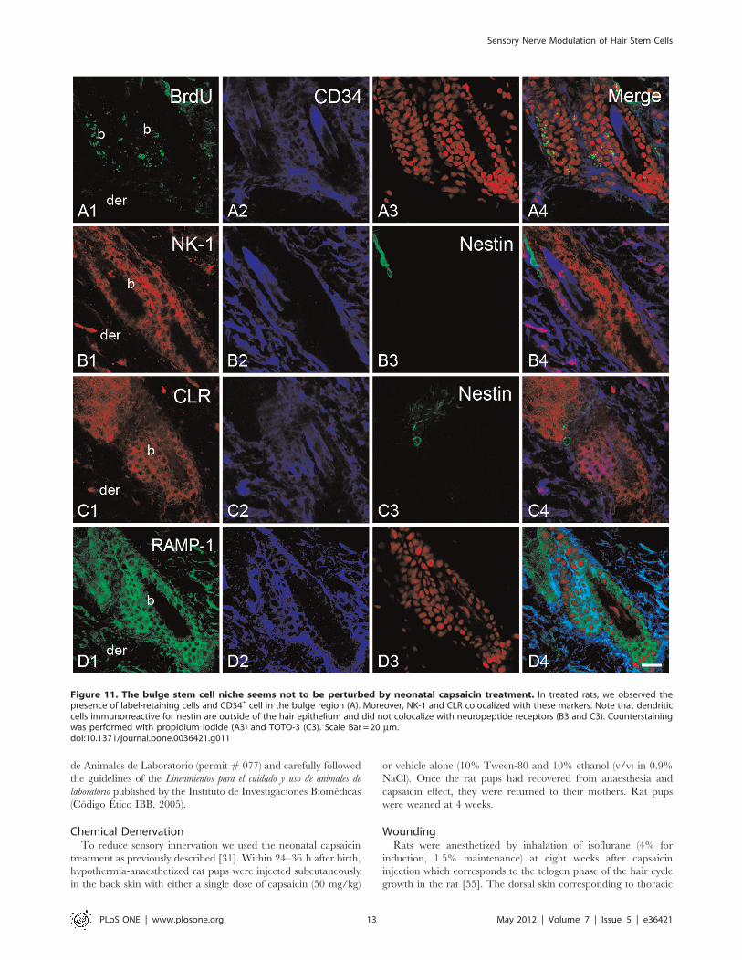
capsaicin-treated rats (Fig. 11). In a complementary set of
experiments we explored whether a nestin-expressing cell
population also displayed the neuropeptide receptors. These cells
are pluripotent stem cells that have been implicated in the hair
growth cycle [23,24]. The antibody for nestin labeled a group of
dendritic cells that were external to the epithelium of the hair
follicle and below the sebaceous gland. In both groups,
immunoreactive cells for nestin did not display immunoreactivity
for neuropeptide receptors (Fig. 11B, 11C and Fig. S5C and
S5D).
Discussion
Claims that sensory innervation exerts beneficial actions during
skin wound repair have been made for decades. Nevertheless,
almost all the efforts have been limited to document the impact of
denervation upon the time of wound closure [2,3,25,26,27]. As far
as we know, it has not been investigated whether the DRG
neurons have a role in the activation or modulation of the
epithelial progenitors that participate in reepithelialization. In this
work, we found that reduced cutaneous innervation induced by
capsaicin impairs bulge stem cell progeny migration to the
epidermis during wound healing. We also provided evidence
suggesting that sensory innervation differentially modulates SC
proliferation of skin epithelia. Overall, we propose that signals
derived from DRG neurons are a critical component of the
signaling microenvironment that modulates epithelial homeostasis
in rodent hairy skin.
In clinical and experimental cases of cutaneous denervation, it
has been argued that the delay in wound closure is an unspecific
consequence of the recurrent damage of the wound site because of
the lack of sensation. However, the fact that capsaicin-treated
rodents develop cornea and skin lesions even when self-grooming
and scratching behaviors are prevented suggests other possibilities
[3,28,29,30]. Recently, we proposed that in conditions of high cell
demand and partial denervation some epithelia are not able to
Figure 4. The migration of follicular cells toward the epidermisis altered in capsaicin-treated rats. The percentage of IdU+/CldU+
in the epidermis and hair follicles was determined at 32 h and 61 hpostwounding from the total number of IdU+. The proportion ofepidermal IdU+/CldU+ nuclei increased over time in the control group,whereas in the treated group decreased over time.doi:10.1371/journal.pone.0036421.g004
Sensory Nerve Modulation of Hair Stem Cells
PLoS ONE | www.plosone.org 6 May 2012 | Volume 7 | Issue 5 | e36421
generate the number of cells that are required for epithelial
expansion [31].The results of the present work point in that
direction revealing that sensory nerves are involved in the
beginning of the process of reepithelialization. Accordingly, the
epidermal proliferation around the excisional wound was impaired
during the first 47 h postwounding in the capsaicin-treated rats. In
contrast, the initial number of BrdU+ cells in the hair follicles was
almost identical in both groups. These results may indicate that
sensory innervation differentially interacts with distinct popula-
tions of epithelial progenitors. Although the low number of BrdU+
cells in the epidermis of treated rats during the first 47 h
postwounding could result from a reduced number of epithelial
progenitors in proliferative phase, this phenomenon could also be
a consequence of a reduced mobilization of transit amplifying cells
from the follicle. In agreement with this notion, the percentage of
IdU+/CldU+ cells in the epidermis increased over time only in the
Figure 5. Sporadic cell death was detected in the hair follicles of capsaicin-treated rats. Epithelial apoptosis was evaluated by terminaldUTP nick end-labeling (TUNEL) assay in skin sections of control (A) and capsaicin-treated rats (B) at 51 h postwounding. Few stained nuclei forTUNEL (green arrows) were observed in the hair follicles of treated rats (B). Inset in B shows a higher magnification of a follicle with four labelednuclei. Note that treated rats (B) presented regions of death epidermis (dotted line). Both groups presented labeled nuclei at the granulation tissue(C). A positive control was prepared by incubating skin sections with DNase I (D). epi, epidermis; der, dermis; sg, sebaceous gland; b, bulge; we,wound edge; hf, hair follicle. Scale bar = 200 mm; inset scale bar = 50 mm.doi:10.1371/journal.pone.0036421.g005
Sensory Nerve Modulation of Hair Stem Cells
PLoS ONE | www.plosone.org 7 May 2012 | Volume 7 | Issue 5 | e36421
Sensory Nerve Modulation of Hair Stem Cells
PLoS ONE | www.plosone.org 8 May 2012 | Volume 7 | Issue 5 | e36421
control group. This effect was not a consequence of a minor
activation of progenitors in the hair follicle because at 32 h
postwounding there was an equal proportion of double-labeled
cells in the follicles of both groups. Apparently, these cells continue
with cell division because we observed that IdU+/CldU+ cell
reached a peak in treated rats at 42 h (Fig. S1A). Moreover, the
percentage of double-labeled cells in the hair follicle decreases in
the treated rats by 61 h, but this reduction was not reflected in an
increase of the proportion of IdU+/CldU+ cells in the epidermis.
An additional point worthy of mention is that the fate of the
majority of the double labeled cells in the hair follicles of treated
rats is not clear. A possible explanation is that the survival of
transit amplifying cells depends on sensory fibers. Although, we
did not find massive events of cell death, it seems that apoptotic
death might be one of the events that impair cell trafficking.
Interestingly, it has been shown that SP promotes the survival of
human fibroblasts by promoting the expression of BCL-2 which is
an inhibitor of apoptosis [32]. An additional function of sensory
nerve fibers could be related to control cell cycle duration of some
epithelial populations. In support of this notion is the fact that the
total number of IdU+/CldU+ cells in the epidermis of treated rats
is relatively constant (Fig. S1B), while its proportion decreases from
32 h to 61 h. Overall these results suggest that deficiencies in the
nerve supply hampered the trafficking of follicle cells toward the
epidermis. Noteworthy, epithelial cell migration is indirectly
promoted by CGRP and SP in cultures of intestinal epithelial
cell lines [33,34]. Although it is arguable that neonatal capsaicin
treatment may directly alter keratinocyte functioning, it is
important to note that TRPV1 receptor is not functional in
keratinocytes probably due to the presence of a dominant negative
splice variant (TRPV1b) [35,36]. Moreover, a TRPV-independent
cell necrotic pathway is only induced by high doses of vanilloids
[35]. Thus, it is conceivable that if any effect might induce the
single dose of capsaicin employed in this study, the affected
keratinocytes would be readily eliminated.
Recently, a transgenic mouse that does not have hair follicles in
the skin of the tail was used to investigate the functional role of the
cells derived from the follicle during epidermal repair after
wounding and during postnatal development [11,22]. From these
studies, it was concluded that the cells from the hair follicles are
a source of epithelial precursors with high proliferative capacity
that allow a rapid epidermal expansion. Although cells from the
follicle are not indispensable for the reepithelialization, their
presence accelerates this process [11]. This data is in agreement
with our observation that capsaicin-treated rats showed a late
increase of epidermal proliferation which also reached the level of
proliferation found in control rats. Altogether, it seems that in the
absence of the arrival of follicular cells, there is an extended
recruitment of epithelial precursors, by unknown mechanisms, in
the epidermis around the wound. Consistent with this notion, the
capsaicin-treated rats presented an increased area of K6
immunoreactivity in the epidermis. The same phenomenon was
observed in the hairless tail of the mutant mouse in which the
expression of K6 is also extended around the wound [11]. This
mechanism of wound repair implies that epithelial precursors
migrate for a longer distance to reach the border of the wound
which would explain the delay in reepithelialization in denervated
tissue. It is also interesting that there was a late increase of cell
proliferation in distal follicles from the lesion site in the capsaicin-
treated rats. Further studies will be needed to understand the
mechanisms that skin employs to promote the activation of distinct
regions of the epithelium to better contend with chronic wounds.
A relevant aspect of our findings is that they support the notion
that DRG neurons have an efferent role during wound healing.
The deficit in epidermal activation and in follicular cell migration
could be the result of an insufficient communication between
nerve terminals and skin epithelium. The capacity of DRG
neurons to regulate biological process is not limited to skin. These
neurons have been implicated in a variety of processes such as
blood flow and pressure regulation, bone metabolism, neurogenic
inflammation, gastric mucosal maintenance, cervix ripening,
testicular descent, and tumorigenesis [33,37,38,39,40,41,42,43].
Consistently, the kind of nerve fibers associated with these
processes comes from small DRG neurons which traditionally
are only conceived as receptors of noxious information. However,
it seems that these neurons are continually and principally serving
an efferent effector role via the nonsynaptic terminal release of
neuromediators [5]. Thus, the term ‘‘noceffector’’ was coined to
recognize the dual role of these neurons [5]. Despite the
importance of noceffectors to understand brain and body
interactions, there are still several unresolved issues regarding
the type of signaling molecules that are involved in this type of
communication. Our analysis of sensory innervation revealed that
capsaicin treatment eliminated both peptidergic (bTub+/CGRP+)
and nonpeptidergic (bTub+/CGRP2) fibers in the skin. Therefore,
it is likely that substances released from nonpeptidergic terminals
also mediate nerve-eptihelial interactions. In fact, nonpeptidergic
Figure 6. Keratin 6 expression in wounded epidermis. Back skin sections of control (A, C) and treated rats (B, D) were inmunostained for K6,a marker of epidermal activation. The edge of the wound is shown at the right of all the photomicrographs. At 32 h postwounding (A, B), theextension of K6 staining was equivalent in the epidermis of both groups. At 61 h (C, D) we found that the region of activated epidermis is expandedin capsaicin-treated rats when compared with control rats (arrowhead). epi, epidermis; der, dermis; sg, sebaceous gland; b, bulge; we, wound edge;hf, hair follicle. Scale bar = 200 mm.doi:10.1371/journal.pone.0036421.g006
Figure 7. Epidermal activation is extended in capsaicin-treatedrats. The graph shows the quantification of the volume of epidermiswith K6 expression in control and treated rats. The volume wasestimated by the stereological probe of Cavalieri. At 62 h, the volume ofdistal and proximal regions of epidermis with K6 immunoreactivity wassignificantly greater in treated rats than in control rats.doi:10.1371/journal.pone.0036421.g007
Sensory Nerve Modulation of Hair Stem Cells
PLoS ONE | www.plosone.org 9 May 2012 | Volume 7 | Issue 5 | e36421
Sensory Nerve Modulation of Hair Stem Cells
PLoS ONE | www.plosone.org 10 May 2012 | Volume 7 | Issue 5 | e36421
fibers have been described as the predominant type of fibers in
several epithelial tissues of the rat, making them a putative source
of signals to keratinocytes that need to be identified (see below)
[7,31]. Another intriguing question about the role of noceffectors
during wound healing is whether a similar mechanism operates in
glabrous skin which completely lacks hair follicles. Likewise, the
human skin is characterized for presenting a very low density of
hair follicles. In both cases, it has not been described if there is an
analogous reservoir of stem cells as in the rodent bulge region that
accelerates the reepithelialization process. In case this reservoir
exists, it will be necessary to determine whether nerve terminals
are part of the stem cell niche.
The local administration of neuropeptides such as CGRP and
SP accelerates wound closure [16,17]. Although these neuropep-
tides could influence a variety of processes like the angiogenesis
during wound healing [15], our results suggest that sensory
innervation influence SC progeny of the rodent hair follicle.
Thus, we evaluated whether the SC niche of the bulge has the
capacity to receive messages from the nerves. By confocal
microscopy, we found that NK-1 and CLR receptors were
present in CD34+ cells and in labeling-retaining cells from the
bulge. This observation is in accordance with previous studies in
which it was described that CLR is present in the outer root
sheath of human hair follicle [44,45,46]. It is possible that CLR
protein in the bulge could bind CGRP because RAMP-1 protein
is also contained in CD34+ cells of the bulge [47]. We note that
CLR and RAMP-1 belong to a group of genes that are
overexpressed in the bulge region [48]. The latter also seem to
occur at the protein level because we observed an intense
immunoreactivity of CLR in some cells of the bulge region. A
caveat with the use of CD34 as a marker of hair SCs is that it
does not label the different stem cell populations that have been
recently identified in the isthmus of the hair follicle [49,50].
However, the fact that neuropeptide receptor immunoreactivity
was also observed along the outer root sheath suggests that
noceffector neurons may influence other epithelial stem cell
populations in the hair follicle. In regard to other skin stem cells,
it has been documented that nestin-expressing cells are
a population of pluripotent stem cells that reside below the
sebaceous gland throughout the hair cycle [23,24]. The precise
role of nestin-expressing cells in hair biology has not been
defined. We observed that immunoreactive cells for nestin are
external to the hair follicle and did not show immunoreactivity
for neuropeptide receptors. Based on its location around hair
follicle, it is difficult to consider that nestin-expressing cells have
a direct role in the reepithelialization process. However, it is
noteworthy that the cellular processes that emerge from its soma
were in intimate contact with nerves (data not shown), suggesting
some kind of functional interaction that needs to be addressed in
future experiments. Such interaction could modulate vascular
formation since nestin-expressing cells play a role in angiogenesis
during wound healing [51].
Based on the finding of CD34 colocalization with neuropeptide
receptors, it is possible that sensory neurons are not only limited to
modulate functional aspects of bulge SCs during wound healing,
but also during hair growth cycle. Indeed, the capsaicin-treated
rats showed less fur regrowth after shaving and SP induce anagen
in mice skin when administered during telogen phase [3,21]. SP
has also been associated with premature induction of catagen in
models of auditive stress while CGRP does not promote anagen
neither in culture of skin explants nor in vivo experiments [21,52].
The specific role of each neuropeptide and other nerve-derived
signals on distinct epithelial populations, with its possible
combinations, needs to be determined. In particular, it will be
important to investigate whether CGRP is involved in maintaining
SC quiescence or in promoting cell differentiation. The latter
seems to be the case since it was found that CGRP raises the
expression of b-catenin and c-myc mRNA in human epidermal
stem cells [53]. The activation of Myc promotes histone
acetylation leading to stem cell differentiation [54]. Whatever
the specific role of CGRP, recent findings denote the relevance of
CGRP signaling for nerve-epithelial interactions. It has been
shown that keratinocytes increase the expression of the b isoform
of CGRP in conditions of chronic pain, nerve damage, and
inflammation [46]. This could mean that keratinocytes compen-
sate subnormal availability of the a CGRP isoform (which is
predominantly expressed in sensory neurons) when certain
alterations of the nerve supply occur. This circumstance, however,
may contribute to chronic pain mechanisms [46].
In summary, our work gives new insights into the efferent role
that dorsal root ganglion neurons have on reepithelialization
during wound healing in rodent skin. In addition, it highlights the
importance of neural regulation on epithelial SC physiology as it
was also described for the hematopoietic SCs which are modulated
by sympathetic neurons [13]. Interestingly, sensory and sympa-
thetic nerves coexist in several peripheral targets. This issue opens
the possibility that both systems could act in concert to regulate
diverse aspects of adult stem cells by a myriad of nerve-derived
signals. The complexity of this scenario is beyond the communi-
Figure 8. CGRP+ and b III tubulin fibers in the epidermis and hair follicles of wounded skin. The panels show photomicrographs of CGRP(A1, B1, E1, F1) and b III tubulin (bTub; A2, B2, E2, F2) immunostaining of skin sections of control (A and E) and treated rats (B and F) at 32 hpostwounding. The bTub+ fibers were abundantly distributed throughout the epidermis of control rats, while the epidermis of treated rats waspractically devoid of this type of fibers. Note that CGRP+ fibers in the treated rats were mainly located in the dermis and not associated withepithelium (B). epi, epidermis; der, dermis; sg, sebaceous gland; b, bulge; hf, hair follicle. Scale bar = 50 mm.doi:10.1371/journal.pone.0036421.g008
Figure 9. The peptidergic fibers are more abundant in controlrats during wound healing. The total length of nerve fibersimmunoreactive for CGRP was estimated with the stereological probeof Space balls.doi:10.1371/journal.pone.0036421.g009
Sensory Nerve Modulation of Hair Stem Cells
PLoS ONE | www.plosone.org 11 May 2012 | Volume 7 | Issue 5 | e36421
cation via classical neurotransmitters and neuropeptides. Recently,
it has been shown in elegant experiments that nonpeptidergic
nerves use Hedghog signaling to maintain a subpopulation of
follicular stem cells that are able to convert into epidermal
progenitors [49]. In the future, it will be essential to understand the
molecular pathways that the nervous system modulate to un-
derstand how neurons regulate the activation and differentiation of
SC in different niches of the body and its possible implications for
tumor formation.
Materials and Methods
Ethics StatementIn this study we used male Wistar rats raised in the animal
facility of the Instituto de Investigaciones Biomedicas of the
Universidad Nacional Autonoma de Mexico. Rats were kept in
a colony room at 21uC on a 12 h illumination cycle (lights on at
6:00 am) with free access to food and water. The experimental
procedures were approved by the Comision para el Cuidado y Uso
Figure 10. Bulge region of the rat hair follicle express neuropeptide receptors. Similar to mouse skin, label-retaining cells were found inthe bulge region of rat hair follicles after 8 weeks of BrdU pulses. By confocal microscopy, we found that bulge region also display CD34 expression(A). Immunoreactivity for SP receptor (NK-1, B) and CGRP receptor (CLR, C) colocalized with CD34+ cells. Note that CLR immunoreactivity wasespecially intense in the region where CD34+ cells were located. The accessory protein RAMP-1, which determines CGRP binding, also colocalizedwith CD34 expression. Counterstaining was performed with propidium iodide (A3) and TOTO-3 (B3, C3, D3). Scale bar = 20 mm.doi:10.1371/journal.pone.0036421.g010
Sensory Nerve Modulation of Hair Stem Cells
PLoS ONE | www.plosone.org 12 May 2012 | Volume 7 | Issue 5 | e36421
de Animales de Laboratorio (permit # 077) and carefully followed
the guidelines of the Lineamientos para el cuidado y uso de animales de
laboratorio published by the Instituto de Investigaciones Biomedicas
(Codigo Etico IBB, 2005).
Chemical DenervationTo reduce sensory innervation we used the neonatal capsaicin
treatment as previously described [31]. Within 24–36 h after birth,
hypothermia-anaesthetized rat pups were injected subcutaneously
in the back skin with either a single dose of capsaicin (50 mg/kg)
or vehicle alone (10% Tween-80 and 10% ethanol (v/v) in 0.9%
NaCl). Once the rat pups had recovered from anaesthesia and
capsaicin effect, they were returned to their mothers. Rat pups
were weaned at 4 weeks.
WoundingRats were anesthetized by inhalation of isoflurane (4% for
induction, 1.5% maintenance) at eight weeks after capsaicin
injection which corresponds to the telogen phase of the hair cycle
growth in the rat [55]. The dorsal skin corresponding to thoracic
Figure 11. The bulge stem cell niche seems not to be perturbed by neonatal capsaicin treatment. In treated rats, we observed thepresence of label-retaining cells and CD34+ cell in the bulge region (A). Moreover, NK-1 and CLR colocalized with these markers. Note that dendriticcells immunorreactive for nestin are outside of the hair epithelium and did not colocalize with neuropeptide receptors (B3 and C3). Counterstainingwas performed with propidium iodide (A3) and TOTO-3 (C3). Scale Bar = 20 mm.doi:10.1371/journal.pone.0036421.g011
Sensory Nerve Modulation of Hair Stem Cells
PLoS ONE | www.plosone.org 13 May 2012 | Volume 7 | Issue 5 | e36421
vertebrae was shaved and cleaned with 70% ethanol. Then, we
made a full-thickness wound with a dermal biopsy punch of 6 mm
of diameter. Wounds were left uncovered and in the cases of
bleeding it was cleaned with sterile cotton. At the end of the
surgery, the rats were placed individually in a cage until they
completely recover from anesthesia. Finally, the rats were returned
to its original cage.
ProliferationTo determine the effect of capsaicin denervation on the
activation of epidermal and follicular progenitors, rats were
injected with a single dose of bromodeoxyuridine (50 mg/kg)
21 h following wounding. Skin samples were collected at 31 h,
47 h, and 61 h postwounding. The rats (n = 5) were anesthetized
with sodium pentobarbital and were transcardially perfused with
0.9% NaCl followed by Zamboni fixative [4% paraformaldehyde;
15% (v/v) saturate picric acid, 0.1 M phosphate buffer, pH 7.4].
The skin samples were postfixed for 24 h in the same fixative.
Then, the samples were successively transferred to 20% and 30%
sucrose. The tissue was embedded in OCT compound (Sakura
Seiki Inc, Tokyo, Japan) and frozen in an orientation that allow to
obtain longitudinal sections of hair follicles, as previously described
[56].
MigrationWe used a double-labeling technique with two analogs of
thymidine to evaluate whether sensory denervation alters the
mobilization of transit amplifying cells from the follicle to the
epidermis [7,57]. This technique is based on the principle that
follicular keratinocytes divide faster than the epidermal keratino-
cytes [7]. For this reason, rats (n = 5) were injected 21 h after
wounding with a single dose of iododeoxyuridine (IdU; 150 nmol/
g). Ten hours later (31 h postwounding) we injected a single dose
of chlorodeoxyuridine (CldU; 150 nmol/g). Skin samples were
collected at 32, 41, and 61 h postwounding and processed as
described above. For all quantitative procedures, the skin sections
were coded and the observers were blinded to the coding
information.
BrdU ImmunohistochemistryThe samples were sectioned into 50 mm slices in a cryostat and
the sections were individually collected in 48-well plates filled with
cryoprotectant solution (25% ethylene glycol and 25% glycerol in
a 0.05 M phosphate buffer) and stored at 220uC. For the analysis,sections were regionalized in two regions. The inner region
comprised 2 mm from the edge of the wound, whereas the outer
region spanned 2 mm further from the border of the inner region.
Therefore, we collected 280 sections corresponding to a total
region of 14 mm614 mm. For stereological quantification,
14 sections were selected with a systematic random sampling.
The sampling interval under this scheme was twenty, which
implies that the first section was selected at random between the
first and the twentieth section. Then, the series of sections were put
onto gelatinized slides washed 26 with 0.1 M phosphate buffer
(PB), rinsed 16 with 0.3% Triton X-100 in PB (PBT), and
incubated with 1% H2O2 in PB for 1 h. After three PB rinses,
sections were incubated with Immuno/DNA retriever at 70uC for
30 min (Bio SB, Santa Barbara, USA), and then washed with PB.
For BrdU immunodetection, DNA was denatured with 1N HCl at
25uC for 30 min and neutralized with 0.1 M sodium borate
buffer. After this step, the biotin-avidin blocking kit (Vector
Laboratories, Burlingame, USA) was used to eliminate unspecific
staining of the sebaceous gland of the hair follicle. The avidin
(100 mL/mL) was diluted in PB and the sections were incubated
for 30 min in this solution. After a rinse, the sections were
incubated with a solution of biotin (100 mL/mL). Finally, sections
were blocked with 5% normal horse serum in PBT for 1 h. The
sections were incubated overnight with mouse anti-BrdU (1:500,
Roche Applied Science, Penzberg, Germany). After PB rinses,
sections were incubated with biotinylated donkey anti-mouse
(Chemicon, Temecula, USA) for 2 h and then with the avidin–
biotin complex (Vector Laboratories) for 90 min. The immuno-
histochemistry reaction was made visible by 3-39-diaminobenzi-
dine/nickel precipitation (Vector Laboratories). After a rinse in
water, sections were incubated in 0.05 M sodium bicarbonate
buffer pH 9.6 for 10 min and then exposed to DAB enhancing
solution (Vector laboratories). Finally, sections were counter-
stained with methyl green.
Quantification of Epithelial ProliferationThe total number of immunoreactive BrdU cells was estimated
with the stereological probe of the optical fractionator [31]. Data
were obtained with a Nikon Labophot-2 microscope equipped
with 106 and 1006 (oil immersion, 1.4 NA) objectives, a motor-
ized x-y-z stage control, and interfaced with StereoInvestigator
Software 9 (MBF Bioscience, Williston, USA). The boundaries of
epidermis and follicle profiles were traced in the areas correspond-
ing to the inner and outer regions described above. BrdU+ nuclei
were counted at uniformly random sampled sites within the
epidermal and follicular tracing. The size of the grid for the
epidermis of the samples taken at 31 h postwounding was
X=927 mm and Y=34 mm with a counting frame measuring
30625 and a height of 21 mm.; for 47 h samples, X=716 mm and
Y=78 mm with a counting frame measuring 30625 and a height
of 25 mm; and for 61 h samples, X= 480 mm and Y=180 mma counting frame measuring 30625 and a height of 24 mm. The
size of the grid for the follicular region of the samples obtained at
31 h was X=172 mm and Y=200 mm with a counting frame
measuring 43632 and a height of 22 mm; for 47 h and 61 h
samples, X= 344 mm and Y=180 mm with a counting frame
measuring 43632 and a height of 24 mm. The guard zone was
10% of the mean section thickness, which was measured at every
sampling site.
IdU/CldU ImmunofluorescenceThe skin samples labeled with IdU and CldU were also
sectioned into 50 mm slices and preserved in cryoprotectant
solution. By a systematic random sampling, we selected 10 sections
with a sampling interval of twenty. This area corresponded to the
inner region around the wound. The series of sections were
washed 26with Tris buffered saline (TBS, pH 7.4) and rinsed 16with TBS containing 0.3% Triton X-100 (TTBS). Then, sections
were incubated with Immuno/DNA retriever at 70uC for 30 min
and then washed with TBS. Also sections were incubated with 1N
HCl at 37uC for 1 h followed by a 10 min wash with 0.1 M
sodium borate buffer. After three washes in TBS, sections were
incubated 1 h in blocking solution containing 5% normal goat
serum and 5% bovine serum albumin in TTBS. The primary
antibodies were diluted in blocking solution and incubated 12 to
16 h at room temperature. The detection of the halogenated
thymidne analogs was accomplished using mouse anti-BrdU
(1:1000; Becton Dickinson, clone B44) for IdU and rat anti-BrdU
(1:300; Serotec, clone BU1/75) for CldU [58]. After three washes,
sections were incubated with donkey anti-mouse conjugated to
Alexa 488 (1:1,000) and donkey anti-rat conjugated to Alexa 584
(1:1,500). After rinses in TBS, sections were counterstained with
DAPI and coverslipped with Dako Fluorescence Mounting
Medium (Dako, Carpinteria, USA).
Sensory Nerve Modulation of Hair Stem Cells
PLoS ONE | www.plosone.org 14 May 2012 | Volume 7 | Issue 5 | e36421
Quantification of Follicular Cell MigrationThe estimation of the total number of IdU+/CldU+ in the
epidermis and upper part of the hair follicle was obtained also by
the optical fractionator probe. Data were obtained with an
Olympus BX51 WI microscope equipped with a 46 and 606(water immersion, 1.2 NA) objectives, a motorized x-y-z stage
control, and the StereoInvestigator Software. The size of the grid
for the epidermis of the samples obtained at 32 h postwounding
was X=392 mm and Y=74 mm with a counting frame
measuring 65665 and a height of 30 mm; for 41 h and 61 h
samples, X= 488 mm and Y=82 mm with a counting frame
measuring 65665 and a height of 26 mm. The size of the grid for
the follicular region in all the samples was X=137 mm and
Y=250 mm. The counting frame measured 65650 and a height
of 30 mm (32 h) or 26 mm (41 h and 61 h). The guard zone in all
the cases was 10% of the mean section thickness, which was
measured at every sampling site. The percentage of the double
labeled nuclei was obtained by determining the fraction of IdU+/
CldU+ nuclei in relation to the total nuclei labeled with IdU.
Evaluation of Programmed Cell DeathTo qualitatively evaluate the presence of apoptotic cells in the
epithelium of wounded skin we used the TUNEL assay and the
immunolabeling for active caspase-3 on series of sections
obtained by random sampling. TUNEL staining was performed
with ApopTag Fluorescein Direct In Situ Apoptosis Detection
Kit (Chemicon) according to the manufacturer’s instructions.
Tissue permeabilization was achieved by incubating sections with
proteinase K 20 mg/mL for 20 min at room temperature. For
caspase-3 immunolabeling, sections were rinsed with PB and
PBT and incubated with Immuno/DNA retriever. After an hour
of blocking with 5% horse serum, sections were incubated
overnight with rabbit polyclonal anti-caspase-3 (1:500, Abcam,
Cambridge, UK). After PB rinses sections were incubated with
donkey anti-rabbit conjugated to Alexa 594. Finally, sections
were counterstained with DAPI and coverslipped with Dako
medium.
Volume Estimation of the Expression of Keratin 6The immunofluorescence to detect epidermal expression of
Keratin 6 (K6) was performed in 14 sections selected by uniform
random sampling. The sections were rinsed 26 in PB and 16 in
PBT. After incubation with Immuno/DNA retriever, the sections
were blocked for 30 min with 5% horse serum in PBT and
incubated for 12–15 h with the monoclonal antibody mouse anti-
K6 (1:500; Biocare Medical, USA). After PB rinses, sections were
incubated for 2 h at room temperature with donkey anti-mouse
conjugated to Alexa 594 (1:500). Finally, sections were incubated
with DAPI and coverslipped with fluorescence mounting
medium.
The volume of the epidermis immunoreactive to K6 was
estimated by the stereological method of Cavalieri using the
Olympus BX51 WI microscope interfaced with StereoInvestigator.
This method is based on the principle that the volume of an object
of irregular shape can be obtained by considering the area of the
object profiles that appear in sections separated by a known
distance. In order to obtain the volume estimation, a uniform
array of points with a known area per point is placed over the
image of the sections. Then, the number of points hitting the K6+
epidermis was counted on each selected sections. The distance
between points was 120 mm.
Photographic documentation of BrdU, IdU/CldU or CK6
immunostaining was performed by creating a seamless montage.
Using a motorized stage, we collected a series of images from skin
sections with overlapping edges and used the function ‘‘Phot-
merge’’ from Phothoshop to align, stitch, and blend them. The
resulting high-resolution images were cropped to assemble the
corresponding figures.
CGRP+ Fiber Length EstimationThe total length of CGRP+ fibers in the inner and outer region
around the wound was quantified by using the space balls method
for design-based stereology [31]. The immunohistochemistry was
performed according to the protocol for BrdU staining, but the
step of HCl incubation was omitted. Fiber detection was
accomplished using rabbit anti-CGRP (1:10,000, Peninsula Labs,
San Diego, USA). Data was obtained in the Olympus BX51 WI
microscope interfaced with StereoInvestigator. The traced region
included the living strata of the epidermis and the dermal region
that contained the upper part of the hair follicles up to the bulge
region. For the quantification, we considered fibers in the
epithelium and in the fibrous component of the dermis. The
stereological parameters were as follows: sampling grid,
711678 mm; radius of the sphere, 12 mm; and guard zones,
13% of section thickness. In additional experiments, double
immunofluorescence was performed with mouse anti-neural III
beta-tubulin (1:500, Promega, Madison, USA).
Immunofluorescence for Neuropeptide ReceptorsAt postnatal day four, a group of control and capsaicin-treated
rats were injected twice daily with BrdU (50 mg/kg) during three
days. After 8 to 10 weeks, these rats were transcardially perfused
with Zamboni fixative. Samples from back skin were collected,
postfixed for 24 h in the same fixative, and successively transferred
to 20% and 30% sucrose. Samples were sectioned into 30 mmslices and stored in cryoprotectant solution. The sections were
incubated for 30 min in Immuno/DNA retriever solutions, for 1 h
at 25uC in HCl, and 10 min in sodium borate. After TBS rinses,
the sections were incubated for 1 h with 5% horse serum and then
incubated overnight with a mixture of primary antibodies. We
used the following primary antibodies: rabbit anti-neurokinin
receptor 1 (NK-1; 1:300, Chemicon), rabbit anti-calcitonin like
protein (CLR; 1:1,000), mouse anti-BrdU (1:300, Roche Applied
Science), goat anti-CD34 (1:100, Santa Cruz laboratories, Santa
Cruz, USA), mouse anti-nestin (1:300, Chemicon), and rabbit anti-
receptor activity modifying protein 1 (RAMP-1; 1:100, Santa Cruz
Laboratories). All the secondary antibodies were raised in donkey
and included anti-mouse conjugated to Alexa 488, anti-rabbit
conjugated to Alexa 594, and anti-goat Alexa 647 (Fig. S6). The
sections were counterstained with either propidium iodide (PI) or
with TOTO-3 depending of the secondary antibody combination.
Sections were imaged on a Zeiss LSM5 Pascal confocal
microscope equipped with Ar/HeNe laser. The combinations
that we analyzed for the presence of neuropeptide receptors in the
bulge region included: NK-1/BrdU/TOTO, NK-1/CD34/PI,
CLR/BrdU/TOTO, CLR/CD34/PI, CLR/BrdU/TOTO,
BrdU/CD34/TOTO, Nestin/CD34/PI, NK-1/Nestin/CD34,
CLR/Nes/CD34 y RAMP1/CD34/TOTO.
StatisticsData are expressed as mean 6 SEM. Statistical analysis was
performed using Origin Pro 8. Statistical differences between
groups were determined by a two-tailed Student’s t-test. Multiple
comparisons were performed by using one-way ANOVA test
followed by a Tukey and Holm-Sidack post hoc test. Any
difference with p,0.05 was considered statistically significant.
Sensory Nerve Modulation of Hair Stem Cells
PLoS ONE | www.plosone.org 15 May 2012 | Volume 7 | Issue 5 | e36421
Supporting Information
Figure S1 The number of epidermal IdU+/CldU+ cellsincreased over time only in control rats. The total number
of immunoreactive nuclei for IdU and CldU in the skin epithelium
was estimated in the inner region to the wound edge (inCtl and
inCap). The graphs show the estimated number of double-labeled
cells in the hair follicles (A) and the epidermis (B).
(EPS)
Figure S2 Apoptotic cell death evaluation. To evaluate
possible events of programmed cell death in the epithelia of
capsaicin-treated rats we performed immunostaining for active
caspase-3 (A–C) and TUNEL assay (E and F). In general, neither
treated rats (B) nor control rats (A) presented labeling for caspase-3
at the epidermis or hair follicles. The immunostaining for caspase-
3 was mainly observed at the granulation tissue in both groups (D).
As a physiological control of cell death we used rat mammary
gland obtained at the fourth day after weaning (C). A similar
pattern of labeling was observed in the mammary gland using the
TUNNEL assay (E). Panel D shows a unique observation where
epidermal cells were positive for TUNNEL assay in a treated rat.
These labeled nuclei were adjacent to a mass of disorganized
material where no living strata were observed. epi, epidermis; der,
dermis; sg, sebaceous gland; b, bulge; hf, hair follicle. Scale
bar = 50 mm.
(TIF)
Figure S3 CGRP+ fibers in the epidermis and hairfollicles of wounded skin. The panels show photomicrographs
of CGRP immunostaining of skin sections of control (A, B, E, F)
and treated rats (C, D, G, H) at 32 h (A, B, C, D) and 61 h (E, F,
G, H) postwounding. The CGRP+ fibers (arrowhead) diminished
with time in the control group. Nevertheless, the number of
peptidergic fibers associated with skin epithelium was severely
reduced in capsaicin-treated rats, independently of postwounding
time. epi, epidermis; der, dermis; sg, sebaceous gland; b, bulge; hf,
hair follicle; hs, hair shaft. Scale bar = 100 mm.
(TIF)
Figure S4 In capsaicin-treated rats, the SP+ fibers werenot associated with skin epithelium. The panels show
photomicrographs of substance P (SP) immunostaining of skin
sections of control (A, B, E, F) and treated rats (C, D, G, H) at 32 h
(A, B, C, D) and 61 h (E, F, G, H) postwounding. In control skin,
the SP+ fibers (arrowheads) were located at the epidermis, at the
subepidermal plexus, in the nearby of hair follicles, and in close
association of blood vessels. In contrast, SP+ fibers in treated rats
were only observed at blood vessels and in the deep dermis. epi,
epidermis; der, dermis; sg, sebaceous gland; b, bulge; hf, hair
follicle. Scale bar = 100 mm.
(TIF)
Figure S5 Label-retaining cells but not nestin immuno-reactive cells colocalize with neuropeptide receptors. Byconfocal microscopy we evaluated the presence of SP receptor
(NK-1, A) and CGRP receptor (CLR, B) on BrdU-retaining cells.
The immunoreactivity for neuropeptide receptors was not
observed in nestin-positive cells. These cells had a dendritic
morphology and were located outside the outer root sheath of the
hair follicle. Scale Bar = 20 mm.
(TIF)
Figure S6 Neuropeptide receptors in the rat spinalcord. The staining pattern for NK-1 (A and B), CLR (C and
D), and RAMP-1 (E) was verified on spinal cord sections. The
label for the three antibodies was mainly observed at the dorsal
horn of the spinal cord as previously described. Panel F shows the
RAMP-1 staining at the epidermis (F). Note that RAMP-1
immunoreactivity is localized in the basal layer of the epidermis.
As background control, skin sections were incubated with only
secondary antibodies. Panel G: anti-mouse conjugated to Alexa
488, anti-rabbit conjugated to Alexa 594, and anti-goat conjugated
to Alexa 647; panel H: anti-mouse (Alexa 488), anti-rabbit (Alexa
594), and TOTO-3 counterstaining. The hair shaft presented
autofluorescence with the three excitation laser lines used. The
sebaceous gland showed faint staining with anti-rabbit antibody.
epi, epidermis; der, dermis; sg, sebaceous gland; b, bulge; hf, hair
follicle; hs, hair shaft; dh, dorsal horn. Scale bar: A and
C=100 mm; B, D, E, F, G, and H=50 mm.
(TIF)
Acknowledgments
The antibody for CLR was kindly gifted by Dr. Nigel W. Bunnet
(Department of Surgery, UCSF). Preliminary data were presented at the
2009 Annual Meeting of the Society for Neuroscience, Chicago, IL.
Author Contributions
Conceived and designed the experiments: EMM. Performed the experi-
ments: EMM CIGH BTM. Analyzed the data: EMM CIGH GGO.
Contributed reagents/materials/analysis tools: GGO. Wrote the paper:
EMM GGO.
References
1. Barker AR, Rosson GD, Dellon AL (2006) Wound healing in denervated tissue.
Ann Plast Surg 57: 339–342.
2. Carr RW, Delaney CA, Westerman RA, Roberts RG (1993) Denervation
impairs cutaneous microvascular function and blister healing in the rat
hindlimb. Neuroreport 4: 467–470.
3. Maggi CA, Borsini F, Santicioli P, Geppetti P, Abelli L, et al. (1987) Cutaneous
lesions in capsaicin-pretreated rats. A trophic role of capsaicin-sensitive
afferents? Naunyn Schmiedebergs Arch Pharmacol 336: 538–545.
4. Smith P, Liu M (2002) Impaired cutaneous wound healing after sensory
denervation in developing rats: effects on cell proliferation and apoptosis. Cell
Tissue Res 307: 281–291.
5. Kruger L (1996) The functional morphology of thin sensory axons: some
principles and problems. Prog Brain Res 113: 255–272.
6. Roosterman D, Goerge T, Schneider SW, Bunnett NW, Steinhoff M (2006)
Neuronal Control of Skin Function: The Skin as a Neuroimmunoendocrine
Organ. Physiol Rev 86: 1309–1379.
7. Taylor G, Lehrer MS, Jensen PJ, Sun T-T, Lavker RM (2000) Involvement of
Follicular Stem Cells in Forming Not Only the Follicle but Also the Epidermis.
Cell 102: 451.
8. Trempus CS, Morris RJ, Bortner CD, Cotsarelis G, Faircloth RS, et al. (2003)
Enrichment for Living Murine Keratinocytes from the Hair Follicle Bulge with
the Cell Surface Marker CD34. J Investig Dermatol 120: 501–511.
9. Cotsarelis G, Sun T-T, Lavker RM (1990) Label-retaining cells reside in the
bulge area of pilosebaceous unit: Implications for follicular stem cells, hair cycle,
and skin carcinogenesis. Cell 61: 1329–1337.
10. Ito M, Liu Y, Yang Z, Nguyen J, Liang F, et al. (2005) Stem cells in the hair
follicle bulge contribute to wound repair but not to homeostasis of the epidermis.
Nat Med 11: 1351–1354.
11. Langton AK, Herrick SE, Headon DJ (2007) An Extended Epidermal Response
Heals Cutaneous Wounds in the Absence of a Hair Follicle Stem Cell
Contribution. J Invest Dermatol 128: 1311–1318.
12. Blanpain C, Fuchs E (2006) Epidermal Stem Cells of the Skin. Annu Rev Cell
Dev Biol 22: 339–373.
13. Katayama Y, Battista M, Kao W-M, Hidalgo A, Peired AJ, et al. (2006) Signals
from the Sympathetic Nervous System Regulate Hematopoietic Stem Cell
Egress from Bone Marrow. Cell 124: 407–421.
14. Jones DL, Wagers AJ (2008) No place like home: anatomy and function of the
stem cell niche. Nat Rev Mol Cell Biol 9: 11–21.
Sensory Nerve Modulation of Hair Stem Cells
PLoS ONE | www.plosone.org 16 May 2012 | Volume 7 | Issue 5 | e36421
15. Toda M, Suzuki T, Hosono K, Kurihara Y, Kurihara H, et al. (2008) Roles of
calcitonin gene-related peptide in facilitation of wound healing and angiogenesis.Biomed Pharmacother 62: 352–359.
16. Engin C (1998) Effects of calcitonin gene-related peptide on wound contraction
in denervated and normal rat skin: A preliminary report. Plast Reconstr Surg101: 1887–1890.
17. Delgado AV, McManus AT, Chambers JP (2005) Exogenous administration ofSubstance P enhances wound healing in a novel skin-injury model. Exp Biol
Med (Maywood) 230: 271–280.
18. Rook JM, Hasan W, McCarson KE (2009) Morphine-induced early delays inwound closure: Involvement of sensory neuropeptides and modification of
neurokinin receptor expression. Biochem Pharmacol 77: 1747–1755.19. Seike M, Ikeda M, Morimoto A, Matsumoto M, Kodama H (2002) Increased
synthesis of calcitonin gene-related peptide stimulates keratinocyte proliferationin murine UVB-irradiated skin. J Dermatol Sci 28: 135.
20. Yu X-J, Li C-Y, Xu Y-H, Chen L-M, Zhou C-L (2009) Calcitonin gene-related
peptide increases proliferation of human HaCaT keratinocytes by activation ofMAP kinases. Cell Biol Int 33: 1144–1148.
21. Peters EM, Botchkarev VA, Botchkareva NV, Tobin DJ, Paus R (2001) Hair-cycle-associated remodeling of the peptidergic innervation of murine skin, and
hair growth modulation by neuropeptides. J Invest Dermatol 116: 236–245.
22. Heath J, Langton AK, Hammond NL, Overbeek PA, Dixon MJ, et al. (2009)Hair Follicles Are Required for Optimal Growth during Lateral Skin Expansion.
J Invest Dermatol 129: 2358–2364.23. Amoh Y, Li L, Katsuoka K, Penman S, Hoffman RM (2005) Multipotent nestin-
positive, keratin-negative hair-follicle bulge stem cells can form neurons. ProcNatl Acad Sci U S A 102: 5530–5534.
24. Uchugonova A, Duong J, Zhang N, Konig K, Hoffman RM (2011) The bulge
area is the origin of nestin-expressing pluripotent stem cells of the hair follicle.J Cell Biochem 112: 2046–2050.
25. Harsum S, Clarke JD, Martin P (2001) A reciprocal relationship betweencutaneous nerves and repairing skin wounds in the developing chick embryo.
Dev Biol 238: 27–39.
26. Westerman RA, Carr RW, Delaney CA, Morris MJ, Roberts RG (1993) Therole of skin nociceptive afferent nerves in blister healing. Clin Exp Neurol 30:
39–60.27. Fukai T, Takeda A, Uchinuma E (2005) Wound healing in denervated rat skin.
Wound Repair Regen 13: 175–180.28. Buck SH, Walsh JH, Davis TP, Brown MR, Yamamura HI, et al. (1983)
Characterization of the peptide and sensory neurotoxic effects of capsaicin in the
guinea pig. J Neurosci 3: 2064–2074.29. Fujita S, Shimizu T, Izumi K, Fukuda T, Sameshima M, et al. (1984) Capsaicin-
induced neuroparalytic keratitis-like corneal changes in the mouse. Exp Eye Res38: 165–175.
30. Carrillo P, Camacho M, Manzo J, Martinez-Gomez M, Salas M, et al. (1998)
Cutaneous Wounds Produced by Capsaicin Treatment of Newborn Rats AreDue to Trophic Disturbances. Neurotoxicol Teratol 20: 75.
31. Martinez-Martinez E, Toscano-Marquez B, Gutierrez-Ospina G (2011) Long-term effects of neonatal capsaicin treatment on intraepidermal nerve fibers and
keratinocyte proliferation in rat glabrous skin. Anat Rec 294: 173–184.32. Jing C, Jia-Han W, Hong-Xing Z (2010) Double-edged effects of neuropeptide
substance P on repair of cutaneous trauma. Wound Repair Regen 18: 319–324.
33. Bulut K, Felderbauer P, Deters S, Hoeck K, Schmidt-Choudhury A, et al. (2008)Sensory neuropeptides and epithelial cell restitution: the relevance of SP- and
CGRP-stimulated mast cells. Int J Colorectal Dis 23: 535–541.34. Felderbauer P, Bulut K, Hoeck K, Deters S, Schmidt WE, et al. (2007)
Substance P induces intestinal wound healing via fibroblasts–evidence for
a TGF-beta-dependent effect. Int J Colorectal Dis 22: 1475–1480.35. Pecze L, Szabo K, Szell M, Josvay K, Kaszas K, et al. (2008) Human
keratinocytes are vanilloid resistant. PLoS One 3: e3419.36. Wang C, Hu H-Z, Colton CK, Wood JD, Zhu MX (2004) An Alternative
Splicing Product of the Murine trpv1 Gene Dominant Negatively Modulates the
Activity of TRPV1 Channels. J Biol Chem 279: 37423–37430.37. Bowers MC, Katki KA, Rao A, Koehler M, Patel P, et al. (2005) Role of
Calcitonin Gene-Related Peptide in Hypertension-Induced Renal Damage.Hypertension 46: 51–57.
38. Offley SC, Guo TZ, Wei T, Clark JD, Vogel H, et al. (2005) Capsaicin-sensitive
sensory neurons contribute to the maintenance of trabecular bone integrity.
J Bone Miner Res 20: 257–267.
39. Huebner AK, Keller J, Catala-Lehnen P, Perkovic S, Streichert T, et al. (2008)
The role of calcitonin and [alpha]-calcitonin gene-related peptide in bone
formation. Arch Biochem Biophys 473: 210–217.
40. Borzsei R, Pozsgai G, Bagoly T, Elekes K, Pinter E, et al. (2008) Inhibitory
action of endomorphin-1 on sensory neuropeptide release and neurogenic
inflammation in rats and mice. Neuroscience 152: 82–88.
41. Mowa CN, Papka RE (2004) The role of sensory neurons in cervical ripening:
effects of estrogen and neuropeptides. J Histochem Cytochem 52: 1249–1258.
42. Yong EX, Huynh J, Farmer P, Ong SY, Sourial M, et al. (2008) Calcitonin
gene-related peptide stimulates mitosis in the tip of the rat gubernaculum in vitro
and provides the chemotactic signals to control gubernacular migration during
testicular descent. J Pediatr Surg 43: 1533–1539.
43. Erin N, Zhao W, Bylander J, Chase G, Clawson G (2006) Capsaicin-induced
inactivation of sensory neurons promotes a more aggressive gene expression
phenotype in breast cancer cells. Breast Cancer Res Treat 99: 351–364.
44. Hagner S, Haberberger RV, Overkamp D, Hoffmann R, Voigt KH, et al.
(2002) Expression and distribution of calcitonin receptor-like receptor in human
hairy skin. Peptides 23: 109–116.
45. Muller FB, Muller-Rover S, Korge BP, Kapas S, Hinson JP, et al. (2003)
Adrenomedullin: expression and possible role in human skin and hair growth.
Br J Dermatol 148: 30–38.
46. Hou Q, Barr T, Gee L, Vickers J, Wymer J, et al. (2011) Keratinocyte expression
of calcitonin gene-related peptide b: Implications for neuropathic and
inflammatory pain mechanisms. Pain 152: 2036–2051.
47. McLatchie LM, Fraser NJ, Main MJ, Wise A, Brown J, et al. (1998) RAMPs
regulate the transport and ligand specificity of the calcitonin-receptor-like
receptor. Nature 393: 333–339.
48. Blanpain C, Lowry WE, Geoghegan A, Polak L, Fuchs E (2004) Self-Renewal,
Multipotency, and the Existence of Two Cell Populations within an Epithelial
Stem Cell Niche. Cell 118: 635–648.
49. Brownell I, Guevara E, Bai CB, Loomis Cynthia A, Joyner Alexandra L (2011)
Nerve-Derived Sonic Hedgehog Defines a Niche for Hair Follicle Stem Cells
Capable of Becoming Epidermal Stem Cells. Cell Stem Cell 8: 552–565.
50. Jaks V, Kasper M, Toftgard R (2010) The hair follicle-a stem cell zoo. Exp Cell
Res 316: 1422–1428.
51. Aki R, Amoh Y, Li L, Katsuoka K, Hoffman RM (2010) Nestin-expressing
interfollicular blood vessel network contributes to skin transplant survival and
wound healing. J Cell Biochem 110: 80–86.
52. Arck PC, Handjiski B, Peters EMJ, Peter AS, Hagen E, et al. (2003) Stress
Inhibits Hair Growth in Mice by Induction of Premature Catagen Development
and Deleterious Perifollicular Inflammatory Events via Neuropeptide Substance
P-Dependent Pathways. Am J Pathol 162: 803–814.
53. Dong J, He Y, Zhang X, Wang L, Sun T, et al. (2010) Calcitonin gene-related
peptide regulates the growth of epidermal stem cells in vitro. Peptides 31:
1860–1865.
54. Frye M, Fisher AG, Watt FM (2007) Epidermal Stem Cells Are Defined by
Global Histone Modifications that Are Altered by Myc-Induced Differentiation.
PLoS ONE 2: e763.
55. Ikawa A, Ishii Y, Suzuki K, Yasoshima A, Suzuki N, et al. (2002) Age-related
changes in the dorsal skin histology in Mini and Wistar rats. Histol Histopathol
17: 419–426.
56. Paus R, Muller-Rover S, Van Der Veen C, Maurer M, Eichmuller S, et al.
(1999) A comprehensive guide for the recognition and classification of distinct
stages of hair follicle morphogenesis. J Invest Dermatol 113: 523–532.
57. Vega CJ, Peterson DA (2005) Stem cell proliferative history in tissue revealed by
temporal halogenated thymidine analog discrimination. Nat Methods 2:
167–169.
58. Burns KA, Kuan C-Y (2005) Low doses of bromo- and iododeoxyuridine
produce near-saturation labeling of adult proliferative populations in the dentate
gyrus. Eur J Neurosci 21: 803–807.
Sensory Nerve Modulation of Hair Stem Cells
PLoS ONE | www.plosone.org 17 May 2012 | Volume 7 | Issue 5 | e36421