Maternal signals for progeny prevention against allergy and asthma
12
REVIEW Maternal signals for progeny prevention against allergy and asthma Leigh Matthew Marsh • Petra Ina Pfefferle • Olaf Pinkenburg • Harald Renz Received: 30 August 2010 / Revised: 8 February 2011 / Accepted: 15 February 2011 / Published online: 3 March 2011 Ó Springer Basel AG 2011 Abstract Allergy and asthma are chronic inflammatory diseases which result from complex gene–environment interactions. Recent evidence indicates the importance of prenatal and postnatal developmental processes in terms of maturation of balanced immune responses. According to the current view, gene–environment interactions during a restricted time frame are responsible for programming of the immune system in favor of allergic immune mechanisms later in life. The interaction between genes and environment is complex and only partially understood; however, heritable epigenetic modifications including chemical additions in and alternative packaging of the DNA have been shown to play a crucial role in this context. Novel data indicate that epigenetic mechanisms contribute to the development of T-helper cell function. Environmental factors, including diesel exhaust particles (DEP), vitamins and tobacco smoke, operate through such mechanisms. Furthermore, the role of environmental microbes provides another and maybe even more important group of exogenous exposures which operates in this critical time frame. Keywords Allergy Asthma Toll-like receptors Innate immune system Allergy protection Prenatal Animal model Introduction Allergies and bronchial asthma are chronic inflammatory diseases that arise from a complex dysregulated interaction between the innate and adaptive immune responses. This results in a predominant activation of T-helper 2 (Th2) cells, leading to the development of a Th2-driven inflammation in response to a respective allergen [1]. It is now well estab- lished that maturation of the adaptive immune system and development of functionally active T cell subsets already starts prenatally. Furthermore, it has been proposed that immune programming by environmental influences may also occur at this early developmental stage. Indeed, studies have demonstrated that many factors affecting the initiation and course of allergies and asthma appear to act within a narrow window of opportunity, either prenatally or early in life [2, 3]. The maturation of the immune system already starts in utero, the most critical phase in the ontogenetic programming of the offspring. Endogenous as well as exogenous exposures may influence the maturation and differentiation of immune cells of the fetus and may thereby contribute to disorders such as allergies and asthma later in life. However, it is still unresolved how the protective sig- nals are transferred from the mother to the developing fetus. Epigenetic mechanisms are proposed to mediate these effects [4]. Within this review, we provide an overview on the interaction of fetal exposures and the developing immune system that may contribute to or protect the progeny against the development of allergies and asthma. The new and exciting field of epigenetics will be highlighted with respect to T cell differentiation and the development of early allergic disease. Furthermore, we emphasize new investi- gations that aim to analyze fetal–host innate immune responses to environmental microbial microorganisms and their possible future application in asthma protection. L. M. Marsh P. I. Pfefferle O. Pinkenburg Department of Clinical Chemistry and Molecular Diagnostics, Philipps-University of Marburg, 35043 Marburg, Germany H. Renz (&) Institute for Laboratory Medicine and Pathobiochemistry, Molecular Diagnostics, Medical Faculty, Philipps University of Marburg, Baldingerstraße 1, 35043 Marburg, Germany e-mail: [email protected] Cell. Mol. Life Sci. (2011) 68:1851–1862 DOI 10.1007/s00018-011-0644-3 Cellular and Molecular Life Sciences 123
-
Upload
uni-marburg -
Category
Documents
-
view
0 -
download
0
Transcript of Maternal signals for progeny prevention against allergy and asthma

REVIEW
Maternal signals for progeny prevention againstallergy and asthma
Leigh Matthew Marsh • Petra Ina Pfefferle •
Olaf Pinkenburg • Harald Renz
Received: 30 August 2010 / Revised: 8 February 2011 / Accepted: 15 February 2011 / Published online: 3 March 2011
� Springer Basel AG 2011
Abstract Allergy and asthma are chronic inflammatory
diseases which result from complex gene–environment
interactions. Recent evidence indicates the importance of
prenatal and postnatal developmental processes in terms of
maturation of balanced immune responses. According to
the current view, gene–environment interactions during a
restricted time frame are responsible for programming of the
immune system in favor of allergic immune mechanisms
later in life. The interaction between genes and environment
is complex and only partially understood; however, heritable
epigenetic modifications including chemical additions in
and alternative packaging of the DNA have been shown to
play a crucial role in this context. Novel data indicate that
epigenetic mechanisms contribute to the development of
T-helper cell function. Environmental factors, including
diesel exhaust particles (DEP), vitamins and tobacco smoke,
operate through such mechanisms. Furthermore, the role of
environmental microbes provides another and maybe even
more important group of exogenous exposures which
operates in this critical time frame.
Keywords Allergy � Asthma � Toll-like receptors �Innate immune system � Allergy protection � Prenatal �Animal model
Introduction
Allergies and bronchial asthma are chronic inflammatory
diseases that arise from a complex dysregulated interaction
between the innate and adaptive immune responses. This
results in a predominant activation of T-helper 2 (Th2) cells,
leading to the development of a Th2-driven inflammation in
response to a respective allergen [1]. It is now well estab-
lished that maturation of the adaptive immune system and
development of functionally active T cell subsets already
starts prenatally. Furthermore, it has been proposed that
immune programming by environmental influences may
also occur at this early developmental stage. Indeed, studies
have demonstrated that many factors affecting the initiation
and course of allergies and asthma appear to act within a
narrow window of opportunity, either prenatally or early in
life [2, 3]. The maturation of the immune system already
starts in utero, the most critical phase in the ontogenetic
programming of the offspring. Endogenous as well as
exogenous exposures may influence the maturation and
differentiation of immune cells of the fetus and may thereby
contribute to disorders such as allergies and asthma later in
life. However, it is still unresolved how the protective sig-
nals are transferred from the mother to the developing fetus.
Epigenetic mechanisms are proposed to mediate these
effects [4]. Within this review, we provide an overview on
the interaction of fetal exposures and the developing
immune system that may contribute to or protect the progeny
against the development of allergies and asthma. The new
and exciting field of epigenetics will be highlighted with
respect to T cell differentiation and the development of early
allergic disease. Furthermore, we emphasize new investi-
gations that aim to analyze fetal–host innate immune
responses to environmental microbial microorganisms and
their possible future application in asthma protection.
L. M. Marsh � P. I. Pfefferle � O. Pinkenburg
Department of Clinical Chemistry and Molecular Diagnostics,
Philipps-University of Marburg, 35043 Marburg, Germany
H. Renz (&)
Institute for Laboratory Medicine and Pathobiochemistry,
Molecular Diagnostics, Medical Faculty,
Philipps University of Marburg, Baldingerstraße 1,
35043 Marburg, Germany
e-mail: [email protected]
Cell. Mol. Life Sci. (2011) 68:1851–1862
DOI 10.1007/s00018-011-0644-3 Cellular and Molecular Life Sciences
123

Fetal development and immune maturation
The fetal phase of life is characterized by cellular prolif-
eration and differentiation processes, and is thereby
determined by a sophisticated regulation of gene expres-
sion. These initial steps in shaping life are not only
governed by the genetic program of the progeny but also by
the maternal environment which is controlled and trans-
ferred through the placenta. Although the placenta separates
the fetus from hazardous influences, it allows crosstalk
between maternal stimuli and responses of the offspring,
potentially mediated by factors of the innate immune sys-
tem. With the onset of immune maturation in the second
trimester of gestation, environmental factors meditated or
transferred by the mother may interfere with this process
and may define a direction in immune cell lineage com-
mitment. One concept of how these environmental effects
may act in the progeny is via epigenetic regulation [5].
Key mechanisms in epigenetic regulation
Epigenetics is the study of heritable changes in gene
expression that occur without directly altering the DNA
sequence. Epigenetic mechanisms are proposed to play a
major role in orchestrating prenatal ontogenetic differen-
tiation, and in regulating metabolic and mitotic cycles
on transcriptional and translational levels. Based on the
genetic blueprint, epigenetic mechanisms alter the pheno-
type without modifying the genetic sequence. This can
result in activation or complete/partial silencing of gene
expression.
The development of allergic phenotypes may involve
environmentally induced epigenetic mechanisms, which
could be triggered by diet, microbial components, aging
or pharmacological agents [5–9]. Epigenetic regulation of
gene expression operates not only during early develop-
ment but also during post-developmental differentiation of
mature cells. These mechanisms include genomic
imprinting, histone modification, altered DNA methylation
and regulation by microRNA.
Genomic imprinting
Under many circumstances, it has been observed that there
is unequal expression of the maternal and paternal alleles.
This is presumably due to reversible modification of gene
activity associated with the sex of the parent. Imprinting
may be regulated via differential DNA methylation in the
promoter regions of reprogrammable genes [7]. For
example, infections during pregnancy have been hypothe-
sized to transmit trans-generationally the imprints of
infections and inflammations. This may reduce the off-
spring’s ability to withstand environmental pathogens and
might lead to a higher morbidity and mortality later in life
[10]. However, this mechanism has not yet been identified
in allergy and asthma.
DNA methylation/demethylation
DNA methylation is the covalent addition of the methyl
group to the C5 position of cytosine that is followed by a
guanine in the dinucleotide sequence CpG. In general,
DNA in higher eucariods is devoid of the CpG sequence;
however, some clustering is observed near gene promoters
and these regions are referred to as CpG islands. The CpG
islands are defined as regions of approximately 500 base
pairs that contain greater than 55% GC content. When the
gene is expressed within a cell, these islands are main-
tained in an unmethylated state (see Fig. 1)
When the island is methylated, the binding of the
transcriptional complex to the gene promoter is inhibited
and gene transcription prevented. Gene repression can be
then passed on to subsequent cell generations without any
changes occurring in the DNA sequence itself. Methyla-
tion of CpG islands is performed through the action of a
set of DNA methyl transferases. Many gene silencing
events occur during early development. However, the
further a cell progresses into the end-stage of cell differ-
entiation (specialization), the further de novo methylation
decreases. In contrast, de novo methylation occurs fre-
quently in cell lines, clones, and cells that become
malignant [11–14].
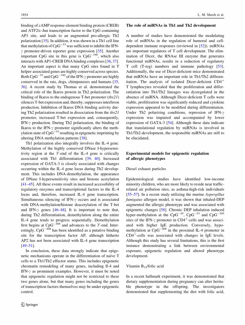
Fig. 1 DNA methylation and histone modification during transcrip-
tional processes. Methylation of CpG islands at promoter regions of
target genes by DNA methyltransferases (DNMT) may contribute to
gene silencing. Methylation of H3 residues at lysine 8 and 27 reduce
the accessibility of the respective DNA segment, while methylation of
lysine 4 at the same residue or acetylation of the H4 residue at lysine
4 increases the accessibility of the DNA
1852 L. M. Marsh et al.
123

Histone acetylation/deacetylation
Histones consist of eight subunits and act as spools for
the DNA molecule. If the histones are tightly packed, then
the transcription apparatus of the cell is unable to
access the DNA and the gene is repressed. Conversely, if
the histones are in a loose formation and remain, ‘‘open’’,
this particular DNA structure is accessible for the DNA
transcription apparatus. This latter state is achieved when
the histones are modified. These modifications occur post-
translationally and include acetylation, methylation, phos-
phorylation and ubiquitylation of certain residues in the
histones. One important mechanism in this regard is acet-
ylation and is performed by enzymes termed histone acetyl
transferases (HAT). Conversely, deacetylation is catalyzed
by histone deacetylases (HDAC). Histone methylation is
regulated via histone methyl transferase at specific residues
of the H3 subunit [15–18]. All of the above-mentioned
mechanisms (acetylation, methylation, etc.) can occur
simultaneously and create a complex pattern on each his-
tone, sometimes referred to as the histone code.
MicroRNA
In addition to modifying transcription, epigenetics can also
affect the translation of mRNA. A recently emerging field
of epigenetic research is the role of micro (mi)RNAs.
miRNAs belong to a class of small non-coding regulatory
RNAs that act through repression of protein expression at
the post-transcriptional level. To date, more than 300
miRNAs have been identified in humans. Each miRNA can
regulate up to several hundred target genes by blocking
translation. These small molecules are believed to regulate
up to one-third of all human genes by promoting the deg-
radation of target mRNAs. Expression of miRNAs may
therefore contribute to the pathogenesis of many human
diseases, and some of which have been recently implied to
be novel valuable diagnostic or even prognostic disease
markers [19–21].
Epigenetic regulation of Th1 and Th2 development
The development of allergic phenotypes in the skin, lung or
the gastrointestinal mucosal tissue is closely linked to the
generation of Th2 T cells. Th2 cells produce a character-
istic cytokine pattern including IL-4, IL-5 and IL-13. IL-4
has been identified as a key cytokine which plays an
important role in the initiation of Th2 development. Fur-
thermore, IL-4 is required to maintain a pool of Th2 cells
as it promotes Th2 cell proliferation. At the other end of the
spectrum, the Th1-related cytokine IFN-c has a broad
spectrum of Th2 counteracting activities, including the
suppression of Th2 T cell responses. Therefore, the regu-
lation of IL-4 and IFN-c production are critical events in
allergic conditions.
Recent data indicate that both IL-4 and IFN-c gene
expression are under close epigenetic regulation. In naıve
(not yet committed) T cells, cytokine genes are only
partially silenced because the baseline level of IFN-c and
Il-4 transcription is evident within a very short time frame
after cell activation. Expression is independent of T-bet or
GATA-3 expression, transcription factors that are criti-
cally involved in Th1 and Th2 cell differentiation,
respectively [22]. In order to progress along the T-helper
cell differentiation pathway, it is important to sustain
expression of one cytokine and repress the other. Cytokine
expression is closely dependent on the expression of the
transcription factors, T-bet and GATA-3. T cell differen-
tiation depends on cell division, it has been suggested that
time was required for one cytokine locus to become
completely accessible while the others to be completely
silenced [23]. Epigenetic modification of cytokine gene
expression was proposed as a mechanism to promote
cytokine gene accessibility for one cytokine locus over the
other as a naıve T cell differentiates into a Th1 or Th2
cell [24–26].
GATA-3 and T-bet mediate many of the chromatin
structural changes that occur during T cell differentiation.
As a result, either the IFN-c or the IL-4 locus becomes
accessible to regulatory enzymes and transcription factors
[24, 27, 28]. Overexpression of T-bet induces DNase I
hypersensitivity of the IFN-c gene locus and enhances
transcription of the interferon gene. T-bet induces and
interacts with its co-factor, HLX. Together, these proteins
activate the IFN-c locus synergistically by promoting
remodeling of the chromatin structure [25, 29, 30].
One important mechanism to alter accessibility of the
IFN-c gene is DNA methylation and demethylation.
Increased IFN-gene expression is observed in T cells
activated in the presence of DNA methylation inhibitors
and also in T cells from DNA methyltransferase-knockout
mice [31–33]. This is further supported by the finding of
decreased DNA methylation in the IFN-c promoter region
of Th1 cells [24, 31]. Conversely, reduced expression of
IFN-c has been associated with increase in de novo
methylation in such cells [34].
Recently, new data have started to reveal how these
methylation patterns may modulate downstream molecular
signaling pathways. The CpG-53 site of the IFN-c promoter
seems to be critically involved in regulating IFN-c gene
expression. CpG-53 resides in a proximal activator protein 1
(AP1) binding site and when methylated alters transcription
factor binding [32, 35, 36]. A subsequent study demon-
strated that methylation of CpG-53 significantly inhibited
Maternal signals for progeny allergoprevention 1853
123
binding of cAMP response element binding protein (CREB)
and ATF2/c-Jun transcription factor to the CpG containing
AP1 site, and leads to an augmented pro-allergic Th2
polarization [35]. In addition, it was shown in a Th1 cell line
that methylation of CpG-53 was sufficient to inhibit the IFN-
c promoter-driven reporter gene expression [35]. Another
important CpG site in this gene is CpG-190, which also
interacts with AP1-CREB DNA binding complexes [36, 37].
An important aspect is that many CpG sites found in T
helper-associated genes are highly conserved across species.
Both CpG-53 and CpG-190 of the IFN-c promoter are highly
conserved in the rats, dogs, chimpanzees and humans [35,
36]. A recent study by Thomas et al. demonstrated the
critical role of the Ikaros protein in Th2 polarization. The
binding of Ikaros to the endogenous T-bet promoter (tbx21)
silences T-bet expression and, thereby, suppresses interferon
production. Inhibition of Ikaros DNA binding activity dur-
ing Th2 polarization resulted in dissociation from the tbx21
promoter, increased T-bet expression and, consequently,
IFN-c production. During Th2 polarization, the binding of
Ikaros to the IFN-c promoter significantly alters the meth-
ylation state of CpG-53 resulting in epigenetic imprinting by
altering DNA methylation patterns [38].
Th1 polarization also integrally involves the IL-4 gene.
Methylation of the highly conserved DNase I-hypersensi-
tivity region at the 30-end of the IL-4 gene is critically
associated with Th1 differentiation [39, 40]. Increased
expression of GATA-3 is closely associated with changes
occurring within the IL-4 gene locus during Th2 develop-
ment. This includes DNA-demethylation, the appearance
of DNase I-hypersensitivity sites and histone acetylation
[41–45]. All these events result in increased accessibility of
regulatory enzymes and transcriptional factors to the IL-4
locus and, therefore, increased IL-4 gene transcription.
Simultaneous silencing of IFN-c occurs and is associated
with DNA-methylation/histone deacetylation of the T-bet
and IFN-c genes [46–48]. It is important to note that,
during Th2 differentiation, demethylation along the entire
IL-4 gene tends to progress sequentially. Demethylation
first begins at CpG-408 and advances to the 30-end. Inter-
estingly, CpG-408 has been identified as a putative binding
site for the transcription factor AP, although hitherto
AP2 has not been associated with IL-4 gene transcription
[49–51].
In conclusion, these data strongly indicate that epige-
netic mechanisms operate in the differentiation of naıve T
cells to a Th1/Th2 effector status. This includes epigenetic
chromatin remodeling of many genes, including Il-4 and
IFN-c as prominent examples. However, it must be noted
that epigenetic regulation might not be restricted to these
two genes alone, but that many genes including the genes
of transcription factors themselves may be under epigenetic
control.
The role of miRNAs in Th1 and Th2 development
A number of studies have demonstrated the modulating
role of miRNAs in the regulation of humoral and cell-
dependent immune responses (reviewed in [52]). miRNAs
are important regulators of T cell development. The elim-
ination of Dicer, the RNAse III enzyme that generates
functional miRNAs, results in a reduction of regulatory
T cell (T-reg) numbers and immune pathology [53].
Additionally, the use of Dicer-deficient mice demonstrated
that miRNAs have an important role in Th1/Th2 differen-
tiation. The analysis of isolated Dicer-deficient CD4?
T lymphocytes revealed that the proliferation and differ-
entiation into Th1/Th2 lineages was dysregulated in the
absence of miRNA. Although Dicer-deficient T cells were
viable, proliferation was significantly reduced and cytokine
expression appeared to be modified during differentiation.
Under Th2 polarizing conditions, repression of IFN-cexpression was impaired and accompanied by lower
expression of GATA-3 [54]. Although these data indicate
that translational regulation by miRNAs is involved in
Th1/Th2-development, the responsible miRNAs are still to
be elucidated.
Experimental models for epigenetic regulation
of allergic phenotypes
Diesel exhaust particles
Epidemiological studies have identified low-income
minority children, who are more likely to reside near traffic-
related air pollution sites, as asthma-high-risk individuals
[55–57]. In a recent study utilizing the murine Aspergillus
fumigatus allergen model, it was shown that inhaled-DEP
augmented the allergic phenotype and was associated with
epigenetic changes [58]. Chronic DEP inhalation induced
hyper-methylation at the CpG-45, CpG-53 and CpG-205
sites of the IFN-c promoter in CD4?-cells and was associ-
ated with higher IgE production. Conversely, hypo-
methylation at CpG-408 in the proximal IL-4 promoter in
CD4?-cells was associated with changes in IgE levels.
Although this study has several limitations, this is the first
instance demonstrating a link between environmental
exposure, epigenetic regulation and altered phenotype
development.
Vitamin B12/folic acid
In a recent hallmark experiment, it was demonstrated that
dietary supplementation during pregnancy can alter herita-
ble phenotype in the offspring. The investigators
demonstrated that supplementing the diet with folic acid,
1854 L. M. Marsh et al.
123

vitamin B12 or other agents can alter mouse coat color. The
increased abundance of methyl donors in the diet enhanced
CpG methylation in the promoter of the agouti gene and
therefore altered coat color [57, 59]. The relevance of a
methyl-rich diet during pregnancy on development of an
allergic phenotype has been shown in a prenatal mouse
model. Mothers fed with a donor-rich diet increased the
severity of the asthmatic phenotype. Increased airway
inflammation was associated with enhanced DNA methyl-
ation of transcription factors that balance inflammation of
the bronchus [60].
Policyclic aromatic hydrocarbons
The traffic-related increase in asthma development, par-
ticularly in the inner city population, might not only be
related to DEP but also to polycyclic aromatic hydrocar-
bons (PAH). In a recent study, the relationship of
transplacental exposure to traffic-related PAH and the
development of childhood asthma was explored. The
investigators monitored maternal PAH exposure and
studied the methylation status in umbilical cord white
blood cells. Over 30 DNA sequences were identified
whose methylation status was dependent on the level of
maternal PAH exposure. The highest concordance
between the levels of methylation and gene expression in
matched fetal placental tissues was found for acyl-coa-
synthetase long-chain family member 3 (ACSL3) [61].
This gene belongs to a family of genes that encodes key
enzymes in fatty acid metabolism [62]. ACSL3 is
expressed in lung and thymic tissue and is responsible for
the intracellular conversion of long-chain fatty acids
[63, 64]. Thus, hypermethylation of this gene is expected
to diminish fatty acid utilization and may also possibly
influence membrane phospholipid composition. Whether
these functional changes directly affect the development
of the asthmatic phenotype is unknown. However, several
epidemiological studies have shown that the fatty acid
composition in milk and other nutrients affect the devel-
opment of allergy and asthma. This effect has been
attributed to the anti-inflammatory and immune-modulat-
ing effects of omega-3 (n-3) fatty acids [65–69]. It is
interesting to note that ACSL3 is located in 2q36.1, a
region that has been recently shown to be associated with
asthma susceptibility [70, 71]. In conclusion, the trans-
placental PAH exposure provides another model situation
that underlies the importance of epigenetic regulation in
terms of phenotype development, although the detailed
cause and effect relationship between PAH exposure,
hypermethylation of the ACSL3 gene and asthma devel-
opment still remains to be fully established.
Gestational smoke exposure
The most convincing data that prenatal environmental
exposure can influence the risk for subsequent asthma
development originate from the work on environmental
tobacco smoke (ETS) exposure. There is overwhelming
evidence that prenatal exposure to ETS is associated with a
number of asthma hallmarks, including impaired respira-
tory function, wheezing, respiratory infections, and altered
airway structure [72–74]. Most recently, the adverse
impact of smoking has also been demonstrated to signifi-
cantly increase the risk of developing atopic dermatitis
[75]. Recent data suggest that the effect of ETS exposure
on asthma development can be transmitted across two
generations [76]. The investigators conducted a case–con-
trol study that included 338 children with asthma and 570
control subjects and used an innovative sampling design to
efficiently investigate the association between in utero
exposure to maternal smoking and asthma occurrence. The
study confirmed that in utero exposure was associated with
increased risk for asthma diagnosis in the first 5 years of
life. Additionally, it is most remarkable that even grand-
maternal smoking during the mother’s fetal period may
present a risk factor for asthma in the grandchild’s gener-
ation. Furthermore, this was independent of maternal
smoking. The risk for asthma in grandchildren was highest
if both the grandmother and the mother smoked during
pregnancy.
From a mechanistic point of view, parental smoking is
associated with higher cord blood IgE levels [77]. Since
IgE does not cross the placental barrier, this provides
evidence of a direct effect of maternal smoking on fetal
immune functions. Recently, maternal smoking has been
associated with stronger neonatal T cell proliferation
[78]. However, the effects on cytokine responses were
not investigated. Another study has provided evidence
that maternal smoking in pregnancy is associated with
lower Th1 responses to mitogenic stimulation as mea-
sured by mRNA expression levels [79]. Whether this is
directly regulated epigenetically has not so far been
investigated, but it must be noted that tobacco smoking
has recently been shown to modify gene expression by
promoter hypermethylation associated with down-regula-
tion of gene transcription in the lung [80]. These data
indicate that, in principle, tobacco smoke has the
capacity to act on the level of epigenetic regulation.
Whether the observed increased risk of asthma devel-
opment is also related to epigenetic events remains to
be investigated. The epidemiological data, particularly
on grandmaternal exposures, at least point in this
direction.
Maternal signals for progeny allergoprevention 1855
123
Fetal innate immune responses to microbial stimuli
Evidence from epidemiological studies
The traditional farming environment confers protection to
allergy and asthma by the contact of farm children with a
wide range of naturally prevalent microbes in barns and
hay lofts as well as in the domestic environment The so
called ‘‘farming effect’’ was first described in epidemio-
logical studies conducted in the Alpine region and then
confirmed for other regions all over the world. Numerous
cross-sectional studies supported the observations that
early contact with animals and consumption of farm-
derived products may protect against allergies and asthma
later in life. In this context, it was shown that the farming
environment may already provide protection against
allergic disorders in utero. Maternal exposure to livestock
and consumption of farm-produced milk during pregnancy
were associated with a reduced risk of allergies in the
offspring (reviewed in [81]). Data from the ongoing birth-
cohort PASTURE-Study supported the modulating influ-
ence of prenatal maternal farming exposure on the
developing immune system of the offspring. Cord blood
samples from neonates born to farming families were
shown to produce substantially more IFN-c and TNF-aafter cytokine stimulation compared to samples from
babies born to non-farming families. This may indicate that
the innate immune system might be involved in interac-
tions between environmental factors and the developing
immune system [82].
Early responses of the innate immune system
The innate immune response to a pathogen is coordinated
by antigen-presenting cells (APC). The main APC popu-
lation is dominated by dendritic cells (DCs), cells that are
crucial for determining T-helper cell fate and, subse-
quently, for the development of asthma. Depletion of
CD11c?–DCs leads to significant abrogation of the char-
acteristic features of experimental asthma. This indicates
that these cells are necessary and sufficient for the
induction of a Th2-driven inflammatory allergic response
[83]. The activation status of DCs has an important role in
this regard. The expression profile of DCs is affected by
the interaction of their so-called pattern recognition
receptors (PRRs) with the microbes’ pathogen-associated
molecular patterns (PAMPs). In contrast to the highly-
specific antigen recognition of the T cell receptors, the
interaction between PAMPs and PRRs is rather unspecific.
PAMP activation initiates different effector cascades, e.g.,
the release of antimicrobial defensins or signaling the
adaptive immune response [84]. The Toll-like receptor
(TLR) family represents the best-characterized class of
PRRs. TLRs represent sensors for microorganisms and
microbial components: TLR 1/6 and TLR 1/2 complexes
are PRRs that predominantly recognize Gram-positive
bacteria such as mycobacteria or lactic acid bacteria;
TLR4 recognizes LPS from Gram-negative bacteria, TLR5
serves as the receptor for flagellin, the main protein in
bacterial flagella; TLR3,7 and 9 are endosomal-associated
TLRs that recognize foreign DNA: double-stranded
(ds)DNA from bacteria and viruses is recognized by TLR3
and 9, TLR9 is able to detect CpG domains in dsDNA,
while TLR7 detects bacterial and virus single-stranded
DNA. Ligand-binding to the TLR activates several intra-
cellular signaling pathways including the NF-jB pathway.
Intracellular TLR signaling (with the exception of TLR3)
occurs via MyD88-dependent or -independent pathways.
TLR-mediated activation of DCs induces expression of
co-stimulatory molecules (CD80 and CD86) as well as of
inflammatory cytokines (TNF-a, INFs, IL-1, IL-6, IL-10,
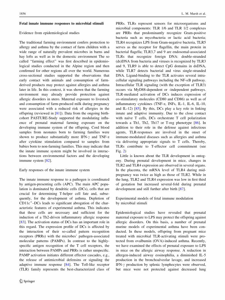
and IL-12) [85]. By this, DCs play a key role in linking
innate and adaptive immunity. Due to the close contact
with naıve T cells, DCs orchestrate T cell polarization
towards a Th1, Th2, Th17 or T-reg phenotype [86]. In
addition to their role in the defense against infectious
agents, TLR-responses are involved in the onset of
immune-modulated diseases such as allergies and asthma
via delivering appropriate signals to T cells. Thereby,
TLRs contribute to T-effector cell commitment (see
Fig. 2)
Little is known about the TLR development in ontog-
eny. During prenatal development in mice, changes in
TLR2 and TLR4 expression are observed in several organs.
In the placenta, the mRNA level of TLR4 during mid-
pregnancy was twice as high as those of TLR2. While in
the lung, TLR2 and TLR4 expression was low in first third
of gestation but increased several-fold during prenatal
development and still further after birth [87].
Experimental models of fetal immune modulation
by microbial stimuli
Epidemiological studies have revealed that prenatal
maternal exposure to LPS may protect the offspring against
allergic disorders. On this basis, a number of prenatal
murine models of experimental asthma have been con-
ducted. In these models, offspring from pregnant mice
treated with microbial TLR-activating stimuli were pro-
tected from ovalbumin (OVA)-induced asthma. Recently,
we have examined the effects of prenatal exposure to LPS
in mice on the allergic airway response. A reduction in
allergen-induced airway eosinophilia, a diminished IL-5
production in the bronchoalveolar lavage, and increased
IFN-c production by splenic lymphocytes were observed,
but mice were not protected against decreased lung
1856 L. M. Marsh et al.
123
function [88]. In a recently published report that describes
LPS application in a rat model of allergic airway inflam-
mation, the reduction in Th2 cytokine production and cell
recruitment in the bronchoalveolar lavage was confirmed.
However, in contrast to the above mentioned mouse
experiments, airway hyperresponsiveness was normalized
in offspring from LPS-treated mothers [89]. The reasons
for these discrepant results in terms of altered lung func-
tions are still unclear, but may be explained by species-
specific effects or methodological differences in lung
function measurements.
Bacteria isolated from the farming environment have
been shown to confer protection against the development of
an allergic phenotype when administered prior to sensiti-
zation [90]. Furthermore, Acinetobacter lwoffii F78, a
stable-derived Gram-negative bacterium, exhibits anti-
allergic properties in a prenatal mouse model. In contrast to
the prenatally tested Lactobacillus rhamnosus GG and LPS,
intranasal maternal application of A. lwoffii F78 resulted in a
reduction of all features of airway inflammation and
normalized airway hyperresponsiveness in OVA-sensitized
offspring. Additionally, a trend towards lower OVA-specific
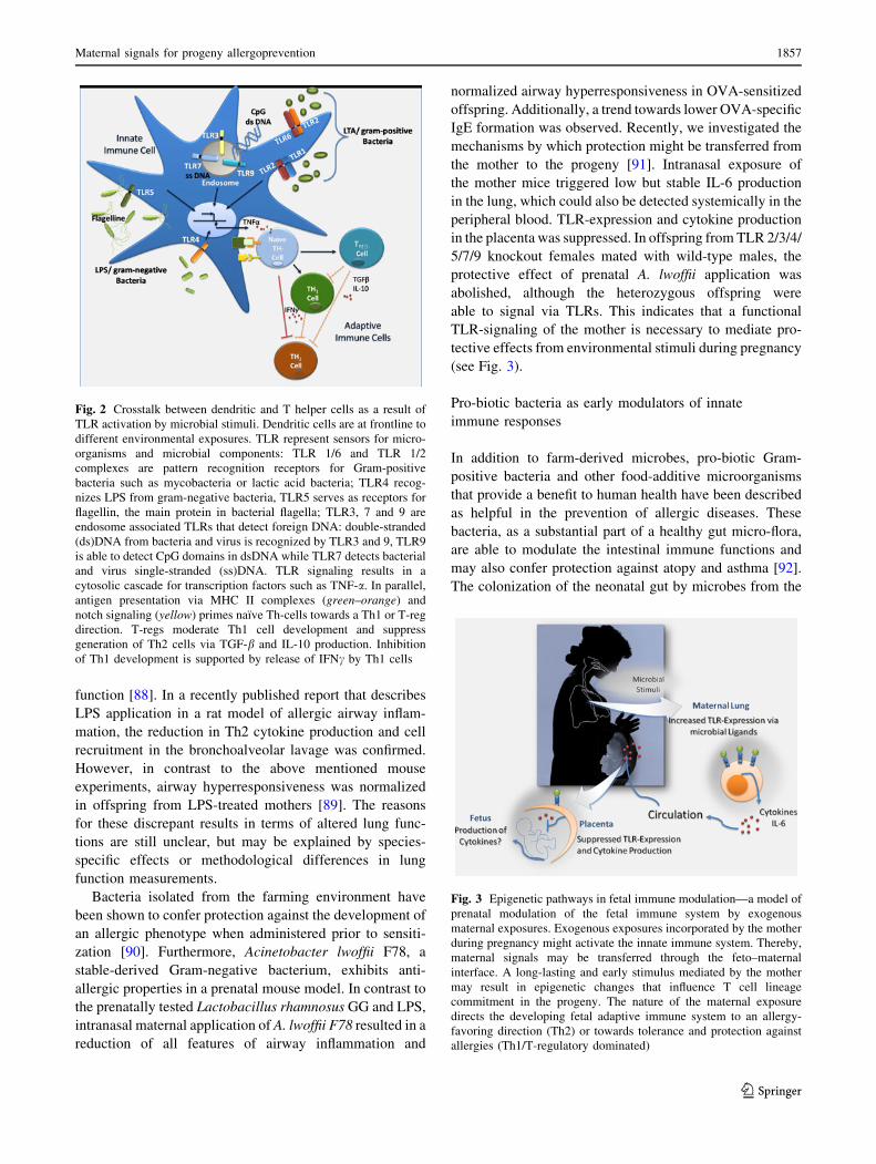
IgE formation was observed. Recently, we investigated the
mechanisms by which protection might be transferred from
the mother to the progeny [91]. Intranasal exposure of
the mother mice triggered low but stable IL-6 production
in the lung, which could also be detected systemically in the
peripheral blood. TLR-expression and cytokine production
in the placenta was suppressed. In offspring from TLR 2/3/4/
5/7/9 knockout females mated with wild-type males, the
protective effect of prenatal A. lwoffii application was
abolished, although the heterozygous offspring were
able to signal via TLRs. This indicates that a functional
TLR-signaling of the mother is necessary to mediate pro-
tective effects from environmental stimuli during pregnancy
(see Fig. 3).
Pro-biotic bacteria as early modulators of innate
immune responses
In addition to farm-derived microbes, pro-biotic Gram-
positive bacteria and other food-additive microorganisms
that provide a benefit to human health have been described
as helpful in the prevention of allergic diseases. These
bacteria, as a substantial part of a healthy gut micro-flora,
are able to modulate the intestinal immune functions and
may also confer protection against atopy and asthma [92].
The colonization of the neonatal gut by microbes from the
Fig. 2 Crosstalk between dendritic and T helper cells as a result of
TLR activation by microbial stimuli. Dendritic cells are at frontline to
different environmental exposures. TLR represent sensors for micro-
organisms and microbial components: TLR 1/6 and TLR 1/2
complexes are pattern recognition receptors for Gram-positive
bacteria such as mycobacteria or lactic acid bacteria; TLR4 recog-
nizes LPS from gram-negative bacteria, TLR5 serves as receptors for
flagellin, the main protein in bacterial flagella; TLR3, 7 and 9 are
endosome associated TLRs that detect foreign DNA: double-stranded
(ds)DNA from bacteria and virus is recognized by TLR3 and 9, TLR9
is able to detect CpG domains in dsDNA while TLR7 detects bacterial
and virus single-stranded (ss)DNA. TLR signaling results in a
cytosolic cascade for transcription factors such as TNF-a. In parallel,
antigen presentation via MHC II complexes (green–orange) and
notch signaling (yellow) primes naıve Th-cells towards a Th1 or T-reg
direction. T-regs moderate Th1 cell development and suppress
generation of Th2 cells via TGF-b and IL-10 production. Inhibition
of Th1 development is supported by release of IFNc by Th1 cells
Fig. 3 Epigenetic pathways in fetal immune modulation—a model of
prenatal modulation of the fetal immune system by exogenous
maternal exposures. Exogenous exposures incorporated by the mother
during pregnancy might activate the innate immune system. Thereby,
maternal signals may be transferred through the feto–maternal
interface. A long-lasting and early stimulus mediated by the mother
may result in epigenetic changes that influence T cell lineage
commitment in the progeny. The nature of the maternal exposure
directs the developing fetal adaptive immune system to an allergy-
favoring direction (Th2) or towards tolerance and protection against
allergies (Th1/T-regulatory dominated)
Maternal signals for progeny allergoprevention 1857
123

neonate environment stimulates intestinal immune system
towards a Th1 phenotype compensating the Th2-bias
established in utero that prevents rejection of the fetus [93].
The shaping of the intestinal immune system by human
colonic DCs occurs predominantly via TLR2 signaling.
Pro-biotic bacteria also induce the production of trans-
forming growth factor (TGF)-b, which in turn stimulates
T-helper cells to release Th1 cytokines. These cytokines
act on B cells by inducing a switch to IgA-production; a
mechanism that reduces inflammation and establishes
clinical tolerance against common antigens. The lack of
microbial stimuli may lead to an elevated IgE production
by B cells and an increased risk of allergic reactions due to
the subsequent activation of mast cells [94].
A large group of bacteria with pro-biotic properties is
represented by the genus Lactobacillus. Lactobacilli were
shown to have immune-modulating capabilities by altering
the cytokine pattern of the host. Reviewing clinical trials of
the last decade with regard to preventive properties of
Lactobacilli, two clinical research groups have provided
contradictory results. The group of Kalliomaki and Isolauri
from Finland reported protective effects on the develop-
ment of atopic eczema in pre- and postnatally-treated high-
risk infants with sustained effects in follow-up studies after
7 years [95]. However, Taylor et al. did not obtain any
significant findings [96]. Another trial in which pro-biotic
substances were applied to high-risk neonates showed
protective effects on atopic eczema [97]. A meta-analysis
done by Lee et al. revealed an overall Odds Ratios below 1
indicating preventive effects of lactic acid bacteria on
pediatric atopic dermatitis [98]. These results were chal-
lenged by Salfeld and Kopp arguing that these calculations
included studies referring to the same study populations
[99]. Unfortunately, comparative data for prenatal asthma
prevention by pro-biotics are currently not available.
Experiments in acute and perinatal animal models of
experimental allergic asthma revealed more consistent
results [100]. In a perinatal murine model of asthma, we
applied L. rhamnosus GG orally to mother mice. This
resulted in a significant reduction of airway eosinophilia
and inflammation in the lung, but failed to normalize lung
function in OVA-sensitized offspring [101].
Immune modulation via TLR9 ligands
Kitagaki et al. demonstrated that application of the TLR9
ligand, CpG containing DNA, prior to sensitization pre-
vented the Th2 inflammatory response and effectively
interferes with the development of atopic airway disease in
a murine model of experimental asthma. Moreover, when
administered in combination with an experimental aller-
gen, CpG promoted the reversal of established eosinophilic
inflammation [102]. Recently, we found that activation of
TLR3 and 7 by viral TLR ligands has both preventive as
well as suppressive effects on experimental asthma and is
mediated by the additive effects of IL-12 and IL-10.
However, this concept is still to be tested in a prenatal
experimental set-up [103].
Helminth infections as perinatal allergy protective
factors
Both helminth infections and allergic diseases are strong
inducers of Th2 responses. Similar to atopic disease,
helminth infections also up-regulate Th2 cytokines, IgE-
production and induce eosinophilia. Asymptomatic per-
sistence of helminth infections in endemic regions appear
to be achieved by activation of regulatory T cells and
systemically elevated levels of IL-10 and possibly of TGF-
b. By this mechanism, helminth infections may have a
down-regulatory effect on the risk of developing allergic
disease [104].
Recent studies have indicated that helminth infections
could affect the early development of immunity. Exposure
to helminths and protozoan infection in the mother may
alter TLR-expression in cells of the innate immune system
and subsequently influence cytokine production in cord
blood mononuclear cells [105, 106]. Experimental set-ups
mainly use the model of schistosomiasis to investigate
allergy-diminishing effects and related mechanisms on
allergic outcomes, but none have yet proved the hypothesis
of in utero protection.
Stimulation of innate immune response by other
environmental exposures
Stimulation of the innate immune system by microbial
exposure may reduce the development of childhood
allergy. Additionally, other environmental factors such as
fatty acids may affect neonatal TLR stimulation and,
therefore, modulate the capacity of TLR to respond to
microbial stimuli. Cord blood cells from neonates born to
non-atopic mothers showed higher levels of IL-10 pro-
duction and FOXP3-expression when stimulated with the
TLR2 agonist peptidoglycan than those born to atopic
mothers [107]. A similar study conducted with cord blood
cells revealed associations between maternal allergy and
significantly higher neonatal IL-12 and IFN-c responses to
TLR2, 3, and 4 activation. TNF-a- and IL-6 responses to
TLR2, 4, and 5 activation were significantly higher in
newborns that later developed allergic disease. Newborns
that developed an allergic disease later on in life had lower
Th1-IFN-c responses to mitogens [108]. Data from a
United States inner city cohort revealed that prenatal stress
affects the innate immunity. Higher prenatal maternal
stress scores were related to an increased IL-8 production
1858 L. M. Marsh et al.
123
after microbial stimulation of TLR and increased TNF-aproduction in cord blood cells [109]. Other factors that may
modulate TLR-mediated innate cytokine responses include
parental allergic and airway diseases, somatic fetal growth,
ethnicity, and season of birth [110]. Common environ-
mental toxic chemicals may also alter TLR signaling.
Decreased expression of transcription factors GATA-3,
T-bet and Foxp3 was observed in offspring from a prenatal
murine model, where mother mice were exposed to a low-
level toluene concentration and the TLR2 ligand peptido-
glucan during pregnancy [111]. Collectively, these findings
suggest that urban prenatal exposures and familial genetic
factors affect the development of the fetal innate immune
system.
TLR gene polymorphisms
TLR gene polymorphisms as heritable factors may be rele-
vant in prenatal development. Epidemiological association
studies have identified certain TLR gene polymorphisms
that are associated with the increased prevalence of allergic
diseases [112]. For example, allergic diseases in farmers’
children could have contributed to a significantly elevated
prevalence of a polymorphism found at the TLR2/-16934
locus [113]. Results from a Swedish study indicated that a
polymorphism in the TLR4-gene is associated with asthma
characterized by a decreased IL-12 production by APCs
after LPS stimulation. On the other hand, TLR1/6 hetero-
dimer polymorphisms are associated with an elevated
protection against childhood asthma [114]. Gene–environ-
ment interactions may also modify TLR2- and CD14-
mediated responses. Individuals growing up in a rural
environment are more susceptible to allergies and asthma
when carrying polymorphisms in the respective TLR genes
[115]. A mutation in the TLR2/R753Q region was shown to
be associated with a modified cytokine production and TLR
expression in patients with atopic dermatitis [116].
Conclusion
Epidemiological, clinical and experimental evidence pro-
vides compelling new insights into the critical role of the pre-
and postnatal environment in terms of immuno-program-
ming, immuno-education and subsequent allergy and asthma
protection. These studies provided the basis on which
underlying mechanisms will be elucidated and discovered.
An increased understanding of the underlying cellular
and molecular networks will be the prerequisite for the
development of novel allergy-protective and -preventive
strategies. Further investigations on the relationship between
the genome, epigenome and transcriptome may lead to
a better understanding of the phenotype development.
Similarly, the understanding of how exogenous exposures
initiate gene–environment interactions on the transcriptional
and translational level in specific cell types and thereby
modify the phenotype may hold the key for novel clinical
approaches in allergy and asthma.
Acknowledgments This work is supported by grants from the
Deutsche Forschungsgemeinschaft (TR22) and the von Behring-
Rontgen-Stiftung (Grant 56-00359).
References
1. Barnes PJ (2009) The cytokine network in chronic obstructive
pulmonary disease. Am J Respir Cell Mol Biol 41:631–638
2. Rowe J, Kusel M, Holt BJ, Suriyaarachchi D, Serralha M,
Hollams E, Yerkovich ST, Subrata LS, Ladyman C, Sadowska
A, Gillett J, Fisher E, Loh R, Soderstrom L, Ahlstedt S, Sly PD,
Holt PG (2007) Prenatal versus postnatal sensitization to envi-
ronmental allergens in a high-risk birth cohort. J Allergy Clin
Immunol 119:1164–1173
3. Ege MJ, Bieli C, Frei R, van Strien RT, Riedler J, Ublagger E,
Schram-Bijkerk D, Brunekreef B, van Hage M, Scheynius A,
Pershagen G, Benz MR, Lauener R, von Mutius E, Braun-
Fahrlander C (2006) Prenatal farm exposure is related to the
expression of receptors of the innate immunity and to atopic
sensitization in school-age children. J Allergy Clin Immunol
117:817–823
4. Vercelli D (2008) Discovering susceptibility genes for asthma
and allergy. Nat Rev Immunol 8:169–182
5. Egger G, Liang G, Aparicio A, Jones PA (2004) Epigenetics in
human disease and prospects for epigenetic therapy. Nature
429:457–463
6. Jaenisch R, Bird A (2003) Epigenetic regulation of gene
expression: how the genome integrates intrinsic and environ-
mental signals. Nat Genet 33(suppl):245–254
7. Jiang YH, Bressler J, Beaudet AL (2004) Epigenetics and
human disease. Annu Rev Genomics Hum Genet 5:479–510
8. Richardson BC (2002) Role of DNA methylation in the regu-
lation of cell function: autoimmunity, aging and cancer. J Nutr
132:2401S–2405S
9. Vercelli D (2004) Genetics, epigenetics, and the environment:
switching, buffering, releasing. J Allergy Clin Immunol
113:381–386
10. Finch CE, Crimmins EM (2004) Inflammatory exposure and
historical changes in human life-spans. Science 305:1736–1739
11. Bestor TH (2000) The DNA methyltransferases of mammals.
Hum Mol Genet 9:2395–2402
12. Hmadcha A, Bedoya FJ, Sobrino F, Pintado E (1999) Methyl-
ation-dependent gene silencing induced by interleukin 1beta via
nitric oxide production. J Exp Med 190:1595–1604
13. Baylin SB, Herman JG (2000) DNA hypermethylation in
tumorigenesis: epigenetics joins genetics. Trends Genet
16:168–174
14. Kawai J, Hirose K, Fushiki S, Hirotsune S, Ozawa N, Hara A,
Hayashizaki Y, Watanabe S (1994) Comparison of DNA
methylation patterns among mouse cell lines by restriction
landmark genomic scanning. Mol Cell Biol 14:7421–7427
15. Nightingale KP, O’Neill LP, Turner BM (2006) Histone modi-
fications: signalling receptors and potential elements of a
heritable epigenetic code. Curr Opin Genet Dev 16:125–136
16. Allfrey VG, Faulkner R, Mirsky AE (1964) Acetylation and
methylation of histones and their role in the regulation of RNA
synthesis. Proc Natl Acad Sci USA 51:786–794
Maternal signals for progeny allergoprevention 1859
123

17. Khan AU, Krishnamurthy S (2005) Histone modifications as key
regulators of transcription. Front Biosci 10:866–872
18. Shi Y, Lan F, Matson C, Mulligan P, Whetstine JR, Cole PA,
Casero RA, Shi Y (2004) Histone demethylation mediated
by the nuclear amine oxidase homolog LSD1. Cell 119:
941–953
19. Lee RC, Feinbaum RL, Ambros V (1993) The C. elegans het-
erochronic gene lin-4 encodes small RNAs with antisense
complementarity to lin-14. Cell 75:843–854
20. Krek A, Grun D, Poy MN, Wolf R, Rosenberg L, Epstein EJ,
MacMenamin P, da Piedade I, Gunsalus KC, Stoffel M, Ra-
jewsky N (2005) Combinatorial microRNA target predictions.
Nat Genet 37:495–500
21. Yanaihara N, Caplen N, Bowman E, Seike M, Kumamoto K, Yi
M, Stephens RM, Okamoto A, Yokota J, Tanaka T, Calin GA,
Liu CG, Croce CM, Harris CC (2006) Unique microRNA
molecular profiles in lung cancer diagnosis and prognosis.
Cancer Cell 9:189–198
22. Grogan JL, Mohrs M, Harmon B, Lacy DA, Sedat JW, Locksley
RM (2001) Early transcription and silencing of cytokine genes
underlie polarization of T helper cell subsets. Immunity
14:205–215
23. Mullen AC, Hutchins AS, Villarino AV, Lee HW, High FA,
Cereb N, Yang SY, Hua X, Reiner SL (2001) Cell cycle con-
trolling the silencing and functioning of mammalian activators.
Curr Biol 11:1695–1699
24. Ansel KM, Lee DU, Rao A (2003) An epigenetic view of helper
T cell differentiation. Nat Immunol 4:616–623
25. Szabo SJ, Sullivan BM, Peng SL, Glimcher LH (2003) Molec-
ular mechanisms regulating Th1 immune responses. Annu Rev
Immunol 21:713–758
26. Wilson CB, Makar KW, Shnyreva M, Fitzpatrick DR (2005)
DNA methylation and the expanding epigenetics of T cell
lineage commitment. Semin Immunol 17:105–119
27. Murphy KM, Reiner SL (2002) The lineage decisions of helper
T cells. Nat Rev Immunol 2:933–944
28. Rao A, Avni O (2000) Molecular aspects of T-cell differentia-
tion. Br Med Bull 56:969–984
29. Mullen AC, Hutchins AS, High FA, Lee HW, Sykes KJ,
Chodosh LA, Reiner SL (2002) Hlx is induced by and geneti-
cally interacts with T-bet to promote heritable T(H)1 gene
induction. Nat Immunol 3:652–658
30. Zheng WP, Zhao Q, Zhao X, Li B, Hubank M, Schatz DG,
Flavell RA (2004) Up-regulation of Hlx in immature Th cells
induces IFN-gamma expression. J Immunol 172:114–122
31. Young HA, Dray JF, Farrar WL (1986) Expression of trans-
fected human interferon-gamma DNA: evidence for cell-specific
regulation. J Immunol 136:4700–4703
32. Young HA, Ghosh P, Ye J, Lederer J, Lichtman A, Gerard JR,
Penix L, Wilson CB, Melvin AJ, McGurn ME (1994) Differ-
entiation of the T helper phenotypes by analysis of the
methylation state of the IFN-gamma gene. J Immunol
153:3603–3610
33. Makar KW, Wilson CB (2004) DNA methylation is a nonre-
dundant repressor of the Th2 effector program. J Immunol
173:4402–4406
34. Mikovits JA, Young HA, Vertino P, Issa JP, Pitha PM, Turco-
ski-Corrales S, Taub DD, Petrow CL, Baylin SB, Ruscetti FW
(1998) Infection with human immunodeficiency virus type 1
upregulates DNA methyltransferase, resulting in de novo
methylation of the gamma interferon (IFN-gamma) promoter
and subsequent downregulation of IFN-gamma production. Mol
Cell Biol 18:5166–5177
35. Jones B, Chen J (2006) Inhibition of IFN-gamma transcription
by site-specific methylation during T helper cell development.
EMBO J 25:2443–2452
36. White GP, Watt PM, Holt BJ, Holt PG (2002) Differential
patterns of methylation of the IFN-gamma promoter at CpG and
non-CpG sites underlie differences in IFN-gamma gene
expression between human neonatal and adult CD. J Immunol
168:2820–2827
37. Ye J, Ghosh P, Cippitelli M, Subleski J, Hardy KJ, Ortaldo JR,
Young HA (1994) Characterization of a silencer regulatory
element in the human interferon-gamma promoter. J Biol Chem
269:25728–25734
38. Thomas RM, Chen C, Chunder N, Ma L, Taylor J, Pearce EJ,
Wells AD (2010) Ikaros silences T-bet expression and inter-
feron-gamma production during T helper 2 differentiation.
J Biol Chem 285:2545–2553
39. Yano S, Ghosh P, Kusaba H, Buchholz M, Longo DL (2003)
Effect of promoter methylation on the regulation of IFN-gamma
gene during in vitro differentiation of human peripheral blood T
cells into a Th2 population. J Immunol 171:2510–2516
40. Lee DU, Agarwal S, Rao A (2002) Th2 lineage commitment and
efficient IL-4 production involves extended demethylation of the
IL-4 gene. Immunity 16:649–660
41. Bird JJ, Brown DR, Mullen AC, Moskowitz NH, Mahowald
MA, Sider JR, Gajewski TF, Wang CR, Reiner SL (1998)
Helper T cell differentiation is controlled by the cell cycle.
Immunity 9:229–237
42. Lee HJ, Takemoto N, Kurata H, Kamogawa Y, Miyatake S,
O’Garra A, Arai N (2000) GATA-3 induces T helper cell type 2
(Th2) cytokine expression and chromatin remodeling in com-
mitted Th1 cells. J Exp Med 192:105–115
43. Takemoto N, Arai K, Miyatake S (2002) Cutting edge: the
differential involvement of the N-finger of GATA-3 in chro-
matin remodeling and transactivation during Th2 development.
J Immunol 169:4103–4107
44. Fields PE, Kim ST, Flavell RA (2002) Cutting edge: changes in
histone acetylation at the IL-4 and IFN-gamma loci accompany
Th1/Th2 differentiation. J Immunol 169:647–650
45. Fields PE, Lee GR, Kim ST, Bartsevich VV, Flavell RA (2004)
Th2-specific chromatin remodeling and enhancer activity in the
Th2 cytokine locus control region. Immunity 21:865–876
46. Glaser R, Kiecolt-Glaser JK (2005) Stress-induced immune
dysfunction: implications for health. Nat Rev Immunol
5:243–251
47. Hewitt SL, High FA, Reiner SL, Fisher AG, Merkenschlager M
(2004) Nuclear repositioning marks the selective exclusion of
lineage-inappropriate transcription factor loci during T helper
cell differentiation. Eur J Immunol 34:3604–3613
48. Mullen AC, High FA, Hutchins AS, Lee HW, Villarino AV,
Livingston DM, Kung AL, Cereb N, Yao TP, Yang SY, Reiner
SL (2001) Role of T-bet in commitment of TH1 cells before
IL-12-dependent selection. Science 292:1907–1910
49. Ansel KM, Djuretic I, Tanasa B, Rao A (2006) Regulation of
Th2 differentiation and Il4 locus accessibility. Annu Rev
Immunol 24:607–656
50. Comb M, Goodman HM (1990) CpG methylation inhibits pro-
enkephalin gene expression and binding of the transcription
factor AP-2. Nucleic Acids Res 18:3975–3982
51. Tykocinski LO, Hajkova P, Chang HD, Stamm T, Sozeri O,
Lohning M, Hu-Li J, Niesner U, Kreher S, Friedrich B,
Pannetier C, Grutz G, Walter J, Paul WE, Radbruch A (2005) A
critical control element for interleukin-4 memory expression in
T helper lymphocytes. J Biol Chem 280:28177–28185
52. Xiao C, Rajewsky K (2009) MicroRNA control in the immune
system: basic principles. Cell 136:26–36
53. Cobb BS, Hertweck A, Smith J, O’Connor E, Graf D, Cook T,
Smale ST, Sakaguchi S, Livesey FJ, Fisher AG, MerkenschlagerM (2006) A role for Dicer in immune regulation. J Exp Med
203:2519–2527
1860 L. M. Marsh et al.
123

54. Muljo SA, Ansel KM, Kanellopoulou C, Livingston DM, Rao A,
Rajewsky K (2005) Aberrant T cell differentiation in the
absence of Dicer. J Exp Med 202:261–269
55. Brauer M, Hoek G, Van Vliet P, Meliefste K, Fischer PH, Wijga
A, Koopman LP, Neijens HJ, Gerritsen J, Kerkhof M, Heinrich
J, Bellander T, Brunekreef B (2002) Air pollution from traffic
and the development of respiratory infections and asthmatic and
allergic symptoms in children. Am J Respir Crit Care Med
166:1092–1098
56. Gehring U, Cyrys J, Sedlmeir G, Brunekreef B, Bellander T,
Fischer P, Bauer CP, Reinhardt D, Wichmann HE, Heinrich J
(2002) Traffic-related air pollution and respiratory health during
the first 2 years of life. Eur Respir J 19:690–698
57. Venn AJ, Lewis SA, Cooper M, Hubbard R, Britton J (2001)
Living near a main road and the risk of wheezing illness in
children. Am J Respir Crit Care Med 164:2177–2180
58. Liu J, Ballaney M, Al-alem U, Quan C et al (2008) Combined
inhaled diesel exhaust particles and allergen exposure alter
methylation of T helper genes and IgE production in vivo.
Toxicol Sci 102:76–81
59. Waterland RA, Jirtle RL (2003) Transposable elements: targets
for early nutritional effects on epigenetic gene regulation. Mol
Cell Biol 23:5293–5300
60. Hollingsworth JW, Maruoka S, Boon K, Garantziotis S, Li Z,
Tomfohr J, Bailey N, Potts EN, Whitehead G, Brass DM, Sch-
wartz DA (2008) In utero supplementation with methyl donors
enhances allergic airway disease in mice. J Clin Invest
118:3462–3469
61. Perera F, Tang WY, Herbstman J, Tang D, Levin L, Miller R,
Ho SM (2009) Relation of DNA methylation of 50-CpG island of
ACSL3 to transplacental exposure to airborne polycyclic aro-
matic hydrocarbons and childhood asthma. PLoS One 4:e4488
62. Mashek DG, Bornfeldt KE, Coleman RA, Berger J, Bernlohr
DA, Black P, DiRusso CC, Farber SA, Guo W, Hashimoto N,
Khodiyar V, Kuypers FA, Maltais LJ, Nebert DW, Renieri A,
Schaffer JE, Stahl A, Watkins PA, Vasiliou V, Yamamoto TT
(2004) Revised nomenclature for the mammalian long-chain
acyl-CoA synthetase gene family. J Lipid Res 45:1958–1961
63. Minekura H, Kang MJ, Inagaki Y, Suzuki H, Sato H, Fujino T,
Yamamoto TT (2001) Genomic organization and transcription
units of the human acyl-CoA synthetase 3 gene. Gene
278:185–192
64. Fujino T, Kang MJ, Suzuki H, Iijima H, Yamamoto T (1996)
Molecular characterization and expression of rat acyl-CoA
synthetase 3. J Biol Chem 271:16748–16752
65. Wijga A, Houwelingen AC, Smit HA, Kerkhof M, Vos AP,
Neijens HJ, Brunekreef B (2003) Fatty acids in breast milk of
allergic and non-allergic mothers: the PIAMA birth cohort
study. Pediatr Allergy Immunol 14:156–162
66. Oddy WH, Pal S, Kusel MM, Vine D, de Klerk NH, Hartmann
P, Holt PG, Sly PD, Burton PR, Stanley FJ, Landau LI (2006)
Atopy, eczema and breast milk fatty acids in a high-risk cohort
of children followed from birth to 5 year. Pediatr Allergy
Immunol 17:4–10
67. Mori TA, Beilin LJ (2004) Omega-3 fatty acids and inflamma-
tion. Curr Atheroscler Rep 6:461–467
68. Woods RK, Raven JM, Walters EH, Abramson MJ, Thien FC
(2004) Fatty acid levels and risk of asthma in young adults.
Thorax 59:105–110
69. Beck M, Zelczak G, Lentze MJ (2000) Abnormal fatty acid
composition in umbilical cord blood of infants at high risk of
atopic disease. Acta Paediatr 89:279–284
70. Bouzigon E, Siroux V, Dizier MH, Lemainque A, Pison C,
Lathrop M, Kauffmann F, Demenais F, Pin I (2007) Scores of
asthma and asthma severity reveal new regions of linkage in
EGEA study families. Eur Respir J 30:253–259
71. Choudhry S, Taub M, Mei R, Rodriguez-Santana J, Rodriguez-
Cintron W, Shriver MD, Ziv E, Risch NJ, Burchard EG (2008)
Genome-wide screen for asthma in Puerto Ricans: evidence for
association with 5q23 region. Hum Genet 123:455–468
72. Magnusson LL, Olesen AB, Wennborg H, Olsen J (2005)
Wheezing, asthma, hayfever, and atopic eczema in childhood
following exposure to tobacco smoke in fetal life. Clin Exp
Allergy 35:1550–1556
73. Alati R, Al Mamun A, O’Callaghan M, Najman JM, Williams
GM (2006) In utero and postnatal maternal smoking and asthma
in adolescence. Epidemiology 17:138–144
74. Elliot JG, Carroll NG, James AL, Robinson PJ (2003) Airway
alveolar attachment points and exposure to cigarette smoke in
utero. Am J Respir Crit Care Med 167:45–49
75. Wang IJ, Hsieh WS, Wu KY, Guo YL, Hwang YH, Jee SH,
Chen PC (2008) Effect of gestational smoke exposure on atopic
dermatitis in the offspring. Pediatr Allergy Immunol
19:580–586
76. Li YF, Langholz B, Salam MT, Gilliland FD (2005) Maternal
and grandmaternal smoking patterns are associated with early
childhood asthma. Chest 127:1232–1241
77. Magnusson CG (1986) Maternal smoking influences cord serum
IgE and IgD levels and increases the risk for subsequent infant
allergy. J Allergy Clin Immunol 78:898–904
78. Devereux G, Barker RN, Seaton A (2002) Antenatal determi-
nants of neonatal immune responses to allergens. Clin Exp
Allergy 32:43–50
79. Noakes PS, Holt PG, Prescott SL (2003) Maternal smoking in
pregnancy alters neonatal cytokine responses. Allergy
58:1053–1058
80. Digel W, Lubbert M (2005) DNA methylation disturbances as
novel therapeutic target in lung cancer: preclinical and clinical
results. Crit Rev Oncol Hematol 55:1–11
81. von Mutius E, Radon K (2008) Living on a farm: impact on
asthma induction and clinical course. Immunol Allergy Clin
North Am 28:631–647 ix–x
82. Pfefferle PI, Buchele G, Blumer N, Roponen M, Ege MJ,
Krauss-Etschmann S, Genuneit J, Hyvarinen A, Hirvonen MR,
Lauener R, Pekkanen J, Riedler J, Dalphin JC, Brunekeef B,
Braun-Fahrlander C, von Mutius E, Renz H, PASTURE Study
Group (2010) Cord blood cytokines are modulated by maternal
farming activities and consumption of farm dairy products
during pregnancy: the PASTURE Study. J Allergy Clin Immu-
nol 125:108–115 e1–3
83. van Rijt LS, Jung S, Kleinjan A, Vos N, Willart M, Duez C,
Hoogsteden HC, Lambrecht BN (2005) In vivo depletion of lung
CD11c? dendritic cells during allergen challenge abrogates the
characteristic features of asthma. J Exp Med 201:981–991
84. Schroder NW (2009) The role of innate immunity in the path-
ogenesis of asthma. Curr Opin Allergy Clin Immunol 9:38–43
85. Bauer S, Muller T, Hamm S (2009) Pattern recognition by Toll-
like receptors. Adv Exp Med Biol 653:15–34
86. Iwamura C, Nakayama T (2008) Toll-like receptors in the
respiratory system: their roles in inflammation. Curr Allergy
Asthma Rep 8:7–13
87. Harju K, Glumoff V, Hallman M (2001) Ontogeny of Toll-like
receptors TLR2 and TLR4 in mice. Pediatr Res 9:81–83
88. Blumer N, Herz U, Wegmann M, Renz H (2005) Prenatal
lipopolysaccharide-exposure n prevents allergic sensitization
and airway inflammation, but not airway responsiveness in a
murine model of experimental asthma. Clin Exp Allergy
35:397–402
89. Cao L, Wang J, Zhu Y, Tseu I, Post M (2010) Maternal endo-
toxin exposure attenuate allergic airway disease in infant rats.
Am J Physiol Lung Cell Mol Physiol Jan 29. (Epub ahead of
print)
Maternal signals for progeny allergoprevention 1861
123
90. Debarry J, Garn H, Hanuszkiewicz A, Dickgreber N, Blumer N,
von Mutius E, Bufe A, Gatermann S, Renz H, Holst O, Heine H
(2007) Acinetobacter lwoffii and Lactococcus lactis strains
isolated from farm cowsheds possess strong allergy-protective
properties. J Allergy Clin Immunol 119:1514–1521
91. Conrad ML, Ferstl R, Teich R, Brand S, Blumer N, Yildirim
AO, Patrascan CC, Hanuszkiewicz A, Akira S, Wagner H, Holst
O, von Mutius E, Pfefferle PI, Kirschning CJ, Garn H, Renz H
(2009) Maternal TLR signaling is required for prenatal asthma
protection by the nonpathogenic microbe Acinetobacter lwoffii
F78. J Exp Med 206:2869–2877
92. Schabussova I, Wiedermann U (2008) Lactic acid bacteria as
novel adjuvant systems for prevention and treatment of atopic
diseases. Curr Opin Allergy Clin Immunol 8:557–564
93. Ouwehand AC (2007) Antiallergic effects of probiotics. J Nutr
137:794S–797S
94. Hart AL, Lammers K, Brigidi P, Vitali B, Rizzello F, Gionchetti
P, Campieri M, Kamm MA, Knight SC, Stagg AJ (2004)
Modulation of human dendritic cell phenotype and function by
probiotic bacteria. Gut 53:1602–1609
95. Kalliomaki M, Salminen S, Poussa T, Isolauri E (2007) Probi-
otics during the first 7 years of life: a cumulative risk reduction
of eczema in a randomized, placebo-controlled trial. J Allergy
Clin Immunol 119:1019–1021
96. Taylor A, Hale J, Wiltschut J, Lehmann H, Dunstan JA, Prescott
SL (2006) Evaluation of the effects of probiotic supplementation
from the neonatal period on innate immune development in
infancy. Clin Exp Allergy 36:1218–1226
97. Moro G, Arslanoglu S, Stahl B, Jelinek J, Wahn U, Boehm G
(2006) A mixture of prebiotic oligosaccharides reduces the
incidence of atopic dermatitis during the first six months of age.
Arch Dis Child 91:814–819
98. Lee J, Seto D, Bielory L (2008) Meta-analysis of clinical trials
of probiotics for prevention and treatment of pediatric atopic
dermatitis. J Allergy Clin Immunol 121:116–121 e11
99. Salfeld P, Kopp MV (2009) Probiotics cannot be generally
recommended for primary prevention of atopic dermatitis.
J Allergy Clin Immunol 124:170
100. Akbari O, Umetsu DT (2005) Role of regulatory dendritic cells
in allergy and asthma. Curr Allergy Asthma Rep 5:56–61
101. Blumer N, Sel S, Virna S, Patrascan CC, Zimmermann S, Herz
U, Renz H, Garn H (2007) Perinatal maternal application of
Lactobacillus rhamnosus GG suppresses allergic airway
inflammation in mouse offspring. Clin Exp Allergy 37:348–357
102. Kitagaki K, Businga TR, Kline JN (2006) Oral administration of
CpG-ODNs suppresses antigen-induced asthma in mice. Clin
Exp Immunol 143:249–259
103. Sel S, Wegmann M, Dicke T, Sel S, Henke W, Yildirim AO,
Renz H, Garn H (2007) Immunomodulatory effects of viral TLR
ligands on experimental asthma depend on the additive effects
of IL-12 and IL-10. J Immunol 178:7805–7813
104. Flohr C, Quinnell RJ, Britton J (2009) Do helminth parasites
protect against atopy and allergic disease? Clin Exp Allergy
39:20–32
105. Cooper PJ (2004) The potential impact of early exposures to
geohelminth infections on the development of atopy. Clin Rev
Allergy Immunol 26:5–14
106. Djuardi Y, Wibowo H, Supali T, Ariawan I, Bredius RG,
Yazdanbakhsh M, Rodrigues LC, Sartono E (2009) Determi-
nants of therelationship between cytokine production in
pregnant womenand their infants. PLoS One 4:e7711
107. Schaub B, Campo M, He H, Perkins D, Gillman MW, Gold DR,
Weiss S, Lieberman E, Finn PW (2006) Neonatal immune
responses to TLR2 stimulation: influence of maternal atopy on
Foxp3 and IL-10 expression. Respir Res 7:40
108. Prescott SL, Noakes P, Chow BW, Breckler L, Thornton CA,
Hollams EM, Ali M, van den Biggelaar AH, Tulic MK (2008)
Presymptomatic differences in Toll-like receptor function in
infants who have allergy. J Allergy Clin Immunol 122:391–399
399. e1–5
109. Wright RJ, Visness CM, Calatroni A, Grayson MH, Gold DR,
Sandel MT, Lee-Parritz A, Wood RA, Kattan M, Bloomberg
GR, Burger M, Togias A, Witter FR, Sperling RS, Sadovsky Y,
Gern JE (2010) Prenatal maternal stress and cord blood innate
and adaptive cytokine responses in an inner-city cohort. Am J
Respir Crit Care Med 182:25–33
110. Gold DR, Bloomberg GR, Cruikshank WW, Visness CM, Sch-
warz J, Kattan M, O’Connor GT, Wood RA, Burger MS, Wright
RJ, Witter F, Lee-Parritz A, Sperling R, Sadovsky Y, Togias A,
Gern JE (2009) Parental characteristics, somatic fetal growth and
season of birth influence innate and adaptive cord blood cytokine
responses. J Allergy Clin Immunol 124:1078–1087
111. Yamamoto S, Tin-Tin-Win-Shwe, Yoshida Y, Kunugita N,
Arashidani K, Fujimaki H (2009) Children’s immunology, what
can we learn from animal studies (2): modulation of systemic
Th1/Th2 immune response in infant mice after prenatal expo-
sure to low-level toluene and toll-like receptor (TLR) 2 ligand.
J Toxicol Sci 34(Suppl 2):SP341–SP348
112. Yang IA, Fong KM, Holgate ST, Holloway JW (2004) The role
of Toll-like receptors and related receptors of the innate immune
system in asthma. Curr Opin Allergy Clin Immunol 6:23–28
113. Eder W, Klimecki W, Yu L, von Mutius E, Riedler J, Braun-
Fahrlander C, Nowak D, Martinez FD, ALEX Study Team
(2004) Toll-like receptor 2 as a major gene for asthma in chil-
dren of European farmers. J Allergy Clin Immunol 113:482–488
114. Kormann MS, Depner M, Hartl D, Klopp N, Illig T, Adamski J,
Vogelberg C, Weiland SK, von Mutius E, Kabesch M (2008)
Toll-like receptor heterodimer variants protect from childhood
asthma. J Allergy Clin Immunol 122:86–92 92. e1-8
115. Smit LA, Siroux V, Bouzigon E, Oryszczyn MP, Lathrop M,
Demenais F, Kauffmann F, Epidemiological Study on the
Genetics and Environment of Asthma, Bronchial Hyperrespon-
siveness, and Atopy (EGEA) Cooperative Group (2009) CD14
and toll-like receptor gene polymorphisms, country living, and
asthma in adults. Am J Respir Crit Care Med 179:363–368
116. Mrabet-Dahbi S, Dalpke AH, Niebuhr M, Frey M, Draing C,
Brand S, Heeg K, Werfel T, Renz H (2008) The Toll-like
receptor 2 R753Q mutation modifies cytokine production and
Toll-like receptor expression in atopic dermatitis. J Allergy Clin
Immunol 121:1013–1019
1862 L. M. Marsh et al.
123