Modulating tumor cell death to enhance radiation response
156
PDF hosted at the Radboud Repository of the Radboud University Nijmegen The following full text is a publisher's version. For additional information about this publication click this link. https://repository.ubn.ru.nl/handle/2066/247703 Please be advised that this information was generated on 2022-07-07 and may be subject to change.
-
Upload
khangminh22 -
Category
Documents
-
view
1 -
download
0
Transcript of Modulating tumor cell death to enhance radiation response

PDF hosted at the Radboud Repository of the Radboud University
Nijmegen
The following full text is a publisher's version.
For additional information about this publication click this link.
https://repository.ubn.ru.nl/handle/2066/247703
Please be advised that this information was generated on 2022-07-07 and may be subject to
change.

Modulating tumor cell death to enhance radiation response
A focus on apoptosis
Shuraila F. Zerp
Modulating tum
or cell death to enhance radiation response: A focus on apoptosis
Shuraila F. Zerp

MODULATING TUMOR CELL DEATH TO ENHANCE RADIATION RESPONSE
A FOCUS ON APOPTOSIS
Shuraila Francisca Zerp

The number of cells in our bodies is defined by an equilibrium of opposing forces: mitosis adds cells, while programmed cell death removes them. Just as too much
cell division can lead to a pathological increase in cell number, so can too little cell death.
H. Robert Horvitz
The research presented in this thesis was performed at the Netherlands Cancer Institute/Antoni van Leeuwenhoek Amsterdam.
ISBN: 978-94-91688-18-8
Cover design by Shuraila Zerp. The front cover is an artful display of a cultured human colon organoid in which the nuclei were stained with bis-benzimide and magnified using a Zeiss fluorescence microscope with DAPI filter. The image was made with a monochrome camera and colorized in Adobe Photoshop. In this organoid culture apoptosis was induced using a combination of radiation and APG-880.
Printed by Nauka, Amsterdam
© 2022, S.F. Zerp
All rights reserved. No part of this thesis may be reproduced, distributed, transmitted, or stored in a retrieval system in any form or by any means without prior written permission of the author and
holder of the copyright.

MODULATING TUMOR CELL DEATH TO ENHANCE RADIATION RESPONSE
A FOCUS ON APOPTOSIS
Proefschrift ter verkrijging van de graad van doctor
aan de Radboud Universiteit Nijmegen
op gezag van de rector magnificus prof. dr. J.H.J.M. van Krieken,
volgens besluit van het college voor promoties
in het openbaar te verdedigen op
woensdag 13 april 2022
om 10.30 uur precies
door
Shuraila Francisca Zerp
geboren op 28 april 1967
te Amsterdam

Promotor:
Prof. dr. M. Verheij
Copromotoren:
Dr. B. van Triest (Antoni van Leeuwenhoek)
Dr. C. Vens (Antoni van Leeuwenhoek)
Manuscriptcommissie:
Prof. dr. S. Heskamp
Prof. dr. J.G. Borst (Leids Universitair Medisch Centrum)
Dr. M.C. De Jong (Antoni van Leeuwenhoek)

CONTENTS
Chapter 1 General Introduction 7-28
Chapter 2 AT-101, a small molecule inhibitor of anti-apoptotic Bcl-2 family members, activates the SAPK/JNK pathway and enhances radiation-induced apoptosis 31-48
Chapter 3 Targeting anti-apoptotic Bcl-2 by AT-101 to increase radiation efficacy: data from in vitro and clinical pharmacokinetic studies in head and neck cancer 51-69
Chapter 4 Enhancing radiation response by a second-generation TRAIL receptor agonist using a new in vitro organoid model system 71-89
Chapter 5 NAD+ depletion by APO866 in combination with radiation in a prostate cancer model, results from an in vitro and in vivo study 91-112
Chapter 6 Summary and general discussion 115-134
Chapter 7 Appendices 137-153
Nederlandse samenvatting (Dutch summary) 138-141
List of abbreviations 142-143
Data management statement as underlined by the FAIR-principles 144
About the author 145
List of publications 146-148
PhD Portfolio 149-150
Dankwoord 151-153


CHAPTER 1General introduction


General introduction
9
1CANCER AND CANCER THERAPY
Worldwide, cancer incidence and mortality rates are increasing. Cancer is a leading cause of death and according to the GLOBOCAN data accounting for approximately 10 million deaths and 19.3 million new cases in 20201. There are different cancer treatment modalities. Currently, the main types of cancer therapy are radiation therapy, surgery, chemotherapy, immunotherapy, targeted therapy, and hormone therapy. Dependent on the type and stage of the disease, patients may receive a combination of treatments such as chemotherapy and surgery, or radiotherapy and chemotherapy often termed chemoradiotherapy.
Radiation therapy
Over the last decades, radiation therapy has evolved enormously. Due to technological innovations radiation therapy has become an essential component in the management of cancer, either alone or in combination with surgery or chemotherapy, both for cure and for palliation. Of the cured cancer patients, approximately 40% received radiation therapy alone or combined with other modalities2. Ionizing radiation primarily targets the cellular DNA. DNA damage is the most crucial effect of radiation therapy and besides lesions such as single-strand breaks, base damage, or sugar damage, the double-strand DNA breaks are predominantly responsible for the lethal effects of radiation3,4. Despite the fact that techniques and regimens of radiation therapy have improved over the years, not all patients benefit equally from this treatment modality. Some tumors are intrinsically less sensitive to radiation than others are. This can be due to more active or efficient DNA damage repair mechanisms in combination with well-functioning cell cycle checkpoints that allow for the damage to be repaired, and/or factors such as proliferative state, a (radio-)protective tumor microenvironment, differences in effective intracellular signaling pathways, and variations in levels of reactive oxygen species5-7. Unfortunately, radiation therapy can lead to unwanted side effects thereby reducing the quality of life of patients. Some toxicities appear early after treatment e.g. damages to the skin, oral mucosa, gastrointestinal tract, while some are late toxicities that usually occur in tissues with slow turnover and can lead to fibrosis, atrophy, or vascular damage8. Since a decrease in the therapeutic dose results in reduced locoregional control and survival rates, lowering the dose to normal tissues can only be considered if the dose in the tumor is not compromised.Side effects can be minimized by changing radiation fractionation regimens or improved dose delivery resulting in increased critical organ sparing. Yet, another promising approach can be the combination of two or more therapies. The

Chapter 1
10
1development of successful combinations of chemo- and radiation therapy has led to more effective treatment options for cancer patients.
Radiation combination therapies
The main purpose of combined radiation treatments is to enhance the therapeutic response while reducing adverse effects on healthy tissues. Combined modality treatments with radiation have shown to improve clinical outcome in many cancer types9. Nowadays, combination therapies with radiation have become standard treatment for the majority of cancers and although these have led to considerable improvements, toxicity and acquired drug resistance are still major limitations.Therefore there is a need for new combinations. A rational and strategic approach to discover these new or improved combinations that modulate the radiation response successfully, is by modulating specific signaling pathways with molecular targeted drugs to increase tumor cell death. Fortunately, discoveries that took place in the field of cell biology such as knowledge of tumor cell biology, the identification of oncogenes and tumor suppressor genes, molecular signaling pathways, and the discovery of the distinctive features in the development of human tumors, have led to the emergence of new targeted combination therapies.Combinations of different therapies could imply an interaction between both modalities. There are different forms of interaction. Targeted combination strategies that result in a more than additive, synergistic effect, are preferred. A synergistic effect is when the combination is more potent than the individual therapies would predict. We aim for this more than additive, synergistic effect. However actual synergism cannot be enforced or predicted but has to be determined empirically10.
Targets for therapy
Some twenty years ago, along with rapid developments in cell biology research, the ‘hallmarks of cancer’ were proposed. These hallmarks represent the biological phenomena that discriminate normal cells from cancer cells11,12. With this conceptual knowledge, new treatment possibilities for human cancer may be developed as cellular pathways can now be monitored and pharmacologically targeted12-14. The discovery of the hallmarks of cancer resulted in potential targets for intervention. Among these targets, some may be suitable for enhancing radiosensitivity. Combinations of radiation with targeted therapies that aim for increasing the biological effects of radiation are an important subject of research. Such therapies can be directed towards inhibition of proliferation and survival signaling, as well as interfering with DNA repair, or overcoming apoptosis resistance15-19.

General introduction
11
1Proliferation signaling
One of the hallmarks of cancer is ‘sustaining proliferative signaling’ or in other words, the ability to keep growing. Knowledge of the proliferative signals in tumor cells as compared to normal cells has, among others, led to the discovery of mutated or defective growth factor receptors and growth factor receptor-mediated pathways. An example of such mutated or overexpressed growth factor receptor is the epidermal growth factor receptor (EGFR), a transmembrane receptor that, when deregulated, leads to permanent signaling towards cell division and thus uncontrolled growth. Overexpression of EGFR or EGFR family members is observed in several tumor types, especially epithelial carcinomas20-25. Activated EGFR can promote (cancer cell) proliferation and survival through downstream signaling pathways such as via RAS, RAF-1, mitogen-activated protein kinases (MAPK), and via PI3K-Akt26. Targeting proliferation pathways with an EGFR–antagonist antibody such as cetuximab or panitumumab or with a small molecule inhibitor such as erlotinib, gefitinib, or lapatinib has proven to be a sensible way to treat cancer. However, drawbacks of these therapies are that not all patients benefit, patients may suffer from toxicity, and the vast majority of patients develop resistance to the treatment27,28.
Cell death
Resisting cell death is a common strategy for cancer cells to escape treatment effects, and sustain proliferation and survival. When normal tissue turnover to maintain homeostasis fails, this can result in tumor growth. When defective in their signal transduction pathways to initiate cell death, cancer cells may also resist treatment or acquire the ability to resist treatment-induced cell death as a result of the therapy. Therefore, in recent years much effort has been put into the understanding of cell death mechanisms in order to use this knowledge to enhance therapeutic benefit. Cells can activate several distinct cell death-inducing pathways. Historically, three different forms of cell death have been classified according to macroscopic morphological alterations. These three forms are apoptosis, necrosis, and autophagy-dependent cell death. Recently, however, many more cell death pathways have been described, including immunogenic cell death, necroptosis, pyroptosis, and mitotic catastrophe29. It should be noted that the list is still expanding and that there may be some overlap in signaling events between different forms of cell death.Apoptosis, first mentioned as a mechanism of controlled cell death in 197230, is a naturally occurring process involved in physiological cellular processes like cellular homeostasis and is characterized by morphological changes including chromatin condensation and membrane blebbing. Most of the anti-cancer therapies trigger

Chapter 1
12
1apoptosis induction. Apoptosis is an important mechanism of radiation-induced cell death9,31,32 and a long-standing interest of our research group. Necrotic cell death is considered an uncontrolled non-energy dependent type of cell death often caused by sudden external damage including heat, radiation, hypoxia etcetera33. In autophagy-dependent cell death, a tightly regulated lysosomal degradation machinery is involved34. Immunogenic cell death is defined as the form of programmed cell death that activates an adaptive immune response29. The type of regulated cell death referred to as necroptosis, is a form of cell death in which the microenvironment plays an important role. Signal transduction pathways initiated by death receptors such as pathogen recognition receptors, or intracellular stress, can lead to activation of RIPK3 and MLKL, ultimately resulting in plasma membrane permeabilization. Pyroptosis is a form of regulated cell death that plays a role in innate immune responses in which specific inflammatory caspases are often involved, as well as pore-forming proteins of the gasdermin protein family. The members of the protein family of gasdermins are considered key factors in the late phase of pyroptosis29. Finally, mitotic catastrophe or mitotic death is a cell death mechanism that can occur in irradiated cells. Cells that are unable to complete mitosis due to (radiation-induced) unrepaired DNA damage tend to undergo this type of cell death that is morphologically defined by multinucleation, micro-, and macronucleation as well as chromatin condensation35. Dependent on the cellular makeup, mitotic catastrophe drives cells toward a programmed cell death that can either be necrotic or apoptotic29,36.
A focus on apoptosis
Almost 50 years after the acknowledgment of apoptosis as a distinct type of cell death, it is generally accepted that there are two major pathways that can lead to apoptotic cell death; the intrinsic and extrinsic pathway. The major events in these two distinct pathways can be seen in Fig. 1. Activation of the extrinsic pathway leading to apoptosis is initiated extracellularly. The signal for programmed cell death comes from outside the cell, mainly from ligands that bind to transmembrane receptors, and signaling via caspases leads to apoptosis. In contrast, the intrinsic pathway leads to apoptosis upon death signals from within the cell. DNA damage caused by chemicals or radiation, or extracellular stress can trigger mitochondrial outer membrane permeabilization (MOMP)29. Crosstalk between the intrinsic and extrinsic pathways is possible via Bid37.

General introduction
13
1
Figure 1: Schematic representation of the extrinsic and intrinsic apoptosis pathways. Based on Yang et al.14
and Tait et al.38.
Radiation and other DNA damaging or chemotherapeutic agents mostly activate the intrinsic pathway to cell death whereas death receptor ligands like CD95L and TRAIL, initiate apoptotic signaling via the extrinsic pathway39.
Bcl-2 family: regulators of the intrinsic pathway to apoptotic cell death
Pivotal players in the intrinsic apoptosis pathway and key regulators of MOMP are the Bcl-2 family proteins40. The family consists of the pro-apoptotic Bcl-2 Homology 3 (BH3) domain-only proteins, as well as effector proteins Bax, Bak, and the pro-survival proteins Bcl-2, Bcl-xL, Bcl-w, Bfl-1, Mcl-1, and Bcl-B. Upon activation the proteins, Bax and Bak induce mitochondrial membrane permeabilization by forming large homomultimeric pores. The activity of Bax and Bak is counteracted by the pro-survival Bcl-2 proteins that prevent their homomultimerization. In response to apoptotic stimuli, BH3-only proteins (Bid, Bim, Bad, Puma, and Noxa) directly activate Bax and Bak. BH3-only proteins release activated Bax and Bak from their pro-survival counterparts such as Bcl2. Released Bax and Bak can induce apoptosis. BH3-mimetics represent a novel class of selective anti-cancer drugs that mimic the function of BH3-only proteins to induce tumor cell kill and an appealing strategy to overcome resistance to anti-cancer therapies41,42. However, although new drugs such as BH3-mimetics have proven some efficacy in hematopoietic cancers, e.g. venetoclax was the first BH3-mimetic in clinical practice to treat chronic lymphocytic leukemia, these drugs have not yet led to improvement in the treatment of solid tumors43-49.

Chapter 1
14
1 Death receptors; initiators of the extrinsic pathway to apoptotic cell death
As described above, the apoptosis pathway can be initiated via the extrinsic pathway of death receptors or pro-apoptotic receptor agonists (PARA) like CD95 or TRAIL-R. Upon stimulation, by the ligand, a cascade of signaling events leads to the activation of an enzyme family of proteases that play an essential role in the propagation of the apoptotic program. First, initiator caspases 2, 8, 9, and 10 are activated that in turn activate the effector caspases 3, 6, and 750. Tumor necrosis factor-related apoptosis-inducing ligand (TRAIL) is a homotrimeric cytokine expressed by immune cells and plays a protective role in immune-mediated tumor surveillance51-54. TRAIL initiates apoptosis by binding to the TRAIL-receptor (TRAIL-R) 1 and TRAIL-R2, also known as Death Receptor 4 (DR4) and Death Receptor 5 (DR5), respectively. TRAIL induces clustering of DR4 and DR5, and can subsequently activate apoptotic pathways independent of mitochondrial processes or p53-status. TRAIL acts preferentially on tumor cells and relatively spares most healthy tissue as was found in early literature on TRAIL and cancer cells55-57. However, the mechanism underlying this difference in TRAIL sensitivity between normal cells and tumor cells is still not fully understood.In theory, the concept of inducing cancer cell apoptosis via TRAIL receptors appears a logical and attractive translational approach. However, until now, none of the TRAIL receptor agonists that were investigated in clinical trials have led to clinical benefit i.e. no increase in overall survival has been reported, and therefore it is considered not effective in the clinic as a monotherapy58-60.Interestingly, the expression of death receptors can also be upregulated by radiation61. In that way, radiation has an impact on the extrinsic pathway as well. In vivo and in vitro studies have shown that combining TRAIL with DNA damaging agents has additive or even synergistic effects in terms of cell death induction62,63. Simultaneous activation of both intrinsic and extrinsic apoptosis pathways has indeed been shown to be particularly effective in inducing cell death64-66. Via active caspase 8, TRAIL receptor signaling can also trigger the intrinsic mitochondrial pathway through Bid cleavage into truncated Bid (tBid). Subsequently, tBid translocates to the mitochondria, causing mitochondrial permeabilization and cytochrome C release67. Recently, an alternative novel model of mitochondrial outer membrane permeabilization has been proposed in which mitochondrial outer membrane lipids activate Bax/Bak68,69.
Pro-apoptotic stress signaling pathway
Ionizing radiation induces cell death by directly or indirectly damaging the nuclear DNA9. However, radiation may also target the plasma membrane where it may activate multiple signal transduction pathways. One of these pathways is the

General introduction
15
1stress-activated protein kinase (SAPK) cascade which transduces death signals from the cell membrane to the nucleus70,71, as is schematically shown in Fig. 2. As mentioned above, sustaining proliferative signaling is a way for tumor cells to survive. Disturbing the balance between proliferative and survival-promoting signaling pathways such as the above-mentioned MAPK/ERK, and death-inducing signaling pathway SAPK/c-Jun N-terminal kinase (JNK) pathway may be a strategy to enhance cell death and could yield new targets for intervention26,72-74. For example, such therapeutic strategies have been investigated in the Netherlands Cancer Institute, for Alkyl-lysophospholipids. This group of synthetic lipids of which Edelfosine and Perifosine are examples has demonstrated to be effective enhancers of the anti-cancer effect of radiation in vitro and in vivo75,76.
Figure 2: Schematic representation of the mitogen-induced MAPK/ERK signaling pathway and the stress-
induced p38, SAPK/JNK pathway.
NAD+ depletion and cell death
Cell death pathways are also modified by metabolic processes commonly altered in cancer cells. The NAD+ pathway with its link to a hallmark of cancer, namely deregulating cellular energetics in which glycolysis inhibitors play a role, is of particular interest77-88. In the 1920s, Otto Warburg demonstrated that tumor cells exhibit alterations in their metabolism when compared with non-malignant cells89,90. Nicotinamide adenine dinucleotide (NAD+) is an essential substrate for cellular maintenance that mediates redox reactions in a number of metabolic pathways, including glycolysis, and is a regulator for NAD+-dependent enzymes. Fig. 3 shows an illustration of the NAD+ biosynthesis pathways. The high rate of glycolysis and DNA synthesis makes

Chapter 1
16
1cancer cells more susceptible to NAD+ depletion than normal cells91. Depletion of NAD+ can reduce viability and growth of cancer cells and additionally makes cancer cells more susceptible to combination therapies such as combination with radiation. A pharmacological inhibitor of the rate-limiting enzyme and most predominant salvage pathway for NAD+ synthesis, nicotinamide phosphoribosyltransferase (NAMPT), can deplete cellular NAD+ levels and inhibit cell proliferation86,87,94-97. Cells also require NAD+ for DNA repair. Upon radiation-induced DNA damage, the natural cellular DNA damage response (DDR) is activated. DDR detects DNA lesions and initiates a signaling network of DNA repair enzyme complexes97. Members of the poly (ADP-ribose) polymerases (PARP) family play an important role in the activation and facilitation of repair of single-stranded DNA breaks. The inhibition of DNA repair in cancer cells can be an interesting strategy for the enhancement of radiation-induced cell death. When activated by (radiation-induced) DNA breaks, PARP cleaves NAD+, generating nicotinamide and ADP-ribose. Multiple ADP-ribose units together form long and branched chains of poly(ADP-ribose) molecules. These units form a complex with other repair enzymes and polymerases to repair the lesion99. When there is a shortage of NAD+, due to depletion in combination with excessive usage caused by DNA damage, cells may die from unrepaired DNA damage together with mitochondrial dysfunction and subsequent drop in ATP100.
Figure 3: scheme of the reactions involved in NAD+ biosynthesis. Nicotinamide adenine dinucleotide (NAD)
Nicotinic acid phosphoribosyltransferase (NAPRT). Quinolinic acid phosphoribosyl-transferase (QAPRT),
Nicotinamide phosphoribosyltransferase (NAMPT), Nicotinamide mononucleotideadenyltransferase (NMNAT),
Nicotinic acid mononucleotide adenyltransferase (NaMNAT). Poly(ADP-ribose) polymerase (PARP).

General introduction
17
1Strategies to enhance radiation response
Taken together, it has been shown that signaling pathways related to growth inhibition, apoptosis, and DNA repair can influence the efficacy of radiation therapy. Successful combination strategies with targeted therapies include combinations with EGFR-antagonists such as cetuximab15,17,101 or more recently and still in clinical evaluation, PARP inhibitors that interfere with radiation-induced DNA damage repair102. Combined chemoradiation with targeted agents is an ongoing subject of investigation18,103,104. Several specific pathways can be exploited in order to enhance radiation-induced cell death. Apoptosis modulation as a strategy to increase radiation response has been a long-standing interest in the Netherlands Cancer Institute. Certain approaches have shown a high degree of tumor-targeting preference and a relative sparing effect on normal tissues especially when the healthy tissue does not express the targeted tumor-promoting features. Moreover, since radiation therapy is applied locally, the combined effect of a systemically applied targeted therapy will exert its expected additive effect predominantly at the irradiated tumor site. The advantage of a locally occurring additive effect may therefore lower the chances of systemic toxicity and may lead to an improvement of the therapeutic ratio. We (and others) showed that apoptotic cell death may be increased by triggering intrinsic or extrinsic apoptotic pathways and that this increased apoptotic cell death can be caused by disturbing the balance between proliferation and stress response induced programmed cell death, or by intervening in metabolic/ DNA damage associated pathways9,13,32,64,71,75,76,102,105-131. As illustrated above, there are multiple cellular pathways that regulate apoptosis in tumor cells. In the context of this thesis and based on the availability of clinically safe pharmacological inhibitors we tested the following molecular targets for radiotherapy combination strategies and investigated their role in the apoptotic response to radiation. 1) Bcl-2 family: Targeting the Bcl-2 family-controlled intrinsic apoptosis signaling pathway with small molecule inhibitor Gossypol or its potent enantiomer AT-101 seemed to be an interesting strategy since overexpression of the anti-apoptotic members of the Bcl-2 family is frequently observed in many different tumor types. Bcl-2 overexpression has been associated with resistance to radio- and chemotherapy and poor clinical outcome106,132-136. AT-101, the more cytotoxic (-) enantiomer of the racemic gossypol phenolic compound that is naturally occurring in the cotton plant, is a pan Bcl-2 inhibitor that binds to Bcl-2, Bxl-xl, and Mcl-1 with high affinity138-140, therefore a combined modality strategy with radiation and AT-101 is a promising approach to overcome resistance. Clinical trials have demonstrated that AT-101 as a single agent is well tolerated141-143.

Chapter 1
18
12) TRAIL pathway: First-generation TRAIL receptor agonists failed to show clinical responses so far, therefore we were interested in the investigation of an improved second-generation type antibody that induces a superior hexavalent clustering of TRAIL receptors144. We investigated this second-generation TRAIL receptor agonist APG-880 that simultaneously binds up to six TRAIL receptors145,146 in combination with radiation. At the time of our research APG-880 was tested in the clinic in a phase I trial144.3) NAMPT synthesis: APO866 (also knowns as FK866 and WK175) was described in literature as an antiproliferation and/or cell death-inducing agent91,96,148-153 and has been clinically tested151,153. The NAMPT enzyme is often overexpressed in cancer cells155,156, therefore investigating a combination of ionizing radiation and APO866 in order to find therapeutic gain is a logical approach.
Outline of this thesis
In Chapter 2, we demonstrate the apoptotic effect of ionizing radiation and Bcl-2 family member inhibitor AT-101 in human leukemic cells. We investigated whether the combination of AT-101 and radiation induces higher levels of apoptosis than the single agents, whether AT-101 activates the SAPK/JNK signaling pathway, as well as the contribution of this pathway to the apoptosis-inducing action. In Chapter 3, we describe the combined effect of AT-101 and radiation in cell lines that show Bcl-2 overexpression. In addition, we have determined human plasma levels of AT-101 obtained from a phase I/II trial. In Chapter 4, the extrinsic apoptosis pathway is the subject of research. In this chapter, we present the effect of the second generation TRAIL receptor antagonist APG-880 in combination with radiation in a new clinically relevant organoid model system. In Chapter 5, we present the results of the research on APO866, a highly specific inhibitor of nicotinamide phosphoribosyltransferase, inhibition of which reduces intracellular NAD+ levels. Depletion of NAD+ leads to changes in energy metabolism. In this way, we exploit the fact that cancer cells have a higher glycolytic rate and therefore a higher NAD+ turnover as compared to normal cells and use this as a possible anti-cancer strategy. We have studied this molecular modulator in combination with radiation in cell line models, which have been shown to have a disruptive NAPRT salvage synthesis route and are therefore highly dependent on the NAMPT NAD+ route. In addition, cancer cells tend to have higher PARP activity leading to more NAD+ consumption. In Chapter 6, a summary of the results and a general discussion is presented, as well as conclusions, future perspectives, and recommendations.

General introduction
19
1REFERENCES 1 Ferlay, J. et al. Cancer statistics for the year 2020: an overview. Cancer Epidemiol. 149,
778–789 (2021).
2 Zubizarreta, E. & Rosenblatt, E. Radiotherapy in Cancer Care: Facing the Global Challenge.
(2017).
3 Lomax, M. E., Folkes, L. K. & O’Neill, P. Biological consequences of radiation-induced DNA
damage: Relevance to radiotherapy. Clin. Oncol. (2013).
4 Huang, R.-X. & Zhou, P.-K. DNA damage response signaling pathways and targets for
radiotherapy sensitization in cancer. Signal Transduct. Target. Ther. 5, 1–19 (2020).
5 Mladenov, E., Magin, S., Soni, A. & Iliakis, G. DNA double-strand break repair as determinant
of cellular radiosensitivity to killing and target in radiation therapy. Front. Oncol. 3, 1–18
(2013).
6 Chaiswing, L., Weiss, H. L., Jayswal, R. D., St. Clair, D. K. & Kyprianou, N. Profiles of
radioresistance mechanisms in prostate cancer. Crit. Rev. Oncog. 23, 39–67 (2018).
7 Krisnawan, V. E., Stanley, J. A., Schwarz, J. K. & Denardo, D. G. Tumor microenvironment
as a regulator of radiation therapy: New insights into stromalmediated radioresistance.
Cancers vol. 12 (2020).
8 Majeed, H. & Gupta, V. Adverse Effects Of Radiation Therapy. StatPearls (2020).
9 Bartelink, H., Schellens, J. H. M. & Verheij, M. The combined use of radiotherapy and
chemotherapy in the treatment of solid tumours. Eur. J. Cancer 38, 216–222 (2002).
10 Chou, T. C. Drug combination studies and their synergy quantification using the chou-
talalay method. Cancer Research vol. 70 440–447 (2010).
11 Hanahan, D. & Weinberg, R. A. The hallmarks of cancer. Cell 100, 57–70 (2000).
12 Hanahan, D. & Weinberg, R. A. Hallmarks of cancer: the next generation. Cell 144, 646–74
(2011).
13 Haas, R. L. M. et al. In vivo imaging of radiation-induced apoptosis in follicular lymphoma
patients. Int. J. Radiat. Oncol. Biol. Phys. 59, 782–787 (2004).
14 Yang, T. J., Haimovitz-Friedman, A. & Verheij, M. Anticancer therapy and apoptosis
imaging. Experimental Oncology vol. 34 269–276 (2012).
15 Nyati, M. K., Morgan, M. A., Feng, F. Y. & Lawrence, T. S. Integration of EGFR inhibitors with
radiochemotherapy. Nature Reviews Cancer (2006).
16 Wilson, G. D., Bentzen, S. M. & Harari, P. M. Biologic basis for combining drugs with radiation.
Semin. Radiat. Oncol. 16, 2–9 (2006).
17 Greenhalgh, T. A., Dearman, C. & Sharma, R. A. Combination of Novel Agents with
Radiotherapy to Treat Rectal Cancer Statement of Search Strategies Used and Sources of
Information. Clin. Oncol. 28, 116–139 (2016).
18 Bitterman, D. S. & Du, K. L. Safety and Efficacy of Combination Targeted Therapy and
Radiotherapy. Am. J. Hematol. / Oncol. (2016).

Chapter 1
20
1 19 Sharma, R. A. et al. Clinical development of new drug-radiotherapy combinations. Nat.
Rev. Clin. Oncol. (2016).
20 Hynes, N. E. Tyrosine kinase signalling in breast cancer. Breast Cancer Res. 2, 154–157 (2000).
21 Scaltriti, M. & Baselga, J. The epidermal growth factor receptor pathway: A model for
targeted therapy. Clin. Cancer Res. 12, 5268–5272 (2006).
22 Hynes, N. E. & MacDonald, G. ErbB receptors and signaling pathways in cancer. Curr. Opin.
Cell Biol. 21, 177–184 (2009).
23 Garcia de Palazzo, I. et al. Expression of mutated epidermal growth factor receptor by
non-smal lung carcinomas. Cancer Res 53, 3217-3220 (1993).
24 Harris, T. J. R. & McCormick, F. The molecular pathology of cancer. Nat. Rev. Clin. Oncol. 7,
251–260 (2010).
25 de Castro-Carpeño, J. et al. EGFR and colon cancer: A clinical view. Clin. Transl. Oncol. 10,
6–13 (2008).
26 Lee, S., Rauch, J. & Kolch, W. Targeting MAPK Signaling in Cancer: Mechanisms of Drug
Resistance and Sensitivity. Int. J. Mol. Sci. 21, 1102 (2020).
27 Gschwind, A., Fischer, O. M. & Ullrich, A. The discovery of receptor tyrosine kinases: Targets
for cancer therapy. Nat. Rev. Cancer 4, 361–370 (2004).
28 Sharma, S. V., Bell, D. W., Settleman, J. & Haber, D. A. Epidermal growth factor receptor
mutations in lung cancer. Nat. Rev. Cancer 7, 169–181 (2007).
29 Galluzzi, L. et al. Molecular mechanisms of cell death: Recommendations of the
Nomenclature Committee on Cell Death 2018. Cell Death Differ. 25, 486–541 (2018).
30 Kerr, J. F. R., Wyllie, A. H. & Currie, A. R. Apoptosis: A Basic Biological Phenomenon with
Wideranging Implications in Tissue Kinetics. Br. J. Cancer 26, 239–257 (1972).
31 Baskar, R., Lee, K. A., Yeo, R. & Yeoh, K. W. Cancer and radiation therapy: Current advances
and future directions. Int. J. Med. Sci. 9, 193–199 (2012).
32 Verheij, M. & Bartelink, H. Radiation-induced apoptosis. Cell Tissue Res. 301, 133–142 (2000).
33 D’Arcy, M. S. Cell death: a review of the major forms of apoptosis, necrosis and autophagy.
Cell Biol. Int. 43, 582–592 (2019).
34 Denton, D. & Kumar, S. Autophagy-dependent cell death. Cell Death Differ. 26, 605–6016
(2019).
35 Castedo, M. et al. Cell death by mitotic catastrophe: A molecular definition. Oncogene
23, 2825–2837 (2004).
36 H Vakifahmetoglu, M Olsson and B Zhivotovsky. Death through a tragedy: mitotic
catastrophe. Cell Death and Differ 15, 1153-1162 (2008).
37 Fulda, S. & Debatin, K. M. Extrinsic versus intrinsic apoptosis pathways in anticancer
chemotherapy. Oncogene 25, 4798–4811 (2006).

General introduction
21
1 38 G Tait, S. W. & Green, D. R. Mitochondria and cell death: outer membrane permeabilization
and beyond. Nature Reviews Molecular Cell Biology 11(9) 621-632 (2010). doi:10.1038/
nrm2952.
39 Amarante-Mendes, G. P. & Griffith, T. S. Therapeutic applications of TRAIL receptor agonists
in cancer and beyond. Pharmacol. Ther. 155, 117–131 (2015).
40 Chipuk, J. E., Moldoveanu, T., Llambi, F., Parsons, M. J. & Green, D. R. The BCL-2 Family
Reunion. Mol. Cell 37, 299–310 (2010).
41 Cragg, M. S., Harris, C., Strasser, A. & Scott, C. L. Unleashing the power of inhibitors of
oncogenic kinases through BH3 mimetics. Nat. Rev. Cancer 9, 321–326 (2009).
42 Merino, D. et al. BH3-Mimetic Drugs: Blazing the Trail for New Cancer Medicines. Cancer
Cell 34, 879–891 (2018).
43 Vogler, M., Dinsdale, D., Dyer, M. J. S. & Cohen, G. M. Bcl-2 inhibitors: Small molecules with
a big impact on cancer therapy. Cell Death Differ. 16, 360–367 (2009).
44 Konopleva, M. et al. Mechanisms of apoptosis sensitivity and resistance to the BH3 mimetic
ABT-737 in acute myeloid leukemia. Cancer Cell 10, 375–388 (2006).
45 Tse, C. et al. ABT-263: A potent and orally bioavailable Bcl-2 family inhibitor. Cancer Res.
(2008).
46 Kang, M. H. & Reynolds, C. P. Bcl-2 Inhibitors: Targeting Mitochondrial Apoptotic Pathways
in Cancer Therapy. (2009).
47 Delbridge, A. R. D. & Strasser, A. The BCL-2 protein family, BH3-mimetics and cancer
therapy. Cell Death Differ. 22, 1071–1080 (2015).
48 Hall, C., Troutman, S. M., Price, D. K., Figg, W. D. & Kang, M. H. Bcl-2 family of proteins as
therapeutic targets in genitourinary neoplasms. Clin. Genitourin. Cancer 11, 10–19 (2013).
49 Adams, J. M. & Cory, S. The BCL-2 arbiters of apoptosis and their growing role as cancer
targets. Cell Death Differ. 25, 27–36 (2018).
50 Riedl, S. J. & Shi, Y. Molecular mechanisms of caspase regulation during apoptosis. Nat.
Rev. Mol. Cell Biol. 5, 897–907 (2004).
51 Bodmer, J. L., Schneider, P. & Tschopp, J. The molecular architecture of the TNF superfamily.
Trends Biochem. Sci. 27, 19–26 (2002).
52 Smyth, M. J. et al. Nature ’ s TRAIL — On a Path to Cancer Immunotherapy. Immunity 18,
1–6 (2003).
53 Takeda, K. et al. Involvement of tumor necrosis factor-related apoptosis-inducing ligand in
surveillance of tumor metastasis by liver natural killer cells. Nat. Med. 7, 94 (2001).
54 Uta Schaefer, Oksana Voloshanenko, Daniela Willen, H. W. TRAIL: a multifunctional cytokine.
Front Biosci. 12, 3813–3824 (2007).
55 Walczak, H. et al. Tumoricidal activity of tumor necrosis factor-related apoptosis-inducing
ligand in vivo. Nat. Med. 5, 157–63 (1999).

Chapter 1
22
1 56 Ashkenazi, A. Targeting death and decoy receptors of the tumour-necrosis factor
superfamily. Nat. Rev. Cancer 2, 420–430 (2002).
57 Falschlehner, C., Emmerich, C. H., Gerlach, B. & Walczak, H. TRAIL signalling: Decisions
between life and death. International Journal of Biochemistry and Cell Biology 39, 1462-
1475 (2007).
58 Von Karstedt, S., Montinaro, A. & Walczak, H. Exploring the TRAILs less travelled: TRAIL in
cancer biology and therapy. Nat. Rev. Cancer 17, 352–366 (2017).
59 Wajant. Molecular Mode of Action of TRAIL Receptor Agonists—Common Principles and
Their Translational Exploitation. Cancers (Basel). 11, 954 (2019).
60 Yuan, X. et al. Developing TRAIL/TRAIL death receptor-based cancer therapies. Cancer
Metastasis Rev. 37, 733–748 (2018).
61 Marini, P. et al. Irradiation specifically sensitises solid tumour cell lines to TRAIL mediated
apoptosis. BMC Cancer 5, 1–11 (2005).
62 Chinnaiyan, A. M. et al. Combined effect of tumor necrosis factor-related apoptosis-
inducing ligand and ionizing radiation in breast cancer therapy. Proc. Natl. Acad. Sci. U. S.
A. 97, 1754–1759 (2000).
63 Belka, C. et al. Sensitization of resistant lymphoma cells to irradiation-induced apoptosis by
the death ligand TRAIL. Oncogene 20, 2190–2196 (2001).
64 Verbrugge, I. et al. Ionizing radiation modulates the TRAIL death-inducing signaling
complex, allowing bypass of the mitochondrial apoptosis pathway. Oncogene 27, 574–584
(2008).
65 Maduro, J. H. et al. Targeting Pro-Apoptotic TRAIL Receptors Sensitizes HeLa Cervical
Cancer Cells to Irradiation-Induced Apoptosis. Int. J. Radiat. Oncol. Biol. Phys. 72, 543–552
(2008).
66 Niemoeller, O. M. & Belka, C. Radiotherapy and TRAIL for cancer therapy. Cancer Lett.
332, 184–193 (2013).
67 Luo, X., Budihardjo, I., Zou, H., Slaughter, C. & Wang, X. Bid, a Bcl2 interacting protein,
mediates cytochrome c release from mitochondria in response to activation of cell surface
death receptors. Cell 94, 481–490 (1998).
68 Huang, K. et al. BH3-only proteins target BCL-xL/MCL-1, not BAX/BAK, to initiate apoptosis.
Cell Res. 19, 942–952 (2019).
69 Luo, X., O’Neill, K. L. & Huang, K. The third model of Bax/Bak activation: A Bcl-2 family feud
finally resolved? F1000Research 9, 1–15 (2020).
70 Faris, M. et al. The c-Jun N-Terminal Kinase Cascade Plays a Role in Stress-Induced Apoptosis
in Jurkat Cells by Up-Regulating Fas Ligand Expression. J. Immunol. 160, 134 LP – 144 (1998).
71 Verheij, M. et al. The role of the stress-activated protein kinase (SAPK/JNK) signaling
pathway in radiation-induced apoptosis. Radiother. Oncol. 47, 225–232 (1998).

General introduction
23
1 72 Zhao, H. F., Wang, J. & To, S. S. T. The phosphatidylinositol 3-kinase/Akt and c-Jun N-terminal
kinase signaling in cancer: Alliance or contradiction? (Review). Int. J. Oncol. 47, 429–436
(2015).
73 Shi, C. et al. Nicotinamide Phosphoribosyltransferase Inhibitor APO866 Prevents IL-1β-
Induced Human Nucleus Pulposus Cell Degeneration via Autophagy. Cell. Physiol.
Biochem. (2018).
74 Yue, J. & López, J. M. Molecular Sciences Understanding MAPK Signaling Pathways in
Apoptosis. 21, 1–22 (2020).
75 Ruiter, G. A., Zerp, S. F., Bartelink, H., Van Blitterswijk, W. J. & Verheij, M. Alkyl-lysophospholipids
activate the SAPK/JNK pathway and enhance radiation-induced apoptosis. Cancer Res.
59, 2457–2463 (1999).
76 Ruiter, G. A., Verheij, M., Zerp, S. F. & Van Blitterswijk, W. J. Alkyl-lysophospholipids as
anticancer agents and enhancers of radiation-induced apoptosis. Int. J. Radiat. Oncol.
Biol. Phys. 49, 415–419 (2001).
77 Ying, W., Alano, C. C., Garnier, P. & Swanson, R. A. NAD+ as a metabolic link between DNA
damage and cell death. J. Neurosci. Res. 79, 216–223 (2005).
78 Mattevi, A. A close look at NAD biosynthesis. Nat. Struct. Mol. Biol. 13, 563–564 (2006).
79 Belenky, P., Bogan, K. L. & Brenner, C. NAD+ metabolism in health and disease. Trends
Biochem. Sci. 32, 12–19 (2007).
80 Yang, H. et al. Nutrient-Sensitive Mitochondrial NAD+ Levels Dictate Cell Survival. Cell 130,
1095–1107 (2007).
81 Van Der Veer, E. et al. Extension of human cell lifespan by nicotinamide
phosphoribosyltransferase. J. Biol. Chem. 282, 10841–10845 (2007).
82 Ying, W. NAD+/NADH and NADP+/NADPH in cellular functions and cell death: Regulation
and biological consequences. Antioxid. Redox Signal. 10, (2008).
83 Imai, S. I. The NAD world: A new systemic regulatory network for metabolism and aaging-
Sirt1, systemic NAD biosynthesis, and their importance. Cell Biochem. Biophys. (2009).
84 Houtkooper, R. H., Cantó, C., Wanders, R. J. & Auwerx, J. The Secret Life of NAD+: An
Old Metabolite Controlling New Metabolic Signaling Pathways. Endocr. Rev. 31, 194–223
(2010).
85 De Figueiredo, L. F., Gossmann, T. I., Ziegler, M. & Schuster, S. Pathway analysis of NAD +
metabolism. Biochem. J. 439, 341–348 (2011).
86 Koch-Nolte, F., Fischer, S., Haag, F. & Ziegler, M. Compartmentation of NAD+-dependent
signalling. FEBS Letters (2011) doi:10.1016/j.febslet.2011.03.045.
87 Bowlby, S. C., Thomas, M. J., D’Agostino, R. B. & Kridel, S. J. Nicotinamide phosphoribosyl
transferase (Nampt) is required for de novo lipogenesis in tumor cells. PLoS One (2012)
doi:10.1371/journal.pone.0040195.

Chapter 1
24
1 88 Xie, N. et al. NAD+ metabolism: pathophysiologic mechanisms and therapeutic potential.
Signal Transduct. Target. Ther. 5, 227 (2020).
89 Warburg, O. & Minami, S. Versuche an Überlebendem Carcinom-gewebe. Klin.
Wochenschr. 2, 776–777 (1923).
90 Otto, A. M. Warburg effect(s)—a biographical sketch of Otto Warburg and his impacts on
tumor metabolism. Cancer Metab. 4, 5 (2016).
91 Khan, J. A., Tao, X. & Tong, L. Molecular basis for the inhibition of human NMPRTase, a novel
target for anticancer agents. Nat. Struct. Mol. Biol. (2006) doi:10.1038/nsmb1105.
92 Kato, H. et al. Efficacy of Combining GMX1777 with Radiation Therapy for Human Head
and Neck Carcinoma. Clin. Cancer Res. 16, 898–911 (2010).
93 von Heideman, A., Berglund, A., Larsson, R. & Nygren, P. Safety and efficacy of NAD
depleting cancer drugs: results of a phase I clinical trial of CHS 828 and overview of
published data. Cancer Chemother. Pharmacol. 65, 1165–72 (2010).
94 Olesen, U. H. et al. Anticancer agent CHS-828 inhibits cellular synthesis of NAD. Biochem.
Biophys. Res. Commun. 367, 799–804 (2008).
95 Watson, M. et al. The Small Molecule GMX1778 Is a Potent Inhibitor of NAD + Biosynthesis:
Strategy for Enhanced Therapy in Nicotinic Acid Phosphoribosyltransferase 1-Deficient
Tumors. Mol. Cell. Biol. 29, 5872–5888 (2009).
96 Wosikowski, K. et al. WK175, a novel antitumor agent, decreases the intracellular
nicotinamide adenine dinucleotide concentration and induces the apoptotic cascade in
human leukemia cells. Cancer Res. 62, 1057–62 (2002).
97 Yaku, K., Okabe, K., Hikosaka, K. & Nakagawa, T. NAD Metabolism in Cancer Therapeutics.
Front. Oncol. 8, 1–9 (2018).
98 Jackson, S. P. & Bartek, J. The DNA-damage response in human biology and disease.
Nature vol. 461 1071–1078 (2009).
99 Javle, M. & Curtin, N. J. The role of PARP in DNA repair and its therapeutic exploitation.
Br. J. Cancer 105, 1114–1122 (2011).
100 Curtin, N. J. & Szabo, C. Poly(ADP-ribose) polymerase inhibition: past, present and future.
Nat. Rev. Drug Discov. 19, 711–736 (2020).
101 Bonner, J. A. et al. Radiotherapy plus cetuximab for locoregionally advanced head and
neck cancer: 5-year survival data from a phase 3 randomised trial, and relation between
cetuximab-induced rash and survival. Lancet Oncol. 11, 21–28 (2010).
102 Verhagen, C. V. M. et al. Extent of radiosensitization by the PARP inhibitor olaparib depends
on its dose, the radiation dose and the integrity of the homologous recombination pathway
of tumor cells. Radiother. Oncol. 116, 358–365 (2015).
103 Morgan, M. A., Parsels, L. A., Maybaum, J. & Lawrence, T. S. Improving the efficacy of
chemoradiation with targeted agents. Cancer Discov. 4, 280–291 (2014).

General introduction
25
1 104 Wang, H., Mu, X., He, H. & Zhang, X.-D. Cancer Radiosensitizers. Trends Pharmacol. Sci. 39,
24–48 (2018).
105 Verheij, M. et al. Requirement for ceramide-initiated SAPK/JNK signalling in stress-induced
apoptosis. Nature vol. 380 75–79 (1996).
106 Ong, F. et al. Prognostic factors in transitional cell cancer of the bladder: an emerging role
for Bcl-2 and p53. Radiother. Oncol. 61, 169–75 (2001).
107 Moonen, L. et al. Apoptosis, proliferation and p53, cyclin D1, and retinoblastoma gene
expression in relation to radiation response in transitional cell carcinoma of the bladder.
Int. J. Radiat. Oncol. 49, 1305–1310 (2001).
108 Bartelink, H. et al. Towards prediction and modulation of treatment response. Radiother.
Oncol. 50(1) 1-11 (1999).
109 Werner, A. B., de Vries, E., Tait, S. W. G., Bontjer, I. & Borst, J. TRAIL Receptor and CD95 Signal
to Mitochondria via FADD, Caspase-8/10, Bid, and Bax but Differentially Regulate Events
Downstream from Truncated Bid. J. Biol. Chem. 277, 40760–40767 (2002).
110 Belka, C. et al. Apoptosis-modulating agents in combination with radiotherapy - Current
status and outlook. Int. J. Radiat. Oncol. Biol. Phys. 58, 542–554 (2004).
111 Kartachova, M. et al. In vivo imaging of apoptosis by 99mTc-Annexin V scintigraphy: visual
analysis in relation to treatment response. Radiother. Oncol. 72, 333–339 (2004).
112 Kartachova, M., Verheij, M., Hoefnagel, C., van Eck, B. & Olmos, R. Methodological Aspects
and Applications of In Vivo Imaging of Apoptosis in Oncology: An Illustrative Review. Curr.
Med. Imaging Rev. 1(3):221-228 (2005).
113 Vink, S. R. et al. Phase I and pharmacokinetic study of combined treatment with perifosine
and radiation in patients with advanced solid tumours. Radiother. Oncol. 80, 207–213
(2006).
114 Wissink, E. H. J. et al. TRAIL enhances efficacy of radiotherapy in a p53 mutant, Bcl-2
overexpressing lymphoid malignancy. Radiother. Oncol. (2006).
115 Vink, S. R. et al. Radiosensitization of squamous cell carcinoma by the alkylphospholipid
perifosine in cell culture and xenografts. Clin. Cancer Res. 1;12(5):1615-22 (2006).
116 Vink, S. R., van Blitterswijk, W. J., Schellens, J. H. M. & Verheij, M. Rationale and clinical
application of alkylphospholipid analogues in combination with radiotherapy. Cancer
Treat. Rev. 33, 191–202 (2007).
117 Kartachova, M. et al. Prognostic Significance of 99m Tc Hynic-rh-Annexin V Scintigraphy
During Platinum-Based Chemotherapy in Advanced Lung Cancer. J. Clin. Oncol. 25,
2534–2539 (2007).
118 Kartachova, M. S., Verheij, M., van Eck, B. L., Hoefnagel, C. A. & Olmos, R. A. V. Radionuclide
Imaging of Apoptosis in Malignancies: Promise and Pitfalls of Tc-Hynic-rh-Annexin V Imaging.
Clin. Med. Oncol. 2, 319–25 (2008).

Chapter 1
26
1 119 Hoebers, F. J. P. et al. 99mTc Hynic-rh-Annexin V scintigraphy for in vivo imaging of apoptosis
in patients with head and neck cancer treated with chemoradiotherapy. Eur. J. Nucl. Med.
Mol. Imaging 35(3):509-18 (2008).
120 Verheij, M. et al. In Vivo Imaging of Apoptosis by Annexin V Scintigraphy: Predictive Value
for Treatment Outcome. Int. J. Radiat. Oncol. 69;3 S62 (2007).
121 Yang, D. et al. AT-101, a small molecule inhibitor of anti-apoptotic Bcl-2 family members,
activates the SAPK/JNK pathway and enhances radiation-induced apoptosis. Radiat.
Oncol. 4, 47 (2009).
122 Verbrugge, I. et al. Combining radiotherapy with APO010 in cancer treatment. Clin.
Cancer Res. 15(6):2031-8 (2009).
123 Verbrugge, I., Maas, C., Heijkoop, M., Verheij, M. & Borst, J. Radiation and anticancer drugs
can facilitate mitochondrial bypass by CD95/Fas via c-FLIP downregulation. Cell Death
Differ. 17, 551–561 (2010).
124 Maas, C. et al. Smac/DIABLO release from mitochondria and XIAP inhibition are essential
to limit clonogenicity of Type I tumor cells after TRAIL receptor stimulation. Cell Death Differ.
17, 1613–1623 (2010).
125 Maas, C., De Vries, E., Tait, S. W. G. & Borst, J. Bid can mediate a pro-apoptotic response
to etoposide and ionizing radiation without cleavage in its unstructured loop and in the
absence of p53. Oncogene 30, 3636–3647 (2011).
126 Verheij, M., Vens, C. & van Triest, B. Novel therapeutics in combination with radiotherapy to
improve cancer treatment: rationale, mechanisms of action and clinical perspective. Drug
Resist. Updat. 13, 29–43 (2010).
127 Alderliesten, M. C. et al. Phosphoinositide phosphatase SHIP-1 regulates apoptosis induced
by edelfosine, Fas ligation and DNA damage in mouse lymphoma cells. Biochem. J. 440,
127–135 (2011).
128 Rooswinkel, R. W., Van De Kooij, B., Verheij, M. & Borst, J. Bcl-2 is a better ABT-737 target
than Bcl-xL or Bcl-w and only Noxa overcomes resistance mediated by Mcl-1, Bfl-1, or Bcl-B.
Cell Death Dis. (2012).
129 Rooswinkel, R. W. et al. Antiapoptotic potency of Bcl-2 proteins primarily relies on their
stability, not binding selectivity. Blood 123, 2806–2815 (2014).
130 Singh, R., Letai, A. & Sarosiek, K. Regulation of apoptosis in health and disease: the balancing
act of BCL-2 family proteins. Nature Reviews Molecular Cell Biology vol. 20 175–193 (2019).
131 Begg, A. C., Stewart, F. A. & Vens, C. Strategies to improve radiotherapy with targeted
drugs. Nat. Rev. Cancer 11, 239–253 (2011).
132 Gallo, O. et al. Cumulative prognostic value ofp53 mutations and bcl-2 protein expression
in head-and-neck cancer treated by radiotherapy. Int. J. Cancer 84, 573–579 (1999).
133 Reed, J. C. et al. BCL-2 family proteins: Regulators of cell death involved in the pathogenesis
of cancer and resistance to therapy. J. Cell. Biochem. 60, 23–32 (1996).
General introduction
27
1 134 Simonian, P. L., Grillot, D. A. M. & Nuñez, G. Bcl-2 and Bcl-XL Can Differentially Block
Chemotherapy-Induced Cell Death. Blood 90, 1208–1216 (1997).
135 Abdullah, L. N. & Chow, E. K. Mechanisms of chemoresistance in cancer stem cells. Clin.
Transl. Med. 2, (2013).
136 Maji, S. et al. Bcl-2 Antiapoptotic Family Proteins and Chemoresistance in Cancer. in
Advances in Cancer Research vol. 137 37–75 (2018).
137 Kitada,S.etal.Discovery,Characterization,andStructure−ActivityRelationshipsStudies
of Proapoptotic Polyphenols Targeting B-Cell Lymphocyte/Leukemia-2 Proteins. J. Med.
Chem. 46, 4259–4264 (2003).
138 Oliver, C. L. et al. In vitro Effectsof the BH3Mimetic, (−)-Gossypol, onHeadandNeck
Squamous Cell Carcinoma Cells. Clin. Cancer Res. 10, 7757–7763 (2004).
139 Oliver, C. L. et al. (-)-Gossypol acts directly on the mitochondria to overcome Bcl-2- and
Bcl-X(L)-mediated apoptosis resistance. Mol. Cancer Ther. 4, 23–31 (2005).
140 Liu, S. et al. The (-)-enantiomer of gossypol possesses higher anticancer potency than
racemic gossypol in human breast cancer. Anticancer Res. 22(1A):33-8 (2002).
141 Flack, M. R. et al. Oral gossypol in the treatment of metastatic adrenal cancer. J. Clin.
Endocrinol. Metab. 76, 1019–1024 (1993).
142 Stein, R. C. et al. A preliminary clinical study of gossypol in advanced human cancer.
Cancer Chemother. Pharmacol. 30, 480–482 (1992).
143 Van Poznak, C. et al. Oral Gossypol in the Treatment of Patients with Refractory Metastatic
Breast Cancer: A Phase I/II Clinical Trial. Breast Cancer Res. Treat. 66, 239–248 (2001).
144 AACR New drugs on the horizon_Susan Morgan-Lappe_published abstract. (2017).
145 Gieffers, C., Kluge, M., Merz, C., Fricke, H. & Hill, O. APG350 Induces Superior Clustering of
TRAIL Receptors and Shows Therapeutic Antitumor Efficacy Independent of Cross-Linking
via Fc γ Receptors. Mol. Cancer Ther. 12, 2735–2747 (2013).
146 Legler, K. et al. The novel TRAIL-receptor agonist APG350 exerts superior therapeutic
activity in pancreatic cancer cells. Cell Death Dis. 9, 445 (2018).
147 Sträter, J. et al. Expression of TRAIL and TRAIL receptors in colon carcinoma: TRAIL-R1 is an
independent prognostic parameter. Clin. Cancer Res. 8, 3734–3740 (2002).
148 Hasmann, M. & Schemainda, I. FK866, a highly specific noncompetitive inhibitor of
nicotinamide phosphoribosyltransferase, represents a novel mechanism for induction of
tumor cell apoptosis. Cancer Res. 63, 7436–42 (2003).
149 Drevs, J., Löser, R., Rattel, B. & Esser, N. Antiangiogenic Potency of FK866/K22.175, A New
Inhibitor of Intracellular NAD Biosynthesis, in Murine Renal Cell Carcinoma. Anticancer Res.
23(6C):4853-8 (2003).
150 Muruganandham, M. et al. Metabolic Signatures Associated with a NAD Synthesis Inhibitor–
Induced Tumor Apoptosis Identified by 1 H-Decoupled- 31 P Magnetic Resonance
Spectroscopy. Clin. Cancer Res. 11, 3503–3513 (2005).

Chapter 1
28
1 151 Holen, K., Saltz, L. B., Hollywood, E., Burk, K. & Hanauske, A.-R. The pharmacokinetics,
toxicities, and biologic effects of FK866, a nicotinamide adenine dinucleotide biosynthesis
inhibitor. Invest. New Drugs 26, 45–51 (2008).
152 Bruzzone, S. et al. Catastrophic NAD+ Depletion in Activated T Lymphocytes through
Nampt Inhibition Reduces Demyelination and Disability in EAE. PLoS One 4, e7897 (2009).
153 Nahimana, A. et al. The NAD biosynthesis inhibitor APO866 has potent antitumor activity
against hematologic malignancies. Blood 113, 3276–3286 (2009).
154 Marek, A. J., Ming, M. E., Bartlett, E. K., Karakousis, G. C. & Chu, E. Y. Acral Lentiginous
Histologic Subtype and Sentinel Lymph Node Positivity in Thin Melanoma. JAMA
Dermatology 152, 836 (2016).
155 Patel, S. T. et al. A novel role for the adipokine visfatin/pre-B cell colony-enhancing factor
1 in prostate carcinogenesis. Peptides 31, 51–57 (2010).
156 Wang, B. et al. NAMPT overexpression in prostate cancer and its contribution to tumor cell
survival and stress response. Oncogene 30, 907–921 (2011).


CHAPTER 2AT-101, a small molecule inhibitor of anti-apoptotic
Bcl-2 family members, activates the SAPK/JNK
pathway and enhances radiation-induced apoptosis
Shuraila F. Zerp
Rianne Stoter
Gitta Kuipers
Dajun Yang
Marc E. Lippman
Wim J. van Blitterswijk
Harry Bartelink
Rogier Rooswinkel
Vincent Lafleur
Marcel Verheij
Radiat Oncol. 2009 Oct 23;4:47

Chapter 2
32
2
ABSTRACT
Background: Gossypol, a naturally occurring polyphenolic compound has been identified as a small molecule inhibitor of anti-apoptotic Bcl-2 family proteins. It induces apoptosis in a wide range of tumor cell lines and enhances chemotherapy- and radiation-induced cytotoxicity both in vitro and in vivo. Bcl-2 and related proteins are important inhibitors of apoptosis and frequently overexpressed in human tumors. Increased levels of these proteins confer radio- and chemoresistance and may be associated with poor prognosis. Consequently, inhibition of the anti-apoptotic functions of Bcl-2 family members represents a promising strategy to overcome resistance to anticancer therapies.
Methods: We tested the effect of (-)-gossypol, also denominated as AT-101, radiation and the combination of both on apoptosis induction in human leukemic cells, Jurkat T and U937. Because activation of the SAPK/JNK pathway is important for apoptosis induction by many different stress stimuli, and Bcl-XL is known to inhibit activation of SAPK/JNK, we also investigated the role of this signaling cascade in AT-101-induced apoptosis using a pharmacologic and genetic approach.
Results: AT-101 induced apoptosis in a time- and dose-dependent fashion, with ED50 values of 1.9 and 2.4 μM in Jurkat T and U937 cells, respectively. Isobolographic analysis revealed a synergistic interaction between AT-101 and radiation, which also appeared to be sequence-dependent. Like radiation, AT-101 activated SAPK/JNK which was blocked by the kinase inhibitor SP600125. In cells overexpressing a dominant-negative mutant of c-Jun, AT-101-induced apoptosis was significantly reduced.
Conclusions: Our data show that AT-101 strongly enhances radiation-induced apoptosis in human leukemic cells and indicate a requirement for the SAPK/JNK pathway in AT-101-induced apoptosis. This type of apoptosis modulation may overcome treatment resistance and lead to the development of new effective combination therapies.
Key Words: (-)-Gossypol/AT-101, Radiation, Apoptosis, SAPK/JNK, Bcl-XL/Bcl-2/Mcl-1

AT-101 activates SAPK/JNK and enhances radiation-induced apoptosis
33
2
BACKGROUND
Modulation of apoptosis sensitivity has emerged as a promising strategy to increase tumor cell kill1. Apoptosis or programmed cell death is a characteristic mode of cell destruction and represents an important regulatory mechanism for removing abundant and unwanted cells during embryonic development, growth, differentiation and normal cell turnover. Radiation and most chemotherapeutic drugs induce apoptosis in a time- and dose-dependent fashion. Failure to eliminate cells that have been exposed to mutagenic agents by apoptosis has been associated with the development of cancer and resistance to anticancer therapy. Indeed, several oncogenes mediate their effects by interfering with apoptotic signaling or by modulation of the apoptotic threshold. Bcl-2 and Bcl-XL are important inhibitors of apoptosis and frequently overexpressed in a variety of human tumors2-7. Increased levels of Bcl-2 and Bcl-XL have been associated with radio- and chemoresistance and poor clinical outcome in various types of cancer8-12. In fact, among all genes studied to date in the NCI’s panel of 60 human tumor cell lines, Bcl-XL shows one of the strongest correlations with resistance to cytotoxic anticancer agents13. Therefore, inhibition of anti-apoptotic Bcl-2 family members represents an appealing strategy to overcome resistance to conventional anticancer therapies. In recent years, several agents targeting the Bcl-2 family proteins have been developed14.Gossypol has been identified as a potent inhibitor of Bcl-XL and, to a lesser extent, of Bcl-215. It is a naturally occurring polyphenolic compound derived from cottonseed and was initially evaluated as an anti-fertility agent. Gossypol induces apoptosis in tumor cells with high Bcl-XL and/or Bcl-2 expression levels, leaving normal cells with low expression levels (e.g. fibroblasts, keratinocytes) relatively unaffected16. Racemic (±)-gossypol is composed of 2 enantiomers: (+)-gossypol and (-)-gossypol (Fig. 1). (-)-gossypol, also denoted as AT-101, binds with high affinity to Bcl-XL, Bcl-2 and Mcl-117 and is a more potent inducer of apoptosis than (+)-gossypol15,16,18. AT-101-induced cell death is associated with apoptosis hallmarks like Bak activation, cytochrome c release and effector caspase 3 cleavage19.
Figure 1: Chemical structure of the (-) and (+) enantiomer of gossypol.

Chapter 2
34
2
Few studies have addressed the effect of gossypol in combination with chemo- or radiotherapy20-25. In vitro, enhanced apoptosis and reduced clonogenicity was observed when AT-101 was combined with radiation in a prostate cancer line22, while CHOP chemotherapy significantly enhanced AT-101-induced cytotoxicity in lymphoma cells21. Recent studies in multiple myeloma cell lines demonstrated synergistic toxicity with dexamethasone25. In head and neck squamous carcinoma cell lines the combination of stat3 decoy and AT-101 as well as the triple combination of erlotinib, stat3 decoy and AT-101 showed significant enhancement of growth inhibition26. Also in vivo the combined treatment of AT-101 with radiation22 or chemotherapy21 resulted in superior anti-tumor efficacy compared to single agent treatment. The interaction between radiation and AT-101 appeared to be sequence-dependent with radiation “sensitizing” the cells for AT-101, but not vice versa22.Activation of SAPK/JNK has been shown to play an important role in apoptosis induction by many stimuli, including radiation and chemotherapeutic drugs27,28. This, together with the observation that one of the major targets of AT-101, Bcl-XL, inhibits SAPK/JNK action29 stimulated us to investigate whether gossypol activates this pathway and whether this contributes to the pro-apoptotic effect of this novel compound. In the present study, we describe the apoptotic effect of ionizing radiation and AT-101 in the human leukemic cell lines U937 and Jurkat T. We determined whether the combination of both treatment modalities would induce higher levels of apoptosis than after single agent treatment and characterized the type of interaction. We also tested the hypothesis that activation of the SAPK/JNK pathway is important for AT-101-induced apoptosis in these cell systems.
METHODS
Reagents
AT-101 was provided by Ascenta Therapeutics, Inc. (Malvern, PA, USA). (±)-Gossypol was purchased from Sigma-Aldrich. Stock solutions were prepared in dimethylsulfoxide to a concentration of 20mM and stored at 4oC. Prior to use an aliquot was diluted in Dulbecco’s modified Eagle’s medium (DMEM; Invitrogen, Carlsbad, CA, USA). Phospho-SAPK/JNK (Thr183/Tyr185) monoclonal antibody was from Cell Signaling Technology, Inc. The SAPK/JNK inhibitor anthrax(1,9-cd)pyrazol-692H)-one (SP600125)30 was obtained from BIOMOL Research Laboratories (Plymouth Meeting, PA, USA) and dissolved in dimethylsulfoxide.

AT-101 activates SAPK/JNK and enhances radiation-induced apoptosis
35
2
Cell culture and irradiation procedure
Human monoblastic leukemia cells (U937) and the human T lymphoid leukemic Jurkat cell line (J16, kindly provided by Prof. J. Borst, The Netherlands Cancer Institute, Amsterdam), both expressing Bcl-XL, Bcl-2 and Mcl-1 (not shown) were grown at a density between 0.1x10⁶ and 1x10⁶ cells/ml respectively in RPMI and Iscove’s modified Dulbecco’s medium (Invitrogen, Carlsbad, CA, USA, Paisley, Scotland), 8% heat-inactivated fetal calf serum, glutamine (2 mM), penicillin (50 U/ml) and streptomycin (50 μg/ml). U937 cells stably transfected with TAM-67 (U937/TAM-67 cells; a kind gift from dr. M.J. Birrer, National Cancer Institute, Rockville, Maryland)31. In selected experiments 2 human head and neck squamous cell carcinoma lines were used (VU-SCC-OE and UM-SCC-11B). These cell lines were grown in DMEM supplemented with 8% heat-inactivated fetal calf serum, glutamine (2 mM), penicillin (50 U/ml) and streptomycin (50 μg/ml). For irradiation experiments, cells were exposed to gamma rays from a 137Cs radiation source (Von Gahlen B.V., Didam, The Netherlands) at an absorbed dose rate of approximately 1 Gy/min. Control cells were sham-irradiated.
Apoptosis assays
Apoptosis was determined by either staining with the DNA-binding fluorochome bis-benzimide (Hoechst 33258, Sigma) to detect morphological nuclear changes or by propidium iodide staining and FACScan analysis to determine the percentage of subdiploid apoptotic nuclei. For the bis-benzimide staining, cells were washed once with PBS and resuspended in 50 μl of 3.7% paraformaldehyde. After 10 min at room temperature, the fixative was removed and the cells were resuspended in 15 μl of PBS containing 16 μg/ml bis-benzimide. Following 15 min incubation, a 10 μl aliquot was placed on a glass slide, and 500 cells per slide were scored in duplicate for the incidence of apoptotic nuclear changes under a Olympus AH2-RFL fluorescence microscope using a UV1 exciter filter. For the propidium iodide staining, cells were seeded at 2x10⁶ cells/ml, 200 μl/well in round-bottomed, 96-well microtiter plates. Cells were lysed in 200 μl Nicoletti Buffer (0.1% sodium citrate, 0.1% Triton X-100, and 50 μg/ml propidium iodide) and the percentage apoptotic nuclei, recognized by their subdiploid DNA content, was determined on a FACScan (Becton Dickinson, San Jose, CA) using Lysys II software.
MTT assay
Cells were grown and treated in 96-well flat-bottomed plates. Cell survival was measured by spectrophotometrical quantification of the formation of blue formazan crystals which are formed when mitochondrial dehydrogenases in viable cells reduce

Chapter 2
36
2
3-(4,5-dimethylthiazol-2-yl)-2,5-diphenyltetrazolium bromide (MTT; Sigma). To this end, treated cells were supplemented with 20 μl of MTT solution (5 mg/ml). After 15-30 min of incubation at 37° C the plates were centrifuged and the supernatant discarded. Formazan crystals were dissolved in 100 μl DMSO. Absorbance at 595 nm was measured using a Victor 2 absorbance reader (Perkin Elmer GMI, Inc, MN, USA).
Western blotting
Western blot analysis was performed to detect activated SAPK/JNK. Cells were washed, replenished with serum free medium and left overnight. Subsequently, the cultures were treated with increasing doses of radiation and/or AT-101, washed and lysed in Triton lysis buffer (20 mM HEPES (pH 7.4), 2 mM EGTA, 50 mM, ß-glycerophosphate, 1% Triton X-100, 2.5 mM MgCl2, 1 mM NA3VO4, 5 μM leupeptin, 2.5 μM aprotinin and 400 μM phenylmethylsulfonyl fluoride) on ice for 15 min. Lysates were clarified by centrifuging for 10 min at 3000 rpm, normalized for protein content and 80 μg of total lysate was loaded on Invitrogen 4-12% acrylamide NuPAGE novex bis-tris gels. Separated proteins were transferred to nitrocellulose membranes and blocked for 1 h with 5% (w/v) Nutrilon Premium (Nutricia Zoetermeer, The Netherlands) in TBS-T. Blots were probed with SAPK/JNK monoclonal antibody (1:500) in 5% Nutrilon in TBS-T. Control blots were probed with total SAPK/JNK polyclonal antibody (1:1000) in 1% Nutrilon in TBS-T. After secondary horseradish peroxidase-conjugated antibody incubation, proteins were detected using the ECL detection system (GE Healthcare, Buckinghamshire, UK) and exposed to Amersham Hyperfilm MP (GE Healthcare, Buckinghamshire, UK).
Statistical analyses
To characterize the interaction between ionizing radiation and gossypol the combination index (CI) was calculated and isobolographic analysis was performed. The combination index was calculated according to the classic isobologram equation descibed by Chou and Talalay32:
CI = (D)1 / (Dx)1 + (D)2 / (Dx)2
In this equation, (Dx)1 and (Dx)2 represent the doses Dx of compounds 1 and 2 alone required to produce an effect, and (D)1 and (D)2 represent isoeffective doses D when compounds 1 and 2 are given simultaneously. The combination index can either indicate additivity (CI = 1), synergism (CI < 1) or antagonism (CI > 1). For isobolographic analysis, full dose response curves of both gossypol and radiation were generated using Graph Pad Prism 4.0 software. From each combination effect classic isobolograms were constructed33. A combination point below the area of additivity indicated a synergistic interaction between both stimuli.

AT-101 activates SAPK/JNK and enhances radiation-induced apoptosis
37
2
RESULTS
Radiation and gossypol induce apoptosis
In both U937 and Jurkat T cells, radiation induced a time- and dose-dependent increase in apoptosis, measured by bis-benzimide staining and FACScan analysis, as reported previously27,34,35. The earliest morphological nuclear changes characteristic for apoptosis were detected after 6 h (not shown). Fig. 2A,B shows the dose-dependency of radiation-induced apoptosis in the two cell lines; ED50 values at t=24 h are presented in Table 1.Like radiation, AT-101 induced typical morphological features of apoptosis in a time- and dose-dependent fashion (Fig. 2C,D). As expected, AT-101 was more potent than the racemic mixture, which is reflected in the difference of their respective ED50 values (Table 1). AT-101-induced apoptosis was observed from 8 h onwards. Both radiation- and AT-101-induced apoptosis was fully inhibited by the pan-caspase inhibitor Z-VAD (data not shown).
Figure 2: Dose-dependent induction of apoptosis by radiation (A, B) and AT-101 (C, D) in human leukemic
U937 (A, C) and Jurkat T cells (B, D). Apoptosis was quantified by FACScan analysis at t = 24 h after treatment.
Data are presented as mean values (± SD) from 3 independent experiments. Inserts in C and D show the time-
dependency of AT-101.

Chapter 2
38
2
Table 1: ED50 values for radiation and gossypol in human leukemic cells
U937 Jurkat T
Radiation (Gy) 21.6 12.6
AT-101 (μM) 2.4 1.9
(±)-Gossypol (μM) 5.8 2.4
Values are derived from full dose-response curves for each stimulus at t=24 h; data are mean values from 2
independent experiments.
Interaction between radiation and AT-101 is synergistic and sequence-dependent
To test the combined effect of both modalities, U937 and Jurkat T cells were irradiated with increasing doses of gamma rays (0-32 Gy) and 24 h later treated with different concentrations of AT-101 (0-10 μM). At various time points up to 24 h after treatment with AT-101, apoptosis was determined by propidium iodide staining and FACScan analysis. The combination of radiation and AT-101 induced more apoptosis than radiation alone and exceeded the sum of the effects caused by the single agent treatments (Fig. 3A). To characterize the type of interaction between both treatment modalities, the Combination Indices were calculated and isobolographic analyses were performed. For these calculations data from full dose-response curves were used. These tests revealed a clear synergistic interaction between radiation and AT-101, as illustrated by a Combination Index of 0.42 and a combined effect that is projected below the area of additivity in the isobologram (Fig. 3B).To determine whether the observed combined effect was sequence-dependent as shown by others22, sequential treatment (radiation followed by AT-101) was compared with concurrent delivery. As shown in Fig. 3C only when radiation was applied prior to AT-101 treatment, supra additive levels of apoptosis were found. The interval between both modalities should at least be 16 h (not shown). In contrast, concurrent treatment did not result in significant interaction which is in agreement with previous observations22. In addition, the effect of AT-101 and radiation on cell viability was measured using the MTT assay under conditions where we showed apoptosis induction to be synergistic. Cells were first irradiated and 24 h later treated with AT- 101. Cell viability was measured another 24 h later. As shown in Fig. 3D, AT-101 induced in a dose-dependent loss of viability, but did not further reduce cell survival after radiation.

AT-101 activates SAPK/JNK and enhances radiation-induced apoptosis
39
2
Figure 3: Synergistic and sequence-dependent interaction between radiation and AT-101 in U937 cells.
A: The combination of radiation and AT-101 induces more apoptosis than the sum of the effects caused by
the single agent treatment. Hatched bars represent the apoptotic effect by AT-101 alone (0-2 μM); black
bars represent the combined effect with radiation (8 Gy). B: Isobolographic analysis of the combined effect
of 40.6% apoptosis (* in A) induced by 0.4 μM AT-101 and 8 Gy radiation. The combination point is projected
below the area of additivity, indicating synergy. The combination index for this point: CI = 0.42. C: Sequence-
dependency of radiation and AT-101. Radiation (6 Gy) and AT-101 (1 μM) were either applied concurrently
(hatched bars) or sequentially (AT-101 24 h after radiation; black bars). Apoptosis was analyzed at t = 24 h after
AT-101. D: MTT cell viability assays in Jurkat T and U937 cells. AT-101 was added at the indicated concentrations
(solid lines); radiation was dosed at 8 Gy (dashed line). Viability was determined at t = 48 h after radiation (i.e.
24 h after AT-101). Data presented in A, C and D are mean values (± SD) from 2 independent experiments.
Gossypol and radiation activate the SAPK/JNK pathway
Because SAPK/JNK-mediated signaling plays an important role in radiation-, chemotherapy- and environmental stress-induced apoptosis27,34, we tested whether gossypol also activates this signaling pathway. As shown in Fig. 4A and consistent with the apoptosis-inducing capacity, AT-101 is a more potent activator of SAPK/JNK than racemic gossypol at equimolar concentrations. SAPK/JNK is activated by AT-101 in

Chapter 2
40
2
a dose- and time-dependent manner (Fig. 4B and C) in a variety of human tumor cell lines, including leukemic (U937, Jurkat T) and carcinoma cells (VU-SCC-OE, UM-SCC-11B). As illustrated in Fig. 4C, the kinetics of AT-101-induced SAPK/JNK activation varied among these different cell lines. The earliest response was observed around 15 min. after treatment. Fig. 4D shows the time-dependent activation of SAPK/JNK by radiation in Jurkat T cells and illustrates the strongly enhanced SAPK/JNK response after combined treatment with radiation and AT-101 in U937 cells.
Figure 4: Gossypol and radiation activate the SAPK/JNK pathway. A: AT-101 is a stronger activator of SAPK/
JNK than racemic (±)-gossypol. U937 cells were treated with equimolar concentrations of AT-101 (5 μM) and
SAPK/JNK activation was analyzed at t = 2 h. (Abbreviations: C = control; AT = AT-101; ± =(±)-gossypol). B: Dose-
dependent SAPK/JNK activation in U937 (upper panel) and Jurkat T cells (lower panel). Cells were treated
with indicated concentrations of AT-101 and SAPK/JNK activation was analyzed at t = 2 h. C: Kinetics of 5 μM
AT-101-induced SAPK/JNK in human leukemic (U937 and Jurkat T) and carcinoma cells (VU-SCC-OE and UM-
SCC-11B). D: Radiation (8 Gy) induces a time-dependent SAPK/JNK activation in Jurkat T cells (upper panel).
In U937 cells, the combination of AT-101 (AT; 5 μM) and radiation (RT; 10 Gy) induces a stronger activation of
SAPK/JNK at t = 2 h than single modality treatment (lower panel).

AT-101 activates SAPK/JNK and enhances radiation-induced apoptosis
41
2
To assess the role of the SAPK/JNK pathway in AT-101-induced apoptosis, we used the kinase inhibitor SP60012530 and the c-Jun dominant-negative deletion mutant TAM-6731 in U937 cells. As shown in Fig. 5A, SP600125 inhibited AT-101-induced SAPK/JNK activation in both cell types studied, while the compound itself had no effect. Treatment with SP600125 also significantly reduced AT-101-induced apoptosis (Fig. 5B). Moreover, in U937 cells stably expressing the dominant negative mutant of c-Jun, TAM-67, AT-101-induced apoptosis was significantly reduced as compared to vector-only controls. Taken together, these findings indicate a requirement for SAPK/JNK signaling in AT-101-induced apoptosis.
Figure 5: AT-101 employs the SAPK/JNK pathway to induce apoptosis. A: AT-101 (5 μM) induced SAPK/JNK in
U937 and Jurkat T cells can be inhibited by the SP600125 kinase inhibitor; t = 90 min. B: Blockade of SAPK/JNK
signaling by kinase inhibitor (SP600125) or dominant-negative c-Jun (TAM-67) inhibits AT-101 (5 μM)-induced
apoptosis at t = 20 h in U937 cells. Data are presented as mean values (± SD) from 2 independent experiments.
*p < 0.005, Student’s t test.
DISCUSSION
Overexpression of anti-apoptotic members of the Bcl-2 family is frequently observed in many different tumor types and has been associated with resistance to radio- and chemotherapy and poor prognosis. The identification of gossypol as an orally available, potent small molecule inhibitor of several anti-apoptotic members of the Bcl-2 family provides a rationally designed strategy to overcome this resistance and improve clinical outcome. In the present studies, we investigated the effect of AT-101 on radiation-induced apoptosis in human U937 and Jurkat T leukemic cells.

Chapter 2
42
2
We demonstrated that AT-101 strongly enhanced radiation-induced apoptosis to levels that exceeded additivity, as shown by isobolographic analysis. Furthermore, activation of the SAPK/JNK pathway, which is known to mediate radiation-induced apoptosis, was found to play an important role in the cytotoxic effects of AT-101.Proteins of the Bcl-2 family mediate mitochondrial permeability and are therefore the key regulators of the intrinsic apoptotic pathways36. Bcl-2 proteins contain regions of amino acid sequence similarity, known as Bcl-2 homology (BH) domains. The family consists of the anti-apoptotic Bcl-2 group (such as Bcl-2, Bcl-XL, Mcl-1), the pro-apoptotic Bax group (Bax, Bak and Bok) and the pro-apoptotic BH3 domain-only group (including Bad, Bid, Noxa, Puma). Bcl-2 family members can homo- and heterodimerize. Dimerization and multimerization is essential for their function. Under normal conditions, BH3 domain-only proteins are either expressed at low levels or remain inactive in the cytoplasm. In response to a unique type of stress stimulus a BH3 domain-only protein is activated and translocates to the mitochondria to exert its pro-apoptotic effect. There are two models that describe how BH3 domain-only proteins work36. According to one model (the direct model), they transiently interact with Bax and/or Bak to induce their homomultimerization forming a pore through which cytochrome c and other apoptogenic mediators are released. Inhibitory Bcl-2 family members can bind and sequester BH3 domain-only molecules, thereby preventing their pro-apoptotic interaction with Bax or Bak. According to another model, the indirect model, Bax and Bak are complexed by inhibitory Bcl-2 family members. BH3 domain-only members release Bax and Bak from such inhibition by displacing them in the complex. In this way, Bax or Bak are also free to form the homomultimer and cause mitochondrial permeabilization.Thus, the anti-apoptotic function of Bcl-XL/Bcl-2 is largely attributed to their ability to interact with pro-apoptotic members of the Bcl-2 family through the hydrophobic BH3 binding a helix, thereby preventing Bax/Bak-mediated release of cytochrome c. According to this mechanism, small molecules that interact with the BH3 binding a helix of Bcl-XL/Bcl-2 will function as Bcl-XL/Bcl-2 antagonists and promote apoptosis. In a search for such candidates, the combination of computer modeling and in vitro fluorescence polarization displacement studies demonstrated a direct inhibition of the binding between a 16-residue Bak BH3 peptide and Bcl-XL and Bcl-2 by gossypol with IC50 values of 0.4 μM and 10 μM, respectively21. Moreover, in silico docking studies using the 3-dimensional structure of Bcl-XL predicted gossypol to bind in the deep hydrophobic groove on the surface of Bcl-XL that is known to be the same site targeted by endogenous antagonists of this protein15.Gossypol has been shown to induce apoptosis in a variety of tumor cell lines overexpressing Bcl-XL and/or Bcl-215,16,18. In addition, an antitumor effect was shown in several cancer cell types37-42. Not many studies, however, have considered the

AT-101 activates SAPK/JNK and enhances radiation-induced apoptosis
43
2
cytotoxic effect of gossypol in combination with radio- and/or chemotherapy. In the human prostate cancer cell line PC-3, AT-101 potently enhanced radiation-induced apoptosis and growth inhibition and reduced clonogenic survival22. (±)-Gossypol induced enhanced radiosensitivity, albeit with substantial variation in a panel of carcinoma cell lines, which primarily resulted from reduced double-strand break repair capacity43. In lymphoma cells the addition of CHOP chemotherapy significantly enhanced AT-101-induced cytotoxicity21. In the present studies we show a dose- and time-dependent induction of apoptosis by AT-101 in two human leukemic cell lines. Consistent with the observation of others44,45, the (-) enantiomer was more potent in inducing apoptosis than racemic gossypol as reflected by the ED50 values. In addition, AT-101 strongly enhanced radiation-induced apoptosis in a sequence-dependent fashion. The type of interaction between both stimuli was synergistic as demonstrated by isobolographic analysis and a combination index smaller than 1.0. The nature of this enhancing effect is unknown, but is clearly the result of partially overlapping and, more importantly, partially distinct mechanisms. Radiation is known to induce the apoptotic cascade via the mitochondria-dependent intrinsic pathway where cytochrome c release is the critical event leading to caspase activation. The major mode of action of gossypol is through its interaction with the BH3-binding groove in Bcl-XL and to a lesser extent in Bcl-2, thereby preventing their interaction with pro-apoptotic proteins and allowing mitochondrial permeabilization. In addition, AT-101 has been found to bind to and inhibit the anti-apoptotic function of Mcl-146. Gossypol may also directly interact with pro-apoptotic Bcl-2 family members (Bax, Bak) and promote their multimerization which is essential for the release of cytochrome c19. Because gossypol has been reported to also increase radiosensitivity22,43, we generated clonogenic survival (data not shown) and cell viability curves, but could not detect significant radiosensitization. This indicates that in the cell systems used apoptosis is the prevailing mode of cell death after the combination of radiation and AT-101. Moreover, this short term cell kill could be fully inhibited by the pan-caspase inhibitor Z-VAD.Activation of SAPK/JNK has been shown to be essential for apoptosis induction by many types of cellular stress, including radiation and chemotherapeutic drugs27,47,48. The SAPK/JNK pathway involves sequential phosphorylation and activation of the proteins MAPK/ERK kinase kinase 1, SAPK/ERK kinase 1, SAPK/JNK and c-Jun. There are several observations by others that prompted us to investigate the effect of gossypol on this pro-apoptotic signaling system. First, because overexpression of one of the prime targets of gossypol, Bcl-XL, was reported to inhibit SAPK/JNK29, we reasoned that blocking this (and other) anti-apoptotic protein, the pro-death signaling would be restored. Second, it has been shown that SAPK/JNK translocates to the mitochondria

Chapter 2
44
2
upon irradiation and other stress factors where it phosphorylates and inactivates anti-apoptotic Bcl-2 family members, including Bcl-2, Bcl-XL and Mcl-1,49,51. Finally, other investigators have recently shown that Bcl-2 antagonists like gossypol, can increase bortezomib-mediated cellular stress and SAPK/JNK activation in lymphoma cells52. We have previously shown that stimulation of the SAPK/JNK pathway is essential for radiation-induced apoptosis in both J16 and U937 cells34,46. In our present studies, we found that in both leukemic cells and squamous cell carcinoma gossypol rapidly activated the SAPK/JNK pathway, notably with AT-101 being more effective than the racemic (±)-gossypol. Importantly, activation of SAPK/JNK preceded the appearance of the typical morphological features of apoptosis, indicating a temporal relation between both events. The pivotal role of SAPK/JNK in AT-101-induced apoptosis was demonstrated by our experiments using the SAPK/JNK inhibitor SP600125 and the dominant-negative mutant of c-Jun. This mutant, denominated TAM-67, lacks the N-terminal transactivation domain of c-Jun, including Ser-63 and Ser-73, the sites of phosphorylation and activation of the SAPK/JNK pathway31. SP600125 significantly inhibited AT-101-induced SAPK/JNK phosphorylation and apoptosis induction. Moreover, in cells overexpressing the TAM-67 mutant, AT-101-induced apoptosis was significantly reduced. Collectively, these data suggest that not only radiation-, but also AT-101-induced apoptosis requires a functional SAPK/JNK signaling system.
CONCLUSIONS
In summary, we have demonstrated that AT-101 strongly enhances radiation-induced apoptosis to supra-additive levels. We present evidence that activation of the SAPK/JNK pathway significantly contributes to the apoptotic effect of AT-101. This combined approach represents an attractive strategy to overcome treatment resistance due to overexpression of anti-apoptotic Bcl-2 family members. We are currently performing preclinical proof-of-principle studies with this novel combined modality treatment in a mouse xenograft tumor model.
COMPETING INTERESTS
The authors declare that they have no competing interests.
ACKNOWLEDGEMENTS
This work was in part financially supported by the Dutch Cancer Society (grants NKI 2001-2570 and NKI 2007-3939).

AT-101 activates SAPK/JNK and enhances radiation-induced apoptosis
45
2
REFERENCES 1 Belka C, Jendrossek V, Pruschy M, Vink S, Verheij M, Budach W: Apoptosis-modulating
agents in combination with radiotherapy-current status and outlook. Int J Radiat Oncol Biol
Phys 2004, 58: 542-554.
2 Hellemans P, van Dam PA, Weyler J, Van Oosterom AT, Buytaert P, Van Marck E: Prognostic
value of bcl-2 expression in invasive breast cancer. Br J Cancer 1995, 72: 354-360.
3 Krajewski S, Krajewska M, Ehrmann J, Sikorska M, Lach B, Chatten J et al.: Immunohistochemical
analysis of Bcl-2, Bcl-X, Mcl-1, and Bax in tumors of central and peripheral nervous system
origin. Am J Pathol 1997, 150: 805-814.
4 Olopade OI, Adeyanju MO, Safa AR, Hagos F, Mick R, Thompson CB et al.: Overexpression
of BCL-x protein in primary breast cancer is associated with high tumor grade and nodal
metastases. Cancer J Sci Am 1997, 3: 230-237.
5 Pena JC, Thompson CB, Recant W, Vokes EE, Rudin CM: Bcl-xL and Bcl-2 expression in
squamous cell carcinoma of the head and neck. Cancer 1999, 85: 164-170.
6 Schneider HJ, Sampson SA, Cunningham D, Norman AR, Andreyev HJ, Tilsed JV et al.: Bcl-2
expression and response to chemotherapy in colorectal adenocarcinomas. Br J Cancer
1997, 75: 427-431.
7 Trask DK, Wolf GT, Bradford CR, Fisher SG, Devaney K, Johnson M et al.: Expression of Bcl-2
family proteins in advanced laryngeal squamous cell carcinoma: correlation with response
to chemotherapy and organ preservation. Laryngoscope 2002, 112: 638-644.
8 Ong F, Moonen LM, Gallee MP, ten Bosch C, Zerp SF, Hart AA et al.: Prognostic factors
in transitional cell cancer of the bladder: an emerging role for Bcl-2 and p53. Radiother
Oncol 2001, 61: 169-175.
9 Reed JC, Miyashita T, Takayama S, Wang HG, Sato T, Krajewski S et al.: BCL-2 family proteins:
regulators of cell death involved in the pathogenesis of cancer and resistance to therapy.
J Cell Biochem 1996, 60: 23-32.
10 Simonian PL, Grillot DA, Nunez G: Bcl-2 and Bcl-XL can differentially block chemotherapy-
induced cell death. Blood 1997, 90: 1208-1216.
11 Gallo O, Chiarelli I, Boddi V, Bocciolini C, Bruschini L, Porfirio B: Cumulative prognostic
value of p53 mutations and bcl-2 protein expression in head-and-neck cancer treated by
radiotherapy. Int J Cancer 1999, 84: 573-579.
12 Minn AJ, Rudin CM, Boise LH, Thompson CB: Expression of bcl-xL can confer a multidrug
resistance phenotype. Blood 1995, 86: 1903-1910.
13 Amundson SA, Myers TG, Scudiero D, Kitada S, Reed JC, Fornace AJ, Jr.: An informatics
approach identifying markers of chemosensitivity in human cancer cell lines. Cancer Res
2000, 60: 6101-6110.
14 Kang MH, Reynolds CP: Bcl-2 inhibitors: targeting mitochondrial apoptotic pathways in
cancer therapy 36. Clin Cancer Res 2009, 15: 1126-1132.

Chapter 2
46
2
15 Kitada S, Leone M, Sareth S, Zhai D, Reed JC, Pellecchia M: Discovery, characterization,
and structure-activity relationships studies of proapoptotic polyphenols targeting B-cell
lymphocyte/leukemia-2 proteins. J Med Chem 2003, 46: 4259-4264.
16 Oliver CL, Bauer JA, Wolter KG, Ubell ML, Narayan A, O’Connell KM et al.: In vitro effects
of the BH3 mimetic, (-)-gossypol, on head and neck squamous cell carcinoma cells. Clin
Cancer Res 2004, 10: 7757-7763.
17 Zhai D, Jin C, Satterthwait AC, Reed JC: Comparison of chemical inhibitors of antiapoptotic
Bcl-2-family proteins. Cell Death Differ 2006, 13: 1419-1421.
18 Liu S, Kulp SK, Sugimoto Y, Jiang J, Chang HL, Dowd MK et al.: The (-)-enantiomer of
gossypol possesses higher anticancer potency than racemic gossypol in human breast
cancer. Anticancer Res 2002, 22: 33-38.
19 Oliver CL, Miranda MB, Shangary S, Land S, Wang S, Johnson DE: (-)-Gossypol acts directly
on the mitochondria to overcome Bcl-2- and Bcl-X(L)-mediated apoptosis resistance. Mol
Cancer Ther 2005, 4: 23-31.
20 Bauer JA, Trask DK, Kumar B, Los G, Castro J, Lee JS et al.: Reversal of cisplatin resistance
with a BH3 mimetic, (-)-gossypol, in head and neck cancer cells: role of wild-type p53 and
Bcl-xL. Mol Cancer Ther 2005, 4: 1096-1104.
21 Mohammad RM, Wang S, Aboukameel A, Chen B, Wu X, Chen J et al.: Preclinical studies
of a nonpeptidic small-molecule inhibitor of Bcl-2 and Bcl-X(L) [(-)-gossypol] against diffuse
large cell lymphoma. Mol Cancer Ther 2005, 4: 13-21.
22 Xu L, Yang D, Wang S, Tang W, Liu M, Davis M et al.: (-)-Gossypol enhances response to
radiation therapy and results in tumor regression of human prostate cancer. Mol Cancer
Ther 2005, 4: 197-205.
23 Li ZM, Jiang WQ, Zhu ZY, Zhu XF, Zhou JM, Liu ZC et al.: Synergistic cytotoxicity of Bcl-xL
inhibitor, gossypol and chemotherapeutic agents in non-Hodgkin’s lymphoma cells.
Cancer Biol Ther 2007,7: 51-60.
24 Meng Y, Li Y, Li J, Li H, Fu J, Liu Y et al.: (-)Gossypol and its combination with imatinib induce
apoptosis in human chronic myeloid leukemic cells. Leuk Lymphoma 2007, 1-9.
25 Kline MP, Rajkumar SV, Timm MM, Kimlinger TK, Haug JL, Lust JA et al.: R-(-)-gossypol (AT-
101) activates programmed cell death in multiple myeloma cells. Exp Hematol 2008.
26 Boehm A, Sen M, Seethala R, Gooding WE, Freilino M, Wong SM et al.: Combined Targeting
of EGFR, STAT3, and Bcl-XL Enhances Antitumor Effects in Squamous Cell Carcinoma of the
Head and Neck. Mol Pharmacol 2008, 73(6):1632-42.
27 Verheij M, Ruiter GA, Zerp SF, van Blitterswijk WJ, Fuks Z, Haimovitz-Friedman A et al.: The role
of the stress-activated protein kinase (SAPK/JNK) signaling pathway in radiation-induced
apoptosis. Radiother Oncol 1998, 47: 225-232.
28 Zanke BW, Boudreau K, Rubie E, Winnett E, Tibbles LA, Zon L et al.: The stress-activated
protein kinase pathway mediates cell death following injury induced by cis-platinum, UV
irradiation or heat. Curr Biol 1996, 6: 606-613.

AT-101 activates SAPK/JNK and enhances radiation-induced apoptosis
47
2
29 Pandey P, Avraham S, Place A, Kumar V, Majumder PK, Cheng K et al.: Bcl-xL blocks
activation of related adhesion focal tyrosine kinase/proline-rich tyrosine kinase 2 and
stress-activated protein kinase/c-Jun N-terminal protein kinase in the cellular response to
methylmethane sulfonate. J Biol Chem 1999, 274: 8618-8623.
30 Bennett BL, Sasaki DT, Murray BW, O’Leary EC, Sakata ST, Xu W et al.: SP600125, an
anthrapyrazolone inhibitor of Jun N-terminal kinase. Proc Natl Acad Sci U S A 2001, 98:
13681-13686.
31 Brown PH, Alani R, Preis LH, Szabo E, Birrer MJ: Suppression of oncogene-induced
transformation by a deletion mutant of c-jun. Oncogene 1993, 8: 877-886.
32 Chou TC, Talalay P: Quantitative analysis of dose-effect relationships: the combined effects
of multiple drugs or enzyme inhibitors. Adv Enzyme Regul 1984, 22: 27-55.
33 Steel GG, Peckham MJ: Exploitable mechanisms in combined radiotherapy-chemotherapy:
the concept of additivity. Int J Radiat Oncol Biol Phys 1979, 5: 85-91.
34 Verheij M, Bose R, Lin XH, Yao B, Jarvis WD, Grant S et al.: Requirement for ceramide-
initiated SAPK/JNK signalling in stress-induced apoptosis. Nature 1996, 380: 75-79.
35 Wissink EH, Verbrugge I, Vink SR, Schader MB, Schaefer U, Walczak H et al.: TRAIL enhances
efficacy of radiotherapy in a p53 mutant, Bcl-2 overexpressing lymphoid malignancy
4. Radiother Oncol 2006, 80: 214-222.
36 Fletcher JI, Huang DC: Controlling the cell death mediators Bax and Bak: puzzles and
conundrums. Cell Cycle 2008, 7: 39-44.
37 Bushunow P, Reidenberg MM, Wasenko J, Winfield J, Lorenzo B, Lemke S et al.: Gossypol
treatment of recurrent adult malignant gliomas. J Neurooncol 1999, 43: 79-86.
38 Flack MR, Pyle RG, Mullen NM, Lorenzo B, Wu YW, Knazek RA et al.: Oral gossypol in the
treatment of metastatic adrenal cancer. J Clin Endocrinol Metab 1993, 76: 1019-1024.
39 Van Poznak C, Seidman AD, Reidenberg MM, Moasser MM, Sklarin N, Van Zee K et al.: Oral
gossypol in the treatment of patients with refractory metastatic breast cancer: a phase I/II
clinical trial. Breast Cancer Res Treat 2001, 66: 239-248.
40 Zhang M, Liu H, Guo R, Ling Y, Wu X, Li B et al.: Molecular mechanism of gossypol-induced
cell growth inhibition and cell death of HT-29 human colon carcinoma cells. Biochem
Pharmacol 2003, 66: 93-103.
41 Zhang M, Liu H, Tian Z, Griffith BN, Ji M, Li QQ: Gossypol induces apoptosis in human PC-3
prostate cancer cells by modulating caspase-dependent and caspase-independent cell
death pathways. Life Sci 2007, 80: 767-774.
42 Wolter KG, Wang SJ, Henson BS, Wang S, Griffith KA, Kumar B et al.: (-)-gossypol inhibits
growth and promotes apoptosis of human head and neck squamous cell carcinoma in
vivo. Neoplasia 2006, 8: 163-172.

Chapter 2
48
2
43 Kasten-Pisula U, Windhorst S, Dahm-Daphi J, Mayr G, Dikomey E: Radiosensitization of
tumour cell lines by the polyphenol Gossypol results from depressed double-strand break
repair and not from enhanced apoptosis. Radiother Oncol 2007, 83:296-303.
44 Shelley MD, Hartley L, Fish RG, Groundwater P, Morgan JJ, Mort D et al.: Stereo-specific
cytotoxic effects of gossypol enantiomers and gossypolone in tumour cell lines. Cancer
Lett 1999, 135: 171-180.
45 Shelley MD, Hartley L, Groundwater PW, Fish RG: Structure-activity studies on gossypol in
tumor cell lines. Anticancer Drugs 2000, 11: 209-216.
46 Loberg RD, McGregor N, Ying C, Sargent E, Pienta KJ: In vivo evaluation of AT-101 (R-(-)-
gossypol acetic acid) in androgen-independent growth of VCaP prostate cancer cells in
combination with surgical castration. Neoplasia 2007, 9: 1030-1037.
47 Ruiter GA, Zerp SF, Bartelink H, van Blitterswijk WJ, Verheij M: Alkyl-lysophospholipids activate
the SAPK/JNK pathway and enhance radiation-induced apoptosis. Cancer Res 1999, 59:
2457-2463.
48 Ruiter GA, Verheij M, Zerp SF, van Blitterswijk WJ: Alkyl-lysophospholipids as anticancer
agents and enhancers of radiation-induced apoptosis. Int J Radiat Oncol Biol Phys 2001,
49: 415-419.
49 Kharbanda S, Saxena S, Yoshida K, Pandey P, Kaneki M, Wang Q et al.: Translocation of
SAPK/JNK to mitochondria and interaction with Bcl-x(L) in response to DNA damage. J Biol
Chem 2000, 275: 322-327.
50 Inoshita S, Takeda K, Hatai T, Terada Y, Sano M, Hata J et al.: Phosphorylation and
Inactivation of Myeloid Cell Leukemia 1 by JNK in Response to Oxidative Stress. J Biol Chem
2002, 277: 43730-43734.
51 Yamamoto K, Ichijo H, Korsmeyer SJ: BCL-2 is phosphorylated and inactivated by an ASK1/
Jun N-terminal protein kinase pathway normally activated at G(2)/M. Mol Cell Biol 1999, 19:
8469-8478.
52 Dasmahapatra G, Lembersky D, Rahmani M, Kramer L, Friedberg J, Fisher RI et al.: Bcl-2
antagonists interact synergistically with bortezomib in DLBCL cells in association with JNK
activation and induction of ER stress 1. Cancer Biol Ther 2009,8(9):808-19.


CHAPTER 3Targeting anti-apoptotic Bcl-2 by AT-101 to increase
radiation efficacy: data from in vitro and clinical
pharmacokinetic studies in head and neck cancer
Shuraila F. Zerp
T. Rianne Stoter
Frank J.P. Hoebers
Michiel W.M. van den Brekel
Ria Dubbelman
Gitta K. Kuipers
M. Vincent M. Lafleur
Ben J. Slotman
Marcel Verheij
Radiat Oncol. 2015 Jul 30;10:158

Chapter 3
52
3
ABSTRACT
Background: Pro-survival Bcl-2 family members can promote cancer development and contribute to treatment resistance. Head and neck squamous cell carcinoma (HNSCC) is frequently characterized by overexpression of anti-apoptotic Bcl-2 family members. Increased levels of these anti-apoptotic proteins have been associated with radio- and chemoresistance and poor clinical outcome. Inhibition of anti-apoptotic Bcl-2 family members therefore represents an appealing strategy to overcome resistance to anti-cancer therapies. The aim of this study was to evaluate combined effects of radiation and the pan-Bcl-2 inhibitor AT-101 in HNSCC in vitro. In addition, we determined human plasma levels of AT-101 obtained from a phase I/II trial, and compared these with the effective in vitro concentrations to substantiate therapeutic opportunities.
Methods: We examined the effect of AT-101, radiation and the combination on apoptosis induction and clonogenic survival in two HNSCC cell lines that express the target proteins. Apoptosis was assessed by bis-benzimide staining to detect morphological nuclear changes and/or by propidium iodide staining and flow-cytometry analysis to quantify sub-diploid apoptotic nuclei. The type of interaction between AT-101 and radiation was evaluated by calculating the Combination Index (CI) and by performing isobolographic analysis. For the pharmacokinetic analysis, plasma AT-101 levels were measured by HPLC in blood samples collected from patients enrolled in our clinical phase I/II study. These patients with locally advanced HNSCC were treated with standard cisplatin-based chemoradiotherapy and received dose-escalating oral AT-101 in a 2-weeks daily schedule every 3 weeks.
Results: In vitro results showed that AT-101 enhances radiation-induced apoptosis with CI’s below 1.0, indicating synergy. This effect was sequence-dependent. Clonogenic survival assays demonstrated a radiosensitizing effect with a DEF37 of 1.3 at sub-apoptotic concentrations of AT-101. Pharmacokinetic analysis of patient blood samples taken between 30 min and 24 h after intake of AT-101 showed a dose-dependent increase in plasma concentration with peak levels up to 300–700 ng/ml between 1.5 and 2.5 h after intake.
Conclusion: AT-101 is a competent enhancer of radiation-induced apoptosis in HNSCC in vitro. In addition, in vitro radiosensitization was observed at clinically attainable plasma levels. These finding support further evaluation of the combination of AT-101 with radiation in Bcl-2-overexpressing tumors .
Keywords: Radiation, Head and neck cancer, Apoptosis, Bcl-2

Targeting anti-apoptotic Bcl-2 by AT-101 to increase radiation efficacy
53
3
BACKGROUND
The current standard treatment for advanced head and neck squamous cell cancer (HNSCC) is concurrent platinum-based chemoradiotherapy1. Despite encouraging results, treatment is still associated with significant toxicity and too many locoregional recurrences2. Besides dose-escalation strategies, molecular targeted drugs represent a new and promising approach to further improve treatment results3. HNSCC is frequently characterized by high expression levels of Bcl-2 family members, in particular anti-apoptotic Bcl-2 and Bcl-xL, which has been associated with radio- and chemoresistance and poor clinical outcome4-8.Bcl-2 family proteins are key regulators of apoptotic pathways9. The family consists of the pro-apoptotic Bcl-2 Homology 3 (BH3) domain-only proteins, effector proteins Bax, Bak, and the pro-survival proteins Bcl-2, Bcl-xL, Bcl-w, Bfl-1, Mcl-1 and Bcl-B. Bax and Bak, upon their activation, induce mitochondrial membrane permeabilization by forming large homomultimeric pores. The activity of Bax and Bak is counteracted by the pro-survival Bcl-2 proteins that prevent their homomultimerization. In response to apoptotic stimuli, BH3-only proteins (Bid, Bim, Bad, Puma and Noxa) activate Bax and Bak by direct interaction, by releasing activated Bax and Bak from their pro-survival counterparts, or more indirectly, by liberating other BH3-only proteins from pro-survival Bcl-2 proteins, allowing these to activate Bax and Bak. BH3-mimetics represent a novel class of selective anti-cancer drugs that mimic the function of BH3-only proteins to induce tumor cell kill, and an appealing strategy to overcome resistance to anti-cancer therapies10.Gossypol was one of the first natural BH3-mimetics and has been identified as a potent inhibitor of Bcl-xL and Bcl-211. It is a polyphenolic dialdehyde derived from natural cottonseed and was originally applied as an anti-fertility agent12. Gossypol induces apoptosis in tumor cells with high levels of Bcl-xL and/or Bcl-2 expression, while leaving normal cells with low expression (such as fibroblasts and keratinocytes) relatively unaffected13,14. Racemic (±)-gossypol consists of 2 enantiomers: (+)-gossypol and (-)-gossypol. (-)-Gossypol, also indicated as and from here on denoted AT-101, binds with high affinity to Bcl-xL, Bcl-2 and Mcl-1, and is more potent in inducing apoptosis compared to (+)-gossypol11,13,15,16. Modulation of apoptosis is a promising strategy to improve radiation-induced tumor cell kill3. We demonstrated in a previous study in leukemic cell lines that the combination of radiation and AT-101 induced more apoptosis than the summation of their separate effects16. This combined effect was additive to synergistic, consistent with results generated in other tumor cell models17,18.Clinical trials have demonstrated that AT-101 is well tolerated as a single agent19-21 and in combination with other conventional therapies, including docetaxel/prednisone

Chapter 3
54
3
and cisplatin/etoposide22,23. In our current phase I/II clinical study, we evaluate the feasibility, toxicity profile and pharmacokinetics of AT-101 in combination with cisplatin-based chemoradiotherapy in patients with locally advanced HNSCC. The present report describes results from in vitro studies on the interaction between AT-101 and radiation in HNSCC cell lines, and from the pharmacokinetic analyses of our clinical phase I/II study in HNSCC patients. We showed that AT-101 is a potent enhancer of radiation-induced apoptosis in vitro, and importantly, that in vitro radiosensitization was observed at clinically achievable plasma levels.
MATERIALS AND METHODS
Reagents
(-)-Gossypol/AT-101 was provided by Ascenta Therapeutics, Inc. (San Diego, CA, USA). Stock solutions were prepared in dimethylsulfoxide to a concentration of 20 μM and stored at 4 °C. Prior to use an aliquot was diluted in Dulbecco’s modified Eagle’s medium (DMEM; GIBCO-BRL, Paisley, Scotland). Polyclonal rabbit anti-Bcl-xL and anti-Mcl-1 was from Cell Signaling Technology, and monoclonal mouse anti-Bcl-2 from Sigma-Aldrich.
Cell culture
Two human head and neck squamous cell carcinoma (HNSCC) cell lines were used in this study. UM-SCC-11B was derived from a primary tumor of the larynx. This cell line was established at the laboratory of Dr. T.E. Carey (University of Michigan, Ann Arbor, MI, USA). VU-SCC-OE, an oral cavity carcinoma cell line, was a kind gift of Professor R.H. Brakenhoff (Department of Otolaryngology/Head and Neck Surgery, VU University Medical Center, Amsterdam, The Netherlands). These cell lines were grown in DMEM supplemented with 8 % heat-inactivated fetal calf serum, glutamine (2 mM), penicillin (50 U/ml) and streptomycin (50 μg/ml) in a humidified incubator with 5 % CO2 at 37 °C. These cell lines were tested to exclude Mycoplasma infection.
Western blotting
To assess expression levels of Bcl-2, Bcl-xL, and Mcl-1, Western blot analysis was performed as previously described16. Equivalent protein loading was confirmed by total protein staining with 0.4 % Ponceau Red in 3 % trichloroacetic acid for 5 min. In these experiments blots were probed with Bcl-xL polyclonal antibody (1:1000) in 5 % nonfat dry milk, Bcl-2 monoclonal antibody (1:000) in 1 % nonfat dry milk, and Mcl-1 polyclonal antibody (1:1000) in 5 % BSA. After secondary horseradish
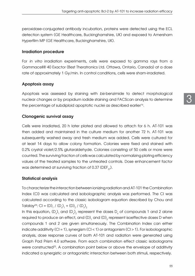
Targeting anti-apoptotic Bcl-2 by AT-101 to increase radiation efficacy
55
3
peroxidase-conjugated antibody incubation, proteins were detected using the ECL detection system (GE Healthcare, Buckinghamshire, UK) and exposed to Amersham Hyperfilm MP (GE Healthcare, Buckinghamshire, UK).
Irradiation procedure
For in vitro irradiation experiments, cells were exposed to gamma rays from a Gammacell® 40 Exactor (Best Theratronics Ltd. Ottawa, Ontario, Canada) at a dose rate of approximately 1 Gy/min. In control conditions, cells were sham-irradiated.
Apoptosis assay
Apoptosis was assessed by staining with bis-benzimide to detect morphological nuclear changes or by propidium iodide staining and FACScan analysis to determine the percentage of subdiploid apoptotic nuclei as described earlier16.
Clonogenic survival assay
Cells were irradiated, 20 h later plated and allowed to attach for 6 h. AT-101 was then added and maintained in the culture medium for another 72 h. AT-101 was subsequently washed away and fresh medium was added. Cells were cultured for at least 14 days to allow colony formation. Colonies were fixed and stained with 0.2% crystal violet/2.5% glutaraldehyde. Colonies consisting of 50 cells or more were counted. The surviving fraction of cells was calculated by normalizing plating efficiency values of the treated samples to the untreated controls. Dose enhancement factor was determined at surviving fraction of 0.37 (DEF37).
Statistical analysis
To characterize the interaction between ionizing radiation and AT-101 the Combination Index (CI) was calculated and isobolographic analysis was performed. The CI was calculated according to the classic isobologram equation described by Chou and Talalay24: CI = (D)1 / (Dx)1 + (D)2 / (Dx)2
In this equation, (Dx)1 and (Dx)2 represent the doses Dx of compounds 1 and 2 alone required to produce an effect, and (D)1 and (D)2 represent isoeffective doses D when compounds 1 and 2 are given simultaneously. The Combination Index can either indicate additivity (CI = 1), synergism (CI < 1) or antagonism (CI > 1). For isobolographic analysis, dose response curves of both AT-101 and radiation were generated using Graph Pad Prism 4.0 software. From each combination effect classic isobolograms were constructed25. A combination point below or above the envelope of additivity indicated a synergistic or antagonistic interaction between both stimuli, respectively.

Chapter 3
56
3
Clinical phase I/II trial
Patient selection criteria
Patients were eligible when aged 18 years or older, with stage III or IV, M0 histologically proven locally advanced inoperable HNSCC of the oral cavity, oropharynx or hypopharynx, and performance status WHO 0-2. Patients had no prior radiotherapy to the head and neck region or prior cisplatin-based chemotherapeutic treatment. Patients were required to have adequate hematologic, liver and renal function, and no uncontrolled arrhythmia. The study was approved by the Ethical Review Committee of The Netherlands Cancer Institute. Signed written informed consent was required before study entry.
Study design
Patients received standard cisplatin-based chemoradiotherapy (consisting of 70 Gy delivered in 35 fractions over 7 weeks, concurrently with 3-weekly 100 mg/m2 cisplatin i.v.) combined with dose-escalating oral administration of AT-101 in a 2-weeks daily schedule every 3 weeks. The starting dose of AT-101 was 10 mg daily and dose-escalation was in steps of 10 mg. Based on previously reported pharmacokinetic parameters26 AT-101 was daily administered 2 h prior to fractionated radiation. The primary endpoint of this study was tolerability of AT-101 administration in combination with standard chemoradiotherapy. Secondary endpoints included pharmacokinetics of AT-101.
Pharmacokinetic evaluation
Blood samples were collected at 30 min after AT-101 intake, and after 1, 2, 3, 4, 5, 6, 7, 8 and 24 h. The 3 ml whole blood samples were collected in EDTA tubes and mixed with 0.3 ml 0.2 M freshly prepared reduced gluthatione, and centrifuged at 4°C. After centrifugation the plasma was transferred in equal portions into 2 tubes containing 75 μl 25 mM acetonitrile maleic anhydride that was air-dried. The samples were stored at -80°C until analysis. Plasma concentrations of AT-101 were determined by an HPLC-UV method derived from literature26 which was optimized and validated. In short, an Agilent Zorbax Stable Bond C-18 column (150 x 4.6 mm I.D. 3.5 μm particle size) was used. Mobile phase consisted of 20% 10 mM KH2PO4 : 80% acetonitrile, at the flow rate of 1.0 ml/min. AT-101 was detected with a UV detector at 236 nm. Quantification was performed using calibration standards. An accelerated stability study was conducted at four different temperatures (37°C, 21°C, 4°C and -20°C) and led to a prediction of

Targeting anti-apoptotic Bcl-2 by AT-101 to increase radiation efficacy
57
3
approximately 88.7% of the original AT-101 concentration in the patient samples up to four years of storage at -80°C.
RESULTS
AT-101 target proteins are expressed in HNSCC cell lines
Western blot analysis demonstrated that HNSCC cell lines, specifically UM-SCC-11B, UM-SCC-14C, UM-SCC-22A and VU-SCC-OE, all expressed the anti-apoptotic proteins Bcl-xL, Bcl-2, and Mcl-1 (Fig. 1). Further investigation revealed that all four cell lines showed responsiveness to both radiation and AT-101 with ED50 values between 6 Gy and 16 Gy for radiation and ED50 values between 16 μM and 44 μM for AT-101. We continued our experiments with UM-SCC-11B and VU-SCC-OE since Bcl-xL and Bcl-2, the major targets of AT-101, were most prominently expressed in UM-SCC-11B and VU-SCC-OE, respectively. Mcl-1, a less predominant target of AT-101, was expressed at lower amounts.
Figure 1: Expression of Bcl-xL, Bcl-2, and Mcl-1 in HNSCC. Western blot analysis demonstrating the expression of
the anti-apoptotic proteins Bcl-xL, Bcl-2, and Mcl-1 in four different head and neck cancer cell lines, UM-SCC-
11B, UM-SCC-14C, UM-SCC-22A and VU-SCC-OE.
Radiation and AT-101 induce apoptosis in HNSCC cell lines
Radiation and AT-101 induced apoptosis in a time- and dose-dependent fashion (Fig. 2). UM-SCC-11B showed a steep dose-response curve up to 25 μM AT-101; in this cell line no further increase in apoptosis was detected up to 100 μM AT-101. Inserts show the time-dependency for both treatments. Apoptosis, induced by radiation and AT-101, as well as the combinations, results of which are described in the next paragraph, could be inhibited by pan-caspase inhibitor Z-VAD-FMK. In addition, we verified apoptosis induction by determining caspase 3 activation by methods described in27 (data not shown).

Chapter 3
58
3
Figure 2: Dose- and time-dependent induction of apoptosis by radiation (A,C) and AT-101 (B,D) in HNSCC
cell lines UM-SCC-11B (A,B) and VU-SCC-OE (C,D). Apoptosis was quantified 72 h after radiation and 48 h
after exposure to AT-101. Data represent mean values ± SEM of an average of 11 independent experiments,
performed in duplicate. Inserts show the time-dependency of radiation- and AT-101-induced apoptosis at the
indicated doses.
Combined effects of radiation and AT-101 are synergistic
As demonstrated in Figs. 3A and 3B, the combination of radiation and AT-101 leads to a more than additive apoptotic effect. Isobolographic analysis, a statistical method to determine the type of interaction, shows a synergistic increase of apoptosis in VU-SCC-OE (Fig. 3C). Isobolographic analysis could not be performed in UM-SCC-11B, because the maximum levels of apoptosis induced by either radiation or AT-101 in these cells did not exceed the apoptosis levels after combined treatment, implicating synergy. Consistent with this observation the CI’s are below 1.0, in these cells.

Targeting anti-apoptotic Bcl-2 by AT-101 to increase radiation efficacy
59
3
Figure 3: Synergistic interaction between radiation and AT-101. Fig. 3a and b illustrate apoptosis induction in
two HNSCC lines after radiation, AT-101 and their combination at the indicated doses. For the combination
experiments, AT-101 was administered 24 h after radiation. These graphs represent the results of at least 3
independent experiments performed in duplo. Fig. 3c shows an isobolographic analysis of the combined effect
of 29 % apoptosis induced by 10 Gy radiation and 15 μM AT-101. The same level of apoptosis can be induced
by 18.4 Gy of radiation or 44 μM of AT-101, respectively. The combination point () below the envelope of
additivity indicates a synergistic interaction between both stimuli. Note that in all conditions shown, the CI that
refers to the combination index, is smaller than 1.0, indicating synergy as well.

Chapter 3
60
3
Synergistic interactions between radiation and AT-101 are sequence dependent
Previously, it has been shown in other cell systems that the synergistic interaction between radiation and AT-101 can be sequence-dependent16,18. We therefore also assessed this phenomenon in our HNSCC cell lines by comparing AT-101 administration 24 h before, during and 24 h after irradiation. Only when radiation preceded AT-101 treatment, this synergistic increase of apoptosis was found (Fig. 4).
Figure 4: Sequence-dependent interaction between radiation and AT-101. Radiation and AT-101 were applied
either concurrently or sequentially at the indicated doses. In the sequential schedule, AT-101 was administered
either 24 h before radiation or 24 h after radiation. A: UM-SCC-11B; B: VU-SCC-OE. Data presented are
representative of at least two experiments in both cell lines.
Clonogenic survival assays
To determine the impact of AT-101 on long-term survival after radiation, we performed clonogenic survival assays. At concentrations of AT-101 below 2 μM (i.e. in the range

Targeting anti-apoptotic Bcl-2 by AT-101 to increase radiation efficacy
61
3
where no significant apoptosis induction was found; Fig. 2) no significant decrease in clonogenic survival was observed (data not shown). Fig. 5 shows that AT-101 at a final concentration of 1 μM reduced clonogenic survival after radiation (DEF37 = 1.3), consistent with a radiosensitizing effect of AT-101.
Figure 5: Effect of AT-101 on clonogenic survival after radiation. Clonogenic survival curves of VU-SCC-OE cells
after radiation in the absence (solid line) or presence of 1 μM AT-101 (normalized dashed line) are shown.
Graphs show representative curves of at least 3 experiments in triplicate, error bars represent SEM.
Pharmacokinetic analysis in patient samples
In an ongoing phase I/II trial, patient blood samples were collected between 30 min and 24 h after oral intake of AT-101. Two dose levels could be analyzed; 10 mg (n=13) and 20 mg (n=1). Fig. 6 shows the pharmacokinetic profiles with a dose-dependent increase in plasma concentration, peaking between 1.5 and 2.5 h at approximately 300 and 700 ng/ml for the 10 mg and 20 mg dose level, respectively. These levels correspond with 0.6-1.35 μM AT-101.

Chapter 3
62
3Figure 6: Clinical pharmacokinetics of AT-101. AT-101 concentration was measured by HPLC in patient plasma
samples collected at 30 min, 1, 2, 3, 4, 5, 6, 7, 8 and 24 h after oral intake of AT-101. Pharmacokinetic profiles
are shown for the AT-101 dose levels of 10 mg (n=13) and 20 mg (n=1). Data represent mean values ± SD.
DISCUSSION
Despite significant improvements in the treatment of patients with inoperable head and neck cancer, recurrence rates remain unacceptably high. Thus, there is a clear need to develop new therapeutic approaches to further enhance the anti-tumor efficacy of existing standard regimens, such as cisplatin-based chemoradiotherapy. Overexpression of anti-apoptotic members of the Bcl-2 family is frequently observed in HNSCC and has been associated with resistance to radio- and chemotherapy and with poor prognosis4-8. Therefore, in the present studies we focused on AT-101, a BH3-mimetic and small molecule inhibitor of pro-survival Bcl-2 proteins, and its potential to increase the cytotoxic effect of radiation in HNSCC in vitro. Our results show that AT-101, only when added after radiation, enhances apoptosis to synergistic levels, and acts as a radiosensitizer in clonogenic survival assays. To address the question whether the effective in vitro concentrations of AT-101 correspond with those achievable in a clinical setting, we determined AT-101 plasma levels in a subset of patients included in our phase I/II trial. Indeed, plasma levels of AT-101 were comparable with the low micromolar radiosensitizing concentrations in vitro.A synthetic class of BH3-mimetics that has been developed recently shows interesting results regarding their capacity to radiosensitize cancer cells, including ABT-737, a molecule with high affinity for Bcl-2 and Bcl-xL, and its analogue the clinically more

Targeting anti-apoptotic Bcl-2 by AT-101 to increase radiation efficacy
63
3
favorable, ABT-263. These compounds do not or only weakly target Mcl-128,29, whereas AT-101 demonstrates a more favorable binding profile towards Mcl-130. Studies with ABT-737 and ABT-263 now suggest that Mcl-1 plays a role in resistance to these compounds.28,31. A recent study on radiation and BH3-mimetics in breast cancer showed treatment with ABT-737 alone elevated Mcl-1. ABT-737 together with radiation, however, demonstrated a synergistic effect on breast cancer cells by downregulation of Mcl-132. Also a study on pancreatic cancer cells evaluated Mcl-1 as a target for radiosensitization33. These studies suggest that ABT-737 or ABT-263 may be suboptimal to target Mcl-1, in particular in combination with radiation.AT-101, the cis- or (-)-enantiomer of racemic Gossypol, is a naturally occurring polyphenolic dialdehyde derived from cottonseed. Gossypol enantiomers, including AT-101, have been used as cytotoxic agents in vitro and in vivo using different tumor cell lines from both solid13-15,34,35 and leukemic origin16. Importantly, only minimal effects were observed on normal cells13,14, indicating a certain degree of tumor selectivity. Several groups have investigated the combined effects of AT-101 and chemo- or radiotherapy17,18,36. In human prostate cancer cells, AT-101 potently enhanced radiation-induced apoptosis and growth inhibition and reduced clonogenic survival18. We showed in two human leukemic cell lines an additive to synergistic interaction between radiation and AT-10116. Interestingly, HNSCC cell lines made resistant to cisplatin retained their apoptosis sensitivity towards AT-10113,34. In vivo, the anti-tumor effect of AT-101 has been tested as single agent37 and in combination with radiation18 and chemotherapy38. In an orthotopic xenograft model of HNSCC with high Bcl-xL expression, daily i.p. injection of AT-101 resulted in a significant tumor growth delay as compared to control animals37. Histopathological analysis showed a decrease in mitotic index and an increase in apoptosis in the AT-101-treated tumors. Treatment was well tolerated, as reversible moderate weight loss was the only observed side effect. In a prostate cancer xenograft model daily oral AT-101 was compared with fractionated radiotherapy and with the combination of both18. Especially when larger tumors were treated, only the combination of AT-101 and radiation achieved significant anti-tumor activity. Tumor tissue specimens showed not only a significant increase in apoptosis after combined treatment, but also a strong inhibition of tumor angiogenesis. No significant weight loss or obvious organ toxicities were observed. From these experimental studies it can be concluded that AT-101 has significant anti-cancer activity as single agent, but is much more effective in combination with other cytotoxic regimens like cisplatin and radiation therapy. Upon oral administration, it has demonstrated little toxic side effects in animals.Clinical studies on AT-101 as single agent or combined with chemotherapy are limited, but indicate good tolerability19-23. Clinical experience with AT-101 in combination with

Chapter 3
64
3
(chemo-)radiotherapy is even sparser39, but accumulating from several ongoing or recently completed phase I studies, including ours in HNSCC. In the present studies, we demonstrate a dose- and time-dependent increase in apoptosis by radiation and AT-101 in two human HNSCC cell lines expressing the pro-survival Bcl-2 family members Bcl-xL, Bcl-2, and Mcl-1, all established targets of AT-101. By performing isobolographic analysis and calculating the Combination Indices, we characterized the type of interaction between both treatments as synergistic. These findings are in agreement with the results from studies using other cell systems16,18. We also found that this synergistic interaction between radiation and AT-101 was only present when AT-101 was added after radiation, as observed in other cell systems as well16,18. This apparent sequence-dependency is poorly understood and thought to be cell cycle related18. In a previous study16, we provided evidence that activation of the SAPK/JNK signal transduction pathway plays a significant role in AT-101-induced apoptosis. Because radiation is a well-known activator of SAPK/JNK40 and it has been shown that SAPK/JNK translocates to the mitochondria upon irradiation where it phosphorylates and inactivates Bcl-xL, Bcl-2 and Mcl-141-43, this mechanism may provide an alternative explanation for the observed sequence-dependency. It has been shown that genetic or pharmacological modulation of radiation-induced apoptosis frequently also impacts on radiosensitivity44,45. Therefore, we evaluated the effect of AT-101 on clonogenic survival after irradiation. Indeed, at concentrations that do not induce significant levels of apoptosis, a clear radiosensitizing effect was observed. This radiosensitizing potential of AT-101 most likely depends on the cell type studied, as it has been demonstrated in certain cell systems17,36, but not in others16. In a number of different tumor cell lines, including HNSCC, radiosensitization by AT-101 was found to result from reduced double-strand break repair46. Others have suggested that increased autophagic cell death plays an important role in AT-101-induced inhibition of clonogenic survival of irradiated glioblastoma cells36.To determine whether the radiosensitizing concentrations of AT-101 are comparable with the plasma levels that can be achieved in patients, we analyzed the pharmacokinetic data collected in our clinical phase I/II study. At daily doses of 10–20 mg, plasma levels peaked around 2 h after intake, suggesting slow absorption. Maximum plasma concentrations were in the micromolar range, corresponding to those that induced radiosensitization in vitro. Both the maximum observed plasma concentration and the time to reach this value are similar to other reports37. Although it is difficult to compare in vitro with in vivo drug concentrations, it is reassuring that no major differences were found. Regarding the scheduling of radiotherapy and AT-101, daily radiation was given just prior to or at maximal plasma concentrations. Evidently, more clinical studies are needed to define safety and efficacy of AT-101 in

Targeting anti-apoptotic Bcl-2 by AT-101 to increase radiation efficacy
65
3
combination with radiation, and to determine intra-tumoral drug concentrations for optimal scheduling.
CONCLUSIONS
In summary, we showed that AT-101 synergistically enhanced radiation-induced apoptosis in HNSCC in vitro in a sequence-dependent manner. In addition, in vitro radiosensitization was observed at clinically achievable plasma levels. These findings provide a rationale to further evaluate AT-101 in combination with standard (chemo-)radiation in Bcl-2-overexpressing tumors, such as head and neck squamous cell carcinoma.
COMPETING INTERESTS
The authors declare no competing interests.
ACKNOWLEDGEMENTS
We thank Ascenta Therapeutics, Inc. (Malvern, PA, USA) for kindly providing us with AT-101 and the department of Bioanalytical and QC Laboratory Slotervaart Hospital (Amsterdam, The Netherlands) for analyzing the patient samples. This work was in part financially supported by the Dutch Cancer Society.

Chapter 3
66
3
REFERENCES 1 Pignon JP, le Maitre A, Maillard E, Bourhis J, Group M-NC. Meta-analysis of chemotherapy
in head and neck cancer (MACH-NC): an update on 93 randomised trials and 17,346
patients. Radiother Oncol. 2009;92(1):4-14. doi:10.1016/j.radonc.2009.04.014.
2 Machtay M, Moughan J, Trotti A, Garden AS, Weber RS, Cooper JS et al. Factors
associated with severe late toxicity after concurrent chemoradiation for locally advanced
head and neck cancer: an RTOG analysis. J Clin Oncol. 2008;26(21):3582-9. doi:10.1200/
JCO.2007.14.8841.
3 Verheij M, Vens C, van Triest B. Novel therapeutics in combination with radiotherapy to
improve cancer treatment: rationale, mechanisms of action and clinical perspective. Drug
Resist Updat. 2010;13(1-2):29-43. doi:10.1016/j.drup.2010.01.002.
4 Abdullah LN, Chow EK. Mechanisms of chemoresistance in cancer stem cells. Clin Transl
Med. 2013;2(1):3. doi:10.1186/2001-1326-2-3.
5 Gallo O, Chiarelli I, Boddi V, Bocciolini C, Bruschini L, Porfirio B. Cumulative prognostic
value of p53 mutations and bcl-2 protein expression in head-and-neck cancer treated by
radiotherapy. Int J Cancer. 1999;84(6):573-9.
6 Ong F, Moonen LM, Gallee MP, ten Bosch C, Zerp SF, Hart AA et al. Prognostic factors
in transitional cell cancer of the bladder: an emerging role for Bcl-2 and p53. Radiother
Oncol. 2001;61(2):169-75.
7 Reed JC, Miyashita T, Takayama S, Wang HG, Sato T, Krajewski S et al. BCL-2 family proteins:
regulators of cell death involved in the pathogenesis of cancer and resistance to therapy.
J Cell Biochem. 1996;60(1):23-32.
8 Simonian PL, Grillot DA, Nunez G. Bcl-2 and Bcl-XL can differentially block chemotherapy-
induced cell death. Blood. 1997;90(3):1208-16.
9 Chipuk JE, Moldoveanu T, Llambi F, Parsons MJ, Green DR. The BCL-2 family reunion. Mol
Cell. 2010;37(3):299-310. doi:10.1016/j.molcel.2010.01.025.
10 Cragg MS, Harris C, Strasser A, Scott CL. Unleashing the power of inhibitors of oncogenic
kinases through BH3 mimetics. Nat Rev Cancer. 2009;9(5):321-6. doi:10.1038/nrc2615.
11 Kitada S, Leone M, Sareth S, Zhai D, Reed JC, Pellecchia M. Discovery, characterization,
and structure-activity relationships studies of proapoptotic polyphenols targeting B-cell
lymphocyte/leukemia-2 proteins. J Med Chem. 2003;46(20):4259-64.
12 Qian SZ, Wang ZG. Gossypol: a potential antifertility agent for males. Annu Rev Pharmacol
Toxicol. 1984;24:329-60. doi:10.1146/annurev.pa.24.040184.001553.
13 Oliver CL, Bauer JA, Wolter KG, Ubell ML, Narayan A, O’Connell KM et al. In vitro effects
of the BH3 mimetic, (-)-gossypol, on head and neck squamous cell carcinoma cells. Clin
Cancer Res. 2004;10(22):7757-63.

Targeting anti-apoptotic Bcl-2 by AT-101 to increase radiation efficacy
67
3
14 Oliver CL, Miranda MB, Shangary S, Land S, Wang S, Johnson DE. (-)-Gossypol acts directly
on the mitochondria to overcome Bcl-2- and Bcl-X(L)-mediated apoptosis resistance. Mol
Cancer Ther. 2005;4(1):23-31.
15 Liu S, Kulp SK, Sugimoto Y, Jiang J, Chang HL, Dowd MK et al. The (-)-enantiomer of gossypol
possesses higher anticancer potency than racemic gossypol in human breast cancer.
Anticancer Res. 2002;22(1A):33-8.
16 Zerp SF, Stoter R, Kuipers G, Yang D, Lippman ME, van Blitterswijk WJ et al. AT-101, a
small molecule inhibitor of anti-apoptotic Bcl-2 family members, activates the SAPK/JNK
pathway and enhances radiation-induced apoptosis. Radiat Oncol. 2009;4:47.
17 Moretti L, Li B, Kim KW, Chen H, Lu B. AT-101, a pan-Bcl-2 inhibitor, leads to radiosensitization
of non-small cell lung cancer. J Thorac Oncol. 2010;5(5):680-7.
18 Xu L, Yang D, Wang S, Tang W, Liu M, Davis M et al. (-)-Gossypol enhances response to
radiation therapy and results in tumor regression of human prostate cancer. Mol Cancer
Ther. 2005;4(2):197-205.
19 Flack MR, Pyle RG, Mullen NM, Lorenzo B, Wu YW, Knazek RA et al. Oral gossypol in the
treatment of metastatic adrenal cancer. J Clin Endocrinol Metab. 1993;76(4):1019-24.
20 Stein RC, Joseph AE, Matlin SA, Cunningham DC, Ford HT, Coombes RC. A preliminary
clinical study of gossypol in advanced human cancer. Cancer Chemother Pharmacol.
1992;30(6):480-2.
21 Van Poznak C, Seidman AD, Reidenberg MM, Moasser MM, Sklarin N, Van Zee K et al. Oral
gossypol in the treatment of patients with refractory metastatic breast cancer: a phase I/II
clinical trial. Breast Cancer Res Treat. 2001;66(3):239-48.
22 Schelman WR, Mohammed TA, Traynor AM, Kolesar JM, Marnocha RM, Eickhoff J et al.
A phase I study of AT-101 with cisplatin and etoposide in patients with advanced solid
tumors with an expanded cohort in extensive-stage small cell lung cancer. Invest New
Drugs. 2014;32(2):295-302. doi:10.1007/s10637-013-9999-7.
23 Sonpavde G, Matveev V, Burke JM, Caton JR, Fleming MT, Hutson TE et al. Randomized
phase II trial of docetaxel plus prednisone in combination with placebo or AT-101, an
oral small molecule Bcl-2 family antagonist, as first-line therapy for metastatic castration-
resistant prostate cancer. Ann Oncol. 2012;23(7):1803-8.
24 Chou TC, Talalay P. Quantitative analysis of dose-effect relationships: the combined effects
of multiple drugs or enzyme inhibitors. Adv Enzyme Regul. 1984;22:27-55.
25 Steel GG, Peckham MJ. Exploitable mechanisms in combined radiotherapy-chemotherapy:
the concept of additivity. Int J Radiat Oncol Biol Phys. 1979;5(1):85-91.
26 Lin H, Gounder MK, Bertino JR, Kong AN, DiPaola RS, Stein MN. A validated HPLC assay
for the determination of R-(-)-gossypol in human plasma and its application in clinical
pharmacokinetic studies. J Pharm Biomed Anal. 2012;66:371-5.
27 Maas C, Verbrugge I, de Vries E, Savich G, van de Kooij LW, Tait SW et al. Smac/DIABLO
release from mitochondria and XIAP inhibition are essential to limit clonogenicity of

Chapter 3
68
3
Type I tumor cells after TRAIL receptor stimulation. Cell Death Differ. 2010;17(10):1613-23.
doi:10.1038/cdd.2010.39.
28 Konopleva M, Contractor R, Tsao T, Samudio I, Ruvolo PP, Kitada S et al. Mechanisms of
apoptosis sensitivity and resistance to the BH3 mimetic ABT-737 in acute myeloid leukemia.
Cancer Cell. 2006;10(5):375-88. doi:10.1016/j.ccr.2006.10.006.
29 Tse C, Shoemaker AR, Adickes J, Anderson MG, Chen J, Jin S et al. ABT-263: a potent and
orally bioavailable Bcl-2 family inhibitor. Cancer Res. 2008;68(9):3421-8. doi:10.1158/0008-
5472.CAN-07-5836.
30 Lessene G, Czabotar PE, Colman PM. BCL-2 family antagonists for cancer therapy. Nat Rev
Drug Discov. 2008;7(12):989-1000. doi:10.1038/nrd2658.
31 Yecies D, Carlson NE, Deng J, Letai A. Acquired resistance to ABT-737 in lymphoma
cells that up-regulate MCL-1 and BFL-1. Blood. 2010;115(16):3304-13. doi:10.1182/
blood-2009-07-233304.
32 Wu H, Schiff DS, Lin Y, Neboori HJ, Goyal S, Feng Z et al. Ionizing radiation sensitizes breast
cancer cells to Bcl-2 inhibitor, ABT-737, through regulating Mcl-1. Radiat Res. 2014;182(6):618-
25. doi:10.1667/RR13856.1.
33 Wei D, Zhang Q, Schreiber JS, Parsels LA, Abulwerdi FA, Kausar T et al. Targeting mcl-1
for radiosensitization of pancreatic cancers. Transl Oncol. 2015;8(1):47-54. doi:10.1016/j.
tranon.2014.12.004.
34 Bauer JA, Trask DK, Kumar B, Los G, Castro J, Lee JS et al. Reversal of cisplatin resistance
with a BH3 mimetic, (-)-gossypol, in head and neck cancer cells: role of wild-type p53 and
Bcl-xL. Mol Cancer Ther. 2005;4(7):1096-104.
35 Wang X, Wang J, Wong SC, Chow LS, Nicholls JM, Wong YC et al. Cytotoxic effect of
gossypol on colon carcinoma cells. Life Sci. 2000;67(22):2663-71.
36 Keshmiri-Neghab H, Goliaei B, Nikoofar A. Gossypol enhances radiation induced
autophagy in glioblastoma multiforme. Gen Physiol Biophys. 2014;33(4):433-42. doi:10.4149/
gpb_2014017.
37 Wolter KG, Wang SJ, Henson BS, Wang S, Griffith KA, Kumar B et al. (-)-gossypol inhibits
growth and promotes apoptosis of human head and neck squamous cell carcinoma in
vivo. Neoplasia. 2006;8(3):163-72.
38 Mohammad RM, Wang S, Aboukameel A, Chen B, Wu X, Chen J et al. Preclinical studies
of a nonpeptidic small-molecule inhibitor of Bcl-2 and Bcl-X(L) [(-)-gossypol] against diffuse
large cell lymphoma. Mol Cancer Ther. 2005;4(1):13-21.
39 Susman E. Cottonseed-oil byproduct as anticancer radiation sensitiser. Lancet Oncol.
2004;5(11):648.
40 Verheij M, Bose R, Lin XH, Yao B, Jarvis WD, Grant S et al. Requirement for ceramide-initiated
SAPK/JNK signalling in stress-induced apoptosis. Nature. 1996;380(6569):75-9.

Targeting anti-apoptotic Bcl-2 by AT-101 to increase radiation efficacy
69
3
41 Inoshita S, Takeda K, Hatai T, Terada Y, Sano M, Hata J et al. Phosphorylation and
Inactivation of Myeloid Cell Leukemia 1 by JNK in Response to Oxidative Stress. J Biol
Chem. 2002;277(46):43730-4.
42 Kharbanda S, Saxena S, Yoshida K, Pandey P, Kaneki M, Wang Q et al. Translocation of
SAPK/JNK to mitochondria and interaction with Bcl-x(L) in response to DNA damage. J Biol
Chem. 2000;275(1):322-7.
43 Yamamoto K, Ichijo H, Korsmeyer SJ. BCL-2 is phosphorylated and inactivated by an
ASK1/Jun N-terminal protein kinase pathway normally activated at G(2)/M. Mol Cell Biol.
1999;19(12):8469-78.
44 Rupnow BA, Murtha AD, Alarcon RM, Giaccia AJ, Knox SJ. Direct evidence that apoptosis
enhances tumor responses to fractionated radiotherapy. Cancer Res. 1998;58(9):1779-84.
45 Wissink EH, Verbrugge I, Vink SR, Schader MB, Schaefer U, Walczak H et al. TRAIL enhances
efficacy of radiotherapy in a p53 mutant, Bcl-2 overexpressing lymphoid malignancy.
Radiother Oncol. 2006;80(2):214-22.
46 Kasten-Pisula U, Windhorst S, Dahm-Daphi J, Mayr G, Dikomey E. Radiosensitization of tumour
cell lines by the polyphenol Gossypol results from depressed double-strand break repair
and not from enhanced apoptosis. Radiother Oncol. 2007;83(3):296-303. doi:10.1016/j.
radonc.2007.04.024.


CHAPTER 4Enhancing radiation response by a second-
generation TRAIL receptor agonist using a new in vitro
organoid model system
Shuraila F. Zerp
Zainab Bibi
Inge Verbrugge
Emile E. Voest
Marcel Verheij
Clin Transl Radiat Oncol. 2020 Jun 9;24:1-9

Chapter 4
72
4
ABSTRACT
Background: For many cancer types, including colorectal carcinoma (CRC), combined modality treatments have shown to improve outcome, but are frequently associated with significant toxicity, illustrating the need for new therapeutic approaches. Based on preclinical data, TRAIL receptor agonists appeared to be promising agents for cancer therapy especially in combination with DNA damaging regimens. Here, we present the combination of the second-generation TRAIL receptor agonist APG-880 with radiation in a new and clinically relevant 3D model system.
Methods: To investigate the effect of APG-880 in combination with radiation we performed short-term cytotoxicity and long-term clonogenic survival assays in established CRC cell lines, and in tumor organoids derived from colon cancer patients.
Results: APG-880 is a potent inducer of apoptosis in CRC cell lines and in patient-derived CRC organoids. Furthermore, a supra-additive effect on cytotoxicity was found when APG-880 and radiation were combined simultaneously, with combination indices around 0.7. Lastly, in the long-term survival assays, we demonstrated a radiosensitizing effect of APG-880 with dose enhancement factors between 1.3 and 1.5.
Conclusions: In a new, clinically relevant CRC-organoid model system we demonstrated a more than additive combined effect between the second-generation TRAIL receptor agonist APG-880 and radiation.
Key words: TRAIL receptor agonist, radiation, clonogenic survival, organoids, colorectal carcinoma.

Enhancing radiation response by a TRAIL-R agonist
73
4
INTRODUCTION
Over 50% of all cancer patients receive radiation therapy during the course of their disease and radiotherapy contributes to approximately 40% of all cancer cures1. Colorectal carcinoma (CRC) is the third most common cancer worldwide with over one million new diagnoses per year and is expected to increase by 60% to more than 2.2 million new cases and 1.1 million deaths by 2030. The highest incidences occur in Western countries2,3. Neoadjuvant (chemo)radiation is one of the standard treatment options for locally advanced CRC, though is associated with significant acute toxicity and surgical morbidity4. Therefore, there is a strong clinical need to develop novel combinations to improve the therapeutic ratio. Radiosensitizers are agents that increase the lethal effect of ionizing radiation and, ideally, achieve greater tumor cytotoxicity than would have been expected from the additive effect of each modality. Some radiosensitizers are used in the clinic already, while others are being studied for new indications. For example, non-specific agents such as cisplatin or 5-FU have shown to improve local control and overall survival when added to radiation therapy and are integrated in standard treatment protocols5,6. Based on the hallmarks of cancer, target-specific radiosensitizers have become available for clinical use, e.g. the EGFR-antagonist cetuximab7 that reduces proliferative signaling. Others are still in early clinical development such as PARP inhibitors that interfere with radiation-induced DNA damage repair8. Since apoptosis is an important mechanism of radiation induced cell death, modulation of apoptosis sensitivity has been shown to increase tumor cell kill and relatively spare normal tissues, thereby enhancing therapeutic outcome1,9,10. Here, we focus on modulation of apoptosis by the tumor necrosis factor-related apoptosis-inducing ligand TRAIL. TRAIL is a homotrimeric cytokine expressed by immune cells and plays a protective role in immune mediated tumor surveillance11,12. In human, TRAIL signals for apoptosis via TRAIL-receptor (TRAIL-R) 1 and TRAIL-R2, also known as Death Receptor 4 (DR4) and Death Receptor 5 (DR5), respectively. TRAIL induces clustering of DR4 and DR5, and can subsequently activate apoptotic pathways independently of mitochondrial involvement and p53-status. TRAIL acts preferentially on tumor cells and is non-toxic to most healthy tissue13,14. TRAIL can also bind ‘decoy receptors’ DCR1 and DCR2 which lack functional death domains15,16 and a soluble receptor called osteoprotegerin17. A novel, second-generation TRAIL-receptor agonist has been engineered that simultaneously binds up to six TRAIL receptors, thereby improving the ability to form receptor clustering on cancer cells to induce superior anti-tumor efficacy in vivo, which is independent of cross-linking to Fc receptors18. The lead molecule, known as APG-880/ ABBV-621 is currently being tested in phase I clinical trials (NCT03082209)19.

Chapter 4
74
4
The rational for combining TRAIL and radiation in our study, is that these agents activate the two distinct pathways leading to apoptosis20. Death receptor ligands like TRAIL, initiate apoptotic signaling via the extrinsic pathway21, whereas radiation and other DNA damaging agents activate the intrinsic pathway to cell death. Simultaneous activation of both pathways has been shown to be particularly effective in inducing cell death22-24. Via active caspase 8, TRAIL receptor signaling can also trigger the intrinsic mitochondrial pathway through Bid cleavage into truncated Bid (tBid). tBid subsequently translocates to the mitochondria, leading to mitochondrial permeabilization and cytochrome C release25.Unfortunately, TRAIL-based therapies have not yet led to improved clinical responses mainly because the first-generation TRAIL receptor agonists (TRAs) failed efficacy and did not meet clinical expectations as tested in phase I and II trials21,26,27. For instance, TRAs dulanermin, mapatumumab, lexatumumab, conatumumab, tigatuzumab or drozitumab, as single agent or in combination studies did not lead to statistically significant anticancer activity in randomized controlled trials28,29. There are several factors that have contributed to this apparent translational failure. First, the extent by which first generation TRAIL molecules were able to bind and cluster their receptors, and subsequently activate the extrinsic pathway has been limited. This was most likely due to the bivalent nature of antibodies that allows crosslinking of only two DRs leading to inefficient DISC formation28. APG-880 induces better hexavalent clustering of TRAIL receptors, and furthermore does not require Fcγ-R-mediated crosslinking for optimal in vivo efficacy suggesting that this second generation molecule may be superior to previously tested TRAs19.Second, the model systems used over the past years to test TRAIL efficacy were suboptimal in their capacity to predict clinical activity. Indeed, 2D cell culturing techniques are now considered structurally and functionally inferior to mimic cancer and are of limited use to predict successful clinical translation30. Tumor-derived organoids have the potential to serve as a pre-clinical model31 and to better predict treatment response of individual patients. As TRAIL-receptors are commonly expressed in colorectal tumor tissue32, we evaluated the efficacy of APG-880 alone and in combination with radiation therapy both in CRC cell lines, and in patient-derived CRC-organoids.
MATERIALS & METHODS
Reagents
APG-880 stock solution (10.6 mg/ml) was provided by AbbVie (North Chicago, IL, USA), aliquoted in 2 μl portions and stored at -80o C. Thawed samples were only used once.

Enhancing radiation response by a TRAIL-R agonist
75
4
Cell culture
Colon cancer derived cell lines HCT116 and HT29 were purchased from the American Type Culture Collection (ATCC). Jurkat cell line J16, were kindly provided by prof. dr. J. Borst (The Netherlands Cancer Institute, Amsterdam). All cell lines were grown according to ATCC protocols.
Organoid culture
Surgical specimens (in case of organoid culture ITO17 and ITO60), or core needle biopsy material (in case of organoid culture ITO77), collected within a clinical trial at the NKI (NL48824.031.14) were dissected, stored in the central biobank and used for the establishment of organoids. For our experiments we used three different biobank stored organoids from three different patients. After institutional approval, the biobank stored organoids ITO17, ITO60 and ITO77 were thawed and cultured according to the protocol earlier described33. In short, organoids were grown in Geltrex LDEV-Free hESC-qualified Reduced Growth Factor Basement Membrane Matrix (Life technologies, #A1413202 Carlsbad, CA, USA) and covered in the appropriate volume of growth medium Advanced DMEM/F-12 (Life Technologies, cat. no. 12634-010) supplemented with 2 mM Glutamax (Invitrogen #35050-079, 10 mM HEPES Invitrogen #15630-056, 100 units/ml and 100 mg/ml of Penicillin / Streptomycin, respectively (Invitrogen #15140-122), 10% Noggin conditioned medium, 20% R-spondin1 conditioned medium, B27 supplement (Invitrogen #17504-044, 1.25 mM N-Acetylcysteine (Sigma-Aldrich #A9165-5G), 50 ng/ml human recombinant EGF (BD bioscience #354052) 10 mM Nicotinamide Sigma-Aldrich #N0636), 500 nM A-83-01 Tocris #2939), 3 μM SB202190 Cayman Chemicals #10010399), 10 μM Prostaglandine E2 (Cayman Chemicals #14010-1) 10 μM Y-27632 Sigma-Aldrich #Y0503)34-36.Regarding the organoids used in this study, organoid ITO77 was derived from a peritoneal metastasis and organoids ITO17 and ITO60 originated from a primary colorectal carcinoma. Their mismatch repair (MMR) status is known: ITO17 and ITO60 are MMR proficient, while ITO77 is MMR deficient.
In vitro irradiation procedure
Cells were exposed to gamma rays from a Gammacell® 40 Exactor (Best Theratronics Ltd. Ottawa, Ontario Canada) at a dose rate of approximately 1 Gy/min.
Apoptosis analysis
Apoptosis was determined by staining cells with bis-benzimide or propidium iodide to detect morphological nuclear changes as described earlier37 or FACScan analysis

Chapter 4
76
4
as described earlier38. A third method of apoptosis detection was performed with the IncuCyte Live-Cell Analysis System. In this assay the activation of caspase-3 or caspase-7 was measured by an inert non-fluorescent DEVD (Asp-Glu-Val-Asp) peptide motif substrate that is able to cross the cell membrane and can be cleaved by activated caspase-3/7 to release a green DNA-binding fluorescent label. In order to detect apoptosis, 5 μM of the caspase-3/7 reagent (IncuCyte, Essen BioScience #4440, Michigan, USA) was added to 96 well culture plates. Cells were in a proliferating state prior to APG-880 and/or radiation treatment. Images (10x amplification) of the cells with fluorescently-labeled nuclei were captured every 4 hours in the IncuCyte® ZOOM System. Subsequent analysis was performed with the IncuCyte® ZOOM System software.
Western blotting
Western blot analysis was performed to detect DR4 and DR5 protein. Cells were treated with radiation and/or APG-880, washed and lysed in Triton lysis buffer as described earlier37. Blots were probed with an anti-DR4 rabbit polyclonal antibody C-terminus (#AB16955 Burlington, MA, USA) and anti-DR5 antibody (TNFRSF10B Mouse anti-human TRAIL R2 clone B-D37 monoclonal antibody cellsciences #CDM237 Newburyport, MA, USA) in 5% Nutrilon in TBS-T. After secondary horseradish peroxidase-conjugated antibody incubation, proteins were detected using the ChemiDoc™ Imaging Systems.
Epitope expression by FACS
DR4 and DR5 epitope expression was determined by flow cytometry as described by Verbrugge et al.22, using rabbit polyclonal anti-DR4 antibody or mouse anti-human TRAIL R2/DR5 monoclonal antibody, unconjugated, Clone B-D37.
Metabolic activity assay
To determine the cytotoxic potential of APG-880 in organoids we dissociated the organoids with TriplE Express (Invitrogen, #12605 Carlsbad, CA, USA) at day 1. We expanded the cells to form small organoids of comparable sizes, and treated them with a defined concentration range at day 4, then performed metabolic activity assays with the CellTiter-Glo® 3D reagent kit (Promega, G9681, Madison, WI 53711 USA) at day 10.
Clonogenic survival assays
Single cells were plated and allowed to attach before treatment. Cells were irradiated and treated with the indicated amount of APG-880, or sham-treated, immediately

Enhancing radiation response by a TRAIL-R agonist
77
4
after irradiation and cultured for at least 14 days to allow colony formation. Colonies were fixed and stained with 0.2% crystal violet/2.5% glutaraldehyde. Colonies consisting of 50 cells or more were counted. The surviving fraction of cells was calculated by normalizing plating efficiency values of the treated samples to the untreated controls.
Organoid survival assay
In order to determine the clonogenic potential of organoids we dissociated growing organoids with TriplE Express (Invitrogen, #12605 Carlsbad, CA, USA) at day 1. We plated the organoid derived single cells in Geltrex LDEV-Free hESC-qualified Reduced Growth Factor Basement Membrane Matrix (Life technologies, #A1413202 Carlsbad, CA, USA) and covered the matrix with growth medium as described above. Immediately after seeding the single cell suspension of the organoids, they were treated with the appropriate dose of radiation and/or concentration APG-880 and cultured for 14 days. Pictures of the colonies were taken with a cooled Hamamatsu ORCA R2 Black and White CCD-camera on a Zeiss AxioObserver microscope with a 10 x / 0.30 ECPlan-Neofluar Ph1 objective, run by ZEN2.3 Zeiss acquisition software. Analysis of the pictures and counting the number of organoids sized at least 50 cubic micrometer was performed with ImageJ. Organoids counts were done manually and double checked by an independent technician, in a blinded setting.
Combination index
The effects on cancer cells by ionizing radiation and APG-880 was characterized by calculating the Combination Index (CI) according to the classic isobologram equation described by Chou and Talalay39: CI = (D)1/(Dx)1 + (D)2/(Dx)2, and was previously applied by Zerp et al.38. The Combination Index can either indicate additivity (CI = 1), synergism (CI < 1) or antagonism (CI > 1).
RESULTS
DR4 and DR5 are expressed by colon cancer cell lines and colon cancer derived organoids
First, we validated by Western blotting that DR4 and DR5 are expressed in HCT116 and HT-29 cell lines (Fig. 1A). Patient-derived CRC organoids also expressed surface DR4 and DR5, as determined by flow cytometry (Fig. 1B-E).

Chapter 4
78
4Figure 1: CRC cell lines and organoids express DR4 and DR5. Western blot analysis of DR4 and DR5 expression
in HT29 and HCT116 cells (A). In (B), (C) and (D), CRC organoids express cell surface DR4 and DR5. Endogenous
DR4 and DR5 expression on the cell surface of three different colorectal carcinoma patient derived organoids,
ITO17, ITO60 and ITO77 analyzed by live-cell flow cytometry. (B) negative control of organoid ITO60 consisting
of secondary antibody only, in (C) anti-DR4 fluorescent staining of ITO60 and in (D) anti DR5 fluorescent staining
of ITO60. (E) Quantification of the percentage DR4 and DR5 positive cells in the different organoids analyzed.
J16 cells serve as a negative control for anti-DR4 as well as a positive control for anti-DR5.
Colon cancer cell lines and colon cancer derived organoids undergo apoptosis upon APG-880 and radiation treatment
To confirm the apoptotic mode of cell death we first validated by Hoechst staining that APG-880 induces DNA condensation/fragmentation, a hallmark of apoptosis in HCT116 and HT29 cell lines and in CRC organoids (Fig. 2). To quantify apoptosis induction, HCT116 and HT29 cell lines were treated with APG-880 and apoptotic cells were counted (Fig. 3). This revealed a dose-dependent induction of apoptosis by APG-880 and higher sensitivity of HCT116 as compared to HT29 cells at both 24 and 48 hr (Fig. 3). In these same cell lines we observed both a time- and dose-dependent induction of apoptosis by radiation (Fig. 4).

Enhancing radiation response by a TRAIL-R agonist
79
4
Due to the 3D nature of the organoids we used a different quantification method that is presented with the results of the combination experiments in section 3.5.
Figure 2: APG-880 induces DNA fragmentation. Staining with Hoechst demonstrated typical apoptotic body
formation visualized by fluorescence microscopy. (A) Nuclei of HT29 control cells. (B) HT29 cells treated with
100 ng/ml APG-880. (C) Organoids ITO60 control. (D) Organoid ITO60 treated with 100 ng/ml APG-880. (E) and
(F) Magnification of a cutout of respectively (C) and (D).

Chapter 4
80
4
Figure 3: APG-880 induces apoptosis in a dose-dependent manner in CRC cell lines. Apoptosis induction by
APG-880 HCT116 (A) and HT29 (B) as read-out by counting cells displaying DNA fragmentation.
Figure 4: Radiation induces apoptosis in a dose-dependent manner in CRC cell lines. Apoptosis induction at 1,
4,7,24 h after radiation in HCT116 and 24, 48,72 h after radiation in HT29.
APG-880 and radiation decrease clonogenic cell survival
We next established the sensitivity of the colon carcinoma derived cell lines HCT116 and HT29 towards radiation and APG-880 in clonogenic survival assays, the gold standard for long-term cytotoxicity. HCT116 was more sensitive than HT29 for both treatment modalities (Fig. 5A and B).

Enhancing radiation response by a TRAIL-R agonist
81
4
Figure 5: APG-880 and radiation decrease clonogenic survival in cell lines. (A) HCT116 (spheres) and HT29
(squares) clonogenic survival curves following increases doses of radiation (n= 3-4 experiments, performed in
triplicate), error bars represent SEM. (B) Clonogenic survival graph of colon carcinoma cell lines HT29 and HCT116
upon APG-880 treatment. Graph shows the results of 1-3 experiments in triplicate, error bars represent SD.
Colon cancer cell lines show a more than additive induction of cell death when exposed to the combination of radiation and APG-880
When both treatment modalities were combined, we found a more than additive effect as quantified by calculating the combination index (CI) (Fig. 6A and B). For HCT116 13% apoptosis was the maximum percentage that could be achieved with radiation alone at t=24h. For HT29 13% apoptosis was the maximum for radiation at t=48h. Therefore, the CI’s calculated for instance at 13% were 0.78 for HCT116 at 24h and 0.67 for HT29 at 48h.
APG-880 enhances radiation responses in organoids
Because the 3D nature of the organoids hampered the Hoechst quantification method used in the cell lines, we applied a different quantification method for cell death/loss of viability, i.e. the cell titer glow 3D assay that is especially developed for measuring cell death in 3D cell culturing conditions (Fig. 6C). The cell titer glow 3D method showed a dose dependent cytotoxicity in three organoid lines tested towards both APG-880 and radiation. Furthermore, and consistent with the results in the cell lines we show in these organoids that within a limited concentration range,

Chapter 4
82
4
i.e. 50-100 ng/ml, combined treatment with APG-880 and radiation is more effective than treatment by radiation or APG-880 alone (Fig. 6C).
Figure 6: Colon cancer cell lines show a more than additive effect when exposed to the combination of
radiation and APG-880.
In graphs (A) and (B) combination experiments are shown where radiation and APG-880 were applied
concurrently and apoptosis was determined after 24 hours for HCT116 and 48 h for HT29. Data represent mean
values ± SEM of an average of 3 independent experiments, performed in duplicate. Combination indices were
calculated from these graphs. (C), (D), (E): Growing organoids were exposed to APG-880 for 6 days. Graph
represents the average of 2 independent experiments in triplicate, error bars represent SEM.
Colon cancer derived cell lines and organoids show enhanced reduction in clonogenic survival when exposed to the combination of radiation and APG-880
In clonogenic survival assays in which APG-880 and radiation were combined, HT29 displayed the most effective decrease in clonogenicity at doses around 100 ng/ml. Above these concentrations APG-880 alone was too cytotoxic and at lower concentrations no reduction in radiation-induced clonogenicity was seen. In Fig. 7 the combined results of 3 independent experiments in triplicate are shown for clonogenic assays with increasing doses of radiation combined with 100 ng/ml APG-880. This combination results in a Dose Enhancement Factor at 37% survival (DEF37) of 1.3. The

Enhancing radiation response by a TRAIL-R agonist
83
4
HCT116 cell line on the other hand, was very sensitive to single modality treatment with APG-880 in the clonogenic survival assay, to an extent that at concentrations of APG-880 higher than 50 ng/ml only a fraction, i.e. 3%, of cells survived (Fig. 5B). That small fraction was considered non-representative and therefore no conclusive results on radiosensitization could be generated. Lower and less toxic concentrations, on the other hand, were ineffective to elicit a radiosensitizing effect. Due to this very small window, no dose range could be identified for combination experiments in HCT116 cells. These results are consistent with the higher sensitivity of HCT116 in cell death assays (Fig. 3, 4 and 5A) and the narrow dose range we found in HT29 cells. When we used a different readout than clonogenic survival, e.g. apoptosis, we did see an enhanced effect in both cell lines (Fig. 6A and B).
Figure 7: APG-880 combined with radiation decreases clonogenic survival in HT29. Clonogenic survival curves
of HT29 cells in the absence (solid line) or presence of 100 ng APG-880 (normalized dashed line) are shown.
Graphs show representative curves of 4 experiments in triplicate, error bars represent SEM. DEF37 = 1.3.

Chapter 4
84
4
Based on DR4 and DR5 expression on organoids, and combined effects between APG-880 and radiation in CRC cell lines, we hypothesized that APG-880 could decrease clonogenic survival in CRC-derived organoids and enhance radiation responses. Indeed, the results in cell lines we tested the same concentration range and found that APG-880 reduces clonogenic survival after radiation, with a DEF37 of 1.5 (Fig. 8).
Figure 8: APG-880 combined with radiation decreases clonogenic survival in organoids. Clonogenic survival
curves of colon carcinoma derived organoids ITO60 in the absence (solid line) or presence of 100 ng/ml
APG-880 (normalized dashed line) are shown. Graphs show representative curves of 3 experiments in triplicate,
error bars represent SEM. DEF37 = 1.5.
DISCUSSION
In our search for a strategy to increase radiation efficacy, we describe here a novel combination of a second-generation TRAIL receptor agonist (APG-880) and radiation in a clinically relevant organoid model system.We found that APG-880 and radiation show an enhanced combined effect in both short term and long term tumor cytotoxicity assays. The simultaneous activation of the

Enhancing radiation response by a TRAIL-R agonist
85
4
intrinsic pathway by APG-880 and the extrinsic apoptotic pathway by radiation21,23 served as our hypothesis for a more than additive effect22,23. Indeed, we found this interaction between both treatments in our apoptosis experiments as well as in our clonogenic assays. The CRC organoid model presented here was selected for its known high TRAIL receptor expression level. As expected, under control culturing conditions the expression levels of both DR4 and DR5 receptors were readily detectable. Furthermore, the CRC model is a relevant model for drug-radiation combinations as colorectal tumors are frequently treated with radiation alone, or in combination with conventional chemotherapy. One of the main limitations of the first-generation TRAs in the in vivo setting was the insufficient clustering and inefficient DISC formation by TRAIL antibodies which likely prohibited effective pro-apoptotic signaling26,28. In addition, it has been shown that TRAIL-R antibodies have to compete with endogenous immunoglobulin G (IgG) for FcγR interaction at physiological concentrations40. Non-antibody TRAs that do not have these limitations like soluble human recombinant TRAs, are potent inducers of apoptosis but due to their short half-life, e.g. about 1 hour for Dulanermin, and the fact that they also bind to the decoy receptors which attenuates their pro-apoptotic capacity, these recombinant TRAs have not been successful. Based on results of PK studies which show a half-life of more than one and a half days41, and the hexavalent receptor clustering, the new second generation agonist also known as ABBV-621 is expected to perform better than the previously studied first generation TRAIL receptor agonists. The tumor organoid models that we use here mimic more closely the heterogeneity and structural similarities of growing tumors33,42. Therefore, organoids are considered to be a more representative model system for cancer research and an additional step between 2D in vitro lab research and preclinical animal research. Limited research has been done on radiation and organoids43. Here we show that patient derived CRC organoids are sensitive to radiation and that the apoptotic pathway plays an important role in this response. Furthermore, we show sensitivity towards the new drug APG-880 and the combination with radiation by two different assays. We tested radiosensitivity and combined effects with the cell titer glow 3D assay in three different patient derived organoids with different sensitivities towards both treatment modalities and all showed a combined effect. Taken together, we demonstrate here that in short-term as well as long-term assays, radiation combined with APG-880 causes an enhanced effect on tumor cell kill in CRC cell lines and in CRC patient-derived organoids. Importantly, this clinically relevant new CRC organoid model system can be leveraged to address mechanistic combination studies with TRAIL-R agonists to better define the role of the apoptotic pathway to radiation treatment sensitivity.

Chapter 4
86
4
Declaration of potential conflict of interest: This work was partially supported by a research grant from AbbVie Inc. The funding source had no role in the study design. AbbVie Inc. participated in the interpretation of data, review, and approval of this publication.
Acknowledgements: We thank S. Kaing for technical support and S. Ooft for providing advice on organoids experiments.

Enhancing radiation response by a TRAIL-R agonist
87
4
REFERENCES 1 R. Baskar, K. A. Lee, R. Yeo, and K. W. Yeoh, “Cancer and radiation therapy: Current
advances and future directions,” Int. J. Med. Sci., vol. 9, no. 3, pp. 193–199, 2012.
2 H. Brenner and C. Chen, “The colorectal cancer epidemic: challenges and opportunities
for primary, secondary and tertiary prevention,” Br. J. Cancer, vol. 119, no. 7, pp. 785–792,
2018.
3 M. Arnold, M. S. Sierra, M. Laversanne, I. Soerjomataram, A. Jemal, and F. Bray, “Global
patterns and trends in colorectal cancer incidence and mortality,” Gut, vol. 66, no. 4,
pp. 683–691, 2017.
4 H. A. M. Swellengrebel et al., “Toxicity and complications of preoperative chemoradiotherapy
for locally advanced rectal cancer,” Br. J. Surg., vol. 98, no. 3, pp. 418–426, 2011.
5 T. B. Brunner, “The rationale of combined radiotherapy and chemotherapy – Joint action
of Castor and Pollux,” Best Pract. Res. Clin. Gastroenterol., vol. 30, no. 4, pp. 515–528, 2016.
6 H. Bartelink, J. H. M. Schellens, and M. Verheij, “The combined use of radiotherapy and
chemotherapy in the treatment of solid tumours,” Eur. J. Cancer, vol. 38, no. 2, pp. 216–222,
2002.
7 T. A. Greenhalgh, C. Dearman, and R. A. Sharma, “Combination of Novel Agents with
Radiotherapy to Treat Rectal Cancer Statement of Search Strategies Used and Sources of
Information,” Clin. Oncol., vol. 28, no. 2, pp. 116–139, 2016.
8 C. V. M. Verhagen et al., “Extent of radiosensitization by the PARP inhibitor olaparib
depends on its dose , the radiation dose and the integrity of the homologous recombination
pathway of tumor cells,” Radiother. Oncol., pp. 1–8, 2015.
9 B. Zhivotovsky, B. Joseph, and S. Orrenius, “Tumor radiosensitivity and apoptosis,” Exp. Cell
Res., vol. 248, no. 1, pp. 10–17, 1999.
10 C. Belka, V. Jendrossek, M. Pruschy, S. Vink, M. Verheij, and W. Budach, “Apoptosis-
modulating agents in combination with radiotherapy - Current status and outlook,” Int. J.
Radiat. Oncol. Biol. Phys., vol. 58, no. 2, pp. 542–554, 2004.
11 M. J. Smyth, K. Takeda, Y. Hayakawa, J. J. Peschon, M. R. M. Van Den Brink, and H. Yagita,
“Nature ’ s TRAIL — On a Path to Cancer Immunotherapy,” Immunity, vol. 18, no. 3, pp. 1–6,
2003.
12 K. Takeda et al., “Involvement of tumor necrosis factor-related apoptosis-inducing ligand
in surveillance of tumor metastasis by liver natural killer cells,” Nat. Med., vol. 7, p. 94, Jan.
2001.
13 A. Ashkenazi, “Targeting death and decoy receptors of the tumour-necrosis factor
superfamily,” Nat. Rev. Cancer, vol. 2, no. 6, pp. 420–430, 2002.
14 C. Falschlehner, C. H. Emmerich, B. Gerlach, and H. Walczak, “TRAIL signalling: Decisions
between life and death,” International Journal of Biochemistry and Cell Biology. 2007.

Chapter 4
88
4
15 S. Von Karstedt, A. Montinaro, and H. Walczak, “Exploring the TRAILs less travelled: TRAIL in
cancer biology and therapy,” Nat. Rev. Cancer, vol. 17, no. 6, pp. 352–366, 2017.
16 S. Wang and W. S. El-Deiry, “TRAIL and apoptosis induction by TNF-family death receptors,”
Oncogene, vol. 22, no. 53 REV. ISS. 7, pp. 8628–8633, 2003.
17 J. G. Emery et al., “Osteoprotegerin is a receptor for the cytotoxic ligand TRAIL,” J. Biol.
Chem., vol. 273, no. 23, pp. 14363–14367, 1998.
18 C. Gieffers, M. Kluge, C. Merz, H. Fricke, and O. Hill, “APG350 Induces Superior Clustering of
TRAIL Receptors and Shows Therapeutic Antitumor Efficacy Independent of Cross-Linking
via Fc γ Receptors Molecular cancer therapeutics vol. 12,12 (2013): 2735-47.
19 “AACR New drugs on the horizon_Susan Morgan-Lappe_published abstract.” .
20 M.O. Hengartner, “The biochemistry of apoptosis.” vol 407, 6805 pp 770-776, 2000
21 G. P. Amarante-Mendes and T. S. Griffith, “Therapeutic applications of TRAIL receptor
agonists in cancer and beyond,” Pharmacol. Ther., vol. 155, pp. 117–131, 2015.
22 I. Verbrugge et al., “Ionizing radiation modulates the TRAIL death-inducing signaling
complex, allowing bypass of the mitochondrial apoptosis pathway,” Oncogene, vol. 27,
no. 5, pp. 574–584, 2008.
23 O. M. Niemoeller and C. Belka, “Radiotherapy and TRAIL for cancer therapy,” Cancer
Lett., vol. 332, no. 2, pp. 184–193, 2013.
24 J. H. Maduro, E. G. E. de Vries, G. J. Meersma, B. M. T. Hougardy, A. G. J. van der Zee,
and S. de Jong, “Targeting Pro-Apoptotic TRAIL Receptors Sensitizes HeLa Cervical Cancer
Cells to Irradiation-Induced Apoptosis,” Int. J. Radiat. Oncol. Biol. Phys., vol. 72, no. 2,
pp. 543–552, 2008.
25 A. B. Werner, E. De Vries, S. W. G. Tait, I. Bontjer, and J. Borst, “TRAIL Receptor and CD95
Signal to Mitochondria via FADD , Caspase-8 / 10 , Bid , and Bax but Differentially Regulate
Events Downstream from Truncated Bid *,” J Biol Chem vol. 277, no. 43, pp. 40760–40767,
2002.
26 P. M. Holland, “Targeting Apo2L/TRAIL receptors by soluble Apo2L/TRAIL,” Cancer Lett., vol.
332, no. 2, pp. 156–162, 2013.
27 M. W. Den Hollander et al., “Translating TRAIL-receptor targeting agents to the clinic,”
Cancer Lett., vol. 332, no. 2, pp. 194–201, 2013.
28 J. Lemke, S. Von Karstedt, J. Zinngrebe, and H. Walczak, “Getting TRAIL back on track for
cancer therapy,” Cell Death Differ., vol. 21, no. 9, pp. 1350–1364, 2014.
29 P. M. Holland, “Death receptor agonist therapies for cancer, which is the right TRAIL?,”
Cytokine Growth Factor Rev., 2013.
30 K. Duval et al., “Modeling Physiological Events in 2D vs. 3D Cell Culture,” Physiology, vol. 32,
no. 4, pp. 266–277, 2017.
31 F. Weeber, S. N. Ooft, K. K. Dijkstra, and E. E. Voest, “Tumor Organoids as a Pre-clinical
Cancer Model for Drug Discovery,” Cell Chem. Biol., vol. 24, no. 9, pp. 1092–1100, 2017.

Enhancing radiation response by a TRAIL-R agonist
89
4
32 J. Sträter et al., “Expression of TRAIL and TRAIL receptors in colon carcinoma: TRAIL-R1 is an
independent prognostic parameter,” Clin. Cancer Res., vol. 8, no. 12, pp. 3734–3740, 2002.
33 F. Weeber, M. Van De Wetering, M. Hoogstraat, K. K. Dijkstra, O. Krijgsman, and T. Kuilman,
“Preserved genetic diversity in organoids cultured from biopsies of human colorectal
cancer metastases,” Proc Natl Acad Sci USA vol. 112, no. 43, pp. 13308–13311, 2015.
34 H. Clevers, “Modeling Development and Disease with Organoids,” Cell, vol. 165, no. 7,
pp. 1586–1597, 2016.
35 P. Jung et al., “Isolation and in vitro expansion of human colonic stem cells” Nat Med vol 4,
no 17(10) pp. 1225-1227.
36 T. Sato et al., “Long-term expansion of epithelial organoids from human colon, adenoma,
adenocarcinoma, and Barrett’s epithelium,” Gastroenterology, vol. 141, no. 5, pp. 1762–
1772, 2011.
37 S. F. Zerp et al., “AT-101, a small molecule inhibitor of anti-apoptotic Bcl-2 family members,
activates the SAPK/JNK pathway and enhances radiation-induced apoptosis,” Radiat.
Oncol., vol. 4, no. 1, p. 47, 2009.
38 S. F. Zerp et al., “Targeting anti-apoptotic Bcl-2 by AT-101 to increase radiation efficacy :
data from in vitro and clinical pharmacokinetic studies in head and neck cancer,” Radiat.
Oncol., pp. 1–9, 2015.
39 T. Chou, “Drug Combination Studies and Their Synergy Quantification Using the Chou-
Talalay Method,” Cancer Res. vol. 70, no. 2, pp. 440-447, 2010.
40 B. A. Abhari et al., “APG350 induces superior clustering of TRAIL receptors and shows
therapeutic antitumor efficacy independent of cross-linking via Fcγ receptors,” Molecular
Cancer Therapeutics 12 pp. 2735-2748, 2013.
41 M. J. Ratain et al., “Phase 1, first-in-human study of TRAIL receptor agonist fusion protein
ABBV-621.,” J. Clin. Oncol., vol. 37, no. 15_suppl, p. 3013, May 2019.
42 K. K. Dijkstra et al., “Generation of Tumor-Reactive T Cells by Co-culture of Peripheral Blood
Lymphocytes and Tumor Organoids,” Cell, vol. 174, no. 6, p. 1586–1598.e12, 2018.
43 P. W. Nagle et al., “The In Vitro Response of Tissue Stem Cells to Irradiation With Different
Linear Energy Transfers,” Radiat. Oncol. Biol., vol. 95, no. 1, pp. 103–111, 2016.


CHAPTER 5NAD+ depletion by APO866 in combination with
radiation in a prostate cancer model,
results from an in vitro and in vivo study
Shuraila F. Zerp
Conchita Vens
Ben Floot
Marcel Verheij
Baukelien van Triest
Radiother Oncol. 2014 Feb;110(2):348-54

Chapter 5
92
5
ABSTRACT
Background: APO866 is a highly specific inhibitor of nicotinamide phosphoribosyltransferase (NAMPT), inhibition of which reduces cellular NAD+ levels. In this study we addressed the potential of NAD+ depletion as an anti-cancer strategy and assessed the combination with radiation.
Methods: The anticipated radiosensitizing property of APO866 was investigated in prostate cancer cell lines PC3 and LNCaP in vitro and in PC3 xenografts in vivo.
Results: We show that APO866 treatment leads to NAD+ depletion. Combination experiments with radiation lead to a substantial decrease in clonogenic cell survival in PC3 and LNCaP cells.
In PC3 xenografts, treatment with APO866 resulted in reduced intratumoral NAD+ levels and induced significant tumor growth delay. Combined treatment of APO866 and fractionated radiation was more effective than the single modalities. Compared with untreated tumors, APO866 and radiation alone resulted in tumor growth delays of 14 days and 33 days, respectively, whereas the combination showed a significantly increased tumor growth delay of 65 days.
Conclusions: Our studies show that APO866-induced NAD+ depletion enhances radiation responses in tumor cell survival in prostate cancer. However, the in vitro data do not reveal a solid cellular mechanism to exploit further clinical development at this moment.
Key words: APO866, radiation, clonogenic survival, NAD+ depletion, preclinical

APO866 combined with radiation
93
5
INTRODUCTION
Nicotinamide adenine dinucleotide (NAD+) is an important molecule in cellular metabolism. It is a coenzyme but also a substrate for enzymes, such as Poly(ADP-ribose) polymerases (PARPs) and Sirtuins1. As such, NAD+ is involved in numerous cellular processes among which are regulation of DNA repair, replication, transcription, apoptosis and transcriptional silencing2-9.In mammalian cells, NAD+ can be synthesized via a de novo pathway using quinolinic acid as a substrate. Alternatively, it can be synthesized by two salvage pathways via nicotinic acid phosphoribosyltransferase (NAPRT), using nicotinic acid as a precursor, and via nicotinamide phosphoribosyltransferase (NAMPT) in which nicotinamide is the precursor10. NAD+ synthesis via NAMPT is the most predominant salvage pathway in mammalian cells11. APO866 is a highly specific inhibitor of NAMPT12-16. Inhibition of NAMPT progressively depletes cellular NAD+ mainly leading to changes in energy metabolism. It has been hypothesized that depletion of NAD+ would specifically affect cancer cells, since cancer cells have a higher glycolytic rate (Warburg effect) and therefore a higher NAD+ turnover as compared to normal cells. Generally, cancer cells also have higher PARP expression and/or activity17 leading to more NAD+ consumption. Published data in hematologic malignancies support this differential effect18. The target enzyme NAMPT is up-regulated in cancers19-21 and its role in prostate cancer is of particular interest22. NAMPT was shown to have a prominent role in prostate cancer development and NAMPT expression was found to be up-regulated early during prostate carcinogenesis. NAMPT overexpression was discovered to be further pronounced in advanced stages during carcinogenesis23. NAMPT knockdown has been shown to suppress tumor phenotypes in PC3 and LNCaP cells in vitro and xenograft studies23. Additional studies demonstrated that the NAD+ salvage pathway via the NAPRT route, cannot be used by the prostate cancer derived PC3 and LNCaP cells because NAPRT protein expression was undetectably low, hence rendering these cells highly dependent on the NAMPT synthesis route for their NAD+ recovery24,25.DNA damage causes a variety of cellular responses such as the activation of PARP that utilizes NAD+ for poly (ADP)-ribosylation. It has been shown that NAD+ depletion caused by PARP activation after extensive DNA damage, can cause cell death1,2,26. We hypothesized that additional NAD+ depletion generated by APO866 treatment may enhance DNA damage induced cell death and therefore tested the potential of combining APO866 with radiation as a DNA damaging agent. In the present study we investigated the cytotoxic capacity of APO866 in prostate cancer cells, followed by the assessment of the radiosensitizing potential of APO866 in vitro and in vivo.Improvement of radiotherapy by pharmacological interventions can lead to therapeutic gain, in particular when exploiting genetic changes specific to tumors27.

Chapter 5
94
5
Here we propose to exploit prostate cancer specific alterations in NAD+ metabolism in combination with radiation.
MATERIALS & METHODS
Reagents
APO866 was provided by Topotarget A/S (Copenhagen, Denmark). For stock solutions see Supplementary Methods.
Cell culture
Tumor cell lines and normal prostate epithelial cells (PrEC) were purchased from the American Type Culture Collection (ATCC) and grown according to ATCC protocols. Bovine aortic endothelial cells (BAEC) were kindly provided by Dr. Haimovitz-Friedman, (MSKCC, New York, USA) and grown according to protocols in Supplementary Methods.
In vitro irradiation procedure
Cells were exposed to gamma rays from a Gammacell® 40 Exactor (Best Theratronics Ltd. Ottawa, Ontario Canada) at a dose rate of approximately 1 Gy/min.
NAD+ measurements
NAD+ depletion by APO866 was measured in cell lysates using the Enzychrom NAD+/NADH Assay kit (BioAssay Systems, Hayward, CA, USA).
MTT and apoptosis assays
MTT assays were performed as described previously28. Apoptosis was determined by staining with bis-benzimide or by propidium iodide staining and FACScan analysis as described earlier28.
Clonogenic survival assays
Cells were plated and allowed to attach before treatment. Cells were irradiated after 90 minutes of APO866 pre-incubation and cultured for at least 14 days to allow colony formation. For LNCaP cells conditioned medium was used. Colonies were fixed and stained with 0.2% crystal violet/2.5% glutaraldehyde. Colonies consisting of 50 cells or more were counted. Non-adherent cells and BAEC cells were plated 1 cell per well, either by cell sorter or by dilution, and allowed to grow for 4 weeks. The surviving fraction of cells was calculated by normalizing plating efficiency values of the treated samples to the untreated controls.

APO866 combined with radiation
95
5
Foci analysis
Gamma-H2AX foci, a measure of double strand breaks, were detected and analyzed according to standard protocols (for details see Supplementary Methods).
In vivo tumor growth delay studies
Female, 10 week old Balb/C nude JAX® mice, weighing 18-23 grams were obtained from Charles River (Charles River, France). Animals were kept and handled according to institutional guidelines complying with Dutch legislation and approved by the Experimental Animal Committee of the Netherlands Cancer Institute. For MTD studies, see Supplementary Methods. For xenograft studies, mice were injected s.c. on the lower back with 8x10⁵-1x10⁶ PC3 cells in 300 μL PBS/Matrigel (BD Biosciences, Bedford, MA). Tumor volume, defined as length x height x depth, was measured 3 times a week using calipers. Treatment was started when the tumor volume reached approximately 100 mm3 (normalized to 100%). Mouse irradiations were performed on a Pantak HF-320 X-ray machine (Pantak, Inc.,
CT, USA), operating at 250 kV, 12 mA with a 0.6 mm Cu filter.
Statistical analysis
For statistical analyses Graph Pad Prism version 6 was used. Significances were calculated using Student’s t-test. P-values were than 0.05 were considered statistically significant.
RESULTS
APO866 causes a decrease in metabolic activity and survival in prostate cancer cells but not in normal cells
We confirmed the NAPRT status that was found by others23,24 on western blot (data not shown) and hypothesized that prostate cancer cells would suffer more from NAMPT inhibition by APO866 treatment than normal cells, thereby allowing tumor-specific treatment. We compared APO866 response in the PC3 and LNCaP cell lines, with non-tumorous PrEC and BAEC cells. Exposure to APO866 for 24h caused a significant decrease in NAD+ levels in both prostate cancer cell lines as in the normal BAEC and PrEC cells (Fig. 1A). When analyzing additional cell lines, we observed that in most lines this inhibition took place at concentrations as low as 1-5 nM (Supplementary Table S1). No dramatic decrease in NAD+ levels was seen beyond 10 nM. Since these data indicated sufficient activity of the NAMPT inhibitor APO866 at low doses, we tested sensitivity to APO866, by determining changes in metabolic activity. As shown

Chapter 5
96
5
in Fig. 1B, PC3 and LNCaP were particularly sensitive to continuous APO866 treatment, while BAEC and PrEC did not show such pronounced decline in metabolic activity. Note that despite similar NAD+ depletion levels, BAEC and PrEC cells were still fully metabolically active (94.1%, +/- 3.1 at 5 nM APO866, Fig. 1B), whereas NAD+ depletion did cause changes in the prostate cancer cells, indicating a greater dependence on NAMPT, as hypothesized. Next, we analyzed cell survival and APO866 sensitivity in clonogenic assays. At depleting concentrations of APO866, we found markedly decreased survival in PC3 and LNCaP cells, while leaving BAEC cell survival unaffected (Fig. 1C). These data demonstrate tumor cell specific kill of prostate cancer derived cell lines by APO866. We were not able to perform clonogenic survival assays with PrEC cells due to their limited proliferation capacity typical of primary cells.
Figure 1: The effect of APO866 on metabolic activity and survival in prostate cancer cells and normal cells.
(A) APO866 induced NAD+ depletion after 24 h of continuous exposure in prostate cancer cells PC3, and
LNCaP, non-tumorous bovine aortic endothelial cells (BAEC) and normal prostate epithelial cells (PrEC).
Data show the mean and SEM of at least 3 experiments for the PC3 and LNCaP, and PrEC and of 2 BAEC
experiments. (B) APO866 induced reduction in metabolic activity in the prostate cancer cells PC3 and LNCaP,
and in the endothelial BAEC and prostate epithelial PrEC cells. MTT assays were performed after 3 days of
continuous exposure to APO866 for tumor cell lines PC3 and LNCaP (72 h), and up to 5 days of continuous
exposure to APO866 for the normal tissue derived cells BAEC (96 h) and PrEC (120 h). Data shown are the
mean and SEM of 6 experiments (PC3) or 2 experiments (LNCaP, BAEC and PrEC) data points are the mean

APO866 combined with radiation
97
5
of 3–26 values, error bars are SEM. (C) APO866 induced cytotoxicity. Survival upon continuous exposure to
APO866 was determined by clonogenic assays in the cell lines PC3, LNCaP and BAEC. Data are the mean of
n = 2–9 experimental values; errors are SEM. (D) NAD+ depletion is indispensible for the anti-proliferative effect.
NAD+ levels were determined in PC3 cells after 24 h of APO866 + 100 μM NAD+ exposure at indicated doses.
NAD+ added to the medium is capable to restore intracellular NAD+ levels.
NAD+ depletion is indispensible for the anti-proliferative effect
We treated PC3 cells with increasing concentrations of APO866. Shortly after APO866 was administered, and replenished the medium with an excess amount of NAD+ (100 μM). We detected a strong reduction of the NAD+ depletion back to control levels (Fig. 1D). The exogenous addition of NAD+ diminished the observed APO866 sensitivity in PC3 and LNCaP in clonogenic assays as well as in MTT assays (Table 1).
Table 1: Cell survival recovery of PC3 cells after at least 14 days of continuous exposure to 4 nM APO866 + 100
μM NAD+ as determined by colony assays. And metabolic activity recovery as determined by the MTT assay
in LNCaP cells after continuous exposure to 20 nM APO866 for 72 h in the presence of 100 μM NAD+. Bars and
error bars represent mean of at least 3 data points and SEM. * represents the significant difference of APO866
treatment as compared to controls, p<0.001 both for PC3 surviving fractions as for LNCaP metabolic activity.
** represents the significant difference of APO866+NAD treatment as compared to APO866 treatment alone,
p=0.0002 and p<0.0001 for PC3 surviving fractions and LNCaP metabolic activity, respectively.
PC3 LNCaP
Surviving fraction Metabolic activity (%)
Control 1.00±0.1 100.00±4.9
APO866 0.01±0.0* 32,58±2.0*
APO866 + NAD 0.76±0.1** 79.33±10.0**
NAD+ depletion does not lead to apoptotic cell death
We investigated whether APO866 exposure induced apoptosis in PC3 cells, but no apoptosis was observed. We further investigated whether APO866 exposure induced apoptosis in cell lines J16, KB, and MCF7, but none of these cell lines did (data not shown). To demonstrate that our apoptosis assay was reliable we used the human monoblastic cell line U937, as these cells undergo apoptosis within 16 hours after radiation29, CD95/FAS or etoposide treatment (Supplementary Fig. S1). We conclude that APO866, despite significant NAD+ depletion, does not induce apoptotic cell death.
APO866 treatment induces autophagy signaling
APO866 treated PC3 and LNCaP cells showed autophagic activity when assayed for endogeneous microtubule-associated protein light chain 3 (LC3). According to

Chapter 5
98
5
comparison of the amount of LC3-II among samples which is considered an accurate indicator of autophagy30, we detect autophagy upon APO866 treatment (4nM) in PC3 cells (Supplementary Fig. S2) as others also did18,31,32.
Gamma-H2AX foci are not increased upon treatment with APO866.
NAD+ depletion can affect the activity of DNA repair protein PARP. In a parylation assay in PC3 cells, we indeed measured a 4 to 6 times inhibiting effect of APO866 treatment on radiation-induced parylation. A specific PARP inhibitor control showed complete inhibition (data not shown). To determine whether the observed anti-proliferative effect was caused by induction of DNA double strand breaks or inhibition of DNA repair, gamma-H2AX foci were measured. Treatment with 4 nM APO866, that results in 99% of cell kill in PC3 or LNCaP cells, caused no increase in gamma-H2AX foci (Supplementary Fig. S3A,B). Radiation with 6 to 8 Gy, causing a similar extent of decrease in clonogenic cell survival, however, did increase gamma-H2AX values by more than 10 fold (Supplementary Fig. S3A), thereby confirming the sensitive detection of DSBs by this assay and excluding DNA damage after APO866 as the primary cause of cell death.
Combined treatment of APO866 with radiation does not alter radiation-induced apoptosis or metabolic activity
We pretreated U937 cells with APO866, left the compound in the medium, and irradiated them after 90 minutes or 24 hours to assess apoptosis induction. Radiation-induced apoptosis was determined by sub-G1 fraction (Supplementary Fig. S4A) or bis-benzimide staining and apoptotic body analysis (data not shown). We did not observe a significant enhancement of radiation-induced apoptosis by APO866, neither after 90 minutes pre-incubation nor after 24 hour pre-incubation before irradiation. We tested whether the combined treatment had any effect on the reduction of metabolic activity seen in Fig. 1B. However, APO866 sensitivity did not change in PC3 cells when combined with radiation after a 90 minutes or 24h pre-incubation (Supplementary Fig. S4B) We conclude that despite efficient NAD+ depletion and a PARP requirement of NAD+ upon irradiation, this did not alter radiation-induced apoptosis or metabolic activity, neither after 90 minutes pre-incubation nor after 24 hour pre-incubation.
Combined treatment of APO866 with radiation further decreases clonogenic cell survival
We tested APO866 mediated radiosensitization by clonogenic survival assays, which are considered the in vitro gold standard for determining cytotoxicity in radiobiology.

APO866 combined with radiation
99
5
PC3 and LNCaP cells were continuously treated with an APO866 concentration that causes a significant drop in NAD+ levels of at least 50%. We confirmed the pronounced cell kill by the APO866 treatment alone in these prostate cancer cell lines shown earlier (Fig. 1C). The combination with radiation resulted in a further decrease of survival (Fig. 2A, B) in both lines.
Figure 2: Combined effect of radiation and APO866 treatment on clonogenic survival. Clonogenic survival
curves of PC3 (A), and LNCaP (B), with and without APO866 (PC3 4 nM APO866, LNCaP 2 nM APO866) are
shown. Control cells were sham irradiated. Radiated and APO866 treated samples were normalized to the
un-irradiated APO866. Dashed lines show the non-normalized values. APO866 was added 90 min before
irradiation and maintained throughout the 14 days assay. Graphs show representative curves of at least 2
experiments in triplo, errors are SEM.

Chapter 5
100
5
Gamma-H2AX foci formation after treatment of APO866 combined with radiation is not increased
To measure a combined genotoxic effect, we analyzed gamma-H2AX induction to detect additional damage that could have resulted from DNA repair inhibition mediated by PARP inhibition. As shown in Fig. 3A and B, addition of APO866 to the cells did not enhance gamma-H2AX foci formation of irradiated PC3 cells. A small increase was visible in the 4 Gy irradiated LNCaP cells. However, this is minor when compared to the increase in residual (24h) foci caused by 1 μM Olaparib treatment, a specific PARP inhibitor that prevents ADP-ribosylation (Fig. 3C). Since Olaparib effectively radiosensitized these cells, we conclude that APO866 does not result in DNA repair inhibition or increase of DNA damage upon irradiation.
Figure 3: Gamma-H2AX analysis after radiation combined with APO866. (A) Gamma-H2AX analysis in:
(A) PC3 cells, (B) LNCaP cells 48 h after 2 nM APO866, or 24 h after radiation with a 24 h APO866 pre-incubation
followed by continuous exposure. (C) PC3 cells were analyzed for gamma-H2AX 24 h after radiation with 4 Gy
with or without pre-incubation with the PARP inhibitor Olaparib (1 μM) for 1 h. Controls are sham irradiated
cells. Bars and error bars represent mean of at least 50 nuclei and SEM, *represents differences between 4 Gy
and controls, p < 0.0001, **represents differences between combined treatment 4 Gy + APO866 and 4 Gy
alone, p < 0.0001 for graph B, p < 0.0001 for graph C.

APO866 combined with radiation
101
5
Combined effect of APO866 and radiation increases tumor growth delay in vivo
We assessed the potential of APO866 treatment and the combination with radiation in prostate cancer in vivo. First we determined the maximum tolerated dose (MTD) of APO866 in mice (Supplementary Fig. S6).
Figure 4: Combined treatment of APO866 and radiation increases tumor growth delay in vivo. (A) Tumor
volumes, of PC3 xenografted tumors, were determined in untreated control mice, Apo866 treated, and
radiation and APO866 treated mice as described in Material and methods and Supplementary Material and
Methods. Data points represent the means of tumor volume measurements in treatment groups of 6 mice
per group; errors are SEM. (B) The time intervals until the tumor reached 300% of the starting volume were
determined in the different treatment groups. Differences in growth delay marked with a star (*) are significant
(p = 0.0242). Bars represent the means of tumor volume measurements of treatment groups of 6 mice per
group; errors are SEM. (C) Survival data of the treated mice are shown. Mice were sacrificed when tumors
reached volumes beyond 500 mm3.
We next performed experiments with engrafted cell line PC3. Tumors showed a small but significant decrease in intratumoral NAD+ levels (Supplementary Fig. S7). We found that APO866 treatment (4 injections in 2 days) was capable of delaying tumor growth by 25.6 (± 6.4) days (Fig. 4A) compared with controls which regrew in 11.1 (± 3.5)

Chapter 5
102
5
days. Radiation (4 fraction in 4 days) resulted in a growth delay of 44.3 (± 10.5) days. The combined treatment resulted in a significant growth delay of 76.4 (± 14.7) days that exceeded that of the APO866 alone treatment by 50.8 (± 16.0) days and of the radiation alone treatment by 32.1 (± 18.1) days. Tumor growth delay in the combined treatment was significantly different from the group that received radiation alone (p= 0.0244; Fig. 4B). The combination index CI (CI= (D1/Dx)1+(D2/Dx)2 ) was 0.73, indicating a synergistic effect.Combined treatment of radiation and APO866 also resulted in an increased survival time when compared to the irradiated only group (Fig. 4C).We conclude that combined treatment of NAMPT inhibitor APO866 with radiation is superior to radiation only in this prostate cancer model.
DISCUSSION
To our knowledge this is the first combined in vitro and in vivo study addressing the effects of APO866 in combination with radiation in solid tumors. Our in vivo data, showing significant growth delay enhancement by the combination APO866 and radiation, support a combination strategy. Based on this observed tumor specific response, and since radiotherapy is part of the standard therapy in early prostate cancer, we presumed the combination treatment of APO866 and radiation could be a promising therapy. We conclude that prostate tumor cells are more susceptible to APO866 treatment than normal cells. A result similar to that of a study comparing hematologic tumor cells and normal hematopoietic progenitor cells18,33,34. Protecting normal tissue from additional radiation damage in combination regimens is a necessity to improve radiotherapy27.In accordance to other studies12,21,35-37 we did not see a complete decline of NAD+ even at concentrations up to 50 nM. This might indicate that APO866 treatment was not sufficient to deplete NAD+ from all sub-cellular storage pools38. Despite incomplete depletion we observed a large effect on inhibition of proliferation in prostate cancer cells. Exogenous replenishment of NAD+ confirmed the survival dependence on NAD+ levels and excluded other indirect drug effects.APO866 was first described as an apoptosis-inducing agent16,39. However, our data as well as those of others18,31 did not reveal any impact on apoptosis. A recent paper recurrently stated that APO866 causes apoptosis in PC3 cells and LNCaP cells23. We could not confirm this. Other studies show that the decrease in proliferation and survival upon APO866 treatment is caspase-independent, since it was not prevented by caspase inhibitors18,31. APO866 induced anti-proliferative effect could be caused by NAD+ depletion induced autophagy18,31,32.

APO866 combined with radiation
103
5
Based on the NAD+ consuming properties of PARPs and Sirtuins, and the secondary energy depletion properties, several studies suggested that APO866 serves as a good candidate for combination treatments with DNA damaging agents and/or DNA repair inhibiting agents15,33,34,40,41. We found a small radiosensitizing effect when combining APO866 with radiation (Fig. 2A and B) in vitro. It should be noted that this was only testable in the small percentage of cells that survive the APO866 treatment. However, we did not observe any enhancement in DNA damage, as measured by gamma-H2AX foci analysis, by APO866 alone or when combined with radiation. Apparently, this mechanism is not responsible for the in vivo results on growth delay enhancement.Current clinical trials address single modality APO866 treatment. A first-in-man study shows APO866 could be safely administered with an MTD of 0.126 mg/m2/h (96 h cont iv inf q 28 d schedule) where thrombocytopenia is the dose-limiting toxicity42. In in vivo studies no toxic effects were found by us or other research groups for the treatment doses used in mice18,32,35,43. At this dose we found a small but significant decrease in NAD+ levels in the isolated tumors. This was after 2 injections indicating that the target was affected by the treatment at the time of radiation. It should be noted, however, that despite this relative small decrease in tumor NAD+ levels, APO866 treatment was sufficient to cause a significant growth delay, especially in combination with radiation. However, the in vitro data do not point out a solid cellular mechanism for enhanced tumor cell death. We detected autophagy, but since the role of autophagy in cancer therapy is poorly understood44 and it is beyond the scope of this study to resolve the contribution of autophagy to the observed effect, clinical continuation of the combination strategy is expelled at this moment. Based on the NAMPT dependency of prostate cancer cells, APO866 as a single agent, may still be a good candidate for prostate cancer treatment.
CONFLICT OF INTEREST STATEMENT
The authors disclose no potential conflicts of interest.
ACKNOWLEDGEMENTS
The authors thank TopoTarget for providing APO866, and Dr A. Begg and Dr. H. te Riele for critically reading the manuscript.

Chapter 5
104
5
REFERENCES 1 Ying W. NAD+/NADH and NADP+/NADPH in cellular functions and cell death: regulation
and biological consequences. Antioxid Redox Signal 2008;10:179-206.
2 Berger NA. Poly(ADP-ribose) in the cellular response to DNA damage. Radiat Res
1985;101:4-15.
3 Blander G, Guarente L. The Sir2 family of protein deacetylases. Annu Rev Biochem
2004;73:417-35.
4 Herceg Z, Wang ZQ. Functions of poly(ADP-ribose) polymerase (PARP) in DNA repair,
genomic integrity and cell death. Mutat Res 2001;477:97-110.
5 Kim MY, Zhang T, Kraus WL. Poly(ADP-ribosyl)ation by PARP-1: ‘PAR-laying’ NAD+ into a
nuclear signal. Genes Dev 2005;19:1951-67.
6 Krishnakumar R, Kraus WL. The PARP side of the nucleus: molecular actions, physiological
outcomes, and clinical targets. Mol Cell 2010;39:8-24.
7 Revollo JR, Grimm AA, Imai S. The NAD biosynthesis pathway mediated by nicotinamide
phosphoribosyltransferase regulates Sir2 activity in mammalian cells. J Biol Chem
2004;279:50754-63.
8 Wacker DA, Ruhl DD, Balagamwala EH et al. The DNA binding and catalytic domains of
poly(ADP-ribose) polymerase 1 cooperate in the regulation of chromatin structure and
transcription. Mol Cell Biol 2007;27:7475-85.
9 Houtkooper RH, Canto C, Wanders RJ, Auwerx J. The secret life of NAD+: an old metabolite
controlling new metabolic signaling pathways. Endocr Rev 2010;31:194-223.
10 Mattevi A. A close look at NAD biosynthesis. Nat Struct Mol Biol 2006;13:563-64.
11 Revollo JR, Grimm AA, Imai S. The regulation of nicotinamide adenine dinucleotide
biosynthesis by Nampt/PBEF/visfatin in mammals. Curr Opin Gastroenterol 2007;23:164-70.
12 Galli U, Ercolano E, Carraro L et al. Synthesis and Biological Evaluation of Isosteric Analogues
of FK866, an Inhibitor of NAD Salvage. ChemMedChem 2008;3:771-79.
13 Khan JA, Tao X, Tong L. Molecular basis for the inhibition of human NMPRTase, a novel
target for anticancer agents. Nat Struct Mol Biol 2006;13:582-88.
14 Kim MK, Lee JH, Kim H et al. Crystal structure of visfatin/pre-B cell colony-enhancing factor
1/nicotinamide phosphoribosyltransferase, free and in complex with the anti-cancer agent
FK-866. J Mol Biol 2006;362:66-77.
15 Pogrebniak A, Schemainda I, Azzam K et al. Chemopotentiating effects of a novel NAD
biosynthesis inhibitor, FK866, in combination with antineoplastic agents. Eur J Med Res
2006;11:313-21.
16 Wosikowski K, Mattern K, Schemainda I et al. WK175, a novel antitumor agent, decreases
the intracellular nicotinamide adenine dinucleotide concentration and induces the
apoptotic cascade in human leukemia cells. Cancer Res 2002;62:1057-62.

APO866 combined with radiation
105
5
17 Zaremba T, Ketzer P, Cole M et al. Poly(ADP-ribose) polymerase-1 polymorphisms,
expression and activity in selected human tumour cell lines. Br J Cancer 2009;101:256-62.
18 Nahimana A, Attinger A, Aubry D et al. The NAD biosynthesis inhibitor APO866 has potent
antitumor activity against hematological malignancies 1. Blood 2009;113:3276-86.
19 Hufton SE, Moerkerk PT, Brandwijk R et al. A profile of differentially expressed genes in primary
colorectal cancer using suppression subtractive hybridization. FEBS Lett 1999;463:77-82.
20 van Beijnum JR, Moerkerk PT, Gerbers AJ et al. Target validation for genomics using peptide-
specific phage antibodies: a study of five gene products overexpressed in colorectal
cancer. Int J Cancer 2002;101:118-27.
21 Galli M, Van Gool F, Rongvaux A, Andris F, Leo O. The nicotinamide phosphoribosyltransferase:
a molecular link between metabolism, inflammation, and cancer. Cancer Res 2010;70:8-11.
22 Patel ST, Mistry T, Brown JE et al. A novel role for the adipokine visfatin/pre-B cell colony-
enhancing factor 1 in prostate carcinogenesis. Peptides 2010;31:51-57.
23 Wang B, Hasan MK, Alvarado E et al. NAMPT overexpression in prostate cancer and its
contribution to tumor cell survival and stress response. Oncogene 2011;30:907-21.
24 Olesen UH, Hastrup N, Sehested M. Expression patterns of nicotinamide
phosphoribosyltransferase and nicotinic acid phosphoribosyltransferase in human
malignant lymphomas. APMIS 2011;119:296-303.
25 Bowlby SC, Thomas MJ, D’Agostino RB, Jr., Kridel SJ. Nicotinamide phosphoribosyl transferase
(Nampt) is required for de novo lipogenesis in tumor cells. PLoS One 2012;7:e40195.
26 Chiarugi A. Poly(ADP-ribose) polymerase: killer or conspirator? The ‘suicide hypothesis’
revisited. Trends Pharmacol Sci 2002;23:122-29.
27 Begg AC, Stewart FA, Vens C. Strategies to improve radiotherapy with targeted drugs. Nat
Rev Cancer 2011;11:239-53.
28 Zerp SF, Stoter R, Kuipers G et al. AT-101, a small molecule inhibitor of anti-apoptotic Bcl-
2 family members, activates the SAPK/JNK pathway and enhances radiation-induced
apoptosis. Radiat Oncol 2009;4:47.
29 Ruiter GA, Zerp SF, Bartelink H, van Blitterswijk WJ, Verheij M. Alkyl-lysophospholipids
activate the SAPK/JNK pathway and enhance radiation-induced apoptosis. Cancer Res
1999;59:2457-63.
30 Mizushima N, Yoshimori T. How to interpret LC3 immunoblotting. Autophagy 2007;3:542-45.
31 Billington RA, Genazzani AA, Travelli C, Condorelli F. NAD depletion by FK866 induces
autophagy. Autophagy 2008;4:385-87.
32 Bruzzone S, Fruscione F, Morando S et al. Catastrophic NAD+ depletion in activated T
lymphocytes through Nampt inhibition reduces demyelination and disability in EAE. PLoS
One 2009;4:e7897.

Chapter 5
106
5
33 Goellner EM, Grimme B, Brown AR et al. Overcoming temozolomide resistance in
glioblastoma via dual inhibition of NAD+ biosynthesis and base excision repair. Cancer Res
2011;71:2308-17.
34 Tang JB, Goellner EM, Wang XH et al. Bioenergetic metabolites regulate base excision
repair-dependent cell death in response to DNA damage. Mol Cancer Res 2010;8:67-79.
35 Busso N, Karababa M, Nobile M et al. Pharmacological inhibition of nicotinamide
phosphoribosyltransferase/visfatin enzymatic activity identifies a new inflammatory
pathway linked to NAD. PLoS One 2008;3:e2267.
36 Olesen UH, Christensen MK, Bjorkling F et al. Anticancer agent CHS-828 inhibits cellular
synthesis of NAD. Biochem Biophys Res Commun 2008;367:799-804.
37 Pittelli M, Formentini L, Faraco G et al. Inhibition of nicotinamide phosphoribosyl transferase:
cellular bioenergetics reveals a mitochondrial insensitive NAD pool. J Biol Chem
2010;285:34106-14.
38 Koch-Nolte F, Fischer S, Haag F, Ziegler M. Compartmentation of NAD+-dependent
signalling. FEBS Lett 2011;585:1651-56.
39 Hasmann M, Schemainda I. FK866, a highly specific noncompetitive inhibitor of
nicotinamide phosphoribosyltransferase, represents a novel mechanism for induction of
tumor cell apoptosis. Cancer Res 2003;63:7436-42.
40 Frost BM, Lonnerholm G, Nygren P, Larsson R, Lindhagen E. In vitro activity of the novel
cytotoxic agent CHS 828 in childhood acute leukemia. Anticancer Drugs 2002;13:735-42.
41 Kato H, Ito E, Shi W et al. Efficacy of combining GMX1777 with radiation therapy for human
head and neck carcinoma. Clin Cancer Res 2010;16:898-911.
42 Holen K, Saltz LB, Hollywood E, Burk K, Hanauske AR. The pharmacokinetics, toxicities, and
biologic effects of FK866, a nicotinamide adenine dinucleotide biosynthesis inhibitor. Invest
New Drugs 2008;26:45-51.
43 Muruganandham M, Alfieri AA, Matei C et al. Metabolic signatures associated with a NAD
synthesis inhibitor-induced tumor apoptosis identified by 1H-decoupled-31P magnetic
resonance spectroscopy. Clin Cancer Res 2005;11:3503-13.
44 Maycotte P, Thorburn A. Autophagy and cancer therapy. Cancer Biol Ther 2011;11:127-37.

APO866 combined with radiation
107
5
SUPPLEMENTARY METHODS.
Reagents
APO866 stock solutions of 10 mg/ml in 0.9% NaCl, 60% polypropylene glycol were stored at 4°C. Further dilutions to 1 mM in ethanol and subsequently in culture medium were made for cellular studies, or to 2 mg/ml in 60% propylene glycol in PBS for the mice studies.
Cell culturing
Bovine aortic endothelial cells (BAEC) were kindly provided by Dr. Haimovitz-Friedman, (MSKCC, New York, USA) and were grown in DMEM at 10% CO2. The culture medium consisted of DMEM supplemented with 1 g/l glucose, 10% bovine calf serum, glutamine (2 mM), penicillin (50 U/ml) and streptomycin (50 mg/ml). These non-malignant cells were kept in culture up to a maximum of 16 population doublings.
Foci analysis
Cells were grown on coverslips until 70% confluence and treated with radiation and/or APO866. Cells were fixed with 2% formaldehyde, washed and incubated with anti-gamma-H2AX antibody (Millipore #05-636) diluted to 1:2000.After incubation with FITC conjugated goat anti-mouse antibody (Sigma, F-0257), coverslips were washed and mounted on slides with Vectashield. Foci were viewed using a Zeiss fluorescence CCD microscope (AxioObserver Z1 with a Hamamatsu ORCA-ER camera). Digital photographs were made of Z-stacks. Nuclear boundaries were manually identified. Pixel intensity values of at least 50 nuclei were analyzed using ImageJ software.
Parylation assay
PC3 cells were treated with 4 nM APO866 with or without 6 Gy using 1 μM PARP inhibitor Olaparib as a control. Parylation was determined with the in vivo PARP chemoluminiscent ELISA assay to quantify polyADP-ribose (Trevigen, Gaithersburg, MD, USA).
Monitoring autophagy
Autophagy was monitored by immunoblotting using an anti LC3 antibody (NB100-2220 Novus Biologicals) to detect LC3 conversion.

Chapter 5
108
5
In vivo MTD studies
Balb/C nude mice were subjected to single i.p. injections of APO866 while increasing the dose by 5 mg/kg intervals in each group. For normal tissue toxicity, weekly body weight measurements were done after 3 different doses of APO866: 10 mg/kg, 15 mg/kg and 20 mg/kg.
In vivo growth delay studies
Tumor bearing mice were divided into six groups consisting of untreated controls (2 groups), APO866 alone (2 groups), irradiation alone, and combined APO866 and irradiation. The additional groups for controls and APO866 alone were designated for intra-tumoral NAD+ measurements. Each treatment group consists of 6 mice. The irradiated mice received 12 Gy in 4 fractions with 24-hour intervals between each fraction. During irradiation, mice were immobilized in a lead jig in order to irradiate only the part of the back where the tumor was localized. APO866 was administered at a 10 mg/kg dose every 12 hours for 4 times, as described by Muruganandham [29]. In the combined treatment schedule, radiation was given 24 hours after the first dose of APO866.For intra-tumoral NAD+ measurements, mice were injected twice with APO866 (10mg/kg) with a 12 hour time interval. We tested reduction in tumor NAD+ levels by APO866 24 hours after the first dose.
Mice were sacrificed when the tumor volume exceeded 500 mm3 or at the end of the experiment (at 90 days after treatment) for tumors not reaching this size.
SUPPLEMENTARY FIGURES AND TABLES
Supplementary table 1: NAD+ depletion by APO866 on a panel of cell lines
PC3 LNCap A431 MCF7 KB BAEC J16 U937
Percentage NAD+ depletion at 1nM APO866
4.9% (+/-24.6)
23.8% (+/- 24.2)
56.5% (+/-16.3)
3.8% 0% 36,6% (+/- 2.1)
68% (+/-12.0)
58.5% (+/- 6.68)
Percentage NAD+ depletion at 5nM APO866
45% (+/- 8.7)
65% (+/- 10.6)
48% (+/- 17.9)
6.9% (+/- 6.2)
0% 56.9% (+/- 0.2)
66.7% (+/- 15.0)
60.4 (+/- 8.04)

APO866 combined with radiation
109
5
Supplementary figure 1: Apoptosis analysis in U937 cells. (A) U937 cells were treated with NAD+ depleting
concentration (0.75nM) of APO866, a concentration that in these cells results in 50% NAD+ depletion,
and analysed for apoptosis at t=72 hours after continuous exposure. NAD+ depletion at 0.75nM in U937
cells is comparable to NAD+ depletion at 4 nM in PC3 cells. Moreover, we assessed apoptosis at APO866
concentrations up to 10 nM and at different time points. (B) Apoptosis induced by anti-CD95 monoclonal
antibody CH-11 (500ng/ml, Immunotech) or Etoposide (10 ng/ μl, Sigma). Pan-caspase inhibitor zVAD-fmk (100
μM, Calbiochem) was added to prove caspase dependent apoptosis. The percentage apoptotic cells was
determined by sub G1 analysis by FACS detecting DNA fragmentation at 3 time points after treatment (12, 14
and 16 hours).
Supplementary figure 2: Conversion of LC3-I to LC3-II as indicator of autophagy. PC3 cells were treated for the
indicated time with an NAD+ depleting concentration APO866 of 4nM. Although the molecular weight of LC3-II
is larger than that of LC3-I it migrates faster in SDS-PAGE.

Chapter 5
110
5
Supplementary figure 3: Double strand breaks as determined by gamma-H2AX foci formation are not
increased upon APO866 treatment. Gamma-H2AX analysis was performed after 48 hours of continuous
APO866 treatment at NAD+ depleting concentrations in (A) PC3 cells (4nM) and (B) LNCaP (2nM). PC3 data
in the right part of (A) demonstrate the radiation dose response after 24h in comparison. Bars and error bars
represent mean of pixel intensity values of at least 50 nuclei and SEM.

APO866 combined with radiation
111
5
Supplementary figure 4: Apoptosis and metabolic activity after combined treatment of APO866 and radiation.
(A) U937 cells have been analyzed for apoptosis at NAD+ depleting concentration (0.5 nM) of APO866 with
and without radiation (4 Gy). In the combination there was an additional 90 minutes treatment followed by
continuous exposure with APO866 prior to radiation. (B) Metabolic activity as determined by the MTT assay
upon continuous 72h APO866 treatment in PC3. Radiated samples were pretreated for 24 hours with APO866
and analyzed after 48 hours. Control cells were sham radiated. Figure shows a representative of 3 experiments.
Bars represent mean of 6 experimental values and errors are SEM. In (A) a significant difference between control
and 4 Gy is indicated with *, p=0.0047. There is no significant difference between 4Gy and the combination
4Gy+APO866, indicated with **, p=0.46. In (B) there is no significant difference between APO866 treatment and
4Gy+APO866 treatment, this is indicated with *, p=0.17.
Supplementary figure 5: NAD+ depletion is indispensible for the loss of clonogenic capacity of the combined
treatment. We wanted to confirm that the effects seen in the combination treatment are caused by NAD+
depletion and not by other potential bystander effects of APO866. We performed a clonogenic survival
after treatment with 4 Gy, combined treatment 4Gy and continuous APO866, combined treatment with the
replenishment with NAD+ containing medium or washing with APO866 free medium. PC3 cells were treated
with 4nM APO866 and the medium was supplemented with 100 μM NAD+ at the same time. In the washed
samples, APO866 was washed away from the medium after 90 minutes. Bars and error bars represent mean of
3 to 6 data points and SEM, * meaning significant difference p=0.003

Chapter 5
112
5
Supplementary figure 6: APO866 MTD analysis. (A) Mice were injected once with the appointed dose. MTD
was assessed after bodyweight measurements of mice. The MTD was found to be 10 mg/kg. Above the MTD,
a significant and sustained reduction of body weight was seen. In addition, increased cell death in the crypts
of the small intestine and a great reduction of hematopoietic cells in spleen and bone marrow was observed
at doses above 10 mg/kg. (B). Pharmacotoxicity. Repeated injections or combination with radiation treatment
could have exacerbated any toxicity. However, the treatment was well tolerated. As expected, exposure to
radiation (12 Gy in 4 fractions) targeted to the xenografted tumors in the flank did not alter body weight in the
mice. Importantly, mice that received 4 i.p. injections of APO866 (10 mg/kg) with a time interval of 12 hours
with or without radiation (12 Gy in 4 fraction with 3 Gy each) did not lose weight, indicating a lack of toxic
effect under these treatment regimes. Datapoints represent means of treatment groups of 6 mice per group,
error bars are SEM’s
Supplementary figure 7: Intratumoral NAD+ measurements. In vivo intratumoral NAD+ measurements show
significant lowering of NAD+ levels in APO866 treated mice. Bars and error bars represent mean and SEM,
p=0.0062.


CHAPTER 6Summary and discussion

Chapter 6
116
6
SUMMARY
Apoptosis as a strategy to enhance radiation-induced cytotoxicity
As cancer incidence is growing every year1, there is a strong need for more effective and/or less toxic treatment strategies. Combining radiation therapy with chemotherapy has been shown to improve outcome, but is also associated with increased toxicity. Targeted agents represent a promising group of compounds to be integrated into radiotherapy schedules, based on their mechanisms of action and favorable biological profile2. We hypothesized that several specific and well-defined molecular signaling pathways can be exploited in order to enhance radiation-induced cell death. This may ultimately improve tumor control while limiting side effects. The main focus of our investigation as presented in this thesis, was on the combination of radiation with molecular modulators that directly or indirectly target programmed cell death known as apoptosis. We have examined modulators of both the intrinsic and extrinsic apoptosis pathways, respectively AT-101 and APG-880, as well as the antiproliferation agent APO866. Our investigations were performed in different model systems appropriate to address the specific research question as will be discussed in the sections below.In this chapter, the main findings are summarized and reflected upon. Finally, conclusions, future perspectives, and recommendations are presented.In chapter 2, we first demonstrated that AT-101, a molecular inhibitor of the intrinsic apoptosis pathway, induced apoptosis and we choose to demonstrate this in well-defined lymphoid model systems in which the apoptotic pathway is sensitive to multiple apoptotic stimuli3, including radiation4. Lymphoid cell lines are characterized by a high turnover, and together with their non-adherent culturing properties, making them excellent model systems to study apoptosis principles. Lymphoid cells undergo a very “clear type” of apoptosis in experimental settings showing most of the classical morphological and biochemical features that can be detected and quantified in a variety of assays5. Furthermore, the lymphoid cells demonstrate a high degree of apoptosis sensitivity, up to 80-90% depending on the apoptotic stimulus. This differs from carcinoma-type cell lines that are relatively apoptosis-resistant, probably in part due to survival signaling from adherence6. We furthermore demonstrated that the observed effect of AT-101 was not exclusively a lymphoid phenomenon. AT-101 was also found to activate the SAPK/JNK pathway, a well-known pro-apoptotic signaling cascade activated by a variety of cellular stress factors, including radiation. In the head and neck squamous cell carcinoma (HNSCC) cell lines that were evaluated in these experiments, we showed that SAPK/JNK signaling is a requirement in the

Summary and discussion
117
6
AT-101-induced apoptotic response. From our experiments in chapter 2, we can conclude that AT-101 enhances radiation-induced apoptosis.In chapter 3, the investigation on the apoptosis modulation by AT-101 was continued in HNSCC cell lines. Targeting regulators of the intrinsic pathway, notably the anti-apoptotic Bcl-2 family members, with AT-101 seemed to be an attractive strategy for the treatment of head and neck cancer. Overexpression of anti-apoptotic members of the Bcl-2 family is frequently observed in many different tumor types, including head and neck cancer. This Bcl-2 family overexpression has been associated with resistance to radio- and chemotherapy and poor clinical outcome7-11. Opposed to the research with the non-adherent cell lines in chapter 2, the adherent characteristics of the carcinoma cell lines were an advantage here. The adherent growth pattern of these cells allowed performing clonogenic assays, the gold standard in radiobiological research for studying intrinsic radiosensitivity and long-term cell survival. Evaluating both short-term and long-term survival provided insight on the potential impact of apoptotic modulators on clonogenic survival and thereby their contribution to long-term outcome after treatment. Additional research on clinical pharmacokinetics demonstrated that the AT-101 concentration in patient plasma was in the same range as the concentration used in the in vitro experiments in which the combined effect was established. However, data on tumor penetration, tumor microenvironment, and immune system effects or on the impact of the combined effect on healthy tissues are still lacking. Therefore, we recommend further evaluation of the combination of AT-101 with radiation in Bcl-2-overexpressing tumors such as HNSCC, with an initial focus on safety, tolerability, and bioavailability.The extrinsic apoptotic pathway was our focus in chapter 4. In this chapter, we present the results of our experiments in which radiation was combined with a second-generation TRAIL receptor antagonist (APG-880). We used a new and clinically relevant colorectal cancer (CRC)-organoid model system for these investigations. This model system mimics the heterogeneity and the structural properties of growing tumors. It is, therefore, more representative as a model system as compared to the 2D cell line models that were the standard in vitro models systems until now. As a consequence, apoptosis analysis methods needed to be adjusted from the 2D-protocols to suit the 3D-organoid model system. Instead of the Hoechst or fluorescence-activated cell sorting (FACS) sub-G1 apoptosis quantification method, we used the cell titer glow 3D assay which is based on the determination of metabolic activity. Because cellular metabolic inactivity is not the same as apoptotic cell death, we first established that the predominant type of cell death was apoptosis in a qualitative manner by visualizing the morphological features of apoptosis by nuclear staining and fluorescent microscopy. This organoid model also allows survival analysis clonogenic assays, which were performed in addition to the cell titer glow 3D quantification method.

Chapter 6
118
6
In this way, we demonstrated that the additive combined effect of radiation and APG-880 was present in short-term and long-term assay in a new and clinically relevant model system. The results of the experiments in chapter 5 using the NAD-depleting agent APO866 were rather unexpected. This molecular modulator of cellular metabolism was selected for its inhibiting effect on cancer cell proliferation since depletion of NAD+ deregulates the cancer cell metabolism and rapid cell turnover is a hallmark of cancer12. Hence, the hypothesis was that in combination with radiation the NAD+ depletion would lead to an enhancement of (apoptotic) cell death and an increase in DNA damage since NAD+-consuming PARP-mediated DNA repair might be affected by NAD+ depletion. This was, however, not observed. The used model system was selected based on the observation that prostate cancer cells often overexpress NAMPT and the expression of the NAPRT enzyme is low in these cells. As described in the general introduction, NAMPT and NAPRT are key enzymes in the salvage pathway for NAD+ synthesis. In general, NAMPT is often highly expressed in carcinoma cancer types13-18. A disruptive NAPRT synthesis route, as seen in prostate cancer cells, makes these cells highly contingent on the NAMPT-dependent NAD+ synthesis pathway. Although a growth delay of the combined radiation treatment could be determined in vivo, there was no well-defined mechanism found in vitro, and while other research groups describe APO866 as an apoptosis-inducing agent19,20, apoptosis could not be determined in our hands. The cell lines did show autophagic activity, as also found by others21,22. Possibly, the drop in ATP, as a consequence of the NAD+ depletion, inhibits full execution of apoptosis since this active form of programmed cell death requires ATP. Additionally, ATP is indispensable for the function of ion pumps and in this way has a role in the regulation of the osmotic stability of cells, exchange of salts and water in and out of cells. The decrease in ATP may therefore lead to cellular swelling, with cell death as a consequence. Furthermore, under conditions of low NAD+ and low ATP levels, the cell cannot execute several vital functions in which NAD+ serves as a co-enzyme. Therefore, signal transduction functions can be impaired as well23. From literature, a comprehensive analysis of the effects that result from NAD+ depletion in tumor cells was found. This showed a series of events leading to cell death, and although both autophagic and apoptotic features were seen, oncosis (cellular swelling) was proposed as the main cause of cell death in a panel of cell lines24. Although our work did not identify a solid underlying molecular mechanism, we did show that in vivo the combination of radiation and APO866 established tumor growth delay. Other research groups aimed to elucidate the APO866-induced cell death mechanism and tried to translate the preclinical results into clinical applications as well, but the instrumental pathway has not been discovered yet. A combination of various pathways such as

Summary and discussion
119
6
autophagy, apoptosis and oncosis could have led to the observed growth delay and cell death in our experiments. Overall, we established that the addition of two molecular modulators, one of the intrinsic apoptosis pathway (AT-101) and one of the extrinsic apoptosis pathway (APG-880) lead to an enhanced effect of radiation, although the molecular mechanisms of the combined effects are still not completely understood. In order to investigate these strategies in a clinical setting more knowledge is required on the pharmacokinetics of the drugs used in our research. Additionally, there is not yet a well-known profile of the side effects of the combined therapy. For AT-101, we have determined that patient plasma levels were in the concentration range of the enhanced radiation effect observed in vitro. Data on tumor penetration, the impact on the tumor microenvironment, the immune system, or on the effect of the combined effect on healthy tissues requires further investigation.
Chapter 6
120
6
DISCUSSION
Modulating apoptosis as a strategy to enhance radiation-induced cytotoxicity
As stated in the general introduction, it is almost 50 years ago that apoptosis was acknowledged as a distinct type of cell death. Accordingly, worldwide research on apoptosis as a target for clinical cancer therapy started in the 1980s and has remained a mainstay in clinical oncology25. This research on apoptosis over the past 40 years, has resulted in a detailed description of the key players and signaling pathways such as Bcl-2 and its family members, death receptors, caspases, and many more downstream players. These proteins that are involved in the apoptotic machinery play defined roles in the intrinsic and extrinsic pathways. The pathways that eventually lead to morphological and biochemical changes are the characteristics of apoptotic cell death. Knowledge of the mechanisms and regulation of apoptosis has also led to the identification of molecular targets and possible modulators of apoptosis that are now in different stages of preclinical and clinical evaluation.
Rationale and current status of the apoptotic modulators investigated in this thesis
Several considerations motivated us to select the molecular modulators described in this thesis. The first molecule of interest was AT-101. AT-101 is the potent enantiomer of gossypol and an inhibitor of the anti-apoptotic Bcl-2 family members Bcl-2, Bxl-xl, and Mcl-1. The safety profile of this pan Bcl-2 inhibitor was well-established as a single agent in various preclinical and clinical studies26-28. For patients with HNSCC, radiation therapy is an important treatment modality and chemoradiation is the standard treatment for many patients with locally advanced disease. HNSCC tumors often overexpress Bcl-2 which has been associated with resistance to radio- and chemotherapy and poor clinical outcome7-11,29. Recent literature on AT-101 shows that this compound is still under clinical evaluation for different cancer types and in different drug combinations28,30-36. A 2019 review on Bcl-2 inhibitors reported that approximately 19 different Bcl-2 inhibitors, among them AT-101, were subject of preclinical or clinical studies in various combinations31. When focussing on studies in current literature, in which the combination AT-101 with radiation was investigated, follow-up was lacking. Our results can therefore serve as a basis for further evaluation of the combination AT-101 with radiation.The second molecular modulator, APG-880, has also been found to be safe as single agent in both preclinical and clinical setting37-41. As was explained in the general introduction, APG-880 is a second-generation TRAIL receptor agonist. It is an improved

Summary and discussion
121
6
type antibody that induces a superior hexavalent clustering of TRAIL receptors leading to a cascade of signaling events of the extrinsic apoptosis pathway. In our studies, APG-880 was combined with radiation in a new clinically relevant organoid model for colon carcinoma. The current treatment for locally advanced distal colorectal cancer is neoadjuvant (chemo)radiation and frequently associated with significant toxicity. Development of novel combinations to improve the therapeutic index are therefore needed. TRAIL-agonists such as APG-880 demonstrate to induce apoptosis in colon carcinoma cell lines and colon carcinoma derived organoids, thereby demonstrating that this concept has the practical potential to serve as a possible solution for instance for the treatment of cancers that show mitochondrion dependent resistance to apoptosis.Recent literature on APG-880 indicates that, like AT-101, this compound is still under clinical evaluation. Preliminary results from clinical trial NCT03082209 report that the compound shows antitumor activity in patients with previously treated solid tumors39. Data from a study with APG-880 as a single agent, as well as combined with Bcl-2 inhibitor venetoclax, showed antitumor activity in patients with blood-related malignancies, for the combination with venetoclax38. Again, our results can serve as a premise for evaluation of combination strategies of APG-880 with radiation.Finally, we studied compound APO866. This inhibitor of NAMPT has also been, preclinically and clinically, tested as safe as single agent. However, until now, efficacy and safety are limited by toxicities such as thrombocytopenia17,42. We used a prostate tumor model that is highly dependent on NAMPT for the NAD+ salvage pathway43. Prostate cancer patients with localized or oligometastatic disease are often treated with radiotherapy. In addition, the NAMPT enzyme is frequently overexpressed in these cells43,44. Investigating prostate cancer cells with a combination of ionizing radiation and APO866 in order to find therapeutic gain is a logical approach in an effort to demonstrate proof-of-principle for the radiation/APO866 combination strategy. We hypothesized that depletion of NAD+ would specifically affect cancer cells since these cells have a higher glycolytic rate (Warburg effect) and therefore a higher NAD+ turnover as compared to normal cells. In addition, cancer cells have a higher consumption of NAD+ due to a higher PARP expression and/or activity45. PARP utilizes NAD+ for poly (ADP)-ribosylation46 and it has been shown that NAD+ depletion caused by PARP activation after extensive DNA damage, can promote cell death46. Inhibition of PARP activity due to shortage of (ADP)-ribose units has an impact on DNA damage repair47.Damaged DNA can be repaired in several ways depending on the type of lesion. The most common lesions involve modified bases, abasic sites and DNA single-strand breaks (SSB), and these are repaired by the base excision repair (BER) pathway. The mismatch repair (MMR) pathway targets various types of replication errors, while the

Chapter 6
122
6
nucleotide excision repair (NER) pathway restores structural damage due to modified nucleotides and plays an important role in cisplatin-induced DNA damage. To repair DNA double-strand breaks, the most lethal type of damage, two major pathways are involved: non-homologous end joining (NHEJ) and homologous recombinational repair (HR). PARP has a prominent role in the repair of damaged DNA. The family of PARP enzymes now consists of 17 members47 and especially nuclear PARP-1 functions as a sensor for SSB. In the case of SSB, PARP-1 binds to sites of DNA damage where it initiates and modulates repair through the BER pathway. In more detail, upon PARP binding, poly(ADP-ribose) (PAR) rapidly accumulates to synthesize poly(ADP-ribose) chains. These PAR-chains allow the recruitment and/or activation of several DNA repair enzymes such as XRCC1 and DNA repair ensues48-50. We expected to find more unrepaired DNA lesions when radiation and APO866 were combined because radiation would initiate DNA damage and subsequent activation of PARP. Accordingly, we hypothesized this would lead to an excessive need for NAD+. In our experiments, however, we did not detect an increase in DNA damage by APO866 alone, nor in combination with radiation. We must conclude that although we did observe a large effect on inhibition of proliferation in prostate cancer cells in vitro and in vivo, our results did not support our expectations regarding the underlying molecular mechanism. The NAD+ lowering effect of APO866 is still subject of interest in many research areas within and outside the field of cancer research since NAD+ levels regulate several pivotal cellular processes such as redox homeostasis, bioenergetics, mitochondrial homeostasis, genomic stability, gene expression, circadian clock, and inflammation18,23,51.
The status of other molecular modulators of apoptosis
Clinical
BH3-mimetics that antagonize anti-apoptotic proteins of the Bcl2 family, as well as IAP inhibitors targeting the intrinsic pathway, are under clinical investigation25,52. The most prominent members of these groups of compounds that are under clinical investigation are listed in table 1.

Summary and discussion
123
6
Table 1: Molecular modulators of the intrinsic currently under clinical investigation
Molecular modulator currently under clinical investigation
Therapeutic target of the intrinsic pathway
Navitoclax (ABT-263) Bcl-2, Bcl-xL, Bcl-w
Venetoclax (ABT-199) Bcl-2
Obatoclax (GX15-070) All Bcl-2 family proteins
APG-1252 Bcl-2, Bcl-xL, Bcl-w
S55746 Bcl-2
APG-2575 Bcl-2
ABBV-155 Bcl-xL
AMG176 Mcl-1
MIK665 Mcl-1
AZD5991 Mcl-1
LCL161 IAP
Birinpant IAP
The extrinsic apoptosis pathway is also a competent target for cancer therapy via activation with agonists of death receptors DR4 and DR5. The clinical results, however, are disappointing thus far. In fact, one of the first monoclonal antibodies against DR4/5, dulanermin, was clinically not very successful as a therapeutic agent53. Other attempts to introduce effective TRAIL agonists such as conatumumab, mapatumumab or tigatuzumab, have emerged since then, but also showed variable results54-58. More recent next-generation compounds, such as DS-8273a and ABBV-621, are currently under clinical evaluation37-39,59, or preclinical investigation in the case of IZI155160. The development of other compounds, such as LBY135 and drozitumab, and lexatumumab, has been discontinued.
Table 2: Molecular modulators of the extrinsic currently under clinical investigation
Molecular modulator currently under clinical investigation
Therapeutic target of the extrinsic pathway
DS-8273a DR5
ABBV-621 DR4/DR5
GEN1029 DR5
Preclinical
Additional molecular modulators that target apoptotic pathways have been investigated mostly in preclinical settings, such as Bax, and Bim inhibitors. Other modulators target proliferation and/or pro-survival pathways. Modulators of proliferation and/or survival pathways such as RAS, RAF, their upstream receptor tyrosine kinases including EGFR, or their downstream targets PI3K/AKT/PKB and mTOR25 can play a role in tipping the balance of life and death towards

Chapter 6
124
6
death61-65. These modulators are mostly still in preclinical development. Cell death can be influenced by phosphorylation of molecules of the intrinsic apoptosis pathway, for instance by the interaction of MAPK pathway proteins with mitochondria/ Bcl-2 family members as is seen in JNK-mediated phosphorylation of Bcl-2, Mcl-1, Bax, Bad, and Bim to suppress anti-apoptotic functions and induce apoptosis63. Therapies that primarily aim to target kinases can in addition also induce apoptosis by sensitizing resistant cells to undergo an apoptotic mode of cell death. For instance, the CDK4/6 inhibitor palbociclib can induce apoptotic cell death in colorectal cancer by sensitizing tumor cells through the activation of the extrinsic pathway66. The inhibition of tyrosine kinase can trigger apoptosis In proliferating osteoblasts and neuroblastoma67,68. In another example, EGFR inhibitors that block PKCa (involved in proliferation and survival via MARCKS proteins) combined with a JAK2 inhibitor (via JAK/STAT also involved in proliferation and survival) were shown to induce apoptosis in glioblastoma69. The aforementioned preclinical studies confirm the cross-talk between proliferation pathways and apoptosis.
New in vitro models and used methods
Organoid model
During the time of our research, a new preclinical in vitro model became available, i.e. the 3D organoid model system which we used in the experiments described in chapter 5. Stem cell-derived 3D cultures were established from tumor tissue obtained from patients. This model system provides the opportunity to study diseases such as cancer in a model that is physiologically closer to human organs compared to the 2D cell cultures grown in plastic dishes/flasks. Therefore the use of 3D culturing methods is considered an asset to add to the gap between in vitro and in vivo research. Despite these apparent advantages one has to remain critical in the interpretation of experimental results. A recent research paper on Erlotinib induced cell death, revealed a different cell death mechanism in 3D cultures in which the TRAIL and SAPK/JNK pathways were involved, as compared to 2D cultures in which a more direct caspase-dependent apoptosis pathway was suggested70.
Long term and short term assays
Although the main focus of our research was apoptosis as a target for enhancing radiation-induced cytotoxicity and we therefore mainly used short-term cell death assays, we also realized that the determination of long-term, in particular clonogenic, survival is the gold standard to assess cellular radiosensitivity in radiation biology. Determining clonogenic survival is considered to have the strongest predictive

Summary and discussion
125
6
value for outcome after radiation. Previously, we and others found that modulating apoptotic cell death has an impact on clonogenic survival and thus most likely on intrinsic radiosensitivity71-73, pointing towards a mechanistic association between both biological endpoints. Therefore, we included the clonogenic survival assay in our experiments to cover both short and long-term cell death read-outs and concluded that also in the experiments presented in this thesis, apoptotic sensitivity, and clonogenic cell death may have a shared mechanistic basis. This contention has important implications. For instance, by confirming that the apoptotic pathway is functional in the cancer cell models under investigation, stimulation of apoptotic cell death becomes a possibility to enhance radiation sensitivity and reduce long-term survival of cancer cells. Additionally, more recent analytical methods such as automated high throughput screening became important tools for the discovery and optimization of new drugs and drug combinations. Here, we selected the apoptosis-inducing molecular modulators concerning their anticipated mechanism of action, clinical safety, and availability.
Combined radiation therapy
As stated in the general introduction the main purpose of combining radiation with other treatment modalities is to enhance the therapeutic response while limiting adverse effects on healthy tissues. We also argued that there is still a need for more effective therapies and focused on new combinations of radiotherapy and apoptosis-modulating agents. Exploiting this strategy, we established enhanced effects on tumor cell death in vitro using relatively new compounds that induce apoptosis via the intrinsic and the extrinsic pathway.Enhancement of radiation sensitivity can be achieved in different manners. This includes shifting the balance from survival signaling towards apoptosis or by killing tumor-initiating cells via apoptosis considering that dead cells can no longer contribute to a renewed outgrowth of the tumor. Moreover, the majority of cancer cells have acquired mutations which causes an inability of cancer cells to execute the apoptotic pathway. In that way, these cancer cells may become less responsive to radiation therapy. A combined strategy with apoptosis modulating agents can therefore unlock cells from the apoptosis inhibiting blockade (resistance) and make the cells again sensitive to radiation-induced apoptosis. One of the bottlenecks to identifying new effective and safe treatment combinations is the premature discard of compounds that fail effectiveness as single agents but may show potent anti-tumor activity in combination with other modalities such as radiotherapy. This phenomenon often prevents the further development of potential radiosensitizers in an early phase. Combining new compounds with radiation based

Chapter 6
126
6
on a well-substantiated rationale can lead to the discovery of new radiosensitizers. In this thesis, we have explored whether combinations of radiation with targeted agents that modulate apoptosis, can enhance tumor cell death and could be promising for clinical translation. Other, more recent strategies that exploit new mechanistic insights for combination with radiation include DNA damage repair inhibition, as has been shown for PARP1-targeting agents such as olaparib. Olaparib is an oral drug that inhibits PARP-1, a nuclear enzyme that plays an important role in DNA damage repair. Targeting DNA repair to induce cancer cell death is an appealing therapeutic concept in cancer therapy50,74,75, because inhibition of damaged DNA repair, especially in combination with DNA-damaging agents such as radiation, leads to accumulated damage, genomic instability, and ultimately in tumor cell death. Additionally, in cancer often one or more DNA damage repair pathways are not functional due to mutations in essential members of the pathway. This is, for example, seen in mutations of tumor suppressor genes BRCA1 and BRCA2 that are involved in homologous recombination DSB repair. Mutations in one repair pathway (HR) can lead to dependence on another repair pathway (BER) that now can be a target for therapy. PARP1 inhibition impairs SSB repair. As a consequence, single-strand breaks are converted into double-strand breaks, requiring HR for repair. In tumors with mutations in BRCA1/2, the HR pathway is compromised and the tumor cells die, a concept that is known as synthetic lethality76. Therefore, PARP1 inhibition is considered an effective strategy for targeted therapy of cancers with mutations in BRCA1/2. Olaparib was developed as targeted therapy and investigated in clinical trials as a single agent. It has proven activity against BRCA1/2 mutation-bearing tumors including from breast and ovarian origin. Based on the maximum tolerated dose determined in phase I studies77 olaparib as single agent is administered in dose schedules of 400 mg twice daily78. In pre-clinical studies, PARP inhibitors have shown potent radiosensitizing activities at much lower doses than as a single agent treatment; up to a 3-10 fold lower dose than the MTD in monotherapy78,79. A recently published phase I combination study of olaparib and radiation, as well as recently published study protocols, also point towards a lower biologically effective dose for olaparib80,81.
Apoptosis and immunogenicity
Importantly, apoptosis is linked to immunological responses. There is evidence that exposure of damage-associated molecular patterns (DAMPs) can contribute to immunogenicity and elicit adaptive immune responses82. These DAMPs can either induce and activate dendritic cells, or mediate adjuvant effects83. Immunogenic cell death, also called immunogenic apoptosis, is characterized by the ability to enhance

Summary and discussion
127
6
immunological responses and is induced by “physical therapeutic methods”, e.g. radiation84-86.
Radiation and immunotherapy combinations
Much interest in new combination therapies has emerged from the recent clinical successes of immunotherapy for cancer. In the context of upcoming combined immuno-radiation therapy schedules87, the radio-immunotherapy combination with immune checkpoint inhibitors is extremely interesting, and, although not part of our studies, it may have interfaces with our apoptosis research. The immune checkpoint inhibitors changed the therapeutic field of cancer patients in a revolutionary way. Over the last ten years, immunomodulatory agents that block immune checkpoints such as CTLA-4, PD-1, and PD-L1 on T-cells have led to new anti-cancer therapies with immune checkpoint inhibitors including CTLA-4 antibodies (ipilimumab), PD-1 antibodies, (nivolumab and pembrolizumab), and PD-L1 antibodies (atezolizumab, durvalumab, avelumab), and new targets are still added to the immune-oncology drug repertoire88. However, in tumor types that do not benefit much from immune checkpoint inhibitor monotherapy such as HNSCC, combinations with radiotherapy may be a solution and this is now tested in trials such as the PembroRad trial89. Preclinical results in mice as well as a clinical result with radiation combined with anti-CTLA-4 resulted not only in regression of the irradiated tumor but also improved response of un-irradiated (abscopal) tumors, confirming a systemic anti-tumor effect from radiation90-92.Additionally, radio-immunotherapy can upregulate the expression of cell surface molecules that can stimulate the effect of radiation. Radiation induces TRAIL-R upregulation and promotes TRAIL-induced disc formation93. Thus, given the fact that apoptosis plays a part in immunotherapy and in vivo radiation can attract immune cells to the site of irradiation, the combinations of apoptosis-inducing agents may contribute to a synergistic clinical effect.
Concluding remarks, future perspectives, and recommendations
With this research, we aimed to identify new strategies to enhance the effect of radiation92 with a focus on apoptosis-modulating agents. The concept of combined radiation therapy in order to improve the outcome for cancer patients is of general interest within the scientific community as highlighted by the efforts of a working group that has been formed in 2009 to focus on clinical and translational issues relating to radiotherapy. This National Cancer Research Institute Clinical and Translational Radiotherapy Research Working Group together with representatives from academia, industry, patient groups, and regulatory bodies addressed the lack of progress

Chapter 6
128
6
that has been made in finding new targets and came up with recommendations to increase the number of combinations to improve clinical outcome94. The working group advocates for a sound scientific basis for drug-radiation combination therapies with regards to radiobiology, molecular biology, pharmacology, and immunology95. Indeed, new drug radiation combinations need to show improved efficacy in clinically relevant models and in which radiation therapy is part of standard therapy, and importantly, these combinations should be clinically safe. Overall, the most promising therapies used to treat cancer patients are combination therapies because there is a greater chance for circumvention of inherent or acquired treatment resistance, fewer side effects, and a better quality of life. Whether the combination of apoptosis-modulating agents and radiotherapy will prove effective clinically, is difficult to predict. The abovementioned obstacles need to be resolved and additional research, notably focused on PK/PD and toxicity, as well as on the elucidation of molecular mechanisms to provide biomarkers, is needed to predict and assess clinical responses.

Summary and discussion
129
6
REFERENCES 1 Bray, F. et al. Global cancer statistics 2018: GLOBOCAN estimates of incidence and
mortality worldwide for 36 cancers in 185 countries. CA. Cancer J. Clin. 68, 394–424 (2018).
2 Lawrence, Y. R. et al. NCI–RTOG Translational Program Strategic Guidelines for the Early-
Stage Development of Radiosensitizers. JNCI J. Natl. Cancer Inst. 105, 11–24 (2013).
3 Boesen-de Cock, J. G. R., Tepper, A. D., de Vries, E., van Blitterswijk, W. J. & Borst, J.
Common Regulation of Apoptosis Signaling Induced by CD95 and the DNA-damaging
Stimuli Etoposide and γ-Radiation Downstream from Caspase-8 Activation. J. Biol. Chem.
274, 14255–14261 (1999).
4 Haas, R. L. M. et al. In vivo imaging of radiation-induced apoptosis in follicular lymphoma
patients. Int. J. Radiat. Oncol. Biol. Phys. 59, 782–787 (2004).
5 Verheij, M. Clinical biomarkers and imaging for radiotherapy-induced cell death. Cancer
and Metastasis Reviews vol. 27 (2008).
6 Legate, K. R., Wickstrom, S. A. & Fassler, R. Genetic and cell biological analysis of integrin
outside-in signaling. Genes Dev. 23, 397–418 (2009).
7 Reed, J. C. et al. BCL-2 family proteins: Regulators of cell death involved in the pathogenesis
of cancer and resistance to therapy. J. Cell. Biochem. 60, 23–32 (1996).
8 Gallo, O. et al. Cumulative prognostic value ofp53 mutations and bcl-2 protein expression
in head-and-neck cancer treated by radiotherapy. Int. J. Cancer 84, 573–579 (1999).
9 Simonian, P. L., Grillot, D. A. M. & Nuñez, G. Bcl-2 and Bcl-XL Can Differentially Block
Chemotherapy-Induced Cell Death. Blood 90, 1208–1216 (1997).
10 Abdullah, L. N. & Chow, E. K. Mechanisms of chemoresistance in cancer stem cells. Clin.
Transl. Med. 2, (2013).
11 Maji, S. et al. Bcl-2 Antiapoptotic Family Proteins and Chemoresistance in Cancer. in
Advances in Cancer Research vol. 137 37–75 (2018).
12 Pavlova, N. N. & Thompson, C. B. The Emerging Hallmarks of Cancer Metabolism. Cell
Metab. 23, 27–47 (2016).
13 Gallí, M., Van Gool, F., Rongvaux, A., Andris, F. & Leo, O. The Nicotinamide
Phosphoribosyltransferase: A Molecular Link between Metabolism, Inflammation, and
Cancer: Fig. 1. Cancer Res. 70, 8–11 (2010).
14 Shackelford, R. E., Mayhall, K., Maxwell, N. M., Kandil, E. & Coppola, D. Nicotinamide
Phosphoribosyltransferase in Malignancy: A Review. Genes and Cancer 4, 447–456 (2013).
15 Lucena-Cacace, A., Otero-Albiol, D., Jiménez-García, M. P., Peinado-Serrano, J. &
Carnero, A. NAMPT overexpression induces cancer stemness and defines a novel tumor
signature for glioma prognosis. Oncotarget 8, 99514–99530 (2017).
16 Lucena-Cacace, A. & Carnero, A. Nicotinamide Phosphoribosyltransferase: Biology, Role
in Cancer, and Novel Drug Target. Cancer Transl. Med. 4, 109–116 (2018).

Chapter 6
130
6
17 Sampath, D., Zabka, T. S., Misner, D. L., O’Brien, T. & Dragovich, P. S. Inhibition of nicotinamide
phosphoribosyltransferase (NAMPT) as a therapeutic strategy in cancer. Pharmacol. Ther.
151, 16–31 (2015).
18 Yaku, K., Okabe, K., Hikosaka, K. & Nakagawa, T. NAD Metabolism in Cancer Therapeutics.
NAD Metab. Cancer Ther. Front. Oncol 8, 622 (2018).
19 Wosikowski, K. et al. WK175, a novel antitumor agent, decreases the intracellular
nicotinamide adenine dinucleotide concentration and induces the apoptotic cascade in
human leukemia cells. Cancer Res. 62, 1057–62 (2002).
20 Hasmann, M. & Schemainda, I. FK866, a highly specific noncompetitive inhibitor of
nicotinamide phosphoribosyltransferase, represents a novel mechanism for induction of
tumor cell apoptosis. Cancer Res. 63, 7436–42 (2003).
21 Nahimana, A. et al. The NAD biosynthesis inhibitor APO866 has potent antitumor activity
against hematologic malignancies. Blood 113, 3276–3286 (2009).
22 Billington, R. A., Genazzani, A. A., Travelli, C. & Condorelli, F. NAD depletion by FK866
induces autophagy. Autophagy 4, 385–387 (2008).
23 Xie, N. et al. NAD+ metabolism: pathophysiologic mechanisms and therapeutic potential.
Signal Transduct. Target. Ther. 5, 227 (2020).
24 Del Nagro, C., Xiao, Y., Rangell, L., Reichelt, M. & O’Brien, T. Depletion of the Central
Metabolite NAD Leads to Oncosis-mediated Cell Death. J. Biol. Chem. 289, 35182–35192
(2014).
25 Carneiro, B. A. & El-Deiry, W. S. Targeting apoptosis in cancer therapy. Nat. Rev. Clin. Oncol.
17, 395–417 (2020).
26 Van Poznak, C. et al. Oral Gossypol in the Treatment of Patients with Refractory Metastatic
Breast Cancer: A Phase I/II Clinical Trial. Breast Cancer Res. Treat. 66, 239–248 (2001).
27 Baggstrom, M. Q. et al. A phase II study of AT-101 (Gossypol) in chemotherapy-sensitive
recurrent extensive-stage small cell lung cancer. J. Thorac. Oncol. 6, 1757–1760 (2011).
28 Song, S. et al. Targeting cancer stem cells with a pan-BCL-2 inhibitor in preclinical and
clinical settings in patients with gastroesophageal carcinoma. Gut 0, 1–115 (2021).
29 Ong, F. et al. Prognostic factors in transitional cell cancer of the bladder: an emerging role
for Bcl-2 and p53. Radiother. Oncol. 61, 169–75 (2001).
30 Swiecicki, P. L. et al. A phase II trial of the BCL-2 homolog domain 3 mimetic AT-101 in
combination with docetaxel for recurrent, locally advanced, or metastatic head and neck
cancer. Invest. New Drugs 34, (2016).
31 Han, Z., Liang, J., Li, Y. & He, J. Drugs and Clinical Approaches Targeting the Antiapoptotic
Protein: A Review. Biomed Res. Int. (2019).
32 Bulut, G., Atmaca, H. & Karaca, B. Trastuzumab in combination with AT-101 induces
cytotoxicity and apoptosis in Her2 positive breast cancer cells. Futur. Oncol. 16, 4485–4495
(2020).

Summary and discussion
131
6
33 Mehner, M. et al. Combined treatment of AT101 and demethoxycurcumin yields an
enhanced anti-proliferative effect in human primary glioblastoma cells. J. Cancer Res.
Clin. Oncol. 146, 117–126 (2020).
34 Stein, M. N. et al. A phase I study of AT-101, a BH3 mimetic, in combination with paclitaxel
and carboplatin in solid tumors. Invest. New Drugs 38, 855–865 (2020).
35 Junaid, M., Akter, Y., Afrose, S. S., Tania, M. & Khan, M. A. Apoptotic Cell Death: Important
Cellular Process as Chemotherapeutic Target. in Drug Targets in Cellular Processes of
Cancer: From Nonclinical to Preclinical Models (ed. Tuli, H. S.) 65–88 (2020).
36 Cao, H., Sethumadhavan, K., Cao, F. & Wang, T. T. Y. Gossypol decreased cell viability and
down-regulated the expression of a number of genes in human colon cancer cells. Sci.
Rep. 11, 5922 (2021).
37 Ratain, M. J. et al. Phase 1, first-in-human study of TRAIL receptor agonist fusion protein
ABBV-621. J. Clin. Oncol. 37, 3013 (2019).
38 de Jonge, M. J. A. et al. First-in-Human Study of Abbv-621, a TRAIL Receptor Agonist Fusion
Protein, in Patients (Pts) with Relapsed/Refractory (RR) Acute Myeloid Leukemia (AML) and
Diffuse Large B-Cell Lymphoma (DLBCL). Blood 134, (2019).
39 Calvo, E. et al. First-in-human study of ABBV-621 in patients (pts) with previously treated sold
tumours: Dose-optimization cohorts. Ann. Oncol. 30, (2019).
40 Tahir, S. K. et al. Abbv-621 Is a Novel and Potent TRAIL Receptor Agonist Fusion Protein
That Induces Apoptosis Alone and in Combination with Navitoclax and Venetoclax in
Hematological Tumors. Blood 130, 2812 LP – 2812 (2017).
41 AACR New drugs on the horizon_Susan Morgan-Lappe_published abstract. (2017).
42 Holen, K., Saltz, L. B., Hollywood, E., Burk, K. & Hanauske, A.-R. The pharmacokinetics,
toxicities, and biologic effects of FK866, a nicotinamide adenine dinucleotide biosynthesis
inhibitor. Invest. New Drugs 26, 45–51 (2008).
43 Wang, B. et al. NAMPT overexpression in prostate cancer and its contribution to tumor cell
survival and stress response. Oncogene 30, 907–921 (2011).
44 Patel, S. T. et al. A novel role for the adipokine visfatin/pre-B cell colony-enhancing factor
1 in prostate carcinogenesis. Peptides 31, 51–57 (2010).
45 Zaremba, T. et al. Poly(ADP-ribose) polymerase-1 polymorphisms, expression and activity in
selected human tumour cell lines. Br. J. Cancer 101, 256–262 (2009).
46 Ying, W. NAD+/NADH and NADP+/NADPH in cellular functions and cell death: Regulation
and biological consequences. Antioxid. Redox Signal. 10, (2008).
47 Gupte, R., Liu, Z. & Kraus, W. L. PARPs and ADP-ribosylation: recent advances linking
molecular functions to biological outcomes. Genes Dev. 31, 101–126 (2017).
48 Pascal, J. M. The comings and goings of PARP-1 in response to DNA damage. DNA Repair
(Amst). 71, 177–182 (2018).

Chapter 6
132
6
49 Wang, Y. Y., Luo, W. & Wang, Y. Y. PARP-1 and its associated nucleases in DNA damage
response. DNA Repair (Amst). 81, 102651 (2019).
50 Curtin, N. J. & Szabo, C. Poly(ADP-ribose) polymerase inhibition: past, present and future.
Nat. Rev. Drug Discov. 19, 711–736 (2020).
51 Nakahata, Y., Sahar, S., Astarita, G., Kaluzova, M. & Sassone-Corsi, P. Circadian Control of
the NAD+ Salvage Pathway by CLOCK-SIRT1. Science (80-. ). 324, 654–657 (2009).
52 Ashkenazi, A., Fairbrother, W. J., Leverson, J. D. & Souers, A. J. From basic apoptosis
discoveries to advanced selective BCL-2 family inhibitors. Nat. Publ. Gr. (2017).
53 Ouyang, X. et al. Phase III study of dulanermin (recombinant human tumor necrosis factor-
related apoptosis-inducing ligand/Apo2 ligand) combined with vinorelbine and cisplatin in
patients with advanced non-small-cell lung cancer. Invest. New Drugs 36, 315–322 (2018).
54 Yuan, X. et al. Developing TRAIL/TRAIL death receptor-based cancer therapies. Cancer
Metastasis Rev. 37, 733–748 (2018).
55 Holland, P. M. Death receptor agonist therapies for cancer, which is the right TRAIL?
Cytokine Growth Factor Rev. (2013).
56 Lemke, J., Von Karstedt, S., Zinngrebe, J. & Walczak, H. Getting TRAIL back on track for
cancer therapy. Cell Death Differ. 21, 1350–1364 (2014).
57 Von Karstedt, S., Montinaro, A. & Walczak, H. Exploring the TRAILs less travelled: TRAIL in
cancer biology and therapy. Nature Reviews Cancer vol. 17 352–366 (2017).
58 Wajant. Molecular Mode of Action of TRAIL Receptor Agonists—Common Principles and
Their Translational Exploitation. Cancers (Basel). 11, 954 (2019).
59 Forero, A. et al. First-in-human study of the antibody DR5 agonist DS-8273a in patients with
advanced solid tumors. Invest. New Drugs 35, (2017).
60 Hutt, M. et al. Superior Properties of Fc-comprising scTRAIL Fusion Proteins. Mol. Cancer
Ther. 16, 2792–2803 (2017).
61 Ruiter, G. A., Zerp, S. F., Bartelink, H., van Blitterswijk, W. J. & Verheij, M. Anti-cancer alkyl-
lysophospholipids inhibit the phosphatidylinositol 3-kinase–Akt/PKB survival pathway.
Anticancer. Drugs 14, 167–173 (2003).
62 Niemi, N. M. & MacKeigan, J. P. Mitochondrial Phosphorylation in Apoptosis: Flipping the
Death Switch. Antioxid. Redox Signal. 19, 572–582 (2013).
63 Yue, J. & López, J. M. Molecular Sciences Understanding MAPK Signaling Pathways in
Apoptosis. 21, 1–22 (2020).
64 Downward, J. Targeting RAS signalling pathways in cancer therapy. Nat. Rev. Cancer 3,
11–22 (2003).
65 Downward, J. Targeting RAS and PI3K in lung cancer. Nature Medicine vol. 14 (2008).
66 Zhang, J., Zhou, L., Zhao, S., Dicker, D. T. & El-Deiry, W. S. The CDK4/6 inhibitor palbociclib
synergizes with irinotecan to promote colorectal cancer cell death under hypoxia. Cell
Cycle 16, 1193–1200 (2017).

Summary and discussion
133
6
67 Pinski, J. et al. Trk receptor inhibition induces apoptosis of proliferating but not quiescent
human osteoblasts. Cancer Res. 62, (2002).
68 Lavoie, J. F. et al. TrkA induces apoptosis of neuroblastoma cells and does so via a p53-
dependent mechanism. J. Biol. Chem. 280, (2005).
69 Wong, R. A. et al. Cooperative blockade of PKCa and JAK2 drives apoptosis in glioblastoma.
Cancer Res. 80, 709–718 (2020).
70 Lee, H.-K. et al. Erlotinib Activates Different Cell Death Pathways in EGFR-mutant Lung
Cancer Cells Grown in 3D Versus 2D Culture Systems. Anticancer Res. 41, 1261–1269 (2021).
71 Held, K. D. Radiation-induced apoptosis and its relationship to loss of clonogenic survival.
Apoptosis 2, 265–82 (1997).
72 Dunne, A. L. et al. Relationship between clonogenic radiosensitivity, radiation-induced
apoptosis and DNA damage/repair in human colon cancer cells. Br. J. Cancer 89, 2277–
2283 (2003).
73 Verbrugge, I. et al. Combining radiotherapy with APO010 in cancer treatment. Clin.
Cancer Res vol 15, 2031-2038 (2009)
74 O’Connor, M. J. Targeting the DNA Damage Response in Cancer. Molecular Cell vol. 60
(2015).
75 Donawho, C. K. et al. ABT-888, an Orally Active Poly(ADP-Ribose) Polymerase Inhibitor that
Potentiates DNA-Damaging Agents in Preclinical Tumor Models. Clin Cancer Res vol 13,
2728-2737 (2007)
76 Hartwell, L. H., Szankasi, P., Roberts, C. J., Murray, A. W. & Friend, S. H. Integrating genetic
approaches into the discovery of anticancer drugs. Science vol. 278 (1997).
77 Fong, P. C. et al. Inhibition of Poly(ADP-Ribose) Polymerase in Tumors from BRCA Mutation
Carriers . N. Engl. J. Med. 361, 123–157 (2009).
78 Bochum, S., Berger, S. & Martens, U. M. Olaparib. Recent Results Cancer Res. 211, 217–233
(2018).
79 Verhagen, C. V. M. et al. Extent of radiosensitization by the PARP inhibitor olaparib depends
on its dose, the radiation dose and the integrity of the homologous recombination pathway
of tumor cells. Radiother. Oncol. 116, 358–365 (2015).
80 Loap, P. et al. Combination of Olaparib and Radiation Therapy for Triple Negative Breast
Cancer: Preliminary Results of the RADIOPARP Phase 1 Trial. Int. J. Radiat. Oncol. Biol. Phys.
109, (2021).
81 de Haan, R. et al. Study protocols of three parallel phase 1 trials combining radical
radiotherapy with the PARP inhibitor olaparib. BMC Cancer 19, 901 (2019).
82 Krysko, D. V. et al. Immunogenic cell death and DAMPs in cancer therapy. Nature Reviews
Cancer vol. 12 (2012).
83 Zelenay, S. & Reis e Sousa, C. Adaptive immunity after cell death. Trends in Immunology
vol. 34 (2013).

Chapter 6
134
6
84 Montico, B., Nigro, A., Casolaro, V. & Dal Col, J. Immunogenic apoptosis as a novel tool for
anticancer vaccine development. Int. J. Mol. Sci. 19, (2018).
85 Galluzzi, L., Kepp, O. & Kroemer, G. Immunogenic cell death in radiation therapy.
Oncoimmunology 2, e26536 (2013).
86 Bloy, N. et al. Trial watch: Radioimmunotherapy for oncological indications.
OncoImmunology vol. 3 (2014).
87 Demaria, S. et al. Radiation dose and fraction in immunotherapy: One-size regimen does
not fit all settings, so how does one choose? Journal for ImmunoTherapy of Cancer vol. 9
(2021).
88 Xin Yu, J., Hubbard-Lucey, V. M. & Tang, J. Immuno-oncology drug development goes
global. Nat. Rev. Drug Discov. 18, 899–900 (2019).
89 Sun, X. S. et al. A phase II randomized trial of pembrolizumab versus cetuximab, concomitant
with radiotherapy (RT) in locally advanced (LA) squamous cell carcinoma of the head and
neck (SCCHN): First results of the GORTEC 2015-01 “PembroRad” trial. J. Clin. Oncol. 36,
6018–6018 (2018).
90 Patel, S. A. & Minn, A. J. Combination Cancer Therapy with Immune Checkpoint Blockade:
Mechanisms and Strategies. Immunity 48, 417–433 (2018).
91 Formenti, S. C. & Demaria, S. Combining radiotherapy and Cancer Immunotherapy: a
Paradigm Shift Cancer and the Immune System: evolving Cross-talk. J Natl Cancer Inst vol.
105105 256–265 (2013).
92 Postow, M. A. et al. Immunologic Correlates of the Abscopal Effect in a Patient with
Melanoma. N. Engl. J. Med. 366, (2012).
93 Verbrugge, I. et al. Ionizing radiation modulates the TRAIL death-inducing signaling
complex, allowing bypass of the mitochondrial apoptosis pathway. Oncogene 27, 574–584
(2008).
94 Sharma, R. A. et al. Clinical development of new drug-radiotherapy combinations. Nat.
Rev. Clin. Oncol. (2016).
95 Kirsch, D. G. et al. The Future of Radiobiology. JNCI J. Natl. Cancer Inst. 110, 329–340 (2018).


CHAPTER 7Appendices
Nederlandse samenvatting (Dutch summary)
List of abbreviations
Data management statement as underlined by the FAIR-principles
About the author
List of publications
PhD Portfolio
Dankwoord

Chapter 7
138
7
NEDERLANDSE SAMENVATTING
In dit proefschrift ligt de focus van het onderzoek op de combinatie van bestraling met moleculaire modulatoren die zich direct of indirect richten op geprogrammeerde celdood, bekend als apoptose. Apoptose is een natuurlijk voorkomend verschijnsel. Het woord apoptose komt uit het Grieks en refereert naar het afvallen van bladeren als in de herfst, of het uitvallen van de blaadjes van bloemen. Het behoort tot de evolutionair geconserveerde regulatiemechanismen waarmee gezonde weefsels regenereren en speelt een grote rol bij het opruimen van beschadigde cellen. Radiotherapie ofwel bestraling kan (kanker-)cellen doden en ongeveer 40% van de kankerpatiënten krijgt vroeg of laat in hun behandelingstraject te maken met radiotherapie. Bestraling grijpt aan op het DNA van de (kanker-)cellen waardoor onherstelbare schade aan het DNA kan ontstaan. Onderdeel van de biologische effecten die na bestraling kunnen ontstaan is onder andere apoptotische celdood. Omdat bestraling ook gezonde weefsels treft, kunnen we patiënten niet met onbeperkt hoge doses bestralen, gezien de te verwachten bijwerkingen (toxiciteit). Aangezien het vermogen om weerstand te bieden tegen apoptose, een belangrijke eigenschap van tumorcellen kan zijn, onderzochten we manieren om bestraling geïnduceerde celdood te versterken. We gebruikten hiervoor specifieke middelen (modulatoren) die aangrijpen op de twee belangrijke paden waarlangs het apoptotische signaal zich in de cel voortplant: de intrinsieke en de extrinsieke apoptose-routes. Beide signaalvoortplantingsroutes monden uit in het uiteenvallen van het DNA en celmembranen, en zorgen daarmee voor het afsterven van de (kanker-)cellen. De modulatoren die aangrijpen op deze routes, zijn respectievelijk AT-101 en APG-880. Daarnaast onderzochten we het antiproliferatie middel APO866 omdat remming van proliferatie ook kan bijdragen aan de inductie van apoptose. We voerden de onderzoeken uit in verschillende modelsystemen die geschikt waren voor de specifieke onderzoeksvraag.In hoofdstuk 2 toonden we eerst aan dat AT-101, een moleculaire remmer van de intrinsieke apoptose route, daadwerkelijk deze vorm van celdood induceerde. We kozen ervoor om dit te onderzoeken in goed gedefinieerde lymfoïde modelsystemen waarin de apoptotische route gevoelig is voor meerdere stimuli waaronder bestraling. Lymfoïde cellijnen worden gekenmerkt door een hoge groeisnelheid, en samen met hun niet-adhererende kweekeigenschappen, maakt dat deze uitstekende modelsystemen zijn om apoptose-principes te bestuderen. Lymfoïde cellen laten vrijwel alle kenmerken van apoptose zien in experimentele opstellingen en vertonen de meeste klassieke morfologische en biochemische kenmerken die kunnen worden gedetecteerd en gekwantificeerd in een verscheidenheid aan testen. Bovendien

Nederlandse samenvatting (Dutch summary)
139
7
vertonen de lymfoïde cellen een hoge mate van apoptose gevoeligheid, tot 80-90%, afhankelijk van de aard en intensiteit van de apoptotische stimulus. Dit is anders dan bij cellijnen van carcinoom origine die relatief resistent zijn tegen apoptose, waarschijnlijk gedeeltelijk als gevolg van de activering van overlevingssignalen veroorzaakt door de hechting aan de kweekschaal. We toonden verder aan dat het waargenomen effect van AT-101 niet uitsluitend een lymfoïde fenomeen was. AT-101 bleek ook de SAPK/JNK-route te activeren. Deze stress route is een bekende pro-apoptotische signaleringscascade die geactiveerd wordt door een variëteit aan (extra-)cellulaire stress factoren waaronder bestraling. De hoofd-hals plaveiselcelcarcinoom (HNSCC) cellijnen die we in deze experimenten hebben onderzocht, lieten zien dat SAPK/JNK-signalering essentieel was in de door AT-101 geïnduceerde apoptotische respons. Uit onze experimenten in hoofdstuk 2 kunnen we concluderen dat AT-101 bestraling geïnduceerde apoptose versterkt.In hoofdstuk 3 zetten we het onderzoek naar AT-101 in HNSCC-cellijnen voort. Het moduleren van de intrinsieke route, met name op het niveau van de anti-apoptotische Bcl-2-familieleden, met AT-101 leek een aantrekkelijke strategie te zijn voor de behandeling van hoofd-halskanker. Overexpressie van anti-apoptotische leden van de Bcl-2-familie wordt namelijk bij veel verschillende tumortypes, waaronder hoofd-halskanker waargenomen. Deze overexpressie van de Bcl-2-familie is eerder in verband gebracht met resistentie tegen radio- en chemotherapie en is vaak geassocieerd met een slechte prognose. Radiotherapie is een belangrijke behandelingsmethode voor HNSCC-patiënten, en chemoradiatie is zelfs de standaard zorg voor veel patiënten met een vergevorderd stadium van de ziekte. In tegenstelling tot het onderzoek met de niet-adherente cellijnen beschreven in hoofdstuk 2, waren de hechtende eigenschappen van de carcinoomcellijnen hier een voordeel. Het groeipatroon van deze hechtende cellen maakte het mogelijk om clonogene testen uit te voeren. Clonogene testen zijn de gouden standaard in radiobiologisch onderzoek voor het bestuderen van intrinsieke stralingsgevoeligheid en celoverleving op lange termijn. Het evalueren van zowel de korte- als de lange termijn overleving gaf inzicht in de potentiële impact van apoptotische modulatoren op deze clonogene overleving en daarmee hun potentiële bijdrage aan de lange termijn uitkomst na behandeling. Aanvullend klinische farmacokinetisch onderzoek toonde aan dat de AT-101 concentratie in het plasma van de patiënt in hetzelfde bereik lag als de concentratie die werd gebruikt in de in vitro experimenten waarin het gecombineerde effect werd vastgesteld. Gegevens over tumorpenetratie, het effect op de tumormicro-omgeving of het effect op het immuunsysteem, of over het effect van het gecombineerde effect op gezonde weefsels ontbreken echter nog. Daarom bevelen we verdere evaluatie aan van de combinatie van AT-101 met bestraling in

Chapter 7
140
7
tumoren die Bcl-2 tot overexpressie brengen, zoals HNSCC, met een initiële focus op veiligheid, tolerantie en biologische beschikbaarheid.De extrinsieke apoptotische route was het aandachtpunt in hoofdstuk 4. In dit hoofdstuk presenteren we de resultaten van onze experimenten waarin straling werd gecombineerd met een tweede generatie TRAIL-receptorantagonist (APG-880). We gebruikten een nieuw en klinisch relevant colorectaal kanker (CRC)-organoïde modelsysteem voor deze studies. Dit modelsysteem bootst de heterogeniteit en de structurele eigenschappen van groeiende tumoren na en is daarom representatiever als modelsysteem in vergelijking met de 2D-cellijnmodellen die tot nu toe de standaard in vitro modelsystemen waren. Als gevolg hiervan moesten de apoptose-analysemethoden worden aangepast vanuit de 2D-protocollen om te passen bij het 3D-organoïde modelsysteem. In plaats van de Hoechst of fluorescentie-geactiveerde celsortering (FACS) sub-G1 apoptose kwantificatiemethode, gebruikten we de celtiterglow 3D-assay, gebaseerd op metabolische activiteit. Omdat cellulaire metabole inactiviteit niet hetzelfde is als apoptotische celdood, hebben we eerst op een kwalitatieve manier vastgesteld dat het overheersende type celdood inderdaad apoptose betrof door de morfologische kenmerken van apoptose te visualiseren met kernkleuring en fluorescentiemicroscopie. Naast de 3D-kwantificatiemethode door celtiterglow, maakt dit organoïde model ook clonogene overlevingsanalyse mogelijk. Op deze manier hebben we aangetoond dat het additieve gecombineerde effect van straling en APG-880 aanwezig was in korte- en lange termijn analyses in een nieuw en klinisch relevant modelsysteem.De resultaten van de experimenten met APO866 in hoofdstuk 5 waren echter niet volgens verwachting. Deze moleculaire modulator van het cellulaire metabolisme werd geselecteerd vanwege zijn remmende effect op de proliferatie van kankercellen, aangezien een tekort aan NAD+ het metabolisme van kankercellen dereguleert, terwijl snelle celvernieuwing juist een kenmerk van kanker is. Daarom was de hypothese dat in combinatie met bestraling de vermindering van NAD+ zou leiden tot een versterking van apoptotische celdood en een toename van DNA-schade, aangezien de NAD+-consumerende PARP-gemedieerde DNA-reparatie kan worden beïnvloed door de NAD+ onttrekking. Dit werd echter niet waargenomen. Het modelsysteem werd geselecteerd op basis van de observatie dat prostaatkankercellen vaak NAMPT tot overexpressie brengen, en dat de expressie van het NAPRT-enzym in deze cellen laag is. NAMPT en NAPRT zijn sleutelenzymen in de salvage route voor NAD+-synthese. Over het algemeen komt NAMPT vaak sterk tot expressie in carcinomen. Een verstoorde NAPRT-syntheseroute, zoals gezien in prostaatkankercellen, maakt deze cellen sterk afhankelijk van de NAMPT - NAD+-syntheseroute. Hoewel een groeivertraging van de gecombineerde bestralingsbehandeling in vivo kon worden bepaald, werd er in vitro geen goed gedefinieerd werkingsmechanisme gevonden. Daarnaast konden we in

Nederlandse samenvatting (Dutch summary)
141
7
onze experimenten geen apoptose detecteren, terwijl andere onderzoeksgroepen APO866 beschrijven als een apoptose-inducerend middel. De cellijnen vertoonden wel autofagie hetgeen ook door andere onderzoekers is gevonden. Mogelijk remt de daling van ATP, als gevolg van de NAD+ deprivatie, de volledige uitvoering van apoptose, aangezien deze actieve vorm van geprogrammeerde celdood ATP vereist. Daarnaast is ATP onmisbaar voor de functie van ionenpompen en speelt op deze manier een rol bij de regulatie van de osmotische stabiliteit van cellen, het transport van zouten en water in en uit cellen. De afname van ATP kan daarom leiden tot celzwelling, met celdood tot gevolg. Bovendien kan de cel onder omstandigheden van lage NAD+- en lage ATP-niveaus verschillende vitale functies waarin NAD+ als co-enzym dient, niet uitvoeren, en kunnen ook signaaltransductiefuncties worden aangetast. In de vakliteratuur vonden we een uitgebreide analyse van de effecten die NAD+ depletie in tumorcellen veroorzaakt. Dit toonde een reeks gebeurtenissen die leidden tot celdood, en hoewel zowel autofagie als apoptose kenmerken werden gezien, werd voornamelijk oncose (cellulaire zwelling) voorgesteld als de belangrijkste oorzaak van celdood in een panel van cellijnen. Hoewel ons werk geen solide onderliggend moleculair mechanisme blootlegde, hebben we aangetoond dat in vivo de combinatie van straling en APO866 vertraging van de tumorgroei veroorzaakte. Andere onderzoeksgroepen hebben ook getracht het door APO866 geïnduceerde celdoodmechanisme op te helderen, en probeerden de preklinische resultaten naar klinische toepassingen te vertalen. Een duidelijk werkingsmechanisme ontbreekt echter. Een combinatie van verschillende routes zoals autofagie, apoptose en oncose zou kunnen hebben geleid tot de waargenomen groeivertraging en celdood in onze experimenten.Over het algemeen zijn combinatietherapieën de meest veelbelovende therapieën voor het genezen van kankerpatiënten omdat er een grotere kans is op het omzeilen van inherente of verworven behandelingsresistentie, minder bijwerkingen en een betere kwaliteit van leven. Of de combinatie van de door ons onderzochte apoptose-modulerende middelen en radiotherapie klinisch effectief zal blijken, is moeilijk te voorspellen. In de Discussie genoemde obstakels moeten worden weggenomen en aanvullend onderzoek, met name gericht op PK/PD, toxiciteit en op de opheldering van moleculaire mechanismen om biomarkers te leveren, is nodig om klinische responsen te voorspellen en te beoordelen.

Chapter 7
142
7
LIST OF ABBREVIATIONS
ADP Adenosine diphosphateAP-1 Activator Protein 1Apaf-1 Apoptotic protease activating factor 1ASK1 Apoptosis signal-regulating kinase 1ATF2 Activating Transcription Factor 2ATP Adenosine triphosphateBcl-2 B-cell lymphoma 2Bcl-w Bcl-2 related proteinBcl-xl Bcl-2 related protein/B-cell lymphoma-extra largeBER Base Excision RepairBfi-1 Bcl-2 related proteinBH3 Bcl-2 Homology 3 (domain)Bid BH3 Interacting-domain death agonistBRCA Breast cancer geneCD95 Cluster of Differentiation 95CRC Colorectal cancerCTLA-4 Cytotoxic T-Lymphocyte associated protein 4DAMP Damage-Associated Molecular PatternsDC Dendritic CellDDR DNA Damage Response and RepairDLK Dual Leucine Zipper-Bearing KinaseDR4/5 Death Receptor 4/5DSB Double Strand BreaksEGFR Epidermal Growth Factor ReceptorERK1/2 Extracellular signal-Regulated Kinases 1/2 ERK5 Extracellular signal-Regulated Kinases 5FACS Fluorescence-Activated Cell SortingHNSCC Head and Neck Squamous Cell CarcinomaHR Homologous RecombinationHtrA2 /Omi High-temperature requirement serine protease/OmiIAP Inhibitors of Apoptosis ProteinJAK2 Janus kinase 2MAPK Mitogen-activated protein kinaseMAPKK Mitogen-activated protein kinase kinaseMAPKKK Mitogen-activated protein kinase kinase kinaseMARCKS Myristoylated Alanine Rich C-Kinase SubstrateMcl-1 Myeloid leukemia cell differentiation protein

List of abbreviations
143
7
MEF2 Myocyte Enhancer Factor 2MEK Mitogen-activated ERK kinaseMEKK Mitogen-activated ERK kinase kinaseMKK3/4/5/6/7 Mitogen-Activated Protein Kinase Kinase 3/4/5/6/7MLK3 Mixed lineage kinase 3 MLKL Mixed lineage kinase domain-likeMMR Mismatch repairMOMP Mitochondrial Outer Membrane PermeabilizationNAD+ Nicotinamide Adenine DinucleotideNADP Nicotinamide Adenine Dinucleotide Phosphate,NAMPT Nicotinamide phosphoribosyltransferaseNAPRT Nicotinic acid phosphoribosyl-transferaseNER Nucleotide excision repair NF-κB Nuclear Factor kappa-light-chain-enhancer of activated B
cellsNHEJ Non-homologous end joiningPAR Poly(ADP-ribose)PARA Pro-apoptotic receptor agonistPARP Poly (ADP-ribose) polymerasePD PharmacodynamicsPD-1 Programmed cell death protein 1PD-L1 Programmed death-ligand 1PI3K Phosphoinositide 3-kinasePKB/AKT protein kinase B/AKTPK PharmacokineticsPKCa Protein kinase C-alphaRIPK3 Receptor Interacting Serine/Threonine Kinase 3SAPK/JNK Stress-activated protein kinase/ c-Jun N-terminal kinaseSMAC/DIABLO Second mitochondria-derived activator of caspases/direct
IAP-binding protein with low pISSB Single Strand BreaksSTAT Signal Transducer and Activator of TranscriptiontBid Truncated BidTpl2 Tumor progression locus 2TNF Tumor Necrosis FactorTRAIL Tumor necrosis factor-Related Apoptosis-Inducing LigandXIAP X-linked inhibitor of apoptosis proteinXRCC1 X-Ray Repair Cross Complementing 1

Chapter 7
144
7
DATA MANAGEMENT STATEMENT
Research data were obtained between January 2009 and April 2018 at the Netherlands Cancer Institute in Amsterdam. Experiments were performed at the departments Cellular Biochemistry, Experimental Therapy, Biological Stress Response, and Cell Biology I. Data are stored on a protected and backed-up server at the NKI referred to by \\nki-2\NKI\Data\B5 at the NKI. Additionally, hard copies of the lab journals are stored at the Cell Biology I department. Research experiments are filed in chronological order. Microscopy imaging data obtained at the Digital microscopy department at the NKI are stored at the Verheij \\nki-2\NKI\Data\B5 data server at the NKI, as well. Flow cytometry data obtained at the flow cytometry facility at the NKI are stored at the server \\res-storage\FACS under username 318.

About the author
145
7
ABOUT THE AUTHOR
Shuraila Zerp was born on April 28, 1967, in Amsterdam. She was raised in Wormerveer, in de Zaanstreek, the Netherlands.After graduating from secondary school Blaise Pascal in Zaandam in 1984, Shuraila studied Biochemistry at the Ir. W. van den Broek Institute in Amsterdam. After her internship at the Department of Biochemistry of the VU Amsterdam, she started as a research technician in the same department on a project investigating Ribosomal RNA in Saccharomyces cerevisiae, in 1989. After four years, she continued working as a research technician at the Leiden University in the Department of Clinical Oncology and participated in research projects on UV- induced gene mutations in the development of human melanoma.Four years later, Shuraila joined the Netherlands Cancer Institute as a Senior Research Technician and participated in multiple research projects in the departments of Cellular Biochemistry, Experimental Therapy, Biological Stress Response, and Cell Biology, all under the supervision of prof. Dr. M. Verheij. This research collaboration was continued for 21 years. While working at the Netherlands Cancer Institute, Shuraila started a Master Oncology at the VU in Amsterdam from which she graduated in 2009. After obtaining her master’s degree in Oncology, Shuraila started her research on modulating tumor cell death to enhance radiation response. In 2020 Shuraila was accepted as external PhD student at RadboudUMC.As a side activity, Shuraila works under the name Labtaal as a science journalist and has written for C2W, Medicines (Figon), Memo, TI Pharma, Het Parool, Oncologie up-to-date. Additionally, as a medical writer, she co-developed content for continuing vocational training for doctors.Currently, Shuraila is working in clinical operations as a clinical projects manager at the Antoni van Leeuwenhoek in Amsterdam.

Chapter 7
146
7
LIST OF PUBLICATIONS
Zerp SF, Bibi Z, Verbrugge I, Voest EE, Verheij M. Clin Transl Radiat Oncol. 2020 Jun 9;24:1-9. Enhancing radiation response by a second-generation TRAIL receptor agonist using a new in vitro organoid model system.
Zerp SF, Stoter TR, Hoebers FJ, van den Brekel MW, Dubbelman R, Kuipers GK, Lafleur MV, Slotman BJ, Verheij M. Radiat Oncol. 2015 Jul 30;10:158.Targeting anti-apoptotic Bcl-2 by AT-101 to increase radiation efficacy: data from in vitro and clinical pharmacokinetic studies in head and neck cancer.
Zerp SF, Vens C, Floot B, Verheij M, van Triest B. Radiother Oncol. 2014 Feb;110(2):348-54. NAD+ depletion by APO866 in combination with radiation in a prostate cancer model, results from an in vitro and in vivo study.
Zerp SF, Stoter R, Kuipers G, Yang D, Lippman ME, van Blitterswijk WJ, Bartelink H, Rooswinkel R, Lafleur V, Verheij M. Radiat Oncol. 2009 Oct 23;4:47. AT-101, a small molecule inhibitor of anti-apoptotic Bcl-2 family members, activates the SAPK/JNK pathway and enhances radiation-induced apoptosis.
Zerp SF, Vink SR, Ruiter GA, Koolwijk P, Peters E, van der Luit AH, de Jong D, Budde M, Bartelink H, van Blitterswijk WJ, Verheij M. Anticancer Drugs. 2008 Jan;19(1):65-75. Alkylphospholipids inhibit capillary-like endothelial tube formation in vitro: antiangiogenic properties of a new class of antitumor agents.
Zerp SF, van Elsas A, Peltenburg LT, Schrier PI. Br J Cancer.1999 Feb;79(5-6):921-6. p53 mutations in human cutaneous melanoma correlate with sun exposure but are not always involved in melanomagenesis.
Veldman RJ, Zerp S, van Blitterswijk WJ, Verheij M. Br J Cancer. 2004 Feb 23;90(4):917-25. N-hexanoyl-sphingomyelin potentiates in vitro doxorubicin cytotoxicity by enhancing its cellular influx.
Ruiter GA, Zerp SF, Bartelink H, van Blitterswijk WJ, Verheij M. Anticancer Drugs. 2003 Feb;14(2):167-73. Anti-cancer alkyl-lysophospholipids inhibit the phosphatidylinositol 3-kinase-Akt/PKB survival pathway.
Ruiter GA, Zerp SF, Bartelink H, van Blitterswijk WJ, Verheij M. Cancer Res. 1999 May 15;59(10):2457-63. Alkyl-lysophospholipids activate the SAPK/JNK pathway and enhance radiation-induced apoptosis.

List of publications
147
7
van Elsas A, Zerp SF, van der Flier S, Krüse KM, Aarnoudse C, Hayward NK, Ruiter DJ, Schrier PI. Am J Pathol. 1996 Sep;149(3):883-93. Relevance of ultraviolet-induced N-ras oncogene point mutations in development of primary human cutaneous melanoma.
van Elsas A, Zerp S, van der Flier S, Krüse-Wolters M, Vacca A, Ruiter DJ, Schrier P. Recent Results Cancer Res. 1995;139:57-67. Analysis of N-ras mutations in human cutaneous melanoma: tumor heterogeneity detected by polymerase chain reaction/single-stranded conformation polymorphism analysis.
Van der Luit AH, Budde M, Zerp S, Caan W, Klarenbeek JB, Verheij M, Van Blitterswijk WJ. Biochem J. 2007 Jan 15;401(2):541-9. Resistance to alkyl-lysophospholipid-induced apoptosis due to downregulated sphingomyelin synthase 1 expression with consequent sphingomyelin- and cholesterol-deficiency in lipid rafts.
Ruiter GA, Verheij M, Zerp SF, Moolenaar WH, Van Blitterswijk WJ. Int J Cancer. 2002 Dec 1;102(4):343-50. Submicromolar doses of alkyl-lysophospholipids induce rapid internalization, but not activation, of epidermal growth factor receptor and concomitant MAPK/ERK activation in A431 cells.
Ruiter GA, Verheij M, Zerp SF, van Blitterswijk WJ. Int J Radiat Oncol Biol Phys. 2001 Feb 1;49(2):415-9. Review. Alkyl-lysophospholipids as anticancer agents and enhancers of radiation-induced apoptosis.
Verheij M, Ruiter GA, Zerp SF, van Blitterswijk WJ, Fuks Z, Haimovitz-Friedman A, Bartelink H. Radiother Oncol. 1998 Jun;47(3):225-32. The role of the stress-activated protein kinase (SAPK/JNK) signaling pathway in radiation-induced apoptosis.
Kraakman LS, Griffioen G, Zerp S, Groeneveld P, Thevelein JM, Mager WH, Planta RJ. Mol Gen Genet. 1993 May;239(1-2):196-204.Growth-related expression of ribosomal protein genes in Saccharomyces cerevisiae.
Alderliesten MC, Klarenbeek JB, van der Luit AH, van Lummel M, Jones DR, Zerp S, Divecha N, Verheij M, van Blitterswijk WJ. Biochem J. 2011 Nov 15;440(1):127-35. Phosphoinositide phosphatase SHIP-1 regulates apoptosis induced by edelfosine, Fas ligation and DNA damage in mouse lymphoma cells.
Veldman RJ, Koning GA, van Hell A, Zerp S, Vink SR, Storm G, Verheij M, van Blitterswijk WJ. J Pharmacol Exp Ther. 2005 Nov;315(2):704-10. Coformulated N-octanoyl-glucosylceramide improves cellular delivery and cytotoxicity of liposomal doxorubicin.

Chapter 7
148
7
Haas RL, de Jong D, Valdés Olmos RA, Hoefnagel CA, van den Heuvel I, Zerp SF, Bartelink H, Verheij M. Int J Radiat Oncol Biol Phys. 2004 Jul 1;59(3):782-7. In vivo imaging of radiation-induced apoptosis in follicular lymphoma patients.
Ong F, Moonen LM, Gallee MP, ten Bosch C, Zerp SF, Hart AA, Bartelink H, Verheij M. Radiother Oncol. 2001 Nov;61(2):169-75. Prognostic factors in transitional cell cancer of the bladder: an emerging role for Bcl-2 and p53.
van Elsas A, Scheibenbogen C, van der Minne C, Zerp SF, Keilholz U, Schrier PI. Melanoma Res. 1997 Aug;7 Suppl 2:S107-13. Review. UV-induced N-ras mutations are T-cell targets in human melanoma.
van Nues RW, Rientjes JM, van der Sande CA, Zerp SF, Sluiter C, Venema J, Planta RJ, Raué HA. Nucleic Acids Res. 1994 Mar 25;22(6):912-9. Separate structural elements within internal transcribed spacer 1 of Saccharomyces cerevisiae precursor ribosomal RNA direct the formation of 17S and 26S rRNA.

PhD Portfolio
149
7
PhD PORTFOLIO
Name PhD-studentS.F. ZerpDepartmentRadiation Oncology
Graduate schoolRadboud Institute for Health SciencesPromotor Prof. Dr. M. VerheijCopromotorsDr. C. Vens and Dr. B. van Triest
TRAINING Year ECTS
COURSES & WORKSHOPS
Research Integrity Round – The dark side of science 2021 1
Scientific Integrity course 2021 2
Grant writing (Mennen training & Consultancy) 2021 1
Project management advanged course (ICM Opleidingen & trainingen) 2021 2
AVL-specifieke BROK 2020 1
Privacy & informatiebescherming (e-learning) 2019 0.1
General safety in laboratories (e-learning) 2019 0.1
Project Management, AVL Academy Amsterdam 2018 2
Richtlijnen colorectale tumoren IKNL 2014 1
GCP R2 guidelines 2013 0.5
GCP basic course 2010 0.5
Drug development course Quality by Design (TI Pharma) 2010 1.5
Drug development Simulation (TI Pharma) 2010 1.5
Laboratory Animal Science, AMC Amsterdam 2008 3
Macroscopic, Microscopic & Pathologic Anatomy of the mouse, OOA PhD course, Amsterdam 2008 3
Drug Discovery and Development OOA PhD course, Amsterdam. 2008 3
Science Journalism (Free University Amsterdam) 2008 3
SEMINARS & LECTURES
Nijmegen 2021 - Bridging Radiotherapy & Immunotherapy 2021 0.5

Chapter 7
150
7
TRAINING Year ECTS
Other
Seminars national and international speakers weekly 2009-2018 4
Division meetings monthly 2009-2018 1
Staff meetings monthly 2009-2018 1.6
Research club weekly 2009-2018 4
SYMPOSIA & CONFERENCES
ICTR Genève 2009 oral presentation entitled APO866: A novel NAD+ depleting cytotoxic agent with radiosensitizing properties 2009 1.25
ESMO Toulouse 2010 oral presentation entitled APO866, an NAD+ depleting agent, combined with PARP inhibitor, olaparib, enhances radiation-induced apoptosis and radiosensitization 2010 1.25
ESMO Toulouse 2012 poster presentation entitled Targeting NAD+ biosynthesis combined with radiation to enhance cell death 2012 1
AACR Chicago 2012 poster presentation entitled NAD+ depletion by APO866 combined with radiation enhances cell death in a prostate cancer model 2012 1.75
ESTRO 2012 poster presentation entitled Combining radiation with the pan-Bcl-2 inhibitor AT-101: in vitro studies and clinical pharmacokinetics in HNSCC 2012 1.75
ESTRO forum 2015 poster presentation entitled Combining radiation with the pan-Bcl-2 inhibitor AT-101: in vitro studies and clinical pharmacokinetics in HNSCC 2015 1.75
TEACHING
Supervision of Bachelor student inHolland (9 months) 2017-2018 3
Poster and oral presentations at local schools 2012-2015 0.3
Science journalism 2009-2020 13
TOTAL 62,35

Dankwoord
151
7
DANKWOORD
Eindelijk mag ik de woorden uitspreken waarop ik al jaren hoopte: Mijn proefschrift is af!
In dit dankwoord wil ik graag de mensen bedanken die een bijdrage hebben geleverd aan de totstandkoming van dit boekje en de mensen die hebben bijgedragen aan de prettige tijd die ik in de onderzoekslaboratoria van het NKI heb doorgebracht. Aangezien mijn onderzoek zich, wegens de vele interne verhuizingen op tenminste drie verschillende afdelingen in wisselende samenstellingen heeft afgespeeld, zijn er veel collega’s en ex-collega’s met wie ik heb samengewerkt. Die kan ik onmogelijk allemaal noemen dus ik licht er een paar uit.
Als eerste mijn promotor prof. dr. Marcel Verheij. Beste Marcel, nu er een einde komt aan onze bijna 25 jaar durende samenwerking wil ik je graag bedanken voor de ontzettend fijne tijd die ik heb gehad in het laboratorium. Daarnaast ook bedankt voor alle kansen, mogelijkheden en uitdagingen die je mij hebt geboden. Onder jouw supervisie kon ik mijn werkzaamheden naar volle tevredenheid invullen, mijn kennis vergroten, opgedane vaardigheden op verschillende manieren inzetten en mijn functie uitbouwen zodat ik telkens nieuwe uitdagingen kon vinden. Hierdoor beschouw ik de ruim 21 jaar dat ik werkzaam was in jouw onderzoeksgroep en de laatste 3 jaren dat jij optrad als mijn promotor als een zeer fijn en geen moment saai deel van mijn (werkzame) leven.
Mijn copromotor, dr. Conchita Vens; Beste Conchita, bedankt voor jouw jaloersmakende vermogen om kritisch mee te denken en je enorme wetenschappelijke kennis op het gebied van radiobiologie. Al in het begin van mijn tijd in het NKI kwam ik regelmatig bij jou op de afdeling Experimentele Therapie, en niet alleen omdat het bestralingsapparaat daar stond. Vooral vanwege jouw uitgebreide ervaring op het gebied van celbiologische experimenten kwam ik regelmatig met vragen en technische problemen bij je. Dank voor alle hulp. Ook bij het meelezen van de General Introduction en Discussion van dit proefschrift en het geven van feedback.
Veel dank gaat ook uit naar mijn copromotor dr. Baukelien van Triest. Baukelien, als Radiation Oncologist en klinisch wetenschappelijk onderzoeker heb je mij geleerd om over de horizon van het preklinische onderzoekslaboratorium te kijken. Onze gezamenlijke inspanningen om het APO866-project tot een succesvol einde te brengen zorgden ondanks de tegenslagen alsnog voor een fijne en hechte werksfeer die ook na het APO866 avontuur nog een tijd (bij een ander project) bleef voortbestaan. Ook wil ik je bedanken voor het geven van feedback op een aantal hoofdstukken van dit proefschrift.

Chapter 7
152
7
De leden van de manuscriptcommissie, prof. dr. S. Heskamp, prof. dr. J.G. Borst en dr. M. C. de Jong wil ik op deze plek ook bedanken voor de tijd die jullie hebben besteed aan het lezen en de kritische beoordeling van dit proefschrift.
Toen ik in 1997 kon kiezen tussen een baan op het lab van prof. dr. Piet Borst of van dr. Wim van Blitterwijk/dr. Marcel Verheij, koos ik tot verbazing van iedereen voor de laatste. Zo kwam ik terecht op de afdeling Cellulaire Biochemie, met behalve de onderzoeksgroepen van van Blitterswijk/Verheij ook de groepen van Borst, Moolenaar, Neefjes en Divecha. Later kwamen de groepen ten Dijke, van Leeuwen en Ovaa, de afdeling versterken. Al deze onderzoekers brachten een diversiteit aan medewerkers, onderzoeksvelden, technieken, en inzichten mee die voor mij vormend waren. In die tijd zocht ik ook regelmatig de expertise op van de collega’s van de afdeling Experimentele Therapie, in het bijzonder van de research-groepen van radiobiologen Stewart en Begg. Onze groep vormde na enkele verhuizingen zelfs een paar jaar een gezamenlijke afdeling met deze onderzoekers. Deze jaren beschouw ik als zeer leerzaam.
De periode dat mijn onderzoek de latere, en nu voor u liggende, vorm begon te krijgen werkte ik samen met masterstudente Rianne en collega’s van het VUMC aan Gossypol. De tijd die volgde, heb ik veel gehad aan de samenwerkingen met Rogier en Inge en mijn kamergenoten Renske en Albert voor fijne gesprekken en gezellige werkdagen.
Andere verhuizingen hadden samenwerkingen tot gevolg met de groepen van te Riele, Jacobs, Jalink, Agami en later Medema en Sonnenberg. Mijn dank gaat uit naar deze groepen voor alle vruchtbare werkbesprekingen en discussies.
Dank ook aan de medewerkers van de Flow Cytometry en de Digital Microscopy, de medewerkers van de Animal Facility en afdeling Radiotherapie.
De laatste periode op de afdeling Cell Biology heeft een boost gegeven aan het starten met, en voltooien van, dit proefschrift. Met enorm veel plezier kijk ik terug op alle discussies, zowel over wetenschap als over allerlei andere zaken, de persoonlijke beslommeringen en de ontzettende leuke, grappige, leerzame tijd in het kantoor met de mannen van de Vens-groep, Martijn, David & Paul en onze buuf Manon. Het kantoor dat de mannen tijdens mijn vakantie zonder overleg hadden omgedoopt tot Mancave. De vroege ochtenden op het nog stille lab samen met Ben waren ook altijd een fijne start van de dag.

Dankwoord
153
7
De samenwerking met de groep van Voest zorgde ervoor dat ik gebruik kon maken van hun organoidebank voor het TRAIL-onderzoek. Aan dit TRAIL-onderzoek heeft HLO-student Zainab ook haar bijdrage geleverd.
Iedereen die ik hier niet heb kunnen noemen; voel je niet vergeten want jouw bijdragen, wetenschappelijk of sociaal, waren niet minder van belang.
Last but not least, het thuisfront; Remco, Moira & Ferris. Mijn gezin, mijn thuis, de constante factor in mijn leven. Bedankt dat jullie mij de kans en ruimte gaven om mijn ambities na te jagen. Daardoor kon ik in 2009 mijn master aan de VU halen terwijl een gezin met kleine kinderen eigenlijk best druk was. Maar ook recent waren jullie behulpzaam en begripvol, toen dit proefschrift geschreven moest worden. Dank ook voor het begrip dat er was als zich weer een avond of weekenddag aandiende waarop er toch nog een proef gedaan moest worden. Moira en Ferris vonden het als kleintjes overigens best interessant om mee te gaan naar het lab, als ik nog even iets moest inzetten of uithalen. Jullie hebben beiden voor een heel andere professionele richting gekozen en dat is goed. Jullie maken eigen keuzes en vinden je eigen weg zoals het hoort. En natuurlijk dank aan Remco, bedenker van de naam Labtaal, man van weinig woorden, en veel stabiliteit. Jij zorgt voor de juiste balans waardoor alles altijd goedkomt.