fMRI activity in the medial temporal lobe during famous face processing
Upload
khangminh22Category
view
2download
0
Citation: Nazih MA and El-Sherif MW. Modified Technique for Medial Patellar Desmotomy in Donkey. Austin J Anat. 2017; 4(1): 1062.
Austin J Anat - Volume 4 Issue 1 - 2017ISSN : 2381-8921 | www.austinpublishinggroup.com Nazih et al. © All rights are reserved
Austin Journal of AnatomyOpen Access
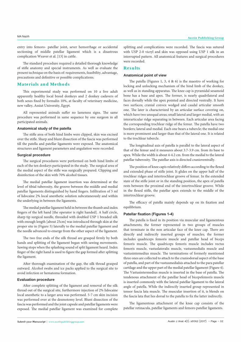
Abstract
In our study, the modified technique of medial patellar desmotomy in donkey was performed on 10 apparently adult healthy alive donkeys of both sexes in addition other, two cadavers. In concern to the upward fixation of patella in donkeys as native animals in Upper Egypt which recorded high incidence of the disease more than found in northern areas. Medial patellar desmotomy is the preferable surgical procedure of choice for treating such problem. Contrary to the standard technique of treatment, the study aimed to introduce a modified easier, less expensive and field applicable desmotomy method. our study give a full detailed applied anatomical description of the patellar ligaments specially the medial one as well as its boundaries; osseous and muscular attachments as well as the extension of the joint capsule. The medial patellar ligament was the longest and the weakest among others on the other hand the middle one was the shortest and of considerable thickness. The best and ideal side for desmotomy was determined in order to avoid massive drawbacks as piercing the joint capsule or even cutting the tendon of the sartorius muscle. Generally nearly similar anatomical results were observed with the most of authors and the donkey was an ideal animal in equine experiment and the study allow the anatomists for further comparative anatomical researches.
Keywords: Donkey; Medial patellar desmotomy; Applied anatomy
the patellar ligaments, which were of clinical importance especially among cattle and horse in the medial patellar desmotomy which indicated for the surgical treatment of the upward fixation of patella Ramakrishna [9] in buffalo, Ali &Hashim [10] in cattle and Tyagi & Singh in ruminanrs. Regarding the latter, the donkeys are widely distributed and extensively used in Egypt for agricultural and transporting issues. Upward fixation of patella is a common disorder of donkeys with a high incidence in Upper Egypt than other areas which necessitate performing medial patellar desmotomy under field conditions and with minimal instrumental requirements. The current work spot a light on detailed description of the patella with its fixation and equilibrium as well as determining the safer and exactly successful site of medial patellar desmotomy as a surgical response in upward fixation of patella among donkeys.
Upward fixation of patella is a condition believed to be according to bad conformation of the hind limb or beginning of training in young age Stick [11] in horse. Displacement of patella occurred temporary which may be spontaneously reduced or permanent requiring surgical correction. Symptoms including extension of the fetlock and phalangeal joints cause the animal to drag the affected limb and if the foot rests on the ground when the extremity is not flexed, it is almost impossible for the animal to step backward Lacorix [12] in horse.
Medial patellar ligament splitting is the surgical procedure of choice, which involves, localizing and anesthetizing the patellar ligament and tissue surrounding, 2-3 cm skin incision, introduction of sharp tipped curved tenotome behind the ligament and splitting it. Complications comprises infection of the surgical site, accidental
IntroductionOn reviewing the available literatures among large domestic
animals, most of authors described the stifle joint and patella in horse and cattle, rather than that of donkey which is scanty. Concerning the surgical and clinical practices, studying a circumscribed area anatomically was an important to serve the mentioned purposes Getty [1], Dyce et al. [2] and König and Liebich [3] in domestic animals. Topographically the anatomical regions allow surgeons and clinicians to perform perfectly Dyce et al.
Most of anatomists cited that the patella was articulated with the femur to form the femoropatellar joint and the knee cap as recorded by König and Liebich [3] in domestic animals, the patella was attached to the tibial tuberosity by the patellar ligaments and they were lateral, middle and medial Getty [1] and König and Liebich [3] in domestic animals and Uddin et al. [4] in cattle. The knee cap was fixed on its position by patellar retinacula, femoropatellar and patellar ligaments König and Liebich [3] in domestic animals. In addition to the muscular insertions for the surrounding tensor fascia lata and quadriceps femoris muscles were recorded by Getty [1], Dyce et al. [2] in domestic animals, King and Mansmann [5], Riegel and Hakola and Orsini and Sack [6] and Jennifer [7] in horse.
The patellar gliding movement was related to the function and strength of tensor fascia lata and quadriceps femoris muscles Hayes [8] and Jennifer [7] in horse.
The exact measurements of the patellar ligaments in cattle were a significant point of study of Uddin et al. [4]. In this aspect, the anatomists spot on investigating the topographical relations of
Research Article
Modified Technique for Medial Patellar Desmotomy in DonkeyMA Nazih* and MW El-SherifDepartment of Anatomy and Surgery, Faculty of veterinary medicine, Assiut University, Egypt
*Corresponding author: MA Nazih, Department of Anatomy and Surgery, Faculty of veterinary medicine, Assiut University, New valley, Egypt
Received: October 27, 2016; Accepted: January 19, 2017; Published: January 24, 2017
Austin J Anat 4(1): id1062 (2017) - Page - 02
MA Nazih Austin Publishing Group
Submit your Manuscript | www.austinpublishinggroup.com
entry into femoro- patellar joint, sever hemorrhage or accidental sectioning of middle patellar ligament which is a disastrous complication Weaver et al. [13] in cattle.
The standard procedure required a detailed thorough knowledge of stifle anatomy and special instruments. As well as evaluate the present technique on the basis of: requirements, feasibility, advantage, precautions and definitive or possible complications.
Materials and MethodsThis experimental study was performed on 10 a live adult
apparently healthy local breed donkeys and 2 donkey cadavers of both sexes fixed by formalin 10%, at faculty of veterinary medicine, new valley, Assiut University, Egypt.
All represented animals suffer no lameness signs. The same procedure was performed in same sequence by one surgeon in all participated animals.
Anatomical study of the patellaThe stifle area of both hind limbs were clipped, skin was excised
over the stifle. Sharp and blunt dissection of the fascia was performed till the patella and patellar ligaments were exposed. The anatomical structures and ligament parameters and angulation were recorded.
Surgical procedureThe surgical procedures were performed on both hind limbs of
each of the ten donkeys participated in the study. The surgical area of the medial aspect of the stifle was surgically prepared. Clipping and disinfection of the skin with 70% alcohol tissues.
The medial patellar ligament insertion was determined at the level of tibial tuberosity, the groove between the middle and medial patellar ligaments distinguished by hand fingers. Infiltration of 5 ml of lidocaine 2% local anesthetic solution subcutaneously and within the underlying in between the ligaments.
The medial patellar ligament held in between the thumb and index fingers of the left hand [the operator is right handed]. A half circle, sharp tip surgical needle, threaded with doubled USP 1 breaded silk with enough length [about 25cm] was introduced through skin at the proper site in (Figure 5) laterally to the medial patellar ligament and the needle advanced to emerge from the other aspect of the ligament.
The two free ends of the silk thread are grasped firmly by both hands and splitting of the ligament began with sawing movements. Sawing stops when the splashing sound of split ligament heard. Index finger of the right hand is used to figure the gap formed after splitting the ligament.
After thorough examination of the gap, the silk thread grasped outward. Alcohol swabs and ice packs applied to the surgical site to avoid infection or hematoma formation.
Evaluation procedureAfter complete splitting of the ligament and removal of the silk
thread out of the surgical site, furthermore injection of 2% lidocaine local anesthetic to a larger area was performed. 5-7 cm skin incision was performed over at the desmotomy level. Blunt dissection of the fascia was performed and the joint capsule and patellar ligaments were exposed. The medial patellar ligament was examined for complete
splitting and complications were recorded. The fascia was sutured with USP 2-0 vicryl and skin was opposed using USP 1 silk in an interrupted pattern. All anatomical features and surgical procedures were recorded.
ResultsAnatomical point of view
The patella (Figures 1, 3, 4 & 6) is the maestro of working for locking and unlocking mechanism of the hind limb of the donkey, as well as in standing apparatus. The knee cap is pyramidal sesamoid bone has a base and apex. The former, is nearly quadrilateral and faces dorsally while the apex pointed and directed ventrally. It have two surfaces; cranial convex wedged and caudal articular smooth one. The later is characterized by an articular surface covering on, which have two unequal areas; small lateral and larger medial, with an interarticular ridge separating in between. Each articular area facing the corresponding trochlear ridge of the femur. The patella have two borders; lateral and medial. Each one bears a tubercle; the medial one is more prominent and larger than that of the lateral one. It is related to the trochlear tubercle.
The longitudinal axis of patella is parallel to the lateral aspect of that of the femur and it measures about 3.7-3.9 cm. from its base to apex. While the width is about 4-4.2 cm. from the medial to the lateral patellar tuberosity. The patellar axis is directed cranioventrally.
The position of knee capis relatively differs according to the flexed and extended phase of stifle joint. It glides on the upper half of the trochlear ridges and intertrochlear groove of femur. In the extended state of the stifle joint or in the standing position, the apex of patella rests between the proximal end of the intertrochlear groove. While in the flexed stifle, the patellar apex extends to the middle of the intertrochlear groove.
The efficacy of patella mainly depends up on its fixation and equilibrium.
Patellar fixation (Figures 1-4)The patella is fixed in its position via muscular and ligamentous
attachments; the former represented in two groups of muscles that terminate in the non articular face of the knee cap. There are directly and indirectly inserted groups of muscles; the former includes quadriceps femoris muscle and patellar head of biceps femoris muscle. The quadriceps femoris muscle includes rectus femoris muscle, vastuslateralis muscle, vastusmedialis muscle and vastusintemedius muscle. The terminations of formerly mentioned three ones are collected to attach to the craniodorsal aspect of the base of patella, and part of the vastusmedialsis attached to the para patellar cartilage and the upper part of the medial patellar ligament (Figure 4). The Vastusintermedius muscle is inserted in the base of patella. The tendenous attachment of the patellar head of bicepefemoris muscle is inserted commonly with the lateral patellar ligament to the lateral angle of patella. While the indirectly inserted group represented in tensor fascia lata muscle. The muscular insertion of it, is blends on the fascia lata that lies dorsal to the patella to fix the latter indirectly.
The ligamentous attachment of the knee cap consists of the patellar retinacula, patellar ligaments and femoro patellar ligaments.
Austin J Anat 4(1): id1062 (2017) - Page - 03
MA Nazih Austin Publishing Group
Submit your Manuscript | www.austinpublishinggroup.com
The patellar retinacula (Figures 1 & 4), is a thick fibrous sheath surrounds the patella and patellar ligaments with their inter ligamentous space that filled by the pericapsular fat. The retinacula attaches to the deep face of fascia lata to covers overlay the craniodorsal aspect of patella and connected laterally to the lateral patellar ligament and medially to the medial patellar one and ventrally it blends with the tibial tuberosity.
The patellar ligaments (Figures are lateral, middle and medial patellar ligaments. The first (Figures 2 & 3) is easily palpable and visible from the overlaying patellar retinacula. It measures about 8.5- 8.7cm in length, 1.6- 1.7cm width and 0.4-0.5cm in thickness. The ligament forms a triangular area with the middle patellar ligament
measures about 1.3- 1.5cm base length. It arises commonly with the patellar termination of the patellar head of biceps femoris muscle in the lateral patellar angle. The upper third of the ligament, receives the tendenous fibers from the patellar head of biceps femoris muscle. The ligament descends vertically on the craniolateral aspect of the pericapsular fat of the stifle joint. At the level of the lateral epicondyle of femur, it crosses over the capsular fat cranial to the origin of the long digital extensor muscle and fibularistertius muscle. The ligament reaches the lateral part of the tibial tuberosity. It bounded laterally by the patellar retinacula. While medially by the pericapsular fat that between it and the middle one. It receives the terminal insertion of the tensor fascia lata muscle.
The middle patellar ligament (Figure 3) is the deepest ligament among the others. It is overlaid by a farrow infiltrates between the two parts of the pericapsular fat. It measures about 8.5- 8.7cm length,
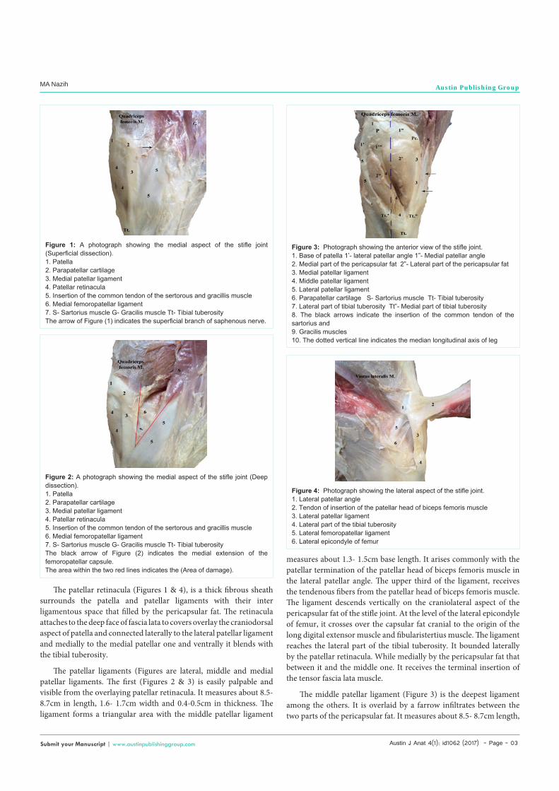
Figure 1: A photograph showing the medial aspect of the stifle joint (Superficial dissection).1. Patella2. Parapatellar cartilage3. Medial patellar ligament4. Patellar retinacula5. Insertion of the common tendon of the sertorous and gracillis muscle6. Medial femoropatellar ligament7. S- Sartorius muscle G- Gracilis muscle Tt- Tibial tuberosityThe arrow of Figure (1) indicates the superficial branch of saphenous nerve.
Figure 2: A photograph showing the medial aspect of the stifle joint (Deep dissection).1. Patella2. Parapatellar cartilage3. Medial patellar ligament4. Patellar retinacula5. Insertion of the common tendon of the sertorous and gracillis muscle6. Medial femoropatellar ligament7. S- Sartorius muscle G- Gracilis muscle Tt- Tibial tuberosityThe black arrow of Figure (2) indicates the medial extension of the femoropatellar capsule.The area within the two red lines indicates the (Area of damage).
Figure 3: Photograph showing the anterior view of the stifle joint.1. Base of patella 1’- lateral patellar angle 1”- Medial patellar angle2. Medial part of the pericapsular fat 2”- Lateral part of the pericapsular fat3. Medial patellar ligament4. Middle patellar ligament5. Lateral patellar ligament6. Parapatellar cartilage S- Sartorius muscle Tt- Tibial tuberosity 7. Lateral part of tibial tuberosity Tt”- Medial part of tibial tuberosity8. The black arrows indicate the insertion of the common tendon of the sartorius and 9. Gracilis muscles10. The dotted vertical line indicates the median longitudinal axis of leg
Figure 4: Photograph showing the lateral aspect of the stifle joint.1. Lateral patellar angle2. Tendon of insertion of the patellar head of biceps femoris muscle3. Lateral patellar ligament4. Lateral part of the tibial tuberosity5. Lateral femoropatellar ligament6. Lateral epicondyle of femur
Austin J Anat 4(1): id1062 (2017) - Page - 04
MA Nazih Austin Publishing Group
Submit your Manuscript | www.austinpublishinggroup.com
7-9 mm width and 5-6mm in thickness. The ligament descends deeply from the patellar apex in an oblique directed attitude (ventro medial) crosses the longitudinal axis of the leg which pass nearly at the middle of the ligament forming an angle measures about (20-22), the ligament directed on the cranial aspect of the femoropatellar and lateral femorotibial capsule. It terminates in the tibial groove on the tibial tuberosity. The middle patellar ligament forms medially a triangular area with the medial patellar one its base measures about 4.0 -4.2cm length.
The medial patellar ligament (Figures 1,3,4 & 5) is the most medially located and more distantly away from the described ones. The ligament is easily palpable, visible and longest patellar ligament among them. It measures about 11.0- 11.4cm length, 1.4- 1.5cm width and 3mm in thickness. It is originated from the medial patellar angle in the form of cartilaginous piece; the para patellar cartilage. The ligament descends vertically on the medial aspect of the stifle and inserted in the medial part of tibial tuberosity. The proximal third of the ligament receives some muscular fibrous attachment of the vastusmedials muscle and covers the medial face of the femoropatellar capsule. While its distal third is commonly inserted with the common tendon of the sartorius and gracilis muscles in the tibial tuberosity.
The gape which formed between the ligament and the tendon previously mentioned, represented in a triangular area (Figure 4) its base measures about 2.0-2.3cm in length and apex measures about 38- 39 degree. The mentioned area is a dangerous region (Area of damage) for the surgical interferes, where the medial femuropatellar ligament is exist as well as the medial extension of the femuropatellar capsule in addition to presence of the superficial branch of saphenous nerve. It is an important to point out that the surgeons should be take care during any operations there and to avoid hazard complications, the surgical instrument preferably introduced from the cranial aspect of the stifle joint, in the gape formed by the pericapsular fat between the middle and the medial one, just above the point of three fingers over the tibial tuberosity. The fat pad here protects the joint capsule out to be punctured.
The medial patellar ligament is related laterally to the pericapsular fat that fills between it and the middle patellarone. While it is related medially to the patellar retinacula.
The para patellar cartilage (Figures 1,3,4 & 5) is a comma shaped cartilaginous piece represents the proximal attachment of the medial patellar ligament. It has a base and apex. The former is convex and attached to the medial patellar angle while the apex is continued to the medial patellar ligament. It has a cranial surface and caudal one; the former is convex while the other is concave. The cartilage has two borders; dorsal convex and ventral concave one. It forms a loop with the medial patellar ligament around the condylar tubercle. The para-patellar cartilage measures about 2.0-2.2 cm in length and 1.1-1.3 cm in width.
There are lateral and medial femoropatellar ligaments fix the patella with the femur. The former one (Figure 2) arises from the lateral epicondyle of femur, it passes in a craniodorsal direction deep to the terminal insertion of the patellar head of biceps femoris muscle, to end in the lateral patellar angle. The ligament measures about 3.5-3.7 cm in length and 1.0-1.2 cm in width. The medial femoro patellar ligament is a funnel shaped ligament have a base measures about 3.0-3.2 cm in width and 3.7-3.8 cm in length. It originates from the
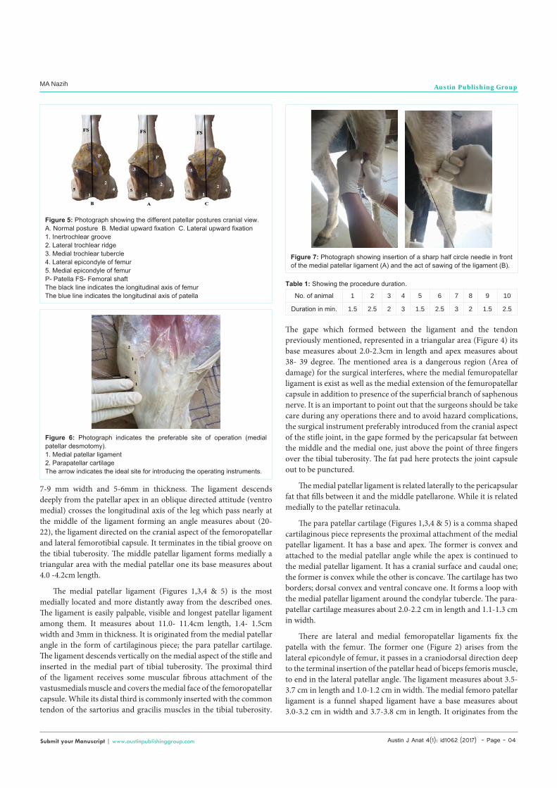
Figure 5: Photograph showing the different patellar postures cranial view.A. Normal posture B. Medial upward fixation C. Lateral upward fixation1. Inertrochlear groove 2. Lateral trochlear ridge3. Medial trochlear tubercle4. Lateral epicondyle of femur5. Medial epicondyle of femurP- Patella FS- Femoral shaftThe black line indicates the longitudinal axis of femurThe blue line indicates the longitudinal axis of patella
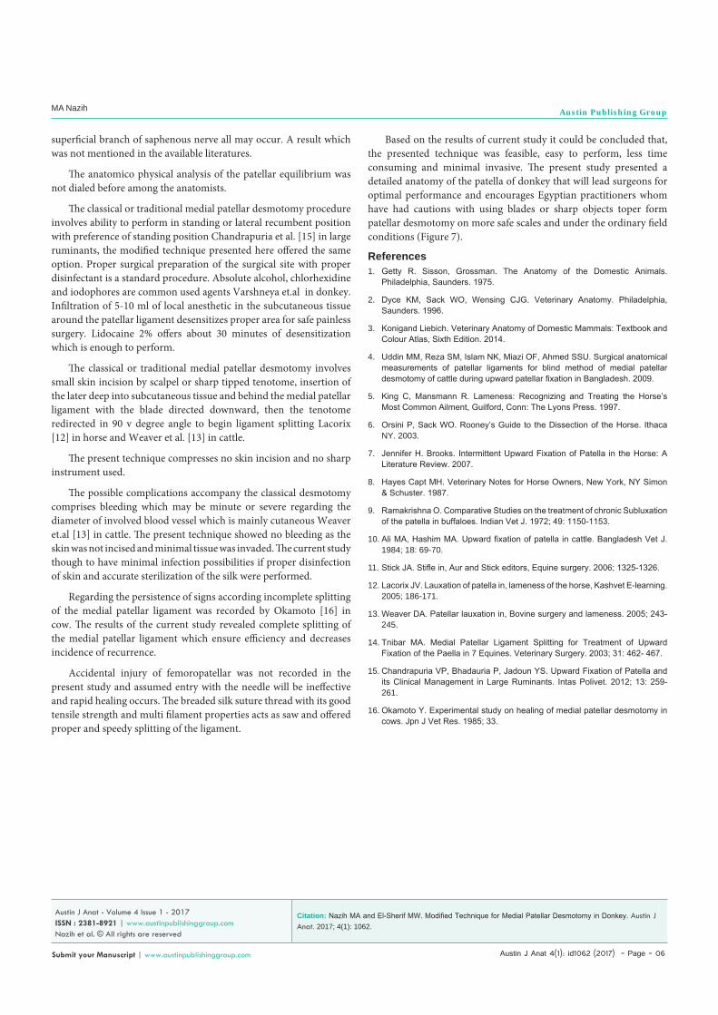
Figure 6: Photograph indicates the preferable site of operation (medial patellar desmotomy).1. Medial patellar ligament2. Parapatellar cartilageThe arrow indicates the ideal site for introducing the operating instruments.
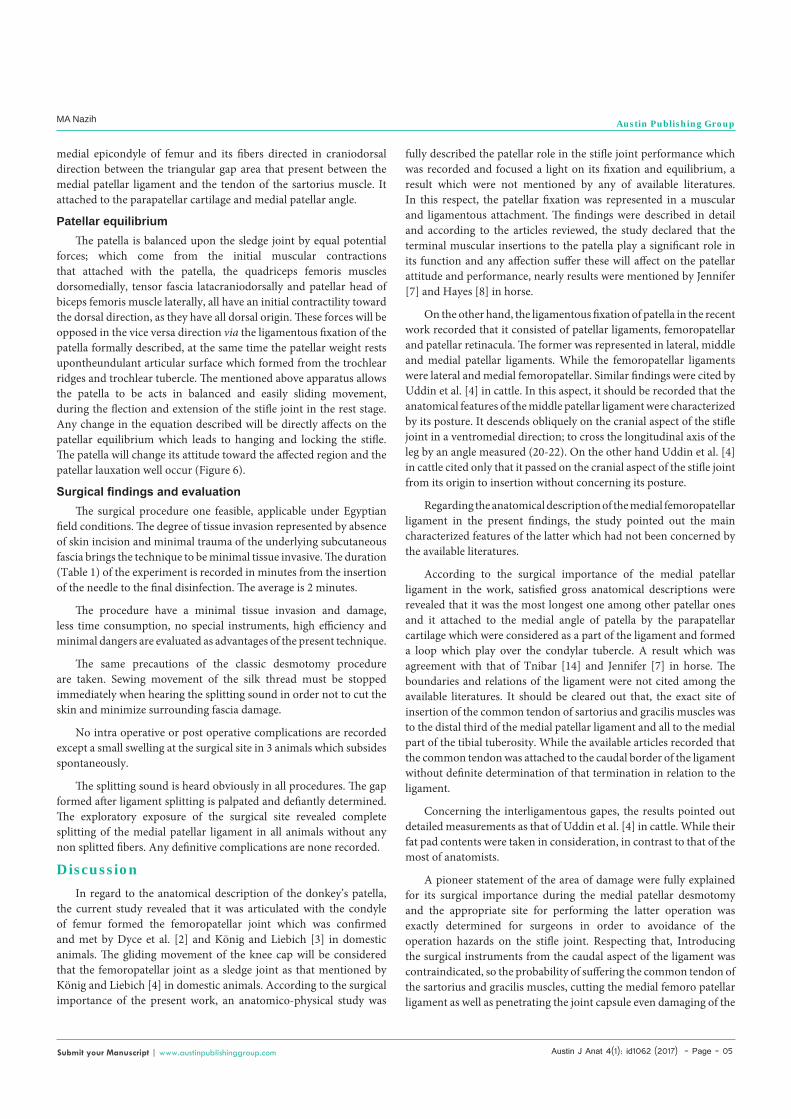
Figure 7: Photograph showing insertion of a sharp half circle needle in front of the medial patellar ligament (A) and the act of sawing of the ligament (B).
No. of animal 1 2 3 4 5 6 7 8 9 10
Duration in min. 1.5 2.5 2 3 1.5 2.5 3 2 1.5 2.5
Table 1: Showing the procedure duration.
Austin J Anat 4(1): id1062 (2017) - Page - 05
MA Nazih Austin Publishing Group
Submit your Manuscript | www.austinpublishinggroup.com
medial epicondyle of femur and its fibers directed in craniodorsal direction between the triangular gap area that present between the medial patellar ligament and the tendon of the sartorius muscle. It attached to the parapatellar cartilage and medial patellar angle.
Patellar equilibriumThe patella is balanced upon the sledge joint by equal potential
forces; which come from the initial muscular contractions that attached with the patella, the quadriceps femoris muscles dorsomedially, tensor fascia latacraniodorsally and patellar head of biceps femoris muscle laterally, all have an initial contractility toward the dorsal direction, as they have all dorsal origin. These forces will be opposed in the vice versa direction via the ligamentous fixation of the patella formally described, at the same time the patellar weight rests upontheundulant articular surface which formed from the trochlear ridges and trochlear tubercle. The mentioned above apparatus allows the patella to be acts in balanced and easily sliding movement, during the flection and extension of the stifle joint in the rest stage. Any change in the equation described will be directly affects on the patellar equilibrium which leads to hanging and locking the stifle. The patella will change its attitude toward the affected region and the patellar lauxation well occur (Figure 6).
Surgical findings and evaluationThe surgical procedure one feasible, applicable under Egyptian
field conditions. The degree of tissue invasion represented by absence of skin incision and minimal trauma of the underlying subcutaneous fascia brings the technique to be minimal tissue invasive. The duration (Table 1) of the experiment is recorded in minutes from the insertion of the needle to the final disinfection. The average is 2 minutes.
The procedure have a minimal tissue invasion and damage, less time consumption, no special instruments, high efficiency and minimal dangers are evaluated as advantages of the present technique.
The same precautions of the classic desmotomy procedure are taken. Sewing movement of the silk thread must be stopped immediately when hearing the splitting sound in order not to cut the skin and minimize surrounding fascia damage.
No intra operative or post operative complications are recorded except a small swelling at the surgical site in 3 animals which subsides spontaneously.
The splitting sound is heard obviously in all procedures. The gap formed after ligament splitting is palpated and defiantly determined. The exploratory exposure of the surgical site revealed complete splitting of the medial patellar ligament in all animals without any non splitted fibers. Any definitive complications are none recorded.
DiscussionIn regard to the anatomical description of the donkey’s patella,
the current study revealed that it was articulated with the condyle of femur formed the femoropatellar joint which was confirmed and met by Dyce et al. [2] and König and Liebich [3] in domestic animals. The gliding movement of the knee cap will be considered that the femoropatellar joint as a sledge joint as that mentioned by König and Liebich [4] in domestic animals. According to the surgical importance of the present work, an anatomico-physical study was
fully described the patellar role in the stifle joint performance which was recorded and focused a light on its fixation and equilibrium, a result which were not mentioned by any of available literatures. In this respect, the patellar fixation was represented in a muscular and ligamentous attachment. The findings were described in detail and according to the articles reviewed, the study declared that the terminal muscular insertions to the patella play a significant role in its function and any affection suffer these will affect on the patellar attitude and performance, nearly results were mentioned by Jennifer [7] and Hayes [8] in horse.
On the other hand, the ligamentous fixation of patella in the recent work recorded that it consisted of patellar ligaments, femoropatellar and patellar retinacula. The former was represented in lateral, middle and medial patellar ligaments. While the femoropatellar ligaments were lateral and medial femoropatellar. Similar findings were cited by Uddin et al. [4] in cattle. In this aspect, it should be recorded that the anatomical features of the middle patellar ligament were characterized by its posture. It descends obliquely on the cranial aspect of the stifle joint in a ventromedial direction; to cross the longitudinal axis of the leg by an angle measured (20-22). On the other hand Uddin et al. [4] in cattle cited only that it passed on the cranial aspect of the stifle joint from its origin to insertion without concerning its posture.
Regarding the anatomical description of the medial femoropatellar ligament in the present findings, the study pointed out the main characterized features of the latter which had not been concerned by the available literatures.
According to the surgical importance of the medial patellar ligament in the work, satisfied gross anatomical descriptions were revealed that it was the most longest one among other patellar ones and it attached to the medial angle of patella by the parapatellar cartilage which were considered as a part of the ligament and formed a loop which play over the condylar tubercle. A result which was agreement with that of Tnibar [14] and Jennifer [7] in horse. The boundaries and relations of the ligament were not cited among the available literatures. It should be cleared out that, the exact site of insertion of the common tendon of sartorius and gracilis muscles was to the distal third of the medial patellar ligament and all to the medial part of the tibial tuberosity. While the available articles recorded that the common tendon was attached to the caudal border of the ligament without definite determination of that termination in relation to the ligament.
Concerning the interligamentous gapes, the results pointed out detailed measurements as that of Uddin et al. [4] in cattle. While their fat pad contents were taken in consideration, in contrast to that of the most of anatomists.
A pioneer statement of the area of damage were fully explained for its surgical importance during the medial patellar desmotomy and the appropriate site for performing the latter operation was exactly determined for surgeons in order to avoidance of the operation hazards on the stifle joint. Respecting that, Introducing the surgical instruments from the caudal aspect of the ligament was contraindicated, so the probability of suffering the common tendon of the sartorius and gracilis muscles, cutting the medial femoro patellar ligament as well as penetrating the joint capsule even damaging of the
Austin J Anat 4(1): id1062 (2017) - Page - 06
MA Nazih Austin Publishing Group
Submit your Manuscript | www.austinpublishinggroup.com
superficial branch of saphenous nerve all may occur. A result which was not mentioned in the available literatures.
The anatomico physical analysis of the patellar equilibrium was not dialed before among the anatomists.
The classical or traditional medial patellar desmotomy procedure involves ability to perform in standing or lateral recumbent position with preference of standing position Chandrapuria et al. [15] in large ruminants, the modified technique presented here offered the same option. Proper surgical preparation of the surgical site with proper disinfectant is a standard procedure. Absolute alcohol, chlorhexidine and iodophores are common used agents Varshneya et.al in donkey. Infiltration of 5-10 ml of local anesthetic in the subcutaneous tissue around the patellar ligament desensitizes proper area for safe painless surgery. Lidocaine 2% offers about 30 minutes of desensitization which is enough to perform.
The classical or traditional medial patellar desmotomy involves small skin incision by scalpel or sharp tipped tenotome, insertion of the later deep into subcutaneous tissue and behind the medial patellar ligament with the blade directed downward, then the tenotome redirected in 90 v degree angle to begin ligament splitting Lacorix [12] in horse and Weaver et al. [13] in cattle.
The present technique compresses no skin incision and no sharp instrument used.
The possible complications accompany the classical desmotomy comprises bleeding which may be minute or severe regarding the diameter of involved blood vessel which is mainly cutaneous Weaver et.al [13] in cattle. The present technique showed no bleeding as the skin was not incised and minimal tissue was invaded. The current study though to have minimal infection possibilities if proper disinfection of skin and accurate sterilization of the silk were performed.
Regarding the persistence of signs according incomplete splitting of the medial patellar ligament was recorded by Okamoto [16] in cow. The results of the current study revealed complete splitting of the medial patellar ligament which ensure efficiency and decreases incidence of recurrence.
Accidental injury of femoropatellar was not recorded in the present study and assumed entry with the needle will be ineffective and rapid healing occurs. The breaded silk suture thread with its good tensile strength and multi filament properties acts as saw and offered proper and speedy splitting of the ligament.
Based on the results of current study it could be concluded that, the presented technique was feasible, easy to perform, less time consuming and minimal invasive. The present study presented a detailed anatomy of the patella of donkey that will lead surgeons for optimal performance and encourages Egyptian practitioners whom have had cautions with using blades or sharp objects toper form patellar desmotomy on more safe scales and under the ordinary field conditions (Figure 7).
References1. Getty R. Sisson, Grossman. The Anatomy of the Domestic Animals.
Philadelphia, Saunders. 1975.
2. Dyce KM, Sack WO, Wensing CJG. Veterinary Anatomy. Philadelphia, Saunders. 1996.
3. Konigand Liebich. Veterinary Anatomy of Domestic Mammals: Textbook and Colour Atlas, Sixth Edition. 2014.
4. Uddin MM, Reza SM, Islam NK, Miazi OF, Ahmed SSU. Surgical anatomical measurements of patellar ligaments for blind method of medial patellar desmotomy of cattle during upward patellar fixation in Bangladesh. 2009.
5. King C, Mansmann R. Lameness: Recognizing and Treating the Horse’s Most Common Ailment, Guilford, Conn: The Lyons Press. 1997.
6. Orsini P, Sack WO. Rooney’s Guide to the Dissection of the Horse. Ithaca NY. 2003.
7. Jennifer H. Brooks. Intermittent Upward Fixation of Patella in the Horse: A Literature Review. 2007.
8. Hayes Capt MH. Veterinary Notes for Horse Owners, New York, NY Simon & Schuster. 1987.
9. Ramakrishna O. Comparative Studies on the treatment of chronic Subluxation of the patella in buffaloes. Indian Vet J. 1972; 49: 1150-1153.
10. Ali MA, Hashim MA. Upward fixation of patella in cattle. Bangladesh Vet J. 1984; 18: 69-70.
11. Stick JA. Stifle in, Aur and Stick editors, Equine surgery. 2006; 1325-1326.
12. Lacorix JV. Lauxation of patella in, lameness of the horse, Kashvet E-learning. 2005; 186-171.
13. Weaver DA. Patellar lauxation in, Bovine surgery and lameness. 2005; 243-245.
14. Tnibar MA. Medial Patellar Ligament Splitting for Treatment of Upward Fixation of the Paella in 7 Equines. Veterinary Surgery. 2003; 31: 462- 467.
15. Chandrapuria VP, Bhadauria P, Jadoun YS. Upward Fixation of Patella and its Clinical Management in Large Ruminants. Intas Polivet. 2012; 13: 259-261.
16. Okamoto Y. Experimental study on healing of medial patellar desmotomy in cows. Jpn J Vet Res. 1985; 33.
Citation: Nazih MA and El-Sherif MW. Modified Technique for Medial Patellar Desmotomy in Donkey. Austin J Anat. 2017; 4(1): 1062.
Austin J Anat - Volume 4 Issue 1 - 2017ISSN : 2381-8921 | www.austinpublishinggroup.com Nazih et al. © All rights are reserved
Copyright © 2022 FDOKUMEN