Medial Opening Wedge High Tibial Osteotomy in Knee ... - MDPI
Upload
independentCategory
view
1download
0
Schizophrenia Research xxx (2014) xxx–xxx
SCHRES-05889; No of Pages 6
Contents lists available at ScienceDirect
Schizophrenia Research
j ourna l homepage: www.e lsev ie r .com/ locate /schres
Altered medial prefrontal activity during dynamic face processing inschizophrenia spectrum patients
Omar Mothersill a,b, Derek W. Morris a,b,d, Sinead Kelly a,b, Emma Jane Rose a,b,c, Arun Bokde b, Richard Reilly b,Michael Gill a,b, Aiden P. Corvin a,b, Gary Donohoe a,b,d,⁎a Neuropsychiatric Genetics Group, Department of Psychiatry, Trinity College Dublin, College Green, Dublin 2, Irelandb Trinity College Institute for Neuroscience, Trinity College Dublin, College Green, Dublin 2, Irelandc Transdisciplinary Science and Translational Prevention Program (TSTPP), Research Triangle Institute International, Baltimore, MD, United Statesd School of Psychology, National University of Ireland Galway, University Road, Galway, Ireland
⁎ Corresponding author at: School of Psychology, NationUniversity Road, Galway, Ireland. Tel.: +353 91 495122.
E-mail address: [email protected] (G. Dono
http://dx.doi.org/10.1016/j.schres.2014.05.0230920-9964/© 2014 Elsevier B.V. All rights reserved.
Please cite this article as: Mothersill, O., etpatients, Schizophr. Res. (2014), http://dx.do
a b s t r a c t
a r t i c l e i n f oArticle history:
Received 14 February 2014Received in revised form 30 April 2014Accepted 18 May 2014Available online xxxxKeywords:schizophreniaface processingemotionmedial prefrontal
Background: Processing the emotional content of faces is recognised as a key deficit of schizophrenia, associatedwith poorer functional outcomes and possibly contributing to the severity of clinical symptoms such as paranoia.At the neural level, fMRI studies have reported altered limbic activity in response to facial stimuli. However,previous studies may be limited by the use of cognitively demanding tasks and static facial stimuli. To addressthese issues, the current study used a face processing task involving both passive face viewing and dynamic socialstimuli. Such a task may (1) lack the potentially confounding effects of high cognitive demands and (2) showhigher ecological validity.Methods: Functional MRI was used to examine neural activity in 25 patients with a DSM-IV diagnosis ofschizophrenia/schizoaffective disorder and 21 age- and gender-matchedhealthy controlswhile they participatedin a face processing task, which involved viewing videos of angry and neutral facial expressions, and a non-
biological baseline condition.Results: While viewing faces, patients showed significantly weaker deactivation of the medial prefrontal cortex,including the anterior cingulate, and decreased activation in the left cerebellum, compared to controls. Patientsalso showed weaker medial prefrontal deactivation while viewing the angry faces relative to baseline.Discussion: Given that the anterior cingulate plays a role in processing negative emotion, weaker deactivationof this region in patients while viewing faces may contribute to an increased perception of social threat. Futurestudies examining the neurobiology of social cognition in schizophrenia using fMRI may help establish targetsfor treatment interventions.© 2014 Elsevier B.V. All rights reserved.
1. Introduction
Social cognition is a broad construct consisting of cognitive process-es that allow people to perceive, interpret and store information aboutthemselves and others (Penn et al., 2008; Van Overwalle, 2009). Exam-ples include recognising emotions from facial expressions or toneof voice, or thinking about the thoughts and goals of others. Deficits insocial cognition have been identified in several neurodevelopmentaldisorders including autism (Frith, 2001), attention deficit hyperactivitydisorder (ADHD) (Uekermann et al., 2010), and schizophrenia (Coutureet al., 2006). In schizophrenia, social cognitive deficits are a definingfeature, affecting quality of life and functional outcomes in work,
al University of Ireland Galway,
hoe).
al., Altered medial prefrontali.org/10.1016/j.schres.2014.0
relationships and independent living (Brekke et al., 2005; McGladeet al., 2008). For example, a recent meta-analysis by Fett et al. (2011)suggests that social cognition predicts more variation in social andoccupational functioning than cognitive performance alone.
One aspect of social cognition that is significantly impaired in schizo-phrenia is processing the emotional content of faces (Li et al., 2010). Forexample, patients have been reported to have difficulties recognisingemotions from faces (Aleman and Kahn, 2005), but are alsomore sensi-tive to negative facial expressions such as anger and fear compared tohealthy controls (Mandal et al., 1998; Evans et al., 2011). Excessivethreat detection from facial expressions has also been hypothesised tocontribute to the development of persecutory delusions (Green andPhillips, 2004), which are associated with patient distress (Freemanet al., 2002) and predict admission to hospital (Castle et al., 1994).
At the neural level, emotional face processing activates limbicregions including the amygdala, which is important for detecting theemotional salience of a stimulus and generating an affective response,
activity during dynamic face processing in schizophrenia spectrum5.023

2 O. Mothersill et al. / Schizophrenia Research xxx (2014) xxx–xxx
and the medial prefrontal cortex (mPFC) and anterior cingulate (ACC),which are important in expressing negative emotion and regulatingother limbic regions (Etkin et al., 2011). Meta-analysis of fMRI studiesby Li et al. (2010) indicates that schizophrenia patients generallyshow reduced activation of the bilateral amygdala in response to emo-tional faces compared to healthy controls, which may contribute to dif-ficulties understanding the emotions of other people. Similarly, Hempelet al. (2003) and Habel et al. (2010) report decreased activation of theACC in patients viewing emotional faces. However, increased activationof the amygdala and ACC have been reported in response to neutralfaces in patients versus controls, whichmay result in patientsmistaken-ly attaching emotional salience to non-emotional expressions (Hallet al., 2008; Habel et al., 2010).
There are two limitations, however, with many previous studies offace processing in schizophrenia. One limitation is that many studieshave used explicit face processing tasks, where participants mustjudge the emotional content of the faces presented and select an emo-tion from a list of two or more. Taylor et al. (2003) have previouslyshown that explicit emotional face processing during a task can reduceneural activity in limbic regions. Some studies have tried to overcomethis limitation, for example, by instructing participants to determinethe gender of faces to ensure attention to the task but also to makesure that the emotion recognition component of the task was implicit(e.g. Phillips et al., 1999). However, this type of task may also modulatelimbic activity, given thewidespread dysconnectivity and cognitive def-icits observed in schizophrenia (Meyer-Lindenberg et al., 2005; Stephanet al., 2009); e.g. these tasks may influence limbic responses in waysthat vary with connectivity, cognitive ability and/or task difficulty(Holt et al., 2006). Therefore, implicit face processing tasks withminimal additional demands may provide a more accurate measure ofneural activity during face processing.
A second limitation is that many of the previous studies of faceprocessing in schizophrenia have used static stimuli, such as Ekman’sPictures of Facial Affect (Ekman et al., 1975) (e.g. Holt et al., 2006).However, human faces and facial expressions are dynamic in nature,and temporal cues contribute to the recognition of facial expressions(e.g. Sato et al., 2004). Therefore, tasks that include dynamic facial ex-pressions may more accurately reflect real world social interactions.fMRI has revealed that several brain regions, including the amygdala,are more active while viewing dynamic facial expressions comparedto static images (Sato et al., 2004), and task-induced functional connec-tivity between the amygdala and cingulate is also increased in responseto dynamic facial expressions compared to static stimuli (Foley et al.,2012). Similar to studies using static images, schizophrenia patientsshow poorer ability to recognise dynamic facial expressions comparedto healthy controls during neuropsychological examination (Johnstonet al., 2010). Also, patients have been reported to show increased limbicactivation while watching dynamic fearful faces compared to controlsunder fMRI (Russell et al., 2007).
The purpose of the present study was to further explore and charac-terise activation differences between schizophrenia/schizoaffective dis-order patients and healthy controls during face processing. We used adynamic face processing task designed by Grosbras and Paus (2006),which is also being used to examine the effects of cognitive remediationtherapy for psychosis (Cognitive Genetics and Therapy Research Group,Ireland), and in the IMAGEN project, a longitudinal neuroimagingproject examining risk factors for mental illness in adolescents(Schumann et al., 2010). We examined neural activation in patientswith schizophrenia/schizoaffective disorder and age- and gender-matched healthy controls during passive viewing of dynamic angryand neutral faces. Specifically, we tested the hypothesis that patientswith schizophrenia or schizoaffective disorder would show alteredlimbic activitywhile passively viewing dynamic angry and neutral facialexpressions, compared to healthy controls.
Identifying differences in these regions in patients during a task thatinvolves both (1) implicit face processing, and (2) dynamic face stimuli
Please cite this article as: Mothersill, O., et al., Altered medial prefrontalpatients, Schizophr. Res. (2014), http://dx.doi.org/10.1016/j.schres.2014.0
is important for better understanding the neurobiological correlates ofsocial cognitive deficits in schizophrenia spectrum patients.
2. Methods
2.1. Sample characteristics
39 patients with a DSM-IV diagnosis of schizophrenia orschizoaffective disorder were recruited for the present study. All sub-jects were right-handed, aged between 18 and 65, had no history ofsubstance misuse in the preceding six months, no prior injury to thehead associatedwith a loss of consciousness ofmore than a fewminutesand provided consent in compliance with the local ethics committee.Five patients were excluded due to excessive movement during func-tional imaging (N3 mm translation and/or 3° rotation), seven patientswere excluded due to bad quality MRI data and/or significant artefactsand two patients were excluded due to missing data for the facesfollow-up task (see Section 2.2), yielding a total of 25 patients.
These patients were then compared to 21 healthy controls matchedfor age and gender from our on-going imaging genetics study onpsychosis (Rose et al., 2012a,b; Mothersill et al., 2013; Rose et al., 2013).
2.2. Face processing task
Participants performed a face processing task designed by Grosbrasand Paus (2006), which has previously been shown to robustly activateseveral brain regions involved in face and emotion processing, such asthe amygdala and medial prefrontal cortex (Grosbras and Paus, 2006;Schneider et al., 2011; Tahmasebi et al., 2012; Mothersill et al., 2013).During the task, participants watched a series of 2-5 second black-and-white video clips of faces starting from a neutral expression, and thenturning into an angry expression or displaying a neutral/ambiguousexpression (e.g. licking of lips). A non-biological control conditionconsisted of videos of black and white concentric circles expandingand contracting. Videos were divided into 18-second blocks consistingof four to seven video clips. Overall five angry, five neutral and ninebaseline blocks were presented (19 blocks in total). The total numberof exposures to each condition was the same between participants. Toensure that participants had paid attention to the face videos, partici-pants completed a face recognition task following their time in the scan-ner based on a still shot of faces presented in the scanner and a series offoils. Patients were excluded if they scored b three/five correct answersfor this follow-up task. Healthy controls were excluded if they scoredb four/five correct answers for this follow-up task.
2.3. MRI acquisition parameters
All participants were imaged using a Philips Intera Achieva 3 T MRIscanner (Philips Medical Systems, Best, The Netherlands) with a SENSE8-channel head coil, in the Trinity College Institute of Neuroscience.Whole-brain BOLD EPI was acquired with 40, 2.4 mm slices, 1 mm slicegap, TR = 2 200 ms, TE = 30 ms, field of view = 220 × 220 mm, flipangle = 75° and spatial resolution = 3.4 × 3.4 × 2.4 mm3. Functionalscanning lasted 160 TRs or ~5.87 minutes.
In addition, a T1-weighted image (180 slices, ~6 minutes) wasacquired using a TFE gradient echo pulse sequence, with slice thicknessof 0.9 mm, a 230 × 230 field of view, and a spatial resolution of 0.9 ×0.9 × 0.9 mm3.
2.4. MRI data pre-processing and analysis
Spatial pre-processing and statistical analysis of MRI data wasperformed using Statistical Parametric Mapping (SPM8, revision4290, http://www.fil.ion.ucl.ac.uk/spm/software/spm8/) and MATLABR2011b (v7.13; http://www.mathworks.co.uk/).
activity during dynamic face processing in schizophrenia spectrum5.023

3O. Mothersill et al. / Schizophrenia Research xxx (2014) xxx–xxx
Functional images were first realigned to themean functional imageto reduce variance due tomovement. The structural imagewas then co-registered to the mean functional image for more precise spatialnormalisation. Functional images were normalised to MNI (MontrealNeurological Institute) space using the unified segmentation approachwith a voxel size of 3 × 3 × 3 mm3 (Ashburner and Friston, 2005), andsubsequently smoothed using a 10 mm FWHM (full width at half max-imum) isotropic Gaussian filter. After pre-processing, graphical plots ofestimated time series of translations and rotations were carefullyinspected for excessive motion in each participant.
Statistical analysis was performed using the general linear model(GLM) (Friston et al., 1994). A boxcar functionwas created for each con-dition and convolved with a haemodynamic response function (HRF).This first-level model included these three condition regressors (i.e.angry faces, neutral faces and baseline) and 6 regressors to modelheadmovement. Next, t-contrasts were used tomodel condition effectsat each voxel for the following contrasts: faces (angry and neutral com-bined) versus baseline and angry faces versus neutral faces. We alsomodelled effects of the angry andneutral conditions separately (i.e. con-trasts of angry faces versus baseline and neutral faces versus baseline),consistent with previous studies of face processing in schizophrenia(e.g. Holt et al., 2006; Habel et al., 2010).
Individual contrast maps were entered into a second-level randomeffects analysis to investigate task effects at the group level (indepen-dent t-test between groups). Results were examined at a p b 0.001(uncorrected) level and clusterswere considered statistically significantat a p b 0.05 level, family-wise error (FWE) corrected for multiplecomparisons across the whole brain at the cluster level. Coordinates ofresults in MNI space were converted to Talairach space using BrainMapGingerALE 2.1 software (Eickhoff et al., 2009; Turkeltaub et al., 2012).Talairach Client 2.4.3 (Lancaster et al., 1997, 2000) was then used foranatomical localisation of these coordinates.
3. Results
3.1. Participant demographics
Independent t-testswere performed to compare age and years of ed-ucation between groups in SPSS (19.0.0); a Pearson’s chi-squared testwas performed to compare gender frequencies between groups. Therewere no significant differences between groups for age or gender(p N 0.05; see Table 1). As there were significant differences betweengroups for years of education, we examined the effects of this variableacross our sample for all contrasts examined, using a multiple regres-sion model in SPM8 with years of education as a covariate of interestand a p b 0.05, corrected statistical threshold. No clusterswere observedat this threshold.
Table 1Participant demographics.
Mean age (s.d.a)
Schizophrenia patients (N = 25) 42.88 (10.99)Healthy controls (N = 21) 38.24 (8.62)Statisticc t = 1.571p value 0.123
χ2 value derived from Pearson’s chi-squared test with variables group and gender.Mean medication (and s.d.) in patient group (chlorpromazine equivalent in mg/day) = 377.52Mean SAPS in patient group (Scale for the Assessment of Positive Symptoms) = 9.Mean SANS in patient group (Scale for the Assessment of Negative Symptoms) = 21.Note - SAPS/SANS scores listed above are mean scores for the schizophrenia patient sample recthe 25 patients included in this fMRI study are a subset.
a s.d. = standard deviation.b Education available for 45 of 46 participants.c t value derived from independent t-tests between groups.
Please cite this article as: Mothersill, O., et al., Altered medial prefrontalpatients, Schizophr. Res. (2014), http://dx.doi.org/10.1016/j.schres.2014.0
3.2. Neural activation
Relative to controls, patients showedweaker deactivation of the ACCand left medial frontal gyrus while viewing faces (angry and neutralcombined) (t(46) = 4.85; p b 0.05, corrected; see Table 2 and Fig. 1).Patients also showed reduced activation of the left cerebellum relativeto controls (t(46) = 4.49; p b 0.05, corrected; see Table 2 and Fig. 1).No significant differences were observed when the groups werecompared using a contrast of angry versus neutral faces.
Examining the angry and neutral face conditions separately, patientsalso showed weaker deactivation of the mPFC/ACC during the angryfaces versus baseline condition (t(46) = 5.94; p b 0.05, corrected; seeTable 3 and Fig. 1), while altered mPFC activity was only observed atuncorrected thresholds (p b 0.001) during the neutral faces versusbaseline condition.
As an additional data quality check, in each individual the averageparameter estimates of all voxels was calculated for each cluster thatshowed a significant activation difference between groups. Next, aver-age parameter estimateswere checked in SPSS (19.0.0) for the presenceof outliers. One outlier was identified for the faces versus baseline con-trast; as removal of this participant did not significantly affect results,results are reported with all participants included.
4. Discussion
This study examined neural activity in patients with schizophreniaor schizoaffective disorder and healthy controls during a dynamicface-processing task. While passively viewing faces (regardless of emo-tional content), patients showed weaker deactivation of the mPFC/ACCcompared to controls, and decreased left cerebellum activation. Whileviewing the angry faces specifically, patients also showed a pattern ofweaker mPFC deactivation relative to controls.
Although schizophrenia patients show deficits in facial emotion per-ception, they are hypersensitive to expressions of fear and anger(Mandal et al., 1998; Evans et al., 2011). For example, Evans et al.(2011) have previously reported that schizophrenia patients show in-creased aversion to angry faces during an associative learning task.Our finding of weaker mPFC/ACC deactivation during angry face pro-cessing in patients compared to healthy controls supports this view,given the role of the ACC in processing negative emotion. Increaseddetection of social threat (or cortical reaction to anger in others) maycontribute to the development of paranoia. However, this interpretationremains speculative due to the clinical heterogeneity of the patientgroup, which contained individuals with differing levels of positivesymptom severity.
Notably, our finding contrasts with one previous imaging study(Habel et al., 2010) which reported reduced ACC activity in schizophre-nia patients compared to controls while viewing angry faces; however,
Mean years of education (s.d.b) Gender (M:F)
14.13 (4.56) 20:518.00 (3.48) 16:5t = 3.168 χ2 = 0.0970.003 0.755
(270.82).
ruited for neuroimaging as part of a psychosis research project in Trinity College, of which
activity during dynamic face processing in schizophrenia spectrum5.023

Table 2Clusters, including individual peaks, showing significant activity differences between schizophrenia patients and controls during face processing (angry and neutral faces combined,relative to baseline), corrected for multiple comparisons at the cluster-level.
Clust-er Extent (voxels) p valuea Direction of effect Cluster peaks t-value Z-value Peak coordinates (MNI)
1 699 b0.001 Patients N controls Right anterior cingulate/ BA 24 4.85 4.32 6 32 4Left medial frontal gyrus/ BA 10 4.71 4.22 -9 56 4Right anterior cingulate/ BA 32 4.33 3.93 9 38 -11
2 168 0.011 Patients b controls Left cerebellum 4.49 4.05 -39 -70 -23Left cerebellum 4.03 3.70 -18 -76 -35Left cerebellum 3.72 3.45 -24 -79 -23
a p values are FWE-corrected for multiple comparisons at the cluster level.
4 O. Mothersill et al. / Schizophrenia Research xxx (2014) xxx–xxx
Habel et al. used an explicit emotional face processing task which theauthors suggestedmay not be comparable to implicit tasks due to differ-ing cognitive demands; e.g. explicit face processing tasksmaymodulateneural activity in limbic regions. Further research using different taskswill be required to elucidate specific differences between explicit andimplicit face processing in schizophrenia.
An alternative interpretation of the present results is that theweakermPFC deactivation observed in patients reflects altered function of the
Fig. 1. Altered neural activity in schizophrenia patients during face processing. Red-yellow: Brcontrols (N = 46; independent t-test between groups; significance is set at p b 0.001 uncorrevalues. Each 2D sagittal slice is labelled with a MNI-coordinate. Clusters are rendered on the ‘
Additional editing of figure (e.g. changing the size/resolution) performed using MS Paint andAngry = Angry versus baseline contrast; a.u. = arbitrary units; mPFC = medial prefrontal cor
Please cite this article as: Mothersill, O., et al., Altered medial prefrontalpatients, Schizophr. Res. (2014), http://dx.doi.org/10.1016/j.schres.2014.0
default mode network, a network of brain regions that are more activewhen a person is not engaged in a cognitive task, i.e. at rest (Raichleet al., 2001). The default mode network, which includes the mPFC,has been hypothesised to play a role in a variety of functions such asself-referential processing that may be important for social cognition(Buckner et al., 2008).
Given that default mode hyperactivity has been observed in schizo-phrenia patients during cognitive tasks (e.g. Garrity et al., 2007), the
ain regions showing altered activity during face processing in patients relative to healthycted without a cluster threshold for display purposes; d.f. = 44). Colour bars represent t-ch256’ brain template using MRIcroGL (http://www.mccauslandcenter.sc.edu/mricrogl/).Paint.NET v3.5.10. Faces = Faces (angry and neutral combined) versus baseline contrast;tex. NB - Fig. 1 intended for colour reproduction.
activity during dynamic face processing in schizophrenia spectrum5.023
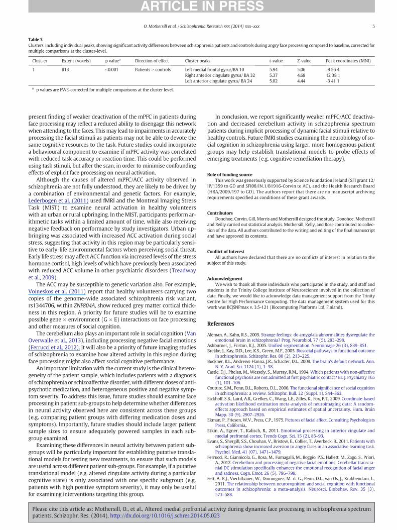
Table 3Clusters, including individual peaks, showing significant activity differences between schizophrenia patients and controls during angry face processing compared to baseline, corrected formultiple comparisons at the cluster-level.
Clust-er Extent (voxels) p valuea Direction of effect Cluster peaks t-value Z-value Peak coordinates (MNI)
1 813 b0.001 Patients N controls Left medial frontal gyrus/BA 10 5.94 5.06 -9 56 4Right anterior cingulate gyrus/ BA 32 5.37 4.68 12 38 1Left anterior cingulate gyrus/ BA 24 5.02 4.44 -3 41 1
a p values are FWE-corrected for multiple comparisons at the cluster level.
5O. Mothersill et al. / Schizophrenia Research xxx (2014) xxx–xxx
present finding of weaker deactivation of the mPFC in patients duringface processing may reflect a reduced ability to disengage this networkwhen attending to the faces. Thismay lead to impairments in accuratelyprocessing the facial stimuli as patients may not be able to devote thesame cognitive resources to the task. Future studies could incorporatea behavioural component to examine if mPFC activity was correlatedwith reduced task accuracy or reaction time. This could be performedusing task stimuli, but after the scan, in order to minimise confoundingeffects of explicit face processing on neural activation.
Although the causes of altered mPFC/ACC activity observed inschizophrenia are not fully understood, they are likely to be driven bya combination of environmental and genetic factors. For example,Lederbogen et al. (2011) used fMRI and the Montreal Imaging StressTask (MIST) to examine neural activation in healthy volunteerswith an urban or rural upbringing. In theMIST, participants perform ar-ithmetic tasks within a limited amount of time, while also receivingnegative feedback on performance by study investigators. Urban up-bringing was associated with increased ACC activation during socialstress, suggesting that activity in this region may be particularly sensi-tive to early-life environmental factors when perceiving social threat.Early life stressmay affect ACC function via increased levels of the stresshormone cortisol, high levels of which have previously been associatedwith reduced ACC volume in other psychiatric disorders (Treadwayet al., 2009).
The ACC may be susceptible to genetic variation also. For example,Voineskos et al. (2011) report that healthy volunteers carrying twocopies of the genome-wide associated schizophrenia risk variant,rs1344706, within ZNF804A, show reduced grey matter cortical thick-ness in this region. A priority for future studies will be to examinepossible gene × environment (G × E) interactions on face processingand other measures of social cognition.
The cerebellum also plays an important role in social cognition (VanOverwalle et al., 2013), including processing negative facial emotions(Ferrucci et al., 2012). It will also be a priority of future imaging studiesof schizophrenia to examine how altered activity in this region duringface processing might also affect social cognitive performance.
An important limitationwith the current study is the clinical hetero-geneity of the patient sample, which includes patients with a diagnosisof schizophrenia or schizoaffective disorder,with different doses of anti-psychotic medication, and heterogeneous positive and negative symp-tom severity. To address this issue, future studies should examine faceprocessing in patient sub-groups to help determinewhether differencesin neural activity observed here are consistent across these groups(e.g. comparing patient groups with differing medication doses andsymptoms). Importantly, future studies should include larger patientsample sizes to ensure adequately powered samples in each sub-group examined.
Examining these differences in neural activity between patient sub-groups will be particularly important for establishing putative transla-tional models for testing new treatments, to ensure that such modelsare useful across different patient sub-groups. For example, if a putativetranslational model (e.g. altered cingulate activity during a particularcognitive state) is only associated with one specific subgroup (e.g.patients with high positive symptom severity), it may only be usefulfor examining interventions targeting this group.
Please cite this article as: Mothersill, O., et al., Altered medial prefrontalpatients, Schizophr. Res. (2014), http://dx.doi.org/10.1016/j.schres.2014.0
In conclusion, we report significantly weaker mPFC/ACC deactiva-tion and decreased cerebellum activity in schizophrenia spectrumpatients during implicit processing of dynamic facial stimuli relative tohealthy controls. Future fMRI studies examining the neurobiology of so-cial cognition in schizophrenia using larger, more homogenous patientgroups may help establish translational models to probe effects ofemerging treatments (e.g. cognitive remediation therapy).
Role of funding sourceThis work was generously supported by Science Foundation Ireland (SFI grant 12/
IP/1359 to GD and SFI08/IN.1/B1916-Corvin to AC), and the Health Research Board(HRA/2009/197 to GD). The authors report that there are no manuscript archivingrequirements specified as conditions of these grant awards.
ContributorsDonohoe, Corvin, Gill, Morris andMothersill designed the study. Donohoe, Mothersill
and Reilly carried out statistical analysis. Mothersill, Kelly, and Rose contributed to collec-tion of the data. All authors contributed to the writing and editing of the final manuscriptand have approved its contents.
Conflict of InterestAll authors have declared that there are no conflicts of interest in relation to the
subject of this study.
AcknowledgmentWe wish to thank all those individuals who participated in the study, and staff and
students in the Trinity College Institute of Neuroscience involved in the collection ofdata. Finally, we would like to acknowledge data management support from the TrinityCentre for High Performance Computing. The data management system used for thiswork was BC|SNPmax v. 3.5-121 (Biocomputing Platforms Ltd, Finland).
References
Aleman, A., Kahn, R.S., 2005. Strange feelings: do amygdala abnormalities dysregulate theemotional brain in schizophrenia? Prog. Neurobiol. 77 (5), 283–298.
Ashburner, J., Friston, K.J., 2005. Unified segmentation. Neuroimage 26 (3), 839–851.Brekke, J., Kay, D.D., Lee, K.S., Green, M.F., 2005. Biosocial pathways to functional outcome
in schizophrenia. Schizophr. Res. 80 (2), 213–225.Buckner, R.L., Andrews‐Hanna, J.R., Schacter, D.L., 2008. The brain's default network. Ann.
N. Y. Acad. Sci. 1124 (1), 1–38.Castle, D.J., Phelan, M., Wessely, S., Murray, R.M., 1994. Which patients with non-affective
functional psychosis are not admitted at first psychiatric contact? Br. J. Psychiatry 165(1), 101–106.
Couture, S.M., Penn, D.L., Roberts, D.L., 2006. The functional significance of social cognitionin schizophrenia: a review. Schizophr. Bull. 32 (Suppl. 1), S44–S63.
Eickhoff, S.B., Laird, A.R., Grefkes, C., Wang, L.E., Zilles, K., Fox, P.T., 2009. Coordinate‐basedactivation likelihood estimation meta‐analysis of neuroimaging data: A random‐
effects approach based on empirical estimates of spatial uncertainty. Hum. BrainMapp. 30 (9), 2907–2926.
Ekman, P., Friesen, W.V., Press, C.P., 1975. Pictures of facial affect. Consulting PsychologistsPress, California,.
Etkin, A., Egner, T., Kalisch, R., 2011. Emotional processing in anterior cingulate andmedial prefrontal cortex. Trends Cogn. Sci. 15 (2), 85–93.
Evans, S., Shergill, S.S., Chouhan, V., Bristow, E., Collier, T., Averbeck, B., 2011. Patients withschizophrenia show increased aversion to angry faces in an associative learning task.Psychol. Med. 41 (07), 1471–1479.
Ferrucci, R., Giannicola, G., Rosa, M., Fumagalli, M., Boggio, P.S., Hallett, M., Zago, S., Priori,A., 2012. Cerebellum and processing of negative facial emotions: Cerebellar transcra-nial DC stimulation specifically enhances the emotional recognition of facial angerand sadness. Cogn. Emot. 26 (5), 786–799.
Fett, A.-K.J., Viechtbauer, W., Dominguez, M.-d.-G., Penn, D.L., van Os, J., Krabbendam, L.,2011. The relationship between neurocognition and social cognition with functionaloutcomes in schizophrenia: a meta-analysis. Neurosci. Biobehav. Rev. 35 (3),573–588.
activity during dynamic face processing in schizophrenia spectrum5.023

6 O. Mothersill et al. / Schizophrenia Research xxx (2014) xxx–xxx
Foley, E., Rippon, G., Thai, N.J., Longe, O., Senior, C., 2012. Dynamic facial expressionsevoke distinct activation in the face perception network: a connectivity analysisstudy. J. Cogn. Neurosci. 24 (2), 507–520.
Freeman, D., Garety, P.A., Kuipers, E., Fowler, D., Bebbington, P.E., 2002. A cognitive modelof persecutory delusions. Br. J. Clin. Psychol. 41 (4), 331–347.
Friston, K.J., Holmes, A.P., Worsley, K.J., Poline, J.P., Frith, C.D., Frackowiak, R.S.J., 1994.Statistical parametric maps in functional imaging: a general linear approach. Hum.Brain Mapp. 2 (4), 189–210.
Frith, U., 2001. Mind blindness and the brain in autism. Neuron 32 (6), 969–979.Garrity, A., Pearlson, G., McKiernan, K., Lloyd, D., Kiehl, K., Calhoun, V., 2007. Aberrant
“default mode” functional connectivity in schizophrenia. Am. J. Psychiatry 164 (3),450–457.
Green, M.J., Phillips, M.L., 2004. Social threat perception and the evolution of paranoia.Neurosci. Biobehav. Rev. 28 (3), 333–342.
Grosbras, M.H., Paus, T., 2006. Brain networks involved in viewing angry hands or faces.Cereb. Cortex 16 (8), 1087–1096.
Habel, U., Chechko, N., Pauly, K., Koch, K., Backes, V., Seiferth, N., Shah, N.J., Stöcker, T.,Schneider, F., Kellermann, T., 2010. Neural correlates of emotion recognition inschizophrenia. Schizophr. Res. 122 (1), 113–123.
Hall, J., Whalley, H.C., McKirdy, J.W., Romaniuk, L., McGonigle, D., McIntosh, A.M., Baig, B.J.,Gountouna, V.-E., Job, D.E., Donaldson, D.I., Sprengelmeyer, R., Young, A.W.,Johnstone, E.C., Lawrie, S.M., 2008. Overactivation of fear systems to neutral faces inschizophrenia. Biol. Psychiatry 64 (1), 70–73.
Hempel, A., Hempel, E., Schönknecht, P., Stippich, C., Schröder, J., 2003. Impairment inbasal limbic function in schizophrenia during affect recognition. Psychiatry Res.Neuroimaging 122 (2), 115–124.
Holt, D.J., Kunkel, L., Weiss, A.P., Goff, D.C., Wright, C.I., Shin, L.M., Rauch, S.L., Hootnick, J.,Heckers, S., 2006. Increased medial temporal lobe activation during the passive view-ing of emotional and neutral facial expressions in schizophrenia. Schizophr. Res. 82(2), 153–162.
Johnston, P.J., Enticott, P.G., Mayes, A.K., Hoy, K.E., Herring, S.E., Fitzgerald, P.B., 2010.Symptom correlates of static and dynamic facial affect processing in schizophrenia:evidence of a double dissociation? Schizophr. Bull. 36 (4), 680–687.
Lancaster, J., Rainey, L., Summerlin, J., Freitas, C., Fox, P., Evans, A., Toga, A., Mazziotta, J.,1997. Automated labeling of the human brain: a preliminary report on the develop-ment and evaluation of a forward-transform method. Hum. Brain Mapp. 5 (4), 238.
Lancaster, J.L., Woldorff, M.G., Parsons, L.M., Liotti, M., Freitas, C.S., Rainey, L., Kochunov, P.V., Nickerson, D., Mikiten, S.A., Fox, P.T., 2000. Automated Talairach atlas labels forfunctional brain mapping. Hum. Brain Mapp. 10 (3), 120–131.
Lederbogen, F., Kirsch, P., Haddad, L., Streit, F., Tost, H., Schuch, P., Wüst, S., Pruessner, J.C.,Rietschel, M., Deuschle, M., Meyer-Lindenberg, A., 2011. City living and urbanupbringing affect neural social stress processing in humans. Nature 474 (7352),498–501.
Li, H., Chan, R.C.K., McAlonan, G.M., Gong, Q., 2010. Facial emotion processing inschizophrenia: a meta-analysis of functional neuroimaging data. Schizophr. Bull. 36(5), 1029–1039.
Mandal, M.K., Pandey, R., Prasad, A.B., 1998. Facial expressions of emotions andschizophrenia: A review. Schizophr. Bull. 24 (3), 399.
McGlade, N., Behan, C., Hayden, J., O'Donoghue, T., Peel, R., Haq, F., Gill, M., Corvin, A.,O'Callaghan, E., Donohoe, G., 2008. Mental state decoding v. mental state reasoningas a mediator between cognitive and social function in psychosis. Br. J. Psychiatry193 (1), 77–78.
Meyer-Lindenberg, A.S., Olsen, R.K., Kohn, P.D., Brown, T., Egan, M.F., Weinberger, D.R.,Berman, K.F., 2005. Regionally specific disturbance of dorsolateral prefrontal-hippocampal functional connectivity in schizophrenia. Arch. Gen. Psychiatry 62 (4),379–386.
Mothersill, O., Morris, D.W., Kelly, S., Rose, E.J., Fahey, C., O'Brien, C., Lyne, R., Reilly, R., Gill,M., Corvin, A.P., Donohoe, G., 2013. Effects of MIR137 on fronto-amygdala functionalconnectivity. Neuroimage. http://dx.doi.org/10.1016/j.neuroimage.2013.12.019.
Penn, D.L., Sanna, L.J., Roberts, D.L., 2008. Social cognition in schizophrenia: an overview.Schizophr. Bull. 34 (3), 408–411.
Phillips, M.L., Williams, L., Senior, C., Bullmore, E.T., Brammer, M.J., Andrew, C., Williams,S.C., David, A.S., 1999. A differential neural response to threatening and non-threatening negative facial expressions in paranoid and non-paranoid schizo-phrenics. Psychiatry Res. Neuroimaging 92 (1), 11–31.
Please cite this article as: Mothersill, O., et al., Altered medial prefrontalpatients, Schizophr. Res. (2014), http://dx.doi.org/10.1016/j.schres.2014.0
Raichle, M.E., MacLeod, A.M., Snyder, A.Z., Powers, W.J., Gusnard, D.A., Shulman, G.L.,2001. A default mode of brain function. Proc. Natl. Acad. Sci. U. S. A. 98 (2), 676–682.
Rose, E.J., Greene, C., Kelly, S., Morris, D.W., Robertson, I.H., Fahey, C., Jacobson, S.,O'Doherty, J., Newell, F.N., McGrath, J., Bokde, A., Garavan, H., Frodl, T., Gill, M.,Corvin, A.P., Donohoe, G., 2012a. The NOS1 variant rs6490121 is associated withvariation in prefrontal function and grey matter density in healthy individuals.Neuroimage 60 (1), 614–622.
Rose, E.J., Morris, D.W., Fahey, C., Robertson, I.H., Greene, C., O'Doherty, J., Newell, F.N.,Garavan, H., McGrath, J., Bokde, A., Tropea, D., Gill, M., Corvin, A.P., Donohoe, G.,2012b. The Effect of the Neurogranin Schizophrenia Risk Variant rs12807809 onBrain Structure and Function. Twin Res. Hum. Genet. 15 (03), 296–303.
Rose, E.J., Morris, D.W., Hargreaves, A., Fahey, C., Greene, C., Garavan, H., Gill, M., Corvin, A.,Donohoe, G., 2013. Neural effects of the CSMD1 genome‐wide associated schizophre-nia risk variant rs10503253. Am. J. Med. Genet. 162 (6), 530–537.
Russell, T., Reynaud, E., Kucharska-Pietura, K., Ecker, C., Benson, P., Zelaya, F., Giampietro,V., Brammer, M., David, A., Phillips, M., 2007. Neural responses to dynamic expres-sions of fear in schizophrenia. Neuropsychologia 45 (1), 107–123.
Sato, W., Kochiyama, T., Yoshikawa, S., Naito, E., Matsumura, M., 2004. Enhanced neuralactivity in response to dynamic facial expressions of emotion: an fMRI study. Cogn.Brain Res. 20 (1), 81–91.
Schneider, S., Peters, J., Bromberg, U., Brassen, S., Menz, M., Miedl, S., Loth, E.,Banaschewski, T., Barbot, A., Barker, G., Conrod, P.J., Dalley, J.W., Flor, H., Gallinat, J.,Garavan, H., Heinz, A., Itterman, B., Mallik, C., Mann, K., Artiges, E., Paus, T., Poline, J.B., Rietschel, M., Reed, L., Smolka, M.N., Spanagel, R., Speiser, C., Strohlr, A., Struve,M., Schumann, G., Buchel, C., IMAGEN consortium, 2011. Boys do it the right way:Sex-dependent amygdala lateralization during face processing in adolescents.Neuroimage 56 (3), 1847–1853.
Schumann, G., Loth, E., Banaschewski, T., Barbot, A., Barker, G., Buchel, C., Conrod, P.J.,Dalley, J.W., Flor, H., Gallinat, J., Garavan, H., Heinz, A., Itterman, B., Lathrop, M.,Mallik, C., Mann, K., Martinot, J.L., Paus, T., Poline, J.B., Robbins, T.W., Rietschel, M.,Reed, L., Smolka, M., Spanagel, R., Speiser, C., Stephens, D.N., Strohle, A., Struve, M.,IMAGEN consortium, 2010. The IMAGEN study: reinforcement-related behaviour innormal brain function and psychopathology. Mol. Psychiatry 15 (12), 1128–1139.
Stephan, K.E., Friston, K.J., Frith, C.D., 2009. Dysconnection in schizophrenia: fromabnormal synaptic plasticity to failures of self-monitoring. Schizophr. Bull. 35 (3),509–527.
Tahmasebi, A.M., Artiges, E., Banaschewski, T., Barker, G.J., Bruehl, R., Büchel, C., Conrod, P.J., Flor, H., Garavan, H., Gallinat, J., Heinz, A., Itterman, B., Loth, E., Mareckova, K.,Martinot, J.L., Poline, J.B., Rietschel, M., Smolka, M.N., Strohle, A., Schumann, G.,Paus, T., IMAGEN consortium, 2012. Creating probabilistic maps of the face networkin the adolescent brain: a multicentre functional MRI study. Hum. Brain Mapp. 33(4), 938–957.
Taylor, S.F., Phan, K.L., Decker, L.R., Liberzon, I., 2003. Subjective rating of emotionallysalient stimuli modulates neural activity. Neuroimage 18 (3), 650–659.
Treadway, M.T., Grant, M.M., Ding, Z., Hollon, S.D., Gore, J.C., Shelton, R.C., 2009. Earlyadverse events, HPA activity and rostral anterior cingulate volume in MDD. PLoSOne 4 (3), e4887. http://dx.doi.org/10.1371/journal.pone.0004887.
Turkeltaub, P.E., Eickhoff, S.B., Laird, A.R., Fox, M., Wiener, M., Fox, P., 2012. Minimizingwithin‐experiment and within‐group effects in activation likelihood estimationmeta‐analyses. Hum. Brain Mapp. 33 (1), 1–13.
Uekermann, J., Kraemer, M., Abdel-Hamid, M., Schimmelmann, B., Hebebrand, J., Daum, I.,Wiltfang, J., Kis, B., 2010. Social cognition in attention-deficit hyperactivity disorder(ADHD). Neurosci. Biobehav. Rev. 34 (5), 734–743.
Van Overwalle, F., 2009. Social cognition and the brain: A meta‐analysis. Hum. BrainMapp. 30 (3), 829–858.
Van Overwalle, F., Baetens, K., Mariën, P., Vandekerckhove, M., 2013. Social Cognition andthe Cerebellum: AMeta-analysis of over 350 fMRI studies. Neuroimage. http://dx.doi.org/10.1016/j.neuroimage.2013.09.033.
Voineskos, A.N., Lerch, J.P., Felsky, D., Tiwari, A., Rajji, T.K., Miranda, D., Lobaugh, N.J., Pollock,B.G., Mulsant, B.H., Kennedy, J.L., 2011. The ZNF804A gene: characterization of a novelneural risk mechanism for the major psychoses. Neuropsychopharmacology 36 (9),1871–1878.
activity during dynamic face processing in schizophrenia spectrum5.023
Copyright © 2022 FDOKUMEN