Natural Rewards Self-Management, Personality, and Achievement Outcomes

Behavioural Brain Research 123 (2001) 165–183
Research report
Neurons in rat medial prefrontal cortex show anticipatory ratechanges to predictable differential rewards in a spatial memory
task
Wayne E. Pratt, Sheri J.Y. Mizumori *Uni�ersity of Utah, Salt Lake City, UT 84112, USA
Received 4 December 2000; received in revised form 26 February 2001; accepted 26 February 2001
Abstract
The present study electrophysiologically examined the contribution of prelimbic and infralimbic neurons in the medialprefrontal cortex (mPFC) to integration of reward and spatial information while rats performed multiple memory trials on adifferentially rewarded eight arm radial maze. Alternate arms consistently held one of two different reward amounts. Similar toprevious examinations of the rat mPFC, few cells showed discrete place fields or altered firing during a delay period. The mostcommon behavioral correlate was a change in neuronal firing rate prior to reward acquisition at arm ends. A small number ofreward-related cells differentiated between high and low reward arms. The presence of neurons that anticipate expected rewardconsequences based on information about the spatial environment is consistent with the hypothesis that the mPFC is part of aneural system which merges spatial information with its motivational significance. © 2001 Elsevier Science B.V. All rightsreserved.
Keywords: Spatial navigation; Neural systems; Reward; Prefrontal cortex; Amygdala; Medial ventral striatum; Electrophysiology
www.elsevier.com/locate/bbr
1. Introduction
Since the seminal studies of Mogenson and co-work-ers, the medial ventral striatum (mVS) has been viewedas one potential region where traditionally definedmemory structures such as the hippocampus and amyg-dala may impact motor structures via pallidal efferents(for review see Ref. [42]). Stimulation of either thehippocampus or amygdala of the rat invokes firing ratechanges within the mVS, and chemical excitation ofthese regions cause behavioral changes that are mVS-dependent [43,47,75–77]. This suggests that perhapsone means for the hippocampus to impact behavior isvia its projections (from the subiculum) to the mVS[35]. The hippocampus itself has been long implicated
as an important structure for rat navigation, as itsremoval results in drastic impairments in spatial tasks(for review see Ref. [2]) and ‘‘place cells’’ (neuronswhich fire when a rat occupies a discrete location in itsspatial environment) are found in the rat hippocampus[48,49,57]. However, despite the further defining ofnecessary sensory and mnemonic components that drivehippocampal representations of space, the precise waythat these signals affect behavior remains to bedetermined.
To begin to address this issue, Lavoie and Mizumori[29] recorded from mVS neurons in freely moving ratsduring performance of a win-shift navigation task, todetermine what neuronal correlates might be found in amotor structure that receives afferent information fromthe hippocampus. Recordings revealed that the nucleusaccumbens and surrounding striatum encoded not onlyspatial information, but also motivationally relevantinformation about the expectation and presence of re-ward within the environment. Furthermore, a subset of
* Corresponding author. Address: Department of Psychology, Uni-versity of Washington, Box 351525, Seattle, WA 98195, USA. Tel.:+1-206-543-2699; fax: +1-206-685-3157..
E-mail address: [email protected] (S.J.Y. Mizumori).
0166-4328/01/$ - see front matter © 2001 Elsevier Science B.V. All rights reserved.PII: S 0 1 6 6 -4328 (01 )00204 -2

W.E. Pratt, S.J.Y. Mizumori / Beha�ioural Brain Research 123 (2001) 165–183166
reward-related neurons distinguished between arms thatheld high and low reward amounts. A recent report byMartin and Ono [32] in a different spatial task (forag-ing for brain stimulation) has confirmed the presence oflocation- and reward-related cells within the mVS.These data suggest that the mVS may be an importantsite for the integration of motivational informationwith spatial representations. Given that a likely sourceof afferent spatial information is the hippocampal com-plex, and that hippocampal place fields appear to beindependent of reward placement in rich spatial envi-ronments [68], it was of interest to determine whatbrain regions forward reward-related information to beintegrated within the mVS. Pratt and Mizumori [54]subsequently recorded from the basolateral nuclei ofthe amygdala (BLA) and demonstrated that they are apossible source of reward and reward expectancy infor-mation during performance of the win-shift spatialtask. The medial prefrontal cortex (mPFC) is also apossible candidate for providing the mVS with spatiallyrelevant reward information [65]. The prelimbic (PL)and, to a lesser extent, the infralimbic (IL) regions ofthe mPFC in the rat are densely innervated by theventral subiculum and the CA1 region of hippocampus[4,5,19,69]. Given the known role of these latter regionsin spatial processing, the mPFC itself may have accessto spatial information. The mPFC also receives affer-ents from several brain regions that process the signifi-cance of rewards and the cues that predict rewards. Inparticular, the PL and IL areas of mPFC receive pro-jections from the medial posterior regions of the BLAof the rat, as well as extensive input from the ventraltegmental area [17,25,33,34,46,70]. Individual neuronswithin the amygdala and tegmentum have been shownto encode rewards and the cues that predict them[28,44,45,60,62].
Given the anatomical convergence of spatial andreward systems, there is a potentially important role ofthe mPFC in the rat for encoding relevant rewardlocations within a spatial environment. Consistent withother brain regions associated with reward systems,much of the prefrontal cortex of the rat supportselectrical self-stimulation (for review see Ref. [58]).When anatomically disconnected from lateral regions ofthe rat prefrontal cortex, stimulation of the medial PFCinduces place preferences, but not taste preferences(sulcal prefrontal cortex stimulation produces tastepreferences under the same experimental condition,[59]). This suggests a functionally specific role for me-dial regions of the prefrontal cortex for reward-placeassociations. Additional support is granted by multipleexperiments in which rats with mPFC lesions are im-paired at acquiring spatial tasks that involve learninglocations of rewards [7,11,18,27,30,51,53,67,74]. Suchimpairments are similar to those found in studies thatlesion the medial striatum [1,10], and are consistent
with the view that the mPFC and the mVS are func-tionally linked. Additionally, recordings from individ-ual units within the mPFC have reported only smallnumbers of cells with location-specific firing [21,52].Instead, correlated neurons within the mPFC appearsensitive to specific behaviors during goal-orientedtasks. Recordings of the rat mPFC to date, however,have not examined this region’s possible contributionfor associations between reward and spatialinformation.
If the mPFC is important for learning about rewardswithin a spatial context, individual neurons should altertheir activity relative to the presence of predictablerewards in a spatial task. Moreover, if such representa-tions are important for the planning of spatial move-ments based on different reward contingencies, someneurons should distinguish between different rewardamounts reliably located within the environment. Thecurrent experiment set out to specifically examine thefiring patterns of rat mPFC neurons during a spatialmemory task involving differential rewards found inpredictable locations. Furthermore, it was of interest tobe able to directly compare the correlates of the mPFCwith previous reports of recorded cells from mVS andthe BLA across studies using comparable analyses.Comparisons of the types and relative number of differ-ent behavioral correlates may provide further insightinto the representations of reward within a spatialcontext, and help to define the individual roles of eachregion within a broader system that may serve tointegrate spatial and reward contingencies to ultimatelydirect behavior.
2. Materials and methods
2.1. Subjects
Subjects consisted of four implanted male Long-Evans rats, obtained from Simonsens Laboratories andhoused in the laboratory. Rats were given 2–3 days toacclimate to individual housing before being reduced toand maintained at 80% ad lib body weight. Handlingwas done daily for no fewer than 7 days prior tobeginning behavioral training. Water was available atall times. Rats were maintained on a 12-h light–darkcycle (lights on at 7 a.m.) in a controlled temperatureenvironment of 21°C. All behavioral testing was doneduring the light phase.
2.2. Apparatus
A semi-automated eight arm radial maze was usedfor this experiment, consisting of eight black Plexiglasrunways (58×5.5 cm) radiating from a center platform(19.5 cm diameter) and supported 79 cm from the floor.
W.E. Pratt, S.J.Y. Mizumori / Beha�ioural Brain Research 123 (2001) 165–183 167
The internal segments of each arm could be raised orlowered by remote control to provide access to thearms from the maze center.
The experiment was conducted within an environ-ment that consisted of an �1.6 m×1.6 m square by 3m deep space enclosed by black drapes. On thesedrapes were several distinct visual cues. The room waslit by four 15 W bulbs located at the corners of theroom.
2.3. Beha�ioral training
Upon reaching 80% body weight, the rats wereplaced on the maze and given access to all eight armswith an abundant supply of chocolate milk on eacharm. Once the rat had visited and consumed reward onall eight arms, they were gradually shaped to receivereward only at the end of each arm. Maximum time perday on the maze was 1 h.
Once it was established that the rat would visit eacharm, they were given trials in which all eight arms werepresented. The rats were allowed to visit each arm forchocolate milk reward (five drops). As the rats left eacharm, the arm was lowered, and the rat was allowed tovisit each radial location only once per trial. One trialconsisted of a visit to all eight rewarded locations. Theintertrial interval was 2 min. This training continueduntil eight such trials were completed within a 1 hperiod.
When this criterion was met, the rats were trained toperform a partial forced-choice memory task. Begin-ning with this training, all arms were baited prior toeach trial with large or small rewards (5 drops or 1drop, respectively) on odd or even arms. Determinationof whether large reward was placed on odd or evenarms was randomly determined for each rat, and re-mained consistent throughout the experiment unlessotherwise noted. During the study portion of each trial,four arms were raised in an order determined from atable of random numbers. As the rat visited each arm,the next arm in the sequence was raised to allow accessafter the rat returned to the maze center. Once an armhad been visited, it was lowered to prevent possiblere-entry until the memory phase of the task.
After presentation of the fourth arm, all arms wereraised. The memory phase of the task required the ratsto continue running the maze until they visited theremaining four arms that had chocolate milk. Duringthis phase, the arms were not lowered as rats left thearms, and any return entries to previously visited armswere recorded as errors. The trial ended once all eightarms were visited. Rats were trained to run 15 suchtrials in an �1-h period. Three rats were trained witha 1-min delay between the study and memory portionsof the task for five of the 15 daily trials (typically trials6–10). The delay was accomplished by confining the
animal on the center platform without access to any ofthe radial maze arms. Visual cues in the environmentremained visible during the delay. After rats performed15 such trials for seven consecutive days, trainingceased and electrodes were surgically implanted.
2.4. Electrode construction
The stereotrode and microdrive design of Mc-Naughton and co-workers [36,37] was adopted for thisexperiment. Two lacquer-coated tungsten wires twistedtogether and coated with epoxilite were threadedthrough a 30 gauge cannula, leaving 1–2 mm of wireexposed at the bottom. Two or three such cannulaswere placed �1 mm apart on each microdrive. Onemicrodrive was implanted over each hemisphere. Priorto surgery, the stereotrode tips were cut at an angle of45° and gold plated to an impedance of 50–150 k�(tested at 1 kHz). Reference electrodes were constructedfrom 114 �m teflon-coated stainless steel wire. Twoground leads of 250 �m teflon-coated stainless steelwire were soldered to a jeweler’s screw. Amphenol pinswere crimped onto the stripped ends of recording elec-trodes and ground wires. During surgery, pins wereinserted into two plastic nine-pin connectors (ScienceTechnology Center, Carleton University, Canada), oneper hemisphere.
2.5. Surgical procedure
Following 24 h of food and water deprivation, ratswere anesthetized with sodium pentobarbital (40 mg/kginitial dose with 0.05 cm3 supplements given as needed)and secured in a rat stereotaxic apparatus (Kopf).Atropine sulfate was administered (0.2 cm3 per rat) tominimize respiratory distress. Burr holes were drilledthrough the skull and two or three stereotrodes perhemisphere were implanted above the mPFC (+3.2 to+4.2 mm from bregma, 0.7–0.9 mm lateral, 1.2 mmventral to brain surface). Two reference electrodes wereplaced in an accessible quiet region of the brain, andtwo ground screws were anchored in the skull. Ratswere injected with 0.2 cm3 Bicillin® L-A intramuscu-larly following surgery to guard against infection.Buprenorphine was available in the event that post-sur-gical analgesic was required. Rats were allowed 1 weekof recovery, at which time they were retrained to thecriterion outlined above.
2.6. Cell recording and beha�ioral tracking
Once rats were running at criterion performancelevels, the stereotrodes were checked daily (up to 6 daysper week) for spontaneous cellular activity on therecording channels. The stereotrodes were lowered in21.8 �m increments, up to 175 �m per day, or until

W.E. Pratt, S.J.Y. Mizumori / Beha�ioural Brain Research 123 (2001) 165–183168
isolated unit activity was encountered. Rats were con-nected to recording equipment by a preamplificationheadstage consisting of seven high input impedancefield effect transistors (Newark Electronics) and a light-emitting diode. Both hemispheres were checked serially.
Electrophysiological data were recorded and ana-lyzed on a DataWave Neuroscience Workstation (Dat-aWave Technologies, Longmont, CO). Incomingsignals were amplified 1000–10 000 times, and filteredat 600 Hz (high pass) and 6 kHz (low pass). Signalswere then passed through a window discriminator thatinitiated a 1 ms sampling period when an impulse fromeither channel passed a user defined threshold. Theentire waveform was recorded by DataWave’s Discov-ery software package. Units were isolated using aninteractive cluster-cutting routine, which processedwaveforms using eight spike parameters (four for eachrecording channel), including the maximum and mini-mum voltages of the sampled waveform, and the laten-cies of these values from the onset of the samplingperiod. Following recording, additional parameterswere used to further isolate waveforms of units, includ-ing a template matching algorithm that was able todistinguish unique waveform shapes. Once units wereisolated, they were subject to analysis for behavioralcorrelates.
The rat’s position was monitored and recorded by anautomatic tracking system (Dragon Tracker, model SA-2, Boulder, CO) that sampled the position of the diodeat a frequency of 20 Hz (resolution 1.5–2.0 cm). Thetime of each position sample and unit event was loggedby the DataWave Neuroscience Workstation.
2.7. Data analysis
Various analysis routines (DataWave Technologies,as well as courtesy of B.L. McNaughton, C.A. Barnes,and S. Leutgeb) were used to analyze unit characteris-tics and behavioral data. Mean spike amplitude, width,and rate across the entire recording session was calcu-lated for each cell.
To determine behavioral correlates, position datawere viewed off-line. Flags were entered into the data attimes during which rats performed specific behaviors:reaching the ends of arms, turning on the arm ends,and initiation of inbound movement on the arms. Be-havior on odd and even arms were also distinguished,so that differences between high and low reward armscould be observed. Any time during the trial in whichthe rat stopped for 1 s or greater on the center platformwas flagged to provide a comparison for similar behav-iors on arm ends. Peri-event time histograms (PETHs)were created to display the change in firing rate inrelation to behavior. These histograms plotted the firingrate of a cell 2.5 s prior to and after the flagged event(reaching arm ends, turns, and inbound movement).
Flags also marked the beginning and end of each trial,as well as transitions between the study and test phasesof each trial. Delay periods were flagged when appro-priate. Entire delay periods were plotted by PETHs,and were compared to histograms composed from thesame amount of time during the intertrial intervals.
In order for a neuron to be considered to have abehavioral correlate, two criterion were required to bemet. First, the firing rate of the cell was required todiffer by a factor of two (i.e., either an increase of 200%or a decrease of 50%) during a 500 ms bin proximal tothe flagged behavior. This criterion was maintained toprovide consistency for cell classification with previousstudies published from our lab, and allow subsequentcomparisons across different neuronal structures duringthe current task [29,54]. Second, to verify that cellsclassified by the above criterion were due to reliablechanges in cell activity during the occurrence of specificbehaviors of the rat on the maze (rather than spuriouschanges in activity for a limited number of occurrencesof a given behavior), repeated-measure multivariateANOVAs were performed. Significant results (P�0.05)of this statistic reflected consistent changes in a neu-ron’s activity in the 2.5 s surrounding a particularbehavioral event. Both of the above criteria were sa-tisfied for all the behavioral correlates reported in thispaper.
To determine whether firing rates differed at rewardencounter on high and low reward arms, independent-groups t-tests were then run between high and lowreward arm visits for the 500 ms bin that showed themaximal effect size. In cases, where a cell showed bothanticipatory and consummatory correlates, one t-testwas performed for each component of the behavior.
Onset and offset of reward effects were determinedby comparing the mean discharge rate of the reward-correlated cell across 200-ms bins. For cells that wereexcitatory, the beginning of the effect was defined aswhen the rate increased to 125% above baseline andremained above this threshold for at least two bins (toavoid classifying a transient change in firing as theonset of an effect). Offset was defined as the time afterwhich the firing rate fell back below 125% of baselinefor two 200-ms bins. Onset and offset of inhibitoryeffects was similarly determined, using a threshold of75% baseline. These times of onset and offset of effectwere determined individually for each neuron on bothhigh and low reward arms. Because of the potentiallylow resolution of this analysis, high and low rewardarms were only considered different if their onset–off-set time of effect differed by 400 ms or more. Thisanalysis was done to determine whether reward-relatedeffects could be explained by the differing size of thereward. Past work with reward correlates of amygdalaneurons has shown that this method of categorizingreward effects is sufficiently sensitive to detect rewardmagnitude differences [54].

W.E. Pratt, S.J.Y. Mizumori / Beha�ioural Brain Research 123 (2001) 165–183 169
Analyses were also computed for each cell in order todetermine a possible spatial correlate. One analysiscompared rates of firing on the outbound and inboundcomponents of each arm (16 rates total). The specificityof each cell was determined by dividing the highest rateby the average of the 15 other rates. Furthermore, thereliability of firing across trials on the highest arm wasdetermined as the proportion of trials in which the cellfired at its highest rate on that arm. Similar to paststudies [41,54] that examined the location selectivity ofneurons, cells that displayed a specificity score of 2.0 orgreater with a reliability of at least 33% of behavioraltrials were considered spatial.
Other analyses were run on special cases. These aredescribed in detail in Section 3.
2.8. Histology
Once the electrodes were lowered through the depthsof the mPFC, rats were deeply anesthetized withsodium pentobarbital and perfused through the heartwith a 0.9% buffered NaCl solution, followed by 10%formalin. Electrodes were retracted and the brain wasremoved and allowed to sink in a 30% sucrose formalinsolution. Forty micrometer frozen sections were thensliced through the penetrated area with a cryostat.Sections were stained with Cresyl violet, and electrodetracks were histologically verified by comparing depthmeasurements at the time of recording with an elec-trode track reconstruction derived from examinationsof the serial sections for each hemisphere.
3. Results
3.1. Beha�ioral results
Rats were trained to criterion performance in anaverage of 26 days (range 16–42 days). They commit-ted few errors, even when subjected to a 1-min delaybetween the study and test phase of each trial. Duringthe recording of 29 prefrontal data sets from three ratstrained with the delay, rats averaged 0.23 errors pertrial during non-delay trials, and 1.1 errors per trialwith the delay. This error difference was statisticallysignificant (t28=6.87, P�0.001) between delay andnon-delay trials, suggesting poorer performance after adelay, despite obvious savings of pre-delay information.
Rats reliably discriminated between high rewardarms and low reward arms during the test phase of eachtrial. A one factor ANOVA for repeated measures wasrun on the number of entries to high reward armsduring the rats’ first four choices in the test phases ofeach 15-trial baseline recording session in which aprefrontal neuron was recorded. This test was signifi-cant (F(3,167)=345.6, P�0.0001). As shown in Fig. 1,
rats visited high reward arms consistently during thebeginning of the test phase, and then progressed to lowreward arms.
3.2. Electrophysiological results
Sixty-one neurons were recorded from the PL and ILregions of the prefrontal cortex (n=4 rats). Firing ratesranged from 0.04 to 45.6 Hz, with a mean rate of 5.96Hz (median=2.76 Hz). This value is consistent withprevious reports as it falls between the mean values forJung et al.’s [21] regular and fast spiking cells reportedfrom this region and is slightly lower than the meanreported by Poucet [52]. No differences were observedbetween firing rates, spike widths, or spike amplitudeacross neurons of different correlate types (see below).Utilizing the criteria explained above, 30 neurons of the61 total (49.2%) were classified as having behavioralcorrelates (Table 1). It should be noted that, in the caseof significant effects surrounding flagged behaviors, thecriterion requiring the cell to vary by a factor of twofrom its mean rate (Section 2) was more conservativethan the multivariate ANOVA test. All cells whichsatisfied the first criterion also demonstrated statisticalsignificance (P�0.05). The inverse, however, was nottrue. All neurons were tested by a multivariateANOVA centered on when the rat reached the reward.Twenty neurons showed significance on the statisticthat did not satisfy other behavioral criteria. Visualexamination of these data did not show convincingcorrelates; often there were small changes in firing ratethat showed a general increase or decrease in firing rateacross the time examined, but that was not alwayscentered on the behavior of interest. All correlatedneurons described below fulfilled both criterion.
Fig. 1. Behavioral demonstration of the reward arm type that ratsvisited during the first four choices of the test phase for each trial.Shown is the probability of the rats entering a high reward arm (�)or low reward arm (�) as the test phase progressed. Rats began thetest phase of each trial by visiting arms with higher reward magni-tude, and subsequently retrieved reward from arms with less choco-late milk.

W.E. Pratt, S.J.Y. Mizumori / Beha�ioural Brain Research 123 (2001) 165–183170
Table 1General breakdown of mPFC neuronal correlates
Type of correlate Number observed
19Total number of cells with reward correlates:10Total number of cells with movement
correlates:6Total number of cells with spatial correlates:
Reward-related neurons (only)9Inhibitory neurons
Inhibition at encounter (3)Anticipatory inhibition (6)
Excitatory neurons 5Excitation at encounter (2)Anticipatory excitation (3)
1Biphasic
4Spatial neurons (only)6Mo�ement neurons (only)
Multiple correlate cellsReward and movement 3
1Reward and spatial location1Movement and spatial location
Total cells with correlates 30Total cells recorded 61
3.3.2. Spatial neuronsSimilar to previous reports [21,52], only a small
number of mPFC neurons displayed spatial correlates.Four cells were exclusively location-specific, andclassified as spatial neurons. Graphic illustration ofthese neurons’ location selectivity are shown in Fig. 4,panels B–E. The average specificity and reliability (seeSection 2 for calculations) was 2.57 and 33%, respec-tively; the mean rate was 3.56 Hz. Previous recordingsof hippocampal place cells in our laboratory (utilizingthe same criterion for place unit activity) show typicalspecificity scores of 5.0–8.0, and reliability scores be-tween 40–50% [38]. Note the generally broader natureof mPFC fields relative to hippocampal place cells. Alsounlike their counterparts in hippocampus, mPFC spa-tial neurons did not show evidence of complex-spike or‘‘burst’’ behavior (data not shown).
3.3.3. Mo�ement neuronsSix neurons showed general movement-sensitive
firing only. Two neurons changed their firing rate as afunction of forward movement by the animal; oneinhibited to forward movement, while the other in-creased its rate. Three neurons doubled their firing rateduring turning movements; one of these additionallyinhibited once the turn was complete and the rat beganits inbound journey to the maze center. The sixthneuron showed the latter pattern only, inhibiting duringinbound forward movements.
3.3.4. Cells with multiple correlatesFive additional neurons had significant firing rate
changes in response to both reward and movement(N=3), movement and spatial location (N=1), orreward and spatial location (N=1). All three neuronswhich were sensitive to reward and movement showedanticipatory inhibition prior to the encounter of thereward, combined with a significant increase in firingrate during the onset of turning behaviors at the end ofthe arms. The one neuron that was sensitive to bothmovement and spatial location only fired during theturns on specific arms (Fig. 4(A), see further descriptionbelow). Similarly, one anticipatory burst reward neuronshowed its highest firing in anticipation of reward whenencountered on one quadrant of the maze.
3.4. Mnemonic properties of prefrontal neurons
3.4.1. Differentiation of reward magnitudeOf the 19 neurons that showed reward-related activ-
ity (15 cells with reward correlates only +4 cells withmultiple correlates including reward-related activity), 14showed changes in firing rate that anticipated the onsetof the reward. The remaining five cells altered theiractivity in response to the start of reward consumption.Generally, rats were run with a constant pattern of high
3.3. Baseline properties of mPFC neurons
3.3.1. Reward neuronsFifteen neurons showed changes in firing rate that
were exclusively centered upon reaching the end of themaze arms, without showing similar changes at move-ment stops on the center platform of the maze. Thesewere classified as reward neurons.
Nine cells of the 15 reward-related neurons inhibitedfiring immediately prior to or after reaching the armends, but did not show a similar pattern during non-re-warded spontaneous stops (defined in Section 2). Six ofthese were anticipatory inhibition neurons that showed asignificant decline in the firing rate more than 200 msprior to the rat’s reaching of both high and low rewardarm ends, and that continued during the consummationof the reward (Fig. 2(A)). Three of the nine inhibitoryneurons decreased firing upon reaching the arm ends,and were classified as inhibition at encounter neurons(Fig. 2(B)).
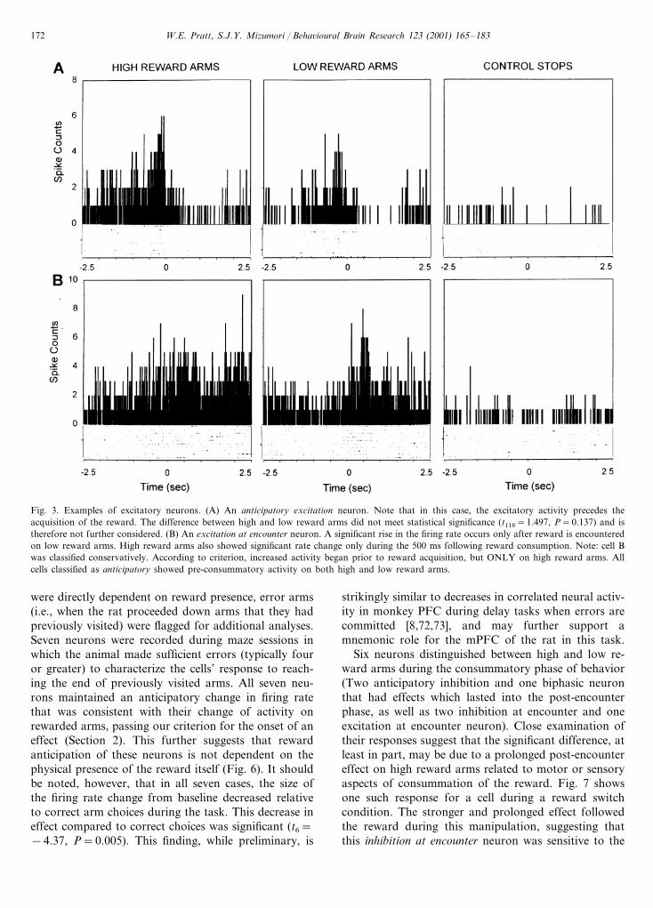
Another class of cells (n=5) increased their firingrate surrounding reward encounter, and was consideredexcitatory. Three of these neurons began their burstingprior to the acquisition of the reward (anticipatoryexcitation neurons, Fig. 3(A)). Two showed a signifi-cant change within 200 ms of reward consumption(excitation at encounter, Fig. 3(B)).
The remaining reward-related cell showed a biphasicpattern of activity to the acquisition of the reward.Immediately prior to reward acquisition, the celldemonstrated a rise in its firing rate that reversed to aninhibitory pattern once the reward was reached.

W.E. Pratt, S.J.Y. Mizumori / Beha�ioural Brain Research 123 (2001) 165–183 171
and low values of reward on specific arms. Each rewardneuron was tested to determine if there was a differencein reward-related activity between different valencearms by comparing firing rates between high and lowrewarded arms during the 500 ms bin during which therate change was greatest using an independent samplest-test. For neurons that showed significant firingchanges during both anticipatory and consummatoryphases of reward acquisition, tests were run on bothphases of the behavior.
Two of the 14 neurons with reward-related anticipa-tory activity distinguished between high and low rewardarms. Both of these neurons inhibited their firing priorto reward acquisition. This suggests that within thepopulation of mPFC neurons that anticipate the onsetof reward there may be a subpopulation of anticipatory
cells that distinguish between the relative value of thereward to be gained. To further examine this possibil-ity, one neuron was tested during five trials when thereward valence of the arms was reversed (previouslyhighly rewarded arms received low rewards, and viceversa). As shown in Fig. 5, during the trials when thereward was reversed, the neuron still showed a signifi-cantly greater anticipatory inhibition on the arms whichthe rat normally recei�ed higher reward.
In addition to suggesting that such neurons are ableto encode expected reward contingencies at specificspatial locations, the above manipulation implies thatpre-encounter firing of reward-related neurons is notthe result of specific visual or odor cues, which mightprovide information about upcoming reward. To deter-mine whether reward-anticipation responses of neurons
Fig. 2. Examples of the two classes of inhibitory neurons encountered. Each PETH is centered on the time that the animal encountered the rewardat the end of each arm. The bin width for each bar is 10 ms. Raster plots beneath each PETH show the spiking activity of each neuron duringmultiple individual encounters. For arm end visits, PETHs are based on 60 encounters (15 win-shift trials) except as noted in this and subsequentfigures. (A) Anticipatory inhibition neurons demonstrated a decline in firing rate by at least 50% prior to encountering the chocolate milk reward.For this neuron, the inhibitory effect continued into the consummatory phase of the behavior. (B) Inhibition at encounter neurons showed a similardecrease that occurred only once the reward was encountered. For both neurons, the effect at the ends of the arms was compared to the firingpatterns when the rats stopped on the center platform (control stops). Note the lack of effect for these control conditions, suggesting that theseneurons are not movement-related. Control stops PETHs presented in this manuscript are derived from an average of 20 center platform stops(range 17–26) over the course of 15 trials.
W.E. Pratt, S.J.Y. Mizumori / Beha�ioural Brain Research 123 (2001) 165–183172
Fig. 3. Examples of excitatory neurons. (A) An anticipatory excitation neuron. Note that in this case, the excitatory activity precedes theacquisition of the reward. The difference between high and low reward arms did not meet statistical significance (t118=1.497, P=0.137) and istherefore not further considered. (B) An excitation at encounter neuron. A significant rise in the firing rate occurs only after reward is encounteredon low reward arms. High reward arms also showed significant rate change only during the 500 ms following reward consumption. Note: cell Bwas classified conservatively. According to criterion, increased activity began prior to reward acquisition, but ONLY on high reward arms. Allcells classified as anticipatory showed pre-consummatory activity on both high and low reward arms.
were directly dependent on reward presence, error arms(i.e., when the rat proceeded down arms that they hadpreviously visited) were flagged for additional analyses.Seven neurons were recorded during maze sessions inwhich the animal made sufficient errors (typically fouror greater) to characterize the cells’ response to reach-ing the end of previously visited arms. All seven neu-rons maintained an anticipatory change in firing ratethat was consistent with their change of activity onrewarded arms, passing our criterion for the onset of aneffect (Section 2). This further suggests that rewardanticipation of these neurons is not dependent on thephysical presence of the reward itself (Fig. 6). It shouldbe noted, however, that in all seven cases, the size ofthe firing rate change from baseline decreased relativeto correct arm choices during the task. This decrease ineffect compared to correct choices was significant (t6=−4.37, P=0.005). This finding, while preliminary, is
strikingly similar to decreases in correlated neural activ-ity in monkey PFC during delay tasks when errors arecommitted [8,72,73], and may further support amnemonic role for the mPFC of the rat in this task.
Six neurons distinguished between high and low re-ward arms during the consummatory phase of behavior(Two anticipatory inhibition and one biphasic neuronthat had effects which lasted into the post-encounterphase, as well as two inhibition at encounter and oneexcitation at encounter neuron). Close examination oftheir responses suggest that the significant difference, atleast in part, may be due to a prolonged post-encountereffect on high reward arms related to motor or sensoryaspects of consummation of the reward. Fig. 7 showsone such response for a cell during a reward switchcondition. The stronger and prolonged effect followedthe reward during this manipulation, suggesting thatthis inhibition at encounter neuron was sensitive to the

W.E. Pratt, S.J.Y. Mizumori / Beha�ioural Brain Research 123 (2001) 165–183 173
current (not expected) characteristics of the reward.Additionally, no inhibition was seen for this cell onarms where the rat made errors.
Consistent with this finding, only three neurons ofour reward-related sample showed a longer effect onlow reward arms that exceeded our 200 ms differencecriterion. A regression analysis on the remaining excita-tory and inhibitory type reward neurons (all but one ofwhich included post-encounter rate changes) revealed asignificant correlation of effect offset between high andlow arms (F(1,13)=6.6, P�0.05). The slope of theregression line was 0.16, close to an expected slope of0.2 that would result from assuming that the highreward (5 drops) takes five times longer to consumethan the low reward (1 drop). Thus, while anticipatoryresponses of reward-related neurons appear to be de-pendent upon reward expectation, most post-encounterneuronal firing rate changes appear to depend on re-ward presentation.
Although post-encounter neuronal changes appear tobe dependent on sensory or motor consequences ofreward consummation, it is important to note thatthese signals may distinguish meaningful differences.
For example, one anticipatory inhibition neuron thatshowed both anticipatory and post-encounter inhibitionto reward presentation (Fig. 8(A)) was subjected to atest during which the reward was changed from choco-late to strawberry milk (Fig. 8(B)). Although the antic-ipatory inhibition remained prior to consumption of thenew reward (consistent with above findings), when therat encountered the new reward there was a significantincrease in the activity of the cell. This distinctionbetween familiar and novel reward, in addition to re-ward magnitude detection by some cells (describedabove), suggests that mPFC neurons may have multipleroles in distinguishing different rewards.
3.4.2. Visual dependence of spatial neuronsTwo of the five neurons with location correlates (one
with a center field and one multiple correlate cell withboth spatial and turn-related properties) were stable formultiple days and subjected to manipulations to deter-mine their stability across environmental manipula-tions. When subjected to a series of trials during whichlights were on for five trials, followed by five trials ofdarkness and then a light restoration phase, these two
Fig. 4. Spot diagrams indicating spatial firing biases for five neurons. Dots represent pixels (within a 64×64 grid centered on the platform) visitedby the rat. Circles represent the spatial locations of bursting activity that exceeded 20% of the neuron’s maximum rate; circle diameters are linearlyrelated to firing rate. Vector lines projecting from the circles represent the direction the animal was traveling during the action potential discharge.One cell shown had a movement correlate (turn) in addition to its spatial bias (A). Cells B–E demonstrated a spatial correlate only. Note thatthe spatial nature of these neurons is not as precise as that observed in hippocampus.

W.E. Pratt, S.J.Y. Mizumori / Beha�ioural Brain Research 123 (2001) 165–183174
Fig. 5. A differential anticipatory inhibition neuron. Note the more pronounced inhibition in anticipation of reaching the end of the high rewardarms when compared to the low reward arms (A). In (B), reward valences were switched between normally high and low reward arms. Thestronger inhibition remains significant on the arms, which the rat traditionally received high reward (B, left column), suggesting that this mPFCneuron anticipates different rewards based on previous experience. Additionally, this suggests that anticipatory neurons are not driven byvisual/odor qualities of the reward. Scaling differences reflect the higher number of counts from 15 trials (N=60 encounters) for the histogramsin (A) versus five trials of data (N=20 encounters) in (B).
spatial neurons both showed an immediate and lastingdecrease in their spatial character. As shown in Fig. 9,during the dark phase the spatial fields of these neuronsare abolished. Even when lights were turned back on,the two neurons did not show instant or completerecovery across five lit trials. One of these neurons wastested a subsequent day, during which the field returnedto its original location. Such slow restoration suggeststhat it is not strictly visual input per se that is drivingthese mPFC neurons; rather, past experience with thevisual environment may play an important role in therepresentations that these neurons develop.
Additionally, these two neurons were tested during aphase in which the reward values of the arms wereswitched (see description above). Both fields remainedunaltered, suggesting a functional distinction between
spatial and place representations for at least a subpopu-lation of mPFC neurons. Other neurons with multiplecorrelates, including one with a reward-related correlatethat was restricted to one region of the maze, appear torepresent units that integrate information across differ-ent information modalities.
3.4.3. Examination of test phase-related acti�ityThirty-nine cells were recorded for five trials with a
1-min delay between the study and test phases of eachtrial. To examine delay-related changes in cellular activ-ity, firing rates were compared between the 1-min delayperiod and the last minute of the intertrial interval fortrials with a delay. A paired t-test showed no tendencyfor the population to increase or decrease rates due tothe delay, t38=0.757, P�0.1. Individual neurons also

W.E. Pratt, S.J.Y. Mizumori / Beha�ioural Brain Research 123 (2001) 165–183 175
showed little evidence of activity changes that werespecific to the delay period. Fig. 10 presents one neu-ron, which did show an elevated discharge at the onsetof the delay period; note that a similar increase is alsoevident during the intertrial interval, a time duringwhich the rat does not have the mnemonic requirementto remember which four arms have been recentlyvisited.
To determine whether reward-related neurons mighthave neuronal activity specific to either the study phase(the presentation of the first four arms of the trial) orthe test phase (once all eight arms are presented) of thetask, histograms were generated for arm visits duringeach phase. These histograms were nearly identicalacross the two phases for all reward-related neurons.Combined with the data from the delay period, these
data do not suggest any phase-dependent activity forrat mPFC neurons during this maze task.
3.5. Histology
Fig. 11 shows the relative locations of the correlatedcells that were recorded from during this experiment.Although some similarly correlated cells appeared tocluster together, overall the correlates were generallydistributed throughout the entire anterior/posterior ex-tent of the mPFC. This is consistent with a recentreport by Jung et al. [20] that has shown that simulta-neously recorded neurons in close proximity withinmPFC do not appear to reflect common correlatesduring radial maze or figure-8 track testing paradigms.The apparent lack of correlates in the posterior ventral
Fig. 6. Examples of anticipatory excitation (A) and anticipatory inhibition (B) neurons that showed similar anticipatory effects when the ratperformed errors, at which time no reward was present. This further suggests the use of spatial cues to anticipate the presence of reward by thesecells, rather than sensory qualities of the reward itself. PETHs for errors are based on four and 11 error occurrences for (A) and (B), respectively.

W.E. Pratt, S.J.Y. Mizumori / Beha�ioural Brain Research 123 (2001) 165–183176
Fig. 7. An example of the reward-dependent nature of the consummatory effect in an inhibition at encounter neuron. The duration of inhibitionwas greatest on the high reward arms (A). When the level of reward was switched between high and low reward arms on the maze, the effectfollowed the reward, exhibiting prolonged inhibition on arms that traditionally held low reward (right column, B). When the reward was switchedback to the usual arms, the greater response of the neuron again followed the reward (not shown). All histograms in this figure are based on 20visits to each reward arm type (5 win-shift trials for each reward condition).
regions of the IL cortex in this study may be explainedby a relatively low sample of neurons recorded fromthis region (N=7).
4. Discussion
This experiment sought to define characteristics ofmPFC unit firing in relation to the acquisition ofdifferential goal values within the current spatial task.Accordingly, the spatial memory task adopted for thisexperiment differed from most experiments using win-shift behavior on an eight arm radial maze by includingdifferent amounts of reward at predictable locations onthe maze. Rats learned the locations of high reward arms,as evidenced by a preference to visit high reward loca-tions during the initial choices of the memory phase ofthe task. Consistent with Jung et al.’s [21] previous study
on rat mPFC neurons, we found neurons that changedfiring rate as a result of movement and of approachingor encountering a reward. Additionally, a small numberof neurons were found to have a spatial bias.
The most common behavioral correlate of PL and ILneurons was reward-related firing, the majority of whichshowed changes in firing prior to the acquisition of thereward (Table 1). Anticipatory changes in neuronal firingwere not dependent upon the presence of visual informa-tion or odor cues associated with the reward itself, asmany cells showed similar changes in firing rate when therat made errors on the arms. Such firing also did notreflect cessation of forward movement since it was notobserved when rats paused at non-rewarded locations.Two anticipatory reward cells demonstrated differentialfiring rates between the high and low reward arms in thistask, suggesting that some mPFC neurons discern differ-

W.E. Pratt, S.J.Y. Mizumori / Beha�ioural Brain Research 123 (2001) 165–183 177
entially rewarded locations. Furthermore, one neuronmaintained its differential pre-encounter firing whenpresented with probe trials during which the rewardvalence of the arms was reversed. This suggests that somemPFC neurons fire in anticipation of specific locationsin which particular rewards are expected to be.
Six neurons showed different firing rates after rewardwas acquired. In most instances, neurons with firing ratechanges that began prior to reward consumption contin-ued with the altered rate through the consummatoryphase of the behavior. Relatively few cells (N=5) wererecorded that initiated firing changes only after thereward was encountered. Post-encounter firing ratechanges appeared to depend on reward presence; formost reward neurons the length of the post-encountereffect was shown to be dependent upon the rewardamount present. One neuron that inhibited prior to andduring normal chocolate milk rewards demonstrated
excitatory bursts upon encountering a novel reward,suggesting that the mPFC may distinguish not onlydifferent amounts of the same reward but also qualita-tively different rewards. The ability of prefrontal units todistinguish between differential reward amounts is con-sistent with a report by Shapiro et al. [66] that showeddifferential firing for mPFC neurons to various levels oflateral hypothalamic stimulation (presumably corre-sponding to different levels of brain stimulation reward).The present report is the first that we are aware of tocharacterize differential activity for reward in a ratperforming a spatial memory task.
Similar to the results reported by Poucet [52] and Junget al. [21], we recorded few location-specific neurons inthis study. Four were determined to be spatially selective,and two additional neurons showed a spatial bias inaddition to a movement (turn) or reward-related corre-late. One spatial neuron and one spatial/movement
Fig. 8. A cell that showed anticipatory inhibition to chocolate milk reward (A) displayed excitatory activity when presented with a novel,strawberry flavor (B). Such activity suggests that mPFC neurons are capable of distinguishing between different rewards. All histograms in thisfigure are based on 20 visits (5 win-shift trials) to each reward arm type in each condition.

W.E. Pratt, S.J.Y. Mizumori / Beha�ioural Brain Research 123 (2001) 165–183178
Fig. 9. Spot diagrams for the two spatial neurons shown in Fig. 3(A) and (B) on a subsequent testing day. Both neurons displayed a spatial biasduring the initial light trials, but when room lights were turned off, the bias was attenuated. Restoring the light did not restore the field. Cell Awas re-tested the following day, at which time the field was restored. This suggests that mPFC spatial responses are modulated by visualexperience.
neuron that was stable across several days demonstratedconsistent location specificity across multiple recordingsessions. This contrasts with cells recorded by Poucet[52], which did not fire in consistent locations across days,and that were subsequently determined to be movementrelated. The spatial nature of the location-specific cellsin this study was dependent on visual experience withinthe maze environment. The place fields of both neuronswere abolished when lights were turned off and remaineddisrupted even once lights were turned back on. Theseresults differ from recordings of hippocampal placefields, which typically either remain in the dark orre-establish themselves immediately once light is restored[31,40]. The more prolonged attenuation of place fieldsin mPFC following loss of visual input and its restorationmay suggest that the mPFC does not encode a strictlyvisual sensory-based spatial representation of the envi-ronment. Rather, the IL and PL may base spatialrepresentations upon past expectations regarding theenvironment.
The existence of multiple correlate cells (N=6, 9.8%)within the mPFC indicate that some neurons within themPFC integrate across spatial, reward, and movementsources of information. This is strikingly similar torecordings from a small number of multiple-correlateneurons from the mVS, which receives projections fromthese areas [29]. This report is not the first to demonstrate
multiple correlates in mPFC neurons. Among the rela-tively sparse literature with rat mPFC recording duringnaturally-motivated tasks, a recent study by Gill et al.[12] has shown that PL neurons from rat prefrontalcortex respond to reward as well as movement during anattentional task. However, a previous study by Jung etal. [21] in a spatial maze task similar to the one used inthis experiment was unclear regarding whether multiplecorrelate cells in their sample were of the same modality(e.g., multiple movement correlates) or cross-modal (e.g.,movement and goal-related). The presence of cross-modal integration within the mPFC may serve to providethe basis for planning movements to spatial locations, afunction that has been attributed to the mPFC by severalresearchers.
Current speculation about the cognitive function of theprefrontal cortex is diverse. Specifically, behavioral im-pairments caused by prefrontal lesions have been sug-gested to be due to impairments in memory for temporalorganization [9,22,24,26], attentional deficits [15,50], im-pairments in strategy switching [56], impairments inworking memory [13,14], or disruption of rule-basedprocesses [23]. It should be noted that none of thesepossibilities are mutually exclusive, and some currentviews of rat mPFC suggest that it plays a role in morethan one of these operations [16,55]. Although thecurrent experiment was not designed to specifically

W.E. Pratt, S.J.Y. Mizumori / Beha�ioural Brain Research 123 (2001) 165–183 179
address the cognitive constructs that such argumentsrepresent, the present study did fail to uncover units thatresponded to specific mnemonic phases present withinthe task. None of the 39 units tested with a 1 min delaydemonstrated changes in firing rate that were consistentwith holding a memory trace across the delay. Inaddition, no reward unit showed differential activitybetween the study or test phases of the task. Thus,individual neurons in the rat PL and IL prefrontal cortexdo not show direct evidence of contributing to workingmemory representations across delays in this spatialworking memory task, at least as examined by the currentanalyses. Although this may appear to be in contrast withthe obvious presence of delay-type neurons within dorso-lateral prefrontal cortex of the monkey, at least twocaveats should be made regarding such an interpretation.First, the nature of the current spatial working memorytask is more complex than the delayed matching tasksutilized in the monkey literature in that animals are
required to hold more than one location in memory ata given time to successfully complete the task. In thismanner, the data are not directly comparable. One reportthat has shown delay-dependant activity of mPFC neu-rons in rats utilized a delayed match to position task ina Y-maze [3], which more closely approximates thetypical behavioral tasks shown to elicit delay-relatedactivity in the monkey dorsolateral prefrontal cortex[13,14]. Additionally, it is possible that individual IL orPL neurons could represent specific sequences of visitedarms, and hold such sequences in memory only duringtrials when that sequence constituted the study phase ofthe task. Given our random presentation of only five suchsequences for any given neuron, our analyses would notbe sensitive to neurons that show differential activity tosuch conditions. The current data, based on a relativelysmall sample of tested neurons, cannot definitively ex-clude the rat mPFC from delay- or memory phase-relatedunit activity during spatial working memory.
Fig. 10. One neuron that displayed increased activity during the delay period (A). Note, however, that a similar increase was observed during the2-min intertrial interval (B). This suggests that the increased firing during the delay period was not due to the specific working memory demandsof the task. Both histograms represent data from five instances when the rat was confined to the center (either during delay periods or intertrialintervals).

W.E. Pratt, S.J.Y. Mizumori / Beha�ioural Brain Research 123 (2001) 165–183180
Fig. 11. Location of correlated neurons within the mPFC. Placement measurements are in millimeters anterior to bregma. Overall, correlates werespread throughout the extent of this region. Legend for symbols: (�) Anticipatory inhibition; (�) Anticipatory excitation; (�) Inhibition atencounter; (�) Excitation at encounter; (+ ) Spatial; ( ) Biphasic; M Movement. Symbols divided by a slash (/) denote placement of multiplecorrelate neurons. This figure adapted from Swanson [71].
However, these data are consistent with behavioralevidence that, although the integrity of the rat mPFC isnecessary for completion of a win-shift task after a30-min delay, mPFC activation during the study phaseand across the entire delay is not critical [63]. Further-more, Floresco et al. [6] have shown that it is theinterconnection between the PL and the ventral subicu-lum that mediates normal post-delay performance.Therefore, it is likely that the working memory repre-sentations important for this active navigation task areachieved by interactions between at least the mPFC andhippocampus, and may be undetectable by individualunit recordings from any one structure.
The present data suggest that an important propertyof PL and IL neurons during performance of thisspatial memory task is the representation of predictablerewards within a complex environment. Such an inter-pretation is consistent with reports that mPFC lesionsimpair the ability of a rat to learn predictable locationsof reinforcement (Section 1). The eventual learning ofmany spatial tasks by mPFC lesioned animals may bemediated by a network of other brain regions also
capable of making reward associations. For example,the ventral striatum also receives input from otherstructures (e.g., the amygdala, see Section 4) that en-code cues that predict rewards. However, mPFC le-sioned animals show consistent impairments whenchallenged in tasks where the goal location is varied,suggesting that this region is particularly importantwhen goal locations must be learned quickly [11,53]. Inthe intact animal, successful learning and recall ofplace-reward associations is likely to recruit processingwithin the mPFC. Rats are impaired during reversibleinactivation of the mPFC after a 30 min delay on theeight arm radial maze [63]. Inactivation of the mPFCmay impair performance during the test phase due toan inability of the rat to predict locations of rewardswithin a familiar spatial environment. This is entirelycompatible with the view that the mPFC is importantfor prospective planning, as has been suggested bySeamans, Floresco, and Phillips [64]. It is also congru-ent with other theories of mPFC function, as loss ofinput regarding reward expectations could be expectedto disrupt the function of other systems that depend on

W.E. Pratt, S.J.Y. Mizumori / Beha�ioural Brain Research 123 (2001) 165–183 181
such knowledge (e.g., those of working memory, atten-tion, etc., see above). Furthermore, other regions ofprefrontal cortex also encode reward expectancy infor-mation. Recordings from orbitofrontal cortex duringolfactory discriminations show that sulcal prefrontalneurons are capable of predicting consequences oflearned odors [61]. The prefrontal cortex as a wholemay serve to predict modality-specific reinforcementcontingencies of the external environment.
4.1. The mPFC as part of a spatial context dependentmoti�ation system
In addition to the current recordings from themPFC, we have previously recorded from the mVS andBLA using the identical task [29,54]. Interesting com-parisons can be made across these three studies. Aspredicted, neurons within both the mPFC and the BLAshow reward-related correlates that could relay infor-mation to mVS neurons. Reward-related activity wasthe predominant correlate for both mPFC and BLA,with 31.1% and 30.7% of all neurons recorded express-ing goal-related firing, respectively. These two regionshad relatively high numbers of cells that fired differen-tially to high and low rewards (mPFC, 31.6%; BLA,53.6% of reward-related cells). The mPFC containedthe highest percentage of anticipatory reward neuronsof any of these three regions (73.7% of reward neuronsrecorded; BLA, 60.7%, mVS, 43.8%). Although thecomparison of individual studies does not allow for adirect test of information flow, these data are consistentwith the hypothesis that inputs from the BLA andmPFC influence the smaller number of reward-relatedneurons within the mVS (15.5% of mVS cells showedreward correlates, 12.5% of these displayed differentialactivity to reward value).
Despite connections from hippocampal regions to allthree structures, the mVS shows the most evidence oflocation-dependant (18.4% of cells) firing than eitherthe BLA (7.7%) or the mPFC (9.8%). Both the mPFCand the mVS showed a more substantial number ofmovement-related neurons (16.4% and 20.3%, respec-tively) than the BLA (2.2%). This is consistent with theproposed functions of these regions for movementpreparation. Finally, all three regions had small num-bers of neurons that integrated across at least two ofthe reward, spatial, and movement modalities. Thismay suggest a functional, as well as anatomical, con-nection across these three brain areas during the perfor-mance of this task.
These data, in addition to experiments publishedfrom other laboratories, support our hypothesis thattwo possible inputs of reward information for the mVSare the BLA and mPFC. Specifically, we suggest thatduring this task, output from the BLA provide infor-mation regarding cues that predict reward, while mPFC
predicts reward location based on spatial contingenciesin the environment. This motivation-relevant informa-tion from the BLA and mPFC is then combined withspatially relevant hippocampal inputs within the mVS.Previously, we have suggested that a possible role forthe mVS, and especially the nucleus accumbens, is tomonitor the effectiveness of behavioral responses, par-ticularly when the rat is exposed to new spatial contexts[39]. Changes in spatial context could be relayed viasubiculum inputs to the mVS, while alterations in re-ward placement or contingencies could be signaled bymPFC and BLA inputs. In particular, the current ex-periment suggests an important role for the mPFC inthe processing of reward and expected reward outcomesin rats performing goal-oriented spatial navigation.
Acknowledgements
This work was supported by grants MH12303 toW.E.P., as well as MH58755 and IBN 9514880 toS.J.Y.M. Thanks to Caroline Hutchings for assistancein collecting a portion of the data, and to Brenton G.Cooper and Alex Guazzelli for helpful comments onthe manuscript.
References
[1] Annett LE, McGregor A, Robbins TW. The effects of ibotinicacid lesions of the nucleus accumbens on spatial learning andextinction in the rat. Behav Brain Res 1989;31:231–42.
[2] Barnes CA. Spatial learning and memory processes: the searchfor their neurobiological mechanisms in the rat. Trends Neurosci1988;11(4):163–9.
[3] Batuev AS, Kursina NP, Shutov AP. Unit activity of the medialwall of the frontal cortex during delayed performance in rats.Behav Brain Res 1990;41:95–102.
[4] Conde F, Maire-Lepoivre E, Audinat E, Crepel F. Afferentconnections of the medial frontal cortex of the rat. II. Corticaland subcortical afferents. J Comp Neurol 1995;352:567–93.
[5] Ferino F, Thierry AM, Glowinski J. Anatomical and electro-physiological evidence for a direct projection from Ammon’shorn to the medial prefrontal cortex in the rat. Exp Brain Res1987;65:421–6.
[6] Floresco SB, Seamans JK, Phillips AG. Selective roles forhippocampal, prefrontal cortical, and ventral striatal circuits inradial-arm maze tasks with or without a delay. J Neurosci1997;17(5):1880–90.
[7] Fritts ME, Asbury ET, Horton JE, Isaac WL. Medial prefrontallesion deficits involving or sparing the prelimbic area in the rat.Physiol Behav 1998;64(3):373–80.
[8] Funahashi S, Bruce CJ, Goldman-Rakic PS. Mnemonic codingof visual space in the monkey’s dorsolateral prefrontal cortex. JNeurophysiol 1989;61(2):331–49.
[9] Fuster JM. The prefrontal cortex: anatomy, physiology, andneuropsychology of the frontal lobe, 3rd ed. Philadelphia: Lip-pincott-Raven, 1997.
[10] Gal G, Joel D, Gusak O, Feldon J, Weiner I. The effects ofelectrolytic lesion to the shell subterritory of the nucleus accum-bens on delayed non-matching-to-sample and four-arm baited

W.E. Pratt, S.J.Y. Mizumori / Beha�ioural Brain Research 123 (2001) 165–183182
eight-arm radial-maze tasks. Behav Neurosci 1997;111(1):92–103.
[11] Gemmell C, O’Mara SM. Medial prefrontal cortex lesions causedeficits in a variable-goal location task but not in object explo-ration. Behav Neurosci 1999;113(3):465–74.
[12] Gill TM, Sarter M, Givens B. Sustained visual attention perfor-mance-associated prefrontal neuronal activity: evidence forcholinergic modulation. J Neurosci 2000;20(12):4745–57.
[13] Goldman-Rakic PS. Circuitry of the prefrontal cortex and theregulation of behavior by representational knowledge. In: PlumF, Mountcastle V, editors. Handbook of physiology, vol. 5.Bethesda, MD: American Physiological Society, 1987:373–417.
[14] Goldman-Rakic PS. Cellular and circuit basis of working mem-ory in prefrontal cortex of nonhuman primates. Prog Brain Res1990;85:325–36.
[15] Granon S, Hardouin J, Courtiere A, Poucet B. Evidence for theinvolvement of the rat prefrontal cortex in sustained attention. QJ Exp Psychol B 1998;51(3):219–33.
[16] Granon S, Poucet B. Involvement of the rat prefrontal cortex incognitive functions: a central role for the prelimbic area. Psycho-biology 2000;28(2):229–37.
[17] Groenewegen HJ, Berendse HW, Wolters JG, Lohman AHM.The anatomical relationship of the prefrontal cortex with thestriatopallidal system, the thalamus and the amygdala: evidencefor a parallel organization. Prog Brain Res 1990;85:95–118.
[18] Harrison LM, Mair RG. A comparison of the effects of frontalcortical and thalamic lesions on measures of spatial learning andmemory in the rat. Behav Brain Res 1996;75:195–206.
[19] Jay TM, Witter MP. Distribution of hippocampal CA1 andsubicular efferents in the prefrontal cortex of the rat studied bymeans of anterograde transport of Phaseolus �ulgaris-Leucoag-glutinin. J Comp Neurol 1991;313:574–86.
[20] Jung MW, Qin Y, Lee D, Mook-Jung I. Relationship amongdischarges of neighboring neurons in the rat prefrontal cortexduring spatial working memory tasks. J Neurosci2000;20(16):6166–72.
[21] Jung MW, Qin Y, McNaughton BL, Barnes CA. Firing charac-teristics of deep layer neurons in prefrontal cortex in rats per-forming spatial working memory tasks. Cereb Cortex1998;8:437–50.
[22] Kesner RP. Neural mediation of memory for time: role of thehippocampus and medial prefrontal cortex. Psychon B Rev1998;5(4):585–96.
[23] Kesner RP. Subregional analysis of mnemonic functions of theprefrontal cortex in the rat. Psychobiology 2000;28(2):219–28.
[24] Kesner RP, Holbrook T. Dissociation of item and order spatialmemory in rats following medial prefrontal cortex lesions. Neu-ropsychologia 1987;25(4):653–64.
[25] Kita H, Kitai ST. Amygdaloid projections to the frontal cortexand the striatum in the rat. J Comp Neurol 1990;298:40–9.
[26] Kolb B. Functions of the frontal cortex of the rat: a comparativereview. Brain Res Rev 1984;8:65–98.
[27] Kolb B, Buhrmann K, McDonald R, Sutherland RJ. Dissocia-tion of the medial prefrontal, posterior parietal, and posteriortemporal cortex for spatial navigation and recognition memoryin the rat. Cereb Cortex 1994;4(6):664–80.
[28] Kosobud AEK, Harris GC, Chapin JK. Behavioral associationsof neuronal activity in the ventral tegmental area of the rat. JNeurosci 1994;14:7117–29.
[29] Lavoie AM, Mizumori SJY. Spatial, movement, and reward-sen-sitive discharge by medial ventral striatum neurons of rats. BrainRes 1994;638:157–68.
[30] Mair RG, Burk JA, Porter MC. Lesions of the frontal cortexhippocampus, and intralaminar thalamic nuclei have distincteffects on remembering in rats. Behav Neurosci1998;112(4):772–92.
[31] Markus EJ, Barnes CA, McNaughton BL, Gladden VL, SkaggsWE. Spatial information content and reliability of hippocampalCA1 neurons: effects of visual input. Hippocampus1994;4(4):410–21.
[32] Martin PD, Ono T. Effects of reward anticipation, rewardpresentation, and spatial parameters on the firing of singleneurons recorded in the subiculum and nucleus accumbens offreely moving rats. Behav Brain Res 2000;116:23–38.
[33] McDonald AJ. Organization of amygdaloid projections to themediodorsal thalamus and prefrontal cortex: a fluorescence ret-rograde transport study in the rat. J Comp Neurol 1987;262:46–58.
[34] McDonald AJ. Organization of amygdaloid projections to theprefrontal cortex and associated striatum in the rat. Neuro-science 1991;44(1):1–14.
[35] McGeorge AJ, Faull RLM. The organization of the projectionfrom the cerebral cortex to the striatum in the rat. Neuroscience1989;29(3):503–37.
[36] McNaughton BL, Barnes CA, Meltzer J, Sutherland RJ.Hippocampal granules are necessary for normal spatial learningbut not for spatially-selective pyramidal cell discharge. ExperBrain Res 1989;76:485–96.
[37] McNaughton BL, O’Keefe J, Barnes CA. The stereotrode: a newtechnique for simultaneous isolation of several single units in thecentral nervous system from multiple unit records. J NeurosciMeth 1983;8:391–7.
[38] Mizumori SJY, Lavoie AM, Kalyani A. Redistribution of spatialrepresentation in the hippocampus of aged rats performing aspatial memory task. Behav Neurosci 1996;110(5):1006–16.
[39] Mizumori SJY, Pratt WE, Ragozzino KE. Function of thenucleus accumbens within the context of the larger striatalsystem. Psychobiology 1999;27(2):214–24.
[40] Mizumori SJY, Ragozzino KE, Cooper BG, Leutgeb S.Hippocampal representational organization and spatial context.Hippocampus 1999;9(4):444–51.
[41] Mizumori SJY, Ward KE, Lavoie AM. Medial septal modula-tion of entorhinal single unit activity in anesthetized and freelymoving rats. Brain Res 1992;570:188–97.
[42] Mogenson GJ, Brudzynski SM, Wu M, Yang CR, Yim CCY.From motivation to action: a review of dopaminergic regulationof limbic-nucleus accumbens-ventral pallidum-pedunculopontinenucleus circuitries involved in limbic-motor integration. In: Kali-vas PW, Barnes CD, editors. Limbic motor circuits and neu-ropsychiatry. Ann Arbor, MI: CRC Press, 1993:193–236.
[43] Mogenson GJ, Nielsen M. A study of the contribution ofhippocampal-accumbens-subpallidal projections to locomotoractivity. Behav Neural Biol 1984;42:38–51.
[44] Muramoto K, Ono T, Nishijo H, Fakuda M. Rat amygdaloidneuron responses during auditory discrimination. Neuroscience1993;52(3):621–36.
[45] Nishijo H, Ono T, Nishino H. Single neuron responses inamygdala of alert monkey during complex sensory stimulationwith affective significance. J Neurosci 1988;8(10):3570–83.
[46] Oades RD, Halliday GM. Ventral tegmental (A10) system: neu-robiology. 1. Anatomy and connectivity. Brain Res Rev1987;12:117–65.
[47] O’Donnell P, Grace AA. Synaptic interactions among excitatoryafferents to nucleus accumbens neurons: hippocampal gating ofprefrontal cortical input. J Neurosci 1995;15(5):3622–39.
[48] O’Keefe J, Dostrovsky J. The hippocampus as a spatial map.Preliminary evidence from unit activity in the freely moving rat.Brain Res 1971;34:171–5.
[49] Olton D, Branch M, Best PJ. Spatial correlates of hippocampalplace activity. Exper Neurol 1978;58:387–409.
[50] Posner MI, Dehaene S. Attentional networks. Trends Neurosci1994;17(2):75–9.

W.E. Pratt, S.J.Y. Mizumori / Beha�ioural Brain Research 123 (2001) 165–183 183
[51] Poucet B. A further characterization of the spatial problem-solv-ing deficit induced by lesions of the medial frontal cortex in therat. Behav Brain Res 1990;41:229–37.
[52] Poucet B. Searching for spatial unit firing in the prelimbic areaof the rat medial prefrontal cortex. Behav Brain Res1997;84:151–9.
[53] Poucet B, Herrmann T. Septum and medial frontal cortex contri-bution to spatial problem-solving. Behav Brain Res1990;37:269–80.
[54] Pratt WE, Mizumori SJY. Characteristics of basolateral amyg-dala neuronal firing on a spatial memory task involving differen-tial reward. Behav Neurosci 1998;112(3):554–70.
[55] Ragozzino ME. The contribution of cholinergic and dopaminer-gic afferents in the rat prefrontal cortex to learning, memory,and attention. Psychobiology 2000;28(2):238–47.
[56] Ragozzino ME, Wilcox C, Raso M, Kesner RP. Involvement ofrodent prefrontal cortex subregions in strategy switching. BehavNeurosci 1999;113(1):32–41.
[57] Ranck JB Jr. Studies on single neurons in dorsal hippocampusformation and septum in unrestrained rats. Part I. Behavioralcorrelates and firing repertoires. Exp Neurol 1973;41:461–531.
[58] Robertson A. Multiple reward systems and the prefrontal cortex.Neurosci Biobehav Rev 1989;13:163–70.
[59] Robertson A, Laferriere A. Disruption of the connections be-tween the mediodorsal and sulcal prefrontal cortices alters theassociability of rewarding medial cortical stimulation to placeand task stimuli in rats. Behav Neurosci 1989;103:770–8.
[60] Sanghera MK, Rolls ET, Roper-Hall A. Visual responses ofneurons in the dorsolateral amygdala of the alert monkey. ExperNeurol 1979;63:610–26.
[61] Schoenbaum G, Chiba AA, Gallagher M. Orbitofrontal cortexand basolateral amygdala encode expected outcomes duringlearning. Nat Neurosci 1998;1(2):155–9.
[62] Schultz W, Apicella P, Ljungberg T. Responses of monkeydopamine neurons to reward and conditioned stimuli duringsuccessive steps of learning a delayed response task. J Neurosci1993;13(3):900–13.
[63] Seamans JK, Floresco SB, Phillips AG. Functional differencesbetween the prelimbic and anterior cingulate regions of the ratprefrontal cortex. Behav Neurosci 1995;109(6):1063–73.
[64] Seamans JK, Floresco SB, Phillips AG. D1 receptor modulationof hippocampal-prefrontal cortical circuits integrating spatialmemory with executive functions in the rat. J Neurosci
1998;18(4):1613–21.[65] Sesack SR, Deutch AY, Roth RH, Bunney BS. Topographical
organization of the efferent projections of the medial prefrontalcortex in the rat: an anterograde tract-tracing study with Phase-olus �ulgaris leucoagglutinin. J Comp Neurol 1989;290:213–42.
[66] Shapiro ML, Li JF, Roberts M. The firing rate of neuronalgroups in the medial prefrontal cortex, but not in the hippocam-pus, is altered by lateral hypothalamic stimulation in behavingrats. Soc Neurosci Abstracts 1998;24:1424.
[67] Silva MG, Boyle MA, Finger S, Numan B, Bouzrara AA, AlmliCR. Behavioral effects of large and small lesions of the ratmedial frontal cortex. Exp Brain Res 1986;65:176–81.
[68] Speakman A, O’Keefe J. Hippocampal complex spike cells donot change their place fields if the goal is moved within a cuecontrolled environment. Eur J Neurosci 1990;14:2339–56.
[69] Swanson LW. A direct projection from Ammon’s horn to pre-frontal cortex in the rat. Brain Res 1981;217:150–4.
[70] Swanson LW. The projections of the ventral tegmental area andadjacent regions: a combined fluorescent retrograde tracer andimmunofluorescence study in the rat. Brain Res Bull 1982;9:321–53.
[71] Swanson LW. Brain maps: structure of the rat brain. New York:Elsevier, 1992.
[72] Watanabe M. Prefrontal unit activity during delayed conditionaldiscriminations in the monkey. Brain Res 1981;225(1):51–65.
[73] Watanabe M. Prefrontal unit activity during delayed conditionalgo/no-go discrimination in the monkey. I. Relation to the stimu-lus. Brain Res 1986;382(1):1–14.
[74] Winocur G, Moscovitch M. Hippocampal and prefrontal cortexcontributions to learning and memory: analysis of lesion andaging effects on maze learning by rats. Behav Neurosci1990;104(4):544–51.
[75] Yang CR, Mogenson GJ. Electrophysiological responses of neu-rons in the nucleus accumbens to hippocampal stimulation andthe attenuation of the excitatory responses by the mesolimbicdopamine system. Brain Res 1984;324:69–84.
[76] Yim CY, Mogenson GJ. Response of nucleus accumbens neu-rons to amygdala stimulation and its modification by dopamine.Brain Res 1982;239:401–15.
[77] Yim CY, Mogenson GJ. Low doses of accumbens dopaminemodulate amygdala suppression of spontaneous exploratory ac-tivity in rats. Brain Res 1989;477:202–10.
.
Copyright © 2022 FDOKUMEN