Extremely acidic, pendulous cave wall biofilms from the Frasassi cave system, Italy

Mixed Culture Electrochemically Active Biofilms and theirMicroscopic and Spectroelectrochemical StudiesMohammad Mansoob Khan, Sajid A. Ansari, Jin-Hyung Lee, Jintae Lee, and Moo Hwan Cho*
School of Chemical Engineering, Yeungnam University, Gyeongsan-si, Gyeongbuk 712-749, South Korea
*S Supporting Information
ABSTRACT: Mixed culture electrochemically active biofilms (EABs) were developed on carbon paper using a sludge withmixed culture bacteria for microscopic and spectroelectrochemical studies because a naturally mixed culture bacterial strain ismore applicable than a pure culture strain. EAB development was confirmed by microbial fuel cells (MFCs) by obtaining aconstant increase in potential (∼0.36 V). Microscopic and spectroscopic studies showed that a mixed culture EABs formed onthe support. Cyclic voltammetry (CV), differential pulse voltammetry (DPV), and electrochemical impedance spectroscopy(EIS), which are nondestructive voltammetry techniques, indicated that the EABs could be source of electrons and usedeffectively for various purposes. Routine in vivo analysis of electron transfer between bacterial cells and the electrode wasperformed, providing insight into the extracellular electron transfer (EET) to the electrode. At low scan rates, CV revealed thecatalytic electron transfer ability of EAB between the cells and electrode and showed exceptional redox activities in the presenceof acetate. DPV and EIS studies showed that EAB in the presence of acetate can charge the surface by producing and storingsufficient electrons, behave as a capacitor, and have features consistent with EET. Finally, microscopic and spectroelectrochemicalstudies confirmed the development of a mixed culture EAB and the EET kinetics of EABs. These studies suggest that mixedculture EABs can be used effectively as a biogenic reducing tool for various applications.
KEYWORDS: Electrochemically active biofilm, Mixed culture, Biotransformation, Bacteria, Electron transfer, Cyclic voltammetry
■ INTRODUCTION
Bioelectrochemical systems (BESs) use living micro-organismsas catalysts in electrochemical reactions. The best known BES isthe microbial fuel cell (MFC), which is a device using metal-respiring bacteria to convert the chemical energy of substratesdissolved in wastewater to electrical energy.1 BESs allowelectricity generation from wastewater, electricity-drivenbioproduction, bioremediation, and biosensing.2,3 Many ofthese processes are quite promising. On the other hand, to date,the performance of bioproduction processes is not yet at thelevel needed for practical applications because it is critical forenabling high catalytic activity of the electrochemically activemicroorganisms (EAMs). Effective electron transfer and highprocess performance needs to be achieved regardless ofwhether the biocatalyst exists as a planktonic cell, a monolayercell surface, or a fully developed biofilm.1−3 On the other hand,despite the many different approaches and extensive research,many questions regarding the functioning of EAMs remain
unanswered. This is certainly due to the complexity ofbioelectrochemical processes because they depend on micro-bial, electrochemical, physicochemical, and operational consid-erations. This versatility and complexity calls for a variety ofanalytical tools to study mixed culture-derived electrochemi-cally active biofilms (EABs).1−4
Bacteria, which forms a multilayered aggregation (i.e., amicrobial biofilm) attached to the electrode, transfer theelectrons derived from oxidation of the substrate (waste ororganic matter) to the electrode. The ability of metal-respiringbacteria to reduce insoluble electron acceptors that cannotenter the cell (e.g., electrodes) requires an efficient extracellularelectron transfer (EET) tool shuttling electrons across theouter membrane of the microbial cell to the electrode.3−5 A
Received: September 1, 2013Revised: November 6, 2013Published: November 19, 2013
Research Article
pubs.acs.org/journal/ascecg
© 2013 American Chemical Society 423 dx.doi.org/10.1021/sc400330r | ACS Sustainable Chem. Eng. 2014, 2, 423−432

range of EET mechanisms from bacteria to the anode issuggested, such as indirect transfer using electron shuttlingproteins (e.g., cytochromes) or low molecular weight electronshuttles (e.g., mediators), via electron conducting “nanowires”(pili) produced by the bacteria or direct EET from the cellsurface redox active proteins.3 Understanding the mechanism ofEET through electroactive microbial biofilms is a challenge infundamental and applied life sciences. To date, electrochemicaltechniques, such as cyclic voltammetry (CV), have been appliedsuccessfully to examine EET process in intact and viablemicrobial biofilms on electrodes, providing important insightinto their redox properties.3−7 Unfortunately, CV alone cannotprovide complete insight into the mechanisms involved in theredox processes. On the other hand, coupling CV withdifferential pulse voltammetry (DPV) and electrochemicalimpedance spectroscopy (EIS) and spectroscopic techniquesovercome these limitations.1−7 These approaches, which haveoften been used to study isolated proteins and enzymesattached to electrodes, are also applicable for examining EABs.The ability of certain microorganisms to connect theirmetabolisms directly to an external electrical power supply isvery exciting, and extensive research on the possibilities ofEABs applications is ongoing.4−7 The best known applicationof EAB is possibly the MFC, which can convert biomass intoelectrical energy. Nevertheless, EABs coated onto electrodeshave recently become popular in other fields, such as thebiosynthesis, bioremediation, and biosensor design.1−7
Recently, mixed culture EABs was reported to be a biogenicreducing tool for the synthesis of nanoparticles (Au, Ag, andCys−Ag),8−10 nanocomposites (Au@TiO2 and Ag@TiO2),
11−13 band gap narrowing (TiO2 and ZnO)14,15 andbiohydrogen production,16 which is novel, simple, and green.The advantage of these protocols is that the use of mixedculture EABs as a reducing tool does not involve externalenergy input, toxic chemicals or expensive solvents. In addition,the reactions take place at room temperature, which makes thesynthesis of nanoparticles and nanocomposites highly suitableand efficient in the field of nanoscience.8−16 Therefore, mixedculture EABs were examined further using a range oftechniques to better understand the biogenic reducing behaviorand other characteristics.This paper presents an overview of the parameters defining
the developments of EABs and the analytical tools to examinethe EABs at different levels. To the best of the author’sknowledge, this is the first report to include a wide range oftechniques, such as microscopic, spectroscopic, electrochemical(CV, DPV, and EIS) and MFC to study, understand,standardize, and characterize the mixed culture EABs. Thiswill further strengthen the field of BESs and the use of mixedculture EABs in nanomaterial syntheses, band gap narrowing ofmetal oxides, biohydrogen production, and bioremediation.Therefore, a standardized framework appears essential.Recently, few of our previously reported studies proposed anovel and optimized approach to develop mixed culture EABsoutside MFC for different purposes.8−16 This approachproduced high performance mixed culture EABs for nanoma-terial syntheses that were considerably faster and easier tohandle, even for the nonprofessionals. The present reportshows how these approaches have provided importantcomplementary information to those obtained by electro-chemical methods, i.e., information on the electron transferkinetics.1,2,17,18 This study will also show that the bioelec-trocatalytic activities of EABs depend mainly on biofilm
formation on the support and substrate (organic matter orwaste) used. This study was not intended to examine themicrobe−electrode material interaction, type of bacteria, orstrain, which might be the scope of a further study.
■ EXPERIMENTAL SECTIONMaterials. Sodium acetate (CH3COONa), NaCl, sodium
phosphate monobasic (NaH2PO4), and sodium phosphate dibasic(Na2HPO4) were obtained from Duksan Pure Chemicals Co. Ltd.South Korea. Carbon paper (without wet-proof) and Pt-coated carbonpaper (0.5 mg Pt/cm2) were purchased from Fuel Cell Earth LLC Co.,USA. Nylon membrane fiber filter (0.45 μm) was obtained fromWhatman, Germany. All the other chemicals used in this study were ofanalytical grade and used as received. All solutions were prepared usingdeionized water prepared using a PURE ROUP 30 water purificationsystem.
Methods. Electrochemical experiments, such as CV, differentialpulse voltammetry (DPV), and electrochemical impedance spectros-copy (EIS), were conducted on a potentiostat (VersaSTAT 3,AMETEK, USA) using Ag/AgCl (saturated with KCl) as the referenceelectrode, Pt gauze as the counter electrode, and EABs on carbonpaper and carbon paper only as the working electrode in 50 mL, 0.1 Mphosphate buffer solution (pH 7; 0.1% NaCl) as the supportingelectrolyte at room temperature. EIS was performed in a 1 mMK3[Fe(CN)6] and K4[Fe(CN)6] solution. The electronic absorption/reflectance spectra (UV−vis spectra) of EABs were obtained using anUV−vis-NIR double beam spectrophotometer (VARIAN, Cary 5000,USA) equipped with a diffuse absorbance/reflectance accessory. EABwas placed in a sample holder that was then placed at the integratingsphere for the absorbance measurements. X-ray diffraction (XRD,PANalytical, X′pert PRO-MPD, Netherlands) was performed using CuKα radiation (λ = 0.15406 nm) and a diffraction angles ranging from10° to 90°. The surface morphology of the EABs was observed byscanning electron microscopy (SEM; Hitachi S-4100, Japan). Themorphology of the nanomaterials was examined by transmissionelectron microscopy (TEM, Tecnai G2 F20, FEI, USA) operating atan accelerating voltage of 200 kV. The potential generated by theMFC was recorded using a digital multimeter (Agilent 34405A, USA)connected to a computer.
Media, Inoculum, Preparation of EABs, and Their Opti-mization in MFC. Primary sludge was collected from a Biogas plant(Paju, South Korea). The chemical oxygen demand (COD) of thesludge was 1200 mg L−1. The sludge served as a mixed culturebacterial inoculum for the formation of mixed culture EABs on carbonpaper and nylon fiber. EABs on carbon paper and nylon fiber weremade, as reported earlier.8−16 The EABs were developed in twodifferent ways. In the first way, in a 200 mL mineral salt medium, 0.2 gsodium acetate was added as a substrate.19 Subsequently, 10 mL of theanaerobic sludge was added and N2 gas was sparged for 5 min to createanaerobic conditions. Finally, carbon paper and nylon fiber (2.5 cm ×4.5 cm) were dipped separately in the bottle and the bottle was sealed.All media, including the bacterial inoculum, were changed every twodays under anaerobic conditions. This was repeated for two weeks.Suitable mixed culture EABs that formed on the carbon paper (Figures1 and 2) were examined and confirmed using MFC by obtaining theappropriate voltage. The mixed culture EAB formed on the nylon fiberwas used only for comparative XRD and UV−vis spectroscopy becausenylon fiber is nonconducting. In the second way, EAB on carbon paperwas also prepared in the MFC anode, as reported earlier forcomparative studies.19,20
To examine the uniform and appropriate development of biofilmson carbon paper, the as-prepared EABs were monitored in the MFCanode to record the optimum voltage using a multimeter. The MFCconsisted of two identical chambers (anode and cathode chamber)separated by a Nafion membrane. Each chamber had an effectivevolume of 250 mL. The bottles were connected by two clampsbetween the flattened ends of the two glass tubes (inner diameter = 1.3cm) fitted with rubber gaskets. In the anode chamber, an EAB wasdeveloped on plain carbon paper (2.5 cm × 4.5 cm) in artificial
ACS Sustainable Chemistry & Engineering Research Article
dx.doi.org/10.1021/sc400330r | ACS Sustainable Chem. Eng. 2014, 2, 423−432424

wastewater using sodium acetate as the electron donor (1 g L−1). Amineral salt medium was added to the anode chamber and inoculatedwith 10 mL of the anaerobic sludge.19 The anode chamber wasbubbled with nitrogen for 10 min to remove oxygen (anode pH ∼7.4). Initially, a conventional H-type MFC was operated to obtain anefficient MFC system for the experiment using Pt coated carbon paperas the cathode electrode.19,20 The cathode chamber was filled with aphosphate buffer solution (50 mM, pH 7) and bubbled continuouslywith air. After 1 day of continuous operation, the MFC produced astable power density of 40 mW/m2, which corresponds to previousresults.20 During EAB development, the MFC was connected to aresistance box at 1000 Ω and run at 30 °C under atmospheric pressure.The potential obtained in this study refers to biofilms formed oncarbon paper for different times (1−15 days). The same biofilm wasused for 15 days to continue the growth of the biofilm and thenmeasure the potential. The above-mentioned mixed culture EABs,which was developed outside the MFC, was tested in the MFC anodeafter 1, 7, and 15 days to confirm the EAB growth by monitoring thepotential.The living mixed culture EABs formed on carbon paper outside the
MFC was used for different spectroscopic, electrochemical andmorphological studies, whereas the EAB formed on the nylon fiberwas used for a comparative XRD and UV−vis spectroscopy only.Pretreatment of EABs for SEM. The morphology of EAB was
examined by SEM after pretreating the EAB formed on carbon paperand nylon fiber using the following modified protocol.21,22 Briefly,EAB formed on carbon paper and nylon fiber were fixed directly byadding glutaraldehyde (2.5% final) and formaldehyde (2% final) andincubated at 4 °C overnight. Postfixation was performed for 90 minwith an osmium tetroxide solution (containing 3 mL of sodiumphosphate buffer 0.2 M, 3 mL of 2% OsO4 and 3 mL deionized water).Further, the EAB was washed and dehydrated by successive 10 minincubations in 50% ethanol, 70% ethanol, 80% ethanol, 90% ethanol,and 95% ethanol followed by two successive 20 min incubations in100% ethanol. After dehydration, the EAB was incubated in isoamylacetate for 20 min and dried with a critical-point dryer (HCP-2,Hitachi, Japan). The pretreated EABs were affixed to the SEM stubsand coated with platinum for 200 s using an ion-sputter (E-1030,Hitachi, Japan). The specimens were examined by SEM. The voltagewas set to 15 kV and viewed at a magnification from ×2000 to×20 000.Electrochemical Studies (CV, DPV, and EIS) of the Mixed
Culture EABs. Electrochemical studies (CV, DPV, and EIS) wereused for different spectral responses, such as the redox response of thecarbon source, EET, and growth of EABs, which highlight the potentialuse of these techniques for examining the redox electrochemistry ofliving mixed culture EABs. The electrochemical measurements (CV,DPV, and EIS) of EABs developed on carbon paper for 1, 7, and 15days, and carbon paper only with and without acetate were performedwith a computer-controlled potentiostat (VersaSTAT 3, USA) with athree electrode arrangement. The EABs developed on carbon paperfor 1, 7, and 15 days were used as the working electrode, platinum
gauze as the counter electrode, and an Ag/AgCl electrode (saturatedwith KCl) as the reference electrode. All the experiments wereperformed anaerobically at 30 °C in succession without stirring. Here,all the electrode potentials referenced to Ag/AgCl (saturated withKCl).
CV was performed at a scan rate of 1 mV s−1 versus Ag/AgCl(saturated with KCl). The parameters for DPV were as follows: Einitial(Ei) = 0.08 V versus Ag/AgCl; pulse height = 50 mV; pulse width =500 ms; step height = 2 mV; step time = 500 ms; scan rate = 4 mV s−1.The EIS experiments were performed for the EABs developed on 1, 7,and 15 days with and without acetate in a 1 mM K3[Fe(CN)6] andK4[Fe(CN)6] solution, maintaining a direct current voltage betweenthe working electrode and reference electrode (potentiostatic EIS)because the electron transfer processes by EAB were observed to bevoltage dependent.23 EIS was performed at 0.0 V versus Ag/AgCl(saturated with KCl), and the frequency was fixed from 104 to 1 Hzwith an amplitude of 10 mV because most biological charge transferphenomena are observed in this range.24 EIS was typically performedonly at the end when the CV and DPV measurements were over aftera long period of time. All the experiments were performed asindependent bioelectrochemical triplicates for a total duration ofapproximately 3 months.
■ RESULTS AND DISCUSSION
Electrochemically Active Biofilms. Electrochemicallyactive biofilms (EABs) are well-known in microbial fuel cellsthat produce electrons and protons.1−7,17,18,25 Recently, ourgroup has started using mixed culture EABs as a biogenicreducing tool for the synthesis of different types of nano-particles (Au, Ag and, Cys−Ag),8−10 nanocomposites (Au@TiO2 and Ag@TiO2),
11−13 band gap narrowing of metaloxides,14,15 biohydrogen production,16 and bioremedia-tion.20,25−27 The use of a mixed culture EAB is more practicalthan a pure culture (single strain) because, in nature, microbialcommunities are a mixed culture and the use of a pure culture isinconvenient, cumbersome, and limited.26−31 Mixed cultureEABs effectively oxidize organic substrates, such as acetate toelectrons, protons, and CO2 without combustion. The above-mentioned synthetic protocols are novel, green, and biogenic.In addition, they utilize the biologically produced electrons andprotons and do not involve any external energy input, whichmakes the syntheses of nanoparticles, nanocomposites,biohydrogen production, and bioremediation highly usefuland efficient in the field of nanomaterial syntheses, sustainableenergy, and environmental remediation.8−16,25−27 Furthermore,other highly sensational observations are that these processesdo not contaminate the products, comprise any harmfulchemicals, capping or reducing agents, and severe treatments.
Figure 1. (a) Proposed mechanism for the development of mixed culture EABs and (b) biological decomposition of acetate and electron transfer forthe nanomaterial synthesis, metal oxide modification, biohydrogen production, and environmental remediation.
ACS Sustainable Chemistry & Engineering Research Article
dx.doi.org/10.1021/sc400330r | ACS Sustainable Chem. Eng. 2014, 2, 423−432425

The entire process takes place in water at room temperaturewhich shows the importance of EABs. Figure 1a shows theproposed mechanism for the development of mixed cultureEABs outside the MFC, whereas Figure 1b shows the synthesisand modification of nanomaterials, biohydrogen production,and environmental remediation by EABs, which is simple, easy,and environmentally friendly. In these processes, which werereported previously, mixed culture EABs were exploited as abiogenic reducing tool that provides an excess of electrons andprotons by biologically decomposing sodium acetate.19,20,32
Figure S1 (Supporting Information) shows TEM images ofgold nanoparticles, Au@TiO2 nanocomposites, and Ag@TiO2nanocomposites synthesized by mixed culture EABs as abiogenic reducing tool for different purposes.10−12
Mechanism for Mixed Culture EAB Development andElectron Transfer. Morphological Study of Mixed Culture
EABs. The surface morphology of the mixed culture EABs wereobserved by SEM, which clearly shows that EABs consisted ofdifferent types of bacteria (Figure 2). The bacteria on thebiofilm were a mixed culture, which may include different typesof bacteria that vary in morphology from spherical, rod, andoval shape. Bacteria, such as spherical and rod shape, shows thepresence of nanowires (pili), which can be responsible forextracellular electron transfer (EET).29 The electrons generatedby bacteria upon the oxidation of acetate could be transportedeffectively by these nanowires to an electrode during differentelectrochemical and biocatalytic reactions.2,29−35 A recentmodel proposed that electrons are transported via pili, whichpossess delocalized electronic states to function as protein wireswith metallic-like conductivity, without the involvement of c-type cytochromes (c-Cyts).33−35 These living wires (nano-wires) can provide direct extracellular electron exchange forbioenergy, bioremediation and biosynthesis.8−16,36 In previousreports, mixed culture EABs were exploited for the synthesis ofdifferent metal nanoparticles, nanocomposites, and biohydro-gen production,8−16,25,26 where these nanowires (pili) couldhave played an important role in EET from bacteria to metalions or metal oxides. Figure S3 and S4 (SupportingInformation) show low magnification SEM images of mixedculture EABs developed on carbon paper for 7 and 15 days;whereas Figure S5 (Supporting Information) shows SEMimages of mixed culture EABs developed on nylon fiber for 15days.
UV−visible Spectra of EABs. UV−vis spectroscopy (DRS) isa desirable method for examining the biological mass onbiofilms.1,2,7,37−40 The heme entities in c-type cytochromes (c-Cyts) showed high molar absorption in the visible range,thereby making visible absorption a simple and effective tool forstudying the in vivo behavior of EABs.39,40 Therefore, DRS wasused to analyze the redox state of the c-Cyts absorption ofEABs. Figure 3a and b shows the electronic absorbance spectraof EABs formed for 15 days on carbon paper and nylon fiber,respectively. Supporting Information Figure S2a and b showsthe reflectance spectra of EABs formed for 15 days on carbonpaper and nylon fiber, respectively. Two different supports werechosen to observe the development of EABs because bothsupports have different porosity, which might affect EABdevelopment. From the spectra (Figure 3a and b), it is clearthat the absorbance increased significantly after the formationof EAB on carbon paper and nylon fiber, which shows thegrowth of the EABs on the different type of supports. Thegrowth of EAB on carbon paper was much better than nylon
Figure 2. SEM images (different magnifications) of the mixed cultureEABs developed on carbon paper for 15 days.
Figure 3. UV−visible spectra of the mixed culture EABs developed on (a) carbon paper and (b) nylon fiber for 15 days.
ACS Sustainable Chemistry & Engineering Research Article
dx.doi.org/10.1021/sc400330r | ACS Sustainable Chem. Eng. 2014, 2, 423−432426

fiber (Figure S5, Supporting Information), which could be dueto the difference in porosity (or roughness) and the conductivenature of the support. In the case of carbon paper, themaximum absorption of pristine carbon paper was observed at∼450 nm, which is due to the surface resonance of carbonpaper.41,42 The EAB developed on carbon paper showed aprimary maximum absorption peak for the Soret band at ∼410nm (Figure 3a), which is due to the π → π* transition in thehemeprotein present in the biofilm that indicates the oxidizedform of c-Cyts. The secondary weak absorption peak at ∼535nm (β band) and the very small peak at ∼660 nm (α band)were observed, which are the characteristics of the reducedform of heme groups in c-Cyts (Figure 3a).39,40,43 Thedevelopment of EAB leads to an increase in the absorptionintensity compared to the spectra of pristine carbon paper. Thisis because EABs contains many proteins, which cause moretransitions, resulting in more intense absorption. Similarly, inthe case of nylon fiber, the absorption maximum for pristinenylon fiber was observed at ∼300 nm (Figure 3b). The EABdeveloped on nylon fiber as support showed absorptionmaxima at ∼300 nm, which is due to a n → σ* transition,and an absorption peak for the Soret band at 440 nm (Figure3b), which was assigned to the n → π* transition indicating theoxidized form of c-Cyts. Two very weak absorption bands wereobserved at ∼615 nm (β band) and ∼655 nm (α band) due tothe π → π* transitions, which could be assigned to the reducedform of heme groups in c-Cyts in different redox states (Figure3b).39,40,43,44 Collinson and Bowden suggested that theintensity and wavelength of the Soret absorption bands aresensitive to the conformational state of the heme groups in c-Cyts and a weakening of the heme crevice blue-shifts the Soretband further to as far as 407 nm.45 On the other hand, theformation of a mixed high-spin/low-spin complex further blue-shifts the Soret band to 398−400 nm; whereas the formation ofa fully high-spin complex results in the appearance of the Soretband at 395 nm. Such shifts were observed in the Soret band ofmixed culture biofilm spectra, indicating the stable and low spinconformational change in heme−protein complex of the c-Cytsin EABs.45 In the case of EABs developed on carbon paper andnylon fiber, an increase in absorption intensity was observed,which was attributed to more transitions by proteins, resultingin more intense absorption. These observations confirmed themixed culture EABs formation on different types of support andEET.Structural (XRD) Study of Mixed Culture EABs. XRD is used
mainly to determine the atomic and molecular structure of acrystal.46 Few studies in the literature reported the use of XRD
to characterize biofilms.47,48 Figure 4a and b shows the XRDpatterns of mixed culture EABs formed on carbon paper andnylon fiber, respectively, for 15 days as well as the XRDpatterns of carbon paper and nylon fiber only. Figure 4a and bshow that the intensity of the XRD peak for carbon paper andnylon fiber from 22−28° and ∼15−35° decreased considerably,which indicates the formation of EAB on carbon paper andnylon fiber, respectively. The mixed culture EAB formation oncarbon paper and nylon fiber caused changes in the incidentand diffracted beam of the X-rays before and after theinteraction with the support materials (carbon paper andnylon fiber). The general formula to measure the relativeintensity is given by the following equation:46
θθ θ
θ= | | + * −⎡⎣⎢
⎤⎦⎥I I F p e A
(1 cos )(sin cos )
( )M0
22
22
(1)
where I is the relative integrated intensity, I0 is the incidentintensity of X-ray beam, F is the structure factor, θ is the Bragg’sangle, A is the absorption factor, p is the multiplicity factor,e−2M is the temperature factor, and [(1 + cos2 θ)/(sin2 θ cos θ)]is the Lorentz polarization factor. From eq 1, the value of A canbe written as A = 1/2μ, where μ is the absorption coefficient.The absorption factor in the intensity equation will be affectedby the thickness of the EABs because the sample is more thanthat of the uncoated layer. Therefore, the absorption coefficientwill increase, resulting in a decrease in intensity.46 Other factorsalso influence the peak intensity, such as crystallinity of thespecimen, change in the incident and diffracted beam,sensitivity of the detector, operating kV/mA, etc. In thisstudy, the EAB layer formed was amorphous in nature, socrystallinity of the specimen at the surface (support) willchange and the intensity of the XRD peak will be reduced.Aleksandrov et al. reported that the amorphous layer affects theincident and reflected-diffracted XRD beam, resulting inadditional specular reflection and a decrease in XRD peakintensity.49 The same behavior was also observed in the presentstudy, which confirms the formation of mixed culture EABs oncarbon paper and nylon fiber.
Electrochemical Studies of EAB. To efficiently understandthe bioelectrochemical system and EET, it is important toexamine redox proteins, such as c type cytochromes (c-Cyts),in viable EABs.39 CV is probably the most widely usedanalytical tool for examining the EET process in EABs.1,2,23,29
For different electrochemical studies, three sets of EABs wereprepared at different time intervals i.e. one day EAB (day 1),seven days (day 7), and fifteen days (15 day). For each type of
Figure 4. XRD patterns of mixed culture EABs formed for 15 days on (a) carbon paper and (b) nylon fiber.
ACS Sustainable Chemistry & Engineering Research Article
dx.doi.org/10.1021/sc400330r | ACS Sustainable Chem. Eng. 2014, 2, 423−432427

EAB, three different types of electrochemical study (CV, DPV,and EIS) were performed to determine the development ofEABs, and their electrochemical response at different appliedpotentials with acetate and without acetate. For the controlexperiments, plain carbon paper was used for different types ofelectrochemical studies, such as CV, DPV, and EIS.Cyclic Voltammetry Studies of EABs. CV was performed for
the highly sensitive detection of EET. CV is used to measurethe catalytic activity of EABs, but the application of CV to aliving bacterial biofilm requires the limiting potential range toprevent harmful oxidation or reduction conditions with theselection of informative scan rates.18,23 CV at low scan ratesrevealed stable catalytic features of EABs.23 For the CVmeasurements, a slow scan rate (1 mV/s) was chosen becausethe EAMs require some time to consume the fuel (organicmatter) and then produce the electrons.23 Under this condition,the rate of electron transfer from the EAB to the electrode wasenhanced. The CV technique can also be used to examine therate limiting steps in current generation by EABs, and the EEToccurs in EABs.7,50,51 Figure 5a shows the cyclic voltammo-grams of EAB of 1, 7, 15 days and carbon paper only, whichindicates that the current increases in proportion to thedevelopment of EAB on carbon paper. Compared to the EABson carbon paper at different days, no redox peaks wereobserved before biofilm formation (Figure 5a). This confirmsthat the redox peaks observed in the case of EAB on 1, 7, and15 days on carbon paper are due solely to the biofilms. Thereduction current appears to be high for the EAB grown forlonger duration, i.e., 15 days, because of an increase in bacterialdensity which enhances the number of electrons generated byoxidizing the acetate, which in turn affects the conductivity andcapacity.Figure 5b shows the cyclic voltammograms of EAB at 1, 7,
and 15 days with acetate, which shows well pronounced redox
peaks for EAB on day 15 with acetate. This suggests that afterthe full development of EAB (day 15) with acetate, EABgenerates an increasing number of electrons by biologicallydecomposing acetate, and the biologically produced electronswere transferred extracellularly to the electrode, which is quitefast.Figure 5b and c shows the catalytic behavior for the
bioelectrooxidation of acetate by the appearance of an oxidationand reduction peak at an onset potential of −0.09 and 0.15 Vduring the scan. The two clear peaks in the presence of asubstrate, and the lack of a peak in the absence of substrate(Figure 5b and c) indicated that the electrons had beenretrieved from acetate oxidation as a result of biocatalysis.Figure 5c shows cyclic voltammograms of EAB at day 15 withand without acetate and shows an increase in current after theoxidation of acetate by the electroactive bacteria, whichproduces electrons after consuming acetate. Compared tocarbon paper and the EAB at day 15, the redox peaks obtainedfor the EAB with acetate was well-defined, pronounced andshowed oxidation and reduction peaks at −0.09 and 0.15 V,respectively. Stable catalytic features with a high signal-to-noiseratio were observed when CV was performed at low scan rates(1 mV/s) for the day 15 EAB with acetate. Under theseconditions, the rate of electron transfer increased rapidly atpotentials > −0.09 V, reaching a limiting current above apotential of 0.15 V (Figure 5b and c). This response, which iscommonly called a “reversible catalytic wave”, indicates thecontinuous regeneration of electrons and EET from the biofilmto the electrode.23 This improvement can be attributed to thehigher active biomass within the biofilm, indicating theoxidizing power of active bacteria and EET of the mixedculture EABs. In addition, considering the current responses ofthe redox peaks with the substrate, it is reasonable to assumethat direct EET using nanowires was the main mechanism
Figure 5. (a−c) CV of EAB developed for days 1, 7, and 15 with and without sodium acetate with a scan rate of 1 mV s−1. (d) DPV of EABdeveloped for 1, 7, and 15 days with and without sodium acetate.
ACS Sustainable Chemistry & Engineering Research Article
dx.doi.org/10.1021/sc400330r | ACS Sustainable Chem. Eng. 2014, 2, 423−432428
adopted by the EABs. These results suggest that a mixedculture bacteria with nanowires facilitated the EET between thebacteria and electrode.Differential Pulse Voltammetry Studies of EABs. DPV
analysis is normally used to better understand the redoxbehavior of EABs and is often used as a complementarytechnique to CV.23,52 DPV can also be used to monitor biofilmsnondestructively across a range of scan rates (up to 50 mV s−1)and pulse heights (up to 100 mV).23 Figure 5d shows that DPVdetected only one peak at ∼−0.4 V for day 1 EAB and shiftedtoward a higher potential on the day 7 and 15 EABs at −0.2and −0.15 V, respectively. Marsili et al. reported that whenDPV is performed on mature biofilms, a sharp peak is observedcompared to immature EABs, which increased in height withthe biofilm.23 Similar observations were also observed whenEAB of different ages were used for the DPV studies (Figure5d). The peak height increases with increasing age of theEABs.52,53 Almost no peak was observed for the plain carbonpaper, whereas a peak began appearing for the day 1 EAB andsuccessively increased for the EABs on days 7 and 15. When theday 15 EAB was used with acetate, the peak height increasedfurther, which indicates a faster EET.52,53 DPV furtherconfirmed the development of EAB on carbon paper, and theformation and transfer of electrons through the electroactivebacteria in the EAB.Electrochemical Impedance Spectroscopic Studies of
EABs. Electrochemical impedance spectroscopy (EIS) is usedto examine biofilm growth, biofilm conductivity, and electrontransfer mechanisms.54,55 EIS is the most common alternatingcurrent method applied for the characterization of aqueousbiointerfaces.23 In most EIS methods, a small sinusoidalpotential perturbation is applied to the sample. The frequencyof this perturbation is changed in the range between a fewmillihertz and 104 Hz. The resulting sinusoidal current isanalyzed using fast Fourier transform techniques to calculatethe impedance (Z) of the interface in the frequency domain toestimate the charge transfer resistance, diffusion at the surfacescovered by protein monolayers, charge transfer time constants,and mechanisms of EET.23,54,55 Applications of EIS in microbialsystems, in which the sample is studied at the open circuitpotential, are numerous. Therefore, EIS is performed typicallyat the open circuit potentials, which reveal the electron transfercharacteristics of the active cells attached to electrodes.23
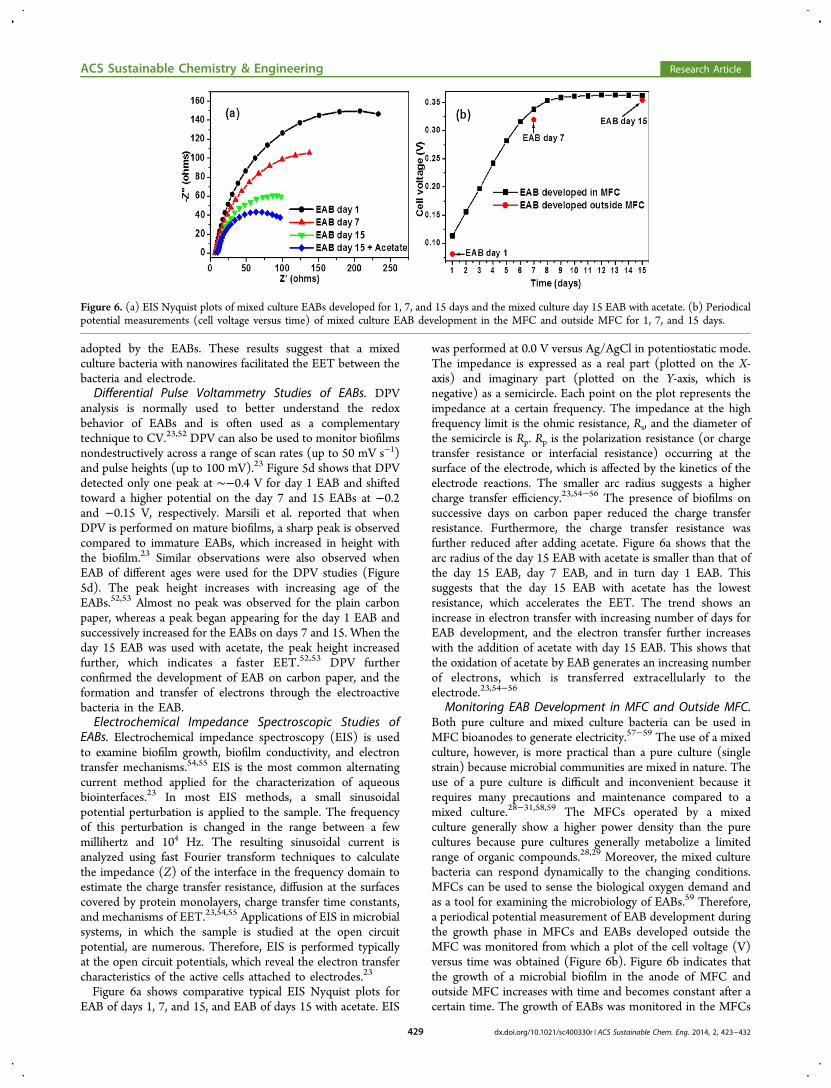
Figure 6a shows comparative typical EIS Nyquist plots forEAB of days 1, 7, and 15, and EAB of days 15 with acetate. EIS
was performed at 0.0 V versus Ag/AgCl in potentiostatic mode.The impedance is expressed as a real part (plotted on the X-axis) and imaginary part (plotted on the Y-axis, which isnegative) as a semicircle. Each point on the plot represents theimpedance at a certain frequency. The impedance at the highfrequency limit is the ohmic resistance, Rs, and the diameter ofthe semicircle is Rp. Rp is the polarization resistance (or chargetransfer resistance or interfacial resistance) occurring at thesurface of the electrode, which is affected by the kinetics of theelectrode reactions. The smaller arc radius suggests a highercharge transfer efficiency.23,54−56 The presence of biofilms onsuccessive days on carbon paper reduced the charge transferresistance. Furthermore, the charge transfer resistance wasfurther reduced after adding acetate. Figure 6a shows that thearc radius of the day 15 EAB with acetate is smaller than that ofthe day 15 EAB, day 7 EAB, and in turn day 1 EAB. Thissuggests that the day 15 EAB with acetate has the lowestresistance, which accelerates the EET. The trend shows anincrease in electron transfer with increasing number of days forEAB development, and the electron transfer further increaseswith the addition of acetate with day 15 EAB. This shows thatthe oxidation of acetate by EAB generates an increasing numberof electrons, which is transferred extracellularly to theelectrode.23,54−56
Monitoring EAB Development in MFC and Outside MFC.Both pure culture and mixed culture bacteria can be used inMFC bioanodes to generate electricity.57−59 The use of a mixedculture, however, is more practical than a pure culture (singlestrain) because microbial communities are mixed in nature. Theuse of a pure culture is difficult and inconvenient because itrequires many precautions and maintenance compared to amixed culture.28−31,58,59 The MFCs operated by a mixedculture generally show a higher power density than the purecultures because pure cultures generally metabolize a limitedrange of organic compounds.28,29 Moreover, the mixed culturebacteria can respond dynamically to the changing conditions.MFCs can be used to sense the biological oxygen demand andas a tool for examining the microbiology of EABs.59 Therefore,a periodical potential measurement of EAB development duringthe growth phase in MFCs and EABs developed outside theMFC was monitored from which a plot of the cell voltage (V)versus time was obtained (Figure 6b). Figure 6b indicates thatthe growth of a microbial biofilm in the anode of MFC andoutside MFC increases with time and becomes constant after acertain time. The growth of EABs was monitored in the MFCs
Figure 6. (a) EIS Nyquist plots of mixed culture EABs developed for 1, 7, and 15 days and the mixed culture day 15 EAB with acetate. (b) Periodicalpotential measurements (cell voltage versus time) of mixed culture EAB development in the MFC and outside MFC for 1, 7, and 15 days.
ACS Sustainable Chemistry & Engineering Research Article
dx.doi.org/10.1021/sc400330r | ACS Sustainable Chem. Eng. 2014, 2, 423−432429

and outside the MFCs for 2 weeks and showed stableperformance. This suggests that the biofilm in this system hasessentially developed within this time frame and the EET hastaken place, which was measured using the MFC. Figure 6bshows a linear increase in the potential of both EABs withrespect to time. This supports the development of mixedculture EABs on carbon paper. The comparative developmentof EABs in the MFCs and outside the MFC under similarconditions showed similar trends of an increase in potential,which suggest that the development of EABs in the MFCs andoutside the MFCs is similar. The development of EABs outsidethe MFC is easier, economical, and straightforward. This is whythis method has been adopted to develop the EABs (outsideMFC) and used for different applications.8−16
From the above studies, discussion and proposed mecha-nism, it can be concluded that after ∼15 days of mixed cultureEAB development outside the MFC, the as-developed mixedcultured EABs can produce sufficient electrons by biologicallydecomposing organic matter, such as acetate, to act as areducing tool. The biologically produced electrons can betransferred extracellularly via nanowires (pili) to the electrodes,metal ions, or metal oxides for nanomaterial synthesis. Fromprevious reports,8−16,25,26 and the present study, it has beenestablished that EAB is an effective tool for the synthesis ofbioinspired nanomaterials, such as metal nanoparticles, nano-composites, biohydrogen production, and environmentalremediation.Stability and Reusability of EABs. The stability and
reusability of EABs were checked by running the CV, DPV,and EIS cycles for 3−4 times, which gave a similar type ofspectra upon repeated use. In MFCs, the same EAB can beused several times. This shows the stability and reusability ofEABs. In previous reports,8−16,25,26 for the synthesis ofnanoparticles and nanocomposites, the same EAB can beused more than one time for identical types of nanoparticles,and nanocomposites synthesis. The repeated use of EAB fornanomaterials synthesis can result in the deposition of as-synthesized nanomaterials on the EABs surface, which reducesthe available surface area and slightly inhibits the EABsactivities. This confirms that the EABs formed on carbonpaper can be used more than once for different purposes, whichestablishes its stability and reusability.
■ CONCLUSIONMicroscopic, spectroscopic, and electrochemical methods (CV,DPV, and EIS) were used to examine mixed culture EABs tounderstand the EET phenomena of the electroactive bacteria tothe carbon electrode via pili after biologically decomposing theacetate as carbon source. By choosing the appropriatetechniques and conditions, these techniques can be used asnondestructive methods, and allow the in vivo determination ofelectron transfer from the whole cells (EABs) to electrodesunder conditions that are comparable to those encountered innatural environments. The proposed methods can be applied towell-defined mixed culture EABs, as well as to complexmicrobial communities and can allow quantitative comparisonsfor the development of better microbial catalysts based ondirect electron transfer between the bacteria and electrodes.The mixed culture EABs can be used as an electron source forvarious applications. The mixed culture EAB-mediated syn-thesis of nanoparticles, nanocomposites, biohydrogen produc-tion, and band gap narrowing of metal oxides provides a greenchemistry approach for the efficient synthesis of a range of
bioinspired nanomaterials without any energy input. Thisapproach can be extended to further strengthening the greenapproaches in nanoscience and nanotechnology.
■ ASSOCIATED CONTENT*S Supporting InformationFurther detailed information about UV−vis and FE-SEM ofEABs developed on carbon paper and nylon fiber. TEM imagesof AuNPs, Au@TiO2, and Ag@TiO2 nanocomposites synthe-sized by EABs. This material is available free of charge via theInternet at http://pubs.acs.org.
■ AUTHOR INFORMATIONCorresponding Author*Tel.: +82 53 810 2517. Fax: +82 53 810 4631. E-mail address:[email protected].
NotesThe authors declare no competing financial interest.
■ ACKNOWLEDGMENTSThis study was supported by Basic Science Research Programthrough the National Research Foundation of Korea (NRF)funded by the Ministry of Education, Science and Technology(Grant No: 2012R1A1A4A01005951).
■ REFERENCES(1) Millo, D. Spectroelectrochemical analyses of electroactivemicrobial biofilms. Biochem. Soc. Trans. 2012, 40, 1284−1290.(2) Harnisch, F.; Rabaey, K. The diversity of techniques to studyelectrochemically active biofilms highlights the need for stand-ardization. ChemSusChem, 2012, 5, 1027−1038.(3) Katuri, K. P.; Kavanagh, P.; Rengaraj, S.; Leech, D. Geobactersulfurreducens biofilms developed under different growth conditionson glassy carbon electrodes: insights using cyclic voltammetry. Chem.Commun. 2010, 46, 4758−4760.(4) Borole, A. P.; Reguera, G.; Ringeisen, B.; Wang, Z.; Feng, Y.;Kim, B. H. Electroactive biofilms: Current status and future researchneeds. Energy Environ. Sci. 2011, 4, 4813−4834.(5) Halan, B.; Buehler, K.; Schmid, A. Biofilms as living catalysts incontinuous chemical syntheses. Trends Biotechnol. 2012, 30, 453−465.(6) Erable, B.; Duteanu, N. M.; Ghangrekar, M. M.; Dumas, C.;Scott, K. Application of electro-active biofilms. Biofouling: J.Bioadhesion Biofilm Res. 2010, 26, 57−71.(7) Babauta, J.; Renslow, R.; Lewandowski, Z.; Beyenal, H.Electrochemically active biofilms: facts and fiction. A review.Biofouling: J. Bioadhesion Biofilm Res. 2012, 28, 789−812.(8) Kalathil, S.; Lee, J.; Cho, M. H. Electrochemically active biofilm-mediated synthesis of silver nanoparticles in water. Green Chem. 2011,13, 1482−1485.(9) Khan, M. M.; Kalathil, S.; Lee, J.; Cho, M. H. Synthesis ofcysteine capped silver nanoparticles byelectrochemically active biofilmand their antibacterial activities. Bull. Korean Chem. Soc. 2012, 33,2592−2596.(10) Khan, M. M.; Kalathil, S.; Han, T. H.; Lee, J.; Cho, M. H.Positively charged gold nanoparticles synthesized by electrochemicallyactive biofilma biogenic approach. J. Nanosci. Nanotechnol. 2013, 13,6079−6085.(11) Kalathil, S.; Khan, M. M.; Banerjee, A. N.; Lee, J.; Cho, M. H. Asimple biogenic route to rapid synthesis of Au@TiO2 nanocompositesby electrochemically active biofilms. J. Nanopart. Res. 2012, 14, 1051−1060.(12) Khan, M. M.; Ansari, S. A.; A. Lee, J.; Cho, M. H. Highly visiblelight active Ag@TiO2 nanocomposites synthesized using an electro-chemically active biofilm: a novel biogenic approach. Nanoscale 2013,5, 4427−4435.
ACS Sustainable Chemistry & Engineering Research Article
dx.doi.org/10.1021/sc400330r | ACS Sustainable Chem. Eng. 2014, 2, 423−432430

(13) Khan, M. M.; Ansari, S. A.; A. Lee, J.; Cho, M. H. Novel Ag@TiO2 nanocomposite synthesized by electrochemically active biofilmfor nonenzymatic hydrogen peroxide sensor. Mater. Sci. Eng., C 2013,33, 4692−4699.(14) Kalathil, S.; Khan, M. M.; Ansari, S. A.; Lee, J.; Cho, M. H. Bandgap narrowing of titanium dioxide (TiO2) nanocrystals by electro-chemically active biofilms and their visible light activity. Nanoscale2013, 5, 6323−6326.(15) Ansari, S. A.; Khan, M. M.; Kalathil, S.; Nisar, A.; Lee, J.; Cho,M. H. Oxygen vacancy induced band gap narrowing of ZnOnanostructures by an electrochemically active biofilm. Nanoscale2013, 5, 9238−9246.(16) Khan, M. M.; Lee, J.; Cho, M. H. Electrochemically activebiofilm mediated bio-hydrogen production catalyzed by positivelycharged gold nanoparticles. Int. J. Hydrogen Energy 2013, 38, 5243−5250.(17) Dulon, S.; Parot, S.; Delia, M. L.; Bergel, A. Electroactivebiofilms: new means for electrochemistry. J. Appl. Electrochem. 2007,37, 173−179.(18) Fricke, K.; Harnisch, F.; Schr oder, U. On the use of cyclicvoltammetry for the study of anodic electron transfer in microbial fuelcells. Energy Environ. Sci. 2008, 1, 144−147.(19) Logan, B. E.; Murano, C.; Scott, K.; Gray, N. D.; Head, I. M.Electricity generation from cysteine in a microbial fuel cell. Water Res.2005, 39, 942−952.(20) Han, T. H.; Khan, M. M.; Kalathil, S.; Lee, J.; Cho, M. H.Simultaneous enhancement of methylene blue degradation and powergeneration in microbial fuel cell by gold nanoparticles. Ind. Eng. Chem.Res. 2013, 3 (52), 8174−8181.(21) Hossain, M. M.; Nakayama, H.; Goto, N. In vitro induction ofapoptosis of developing brain cells by 5-azacytidine. Int. J. Dev.Neurosci. 1996, 14, 11−17.(22) Lee, J.-H.; Cho, M. H.; Lee. 3-Indolylacetonitrile decreasesEscherichia coli O157:H7 biofilm formation and Pseudomonasaeruginosa virulence. J. Environ. Microbiol. 2011, 13, 62−73.(23) Marsili, E.; Rollefson, J. B.; Baron, D. B.; Hozalski, R. M.; Bond,D. R. Microbial biofilm voltammetry: direct electrochemical character-ization of catalytic electrode-attached biofilms. Appl. Environ. Microbiol.2008, 74, 7329−7337.(24) Barsoukov, E.; Macdonald, J. R. Impedance spectroscopy-theory,experiment, and applications, 2nd ed.; Wiley Interscience: Hoboken, NJ,2005.(25) Kalathil, S.; Lee, J.; Cho, M. H. Gold nanoparticles produced insitu mediate bioelectricity and hydrogen production in a microbial fuelcell by quantized capacitance charging. ChemSusChem 2013, 6, 246−250.(26) Kalathil, S.; Khan, M. M.; Lee, J.; Cho, M. H. Production ofbioelectricity, bio-hydrogen, high value chemicals and 3 bioinspirednanomaterials by electrochemically active biofilms. Biotech. Adv. 2013,31, 915−924.(27) Kalathil, S.; Lee, J.; Cho, M. H. Catalytic role of Au@TiO2
nanocomposite on enhanced degradation of an azo-dye by electro-chemically active biofilms: a quantized charging effect. J. Nanopart. Res.2013, 15, 1392−1397.(28) Kim, B. H.; Chang, I. S.; Gadd, G. M. Challenges in microbialfuel cell development and operation. Appl. Microbiol. Biotechnol. 2007,76, 485−494.(29) Rittmann, B. E.; Krajmalnik-Brown, R.; Halden, R. U. Pre-genomic, genomic and post-genomic study of microbial communitiesinvolved in bioenergy. Nat. Rev. Microbiol. 2008, 6, 604−612.(30) Liu, Y.; Harnisch, F.; Fricke, K.; Schroder, U.; Climent, V.; Feliu,J. M. The study of electrochemically active microbial biofilms ondifferent carbon-based anode materials in microbial fuel cells. Biosens.Bioelectron. 2010, 25, 2167−2171.(31) Patil, S. A.; Harnisch, F.; Koch, C.; Hubschmann, T.; Fetzer, I.;Carmona-Martínez, A. A.; Muller, S.; Schroder, U. Electroactive mixedculture derived biofilms in microbial bioelectrochemical systems: Therole of pH on biofilm formation, performance and composition.Bioresour. Technol. 2011, 102, 9683−9690.
(32) Lovley, D. R. The microbe electric: conversion of organic matterto electricity. Curr. Opin. Biotechnol. 2008, 19, 564−571.(33) Lovley, D. R. Live wires: direct extracellular electron exchangefor bioenergy and the bioremediation of energy-related contamination.Energy Environ. Sci. 2011, 4, 4896−4906.(34) Malvankar, N. S.; Vargas, M.; Nevin, K. P.; Franks, A. E.; Leang,C.; Kim, B. C.; Inoue, K.; Mester, T.; Covalla, S. F.; Johnson, J. P.;Rotello, V. M.; Tuominen, M. T.; Lovley, D. R. Tunable metallic-likeconductivity in microbial nanowire networks. Nat. Nanotechnol. 2011,6, 573−579.(35) Malvankar, N. S.; Tuominen, M. T.; Lovley, D. R. Comment on“On electrical conductivity of microbial nanowires and biofilms”.Energy Environ. Sci. 2011, 4, 4366; Energy Environ. Sci. 2012, 5, 6247−6249.(36) Lovley, D. R.; Nevin, K. P. A shift in the current: newapplications and concepts for microbe-electrode electron exchange.Curr. Opin. Biotechnol. 2011, 22, 1−8.(37) Nakamura, R.; Ishii, K.; Hashimoto, K. Electronic absorptionspectra and redox properties of C type cytochromes in living microbes.Angew. Chem., Int. Ed. 2009, 48, 1606−1608.(38) Okamoto, A.; Nakamura, R.; Ishii, K.; Hashimoto, K. In vivoelectrochemistry of c-type cytochrome-mediated electron-transfer withchemical marking. ChemBioChem 2009, 10, 2329−2332.(39) Jain, A.; Gazzola, G.; Panzera, A.; Zanoni, M.; Marsili, E. Visiblespectroelectrochemical characterization of Geobacter sulfurreducensbiofilms on optically transparent indium tin oxide electrode.Electrochim. Acta 2011, 56, 10776−10785.(40) Inoue, K.; Qian, X.; Morgado, L.; Kim, B. C.; Mester, T.;Izallalen, M.; Salgueiro, C. A.; Lovley, D. R. Purification andcharacterization of omcz, an outer-surface, octaheme c-typecytochrome essential for optimal current production by geobactersulfurreducens. Appl. Environ. Microbiol. 2010, 76, 3999−4007.(41) Singh, D. K.; Iyer, P. K.; Giri, P. K. Effect of high voltage electricpulse on microstructure of fine particles. Nanotrends: J. Nanotechnol.Appl. 2008, 4, 55−58.(42) Liu, H.; Nishide, D.; Tanaka, T.; Kataura, H. Large-scale single-chirality separation of single-wall carbon nanotubes by simple gelchromatography. Nat. Commun. 2011, 2, 309.(43) Beitlich, T.; Kuhnel, K.; Schulze-Briese, C.; Shoeman, R. L.;Schlichting, I. Cryoradiolytic reduction of crystalline heme proteins:analysis by UV-Vis spectroscopy and X-ray crystallography. J.Synchrotron Rad. 2007, 14, 11−23.(44) Liu, Y.; Bond, D. R. Long-distance electron transfer by G.sulfurreducens biofilms results in accumulation of reduced c-typecytochromes. ChemSusChem 2012, 5, 1047−1053.(45) Collinson, M.; Bowden, E. F. UV-Visible Spectroscopy ofadsorbed cytochrome c on tin oxide electrodes. Anal. Chem. 1992, 64,1470−1476.(46) Cullity, B. D.; Stock, S. R. Elements of X-ray diffraction, 3rd ed.;Prentice Hall, 2001; Chapter 4, pp 123−157.(47) Florea, L. J.; Noe-Stinson, C. L.; Brewer, J.; Fowler, R.; Kearns,J. B.; Greco, A. M. Iron oxide and calcite associated with leptothrix sp.biofilms within an estavelle in the upper floridan aquifer. Int. J. Speleol.2011, 40, 205−219.(48) Hu, X. B.; Xu, K.; Wang, Z.; Liding, L.; Qiangren, A.Characteristics of biofilm attaching to carriers in moving bed biofilmreactor used to treat vitamin C wastewater. Scanning 2013, 35, 283−291.(49) Aleksandrov, P. A.; Afanasiev, A. M.; Melkonyan, M. K.;Stepanov, S. A. X-Ray diffraction under specular reflection conditionson crystals with an amorphous surface film. Phys. Stat. Solidi A 1984,81, 47−53.(50) Katuri, K. P.; Rengaraj, S.; Kavanagh, P.; Flaherty, O.; Leech, D.Charge transport through geobacter sulfurreducens biofilms grown ongraphite rods. Langmuir 2012, 28, 7904−7913.(51) Strycharz, S. M.; Malanoski, A. P.; Snider, R. M.; Yi, H.; Lovley,D. R.; Tender, L. M. Application of cyclic voltammetry to investigateenhanced catalytic current generation by biofilm-modified anodes of
ACS Sustainable Chemistry & Engineering Research Article
dx.doi.org/10.1021/sc400330r | ACS Sustainable Chem. Eng. 2014, 2, 423−432431

Geobacter sulfurreducens strain DL1 vs. variant strain KN400. EnergyEnviron. Sci. 2011, 4, 896−913.(52) Marsili, E.; Baron, D. B.; Shikhare, I.; Coursolle, D.; Gralnick, J.;Bond, D. R. Shewanella secretes flavins that mediate extracellularelectron transfer. Proc. Natl. Acad. Sci. U.S.A. 2008, 105, 3968−3973.(53) Jain, A.; Connolly, J. O.; Woolley, R.; Krishnamurthy, S.; Marsili,E. Extracellular electron transfer mechanism in Shewanella loihica pv- 4biofilms formed at indium tin oxide and graphite electrodes. Int. J.Electrochem. Sci. 2013, 8, 1778−1793.(54) Dominguez-Benetton, X.; Sevda, S.; Vanbroekhoven, K.; Pant,D. The accurate use of impedance analysis for the study of microbialelectrochemical systems. Chem. Soc. Rev. 2012, 41, 7228−7246.(55) Malvankar, N. S.; Tuominen, M. T.; Lovley, D. R. Biofilmconductivity is a decisive variable for high-current-density Geobactersulfurreducens microbial fuel cells. Energy Environ. Sci. 2012, 5, 5790−95797.(56) Ramasamy, R. P.; Ren, Z.; Mench, M. M.; Regan, J. M. Impactof initial biofilm growth on the anode impedance of microbial fuelcells. Biotechnol. Bioeng. 2008, 101, 101−108.(57) Bond, D. R.; Lovley, D. R. Electricity production by Geobactersulfurreducens attached to electrodes. Appl. Environ. Microbiol. 2003,69, 1548−1555.(58) Chaudhuri, S. K.; Lovley, D. R. Electricity generation by directoxidation of glucose in mediatorless microbial fuel cells. Nat.Biotechnol. 2003, 21, 1229−1232.(59) Chang, I. S.; Jang, J. K.; Gil, G. C.; Kim, M.; Kim, H. J.; Cho, B.W.; Kim, B. H. Continuous determination of biochemical oxygendemand using microbial fuel cell type biosensor. Biosens. Bioelectron.2004, 19, 607−613.
ACS Sustainable Chemistry & Engineering Research Article
dx.doi.org/10.1021/sc400330r | ACS Sustainable Chem. Eng. 2014, 2, 423−432432
Copyright © 2022 FDOKUMEN