Mitochondrial composition, function and stress response in plants
20
Journal of Integrative Plant Biology 2012, 54 (11): 887–906 Invited Expert Review Mitochondrial Composition, Function and Stress Response in Plants F Richard P. Jacoby 1,2 , Lei Li 1,2 , Shaobai Huang 1,2 , Chun Pong Lee 3 , A. Harvey Millar 1,2 ∗ and Nicolas L. Taylor 1,2 1 ARC Centre of Excellence in Plant Energy Biology and 2 Centre for Comparative Analysis of Biomolecular Networks (CABiN), MCS Building M316, The University of Western Australia, 35 Stirling Highway, Crawley, WA 6009, Western Australia, Australia 3 Department of Plant Sciences, University of Oxford, South Parks Road, Oxford OX1 3RB, United Kingdom ∗ Corresponding author E-mail: [email protected] F Articles can be viewed online without a subscription. Available online on 10 October 2012 at www.jipb.net and www.wileyonlinelibrary.com/journal/jipb doi: 10.1111/j.1744-7909.2012.01177.x A. Harvey Millar (Corresponding author) Abstract The primary function of mitochondria is respiration, where catabolism of substrates is coupled to ATP synthesis via oxidative phosphorylation. In plants, mitochondrial composition is relatively complex and flexible and has specific pathways to support pho- tosynthetic processes in illuminated leaves. This review begins with outlining current models of mitochondrial composition in plant cells, with an emphasis upon the assembly of the complexes of the classical electron transport chain (ETC). Next, we focus upon the comparative analysis of mitochondrial function from different tissue types. A prominent theme in the plant mitochondrial literature involves linking mitochondrial composition to environmental stress responses, and this review then gives a detailed outline of how oxidative stress impacts upon the plant mitochondrial proteome with particular attention to the role of transition metals. This is followed by an analysis of the signaling capacity of mitochondrial reactive oxygen species, which studies the transcriptional changes of stress responsive genes as a framework to define specific signals emanating from the mitochondrion. Finally, specific mitochondrial roles during exposure to harsh environments are outlined, with attention paid to mitochondrial delivery of energy and intermediates, mitochondrial support for photosynthesis, and mitochondrial processes operating within root cells that mediate tolerance to anoxia and unfavorable soil chemistries. Keyword: Plant mitochondria; respiration; oxidative stress; electron transport chain; complex assembly; ROS signaling; carbon. Jacoby RP, Li L, Huang S, Lee CP, Millar AH, Taylor NL (2012) Mitochondrial composition, function and stress response in plants. J. Integr. Plant Biol. 54(11), 887–906. Introduction The ATP needed for cellular maintenance and growth in or- ganisms comes from respiration. It is the fundamental energy- conserving process that couples the transfer of potential energy from the oxidation of reduced organic matter to high-energy intermediates and heat. In aerobic respiration, which yields the highest efficiency of conversion to high-energy interme- diates, mitochondria carry out the final steps to generate the bulk of the ATP through oxidative phosphorylation driven by oxidation of organic acids, to release CO 2 and reduce O 2 to water. However, mitochondria also play roles in a variety of important cellular processes associated with carbon, nitrogen, phosphorus and sulfur metabolism in plants. In photosynthetic tissues, mitochondria function is indispensable for chloroplast function. Mitochondria are key agents in how plants respond C 2012 Institute of Botany, Chinese Academy of Sciences
Transcript of Mitochondrial composition, function and stress response in plants
Journal of Integrative Plant Biology 2012, 54 (11): 887–906
Invited Expert Review
Mitochondrial Composition, Function and StressResponse in PlantsF
Richard P. Jacoby1,2, Lei Li1,2, Shaobai Huang1,2, Chun Pong Lee3, A. Harvey Millar1,2∗
and Nicolas L. Taylor1,21ARC Centre of Excellence in Plant Energy Biology and 2Centre for Comparative Analysis of Biomolecular Networks (CABiN), MCSBuilding M316, The University of Western Australia, 35 Stirling Highway, Crawley, WA 6009, Western Australia, Australia3Department of Plant Sciences, University of Oxford, South Parks Road, Oxford OX1 3RB, United Kingdom∗Corresponding author
E-mail: [email protected] Articles can be viewed online without a subscription.Available online on 10 October 2012 at www.jipb.net and www.wileyonlinelibrary.com/journal/jipbdoi: 10.1111/j.1744-7909.2012.01177.x
A. Harvey Millar
(Corresponding author)
Abstract
The primary function of mitochondria is respiration, wherecatabolism of substrates is coupled to ATP synthesis via oxidativephosphorylation. In plants, mitochondrial composition is relativelycomplex and flexible and has specific pathways to support pho-tosynthetic processes in illuminated leaves. This review beginswith outlining current models of mitochondrial composition in plantcells, with an emphasis upon the assembly of the complexes ofthe classical electron transport chain (ETC). Next, we focus uponthe comparative analysis of mitochondrial function from differenttissue types. A prominent theme in the plant mitochondrial literatureinvolves linking mitochondrial composition to environmental stress
responses, and this review then gives a detailed outline of how oxidative stress impacts upon theplant mitochondrial proteome with particular attention to the role of transition metals. This is followedby an analysis of the signaling capacity of mitochondrial reactive oxygen species, which studies thetranscriptional changes of stress responsive genes as a framework to define specific signals emanatingfrom the mitochondrion. Finally, specific mitochondrial roles during exposure to harsh environments areoutlined, with attention paid to mitochondrial delivery of energy and intermediates, mitochondrial supportfor photosynthesis, and mitochondrial processes operating within root cells that mediate tolerance toanoxia and unfavorable soil chemistries.
Keyword: Plant mitochondria; respiration; oxidative stress; electron transport chain; complex assembly; ROS signaling; carbon.
Jacoby RP, Li L, Huang S, Lee CP, Millar AH, Taylor NL (2012) Mitochondrial composition, function and stress response in plants. J. Integr. PlantBiol. 54(11), 887–906.
Introduction
The ATP needed for cellular maintenance and growth in or-
ganisms comes from respiration. It is the fundamental energy-
conserving process that couples the transfer of potential energy
from the oxidation of reduced organic matter to high-energy
intermediates and heat. In aerobic respiration, which yields
the highest efficiency of conversion to high-energy interme-
diates, mitochondria carry out the final steps to generate the
bulk of the ATP through oxidative phosphorylation driven by
oxidation of organic acids, to release CO2 and reduce O2 to
water. However, mitochondria also play roles in a variety of
important cellular processes associated with carbon, nitrogen,
phosphorus and sulfur metabolism in plants. In photosynthetic
tissues, mitochondria function is indispensable for chloroplast
function. Mitochondria are key agents in how plants respond
C© 2012 Institute of Botany, Chinese Academy of Sciences
888 Journal of Integrative Plant Biology Vol. 54 No. 11 2012
to oxidative stress, and plant mitochondria possess unique
respiratory properties to enable these processes. Understand-
ing the control and regulation of the respiratory processes
is vital to alter the rate of plant biomass production and to
explain plant growth and its variability in different environmental
conditions. An exhaustive analysis of all the elements involved
in these processes is not possible here, but by using specific
examples and recent discoveries we can highlight some key
elements in the structure, mechanism and regulation of this
process. Firstly, we will consider the composition of the key
processes in mitochondrial respiration, our understanding of
the mechanism and regulation of the assembly process that
builds the machinery and the differential steady-states and
roles of these functions in different plant tissue types. Secondly,
we will review our understanding of the regulatory changes
induced by internal factors (signalling processes, redox control
and oxidative stress) and by the environment (salinity, osmotic
stress and nutrient deprivation).
Plant Mitochondrial Composition andAssembly
The functional steps of the respiratory apparatus in plant
mitochondria can be framed as a sequential set of processes,
involving the transport of reduced glycolytic products from
the cytosol into the mitochondrion, and then encompassing
a series of reactions leading to the release of CO2 and
reduction of O2 to water. Firstly, a set of carriers and channels
allow substrates and cofactors from the cytosol to enter the
mitochondria and also facilitate the release of the products
of respiration to the rest of the cell. Next, the tricarboxylic
acid cycle (TCAC) and associated enzymes undertake the
oxidative decarboxylation of organic acids to reduce NAD(P)+
and FAD+ to NAD(P)H and FADH2, respectively, and to drive
substrate level phosphorylation of ADP to ATP. Thirdly, the
classical OXPHOS electron transport chain (ETC) couples the
oxidation of NAD(P)H and FADH2 to the reduction of O2 and
the co-committed translocation of protons used to build an
electrical gradient to drive oxidative phosphorylation. Finally,
non-phosphorylating bypasses of the electron transport chain,
the alternative oxidase and rotenone-insensitive NAD(P)H de-
hydrogenases, can alter the gearing between the TCA cycle
and OXPHOS to facilitate the anaplerotic function of plant
mitochondria for organic acid provision to cellular biosynthetic
pathways without the full TCA cycle. This machinery and
the regulation of pathways to assemble it define the primary
functional composition of mitochondria.
Respiratory metabolite transporters
The two membranes of mitochondria have very different per-
meability properties. The outer membrane allows relatively non-
specific transport of small molecules from the cytosol into the
inter-membrane space (Mannella 1992; Mannella et al. 2001).
The inner membrane contains very selective transporters for
small molecules to the matrix space. This allows a complex set
of inner membrane carrier functions to have a large influence
on the functions of mitochondria (Laloi 1999). Transport across
the outer membrane is largely via the voltage dependent
anion channels (VDAC), that form β-barrel pores for the move-
ment of respiratory substrates and products up to 1000 Da
(Mannella and Tedeschi 1987; Robert et al. 2012). A family of
related mitochondrial inner membrane carriers operate for the
transport of organic acids, amino acids, inorganic phosphate
and nucleotides. Complementation assays have defined the
mitochondrial inorganic phosphate carriers (Hamel et al. 2004),
and adenine di- and tri-nucleotide carriers (Palmieri et al. 2008)
in plants. A general carrier able to transport a variety or both di-
and tricarboxylic acids is likely to carry the bulk of organic acid
traffic (Picault et al. 2002). Basic amino acid carriers have been
identified that transport arginine, ornithine, lysine and histidine
(Catoni et al. 2003a; Hoyos et al. 2003), a succinate-fumarate
carrier has been identified (Catoni et al. 2003b) and the cofactor
NAD+ has a specific carrier in plant mitochondria (Palmieri
et al. 2009).
Tricarboxylic acid cycle
The nine enzymes of the TCAC represent the major carbon
metabolising machinery present in plant mitochondria. Pyru-
vate is directly transported across the inner membrane or
generated in the matrix from malate by the action of malic
enzyme (ME). It is then oxidised by the pyruvate dehydroge-
nase complex (PDC) to form acetyl-CoA. PDC comprises three
enzymes E1 (2-oxo acid dehydrogenase), E2 (acyltransferase)
and E3 (lipoamide dehydrogenase) (Guan et al. 1995; Luethy
et al. 1995). This complex is regulated by phosphorylation of
E1, lowering PDC function in the day and increasing PDC
function at night (Thelen et al. 2000). Citrate synthase (CS)
catalyses the condensation of acetyl CoA with the dicarboxylate
oxaloacetate, yielding citrate and releasing the CoA cofactor
(La Cognata et al. 1996). Over-expression of citrate syn-
thase in Arabidopsis enhances growth under low phosphorous
conditions due to enhanced citrate excretion from the roots
to increase inorganic phosphate availability (Koyama et al.
2000). Citrate is converted to isocitrate via aconitase (ACO),
and isocitrate dehydrogenase (IDH) oxidises isocitrate to form
2-oxoglutarate. 2-oxoglutarate dehydrogenase, succinyl CoA
ligase, succinate dehydrogenase (complex II see below), fu-
marase and malate dehydrogenase compete the cycle by
reforming oxaloacetate. Recent studies of TCA cycle mutants
have shown the wide impact these enzymes have not only in
TCA cycle function but as steps for the anaplerotic delivery
of organic acids for other processes in plant cells such as
Plant Mitochondrial Composition and Stress Responses 889
photosynthetic performance, plant biomass, photorespiration,
nitrogen assimilation and amino acid metabolism, and even
stomatal function. Antisense mutants of malate dehydroge-
nase (MDH) and aconitase in tomato exhibit faster rates of
photosynthetic CO2 assimilation rates and higher ascorbate
levels (Nunes-Nesi et al. 2005). Antisense of fumarase leads
to substantial inhibition of photosynthetic performance and
stomatal function (Nunes-Nesi et al. 2007). Knockdown of
succinate dehydrogenase can alter stomatal aperture, change
nitrogen use efficiency and alter disease signalling (Araujo
et al. 2011; Fuentes et al. 2011; Gleason et al. 2011).
Oxidative phosphorylation (OXPHOS) apparatus
The so-called classical ETC is comprised of four large protein
complexes (I, II, III, IV) that interact with each other via the
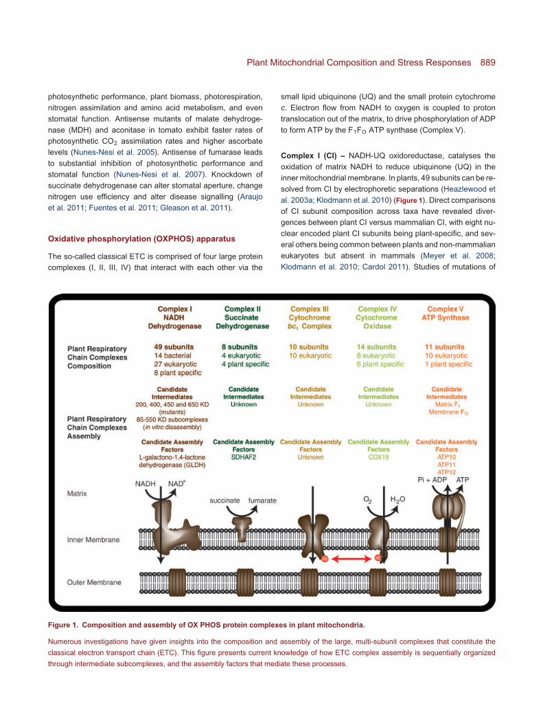
Figure 1. Composition and assembly of OX PHOS protein complexes in plant mitochondria.
Numerous investigations have given insights into the composition and assembly of the large, multi-subunit complexes that constitute the
classical electron transport chain (ETC). This figure presents current knowledge of how ETC complex assembly is sequentially organized
through intermediate subcomplexes, and the assembly factors that mediate these processes.
small lipid ubiquinone (UQ) and the small protein cytochrome
c. Electron flow from NADH to oxygen is coupled to proton
translocation out of the matrix, to drive phosphorylation of ADP
to form ATP by the F1FO ATP synthase (Complex V).
Complex I (CI) – NADH-UQ oxidoreductase, catalyses the
oxidation of matrix NADH to reduce ubiquinone (UQ) in the
inner mitochondrial membrane. In plants, 49 subunits can be re-
solved from CI by electrophoretic separations (Heazlewood et
al. 2003a; Klodmann et al. 2010) (Figure 1). Direct comparisons
of CI subunit composition across taxa have revealed diver-
gences between plant CI versus mammalian CI, with eight nu-
clear encoded plant CI subunits being plant-specific, and sev-
eral others being common between plants and non-mammalian
eukaryotes but absent in mammals (Meyer et al. 2008;
Klodmann et al. 2010; Cardol 2011). Studies of mutations of
890 Journal of Integrative Plant Biology Vol. 54 No. 11 2012
CI subunits have shown that plants can survive without CI due
to the activity of alternative NAD(P)H dehydrogenases (see
below). Such mutants have a variety of interesting phenotypes
including viral infection tolerance, prolonged hydration under
water-deficient conditions and altered organic and amino acid
concentrations (Dutilleul et al. 2003; Meyer et al. 2009). In
Neurospora crassa, CI assembly analysis using radio-labelled
pulse chase in mutants has revealed that the matrix and mem-
brane arms assemble independently via separate pathways
(Tuschen et al. 1990; Kuffner et al. 1998; Schulte 2001; Videira
and Duarte 2002; Mimaki et al. 2012). By using a combination
of radio-labelled pulse-chase experiments, in vitro mitochon-
drial import and monitoring of tagged CI subunits, several CI
assembly models have also been proposed in human cells
in assembly disturbed systems (Ugalde et al. 2004; Lazarou
et al. 2007; Vogel et al. 2007; Mimaki et al. 2012). In plants,
controlled dissociation of CI using low concentrations of SDS
followed by BN-PAGE and peptide mass spectrometry enabled
the visualisation and compositional analysis of 10 subcom-
plexes between 550 – 85 kDa in size, giving detailed insights
into the internal architecture of CI (Klodmann et al. 2010)
(Figure 1). Using Arabidopsis CI subunit knockout mutants, 200,
400, 450 and 650 kDa membrane arm subcomplexes have
been identified using BN-PAGE and antibodies. It is proposed
that these subcomplexes are assembly intermediates during CI
formation, which accumulate when specific subunits are absent
(Meyer et al. 2011). The first two assembly factors known for
CI, CIA40 and CIA84, were discovered in N. crassa (Kuffner
et al. 1998). Nine assembly factors including NDUFAF2/B17.2,
NDUFAF1/CIA30, C20orf7, C80orf38, NDUFAF4, NDUFAF3,
NUBPL, FOXRED1 and ACAD9 have been found in humans
and deficiency can impair CI assembly and lead to clinical phe-
notypes in patients (Nouws et al. 2012). Little is known about
assembly factors in plants, with only L-galactono-1,4-lactone
dehydrogenase (GLDH) described as a potential assembly
factor in Arabidopsis (Pineau et al. 2008) (Figure 1). Given the
conserved core subunits but divergence of accessory subunits
amongst eukaryotes, it is not yet clear whether the accessory
subunits play different roles in complex I assembly and thus
whether or not the assembly of CI follows the same pathway in
different organisms.
Complex II (CII) – Succinate dehydrogenase, is an enzyme of
both the TCAC and the respiratory ETC. In all organisms, it is
made from four core subunits: a flavoprotein (SDHI), an iron-
sulphur subunit (SDH2) and two membrane anchor subunits
(SDH3 and SDH4). Purification of the complex using BN-PAGE
has revealed the common core subunits, but also four proteins
of unknown function that co-migrate with the complex (Eubel
et al. 2003; Millar et al. 2004a) (Figure 1). In Arabidopsis, all
SDH subunits are encoded in the nuclear genome. Knockout
mutants of the SDH1 gene are embryo lethal (Leon et al. 2007),
but knockdown of SDH1 and SDH2 lead to phenotypes as-
sociated with altered stomatal aperture, altered mitochondrial
ROS production and altered nitrogen use efficiency (Fuentes
et al. 2011; Gleason et al. 2011). Several proteins assisting
CII assembly have been described in yeast and mammalian
cells, but only SDHAF1 and SDHAF2 are considered to be real
assembly factors that directly and specifically aid CII assembly
(Ghezzi et al. 2009; Hao et al. 2009; Rutter et al. 2010). In
Arabidopsis, knockdown of the SDHAF2 homolog lowers SDH
assembly and markedly reduces root growth (Huang et al.
2012) (Figure 1).
Complex III (CIII) – Ubiquinone-cytochrome c oxidoreductase,
contains 10 subunits including the bifunctional core proteins
that act both in CIII function and as the matrix processing
peptidase, removing presequences from imported matrix pro-
teins (Figure 1). Only one subunit of this complex, cytochrome
b, is encoded by the plant mitochondrial genome (Unseld
et al. 1997), with the remaining nine all encoded by the
nuclear genome. In BN-PAGE separations from Arabidopsismitochondria, all of these subunits have been identified and
linked back to a set of mostly single copy genes (Werhahn and
Braun 2002; Meyer et al. 2008). Yeast provides an ideal model
system to study CIII, due to its ability to survive by fermentation
in the absence of the complex, making gene knock-out and
mutagenesis possible (Smith et al. 2012). CIII assembly follows
a modular assembly model including, early core subcomplex,
late core subcomplex and a dimeric CIII states (Smith et al.
2012). There have been 13 assembly factors implicated in
aiding the different stages of CIII assembly in yeast. Two of
these, BCS1L and TTC19, were also found to have functional
homologs in mammalian CIII assembly (Diaz et al. 2011; Smith
et al. 2012). Little is known about CIII assembly or functional
assembly factors in plants.
Complex IV (CIV) – Cytochrome c oxidase, is the terminal
oxidase of the classical ETC. Purification of CIV in plants
originally found only seven or eight subunits (Peiffer et al.
1990), but more recently, a CIV complex containing 14 protein
bands was separated from Arabidopsis (Millar et al. 2004a)
(Figure 1). Eight proteins homologous to known CIV subunits
from other organisms, together with a further six proteins that
may represent plant specific CIV subunits, were identified.
Analysis of human CIV via BN-PAGE separation has revealed
an assembly pathway characterized by the sequential incorpo-
ration of CIV subunits, initiated by subunit 1 and subsequently
progressing through several discrete assembly intermediates
(Barrientos et al. 2009). Studies in yeast have revealed over
40 assembly factors that aid different stages of CIV assembly,
but only a few homologs for these factors have been defined
in humans (Barrientos et al. 2009; Diaz et al. 2011). A plant
homolog of yeast assembly factor COX19 has been studied and
Plant Mitochondrial Composition and Stress Responses 891
found capable of complementing the yeast cox19 null mutant
and might play a role in the biogenesis of plant cytochrome coxidase to replace damaged forms of the enzyme (Attallah et al.
2007) (Figure 1). However, it seems evident that our knowledge
about the assembly of CIV in plants is still incomplete.
Complex V (CV) – ATP synthase is a membrane-bound F1F0
type H+-ATP synthase that catalyses the terminal step in
oxidative phosphorylation through which ATP is produced. It
is composed of a hydrophilic F1 component which catalyses
ATP formation and protrudes into the matrix and a hydrophobic
F0 component which channels protons through the membrane
while also anchoring the whole complex to the mitochondrial
inner membrane (Senior 1990; Hamasur and Glaser 1992;
Velours and Arselin 2000; Heazlewood et al. 2003b). The
general structure and the core subunits of the enzyme are
highly conserved in both prokaryotic and eukaryotic organisms
(Millar et al. 2011). In plant, most of mitochondrial F1 ATP
synthase subunits are encoded in the nucleus and translated
in the cytosol before being imported into the mitochondria (β,
γ, δ and ε), while most of the F0 subunits are encoded in the
plant mitochondrial genome and translated in the mitochondrial
matrix (a, b, c and A6L) (Jansch et al. 1996; Heazlewood et
al. 2003c; Sabar et al. 2003; Sabar et al. 2005) (Figure 1).
In plants, the F1α subunit is encoded in the mitochondrial
genome in most species. Alterations of mitochondrial-encoded
subunits of the F1F0-ATP synthase are frequently associated
with cytoplasmic male sterility (CMS) in plants, presumably due
to the high ATP demand of floral tissues (Xu et al. 2008). While
knockouts of ATP synthase core subunits are lethal in plants, in-
ducible knockdown with a dexamethasone-inducible promoter
has enabled investigations into the tissue-specific phenotypes
incurred by slowing the rates of mitochondrial ADP:ATP cycling
across a range of developmental stages (Robison et al. 2009).
Induction of the knockdown during germination in the light leads
to seedling lethality. Other phenotypes include the stunting of
dark-grown (etiolated) seedlings, downward curling or wavy-
edged leaf margins of light-grown plants, and ball-shaped
unexpanded flowers (Robison et al. 2009), highlighting the high
energetic demand of key growth stages.
The subunits that form the F1 component are kept in tight
stoichiometry in prokaryotic and eukaryotic organisms through
regulation of the assembly process (Senior 1990; Hamasur
and Glaser 1992; Velours and Arselin 2000; Li et al. 2012).
Models of yeast mitochondrial F1F0 ATP synthase assembly
involve two separate but coordinately regulated pathways,
where two separate subcomplexes are assembled in parallel,
before converging to form functional F1F0 (Rak and Tzagoloff
2009; Rak et al. 2011). Recent research in Arabidopsis using
progressive 15N labeling has measured differential rates of
turnover between different subpopulations of the F1 subcom-
plex. Intriguingly, the same subunits of F1 can exhibit faster or
slower turnover rates depending upon the intra-mitochondrial
localization they are found in, or upon the quaternary structure
of the F1 subcomplex that they constitute. For instance, sub-
units of F1 that were detected within the matrix-localized and
membrane-associated F1 subcomplexes both exhibited faster
turnover rates compared to those same subunits detected
within the intact, membrane-spanning F1F0 complex (Li et al.
2012). The proposed assembly model for plant CV comprises
three steps, the first being the formation of a rapidly turned over
F1 subcomplex in the matrix, then an intermediate stage where
F1 associates with the inner membrane and still turns over at a
fast rate, and then a final unison of F1 with FO to form functional
CV (Li et al. 2012) (Figure 1). This model of CV assembly
was corroborated by in vitro import assays where radiolabelled
CV subunits were incubated with isolated mitochondria, and
assembly intermediates visualised by scintillation counting of
BN-PAGE separations (Li et al. 2012). ATP synthase assembly
factors including Atp10, Atp11, Atp12, Atp22, Atp23 and Fmc1
have been discovered in yeast (Pickova et al. 2005; Osman
et al. 2007). Atp11, Atp12 and Fmc1 mediate the formation of
the F1 subcomplex while Atp10, Atp22 and Atp23 are essential
for the formation of F0 (Pickova et al. 2005; Osman et al. 2007).
Hsp60 and Hsp70 also contribute to efficient CV assembly
(Osman et al. 2007). A phylogenetic analysis of ATP synthase
assembly factors has found Atp11 and Atp12 are preserved
in almost all eukaryotic organisms, including plants, while the
other assembly factors show evidence of divergent evolution
across taxa (Pickova et al. 2005) (Figure 1). However, a detailed
study of the presence and conservation of CV assembly factors
across sequenced plant genomes has not been undertaken to
our knowledge.
Alternative electron transport pathways
In addition to the classical OXPHOS machinery, plant mito-
chondria contain non-phosphorylating respiratory bypasses of
electron transport and of proton-coupled ATP synthesis. These
pathways were first identified by the ability of plant mitochondria
to respire in the presence of cyanide and rotenone, potent
inhibitors of CIV and CI, respectively, and to exhibit natively
uncoupled respiration in the absence of an ADP source.
Alternative oxidase (AOX) – The cyanide-insensitive respi-
ration is catalysed by the alternative oxidase (AOX), a diiron
quinol oxidase that branches from the respiratory chain at UQ
and reduces oxygen to water without proton translocation. AOX
appears to play an antioxidant role in plant mitochondria, is
actively induced by oxidative stress (Van Aken et al. 2009) and
the different genes for the oxidase have been shown to be both
tissue- and development-specific in their expression patterns
(Saisho et al. 2001; Thirkettle-Watts et al. 2003). Knockout of
AOX leads to anthocyanin and ROS accumulation in the leaves
892 Journal of Integrative Plant Biology Vol. 54 No. 11 2012
under the combination of high light and drought stress (Giraud
et al. 2008).
Alternative NADH dehydrogenases – These type II NAD(P)H
dehydrogenases are found on both sides of the inner mi-
tochondrial membrane. External or cytosolic NADH and
NADPH can be oxidised via these dehydrogenases which
are insensitive to the CI inhibitor rotenone. These path-
ways operate without the translocation of protons (Finnegan
et al. 2004; Rasmusson et al. 2004). The Arabidopsisgenome contains seven genes encoding these Type II
NAD(P)H dehydrogenases (Michalecka et al. 2003; Moore
et al. 2003), falling into three subgroups: Atnda (1 and 2), Atndb
(1 – 4) and Atndc1. A further complication in Arabidopsis is the
dual localisation of several of the alternative dehydrogenases
in subcellular compartments other than mitochondria (Carrie
et al. 2008).
Uncoupling proteins (UCPs) – UCPs are members of
the mitochondrial carrier family of proteins and have been
the focus of considerable study as pathways for non-
phosphorylating/uncoupled respiration by virtue of their ability
to transport H+ back across the inner membrane, dissipat-
ing the electrical potential built by the ETC. UCPs can be
activated by reactive oxygen species (ROS) and this effect
may indicate an important biochemical control mechanism
for the engagement of this pathway in vivo (Considine et al.
2003). Analysis of knockouts of UCP (AtUCP1) showed that its
absence led to localized oxidative stress but did not impair the
ability of the plant to withstand a wide range of abiotic stresses.
However, knockout of UCP1 limited the photorespiration rate
of plants and reduced the photosynthetic carbon assimilation
rate (Sweetlove et al. 2006). This suggests that the main
role of UCP1 in leaves is to maintain the redox poise of
the mitochondrial ETC to facilitate photosynthesis (Sweetlove
et al. 2006).
Plant Mitochondrial CompositionVariation in Different Tissues
In response to alterations in cellular metabolic and energy
demands, mitochondria often undergo changes in their mor-
phology and respiratory capacity by regulating the composition
and abundance of the protein machinery that has been outlined
above. In this way, mitochondria are dynamically tuned to meet
the specific need for energy in different tissue types or in re-
sponse to the environment. These differences, or heterogeneity
of mitochondria, have been observed through reports of tissue
selective phenotypes of mutants, through evidence of tran-
scriptional programming of mitochondrial functions and through
examples of steady-state differences in organelle composition
and post-translational differentiation of mitochondrial function
in different tissues.
Mutations of nuclear genes encoding mitochondrial proteins
have been reported to yield organ-specific plant phenotypes
in a number of recent reverse-genetics studies. These include
delayed development and flowering by loss of PPR proteins
(de Longevialle et al. 2007; Sosso et al. 2012), altered leaf
morphology and/or photosynthetic capacity by loss of CI, CII
or mitochondrial malate dehydrogenase (Meyer et al. 2009;
Tomaz et al. 2010; Fuentes et al. 2011), and alteration in
root morphology and respiratory rate and inhibition of stomatal
function by loss of fumarase (Nunes-Nesi et al. 2007; van
der Merwe et al. 2009). These observed phenotypes could be
explained by: (i) the inability of mitochondria to meet energy
demands in a particular tissue, and/or (ii) the incompatibility
of a mutation in the mitochondrial proteome that requires the
expression of particular isoforms of proteins, the assembly
of particular complexes, and/or the stoichiometry of different
components in pathways for tissue-specific functions.
A number of nuclear-encoded mitochondrial respiratory com-
ponents have been shown to be co-regulated in various veg-
etative and reproductive organs at the transcriptional level
(Gonzalez et al. 2007; Lee et al. 2011). Promoter analyses
of the co-regulated components have uncovered common site
II motifs in the proximal promoter of these genes that may
direct organ-specific, metabolic, environmental and develop-
mental responses (Welchen and Gonzalez 2005; Gonzalez et
al. 2007). Analysis of broader functional categories of genes
has revealed that components of CI and CV are constitu-
tively expressed, whereas genes encoding for mitochondrial
photorespiratory machinery and heat shock proteins are ex-
pressed selectively across the plant tissues examined (Lee
et al. 2011). While there are a number of examples where
there is a strong correlation between transcript abundance
and protein abundance/activity across the tissues examined,
there are many cases that show otherwise, notably for NAD-
malic enzyme, aldehyde dehydrogenase and thioredoxin re-
ductase (Lee et al. 2012). Therefore, caution has to be taken
when interpreting tissue-specific differences in the activity
of enzymatic steps based on differences in transcript data
alone.
To analyze the specialized role of mitochondria during plant
development, extensive mitochondrial proteomic comparisons
of vegetative (cell culture, root and shoot) and reproductive
(silique, stem and flower) phases of development have been
recently reported in Arabidopsis (Lee et al. 2012). Using dif-
ferential 2-D gel electrophoresis, a total of 83 non-redundant
proteins consisting of components of the TCA cycle and pho-
torespiration as well as enzymes that depend on the supply of
intermediates from these metabolic pathways were identified.
While the abundance of individual subunits in the ETC gen-
erally remains unchanged across the vegetative tissue types
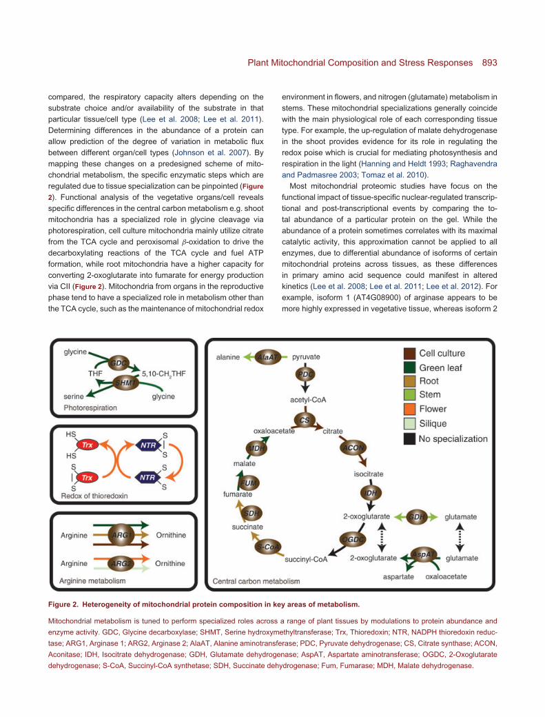
Plant Mitochondrial Composition and Stress Responses 893
compared, the respiratory capacity alters depending on the
substrate choice and/or availability of the substrate in that
particular tissue/cell type (Lee et al. 2008; Lee et al. 2011).
Determining differences in the abundance of a protein can
allow prediction of the degree of variation in metabolic flux
between different organ/cell types (Johnson et al. 2007). By
mapping these changes on a predesigned scheme of mito-
chondrial metabolism, the specific enzymatic steps which are
regulated due to tissue specialization can be pinpointed (Figure
2). Functional analysis of the vegetative organs/cell reveals
specific differences in the central carbon metabolism e.g. shoot
mitochondria has a specialized role in glycine cleavage via
photorespiration, cell culture mitochondria mainly utilize citrate
from the TCA cycle and peroxisomal β-oxidation to drive the
decarboxylating reactions of the TCA cycle and fuel ATP
formation, while root mitochondria have a higher capacity for
converting 2-oxoglutarate into fumarate for energy production
via CII (Figure 2). Mitochondria from organs in the reproductive
phase tend to have a specialized role in metabolism other than
the TCA cycle, such as the maintenance of mitochondrial redox
Figure 2. Heterogeneity of mitochondrial protein composition in key areas of metabolism.
Mitochondrial metabolism is tuned to perform specialized roles across a range of plant tissues by modulations to protein abundance and
enzyme activity. GDC, Glycine decarboxylase; SHMT, Serine hydroxymethyltransferase; Trx, Thioredoxin; NTR, NADPH thioredoxin reduc-
tase; ARG1, Arginase 1; ARG2, Arginase 2; AlaAT, Alanine aminotransferase; PDC, Pyruvate dehydrogenase; CS, Citrate synthase; ACON,
Aconitase; IDH, Isocitrate dehydrogenase; GDH, Glutamate dehydrogenase; AspAT, Aspartate aminotransferase; OGDC, 2-Oxoglutarate
dehydrogenase; S-CoA, Succinyl-CoA synthetase; SDH, Succinate dehydrogenase; Fum, Fumarase; MDH, Malate dehydrogenase.
environment in flowers, and nitrogen (glutamate) metabolism in
stems. These mitochondrial specializations generally coincide
with the main physiological role of each corresponding tissue
type. For example, the up-regulation of malate dehydrogenase
in the shoot provides evidence for its role in regulating the
redox poise which is crucial for mediating photosynthesis and
respiration in the light (Hanning and Heldt 1993; Raghavendra
and Padmasree 2003; Tomaz et al. 2010).
Most mitochondrial proteomic studies have focus on the
functional impact of tissue-specific nuclear-regulated transcrip-
tional and post-transcriptional events by comparing the to-
tal abundance of a particular protein on the gel. While the
abundance of a protein sometimes correlates with its maximal
catalytic activity, this approximation cannot be applied to all
enzymes, due to differential abundance of isoforms of certain
mitochondrial proteins across tissues, as these differences
in primary amino acid sequence could manifest in altered
kinetics (Lee et al. 2008; Lee et al. 2011; Lee et al. 2012). For
example, isoform 1 (AT4G08900) of arginase appears to be
more highly expressed in vegetative tissue, whereas isoform 2
894 Journal of Integrative Plant Biology Vol. 54 No. 11 2012
(AT4G08870) is more abundant in reproductive organs (Lee
et al. 2012). Isoform-specific differences in vegetative and
reproductive development have also been observed when
each of the four voltage-dependent anion channels (VDAC)
isoforms were disrupted (Tateda et al. 2011). Post-translational
events also play a pivotal role in regulating enzyme activity
and thus the flux of a metabolic pathway through modifications
of proteins and the assembly of enzyme complexes. Some
of the protein modifications observed on 2-D gels, especially
truncated products and pI-shifted proteins, have often been
perceived as artefacts introduced during sample preparation.
However, in a recent survey of the mitochondrial proteome
from different organ/cell types (Lee et al. 2012), many proteins
in the TCA cycle, ETC and photorespiration undergo post-
translational modifications in a tissue specific fashion that are
highly reproducible across biological replicates, suggesting that
these changes are not random. The functional implications of
specialized differences in post-translational modifications on
the contribution of mitochondrial metabolism in different tissues
remains to be explored.
Plant Mitochondrial Oxidative Stress andCellular Signalling
Environmental, biotic, abiotic and chemical stresses applied
to plants are well known to induce oxidative stress in plant
cells. These stresses alter plant metabolism, growth and de-
velopment and, at their extremes, can lead to death. Recently,
a number of studies have begun to examine the changes
that occur within plant mitochondria following the induction of
oxidative stress. The accumulation of ROS, ROS induced lipid
peroxidation, changes in metal content, changes in protein
abundance and their interactions in mitochondria following
exposure to external stress, and the role of these changes
in signaling beyond mitochondria, combine to define the impor-
tance of mitochondria as environmental sensors.
Accumulation of ROS in mitochondria
Mitochondria contain two terminal oxidases that reduce oxygen
to water and the entire ETC is known to be a significant source
of ROS under normal conditions. However, under steady state
conditions, this ROS production is dealt with by antioxidant
enzymes and small molecules to limit cellular damage. How-
ever, under some conditions, these defenses are overwhelmed
and ROS accumulate (Figure 3). Superoxide is produced
in mitochondria by peripheral single electron transfers from
reduced components in the ETC to oxygen (Moller 2001).
Classically, the ubiquinone pool and components in CI and
CIII have been implicated, however recently CII has also been
shown to produce significant superoxide (Quinlan et al. 2012).
Measurements suggest that 2–5% of oxygen consumption by
mitochondria is due to single electron superoxide formation,
while the majority of oxygen consumption occurs at the terminal
oxidases by four electron reduction of oxygen to water. The
rate of superoxide production by mitochondria depends on
the concentration of oxygen and on the redox poise of ETC
components. Therefore, ROS production by mitochondria is
low during hypoxic conditions (Noctor et al. 2007), is elevated
when respiration inhibitors block the ETC and cause over-
reduction of earlier ETC components (Maxwell et al. 1999),
and can be altered by environmental factors and chemicals
that alter the rate of these peripheral electron transfer reactions
(Moller 2001; Moller et al. 2007; Noctor et al. 2007). Notably,
nitric oxide is a potent inhibitor of the mitochondrial ETC
and its generation during plant stress may be critical in the
elevation of ROS production from mitochondria in plants (Millar
and Day 1996; Yamasaki et al. 2001; Zottini et al. 2002).
ROS have been shown to have a direct inhibitory effect on
a number of mitochondrial enzymes including components of
the ETC. Most notably H2O2 can inhibit the TCA cycle enzyme
aconitase by modification of its 4Fe-4S cluster (Verniquet et al.
1991).
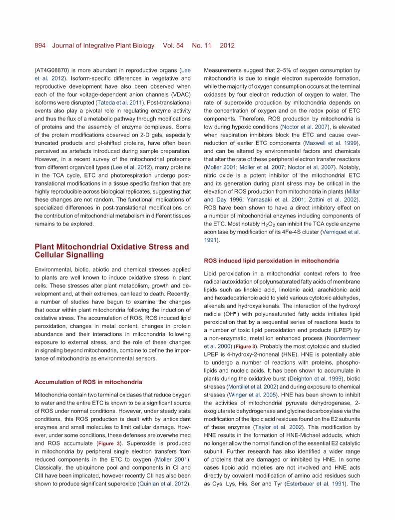
ROS induced lipid peroxidation in mitochondria
Lipid peroxidation in a mitochondrial context refers to free
radical autoxidation of polyunsaturated fatty acids of membrane
lipids such as linoleic acid, linolenic acid, arachidonic acid
and hexadecatrienoic acid to yield various cytotoxic aldehydes,
alkenals and hydroxyalkenals. The interaction of the hydroxyl
radicle (OH �) with polyunsaturated fatty acids initiates lipid
peroxidation that by a sequential series of reactions leads to
a number of toxic lipid peroxidation end products (LPEP) by
a non-enzymatic, metal ion enhanced process (Noordermeer
et al. 2000) (Figure 3). Probably the most cytotoxic and studied
LPEP is 4-hydroxy-2-nonenal (HNE). HNE is potentially able
to undergo a number of reactions with proteins, phospho-
lipids and nucleic acids. It has been shown to accumulate in
plants during the oxidative burst (Deighton et al. 1999), biotic
stresses (Montillet et al. 2002) and during exposure to chemical
stresses (Winger et al. 2005). HNE has been shown to inhibit
the activities of mitochondrial pyruvate dehydrogenase, 2-
oxoglutarate dehydrogenase and glycine decarboxylase via the
modification of the lipoic acid residues found on the E2 subunits
of these enzymes (Taylor et al. 2002). This modification by
HNE results in the formation of HNE-Michael adducts, which
no longer allow the normal function of the essential E2 catalytic
subunit. Further research has also identified a wider range
of proteins that are damaged or inhibited by HNE. In some
cases lipoic acid moieties are not involved and HNE acts
directly by covalent modification of amino acid residues such
as Cys, Lys, His, Ser and Tyr (Esterbauer et al. 1991). The
Plant Mitochondrial Composition and Stress Responses 895
Figure 3. Proposed scheme of the interaction of ROS, proteins, metals and lipid peroxidation end products in plant mitochondria
exposed to stresses.
When a plant is exposed to environmental stress this must be sensed either by a receptor on the cell wall/plasma membrane or directly
inside the cell. This is then likely signalled to the nucleus and the mitochondria. Inside the organelles an accumulation of ROS can occur
when antioxidants and antioxidant proteins (AP) are overwhelmed and this leads to the production of lipid peroxidation end products (LPEP),
damaged proteins (DP), unfolded proteins (UP) and release of transition metals (TM). The accumulated ROS can directly inhibit proteins,
while accumulated LPEP can modify proteins with lipoic acid cofactors (ML) directly amino acid (ML). Accumulated transition metals can
facilitate metal catalyzed oxidation leading to the formation or carbonyl groups (CB). The organelles may also signal the accumulation of ROS
to the nucleus. Either the external sensing of stress or organellar signalling of ROS leads to the production of new proteins by the nucleus,
including replacement proteins sHSPs, proteases and antioxidant proteins (AP). These are then involved in the refolding of unfolded proteins
(UP) or the degradation of damaged proteins (DP). AP, Antioxidant proteins; CB, Carbonyl modified amino acids; DP, Damaged proteins;
LPEP, Lipid peroxidation end-products; MA, Michael adducts formation on amino acids by LPEP; ML, Michael adducts formation on lipoic
acid by LPEP; sHSP, Small heat shock proteins; TM, Transition metals; UP, Unfolded proteins.
alternative oxidase (AOX) and NAD-malic enzyme are inhibited
by HNE (Winger et al. 2005), and it has been proposed in both
enzymes that modification of a critical cysteine residue near
the active site might be responsible (Millar and Leaver 2000;
Winger et al. 2005). The likely pathways for the detoxification
of lipid peroxidation end-products such as HNE are yet to
be elucidated in plants, but it is likely to involve glutathione-
s-transferase (GST) conjugation of modified peptides and
may also involve aldehyde dehydrogenases and/or aldose
reductases.

896 Journal of Integrative Plant Biology Vol. 54 No. 11 2012
Metallome changes during oxidative stress
Plant mitochondria contain the transition metals Fe, Cu, Zn and
Mn as well as trace levels of Co and Mo (Tan et al. 2010). The
redox cycling metals Cu and Fe tend to be concentrated to
the integral membrane proteome likely due to the abundance
of Cu- and Fe-containing ETC components and account for
approximately 75% of the mitochondrial metallome (Tan et al.
2010). Treatment of cultured cells with chemicals known to
induce oxidative stress induce a reduction of peripheral mem-
brane Fe and integral membrane Cu content, suggesting dam-
age to membrane-associated ferro-proteins and membrane-
embedded cupro-proteins (Tan et al. 2010). Significant losses
have also been seen for soluble fraction Fe, Cu and Mn
suggesting damage to metallo-matrix proteins and release of
Fe, Cu and Mn. These labile transition metals, in particular
redox active copper and iron ions, typically react with hydrogen
peroxide in Fenton type reactions to catalyze the formation
of OH �. In addition, other redox-cycling reactions within the
mitochondria exist and they are capable of eliciting metal-
catalyzed oxidation (MCO) (Figure 3). Metal-catalyzed oxidation
of proteins results in the oxidation of susceptible amino acids
such as arginine, lysine, proline and histidine (Stadtman 1993),
among a plethora of other poorly characterized consequences.
One of the major by-products of MCO of proteins is the
irreversible formation of carbonyl derivatives. These carbonyls
are highly reactive and may cause protein aggregation if the
damaged proteins are not degraded. These reactive carbonyls
are often studied as markers of oxidative stress in both plants
and animals. However, the impacts of protein carbonylation in
plants at a subcellular level remains poorly understood.
Proteome changes during oxidative stress
A number of studies have revealed global changes in protein
abundance of mitochondrial proteins following conditions that
induce oxidative stress in a wide range of plant species (Sweet-
love et al. 2002; Taylor et al. 2005; Chen et al. 2009; Taylor
et al. 2009; Jacoby et al. 2010; Huang et al. 2011; Komatsu
et al. 2011; Hossain et al. 2012; Tan et al. 2012). Recently it has
also been shown that the large respiratory subunits of the ETC
also coordinate protein changes to alter respiration in response
to oxidative stress conditions (Tan et al. 2012). In addition to
these changes are changes in proteins responding to ROS
and the damage they cause. For example the mitochondrion
is protected by a number of antioxidant enzymes that detoxify
ROS and many have been observed to vary in abundance dur-
ing oxidative stress including: Mn-superoxide dismutase (Jiang
et al. 2007), ascorbate peroxidase (Dooki et al. 2006) mon-
odehydroascorbate reductase (Sarry et al. 2006), glutathione
peroxidase (Jiang et al. 2007) and peroxiredoxins (Sweetlove
et al. 2002; Sarry et al. 2006). The importance of these organel-
lar antioxidant defense mechanisms in plant stress tolerance
has been highlighted by transgenic manipulation of the expres-
sion of these antioxidant enzymes (Allen et al. 1997). Changes
in abundance of GST proteins are detected in almost every
stress proteome study, sometimes accompanied by aldehyde
dehydrogenases (Cui et al. 2005; Ndimba et al. 2005; Sarry
et al. 2006), both of which may be involved in the detoxification
of lipid-peroxidation end-products. Many metalloproteins have
been shown to change in abundance following exposure to
stress including Mn-SOD (Jiang et al. 2007), the Fe-S center
containing CIII UCR1 (Tan et al. 2012), CI 75 kDa subunit
(Taylor et al. 2005) and the copper interacting CI subunit B16
(Tan et al. 2012). In addition to these direct protein changes,
other proteins have been observed to increase in abundance
including mitochondrial class I and mitochondrial class II small
heat shock proteins (sHsps) (Siddique et al. 2008) (Figure 3).
It is generally accepted that sHsps alleviate the deleterious
effects of stresses by preventing protein denaturation and
aggregation, as well as facilitating the correct refolding of
denatured proteins. Similarly increases in constitutive ser-
ine protease activity can be induced by oxidative stress in
mitochondria (Sweetlove et al. 2002) although the specific
Clp and FtsH serine proteases responsible for this remain
unresolved. Mitochondria also contain Lon metalloproteases
(Sarria et al. 1998; Rigas et al. 2009) and together with
the serine proteases it seems likely that these proteins are
responsible for the degradation of oxidatively damaged proteins
(Figure 3).
Regulation of downstream gene expression such asGSTs and HSPs
Due to the complexity of the interconnected ROS signalling
network that operates within plant cells (Mittler et al. 2004), it is
difficult to attribute specific cellular responses to mtROS signals
alone (Møller and Sweetlove 2011). However, by analysing the
gene expression patterns elicited by respiratory inhibitors and
the transcriptional anomalies exhibited by plants carrying muta-
tions to mitochondrial genes, it appears that the expression pat-
terns of certain gene clusters could be key indicators of mtROS
signals. Here we briefly outline how the molecular signatures
generated by mtROS signals could potentially be deduced
by analysing transcriptional upregulation within subsets of the
glutathione-s-transferases (GSTs) and heat shock proteins
(HSPs). In maize, expression patterns of gstIII and gstI genes
were similar between NCS4 (mitochondrial ribosome mutant
affecting translation) and NCS6 (CIV mutant) compared to their
respective wild type (Karpova et al. 2002). However, both gstIIIand gstI were increased in the NCS2 (CI mutant) (Karpova et al.
2002). Therefore, dysfunction of CI (NCS2), but not dysfunction
of either CIV (NCS6) or mitochondrial translation (NCS4),
induces these GSTs. Interestingly, treatment with exogenous
Plant Mitochondrial Composition and Stress Responses 897
H2O2 can also induce expression of gstIII and gstI genes in
maize leaves (Karpova et al. 2002), perhaps suggesting that
the NCS2 mutation (CI) exerts its signaling effect via H2O2
signals derived from mitochondrion, whereas the NCS4 (ribo-
some) and NCS6 (CIV) mutations communicate mitochondrial
dysfunction via a different route. These mechanisms of ROS
signaling could be conserved between species, as mutations to
CI in Arabidopsis induce accumulation of ROS (Lee et al. 2002;
Meyer et al. 2009), and higher expression levels of GSTF3
and GSTF6 transcripts (Meyer et al. 2009). The activity of the
Arabidopsis GSTF8 promoter has been defined as a marker for
defense response during the early stages of stress exposure
(Sappl et al. 2009), and recently, a forward genetics approach
that employed GSTF8 promoter activity to define mutants with
altered stress responses identified a novel CII mutant, dsr1(Gleason et al. 2011). Analysis of GSTF8 promoter activity
upon application of exogenous chemicals revealed that the
dsr1 mutant could not mediate GSTF8 promoter activity in
response to salicylic acid (SA), despite this treatment eliciting
strong GSTF8 promoter activity in a wild type background.
However, external supply of H2O2 recovered the induction
of GSTF8 promoter activity in dsr1. These results suggest
a stress signaling model whereby upstream SA signals are
converted into mtROS signals via H2O2 production at CII. This
signal is then transmitted to the nucleus to elicit downstream
responses such as induction of a certain subset of GST
transcripts.
Numerous studies have measured increased expression of
genes encoding heat shock proteins upon disruption of the ETC
via genetic mutation or treatment with respiratory inhibitors. For
example, higher abundance of HSP transcripts was observed
in Arabidopsis CI mutant lines (Meyer et al. 2009), in cultured
Arabidopsis cells treated with CI inhibitors (Garmier et al. 2008),
and in maize NCS mutants including NCS2 (CI), NCS4 (riboso-
mal translation) and NCS6 (CIV) (Kuzmin et al. 2004). Further
dissection of the triggers that can elicit higher expression of
sHSP22A suggest that its promoter activation depends upon
a decrease in the potential difference across the mitochondrial
inner membrane, independent of Ca2+ signals (Kuzmin et al.
2004). A study in C. elegans suggests that the induction of
mitochondrial heat shock proteins is regulated by the prote-
olytic cleaving of specific mitochondrial proteins to generate
messenger peptides which are exported to the nucleus, with
the divergent amino acid sequences across a range of dif-
ferent messenger peptides providing the specificity required
to elicit the subsequent induction of a specific set of genes
(Haynes et al. 2010; Moller and Sweetlove 2011). This offers a
tempting explanation for the link between sHSP22A induction
and lower potential difference across the mitochondrial inner
membrane in maize (Kuzmin et al. 2004), as the depolarization
of mitochondrial membrane potential could permit the export of
signaling peptides. It can be posited that different mitochondrial
stress signals can activate divergent pathways of mitochondrial
retrograde signaling, as within the aforementioned suite of
maize mutants, there are significant overlaps amongst HSP
gene family expression but divergent transcriptional responses
within the set of GSTs, with higher expression of GSTI and
GSTIII only being elicited by CI mutation. Molecular models
of specific mitochondrial retrograde signaling pathways are
maturing and detailed analysis of expression patterns within
and between sets of stress responsive genes can further our
understanding of the molecular signaling processes communi-
cated by mtROS. This opportunity is particularly timely as well
characterised suites of ETC mutants are now available across
a range of species.
Plant Mitochondrial Roles in HarshEnvironments
Insights into how the respiratory roles of mitochondria in cells
operate under harsh environmental conditions encountered
by intact plants have been derived by analyzing phenotypes
across a wide range of physical scales (Figure 4). Ecosystem
studies have shown that plant respiration rates were a major
factor underpinning slower rates of forest and crop productivity
across Europe during a 2003 heatwave, with prolonged heat
and drought temporarily switching certain European forests
into sources of atmospheric carbon, rather than sinks (Ciais
et al. 2005). At an individual plant level, physiological studies
have defined that respiratory rates are a key determinant of
growth reductions under a range of stresses such as extreme
temperatures, drought and salinity (Atkin et al. 2005; Atkin
and Macherel 2009; Jacoby et al. 2011). The most powerful
theoretical framework analysing these responses is the process
of carbon balance, where growth rate is positioned as the
sum total of carbon captured via photosynthesis minus carbon
expended by respiration (McCree 1986; Amthor 2000). Tissue-
level experiments have shown that some stresses, such as
temperature, can induce rapid changes to respiratory rates
of excised tissue, probably mediated through thermal effects
on the kinetics of the ETC (Kurimoto et al. 2004; Armstrong
et al. 2006), whereas exposing excised tissue to external
NaCl has little impact on respiratory rates in the short term,
probably because salinity exerts its effects on respiration
rates through alterations to substrate provision and cellular
energetic demand (Flowers 1972). Oxygen uptake rates of
isolated mitochondria exposed to harsh conditions in vitro have
defined the routes of mitochondrial electron transport that can
withstand high concentrations of toxic substances such as
NaCl, lipid peroxidation products and cadmium (Miller et al.
1973; Hamilton and Heckathorn 2001; Winger et al. 2007),
while analyses of ETC biochemistry in mitochondria isolated
from plants exposed to harsh stresses have repeatedly shown
898 Journal of Integrative Plant Biology Vol. 54 No. 11 2012
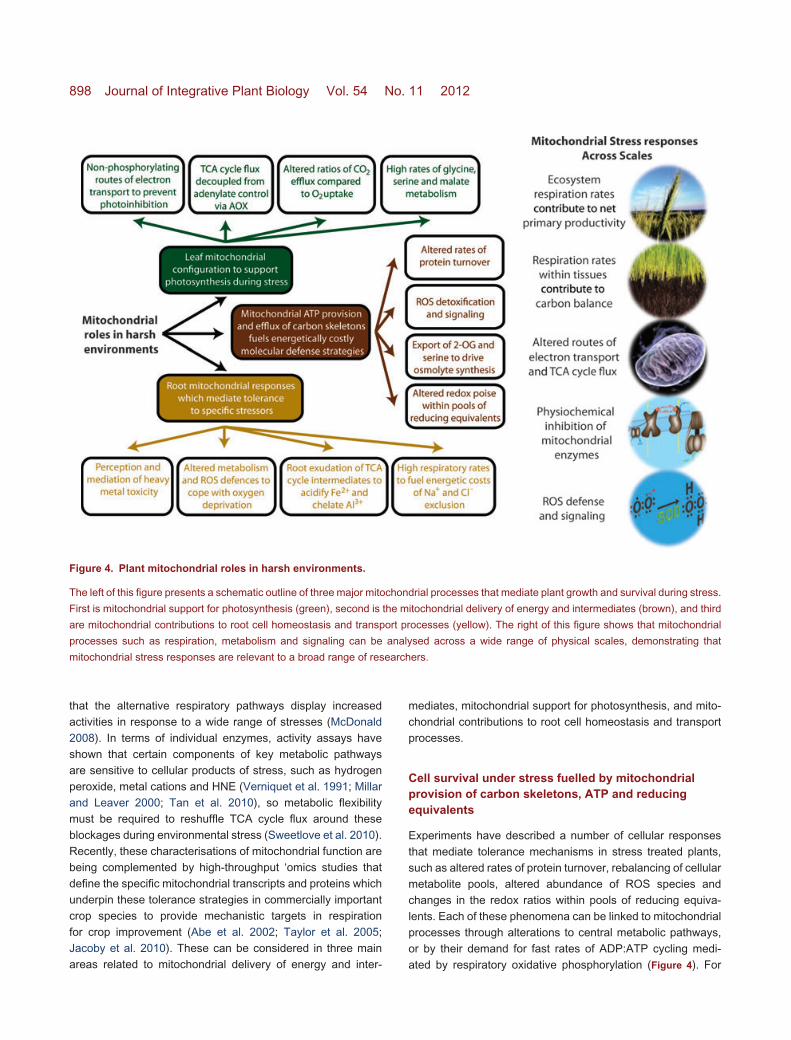
Figure 4. Plant mitochondrial roles in harsh environments.
The left of this figure presents a schematic outline of three major mitochondrial processes that mediate plant growth and survival during stress.
First is mitochondrial support for photosynthesis (green), second is the mitochondrial delivery of energy and intermediates (brown), and third
are mitochondrial contributions to root cell homeostasis and transport processes (yellow). The right of this figure shows that mitochondrial
processes such as respiration, metabolism and signaling can be analysed across a wide range of physical scales, demonstrating that
mitochondrial stress responses are relevant to a broad range of researchers.
that the alternative respiratory pathways display increased
activities in response to a wide range of stresses (McDonald
2008). In terms of individual enzymes, activity assays have
shown that certain components of key metabolic pathways
are sensitive to cellular products of stress, such as hydrogen
peroxide, metal cations and HNE (Verniquet et al. 1991; Millar
and Leaver 2000; Tan et al. 2010), so metabolic flexibility
must be required to reshuffle TCA cycle flux around these
blockages during environmental stress (Sweetlove et al. 2010).
Recently, these characterisations of mitochondrial function are
being complemented by high-throughput ‘omics studies that
define the specific mitochondrial transcripts and proteins which
underpin these tolerance strategies in commercially important
crop species to provide mechanistic targets in respiration
for crop improvement (Abe et al. 2002; Taylor et al. 2005;
Jacoby et al. 2010). These can be considered in three main
areas related to mitochondrial delivery of energy and inter-
mediates, mitochondrial support for photosynthesis, and mito-
chondrial contributions to root cell homeostasis and transport
processes.
Cell survival under stress fuelled by mitochondrialprovision of carbon skeletons, ATP and reducingequivalents
Experiments have described a number of cellular responses
that mediate tolerance mechanisms in stress treated plants,
such as altered rates of protein turnover, rebalancing of cellular
metabolite pools, altered abundance of ROS species and
changes in the redox ratios within pools of reducing equiva-
lents. Each of these phenomena can be linked to mitochondrial
processes through alterations to central metabolic pathways,
or by their demand for fast rates of ADP:ATP cycling medi-
ated by respiratory oxidative phosphorylation (Figure 4). For
Plant Mitochondrial Composition and Stress Responses 899
instance, plants display differential rates of protein turnover
relative to unstressed controls in response to a wide range
of stresses such as drought, osmotic stress and heat stress
(Dungey and Davies 1982; Zagdanska 1995; Huang et al.
2012), and it has been well established that this protein quality
control network incurs a significant energetic cost, as ATP is
hydrolysed to fuel the refolding and degradation of damaged
proteins, and also the synthesis of new replacement proteins
(Moller et al. 2007). Although the plastidic ETC produces a
large amount of cellular ATP in illuminated leaves, this is all
consumed by the process of photosynthetic carbon reduction;
therefore, mitochondrial electron transport provides the ATP
during day and night in both roots and shoots for all other
cellular operations. As a consequence, robust mitochondrial
function in all tissues is crucial to plant survival under stress
conditions. Another molecular signature of harsh environmen-
tal conditions is the accumulation of high concentrations of
particular metabolites, such as proline, glycine betaine (GB)
and GABA (Hare et al. 1998). At a molecular level, these
molecules can stabilize proteins, scavenge ROS, and serve as
alternative energy sources when classical metabolic pathways
are substrate-limited or biochemically-inhibited (Arakawa and
Timasheff 1985; Verslues and Sharp 1999; Chen and Dickman
2005). The mitochondrial role in regulation of proline and GABA
concentrations comes through the abundance and activity of
catabolic proteins such as ProDH, P5CDH and GABA-T that
are located in the mitochondrial matrix (Miller et al. 2009;
Renault et al. 2010), while a mitochondrial role in regulating
cellular concentrations of GB comes through the provision of
photorespiratory serine by the reactions of GDC and SHMT
in mitochondria (Bhuiyan et al. 2007). Further links between
mitochondrial function and cellular metabolic status come
through the provision of 2-OG via the TCA cycle, as 2-OG
is a precursor for many N-containing metabolites in plant cells,
and exposure to abiotic stress can alter the rate of TCA cycle
flux (Baxter et al. 2007; Sweetlove et al. 2010). Increased
ROS abundance is a convergence point for a wide range of
stress treatments, and stress treated plants commonly exhibit
shifts in the redox poise of ascorbate and glutathione to more
oxidized states following stress treatments (Foyer and Noctor
2011). Mitochondrial regulation of these phenomena has been
illustrated by transgenic studies showing that manipulation of
mitochondrial enzymes can alter whole-plant redox balance
and stress tolerance (Dutilleul et al. 2003; Morgan et al. 2008;
Tomaz et al. 2010).
Mitochondria support for photosynthesis duringenvironmental challenge
Photosynthetic carbon capture is a coordinated process that
requires mutual cooperation between organelles, and mito-
chondrial functions are particularly important in sustaining
photosynthesis under high light. It is now widely accepted that
fast photosynthetic rates and the avoidance of photoinhibition
are dependent upon mitochondrial function being configured
to rapidly transfer electrons from NADH into water through the
non-phosphorylating bypasses of the classical ETC (Millar et al.
2011) (Figure 4). This framework is supported by several lines of
evidence, such as measurements showing that the abundance
and activity of this particular set of mitochondrial enzymes
are induced by high light (Noguchi and Yoshida 2008), while
toxic inhibition of the mitochondrial ETC leads to decreased
photosynthetic rates (Saradadevi and Raghavendra 1992),
and compellingly evidence from knockouts of genes encoding
mitochondrial proteins that result in lower photosynthetic rates
and acute sensitivity to high light coupled to drought (Sweetlove
et al. 2006; Giraud et al. 2008). Rates of plant growth are
largely determined by the balance between photosynthesis and
respiration, with more productive, fast-growing plants generally
allocating a smaller fraction of their daily fixed carbon to
respiratory CO2 production. This reserves a larger fraction of
fixed carbon to allocate into synthesising new tissue, which
is primarily constructed by the accumulation of carbohydrates
(Poorter et al. 1990; De Block and Van Lijsebettens 2011).
However, there appears to be a limit to the productivity
increases that can be acquired through slower respiratory
rates, with canola experiments showing that plants selected for
slightly slower respiration rates displaying increased biomass
production, but plants where respiration rates had fallen below
a certain threshold displayed dramatically slower growth rates
probably because cellular energy supply cannot match baseline
demand (Hauben et al. 2009). Although this study did not
investigate a stress condition, the results identify an important
limitation in strategies that aim to enhance biomass accumula-
tion through selecting for uniformly lower respiration rates, as
slow respiration would likely be a disadvantage during transient
stressful periods which require increased rates of mitochondrial
energy production to fuel energetically costly cellular defense
processes. Respiratory elasticity is likely to be the most useful
trait, enabling slow respiration rates to promote growth during
optimal conditions, but faster respiration rates to fuel defense
during transient stress periods.
Root-specific mitochondrial processes mediatingtolerance to unfavourable soil conditions
Oxidative phosphorylation in mitochondria is the main source
of ATP in root tissue, and mitochondrial processes are also
involved in the tolerance of plants to root-specific stresses, such
as low oxygen and toxic soil conditions (Figure 4). Root tissue
is prone to dramatic fluctuations in cellular oxygen concentra-
tions, owing to the low solubility of oxygen in water coupled
with hydrological flood-drain cycles imposed by variations in
rainfall or soil drainage, in both rainfed and irrigated agricultural
900 Journal of Integrative Plant Biology Vol. 54 No. 11 2012
systems. Under low oxygen, mitochondrial metabolism shifts
away from the classical TCA cycle and ETC, towards other
mitochondrial processes such as amino acid metabolism (Millar
et al. 2004b; Taylor et al. 2010). Mitochondrial ROS defenses
such as MnSOD accumulate under anoxia, presumably in
anticipation of the forthcoming ROS burst that will occur upon
re-oxygenation (Millar et al. 2004b; Shingaki-Wells et al. 2011).
Large areas of the earth’s surface are covered by soils with
chemical properties that are sub-optimal for plant growth, due
to high concentrations of salts or heavy metals, insufficient
bioavailability of essential nutrients like iron, phosphate, sul-
phur and nitrogen, as well as unfavourably acidic or alkali
pH. The perception of heavy metal toxicity has been shown
to involve mitochondrial ROS signals (Garnier et al. 2006), and
the key role of mitochondrial ROS defenses within root cells has
been pinpointed by the dramatic root growth reductions elicited
by exposing mitochondrial peroxiredoxin mutants to high cad-
mium concentrations (Finkemeier et al. 2005). Exudation of
TCA cycle intermediates have been linked to iron absorption
from soils where neutral pHs render the iron insoluble, as these
acidic metabolites acidify the soil, thus solubilising sequestered
iron to increase bioavailability (Vigani 2012). Conversely, alu-
minium toxicity can be alleviated by exudation of citrate and
isocitrate, as these TCA cycle intermediates have chelating
properties that decrease bioavailability of the toxic Al3+ ion
(Ma et al. 2001; Fujii et al. 2012). Sodium exclusion is a
well defined mechanism of plant NaCl tolerance, and strong
links between the degree of sodium exclusion and the rate
of root respiration have been demonstrated by the collapse
of sodium exclusion following anoxia treatment of roots (Drew
and Lauchli 1985). Radioisotope tracer studies have shown that
rates of root respiration in saline media are positively correlated
to sodium exclusion capacity between rice varieties (Malagoli
et al. 2008), further illustrating this relationship.
Future Perspectives
As the powerhouses of eukaryotic cells, mitochondria rep-
resent an ancient but flexible factory in cells that enable cell
function, growth and division through energy metabolism.
While our knowledge of how plant mitochondria work is
rapidly increasing, much is still being extrapolated from
yeast and mammalian systems without direct evidence in
plants. Detailed insights into the assembly of mitochon-
drial machinery, the signalling by mitochondrial of oxidative
stress and the regulation of respiratory rate are still needed
in order to maximise respiration for plant protection in harsh
environments and to minimize respiratory losses to enhance
plant yields.
Acknowledgements
This work was supported by the Australian Research Council(ARC) ARC Centre of Excellence for Plant Energy Biology(CE0561495). RPJ is supported by a Grains Research andDevelopment Corporation (GRDC) PhD scholarship, LL wasfunded by Scholarship International Research Fees (SIRF),University International Stipend (UIS) and a Top Up Scholarshipfor UIS. AHM is supported by the Australian Research Council(ARC) as an ARC Future Fellow.
Received 30 Jul. 2012 Accepted 2 Oct. 2012
References
Abe F, Saito K, Miura K, Toriyama K (2002) A single nucleotide
polymorphism in the alternative oxidase gene among rice varieties
differing in low temperature tolerance. FEBS Lett. 527, 181–185.
Allen RD, Webb RP, Schake SA (1997) Use of transgenic plants to
study antioxidant defenses. Free Radic. Biol. Med. 23, 473–479.
Amthor JS (2000) The McCree-de Wit-Penning de Vries-Thornley
respiration paradigms: 30 years later. Ann. Bot. 86, 1–20.
Arakawa T, Timasheff SN (1985) The stabilization of proteins by
osmolytes. Biophys. J. 47, 411–414.
Araujo WL, Nunes-Nesi A, Osorio S, Usadel B, Fuentes D, Nagy R,
Balbo I, Lehmann M, Studart-Witkowski C, Tohge T, Martinoia
E, Jordana X, DaMatta FM, Fernie AR (2011) Antisense inhibition
of the iron-sulphur subunit of succinate dehydrogenase enhances
photosynthesis and growth in tomato via an organic acid-mediated
effect on stomatal aperture. Plant Cell 23, 600–627.
Armstrong AF, Logan DC, Tobin AK, O’Toole P, Atkin OK (2006)
Heterogeneity of plant mitochondrial responses underpinning respi-
ratory acclimation to the cold in Arabidopsis thaliana leaves. Plant
Cell Environ. 29, 940–949.
Atkin OK, Bruhn D, Hurry VM, Tjoelker MG (2005) The hot and
the cold: Unravelling the variable response of plant respiration to
temperature. Funct. Plant Biol. 32, 87–105.
Atkin OK, Macherel D (2009) The crucial role of plant mitochondria in
orchestrating drought tolerance. Ann. Bot. 103, 581–597.
Attallah CV, Welchen E, Pujol C, Bonnard G, Gonzalez DH (2007)
Characterization of Arabidopsis thaliana genes encoding functional
homologues of the yeast metal chaperone Cox19p, involved in
cytochrome c oxidase biogenesis. Plant Mol. Biol. 65, 343–355.
Barrientos A, Gouget K, Horn D, Soto IC, Fontanesi F (2009)
Suppression mechanisms of COX assembly defects in yeast and
human: Insights into the COX assembly process. Biochim. Biophys.
Acta 1793, 97–107.
Baxter CJ, Redestig H, Schauer N, Repsilber D, Patil KR, Nielsen
J, Selbig J, Liu J, Fernie AR, Sweetlove LJ (2007) The metabolic
response of heterotrophic Arabidopsis cells to oxidative stress. Plant
Physiol. 143, 312–325.
Plant Mitochondrial Composition and Stress Responses 901
Bhuiyan NH, Hamada A, Yamada N, Rai V, Hibino T, Takabe T
(2007) Regulation of betaine synthesis by precursor supply and
choline monooxygenase expression in Amaranthus tricolor . J. Exp.
Bot. 58, 4203–4212.
Cardol P (2011) Mitochondrial NADH:ubiquinone oxidoreductase
(complex I) in eukaryotes: A highly conserved subunit composition
highlighted by mining of protein databases. Biochim. Biophys. Acta
1807, 1390–1397.
Carrie C, Murcha MW, Kuehn K, Duncan O, Barthet M, Smith
PM, Eubel H, Meyer E, Day DA, Millar AH, Whelan J (2008)
Type II NAD(P)H dehydrogenases are targeted to mitochondria and
chloroplasts or peroxisomes in Arabidopsis thaliana. FEBS Lett.
582, 3073–3079.
Catoni E, Desimone M, Hilpert M, Wipf D, Kunze R, Schneider A,
Flugge UI, Schumacher K, Frommer WB (2003a) Expression
pattern of a nuclear encoded mitochondrial arginine- ornithine
translocator gene from Arabidopsis. BMC Plant Biol. 3, 1.
Catoni E, Schwab R, Hilpert M, Desimone M, Schwacke R, Flugge
UI, Schumacher K, Frommer WB (2003b) Identification of an
Arabidopsis mitochondrial succinate-fumarate translocator. FEBS
Lett. 534, 87–92.
Chen CB, Dickman MB (2005) Proline suppresses apoptosis in the
fungal pathogen Colletotrichum trifolii. Proc. Natl. Acad. Sci. USA
102, 3459–3464.
Chen X, Wang Y, Li J, Jiang A, Cheng Y, Zhang W (2009) Mitochon-
drial proteome during salt stress-induced programmed cell death in
rice. Plant Physiol. Biochem. 47, 407–415.
Ciais P, Reichstein M, Viovy N, Granier A, Ogee J, Allard V, Aubinet
M, Buchmann N, Bernhofer C, Carrara A, Chevallier F, De
Noblet N, Friend AD, Friedlingstein P, Grunwald T, Heinesch B,
Keronen P, Knohl A, Krinner G, Loustau D, Manca G, Matteucci
G, Miglietta F, Ourcival JM, Papale D, Pilegaard K, Rambal
S, Seufert G, Soussana JF, Sanz MJ, Schulze ED, Vesala T,
Valentini R (2005) Europe-wide reduction in primary productivity
caused by the heat and drought in 2003. Nature 437, 529–533.
Considine MJ, Goodman M, Echtay KS, Laloi M, Whelan J, Brand
MD, Sweetlove LJ (2003) Superoxide stimulates a proton leak
in potato mitochondria that is related to the activity of uncoupling
protein. J. Biol. Chem. 278, 22298–22302.
Cui S, Huang F, Wang J, Ma X, Cheng Y, Liu J (2005) A proteomic
analysis of cold stress response in rice seedlings. Proteomics 5,
3162–3172.
De Block M, Van Lijsebettens M (2011) Energy efficiency and energy
homeostasis as genetic and epigenetic components of plant perfor-
mance and crop productivity. Curr. Opin. Plant Biol. 14, 275–282.
de Longevialle AoF, Meyer EH, Andres C, Taylor NL, Lurin C, Millar
AH, Small ID (2007) The pentatricopeptide repeat gene OTP43
is required for trans-splicing of the mitochondrial nad1 Intron 1 in
Arabidopsis thaliana. Plant Cell 19, 3256–3265.
Deighton N, Muckenschnabel I, Goodman BA, Williamson B (1999)
Lipid peroxidation and the oxidative burst associated with infection
of Capsicum annuum by Botrytis cinerea. Plant J. 20, 485–492.
Diaz F, Kotarsky H, Fellman V, Moraes CT (2011) Mitochondrial dis-
orders caused by mutations in respiratory chain assembly factors.
Sem. Fetal Neonatal. Med. 16, 197–204.
Dooki AD, Mayer-Posner FJ, Askari H, Zaiee AA, Salekdeh GH
(2006) Proteomic responses of rice young panicles to salinity.
Proteomics 6, 6498–6507.
Drew MC, Lauchli A (1985) Oxygen dependent exclusion of sodium
ions from shoots by roots of Zea mays (cv Pioneer 3906) in relation
to salinity damage. Plant Physiol. 79, 171–176.
Dungey NO, Davies DD (1982) Protein turnover in isolated barley
leaf segments and the effects of stress. J. Exp. Bot. 33, 12–
20.
Dutilleul C, Garmier M, Noctor G, Mathieu C, Chetrit P, Foyer
CH, de Paepe R (2003) Leaf mitochondria modulate whole cell
redox homeostasis, set antioxidant capacity, and determine stress
resistance through altered signaling and diurnal regulation. Plant
Cell 15, 1212–1226.
Esterbauer H, Schaur RJ, Zollner H (1991) Chemistry and biochem-
istry of 4-hydroxynonenal, malonaldehyde and related aldehydes.
Free Rad. Biol. Med. 11, 81–128.
Eubel H, Jansch L, Braun HP (2003) New insights into the respiratory
chain of plant mitochondria. Supercomplexes and a unique compo-
sition of complex II. Plant Physiol. 133, 274–286.
Finkemeier I, Goodman M, Lamkemeyer P, Kandlbinder A, Sweet-
love LJ, Dietz KJ (2005) The mitochondrial type II peroxiredoxin F
is essential for redox homeostasis and root growth of Arabidopsis
thaliana under stress. J. Biol. Chem. 280, 12168–12180.
Finnegan P, Soole KL, Umbach AL (2004) Alternative mitochondrial
electron transport proteins in higher plants. In: Day DA, Millar AH,
Whelan J, eds. Plant Mitochondria: From Genome to Function –
Advances in Photosynthesis and Respiration. Springer. pp. 163–
230.
Flowers TJ (1972) Salt tolerance in Suaeda maritima – Effect of
sodium chloride on growth, respiration, and soluble enzymes in a
comparative study with Pisum sativum. J. Exp. Bot. 23, 310.
Foyer CH, Noctor G (2011) Ascorbate and glutathione: The heart of
the redox hub. Plant Physiol. 155, 2–18.
Fuentes D, Meneses M, Nunes-Nesi A, Araujo W, Tapia R, Gomez
I, Holuigue L, Gutierrez RA, Fernie AR, Jordana X (2011) A
deficiency in the flavoprotein of Arabidopsis mitochondrial complex
II results in elevated photosynthesis and better growth in nitrogen-
limiting conditions. Plant Physiol. 10.1104/pp.1111.183939.
Fujii M, Yokosho K, Yamaji N, Saisho D, Yamane M, Takahashi
H, Sato K, Nakazono M, Ma JF (2012) Acquisition of aluminium
tolerance by modification of a single gene in barley. Nat. Commun.
3, 713.
Garmier M, Carroll AJ, Delannoy E, Vallet C, Day DA, Small ID,
Millar AH (2008) Complex I dysfunction redirects cellular and
mitochondrial metabolism in Arabidopsis. Plant Physiol. 148, 1324–
1341.
Garnier L, Simon-Plas F, Thuleau P, Agnel JP, Blein JP, Ranjeva
R, Montillet JL (2006) Cadmium affects tobacco cells by a series
902 Journal of Integrative Plant Biology Vol. 54 No. 11 2012
of three waves of reactive oxygen species that contribute to cyto-
toxicity. Plant Cell Environ. 29, 1956–1969.
Ghezzi D, Goffrini P, Uziel G, Horvath R, Klopstock T, Lochmuller
H, D’Adamo P, Gasparini P, Strom TM, Prokisch H, Invernizzi
F, Ferrero I, Zeviani M (2009) SDHAF1, encoding a LYR complex-
II specific assembly factor, is mutated in SDH-defective infantile
leukoencephalopathy. Nature Genet. 41, 654–656.
Giraud E, Ho LHM, Clifton R, Carroll A, Estavillo G, Tan YF, Howell
KA, Ivanova A, Pogson BJ, Millar AH, Whelan J (2008) The
absence of alternative oxidase1a in Arabidopsis results in acute
sensitivity to combined light and drought stress. Plant Physiol. 147,
595–610.
Gleason C, Huang S, Thatcher LF, Foley RC, Anderson CR, Carroll
AJ, Millar AH, Singh KB (2011) Mitochondrial complex II has a
key role in mitochondrial-derived reactive oxygen species influence
on plant stress gene regulation and defense. Proc. Natl. Acad. Sci.
USA 108, 10768–10773.
Gonzalez DH, Welchen E, Attallah CV, Comelli RN, Mufarrege
EF (2007) Transcriptional coordination of the biogenesis of the
oxidative phosphorylation machinery in plants. Plant J. 51, 105–
116.
Guan Y, Rawsthorne S, Scofield G, Shaw P, Doonan J (1995)
Cloning and characterization of a dihydrolipoamide acetyltrans-
ferase (E2) subunit of the pyruvate dehydrogenase complex from
Arabidopsis thaliana. J. Biol. Chem. 270, 5412–5417.
Hamasur B, Glaser E (1992) Plant mitochondrial F0F1 ATP synthase.
Identification of the individual subunits and properties of the purified
spinach leaf mitochondrial ATP synthase. Eur. J. Biochem. 205,
409–416.
Hamel P, Saint-Georges Y, de Pinto B, Lachacinski N, Altamura
N, Dujardin G (2004) Redundancy in the function of mitochondrial
phosphate transport in Saccharomyces cerevisiae and Arabidopsis
thaliana. Mol. Microbiol. 51, 307–317.
Hamilton EW, Heckathorn SA (2001) Mitochondrial adaptations to
NaCl. Complex I is protected by anti-oxidants and small heat shock
proteins, whereas complex II is protected by proline and betaine.
Plant Physiol. 126, 1266–1274.
Hanning I, Heldt HW (1993) On the function of mitochondrial
metabolism during photosynthesis in spinach (Spinacia oleracea
L.) leaves (Partitioning between respiration and export of redox
equivalents and precursors for nitrate assimilation products). Plant
Physiol. 103, 1147–1154.
Hao HX, Khalimonchuk O, Schraders M, Dephoure N, Bayley JP,
Kunst H, Devilee P, Cremers CW, Schiffman JD, Bentz BG,
Gygi SP, Winge DR, Kremer H, Rutter J (2009) SDH5, a gene
required for flavination of succinate dehydrogenase, is mutated in
paraganglioma. Science 325, 1139–1142.
Hare PD, Cress WA, Van Staden J (1998) Dissecting the roles of
osmolyte accumulation during stress. Plant Cell Environ. 21, 535–
553.
Hauben M, Haesendonckx B, Standaert E, Van Der Kelen K, Azmi
A, Akpo H, Van Breusegem F, Guisez Y, Bots M, Lambert B,
Laga B, De Block M (2009) Energy use efficiency is characterized
by an epigenetic component that can be directed through artificial
selection to increase yield. Proc. Natl. Acad. Sci. USA 106, 20109–
20114.
Haynes CM, Yang Y, Blais SP, Neubert TA, Ron D (2010) The matrix
peptide exporter HAF-1 signals a mitochondrial UPR by activating
the transcription factor ZC376.7 in C. elegans. Mol. Cell 37, 529–
540.
Heazlewood JL, Howell KA, Millar AH (2003a) Mitochondrial complex
I from Arabidopsis and rice: Orthologs of mammalian and fungal
components coupled with plant-specific subunits. Biochim. Biophys.
Acta 1604, 159–169.
Heazlewood JL, Howell KA, Whelan J, Millar AH (2003b) Towards
an analysis of the rice mitochondrial proteome. Plant Physiol. 132,
230–242.
Heazlewood JL, Whelan J, Millar AH (2003c) The products of the
mitochondrial orf25 and orfB genes are FO components in the plant
F1FO ATP synthase. FEBS Lett. 540, 201–205.
Hossain Z, Nouri MZ, Komatsu S (2012) Plant cell organelle pro-
teomics in response to abiotic stress. J. Proteome Res. 11, 37–48.
Hoyos ME, Palmieri L, Wertin T, Arrigoni R, Polacco JC, Palmieri
F (2003) Identification of a mitochondrial transporter for basic
amino acids in Arabidopsis thaliana by functional reconstitution into
liposomes and complementation in yeast. Plant J. 33, 1027–1035.
Huang BR, Rachmilevitch S, Xu JC (2012) Root carbon and protein
metabolism associated with heat tolerance. J. Exp. Bot. 63, 3455–
3465.
Huang S, Millar AH, Taylor NL (2011) The plant mitochondrial pro-
teome composition and stress response: Conservation and diver-
gence between monocots and dicots. In: Kempken F, ed. Advances
in Plant Biology. Springer, New York. pp. 533.
Huang S, Taylor NL, Stroher E, Fenske R, Millar AH (2012) Succinate
dehydrogenase assembly factor 2 is needed for assembly and
activity of mitochondrial complex II and for normal root elongation
in Arabidopsis. Plant J. (in press).
Jacoby RP, Millar AH, Taylor NL (2010) Wheat mitochondrial pro-
teomes provide new links between antioxidant defense and plant
salinity tolerance. J. Prot. Res. 9, 6595–6604.
Jacoby RP, Taylor NL, Millar AH (2011) The role of mitochondrial
respiration in salinity tolerance. Trends Plant Sci. 16, 614–623.
Jansch L, Kruft V, Schmitz UK, Braun HP (1996) New insights into
the composition, molecular mass and stoichiometry of the protein
complexes of plant mitochondria. Plant J. 9, 357–368.
Jiang Y, Yang B, Harris NS, Deyholos MK (2007) Comparative pro-
teomic analysis of NaCl stress-responsive proteins in Arabidopsis
roots. J. Exp. Bot. 58, 3591–3607.
Johnson DT, Harris RA, French S, Blair PV, You J, Bemis KG, Wang
M, Balaban RS (2007) Tissue heterogeneity of the mammalian
mitochondrial proteome. Am. J. Physiol. 292, C689–C697.
Karpova OV, Kuzmin EV, Elthon TE, Newton KJ (2002) Differential
expression of alternative oxidase genes in maize mitochondrial
mutants. Plant Cell 14, 3271–3284.
Plant Mitochondrial Composition and Stress Responses 903
Klodmann J, Sunderhaus S, Nimtz M, Jansch L, Braun HP (2010)
Internal architecture of mitochondrial complex I from Arabidopsis
thaliana. Plant Cell 22, 797–810.
Komatsu S, Yamamoto A, Nakamura T, Nouri MZ, Nanjo Y,
Nishizawa K, Furukawa K (2011) Comprehensive analysis of
mitochondria in roots and hypocotyls of soybean under flooding
stress using proteomics and metabolomics techniques. J. Prot. Res.
10, 3993–4004.
Koyama H, Kawamura A, Kihara T, Hara T, Takita E, Shibata D
(2000) Overexpression of mitochondrial citrate synthase in Ara-
bidopsis thaliana improved growth on a phosphorus-limited soil.
Plant Cell Physiol. 41, 1030–1037.
Kuffner R, Rohr A, Schmiede A, Krull C, Schulte U (1998) Involve-
ment of two novel chaperones in the assembly of mitochondrial
NADH:Ubiquinone oxidoreductase (complex I). J. Mol. Biol. 283,
409–417.
Kurimoto K, Day DA, Lambers H, Noguchi K (2004) Effect of
respiratory homeostasis on plant growth in cultivars of wheat and
rice. Plant Cell Environ. 27, 853–862.
Kuzmin EV, Karpova OV, Elthon TE, Newton KJ (2004) Mitochondrial
respiratory deficiencies signal pu-regulation of genes for heat shock
proteins. J. Biol. Chem. 279, 20672–20677.
La Cognata U, Landschutze V, Willmitzer L, Muller-Rober B (1996)
Structure and expression of mitochondrial citrate synthases from
higher plants. Plant Cell Physiol. 37, 1022–1029.
Laloi M (1999) Plant mitochondrial carriers: An overview. Cell Mol. Life
Sci. 56, 918–944.
Lazarou M, McKenzie M, Ohtake A, Thorburn DR, Ryan MT (2007)
Analysis of the assembly profiles for mitochondrial- and nuclear-
DNA-encoded subunits into complex I. Mol. Cell Biol. 27, 4228–
4237.
Lee BH, Lee H, Xiong L, Zhu JK (2002) A mitochondrial complex I
defect impairs cold-regulated nuclear gene expression. Plant Cell
14, 1235–1251.
Lee CP, Eubel H, O’Toole N, Millar A (2011) Combining proteomics
of root and shoot mitochondria and transcript analysis to define
constitutive and variable components in plant mitochondria. Phyto-
chemistry 72, 1092–1108.
Lee CP, Eubel H, O’Toole N, Millar AH (2008) Heterogeneity of the
mitochondrial proteome for photosynthetic and non-photosynthetic
Arabidopsis metabolism. Mol. Cell Proteomics 7, 1297–1316.
Lee CP, Eubel H, Solheim C, Millar AH (2012) Mitochondrial proteome
heterogeneity between tissues from the vegetative and reproductive
stages of Arabidopsis thaliana development. J. Prot. Res. 11, 3326–
3343.
Leon G, Holuigue L, Jordana X (2007) Mitochondrial complex II
Is essential for gametophyte development in Arabidopsis. Plant
Physiol. 143, 1534–1546.
Li L, Carrie C, Nelson C, Whelan J, Millar AH (2012) Accumulation of
newly synthesised F1 in vivo in Arabidopsis mitochondria provides
evidence for modular assembly of the plant F1Fo ATP synthase. J.
Biol. Chem. 287, 25749–25757.
Luethy MH, Miernyk JA, Randall DD (1995) The mitochondrial pyru-
vate dehydrogenase complex: Nucleotide and deduced amino-acid
sequences of a cDNA encoding the Arabidopsis thaliana E1 alpha-
subunit. Gene 164, 251–254.
Ma JF, Ryan PR, Delhaize E (2001) Aluminium tolerance in plants and
the complexing role of organic acids. Trends Plant Sci. 6, 273–278.
Malagoli P, Britto DT, Schulze LM, Kronzucker HJ (2008) Futile
Na+ cycling at the root plasma membrane in rice (Oryza sativa L.):
Kinetics, energetics, and relationship to salinity tolerance. J. Exp.
Bot. 59, 4109–4117.
Mannella CA (1992) The ‘ins’ and ‘outs’ of mitochondrial membrane
channels. Trends Biochem. Sci. 17, 315–320.
Mannella CA, Pfeiffer DR, Bradshaw PC, Moraru II, Slepchenko
B, Loew LM, Hsieh CE, Buttle K, Marko M (2001) Topology
of the mitochondrial inner membrane: Dynamics and bioenergetic
implications. IUBMB Life 52, 93–100.
Mannella CA, Tedeschi H (1987) Importance of the mitochondrial outer
membrane channel as a model biological channel. J. Bioenerg.
Biomembr. 19, 305–308.
Maxwell DP, Wang Y, McIntosh L (1999) The alternative oxidase
lowers mitochondrial reactive oxygen production in plant cells. Proc.
Natl. Acad. Sci. USA 96, 8271–8276.
McCree KJ (1986) Whole-plant carbon balance during osmotic ad-
justment to drought and salinity stress. Aust. J. Plant Physiol. 13,
33–43.
McDonald AE (2008) Alternative oxidase: An inter-kingdom perspec-
tive on the function and regulation of this broadly distributed
‘cyanide-resistant’ terminal oxidase. Funct. Plant Biol. 35, 535–
552.
Meyer EH, Solheim C, Tanz SK, Bonnard G, Millar AH (2011) Insights
into the composition and assembly of the membrane arm of plant
complex I through analysis of subcomplexes in Arabidopsis mutant
lines. J. Biol. Chem. 286, 26081–26092.
Meyer EH, Taylor NL, Millar AH (2008) Resolving and identifying
protein components of plant mitochondrial respiratory complexes
using three dimensions of gel electrophoresis. J. Prot. Res. 7, 786–
794.
Meyer EH, Tomaz T, Carroll AJ, Estavillo G, Delannoy E, Tanz SK,
Small ID, Pogson BJ, Millar AH (2009) Remodeled respiration in
ndufs4 with low phosphorylation efficiency suppresses Arabidopsis
germination and growth and alters control of metabolism at night.
Plant Physiol. 151, 603–619.
Michalecka AM, Svensson AS, Johansson FI, Agius SC, Johanson
U, Brennicke A, Binder S, Rasmusson AG (2003) Arabidopsis
genes encoding mitochondrial type II NAD(P)H dehydrogenases
have different evolutionary origin and show distinct responses to
light. Plant Physiol. 133, 642–652.
Millar AH, Day DA (1996) Nitric oxide inhibits the cytochrome oxidase
but not the alternative oxidase of plant mitochondria. FEBS Lett.
398, 155–158.
Millar AH, Eubel H, Jansch L, Kruft V, Heazlewood JL, Braun
HP (2004a) Mitochondrial cytochrome c oxidase and succinate
904 Journal of Integrative Plant Biology Vol. 54 No. 11 2012
dehydrogenase complexes contain plant specific subunits. Plant
Mol. Biol. 56, 77–90.
Millar AH, Leaver CJ (2000) The cytotoxic lipid peroxidation product,
4-hydroxy-2-nonenal, specifically inhibits decarboxylating dehydro-
genases in the matrix of plant mitochondria. FEBS Lett. 481, 117–
121.
Millar AH, Trend AE, Heazlewood JL (2004b) Changes in the mi-
tochondrial proteome during the anoxia to air transition in rice
focus around cytochrome-containing respiratory complexes. J. Biol.
Chem. 279, 39471–39478.
Millar AH, Whelan J, Soole KL, Day DA (2011) Organization and
regulation of mitochondrial respiration in plants. Ann. Rev. Plant
Biol. 62, 79–104.
Miller G, Honig A, Stein H, Suzuki N, Mittler R, Zilberstein A (2009)
Unraveling delta(1)-pyrroline-5-carboxylate-proline cycle in plants
by uncoupled expression of proline oxidation enzymes. J. Biol.
Chem. 284, 26482–26492.
Miller RJ, Bittell JE, Koeppe DE (1973) Effect of cadmium on elec-
tron transport and energy transfer reactions in corn mitochondria.
Physiol. Plant. 28, 166–171.
Mimaki M, Wang X, McKenzie M, Thorburn DR, Ryan MT (2012)
Understanding mitochondrial complex I assembly in health and
disease. Biochim. Biophys. Acta 1817, 851–862.
Mittler R, Vanderauwera S, Gollery M, Van Breusegem F (2004)
Reactive oxygen gene network of plants. Trends Plant Sci. 9, 490–
498.
Moller IM (2001) Plant mitochondria and oxidative stress: Electron
transport, NADPH turnover, and metabolism of reactive oxygen
species. Ann. Rev. Plant Physiol. Plant Mol. Biol. 52, 561–
591.
Moller IM, Jensen PE, Hansson A (2007) Oxidative modifications to
cellular components in plants. Ann. Rev. Plant Biol. 58, 459–481.
Møller IM, Sweetlove LJ (2011) ROS signalling – specificity is required.
Trends Plant Sci. 15, 370–374.
Montillet J-L, Agnel J-P, Ponchet M, Vailleau F, Roby D, Trianta-
phylides C (2002) Lipoxygenase-mediated production of fatty acid
hydroperoxides is a specific signature of the hypersensitive reaction
in plants. Plant Physiol. Biochem. 40, 633–639.
Moore CS, Cook-Johnson R, Rudhe C, Whelan J, Day DA, Wiskich
JT, Soole KL (2003) Identification of AtNDI1, an internal non-
phosphorylating NAD(P)H dehydrogenase in Arabidopsis thaliana
mitochondria. Plant Physiol. 133, 1968–1978.
Morgan MJ, Lehmann M, Schwarzlander M, Baxter CJ, Sienkiewicz-
Porzucek A, Williams TCR, Schauer N, Fernie AR, Fricker MD,
Ratcliffe RG, Sweetlove LJ, Finkemeier I (2008) Decrease in
manganese superoxide dismutase leads to reduced root growth
and affects tricarboxylic acid cycle flux and mitochondrial redox
homeostasis. Plant Physiol. 147, 101–114.
Ndimba BK, Chivasa S, Simon WJ, Slabas AR (2005) Identification of
Arabidopsis salt and osmotic stress responsive proteins using two-
dimensional difference gel electrophoresis and mass spectrometry.
Proteomics 5, 4185–4196.
Noctor G, De Paepe R, Foyer CH (2007) Mitochondrial redox biology
and homeostasis in plants. Trends Plant Sci. 12, 125–134.
Noguchi K, Yoshida K (2008) Interaction between photosynthesis and
respiration in illuminated leaves. Mitochondrion 8, 87–99.
Noordermeer MA, Feussner I, Kolbe A, Veldink GA, Vliegen-
thart JFG (2000) Oxygenation of (3Z)-alkenals to 4-hydroxy-(2E)-
alkenals in plant extracts: A nonenzymatic process. Biochem.
Biophys. Res. Comm. 277, 112–116.
Nouws J, Nijtmans LG, Smeitink JA, Vogel RO (2012) Assembly
factors as a new class of disease genes for mitochondrial complex
I deficiency: Cause, pathology and treatment options. Brain 135,
12–22.
Nunes-Nesi A, Carrari F, Gibon Y, Sulpice R, Lytovchenko A,
Fisahn J, Graham J, Ratcliffe RG, Sweetlove LJ, Fernie AR
(2007) Deficiency of mitochondrial fumarase activity in tomato plants
impairs photosynthesis via an effect on stomatal function. Plant J.
50, 1093–1106.
Nunes-Nesi A, Carrari F, Lytovchenko A, Smith AM, Loureiro ME,
Ratcliffe RG, Sweetlove LJ, Fernie AR (2005) Enhanced photo-
synthetic performance and growth as a consequence of decreasing
mitochondrial malate dehydrogenase activity in transgenic tomato
plants. Plant Physiol. 137, 611–622.
Osman C, Wilmes C, Tatsuta T, Langer T (2007) Prohibitins interact
genetically with Atp23, a novel processing peptidase and chaperone
for the F1Fo-ATP synthase. Mol. Biol. Cell 18, 627–635.
Palmieri F, Rieder B, Ventrella A, Blanco E, Do PT, Nunes-Nesi
A, Trauth AU, Fiermonte G, Tjaden J, Agrimi G, Kirchberger
S, Paradies E, Fernie AR, Neuhaus HE (2009) Molecular iden-
tification and functional characterization of Arabidopsis thaliana
mitochondrial and chloroplastic NAD+ carrier proteins. J. Biol.
Chem. 284, 31249–31259.
Palmieri L, Santoro A, Carrari F, Blanco E, Nunes-Nesi A, Arrigoni
R, Genchi F, Fernie AR, Palmieri F (2008) Identification and char-
acterization of ADNT1, a novel mitochondrial adenine nucleotide
transporter from Arabidopsis. Plant Physiol. 148, 1797–1808.
Peiffer WE, Ingle RT, Ferguson-Miller S (1990) Structurally unique
plant cytochrome c oxidase isolated from wheat germ, a rich
source of plant mitochondrial enzymes. Biochemistry 29, 8696–
8701.
Picault N, Palmieri L, Pisano I, Hodges M, Palmieri F (2002) Identifi-
cation of a novel transporter for dicarboxylates and tricarboxylates in
plant mitochondria. Bacterial expression, reconstitution, functional
characterization, and tissue distribution. J. Biol. Chem. 277, 24204–
24211.
Pickova A, Potocky M, Houstek J (2005) Assembly factors of F1FO-
ATP synthase across genomes. Proteins 59, 393–402.
Pineau B, Layoune O, Danon A, De Paepe R (2008) L-galactono-1,4-
lactone dehydrogenase is required for the accumulation of plant
respiratory complex I. J. Biol. Chem. 283, 32500–32505.
Poorter H, Remkes C, Lambers H (1990) Carbon and nitrogen
economy of 24 wild species differing in relative growth rate. Plant
Physiol. 94, 621–627.
Plant Mitochondrial Composition and Stress Responses 905
Quinlan CL, Orr AL, Perevoshchikova IV, Treberg JR, Ackrell BA,
Brand MD (2012) Mitochondrial complex II can generate reactive
oxygen species at high rates in both the forward and reverse
reactions. J. Biol. Chem. 10.1074/jbc.M1112.374629.
Raghavendra AS, Padmasree K (2003) Beneficial interactions of
mitochondrial metabolism with photosynthetic carbon assimilation.
Trends Plant Sci. 8, 546–553.
Rak M, Gokova S, Tzagoloff A (2011) Modular assembly of yeast
mitochondrial ATP synthase. EMBO J. 30, 920–930.
Rak M, Tzagoloff A (2009) F1-dependent translation of mitochondrially
encoded Atp6p and Atp8p subunits of yeast ATP synthase. Proc.
Natl. Acad. Sci. USA 106, 18509–18514.
Rasmusson AG, Soole KL, Elthon TE (2004) Alternative NAD(P)H
dehydrogenases of plant mitochondria. Ann. Rev. Plant Biol. 55,
23–39.
Renault H, Roussel V, El Amrani A, Arzel M, Renault D, Bouchereau
A, Deleu C (2010) The Arabidopsis pop2–1 mutant reveals the
involvement of GABA transaminase in salt stress tolerance. BMC
Plant Biol. 10, 20.
Rigas S, Daras G, Laxa M, Marathias N, Fasseas C, Sweetlove LJ,
Hatzopoulos P (2009) Role of Lon1 protease in post-germinative
growth and maintenance of mitochondrial function in Arabidopsis
thaliana. New Phytol. 181, 588–600.
Robert N, d’Erfurth I, Marmagne A, Erhardt M, Allot M, Boivin K,
Gissot L, Monachello D, Michaud M, Duchene AM, Barbier-
Brygoo H, Marechal-Drouard L, Ephritikhine G, Filleur S (2012)
Voltage-dependent-anion-channels (VDACs) in Arabidopsis have a
dual localization in the cell but show a distinct role in mitochondria.
Plant Mol. Biol. 78, 431–446.
Robison MM, Ling X, Smid MP, Zarei A, Wolyn DJ (2009) Anti-
sense expression of mitochondrial ATP synthase subunits OSCP
(ATP5) and gamma (ATP3) alters leaf morphology, metabolism
and gene expression in Arabidopsis. Plant Cell Physiol. 50, 1840–
1850.
Rutter J, Winge DR, Schiffman JD (2010) Succinate dehydrogenase
– Assembly, regulation and role in human disease. Mitochondrion
10, 393–401.
Sabar M, Balk J, Leaver CJ (2005) Histochemical staining and
quantification of plant mitochondrial respiratory chain complexes
using blue-native polyacrylamide gel electrophoresis. Plant J. 44,
893–901.
Sabar M, Gagliardi D, Balk J, Leaver CJ (2003) ORFB is a subunit of
F(1)F(O)-ATP synthase: Insight into the basis of cytoplasmic male
sterility in sunflower. EMBO Rep. 4, 1–6.
Saisho D, Nakazono M, Lee K, Tsutsumi N, Akita S, Hirai A
(2001) The gene for alternative oxidase-2 (AOX2) from Arabidopsis
thaliana consists of five exons unlike other AOX genes and is
transcribed at an early stage during germination. Genes Genet.
Syst. 76, 89–97.
Sappl PG, Carroll AJ, Clifton R, Lister R, Whelan J, Millar AH,
Singh KB (2009) The Arabidopsis glutathione transferase gene
family displays complex stress regulation and co-silencing multiple
genes results in altered metabolic sensitivity to oxidative stress.
Plant J. 58, 53–68.
Saradadevi K, Raghavendra AS (1992) Dark respiration protects
photosynthesis against photoinhibtion in mesophyll protoplasts of
pea (Pisum sativum). Plant Physiol. 99, 1232–1237.
Sarria R, Lyznik A, Vallejos CE, Mackenzie SA (1998) A cytoplasmic
male sterility-associated mitochondrial peptide in common bean is
post-translationally regulated. Plant Cell 10, 1217–1228.
Sarry JE, Kuhn L, Ducruix C, Lafaye A, Junot C, Hugouvieux V,
Jourdain A, Bastien O, Fievet JB, Vailhen D, Amekraz B, Moulin
C, Ezan E, Garin J, Bourguignon J (2006) The early responses
of Arabidopsis thaliana cells to cadmium exposure explored by
protein and metabolite profiling analyses. Proteomics 6, 2180–
2198.
Schulte U (2001) Biogenesis of respiratory complex I. J. Bioenerg.
Biomembr. 33, 205–212.
Senior AE (1990) The proton-translocating ATPase of Escherichia coli.
Ann. Rev. Biophys. Biophys. Chem. 19, 7–41.
Shingaki-Wells RN, Huang SB, Taylor NL, Carroll AJ, Zhou WX,
Millar AH (2011) Differential molecular responses of rice and wheat
coleoptiles to anoxia reveal novel metabolic adaptations in amino
acid metabolism for tissue tolerance. Plant Physiol. 156, 1706–
1724.
Siddique M, Gernhard S, von Koskull-Doring P, Vierling E, Scharf
KD (2008) The plant sHSP superfamily: Five new members in
Arabidopsis thaliana with unexpected properties. Cell Stress Chap-
erones 13, 183–197.
Smith PM, Fox JL, Winge DR (2012) Biogenesis of the cytochrome
bc(1) complex and role of assembly factors. Biochim. Biophys. Acta
1817, 276–286.
Sosso D, Mbelo S, Vernoud V, Gendrot G, Dedieu A, Chambrier
P, Dauzat M, Heurtevin L, Guyon V, Takenaka M, Rogowsky
PM (2012) PPR2263, a DYW-subgroup pentatricopeptide repeat
protein, is required for mitochondrial nad5 and cob transcript editing,
mitochondrion biogenesis, and maize growth. Plant Cell 24, 676–
691.
Stadtman ER (1993) Oxidation of free amino acids and amino acid
residues in proteins by radiolysis and by metal-catalyzed reactions.
Annu. Rev. Biochem. 62, 797–821.
Sweetlove LJ, Beard KFM, Nunes-Nesi A, Fernie AR, Ratcliffe RG
(2010) Not just a circle: Flux modes in the plant TCA cycle. Trends
Plant Sci. 15, 462–470.
Sweetlove LJ, Heazlewood JL, Herald V, Holtzapffel R, Day DA,
Leaver CJ, Millar AH (2002) The impact of oxidative stress on
Arabidopsis mitochondria. Plant J. 32, 891–904.
Sweetlove LJ, Lytovchenko A, Morgan M, Nunes-Nesi A, Taylor
NL, Baxter CJ, Eickmeier I, Fernie AR (2006) Mitochondrial
uncoupling protein is required for efficient photosynthesis. Proc.
Natl. Acad. Sci. USA 103, 19587–19592.
Tan YF, Millar AH, Taylor NL (2012) Components of mitochondrial
oxidative phosphorylation vary in abundance following exposure to
cold and chemical stresses. J. Prot. Res. 11, 3860–3879.

906 Journal of Integrative Plant Biology Vol. 54 No. 11 2012
Tan YF, O’Toole N, Taylor NL, Millar AH (2010) Divalent metal ions
in plant mitochondria and their role in interactions with proteins
and oxidative stress-induced damage to respiratory function. Plant
Physiol. 152, 747–761.
Tateda C, Watanabe K, Kusano T, Takahashi Y (2011) Molecular and
genetic characterization of the gene family encoding the voltage-
dependent anion channel in Arabidopsis. J. Exp. Bot. 62, 4773–
4785.
Taylor NL, Day DA, Millar AH (2002) Environmental stress causes
oxidative damage to plant mitochondria leading to inhibition of
glycine decarboxylase. J. Biol. Chem. 277, 42663–42668.
Taylor NL, Heazlewood JL, Day DA, Millar AH (2005) Differential im-
pact of environmental stresses on the pea mitochondrial proteome.
Mol. Cell Proteomics 4, 1122–1133.
Taylor NL, Howell KA, Heazlewood JL, Tan TYW, Narsai R, Huang
SB, Whelan J, Millar AH (2010) Analysis of the rice mitochondrial
carrier family reveals anaerobic accumulation of a basic amino acid
carrier involved in arginine metabolism during seed germination.
Plant Physiol. 154, 691–704.
Taylor NL, Tan YF, Jacoby RP, Millar AH (2009) Abiotic environmen-
tal stress induced changes in the Arabidopsis thaliana chloroplast,
mitochondria and peroxisome proteomes. J. Proteomics 72, 367–
378.
Thelen JJ, Miernyk JA, Randall DD (2000) Pyruvate dehydrogenase
kinase from Arabidopsis thaliana: A protein histidine kinase that
phosphorylates serine residues. Biochem. J. 349, 195–201.
Thirkettle-Watts D, McCabe TC, Clifton R, Moore C, Finnegan
PM, Day DA, Whelan J (2003) Analysis of the alternative
oxidase promoters from soybean. Plant Physiol. 133, 1158–
1169.
Tomaz T, Bagard M, Pracharoenwattana I, Linden P, Lee CP,
Carroll AJ, Stroher E, Smith SM, Gardestrom P, Millar AH
(2010) Mitochondrial malate dehydrogenase lowers leaf respiration
and alters photorespiration and plant growth in Arabidopsis. Plant
Physiol. 154, 1143–1157.
Tuschen G, Sackmann U, Nehls U, Haiker H, Buse G, Weiss
H (1990) Assembly of NADH: Ubiquinone reductase (complex I)
in Neurospora mitochondria. Independent pathways of nuclear-
encoded and mitochondrially encoded subunits. J. Mol. Biol. 213,
845–857.
Ugalde C, Vogel R, Huijbens R, Van Den Heuvel B, Smeitink J,
Nijtmans L (2004) Human mitochondrial complex I assembles
through the combination of evolutionary conserved modules: A
framework to interpret complex I deficiencies. Hum. Mol. Genet.
13, 2461–2472.
Unseld M, Marienfeld JR, Brandt P, Brennicke A (1997) The mi-
tochondrial genome of Arabidopsis thaliana contains 57 genes in
366,924 nucleotides. Nature Genet. 15, 57–61.
Van Aken O, Giraud E, Clifton R, Whelan J (2009) Alternative
oxidase: A target and regulator of stress responses. Physiol. Plant.
137, 354–361.
van der Merwe MJ, Osorio S, Moritz T, Nunes-Nesi A, Fernie AR
(2009) Decreased mitochondrial activities of malate dehydrogenase
and fumarase in tomato lead to altered root growth and architecture
via diverse mechanisms. Plant Physiol. 149, 653–669.
Velours J, Arselin G (2000) The saccharomyces cerevisiae ATP
synthase. J. Bioenerg. Biomembr. 32, 383–390.
Verniquet F, Gaillard J, Neuburger M, Douce R (1991) Rapid inac-
tivation of plant aconitase by hydrogen peroxide. Biochem. J. 276,
643–648.
Verslues PE, Sharp RE (1999) Proline accumulation in maize (Zea
mays L.) primary roots at low water potentials. II. Metabolic source
of increased proline deposition in the elongation zone. Plant Physiol.
119, 1349–1360.
Videira A, Duarte M (2002) From NADH to ubiquinone in Neurospora
mitochondria. Biochim. Biophys. Acta 1555, 187–191.
Vigani G (2012) Discovering the role of mitochondria in the iron
deficiency-induced metabolic responses of plants. J. Plant Physiol.
169, 1–11.
Vogel RO, van den Brand MA, Rodenburg RJ, van den Heuvel LP,
Tsuneoka M, Smeitink JA, Nijtmans LG (2007) Investigation of
the complex I assembly chaperones B17.2L and NDUFAF1 in a
cohort of CI deficient patients. Mol. Gen. Met. 91, 176–182.
Welchen E, Gonzalez DH (2005) Differential expression of the Ara-
bidopsis cytochrome c genes Cytc-1 and Cytc-2. Evidence for the
involvement of TCP-domain protein-binding elements in anther- and
meristem-specific expression of the Cytc-1 gene. Plant Physiol.139,
88–100.
Werhahn W, Braun HP (2002) Biochemical dissection of the mitochon-
drial proteome from Arabidopsis thaliana by three-dimensional gel
electrophoresis. Electrophoresis 23, 640–646.
Winger AM, Millar AH, Day DA (2005) Sensitivity of plant mitochondrial
terminal oxidases to the lipid peroxidation product 4-hydroxy-2-
nonenal (HNE). Biochem. J. 387, 865–870.
Winger AM, Taylor NL, Heazlewood JL, Day DA, Millar AH (2007)
The cytotoxic lipid peroxidation product 4-hydroxy-2-nonenal co-
valently modifies a selective range of proteins linked to respira-
tory function in plant mitochondria. J. Biol. Chem. 282, 37436–
37447.
Xu P, Yang Y, Zhang Z, Chen W, Zhang C, Zhang L, Zou S,
Ma Z (2008) Expression of the nuclear gene TaF(A)d is under
mitochondrial retrograde regulation in anthers of male sterile wheat
plants with timopheevii cytoplasm. J. Exp. Bot. 59, 1375–1381.
Yamasaki H, Shimoji H, Ohshiro Y, Sakihama Y (2001) Inhibitory
effects of nitric oxide on oxidative phosphorylation in plant mito-
chondria. Nitric Oxide 5, 261–270.
Zagdanska B (1995) Respiratory energy demand for protein turnover
and ion transport in wheat leaves upon water deficit. Physiol. Plant.
95, 428–436.
Zottini M, Formentin E, Scattolin M, Carimi F, Lo Schiavo F, Terzi M
(2002) Nitric oxide affects plant mitochondrial functionality in vivo.
FEBS Lett. 515, 75–78.
(Co-Editor: Jianping Hu)