Missense mutations in COL8A2, the gene encoding the alpha2 chain of type VIII collagen, cause two...
10
© 2001 Oxford University Press Human Molecular Genetics, 2001, Vol. 10, No. 21 2415–2423 Missense mutations in COL8A2, the gene encoding the α2 chain of type VIII collagen, cause two forms of corneal endothelial dystrophy Susmito Biswas 1,2 , Francis L. Munier 3,4 , Jill Yardley 2 , Niki Hart-Holden 2 , Rahat Perveen 2 , Pascal Cousin 4 , John E. Sutphin 5 , Bruce Noble 6 , Mark Batterbury 7 , Cay Kielty 8 , Anna Hackett 9 , Richard Bonshek 1 , Alan Ridgway 1 , David McLeod 1 , Val C. Sheffield 5 , Edwin M. Stone 5 , Daniel F. Schorderet 4 and Graeme C. M. Black 1,2, * 1 Academic Department of Ophthalmology, Manchester Royal Eye Hospital, Oxford Road, Manchester M13 9WH, UK, 2 University Department of Molecular Genetics and Regional Genetics Service, St Mary’s Hospital, Hathersage Road, Manchester M13 0JH, UK, 3 Hôpital Jules Gonin, Avenue de France, Lausanne CH 1004, Switzerland, 4 Division Autonome de Génétique Médicale, CHUV, Lausanne CH 1011, Switzerland, 5 Department of Ophthalmology and Visual Sciences, The University of Iowa College of Medicine, 200 Hawkins Drive, Iowa City, IA, USA, 6 Department of Ophthalmology, The General Infirmary at Leeds, Belmont Grove, Leeds, West Yorkshire LS2 9NS, UK, 7 Department of Ophthalmology, Royal Liverpool University Hospital, Liverpool, UK, 8 School of Medicine, University of Manchester, Oxford Road, Manchester, UK, 9 Hunter Genetics, PO Box 84, Waratah, Newcastle, NSW 2291, Australia Received June 13, 2001; Revised and Accepted July 30, 2001 Corneal clarity is maintained by its endothelium, which functions abnormally in the endothelial dystrophies, leading to corneal opacification. This group of conditions includes Fuchs’ endothelial dystrophy of the cornea (FECD), one of the commonest indications for corneal transplantation performed in developed countries, posterior poly- morphous dystrophy (PPCD) and the congenital hereditary endothelial dystrophies (CHED). A genome-wide search of a three-generation family with early-onset FECD demonstrated significant linkage with D1S2830 (Z max = 3.72, θ = 0.0). Refine- ment of the critical region defined a 6–7 cM interval of chromosome 1p34.3–p32 within which lies the COL8A2 gene. This encodes the 703 amino acid α2 chain of type VIII collagen, a short-chain collagen which is a component of endothelial basement membranes and which represented a strong candi- date gene. Analysis of its coding sequence defined a missense mutation (gln455lys) within the triple helical domain of the protein in this family. Mutation analysis in patients with FECD and PPCD demon- strated further missense substitutions in familial and sporadic cases of FECD as well as in a single family with PPCD. This is the first description of the molec- ular basis of any of the corneal endothelial dystro- phies or of mutations in type VIII collagen in association with human disease. This suggests that the underlying pathogenesis of FECD and PPCD may be related to disturbance of the role of type VIII collagen in influencing the terminal differentiation of the neural crest derived corneal endothelial cell. INTRODUCTION The anterior segment of the vertebrate eye is highly specialized and comprises the cornea, trabecular meshwork, iris and lens, whose co-development is crucial to normal vision. Amongst these the cornea is the major refracting structure consisting of an anterior stratified epithelium, a paucicellular stroma and an endothelium covering its posterior aspect. The endothelium is a monolayer of polygonal cells which is central to anterior segment development and which maintains corneal clarity by keeping the stroma in a state of relative dehydration (1). A number of inherited disorders of the cornea have been described in humans (2). The endothelial (posterior) corneal dystrophies, which result from primary endothelial dysfunc- tion, include Fuchs’ endothelial dystrophy (FECD; MIM136800), posterior polymorphous dystrophy (PPCD; MIM122000) and congenital hereditary endothelial dystrophy (CHED; MIM121700), which are all thought to represent defects of neural crest terminal differentiation (3). The group shares many features including corneal decompensation, altered morphology of endothelial cells, and secretion of an abnormal posterior collagenous layer in the posterior zone of Descemet’s membrane (DM), the endothelial basement membrane (4,5). *To whom correspondence should be addressed at: Department of Clinical Genetics, St Mary’s Hospital, Hathersage Road, Manchester M13 0JH, UK. Tel: +44 161 276 6094; Fax: +44 161 276 6145; Email: [email protected]
Transcript of Missense mutations in COL8A2, the gene encoding the alpha2 chain of type VIII collagen, cause two...
© 2001 Oxford University Press Human Molecular Genetics, 2001, Vol. 10, No. 21 2415–2423
Missense mutations in COL8A2, the gene encoding the α2 chain of type VIII collagen, cause two forms of corneal endothelial dystrophySusmito Biswas1,2, Francis L. Munier3,4, Jill Yardley2, Niki Hart-Holden2, Rahat Perveen2, Pascal Cousin4, John E. Sutphin5, Bruce Noble6, Mark Batterbury7, Cay Kielty8, Anna Hackett9, Richard Bonshek1, Alan Ridgway1, David McLeod1, Val C. Sheffield5, Edwin M. Stone5, Daniel F. Schorderet4 and Graeme C. M. Black1,2,*
1Academic Department of Ophthalmology, Manchester Royal Eye Hospital, Oxford Road, Manchester M13 9WH, UK, 2University Department of Molecular Genetics and Regional Genetics Service, St Mary’s Hospital, Hathersage Road, Manchester M13 0JH, UK, 3Hôpital Jules Gonin, Avenue de France, Lausanne CH 1004, Switzerland, 4Division Autonome de Génétique Médicale, CHUV, Lausanne CH 1011, Switzerland, 5Department of Ophthalmology and Visual Sciences, The University of Iowa College of Medicine, 200 Hawkins Drive, Iowa City, IA, USA, 6Department of Ophthalmology, The General Infirmary at Leeds, Belmont Grove, Leeds, West Yorkshire LS2 9NS, UK, 7Department of Ophthalmology, Royal Liverpool University Hospital, Liverpool, UK, 8School of Medicine, University of Manchester, Oxford Road, Manchester, UK, 9Hunter Genetics, PO Box 84, Waratah, Newcastle, NSW 2291, Australia
Received June 13, 2001; Revised and Accepted July 30, 2001
Corneal clarity is maintained by its endothelium,which functions abnormally in the endothelialdystrophies, leading to corneal opacification. Thisgroup of conditions includes Fuchs’ endothelialdystrophy of the cornea (FECD), one of thecommonest indications for corneal transplantationperformed in developed countries, posterior poly-morphous dystrophy (PPCD) and the congenitalhereditary endothelial dystrophies (CHED). Agenome-wide search of a three-generation familywith early-onset FECD demonstrated significantlinkage with D1S2830 (Zmax = 3.72, θ = 0.0). Refine-ment of the critical region defined a 6–7 cM interval ofchromosome 1p34.3–p32 within which lies theCOL8A2 gene. This encodes the 703 amino acid α2chain of type VIII collagen, a short-chain collagenwhich is a component of endothelial basementmembranes and which represented a strong candi-date gene. Analysis of its coding sequence defined amissense mutation (gln455lys) within the triplehelical domain of the protein in this family. Mutationanalysis in patients with FECD and PPCD demon-strated further missense substitutions in familial andsporadic cases of FECD as well as in a single familywith PPCD. This is the first description of the molec-ular basis of any of the corneal endothelial dystro-phies or of mutations in type VIII collagen inassociation with human disease. This suggests that
the underlying pathogenesis of FECD and PPCD maybe related to disturbance of the role of type VIIIcollagen in influencing the terminal differentiation ofthe neural crest derived corneal endothelial cell.
INTRODUCTION
The anterior segment of the vertebrate eye is highly specializedand comprises the cornea, trabecular meshwork, iris and lens,whose co-development is crucial to normal vision. Amongstthese the cornea is the major refracting structure consisting ofan anterior stratified epithelium, a paucicellular stroma and anendothelium covering its posterior aspect. The endothelium isa monolayer of polygonal cells which is central to anteriorsegment development and which maintains corneal clarity bykeeping the stroma in a state of relative dehydration (1).
A number of inherited disorders of the cornea have beendescribed in humans (2). The endothelial (posterior) cornealdystrophies, which result from primary endothelial dysfunc-tion, include Fuchs’ endothelial dystrophy (FECD;MIM136800), posterior polymorphous dystrophy (PPCD;MIM122000) and congenital hereditary endothelial dystrophy(CHED; MIM121700), which are all thought to representdefects of neural crest terminal differentiation (3). The groupshares many features including corneal decompensation,altered morphology of endothelial cells, and secretion of anabnormal posterior collagenous layer in the posterior zone ofDescemet’s membrane (DM), the endothelial basementmembrane (4,5).
*To whom correspondence should be addressed at: Department of Clinical Genetics, St Mary’s Hospital, Hathersage Road, Manchester M13 0JH, UK. Tel: +44 161 276 6094; Fax: +44 161 276 6145; Email: [email protected]
2416 Human Molecular Genetics, 2001, Vol. 10, No. 21
FECD is the commonest primary disorder of the cornealendothelium. Epidemiological data regarding its incidence orprevalence are unavailable. An indirect measure of the impactof the disease comes from surveys of clinical indications forcorneal transplantation which rank FECD as one of thecommonest indications for corneal transplantation (up to 19%)performed in developed countries (6–10). The high prevalenceof this dystrophy co-existing with cataract in an older agegroup means that it is a risk factor for the requirement for pene-trating keratoplasty (PK) subsequent to cataract surgery.Symptoms of painful visual loss result from corneal decom-pensation. Signs may be present from the fourth decade of lifeonwards with the development in the central cornea of focalwart-like guttata arising from DM, which is thickened byabnormal collagenous deposition. There is reduced endothelialfunction and cell density as well as cellular pleomorphism(11). FECD is usually a sporadic condition but familial highlypenetrant forms showing autosomal dominant inheritance arealso recognized (12–14).
PPCD is a rare bilateral corneal endothelial dystrophy that isinherited in an autosomal dominant manner. The clinicalfeatures usually present earlier than FECD, being from birthonwards. The condition is characterized by formation ofblister-like lesions within the corneal endothelium or byregions of endothelial basement membrane thickening withassociated corneal oedema. There is replacement of the normalamitotic endothelial cells by epithelial-like cells (15) thatpossess abundant intermediate filaments, desmosomes andmicrovilli (16). The endothelium becomes multilayered andthe abnormally proliferating cells may extend outwards fromthe cornea over the trabecular meshwork to cause glaucoma. Inthis regard, PPCD resembles iridocorneal endothelial (ICE)syndrome, a unilateral condition which is also associated withabnormal endothelial proliferation (17). Molecular data on theendothelial dystrophies are limited: a single family with PPCDwas linked to a 30 cM region of chromosome 20q. DominantCHED (CHED1) was then mapped to a 2.7 cM region withinthis interval, suggesting either that there is a cluster of genes inthis region or that there is allelic heterogeneity (18,19). In addi-tion an autosomal recessive form of CHED (CHED2) maps to20p (20).
In a multigenerational family with an early-onset form ofFECD we demonstrated linkage to a 6–7 cM region of1p34.3–p32 containing the COL8A2 gene which encodes theα2 chain of type VIII collagen. Through analysis of the codingsequence of COL8A2 we defined mutations in both familialand sporadic FECD as well as in a family with PPCD. This isthe first description of the molecular basis of any of the cornealendothelial dystrophies or of mutations in type VIII collagen inassociation with human disease.
RESULTS
Clinical details
Members of a three-generation family (FECDPed1, Fig. 1)from the north-east of England, with early-onset FECD(i.e. third and fourth decades), were independently examinedby three ophthalmologists (S.Biswas, G.C.M.Black andB.Noble). There was clear evidence of male to male transmis-sion and an absence of affected offspring from unaffected
individuals. Amongst the affected individuals from familyFECDPed1, diagnosis was made between the ages of 21 and48 years. Ungrafted individuals had visual acuities between 6/4and 6/60. Individuals in their fourth to fifth decades had themost advanced grade of disease with coalescence of areas ofepithelial oedema forming sub-epithelial bullae. The youngestaffected individual examined was pre-symptomatic in his thirddecade with 1–2 mm of grouped central corneal guttata. Fiveout of 12 individuals from the family had undergone cornealtransplantation. The histopathological findings confirmed adiagnosis of FECD.
Two individuals from pedigree FECDPed2 (Fig. 2) wereexamined and found to have clinical evidence of FECD. Theproband had undergone bilateral corneal transplantation by hissixth decade with histopathological evidence of FECD. Thedaughter, whose symptoms began in her third decade, hadundergone a unilateral corneal transplant. The other eye hadadvanced changes consistent with FECD with confluentcentral guttata and sub-epithelial bullae. DNA was available onsix other affected family members all of who had been diag-nosed between the third and fifth decades.
Two individuals from family PPCDPed1 were diagnosedwith PPCD. Both individuals had already undergone bilateralPK for corneal decompensation secondary to endothelialdystrophy. This was performed on the proband in her thirddecade and on her father in his sixth decade. No other familymembers were available for examination.
Ultrastructural analysis
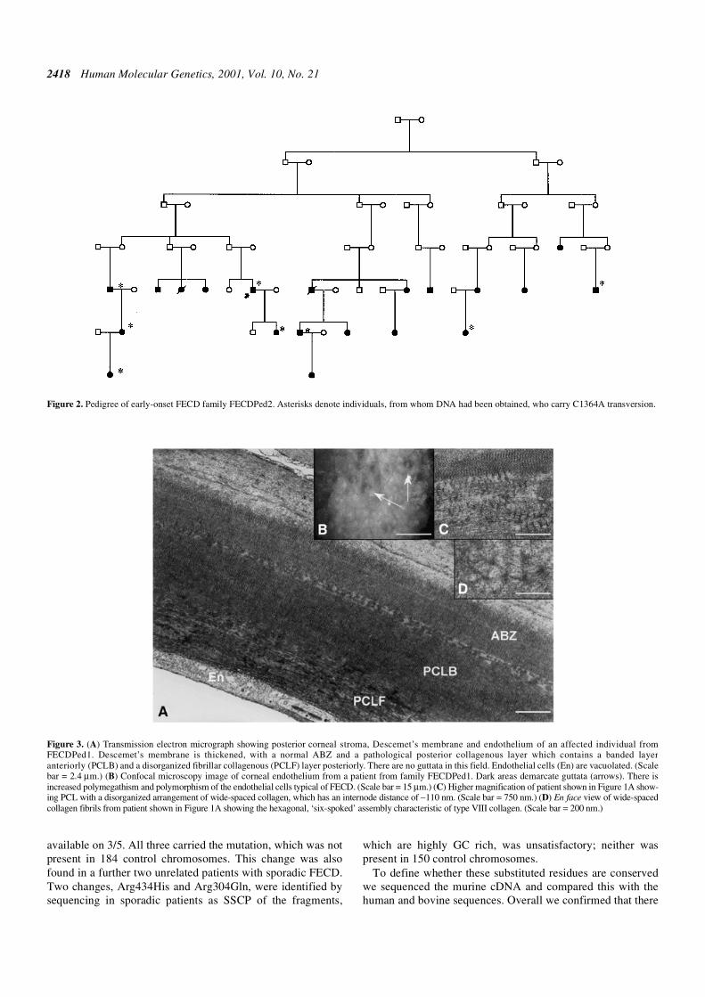
Ultrastructural analysis in affected individuals from familyFECDPed1 demonstrated characteristic secretion of anabnormal posterior collagenous layer (PCL) between theendothelium and a thickened DM (Fig. 3). The PCL was organ-ized into a hexagonal matrix resembling that normally formedby type VIII collagen (Fig. 3). In both affected individualsfrom family PPCDPed1, ultrastructural analysis of cornealmaterial removed at PK revealed endothelial cell multilayeringwith desmosomal intercellular attachments and surface cellswith short microvilli consistent with epithelial-like metaplasiaof the endothelial cells. This confirmed a diagnosis of PPCD inthis family.
Linkage analysis and sequencing in family FECDPed1
Fifteen members of family FECDPed1, covering three gener-ations, in which disease status was confidently assigned, wereselected for linkage analysis. The only unaffected individualsincluded in the analysis were in their fifth decade. Exclusionanalysis was performed using microsatellite markers D20S48and D20S417 coupled with the PPCD and CHED 1 loci (19).Using two-point linkage analysis no significant linkage wasobtained with either marker (LOD score less than –7). There-after, a genome-wide search was undertaken using 382markers covering all 22 autosomes. High LOD scores wereobtained for markers D1S2830 and D17S938 [Zmax = 3.72,recombinant fraction (θ) = 0.0, and Zmax = 3.78, θ = 0.0 respect-ively]. Further genotyping, using microsatellite markers closeto D17S938, revealed no shared haplotype amongst all theaffected members to within 50 kb of marker D17S938,implying that this marker was linked by chance alone.
Human Molecular Genetics, 2001, Vol. 10, No. 21 2417
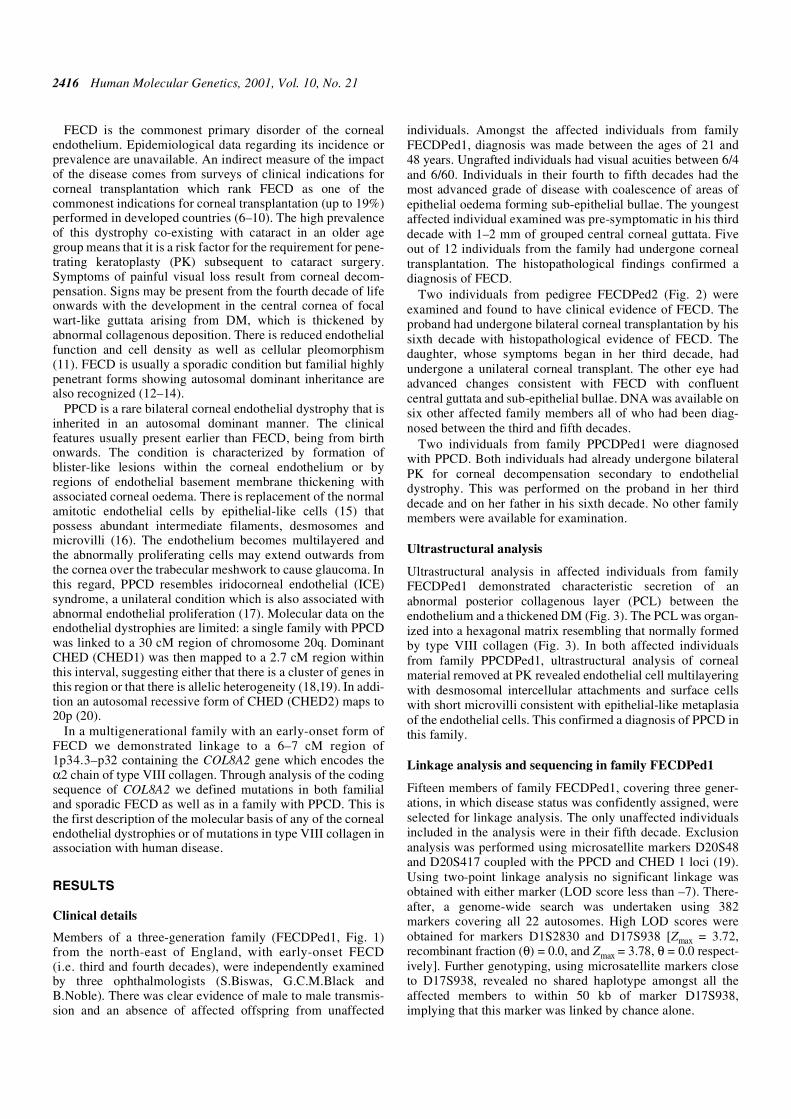
Having demonstrated significant linkage to a region ofchromosome 1, refinement of the critical region was under-taken using further microsatellite markers (Fig. 1). Haplotypeanalysis of the pedigree shows unaffected individual III-5 to berecombinant distal to marker D1S255 and a recombination ispresent in unaffected individual III-6 proximal to markerD1S233. These two individuals are aged 45 (III-5) and 53years (III-6) and have entirely normal corneal examination. Inaddition they each have three children aged between 23 and 34years, all of whom have normal corneal examinations; themajority of affected individuals within this family hadabnormal corneal endothelia by age 20–25 years. These recom-binants define a 6–7 cM critical interval that locates a putativedisease gene to chromosome 1p34.3–p32.
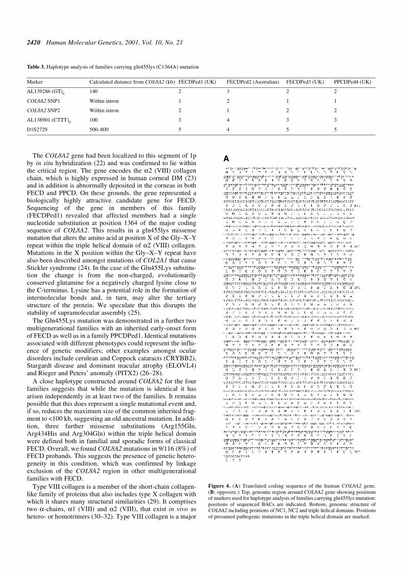
The COL8A2 gene, which encodes the α2 chain of type VIIIcollagen, lies within this region and represented a strong candi-date gene in this family. The gene is fully encompassed withinthe genomic clone RP4-665N4 which has been fully sequenced(GenBank accession no. AL138787). The 2112 bp codingsequence is contained within two exons separated by an intronof 562 bp (Fig. 4A and B). These were sequenced in all familymembers revealing a C→A transversion at position 1364 fromthe beginning of the coding sequence (Fig. 5A and B). Thisresults in a codon 455 glutamine to lysine substitution. The
mutation was present in all 13 affected individuals tested andno unaffected members of the family and was not present in488 ethnically matched control chromosomes.
COL8A2 mutation analysis in patients with FECD and PPCD
Sixteen overlapping primer pairs spanning the coding region ofCOL8A2 were designed to amplify genomic DNA from a panelof affected individuals including 115 unrelated patients withFECD and 15 with PPCD (Table 1). Eight further probandscarried missense changes that were not identified within ethni-cally matched normal control populations and are presumed tobe pathogenic alterations (Table 2).
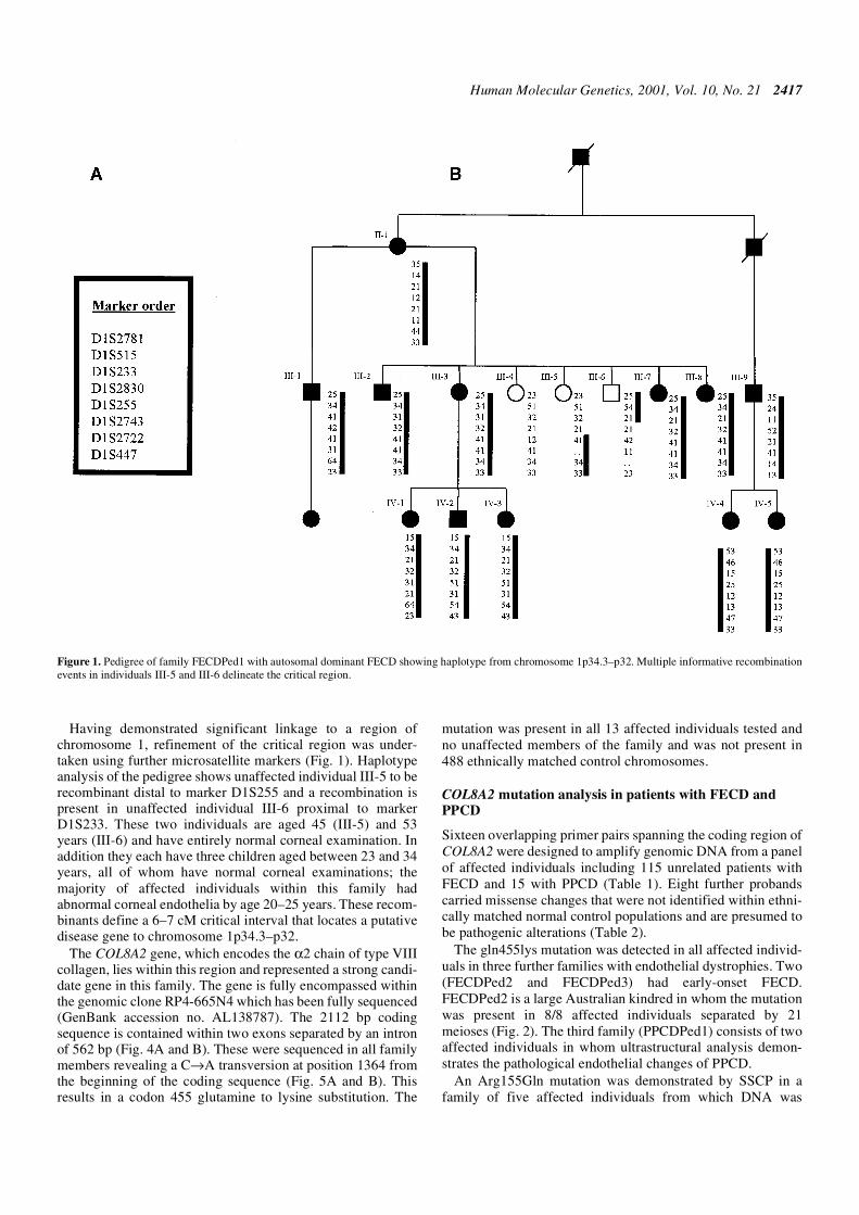
The gln455lys mutation was detected in all affected individ-uals in three further families with endothelial dystrophies. Two(FECDPed2 and FECDPed3) had early-onset FECD.FECDPed2 is a large Australian kindred in whom the mutationwas present in 8/8 affected individuals separated by 21meioses (Fig. 2). The third family (PPCDPed1) consists of twoaffected individuals in whom ultrastructural analysis demon-strates the pathological endothelial changes of PPCD.
An Arg155Gln mutation was demonstrated by SSCP in afamily of five affected individuals from which DNA was
Figure 1. Pedigree of family FECDPed1 with autosomal dominant FECD showing haplotype from chromosome 1p34.3–p32. Multiple informative recombinationevents in individuals III-5 and III-6 delineate the critical region.
2418 Human Molecular Genetics, 2001, Vol. 10, No. 21
available on 3/5. All three carried the mutation, which was notpresent in 184 control chromosomes. This change was alsofound in a further two unrelated patients with sporadic FECD.Two changes, Arg434His and Arg304Gln, were identified bysequencing in sporadic patients as SSCP of the fragments,
which are highly GC rich, was unsatisfactory; neither waspresent in 150 control chromosomes.
To define whether these substituted residues are conservedwe sequenced the murine cDNA and compared this with thehuman and bovine sequences. Overall we confirmed that there
Figure 2. Pedigree of early-onset FECD family FECDPed2. Asterisks denote individuals, from whom DNA had been obtained, who carry C1364A transversion.
Figure 3. (A) Transmission electron micrograph showing posterior corneal stroma, Descemet’s membrane and endothelium of an affected individual fromFECDPed1. Descemet’s membrane is thickened, with a normal ABZ and a pathological posterior collagenous layer which contains a banded layeranteriorly (PCLB) and a disorganized fibrillar collagenous (PCLF) layer posteriorly. There are no guttata in this field. Endothelial cells (En) are vacuolated. (Scalebar = 2.4 µm.) (B) Confocal microscopy image of corneal endothelium from a patient from family FECDPed1. Dark areas demarcate guttata (arrows). There isincreased polymegathism and polymorphism of the endothelial cells typical of FECD. (Scale bar = 15 µm.) (C) Higher magnification of patient shown in Figure 1A show-ing PCL with a disorganized arrangement of wide-spaced collagen, which has an internode distance of ∼110 nm. (Scale bar = 750 nm.) (D) En face view of wide-spacedcollagen fibrils from patient shown in Figure 1A showing the hexagonal, ‘six-spoked’ assembly characteristic of type VIII collagen. (Scale bar = 200 nm.)
Human Molecular Genetics, 2001, Vol. 10, No. 21 2419
is >90% sequence identity in mouse, human and bovine triplehelical domains (data not shown) (21). All of these mutatedresidues lie within the triple helical domain and are conservedin all three species.
In addition, a number of other sequence changes weredetected amongst both affected individuals and normalcontrols (Table 2). Of these eight changes, four were silent andfour missense. Of the latter, the two within the triple helicaldomain (Gly357Arg, Pro575Leu) were only detected amongstfamilial FECD patients and were not found amongst theunaffected controls tested (488 and 186 chromosomes, respec-tively). However, these cannot be presumed to be pathogenic
as they were not found in all affected individuals within thesefamilies and therefore did not fully co-segregate with thedisease phenotype.
Haplotype analysis of families carrying Gln455Lys mutation
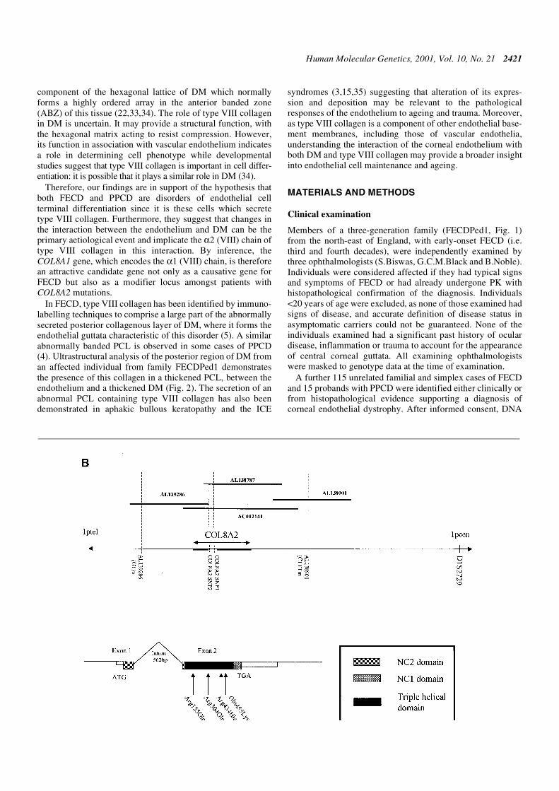
A close haplotype was constructed using five markers aroundCOL8A2 for the four Gln455Lys families (Fig. 4B). TwoFECD families (FECDPed1 and FECDPed3) and the PPCDfamily originate from Northern England and share a commonhaplotype across the region, suggesting the presence of acommon founder mutation within these three families. Incontrast, the Australian family FECDPed2 did not share thishaplotype. This analysis included two separate SNPs withinthe intron interrupting the coding sequence (Table 3) which lie1–1.5 kb from the putative mutation; this strongly suggests thatFECDPed2 carries an identical but independent mutation.
DISCUSSION
A positional candidate strategy was employed to identify thedefective gene in a family (FECDPed1) with a highly pene-trant, early-onset form of FECD. Genetic mapping in thisfamily revealed linkage of FECD to a 6–7 cM interval of1p32.3–p34.3. In family FECDPed1 symptoms commencedduring the third and fourth decades, with clinical signs of diseasedetectable earlier, overcoming the difficulties of analysing late-onset diseases such as FECD, where families of more than twogenerations are scarce and assignment of disease status inyounger individuals is uncertain.
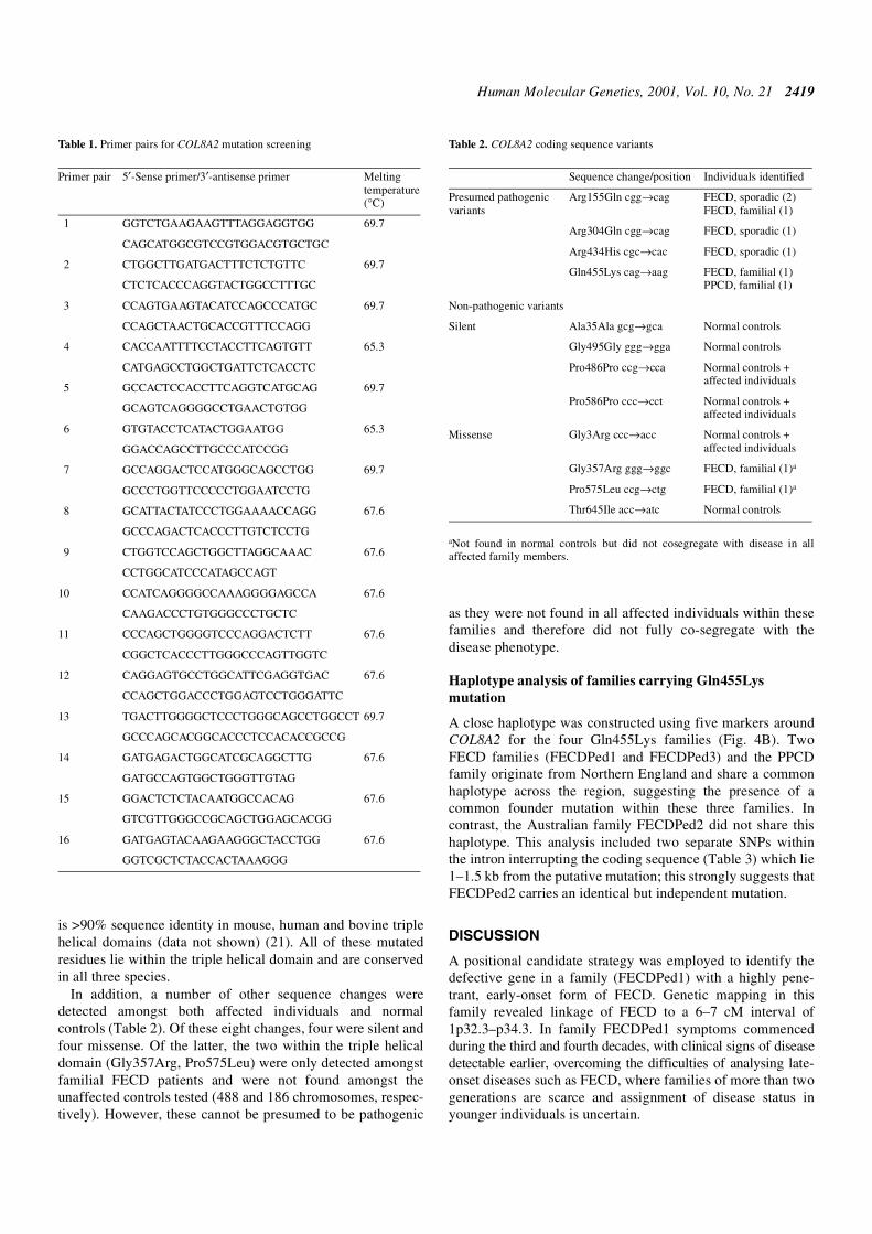
Table 1. Primer pairs for COL8A2 mutation screening
Primer pair 5′-Sense primer/3′-antisense primer Melting temperature (°C)
1 GGTCTGAAGAAGTTTAGGAGGTGG 69.7
CAGCATGGCGTCCGTGGACGTGCTGC
2 CTGGCTTGATGACTTTCTCTGTTC 69.7
CTCTCACCCAGGTACTGGCCTTTGC
3 CCAGTGAAGTACATCCAGCCCATGC 69.7
CCAGCTAACTGCACCGTTTCCAGG
4 CACCAATTTTCCTACCTTCAGTGTT 65.3
CATGAGCCTGGCTGATTCTCACCTC
5 GCCACTCCACCTTCAGGTCATGCAG 69.7
GCAGTCAGGGGCCTGAACTGTGG
6 GTGTACCTCATACTGGAATGG 65.3
GGACCAGCCTTGCCCATCCGG
7 GCCAGGACTCCATGGGCAGCCTGG 69.7
GCCCTGGTTCCCCCTGGAATCCTG
8 GCATTACTATCCCTGGAAAACCAGG 67.6
GCCCAGACTCACCCTTGTCTCCTG
9 CTGGTCCAGCTGGCTTAGGCAAAC 67.6
CCTGGCATCCCATAGCCAGT
10 CCATCAGGGGCCAAAGGGGAGCCA 67.6
CAAGACCCTGTGGGCCCTGCTC
11 CCCAGCTGGGGTCCCAGGACTCTT 67.6
CGGCTCACCCTTGGGCCCAGTTGGTC
12 CAGGAGTGCCTGGCATTCGAGGTGAC 67.6
CCAGCTGGACCCTGGAGTCCTGGGATTC
13 TGACTTGGGGCTCCCTGGGCAGCCTGGCCT 69.7
GCCCAGCACGGCACCCTCCACACCGCCG
14 GATGAGACTGGCATCGCAGGCTTG 67.6
GATGCCAGTGGCTGGGTTGTAG
15 GGACTCTCTACAATGGCCACAG 67.6
GTCGTTGGGCCGCAGCTGGAGCACGG
16 GATGAGTACAAGAAGGGCTACCTGG 67.6
GGTCGCTCTACCACTAAAGGG
Table 2. COL8A2 coding sequence variants
aNot found in normal controls but did not cosegregate with disease in allaffected family members.
Sequence change/position Individuals identified
Presumed pathogenic variants
Arg155Gln cgg→cag FECD, sporadic (2) FECD, familial (1)
Arg304Gln cgg→cag FECD, sporadic (1)
Arg434His cgc→cac FECD, sporadic (1)
Gln455Lys cag→aag FECD, familial (1) PPCD, familial (1)
Non-pathogenic variants
Silent Ala35Ala gcg→gca Normal controls
Gly495Gly ggg→gga Normal controls
Pro486Pro ccg→cca Normal controls + affected individuals
Pro586Pro ccc→cct Normal controls + affected individuals
Missense Gly3Arg ccc→acc Normal controls + affected individuals
Gly357Arg ggg→ggc FECD, familial (1)a
Pro575Leu ccg→ctg FECD, familial (1)a
Thr645Ile acc→atc Normal controls
2420 Human Molecular Genetics, 2001, Vol. 10, No. 21
The COL8A2 gene had been localized to this segment of 1pby in situ hybridization (22) and was confirmed to lie withinthe critical region. The gene encodes the α2 (VIII) collagenchain, which is highly expressed in human corneal DM (23)and in addition is abnormally deposited in the corneas in bothFECD and PPCD. On these grounds, the gene represented abiologically highly attractive candidate gene for FECD.Sequencing of the gene in members of this family(FECDPed1) revealed that affected members had a singlenucleotide substitution at position 1364 of the major codingsequence of COL8A2. This results in a gln455lys missensemutation that alters the amino acid at position X of the Gly–X–Yrepeat within the triple helical domain of α2 (VIII) collagen.Mutations in the X position within the Gly–X–Y repeat havealso been described amongst mutations of COL2A1 that causeStickler syndrome (24). In the case of the Gln455Lys substitu-tion the change is from the non-charged, evolutionarilyconserved glutamine for a negatively charged lysine close tothe C-terminus. Lysine has a potential role in the formation ofintermolecular bonds and, in turn, may alter the tertiarystructure of the protein. We speculate that this disrupts thestability of supramolecular assembly (25).
The Gln455Lys mutation was demonstrated in a further twomultigenerational families with an inherited early-onset formof FECD as well as in a family PPCDPed1. Identical mutationsassociated with different phenotypes could represent the influ-ence of genetic modifiers; other examples amongst oculardisorders include cerulean and Coppock cataracts (CRYBB2),Stargardt disease and dominant macular atrophy (ELOVL4)and Rieger and Peters’ anomaly (PITX2) (26–28).
A close haplotype constructed around COL8A2 for the fourfamilies suggests that while the mutation is identical it hasarisen independently in at least two of the families. It remainspossible that this does represent a single mutational event and,if so, reduces the maximum size of the common inherited frag-ment to <100 kb, suggesting an old ancestral mutation. In addi-tion, three further missense substitutions (Arg155Gln,Arg434His and Arg304Gln) within the triple helical domainwere defined both in familial and sporadic forms of classicalFECD. Overall, we found COL8A2 mutations in 9/116 (8%) ofFECD probands. This suggests the presence of genetic hetero-geneity in this condition, which was confirmed by linkageexclusion of the COL8A2 region in other multigenerationalfamilies with FECD.
Type VIII collagen is a member of the short-chain collagen-like family of proteins that also includes type X collagen withwhich it shares many structural similarities (29). It comprisestwo α-chains, α1 (VIII) and α2 (VIII), that exist in vivo ashetero- or homotrimers (30–32). Type VIII collagen is a major
Table 3. Haplotype analysis of families carrying gln455lys (C1364A) mutation
Marker Calculated distance from COL8A2 (kb) FECDPed1 (UK) FECDPed2 (Australian) FECDPed3 (UK) PPCDPed4 (UK)
AL139286 (GT)n 140 2 3 2 2
COL8A2 SNP1 Within intron 1 2 1 1
COL8A2 SNP2 Within intron 2 1 2 2
AL138901 (CTTT)n 100 3 4 3 3
D1S2729 500–800 5 4 5 5
Figure 4. (A) Translated coding sequence of the human COL8A2 gene.(B; opposite.) Top, genomic region around COL8A2 gene showing positionsof markers used for haplotype analysis of families carrying gln455lys mutation:positions of sequenced BACs are indicated. Bottom, genomic structure ofCOL8A2 including positions of NC1, NC2 and triple helical domains. Positionsof presumed pathogenic mutations in the triple helical domain are marked.
Human Molecular Genetics, 2001, Vol. 10, No. 21 2421
component of the hexagonal lattice of DM which normallyforms a highly ordered array in the anterior banded zone(ABZ) of this tissue (22,33,34). The role of type VIII collagenin DM is uncertain. It may provide a structural function, withthe hexagonal matrix acting to resist compression. However,its function in association with vascular endothelium indicatesa role in determining cell phenotype while developmentalstudies suggest that type VIII collagen is important in cell differ-entiation: it is possible that it plays a similar role in DM (34).
Therefore, our findings are in support of the hypothesis thatboth FECD and PPCD are disorders of endothelial cellterminal differentiation since it is these cells which secretetype VIII collagen. Furthermore, they suggest that changes inthe interaction between the endothelium and DM can be theprimary aetiological event and implicate the α2 (VIII) chain oftype VIII collagen in this interaction. By inference, theCOL8A1 gene, which encodes the α1 (VIII) chain, is thereforean attractive candidate gene not only as a causative gene forFECD but also as a modifier locus amongst patients withCOL8A2 mutations.
In FECD, type VIII collagen has been identified by immuno-labelling techniques to comprise a large part of the abnormallysecreted posterior collagenous layer of DM, where it forms theendothelial guttata characteristic of this disorder (5). A similarabnormally banded PCL is observed in some cases of PPCD(4). Ultrastructural analysis of the posterior region of DM froman affected individual from family FECDPed1 demonstratesthe presence of this collagen in a thickened PCL, between theendothelium and a thickened DM (Fig. 2). The secretion of anabnormal PCL containing type VIII collagen has also beendemonstrated in aphakic bullous keratopathy and the ICE
syndromes (3,15,35) suggesting that alteration of its expres-sion and deposition may be relevant to the pathologicalresponses of the endothelium to ageing and trauma. Moreover,as type VIII collagen is a component of other endothelial base-ment membranes, including those of vascular endothelia,understanding the interaction of the corneal endothelium withboth DM and type VIII collagen may provide a broader insightinto endothelial cell maintenance and ageing.
MATERIALS AND METHODS
Clinical examination
Members of a three-generation family (FECDPed1, Fig. 1)from the north-east of England, with early-onset FECD (i.e.third and fourth decades), were independently examined bythree ophthalmologists (S.Biswas, G.C.M.Black and B.Noble).Individuals were considered affected if they had typical signsand symptoms of FECD or had already undergone PK withhistopathological confirmation of the diagnosis. Individuals<20 years of age were excluded, as none of those examined hadsigns of disease, and accurate definition of disease status inasymptomatic carriers could not be guaranteed. None of theindividuals examined had a significant past history of oculardisease, inflammation or trauma to account for the appearanceof central corneal guttata. All examining ophthalmologistswere masked to genotype data at the time of examination.
A further 115 unrelated familial and simplex cases of FECDand 15 probands with PPCD were identified either clinically orfrom histopathological evidence supporting a diagnosis ofcorneal endothelial dystrophy. After informed consent, DNA
2422 Human Molecular Genetics, 2001, Vol. 10, No. 21
was extracted from these individuals and families to compilepanels for mutation screening.
Ultrastructural analysis
Tissue was dissected (segments <1 mm3), immersed in 2.5%glutaraldehyde in 0.1 M sodium cacodylate buffer and post-fixed in 1% osmium tetroxide in 0.05 M sodium cacodylatebuffer for 1 h. The tissue was dehydrated through a gradedethanol series and embedded araldite resin. Resin-embeddedblocks were cut into semi-thin (1 µm) and thin (65 nm)sections. Semi-thin sections were stained with toluidine blue,thin sections with uranyl acetate and lead citrate. Thin sectionswere placed onto nickel grids and transmission electron micro-scopy carried out using a JEOL 1010 instrument.
Linkage analysis
After informed consent and ethical approval, blood sampleswere taken from 13 affected and four unaffected members ofthe family. Genomic DNA was extracted from lymphocytes(36). Amplification of markers D20S48 and D20S417 bypolymerase chain reaction (PCR) was performed by using100 ng of genomic DNA, 2 µl (0.2 µM) of forward and reverseprimers, 3 µl of dH2O, 10 µl of PCR Reddymix™ master mix[0.75 mM each dNTPs, 67 mM Tris–HCl (pH 8.0 at 25°C),16 mM (NH4)2SO4, 3.7 mM MgCl2, 0.085 mg/ml BSA, 0.5 UTaq polymerase (ABgene; ABgene, Epsom, Surrey, UK)].PCR amplification was performed on a dry block thermalcycler. Samples were denatured at 95°C for 5 min and thefollowing cycling conditions were applied: 32 cycles at 94°Cfor 45 s, 60°C for 45 s, 72°C for 45 s. Final extension was at
72°C for 5 min. Amplified products were phenol-extracted andloaded onto 8% polyacrylamide gel. Electrophoresis wascarried out at 400 V for 3 h and products were silver-stainedaccording to standard methods.
A genome-wide search was carried out using a set of 382fluorescent-labelled PCR primers (ABI Prism™ Linkagemapping set version 2; PE Applied Biosystems, Perkin-ElmerCorp., Foster City, CA) to amplify microsatellite loci, with∼10 cM spacing, across the genome. A 15 µl PCR reaction mixcomprised 100 ng of patients’ genomic DNA, 1 µl of primermix, 1.5 µl of 10× GeneAmp PCR buffer II, 1.5 µl ofGeneAmp dNTP mix (2.5 mM), 0.12 µl of AmpliTaq Gold™DNA polymerase (5 U/µl), 1.5 µl of MgCl2 (25 mM) and3.8 µl of dH2O. DNA from CEPH individual 1347-02 acted aspositive control; dH2O acted as negative control. The PCRconditions for amplification were as per the manufacturer’sinstruction for the GeneAmp PCR system 9600 thermalcycler (PE Applied Biosystems). Two microlitres of pooledPCR product was mixed with 2 µl of size standard [GeneScan
400 HD (ROX); PE Applied Biosystems] and 24 µl of de-ionizedformamide (Amresco Solon, Solon, OH). After denaturing at95°C for 2 min and placing on ice, 2 µl of this mix was injectedinto a polymer (GeneScan STR POP4D)-containing capillaryand analysed on an ABI prism 310 Genetic Analyser usingGeneScan software. In addition, several other markers, ascer-tained from the Généthon linkage map database, were analysedusing the PCR conditions described above: D1S2781, D1S513,D1S233, D1S2676, D1S2832, D1S2729, D1S496, D1S2830,D1S255, D1S2743, D1S2722 and D1S447. These markersspanned a 20 cM region around the ABI-prism markerD1S234, wherein a LOD score of 2.03 was obtained.
The genotype data obtained from the family were enteredinto a PC using CYRILLIC 2.0 software for data storage andretrieval. These data were exported into the LINKAGEprogram for linkage analysis and LOD scores were calculatedusing MLINK (37) and using allele frequency data availablefrom the genome database (GDB).
DNA sequencing
Primers were designed from the major coding region ofCOL8A2, whose sequence was elucidated from a bacterial arti-ficial chromosome clone containing the gene sequence(Table 1). These were used to amplify the gene in two affectedand two unaffected individuals and one control individual andlater to confirm the sequence in all members of FECDPed1.Sterile water was substituted for genomic DNA as a negativecontrol. Thirty microlitre PCR reactions were set up using100 ng of genomic DNA, 3 µl (0.3 µM) of forward and reverseprimers, 15 µl of Reddymix™ master mix and 4.5 µl of dH2O.PCR reactions were carried out on a dry block thermal cycler.Reaction mixtures were denatured at 95°C for 5 min, then 30cycles at 94°C for 45 s, X°C for 45 s (where ‘X’ is the opti-mized annealing temperature for each primer pair shown inTable 1) and 72°C for 45 s. Final extension was performed at72°C for 10 min. The amplified products were cleaned using2 U shrimp alkaline phosphatase (1 U/µl) (USB Corporation,Cleveland, OH) and 4 U exonuclease I (10 U/µl) (USB) on athermal cycler at 37°C for 38 min, 85°C for 20 min and 20°Cfor 2 min. A 10 µl sequencing reaction comprised 5 µl ofcleaned PCR product, 0.7 µl of forward or reverse primer, 1 µl
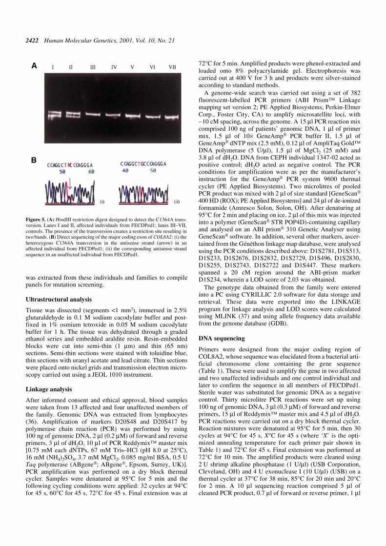
Figure 5. (A) HindIII restriction digest designed to detect the C1364A trans-version. Lanes I and II, affected individuals from FECDPed1; lanes III–VII,controls. The presence of the transversion creates a restriction site resulting intwo bands. (B) Direct sequencing of the major coding exon of COL8A2: (i) theheterozygous C1364A transversion in the antisense strand (arrow) in anaffected individual from FECDPed1; (ii) the corresponding antisense strandsequence in an unaffected individual from FECDPed1.
Human Molecular Genetics, 2001, Vol. 10, No. 21 2423
of Thermo sequenase™ II mix A (Amersham PharmaciaBiotech Inc., Buckingham, UK), 1 µl of Thermo sequenase™II mix B and 2.3 µl of dH2O. Sequencing amplification wasperformed under the following conditions: 94°C for 30 s forone cycle, 30 cycles at 95°C for 20 s, 55°C for 20 s, 60°C for 1 min.Precipitation of DNA was performed using 1 µl of sodiumacetate (1.5 M)/EDTA buffer (250 mM) (Amersham PharmaciaBiotech Corp.) and absolute alcohol. DNA samples were dena-tured with formamide loading dye and run on an acrylamidegel. Sequencing was carried out on an ABI-377 analyser (PEApplied Biosystems) and the results analysed usingGeneScan software.
ACKNOWLEDGEMENTS
The authors thank Heidi Haines and Kelly Kopp for theirexcellent technical assistance, and Gregory S.H.Ogawa foridentifying one of the families who participated in this study.G.B. is a Wellcome Trust Clinician Scientist Fellow (reference51390/Z). S.B. was supported by Manchester Royal EyeHospital research endowments. R.P. is supported by ActionResearch. We would like to acknowledge support from theSwiss National Science Foundation (32-053750.98), theNational Eye Institute of the United States (EY11543) and thefamilies who donated samples to make this work possible.
REFERENCES
1. Waring, G.O., Bourne, W.M., Edelhauser, H.F. and Kenyon, K.R. (1982) The corneal endothelium. Normal and pathologic structure and function. Ophthalmology, 89, 531–590.
2. Klintworth, G. (1999) Advances in the molecular genetics of corneal dystrophies. Am. J. Ophthalmol., 128, 747–754.
3. Bahn, C.F., Falls, H.F., Varley, B.S., Meyer, R.F., Edelhauser, H.F. and Bourne, W.M. (1984) Classification of corneal endothelial disorders based on neural crest origin. Ophthalmology, 91, 558–563.
4. McCartney, A.C. and Kirkness, C.M. (1988) Comparison between posterior polymorphous dystrophy and congenital hereditary endothelial dystrophy of the cornea. Eye, 2, 63–70.
5. Levy, S.G., Moss, J., Sawada, H., Dopping-Hepenstal, P.J. and McCartney, A.C. (1996) The composition of wide-spaced collagen in normal and diseased Descemet’s membrane. Curr. Eye Res., 15, 45–52.
6. Lang, G.K. and Naumann, G.O.H. (1987) The frequency of corneal dystrophies requiring keratoplasty in Europe and the USA. Cornea, 6, 209–211.
7. Mohamadi, P., McDonnell, J.M., Irvine, J.A., McDonnell, P.J., Rao, N. and Smith, R.E. (1989) Changing indications for penetrating keratoplasty, 1984–1988. Am. J. Ophthalmol., 107, 550–552.
8. Brady, S.E., Rapuano, C.J., Arentsen, J.J., Cohen, E.J. and Laibson, P.R. (1989) Clinical indications for procedures associated with penetrating keratoplasty, 1983–1988. Am. J. Ophthalmol., 108, 118–122.
9. Ramsay, A.S., Lee, W.R. and Mohammed, A. (1997) Changing indications for penetrating keratoplasty in the west of Scotland from 1970 to 1995. Eye, 11, 357–360.
10. Alldredge, O.C. and Krachmer, J.H. (1981) Clinical types of corneal transplant rejection. Their manifestations, frequency, preoperative corre-lates, and treatment. Arch. Ophthalmol., 99, 599–604.
11. Bigar, F. (1982) Specular microscopy of the corneal endothelium. Optical solutions and clinical results. Dev. Ophthalmol., 6, 1–94.
12. Krachmer, J.H., Purcell, J.J.,Jr, Young, C.W. and Bucher, K.D. (1978) Corneal endothelial dystrophy. A study of 64 families. Arch. Ophthalmol., 96, 2036–2039.
13. Rosenblum, P., Stark, W.J., Maumenee, I.H., Hirst, L.W. and Maumenee, A.E. (1980) Hereditary Fuchs’ dystrophy. Am. J. Ophthalmol., 90, 455–462.
14. Magovern, M., Beauchamp, G.R., McTigue, J.W., Fine, B.S. and Baumiller, R.C. (1979) Inheritance of Fuchs’ combined dystrophy. Ophthalmology, 86, 1897–1923.
15. Levy, S.G., Moss, J., Noble, B.A. and McCartney, A.C. (1996) Early onset posterior polymorphous dystrophy. Arch. Ophthalmol., 114, 1265–1268.
16. Ross, J.R., Foulks, G.N., Sanfillipo, F.P. and Howell, D.N. (1995) Immuno-histochemical analysis of the pathogenesis of posterior polymorphous dystrophy. Arch. Ophthalmol., 113, 340–345.
17. Howell, D.N., Damms, T., Burchette, J.L.,Jr and Green, W.R. (1997) Endothelial metaplasia in the iridocorneal endothelial syndrome. Invest. Ophthalmol. Vis. Sci., 38, 1896–1901.
18. Heon, E., Mathers, W.D., Alward, W.L., Weisenthal, R.W., Sunden, S.L., Fishbaugh, J.A., Taylor, C.M., Krachmer, J.H., Sheffield, V.C. and Stone, E.M. (1995) Linkage of posterior polymorphous corneal dystrophy to 20q11. Hum. Mol. Genet., 4, 485–488.
19. Toma, N.M., Ebenezer, N.D., Inglehearn, C.F., Plant, C., Ficker, L.A. and Bhattacharya, S.S. (1995) Linkage of congenital hereditary endothelial dystrophy to chromosome 20. Hum. Mol. Genet., 4, 2395–2398.
20. Hand, C.K., Harmon, D.L., Kennedy, S.M., FitzSimon, J.S., Collum, L.M. and Parfrey, N.A. (1999) Localization of the gene for autosomal recessive congenital hereditary endothelial dystrophy (CHED2) to chromosome 20 by homozygosity mapping. Genomics, 61, 1–4.
21. Mann, K., Jander, R., Korsching, E., Kuhn, K. and Rauterberg, J. (1990) The primary structure of a triple helical domain of collagen type VIII from bovine Descemet’s membrane. FEBS Lett., 273, 168–172.
22. Muragaki, Y., Jacenko, O., Apte, S., Mattei, M.-G., Ninomiya, Y. and Olsen, B.R. (1991) The a2 (VIII) collagen gene. A novel member of the short chain collagen family located on the human chromosome 1. J. Biol. Chem., 266, 7721–7727.
23. Kapoor, R., Sakai, L.Y., Funk, S., Roux, E., Bornstein, P. and Sage, E.H. (1988) Type VIII collagen has a restricted distribution in specialised extracellular matrices. J. Cell Biol., 107, 721–730.
24. Richards, A.J., Baguley, D.M., Yates, J.R., Lane, C., Nicol, M., Harper, P.S., Scott, J.D. and Snead, M.P. (2000) Variation in the vitreous phenotype of Stickler syndrome can be caused by different amino acid substitutions in the X position of the type II collagen Gly-X-Y triple helix. Am. J. Hum. Genet., 67, 1083–1094.
25. Sawada, H., Konomi, H. and Hirosawa, K. (1990) Characterisation of collagen in the hexagonal lattice of Descemet’s membrane: its relation to type VIII collagen. J. Cell Biol., 110, 219–227.
26. Doward, W., Perveen, R., Lloyd, I.C., Ridgway, A.E.A., Wilson, L. and Black, G.C. (1999) A mutation in the RIEG1 gene associated with Peters’ anomaly. J. Med. Genet., 36, 152–155.
27. Zhang, K., Kniazeva, M., Han, M., Li, W., Yu, Z., Yang, Z., Li, Y., Metzker, M.L., Allikmets, R., Zack, D.J. et al. (2001) A 5-bp deletion in ELOVL4 is associated with two related forms of autosomal dominant macular dystrophy. Nat. Genet., 27, 89–93.
28. Gill, D., Klose, R., Munier, F.L., McFadden, M., Priston, M., Billingsley, G., Ducrey, N., Schorderet, D.F. and Heon, E. (2000) Genetic heterogeneity of the Coppock-like cataract: a mutation in CRYBB2 on chromosome 22q11.2. Invest. Ophthalmol. Vis. Sci., 41, 159–165.
29. Yamaguchi, N., Benya, P.D., van der Rest, M. and Ninomiya, Y. (1989) The cloning and sequencing of a1 (VIII) collagen cDNAs demonstrate that type VIII collagen is a short chain collagen and contains triple-helical and carboxyl-terminal non-triple helical domains similar to those of type X collagen. J. Biol. Chem., 264, 16022–16029.
30. Greenhill, N.S., Ruger, B.M., Hasan, Q. and Davis, P. (2000) The a1 (VIII) and a2 (VIII) collagen chains form two distinct homotrimeric proteins in vivo. Matrix Biol., 19, 19–28.
31. Illidge, C., Kielty, C.M. and Shuttleworth, C.A. (2001) Collagen VIII can form stable homotrimers and heterotrimers. Int. J. Biochem. Cell Biol., 31, 521–529.
32. Illidge, C., Kielty, C. and Shuttleworth, A. (1998) The a1 (VIII) and a2 (VIII) chains of type VIII collagen can form stable homotrimeric mole-cules. J. Biol. Chem., 273, 22091–22095.
33. Sage, H., Treub, B. and Bornstein, P. (1983) Biosynthetic and structural properties of endothelial cell type VIII collagen. J. Biol. Chem., 258, 13391–13401.
34. Shuttleworth, C.A. (1997) Type VIII collagen. Int. J. Biochem. Cell Biol., 29, 1145–1148.
35. Kenney, M.C. and Chwa, M. (1990) Abnormal extracellular matrix in corneas with pseudophakic bullous keratopathy. Cornea, 9, 115–121.
36. Sambrook, J., Fritsch, E.F. and Maniatis, T. (1989) Molecular Cloning: A Laboratory Manual, 2nd edn. Cold Spring Harbor Laboratory Press, Cold Spring Harbor, NY.
37. Lathrop, G.M., Lalouel, J.M., Julier, C. and Ott, J. (1984) Strategies for multi-point linkage analysis in humans. Proc. Natl Acad. Sci. USA, 81, 3443–3446.

2424 Human Molecular Genetics, 2001, Vol. 10, No. 21