miR-146a aggravates cognitive impairment and Alzheimer ...
9
ARTICLE IN PRESS +Model NRL-1573; No. of Pages 9 Neurología xxx (xxxx) xxx—xxx NEUROLOGÍA www.elsevier.es/neurologia ORIGINAL ARTICLE miR-146a aggravates cognitive impairment and Alzheimer disease-like pathology by triggering oxidative stress through MAPK signaling H. Zhan-qiang a , Q. Hai-hua b , Z. Chi c , W. Miao a , Z. Cui a , L. Zi-yin a , H. Jing a , W. Yi-wei a,* a Department of General medicine, Affiliated Hospital of Chengde Medical College, Chengde 067000, China b Department of Dermatology, Affiliated Hospital of Chengde Medical College, Chengde 067000, China c Department of Neurology, Affilicated Hospital of Chengde Medical College, Chengde 067000, China Received 25 September 2020; accepted 26 December 2020 KEYWORDS Alzheimer disease; miR-146a-5p; Reactive oxygen species; MAPK signaling; Amyloid- Abstract Introduction: Mir-146a-5p has been widely recognized as a critical regulatory element in the immune response. However, recent studies have shown that miR-146a-5p may also be involved in the development of Alzheimer disease (AD). Regrettably, the related mechanisms are poorly understood. Here, we investigated the effects of miR-146a in mice models and SH-SY5Y cells treated with amyloid (A) 1—42 . Methods: To create a model of AD, SH-SY5Y cells were treated with A 1—42 and mice received intracerebroventricular injections of A 1—42 . Then, the transcriptional levels of miR-146a were estimated by real-time PCR. We transiently transfected the miR-146a-5p mimic/inhibitor into cells and mice to study the role of miR-146a. The role of signaling pathways including p38 and reactive oxygen species (ROS) was studied by using specific inhibitors. A and amyloid-beta precursor protein (APP)levels were measured by immunoblotting. Furthermore, A expression was analyzed by immunofluorescence and histochemical examinations. Results: A 1—42 -stimulated SH-SY5Y cells displayed increased transcriptional levels of miR- 146a and APP. Moreover, the p38 MAPK signaling pathway and ROS production were activated upon stimulation with a miR-146a-5p mimic. However, treatment with a miR-146a-5p inhibitor decreased the levels of APP, ROS, and p-p38 MAPK. A similar phenomenon was also observed in the animals treated with A 1—42 , in which miR-146a upregulation increased the expression of A, p-p38, and ROS, while the inhibition of miR-146a had the opposite effect. This suggests that miR-146a increases A deposition and ROS accumulation via the p-p38 signaling pathway. Conclusions: Our research demonstrates that miR-146a-5pa increases A deposition by trig- gering oxidative stress through activation of MAPK signaling. © 2021 Sociedad Espa˜ nola de Neurolog´ ıa. Published by Elsevier Espa˜ na, S.L.U. This is an open access article under the CC BY-NC-ND license (http://creativecommons.org/licenses/by-nc-nd/ 4.0/). ∗ Corresponding author. E-mail address: wangyiwei9511 [email protected] (W. Yi-wei). https://doi.org/10.1016/j.nrl.2020.12.006 0213-4853/© 2021 Sociedad Espa˜ nola de Neurolog´ ıa. Published by Elsevier Espa˜ na, S.L.U. This is an open access article under the CC BY-NC-ND license (http://creativecommons.org/licenses/by-nc-nd/4.0/). Please cite this article as: H. Zhan-qiang, Q. Hai-hua, Z. Chi et al., miR-146a aggravates cognitive impair- ment and Alzheimer disease-like pathology by triggering oxidative stress through MAPK signaling, Neurología, https://doi.org/10.1016/j.nrl.2020.12.006
-
Upload
khangminh22 -
Category
Documents
-
view
0 -
download
0
Transcript of miR-146a aggravates cognitive impairment and Alzheimer ...
ARTICLE IN PRESS+Model
NRL-1573; No. of Pages 9
Neurología xxx (xxxx) xxx—xxx
NEUROLOGÍAwww.elsevier.es/neurologia
ORIGINAL ARTICLE
miR-146a aggravates cognitive impairment and
Alzheimer disease-like pathology by triggering
oxidative stress through MAPK signaling
H. Zhan-qiang a, Q. Hai-huab, Z. Chi c, W. Miao a, Z. Cui a, L. Zi-yin a, H. Jing a, W. Yi-wei a,∗
a Department of General medicine, Affiliated Hospital of Chengde Medical College, Chengde 067000, Chinab Department of Dermatology, Affiliated Hospital of Chengde Medical College, Chengde 067000, Chinac Department of Neurology, Affilicated Hospital of Chengde Medical College, Chengde 067000, China
Received 25 September 2020; accepted 26 December 2020
KEYWORDSAlzheimer disease;miR-146a-5p;Reactive oxygenspecies;MAPK signaling;Amyloid-�
Abstract
Introduction: Mir-146a-5p has been widely recognized as a critical regulatory element in the
immune response. However, recent studies have shown that miR-146a-5p may also be involved
in the development of Alzheimer disease (AD). Regrettably, the related mechanisms are poorly
understood. Here, we investigated the effects of miR-146a in mice models and SH-SY5Y cells
treated with amyloid � (A�)1—42.
Methods: To create a model of AD, SH-SY5Y cells were treated with A�1—42 and mice received
intracerebroventricular injections of A�1—42. Then, the transcriptional levels of miR-146a were
estimated by real-time PCR. We transiently transfected the miR-146a-5p mimic/inhibitor into
cells and mice to study the role of miR-146a. The role of signaling pathways including p38 and
reactive oxygen species (ROS) was studied by using specific inhibitors. A� and amyloid-beta
precursor protein (APP)levels were measured by immunoblotting. Furthermore, A� expression
was analyzed by immunofluorescence and histochemical examinations.
Results: A�1—42-stimulated SH-SY5Y cells displayed increased transcriptional levels of miR-
146a and APP. Moreover, the p38 MAPK signaling pathway and ROS production were activated
upon stimulation with a miR-146a-5p mimic. However, treatment with a miR-146a-5p inhibitor
decreased the levels of APP, ROS, and p-p38 MAPK. A similar phenomenon was also observed
in the animals treated with A�1—42, in which miR-146a upregulation increased the expression
of A�, p-p38, and ROS, while the inhibition of miR-146a had the opposite effect. This suggests
that miR-146a increases A� deposition and ROS accumulation via the p-p38 signaling pathway.
Conclusions: Our research demonstrates that miR-146a-5pa increases A� deposition by trig-
gering oxidative stress through activation of MAPK signaling.
© 2021 Sociedad Espanola de Neurologıa. Published by Elsevier Espana, S.L.U. This is an open
access article under the CC BY-NC-ND license (http://creativecommons.org/licenses/by-nc-nd/
4.0/).
∗ Corresponding author.
E-mail address: wangyiwei9511 [email protected] (W. Yi-wei).
https://doi.org/10.1016/j.nrl.2020.12.006
0213-4853/© 2021 Sociedad Espanola de Neurologıa. Published by Elsevier Espana, S.L.U. This is an open access article under the CC
BY-NC-ND license (http://creativecommons.org/licenses/by-nc-nd/4.0/).
Please cite this article as: H. Zhan-qiang, Q. Hai-hua, Z. Chi et al., miR-146a aggravates cognitive impair-ment and Alzheimer disease-like pathology by triggering oxidative stress through MAPK signaling, Neurología,https://doi.org/10.1016/j.nrl.2020.12.006
ARTICLE IN PRESS+Model
NRL-1573; No. of Pages 9
H. Zhan-qiang, Q. Hai-hua, Z. Chi et al.
PALABRAS CLAVEEnfermedad deAlzheimer;miR-146a-5p;Especies reactivas deoxígeno;Senalización MAPK;�-amiloide
miR-146a empeora el deterioro cognitivo y la patología tipo Alzheimer al promover el
estrés oxidativo a través de la vía de senalización MAPK
Resumen
Introducción: miR-146a-5p es un elemento regulador clave en la respuesta inmune. Sin
embargo, estudios recientes sugieren que miR-146a-5p también está involucrado en el desar-
rollo de la enfermedad de Alzheimer (EA), aunque aún no se conoce con exactitud el mecanismo
por el que esto sucede. Analizamos los efectos de miR-146a en un modelo animal y en células
SH-SY5Y expuestas a �-amiloide (A�)1-42.
Métodos: Tratamos células SH-SY5Y con A�1-42 e inyectamos A�1-42 en los ventrículos cere-
brales de ratones para generar un modelo celular y otro animal de EA. Estimamos los niveles
transcripcionales de miR-146a mediante PCR en tiempo real. Al mismo tiempo, transfectamos
temporalmente las células y los ratones con imitador/inhibidor de miR-146a-5p para evaluar el
papel de miR-146a. Estudiamos el papel de algunas vías de senalización, como la de p38, y los
niveles de especies reactivas de oxígeno (ERO) con inhibidores específicos. Los niveles de A� y
de proteína precursora amiloidea (APP) se determinaron con inmunoblot. También se analizó
la expresión de A� mediante inmunofluorescencia y análisis histoquímico.
Resultados: Las células SH-SY5Y expuestas a A�1-42 mostraron altos niveles transcripcionales
de miR-146a y APP. La vía de senalización p-38 MAPK y la producción de EROs se activaron al
utilizar un imitador de miR-146a-5p. Sin embargo, el bloqueo de miR-146a-5p con un inhibidor
redujo los niveles de APP, EROs y p-p38 MAPK. Se observó un fenómeno similar en los ratones
tratados con A�1-42: la sobrerregulación de miR-146a aumentó la expresión de A�, p-p38 y EROs,
mientras que la inhibición de miR-146a tuvo el efecto contrario. Esto sugiere que miR-146a está
involucrado en el aumento de acumulación de A� y de producción de EROs por medio de la vía
de senalización p-p38.
Conclusiones: Nuestro estudio muestra que miR146a-5p aumenta la acumulación de A� al
promover el estrés oxidativo a través de la activación de la vía de senalización MAPK.
© 2021 Sociedad Espanola de Neurologıa. Publicado por Elsevier Espana, S.L.U. Este es un
artıculo Open Access bajo la licencia CC BY-NC-ND (http://creativecommons.org/licenses/by-
nc-nd/4.0/).
Introduction
Alzheimer’s disease (AD), the most common degenerativedisorder of the nervous system, exhibits typical clinicalpathological changes linked to the dysfunction of cognitiveability.1 The pathogenesis of AD is an extremely complicatedprocess, of which multiple hypotheses have been proposed.The most usual is the amyloid deposition hypothesis whichsuggests the accumulation of the neurotoxic A� fragmentsderived from amyloid precursor protein (APP) by the actionof �-secretase and �-secretase proteases.2,3 Recently, astudy revealed that A� deposition in the brain triggers oxida-tive stress, affecting the reduction ability of mitochondria,causing neuronal death.4 Besides, antioxidants are known toreverse neuronal cell apoptosis induced by A�.5 Therefore,these studies emphasize that oxidative stress is a significantfactor in A�-triggered neuronal death, which gradually mayturn to AD.
MicroRNAs (miRNAs), a kind of small non-coding RNAs, areknown to play a major post-transcriptional regulatory role ingene expression. Recently, the involvement of many miRNAsin modulating key disease genes, such as APP and BACE1,suggested that their dysfunction may also contribute to thepathology of AD.6 miR-146a-5p, the most widely studiedmiRNAs, is the key modulator of immune response and hasbeen linked to a variety of neuroinflammation processes.7
The up-regulation of miR-146a-5p activates mitogen pro-tein kinase to exacerbate neuroinflammation and oxidative
stress.8 Interestingly, studies based on in vitro and in vivomodels of AD found that gradual up-regulation of miR-146a-5p was linked to the progression of AD, while the otherstudies suggested that miR-146q-5p was closely related toA� deposition and synaptic pathological changes.9 Mean-while, clinical studies also revealed that compared to thehealthy elderly group, miR-146a-5p was significantly up-regulated in the brain tissue of AD patients.10 In general,both preclinical and clinical trials strongly put forward therole of miR-146a-5p in the pathogenesis of AD. Therefore,we conjectured that regulating miR-146a-5p could be anovel therapeutic strategy in AD.
However, for that, the mechanistic details of miR-146a-5p role in oxidative stress-induced neuronal degenerationin AD need to be examined. Therefore, in this study,we evaluated whether the down-regulation of miR-146a-5p would cease the progression of AD. Also, we discussedthe relevant pathological changes in AD resulting from thedown-regulation of miR-146a-5p.
Material and methods
Cell culture, transfection, and A�1-42 treatment
SH-SY5Y cells were obtained from Procell (No.CM-0208,China) and cultured in MEM/F12 medium (Procell,
2
ARTICLE IN PRESS+Model
NRL-1573; No. of Pages 9
Neurología xxx (xxxx) xxx—xxx
No.PM151220, China) (Israel Biological Industries) with10%fetal bovine serum (FBS) at 37 ◦C. To induce cell differen-tiation, MEM/F12 medium with 1% FBS and 10 �M all-transretinoic acid (RA) (Aladdin, No.CFLD-R106320, China) wasused for 7 days, and the culture medium was changed every3 days. After the RA treatment, the resultant morphologicalchanges in SH-SY5Y cells were verified microscopically(200x magnification), and the differentiated cells wereused for all subsequent studies. For transfection studies,SH-SY5Y cells were transfected with miR-146a-5p mimic(50 nM; RiboBio, No.miR10000449-1-5, China) or miR-146a-5p inhibitor (100 nM; RiboBio, No.miR20000449-1-5, China)using Lipofectamine3000 (Invitrogen, No.L3000-015, USA)according to the manufacturer’s instructions. After incuba-tion for 24 h, these cells were harvested and undergoingfurther tests, meanwhile, the transfection efficiency alsowas monitored. To establish the in vitro AD cell model,A�1—42 (GenScript, No.RP10017, USA) was dissolved inhexafluoroisopropanol (HFIP) for 10 min. The HFIP waspre-cooled in advance and after volatilization, the formedA�1—42 protein film was dissolved in DMSO, and the SH-SY5Ycells were treated with 1 �M A�1—42 for 24 h. To evaluate theeffect of MAPK signaling, the SH-SY5Y cells were dividedinto the following groups with three replicates in each: (1)the control group without treatment, (2) the model groupwith A�1—42 treatmen, (3) the Model+miR-146a-5p mimicgroup with miR-146a-5p mimic transfection and A�1—42
treatmen, (4) the Model+miR-146a-5p mimic+ FGA-19 groupwas treated with A�1—42 to establish the in vitro AD cellmodel, and transfected with miR-146a-5p mimic, then FGA-19 (Aladdin, No.5.30486.0001, China) as the p38 MAPKinaseinhibitor was added in the cells for 24 h with concentrationof 50 �M, (5) the Model+miR-146a-5p mimic+ FGA-19+NACgroup was treated similarly to the Model+miR-146a-5pmimic+ FGA-19 group, in addition, the cells were treatedwith N-Acetyl-cysteine (NAC, Abcam, No.AB60264, USA) of1 mM for 2 h, as antioxidant to scavenge ROS.
Animals and treatment
The animal experiment was approved by the Animal EthicsCommittee of the Affiliated Hospital of Chengde MedicalCollege (No.20200330-06). All animal studies strictly com-plied with the relevant regulations of the Animal EthicsCommittee and abided by the 3R principle in the designand implementation of experiments. Six-month-old maleC57bl/6J mice were randomly assigned into four groups(n = 5 per group), namely the control group without treat-ment, the AD group with A�1—42 treatment, the AD+miR-146mimic group with miR-146a-5p mimic transfection andA�1—42 treatment, and the AD+miR-146 inhibitor group withmiR-146a-5p inhibitor transfection and A�1—42 treatment.The lentiviral expression vectors of miR-146a-5p mimic,miR-146a-5p inhibitor and negative control were synthe-sized by Thermo Scientific company with their titer of1 × 109 PFU/mL. Except for the control group, the otherthree groups of animals were intracerebroventricularinjected with A�1—42 which was prepared by dissolvingin distilled water at 0.2 mg per mL. On contrary, theanimals in the control group received the same volume ofdistilled water (vehicle control). Moreover, the mice in the
AD+miR-146 mimic group were also intracerebroventricularinjected with 3 �L lentiviral expression vector of miR-146a-5p mimic, the treatment in the AD+miR-146 inhibitor groupwas similar. After two weeks of the treatment period, theanimals were examined for cognitive behavior using theMorris water maze (MWM) test. After that, all the mice withfive in each group were anesthetized and sacrificed to obtainthe hippocampal tissues for further study. Besides, threesections of hippocampal tissues per mouse were analyzed.
Surgical procedure
The mice were placed in the isoflurane anesthesia device,adjusted to scale 2, with a 400cc/min air flow rate.Post-anesthesia, the mice were quickly fixed to the brainstereotaxic device and a wound was cut with a sterile scalpelblade. The brain surface was disinfected using an cottonswab dipped with alcohol. Then, an insertion point (coordi-nate: ML ± 1.0; AP 0.3) with 0.5 mm diameter aperture wasdrilled with a hand drill using a stereotactic instrument, andA�1—42 was injected to establish the animal model of AD.
Morris water maze (MWM)
MWM behavioral experiment was performed on the 14thday after the operation. In the experiment, a circular pool(50 cm high and 20 cm in diameter) was filled with opaquewater (22 ± 3 ◦C), and a circular platform (10 cm in diameter,28 cm high) was placed 1 cm below the surface of the water.The MWM was theatrically divided into four quadrants, andthe hidden platform was placed in one of the quadrants.The experiment was divided into two stages. Stage one, thefirst 5 days was used for the positioning and navigation, inwhich each mouse was tested 4 times a day. In each test,the mice were placed in the water from different quadrantsand allowed to swim for up to 90 s to find the platform andrest on it for 10 s. If the mouse failed to find the platformwithin the specified time, it was guided to the platform andstand for 10 s, meanwhile, the escape latency was recordedas 90 s. In the second stage of free exploration, the plat-form was hidden on the 6th day. Then, the swimming time,distance of the mouse in the quadrant before the platformwithin 90 s, and the number of times it crosses the platformwas recorded. The acquired data was analyzed.11
Estimation of cellular reactive oxygen species(ROS) generation
According to commercial regulations, the DCFH-DA methodwas used to estimate the production of cellular ROS.DCFH-DA is a fluorescent dye detecting ROS level, whichcan be converted to oxidized fluorescence dye 2’,7’-dichlorofluorescein (DCF) at the presence of ROS. First,SH-SY5Y (4 × 10⁴) cells were seeded into a 96-well plateand exposed to A�1—42 for 1day (25 �M). After this, the cellswere incubated for 30 min in the DMEM medium containing5 � DCFH-DA (MedChemExpress, No.HY-D0940, USA) underdark conditions at room temperature (RT), and the cellswere washed with PBS. Moreover, the nucleus with DAPI(Bio-Rad, No.1351303, USA) to distinguish apoptotic cells.
3
ARTICLE IN PRESS+Model
NRL-1573; No. of Pages 9
H. Zhan-qiang, Q. Hai-hua, Z. Chi et al.
Fluorescence images were captured immediately using a flu-orescence microscope (Zeiss Axio Imager Z2, Germany) at10× magnification.
A�1—42 ELISA
A�1—42 analysis was performed according to the A�1—42 ELISAkit (Invitrogen, No.KHB3441, USA) operating instructions.Cell homogenates of the hippocampal tissue were prepared.Meanwhile, A� standard solution was prepared and the testsamples were diluted with the standard dilution buffer pro-vided in the kit. Then, 100 �L of standards was added tothe appropriate microtiter wells in triplicates and incu-bated overnight at 4 ◦C. The next day, the liquid in the96-well plate was completely removed and washed 3 timeswith washing buffer. Then, the A�1—42 antibody was addedto the sample and incubated for 60 min at 37 ◦C. Again, 3times washing was performed with the cleaning solution andincubation with the secondary antibody was carried out for30 min at RT. Once again, after washing each well at least 3times, 100 �L of stable color developing solution was addedto each well. Lastly, the absorbance was recorded at 450 nmto calculate the concentration of A�1—42 in the correspond-ing samples using the standard curve.12 The antibodies andsolutions were provided in the kit.
Immunohistochemistry
Using the cryostat, coronal sections (thickness: 40 �m) ofbrain tissue were obtained. These were washed 3 timeswith 1% PBS for 5 min and then incubated with 5% bovineserum albumin in an incubator at RT for 0.5 h. Next, thesewere incubated with primary antibodies (in PBS, Anti-A�1—42;1:1000; Invitrogen, No.MA5-36246, USA) at RT for 60 min,and then overnight at 4 ◦C. After overnight incubation, tis-sue sections were bought to RT and washed 3 times with 1%PBS for 5 min. Followed by incubation with the secondaryantibodies (goat anti-rabbit; 1:1000; Invitrogen, No.A32731,USA) at 37 ◦C for 1 h, the brain slices were washed 3 timeswith 1% PBS for 5 min. Lastly, the brain tissue sectionswere dipped in the DAB (Sigma, No.11718096001, USA) chro-mogenic solution with the deposition of A�1—42 stained darkbrown and mounted on glass slides. After dried in a 37 ◦Cincubator, tissue sections were dehydrated using ethanolgradient and turned transparent with xylene. Finally, theimages were acquired using an optical microscope.13
RNA extraction
The total RNA of each sample was extracted by the RNAisoPlus Reagent (Takara, No.M9108, Japan). The frozen cellswere lysed using TRIZOL and placed at RT for 5 min forcompleted isolution. Then, 0.2 ml of chloroform was addedto every 1 ml of the lysed sample and mixed vigorously for15 s. The mixture was incubated at 15—30 ◦C for 2—3 minand then centrifuged at 12,000 rpm for 15 min at 4 ◦C. Aftercentrifugation, the RNA, distributed in the water phase,was precipitated using an equal volume of isopropanol.After the precipitation, the RNA pellet was rinsed withat least 1 ml of 75% ethanol (75% ethanol prepared with
DEPCH2O) and re-centrifuged at 7000 rpm at 4 ◦C for 5 min.Next, most of the ethanol solution was carefully removedand the RNA pellet was air-dried at RT for 5—10 min. Lastly,The RNA pellet was dissolved in 40 �l of RNase-free waterand stored at −80 ◦C for later use.
qRT-PCR Assay
For qRT-PCR, cDNA synthesis was performed according to themanufactures’ instruction for the PrimeScriptTM RT MasterMix Kit (Takara, No.RR036B, Japan). The reverse transcrip-tase MMLV along with the reaction mixture was incubatedat 70 ◦C for 3 min, and then immediately transferred to icewater. Then, 0.5 �l of reverse transcriptase was added andincubated at 37 ◦C for 60 min. Next, final incubation wasperformed at 95 ◦C for 3 min to obtain the cDNA which wasstored at −80 ◦C. The housekeeping gene, �-actin was usedas an internal standard. The specific primers used were: formiR-146a-5p, 5’-3’ (forward) GGG GTG AGA ACT GAA TTCCAT and 5’-3’ (reverse) CAG TGC GTG TCG TGG AGT; for�-actin, 5’-3’ (forward) TGG CAC CCA GCA CAA TGA A and5’-3’ (reverse) CTA AGT CAT AGT CCG CCT AGA AGC A. Thetarget gene and housekeeping gene of each sample weredesigned and synthesized by Shanghai GenePharma Com-pany (China), and subjected to real-time PCR by means ofthe SYBR@Premix Ex TaqTM (Tli RNaseH Plus) Kit (Takara,No.RR820A, Japan). Real-time PCR was performed with thefollowing cycling conditions: 95 ◦C for 30 s, 40 cycles of 95 ◦Cfor 5 s, 60 ◦C for 30 s and, after that, 95 ◦C for 15 s, 60 ◦Cfor 1 min, 95 ◦C for 15 s. PCR products were electrophoresedon a 2% agarose gel, and stained with GoldViewTM (Shang-hai yuanye Bio-Technology, No.R20977, China) to detect theamplified product. The relative expression level was calcu-lated using the 2-��CT method.
Western blotting
SH-SY5Y cells and hippocampal tissue were lysed with RIPAbuffer (Abcam, No.AB156034, USA) under specific condi-tions and separated on SDS polyacrylamide gel (Abcam,No.AB139597, USA). The protein bands were then trans-ferred onto a PVDF membrane (Sigma, No.3010040001, USA)which was blocked with 5% skimmed milk. After blocking,three times washing was performed with PBS. Then, themembrane was incubated overnight with the primary anti-bodies, APP antibody (1:500, Abclonal, No.A11912, China),A�1—42 antibody (1:200, Abcam, No.P05067, USA), p38 MAPKantibody (1:500, Abcam, No.AB170099, USA), p-p38 MAPKantibody (1:1000, Beijing Biolab, No.K22589-TZH, China)or GADPH antibody (1:5000, Cell Signaling Technology, No.4967, USA) at 4 ◦C. On the second day, after rewarm-ing for 30 min and 3 times washing with PBS, membraneswere incubated with the corresponding HRP-conjugatedgoat anti-rabbit IgG or HRP-conjugated goat anti-mouseIgG (1:10,000, Sigma, No.A0545 and SAB3700986, USA) sec-ondary antibodies for 30 min at RT. The membranes wereagain washed 3 times with PBS. Lastly, the ECL reagent(Sigma, No.WBULS0500, USA) was used to illuminate the tar-get protein bands, and the quantitative analysis of proteinwas performed by a Gel-Pro-Analyzer imaging system.14
4
ARTICLE IN PRESS+Model
NRL-1573; No. of Pages 9
Neurología xxx (xxxx) xxx—xxx
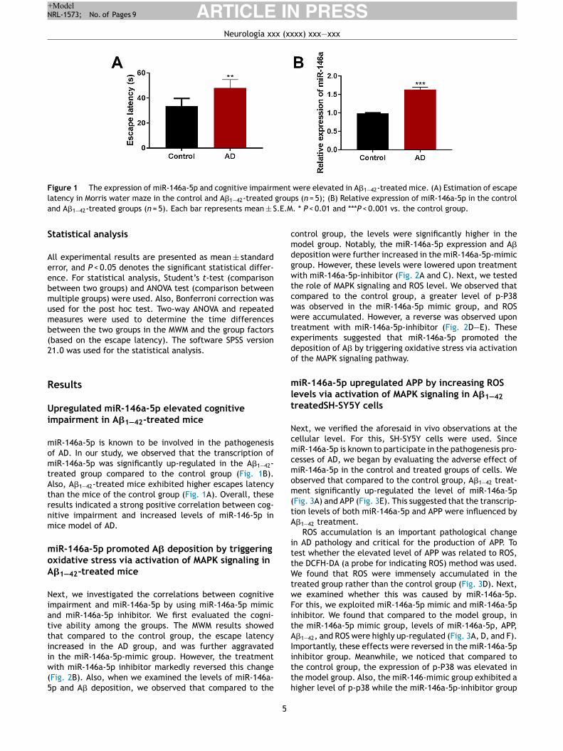
Figure 1 The expression of miR-146a-5p and cognitive impairment were elevated in A�1—42-treated mice. (A) Estimation of escape
latency in Morris water maze in the control and A�1—42-treated groups (n = 5); (B) Relative expression of miR-146a-5p in the control
and A�1—42-treated groups (n = 5). Each bar represents mean ± S.E.M. * P < 0.01 and ***P < 0.001 vs. the control group.
Statistical analysis
All experimental results are presented as mean ± standarderror, and P < 0.05 denotes the significant statistical differ-ence. For statistical analysis, Student’s t-test (comparisonbetween two groups) and ANOVA test (comparison betweenmultiple groups) were used. Also, Bonferroni correction wasused for the post hoc test. Two-way ANOVA and repeatedmeasures were used to determine the time differencesbetween the two groups in the MWM and the group factors(based on the escape latency). The software SPSS version21.0 was used for the statistical analysis.
Results
Upregulated miR-146a-5p elevated cognitiveimpairment in A�1—42-treated mice
miR-146a-5p is known to be involved in the pathogenesisof AD. In our study, we observed that the transcription ofmiR-146a-5p was significantly up-regulated in the A�1—42-treated group compared to the control group (Fig. 1B).Also, A�1—42-treated mice exhibited higher escapes latencythan the mice of the control group (Fig. 1A). Overall, theseresults indicated a strong positive correlation between cog-nitive impairment and increased levels of miR-146-5p inmice model of AD.
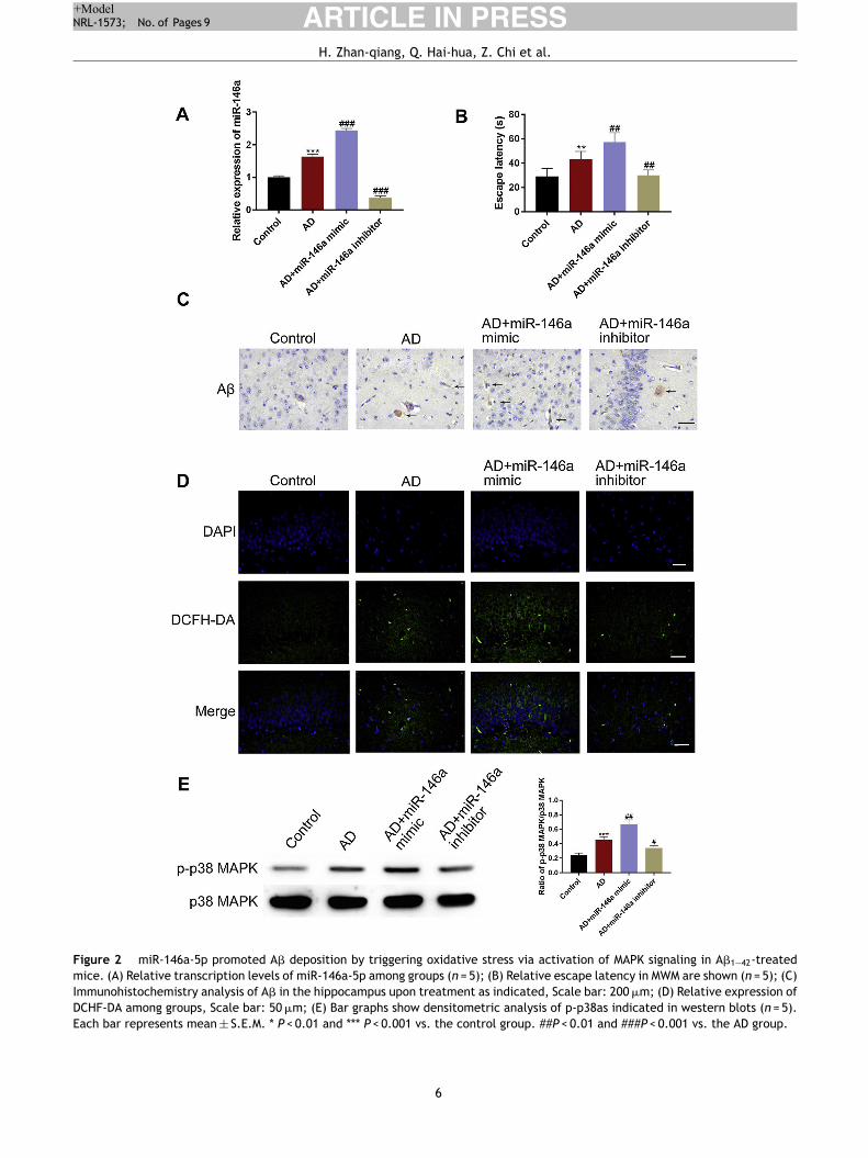
miR-146a-5p promoted A� deposition by triggeringoxidative stress via activation of MAPK signaling inA�1—42-treated mice
Next, we investigated the correlations between cognitiveimpairment and miR-146a-5p by using miR-146a-5p mimicand miR-146a-5p inhibitor. We first evaluated the cogni-tive ability among the groups. The MWM results showedthat compared to the control group, the escape latencyincreased in the AD group, and was further aggravatedin the miR-146a-5p-mimic group. However, the treatmentwith miR-146a-5p inhibitor markedly reversed this change(Fig. 2B). Also, when we examined the levels of miR-146a-5p and A� deposition, we observed that compared to the
control group, the levels were significantly higher in themodel group. Notably, the miR-146a-5p expression and A�
deposition were further increased in the miR-146a-5p-mimicgroup. However, these levels were lowered upon treatmentwith miR-146a-5p-inhibitor (Fig. 2A and C). Next, we testedthe role of MAPK signaling and ROS level. We observed thatcompared to the control group, a greater level of p-P38was observed in the miR-146a-5p mimic group, and ROSwere accumulated. However, a reverse was observed upontreatment with miR-146a-5p-inhibitor (Fig. 2D—E). Theseexperiments suggested that miR-146a-5p promoted thedeposition of A� by triggering oxidative stress via activationof the MAPK signaling pathway.
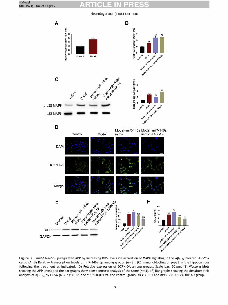
miR-146a-5p upregulated APP by increasing ROSlevels via activation of MAPK signaling in A�1—42
treatedSH-SY5Y cells
Next, we verified the aforesaid in vivo observations at thecellular level. For this, SH-SY5Y cells were used. SincemiR-146a-5p is known to participate in the pathogenesis pro-cesses of AD, we began by evaluating the adverse effect ofmiR-146a-5p in the control and treated groups of cells. Weobserved that compared to the control group, A�1—42 treat-ment significantly up-regulated the level of miR-146a-5p(Fig. 3A) and APP (Fig. 3E). This suggested that the transcrip-tion levels of both miR-146a-5p and APP were influenced byA�1—42 treatment.
ROS accumulation is an important pathological changein AD pathology and critical for the production of APP. Totest whether the elevated level of APP was related to ROS,the DCFH-DA (a probe for indicating ROS) method was used.We found that ROS were immensely accumulated in thetreated group rather than the control group (Fig. 3D). Next,we examined whether this was caused by miR-146a-5p.For this, we exploited miR-146a-5p mimic and miR-146a-5pinhibitor. We found that compared to the model group, inthe miR-146a-5p mimic group, levels of miR-146a-5p, APP,A�1—42, and ROS were highly up-regulated (Fig. 3A, D, and F).Importantly, these effects were reversed in the miR-146a-5pinhibitor group. Meanwhile, we noticed that compared tothe control group, the expression of p-P38 was elevated inthe model group. Also, the miR-146-mimic group exhibited ahigher level of p-p38 while the miR-146a-5p-inhibitor group
5
ARTICLE IN PRESS+Model
NRL-1573; No. of Pages 9
H. Zhan-qiang, Q. Hai-hua, Z. Chi et al.
Figure 2 miR-146a-5p promoted A� deposition by triggering oxidative stress via activation of MAPK signaling in A�1—42-treated
mice. (A) Relative transcription levels of miR-146a-5p among groups (n = 5); (B) Relative escape latency in MWM are shown (n = 5); (C)
Immunohistochemistry analysis of A� in the hippocampus upon treatment as indicated, Scale bar: 200 �m; (D) Relative expression of
DCHF-DA among groups, Scale bar: 50 �m; (E) Bar graphs show densitometric analysis of p-p38as indicated in western blots (n = 5).
Each bar represents mean ± S.E.M. * P < 0.01 and *** P < 0.001 vs. the control group. ##P < 0.01 and ###P < 0.001 vs. the AD group.
6
ARTICLE IN PRESS+Model
NRL-1573; No. of Pages 9
Neurología xxx (xxxx) xxx—xxx
Figure 3 miR-146a-5p up-regulated APP by increasing ROS levels via activation of MAPK signaling in the A�1—42-treated SH-SY5Y
cells. (A, B) Relative transcription levels of miR-146a-5p among groups (n = 3); (C) Immunoblotting of p-p38 in the hippocampus
following the treatment as indicated. (D) Relative expression of DCFH-DA among groups, Scale bar: 50 �m; (E) Western blots
showing the APP levels and the bar graphs show densitometric analysis of the same (n = 3). (F) Bar graphs showing the densitometric
analysis of A�1—42 by ELISA (n3). * P < 0.01 and *** P < 0.001 vs. the control group. ## P < 0.01 and ### P < 0.001 vs. the AD group.
7
ARTICLE IN PRESS+Model
NRL-1573; No. of Pages 9
H. Zhan-qiang, Q. Hai-hua, Z. Chi et al.
displayed a lower level of p-p38 (Fig. 3C). Overall, theseresults indicated that the expression of APP and A�1—42 wereinfluenced by miR-146 via activation of MAPK signaling andoxidative stress.
Discussion
Most degenerative diseases of the nervous system are closelyrelated to age.15 AD is a disease of severe dementia with agrowing incidence in humans. Understanding the pathogene-sis at the cellular can reveal novel insights for the preventionand treatment of AD. In our study, we firstly verified thatmiR-146a-5p promoted the development of AD by deposit-ing A� in the animal model. This was also true in cell assays.We think that the potential mechanism could be related toincreased oxidative stress via activation of MAPK signaling.
miR-146a-5p is widely regarded as an inflammation-related microRNA with immunomodulatory effects.16 Mul-tiple reports showed that miR-146a-5p could inhibit theinterleukin-1 receptor-related kinase 1 (IRAK1) and down-regulate NF-�b activity in the cognitive and memory-relatedbrain regions, such as the hippocampus and prefrontal cor-tex in AD model mice.17,18 Recently, other studies alsoimplicated miR-146a-5p in aging processes using the exper-imental animal models of AD.19,20 From the postmortembrain autopsy of AD patients, studies found that miR-146a-5pwas highly expressed in the CSF, serum, and plasma. There-fore, to verify the correlation between AD and miR-146a-5p,we first tested the level of miR-146a-5p in A�1—42 treatedcells and animals. Notably, our findings are consistent withprevious studies and found significantly higher levels of miR-146a-5p in the AD model group. Interestingly, we also foundthat reduced levels of miR-146 markedly decreased APP anddeposition of A� in the hippocampus regions of the brain.
Several hypotheses put oxidative stress as the key fac-tor in the pathophysiology of AD. With the accumulationof ROS, synaptic activity gradually decreases which leadsto abnormal metabolism triggering the accumulation of A�
and the hyper-phosphorylation of Tau protein. This ulti-mately causes mitochondrial dysfunction and neuronal celldeath.5 The autopsy analysis of mouse models and ADpatients revealed that an unbalanced redox state leads toincreased ROS causing mitochondrial dysfunction and A�
peptide aggregation.5,21 In our study, the ROS level weremuch higher in the model group than that of the controlgroup, both in animal and cell studies, which also was con-sistent with the previous reports. Importantly, up-regulatedmiR-146a-5p further aggravated oxidative stress in the ADmodel group.
MAPKs, belonging to the class of protein Ser/Thrkinases, can transform extracellular stimuli into intracel-lular responses, and thereby regulate many physiologicalprocesses.22 ERK1/2, p38 MAPK, and c-Jun N-terminalkinases (JNKs) are the most widely studied MAPKs. Notably,MAPK signaling is also known to regulate the generation ofROS. In our study, the miR-146a-5p-mimic group exhibitedabnormally increased levels of ROS, though this could bereversed by using FGA-19 (a MAPK inhibitor). The elevatedA� levels were caused by oxidative stress and the unbal-anced redox state was triggered by miR-146a-5p-induced
MAPK signaling. These results strongly indicate that in theA�-treated cells and animal models, ROS production isdependent on the activation of the MAPK signaling pathway.Also, we think that miR-146a-5p inhibitors can be used toblock MAPK signaling. In addition, there are some limita-tions should be noted in our study. For example, it indeed isdifficult to differentiate the administer A� from the gener-ated with our study design, which limits the research aboutrelationship of miR-146a-5p and A�. In this study, the micemodel of AD is induced by the intracerebroventricular injec-tion of the A�1—42, however, there are relatively mature andwidely used mice models of AD which can be purchased fromcompany or research institution, and will be used directly inour future study.
Conclusions
In conclusion, using the animal and cellular models, wereported that miR-146a-5p promotes the development of ADand aggravates A� deposition which is potentially caused byoxidative stress via activation of MAPK signaling.
Funding
This research did not receive any specific grant from fundingagencies in the public, commercial, or not-for-profit sectors.
Conflict of interests
The authors declare that they have no conflict of interest.
References
1. Aggarwal NT, Shah RC, DA B. Alzheimer’s disease: unique mark-
ers for diagnosis & new treatment modalities. Indian J Med Res.
2015:142.
2. Sun X, Bromley-Brits K, Song W. Regulation of beta-site
APP-cleaving enzyme 1 gene expression and its role in
Alzheimer’s disease. J Neurochem. 2012;120(Suppl. 1):62—70,
http://dx.doi.org/10.1111/j.1471-4159.2011.07515.x.
3. Zhang S, Wang Z, Cai F, Zhang M, Wu Y, Zhang J. W S.
BACE1 cleavage site selection critical for amyloidogenesis and
Alzheimer’s pathogenesis. J Neurosci. 2017;37:6915—25.
4. Praticò D. Amyloid beta, mitochondrial structural and
functional dynamics in Alzheimer’s disease. Am J Med.
2000;109:577—85.
5. Wang X, Su B, Siedlak SL, Moreira PI, Fujioka H,
Wang Y, et al. Amyloid-beta overproduction causes
abnormal mitochondrial dynamics via differential
modulation of mitochondrial fission/fusion pro-
teins. Proc Natl Acad Sci USA. 2008;105:19318—23,
http://dx.doi.org/10.1073/pnas.0804871105.
6. Amaral SA, Pereira TSF, Brito JAR, Cortelli SC, Cortelli JR,
Gomez RS, et al. Comparison of miRNA expression profiles in
individuals with chronic or aggressive periodontitis. Oral Dis.
2019;25:561—8, http://dx.doi.org/10.1111/odi.12994.
7. Jiang S, Hu Y, Deng S, Deng J, Yu X, Huang G, et al. miR-146a
regulates inflammatory cytokine production in Porphyromonas
8
ARTICLE IN PRESS+Model
NRL-1573; No. of Pages 9
Neurología xxx (xxxx) xxx—xxx
gingivalis lipopolysaccharide-stimulated B cells by targeting
IRAK1 but not TRAF6. Biochim Biophys Acta Mol Basis Dis.
2018;1864:925—33.
8. Sun W, Wang Q, Guo Y, Zhao Y, Wang X, Zhang Z,
et al. Selenium suppresses inflammation by indu-
cing microRNA-146a in Staphylococcus aureus-infected
mouse mastitis model. Oncotarget. 2017;8:110949—64,
http://dx.doi.org/10.18632/oncotarget.20740.
9. Caldeira C, Cunha C, Vaz AR, Falcao AS, Barateiro A, Seixas
E, et al. Key aging-associated alterations in primary microglia
response to beta-amyloid stimulation. Front Aging Neurosci.
2017;9:277, http://dx.doi.org/10.3389/fnagi.2017.00277.
10. Sierksma A, Lu A, Salta E, Eynden EV, Callaerts-Vegh Z, D’Hooge
R, et al. Deregulation of neuronal miRNAs induced by amyloid-�
or TAU pathology. Mol Neurodegener. 2018;13:54.
11. Bromley-Brits K, Deng Y. W S. Morris water maze test for learning
and memory deficits in Alzheimer’s disease model mice. J Vis
Exp. 2011;53:e2920.
12. Liu Y, Wang J, Hsiung GR. W S. Trehalose inhibits A� generation
and plaque formation in Alzheimer’s disease. Mol Neurobiol.
2020;57:3150—7.
13. Luo Y, Yang WNL. Anodal transcranial direct current stimulation
can improve spatial learning and memory and attenuate A�42
burden at the early stage of Alzheimer’s disease in APP/PS1
transgenic mice. Front Aging Neurosci. 2020;12:134.
14. Zhao J, Bian C, Liu M, Zhao Y, Sun TFX. Orchiectomy and
letrozole differentially regulate synaptic plasticity and spatial
memory in a manner that is mediated by SRC-1 in the hippocam-
pus of male mice. J Steroid Biochem Mol Biol. 2018;178:354—68.
15. Praticò DND. Oxidative injury in diseases of the central
nervous system: focus on Alzheimer’s disease. Am J Med.
2000;109:577—658.
16. Wang H, Zhang Y, Wu X, Wang Y, Cui H, Li X, et al.
Regulation of human natural killer cell IFN-gamma
production by microRNA-146a via targeting the NF-
kappaB signaling pathway. Front Immunol. 2018;9:293,
http://dx.doi.org/10.3389/fimmu.2018.00293.
17. Kubota K, Nakano M, Kobayashi E, Mizue Y, Chikenji T,
Otani M, et al. An enriched environment prevents diabetes-
induced cognitive impairment in rats by enhancing exosomal
miR-146a secretion from endogenous bone marrow-derived
mesenchymal stem cells. PLOS ONE. 2018;13:e0204252,
http://dx.doi.org/10.1371/journal.pone.0204252.
18. Dong H, Li J, Huang L, Chen X, Li D, Wang T, et al. Serum
microRNA profiles serve as novel biomarkers for the diagnosis
of Alzheimer’s disease. Dis Markers. 2015:e625659.
19. Jiang M, Xiang Y, Wang D, Gao J, Liu D, Liu Y, et al. Dys-
regulated expression of miR-146a contributes to age-related
dysfunction of macrophages. Aging Cell. 2012;11:29—40,
http://dx.doi.org/10.1111/j.1474-9726.2011.00757.x.
20. Deng S, Wang H, Jia C, Zhu S, Chu X, Ma Q, et al.
MicroRNA-146a induces lineage-negative bone marrow cell
apoptosis and senescence by targeting polo-like kinase 2
expression. Arterioscler Thromb Vasc Biol. 2017;37:280—90,
http://dx.doi.org/10.1161/ATVBAHA.116.308378.
21. Cha MY, Han SH, Son SM, Hong HS, Choi YJ, Byun J. I M-J.
Mitochondria-specific accumulation of amyloid � induces mito-
chondrial dysfunction leading to apoptotic cell death. PLoS
ONE. 2012;7:e34929.
22. Widmann C, Gibson S, Jarpe MB, Johnson GL. Mitogen-activated
protein kinase: conservation of a three-kinase module from
yeast to human. Physiol Rev. 1999;79:143—80.
9