Differential impact of chronic ozone exposure on expanding and fully expanded poplar leaves
Maderas. Ciencia y tecnología 8(2): 93-106, 2006
93
ISSN 0717-3644ISSN online 0718-221X
1, 2 Federal Research Centre for Forestry and Forest Products/Institute for WoodBiology and Wood Protection, Leuschnerstrasse 91, D-21031 Hamburg/Germany.Correspondig author: [email protected]: December 21, 2005. Accepted: April 13, 2006.
MICROSCOPIC STUDIES ON MODIFIED WALL STRUCTURE ANDLIGNIN TOPOCHEMISTRY IN XYLEM FIBRES OF POPLAR AFTER
WOUNDING
Claus Frankenstein1, Uwe Schmitt2
In memoriam of Dr. Walter G. KAUMAN
ABSTRACT
Information about fine structure following wounding in differentiating xylem tissue is still scarce.This study provides information on cell wall modifications with special emphasis on lignin distributionin xylem fibres of poplar differentiating at the time of wounding. Samples were collected from woundedPopulus spp. trees after response periods of up to twenty-three months and processed for microscopicanalyses. General studies on the wall structure of wound-adjacent xylem fibres were carried out withlight and transmission electron microscopy, whereas lignin distribution patterns of these cells wereexamined by UV-microspectrophotometry.
Xylem fibres close to the wound and within a transition zone between differentiated xylem laiddown prior to and tissue laid down after wounding developed a distinctively thicker secondary wall thannormal fibres. These modified walls also showed a slightly higher lignin content, than normal and aheterogeneous lignin distribution in the middle lamella and the secondary wall.
Wounding in poplar induces a modified wall structure and lignin topochemistry in xylem fibresdifferentiating at the time of wounding. It is assumed that this wound response is part of thecompartmentalization process and therefore contributes to an increased resistance.
Keywords: Wound reaction, poplar, Populus tremula L. x P. tremuloides Michx., fibre cell wall, lignindistribution, UV-microspectrophotometry
INTRODUCTION
Hardwood lignin is a complex 3-dimensional co-polymer consisting mainly of guaiacyl and syringylunits. As a major cell wall component of xylem elements, lignin distribution, content and compositionhave a significant influence on technical wood properties as well as on decay resistance. It is thereforeof interest is know how external stress factors such as like wounding influence cell wall ultra structureand lignification.
The defence mechanisms induced by different types of injury can be subdivided into active andpassive mechanisms. These highly variable reactions have been subject of numerous investigations atmacroscopic and microscopic levels (e.g. Sharon, 1973; Mullick, 1977; Shortle, 1979; Shigo, 1984;Schmitt and Liese, 1990, 1992, 1993; Liese and Dujesiefken, 1996; Pearce, 2000). Tree vitality, seasonalinfluences and the extent of wounding are factors which determine whether reactions are restricted toareas close to the wound or whether larger areas of the surrounding tissue are affected. Wounds affectingthe cambial region often lead to the formation of callus tissue and subsequently, modified xylem andphloem (e.g. Liese and Dujesiefken, 1989; Fink, 1999; Grünwald et al., 2002). Within the xylem different

Maderas. Ciencia y tecnología 8(2): 93-106, 2006
94
Universidad del Bío - Bío
reaction patterns lead to compartmentalization of the wounded area (Sharon, 1973; Shigo and Marx,1977; Bauch et al., 1980; Rademacher et al, 1984; Shigo 1984; Lowerts et al., 1986; Liese and Dujesiefken,1996). The formation of a so, called boundary layer which is mainly characterized by the synthesis ofphenolic compounds in parenchyma cells with subsequent extrusion into neighbouring cells, i. e. fibresand vessels, as well as the formation of tyloses are the most prominent compartmentalization mechanismsin hardwoods at the microscopic level (e.g. Schmitt and Liese, 1990; Pearce, 2000). Earlier studies onwound reactions in poplar mainly focused on reactions of the bark, such as the formation of the ligno-suberized zone, new periderms and the influence of pressure on tissue differentiation (Kaufert, 1937;Soe, 1959; Brown and Sax, 1962; Trockenbrodt and Liese, 1991; Bucciarelli et al., 1999). The formationof a wound-callus and the wound induced alterations within the xylem of poplar have already beendescribed at the light microscopic level (e.g. Buntrock, 1989; Frankenstein et al., 2005).
The following contribution is focused on the modification of xylem elements within a transitionzone between differentiated xylem laid down prior to wounding and tissue laid down after wounding.Special emphasis is given to modifications of the cell wall ultrastructure and the lignin distribution,using light- and transmission electron microscopy as well as cellular UV-microspectrophotometry.
MATERIAL AND METHODS
Rectangular wounds of 10 cm x 10 cm were made on 07 July 2002 and 23 June 2003 by removingthe bark from stems of four mature poplar trees (Populus spp.) using a saw and a chisel. Samples werecollected from the lateral wound edge after 2, 4, 8, 10, 17, 62 and 95 weeks of wound response (Fig. 1).The wound tissue, the adjacent modified xylem and unaffected xylem were removed with a chisel andrazor blades.
Figure 1 Callus formation at the lateral wound edges 95 weeks after wounding; the rectangle indicatesthe position of tissue sampling for microscopy and UV-microspectrophotometry.
MicroscopyFor conventional light-microscopy, samples (10 x 10 x 8 mm3) were either fixed for three days in a
phosphate-buffered solution of 37% formaldehyde, washed in distilled water, and embedded inpolyethylene-glycol (PEG) or the fixed samples were dehydrated in a graded series of propanol andembedded in glycol methacrylate (Technovit 7100). 6µm thick transverse sections were cut with a
Maderas. Ciencia y tecnología 8(2): 93-106, 2006
95
Microscopic studies...: Frankenstein and Schmitt.
rotary microtome and stained for 1.5h with a standard Giemsa or a neutral red solution. Transversesections 10 µm thick were cut from the PEG embedded material with a sliding microtome and stainedwith a standard safranine/astra blue solution. Sections stained with neutral red were examined atwavelengths between 380 – 490 nm.
For TEM, samples from the same series were reduced to a final size of 3 x 3 x 8 mm3, fixed for oneday in a mixture of 5% glutaraldehyde and 8% paraformaldehyde, then partly postfixed with a 1%Osmium tetroxide solution, washed in 0.1 M cacodylate buffer (pH 7.3), dehydrated in a graded seriesof acetone and embedded in Spurr´s epoxy resin (Spurr, 1969).
Ultra-thin (80-100 nm) transverse sections were either double-stained with uranyl acetate and leadcitrate or with potassium permanganate according to Donaldson (1992). The samples were examinedwith a Philips CM 12 TEM at an accelerating voltage of 60 or 80 kV.
UV-microspectrophotometry
For UV-microspectrophotometry, the samples were processed as described for TEM. 1µm transversesections were prepared, mounted onto quartz slides, immersed in a drop of non-UV absorbing glycerineand covered with a quartz cover slip. Ultrafluar objectives lenses were used to observe the samples.
Scanning UV-microspectrophotometry was carried out using a ZEISS UMSP 80microspectrophotometer equipped with an Osram high-pressure xenon lamp, an ultrafluar quartzcondenser and a scanning stage enabling the determination of image profiles at a constant wavelengthof 280 nm using the scan programme APAMOS (Automatic-Photometric-Analysis of Microscopic Objectsby Scanning, Zeiss). This wavelength represents the typical absorbance maximum of lignified cell walls.The scan programme digitises rectangular tissue portions with a local geometrical resolution of 0.25 µm2
and a photometrical resolution of 4096 grey scale levels, converted into 14 basic colours representingthe measured absorbance intensities (Koch and Kleist, 2001).
Specimens were additionally analysed by point measurements with a spot size of 1 µm2. The spectrawere taken at wavelengths from 240 to 400 nm in 2 nm steps using the programme LAMWIN (Zeiss).These point measurements for a semi quantitative determination of the lignin content were automaticallyrepeated 50 times at each spot for individual wall layers, i. e., compound middle lamella (cml), S2layers of the secondary wall (S2) and cell corners (cc).
RESULTS & DISCUSSION
MicroscopyLight microscopy revealed that wounding induced the formation of modified xylem along a transition
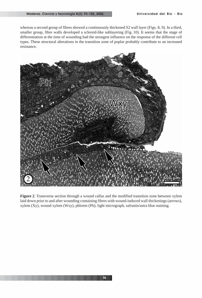
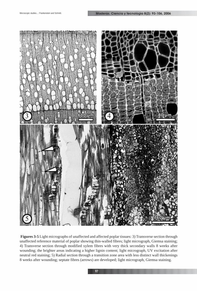
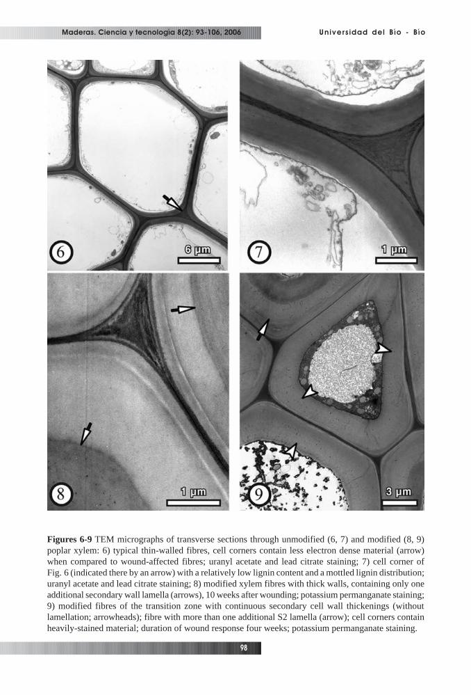
zone between xylem laid down prior to wounding and tissue laid down after wounding (Fig. 2). Thenumber of fibres showing cell wall alterations decreased with increasing distance from the wound edge.Within this zone which was identified as xylem differentiating at the time of wounding, the formation ofunusually thick-walled fibres was the most prominent structural feature (Figs. 3, 4). Septated fibreswere regularly found in the transition zone (Fig. 5). Neighbouring cells of similar shape and thereforeprobably also septated fibres were in some cases filled with calcium-oxalate crystals. Corresponding toour observations, the formation of a tangential band of thick-walled, flattened fibres between newlyformed and regular xylem induced by insertion of a pin, was reported for beech wood (Schmitt etal.,2000). Lowerts et al. (1986) reported the formation of sclereids within wound-associated xylem ofLiriodendron tulipifera L. as a regular feature. Electron microscopy showed that the affected poplarfibres, in contrast to unaffected fibres (Figs. 6, 7), deposited additional secondary wall (S2) materialleading to different patterns of wall thickenings with cell corners predominantly consisting of heavily-stained material (Figs. 8-11). One group of xylem fibres developed an additional secondary wall layer,
Maderas. Ciencia y tecnología 8(2): 93-106, 2006
96
Universidad del Bío - Bío
whereas a second group of fibres showed a continuously thickened S2 wall layer (Figs. 8, 9). In a third,smaller group, fibre walls developed a sclereid-like sublayering (Fig. 10). It seems that the stage ofdifferentiation at the time of wounding had the strongest influence on the response of the different celltypes. These structural alterations in the transition zone of poplar probably contribute to an increasedresistance.
Figure 2. Transverse section through a wound callus and the modified transition zone between xylemlaid down prior to and after wounding containing fibres with wound-induced wall thickenings (arrows),xylem (Xy), wound xylem (Wxy), phloem (Ph); light micrograph, safranin/astra blue staining.
Maderas. Ciencia y tecnología 8(2): 93-106, 2006
97
Microscopic studies...: Frankenstein and Schmitt.
Figures 3-5 Light micrographs of unaffected and affected poplar tissues: 3) Transverse section throughunaffected reference material of poplar showing thin-walled fibres; light micrograph, Giemsa staining;4) Transverse section through modified xylem fibres with very thick secondary walls 8 weeks afterwounding; the brighter areas indicating a higher lignin content; light micrograph, UV excitation afterneutral red staining; 5) Radial section through a transition zone area with less distinct wall thickenings8 weeks after wounding; septate fibres (arrows) are developed; light micrograph, Giemsa staining.
Maderas. Ciencia y tecnología 8(2): 93-106, 2006
98
Universidad del Bío - Bío
Figures 6-9 TEM micrographs of transverse sections through unmodified (6, 7) and modified (8, 9)poplar xylem: 6) typical thin-walled fibres, cell corners contain less electron dense material (arrow)when compared to wound-affected fibres; uranyl acetate and lead citrate staining; 7) cell corner ofFig. 6 (indicated there by an arrow) with a relatively low lignin content and a mottled lignin distribution;uranyl acetate and lead citrate staining; 8) modified xylem fibres with thick walls, containing only oneadditional secondary wall lamella (arrows), 10 weeks after wounding; potassium permanganate staining;9) modified fibres of the transition zone with continuous secondary cell wall thickenings (withoutlamellation; arrowheads); fibre with more than one additional S2 lamella (arrow); cell corners containheavily-stained material; duration of wound response four weeks; potassium permanganate staining.
Maderas. Ciencia y tecnología 8(2): 93-106, 2006
99
Microscopic studies...: Frankenstein and Schmitt.
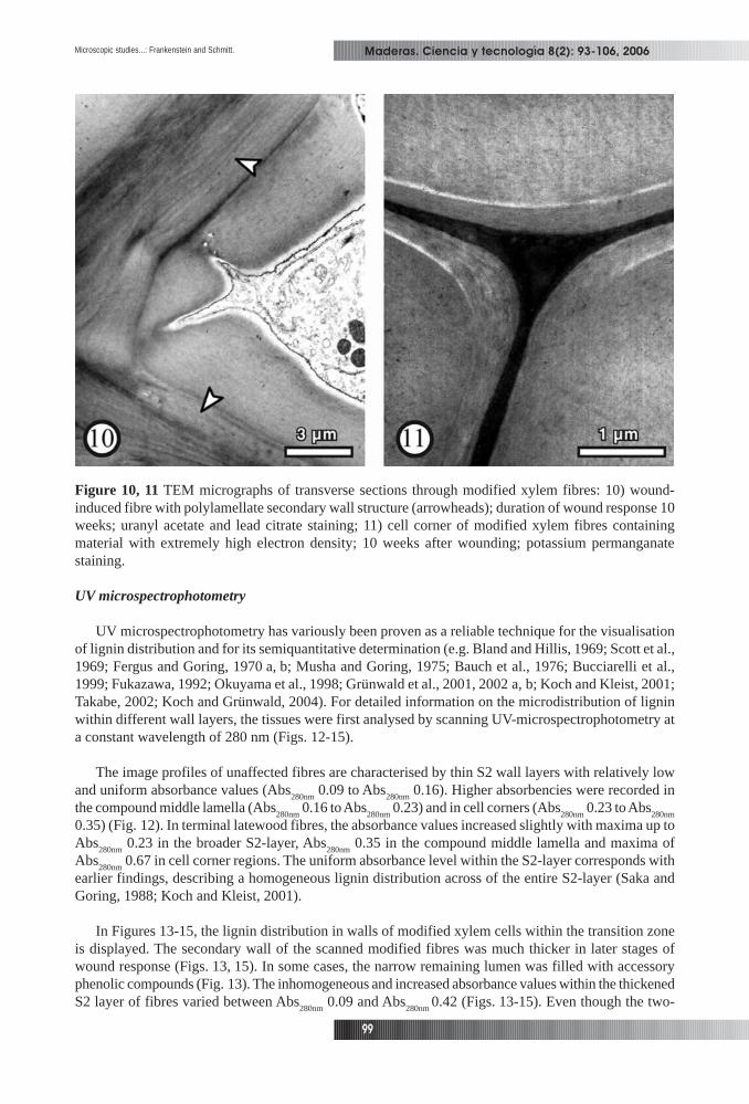
Figure 10, 11 TEM micrographs of transverse sections through modified xylem fibres: 10) wound-induced fibre with polylamellate secondary wall structure (arrowheads); duration of wound response 10weeks; uranyl acetate and lead citrate staining; 11) cell corner of modified xylem fibres containingmaterial with extremely high electron density; 10 weeks after wounding; potassium permanganatestaining.
UV microspectrophotometry
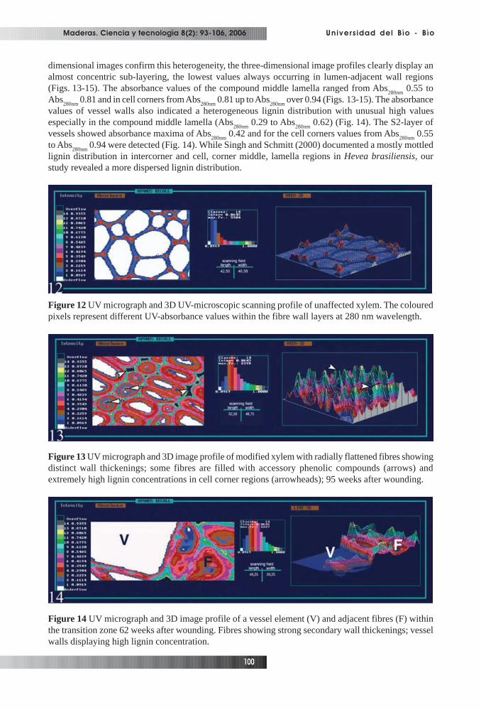
UV microspectrophotometry has variously been proven as a reliable technique for the visualisationof lignin distribution and for its semiquantitative determination (e.g. Bland and Hillis, 1969; Scott et al.,1969; Fergus and Goring, 1970 a, b; Musha and Goring, 1975; Bauch et al., 1976; Bucciarelli et al.,1999; Fukazawa, 1992; Okuyama et al., 1998; Grünwald et al., 2001, 2002 a, b; Koch and Kleist, 2001;Takabe, 2002; Koch and Grünwald, 2004). For detailed information on the microdistribution of ligninwithin different wall layers, the tissues were first analysed by scanning UV-microspectrophotometry ata constant wavelength of 280 nm (Figs. 12-15).
The image profiles of unaffected fibres are characterised by thin S2 wall layers with relatively lowand uniform absorbance values (Abs
280nm 0.09 to Abs
280nm 0.16). Higher absorbencies were recorded in
the compound middle lamella (Abs280nm
0.16 to Abs280nm
0.23) and in cell corners (Abs280nm
0.23 to Abs280nm
0.35) (Fig. 12). In terminal latewood fibres, the absorbance values increased slightly with maxima up toAbs
280nm 0.23 in the broader S2-layer, Abs
280nm 0.35 in the compound middle lamella and maxima of
Abs280nm
0.67 in cell corner regions. The uniform absorbance level within the S2-layer corresponds withearlier findings, describing a homogeneous lignin distribution across of the entire S2-layer (Saka andGoring, 1988; Koch and Kleist, 2001).
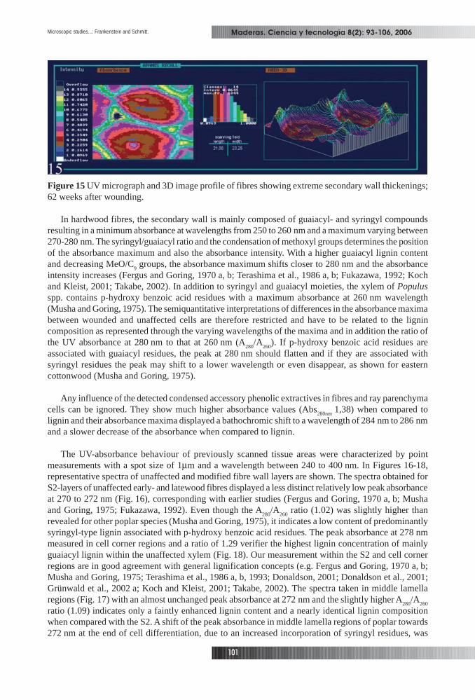
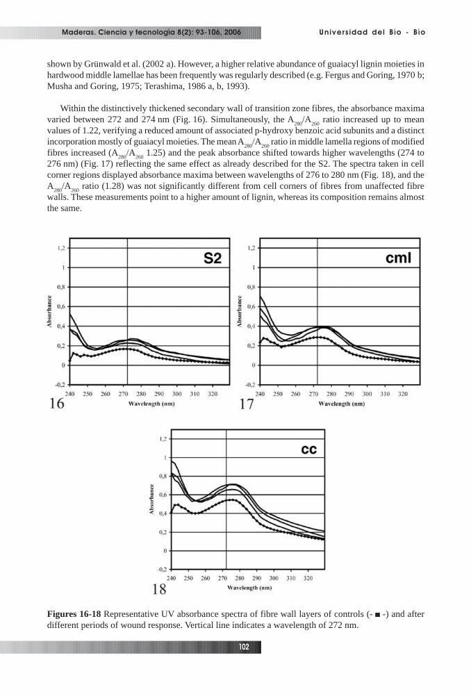
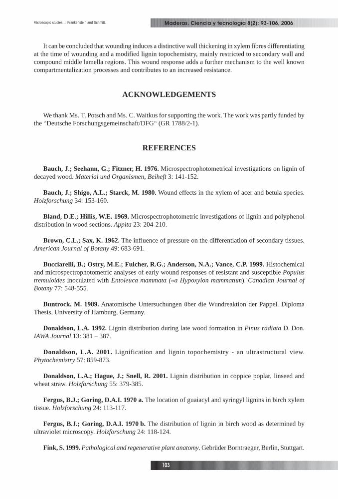
In Figures 13-15, the lignin distribution in walls of modified xylem cells within the transition zoneis displayed. The secondary wall of the scanned modified fibres was much thicker in later stages ofwound response (Figs. 13, 15). In some cases, the narrow remaining lumen was filled with accessoryphenolic compounds (Fig. 13). The inhomogeneous and increased absorbance values within the thickenedS2 layer of fibres varied between Abs
280nm 0.09 and Abs
280nm0.42 (Figs. 13-15). Even though the two-
Maderas. Ciencia y tecnología 8(2): 93-106, 2006
100
Universidad del Bío - Bío
dimensional images confirm this heterogeneity, the three-dimensional image profiles clearly display analmost concentric sub-layering, the lowest values always occurring in lumen-adjacent wall regions(Figs. 13-15). The absorbance values of the compound middle lamella ranged from Abs
280nm 0.55 to
Abs280nm
0.81 and in cell corners from Abs280nm
0.81 up to Abs280nm
over 0.94 (Figs. 13-15). The absorbancevalues of vessel walls also indicated a heterogeneous lignin distribution with unusual high valuesespecially in the compound middle lamella (Abs
280nm 0.29 to Abs
280nm 0.62) (Fig. 14). The S2-layer of
vessels showed absorbance maxima of Abs280nm
0.42 and for the cell corners values from Abs280nm
0.55to Abs
280nm 0.94 were detected (Fig. 14). While Singh and Schmitt (2000) documented a mostly mottled
lignin distribution in intercorner and cell, corner middle, lamella regions in Hevea brasiliensis, ourstudy revealed a more dispersed lignin distribution.
Figure 12 UV micrograph and 3D UV-microscopic scanning profile of unaffected xylem. The colouredpixels represent different UV-absorbance values within the fibre wall layers at 280 nm wavelength.
Figure 13 UV micrograph and 3D image profile of modified xylem with radially flattened fibres showingdistinct wall thickenings; some fibres are filled with accessory phenolic compounds (arrows) andextremely high lignin concentrations in cell corner regions (arrowheads); 95 weeks after wounding.
Figure 14 UV micrograph and 3D image profile of a vessel element (V) and adjacent fibres (F) withinthe transition zone 62 weeks after wounding. Fibres showing strong secondary wall thickenings; vesselwalls displaying high lignin concentration.
Maderas. Ciencia y tecnología 8(2): 93-106, 2006
101
Microscopic studies...: Frankenstein and Schmitt.
Figure 15 UV micrograph and 3D image profile of fibres showing extreme secondary wall thickenings;62 weeks after wounding.
In hardwood fibres, the secondary wall is mainly composed of guaiacyl- and syringyl compoundsresulting in a minimum absorbance at wavelengths from 250 to 260 nm and a maximum varying between270-280 nm. The syringyl/guaiacyl ratio and the condensation of methoxyl groups determines the positionof the absorbance maximum and also the absorbance intensity. With a higher guaiacyl lignin contentand decreasing MeO/C
9 groups, the absorbance maximum shifts closer to 280 nm and the absorbance
intensity increases (Fergus and Goring, 1970 a, b; Terashima et al., 1986 a, b; Fukazawa, 1992; Kochand Kleist, 2001; Takabe, 2002). In addition to syringyl and guaiacyl moieties, the xylem of Populusspp. contains p-hydroxy benzoic acid residues with a maximum absorbance at 260 nm wavelength(Musha and Goring, 1975). The semiquantitative interpretations of differences in the absorbance maximabetween wounded and unaffected cells are therefore restricted and have to be related to the lignincomposition as represented through the varying wavelengths of the maxima and in addition the ratio ofthe UV absorbance at 280 nm to that at 260 nm (A
280/A
260). If p-hydroxy benzoic acid residues are
associated with guaiacyl residues, the peak at 280 nm should flatten and if they are associated withsyringyl residues the peak may shift to a lower wavelength or even disappear, as shown for easterncottonwood (Musha and Goring, 1975).
Any influence of the detected condensed accessory phenolic extractives in fibres and ray parenchymacells can be ignored. They show much higher absorbance values (Abs
280nm1,38) when compared to
lignin and their absorbance maxima displayed a bathochromic shift to a wavelength of 284 nm to 286 nmand a slower decrease of the absorbance when compared to lignin.
The UV-absorbance behaviour of previously scanned tissue areas were characterized by pointmeasurements with a spot size of 1µm and a wavelength between 240 to 400 nm. In Figures 16-18,representative spectra of unaffected and modified fibre wall layers are shown. The spectra obtained forS2-layers of unaffected early- and latewood fibres displayed a less distinct relatively low peak absorbanceat 270 to 272 nm (Fig. 16), corresponding with earlier studies (Fergus and Goring, 1970 a, b; Mushaand Goring, 1975; Fukazawa, 1992). Even though the A
280/A
260 ratio (1.02) was slightly higher than
revealed for other poplar species (Musha and Goring, 1975), it indicates a low content of predominantlysyringyl-type lignin associated with p-hydroxy benzoic acid residues. The peak absorbance at 278 nmmeasured in cell corner regions and a ratio of 1.29 verifier the highest lignin concentration of mainlyguaiacyl lignin within the unaffected xylem (Fig. 18). Our measurement within the S2 and cell cornerregions are in good agreement with general lignification concepts (e.g. Fergus and Goring, 1970 a, b;Musha and Goring, 1975; Terashima et al., 1986 a, b, 1993; Donaldson, 2001; Donaldson et al., 2001;Grünwald et al., 2002 a; Koch and Kleist, 2001; Takabe, 2002). The spectra taken in middle lamellaregions (Fig. 17) with an almost unchanged peak absorbance at 272 nm and the slightly higher A
280/A
260
ratio (1.09) indicates only a faintly enhanced lignin content and a nearly identical lignin compositionwhen compared with the S2. A shift of the peak absorbance in middle lamella regions of poplar towards272 nm at the end of cell differentiation, due to an increased incorporation of syringyl residues, was
Maderas. Ciencia y tecnología 8(2): 93-106, 2006
102
Universidad del Bío - Bío
shown by Grünwald et al. (2002 a). However, a higher relative abundance of guaiacyl lignin moieties inhardwood middle lamellae has been frequently was regularly described (e.g. Fergus and Goring, 1970 b;Musha and Goring, 1975; Terashima, 1986 a, b, 1993).
Within the distinctively thickened secondary wall of transition zone fibres, the absorbance maximavaried between 272 and 274 nm (Fig. 16). Simultaneously, the A
280/A
260 ratio increased up to mean
values of 1.22, verifying a reduced amount of associated p-hydroxy benzoic acid subunits and a distinctincorporation mostly of guaiacyl moieties. The mean A
280/A
260 ratio in middle lamella regions of modified
fibres increased (A280
/A260
1.25) and the peak absorbance shifted towards higher wavelengths (274 to276 nm) (Fig. 17) reflecting the same effect as already described for the S2. The spectra taken in cellcorner regions displayed absorbance maxima between wavelengths of 276 to 280 nm (Fig. 18), and theA
280/A
260 ratio (1.28) was not significantly different from cell corners of fibres from unaffected fibre
walls. These measurements point to a higher amount of lignin, whereas its composition remains almostthe same.
Figures 16-18 Representative UV absorbance spectra of fibre wall layers of controls (- -) and afterdifferent periods of wound response. Vertical line indicates a wavelength of 272 nm.
Maderas. Ciencia y tecnología 8(2): 93-106, 2006
103
Microscopic studies...: Frankenstein and Schmitt.
It can be concluded that wounding induces a distinctive wall thickening in xylem fibres differentiatingat the time of wounding and a modified lignin topochemistry, mainly restricted to secondary wall andcompound middle lamella regions. This wound response adds a further mechanism to the well knowncompartmentalization processes and contributes to an increased resistance.
ACKNOWLEDGEMENTS
We thank Ms. T. Potsch and Ms. C. Waitkus for supporting the work. The work was partly funded bythe “Deutsche Forschungsgemeinschaft/DFG“ (GR 1788/2-1).
REFERENCES
Bauch, J.; Seehann, G.; Fitzner, H. 1976. Microspectrophotometrical investigations on lignin ofdecayed wood. Material und Organismen, Beiheft 3: 141-152.
Bauch, J.; Shigo, A.L.; Starck, M. 1980. Wound effects in the xylem of acer and betula species.Holzforschung 34: 153-160.
Bland, D.E.; Hillis, W.E. 1969. Microspectrophotometric investigations of lignin and polyphenoldistribution in wood sections. Appita 23: 204-210.
Brown, C.L.; Sax, K. 1962. The influence of pressure on the differentiation of secondary tissues.American Journal of Botany 49: 683-691.
Bucciarelli, B.; Ostry, M.E.; Fulcher, R.G.; Anderson, N.A.; Vance, C.P. 1999. Histochemicaland microspectrophotometric analyses of early wound responses of resistant and susceptible Populustremuloides inoculated with Entoleuca mammata («a Hypoxylon mammatum).‘Canadian Journal ofBotany 77: 548-555.
Buntrock, M. 1989. Anatomische Untersuchungen über die Wundreaktion der Pappel. DiplomaThesis, University of Hamburg, Germany.
Donaldson, L.A. 1992. Lignin distribution during late wood formation in Pinus radiata D. Don.IAWA Journal 13: 381 – 387.
Donaldson, L.A. 2001. Lignification and lignin topochemistry - an ultrastructural view.Phytochemistry 57: 859-873.
Donaldson, L.A.; Hague, J.; Snell, R. 2001. Lignin distribution in coppice poplar, linseed andwheat straw. Holzforschung 55: 379-385.
Fergus, B.J.; Goring, D.A.I. 1970 a. The location of guaiacyl and syringyl lignins in birch xylemtissue. Holzforschung 24: 113-117.
Fergus, B.J.; Goring, D.A.I. 1970 b. The distribution of lignin in birch wood as determined byultraviolet microscopy. Holzforschung 24: 118-124.
Fink, S. 1999. Pathological and regenerative plant anatomy. Gebrüder Borntraeger, Berlin, Stuttgart.
Maderas. Ciencia y tecnología 8(2): 93-106, 2006
104
Universidad del Bío - Bío
Frankenstein, C.; Schmitt, U.; Waitkus, W.; Eckstein, D. 2005. Wound callus formation – amicroscopic study on poplar (Populus tremula L. x Populus tremuloides Michx.). Journal of AppliedBotany and Food Quality 79: 44 – 51.
Fukazawa, K. 1992. Ultraviolet microscopy. In: Lin, S.Y.; Dence C.W., eds. Methods in ligninchemistry. Springer Verlag, Berlin: 110-121.
Grünwald, C.; Ruel, K.; Joselau, J.P.; Fladung, M. 2001. Morphology, wood structure and cellwall composition of rolC transgenic and non-transformed aspen trees. Trees 15: 503-517.
Grünwald, C.; Stobbe, H.; Schmitt, U. 2002. Entwicklungsstufen der seitlichen Wundüberwallungvon Laubgehölzen. Forstwissenschaftliches Zentralblatt 121: 50-58.
Grünwald, C.; Ruel, K.; Kim, Y.S.; Schmitt, U. 2002 a. On the cytochemistry of cell wall formationin poplar trees. Plant Biology 4: 13-21.
Grünwald, C.; Ruel, K.; Schmitt, U. 2002 b. Differentiation of xylem cells in rolC transgenicaspen trees – a study of secondary cell wall development. Annals of Forest Science 59: 679-685.
Kaufert, F. 1937. Factors influencing the formation of periderm in Aspen. American Journal ofBotany 24: 24-30.
Koch, G.; Kleist, G. 2001. Application of scanning UV microspectrophotometry to localise ligninsand phenolic extractives in plant cell walls. Holzforschung 55: 563-567.
Koch, G.; Grünwald, C. 2004. Application of UV microspectrophotometry for topochemicaldetection of lignin and phenolic extractives in wood fibre cell walls. In: Schmitt, U.; Ander, P.; Barnett,J.R.; Emons, A.M.C.; Jeronimidis, G.; Saranpää, P.; Tschegg, S., eds. Wood fibre cell walls: Methods tostudy their formation, structure and properties. Swedish University of Agricultural Science, Uppsala:119-130.
Liese, W.; Dujesiefken, D. 1989. Wundreaktionen bei Bäumen. Tharandt: Tagungsbericht, 2.Symposium, Ausgewählte Probleme der Gehölzphysiologie – Gehölze, Mikroorganismen und Umwelt:75-80.
Liese, W.; Dujesiefken, D. 1996. Wound reactions of trees. In: Raychaudhuri, S.P.; Maramorosch,K., eds. Forest trees and palms: Diseases and control. IBH publishing, Oxford, New Delhi, Calcutta:21-35.
Lowerts, G.; Wheeler, E.A.; Kellison, R.C. 1986. Characteristics of wound-associated wood ofYellow-Poplar (Lirodendron tulipifera L.). Wood and Fiber Science 18: 537-552.
Mullick, D.B. 1977. The non-specific nature of defence in bark and wood during wounding, insectand pathogen attack. Recent Advances in Phytochemistry 11: 395-441.
Musha, Y.; Goring, D.A.I. 1975. Distribution of syringyl and guaiacyl moieties in hardwoods asindicated by ultraviolet microscopy. Wood Science and Technology 9: 45-58.
Okuyama, T.; Takeda, H.; Yamamoto, H.; Yoshida, M. 1998. Relation between growth stress andlignin concentration in the cell wall: ultraviolet microscopic spectral analysis. Journal of Wood Science44: 83-89.
Maderas. Ciencia y tecnología 8(2): 93-106, 2006
105
Microscopic studies...: Frankenstein and Schmitt.
Pearce, R.B. 2000. Decay development and its restriction in trees. Journal of Arboriculture 26: 1-11.
Rademacher, P.; Bauch, J.; Shigo, A.L. 1984. Characteristics of xylem formed after wounding inAcer, Betula and Fagus. IAWA Bulletin 5: 141-151.
Saka, S.; Goring, D.A.I. 1988. Localization of lignins in wood cell walls. In: Higuchi, T., ed.Biosynthesis and Biodegradation of wood components. Academic Press, New York: 51-62.
Schmitt, U.; Liese, W. 1990. Wound reaction of the parenchyma in Betula. IAWA Bulletin 11: 413-420.
Schmitt, U.; Liese, W. 1992. Seasonal influences on early wound reactions in Betula and Tilia.Wood Science and Technology 26: 405-412.
Schmitt, U.; Liese, W. 1993. Response of xylem parenchyma by suberization in some hardwoodsafter mechanical injury. Trees 8: 23-30.
Schmitt, U.; Möller, R.; Eckstein, D. 2000. Seasonal wood formation dynamics of Beech (Fagussylvatica L.) and Black Locust (Robinia pseudoacacia L.) as determined by the “Pinning” technique.Journal of Applied Botany 74: 10-16.
Scott, J.A.N.; Procter, A.R.; Fergus, B.J.; Goring, D.A.I. 1969. The application of ultravioletmicroscopy to the distribution of lignin in wood. Description and validity of the technique. Wood Scienceand Technology 3: 73-92.
Sharon, E.M. 1973. Some histological features of Acer saccharum wood formed after wounding.Canadian Journal of Forest Research 3: 83-89.
Shigo, A.L. 1984. Compartmentalization: A conceptual framework for understanding how treesgrow and defend themselves. Annual Review of Phytopathology 22: 189-214.
Shigo, A.L.; Marx, H.G. 1977. Compartmentalization of decay in trees. USDA/ Forest Service andAgriculture Information Bulletin 405: 1-73.
Shortle, W.C. 1979. Compartmentalization of decay in red maple and hybrid poplar trees.Phytopathology 69: 410-413.
Singh, A.P.; Schmitt, U. 2000. High variability in the distribution of lignin in the middle lamella ofRubber-wood (Hevea brasiliensis) cells. In: Kim, Y.S., ed. New horizons in wood anatomy. ChonnamNat´l University Press, Kwangju: 203-207.
Soe, K. 1959. Anatomical studies of bark regeneration following scoring. Journal of the ArnoldArboretum 40: 260-267.
Spurr, A.R. 1969. A low viscosity epoxy resin embedding medium for electron microscopy. Journalof Ultrastructural Research 26: 31-43.
Takabe, K. 2002. Cell walls of woody plants: Autoradiography and ultraviolet microscopy. In:Chaffey, N., ed. Wood formation in trees. Taylor & Francis, London: 159-177.
Terashima, N.; Fukushima, K.; Takabe, K. 1986 a. Heterogeneity in formation of lignin. VIII. Anautoradiographic study on the formation of guaiacyl and syringyl lignin in Magnolia kobus DC.Holzforschung 40: 101-105.
Maderas. Ciencia y tecnología 8(2): 93-106, 2006
106
Universidad del Bío - Bío
Terashima, N.; Fukushima, K.; Tsuchiya, S.; Takabe, K. 1986 b. Heterogeneity in formation oflignin. VII. An autoradiographic study on the formation of guaiacyl and syringyl lignin in poplar. Journalof Wood Chemistry and Technology 6: 495-504.
Terashima, N.; Fukushima, K.; Takabe, K. 1993. Comprehensive model of the lignified plant cellwall. In: Jung, H.G.; Buxton, D.R.; Hatfield, R.D.; Ralph, J., eds. Forage cell wall structure anddigestibility. ASA-CSSA-SSSA, WI, Madison: 247-270.
Trockenbrodt, M.; Liese, W. 1991. Untersuchungen zur Wundreaktion in der Rinde von Populustremula L. und Platanus x acerifolia (Ait.) Willd. Angewandte Botanik 65: 279-287.
Copyright © 2022 FDOKUMEN