HeLM: a macrophyte-based method for monitoring and ... - IKEE
Upload
moscowstateCategory
view
0download
0
This article appeared in a journal published by Elsevier. The attachedcopy is furnished to the author for internal non-commercial researchand education use, including for instruction at the authors institution
and sharing with colleagues.
Other uses, including reproduction and distribution, or selling orlicensing copies, or posting to personal, institutional or third party
websites are prohibited.
In most cases authors are permitted to post their version of thearticle (e.g. in Word or Tex form) to their personal website orinstitutional repository. Authors requiring further information
regarding Elsevier’s archiving and manuscript policies areencouraged to visit:
http://www.elsevier.com/copyright
Author's personal copy
Microdistribution of 241Am in structures of submerged macrophyteElodea canadensis growing in the Yenisei River
L. Bondareva a,*, I. Vlasova b, O. Mogilnaya a, A. Bolsunovsky a, S. Kalmykov b
a Institute of Biophysics, Siberian Branch of Russian Academy of Sciences, 50 Akademgorodok, Krasnoyarsk 660036, Russiab Chemistry Department Lomonosov Moscow State University, Leninskie Gory, Moscow 119991, Russia
a r t i c l e i n f o
Article history:Received 27 March 2009Received in revised form6 July 2009Accepted 4 August 2009Available online 31 August 2009
Keywords:Elodea canadensisAmericiumMicrodistributionLiquid-scintillation spectrometryAlpha-track analysis
a b s t r a c t
A submerged macrophyte of the Yenisei River, Elodea canadensis, was used to study the microdistributionof the artificial radionuclide 241Am among different components of the plant. The total amount of 241Amadded to the experimental system was 1850� 31 Bq/L. The total amount of 241Am accumulated by theplants was 182 Bq per sample, or 758,333� 385 Bq/kg dry mass. It has been found that the major portionof 241Am accumulated by E. canadensis, up to 85%, was bound to solid components of the cells. It isobserved that the microdistribution of 241Am within different components of the submerged plant E.canadensis was not uniform. 241Am distribution vary depending on the age of the leaf blades, the state ofthe cells and morphological features of the plant stem.
� 2009 Elsevier Ltd. All rights reserved.
1. Introduction
The testing of nuclear weapons and operation of nuclear fuelcycle facilities has led to accumulation of artificial radionuclides inthe environment. Whatever the pathway via which artificialradionuclides enter the environment (via aeolian transfer, withsurface waters, with atmospheric precipitation), a considerablepart of these radionuclides are transported to surface waters, wherethey interact with components of the ecosystem. Thus, radionu-clides are directly or indirectly involved in material cycling, gettingredistributed among living organisms (including aquatic plants),sediments and floodplain soils, surface waters, and underflowgroundwater. The most interesting ecosystem component forstudying accumulation, localization, and retention of radionuclidesis aquatic plants.
Studying of radionuclide accumulation by plants under varyingconditions of the environment is a complex and difficult task. Forinstance, a study on biosynthetic activity of plants has to take intoaccount 15–20 major physical and biological parameters. Forprocesses of radionuclide accumulation the main factor is plantbiosynthesis rate. Accumulation processes in land plants have beenstudied quite comprehensively. It was reported that the interactions
of plant with radionuclides occur at two levels: either in the aerial orshoot portion of the plant, or rhizosphere or soil zone of the plant.Accumulation by these two modes is, as one would expect, inter-related, because radionuclides reaching the foliage and shoot of theplant will eventually be eluted or eroded from the leaves and ulti-mately reach the soil. The two modes were only distinguished inmodel experiments as this is impracticable under real conditions(Koranda and Robison, 1978). The most interesting material forstudying the processes of radionuclide accumulation by biomass isthe typical vascular plant, either graminoid, herbaceous, or woody,with conventional organs of photosynthesis, stems, and a rootsystem (Koranda and Robison, 1978).
Certain species of submerged macrophytes receive most of theirnutrients from the water and this can be conveniently used to studyaccumulation mechanisms. Radionuclides, however, can be presentin the water as both suspensions and true solutions, which makesevaluation of their contributions to the total concentration ofradionuclides in the plants extremely difficult. Thus, the mecha-nism of radionuclide uptake by biomass of aquatic plants cannot beinterpreted unambiguously. The distribution of radionuclidesamong the structures of the plants can most effectively be studiedusing model systems. Elodea canadensis is a cosmopolitansubmerged macrophyte with a wide trophic amplitude playing animportant role in the ecology of many littoral zones (Simpson,1986; Kahkonen and Manninen, 1998; Carbiener et al., 1990;Abernethy et al., 1996). E. canadensis is known to be invasive having* Corresponding author. Tel.: þ7 391 249 4572; fax: þ7 391 243 3400.
E-mail address: [email protected] (L. Bondareva).
Contents lists available at ScienceDirect
Journal of Environmental Radioactivity
journal homepage: www.elsevier .com/locate/ jenvrad
0265-931X/$ – see front matter � 2009 Elsevier Ltd. All rights reserved.doi:10.1016/j.jenvrad.2009.08.003
Journal of Environmental Radioactivity 101 (2010) 16–21

Author's personal copy
the ability to propagate rapidly by asexual reproduction. Stemfragments and winter buds are easily dispersed by water currentsand animals (Barrat-Segretain, 2001). This species has beensuccessfully used to accumulate selected heavy metals (e.g. Ni, Cr),organophosphorus pesticides, phytodegrade p,p_-DDT and theenantiomers of o,p_-DDT through its root system as well as byuptake by the plant body (Kahkonen and Manninen, 1998; Garrisonet al., 2000; Jianping et al., 2000). According to Karen et al. (1998)and Garrison et al. (2000), as this species may be able to serve asa sink for toxic chemicals, removing and possibly metabolizingthem before they can impact the biota, this species could be used toremediate contaminated sites. Although Stoyanova and Tchakalova(1993, 1999) proved that some heavy metals like Pb, Cu or Cd causedestruction of its protoplast, E. canadensis is easy to culture andhandle, is tolerant for a broad range of environmental conditionsand has a fast growth rate (Lee et al., 1982; Fritioff and Greger, 2007)and as a sessile organism may give an integrated record of pollutionwithin a particular part of the river (Samecka-Cymerman andKempers, 2003). The authors of this study have carried out severalseries of experiments on accumulation of 241Am, 99Tc and stableFe(III) by E. canadensis shoots (Bolsunovsky et al., 2005; Bolsu-novsky and Bondareva, 2008; Bondareva and Kalyakina, 2008).However, no data on the distribution of the radionuclide over thesurface of leaves and stems have been obtained so far.
The purpose of this study was to determine factors affectingmicrodistribution of 241Am in structures of E. canadensis –a submerged macrophyte growing in the Yenisei River.
2. Materials and methods
2.1. Materials
Experiments were performed on E. canadensis (Canadian waterweed) – a widelyoccurring submerged macrophyte species – collected from the Yenisei River. Young,3-cm long, shoots were used; the total fresh biomass amounted to 6.5 g. 241Am ina 2 M HNO3 solution was twice added to the 200-ml experimental system. After thefirst addition of 241Am, immediately before plants were placed into the system, pH ofthe water was adjusted to w7.0. The second portion of 241Am was also neutralized topH w 7.0 prior to being added to the experimental system. Neutralization was per-formed using HNO3 (0.1 M) and NaOH (0.1 M) solutions. The total amount of 241Amin the experimental system was 1850� 31 Bq/L, or 370 Bq in 200 ml. 241Am activityin the water was determined using g-spectrometry and liquid-scintillation tech-niques (Rusconi et al., 2006).
2.2. Sample preparation
2.2.1. Preparation of samples after finish of experiment for g-spectrometry andliquid-scintillation spectrometry
At the end of the experiment, the plants were taken out of the system, rinsed indistilled water (pH w 5.5), and blotted with water absorbing paper. The total activityof 241Am accumulated by plants was determined g-spectrometrically. Some of theplants were homogenized in a glass mortar. The solid component of biomass (cellwalls, membranes, intracellular organelles, etc.) was separated from the liquid byfiltration, using a 0.45 mm pore size ‘‘Sartorius’’ filter. The plant fractions were dis-solved in H2O2 in the presence of HNO3. The solutions were evaporated to moist saltsand transferred to the vials containing liquid scintillation cocktail for liquid-scin-tillation spectrometry.
2.2.2. Preparation of samples for Alpha-track analysis (ATA)Plants with the least damaged, as assessed visually, leaves and stems were
selected for analysis. They were separated into two parts: 1) the part of the plant thathad emerged in the course of the experiment (the juvenile part) and 2) the part ofthe plant that initially was 3 cm long (the senescent part). Then the plant parts werespread flat and dried between glass slides.
2.2.3. Preparation of transversely cut stem sections for electron microscopyPlant stems taken out of the experimental system were cut into segments with
the cross-sectional area up to 3 mm2. The segments were fixed, dehydrated, andembedded in an Epon 812 – Araldit M (Serva) resin mixture (1:1). When polymer-ization was completed, the samples were trimmed on a Reichert TM 60 blocktrimmer. The prepared longitudinally and transversely cut segments of biologicaltissues were trimmed using a Reichert UM-03 ultramicrotome glass knife (Puzyret al., 1998; Floriani, 2005).
2.3. Methods
2.3.1. Liquid-scintillation spectrometry241Am concentration in the water and other liquids was measured using liquid-
scintillation spectrometry on a ‘‘Tri-Carb-2800’’ spectrometer (‘‘Canberra Packard’’,USA). Immediately before measurement, an aliquot of the liquid was mixed with anUltima Gold AB scintillation cocktail at a ratio of 8:12 (sample: cocktail) in a plasticvial. The volume of the measured samples was 20 ml. Each measurement lasted300–420 min.
2.3.2. Gamma-spectrometry241Am concentration in liquid and solid samples was measured on a ‘‘Canberra,
USA’’ g-spectrometer coupled to an HPGe hyper-purity germanium detector, capableof measuring g-spectra in the energy range from 30 to 3000 keV. The g-spectra wereprocessed using the CANBERRA GENIE PC software (USA).
2.3.3. Alpha-track analysisThe prepared flat biological samples were covered with fragments of CR-39
polycarbonate a-track detector and left tightly pressed for a certain time period,which varied between 87 h and 1130 h, depending on 241Am concentration. Then,fragments of the a-track detector were separated from the plant samples and placedinto a 7.25 M NaOH solution, where they were left to stay for 6 h at 70 �C.
2.3.4. Optical microscopyThe a-particle tracks thus detected were analyzed using an ‘‘Olympus’’ BX-51
optical microscope at magnifications 50�, 100�, 200� with camera E-330 andsoftware Image Scope M.
3. Results and discussion
3.1. Accumulation and distribution of 241Am in E. canadensissamples
The experiment on accumulation of 241Am by Elodea shootslasted 216 h (9 d) and was stopped when the amounts of 241Am inthe components of the Elodea – water experimental system reachedequilibrium. The total amount of 241Am added to the system was1850� 31 Bq/L, or 370� 6 Bq in 200 ml. The radionuclide wasadded twice. Its primary added activity was 750� 7 Bq/L. After120 h, when a significant activity decrease was registered (up to120� 7 Bq/L), more 241Am was added. After the second addition of241Am its total activity was 1210 Bq/L, or w240 Bq per sample. After216 h of the experiment, 241Am activity in the water dropped to388� 4 Bq/L (78 Bq). The total amount of 241Am accumulated bythe plants was 182 Bq per sample, or 758,333� 385 Bq/kg drymass. After the biomass was separated into the solid cell parts (cellwalls, membranes, intracellular organelles, etc.) and the intracel-lular fluid, those components were also measured to determine241Am activity. About 85% of accumulated 241Am was found in thestructural (solid) parts of the cells.
3.2. 241Am microdistribution
The density of a-particle tracks (the number of tracks/mm2) fordifferent structural parts of plant samples was determined usingsoftware Image Scope M. This software was used to measure thearea of a leaf or stem section on which the number of tracks wascalculated. The calculation of the specific activity of a samplesegment is just an estimate because the distance between thesample and the detector is indeterminate as is the contribution ofa-particle tracks on leaf underside. For the spread flat dried plantsamples track registration coefficient 0.7 was used. The registrationcoefficient for the stem segments embedded in epoxy resins andtrimmed was 0.9, which is characteristic of smoothly trimmedsections adhering to the detector (Ilic and Durrani, 2003; Flerov andBerzina, 1979). As samples analyzed using the a-track detector wereall exposed to the same conditions, activities in different structuralparts of the sample can be compared neglecting registration coef-ficient. Results of the analysis are listed in Table 1.
L. Bondareva et al. / Journal of Environmental Radioactivity 101 (2010) 16–21 17
Author's personal copy
Alpha-track analysis of E. canadensis samples that had beenmaintained in 241Am-spiked solution showed the following.Microdistribution of 241Am both on the plant surface and inside theplant was non-uniform. We found at least three causes thatdetermined 241Am distribution:
1. Shoot age – based on the analysis of whole plants: between thejuvenile and the senescent parts of the shoot.
2. The state of the cells: living and dead – based on the analysis ofthe leaf blade surface: between the main part of the leaf blade(green-colored cells) and the dead parts of leaf epidermis(brown-colored cells).
3. Morphological features of the plant stem – based on the anal-ysis of the transversely cut stem segment: between theexternal surface of the stem and its inner part.
Let us discuss these dependencies of 241Am microdistribution instructures of E. canadensis, a submerged macrophyte of the YeniseiRiver.
3.2.1. 241Am microdistribution relative to the age of E. canadensisshoots
Leaves of the juvenile part of Elodea shoots contained loweractivity than leaves of the senescent part of the shoots (Fig.1, Table 1).Specific activity of leaf blades (in green-colored cells) averaged about6 mBq/mm2 in the juvenile part and about 13 mBq/mm2 in thesenescent part, i.e. the difference was approximately twofold. Wethink this can be accounted for by the following. The senescent partoriginally grew in natural flowing water, which contains varioussalts, including sulfates, carbonates, phosphates, etc. Compoundsdissolved in the water gradually form a layer on the surface of
submerged plants. This layer cannot be completely removed byrinsing the collected plants in the running water. When the plant isplaced into the solution containing 241Am, the radionuclide adsorbsonto the plant surface and gets bound to this layer. On the juvenilepart of the shoot, which had emerged during the experiment, thislayer is insignificant. Hence, the amount of accumulated 241Am ismuch smaller. Thus, 241Am first accumulates in the salt layer on theplant surface and then penetrates into the biomass through thecuticle which, together with epidermal cells, serves as a barrier tovarious ions that try to penetrate inside the leaf biomass. The cuticleis essentially the interface between the leaf environment and livingstructures of the leaf. The cuticle is a selectively permeablemembrane, cationic, negatively charged, and basically hydrophobic.The cuticular layer is usually not continuous in juvenile (young)leaves (Koranda and Robison, 1978). Substances (for example,radionuclides) move through cuticle by simple diffusion and showa linear transport with time. Thus, in the absence of the cuticle on thejuvenile leaf, very little radioactivity accumulates on its surface.
3.2.2. 241Am microdistribution relative to the state of the cells(living, dead) on the plant leaf surface
Non-uniformity in the microdistribution of 241Am attributed tothe state of the plant cells was registered in leaf blades of bothstudied ages. Dead epidermal cells, brown or pale gray, situatedboth in the central part of the leaf blades and along their edges,accumulated larger activities of 241Am than living green cells ofthe leaves. In juvenile shoots, dead cells of the brown edges of theleaves contained twice more 241Am per unit area than livinggreen cells. Also, some of the brown spots located on leaf bladescontained 4 times more 241Am than the green part of the leaf(Fig. 2a–c, Table 1). The reason may be that the integrity of the
Table 1Microdistribution of 241Am among different parts of Elodea canadensis samples.
Plant part Sample no. Density of a-tracks, mm�2 Error, % ATA exposure, h Activity, mBq/mm2
Juvenile part of the shoot, green cells of leaf blades 1 685 �15 89 3.12 1875 �10 89 8.4
Senescent part of the shoot, green cells of leaf blades 1–4 2470 �5 87 11.31–2 2620 �5 87 12.02 3230 �5 87 14.7
Senescent part of the shoot, brown edge of the leaf 1–4 10,630 �5 87 48.52 6590 �5 87 30.1
Senescent part of the shoot, brown spot in the middle of the leaf 1–2 4660 �5 87 21.3Juvenile part of the shoot, brown spot 1 5280 �5 89 23.5Juvenile part of the shoot, brown edge of the leaf 2 3300 �10 89 14.7Stem, trimmed transverse cut 1 720 �10 1130 0.3
2 710 �10 1130 0.3
Fig. 1. Juvenile and senescent shoots of Elodea canadensis: left – the general view of the prepared sample, right – a fragment of the a-track detector, white spots – densely spaceda-tracks. The senescent shoot is oriented vertically; the juvenile shoot grows at an angle to the main stem, on its left side. The bar is 5 mm.
L. Bondareva et al. / Journal of Environmental Radioactivity 101 (2010) 16–2118

Author's personal copy
cells was disrupted and they were practically hollow. Thus, withtime, the cavities were filled with the 241Am-spiked solution.There 241Am got firmly bound to the internal surface of cell walls,thus increasing the density of a-particle tracks from exposedsamples.
Similar relationship was observed for the leaf blades of sen-escent shoots. The edges and spots on the leaf blades contained 2–4times more 241Am than living green cells (Fig. 3, Table 1).
A possible reason is that when the cells die, their componentsare released outside and the resulting cavities are filled with the
Fig. 2. a. A fragment of the leaf of the juvenile shoot, with brown epidermis along the edge. Left – the general view of the prepared sample, right – the corresponding fragment ofthe a-track detector. ATA: 89 h. The bar is 0.5 mm. b. A fragment of the leaf of the juvenile shoot, with brown spots. Left – the general view of the prepared sample, right – thecorresponding fragment of the a-track detector. ATA: 89 h. The bar is 0.5 mm. c. A fragment of the leaf of the juvenile shoot injured near epithelium. Left – the general view of theprepared sample, right – the corresponding fragment of the a-track detector. ATA: 89 h. The bar is 0.5 mm.
Fig. 3. A senescent shoot, brown edges of the leaves and brown spots. ATA: 87 h. The bar is 0.5 mm.
L. Bondareva et al. / Journal of Environmental Radioactivity 101 (2010) 16–21 19
Author's personal copy
solution containing the radionuclide. Thus, 241Am is absorbed byboth outer and inner sides of epidermal cell walls, which results inincreased radionuclide concentration.
3.2.3. 241Am microdistribution relative to morphological features ofthe plant stem
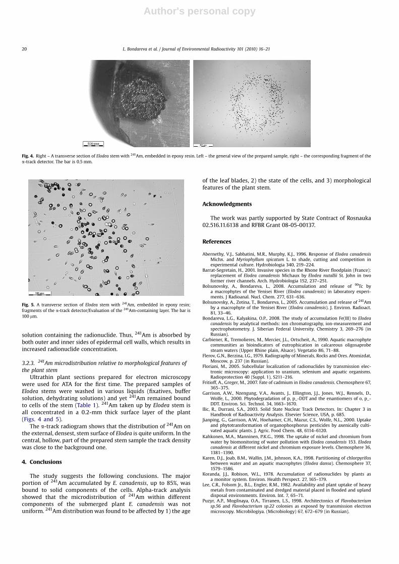
Ultrathin plant sections prepared for electron microscopywere used for ATA for the first time. The prepared samples ofElodea stems were washed in various liquids (fixatives, buffersolution, dehydrating solutions) and yet 241Am remained boundto cells of the stem (Table 1). 241Am taken up by Elodea stem isall concentrated in a 0.2-mm thick surface layer of the plant(Figs. 4 and 5).
The a-track radiogram shows that the distribution of 241Am onthe external, densest, stem surface of Elodea is quite uniform. In thecentral, hollow, part of the prepared stem sample the track densitywas close to the background one.
4. Conclusions
The study suggests the following conclusions. The majorportion of 241Am accumulated by E. canadensis, up to 85%, wasbound to solid components of the cells. Alpha-track analysisshowed that the microdistribution of 241Am within differentcomponents of the submerged plant E. canadensis was notuniform. 241Am distribution was found to be affected by 1) the age
of the leaf blades, 2) the state of the cells, and 3) morphologicalfeatures of the plant stem.
Acknowledgments
The work was partly supported by State Contract of Rosnauka02.516.11.6138 and RFBR Grant 08-05-00137.
References
Abernethy, V.J., Sabbatini, M.R., Murphy, K.J., 1996. Response of Elodea canadensisMichx. and Myriophyllum spicatum L. to shade, cutting and competition inexperimental culture. Hydrobiologia 340, 219–224.
Barrat-Segretain, H., 2001. Invasive species in the Rhone River floodplain (France):replacement of Elodea canadensis Michaux by Elodea nutallii St. John in twoformer river channels. Arch. Hydrobiologia 152, 237–251.
Bolsunovsky, A., Bondareva, L., 2008. Accumulation and release of 99Tc bya macrophytes of the Yenisei River (Elodea canadensis) in laboratory experi-ments. J Radioanal. Nucl. Chem. 277, 631–636.
Bolsunovsky, A., Zotina, T., Bondareva, L., 2005. Accumulation and release of 241Amby a macrophyte of the Yenisei River (Elodea canadensis). J. Environ. Radioact.81, 33–46.
Bondareva, L.G., Kalyakina, O.P., 2008. The study of accumulation Fe(III) to Elodeacanadensis by analytical methods: ion chromatography, ion-measurement andspectrophotometry. J. Siberian Federal University. Chemistry 3, 269–276 (inRussian).
Carbiener, R., Tremolieres, M., Mercier, J.L., Ortscheit, A., 1990. Aquatic macrophytecommunities as bioindicators of eutrophication in calcareous oligosaprobesteam waters (Upper Rhine plain, Alsace). Vegetatio 86, 71–88.
Flerov, G.N., Berzina, I.G., 1979. Radiography of Minerals, Rocks and Ores. Atomizdat,Moscow, p. 237 (in Russian).
Floriani, M., 2005. Subcellular localization of radionuclides by transmission elec-tronic microscopy: application to uranium, selenium and aquatic organisms.Radioprotection 40 (Suppl. 1), S211–216.
Fritioff, A., Greger, M., 2007. Fate of cadmium in Elodea canadensis. Chemosphere 67,365–375.
Garrison, A.W., Nzengung, V.A., Avants, J., Ellington, J.J., Jones, W.J., Rennels, D.,Wolfe, L., 2000. Phytodegradation of p, p_-DDT and the enantiomers of o, p_-DDT. Environ. Sci. Technol. 34, 1663–1670.
Ilic, R., Durrani, S.A., 2003. Solid State Nuclear Track Detectors. In: Chapter 3 inHandbook of Radioactivity Analysis. Elsevier Science, USA, p. 685.
Jianping, G., Garrison, A.W., Hoehamer, C.H., Mazur, C.S., Wolfe, N.L., 2000. Uptakeand phytotransformation of organophosphorus pesticides by axenically culti-vated aquatic plants. J. Agric. Food Chem. 48, 6114–6120.
Kahkonen, M.A., Manninen, P.K.G., 1998. The uptake of nickel and chromium fromwater by biomonitoring of water pollution with Elodea canadensis 153. Elodeacanadensis at different nickel and chromium exposure levels. Chemosphere 36,1381–1390.
Karen, D.J., Joab, B.M., Wallin, J.M., Johnson, K.A., 1998. Partitioning of chlorpyrifosbetween water and an aquatic macrophytes (Elodea dansa). Chemosphere 37,1579–1586.
Koranda, J.J., Robison, W.L., 1978. Accumulation of radionuclides by plants asa monitor system. Environ. Health Perspect. 27, 165–179.
Lee, C.R., Folsom Jr., B.L., Engler, R.M., 1982. Availability and plant uptake of heavymetals from contaminated and dredged material placed in flooded and uplanddisposal environments. Environ. Int. 7, 65–71.
Puzyr, A.P., Mogilnaya, O.A., Tirranen, L.S., 1998. Architectonics of Flavobacteriumsp.56 and Flavobacterium sp.22 colonies as exposed by transmission electronmicroscopy. Microbilogiya. (Microbiology) 67, 672–679 (in Russian).
Fig. 4. Right – A transverse section of Elodea stem with 241Am, embedded in epoxy resin. Left – the general view of the prepared sample, right – the corresponding fragment of thea-track detector. The bar is 0.5 mm.
Fig. 5. A transverse section of Elodea stem with 241Am, embedded in epoxy resin;fragments of the a-track detector/Evaluation of the 241Am-containing layer. The bar is100 mm.
L. Bondareva et al. / Journal of Environmental Radioactivity 101 (2010) 16–2120

Author's personal copy
Rusconi, R., Forte, M., Caresana, M., et al., 2006. The evolution of uncertainty in low-level LSC measurements of water samples. Appl. Radiat. Isot. 64, 1124–1129.
Samecka-Cymerman, A., Kempers, A.J., 2003. Biomonitoring of water pollution withElodea canadensis. A case study of three small polish rivers with different levelsof pollution. Water Air Soil Pollut. 145, 139–153.
Simpson, D.A., 1986. Taxonomy of Elodea Michx in the British Isles. Watsonia 16, 1–4.
Stoyanova, D.P., Tchakalova, E.S., 1993. The effect of lead and copper on thephotosynthetic apparatus in Elodea canadensis Tich. Photosynthetica 28,63–74.
Stoyanova, D.P., Tchakalova, E.S., 1999. Cadmium induced ultrastructural changesin shoot apical meristrem of Elodea canadensis Rich. Photosynthetica 37,47–52.
L. Bondareva et al. / Journal of Environmental Radioactivity 101 (2010) 16–21 21
Copyright © 2022 FDOKUMEN