Microbial Communities and Bioactive Compounds in Marine Sponges of the Family Irciniidae—A Review
34
Mar. Drugs 2014, 12, 5089-5122; doi:10.3390/md12105089 marine drugs ISSN 1660-3397 www.mdpi.com/journal/marinedrugs Review Microbial Communities and Bioactive Compounds in Marine Sponges of the Family Irciniidae—A Review Cristiane C. P. Hardoim and Rodrigo Costa * Microbial Ecology and Evolution Research Group, Centre of Marine Sciences (CCMar), University of Algarve (UAlg), Gambelas, 8005-139 Faro, Portugal; E-Mail: [email protected] * Author to whom correspondence should be addressed; E-Mail: [email protected]; Tel.: +351-289-800-051; Fax: +351-289-800-069. External Editor: Kirk R. Gustafson Received: 21 August 2014; in revised form: 12 September 2014 / Accepted: 16 September 2014 / Published: 30 September 2014 Abstract: Marine sponges harbour complex microbial communities of ecological and biotechnological importance. Here, we propose the application of the widespread sponge family Irciniidae as an appropriate model in microbiology and biochemistry research. Half a gram of one Irciniidae specimen hosts hundreds of bacterial species—the vast majority of which are difficult to cultivate—and dozens of fungal and archaeal species. The structure of these symbiont assemblages is shaped by the sponge host and is highly stable over space and time. Two types of quorum-sensing molecules have been detected in these animals, hinting at microbe-microbe and host-microbe signalling being important processes governing the dynamics of the Irciniidae holobiont. Irciniids are vulnerable to disease outbreaks, and concerns have emerged about their conservation in a changing climate. They are nevertheless amenable to mariculture and laboratory maintenance, being attractive targets for metabolite harvesting and experimental biology endeavours. Several bioactive terpenoids and polyketides have been retrieved from Irciniidae sponges, but the actual producer (host or symbiont) of these compounds has rarely been clarified. To tackle this, and further pertinent questions concerning the functioning, resilience and physiology of these organisms, truly multi-layered approaches integrating cutting-edge microbiology, biochemistry, genetics and zoology research are needed. OPEN ACCESS
-
Upload
independent -
Category
Documents
-
view
0 -
download
0
Transcript of Microbial Communities and Bioactive Compounds in Marine Sponges of the Family Irciniidae—A Review
Mar. Drugs 2014, 12, 5089-5122; doi:10.3390/md12105089
marine drugs ISSN 1660-3397
www.mdpi.com/journal/marinedrugs
Review
Microbial Communities and Bioactive Compounds in Marine Sponges of the Family Irciniidae—A Review
Cristiane C. P. Hardoim and Rodrigo Costa *
Microbial Ecology and Evolution Research Group, Centre of Marine Sciences (CCMar),
University of Algarve (UAlg), Gambelas, 8005-139 Faro, Portugal;
E-Mail: [email protected]
* Author to whom correspondence should be addressed; E-Mail: [email protected];
Tel.: +351-289-800-051; Fax: +351-289-800-069.
External Editor: Kirk R. Gustafson
Received: 21 August 2014; in revised form: 12 September 2014 / Accepted: 16 September 2014 /
Published: 30 September 2014
Abstract: Marine sponges harbour complex microbial communities of ecological and
biotechnological importance. Here, we propose the application of the widespread sponge
family Irciniidae as an appropriate model in microbiology and biochemistry research.
Half a gram of one Irciniidae specimen hosts hundreds of bacterial species—the vast
majority of which are difficult to cultivate—and dozens of fungal and archaeal species.
The structure of these symbiont assemblages is shaped by the sponge host and is highly
stable over space and time. Two types of quorum-sensing molecules have been detected in
these animals, hinting at microbe-microbe and host-microbe signalling being important
processes governing the dynamics of the Irciniidae holobiont. Irciniids are vulnerable to
disease outbreaks, and concerns have emerged about their conservation in a changing
climate. They are nevertheless amenable to mariculture and laboratory maintenance, being
attractive targets for metabolite harvesting and experimental biology endeavours. Several
bioactive terpenoids and polyketides have been retrieved from Irciniidae sponges, but the
actual producer (host or symbiont) of these compounds has rarely been clarified. To tackle
this, and further pertinent questions concerning the functioning, resilience and physiology
of these organisms, truly multi-layered approaches integrating cutting-edge microbiology,
biochemistry, genetics and zoology research are needed.
OPEN ACCESS
Mar. Drugs 2014, 12 5090
Keywords: host-microbe interactions; microbial diversity; polyketide synthases; secondary
metabolites; symbiosis
1. Introduction
The phylum Porifera (sponges) represents the oldest extant metazoan lineage on Earth, with fossil
records dating to around 580 million years ago [1,2]. The ancestry of marine sponges might even be
older than previously thought, as chemical records found for demosponges place the origin of these
animals back to more than 635 million years ago at the transition between the Ediacaran
(Neoproterozoic) and the Cambrian (Paleozoic) periods [3]. Sponges are sessile filter-feeders, with
capability to filter thousands of litres of water per day [4–6]. They can be found in almost all of the
232 marine ecoregions of the World [7]. Eleven such ecoregions encompass 201–461 species, and are
thus considered hot spots of marine sponge diversity [2,7] (Table 1). These animals are restricted to
aquatic habitats and divided into four classes: Demospongiae, Hexactinellida, Calcarea and
Homoscleromorpha, which are further distributed into 25 orders, 128 families, and 680 genera. The
class Demospongiae is by far the richest and most widespread, containing 83% of the 8553 formally
recognized species described so far [2].
Table 1. Hot spots of marine sponge biodiversity.
Realms 1 Provinces Ecoregions Number of Species 2
Tropical Atlantic Tropical North-western Atlantic Eastern Caribbean 208
Tropical Atlantic Tropical North-western Atlantic Greater Antilles 275
Temperate Northern Atlantic Northern European Seas Celtic Seas 295
Temperate Northern Atlantic Lusitanian South European Atlantic Shelf 231
Temperate Northern Atlantic Lusitanian Azores, Canaries, Madeira 458
Temperate Northern Atlantic Mediterranean Sea Western Mediterranean 461
Western Indo-Pacific West and South Indian Shelf South India and Sri Lanka 211
Temperate Northern Pacific Warn Temperate Northwest Atlantic Central Kuroshio Current 377
Central Indo-Pacific Tropical South-western Pacific New Caledonia 256
Temperate Australasia East Central Australian Shelf Manning-Hawkesbury 278
Temperate Australasia Southeast Australian Shelf Bassian 375
1 “Realms”, “Provinces” and “Ecoregions” are defined according to Spalding and colleagues [7]; 2 The number of sponge
species per Ecoregion [7] was obtained from van Soest and colleagues [2].
Although prokaryotic microorganisms are the most important component of their diet, sponges are
known to form symbiotic associations with abundant and diverse bacteria and, in some cases, up to
38% of sponge wet weight consists of bacterial cells [4–6]. So far, 10 recognized bacterial phyla and
18 candidate bacterial phyla have been detected in marine sponges [5,6,8]. Several lineages within
these phyla are involved in the production of secondary metabolites [9], which may enhance host
defence mechanisms against predators and invading pathogens. Besides bacteria, archaea and fungi are
consistently found as constituent members of the marine sponge microbiome, and specific host-related
functions have been proposed or reported for these groups [6,9]. In the light of the enormous richness
Mar. Drugs 2014, 12 5091
of marine sponges across the globe, their contribution as the most prolific sources of marine bioactive
compounds and the actual participation of their microbial symbionts in secondary metabolite
biosynthesis, it is reasonable to posit that marine sponge microbiomes constitute as-yet uncharted,
extremely fertile reservoirs of genetic and metabolic novelties. In this context, the present review
covers microbial community structure, diversity and bioactivities reported for marine sponges of the
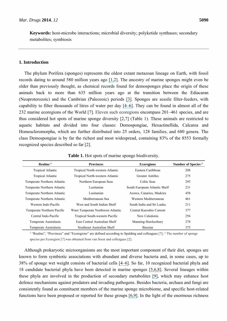
family Irciniidae (Demospongiae, Dictyoceratida), known for its wide geographical distribution
(Figure 1), chemical complexity and distinct microbiota. In addition, we make use of the Irciniidae
family as a model taxon to revisit our knowledge of marine sponge (host and symbiont) cell culturing,
metagenomics-based gene discovery, experimental biology, cell-cell signalling and disease. We follow
the concept of symbiosis as originally proposed by Anton de Bary (see Taylor et al. [6]) to describe
two or more organisms living in close physical association over time. This definition does not imply
any sort of mutual benefit between hosts and symbionts.
Figure 1. Geographical distribution of exemplarily Irciniidae species. The map was drawn
using data available at the World Porifera Database; of note is the wide longitudinal
occurrence of the genus Psammocinia.
2. The Irciniidae Family as a Model Taxon in Sponge Microbiology Research
With 8553 catalogued and 17,000 estimated species, comprehensively describing microbial diversity,
metabolism and functioning in marine sponges is a complex task. Determining how these features
relate to host species, habitat, depth and environmental gradients therefore constitutes a true challenge.
Given this, the need to identify and develop model hosts in sponge microbiology research has been
Mar. Drugs 2014, 12 5092
discussed by specialists at recent meetings such as the 1st International Symposium on Sponge
Microbiology [10] and the Lower Invertebrates Symbiosis with Microorganisms Workshop held in
Eilat, Israel in 2012. In the past 15 years, intense molecular microbiology research performed on, for
example, Aplysina aerophoba from the Mediterranean Sea [11–15], Rhopaloeides odorabile from the
Great Barrier Reef (Australia) [16–20] and Ircinia spp. from the Caribbean [21–25] have naturally
turned these organisms into model sponge hosts. Strategic and long-term research on key species will
most likely improve our understanding of sponge-microbe interactions in a more mechanistic fashion,
allowing us to extract pertinent information from a core of relevant hosts that could represent the
whole. In this regard, a likely rewarding approach to the choice of model organisms is looking for
taxonomic ranks above the species level. This would widen the biodiversity spectrum of the target
animals, expanding the geographical, habitat and niche breadths as well as the phenotypic and genotypic
plasticity ranges under study. Such a strategy could thus facilitate the assessment of microbiome diversity
and function as a response to environmental and host-related factors within a well-contextualized
phylogenetic framework. The family Irciniidae is composed only of marine species divided into three
genera, namely Ircinia, Sarcotragus and Psammocinia, embracing 75, 11 and 25 accepted species,
respectively [26] (see Figure 2 for pictures of some Irciniidae species). Most species have been
registered mainly from tropical to temperate regions [27] (see Figure 1 for the geographical distribution
of exemplarily species) and inhabit the epipelagic layer (i.e., photic zone where there is enough light
for photosynthesis) from zero to 60 m depths. However, a few specimens have been collected from the
mesopelagic layer at about 365 m [28]. Irciniidae species exhibit a wide variety of shapes (for a
technical description, see [27]) and lack authentic spicules, the archetypical mineral skeleton-forming
structures in the majority of demosponges [29]. Instead, fine collagenous and terminally enlarged
spongin filaments are typical features of this family. These constitute the organic fibre skeleton,
making the sponges very difficult to tear. Several reasons suggest the Irciniidae as a suitable model
taxon for microbiology and biotechnology research, as follows:
1. The existence of several sympatric species within the family allows testing the host species-specific
hypothesis of microbiome composition in sponges using a solid phylogenetic background;
2. The wide geographical distribution of some species permits testing hypotheses of symbiont/
metabolite maintenance across biogeographical gradients;
3. Irciniidae species are chemically rich, a feature that might relate with microbial diversity and
suggests biotechnological potential;
4. Irciniidae species have been reported as high microbial abundance sponges. This suggests high
metabolic activity and/or selective enrichment of their symbionts, which most likely play
essential roles for host fitness and survival;
5. Bacterial species from at least 15 phyla have been found to be enriched in Irciniidae hosts when
compared with surrounding seawater. This distinct microbiome composition is indicative of
close host-symbiont relationships;
6. The order Dictyoceratida consists of four families—including Irciniidae—collectively known
as “keratose” sponges because their skeleton is made of a complex matrix of protein fibres
instead of mineral elements (i.e., true spicules), making these families interesting
representatives of a peculiar life strategy within marine sponges.
Mar. Drugs 2014, 12 5093
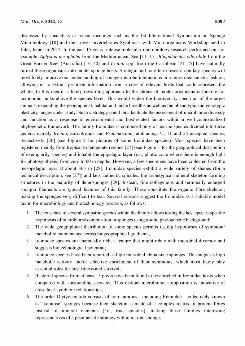
Figure 2. In situ pictures of Ircinia variabilis (a); Ircinia felix (b); Ircinia campana (c);
Sarcotragus spinosulus (d) and of a fixed specimen of Psammocinia compacta (e); Photos
courtesy: Francisco R. Pires (a–d); Panel (e) is reproduced with permission from
Guilherme Muricy (Department of Invertebrates, Federal University of Rio de Janeiro, Brazil).
(a) (b)
(c) (d)
(e)
In the following sections, we provide a historical perspective on the marine sponge microbiome,
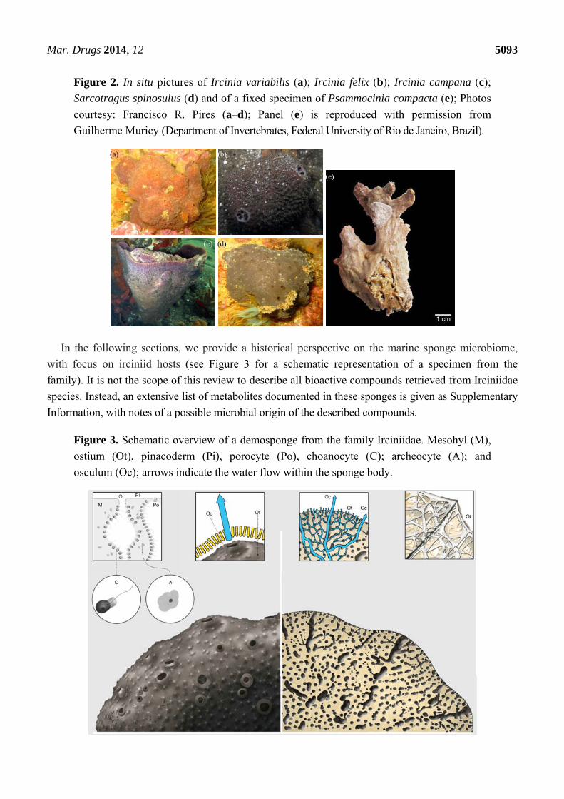
with focus on irciniid hosts (see Figure 3 for a schematic representation of a specimen from the
family). It is not the scope of this review to describe all bioactive compounds retrieved from Irciniidae
species. Instead, an extensive list of metabolites documented in these sponges is given as Supplementary
Information, with notes of a possible microbial origin of the described compounds.
Figure 3. Schematic overview of a demosponge from the family Irciniidae. Mesohyl (M),
ostium (Ot), pinacoderm (Pi), porocyte (Po), choanocyte (C); archeocyte (A); and
osculum (Oc); arrows indicate the water flow within the sponge body.
Mar. Drugs 2014, 12 5094
3. Microbial Diversity and Bioactivities in the Family Irciniidae
Microbial diversity surveys have focused primarily on Caribbean species belonging to the genus
Ircinia [21–24,30–33] and on Ircinia and Sarcotragus species from the eastern Atlantic Ocean and
Mediterranean Sea [34–40]. However, no direct comparison between prokaryotic communities from
irciniids inhabiting these regions has been performed. Previous studies classified members of the
Irciniidae family as high microbial abundance (HMA) sponges [31,38], highlighting their capability of
hosting highly dense microbiomes, ranging from 108–1010 prokaryotic cells·g−1 of sponge (fresh
weight). Although several attempts have been made to isolate secondary metabolites from these
sponges (see Supplementary Information), little attention was paid to determine the actual producer
(i.e., host or symbiont) of such compounds.
3.1. Early Microbiology Studies
First insights into the microbial abundance and morphological diversity in sponges were obtained
by electron microscopy—especially transmission electron microscopy (TEM) [4,41], still an important
tool in several studies [12,42–45]. In the early 1970s, Sara [46] showed that I. variabilis was populated
by the cyanobacteria Aphanocapsa feldmanni and A. raspaigellae. The former was detected in the
cortical mesohyl—the intercellular matrix of sponges—and inside sponge cells, while the latter
was found only in the mesohyl, surrounded by a lacunar space. Both species reproduced outside the
host’s cells. A true cooperative relationship was suggested between host and symbionts, whereby
Aphanocapsa spp. supplied the sponge with organic material originated from photosynthesis, protected
the host from excessive illumination and could use the nitrogenous compounds eliminated by the
sponge, whereas I. variabilis offered shelter and protection for the Cyanobacteria species [46]. Using
scanning and transmission electron microscopy, Wilkinson [47,48] revealed higher abundance of
heterotrophic bacteria in the dense mesohyl of I. wistarii from the Great Barrier Reef (Australia) when
compared with surrounding seawater (SSW). These symbionts concentrated around the inhalant canals
of the sponge. Although pioneering TEM analyses enabled first assessments of the abundance and
morphology of bacteria in sponges, the taxonomic diversity and composition of these symbionts
remained enigmatic until the first culture-dependent and -independent inventories of sponge-associated
bacteria were created. These were ultimately responsible for the enormous increase in knowledge
regarding sponge microbiome diversity in the last 35 years.
3.2. Bacteria
3.2.1. Diversity
3.2.1.1. Culture-Dependent Approaches
Wilkinson [48] isolated 87 bacterial colonies from I. wistarii and performed 76 tests measuring
morphology, physiology and metabolic capabilities. Fifty-nine isolates were grouped into six
well-defined clusters, whereas 28 isolates were too ambiguous to be assigned to any clade.
Surprisingly, these isolates were able to metabolize a wide range of compounds and, for the first time,
comprehensive information on the physiology of sponge-associated bacteria was provided. During the
Mar. Drugs 2014, 12 5095
subsequent 25 years, no attempt was made to cultivate symbionts from Irciniidae species,
whereas several other sponge hosts have been approached in this manner. These studies usually
reported on Alpha- and Gamma classes of Proteobacteria as the prevailing members of the culturable
sponge-associated microbiota [42,43,49]. Likewise, the culturable assemblage of Ircinia sp.
(St. Giovanni, Croatia) was later found to be dominated by Alpha and Gammaproteobacteria [50].
In another survey, Esteves and colleagues [37] isolated more than 270 bacterial strains from
S. spinosulus and I. variabilis on marine agar. These were classified into 17 genera, and 10 putative
new species were detected. In accordance with several cultivation-dependent studies performed with
other hosts [42,51–53], the bacterial genera Pseudovibrio, Ruegeria and Vibrio (Proteobacteria)
prevailed among the retrieved symbionts [37]. Using six Actinobacteria-specific media, Tabares and
colleagues [54] isolated only one single strain, Arthrobacter sp. BA51, from I. felix. The main
advantage of bacterial cultivation is that this allows the determination of the putative functions of these
symbionts. For instance, some studies [37,50] have tested bacterial cultures from Irciniidae specimens
for in vitro antimicrobial activity (see Section 3.2.2), an approach widely used in sponge microbiology.
However, the biases inherent in culture-dependent methods, such as <1% of the total bacterial cells
being recovered [42,55], with most of the isolates belonging to the Proteobacteria [6,43,56], have
favoured the use of cultivation-independent techniques in the characterization of sponge-associated
bacterial communities.
3.2.1.2. Cultivation-Independent Approaches
The application of DNA-based, molecular approaches to the marine sponge microbiome, relying
mainly on the sequencing or fingerprinting of 16S rRNA genes, has illuminated our view of the
bacterial community structure and diversity in these organisms by circumventing the well-known
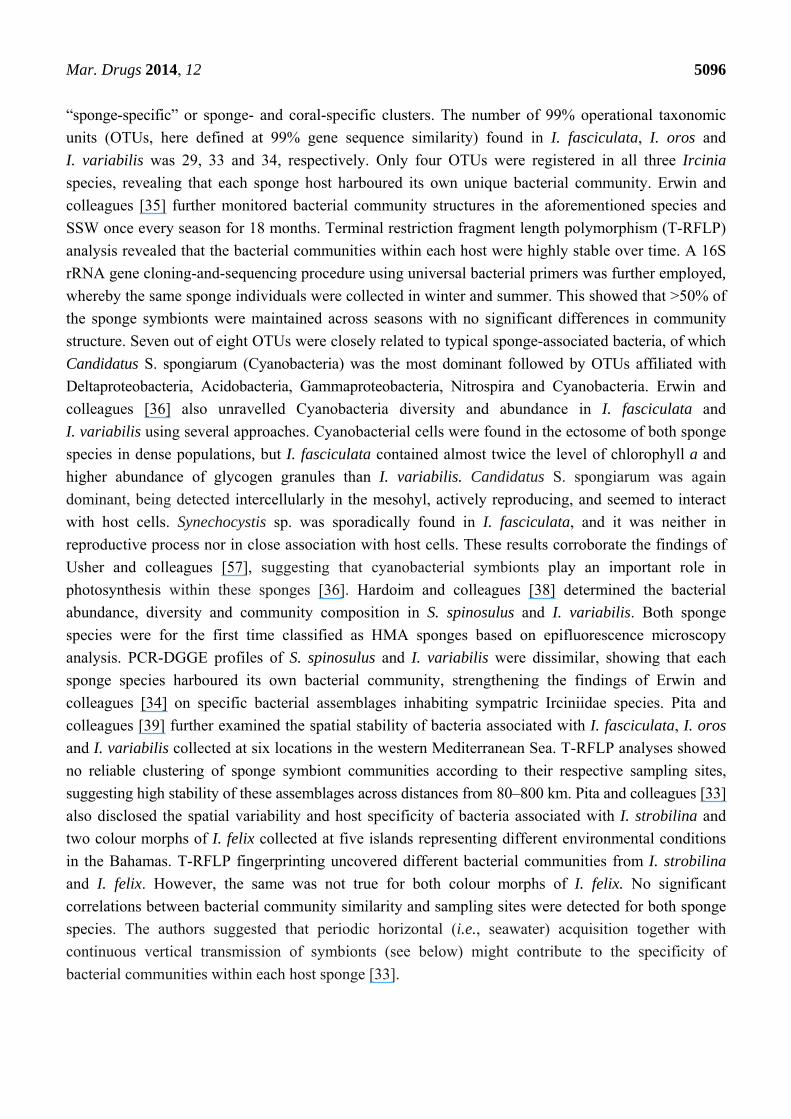
limitations of culturing techniques. Indeed, a meta-analysis performed with 16S rRNA gene sequences
derived from irciniid sponges via cultivation-dependent and -independent methods reveals that the
latter strategy uncovers much higher bacterial diversity from these hosts than the former (Figure 4).
Below, we first address pioneering fingerprinting/cloning-and-sequencing studies, and then recent
surveys that use next generation sequencing to inspect bacterial community structure in marine sponges
with cultivation-independent methods.
Using a Cyanobacteria-specific primer pair, Usher and colleagues [57] detected, by polymerase
chain reaction-denaturing gradient gel electrophoresis (PCR-DGGE), two closely related species of
Synechococcus in I. variabilis. PCR-DGGE fingerprinting and clone-and-sequencing of 16S rRNA
gene fragments using universal bacterial primers were later applied to determine the structure of bacterial
communities associated with I. felix and I. strobilina from Key Largo, Florida, USA [23,30,31];
I. strobilina from Sweetings Cay, Bahamas [32]; and the Mediterranean I. variabilis, I. fasciculata
(currently accepted taxon: Sarcotragus fasciculatus. For the sake of clarity, in this review we will keep
the source name Ircinia fasciculata whenever referring to previous studies) and I. oros [34]. These
studies revealed Acidobacteria, Actinobacteria, Chloroflexi, Cyanobacteria, Gemmatimonadetes,
Nitrospira and Proteobacteria (Alpha, Delta and Gamma classes) as the prevailing bacterial phyla in
Ircinia spp. It was also demonstrated that irciniids harboured a distinct microbiome compared to
SSW [23,30–34]. Erwin and colleagues [34] showed that 38 out of 56 sequences fell within
Mar. Drugs 2014, 12 5096
“sponge-specific” or sponge- and coral-specific clusters. The number of 99% operational taxonomic
units (OTUs, here defined at 99% gene sequence similarity) found in I. fasciculata, I. oros and
I. variabilis was 29, 33 and 34, respectively. Only four OTUs were registered in all three Ircinia
species, revealing that each sponge host harboured its own unique bacterial community. Erwin and
colleagues [35] further monitored bacterial community structures in the aforementioned species and
SSW once every season for 18 months. Terminal restriction fragment length polymorphism (T-RFLP)
analysis revealed that the bacterial communities within each host were highly stable over time. A 16S
rRNA gene cloning-and-sequencing procedure using universal bacterial primers was further employed,
whereby the same sponge individuals were collected in winter and summer. This showed that >50% of
the sponge symbionts were maintained across seasons with no significant differences in community
structure. Seven out of eight OTUs were closely related to typical sponge-associated bacteria, of which
Candidatus S. spongiarum (Cyanobacteria) was the most dominant followed by OTUs affiliated with
Deltaproteobacteria, Acidobacteria, Gammaproteobacteria, Nitrospira and Cyanobacteria. Erwin and
colleagues [36] also unravelled Cyanobacteria diversity and abundance in I. fasciculata and
I. variabilis using several approaches. Cyanobacterial cells were found in the ectosome of both sponge
species in dense populations, but I. fasciculata contained almost twice the level of chlorophyll a and
higher abundance of glycogen granules than I. variabilis. Candidatus S. spongiarum was again
dominant, being detected intercellularly in the mesohyl, actively reproducing, and seemed to interact
with host cells. Synechocystis sp. was sporadically found in I. fasciculata, and it was neither in
reproductive process nor in close association with host cells. These results corroborate the findings of
Usher and colleagues [57], suggesting that cyanobacterial symbionts play an important role in
photosynthesis within these sponges [36]. Hardoim and colleagues [38] determined the bacterial
abundance, diversity and community composition in S. spinosulus and I. variabilis. Both sponge
species were for the first time classified as HMA sponges based on epifluorescence microscopy
analysis. PCR-DGGE profiles of S. spinosulus and I. variabilis were dissimilar, showing that each
sponge species harboured its own bacterial community, strengthening the findings of Erwin and
colleagues [34] on specific bacterial assemblages inhabiting sympatric Irciniidae species. Pita and
colleagues [39] further examined the spatial stability of bacteria associated with I. fasciculata, I. oros
and I. variabilis collected at six locations in the western Mediterranean Sea. T-RFLP analyses showed
no reliable clustering of sponge symbiont communities according to their respective sampling sites,
suggesting high stability of these assemblages across distances from 80–800 km. Pita and colleagues [33]
also disclosed the spatial variability and host specificity of bacteria associated with I. strobilina and
two colour morphs of I. felix collected at five islands representing different environmental conditions
in the Bahamas. T-RFLP fingerprinting uncovered different bacterial communities from I. strobilina
and I. felix. However, the same was not true for both colour morphs of I. felix. No significant
correlations between bacterial community similarity and sampling sites were detected for both sponge
species. The authors suggested that periodic horizontal (i.e., seawater) acquisition together with
continuous vertical transmission of symbionts (see below) might contribute to the specificity of
bacterial communities within each host sponge [33].
Mar. Drugs 2014, 12 5097
Figure 4. Phylum-level classification of bacteria detected in Irciniidae sponges by
cultivation-independent (a) and cultivation-dependent (c) 16S rRNA gene surveys, and
their respective metadata (b,d); the analysis covers all sequences available at NCBI until
December 2013, with the exception of 454-pyrosequencing sequence reads. The phylum
Proteobacteria is represented in (a) and (c) by the classes Alpha, Gamma and
Deltaproteobacteria. The total number of sequences analysed in (a) and (c) is given in brackets.
Gammaproteobacteria
Cyanobacteria
Alphaproteobacteria
Deltaproteobacteria
ChloroflexiAcidobacteria
Actinobacteria
BacteroidetesNitrospira
Firmicutes
Planctomycetes
Unknown affiliation
Lentisphaerae
Poribacteria
Others
Gemmatimonadetes
Uncultured bacteria (n = 1270)(a)
(b)
Sponge species Sampling region Number of sequences Method Reference
I. strobilina Caribbean 244 clones [23] I. strobilina Caribbean 34 clones [24]
I. felix Caribbean 112 DGGE bands [30] I. strobilina Caribbean 80 clones [32]
I. strobilina, I. felix (tan and white morphs)
Caribbean 234
clones [33]
I. variabilis, I. fasciculata Mediterranean 85 clones [34] I. variabilis, I. fasciculata, I. oros Mediterranean 229 clones [35] I. variabilis, I. fasciculata, I. oros Mediterranean 233 clones [36]
I. variabilis, S. spinosulus Eastern Atlantic 15 DGGE bands [38] I. variabilis Mediterranean 2 clones [57]
I. felix Caribbean 2 clones [58]

Mar. Drugs 2014, 12 5098
Figure 4. Cont.
Cultured bacteria (n = 328)(c)
(d)
Sponge species Sampling region Number of sequences Reference
I. strobilina Caribbean 32 [23] I. strobilina Caribbean 10 [25]
I. variabilis, S. spinosulus Eastern Atlantic 279 [37] Ircinia sp. Mediterranean 5 [50]
I. felix Caribbean 1 [54] Ircinia sp. Mediterranean 1 [59]
To overcome limitations common to PCR-DGGE and T-RFLP fingerprinting (i.e., impossibility to
identify the entire prokaryotic community) and cloning-and-sequencing procedures (i.e., restrictions
regarding library sizes), next-generation sequencing (NGS, e.g., 454-pyrosequencing) has been
more often applied in recent years for the characterization of sponge symbiont diversity [20,60]. Using
454-pyrosequencing, 16 bacterial phyla and 1199 OTUs (95% gene similarity cut-off) were recovered
from I. ramosa sampled from the Great Barrier Reef (Australia), whereas 30 bacterial phyla and 6800
OTUs were recovered from SSW [20]. With this technology, Schmitt and colleagues [14] uncovered
159, 179, 86 and 111 bacterial OTUs (97% cut-off) from Ircinia sp., I. felix, I. variabilis and
I. gigantea (note by authors: non-valid taxon) specimens sampled from the Indian Pacific, Caribbean
and Mediterranean Seas, and the Great Barrier Reef, respectively. Overall, the dominant bacterial
phyla associated with Ircinia spp. were Acidobacteria, Chloroflexi, Poribacteria and Proteobacteria
(Alpha and Gamma classes). Both surveys suggested that indeed the bacterial community associated
with marine sponges was species-specific [14,20]. Likewise, 454-pyrosequencing was employed in
recent studies covering cultivation bias, host specificity and temporal dynamics of microbial
communities associated with Irciniidae species in the north Atlantic. Hardoim and colleagues [61]
addressed the effects of three methods of sample handling on bacterial community composition in

Mar. Drugs 2014, 12 5099
S. spinosulus and I. variabilis. Using cultivation-independent methods, the bacteriome of S. spinosulus
and I. variabilis was indeed found to be species-specific, deepening the observations made by means
of PCR-DGGE profiling [38], whereas similar bacterial assemblages were detected in both sponge
species via culturing, highlighting the bias associated with describing sponge microbiome diversity
based solely on cultivation attempts. These authors also localized bacterial symbionts in I. variabilis
and S. spinosulus by fluorescence in situ hybridization (FISH). They observed that bacterial cells were
almost exclusively detected in-between sponge cells and ususally absent on the spongin fibers that
form the skeleton of Irciniidae sponges (Figure 5). In addition, Hardoim and Costa [62] unravelled the
temporal dynamics of the bacterial community associated with S. spinosulus over three sucessive
years. PCR-DGGE and tag-pyrosequencing showed that the bacterial assemblage was stable over time,
with the majority of dominant bands and OTUs detected in all years. S. spinosulus was dominated by
eight bacterial phyla, of which only two presented different relative abundances over the years. Only
27 OTUs, distributed in nine bacterial phyla, were detected in all 12 sponge specimens. Interestingly,
in spite of its low diversity at the phylotype level, this minimal core of symbionts shared by all animals
analysed displayed high bacterial richness at the phylum level, comparable to that observed in a single
sponge hosting about 200 OTUs. This suggests that phylum diversification is fundamental to sponge
functioning, and that high redundancy of phylotypes within each phylum likely aids the sponge host in
maintaining its repertoire of bacterial phyla in face of local environmental changes or across different
developmental stages.
In summary, the recent application of NGS has enabled a better understanding of the
“species-level” bacterial diversity in irciniid sponges in comparison with previous molecular
technologies. For instance, between 29 and 33 OTUs (99% cut-off) have been reported for Ircinia spp.
using the cloning-and-sequencing method [34], whereas hundreds to thousands of OTUs have been
recovered from irciniids via NGS [14,20,61,62], unmasking the complex microbiomes that inhabit
these animals. Nevertheless, previous findings made based on less powerful tools, such as the trends
for spatial and temporal stability of these symbiont assemblages, have been confirmed by deep
sequencing surveys.
Besides the use of the 16S rRNA gene as a phylogenetic marker to describe sponge-associated
bacterial communities, other genes have been selected to uncover putative functions from sponge
symbionts, especially those involved in biogeochemical cycling [6]. Among the elements essential to
life, nitrogen is one of the most limiting factors affecting ecosystem functioning [63]. The nifH gene
(which encodes an iron-containing dinitrogenase reductase) has been widely used in diversity
assessments of N-fixing bacteria in aquatic and terrestrial environments [64–66]. Mohamed and
colleagues [21] determined the expression of nifH genes in I. strobilina. From the cDNA clone library
made for this species, 44 nifH gene clones were retrieved. They clustered within a group formed
exclusively by cyanobacterial phylotypes, indicating that Cyanobacteria symbionts were the prevailing
N-fixers in I. strobilina. Mohamed and colleagues [24] also disclosed the diversity of aerobic
ammonia-oxidizing bacteria (AAOB) associated with I. strobilina using cloning-and-sequencing of the
amoA gene (which encodes the catalytic α-subunit of the ammonia-monooxygenase enzyme). All
amoA sequences fell into two clusters affiliated with Nitrosospira (Betaproteobacteria), suggesting that
they were the main nitrifiers in I. strobilina. However, no expression of the amoA gene could be
detected [24]. Hardoim and Costa [62] used the amoA gene to unravel the temporal dynamics of
Mar. Drugs 2014, 12 5100
ammonia-oxidizing bacteria (AOB) in S. spinosulus by PCR-DGGE fingerprinting. Few AOB
phylotypes could be detected in the PCR-DGGE profiles of S. spinosulus over the three successive
years, suggesting that the AOB community in this species was stable over time. As sponges comprise a
major portion of the biomass in many coral reefs, microbial mediated nitrogen metabolism within the
animal host is expected to have a major impact on the nitrogen budget of these environments [21,24].
More studies are still necessary to ascertain the activity and identity of N-cycling players in
marine sponges.
Figure 5. Confocal laser scanning microscopy images of fluorescent in situ hybridization
(FISH-CLSM) stained bacteria in S. spinosulus; volume rendering images (left in
each panel) and their corresponding 3D reconstructions (right in each panel) are shown
for hybridizations with the Cy3-labeled universal bacterial probe (red cells) coupled
to ALEXA488- or Cy5-labeled group-specific probes targeting the Alpha- and
Gammaproteobacteria classes; Gammaproteobacteria cells appear as yellowish cells in the
volume rendering images (a) and as green objects in the 3D reconstruction (b). For
co-hybridizations including bacterial, alpha and gammaproteobacterial probes, the latter
two groups are represented by yellowish and pink cells, respectively (c,d). Sponge
background structure, vastly dominated by profuse spongin filaments, is shown overall in
blue or cyan.
(a) Gammaproteobacteria + Bacteria (b) Gammaproteobacteria + Bacteria
(c) Alphaproteobacteria + Gammaproteobacteria + Bacteria (d) Alphaproteobacteria + Gammaproteobacteria + Bacteria
Altogether, Irciniidae species harbour complex and diverse bacteriomes. These are to a large extent
maintained by the host species over seasons, years, environmental gradients and geographical regions.
Through an in-depth phylogenetic assessment comprising all bacterial 16S rRNA gene sequences

Mar. Drugs 2014, 12 5101
(excluding NGS and PCR-DGGE short sequence reads) from Irciniidae sponges publicly available
until December 2013, we here substantiate the view of high spatial bacteriome stability in these
sponges (Supplementary Figure S1). In fact, bacterial phylogenetic signatures common to the major
oceanic regions studied so far could be found within several typical sponge-associated phyla such as
the Acidobacteria, Actinobacteria, Bacteroidetes, Chloroflexi, Cyanobacteria and Alpha, Delta and
Gamma classes of the Proteobacteria (Supplementary Figure S1a–i). Irciniidae species appear,
moreover, to hold their own distinct bacterial communities. These differ from host to host within the
same habitat and from the surrounding bacterioplankton communities found in seawater. Thus,
intrinsic aspects of the host species—for instance habitat preference, specific geographic distribution
and evolutionary history—may cooperatively shape the structure of bacterial symbiont communities in
these sponges [34]. Several bacterial taxa associated with Irciniidae species are known for their
physiological and metabolic features—of which inhibitory, primary production and nitrogen
metabolism capacities can be highlighted—indicating that they play essential roles in the establishment
and maintenance of the sponge host.
3.2.2. Bioactivities
Few surveys have described biological activities obtained from bacterial strains—or from their
respective metabolite extracts—isolated from Irciniidae species [37,50,67,68]. Although these studies
usually do not unveil the identity of the compounds underlying the observed activities, they
collectively highlight a broad spectrum of functions of ecological or applied interest within the
culturable sponge-associated microbiota. For instance, Thakur and colleagues [68] addressed the
hypothetical importance of I. fusca-associated bacteria in host defence against bacterial epibionts.
In total, 25 isolates were recovered from the sponge surface in five sampling events. Crude sponge
extracts were obtained from all events, but only two were active against isolates retrieved from the
same sampling. Strains identified as Micrococcus sp. and Bacillus sp. exhibited antibacterial and
antifouling activities, highlighting the potential of both genera to inhibit the growth of epibiotic
bacteria on the sponge surface. These results suggest that sponge-associated bacteria can modulate the
growth of their own cells as well as their neighbours by producing bioactive metabolites. Esteves and
colleagues [37] described the in vitro antagonistic activity of 155 distinct genotypes isolated from
S. spinosulus and I. variabilis towards two relevant clinical strains: Escherichia coli NCTC 9001
(Gram-negative) and Staphylococcus aureus NCTC 6571 (Gram-positive). A few isolates (n = 18)
were active against both strains, whereas 44 and 27 isolates showed antimicrobial activity towards
E. coli and S. aureus, respectively. The most active genus isolated from Irciniidae species was Vibrio
with 27 and 12 of the isolates inhibiting E. coli and S. aureus, respectively. From S. fasciculatus
collected from the Bay of Bengal (India), five Firmicutes isolates and one Proteobacteria isolate
inhibited β-glucosidase activity [69]. Glucosidase inhibitors may be of use in the treatment of viral
infections, cancer and genetic disorders [70]. These studies emphasized the biotechnological potential
of the culturable bacterial fraction of Irciniidae sponges, encouraging more research into the
mechanisms and compounds underpinning such activities.
Mar. Drugs 2014, 12 5102
3.2.3. Bioactive Compounds
Some studies identified the bioactive compounds extracted from Irciniidae-associated bacteria.
Several cyclic peptides have been retrieved from Bacillus and Staphylococcus strains isolated from
I. variabilis from the Bay of Naples, Italy [71]. Of these, the compound cyclo-(L-prolyl-L-tyrosine)
could modulate the activity of the LuxR-based quorum sensing system of bacteria [72], whereas all
other compounds investigated presented structural similarities to compounds known to interact with
LuxR-based biosensors [72,73]. Thus, such cyclic peptides may play a role in the marine sponge
microbiome as quorum sensing signal molecules [71]. Furthermore, a Bacillus pumilus strain was
isolated from Ircinia sp. and found to produce five surfactin-like substances (cyclic acyldepsipeptides)
called bacircines [74,75]. These compounds showed cytotoxic effects on the development of sea urchin
eggs. One exhibited antitumour activity against Ehrlich ascites carcinoma and anti-HIV activity [74,75].
These studies demonstrate that the functional screening of compounds extracted from sponge-associated
bacteria is a promising, direct route to manifold interesting bioactivities, which nevertheless
remains underexplored.
3.3. Archaea
In comparison with the multi-layered molecular research undertaken in the past decade on
sponge-associated bacteria, fewer surveys have addressed archaeal community composition and
diversity in marine sponges. Overall, these studies uncovered less diverse archaeal assemblages from
sponges than the corresponding bacterial communities [6,8,76].
3.3.1. Diversity
Phylogenetic inference performed with three distinct archaeal 16S rRNA gene sequences obtained
from Sarcotragus sp. (Jeju Island, Korea) revealed that they were affiliated to marine group
I Crenarchaeota (now known as Thaumarchaeota, see [77–79]). Two of them grouped within clusters
containing only sponge-derived sequences, but without bootstrap support [80]. Further analysis showed
that one of these sequences indeed belonged to a Thaumarchaeota “sponge-specific” cluster (SC175) [76].
Using PCR-DGGE and 454-pyrosequencing, we demonstrated that the archaeal community
associated with S. spinosulus was stable over three successive years. In 11 out of 12 S. spinosulus
specimens, one single OTU affiliated to Nitrosopumilus dominated the archaeal assemblage, whereas
the remaining specimen was dominated by Cenarchaeum sp. Candidatus Nitrosopumilus maritimus
and Cenarchaeum sp. are known ammonia oxidizers. This indicates that the replacement of the
dominant archaeal symbiont observed for one S. spinosulus individual likely does not interfere with the
ammonia-oxidation potential in this host [62]. Overall, the diversity of archaea associated with other
sponge species was as well encompassed by only few phylotypes [6,76,81,82]. Despite their low
diversity, it appears that Archaea, and not Bacteria, are the prevalent players in the process of
ammonia-oxidation in marine sponges [83–85]. It still needs to be determined which other possible
roles, if any, archaeal symbionts play in association with Irciniidae and marine sponges as a whole.

Mar. Drugs 2014, 12 5103
3.3.2. Bioactivities and Bioactive Compounds
Neither in vitro bioactivities of archaeal cultures and their metabolite extracts nor identified
bioactive compounds from sponge-associated Archaea were found in our literature survey.
3.4. Fungi
As pointed out by Webster and Taylor [8], the study of the diversity and functioning of sponge-derived
fungi still requires more effort. Fungi have been approached primarily in a cultivation-dependent
manner, probably because of their higher amenability to laboratory domestication than that displayed
by bacteria and archaea. Whereas this facilitates functional screenings for manifold activities and full
genome exploration of the obtained pure cultures, it is felt that comprehensive analyses of fungal
diversity and maintenance across temporal and spatial scales could benefit more from dedicated
cultivation-independent studies.
3.4.1. Diversity
The diversity of fungi associated with I. oros and I. variabilis collected at Malta was assessed using
several media and incubation for over 30 days at room temperature (~20 °C) [86]. Both sponge species
were dominated by Aspergillus spp. corresponding to 39.4% and 50%, respectively, of the total
isolates followed by Penicillium spp. (13.6% and 27%). Whereas 13 and 10 fungal genera were found
in I. oros and I. variabilis, respectively, only five of them were shared by both [86]. The diversity of
culturable fungi was also assessed in Psammocinia sp. (Sedot-Yam, Israel) [87]. Potato dextrose
medium amended with fungicides was used in two approaches: direct plating of inner sponge
fragments and direct plating of sponge extracts (“sponge-compressed” method). The Petri dishes were
incubated at 25 °C in the dark from three to 30 days. Sequencing of the internal transcribed spacer
(ITS) of rRNA genes was used to identify the fungal strains. Overall, 220 pure cultures were isolated
and assigned to 85 fungal taxa, of which the majority was obtained using the sponge-compressed
method (n = 76). Most of the strains (94%) were identified as Ascomycota, whereas Basidiomycota
(4%) and Glomeromycota (2%) were minor components of the cultured fungal community.
From Ascomycota, 80 “species-level” taxa were recovered and among them 25 were identified as
Eurotiales (Aspergillus and Penicillium spp.), five as Capnodiales (Cladosporium spp.), 15 as
Pleosporales (Bionectria, Fusarium, Phoma, and Preusia spp.) and 35 taxa as Hypocreales (Acremonium
and Trichoderma spp.). When sponge-compressed samples were plated onto medium amended with
different fungicides, further 28 taxa that were not obtained with the previous approach could be
isolated, thus enabling an increase in the diversity of fungi cultured from the sponge. Using a
coverage-based algorithm, the authors estimated that only 15% of the fungi associated with
Psammocinia sp. could be recovered [87]. Notably, even though both studies used distinct approaches,
the fungal diversity obtained from I. oros, I. variabilis [86] and Psammocinia sp. [87] was alike.
3.4.2. Bioactivities
Accompanying their assessment of fungal diversity, Paz and colleagues [87] also investigated the
inhibitory activities of fungi isolated from Psammocinia sp. using a diffusion bioassay. Thirty-six

Mar. Drugs 2014, 12 5104
fungal cultures showed in vitro antagonism against at least one of the test fungi (Alternaria alternata,
Rhizoctonia solani and Neurospora crassafour) or against the oomycete Pythium aphanidermatum.
Some isolates (Trichoderma, Acremonium, Bionectria, Verticillium, Penicillium and Aspergillus spp.)
were able to secret inhibitory compounds into the growth medium, although those were not identified.
It was demonstrated that distinct Trichoderma spp. isolated from Psammocinia sp. were capable to
mycoparasite Fusarium equiseti isolated from the same sponge species [87].
3.4.3. Bioactive Compounds
Penicillium chrysogenum was isolated from the Mediterranean I. fasciculata collected in the Bight
of Fetovaia (Italy) and was shown to produce the sorbicillinoid alkaloids sorbicillactones A and B [88].
Sorbicillactone A exhibited remarkable cytotoxic activity towards murine leukaemia lymphoblast
(L5178y) cells and anti-HIV activity. This compound is also a promising neuroprotective metabolite.
Three other sorbicillinoid compounds (oxosorbicillinol, sorbicillin and bisvertinolone) were identified
from P. chrysogenum, along with the alkaloids meleagrine and roquefortine C [88]. The isolate
Microascus sp. K14 from I. variabilis also produced the anti-fungal compound fungerin [86].
Irciniids harbour a complex, readily culturable fungal diversity. The potential for uncovering novel
fungal-derived bioactive compounds from sponges will become more apparent with the improvement
of cultivation and screening techniques and/or the accomplishment of in-depth molecular surveys such
as whole genome sequencing of fungal symbionts. This will also improve our understanding of the
roles played by sponge-associated fungi within the host.
3.5. Other Microeukaryotes
Although diatoms and dinoflagellates have been detected in marine sponges [6], no study involved
irciniids. There is thus a need to unravel microeukaryote taxonomic, functional and metabolic
diversities within sponges as a first step in the pursuit of their biotechnological potential.
In summary, much of the microbial diversity research undertaken so far, considering both marine
sponges as a whole and the family Irciniidae, has focused on bacterial communities. Such an interest
may reflect the higher relevance of this particular group of symbionts in host fitness. Nevertheless,
more effort to unveil the spatial-temporal diversity, composition and abundance of archaeal and
microeukaryote assemblages—and their corresponding biological activities—in marine sponges is
needed for a balanced picture of the marine sponge holobiont. Within the family Irciniidae, more
attention to the genus Ircinia was found. Attempts to approach the microbial ecology of S. spinosulus
have been published only recently [37,38,62], and the highly selective and conserved character of its
microbiome makes it a valuable species for comparative studies with other irciniids in the
Atlanto-Mediterranean zone and beyond. Besides the recent study by Paz and colleagues on fungal
diversity and inhibitory activities [87], very limited information exists on the microbial ecology of
Psammocinia spp., in spite of their distinct geographical range (Figure 1) and manifold bioactivities
(see Supplementary Information). Collectively, approaching the microbiology of these three genera
might constitute a rewarding strategy to understanding the microbiology of marine sponges on a global
scale with a robust, underlying comparative framework.
Mar. Drugs 2014, 12 5105
4. Vertical Transmission of Sponge Symbionts
Vertical transmission is an important mechanism by which sponge-specific symbionts are passed
from the parental to the next generation via larvae or gametes. It has been investigated in several
sponges and, when present, has usually been interpreted as evidence for an intimate pattern of
relationship between host and the transmitted symbionts [30,52,58,89–91].
Vertical transmission was proposed as a mode of symbiont inheritance in I. felix through the
detection of bacteria in adult, larval and juvenile stages of the sponge [30]. This study showed, via
TEM, that adults contained large and complex bacterial communities, and that high bacterial
abundance was also observed extracellularly in the inner region of the larvae, whereas the outer region
was almost free of microorganisms. When the host entered the juvenile stage, bacterial cells were
found primarily in the mesohyl. Bacterial 16S rRNA gene sequences obtained from I. felix adults and
offspring (larvae and/or juvenile) belonged to the phyla Acidobacteria, Chloroflexi, Gemmatimonadetes
and Proteobacteria (Alpha, Delta, and Gamma classes), and to one lineage of uncertain affiliation. In a
subsequent study, Schmitt and colleagues [58] detected several vertically transmitted phylogenetic
clusters (VT-clusters), composed by sequences obtained from I. felix adults and offspring. VT-clusters
affiliated with the Acidobacteria, Chloroflexi, Gemmatimonadetes, Nitrospira and Proteobacteria
phyla, suggesting vertical transmission as a mechanism through which a complex bacterial consortium
can be formed and maintained within I. felix. We still do not know what their function may be in the
larvae, nor in settlement, which may occur from a few minutes to several hours after release [92].
Once settled, sponge larvae undergo a rapid metamorphosis to an early juvenile stage. Thus, bacterial
symbionts might protect the juvenile from predators by producing antibiotics and deterrent
compounds. Arguably, the best example of such an ecological function was obtained by Lopanik and
colleagues [93–96] and Sudek and colleagues [97] while studying the association between the bacterial
symbiont Candidatus Endobugula sertula and the bryozoan Bugula neritina. Candidatus E. sertula was
found to produce the bioactive polyketides bryostatins. These protected the larvae against predators
and were observed in all life stages of B. neritina. Bacterial symbionts in sponge larvae may also
perform important housekeeping functions or constitute a source of food, since the larvae are unable to
take up food particles from seawater [98].
Even though Schmitt and colleagues [58] also described two archaeal VT-clusters from the marine
sponges Agelas conifera and Luffariella variabilis, there is to date no specific documentation of
archaeal vertical transmission in Irciniidae sponges, although its existence can be presumed based on
available data. Our understanding of fungal acquisition by marine sponges remains otherwise very
limited, with neither dedicated studies of this group in sponge larvae or juveniles nor an existing
conceptual model on how sponges recruit and maintain these symbionts.
5. Bacterial Communication and Signalling Molecules
Quorum sensing (QS) is a mechanism by which microorganisms monitor and regulate their
population size through chemical signalling [99]. When quorum-sensing molecules (QSM) released by
microorganisms into the extracellular environment reach a threshold concentration, they initiate a
signalling transduction cascade that regulates the expression of several target genes in an orchestrated
response to the prevailing conditions. QS is a complex mechanism involved in bacterial virulence,

Mar. Drugs 2014, 12 5106
swarming motility, conjugal plasmid transfer, biofilm maturation, and antibiotic production and
resistance [99]. As such, it affects several activities or areas relevant to human health and the
environment (e.g., bacterial multidrug resistance, aquaculture, water purification). These might be
impacted by negative feedbacks ensued from QS and its mediation of bacterial metabolism [99,100].
Solutions to QS-derived drawbacks may arise from the activities of the symbiont communities
themselves. For instance, from the marine sponge Luffariella variabilis, the compounds manoalide,
secomanolide and manoalide monoacetate were obtained and showed to be important QS inhibitors [101].
Furthermore, the alkaloids ageliferin and mauritamide B obtained from Agelas conifera and A. nakamurai,
respectively, inhibited the QS of the bacterial reporter Chromobacterium violaceum CV017, whereas
seven compounds isolated from distinct marine sponges inhibited the QS of C. violaceum CV017 and
showed antibiotic properties [102].
One of the most studied groups of QSM, N-acyl homoserine lactones (AHLs), was investigated in
I. strobilina inhabiting shallow waters in Key Largo, Florida [22]. Two Alphaproteobacteria and three
Gammaproteobacteria isolates were found to produce AHLs, indicating that QS systems play an
important role in sponge microbiome dynamics as observed for other systems. The sponge mesohyl
constitutes a nutrient rich environment when compared to oligotrophic seawater, thus colonizing
bacteria may reach high densities within this microenviroment, allowing them to perform activities
mediated by quorum sensing.
Autoinducer-2 (AI-2) is one well-characterized molecule involved in bacterial interspecies
communication [99,100]. Briefly, during AI-2 biosynthesis, S-adenosylmethionine is transformed into
4,5-dihydroxy-2,3-pentanedione (DPD) by a sequence of three enzymatic reactions, where the last is
catalyzed by the enzyme LuxS. Since DPD is unstable, it naturally cyclizes, generating a range of
furanones in the presence of water. Vibrio harveyi, for example, detects the borate diester form of AI-2
using the LuxP/LuxQ signaling cascade, which may be exclusive to Vibrio organisms. AI-2 is associated
with a broad range of functions; for instance, virulence in V. vulnificus and V. cholera, type III secretion,
protease production, luminescence, colony morphology and siderophore production in V. harveyi [99].
Several surveys detected Vibrio spp. associated with different marine sponges [37,49–51,103–106],
indicating that they may be capable of performing an array of functions within the sponge host. From
I. strobilina (Key Largo, USA), 10 out of 40 isolates were identified as Vibrio spp. based on 16S
rRNA gene sequencing [25] and found to be closely related to V. harveyi (n = 5) and V. campbelli
(n = 4). Notably, all 10 Vibrio isolates were capable of synthesizing AI-2 molecules. Furthermore,
30 distinct Vibrio luxS gene sequences were identified, of which 28 were closely related to the luxS
gene of V. harveyi and the other two were affiliated with the luxS gene of V. parahaemolyticus.
Interestingly, two luxS gene sequences were distantly related to any known Vibrio luxS sequence.
These were proposed to represent a sponge-specific luxS cluster [25].
Two of the five known classes of QSM have already been detected in the bacterial community
associated with I. strobilina, and more may be found as research advances. Considering the complexity
of the Irciniidae microbiome, future investigations involving, for example, full genome sequencing of
symbionts and deep sponge metagenomic mining (Figure 6) for QS regulatory genes may result in the
discovery of other classes of QSM. It is too early to determine the precise role(s) of QS in gene
regulation and metabolism of both symbionts and host. The application potential of QS regulation can
be nevertheless exemplified from other host-microbe associations in the marine realm. For instance,
Mar. Drugs 2014, 12 5107
Shewanella sp. strain MIB010, isolated from the intestine of the ayu fish, is an effective agent against
the QS-regulated biofilm formed by the fish pathogen V. anguillarum [100].
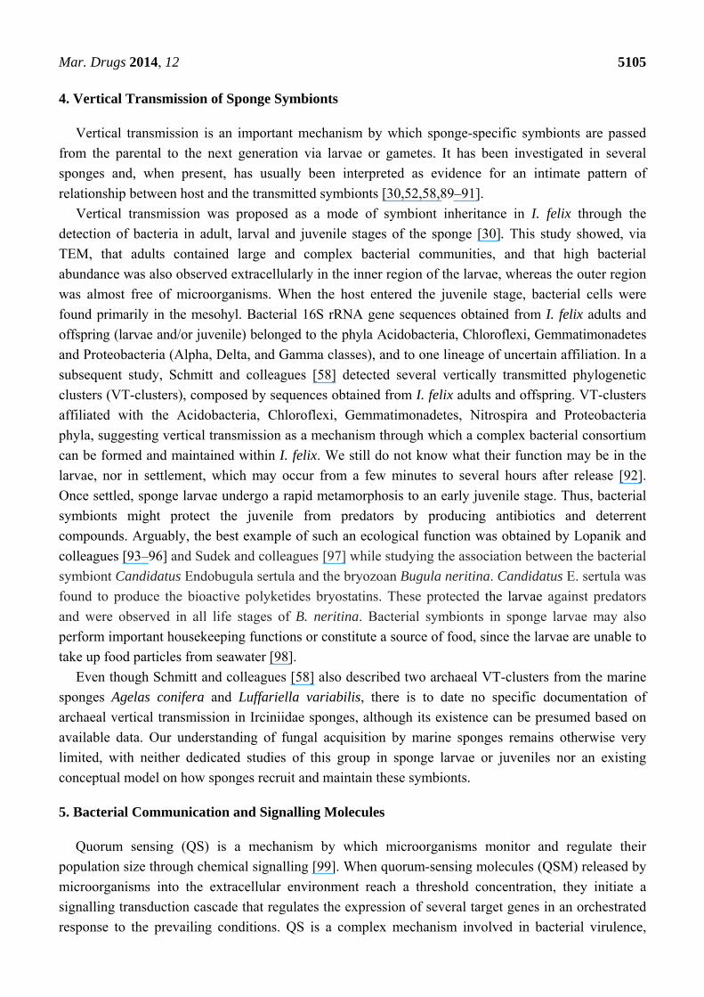
Figure 6. Integrated molecular approach for the study of symbiont communities in
Irciniidae sponges.
Sponge sample Symbiont cultivation / Single cell sorting
Reconstruct genomes from metagenomes
Functional gene profiling, mining of novel gene clusters
Probe and primer design
Gene diversity
Microarray hybridization
Stable isotope probing
DNA/RNA (cDNA)
extraction
PCR amplification
NGS
Gene diversity
Metagenome/transcriptome sequencing; large-insert libraries
Gene abundance
In situ hybridization-microscopy (FISH-CLSM)
genome sequencing
Microbial cell suspensions
Target functional and/or phylogenetic marker genes
q-PCR
Fingerprinting (DGGE, T-RFLP)
Cell localization and abundance
Link phylogeny and function, assess metabolism
Cell lysis
6. Diseases Affecting Irciniids
Many studies have shown that microorganisms might also cause disease in marine sponges [107]. In
1884, a disease affected Indian Ocean Ircinia spp., whereby fungal filaments were shown to destroy
the sponge body, leaving only the hard spongin fibres [107]. In addition, the abundance of I. variabilis
in the benthic community of the Marsala Lagoon (Italy) decreased from 6.9% in 1986 to 3.0% in 1989,
due to a disease characterized by white patches on the surface of affected individuals [108]. Several
Ircinia specimens from Guigalatupo, Panamá, were lost over five censuses performed from 1984 to
1998, whereby spreading lesions were seen in I. felix, I. campana and I. strobilina [109]. A mass
mortality event was reported in 1994 affecting S. spinosulus and Ircinia sp. in the Mediterranean Sea,
characterized by the decay of their spongin filaments [107]. Four more recent disease outbreaks
affected Irciniidae species in the Mediterranean, Ionian and Adriatic Seas [59,110–112], eventually
with severe consequences for the infected populations [111]. Whereas loss of photosynthetic
cyanobacteria was documented among diseased specimens in one case [111], higher abundance of
Vibrio spp. was observed in another [59]. In one study [110], a twisted rod bacterium was proposed as
the etiological agent because it penetrated the sponge body, proliferated, and outcompeted a large
range of bacterial symbionts. Shifts in symbiont communities towards a more virulent state
because of elevated seawater temperature have been hypothesized to trigger the mass mortality
events [59,110–112]. In this context, Pita and colleagues [113] subjected I. fasciculata and I. oros to
aquaria experiments under high temperature (25 °C), food shortage (0.1 μm-filtered seawater) and both
Mar. Drugs 2014, 12 5108
factors in combination. T-RFLP analysis showed that the sponge-associated bacterial communities did
not change after acclimation time and during the next three weeks of experiment. TEM revealed that
the ectosome of I. fasciculata was dominated by Candidatus S. spongiarum. Under high temperature
and food shortage, both healthy and damaged cyanobacterial cells were observed. For I. oros under all
treatments, no signs of sponge or bacterial cell degradation or cyanobacterial cells were detected. Thus,
high temperature and food shortage up to three weeks did not disrupt the bacterial communities
associated with both sponge hosts. The authors suggested that these factors could not alone explain the
mass mortality events registered for Ircinia spp. in the Mediterranean Sea [113].
To date, few surveys have identified bacterial species as the etiological agents of disease in
sponges [59,107]. More often, a clear cause-effect relationship between agent and disease is difficult to
achieve [107,114]. More research is needed to improve our capability of identifying etiological agents
and unravelling how biotic and abiotic features influence marine sponge disease incidence in a
changing climate.
7. Experimental Microbial Ecology
Sponge microbiology is a fertile field for experimentation. Most of the first, experimental
sponge microbial ecology studies kept the host animals for varying periods in aquaria, and tested
whether the structure and function of their associated bacterial communities shifted during the
maintenance periods [23,115,116]. Surveys using other sponge hosts reported changes in bacterial
community composition of specimens kept in aquaria over six months and two years [115,116].
In contrast, evidence exists of recovery within Irciniidae symbiont communities back to the in-situ stage
after some time in captivity [23]. Specifically, Mohamed and colleagues applied molecular techniques to
monitor bacterial communities associated with I. strobilina maintained in aquaculture [23]. Thirty-five
OTUs were retrieved from wild specimens, while 48 and 47 OTUs were recovered from specimens
kept in aquaria for three or nine months, respectively. Overall, these OTUs were affiliated to five or
six bacterial phyla depending on the category (wild, 3- and 9-month sponges). Although the highest
diversity was observed for the 3-month sponges, 9-month sponges showed an intermediate diversity
between wild and 3-month sponges, which suggested acclimatization of the holobiont to the
aquaculture system. From the sponges maintained in aquaculture for three and nine months, chemical
fingerprints of small molecules including primary and secondary metabolites extracted with ethanol
were analysed by liquid chromatography-mass spectrometry and compared to the wild type.
The analyses revealed no major changes in the natural product profile of I. strobilina upon transfer to
aquaculture [23]. This suggests that, although the bacterial community from aquarium-maintained
I. strobilina differed from those of the wild specimens along the assay, the major functions
were retained. This could be explained by the functional redundancy of sponge bacterial
symbionts [117,118]. So far, no study approached sponge-associated archaeal or fungal communities
during maintenance in aquaria.
It is still to be determined whether sponge microbial communities can be successfully stabilized in
aquaculture, and longer surveys may shed more light. Technical problems in maintaining sponges in
captivity have hindered experimental progress, which is fundamental to elucidate how the marine
sponge microbiome might be affected by e.g., temperature increase [119,120], water acidification [121]
Mar. Drugs 2014, 12 5109
or invasive pathogens (see Section 6 above). However, Ircinia spp. have shown amenability to
experimental handling [23,113], a feature that reinforces their suitability as a model taxon in
microbiology and biochemistry studies.
8. Metagenomics-Based Discovery of Secondary Metabolites Biosynthetic Gene Clusters
Polyketides are usually found in sponges and comprise a structurally varied class of compounds that
have attracted considerable attention due to their highly potent cytotoxicity [122]. Polyketide synthases
(PKS) are multifunctional enzymes of typical microbial origin that catalyse polyketides biosynthesis.
However, the complexity of the bacterial assemblages associated with marine sponges makes
identification of the actual PKS producers difficult. Some techniques have been applied to overcome
these limitations; for instance, cloning-and-sequencing, genome, single-cell genome and metagenome
mining of PKS encoding operons (Figure 6). Targeting the biosynthetic gene clusters, two pederin-type
PKS systems putatively involved in the synthesis of antitumour polyketides were located in the
metagenomic DNA of Theonella swinhoei [123]. Likewise, metagenomic libraries generated from cell
fractions enriched for filamentous bacteria associated with Discodermia dissoluta revealed that they
consisted of several non-ribosomal synthase (NRPS) as well as mixed PKS-NRPS gene clusters [124].
Moreover, genomic mining of two bacterial cells sorted from Aplysina aerophoba and belonging to the
phyla Poribacteria and Chloroflexi revealed Sup-PKS (“sponge symbionts ubiquitous PKS gene”)
and NRPS gene clusters in the screened genomes, respectively [15]. Single-cell genomics further
uncovered at least two PKSs, one of which affiliated with the sponge-specific “Sup-type PKS” from a
Poribacteria cell sourced from A. aerophoba [125]. Collectively, these studies demonstrate the
wide applicability of metagenomics to studying the function and bioactive potential of marine
sponge symbionts.
Within Irciniidae sponges, psymberin was isolated from Psammocinia aff. bulbosa collected at
Milne Bay (Papua New Guinea) [126]. The complete sequencing of three fosmids revealed common
features of bacterial genomic architecture: no introns were found, the genes were preceded by
Shine-Dalgarno sequences, the space between genes suggesting that the transcribed mRNA was
polycistronic and the close relationship to genes exclusively from bacteria. Nevertheless, the low
similarity of these genetic signatures to genes from Pseudomonas sp. or other prokaryotes hampered
the correct identification of the bacterial producer. Finally, a strong correlation among bacterial
abundance, the presence of Poribacteria and Sup-PKS was observed in the Caribbean I. felix, and
it was proposed that Poribacteria was most likely the producer of mid-chain-branched fatty acids
(MBFAs) from this sponge [127].
9. Cultivation of Irciniidae Species
Even though marine sponges produce the largest variety of secondary metabolites in marine
ecosystems, few compounds have reached commercial production. This is because of the normally
minute amounts readily found in the sponge body and of the impossibility to collect sufficiently large
quantities of sponge biomass from their natural habitats, which is intolerable given the foreseen
impacts on the marine environment and natural sponge populations. To circumvent this, many
techniques have been developed to cultivate sponge species [6,128–130]. Below we highlight attempts
Mar. Drugs 2014, 12 5110
to sponge cultivation using Irciniidae. We divide these attempts into three modalities: sponge
mariculture, cell culture and larviculture.
9.1. Sponge Mariculture
In a preliminary study, Wilkinson and Vacelet [131] failed to transplant I. variabilis collected from
Endoume (near Marseille) at 3–4 m from well-shaded cave walls and rock cliffs to a 7 m deep rock
shelf around 30 m from the sampling place. All specimens died a few days after sampling, most likely
due to severe damage caused by sampling, cutting and sewing of I. variabilis into plastic plaques.
Duckworth and colleagues [132] examined the feasibility of cultivating Psammocinia hawere
in situ. Fifty specimens of P. hawere were collected from 10–20 m at “Ti Point Reef” (New Zealand).
They were cut into cubes of four different sizes and transplanted to two locations, along with small
whole sponges as undamaged controls. Three depths (5, 10 and 17 m) and two culture systems were
used, and the experiment was conducted in summer (82 days) and winter (88 days). The survivorship
was considered high (276 out of 360 explants), even though 2/3 of the explants (226) lost weight.
P. hawere explants transported to deeper (10 and 17 m) waters in winter showed the highest growth
and survivorship rates. This was related to the lower UV radiation as well as the cooler water
temperatures, which helped to accelerate pinacoderm healing. P. hawere is known to incorporate
detritus from the sediment into its body or fibres, which may have enhanced the surface consolidation
during the healing process. Growth and survivorship improved with the size of the explant and the
proportion of the intact pinacoderm. Independently of the culture systems used, P. hawere displayed
better growth and survivorship at 10 and 17 m than at 5 m.
Van Treeck and colleagues [133] sampled 52 and 100 specimens of I. variabilis at the
north-western coast of Corsica in summer and spring, respectively. They were transplanted to naturally
formed sand patches in-between seagrass meadows at 15 m. Prior to transplanting, the sponge
specimens were cut into different sizes underwater. The explants were placed between two frames,
closed, mounted and transported to the experimental sites. The survival rates of I. variabilis were high
within the first six months, followed by a reduction of 25% in population size, most likely due to a
strong storm, after which the remaining sponge continued to grow. The specimens stocked in spring
survived better. In the first 12 months, I. variabilis biomass increased by 90%; however, there was
great variation between maximum and minimum rates (220 and −43%, respectively) [133].
In another study [134], I. ramosa was collected from Bone Lola reef (Indonesia) and cut in situ into
explant sizes of around 30 mm where at least one side contained exopinacoderm. A polyethylene rope
was passed through the sponge body, each rope carrying around nine explants. Ropes were attached to
PVC frames that were placed horizontally ~20 cm above the reef bottom between 12–15 m deep, and
secured with iron pegs at the exposed side of the submerged reef. A high survival rate of 92% was
observed for I. ramosa during the four months. A slight but significant increase in length was detected,
and a change in shape was observed which could not be precisely measured. However, only few
explants grew during the trials [134].
Mar. Drugs 2014, 12 5111
9.2. Sponge Cell Culture
In a series of studies, de Rosa and colleagues [135–137] meticulously monitored the chemical
composition of I. muscarum cells grown on a simple culture medium. Cell suspensions from
I. muscarum (Gulf of Naples, Italy) were resuspended in sterile seawater complemented with
antibiotics during the first two weeks of cultivation. The cultures were incubated at 18 °C in the dark
and at 22 °C in the light and every third day the medium was renewed. From the third week onward,
cultures were kept in Dulbecco’s modified Eagle’s medium supplemented with glucose. This medium
was again renewed every third day. One week after inoculation, cells were observed attached to the
bottom of culture chambers along with some aggregates. Cells were actively dividing in both culture
conditions and stationary phase was reached after four days. Overall, 17 sterols were recovered.
Around 90% of the total free sterols in cultured I. muscarum were represented by Δ5−7 sterols; from
which 7-dehydrocholesterol, ergosterol and 7-dehydrositosterol prevailed. The medium was then
supplemented with water-soluble cholesterol, which increased the number of observed cells by 70%.
Oleic acid was one of the main acids in the lipids of the intact sponge and was found in low
concentration in cultured cells. When the media were supplemented with oleic and linoleic
water-soluble fatty acids, no growth promotion was observed. However, they were completely
metabolized by the cells. The concentration of total lipids was higher in the intact animal, and cells
developed in the dark had higher total lipids than those grown under light. Several structurally distinct
groups of volatile compounds were observed, and their concentrations indicated that bacteria (from
diet or symbionts) could be involved in their formation. A number of free amino acids were detected in
the intact sponge, whereas practically none was found in the cell cultures. The major difference in
carbohydrate composition between intact and cultured sponge cells was the presence of γ-lactones of
3-desoxy-arabino-hexonic acid and 3-desoxy-ribohexonic acids in higher concentrations in the cell
cultures, suggesting shifts in their carbohydrate metabolism. The cultured cells did not produce any
secondary metabolites as the major cell energy supplies had likely been allocated to the biosynthesis of
primary compounds [135–137].
9.3. Sponge Larviculture
With the aim of culturing sponges from larvae, de Caralt and colleagues [138] transported mature
individuals of I. oros sampled from I’Escala (Mediterranean Sea) to an open aquaculture system,
where they released larvae after about 45 days. Swimming larvae were then collected and transferred
to six-multi-well dishes. These were placed in aquaria with filtered seawater (0.7 µm pore diameter) at
the same temperature of the field (20 °C). Settlement of the larvae on the six-multi-well dish bottom
started after 24 h and larvae metamorphosed into juveniles after five to seven days. An exhalant tube
should be formed after this period; however, no visible inhalant/exhalant orifices were observed
among the settlers of I. oros and, at this point, most juveniles died before the skeletal fibres could be
formed. Although settlement success was considered high (94.5%), no survival of I. oros specimens
was observed after 50 days of experiment. Success in settlement could be related to favourable
conditions such as still water and no substrate competition found by the larvae in the laboratory [138].
The first two weeks of culture showed the maximum increase in area, but this was primarily due to
Mar. Drugs 2014, 12 5112
rearrangement of the biomass during metamorphosis, rather than true growth [138]. Therefore,
unknown and specific environmental cues are likely needed to enable full development of sponge
larvae into mature individuals in the laboratory.
From the three modalities approached above, for Irciniidae species mariculture is apparently the
most feasible (host) culturing methodology, rather than cell or larvae cultivation. However, whether
irciniids will produce secondary metabolites under these conditions remains to be tested. Osinga and
colleagues [128] reported on five sponge species that, grown in mariculture, were able to keep the
biosynthesis of target metabolites, demonstrating that this approach can be rewarding. Optimization of
explant size, depth of transplantation, temperature ranges, and exposure to light and water currents
might improve sponge productivity and bioactive compound availability. Besides mariculture, ex-situ
culture (i.e., aquaculture) of adult specimens and semi-synthesis of metabolites are promising
alternatives. In the former, the sponge grows under controlled conditions in flow-through or recirculation
systems, and the experimental costs and yields of bioactive compounds would be comparable to
mariculture. In the latter, a biosynthetic precursor produced by a genetically modified bacterium
triggers a limited number of further chemical reactions to the synthesis of the final product. For
instance, using the antibiotic cyanosafracin B, which was obtained from Pseudomonas fluorescens via
bacterial fermentation, an analogue of the antitumor compound ecteinascidin 743 isolated from the
tunicate Ecteinascidia turbinata was effectively synthesized [130]. Combining biological and chemical
approaches might be an effective way to cultivate sponges and recover their secondary metabolites.
10. Concluding Remarks and Outlook
Irciniidae-microbe interactions are pivotal to host health and have been evolving since the
hosts’ origins. In recent years, a few fundamental issues regarding these interactions have been
successfully addressed. For instance, we now know that distinct bacterial symbionts are selectively
enriched within the sponge body and, eventually, vertically transmitted, and that such symbiont
communities are stable along time and across geographical regions. Nevertheless, many other
questions remain unanswered. How full symbiont genomes evolve across space and time, both within
individuals of the same species and between different host species? Do single gene-based analyses
(e.g., 16S rRNA gene sequencing) suffice to completely determine microbial diversification and
evolution in marine sponges? What are the functions played by the symbionts in host health and
functioning, and which symbiont does what? How do the vertically transmitted microorganisms affect
larval settlement and juvenile establishment? What are the benefits of quorum sensing for the host?
Are secondary metabolites produced by symbionts effectively involved in host’s chemical defence? Is
there a true etiological agent (or many) underpinning disease outbreaks in Irciniidae species? Is the
existent technology for their laboratory maintenance robust and widely applicable? Furthermore, the
majority of microbiology studies of Irciniidae species have so far mainly determined bacterial
community structures in these hosts. Although useful in shedding new light on our understanding of
the diversity and stability of bacterial assemblages in marine sponges, these inventories usually do not
unravel the biotechnological potential of the bacterial partners. Other microbial associates, such as
archaea, fungi and other microeukaryotes—and their biotechnological value—have been left almost
unaddressed. Thus, only by integrating the full holobiome in future surveys will a more comprehensive
Mar. Drugs 2014, 12 5113
picture emerge, allowing better understanding of host function. Currently, several advanced molecular
approaches such as metatranscriptomics, metaproteogenomics and single-cell genomics, may help
answer these questions (Figure 6). Indeed, their recent application has already been useful in
disentangling the phylogeny-function dilemma, and in unveiling the life-style and secondary metabolite
biosynthesis capacity of several, so-far uncultured, marine sponge symbionts [15,125,139–141]. Finally,
few alternative culturing protocols have been attempted so far for marine sponge microbial associates.
The spectrum of microorganisms cultured from these hosts is likely to grow in the next few years as
researchers develop dedicated protocols to the capture of “hard-to-culture” symbionts. In this regard,
ever-increasing genetic datasets retrieved via cultivation-independent methods can help in the
identification of symbiont metabolic pathways and physiological demands that could be explored in
future cultivation trials. Given the aforementioned challenges, a joint interdisciplinary effort is clearly
needed, from fields ranging from microbiology to chemistry, and from genetics to zoology, to advance
the knowledge of the oldest extant metazoan-microbe interaction system on Earth. Ideally, this would
couple laboratory experimentation with integrative systems biology strategies; therefore, we reinforce
the need to select marine sponge models, and provide a multitude of reasons in favour of the Irciniidae
family as one such model.
Acknowledgments
This research was financed by the Portuguese Foundation for Science and Technology (FCT)
through the research projects PTDC/MAR/101431/2008 and PTDC/BIA-MIC/3865/2012, granted to
RC. It was further partially supported by the European Regional Development Fund (ERDF)
through the Operational Competitiveness Programme (COMPETE) and by national funds through FCT
(Foundation for Science and Technology), under the project “PEst-C/MAR/LA0015/2011”.
CCPH was the recipient of a PhD fellowship granted by FCT (Grant No. SFRH/BD/60873/2009).
Massimiliano Cardinale is thanked for his guidance in FISH-CLSM analyses. We acknowledge Gobius
Ciência e Comunicação (Portugal) for the artwork designs in Figures 1 and 3.
Author Contributions
Cristiane C. P. Hardoim and Rodrigo Costa conceived and designed the study. Cristiane C. P. Hardoim
collected the literature and analyzed the data. Cristiane C. P. Hardoim and Rodrigo Costa wrote
the manuscript.
Conflicts of Interest
The authors declare no conflict of interest.
References
1. Li, C.W.; Chen, J.Y.; Hua, T.E. Precambrian sponges with cellular structures. Science 1998, 279,
879–882.
Mar. Drugs 2014, 12 5114
2. Van Soest, R.W.M.; Boury-Esnault, N.; Vacelet, J.; Dohrmann, M.; Erpenbeck, D.;
de Voogd, N.J.; Santodomingo, N.; Vanhoorne, B.; Kelly, M.; Hooper, J.N.A. Global diversity of
sponges (Porifera). PLoS One 2012, 7, e35105.
3. Love, G.D.; Grosjean, E.; Stalvies, C.; Fike, D.A.; Grotzinger, J.P.; Bradley, A.S.; Kelly, A.E.;
Bhatia, M.; Meredith, W.; Snape, C.E.; et al. Fossil steroids record the appearance of
Demospongiae during the Cryogenian period. Nature 2009, 457, 718–722.
4. Vacelet, J.; Donadey, C. Electron-microscope study of association between some sponges and
Bacteria. J. Exp. Mar. Biol. Ecol. 1977, 30, 301–314.
5. Hentschel, U.; Piel, J.; Degnan, S.M.; Taylor, M.W. Genomic insights into the marine sponge
microbiome. Nat. Rev. Microbiol. 2012, 10, 641–675.
6. Taylor, M.W.; Radax, R.; Steger, D.; Wagner, M. Sponge-associated microorganisms: Evolution,
ecology, and biotechnological potential. Microbiol. Mol. Biol. Rev. 2007, 71, 295–347.
7. Spalding, M.D.; Fox, H.E.; Halpern, B.S.; McManus, M.A.; Molnar, J.; Allen, G.R.;
Davidson, N.; Jorge, Z.A.; Lombana, A.L.; Lourie, S.A.; et al. Marine ecoregions of the world:
A bioregionalization of coastal and shelf areas. Bioscience 2007, 57, 573–583.
8. Webster, N.S.; Taylor, M.W. Marine sponges and their microbial symbionts: Love and other
relationships. Environ. Microbiol. 2012, 14, 335–346.
9. Thomas, T.R.A.; Kavlekar, D.P.; LokaBharathi, P.A. Marine drugs from sponge-microbe
association: A review. Mar. Drugs 2010, 8, 1417–1468.
10. Taylor, M.W.; Hill, R.T.; Hentschel, U. Meeting report: 1st International Symposium on Sponge
Microbiology. Mar. Biotechnol. 2011, 13, 1057–1061.
11. Bayer, K.; Scheuermayer, M.; Fieseler, L.; Hentschel, U. Genomic mining for novel
FADH2-dependent halogenases in marine sponge-associated microbial consortia. Mar. Biotechnol.
2013, 15, 63–72.
12. Fieseler, L.; Horn, M.; Wagner, M.; Hentschel, U. Discovery of the novel candidate phylum
“Poribacteria” in marine sponges. Appl. Environ. Microbiol. 2004, 70, 3724–3732.
13. Hentschel, U.; Hopke, J.; Horn, M.; Friedrich, A.B.; Wagner, M.; Hacker, J.; Moore, B.S.
Molecular evidence for a uniform microbial community in sponges from different oceans.
Appl. Environ. Microbiol. 2002, 68, 4431–4440.
14. Schmitt, S.; Tsai, P.; Bell, J.; Fromont, J.; Ilan, M.; Lindquist, N.; Perez, T.; Rodrigo, A.;
Schupp, P.J.; Vacelet, J.; et al. Assessing the complex sponge microbiota: Core, variable and
species-specific bacterial communities in marine sponges. ISME J. 2012, 6, 564–576.
15. Siegl, A.; Hentschel, U. PKS and NRPS gene clusters from microbial symbiont cells of marine
sponges by whole genome amplification. Environ. Microbiol. Rep. 2010, 2, 507–513.
16. Fan, L.; Liu, M.; Simister, R.; Webster, N.S.; Thomas, T. Marine microbial symbiosis heats up:
The phylogenetic and functional response of a sponge holobiont to thermal stress. ISME J. 2013,
7, 991–1002.
17. Pantile, R.; Webster, N. Strict thermal threshold identified by quantitative PCR in the sponge
Rhopaloeides odorabile. Mar. Ecol. Prog. Ser. 2011, 431, 97–105.
18. Simister, R.; Taylor, M.W.; Tsai, P.; Webster, N. Sponge-microbe associations survive high
nutrients and temperatures. PLoS One 2012, 7, e52220.
Mar. Drugs 2014, 12 5115
19. Webster, N.S.; Cobb, R.E.; Negri, A.P. Temperature thresholds for bacterial symbiosis with a
sponge. ISME J. 2008, 2, 830–842.
20. Webster, N.S.; Taylor, M.W.; Behnam, F.; Lucker, S.; Rattei, T.; Whalan, S.; Horn, M.;
Wagner, M. Deep sequencing reveals exceptional diversity and modes of transmission for
bacterial sponge symbionts. Environ. Microbiol. 2010, 12, 2070–2082.
21. Mohamed, N.M.; Colman, A.S.; Tal, Y.; Hill, R.T. Diversity and expression of nitrogen fixation
genes in bacterial symbionts of marine sponges. Environ. Microbiol. 2008, 10, 2910–2921.
22. Mohamed, N.; Cicirelli, E.; Kan, J.; Chen, F.; Fuqua, C.; Hill, R. Diversity and quorum-sensing
signal production of Proteobacteria associated with marine sponges. Environ. Microbiol. 2008,
10, 75–86.
23. Mohamed, N.; Rao, V.; Hamann, M.; Kelly, M.; Hill, R. Monitoring bacterial diversity of the
marine sponge Ircinia strobilina upon transfer into aquaculture. Appl. Environ. Microbiol. 2008,
74, 4133–4143.
24. Mohamed, N.M.; Saito, K.; Tal, Y.; Hill, R.T. Diversity of aerobic and anaerobic
ammonia-oxidizing bacteria in marine sponges. ISME J. 2010, 4, 38–48.
25. Zan, J.D.; Fuqua, C.; Hill, R.T. Diversity and functional analysis of luxS genes in vibrios from
marine sponges Mycale laxissima and Ircinia strobilina. ISME J. 2011, 5, 1505–1516.
26. Van Soest, R.W.M.; Boury-Esnault, N.; Hooper, J.N.A.; Rützler, K.; de Voogd, N.J.;
Alvarez de Glasby, B.; Hajdu, E.; Pisera, A.B.; Manconi, R.; Schoenberg, C.; et al. World
Porifera Database. Available online: http://www.marinespecies.org/porifera (accessed on 20
December 2013).
27. Cook, S.C.; Bergquist, P.R. Family Irciniidae Gray, 1867. In System Porifera: A Guide to the
Classification of Sponges; Hooper, J.N.A., van Soest R.W.M., Eds.; Kluwer Academic/Plenum
Publishers: New York, NY, USA, 2002; pp. 1022–1027.
28. Cook, S.; Bergquist, P. New species of dictyoceratid sponges from New Zealand: Genus Ircinia
(Porifera: Demospongiae: Dictyoceratida). N. Z. J. Mar. Freshw. Res. 1999, 33, 545–563.
29. Wörheide, G.; Dohrmann, M.; Erpenbeck, D.; Larroux, C.; Maldonado, M.; Voigt, O.;
Borchiellini, C.; Lavrov, D.V. Deep phylogeny and evolution of sponges (Phylum Porifera).
In Advances in Marine Biology; Becerro, M.A., Uriz, M.J., Maldonado, M., Turon, X., Eds.;
Academic Press: Amsterdam, The Netherlands. 2012; pp. 1–18.
30. Schmitt, S.; Weisz, J.; Lindquist, N.; Hentschel, U. Vertical transmission of a phylogenetically
complex microbial consortium in the viviparous sponge Ircinia felix. Appl. Environ. Microbiol.
2007, 73, 2067–2078.
31. Weisz, J.; Hentschel, U.; Lindquist, N.; Martens, C. Linking abundance and diversity of
sponge-associated microbial communities to metabolic differences in host sponges. Mar. Biol.
2007, 152, 475–483.
32. Yang, J.K.; Sun, J.; Lee, O.O.; Wong, Y.H.; Qian, P.Y. Phylogenetic diversity and
community structure of sponge-associated bacteria from mangroves of the Caribbean Sea.
Aquat. Microb. Ecol. 2011, 62, 231–240.
33. Pita, L.; López-Legentil, S.; Erwin, P.M. Biogeography and host fidelity of bacterial
communities in Ircinia spp. from the Bahamas. Microb. Ecol. 2013, 66, 437–447.
Mar. Drugs 2014, 12 5116
34. Erwin, P.M.; López-Legentil, S.; González-Pech, R.; Turon, X. A specific mix of generalists:
Bacterial symbionts in Mediterranean Ircinia spp. FEMS Microbiol. Ecol. 2012, 79, 619–637.
35. Erwin, P.M.; Pita, L.; López-Legentil, S.; Turon, X. Stability of sponge-associated bacteria
over large seasonal shifts in temperature and irradiance. Appl. Environ. Microbiol. 2012, 78,
7358–7368.
36. Erwin, P.M.; López-Legentil, S.; Turon, X. Ultrastructure, molecular phylogenetics, and
chlorophyll a content of novel cyanobacterial symbionts in temperate sponges. Microb. Ecol.
2012, 64, 771–783.
37. Esteves, A.I.S.; Hardoim, C.C.P.; Xavier, J.R.; Gonçalves, J.M.S.; Costa, R. Molecular richness
and biotechnological potential of bacteria cultured from Irciniidae sponges in the north-east
Atlantic. FEMS Microbiol. Ecol. 2013, 85, 519–536.
38. Hardoim, C.C.P.; Esteves, A.I.S.; Pires, F.R.; Gonçalves, J.M.S.; Cox, C.J.; Xavier, J.R.;
Costa, R. Phylogenetically and spatially close marine sponges harbour divergent bacterial
communities. PLoS One 2012, 7, e53029.
39. Pita, L.; Turon, X.; López-Legentil, S.; Erwin, P.M. Host rules: Spatial stability of bacterial
communities associated with marine sponges (Ircinia spp.) in the western Mediterranean Sea.
FEMS Microbiol. Ecol. 2013, 86, 268–276.
40. Usher, K.; Kuo, J.; Fromont, J.; Toze, S.; Sutton, D. Comparative morphology of five species of
symbiotic and non-symbiotic coccoid Cyanobacteria. Eur. J. Phycol. 2006, 41, 179–188.
41. Wilkinson, C.R. Microbial associations in sponges. III. Ultrastructure of in situ associations in
coral-reef sponges. Mar. Biol. 1978, 49, 177–185.
42. Webster, N.S.; Hill, R.T. The culturable microbial community of the Great Barrier Reef
sponge Rhopaloeides odorabile is dominated by an alpha-proteobacterium. Mar. Biol. 2001, 138,
843–851.
43. Hentschel, U.; Usher, K.M.; Taylor, M.W. Marine sponges as microbial fermenters. FEMS
Microbiol. Ecol. 2006, 55, 167–177.
44. Hardoim, C.C.P.; Costa, R.; Araújo, F.V.; Hajdu, E.; Peixoto, R.; Lins, U.; Rosado, A.S.;
van Elsas, J.D. Diversity of bacteria in the marine sponge Aplysina fulva in Brazilian coastal
waters. Appl. Environ. Microbiol. 2009, 75, 3331–3343.
45. Kamke, J.; Taylor, M.W.; Schmitt, S. Activity profiles for marine sponge-associated bacteria
obtained by 16S rRNA vs. 16S rRNA gene comparisons. ISME J. 2010, 4, 498–508.
46. Sara, M. Ultrastructural aspects of symbiosis between two species of genus Aphanocapsa
(Cyanophyceae) and Ircinia variabilis (Demospongiae). Mar. Biol. 1971, 11, 214–221.
47. Wilkinson, C.R. Microbial associations in sponges. I. Ecology, physiology and
microbial-populations of coral-reef sponges. Mar. Biol. 1978, 49, 161–167.
48. Wilkinson, C.R. Microbial associations in sponges. II. Numerical-analysis of sponge and water
bacterial populations. Mar. Biol. 1978, 49, 169–176.
49. Hentschel, U.; Schmid, M.; Wagner, M.; Fieseler, L.; Gernert, C.; Hacker, J. Isolation and
phylogenetic analysis of bacteria with antimicrobial activities from the Mediterranean sponges
Aplysina aerophoba and Aplysina cavernicola. FEMS Microbiol. Ecol. 2001, 35, 305–312.
50. Muscholl-Silberhorn, A.; Thiel, V.; Imhoff, J.F. Abundance and bioactivity of cultured
sponge-associated bacteria from the Mediterranean Sea. Microb. Ecol. 2008, 55, 94–106.
Mar. Drugs 2014, 12 5117
51. Thiel, V.; Imhoff, J.F. Phylogenetic identification of bacteria with antimicrobial activities
isolated from Mediterranean sponges. Biomol. Eng. 2003, 20, 421–423.
52. Enticknap, J.J.; Kelly, M.; Peraud, O.; Hill, R.T. Characterization of a culturable
alphaproteobacterial symbiont common to many marine sponges and evidence for vertical
transmission via sponge larvae. Appl. Environ. Microbiol. 2006, 72, 3724–3732.
53. O’Halloran, J.A.; Barbosa, T.M.; Morrissey, J.P.; Kennedy, J.; O’Gara, F.; Dobson, A.D.W.
Diversity and antimicrobial activity of Pseudovibrio spp. from Irish marine sponges.
J. Appl. Microbiol. 2011, 110, 1495–1508.
54. Tabares, P.; Pimentel-Elardo, S.M.; Schirmeister, T.; Huenig, T.; Hentschel, U.
Anti-protease and immunomodulatory activities of bacteria associated with Caribbean sponges.
Mar. Biotechnol. 2011, 13, 883–892.
55. Friedrich, A.B.; Fischer, I.; Proksch, P.; Hacker, J.; Hentschel, U. Temporal variation of
the microbial community associated with the Mediterranean sponge Aplysina aerophoba.
FEMS Microbiol. Ecol. 2001, 38, 105–113.
56. Hugenholtz, P. Exploring prokaryotic diversity in the genomic era. Genome Biol. 2002, 3,
doi:10.1186/gb-2002-3-2-reviews0003.
57. Usher, K.; Fromont, J.; Sutton, D.; Toze, S. The biogeography and phylogeny of unicellular
cyanobacterial symbionts in sponges from Australia and the Mediterranean. Microb. Ecol. 2004,
48, 167–177.
58. Schmitt, S.; Angermeier, H.; Schiller, R.; Lindquist, N.; Hentschel, U. Molecular microbial
diversity survey of sponge reproductive stages and mechanistic insights into vertical transmission
of microbial symbionts. Appl. Environ. Microbiol. 2008, 74, 7694–7708.
59. Stabili, L.; Cardone, F.; Alifano, P.; Tredici, S.M.; Piraino, S.; Corriero, G.; Gaino, E. Epidemic
mortality of the sponge Ircinia variabilis (Schmidt, 1862) associated to proliferation of a Vibrio
bacterium. Microb. Ecol. 2012, 64, 802–813.
60. Lee, O.O.; Wang, Y.; Yang, J.K.; Lafi, F.F.; Al-Suwailem, A.; Qian, P.Y. Pyrosequencing
reveals highly diverse and species-specific microbial communities in sponges from the Red Sea.
ISME J. 2011, 5, 650–664.
61. Hardoim, C.C.P.; Cardinale, M.; Cúcio, A.; Esteves, A.I.S.; Berg, G.; Xavier, J.R.; Cox, C.;
Costa, R. Bacterial cultivation bias examined for keratose marine sponges. Unpublised work, 2014.
62. Hardoim, C.C.P.; Costa, R. Temporal dynamics of prokaryotic communities in the marine
sponge Sarcotragus spinosulus. Mol. Ecol. 2014. 23, 3097–3112.
63. Fiore, C.L.; Jarett, J.K.; Olson, N.D.; Lesser, M.P. Nitrogen fixation and nitrogen
transformations in marine symbioses. Trends Microbiol. 2010, 18, 455–463.
64. Zehr, J.P.; Mellon, M.T.; Zani, S. New nitrogen-fixing microorganisms detected in
oligotrophic oceans by amplification of nitrogenase (nifH) genes. Appl. Environ. Microbiol.
1998, 64, 3444–3450.
65. Zehr, J.P.; Jenkins, B.D.; Short, S.M.; Steward, G.F. Nitrogenase gene diversity and microbial
community structure: A cross-system comparison. Environ. Microbiol. 2003, 5, 539–554.
66. Man-Aharonovich, D.; Kress, N.; Bar Zeev, E.; Berman-Frank, I.; Beja, O. Molecular ecology
of nifH genes and transcripts in the eastern Mediterranean Sea. Environ. Microbiol. 2007, 9,
2354–2363.
Mar. Drugs 2014, 12 5118
67. Thakur, N.; Anil, A. Antibacterial activity of the sponge Ircinia ramosa: Importance of its
surface-associated bacteria. J. Chem. Ecol. 2000, 26, 57–71.
68. Thakur, N.; Anil, A.; Müller, W. Culturable epibacteria of the marine sponge
Ircinia fusca: Temporal variations and their possible role in the epibacterial defense of the host.
Aquat. Microb. Ecol. 2004, 37, 295–304.
69. Pandey, S.; Sree, A.; Dash, S.S.; Sethi, D.P.; Chowdhury, L. Diversity of marine bacteria
producing beta-glucosidase inhibitors. Microb. Cell. Fact. 2013, 12, doi:10.1186/1475-2859-12-35.
70. Asano, N. Glycosidase inhibitors: Update and perspectives on practical use. Glycobiology 2003,
13, 93R–104R.
71. De Rosa, S.; Mitova, M.; Tommonaro, G. Marine bacteria associated with sponge as source of
cyclic peptides. Biomol. Eng. 2003, 20, 311–316.
72. Holden, M.T.G.; Chhabra, S.R.; de Nys, R.; Stead, P.; Bainton, N.J.; Hill, P.J.; Manefield, M.;
Kumar, N.; Labatte, M.; England, D.; et al. Quorum-sensing cross talk: Isolation and chemical
characterization of cyclic dipeptides from Pseudomonas aeruginosa and other gram-negative
bacteria. Mol. Microbiol. 1999, 33, 1254–1266.
73. Degrassi, G.; Aguilar, C.; Bosco, M.; Zahariev, S.; Pongor, S.; Venturi, V. Plant growth-promoting
Pseudomonas putida WCS358 produces and secretes four cyclic dipeptides: Cross-talk with
quorum sensing bacterial sensors. Curr. Microbiol. 2002, 45, 250–254.
74. Kalinovskaya, N.; Kuznetsova, T.; Rashkes, Y.; Milgrom, Y.; Milgrom, E.; Willis, R.; Wood, A.;
Kurtz, H.; Carabedian, C.; Murphy, P.; et al. Surfactin-like structures of five cyclic depsipeptides
from the marine isolate of Bacillus pumilus. Russ. Chem. Bull. 1995, 44, 951–955.
75. Prokof'eva, N.; Kalinovskaya, N.; Luk’yanov, P.; Shentsova, E.; Kuznetsova, T. The
membranotropic activity of cyclic acyldepsipeptides from bacterium Bacillus pumilus, associated
with the marine sponge Ircinia sp. Toxicon 1999, 37, 801–813.
76. Simister, R.L.; Deines, P.; Botte, E.S.; Webster, N.S.; Taylor, M.W. Sponge-specific
clusters revisited: A comprehensive phylogeny of sponge-associated microorganisms.
Environ. Microbiol. 2012, 14, 517–524.
77. Brochier-Armanet, C.; Boussau, B.; Gribaldo, S.; Forterre, P. Mesophilic crenarchaeota:
Proposal for a third archaeal phylum, the Thaumarchaeota. Nat. Rev. Microbiol. 2008, 6,
245–252.
78. Spang, A.; Hatzenpichler, R.; Brochier-Armanet, C.; Rattei, T.; Tischler, P.; Spieck, E.;
Streit, W.; Stahl, D.A.; Wagner, M.; Schleper, C. Distinct gene set in two different lineages of
ammonia-oxidizing Archaea supports the phylum Thaumarchaeota. Trends Microbiol. 2010, 18,
331–340.
79. Pester, M.; Schleper, C.; Wagner, M. The Thaumarchaeota: An emerging view of their
phylogeny and ecophysiology. Curr. Opin. Microbiol. 2011, 14, 300–306.
80. Lee, E.Y.; Lee, H.K.; Lee, Y.K.; Sim, C.J.; Lee, J.H. Diversity of symbiotic archaeal
communities in marine sponges from Korea. Biomol. Eng. 2003, 20, 299–304.
81. Webster, N.S.; Watts, J.E.M.; Hill, R.T. Detection and phylogenetic analysis of novel
crenarchaeote and euryarchaeote 16S ribosomal RNA gene sequences from a Great Barrier Reef
sponge. Mar. Biotechnol. 2001, 3, 600–608.
Mar. Drugs 2014, 12 5119
82. Webster, N.S.; Negri, A.P.; Munro, M.; Battershill, C.N. Diverse microbial communities inhabit
Antarctic sponges. Environ. Microbiol. 2004, 6, 288–300.
83. Hoffmann, F.; Radax, R.; Woebken, D.; Holtappels, M.; Lavik, G.; Rapp, H.T.; Schlappy, M.L.;
Schleper, C.; Kuypers, M.M.M. Complex nitrogen cycling in the sponge Geodia barretti.
Environ. Microbiol. 2009, 11, 2228–2243.
84. Han, M.Q.; Liu, F.; Zhang, F.L.; Li, Z.Y.; Lin, H.W. Bacterial and archaeal symbionts in the
south China Sea sponge Phakellia fusca: Community structure, relative abundance, and
ammonia-oxidizing populations. Mar. Biotechnol. 2012, 14, 701–713.
85. Cardoso, J.; van Bleijswijk, J.D.L.; Witte, H.; van Duyl, F.C. Diversity and abundance
of ammonia-oxidizing Archaea and Bacteria in tropical and cold-water coral reef sponges.
Aquat. Microb. Ecol. 2013, 68, 215–230.
86. Höller, U.; Wright, A.D.; Matthee, G.F.; Konig, G.M.; Draeger, S.; Aust, H.J.; Schulz, B.
Fungi from marine sponges: Diversity, biological activity and secondary metabolites. Mycol. Res.
2000, 104, 1354–1365.
87. Paz, Z.; Komon-Zelazowska, M.; Druzhinina, I.S.; Aveskamp, M.M.; Shnaiderman, A.;
Aluma, Y.; Carmeli, S.; Ilan, M.; Yarden, O. Diversity and potential antifungal properties of
fungi associated with a Mediterranean sponge. Fungal Divers. 2010, 42, 17–26.
88. Bringmann, G.; Lang, G.; Gulder, T.; Tsuruta, H.; Muhlbacher, J.; Maksimenka, K.; Steffens, S.;
Schaumann, K.; Stohr, R.; Wiese, J.; et al. The first sorbicillinoid alkaloids, the antileukemic
sorbicillactones A and B, from a sponge-derived Penicillium chrysogenum strain. Tetrahedron
2005, 61, 7252–7265.
89. Ereskovsky, A.V.; Gonobobleva, E.; Vishnyakov, A. Morphological evidence for vertical
transmission of symbiotic bacteria in the viviparous sponge Halisarca dujardini Johnston
(Porifera, Demospongiae, Halisarcida). Mar. Biol. 2005, 146, 869–875.
90. Sharp, K.H.; Eam, B.; Faulkner, D.J.; Haygood, M.G. Vertical transmission of diverse microbes
in the tropical sponge Corticium sp. Appl. Environ. Microbiol. 2007, 73, 622–629.
91. Lee, O.O.; Chui, P.Y.; Wong, Y.H.; Pawlik, J.R.; Qian, P.Y. Evidence for vertical transmission
of bacterial symbionts from adult to embryo in the Caribbean sponge Svenzea zeai.
Appl. Environ. Microbiol. 2009, 75, 6147–6156.
92. Lindquist, N.; Bolser, R.; Laing, K. Timing of larval release by two Caribbean demosponges.
Mar. Ecol. 1997, 155, 309–313.
93. Lopanik, N.; Lindquist, N.; Targett, N. Potent cytotoxins produced by a microbial symbiont
protect host larvae from predation. Oecologia 2004, 139, 131–139.
94. Lopanik, N.; Gustafson, K.R.; Lindquist, N. Structure of bryostatin 20: A symbiont-produced
chemical defense for larvae of the host bryozoan, Bugula neritina. J. Nat. Prod. 2004, 67,
1412–1414.
95. Lopanik, N.B.; Targett, N.M.; Lindquist, N. Isolation of two polyketide synthase gene
fragments from the uncultured microbial symbiont of the marine bryozoan Bugula neritina.
Appl. Environ. Microbiol. 2006, 72, 7941–7944.
Mar. Drugs 2014, 12 5120
96. Lopanik, N.B.; Shields, J.A.; Buchholz, T.J.; Rath, C.M.; Hothersall, J.; Haygood, M.G.;
Hakansson, K.; Thomas, C.M.; Sherman, D.H. In vivo and in vitro trans-acylation by BryP, the
putative bryostatin pathway acyltransferase derived from an uncultured marine symbiont.
Chem. Biol. 2008, 15, 1175–1186.
97. Sudek, S.; Lopanik, N.B.; Waggoner, L.E.; Hildebrand, M.; Anderson, C.; Liu, H.B.; Patel, A.;
Sherman, D.H.; Haygood, M.G. Identification of the putative bryostatin polyketide synthase gene
cluster from “Candidatus Endobugula sertula”, the uncultivated microbial symbiont of the marine
bryozoan Bugula neritina. J. Nat. Prod. 2007, 70, 67–74.
98. Jaeckle, W.B. Transport and metabolism of alanine and palmitic acid by field-collected larvae of
Tedania ignis (Porifera, Demospongiae)—Estimated consequences of limited label translocation.
Biol. Bull. 1995, 189, 159–167.
99. Bandara, H.; Lam, O.L.T.; Jin, L.J.; Samaranayake, L. Microbial chemical signaling: A current
perspective. Crit. Rev. Microbiol. 2012, 38, 217–249.
100. Kalia, V.C. Quorum sensing inhibitors: An overview. Biotechnol. Adv. 2013, 31, 224–245.
101. Skindersoe, M.E.; Ettinger-Epstein, P.; Rasmussen, T.B.; Bjarnsholt, T.; de Nys, R.; Givskov, M.
Quorum sensing antagonism from marine organisms. Mar. Biotechnol. 2008, 10, 56–63.
102. Dobretsov, S.; Teplitski, M.; Bayer, M.; Gunasekera, S.; Proksch, P.; Paul, V.J. Inhibition of
marine biofouling by bacterial quorum sensing inhibitors. Biofouling 2011, 27, 893–905.
103. Dieckmann, R.; Graeber, I.; Kaesler, I.; Szewzyk, U.; von Dohren, H. Rapid screening and
dereplication of bacterial isolates from marine sponges of the Sula Ridge by Intact-Cell-MALDI-TOF
mass spectrometry (ICM-MS). Appl. Microbiol. Biotechnol. 2005, 67, 539–548.
104. Anand, T.P.; Bhat, A.W.; Shouche, Y.S.; Roy, U.; Siddharth, J.; Sarma, S.P. Antimicrobial
activity of marine bacteria associated with sponges from the waters off the coast of south east
India. Microbiol. Res. 2006, 161, 252–262.
105. Hoffmann, M.; Fischer, M.; Ottesen, A.; McCarthy, P.J.; Lopez, J.V.; Brown, E.W.;
Monday, S.R. Population dynamics of Vibrio spp. associated with marine sponge microcosms.
ISME J. 2010, 4, 1608–1612.
106. Flemer, B.; Kennedy, J.; Margassery, L.M.; Morrissey, J.P.; O’Gara, F.; Dobson, A.D.W.
Diversity and antimicrobial activities of microbes from two Irish marine sponges, Suberites
carnosus and Leucosolenia sp. J. Appl. Microbiol. 2012, 112, 289–301.
107. Webster, N.S. Sponge disease: A global threat? Environ. Microbiol. 2007, 9, 1363–1375.
108. Gaino, E.; Pronzato, R.; Corriero, G.; Buffa, P. Mortality of commercial sponges—Incidence in
two Mediterranean areas. Boll. Zool. 1992, 59, 79–85.
109. Wulff, J.L. Rapid diversity and abundance decline in a Caribbean coral reef sponge community.
Biol. Conserv. 2006, 127, 167–176.
110. Maldonado, M.; Sanchez-Tocino, L.; Navarro, C. Recurrent disease outbreaks in corneous
demosponges of the genus Ircinia: Epidemic incidence and defense mechanisms. Mar. Biol.
2010, 157, 1577–1590.
111. Cebrian, E.; Uriz, M.J.; Garrabou, J.; Ballesteros, E. Sponge mass mortalities in a warming
Mediterranean sea: Are Cyanobacteria-harboring species worse off? PLoS One 2011, 6, e20211.
112. di Camillo, C.G.; Bartolucci, I.; Cerrano, C.; Bavestrello, G. Sponge disease in the Adriatic Sea.
Mar. Ecol. 2013, 34, 62–71.
Mar. Drugs 2014, 12 5121
113. Pita, L.; Erwin, P.M.; Turon, X.; López-Legentil, S. Till death do us part: Stable sponge-bacteria
associations under thermal and food shortage stresses. PLoS One 2013, 8, e80307.
114. Luter, H.M.; Whalan, S.; Webster, N.S. Prevalence of tissue necrosis and brown spot lesions in a
common marine sponge. Mar. Freshw. Res. 2010, 61, 484–489.
115. Mohamed, N.M.; Enticknap, J.J.; Lohr, J.E.; McIntosh, S.M.; Hill, R.T. Changes in
bacterial communities of the marine sponge Mycale laxissima on transfer into aquaculture.
Appl. Environ. Microbiol. 2008, 74, 1209–1222.
116. Isaacs, L.T.; Kan, J.J.; Nguyen, L.; Videau, P.; Anderson, M.A.; Wright, T.L.; Hill, R.T.
Comparison of the bacterial communities of wild and captive sponge Clathria prolifera from the
Chesapeake Bay. Mar. Biotechnol. 2009, 11, 758–770.
117. Thomas, T.; Rusch, D.; DeMaere, M.Z.; Yung, P.Y.; Lewis, M.; Halpern, A.; Heidelberg, K.B.;
Egan, S.; Steinberg, P.D.; Kjelleberg, S. Functional genomic signatures of sponge bacteria reveal
unique and shared features of symbiosis. ISME J. 2010, 4, 1557–1567.
118. Fan, L.; Reynolds, D.; Liu, M.; Stark, M.; Kjelleberg, S.; Webster, N.S.; Thomas, T. Functional
equivalence and evolutionary convergence in complex communities of microbial sponge
symbionts. Proc. Natl. Acad. Sci. USA 2012, 109, E1878–E1887.
119. Simister, R.; Taylor, M.W.; Tsai, P.; Fan, L.; Bruxner, T.J.; Crowe, M.L.; Webster, N.
Thermal stress responses in the bacterial biosphere of the Great Barrier Reef sponge
Rhopaloeides odorabile. Environ. Microbiol. 2012, 14, 3232–3246.
120. Webster, N.; Pantile, R.; Botte, E.; Abdo, D.; Andreakis, N.; Whalan, S. A complex life cycle
in a warming planet: Gene expression in thermally stressed sponges. Mol. Ecol. 2013, 22,
1854–1868.
121. Duckworth, A.R.; Peterson, B.J. Effects of seawater temperature and pH on the boring rates of
the sponge Cliona celata in scallop shells. Mar. Biol. 2013, 160, 27–35.
122. Staunton, J.; Weissman, K.J. Polyketide biosynthesis: A millennium review. Nat. Prod. Rep.
2001, 18, 380–416.
123. Piel, J.; Butzke, D.; Fusetani, N.; Hui, D.Q.; Platzer, M.; Wen, G.P.; Matsunaga, S. Exploring the
chemistry of uncultivated bacterial symbionts: Antitumor polyketides of the pederin family.
J. Nat. Prod. 2005, 68, 472–479.
124. Schirmer, A.; Gadkari, R.; Reeves, C.D.; Ibrahim, F.; DeLong, E.F.; Hutchinson, C.R.
Metagenomic analysis reveals diverse polyketide synthase gene clusters in microorganisms
associated with the marine sponge Discodermia dissoluta. Appl. Environ. Microbiol. 2005, 71,
4840–4849.
125. Siegl, A.; Kamke, J.; Hochmuth, T.; Piel, J.; Richter, M.; Liang, C.G.; Dandekar, T.;
Hentschel, U. Single-cell genomics reveals the lifestyle of Poribacteria, a candidate phylum
symbiotically associated with marine sponges. ISME J. 2011, 5, 61–70.
126. Fisch, K.; Gurgui, C.; Heycke, N.; van der Sar, S.; Anderson, S.; Webb, V.; Taudien, S.;
Platzer, M.; Rubio, B.; Robinson, S.; et al. Polyketide assembly lines of uncultivated sponge
symbionts from structure-based gene targeting. Nat. Chem. Biol. 2009, 5, 494–501.
Mar. Drugs 2014, 12 5122
127. Hochmuth, T.; Niederkruger, H.; Gernert, C.; Siegl, A.; Taudien, S.; Platzer, M.; Crews, P.;
Hentschel, U.; Piel, J. Linking chemical and microbial diversity in marine sponges: Possible
role for Poribacteria as producers of methyl-branched fatty acids. Chembiochem 2010, 11,
2572–2578.
128. Osinga, R.; Tramper, J.; Wijffels, R.H. Cultivation of marine sponges. Mar. Biotechnol. 1999, 1,
509–532.
129. Rinkevich, B. Marine invertebrate cell cultures: New millennium trends. Mar. Biotechnol. 2005,
7, 429–439.
130. Sipkema, D.; Osinga, R.; Schatton, W.; Mendola, D.; Tramper, J.; Wijffels, R.H. Large-scale
production of pharmaceuticals by marine sponges: Sea, cell, or synthesis? Biotechnol. Bioeng.
2005, 90, 201–222.
131. Wilkinson, C.R.; Vacelet, J. Transplantation of marine sponges to different conditions of light
and current. J. Exp. Mar. Biol. Ecol. 1979, 37, 91–104.
132. Duckworth, A.; Battershill, C.; Bergquist, P. Influence of explant procedures and environmental
factors on culture success of three sponges. Aquaculture 1997, 156, 251–267.
133. Van Treeck, P.; Eisinger, M.; Müller, J.; Paster, M.; Schuhmacher, H. Mariculture trials with
Mediterranean sponge species—The exploitation of an old natural resource with sustainable and
novel methods. Aquaculture 2003, 218, 439–455.
134. De Voogd, N. An assessment of sponge mariculture potential in the Spermonde Archipelago,
Indonesia. J. Mar. Biol. Assoc. UK 2007, 396, 1777–1784.
135. De Rosa, S.; de Caro, S.; Tommonaro, G.; Slantchev, K.; Stefanov, K.; Popov, S. Development
in a primary cell culture of the marine sponge Ircinia muscarum and analysis of the polar
compounds. Mar. Biotechnol. 2001, 3, 281–286.
136. De Rosa, S.; Tommonaro, G.; Slantchev, K.; Stefanov, K.; Popov, S. Lipophylic metabolites
from the marine sponge Ircinia muscarum and its cell cultures. Mar. Biol. 2002, 140, 465–470.
137. De Rosa, S.; de Caro, S.; Iodice, C.; Tommonaro, G.; Stefanov, K.; Popov, S. Development in
primary cell culture of demosponges. J. Biotechnol. 2003, 100, 119–125.
138. De Caralt, S.; Uriz, M.J.; Wijffels, R.H. Cell culture from sponges: Pluripotency and
immortality. Trends Biotechnol. 2007, 25, 467–471.
139. Piel, J.; Hui, D.Q.; Wen, G.P.; Butzke, D.; Platzer, M.; Fusetani, N.; Matsunaga, S. Antitumor
polyketide biosynthesis by an uncultivated bacterial symbiont of the marine sponge
Theonella swinhoei. Proc. Natl. Acad. Sci. USA 2004, 101, 16222–16227.
140. Liu, M.; Fan, L.; Zhong, L.; Kjelleberg, S.; Thomas, T. Metaproteogenomic analysis of a
community of sponge symbionts. ISME J. 2012, 6, 1515–1525.
141. Kamke, J.; Sczyrba, A.; Ivanova, N.; Schwientek, P.; Rinke, C.; Mavromatis, K.; Woyke, T.;
Hentschel, U. Single-cell genomics reveals complex carbohydrate degradation patterns in
poribacterial symbionts of marine sponges. ISME J. 2013, 7, 2287–2300.
© 2014 by the authors; licensee MDPI, Basel, Switzerland. This article is an open access article
distributed under the terms and conditions of the Creative Commons Attribution license
(http://creativecommons.org/licenses/by/4.0/).