Fresh water sponges; a monograph - World Register of Marine ...
Upload
khangminh22Category
view
0download
0
The Chemistry of Marine Sponges∗ 4Sherif S. Ebada and Peter Proksch
Contents
4.1 Introduction . . . . . . . . . . . . . . . . . . . . . . . . . . . . . . . . . . . . . . . . . . . . . . . . . . . . . . . . . . . . . . . . . . . . . . . . . . . . . . . . 192
4.2 Alkaloids . . . . . . . . . . . . . . . . . . . . . . . . . . . . . . . . . . . . . . . . . . . . . . . . . . . . . . . . . . . . . . . . . . . . . . . . . . . . . . . . . . 193
4.2.1 Manzamine Alkaloids . . . . . . . . . . . . . . . . . . . . . . . . . . . . . . . . . . . . . . . . . . . . . . . . . . . . . . . . . . . . . 193
4.2.2 Bromopyrrole Alkaloids . . . . . . . . . . . . . . . . . . . . . . . . . . . . . . . . . . . . . . . . . . . . . . . . . . . . . . . . . . 196
4.2.3 Bromotyrosine Derivatives . . . . . . . . . . . . . . . . . . . . . . . . . . . . . . . . . . . . . . . . . . . . . . . . . . . . . . . 208
4.3 Peptides . . . . . . . . . . . . . . . . . . . . . . . . . . . . . . . . . . . . . . . . . . . . . . . . . . . . . . . . . . . . . . . . . . . . . . . . . . . . . . . . . . . . 217
4.4 Terpenes . . . . . . . . . . . . . . . . . . . . . . . . . . . . . . . . . . . . . . . . . . . . . . . . . . . . . . . . . . . . . . . . . . . . . . . . . . . . . . . . . . . 240
4.4.1 Sesterterpenes (C25) . . . . . . . . . . . . . . . . . . . . . . . . . . . . . . . . . . . . . . . . . . . . . . . . . . . . . . . . . . . . . . . 241
4.4.2 Triterpenes (C30) . . . . . . . . . . . . . . . . . . . . . . . . . . . . . . . . . . . . . . . . . . . . . . . . . . . . . . . . . . . . . . . . . . 250
4.5 Concluding Remarks . . . . . . . . . . . . . . . . . . . . . . . . . . . . . . . . . . . . . . . . . . . . . . . . . . . . . . . . . . . . . . . . . . . . . . 268
4.6 Study Questions . . . . . . . . . . . . . . . . . . . . . . . . . . . . . . . . . . . . . . . . . . . . . . . . . . . . . . . . . . . . . . . . . . . . . . . . . . . 269
References . . . . . . . . . . . . . . . . . . . . . . . . . . . . . . . . . . . . . . . . . . . . . . . . . . . . . . . . . . . . . . . . . . . . . . . . . . . . . . . . . . . . . . . 270
Abstract
Marine sponges continue to attract wide attention from marine natural product
chemists and pharmacologists alike due to their remarkable diversity of bioac-
tive compounds. Since the early days of marine natural products research in
∗The section on sponge-derived “terpenes” is from a review article published in Marine Drugs
8: 313–346 (2010) and is reproduced here by permission of the journal.
S.S. Ebada (*)
Institute of Pharmaceutical Biology and Biotechnology, Heinrich-Heine University,
Universit€atsstrasse 1, Duesseldorf, Germany
and
Department of Pharmacognosy and Phytochemistry, Faculty of Pharmacy, Ain-Shams University,
Organization of African Unity, Street 1, Cairo, Egypt
e-mail: [email protected], [email protected]
P. Proksch
Institute of Pharmaceutical Biology and Biotechnology, Heinrich-Heine University,
Universit€atsstrasse 1, Duesseldorf, Germany
e-mail: [email protected]
E. Fattorusso, W. H. Gerwick, O. Taglialatela-Scafati (eds.),
Handbook of Marine Natural Products, DOI 10.1007/978-90-481-3834-0_4,# Springer Science+Business Media B.V. 2012
191

the 1960s, sponges have notoriously yielded the largest number of new metab-
olites reported per year compared to any other plant or animal phylum known
from the marine environment. This not only reflects the remarkable productivity
of sponges with regard to biosynthesis and accumulation of structurally diverse
compounds but also highlights the continued interest of marine natural product
researchers in this fascinating group of marine invertebrates.
Among the numerous classes of natural products reported from marine
sponges over the years, alkaloids, peptides, and terpenoids have attracted par-
ticularly wide attention due to their unprecedented structural features as well as
their pronounced pharmacological activities which make several of these metab-
olites interesting candidates for drug discovery. This chapter consequently
highlights several important groups of sponge-derived alkaloids, peptides, and
terpenoids and describes their biological and/or pharmacological properties.
4.1 Introduction
The true chemical and biological diversity of the marine environment is far from
being understood today, and the oceans continue to be unique resources for
bioprospecting providing a diverse array of natural products that are encountered
in each group of marine organisms from prokaryotic bacteria and cyanobacteria to
the top predators of the food chain such as sharks [1]. Nevertheless, the largest
numbers of natural products reported since the early days of marine natural product
research in the 1960s until today originate from marine invertebrates, with sponges
being clearly at the top [2]. More than 5,300 different natural products are known
from sponges, and more than 200 additional new metabolites from sponges are
reported each year [3]. It is widely accepted today that the rich chemical diversity of
sponges provides an effective chemical defense to these sedentary and soft-bodied
organisms against environmental threats such as predation, overgrowth by fouling
organisms, or microbial infections [4].
Numerous sponge-derived natural products such as the halichondrins,
discodermolide, hemiasterlins, and arenastatin A exhibit also pronounced activities
in pharmacologically driven screening programs and have been identified as lead
compounds for the treatment of tumors, inflammation, and other diseases [5]. On
the other hand, evidence is mounting that not all sponge-derived natural products
are necessarily biosynthesized by sponges but may rather trace back to bacteria and
other microorganisms that either reside within sponges or are inhaled by filter
feeding [6, 7]. Examples of metabolites originally isolated from sponges that are
in fact biosynthetic products of microorganisms include okadaic acid,
a phosphatase inhibitor obtained from Halichondria sponges [8] that was shown
later to be produced by dinoflagellates of the genus Prorocentrum [9]. Other
examples include manzamine A and its 8-hydroxy derivative, antimalarial com-
pounds first obtained from marine sponge of the genus Haliclona, that were isolatedfrom aMicromonospora strain from the sponge Acanthostrongylophora sp. [7]. Formany other compounds originally reported from sponges, a microbial origin is
192 S.S. Ebada and P. Proksch

likewise suspected even though clear evidence apart from obvious structural anal-
ogies with known microbial compounds is still lacking.
In this chapter, we will survey prominent groups of secondary metabolites derived
frommarine sponges andwill focus on alkaloids, peptides, and terpeneswhich continue
to attract wide attention of marine natural product researchers around the globe.
4.2 Alkaloids
Alkaloids are one of the most important classes of natural products providing drugs
since ancient times. During the last decades, marine-derived alkaloids proved to be
particularly important for drug discovery as exemplified by the new antitumor drug
Yondelis® (ET-743). Marine alkaloids represent about one quarter of the more than
25,000 marine natural products reported to date, and a little more than half of them
were obtained from sponges [2]. The present review will focus on three typical and
major classes of sponge-derived alkaloids which include manzamine alkaloids,
bromopyrrole alkaloids, and bromotyrosine-derived compounds.
4.2.1 Manzamine Alkaloids
The manzamines are polycyclic b-carboline-derived alkaloids. Manzamine A (1)
(Fig. 4.1), the prototype of this group of compounds, was first isolated (as hydro-
chloride salt) in 1986 from an Okinawan sponge of the genus Haliclona and
exhibited significant in vitro cytotoxicity against P388 murine leukemia cells
with an IC50 of 0.07 mg/mL (0.13 mM) [10].
Since the first report of 1, more than 50 further manzamine-type alkaloids have
been identified from nine other different sponge genera belonging to four different
taxonomic orders [11–14]. These compounds are characterized by featuring a fused
and bridged tetra- or pentacyclic ring system, which is linked to a b-carboline moiety
through an apparent Pictet-Splenger reaction involving an aldehyde known as ircinal
A (2) as shown in Scheme 4.1 [15]. This proposed biogenetic pathway was supported
NN
NOH
N
BA
D
C
E
10
1236
1
34 5
88a9a
1315
20
22 24
26
28
32
33
35
1
HCHO
NOH
N
10
1236
1
1315
20
22 24
26
28
32
33
35
2
CHO
NOH
10
1236
1
1315
20
22 24
26
28
32
33
35
3
HN
Fig. 4.1 Structures of manzamine A (1), ircinals A (2), and B (3)
4 The Chemistry of Marine Sponges 193

N H
CO
2H
NH
2
C10
C3
OH
C
+
+N
H3
+C
HO
OH
C
N H
N
N
Man
zam
ine
C (
5)
CH
O
OH
C
OH
CCH
O
CH
O
CH
O
NH
3
NH
3
N+
N
C3
C10
N+
NN
N+
H
N
HN
HC
HO
H
N H
CO
2H
NH
2Tr
p
ab
c
N
HN
H
H
NN H O
N
N
H
H
NN H O
H
H
Man
zam
ine
A (
1)M
anza
min
e B
(4)
Sch
eme4.1
Plausible
biogenetic
pathway
ofmanzamines
A–CproposedbyBaldwin
andWhitehead[15]
194 S.S. Ebada and P. Proksch

by isolation of ircinals A (2) and B (3) (Fig. 4.1) from the Okinawan marine sponge
Ircinia sp. [16]. Two further alkaloids isolated from a sponge of the genus
Amphimedon which were designated as ircinols A and B were depicted to have
opposite absolute configurations compared to 2 and 3, respectively [17] (Fig. 4.1).In addition to ircinols A and B, Kobayashi et al. in 1994 isolated a further
b-carboline alkaloid, keramaphidin B, that was considered as a plausible biogenetic
precursor of ircinals A (2) and B (3) and manzamine alkaloids as well [18, 19]
(Scheme 4.1).
The broad range of bioactivities exhibited by manzamine alkaloids includes
cytotoxicity [10], insecticidal [20], and antibacterial [21] as well as the interesting
in vivo curative activity against malaria in animal models [22, 23]. These findings
increasingly stimulated research efforts to synthesize manzamine alkaloids and their
derivatives as well to conduct structure-activity relationship (SAR) studies [24–26].
In 2008, Ibrahim et al. performed a SAR study on the 2-N-methyl modifications
of manzamine A [27]. In this study, mono- and dimethylated quaternary
carbolinium cations of manzamine A (1) were synthesized (6–8) (Fig. 4.2) and
evaluated for their in vitro antiplasmodial and antimicrobial activities, cytotoxicity,
and also for their potential as inhibitors of glycogen synthase kinase (GSK-3b)activity using molecular docking studies. Among the synthesized analogues,
N
H
H
N+
N
OH
H3C
CF3SO3−
6
N
H
N
N
H
H
N+
N
OCH3
H3C
CF3SO3−
7
H
N
N
H
H
NN
OHCH3
8
Fig. 4.2 Structures of synthetic mono- and dimethylated quaternary carbonilium cations (6–8) ofmanzamine A
4 The Chemistry of Marine Sponges 195

2-N-methylmanzamine A (6) exhibited in vitro antiplasmodial activity (IC50
0.7–1.0 mM) but was less potent than manzamine A (1) (IC50 0.017–0.02 mM).
However, 6 was less cytotoxic to mammalian kidney fibroblasts (Vero cells), and
hence, the selectivity index was in the same range as for manzamine A (Fig. 4.2).
A new group of manzamine-related compounds includes nakadomarin A (9)
[28], ma’eganedin A (10) [29], manadomanzamines A (11) and B (12) [30], and
zamamidines A–C (13–15) [31, 32] which were isolated from different specimens
of the sponge Amphimedon collected from Kerawa and Seragaki Islands, Okinawa,
along with the unprecedented manzamine dimer, neo-kauluamine (16), from the
common Indo-Pacific sponge genus Acanthostrongylophora [33, 34].
Zamamidines A–C (13–15) as well as manzamine A revealed inhibitory
activities against Trypanosoma brucei brucei, the parasite causing sleeping sick-
ness, (IC50 values: 1.4, 1.4, 0.4, and 0.07 mM) and against Plasmodium falciparum(IC50: 9.6, 16.3, 0.8, and 1.8 mM, respectively) [32]. In vitro analysis of several
manzamine analogues against Toxoplasma gondii indicated significant activity.
Manzamine A (1) displayed 70% inhibition of the parasite at 0.1 mM concentrations
without displaying host cell toxicity. The activity significantly increased at
concentrations of 1 and 10 mM even though it was accompanied by an increase in
host-cell toxicity. As a result, manzamine A was selected for in vivo analysis.
A daily intraperitoneal (i.p.) dose of 8 mg/kg of manzamine A, given for 8 consec-
utive days, beginning on day 1 following the infection, prolonged the survival
of SW mice to 20 days, as compared to 16 days for untreated controls [35].
Additional information on the antiprotozoan activity of manzamines can be found
in ▶Chap. 21 (Fig. 4.3).
SAR studies and optimized dosing will probably further improve the in vivo
efficacy of the manzamines against T. gondii. All new and known manzamines,
with the exception of neo-kauluamine (16), induced 98–99% inhibition of Myco-bacterium tuberculosis (H37Rv) with an MIC< 12.5 mg/mL. Manzamine A, E, and
8-hydroxymanzamine A exhibited MIC endpoints of 2.8, 5.5, and 5.5 mM, respec-
tively [35].
In conclusion, manzamine alkaloids are valuable candidates for further investi-
gation and possibly even development as promising leads against malaria and other
serious infectious diseases. The need for developing antimalarials derived from
novel structural classes and with unique mechanisms of action is important for the
long-term and sustainable control of drug resistant Plasmodium strains.
The occurrence of manzamine alkaloids in seemingly unrelated sponge genera
has raised speculations about a possible microbial origin of these compounds [36].
4.2.2 Bromopyrrole Alkaloids
Bromopyrrole alkaloids constitute a class of marine compounds found exclusively
in marine sponges. Oroidin (17) (Fig. 4.4) was the prototype of this group and was
first isolated from the marine sponge Agelas oroides in 1971 [37]. Oroidin (17) is
considered as the key precursor for this group of compounds since many
196 S.S. Ebada and P. Proksch

NO
H
H NH
9
N
N
H
H
NN H
OH
H HO
H
15
10
N H
NH H
O
H
H
N
HN
OH
11:
22β−
Η12
: 22
α−Η
N
HN
H
H
NN H
OH
H
N
HN
13
N
HN
H
NN H
O
H
N
HN
H 14
N
N
H
H
NN H
OH
H
N
HN
H
15
N
N
H
H
NN H O
H
O
N
N
H
H
N
H NOH
HO
OH
O
A
B
16
Fig.4.3
Structuresofmanzamine-relatedalkaloids(9–16
)
4 The Chemistry of Marine Sponges 197

bromopyrrole alkaloids constituting a pyrrole-imidazole unit can be considered as
metabolic derivatives of the C11N5 skeleton of oroidin.
Since the discovery of oroidin, more than 150 derivatives, with a wide variety of
structures and interesting bioactivities, have been isolated frommore than 20 different
sponge taxa from different genera belonging mainly to the families
Agelasidae, Axinellidae, and Halichondridae [38]. Their deterrent activity against
predators is of ecological significance as shown for Caribbean reef sponges of the
genus Agelas [39, 40]. Bromopyrrole alkaloids are also of interest due to their
pronounced pharmacological activities including cytotoxicity, antimicrobial, and
immunosuppressive activities which have driven the research interest of natural
product chemists toward their total syntheses primarily during the last decade. These
synthetic efforts lead to successful total syntheses of many bromopyrrole alkaloids
such as dimeric pyrrole-imidazole alkaloids including sceptrin, oxysceptrin, and
ageliferin [41]; nagelamides D [42] and E [41]; and hymenialdisine analogues [72].
N
HN
NH
N
R3
R2
O
NH2
R1
R1 R2 R3
1718
1920 CH3
H Br Br
H H H
H H Br
Br Br
Hymenidin (2-debromooroidin) (18), clathrodin (2,3-debromooroidin) (19),
and sventrin (pyrrole N-methyloroidin) (20) were isolated from an unspecified
Okinawan marine sponge of the genus Hymeniacidon [43] and from the Caribbean
sponges Agelas clathrodes [44], and A. sventres [45], respectively. The biological
activities of these compounds were found to be linked to the bromination pattern of
the pyrrole moiety. For example, in a feeding assay, hymenidin exhibited
lower deterrence against fishes as compared to oroidin [46]. N-methylation of
pyrrole moiety in sventrin (20) likewise reduced fish feeding deterrency [45]. The
reduction of voltage-dependent calcium elevation in PC12 cells was found to be
directly proportional to the number of bromine atoms associatedwith the pyrrole ring
in oroidin and hymenidin [47]. Oroidin, hymenidin, and clathrodin furthermore
revealed pronounced anticholinergic and antiserotonergic activities [43, 45].
NH
NH
HN
N
Br
Br
O
NH2
17
1
2
34
56 8
9
10
11
12
14
Fig. 4.4 Oroidin, the
prototype precursor of
bromopyrrole alkaloids
198 S.S. Ebada and P. Proksch

NH
HN N
HN
R2
R1
O
R3
NH2
O
NH
HN NH
HN
R5
R4
O
R6
O
O
21
22
23
24
R1 R2 R3
Br Br H
Br Br OH
H Br H
25
26
27
R1 R2 R3
H Br H
Br Br OH
Br Br OH
H Br OH
Dispacamides A–D (21–24) were isolated from four different species of
the genus Agelas namely, A. conifer, A. longissima, A. clathrodes, and A. dispar[48, 49], in which the 2-aminoimidazole moiety was oxidized to an alkylidene
glycocyamidine.DispacamideA (21) andB (22) differ fromoroidin (17) and hymenidin
(18), respectively, both with regard to the position of the double bond in the amine side
chain and with regard to the presence of an aminoimidazolone moiety. Compounds
(21) and (22) were found inactive with regard to anticholinergic and antiserotonergic
activities. On the contrary, all dispacamides exhibited a remarkable antihistaminic
activity on the guinea pig ileum through a reversible noncompetitive binding to hista-
mine receptors, with dispacamide A (21) being the most active derivative [48].
Dispacamide C (23) and D (24) showed only mild activity in comparison which
indicated the importance of the hydroxyl group in the side chain and also implicated
that its orientation resulted in a notable reduction of antihistaminic activity [49].
Recently, debromodispacamides B and D were reported from Agelas mauritianacollected off the Solomon Islands [50].
Mukanadin A (¼ dispacamide D) (24) and B (25) have been isolated from the
Okinawan sponge Agelas nakamurai [51]. Mukanadin D (26) was obtained from
the Jamaican sponge Didiscus oleata [52]. In mukanadin B (25), D (26), and
compound (27), isolated from Axinella verrucosa [53], the 2-aminoimidazole unit
of dispacamides is replaced by a hydantoin moiety.
NH
HN
NH+HN
R2
R1
O
R3
NH
NH
SO3−
28
29
30
31
R1 R2 R3
Br Br OH
Br Br H
H Br OH
H Br H
4 The Chemistry of Marine Sponges 199

Mauritamide A, isolated from the Fijian sponge Agelas mauritiana, was
the first bromopyrrole alkaloid featuring a rare taurine moiety [54]. Taurine
moieties are also present in tauroacidin A (28) and B (29), isolated from an
Okinawan Hymeniacidon sp. [55]; in taurodispacamide (30), isolated from the
Mediterranean sponge Agelas oroides [56]; and in its debromo derivative (31),
isolated from Axinella verrucosa [53]. Interestingly, various pharmacological
activities have been reported for these four compounds (28–31). Tauroacidins
A (28) and B (29) inhibited EGF receptor kinase and c-erbB-2 kinase activities
(IC50 ¼ 20 mg/mL) [55]. Taurodispacamide (30) showed significant antihistaminic
activity [56], while its debromo derivative (31) showed potent neuroprotection
through acting as glutamate and serotonin antagonist [53].
32
33
NH
HN
Br
O
ONH
HN
H
H
OR
O
R = H
R = CH3
NH
HN
Br
O
ONH
HN
H
H
OCH3
O
34
Slagenins A–C (32–34) were isolated from the Okinawan marine sponge Agelasnakamurai [57]. They are characterized by an additional cyclization step in
comparison to oroidin (17), the key precursor for this class. Both slagenins B (33)
and C (34) were proven to be cytotoxic against murine leukemia L1210 cells in
vitro with IC50 values of 21 and 19.5 mM, respectively, although slagenin A (32)
proved to be inactive [57].
Polycyclic bromopyrrole alkaloids are thought to be derived from
oroidin, the parent compound, through formation of one (or more) C–C or
C–N bonds. Polycyclic bromopyrrole alkaloids can be divided into five
major classes based on the oroidin atoms involved in the cylcization
(Scheme 4.2) [56].
Debromohymenialdisine (35a), hymenialdisine (36a), and 3-bromohymen-
ialdisine (37) (Scheme 4.2) represent the first cyclization mode of oroidin
which occurs between C4 and C10. Debromohymenialdisine (35a) was first
isolated from the Great Barrier Reef sponge Phakellia sp. in 1980 [58], while
hymenialdisine (36a) was reported 2 years later from different sponge
genera including Acanthella, Axinella, and Hymeniacidon [59–61]. 3-
Bromohymenialdisine (37) was first isolated from the tropical marine
sponge Axinella carteri [62]. All compounds (35a, 36a, and 37) were tested
for cytotoxicity and insecticidal activity. In a cytotoxicity assay, debromohyme-
nialdisine (35a) was the most active compound against mouse lymphoma
L5178Y cells with IC50 value of 1.8 mg/mL (4.1 mM) [62], while
hymenialdisine (36a) and 3-bromohymenialdisine (37) were essentially
200 S.S. Ebada and P. Proksch

equitoxic with IC50 ¼ 3.9 mg/mL for both. Both 35a and 36a exhibited
insecticidal activity toward larvae of the pest insect Spodoptera littoralis (LD50
values of 88 and 125 ppm, respectively), whereas 37 proved to be inactive in
this assay [62].
43a
44a
45a
R1 = Br R2 = H
NH
NH
O
R1
HN
NHO
O
R2
HNNH
O
R1
NH
HN O
O
R2
HNNH
O
R
N
NHO
NH2
R1 = H R2 = H
R1 = Br R2 = Br
43b
44b
45b
R1 = Br R2 = H
R1 = H R2 = H
R1 = Br R2 = Br
35b
36b
R = H
R = Br
Axinohydantoins (43–45) feature the same fused pyrroloazepine ring as present in
hymenialdisine (36a). However, instead of a glycocyamidine ring, a pyrroloazepine
ring is linked to a hydantoin ring via either (E) or (Z) double bond. (E)-axinohydantoin(43a) was isolated in 1990 from the sponge Axinella sp., and its structure was proven
NH
HN
NH
N
Br
Br
O
NH2
171
2
34
56 8
9
10
11
12
14
NH
NH
O
R1
N
HN
OH2N
C4/C10
N
NN
HN
OH2N
Br
Br
HO
N1/C12+N7/C1235a: R1 = R2 = H36a: R1 = Br, R2 = H37: R1 = R2 = Br
38
NBr
Br
39
N
O
HN
N
H2NN1/C12+N7/C11
NH
N
N
HN
NH2
O
Br
Br
40
C4/C12+N7/C11 NBr
Br
NH
O
N NH
O
H
H
HHO
H3C41
N1/C9+C8/C12
N
NH
N
NH
H2N
Br
Br
O
N1/C9
42
R2
Scheme 4.2 An overview of cyclization modes of the oroidin skeleton [56]
4 The Chemistry of Marine Sponges 201

by X-ray crystallography [63]. (Z)-Axinohydantoin (43b) as well as the bromine
derivatives (44b, 45b) were reported in 1997 [64] and 1998 [65], respectively. As
for the axinohydatoins, the (E) isomers of debromohymenialdisine (35b) and
hymenialdisine (36b) have also been isolated [66].Hymenin (46) is an a-adrenoceptor antagonist isolated from Hymeniacidon sp.
and furthermore exhibits antibacterial activity against Bacillus subtilis and
Escherichia coli [67]. Another hymenin analogue differing in the presence of
a double bond between C9 and C10 was isolated from Pseudaxynissa cantharella[68], and it was named stevensine (¼ odiline) (48). 2-Debrominated derivatives of
both hymenin (47) and stevensine (49) were isolated from the Indo-Pacific sponge
Stylissa carteri [69]. Stevensine (48) has been demonstrated to play a major role in
the chemical defense of the reef sponge Axinella corrugata against predators [70].
Hymenialdisine and its analogues are of interest due to their strong
inhibitory activity against several protein kinases, such as CDKs, GSK-3b, CK1,and Chk1, which are involved in regulating vital cellular functions such as gene
expression, cellular proliferation, membrane transport, and apoptosis [71, 74].
Targeting these kinases has been appealing for the treatment of diseases
like Alzheimer’s disease, type II diabetes, and cancer [72, 73] thereby providing
a rationale for medication. In addition, hymenialdisines inhibited formation
of several proinflammatory cytokines (IL-1, IL-2, IL-6, and NO) through
inhibition of the NF-kB signaling pathway [71] which is potentially valuable
for treatment of serious inflammatory diseases such as rheumatoid arthritis and
osteoarthritis or for treatment of cancer.
46
47
R = Br
R = H
HNNH
O
R
N
HN
H2N H2N
Br
HNNH
O
R
N
HNBr
48
49
R = Br
R = H
In 2009, an elegant review summarized all known hymenialdisines and
provided a summary of their protein kinase inhibitory activities [75]. It came to
the conclusion that (1) halogenation at R1 and R2 of the pyrrole ring does not
significantly increase or decrease the activity of these compounds, (2) pyrrole
derivatives appear to be more potent inhibitors than indole analogues which
resulted in up to a fourfold reduction in activity, (3) a change in the geometry of
the double bond (either (E) or (Z)) does not influence the activity, and (4) the
existence of an aminoimidazolone ring, in particular the guanidine moiety, is
crucial for the activity. Modifying the amino group dramatically decreased
202 S.S. Ebada and P. Proksch

activity possibly due to steric hindrance and loss of hydrogen bonding and hymenin
(46), which features an aminoimidazole ring, proved to have much lower
kinase inhibitory activity than hymenialdisine analogues [75].
The second cyclization mode, occurring between both N1 and N7 with C12, is
exemplified, to the best of our knowledge, only in dibromoagelospongin (38) -
(Scheme 4.2) which was isolated from the marine sponge Agelas sp. collected off
the Tanzanian coasts [76]. Dibromoagelospongin (38) is closely related to
dibromophakellin (39) (Scheme 4.2) in which N7 is bonded to C11 instead. The
latter was first isolated in 1971 by Sharma et al. [77]. Later, several other phakellins
have been reported from Pseudaxynissa cantharella [68] and Agelas sp. [78].
In 1997, a further derivative, phakellistatin, which differs from 39 in having
a urea-type carbonyl instead of amino group present in the guanidine moiety, was
isolated from the Indian Ocean sponge Phakellia mauritiana [79]. Phakellistatin
exhibited potent antiproliferative activity against a variety of human cancer cell
lines (IC50 values from 0.3 to 0.4 mM) [79].
The next cyclization mode of oroidin involving N7/C11 and C4/C12 affords a
class of compounds called isophakellins, from which dibromoisophakellin (40) wasthe first derivative reported [80]. The compounds of this class are isomeric com-
pared to the phakellins with the only difference in the linkage of imidazole C12
with C4 instead of N1.
The group of phakellins and isophakellins includes structurally complex
metabolites known as palau’amines (50–52) or styloguanidines (53–55), respec-tively. In each of these compounds, the corresponding basic skeleton of phakellin or
isophakellin is conjugated with an aminoimidazolyl propene unit.
50
51
52
R1 = H R2 = H
R1 = H R2 = Br
R1 = Br R2 = Br
53
54
55
R1 = H R2 = H
R1 = Br R2 = H
R1 = Br R2 = Br
N
N
R2R1
N
NH
H
O
HNN H
H
H2N
H2N H2N
H2NCl NH2
HO N
NH
R2R1
N
NH
H
O
HNN
H
H
Cl NH2
HO
Palau’amines (50–52) were reported from Stylotella agminata [81] and from
Stylotella aurantium [82]. They revealed potent cytotoxicity against several cancer
cell lines with IC50 values from 0.1 to 10 mg/mL along with antibacterial, antifun-
gal, and immunosuppressive activities [81, 82]. Styloguanidines (53–55) were
obtained together with palau’amines from the sponge Stylotella aurantium col-
lected in the Yap Sea and exhibited potent inhibition of chitinase, a key enzyme
involved, e.g., in the ecdysis of insects and crustaceans. The inhibition of this
4 The Chemistry of Marine Sponges 203

enzyme affects the settlement of barnacles and hence could be a potential target for
antifouling agents [83].
Agelastatin A (41) (Scheme 4.2) was the first reported member of the N1/C9 and
C8/C12 cyclization series and was isolated from Agelas dendromorpha [84]. Otherderivatives were later reported from the same sponge [85] as well as from
Cymbastela sp. [86]. Agelastatins are potent antiproliferative agents against severalcancer cell lines [85]. Moreover, agelastatin A (41) inhibited glycogen synthase
kinase (GSK-3b), which could be useful for treatment of serious diseases including
Alzheimer’s disease, cancer, and type II diabetes [71].
The last proposed cyclizationmode of oroidin (17) occurring betweenN1 and C9,as shown in Scheme 4.2, affords cyclooroidin (42), first reported in the year 2000
from the Mediterranean sponge Agelas oroides [56]. Other members of this class of
compounds include agesamides, isolated from an Okinawan sponge Agelas sp. [87],which differ from 42 in featuring a hydantoin ring instead of a glycocyamidine ring.
Oxycyclostylidol, another compound of this class was the first pyrrole-imidazole
alkaloid, reported containing an oxidized pyrrole moiety [88].
Cyclooroidin (42) can be considered as a biosynthetic precursor of the nonimidazole
bromopyrrole alkaloids including longamides (56–60) and aldisines (61–64).
56
57
R = Br
R = H
58
59
60
R = H
R = C2H5
R = CH3
61
62
63
64
R1 = H R2 = H
HN
N
O
Br
R1OH
91
N
NH
Br
Br
O
9
RO O
10 NH
R2
R1NH
O
O
2
3
R1 = Br R2 = H
R1 = H R2 = Br
R1 = Br R2 = Br
LongamideA (56) was identified as a novel unusual pyrrolopiperazine alkaloid andwas isolated as (9 S) isomer from the Caribbean sponge Agelas longissima [89]. The
2-debromo derivative (¼ mukanadin A) (57) was obtained from the sponge Axinellacarteri [90] together with 2-bromoaldisine (62), while aldisine (61) was reported fromthe sponge Hymeniacidon aldis [91]. 3-Bromoaldisine (63) and 2,3-dibromoaldisine
(64) were isolated from Axinella damicornis and Stylissa flabelliformis [92].
Longamide B (58) has been isolated as a racemate from the Caribbean sponge Agelasdispar [93]. Hanishin, longamide B ethyl ester (59) was named after the Hanish
Islands (Red Sea) where the sponge Acanthella carteri was collected [94].Other smaller bromopyrrole alkaloids (65–73), which are considered as
building blocks leading to oroidin (17), have been isolated from Agelas oroides(65) [36], Agelas flabelliformis (66) [96], Acanthella carteri (67, 68) [94], Axinelladamicornis (69, 70) [92], Agelas nakamurai (71, 74) [97], Acanthostylotella sp.
204 S.S. Ebada and P. Proksch

(72, 73) [98], Homaxinella sp. (75) [95], and Axinella verrucosa (76) [53].
Acanthamides A–D (77–80) were isolated from an Indonesian marine sponge
Acanthostylotella sp. [98]. Antipredatory activity seems to be the main ecological
function for these small bromopyrroles [36, 92, 94].
NH
R4
O
R3R2
R1 NH
NHR4
O
R3R2
R1
Br H H CH2OCH3
Br Br H CH2OCH3
H Br H CH2OCH3
H Br Br (CH2)3OCH3
(CH2)3OC2H5
(CH2)2OCH3
(CH2)2OC2H5
H Br Br
H Br Br
H Br Br
74
75
76
77
78
79
80
R4R3R2R1
Br Br H OH
Br Br H OCH3
Br Br H NH2
Br H H NH2
NH2
OH
NH2
H Br Br
H Br Br
Br H H
65
66
67
68
69
70
71
OCH3
OH
H Br Br
Br H Br
72
73
R4R3R2R1
In 1981, dimeric bromopyrrole alkaloids have been reported for the first time
from the Caribbean sponge Agelas sceptrum which gave the name sceptrin to one of
the metabolites isolated (81) [99]. Historically, 81 is considered as the parent
compound of this group and represents a symmetrical dimer of 2-debromooroidin.
Sceptrin (81) displayed a broad spectrum of bioactivities such as antimicrobial
activity against different bacterial and fungal pathogens [99]. In addition, it
exhibited antiviral [100], antimuscarinic [45], and antihistaminic properties [49].
NH
HN
NHHN
HN NH
NH
NH
Br
Br
O
O
NH2
NH2Cl
Cl
81
Later, dimeric bromopyrrole alkaloids were also reported from other
genera of marine sponges including Stylissa [101, 102], Axinella [103],
Hymeniacidon [104], and most frequently from Agelas. Among this group of
4 The Chemistry of Marine Sponges 205

dimeric alkaloids, two series can be identified which include ageliferins
and nagelamides.
Ageliferin (82), bromoageliferin (83), and dibromoageliferin (84) were
first isolated from Agelas conifera and A. cf. mauritiana in 1989 by Rinehart
[105]. Afterward, their detailed structural elucidation and stereochemi-
stry were reported in 1990 by Kobayashi et al. [106]. Both 83 and 84reduced voltage-dependent calcium entry in PC12 cells which leads to
vasorelaxation [107].
Seven further ageliferin derivatives (85–91), methylated at one or at
several of the pyrrole nitrogens, together with the formerly isolated bromo- (83)and dibromoageliferin (84) were obtained from the calcareous sponge Astrosclerawilleyana collected off Ant Atoll, Pohnpei, Micronesia [108].
82
83
84
85
86
87
88
89
90
91
NH
NNH2
NHN
NH2
NHHN
NR1
NR1� O
R2
R3
R2�
R3�
O
R3R2R1 R3�R2�R1�
CH3
H
CH3 H
H H Br H H Br
H Br Br
Br
Br
Br
Br
Br
Br
Br
Br Br
H Br Br H
H
H Br
H H CH3
CH3
CH3
CH3
CH3
CH3
CH3
H
H H
H
H Br
H H
H Br
H Br
H Br
Br
Br
H Br
Br Br
Br Br
Nagelamides comprise 15 dimeric bromopyrrole derivatives including
nagelamides A–H [109], J–L [110, 111], and O–R [112, 113]. All of them
were reported by Kobayashi et al. from different collections of an unspecified
Okinawan sponge of the genus Agelas collected off Seragaki beach.
206 S.S. Ebada and P. Proksch

92 R = H93 R = OH
NH
H2N
HNNH2
O
NH
Br
BrR
H2N
NHNH
HN
O
Br
Br NH2
10
15�
NH
HN
HNNH2
O
NH
Br
Br
HN
NHNH
HN
O
Br
Br NH2
10
15�
9
10�
9�
94 Δ9,10Δ9�,10�
95 9,9�,10,10�-tetrahydro
10�
9�
HN
NNH
HN
O
Br
Br NH2
N
HN NH2
OCH3
HHNN
H
Br
Br
O H
96
10�
9�
NH2
NH2
NH2
NH
NH
O
NH
BrBr
NH
NHNHNH
O
Br
Br
9
O
97
Nagelamides A–D (92–95) are connected via a C-C bond between
C10 and C15’. Nagelamides E–G were proven to be diastereomers of ageliferin
(82), bromoagelifern (83), and dibromoageliferin (84), respectively. Nagelamide J
(96) is the first bromopyrrole alkaloid possessing a cylcopentane ring fused to an
aminoimidazole ring [110]. In addition, nagelamide L (97) was identified as a
unique dimeric bromopyrrole alkaloid containing an ester linkage [111].
4 The Chemistry of Marine Sponges 207

98 99
N O
HN
N
HN
HN
NH
NH
HN
NH2
BrBr
BrBr
O
H2N
HN
SO3−
HN
NHNH
HN
O
Br
Br NH2
10�
9�
NH
HN NH2
ON
NH
BrBr
Nagelamide Q (98) is a rare dimeric bromopyrrole alkaloid possessing a
pyrrolidine ring, while nagelamide R (99) was the first bromopyrrole alkaloid
featuring an oxazoline ring [113]. Nagelamides have been tested for antimicrobial
activity against a vast array of bacterial and fungal pathogens including Bacillussubtilis, Escherichia coli, Micrococcus luteus, Staphylococcus aureus,Trichophyton mentagrophytes, Cryptococcus neoformans, Candida albicans, andAspergillus niger. Most of the nagelamides showed antimicrobial activity with MIC
values between 7.7 and 38.4 mM [109–113]. Nagelamides A, G, and H showed also
inhibitory activity against protein phosphatase 2A, a major serine/threonine protein
phosphatase involved in cellular growth and potentially in cancer development,
with IC50 values of 48, 13, and 46 mM, respectively [109].
4.2.3 Bromotyrosine Derivatives
(+)-Aeroplysinin-1 (100, Scheme 4.3) was the first reported member of this group
of alkaloids. It was obtained from marine sponges belonging to the order
Verongida. In 1970, Fattorusso et al. isolated 100 from Verongia aerophobacollected off the Bay of Naples (Italy) [114]. Later, it was also reported from
other marine sponges from different geographic locations including Psammoposillapurpurea (Marshall Islands) [115], Aplysina laevis (Australia) [116], and Aplysiacaissara (Brazil) [117]. Its (�)-isomer was first isolated from Ianthella ardis [118].
Aeroplysinin-1 (100) proved to be antiproliferative in small micromolar doses
against various cancer cell lines such asHela (human cervical carcinoma) andL5178Y
(mouse lymphoma) in addition to human mammary and colon carcinoma cell lines
[119–121]. In 2002, Rodrıguez-Nieto et al. reported that 100 inhibited the growth of
BAECs (bovine aortic endothelial cells) and induced apoptotic cell death [122].
Aeroplysinin-1 inhibits the endothelial cell migration and capillary tube formation
in matrigel through inhibition of matrix-metalloproteinase 2 and urokinase in endo-
thelial cell conditioned medium [119]. The same study reported in vivo inhibition of
208 S.S. Ebada and P. Proksch

angiogenesis as demonstrated in the CAM (chick chorioallantoic membrane) assay
and matrigel plug assay. Aeroplysinin-1 exhibited preferential cytotoxicity against
L5178Y cells compared to murine spleen lymphocytes through reducing the incorpo-
ration rates of 3 H-thymidine [119]. In vivo experiments by the same group revealed an
antileukemic activity of 100 in L5178Y cell/NMRI mouse system. In conclusion,
aeroplysinin-1 proved to be an inhibitor of both angiogenesis and tumor proliferation.
Various ecological and biochemical studies of Aplysina sponges suggested that
aeroplysinin-1 arises through enzymatic biotransformation of more complex bro-
minated isoxazoline alkaloids as a response to sponge tissue damage yielding
O
NHN O
O
OH
NH
O
N
HO
OH
O
OH
Br
Br
BrBr
OCH3
Br Br
OCH3
Isofistularin-3 (102)
Br
OCH3
Br
HO
CN
HO
Br
O
Br
HO
CONH2
O
NHN
N
NH
NH2
BrBr
OCH3
HO
O
Aeroplysinin-1 (100)101
Aplysinamisin-1 (103:Δ14(15))Aerophobin-2 (104: 14,15-dihydro)
Scheme 4.3 Enzymatic transformation of isoxazoline alkaloids in Aplysina sponges into
aeroplyisinin-1 (100) and a dienone analogue (101) [120, 123–127]
4 The Chemistry of Marine Sponges 209

aerplysinin-1 (100) and a dienone analogue (101) (Scheme 4.3) [120, 123–127]
(Scheme 4.3).Receptor tyrosine kinases (RTKs), such as the epidermal growth factor receptor
(EGFR) and the platelet-derived growth factor receptor (PDGFR), are critically
involved in the transduction of mitogenic signals across the plasma membrane and
therefore in the regulation of cell growth and proliferation. Enhanced RTK activity
is associated with proliferative diseases such as cancer, psoriasis, and atheroscle-
rosis, while decreased function may be associated, for instance, with diabetes
[128]. Aeroplysinin-1 inhibited tyrosine kinase activity of EGFR in vitro and
blocked ligand-induced endocytosis of the EGF receptor and PDGF receptor
in vitro [129]. Hence, a proposed EGFR-dependent mechanism of action was
depicted; however, the compound showed no activity when tested in a whole-cell
assay system [129].
Therefore, a series of aeroplysinin-1 analogues was synthesized and evaluated as
RTK inhibitors to enhance membrane permeability in cell-based assays with retained
tendency for nucleophilic covalent binding to the enzyme-binding site [128]. Four
synthetic analogues (105–108) exhibited promising inhibitory activity against EGFR
and PDGFR tyrosine kinases in cellular assays with IC50 values in the lowmicromolar
range, but none of them have yet been assessed for antiangiogenesis.
105
O
Br
Br
H3CO H3CO H3CO
O
O
Br
Br
CN
OH O
OH
Br
BrO
OH
Br
Br
106 107 108
Psammaplin A is a symmetrical brominated tyrosine metabolite containing
a disulfide linkage and is usually obtained as a mixture of isomers, [(E,E)-isomer
(109) and (E,Z)-isomer (110)] which differ in the configuration of their oxime groups.
Br
HO
NH
SS
HNHON
O
O
NOH
OH
Br
109
110
: (E,E )-isomer
: (E,Z )-isomer
In 1987, psammaplin A was reported simultaneously by Quinoa and Crews [130]
and Rodrıguez et al. [131] from Psammaplysilla purpurea and Thorectopsammaxana sponges, both belonging to the order Verongida. Psammaplin A was also
obtained from Aplysinella rhax [132] and Pseudoceratina purpurea [133] in addi-
tion to two Korean non-Verongid sponges of the genera Jaspis and Poecillastra
210 S.S. Ebada and P. Proksch

[134, 135]. Later, a vast diversity of psammaplins have been reported, including 13
monomeric psammaplins (A–M) isolated from the Indo-Pacific sponge
Pseudoceratina purpurea [133], the Fijian sponge Aplysinella rhax (thought to be
synonymous with Pseudoceratina purpurea) [136] and from two-sponge associa-
tions of Jaspis sp. And Poecillastra sp. collected from Korean waters [135].
In addition, dimeric psammaplin derivatives were obtained which occur either in
an opened form such as bisaprasin A (111) and bispsammaplin A (112) or in
a cyclized form represented by cyclobispsammaplin A (113). All of these com-
pounds were reported from different Korean collections of sponges of the genera
Jaspis and Poecillastra [134, 135]. Moreover, 11’-sulfate derivatives of 109 and
111 were isolated from the sponge Aplysinella rhax collected from Queensland
(Australia) [132].
OH
Br
HN
S S
HN
O
N
O
NHO OH
OH
Br
OH
Br
NH
SSNH
Br
OH
N
O O
NHO OH
5
7�
2�
7
2�
7�
111
OH
Br
S S
HN
HN
O
N
O
NHO OH
O
Br OH
Br
SSNH
NH
Br
OH
N
O O
NHO OH
5
7�
2�
5�
7�
7
2�
7�
112
OBr
HN
S S
HN
O
N
O
NHO OH
O
Br OH
Br
NH
SSNH
Br
OH
N
O O
NHO OH
7�
2�
5�
7�
7
2�
7�
6 2
12 12�
113
The cytotoxicity of psammaplin A (PsA, 109) toward a vast array of human
cancer cells was studied by numerous groups [134, 135, 137–139]. PsA inhibits the
4 The Chemistry of Marine Sponges 211

proliferation of BAECs [139]. It furthermore inhibits DNA synthesis and DNA
gyrase [140], farnesyl protein transferase and leucine aminopeptidase [137],
chitinase B from Serratia mercescens [136], in vitro replication of SV40 DNA
through a–primase targeting [138], activation of peroxisome proliferator–activated
receptor gamma [141], and histone deacetylases (HDACs) in cell-based assays, the
latter having been highlighted as one of the key enzymes involved in both onco-
genesis and angiogenesis, inducing cell cycle arrest and apoptosis [143–145]. PsA
also inhibited DNA methyltransferase activity in vitro [142]. However, the lack of
in vivo inhibition of DNA methylation suggests that this enzyme is not a major
cellular target. Furthermore, it inhibited aminopeptidase N (APN), a Zn-dependent
metalloproteinase that has been implicated in tumor invasion and angiogenesis
[139]. Both homodimers and heterodimers were further assayed for antibacterial
activity against methicillin-resistant Staphylococcus aureus and structure refine-
ment of promising lead compounds through parallel synthesis afforded a series of
antibacterial agents significantly more active than the natural product [140, 146].
Purealidin A (114) represents the prototype of a group of bromotyrosine-derived
alkaloids that includes 17 additional purealidins (B–H and J–S) [147–153]. All
compounds were isolated from different collections of the Okinawan sponge
Psammaplysilla purea except for purealidins J and S that were obtained from the
Fijian sponge Druinella sp. [153].
OH2N
Br
Br
HN
NH
HN
NH2+
O
N1
5
913
OH
114
OH2NHN Br
Br
O NH2O
N
OHBr
Br
115
OH
Br
H3CO
Br
HN
NH
N
O
NHO
116
OCH3
BrBr
HOO
NHN O
O
Br
Br N(CH3)2
117
OCH3
BrBr
HOO
NHN
OO
Br
Br
N(CH3)2
118
212 S.S. Ebada and P. Proksch

Purealidin A (114) exhibited cytotoxic activity against murine leukemia
L1210 cells in vitro with IC50 value of 2.1 mM [147], while purealidins C (115),N (116), P (117), and Q (118) showed cytotoxicity against human epidermoid
carcinoma KB cells (IC50: 4.3, 0.16, 10.2, and 1.6 mM, respectively) and murine
lymphoma L1210 cells (IC50: 3.2, 0.15, 3.8, and 1.3 mM, respectively) [148, 152].
In both cell lines, purealidin N (116) was found to be the most potent analogue
followed by purealidin Q (118). Furthermore, purealidin C (115) proved antifungal
and antibacterial [148] while purealidins P (117) and Q (118) revealed inhibitory
activity against EGF receptor kinase with IC50 values of 24.2 and 14.8 mM,
respectively [152]. Eight purpurealidins (A–H) were isolated from the Indian sponge
Psammaplysilla purpurea [154].
O
N
O
Br
HN
O N(CH3)2
Br
Br
O
1
7
5
10
1715
19
119
All purpurealidins were screened for antibacterial and antifungal activities. Only
purpurealidin B (119) showed moderate activity against Staphylococcus aureus,Escherichia coli, and Vibrio cholerae and weak activity against Shigella flexineriwith MIC values of 10, >12, 25, and 100 mg/mL, respectively [154]. Suggested
ecological roles for these bromotyrosine compounds are mainly feeding
deterrents [121].
Psammaplysenes A–D (120–123) represent a group of bromotyrosine-derived
alkaloids which have been obtained from Psammaplysilla sp. collected in the
Indian Ocean [155] and from the Australian sponge Psammoclemma sp. [156].
Psammaplysene A (120) compensated for the loss of PTEN tumor suppressor
by relocating the transcription factor FOXO1a to the nucleus (IC50¼ 5 mM) compared
to psammaplysene B (121) (IC50 ¼ 20 mM) [155]. The growth-inhibiting PTEN
phosphatase proved important for regulation of the growth-promoting PI3-kinase
signal. Hence, loss of functional mutations in PTEN can result in an inappropriate
increase in stimulatory signals, and such mutations have been linked with Cowden’s
disease, a hereditary disease with a predisposition to breast, thyroid, and other cancers
[155]. Both psammaplysene C (122) and D (123) showed cytotoxicity (IC50 ¼ 7 mM).
Hence, their bioactivity in both P2X7 and hemolysin specificity assays was attributed
to cytotoxicity against the premonocytic cell line THP-1, which expresses the P2X7
receptor [156]. In 2005, Georgiades and Clardy synthesized 120 and 121 by a flexibleefficient route using 4-iodophenol as a common starting substrate [157].
4 The Chemistry of Marine Sponges 213

120
121
122
123
R1 = Br R2 = CH3 R3 = H
N O
N
Br
Br
O
Br
O NR2
R1
13
10
1416
19
2324
25
R3
R1 = Br R2 = H R3 = H
R1 = H R2 = CH3 R3 = CH3
R1 = Br R2 = CH3 R3 = CH3
The sponge Aplysina gerardogreeni collected at the Gulf of California
yielded four cytotoxic dibromotyrosine-derived metabolites, aplysinones A–D
(124–127) [158]. Cytotoxicity of aplysinones (A–D) was evaluated against
three human tumor cell lines MDA-MB-231 (breast adenocarcinoma), A-549
(lung carcinoma), and HT-29 (colon adenocarcinoma) [158]. Against MDA-
MB-231 cells, all aplysinones showed cytotoxicity with IC50 values between
3.0 and 7.6 mM, whereas only aplysinone B (125) showed cytotoxicity against
lung carcinoma A-549 cells with an IC50 value of 4.1 mM. Furthermore,
alplysinones A (124), B (125), and D (127) proved cytotoxic against colon
adenocarcinoma HT-29 cells with IC50 values of 9.1, 3.0, and 11.3 mM,
respectively [158].
O N
H3CO
Br
Br
OH
NH
NH
O O
N O
O
Br
Br
OH
n
124 n = 2127 n = 3
O N
O
Br
Br
OH
NH
NH
O O
N O
O
Br
Br
OH125
O N
H3CO
Br
Br
OH
NH
NH
O O
N O
O
Br
O
126
214 S.S. Ebada and P. Proksch

Antithrombotics (¼ anticoagulants) are crucial therapeutic agents for a number of
thrombotic disorders such as myocardial infarction, angina, pulmonary embolism, and
cerebrovascular incidences. The conventional antithrombotic therapy is performed by
intravenous administration of heparin followed by oral treatment with warfarin. Besides
being indirect and nonspecific inhibitors of coagulation serine proteases, both heparin
and warfarin require very careful and costly monitoring to ensure safe therapeutic drug
levels over treatment duration due to the high risk of bleeding. Therefore, enormous
efforts focused on new plausible drug candidates with an improved efficacy-to-safety
index compared to heparin and warfarin. Factor XIa (FXIa) is a trypsin-like serine
protease that plays amajor role in the amplification phase of the coagulation cascade and
in maintaining clot integrity. FXIa is a unique target as its specific inhibitors might
inhibit thrombosis without intimate interruption of normal hemostasis and thus might
prevent or minimize the risks of hemostatic complications. In an attempt to achieve this
target, Buchanan et al. isolated a series of bromotyrosine-derived alkaloids from two
different collections of theAustralianVerongid sponge Suberea clavata, trivially named
as clavatadine A–E (128–132) [159, 160]. All clavatadines were tested for their
inhibitory activities against factor Xia. Only clavatadines A (128) and B (129) inhibitedselectively FXIa with IC50 values of 1.3 and 27 mM, respectively [159], while other
clavatadines showed only weak inhibitory activity (17–37%) against FXIa at concen-
trations up to 222 mM[160]. The crystal structure andmolecular docking of 128 enabledunderstanding of SARs. Conclusively, they revealed that clavatadine A (128) can
approach/bind in the S1–S1’ pocket of FXIa by favorable interactions with Asp189 at
its guanidine group on one end and the free carboxylate to either Arg37D or Lys192 of
the other. This would result in a close contact between the side chain of Ser195 and the
carbamate group of 128, which eventually leads to the covalent binding with FXIa.
Clavatadine B (129) is more than one order of magnitude less potent than 128,presumably due to weaker interactions between its amide group and either Arg37D or
Lys192, compared to the carboxylate moiety in 128 [159].
O
Br Br
NH
HNH2N
H2N
NH
O
OH
HO
O
OH
Br Br
HN
NH
NH
O
O
H2N
O
128 129
O
O
NHN
HN NH2
NHO
BrBr
n
OH
Br
NH
HN NH2
NHNHO
O
130 n = 4
131 n = 5
132
4 The Chemistry of Marine Sponges 215

Bastadins are heterodimers biogenetically derived from oxidative coupling of
two brominated tyrosine-tyramine amides. To date, a total of 24 bastadin analogues
have been isolated from the Indo-Pacific Verongid sponges Ianthella basta [161–
171], Ianthella quadrangulata [172, 173], Ianthella sp. [174], and Psammaplysillapurpurea [175, 176] in addition to the Dendroceratid sponge Dendrilla cactos[177].
Br
HO
HN
O
NOH
Br
OH
O
Br
NH
Br
HO
N
O
OH
15
69
10
14
18 20
25
29
28
33
37 Br
OH
OH
Br
NH
N
O
HN
N
O
Br
HO
Br
HO
HO
HO
19
10
14 18
25
30
28
33
37
134
135
HN
O
NOH
O
OH
15
6
9
10
33
37
HO
11
Br
BrBr
O
NH
O
Br
NOH
14
17
20
25
29
136
137
138
139
140
R1 R2 R3
H
H
H
H
H Br, EΔ5,6
Br Br
H H
Br H
Br Br Br
141
BrHN
O
NOH
Br
OH
O
Br
NH
O
HO
N
O
OH
15
69
10
14
18 20
25
29
28
33
37
R2
R1
R3
216 S.S. Ebada and P. Proksch

Bastadins are structurally classified into three groups, according to the proposed
biosynthetic pathway by Jaspars et al. [166] (Scheme 4.4), depending upon the
degree and position of the phenolic couplings linking the monomeric units such as
hemibastadin (133) (Scheme 4.4) [165, 178]. The linear (¼ acyclic) bastadins
contain a single ether or biaryl linkage like in bastadin 1 (134) and bastadin 3
(135), while the macrocylic members of the series possess either the bastarane
skeleton (Scheme 4.4) including bastadin 4 (136), 6 (137), 9 (138), 16 (139), and 24(140) in which the hemibastadin units are linked by phenolic ethers from C10 to
C14 and from C29 to C33, or the isobastarane skeleton (Scheme 4.4) such as
bastadin 13 (141), in which ethers link C9 to C14 and C29 to C33 (Scheme 4.4).Bastadins were first isolated from the Australian sponge Ianthella basta and
revealed potent in vitro antimicrobial activity against Gram-positive bacteria [161,
162]. Since then, bastadins have demonstrated a vast array of biological activities
including cytotoxicity [163, 164, 173, 175, 177, 179], anti-inflammatory activity [163],
inhibitory activity of topoisomerase II, dehydrofolate reductase, inosine 5’-phosphate
dehydrogenase, 12- and 15-human lipoxygenases [166, 175, 180], and agonisitic
activity toward the sarcoplasmic reticulum Ca2+ channel through modulation of the
Ry1R FKBP12 receptor complex [168, 181, 182]. In addition, the antiangiogenic
activity of the bastadins has been reported byKobayashi et al. [169, 170]. Furthermore,
Proksch et al. investigated the antifouling activity of selected sponge metabolites on
settling of barnacle larvae, or cyprids, of Balanus improvisus [171].The study included selected sponge-derived natural products displaying pro-
nounced activity in other bioassays such as ageliferin (82), isofistularin-3 (102)(Scheme 4.3), and sceptrin (81) which are known to be fish deterrents [183, 184]
and hymenidin (18) and 81 inhibit serotonin being involved in cyprid settling
behavior [106] in addition to psammaplin A (109), hemibastadin (133), bastadins3 (135), 4 (136), 9 (138), and 16 (139). In this study, only bastadins and psammaplin
A inhibited the settlement of the cyprids in a dose-dependent manner in a range of
0.1–10 mM [171].
However, hemibastadin (133) and psammaplin A (109) proved to be toxic to B.improvisus when tested at a dose of 10 mM. The antifouling activity of the active
compounds is linked to the oxime moiety as a uniting structural feature. In order to
justify this hypothesis, two synthetic products including debromohemibastadin-1
and L-tyrosinyltyramine, differing only in the presence of an oxime vs. amino
function, were investigated. Although L-tyrosinyltyramine proved to be completely
inactive even at a concentration of 100 mM, the oxime-bearing debromohemi-
bastadin inhibited barnacle settlement at almost similar concentrations as the bro-
minated derivatives thus corroborating the importance of the oxime function [171].
4.3 Peptides
Bioactive peptides from marine sponges are receiving considerable interest
by natural product chemists and pharmacologists alike and represent
4 The Chemistry of Marine Sponges 217

NH
2
CO
OH
Br
HO X
Hem
ibas
tad
in (
133)N
N H
Br
HO X1
Br
OH
X2
OH
O
Mo
dif
ied
Tyr
osi
ne
X o
r X
n =
H o
r B
r
N
N H
O
X1
l
Br
X2
Br
l
B
A
NH N
O
X1
HO♦
X2
♦
OH
B
A OH
OH
BA
NH N
O
X1
lBr
X2
♦
OH
B
AOH
C
N
N H
O
X1
HO ♦
X2
Br
l
B
A OH
D
A +
B
C +
D
N
N H
O
Br
l
X19
Br
X29
O
B9
A9
NH N
O
X1
HO♦
X2
OH
B
A
OH
OH
EP
reb
asta
din
N
N H
O
Br
X19
Br
X29
O
B9
A9
NH N
O
X1
HO
X2
OH
B
A
OH
OH
Iso
bas
tad
in
O
NH N
O
X1
l
Br
X2
OH
B
AOH
N
N H
O
♦H
O
X19
Br
X29
O
B9
A9
OH
F
NH N
O
X1
Br
X2
OH
B
AOH
N
N H
O
HO
X19
Br
X29
O
B9
A9
OH
Bas
tad
in
O
1
10
91
12 12
1415
14
15
252529 2934373437
Sch
eme4.4
Biosynthetic
pathway
ofbastadinsas
proposedbyJasparset
al.[166].Donorsite
¼●
(phenolicoxygen)Acceptorsite
¼♦(arylbromine)
218 S.S. Ebada and P. Proksch

a well-established sector of marine natural products research. Most bioactive
peptides from marine sponges comprise unique structures in comparison with
those from other sources. For example, marine peptides are often cyclic or linear
peptides containing unusual amino acids which are either rare or even absent in
terrestrial and microbial peptides. In addition they frequently contain uncommon
linkages between amino acids such as kapakahines isolated from a Pohnpei sponge
Cribrochalina olemda [185–187].
Discodermin A (142) was the first bioactive peptide isolated from the marine
sponge Discodermia kiiensis collected at Shikine Island (Japan) [188, 189]. Furtherchemical investigation of the extract of the same sponge resulted in the isolation of
three additional discodermins B–D (143–145) [190], whereas bioassay-guided frac-tionation of the extract of D. kiiensis collected off Atami in the Gulf of Sagami
(Japan) led to the isolation of discodermin E [191]. Structural study of discodermin
E revealed the presence of a D-kynurenine residue replacing a D-Trp residue and
a reversed sequence of the 12th and 13th residues from the N-terminus compared to
discodermin A (142) [191]. In addition to discodermin E, three further congeners
including discodermins F–H (146–148) were obtained from the latter sponge [192].
Discodermins A–H and the structurally related discobahamins A and B [193],
polydiscamides A–D [194, 195], and halicylindramides A–E [196, 197] have been
obtained frommarine sponges of the generaDiscodermia, Iricina andHalichondria,respectively. They represent a group of bioactive peptides containing 13 to 14
known as well as rare amino acid residues with a macrocyclic ring formed by
lactonization of a threonine moiety with the carboxy terminal of the peptide chain.
Halicylindramide E (149) is an exception as it is a linear peptide composed of 11
amino acids.
142
143
144
145
146
147
148
R4R3R2R1
CH3CH3
CH3
CH3
CH3 CH3
CH3
CH3
CH3
CH3
OO
HN
N
O
NH
O
NH
HN
O
NH
HN
NH
HN
N
O
NH2O
OH
O
NH2
O
O
SO3−
O
NH
NH2+H2N
O
O
R4
NH
O
R3
O
N
HN
O
O
NH
H
O R1
R2
HND-Ala(D-Aba)
L-Pro
L-Phe(L-Tyr)
D-t-Leu(D-Val)
D-t-Leu(L-Val)
(L-β-MeIle)
D-Trp
L-Arg
L-Thr-1
L-Thr-2(12)
L-MeGln
D-Leu
L-Asn(13)
Sar
D-Cys(O3H)
C2H5
H
H
H
H
H H
H H
H
H
H
H
H
H
H
H OH
4 The Chemistry of Marine Sponges 219

149
O
HN
N
HN
O
NH
HN
NH
HN
OH
H2N O
OSO3O
NH
NH2+H2N
O
O
NH
O
O
N
HN
O
O
NH
H
O
Br
HND-Ala-1
L-Pro-3
L-BrPhe-2
D-Val-4
L-t-Leu-5
D-Trp-6
L-Arg-7
D-Cys(O-3H)-8
L-Thr-9
L-MeGln-10
D-Phe-11
NH2
O
Discodermins A–D (142–145) revealed in vitro antibacterial activity [188–191].They were later found to be potent inhibitors of phospholipase A2 (PLA2)
and 142 inhibited the tumor promotion activity of okadaic acid [191]. In addition,
discodermins F–H (146–148) were cytotoxic against P388 murine leukemia
cells with IC50 values of 0.6, 0.23, and 0.6 mM, respectively [192]. Discobahamins
A and B exhibited weak antifungal activity against Candida albicans [193].
Polydiscamide A inhibited the proliferation of human lung cancer A549 cell
line (IC50 ¼ 0.4 mM) in vitro and the growth of Bacillus subtilis (MIC of 1.8 mM)
[194]. Interestingly, polydiscamides B–D acted as pain modulators by
activating the sensory neuron-specific G protein coupled receptors (SNSRs),
which are expressed solely in dorsal root ganglia [198]. Previous studies showed
that SNSRs are key players in both acute and persistent pain [199]. Due to the
highly restricted distribution of SNSRs in the body, ligands that interact
with these receptors may potentially modulate pain with very few side effects
[195]. Polydiscamides B–D showed potent agonist activity against human
SNSR with EC50 values of 1.26, 3.57, and 2.80 mM [195], and they were the first
examples of nonendogenous compounds with human SNSR agonist activity.
Therefore, they could potentially be modified for therapeutic use as pain
modulators.
Halicylindramides A–D, featuring D-Phe and L-BrPhe instead of D-Leu and
L-Phe (or L-Tyr) in discodermins, respectively, exhibited antifungal activity
against Mortierella ramanniana at 7.5 mg/disk as well as cytotoxic activity against
P388 murine leukemia cells with IC50 values of 0.3, 0.1, 0.01, and 1.2 mM, respectively
[196, 197].
Two families of closely related cyclic depsipeptides, the jaspamides and the
geodiamolides, have been isolated from a variety of tropical marine sponges.
Jaspamide (jasplakinolide) (150), obtained independently from Jaspis sp. collectedoff Palau [200] and Fiji [201] in 1986, was the first member of this group of
depsipeptides to be reported. Thereafter, several reports on the presence of
jaspamide in other sponge genera, including Auletta cf. constricta [202] and
Hemiasterella minor [203] were published. Geodiamolides A (154) and B (155)were isolated from the Caribbean sponge Geodia sp. [204].
220 S.S. Ebada and P. Proksch

HN O
N
NH
O
O
O
O
R3
R1
R4HN
Br
13
5
7
911
12
1415
16
17
1819
21
2426
27
30
OH
3334
35
36
R5
R2
150
151
152
153
R5R4R3R2R1
CH3 CH3 H
H H CH3 H
CH3
H CH3
CH3
CH3
CH3
H H H
CH3 H CH3 OH
After the discovery of jaspamide (150) and its pronounced biological activities
which include antifungal [205], anthelmintic, insecticidal [200, 201], and cytotoxic
activity [206], sponges of the genus Jaspis have received considerable attention.
Sixteen additional jaspamide derivatives (B–H and J–R) have been isolated from
different collections of the marine sponge Jaspis splendens [207–211]. All jaspamide
derivatives exhibited a consistent antiproliferative activity with IC50 values ranging
from 0.01 to 10 mM [210, 211] when tested against MCF-7 human breast adenocar-
cinoma, HT-29 colon carcinoma, or L5178Y mouse lymphoma cell lines.
The antimicrofilament activity was measured and paralleled the observed in vitro
cytotoxicity. A thorough structural analysis indicated that the sole invariable residue
in all jaspamides is b-tyrosine whereas a wide variability of the alkylation and
oxidation pattern of the polypropionate subunit as well as of the identity of the
first two amino acids, namely, alanine and abrine (N-methyltryptophan), of the
tripeptide portion was observed. Therefore, the b-tyrosine residue appeared to be
the only strict common feature essential for biological activity in the tested tumor
cell lines, mainly assuring a b-turn motif for the folding of the entire molecule [210].
Moreover, the modifications of the abrine residue, claimed as essential for the
observed biological activity [212], appeared to have little influence on the observed
antiproliferative effect with the exception of jaspamide N (153), where the b-hydroxylation of tryptophan unit causes a diminution of the biological activity. As
suggested by Maier [213], the 1,3 methyl groups of the polypropionate subunit,
imposing to the macrocycle conformational constraints through syn pentane
4 The Chemistry of Marine Sponges 221

interactions, play a significant role in assuring a correct folding of the entire
molecule as suggested by the drop of activity for jaspamides F (151) and H (152).Geodiamolides A (154) and B (155) were first isolated from the Caribbean
sponge Geodia sp. [204]. Geodiamolides C–F [214] and geodiamolide G (156)[215] have been reported from a Cymbastela sp. collected in Papua New Guinea.
Geodiamolides H and I have been reported from the marine sponge Geodia sp.
collected off Macqueripe Bay (Trinidad) [216]. Geodiamolide TA was isolated
from the South African sponge Hemiasterella minor [203], while the structurally
related neosiphoniamolide A has been obtained from the New Caledonian sponge
Neosiphonia superstes [217]. In addition, geodiamolides J–P and R were isolated
from the marine sponge Cymbastela sp. collected in Papua New Guinea [218].
OHN
NH
N
OO
OOHO
R
1 3
5
7
9
11
108
13
12
14
1518
19
22
23
24
25
26
27
28
OHN
NH
N
OO
OOHO
I
13
5
7
9
11
108
13
12
14
1518
19
22
23
24
25
26
27
28
154 R = I
155 R = Br156
There are now, to the best of our knowledge, 19 known members of the
geodiamolide family of cyclodepsipeptides. Variations have been observed in all
three amino acid positions and also in the polyketide portion of the molecule.
However, comparison of their cytotoxicities showed that significant variation in
the three amino acid residues causes only minor changes in the levels of cytotox-
icity exhibited by this class of compounds. In contrast, geodiamolide G (156)(in vitro human glioblastoma/astrocytoma U373, IC50 12 mM; in vitro inhibition
of human ovarian carcinoma HEY, IC50 13.4 mM) [215], with its modified polyke-
tide fragment, is significantly less toxic than the analogous geodiamolide A (154)(in vitro inhibition of human glioblastoma/astrocytoma U373, IC50 0.02 mM;
in vitro inhibition of human ovarian carcinoma HEY, IC50 0.07 mM) [218].
The jaspamide/gleodiamolide family of metabolites occurs across taxonomically
distant groups of sponge species [200–218]. It has been suggested that microorgan-
isms associated with the respective spongesmay be responsible for the production of
these metabolites [203]. The isolation of chondramides, which are jaspamide ana-
logues, from cultures of various strains of Chondromyces crocatus [219] stronglysupported the hypothesis of a microbial origin for the jaspamides/geodiamolides.
Hemiasterlin (157) is a prototype of a group of antimitotic tripeptides which was
first isolated together with geodiamolide TA in 1994 from the marine sponge
Hemiasterella minor [203]. It revealed significant cytotoxicity against P388 leukemia
cell line with IC50 value of � 0.02 mM [203]. The related isomers hemiasterlins
222 S.S. Ebada and P. Proksch

A (158) andB (159) were obtained from sponges of the genusAuletta andCymbastellain 1995 [215], whereas a fourth analogue, hemiasterlin C (160) was isolated from the
marine sponge Siphonochalina sp. collected off the coast of Papua New Guinea in
1999 [220]. In 1996, an X-ray crystal structure analysis of the hemiasterlin methyl
ester confirmed its linear structure and unusual amino acids existence [221]. All
hemiasterlins (157–160) exhibited pronounced in vitro cytotoxicity against a variety
of human and murine cell lines with IC50 values in the nanomolar range [215]. The
potent antiproliferative activity of hemiasterlins was found to be due to the induction
ofmitotic arrest inmetaphasewith cellular dynamics similar to those of known tubulin
binders, such as the chemotherapeutics paclitaxel or vinblastine, at ED50 values
ranged from 0.5 nM (hemiasterlin) to 28 nM (hemiasterlin B) [222].
NR1
NH
NHCH3
OR2
N
O
OH
OCH3
NH
NHCH3
ON
O
OH
OCH3
R
157 R1 = CH3 R2 = CH3
158 R1 = H R2 = CH3
159 R1 = H R2 = H
160 R1 = CH3 R2 = H
HTI-286 R = H
HTI-286 analogue R = OCH3
Extensive SAR studies demonstrated that HTI-286, a simpler synthetic analogue
of hemiasterlin (157) with a phenyl substituent replacing the N-methyltryptophan,
is more potent than 157 [221], whereas an analogue of HTI-286 with a para-methoxyl substituent on the benzene ring was even more potent [220]. Other
structural elements, including the geminal b,b-dimethyl group and the N-methyl
on the first amino acid residue (N terminus), the isopropyl and an olefin in the
homologated g-amino acid (C terminus) including a terminal carboxylic acid or
methyl ester, were essential for activity. The aryl side chain on the N terminus could
be replaced synthetically by alkyl groups (e.g., tert-butyl) while still retaining
potent activity [223–226]. Preclinical studies showed that HTI-286 causes tumor
regression and growth inhibition of human xenografts in mice [227]. An open-label
phase I clinical trial of HTI-286 was completed in patients with advanced solid
tumors; however, there were no objective responses and common toxicities
observed including neutropenia, nausea, alopecia and pain [228]. Therefore,
phase II trials have been halted. Nevertheless, there is still interest in HTI-286
according to recent results including high antitumor activity in androgen-dependent
and androgen-independent mouse models of refractory prostate cancer and in
a newly established in vitro taxane-resistant prostate PC-3 cell line [229].
Arenastatin A (161) is another example of cytotoxic cyclic depsipeptides that
was isolated from the Okinawan marine sponge Dysidea arenaria [230].
Arenastatin A exhibited potent cytotoxicity against KB human epidermoid
4 The Chemistry of Marine Sponges 223
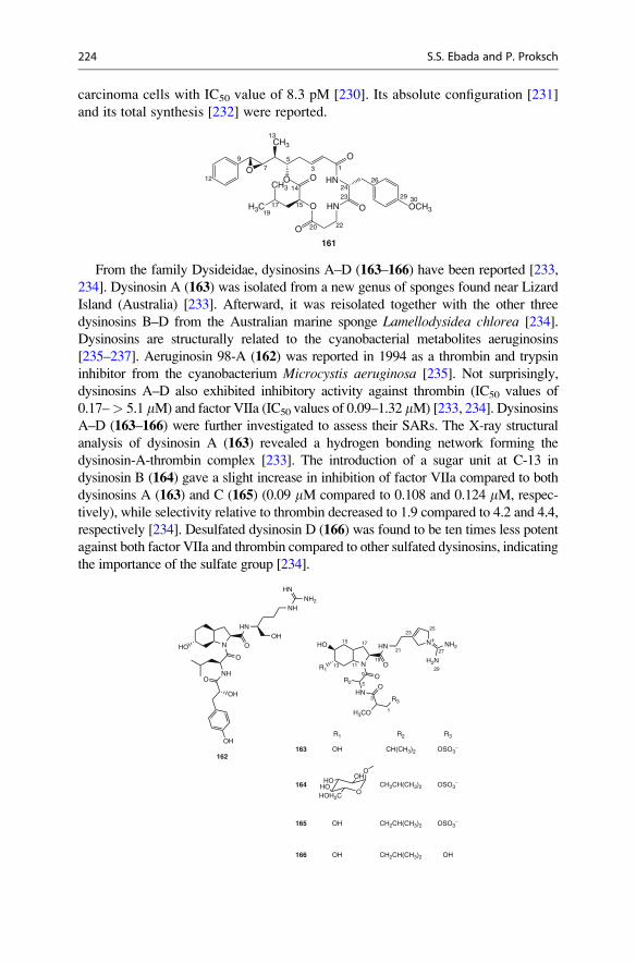
carcinoma cells with IC50 value of 8.3 pM [230]. Its absolute configuration [231]
and its total synthesis [232] were reported.
OO
O
HN
OCH3HNO
O
O
H3C
CH3
O
CH3
13
57
9
13
14
151719
20 22
2324
26
29 30
12
161
From the family Dysideidae, dysinosins A–D (163–166) have been reported [233,
234]. Dysinosin A (163) was isolated from a new genus of sponges found near Lizard
Island (Australia) [233]. Afterward, it was reisolated together with the other three
dysinosins B–D from the Australian marine sponge Lamellodysidea chlorea [234].
Dysinosins are structurally related to the cyanobacterial metabolites aeruginosins
[235–237]. Aeruginosin 98-A (162) was reported in 1994 as a thrombin and trypsin
inhibitor from the cyanobacterium Microcystis aeruginosa [235]. Not surprisingly,
dysinosins A–D also exhibited inhibitory activity against thrombin (IC50 values of
0.17–> 5.1 mM) and factor VIIa (IC50 values of 0.09–1.32 mM) [233, 234]. Dysinosins
A–D (163–166) were further investigated to assess their SARs. The X-ray structural
analysis of dysinosin A (163) revealed a hydrogen bonding network forming the
dysinosin-A-thrombin complex [233]. The introduction of a sugar unit at C-13 in
dysinosin B (164) gave a slight increase in inhibition of factor VIIa compared to both
dysinosins A (163) and C (165) (0.09 mM compared to 0.108 and 0.124 mM, respec-
tively), while selectivity relative to thrombin decreased to 1.9 compared to 4.2 and 4.4,
respectively [234]. Desulfated dysinosin D (166) was found to be ten times less potent
against both factor VIIa and thrombin compared to other sulfated dysinosins, indicating
the importance of the sulfate group [234].
NHO
NHO
OH
OH
O
HNOH
NHNH2
HN
O
162
NR1
HN
O
HN
O
HO
R2O
H3CO
R3
N+NH2
H2N
1
3
5
9
1113
15 17
19
21
2325
27
29
R1 R2 R3
163
164
165
166
OH CH(CH3)2 OSO3−
O
OOH
HOHOHOH2C
CH2CH(CH3)2 OSO3−
CH2CH(CH3)2 OSO3−
CH2CH(CH3)2 OH
OH
OH
224 S.S. Ebada and P. Proksch

Phakellistatins and hymenamides are two groups of proline-rich cyclopeptides.
They were isolated mainly from different species of the marine sponge genera
Phakellia and Hymeniacidon, respectively. However, phakellistatins 1–14 and
isophakellin 3 were isolated from different species of the genus Phakellia [238–
247], and two conformers of phakellistatin 2 have been reported from the Fijian
marine sponge Stylotella aurantium [248]. Phakellistatin 1 (167) was the first
reported congener from the Indo-Pacific (Truk Archipelago) marine sponge
Phakellia costata [238]. Phakellistatin 1 revealed moderate cytotoxicity against
P388 cells (IC50 ¼ 9.0 mM) [238]. Amongst the reported phakellistatins to date,
phakellistatins 2 (168) [239], 6 (169) [242], and 11 (170) [244] exhibited the most
potent antiproliferative activity against P388 murine leukemia cell line with IC50
values of 0.4, 0.2, and 0.2 mM, respectively. Moreover, phakellistatin 6 (169) wasmore active than phakellistatin 2 (168) against a panel of six human cancer cell
lines, namely, ovarian (OVCAR-3), brain (SF-295), renal (A498), lung (NCI-
H460), colon (KM2OL2), and melanoma (SK-MEL-5) cell lines, with GI50 ranges
of 0.02–0.09 mM [242] and 1.2–3.6 mM [239], respectively.
NH
N
O
NH
N
O O
NH
O
O
N
HNO
OH
O HONH
O
NO
HNN
OHN
O
HNN
OO O
NH
N
NH
NH
O
O
O
NHN
O
O
N
NHOO
HN
ONO
N
NHO
H2N
O
NH
O HN
O
NH
OO
N O
167 168
169 170
Hymenamides (A–K) have been isolated from the Okinawan marine sponge
Hymeniacidon sp. [249–252]. Hymenamides A (171) and B (172) were reported in
1994, and only the latter exhibited cytotoxicity against L1210 and KB tumor cell lines
4 The Chemistry of Marine Sponges 225

with IC50 values of 3.8 and 7.2 mM, respectively [249]. In addition to hymenamide B,
only hymenamide J (174) showed cytotoxicity against L1210 and KB tumor cell lines
(IC50 values of 2.4 and 0.7 mM, respectively), while hymenamide H (173) exhibitedcytotoxicity only against L1210 cells (IC50 ¼ 7.0 mM) [252].
Beside hymenamides, hymenistatin 1, a proline-rich cyclo-octapeptide, was also
purified from the marine sponge Hymeniacidon sp. collected in Palau [253].
Hymenistatin 1 was active against the P388 murine leukemia cell line (IC50 3.9
mM) [253]. In addition, it was found to exert an immunosuppressive effect (both in
the humoral and cellular immune responses) comparable to that of the well-known
immunosuppressive agent cyclosporin [254].
NH
NH
N
O
HN
O
NH
NH2HN
O
NH
N
HNO
O N
O
O
NH
N
N
O
O
HNHN
O
CONH2
ONH
OONH
OHOOC
NHN
OHN
OH
OO
N
HN
OHNN
OO
O
NH
O
NH NH
NHN
O
O OOHN
NH
OCOOH
HN
O
O
NH
NH
NH
O
H2N
OHOH
171172
173174
N N
N
O
NH
O
HN
NH
O
NH
NH2
O
O
O
Trp-2
Trp-1
175
From other sponges of the same order Halichondrida, axinastatins [255, 256],
axinellins A–C [257, 258], and stylissamides A–D [259] have been reported
226 S.S. Ebada and P. Proksch

from different species of the marine sponge genus Axinella, the Fijian marine
sponge Stylotella aurantium and Stylissa caribica, respectively. They all
exhibited mild to moderate antiproliferative activity. However, from
a Jamaican collection of Stylissa caribica, two proline-rich cyclic heptapeptides,
stylisins 1 and 2, have been isolated. In contrast to other peptides, stylisins 1 and
2 proved inactive in antimicrobial, antimalarial, anticancer, anti-HIV-1,
anti-Mtb, and anti-inflammatory assays [260]. Kapakahines A–G are also pro-
line-containing cyclic peptides isolated from a Pohnpei sponge Cribrochalinaolemda (order Haplosclerida) [185–187]. They share a unique structural feature:
two tryptophan residues (Trp-1 and Trp-2) are not linked by an amide bond
but by a N–C bond from the indole nitrogen of Trp-1 to the b-indole carbon of
Trp-2. Kapakahine B (175) was the first reported congener, and it exhibited
moderate cytotoxicity against P388 cells (IC50 ¼ 5.9 mM) [185]. All other
kapakahine derivatives exhibited moderate to weak cytotoxic activity
[186, 187]. Their structures, however, were found to be unique and of biogenetic
interest.
Haligramides A and B were also identified as proline-rich cyclopeptides from
the marine sponge Haliclona nigra collected from the northern coast of Papua
New Guinea [261]. The cyclic depsipeptides, halipeptins (A–D), were isolated
from another species of the genus Haliclona collected in Vanuatu [262, 263].
Although, haligramides A and B exhibited moderate activity in the cytotoxicity
assay against four human tumor cell lines [261], halipeptin A (176) revealed no
significant results in either cytotoxicity, antifungal, antiviral, or antimicrobial
assays [262].
176
HNN
N
O
OCH3
NH
OHSO O
O
O
Interestingly, halipeptin A (176) was found to possess potent in vivo anti-
inflammatory activity, causing about 60 % inhibition of edema in mice at the
dose of 0.3 mg/kg (i.p.) compared to indomethacin and naproxen (ED50 of 12 and
40 mg/kg, respectively) [262].
Recently, euryjanicins (A–D) have been reported from the Caribbean
marine sponge Prosuberites laughlini [264, 265]. They are cylcoheptapeptides
containing two or three prolines, an array of apolar residues, and one or two
aromatic residues. Euryjanicins A, C, and D each contain a serine residue, whereas
euryjanicin B possesses one threonine unit. When tested against the National
Cancer Institute 60 tumor cell line panel, all euryjanicins displayed weak
cytotoxicity [265].
4 The Chemistry of Marine Sponges 227

Theonellamides (A–F) are a group of cytotoxic peptides with novel amino acid
residues and complex bicyclic macrocyclic rings isolated from the marine sponge
Theonella sp. collected off Hachijo-jima Island (Japan) [266, 267].
177
HN NH
HN
NH O
HN
NH
OH
BrO
OH
O
O
OOHCOOH
O
NH
O
HN
O
NH
H2NOC OHO H
N
H2NOCO
NH
O
NN
NH
O
HO
Br
OH
Theonellamide F (177) was the first reported congener exhibiting antifungal
activity against pathogenic fungi Candida sp., Trichphyton sp., and Aspergillus sp.at MIC values between 1.8 and 7.3 mM. It is also cytotoxic against L1210 and P388
leukemia cells with IC50 values of 1.9 and 1.6 mM, respectively [266]. In addition,
all other theonellamides (A–E) were cytotoxic against P388 cells (IC50’s between
0.5 and 2.8 mM) [267].
Polytheonamides (A–C) are polypeptides consisting of 20 amino acid residues
with unprecedented structural features. They were reported from the marine sponge
Theonella swinhoei (Hachijo-jima Island, Japan) [268, 269]. Polytheonamides
exhibited potent cytotoxic activity against P388 cells (IC50 ¼ 13.5–15.5 pM) [269].
Cyclotheonamides A (178) and B (179) are cyclic peptides containing unusual
amino acid residues, i.e., vinylogous tyrosine (V-Tyr), a-ketohomoarginine (K-
Arg), and b-linked-diaminopropionic acid (Dpr). They were isolated from the
marine sponge Theonella swinhoei (Japan) [270] together with two additional
congeners C and D [271], whereas cyclotheonamide E was isolated from
a morphologically different specimen of Theonella swinhoei [271]. Chemical
investigation of the marine sponge Theonella sp. collected off Tanegashima Island
led to the isolation of cyclotheonamides E2 and E3 [272], while cyclotheonamides
E4 and E5 were obtained from the Okinawan marine sponge Ircinia sp. [273].
Cyclotheonamides (A–E) and (E2–E5) were found to possess potent inhibitory
activity against serine proteases including thrombin, trypsin, and plasmin [270–
273]. Their mode of action was elucidated by X-ray crystallography of the complex
between cyclotheonamide A (178) and human a-thrombin which disclosed that
(1) the binding of 178 to the catalytic triad of the enzyme is achieved through
forming a network of hydrogen bonds between the a-keto group of the K-Arg
residue and the hydroxyl group of Ser195 of the enzyme, (2) V-Tyr residue proved
to be involved in the bonding mechanism, and (3) cyclotheonamide D which
228 S.S. Ebada and P. Proksch

possesses D-Leu instead of D-Phe in 178 showed comparable activity against
thrombin; thus, a further hydrophobic amino acid can replace D-Phe [271].
178 R = H
179 R = CH3
HN
HN
ONH
HN
O
OH
O
NH
R
O
NO
O
HN
H2N NH
O
However, a comparative X-ray study against either human a-thrombin or bovine
b-trypsin revealed that cyclotheonamide A (178) inhibited trypsin stronger
than thrombin (IC50 ¼ 16 and 23 nM, respectively) [272]. These results were
substantiated to the more favorable (1) aromatic interaction of the D-Phe in 178 with
Tyr39 and Phe41 in trypsin than with Glu39 and Leu41 in thrombin and (2) interaction
ofN-formyl Dpr residue with Gly174 and Gln175 in trypsin than Ile174 and Arg175 in
thrombin [272]. Since the cyclotheonamides exhibited potent inhibitory activity
against serine proteases, the marine sponge T. swinhoei has been thoroughly investi-
gated. From a specimen collected off Hachijo-jima Island in 1993, pseudotheonamides
A1, A2, B2, C, D, and dihydrocyclotheonamide A were isolated [273].
Pseudotheonamides A1 (180), A2 (181), and B2 (182) are linear pentapeptides
embracing the rare piperazinone and piperidinoiminoimidazolone ring systems,
while pseudotheonamide C (183) contains V-Tyr instead of a piperazinone ring.
Pseudotheonamide D, a tetrapeptide lacking a C-terminal K-Arg unit and
dihydrocyclotheonamide A, is a reduction product of cyclotheonamide A (178).Peptides 180–183 and dihydrocyclotheonamide A inhibited thrombin with IC50
values of 1.0, 3.0, 1.3, 0.19, 1.4, and 0.33 mM, respectively, whereas they inhibited
trypsin with IC50 values of 4.5, >10, 6.2, 3.8, >10, and 6.7 mM, respectively [274].
As revealed by a SAR study of cyclotheonamides, potent inhibition of serine
proteases is associated with the presence of the a-keto group of K-Arg residue
[270–273]. Therefore, it was not surprising that pseudotheonamides, in which the
a-keto group was either modified or missing, showed only moderate activity [274].
Thus, cyclotheonamides and pseudotheonamides inhibited serine proteases includ-
ing trypsin and thrombin. These results suggested that cyclotheonamides and
pseudotheonamides may be useful for treatment of asthma and other disorders
associated with inflammation of the respiratory tract in addition to coagulatory
disorders [273].
4 The Chemistry of Marine Sponges 229

HN
HN
OH
O
ONH
HN
OHO
NHN
N
NHO
NH
O
H
HO
180
HN
HN
OH
O
ONH
HN
OHO
NHN
N
NHO
NH
O
H
HO
181
HN
N
OH
O
ON
HN
OHO
NHN
N
NHO
NH
O
H
HO
182
NH
HN
OH
O
HN
OHO
NHN
N
NHO
NH
O
HONH2
O
183
Theonellapeptolides are a group of tridecapeptide lactones characterized by the
presence of high proportions of D-amino acids, N-methyl amino acids and b-amino
acids. Theonellapeptolide Id (184) was the first reported congener from the Okinawan
marine sponge Theonella swinhoei [275]. From the same specimen, theonella-
peptolides (Ia–Ic, Ie, and Iid) have also been purified [276–280], whereas theonella-
peptolide Iie was isolated from an Indonesian specimen of T. swinhoei collected usingSCUBA in Baranglompo Island [281]. From the same Indonesian specimen, four
cyclic depsipeptides named barangamides (A–D) have been reported [281, 282].
184
HN O
NMe HN
NH
NMe HN
HN
NMe
MeN
HN
O
O
NHO
MeN
NH OOMe
O
OO
O
O O
O
O
O
O
Biological activity of theonellapeptolides include cytotoxicity, ion-transport
activity for Na+, K+, and Ca2+ ions [278], Na+, K+-ATPase inhibitory activity [283],
and mild immunosuppressive activity [281]. However, other theonellapeptolide
230 S.S. Ebada and P. Proksch

(III series) exhibiting in vitro cytotoxicity against the P388 cell line (IC50 ¼ 5.2 mM)
have also been reported from a New Zealand deep-water sponge, Lamellomorphastrongylata, belonging to a different order compared to T. swinhoei [284].
The cyclic depsipeptides, papuamides (A–D), were isolated from two-sponge
associations of Theonella mirabilis and T. swinhoei collected in Papua New Guinea
[285]. They contain a number of unusual amino acids including 3,4-dimethyl-
glutamine, b-methoxytyrosine, 3-methoxyalanine, and 2,3-diaminobutanoic acid or
2-amino-2-butenoic acid residues. Papuamides also contain a previously undescribed
2,3-dihydroxy-2,6,8-trimethyldeca-(4Z,6E)-dienoic acid moiety linked to a terminal
glycine residue. In addition, they were the first marine-derived peptides reported to
contain 3-hydroxyleucine and homoproline residues [285]. Papuamides A (185) andB (186) inhibited the infection of human T-lymphoblastoid cells by HIV-1RF in vitro
with an EC50 of approximately 2.6 nM [285]. Papuamide A (185) was also cytotoxicagainst a panel of human cancer cell lines with a mean IC50 of 0.05 mM [285].
Mirabamides (A–D) are cyclic depsipeptides structurally related to papuamides.
However, mirabamides were isolated from an aqueous extract of the marine sponge
Siliquariaspongia mirabilis (Chuuk Lagoon, Micronesia), and they differ from
papuamides in either being chlorinated (4-chlorohomoproline) and/or glycosylated
(b-methoxytyrosine 4’-O-a-L-rhamnopyranoside) derivatives [286]. A comparative
study to determine the effects of mirabamides on HIV-1 infection was performed
using two different viral strains, namely, HXB2 (T-cell-tropic virus) and SF162
(macrophage-tropic virus) in HIV-1 neutralization assay. In addition, HIV-1 enve-
lope-mediated cell fusion assay was also carried out to determine whether the com-
pounds act at early stages of infection, i.e., viral entry.
R1 R2 R3
185
186
187
188
189
CH3 H
NH O
ON
NHNH
HN
N
OCH3
OH
OO
O
O
H3CO
O
O
HN O
HN NH2
OOH2N
HN O
HN
NH
OH
OR2
O
OOH
HO
R1
R3
CH3 β-L-Rha Cl
Cl
HH
H
H
H
CH3
CH3 β-L-Rha H
Results of testing mirabamides (A–D) and papuamide A revealed that
mirabamides A (187), C (188), and D (189), as well as papuamide A (185) inhibit
4 The Chemistry of Marine Sponges 231

HIV-1 envelope-mediated fusion with activities comparable to those observed in the
neutralization assays [286].MirabamideA (187) and papuamideA (185) were foundto be the most potent inhibitors in the fusion assay, with respective IC50 values of 41
and 73 nM, while mirabamides C (188) and D (189) inhibited fusion at lower
micromolar concentrations (IC50’s between 0.14 and 3.9 mM) [286]. Mirabamide
B is the only mirabamide containing a 2,3-dehydro-2-aminobut-anoic acid residue
instead of the 2,3-diaminobutanoic acid residue (Dab) that is present in other
mirabamides as well as in papuamides A (185) and B (186). Interestingly,
mirabamide B was consistently found to be less potent than other mirabamides
(187–189) in both neutralization and fusion assays. Thus, the Dab residue in this
class of peptides appeared to be important for anti-HIV activity [286].
As for the role of b-methoxytyrosine residue (b-OMe Tyr), mirabamide A (187),with rhamnosylated b-Tyr residue, was found to be the most potent analogue among
the four mirabamides. This indicates that Tyr residues bearing substitutions at the 4’
position can be tolerated with no deleterious effect on antiviral activity and that the
presence of a free 4’ hydroxyl on the Tyr unit is not essential [286]. In summary, the
reported results disclosed thatmirabamideA (187) is equipotent to papuamideA (185)(which also contains a b-OMe Tyr residue), whereas theopapuamide A (190), whichwas isolated from a Papua New Guinea lithistid sponge Theonella swinhoei and lacksa b-OMe Tyr residue, was inactive [287]. One hypothesis that would be consistent
with these results is that the b-OMe Tyr residue imparts a specific conformation
required for binding to target protein(s) involved in HIV-1 entry [286]. However,
neither 4-chlorohomoproline nor b-methoxytyrosine 4’-O-a-L-rhamnopyranoside
that are present in mirabamides affected their anti-HIV activities [286].
Celebesides A–C (193–195) and theopapuamides (B–D) together with 190 wereisolated from the marine sponge Siliquariaspongia mirabilis (Sulawesi Island,
Indonesia) [288].
NH
NHO
NMe
NH
NHNH
NH2 O
R2
NH
NH
O
O
OH
OHO
O
O
O
NH2
NH
NH
O
NH2
O
NH2
H3COO
O
NMe
O
NH2O
O
OH O
OH O
NHR1
190 R1 = H R2 = OH
191 R1 = COCH3 R2 = OH
192 R1 = COCH3 R2 = H
232 S.S. Ebada and P. Proksch

HN
HN
NH
HN
R2
OO
O
O
O
MeN
O
HO
O
CONH2
OR1OCONH2
OH
193 R1 = PO3H2 R2 = C2H5
194 R1 = PO3H2 R2 = CH3
195 R1 = H R2 = C2H5
Celebesides are unusual cyclic depsipeptides that comprise a polyketide moiety
established as 7,9-dihydroxy-8,10-dimethyltrideca-2,4-dienoic acid (Ddtd) and five
amino acid residues including an uncommon 3-carbamoyl threonine and
a phosphoserine residue in celebesides A (193) and B (194). Celebeside A (193)neutralized HIV-1 in a single-round infectivity assay with an IC50 value of 2.1 mM,
while its nonphosphorylated analogue, celebeside C (195), was inactive at concen-trations as high as 60 mM [288]. This correlates the observed anti-HIV activity of
celebesides to the presence of phosphoserine in the molecule.
Theopapuamides A–C (190–192) showed cytotoxicity against HCT-116 human
colon carcinoma cells in vitro with IC50 values between 1.3 and 2.5 mM, and they
also exhibited strong antifungal activity against wild-type and amphotericin B–
resistant strains of Candida albicans at loads of 1–5 mg/disk [288]. However,
celebesides (A–C) displayed neither antibacterial nor antifungal activities against
the tested microorganisms at concentrations up to 50 mg/disk [288].
Perthamides (B–D) are further examples of cyclic depsipeptides isolated from
two different collections of the lithistid sponge Theonella swinhoei [289, 290].Perthamide B was identified as a cytotoxic peptide [289], while perthamides
C (196) and D (197) lacked antiproliferative activity on KB cell line up to a dose
of 10 mg/mL [290].
NH
NH
NH
MeN
O
OOOH2N
NH
N
HN
H3COO
HN
NH2
O
O
OR
O
OH
O
OH
196 R = OH
197 R = H
4 The Chemistry of Marine Sponges 233

Interestingly, perthamides C (196) and D (197) reduced carrageenan-inducedmouse
paw edema significantly in vivo causing 60% and 46% reduction of edema at a dose of
300 mg/kg (i.p.) similar to halipeptin A (176) [290]. These data compare well with
the most common NSAID sold in the pharmaceutical market, naproxen
(ED50 ¼ 40 mg/kg), and clearly indicate that perthamides C and D in addition to
halipeptinA are about 100 timesmore potent. Furthermore, the observed in vivo activity
implies that these compounds are able to access the site of inflammation [262, 290].
The microsclerodermins (A–I) are a family of antifungal cyclic hexapeptides
which have been isolated from different collections of the lithistid sponges
Theonella sp. and Microscleroderma sp. [291–293]. Microsclerodermins A (198)and B (199) were the first reported members of this family from the New
Caledonian lithistid sponge Microscleroderma sp. [291]. Both of them, together
with microsclerodermin F, showed potent antifungal properties by inhibiting the
growth of C. albicans at 2.5 and 1.5 mg/disk, respectively [291–293]. In addition,
microsclerodermins F–I showed nearly similar in vitro cytotoxic activity against
HCT-116 cell line (IC50’s of 1.0–2.5 mM) [293].
198 R = OH
199 R = H
HN
NMe
HN
O NHO
O
HN
HN
O
NH
COOH
OH O
NH
O
OH
H3CO
OH
OH
OH
O
R
Koshikamide A1 (200) and A2 are linear peptides that were isolated from the
sponge Theonella sp. collected off the Koshiki-jima Island (Japan) [294, 295].
Koshikamide B (201) was isolated from two separate collections of the marine
sponge Theonella sp. [296]. Koshikamide B (201) is a 17-residue peptide lactone
composed of six proteinogenic amino acids, two D-isomers of proteinogenic amino
acids, seven N-methylated amino acids, and two unusual amino acid residues,
namely, Nd-carbamoylasparagine and 2-(3-amino-2-hydroxy-5-oxopyrrolidin-2-
yl) propionic acid. Koshikamides A1 (200), A2 and B (201) exhibited cytotoxicity
against P388 murine leukemia cells in vitro with IC50 values of 1.7, 4.6, and
0.2 mM, respectively [294–296].
234 S.S. Ebada and P. Proksch

200
H3CONH
MeN
NMe
MeN
NMe
MeN
NH
O
O
OO
O
NH2
O
O
OO
NH2
O
HN N
O
N OH
OO
H3CONH
MeN
NMe
MeN
NMe
MeN
NH
O
O
OO
O
H2N H2N H2N
H2N
NH2
O
O
OO
O
NH
MeN
NH
O
MeN
O
O
O
HN
NH
OO
O
OO
O
NH
O
HN
OHN
OO
O
NH
HN
O
OH
201
Keramamides (A–J and K–N) are a group of cyclic peptides isolated from different
collections of the Okinawan marine sponge Theonella sp. [297–302]. They were
named after the location of the first collection of the sponge which was Kerama
Island, Okinawa. Keramamides A (202) and L (208) are characterized by having an
ureido bond and a 6-chloro-N-methyltryptophan residue. Keramamides B (203) and(C–E) contain an oxazole ring as a structural feature, while keramamides (F–H) and
(J–K) share the presence of a thiazole ring. Keramamides M (209) and N (210) aresulfate esters of keramamides D (204) and E (205), respectively.
Most of the known keramamides feature in vitro cytotoxic activity when tested
against L1210 murine lymphoma (IC50’s between 0.5 and 2.3 mM) and KB human
epidermoid carcinoma (IC50’s between 0.45 and 6.2 mM) cell lines [299–302].
Among the tested congeners, keramamides K (207) and L (208) were the most
potent derivatives with IC50 values of 0.77 and 0.5 mM against L1210 cells and 0.45
and 0.97 mM against KB cells, respectively [301].
OO
MeN
O
NH
HN
NH
O
NH
O
NH
OH
O
NH
R1
Cl
HN
O
202 R1 = OH
208 R1 = H
4 The Chemistry of Marine Sponges 235
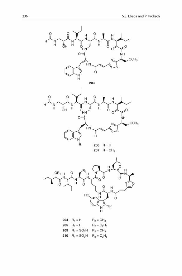
H NH
NH
HN
NH
HN
O
OH
O
O
O
OO
NH
O
OCH3N
SHN
HN
O
ON
R 206 R = H
207 R = CH3
N
NH
HN
HN
O
O
HN
N O
O
O
OO
HN
NH
HN
O
R2O
O
OR1
NH
Br
HO
204 R1 = H R2 = CH3
205 R1 = H R2 = C2H5
209 R1 = SO3H R2 = CH3
210 R1 = SO3H R2 = C2H5
HNH
HN
NH
HN
O
OH
O
O
O
OO
NH
O
OCH3N
SHN
HN
O
ONH
203
H N
236 S.S. Ebada and P. Proksch

Callipeltins are a group of marine peptides with unusual structural features and
remarkable biological activities isolated from the marine sponges Callipelta sp.
[303, 304] and Latrunculia sp. [305–307]. Callipeltin A (211) is the parent com-
pound of this family which comprises 12 further congeners (B–M). Apart from the
cyclic congeners, callipeltins A and B, all the other callipeltins are linear deriva-
tives structurally related to callipeltin C (212) which, in turn, represents the acycliccounterpart of callipeltin A (211). Structurally, the most distinctive feature of
callipeltins is the presence of several nonproteinogenic units while from
a biological point of view, callipeltin A displays a broad range of bioactivities,
including antiviral, antifungal, and cytotoxicity against several human tumor cell
lines and regulatory activity of the myocardial force of contractions [303–309]. The
unusual structural features of callipeltins and the interesting biological activities
have aroused considerable interest among the synthetic chemistry community.
NH
HNO
NH
NMe
MeN
NH
O
O
OO
OO
O
HN
NH
OH
NH
NH
O
NH2
NH2
NH2
NH
O
O
OH
OH
NH
H2N
NH
O
OH
O
HN
O
OH
211
H3CO
NH
HO
O HN
HN
NH
OH
NH O
O
OH
OH
NH
H2N
NH
NH2O
O
HN
O
OH
O
NH
MeN
NH
H2N NH
O
O
NH
NH2O
O
H3CO
MeN
OH
COOH
O
212
As a result, all nonproteinogenic units in this group of metabolites, namely,
(3 S,4R)-3,4-dimethyl-L-pyroglutamic acid (the N-terminus unit in callipeltin B)
[310], (2R,3R,4 S)-4-amino-7-guanidino-2,3-dihydroxyheptanoic acid [311],
(2R,3R,4R)-3-hydroxy-2,4,6-trimethylheptanoic acid [312] linked to the
N-terminus of callipeltins A, C, D, and F–I; and (R)-b-methoxy-D-tyrosine were
obtained in a stereoselective manner [313].
4 The Chemistry of Marine Sponges 237

Homophymine A (213) was isolated as the principal active constituent of the
New Caledonian lithistid sponge Homophymia sp. [314]. Homophymine A is
a cyclic depsiundecapeptide structurally related to other aforementioned marine
peptides isolated from the order Lithistida such as callipeltin A, papuamides,
theopapuamides, and mirabamides which are well known for their potent HIV-
inhibitory activity. In addition to the common structural features, homophymine
A (213) contains two hitherto unprecedented residues, i.e., the 4-amino-2,3-
dihydroxy-1,7-heptandioic acid and 2-amino-3-hydroxy-4,5-dimethylhexanoic
acid. Homophymine A exhibited cytoprotective activity against HIV-1 infection
with an IC50 of 75 nM [314]. Recently, nine additional homophymines, namely,
B–E (214–217) and A1–E1 (213a–217a) have been isolated from the same sponge
[315]. From the structural point of view, homophymines (A1–E1) feature the
4-amino-6-carbamoyl-2,3-dihydroxyhexanoic acid residue compared to the
corresponding (A–E) possessing the same residue in its carboxy form.
NH
HN
NH
OHN
O
OH
O
NH2
O
O
NMe
NH
O
O
OH
OH
NH
R2
O
HN
NH2
O
N O
NH
HOO
HN
O
H3CO
O
NH2
O
O
R1
O NH2
R1 R2
213213a
214214a
215215a
216216a
217217a
OOH
OHNH2
OOH
OHNH2
OOH
OHNH2
OOH
OHNH2
OOH
OHNH2
238 S.S. Ebada and P. Proksch

All homophymines were subjected to an antiproliferative activity assay against
a wide panel of cell lines including human and simian cancer and noncancer cells
and displayed potent cytotoxicity (IC50 values between 2 and 100 nM) [315].
A comparison of activities against different tumor cell lines showed a moderate
selectivity toward PC3 human prostate and OV3 ovarian carcinoma cell lines.
However, when the sensitive and their resistant counterpart cell lines were com-
pared (MCF7/MCF7R, HCT116/HCT15, HLC60/HL60R), no significant differ-
ence was observed [315]. Homophymine A1–E1 (213a–217a) exert stronger
biological activity compared to the corresponding congeners A–E (213–217)[315]. Further extensive biological investigations were carried out to distinguish
whether the homophymines possess antiproliferative and/or acute toxic activity.
The results strongly suggested that the toxic effect of homophymines was due to an
acute and a nonspecific toxicity toward various tested human cell lines [315].
Azumamides A–E (218–222), isolated from the Japanese marine spongeMycaleizuensis, are five cyclic tetrapeptides that exhibit potent histone deacetylase
(HDAC) inhibitory activity (IC50’s of 0.045–1.3 mM) [316]. Furthermore,
azumamide A (218) at a concentration of 19 mM significantly inhibited angiogen-
esis [316]. Only few examples of marine HDAC inhibitors are known up to date
such as psammaplin A (PsA, 109) (see Section 4.2.3) obtained from marine sponge
Pseudoceratina purpurea [133], whereas azumamides A–E (218–222) were the
first examples of cyclic peptides with HDAC inhibitory activity isolated from
marine organisms.
NH
HN
HN
OOO
NH
O
R4
R3
R1
O
R2
218
219
220
221
222
R1 R2 R3 = R4 IC50(μM) (HDAC)
NH2
NH2
NH2
0.045
0.11
0.11
H CH3
CH3
CH3
CH3
OH
OH OH
H
H
H 1.3
OH 0.064
4 The Chemistry of Marine Sponges 239

After the crystal structure of the complex between a HDAC-like protein and the
HDAC inhibitor, trichostatin A (TSA) [317], had been solved, interesting insights
into the binding modes of HDAC inhibitors were disclosed [318]. According to the
TSA structure, a plausible binding mode to the enzyme is by inserting the long
aliphatic chain of TSA into the hydrophobic pocket of the enzyme. Then, the
terminal hydroxymic group coordinates with a zinc ion at the polar bottom of the
pocket while the aromatic dimethylaminophenyl portion of TSA makes contact
with the pocket entrance and an adjacent surface group thereby capping the pocket.
Trapoxin A [319] and apicidin [320] are the parent congeners of two classes of
HDAC inhibitory cyclic tetrapeptides containing groups that may be analogous to
the aliphatic side chain of TSA. Trapoxins possess a 2-amino-8-oxo-9,10-epoxy-
decanoic acid, in which the epoxide moiety is thought to bind irreversibly to the
enzyme [317], while the apicidins comprise a 2-amino-8-oxo-decanoic acid, with
the keto group being responsible for zinc chelation [321].
The corresponding terminal function for azumamides is probably the amide or
the carboxylate group. Despite that the affinity of an amide group to zinc is much
weaker than that of a carboxylic acid, azumamides A (218) and B (219) with an
amide end showed equivalent levels of HDAC inhibitory activity as C (220) andE (222) congeners, which have carboxylate functionality [316].
In conclusion, many peptides from marine sponges showed potent activities
such as antitumoral, antiviral, immunosuppressive, antifungal as well as cardiac
stimulant properties. However, these bioactivities cannot clearly explain their in
situ role inside the marine sponges. Interestingly, the most striking feature of this
class of metabolites is the preponderance of nonproteinogenic amino acids
suggesting that these metabolites are the result of the NRPS/PKS pathways of
microorganisms, proposing that symbiotic microorganisms, including
cyanobacteria, are the real producers of these metabolites. Therefore, the symbi-
otic associations within sponges and other marine invertebrates need further
intensive research which will ultimately unravel the innate sources of these
bioactive metabolites.
4.4 Terpenes
Terpenes include primary and secondary metabolites, all biosynthesized from the
five carbon isoprene building units [322]. Structural modification of these isoprene
units leads a massively diverse range of derivatives with a wide array of chemical
structures and biological properties.
Steroidal terpenoids were the first marine isoprenes to be discovered by
Bergmann during the 1930s–1940s, particularly sterols that were obtained from
various marine macroorganisms [323]. Since then, a huge number of terpenoidal
secondary metabolites have been obtained from marine resources. In addition to our
recent review on this topic [324], we will survey two major classes of marine
isoprenes from sponges, namely, the sesterterpenoids (C25) and triterpenoids (C30)
with particular attention of their biological activities.
240 S.S. Ebada and P. Proksch

4.4.1 Sesterterpenes (C25)
Manoalide (223) is the parent compound of a series of marine sponge metabolites
belonging to the sesterterpene class. Manoalide was first reported in 1980 by
Scheuer from the marine sponge Luffariella variabilis (class Demospongiae;
order Dictyoceratida; family Thorectidae) collected in Palau [325] with activity
as an antibiotic against Streptomyces pyogenes and Staphylococcus aureus.One year later, Scheuer reported three additional related metabolites from the
same Palauan sponge, namely, secomanoalide (224), (E)-neomanoalide (225), and(Z)-neomanoalide (226) [326].
O
O
OHO
HO
1
3
5791113
1517
19
21 23
25
2
3
67
1011
14
15
23
25
CHO
O
OH
O
HO
24223 224
67
1011
14
15
23 OH
OO
OH
2325
24
7
1011
14
15
23 OH
OO
OH
2325
24
6
225
226
All three compounds, as well as the parent compound (223), displayed
antibacterial activity against Gram positive bacteria (Staphylococcus aureus and
Bacillus subtilis) but were inactive against Escherichia coli, Pseudomonasaeruginosa and Candida albicans [326].
Later, marine sponges belonging to the family Thorectidae, including species of
the genera Luffariella [327–339], Hyrtios [340, 341], Thorectandra [342],
Cacospongia [343, 344], Fasciospongia [345–348], Acanthodendrilla [349], and
Aplysinopsis [350], were also found to be rich sources of novel bioactive
sesterterpenoids related to manoalide.
Manoalide was further investigated and found to be a potent inhibitor of phos-
pholipase A2 (PLA2) [351–358]. Subsequently, many structurally related metabo-
lites with PLA2 inhibitory activity were also reported [328, 359–365]. PLA2 is an
enzyme that specifically catalyzes the hydrolysis of phospholipids at the SN-2position to produce a lysophospholipid and arachidonic acid, which in turn provides
the substrate for proinflammatory mediators such as leukotrienes, prostaglandins,
and thromboxanes, collectively known as the eicosanoids [361]. Since manoalide
revealed an irreversible inhibition of phospholipase A2 (PLA2) [353], the structure-
activity relationships (SAR) of this compound attracted scientific interests to study
4 The Chemistry of Marine Sponges 241

and to understand both PLA2 function and mechanism of action in the whole cell.
Therefore, several studies were successfully performed to determine the contribu-
tions of the various functional groups incorporated in 223 and its analogues, such asthe g-hydroxybutenolide, a-hydroxydihydropyran, and trimethylcyclohexenyl ring
systems, to the efficacy as PLA2 inhibitors [356, 361, 365]. These studies indicated
that (1) the existence of the hemiacetal in the a-hydroxydihydropyran ring is crucialfor irreversible binding, (2) the g-hydroxybutenolide ring is involved in the initial
interaction with PLA2, and (3) the hydrophobic nature of the trimethylcyclohexenyl
ring system allows nonbonded interactions with the enzyme that enhances the
potency of these analogues. These studies suggested that the closed ring form of
manoalide is the predominant molecular moiety that accounts for the selective and
potent inhibition of PLA2 [356].
Manoalide analogues also exhibited other bioactivities including molluscicidal
[330], cytotoxicity [333, 334, 336, 340, 343, 346, 349, 350, 367–369], inhibitory
activity of Cdc25 phosphatase [366], nicotinic antagonistic activity [332], and fish
deterrent properties [346, 369]. Therefore, chemical synthesis and derivatization of
manoalide attracted much interest leading to a better understanding of the structure-
activity relationships (SAR) and/or for the plausible mechanism of action [355,
358–360, 363, 364, 370, 371]. Manoalide (223) was licensed to Allergan Pharma-
ceuticals and reached phase II clinical trials as a topical antipsoriatic. Its develop-
ment was, however, discontinued due to formulation problems. The compound is
now commercially available as a biochemical standard tool to block the action of
PLA2 [372].
Luffariellolide (227) is a sesterterpenoid analogue of secomanoalide (224),which was first reported from a Palauan sponge Luffariella sp. [328]. Structurally,
luffariellolide differed in having C-24 as methyl group instead of an aldehyde
functionality as in secomanoalide, and it was obtained as the (Z) isomer as well.
227 R = H228 R = CH3
229 R = CH2CH3
2
3
6
7
101114
15
2325
O
O
RO24
230
231
** 2
3
6
7
1011
14
2325 O
O
HO24
**OH
O
15
18
2022
18-β−(OH)
18-α−(OH)
242 S.S. Ebada and P. Proksch

In contrast to the irreversible inhibitory action of manoalide (223) toward PLA2,
luffariellolide (227) is a slightly less potent but a partially reversible inhibitor. Thismeant that 227 became a more preferable anti-inflammatory agent for potential
pharmacological investigation [328]. In addition to luffariellolide (227), its 25-O-methyl (228) and 25-O-ethyl derivatives (229), five related sesterterpenes,
acantholides (A–E), were obtained from the Indonesian sponge Acanthodendrillasp. [349]. Acantholide D (230) and E (231) represent rare variants for the C14–C20
segment in this type of linear sesterterpenes in which they have the 1-acetylcy-
clopentan-5-ol moiety replacing the trimethylcyclohexenyl ring. Luffariellolide
(227) and its 25-O-methyl congener (228), as well as acantholide E (231), werecytotoxic against the mouse lymphoma L5178Y cell line with IC50 values of 8.5,
1.8, and 16.8 mM, respectively. Interestingly, these results suggest that the 25-O-methyl group in 228 and the stereochemistry of 1-acetylcyclopentan-5-ol in 231play an important role [349]. Luffariolides (A–J) represent a related group of
sesterterpenoidal analogues, which have been obtained from different collections
of the Okinawan marine sponge Luffariella sp. [333, 334, 336].
All luffariolides exhibited significant cytotoxicity against murine lymphoma
L1210 cells with IC50 values ranging between 2.9–19.3 mM. Among them,
luffariolides A (232, IC50 2.9 mM), B (233, IC50 3.23 mM), E (234, IC50 3.0 mM),
and F (235, IC50 3.8 mM) were the most active ones [333, 334, 336].
1014
2324
6
HO O
O
OH2
4
25
61014
23
225
24
HO
OO
4
232
233
O
OO
O2
3
610
14
2324
H
H
25
4
234
1014
23
OO
OH
225
24
6
HO
OH
16
235
4 The Chemistry of Marine Sponges 243

Luffariellins A (236) and B (237) [327] together with their respective 25-acetoxyderivatives (240 and 241) [338] were isolated from the marine sponge Luffariellavariabilis collected off different locations in Palau and in Australia, whereas
luffariellins C (238) and D (239) were obtained from the shell-less marine mollusc
Chromodoris funerea collected from the Kaibakku lake shores in Palau [373].
O
R
O
O
OH
236 R = OH
238 R = H
240 R = OAc
1
2
10
23 24
25
14
CHO
O
OH
O
R
14
2
10
23
25
24
237 R = OH
239 R = H
241 R = OAc
Luffariellins (236–241) are all characterized by the 1-isoproprenyl-2-
methylcyclopentane ring system replacing the trimethylcyclohexenyl moiety in
other manoalide analogues. Despite this discrepancy in chemical structure,
luffariellins A (236) and B (237) retain identical functional groups as present in
manoalide (223) and secomanoalide (224), respectively. Therefore, not surpris-ingly, each respective pair was shown to have similar anti-inflammatory properties
to 223 and 224 [327]. Luffarin metabolites comprise another group of compounds
represented by 28 derivatives. Twenty-six of them, luffarins (A–Z), have been
reported from the Australian marine sponge Luffariella geometrica [332], while
the other two were obtained from the Adriatic Sea sponge Fasciospongia cavernosa[348]. Based on the chemical structures, luffarins have been classified into 14 bicyclic
sesterterpenes, luffarins (A–N); one bicyclic bisnorsesterterpene, luffarin O; one
monocyclic sesterterpene, luffarin P; and six acyclic sesterterpenes, luffarin (Q–V),
in addition to four diterpenoidal derivatives, luffarin (W–Z) [332]. All luffarins were
tested for antimicrobial activity against Staphylococcus aureus,Micrococcus sp., andSaccharomyces cerevisiae. Only luffarins C–F (244–247), K (248), and L (249)showed activity against both S. aureus and Micrococcus sp. [332], whereas luffarins
244 S.S. Ebada and P. Proksch

A (242) and M (250) revealed only mild activity against the latter. Moreover, some
luffarins were also found to be effective inhibitors of nicotinic receptors [332].
O
OH
O
O
HO
1
4
810
22
24 25
13
14 17
18
19
20
21
23
242
1
4
810
24 25
23
O
OHO
O
HO 13
16
19
20
21
243
1
4
810
23 22
21
O
HO
13
15
19
17
20
243a
244
245 R = CH(OCH3)2 15(16)-trans
246 R = CH2OH 15(16)-trans
247 R = CH2OH 15(16)-cis
R = CHO 15(16)-trans 248
249 R = CH2OH 13(14)-cis
250 R = CHO 13(14)-trans
R = CH2OH 13(14)-trans
O
O
HO1
4
810
24 25
17
18
1920
23
13
R15
1
4
810
24 23
2221
R
O
OH
O
13 1517
19
20
Biosynthetically, a relationship could be recognized between the various
luffarins as illustrated in Scheme 4.5. Luffarins appear to belong to the same
enantiomeric series as reported for manoalide-type marine natural products. It is
also curious to note that no acyclic luffarins incorporated the hydroxylated
butenolide functionality. Perhaps the most interesting luffarins from a biosynthetic
point of view are luffarins B (243) and O (243a), which were the first examples of
a hitherto unknown cyclization pattern in compounds of this class [332].
Another example of bicyclic sesterterpenes are thorectandrols A–E (253–257)that were isolated from a Palauan collection of the marine sponge Thorectandra sp.[367, 368] together with the parent compounds of this group palauolide (251) and
4 The Chemistry of Marine Sponges 245

palauolol (252). Palauolide (251) was obtained first as an antimicrobial
sesterterpene from a three-sponge association collected in Palau [374], while
palauolol (252) was identified as an anti-inflammatory sesterterpene from the
Palauan sponge Fascaplysinopsis sp.,and chemically, it was recognized as being
a secondary alcohol that upon dehydration yields 251 [375].
All thorectandrols (253–257) in addition to palauolide (251) and palauolol (252)were tested for antiproliferative activity against 6 to 12 human tumor cell lines
depending on sample availability [368]. Palauolol (252) was active against all
tested cell lines except A549 (non-small cell lung cancer), with IC50 values in the
range 1.2–1.7 mM, while palauolide (251) showed a diminished activity. On the
other hand, thorectandrols (A–E) revealed only weak to no cytotoxicity against
the tested cell lines (IC50’s 70–100 mM). While firm deductions on the structural
requirements for activity were not possible, it appeared that the presence of both the
hemiacetal lactone functionality and the 16-hydroxyl group in palauolol (252)enhanced cytotoxicity compared to palauolide (251) and other thorectandrols [368].
Cacospongionolides (258–262) were isolated from different collections of the
marine sponge Fasciospongia cavernosa (¼ Cacospongia mollior) collected from
the Mediterranean Sea [343, 346, 369, 376]. Cacospongionolides A (258), B (259),and its 25-deoxy derivative (260) revealed a bicyclic sesterterpenoidal skeleton,
resembling luffarins and thorectandrols, with the addition of a g-hydroxybutenolidemoiety. The other cacospongionolides C (261) and D (262) are acyclic
diterpenoidal derivatives. Despite the structural relation with luffarins and
thorectandrols, cacospongionolides (258–260) together with cacospongionolide
D (262) exhibited significant cytotoxicity [343, 346, 369, 376]. This notion
CO2H
CHO
OH
CHO
O
OH
CHO
O
HO
B
A
A B
O
OH
CHOO
HOD
C
C D
O
O
O
HO
HOO
OH
HO
OO
Scheme 4.5 Postulated biosynthetic relationship between all known Luffariella metabolites
[332]
246 S.S. Ebada and P. Proksch

suggested a possible relation between the presence of the g-hydroxybutenolidemoiety and the cytotoxicity.
O OHO
1
58
11 13
15
1719
25
2021
22
23
24
251
O
OH
OR2
1
58
1113
1516
1719
25
R1
2021
22
23
24
252 R1 = H R2 = OH
253 R1 = OAC R2 = H
256 R1 = H R2 = H
257 R1 = OH R2 = H
OO
1
58
1113
2021
22
23
24
OH25
1517
19
R
254 R = H
255 R = OAc
Petrosaspongiolides A (263) and B (264) were the first cheilantane sesterterpenelactones to be isolated from a New Caledonian sponge incorrectly assigned to the
genus Dactylospongia [377] and then reassigned as a new genus and a new species:
Petrosaspongia nigra (Bergquist 1995 sp. nov., class Demospongiae; order
Dictyoceratida; family Spongidae) [378]. From another New Caledonian collection
of the same sponge, 15 additional petrosaspongiolide congeners (C–R) were iso-
lated [379, 380].
O
O
O
HO
1
54
810
1113
16
1719
2021
22
23
24
25
258
O
O
O
R
1
54
810
1113
16
1719
2021
22
23
24
25
259 R = OH
260 R = H
O
O
HO
31
16
71115
19 18 17
261
O
O
O
HO
7
4
31
24
25
111519
21 22 23
262
4 The Chemistry of Marine Sponges 247

OHH
O
R1
H
OH
R2
O1
4
910
11 13
1617 19
20
22 23
2425
263 R1 = CH3 R2 = OAc
265 R1 = CH2OAc R2 = OAc
OHH
O
H
OH
OCOCH3
O1
4
910
11 13
1617 19
20
22 23
2425
21 264
1
4
910
11 1316
17
19
20
22 23
24
21
O
R
CH2COOH
266 R = CH3
266a R = CH2OH
1
4
910
11 13
1619
20
22 23
24
21
N
CH2CH2COOH
267
From the chloroform extract of another Dictyoceratida sponge of the genus
Spongia, 21-hydroxy derivatives of petrosaspongiolides K (266a) and P (270a)were isolated in addition to four other pyridinium alkaloids named spongidines
A–D (273–276) [381]. Spongidines were found to be structurally related to
petrosaspongiolide L (267) particularly in the presence of pyridine ring. Petrosas-
pongiolides (A–L) were subjected to in vitro cytotoxicity assay against the human
bronchopulmonary NSCLC-N6 carcinoma cell line. They revealed IC50 values
ranging between 1.0–32.2 mM [379]. Petrosaspongiolides C (265) and K (266)exhibited the highest potency with IC50 values of 1.0 and 3.5 mM, respectively.
However, petrosaspongiolides A (263) and B (264) were the least cytotoxic con-
geners in vitro with IC50 values of 28.0 and 32.2 mM, respectively; 263 inhibited
tumoral proliferation in vivo at 20 mg/kg without significant toxicity when tested on
immunosuppressed rats carrying a bronchopulmonary tumor (NSCLC-N6) [379].
1
4
910
11 1316
19
20
22 2324 O
O
H
H
OR2
HO
O
R1
25
268 R1 = CH3 R2 = OAc
269 R1 = CH2OAc R2 = OAc
270 R1 = CH3 R2 = H
270a R1 = CH2OH R2 = H
271 R1 = CH2OAc R2 = H
1
4
910
11 13
1619
20
22 23
24
21
25O
COOH
O
HO
272
248 S.S. Ebada and P. Proksch

Petrosasponiolides M–R (268–272) revealed the presence of a g-hydroxybutenolide moiety and a hemiacetal function. Due to these structural
similarities to manoalide (223), petrosaspongiolides (M–R) have received special
attention from the scientific community to study their inhibitory activity against PLA2
from different resources to point out their specificity. Two main groups of PLA2
enzymes have been reported [382], the secretory PLA2 (sPLA2 groups I, II, III, V, IX,
andXwith relatively small molecular weights) and the cytosolic PLA2 (cPLA2 groups
IV, VI, VII, and VIII with higher molecular weights). Inhibition of specific PLA2
constitutes a potentially useful approach for treating a wide variety of inflammatory
disorders such as septic shock, adult respiratory distress syndrome, arthritis, and acute
pancreatitis [381]. Petrosaspongiolides M–R (268–272) together with 21-hydroxy
derivatives of petrosaspongiolides K (266a) and P (270a), and spongidines A–D
(273–276) were tested on five different sPLA2s belonging to the groups I (Naja najavenom and porcine pancreatic enzymes), II (human synovial recombinant and rat air-
pouch secretory enzymes), and III (bee venom enzyme) [380, 381].
273 R1 = CH3 R2 = CH2COOH R3 = H
274 R1 = CH2OAc R2 = CH2COOH R3 = H
275 R1 = CH3 R2 = CH2COOH R3 = CH2CH2COOH
276 R1 = CH3 R2 = CH2CH2SO3H R3 = H
1
4
910
11 13
16
R1
NR2
R3
+
Among petrosaspongiolide derivatives, 268 and 270a inhibited mainly human syno-
vial PLA2 with IC50 values of 1.6 and 5.8 mM, respectively, compared to manoalide
(223) (IC50¼ 3.9 mM) [380, 381]. PetrosaspongiolideM (268) also inhibited bee venomPLA2 enzyme with IC50 of 0.6 mM, compared to 223 (IC50 of 7.5 mM) [380]. The
mechanism of action of petrosaspongiolides M–R (268–272) as anti-inflammatory
marine metabolites has been the topic for many research articles [383–388]. The
covalent binding of 268 to bee venomPLA2 has been investigated bymass spectrometry
and molecular modeling. The mass increment observed was consistent with the forma-
tion of a Schiff base by reaction of a PLA2 amino group with the hemiacetal function at
the C-25 atom of the petrosaspongiolide M g-hydroxybutenolide ring [383]. The
molecular mechanism of inactivating the bee venom and the human type IIA secretory
PLA2s by petrosaspongiolides R (272) [387] andM (268) [388], respectively, has beeninvestigated. In both cases, either covalent (imine formation) and/or noncovalent (van
der Waals) interactions contributed to the inhibitory activity against PLA2 enzymes
[387, 388]. Due to potent anti-inflammatory properties of petrosaspongiolides, their
chemical synthesis has been investigated. Recently, the first enantioselective synthesis
of petrosaspongiolide R (272) has been successfully performed [389].
4 The Chemistry of Marine Sponges 249

4.4.2 Triterpenes (C30)
Steroidal triterpenes were the first marine isoprenes to be discovered in the 1930s.
Scientific interest has been driven toward these metabolites due to the isolation of
biosynthetically unprecedented derivatives possessing a broad spectrum of bioactivity
(ies). Marine triterpenoids have been reported from various marine macroorganisms.
In this section, we survey two examples of triterpenoidal metabolites, namely,
isomalabaricane triterpenes and steroidal saponins obtained from marine sponges,
with particular attention being drawn to their pharmacological significance.
4.4.2.1 Isomalabaricane TriterpenesMalabaricol (277) is the chief triterpene constituent of a yellow pigment obtained from
the wood of the terrestrial plant Ailanthus malabarica (family Simaroubaceae), after
which the whole group of related compounds was named [390–392]. Malabaricane,
the trivial name of this group of compounds, was given to the hydrocarbon
system (3 S*,3aR*,5aS*,9aS*,9bS*)-3a,6,6,9a-tetramethyl-3-(1,5,9-trimethyldecyl)
perhydr-obenz[e]indene, where the tricylic nucleus has a trans-anti-trans ring junction[391, 392].
O
O
OH
1
34
89
10
11 1314 17
24
26
20
28 29
19
30 18
21
27
277
The malabaricanes are structurally characterized by a tricyclic triterpenoid core
and a conjugated polyene side chain [390–392], whereas the isomalabaricane
skeleton is embedded in a 4,4,8,10-tetramethyl-perhydrobenz[e]indene with
a trans-syn-trans ring junction, that leads to an unfavorable twist-boat conformation
for the central ring [393, 394]. Isomalabaricane triterpenes were first reported from
a Fijian collection of the sponge Jaspis stellifera [393] and the Somalian marine
sponge Stelletta sp. [394]. Since then, they have been isolated from several genera
of marine sponges belonging to the order Astrophorida including members of the
genera Rhabdastrella [395, 400, 402, 406, 413, 414, 416, 420], Stelletta [397–399,
405, 408, 412], Jaspis [401, 407, 409, 418, 419, 421, 422], and Geodia [403, 410,
415]. Isomalabaricane triterpenoids having polyene conjugated functionality can be
classified into three groups: (1) stelletins principally possessing the g-pyronefunctionality, which could be ring-opened in some of its congeners yielding the
side chain with terminal free carboxylic acid and methyl moieties, (2) stelliferins
oxygenated at C-22, and (3) globostellatic acids whose main feature is a carboxyl
group at C-4. In addition to triterpenoids, the isomalabaricane core has also been
recognized in some sesqui- and/or sesterterpenes. The isomalabaricane terpenoids
were sometimes trivially named according to their sponge origin. Upon light
exposure, the isomalabaricane-type terpenes readily isomerize at the C-13 position.
250 S.S. Ebada and P. Proksch

Therefore, during isolation and characterization processes, they rapidly equilibrate
into a 1:1 mixture of the 13E and 13Z isomers [398–400, 408, 409, 418, 419].
Nevertheless, these compounds continue to gain a great deal of attention because of
their significant cytotoxic activity [399, 409], whereas the nature of the natural
isomer, either 13E or 13Z or both, is still unresolved. Recently it was reported that
the 1H NMR spectrum of a crude extract obtained from the fresh sponge
Rhabdastrella aff. distinca (Hainan, the South China Sea) revealed that it mostly
contained isomalabaricanes with the 13E-configuration (H-15 of most derivatives
appeared around 7.0 ppm). Thus, the 13Z isomers were suggested in this case to be
formed through isomerization during the isolation and analytical procedures [406].
Stelletins comprise the first group of isomalabaricane-type triterpenoids.
Stelletin A (278) was recognized in 1981 as a yellow triterpenoidal pigment from
the Fijian marine sponge Jaspis stellifera [393]. Later, it was obtained together withits E isomer, stelletin B (279), from the marine sponge Stelletta tenuis collected offHainan Island, China [397]. Stelletin A (278) revealed significant cytotoxicity
against murine leukemia P388 cell line with IC50 of 2.1 nM [397].
OO
O
34
510 8
12
1314
1517
19
24
18
20
21
22
2627
28 29
30R1
R2
O
O3
4510 8
1213
14
1517
19
24
18
20
21
22
26
27
28 29
30
COOH
25
278 : R1,R2 = O, Δ13(14) = Z13(14)
279 : R1,R2 = O, Δ13(14) = E
280 : R1 = OAc, R2 = H, Δ13(14) = E
281 : R1 = OAc, R2 = H, Δ13(14) = Z
282 : Δ13(14) = Z, Δ24(25) = E
283 : Δ13(14) = E, Δ24(25) = Z
284 : Δ13(14), Δ24(25) = Z
285 : Δ13(14), Δ24(25) = E
Stelletin G (284), with an opened g-pyrone and featuring terminal -COOH and
-CH3 functionalities, was isolated together with 278 from J. stellifera [393]. Later,
stelletins G (284) was reported from the Australian marine sponge Stelletta sp.
together with stelletins E (282) and F (283) [398]. The E isomer of stelletin G (284)was isolated from the marine sponge Rhabdastrella globostellata collected from the
South China Sea, and it was given the trivial name rhabdastrellic acid–A (285)
4 The Chemistry of Marine Sponges 251

[395, 396]. Research interests have been intensively driven toward this group of
triterpenoidal derivatives, which led to the isolation of eight further stelletins C, D,
and H–M [398–400, 402, 405] in addition to 22,23-dihydrostelletin D [401].
Rhabdastrellins A–F (286–291), along with stelletins L (292) and M (293),were obtained from the marine sponge Rhabdastrella aff. distinca collected from
a coral reef off Hainan in the South China Sea [406]. Four of the rhabdastrellins
(286–289) exhibited a primary alcohol moiety at C-29 instead of a methyl group
as for the stelletins and the other two rhabdastrellins E (290) and F (291). Allrhabdastrellins and stelletins L and M share a hydroxyl group at C-3 instead of
a carbonyl group as in other stelletins [406]. The antiproliferative profile of
stelletins A–F (278–283) has been examined at the National Cancer Institute
(NCI, Australia) against 60 cell lines. Due to the rapid isomerization upon light
exposure, stelletins were tested as isomeric pairs. Stelletin C(280)/D(281) pairwas the most potent derivative with a mean panel GI50 of 0.09 mM. The stelletin
E(282)/F(283) pair was approximately ten times less potent (mean GI50 of 0.98
mM) [399]. Apoptotic cell death is a stress response of cells to cytotoxic agents
that might be executed either through a receptor-mediated pathway that activates
caspase-8 or through a receptor-independent pathway that involves the cyclin-
kinase inhibitors p53/p21. Both pathways lead to a translocation of proapoptotic
Bax protein to the mitochondria, thereby resulting in a dissipation of mitochon-
drial membrane potential, activation of caspase-3, and execution of the apoptotic
machinery [404].
O
HO3
4510 8
1213
19
28 29
30
OH
14
18
20
21
O
O
22
27286
O
HO3
4510 8
1213
19
28 29
30
OH
OHHO
14
18
20
21
25
26
27
287 : Δ13(14) = E
288 : Δ13(14) = Z
O
HO3
4510 8
1213
1415
17
19
24
18
2021
22
26
27
28 29
30
25
O
OH
OH
289
O
HO3
4510 8
1213
14
1517
19
24
18
20
21
22
26
27
28 29
30
COOR
25
290 : R = CH3, Δ13(14) = E
291 : R = CH3, Δ13(14) = Z
292 : R = H, Δ13(14) = E
293 : R = H, Δ13(14) = Z
252 S.S. Ebada and P. Proksch

Stelletin A (278) demonstrated a differential cytotoxicity against human leuke-
mia HL-60 cells (IC50 0.9 mM) compared to human prostate cancer LNCaP cells
(IC50 260 mM) by activation of NADPH oxidase, which induces oxidative cell death
through a FasL–caspase-3-apoptotic pathway [403]. Stelletins B (279) and E (282)revealed selective cytotoxicity toward p21-deficient human colon tumor HCT-116
cells with IC50 values of 0.043 and 0.039 mM, respectively [400]. Stelletins L (292)and M (293) exhibited selective cytotoxicity against stomach cancer AGS cells with
IC50 values of 3.9 and 2.1 mM, respectively [405]. Rhabdastrellic acid–A (285) alsoinhibited proliferation of human leukemia HL-60 cells with an IC50 value of 1.5 mMthrough inhibition of the PI3K/Akt pathway and induction of caspase-3–dependent
apoptosis [396]. Only rhabdastrellin A (286) possessed moderate inhibitory activity
toward human leukemia HL-60 cells (IC50 ¼ 8.7 mM) while other rhabdastrellins
were inactive (IC50 > 20 mM) [406].
Stelliferins are the second group of isomalabaricane triterpenes. To the best of
our knowledge, 13 compounds belonging to this group have been reported. In
addition to stelliferins A–F (294–299), which have been isolated from the Okina-
wan marine sponge Jaspis stellifera [407], stelliferin G (300) and 29-hydroxy
derivatives of stelliferins A (301) and E (302) have been isolated from an
unidentified species of the genus Jaspis collected near Tonga [409].
The 29-hydroxy derivative of stelliferin D (303) together with 3-epimeric iso-
mers of 301 and 302 were reported from the marine sponge Stelletta globostellatacollected by SCUBA off Mage-jima Island, Japan [408], whereas stelliferin
riboside (294a), the first example of a glycosylated stelliferin, was isolated from
the Fijian sponge Geodia globostellata [410].
294 : R1 = H R2 = H 294a : R1 = H R2 = α-ribopyranosyl298 : R1 = H, R2 = OAc301 . R1 = OH R2 = H302 . R1 = OH R2 = OAc
O
O3
4510 8
1213
14
1517
19
24
18
20
21
2226
27
2829
30
25
H3C
O
R1
R2O
O
O
34
510 8
1213
14
1517
19
18
2829
30
H3C
O
O20
21
22
24
2526
27
296
O
O
34
510 8
1213
14
1819
2829
30
H3C
O
RO
15
17 20
21
22
2425
27
26295 R = H299 R = OAc
2124
4 The Chemistry of Marine Sponges 253

O
HO
34
510 8
1213
14
1517
19
24
18
20
21
2226
27
2829
30
25
OH
OH3C
O
300
O
HO
34
510 8
1213
14
1517
19
18
2829
30
20
21
22
24 25
26
27
R
297 R = H303 R = OH
Stelliferins A–F (294–299) exhibited potent in vitro antineoplastic activities
against murine lymphoma L1210 cells (IC50 of 1.1–5.0 mM) and human epidermoid
carcinoma KB cells (IC50 of 2.8–13.0 mM) [407], while the isomeric mixture of
stelliferin G (300) and 29-hydroxystelliferin A (301) showed the highest inhibitory
activity against the melanoma MALME-3 M cell line with IC50 values of 0.2 and
0.4 mM, respectively [409]. Stelliferin riboside (294a) displayed moderate cytotox-
icity against ovarian A2780 cancer cells (IC50 ¼ 60 mM) [410].
Due to the significant antiproliferative activity exhibited by stelletins and
stelliferins, research efforts have been directed toward their chemical synthesis.
In 1999, Raeppel et al. successfully synthesized the common trans-syn-transperhydrobenz[e]indene moiety in the isomalabaricane-type terpenoids, which
enabled the chemical synthesis of stelletins and stelliferins [411].
Globostellatic acid (304) is the prototype of the third group of isomalabaricane-
type triterpenoids sharing carboxylation at C-4. It was first isolated together with three
other derivatives, globostellatic acids (B–D), from the marine sponge Stellettaglobostellata collected off Mage Island near Kagoshima, Japan [412]. Other
globostellatic acid congeners, (F–M), and X methyl esters, have been reported from
different collections of the Indonesian marine sponge Rhabdastrella globostellata[413, 414]. Globostellatic acids revealed potent cytotoxicity similar to the stelletins
and stelliferins. Globostellatic acids (A–D) demonstrated significant cytotoxicity
against murine leukemia P388 cells with IC50 values of 0.2–0.8 mM [412]. For
cytotoxicity toward mouse lymphoma L5178Y cells, the 3-O-deacetyl congeners,globostellatic acids H/I (305/306) were the most active with an IC50 of 0.31 nM.
However, acetylation of the C-3 hydroxyl group decreases its bioactivity abruptly, as
in globostellatic acids J/K (307/308), with an IC50 of 8.28 nM. The reverse was found
for the 13Z isomer of stelliferin riboside (294a) that revealed higher activity than its
3-O-deacetyl congener with IC50 values of 0.22 and 2.40 nM, respectively [413].
On the other hand, globostellatic acids showed only moderate or no cytotoxicity
against either human cervix carcinoma HeLa or rat pheochromocytoma PC-12 cell
lines [413]. Two globostellatic acid X methyl esters (309 and 310), possessing the
13E-geometry, inhibited proliferation of human umbilical vein endothelial cells
254 S.S. Ebada and P. Proksch

(HUVECs), 80- to 250-fold greater in comparison to several other cell lines and hence
inhibited angiogenesis which, if pathologically uncontrolled, accompanies several
diseases such as atherosclerosis, arthritis, diabetic retinopathy, and cancer. 13E,17E-Globostellatic acid X methyl ester (309) also inhibited basic fibroblast growth factor
(bFGF)-induced tubular formation and vascular endothelial growth factor (VEGF)-
inducedmigration of HUVECs. In addition, 309 induced apoptosis of HUVECswithoutaffecting their VEGF-induced phosphorylation of ERK1/2 kinases [414].
Geoditins, which are stelliferin-related isomalabaricane triterpenoids, are mainly
oxygenated at both C-22 and C-25. Five geoditins (311–315) were obtained from
the marine sponges Geodia japonica [415] and Rhabdastrella aff. distinca [416]
collected at different locations in the South China Sea.
O
O3
4510 8
1213
14
18
19
2829
30
H3C
O
15
17 20
21
22
2425
27
26
OH
O
O
OH304
O
RO3
4510 8
1213
14
1819
2829
30 15
17 20
21
222425
27
26OH
O
OHO305 R = H Δ13,(14)E
307 R = OCOCH3 Δ13,(14)E
306 R = H Δ13,(14)Z
308 R = OCOCH3 Δ13,(14)Z
O
O3
4510 8
1213
14
1819
2829
30 15
17 20
21
22
24 25
27
26O
OH3C
H3C
O
309 Δ17,(20)E
310 Δ17,(20)Z
O
R2
34
510 8
1213
14
1517
19
18
2827
29
20
21
22
23
2425
26
O
O
R1
311: R1,R2 = O, Δ13(14) = Z, Δ23(24) = E
312: R1 = H R2 = OAc, Δ13(14) = Z, Δ23(24) = E
313: R1,R2 = O, Δ13(14) = Z, Δ23(24) = Z
314: R1,R2 = O, Δ13(14) = E, Δ23(24) = Z
315: R1 = H R2 = OAc, Δ13(14) = Z, Δ23(24) = Z
4 The Chemistry of Marine Sponges 255

Geoditins (311–315) were submitted for bioassays against several human tumor
cell lines including HL-60 (promyelocytic leukemia), PC-3MIE8 (prostate carci-
noma), BGC-823 (gastric carcinoma), MDA-MB-423 (breast carcinoma), Bel-7402
(hepatocellular carcinoma), and HeLa (cervical carcinoma) cells. Isogeoditin
A (313) showed significant cytotoxicity toward the former three cell lines with
IC50 values of 0.3, 0.2, and 1.0 mM, respectively. 13E-isogeoditin A (314) revealedno cytotoxic activity, implying that the Z-geometry at C-13 enhances antiproli-
ferative activity compared to the E-form [416]. Geoditin A (311) proved to be
cytotoxic against HL-60 cells (IC50 ¼ 6.7 mM), while geoditin B (312) exhibitedrelatively weak cytotoxicity. Mechanistically, geoditin A (311) markedly induced
reactive oxygen species (ROS), decreased mitochondrial membrane potential, and
mediated a caspase-3 apoptosis pathway [417].
Jaspiferals (316–325) and aurorals (326–329) are isomalabaricane-type terpe-
noids differentiated into nortriterpenoids, norsesterterpenoids, and norditerpenes
possessing a 3a-hydroxy group. Jaspiferals A–G (316–322) were purified from the
Okinawan marine sponge Jaspis stellifera [418], while the 3-O-acetyl and methyl
ester derivatives of jaspiferals B (323), D (324), and E (325) were obtained from
a new species of Jaspis collected at the Vanuatu Islands [419]. Aurorals (326–329)have been isolated from the New Caledonian marine sponge Rhabdastrellaglobostellata [420]. Jaspiferals A–G (316–322) exhibited in vitro cytotoxicity
against murine lymphoma L1210 cells with IC50 values of 1.6–10.4 mM, whereas
only jaspiferals E–G (320–322) revealed antineoplastic activity against human
epidermoid carcinoma KB cells (IC50 of 5.2–14.7 mM) [418]. Jaspiferal G (322)exhibited antifungal activity against Cryptococcus neoformans (MIC, 144 mM) and
Trichophyton memtagrophytes (MIC, 36 mM) and antibacterial activity against
Sarcina lutea (MIC, 144 mM), while the mixture of jaspiferals E (320) and F (321)showed antifungal activity against T. memtagrophyes (MIC, 134 mM) [418]. On the
other hand, the 3-O-acetyl, methyl ester derivatives of jaspiferals B (323), D (324),and E (325) revealed weak cytotoxicity against L1220 cells (IC50 > 8.8 mM) [419].
316: R1 = OH R2 = H, Δ13(14) = Z
O
R1
34
510 8
1213
14
1517
19
18
26
27
20
21
22
23
24CHO
OR2
O
317: R1 = OH R2 = H, Δ13(14) = E
323: R1 = OAc R2 = CH3, Δ13(14) = E
O
R3R4
34
510 8
1213
14
1517
19
18
24
25
CHO20
21
22
318: R1 = OH R2 = COOH, Δ13(14) = Z
319: R1 = OH R2 = COOH, Δ13(14) = E
324: R1 = OAc R2 = COOCH3, Δ13(14) = E
326: R1 = OH R2 = CH2OH, Δ13(14) = Z
327: R1 = OH R2 = CH2OH, Δ13(14) = E
256 S.S. Ebada and P. Proksch

O
HOR
CHO
34
510 8
1213
14
1517
19
18
21
22
320: R = COOH, Δ13(14) = Z
321: R = COOH, Δ13(14) = E
328: R = CH2OH, Δ13(14) = Z
329: R = CH2OH, Δ13(14) = E
O
R1R2
CHO34
510 8
1213
14
1517
16
21
22
322: R1 = OH R2 = COOH
325: R1 = OAc R2 = COOCH3
Aurorals (326–329), which differ from jaspiferals C–F (318–321) by the pres-
ence of a primary alcohol group at the C-4 position, exhibited stronger cytotoxicity
against KB cells. The isomeric mixtures of aurorals (326/327), (328/329), andjaspiferals C/D (318/319) showed IC50 values of 0.5, 22.2, and 13.3 mM, respec-
tively, while jaspiferals E/F (320/321) were inactive up to 27 mM [420].
Jaspolides represent another example of isomalabaricane-type terpenoids of
either monomeric or dimeric congeners. Monomeric congeners of jaspolides
could be classified into triterpenes, jaspolides A (330) and B (331); sesterterpene,jaspolide F (335); diterpenes, jaspolides C (332) and D (333); and nortriterpene,
jaspolide E (334) which were all isolated from the marine sponge Jaspis sp.
collected from the South China Sea [421]. A presumable biogenetic transformation
scheme of jaspolides A–F (330–335) (Scheme 4.6) revealed that light-induced
isomerization is responsible for the jaspolides A/B (330/331) and C/D (332/333)isomeric pairs. In addition, it substantiated jaspolide D (333) as a precursor to
jaspolide F (335), formed through condensation with an isoprenyl pyrophosphate
(IPP) followed by oxidation at a terminal methyl group [421]. Jaspolides G (336)and H (337) are dimeric isomalabaricane congeners which were isolated from the
same Chinese sponge Jaspis sp., and their proposed biogenetic pathway
(Scheme 4.7) suggested that they were derived from stelletin A (278) yielding the
left moiety and the nortriterpene, geoditin A (311) yielding the right moiety [422].
Jaspolide B (331) arrested HL-60 cells in the G2/M phase of the cell cycle and
induced apoptosis in a dose- and time-dependent manner. Jaspolide B with an IC50
value of 0.61 mM exhibited a comparable efficacy to that of paclitaxel (IC50 ¼ 0.78
mM). These results suggested 331 to be a promising anticancer agent for chemo-
therapy of leukemia by prohibiting cell cycle progression at the G2/M phase and
triggering apoptosis [423].
In a further study with human hepatoma cells, jaspolide B (331) inhibited the
growth of Bel-7402 and HepG2 cells with IC50 values of 29.1 and 29.5 mM,
respectively. Incubation with 0.5 mM of 331 caused time-dependent induction of
4 The Chemistry of Marine Sponges 257

O
O
CH
O3
4510
8
1213
141517
16
19
20
1833
2
Lig
ht
O
O
CH
O
34
5108
1213
14
1517
16
19
20
18
PP
O
O
O
34
5108
1213
141517
19
18
24
25
20
21
22
23
O
O
O
O
O
34
5108
1213
141517
19
18
24
25
CH
O
20
21
22
23
O
O
34
5108
1213
141517
19
18
24
25
CH
O
20
21
22
2333
5
333
PP
O
O
O
34
5108
1213
141517
19
18
2928
30
20
2122
2324
2526
27
O
O
34
5108
1213
141517
19
18
28
30
20
2122
2324
2526
27O
O
Sch
eme4.6
(continued)
258 S.S. Ebada and P. Proksch

O
HO
34
5108
1213
141517
19
18
2928
30
20
21
2223
24
2526
27
O
O
HO
HO
34
5108
1213
141517
19
18
2928
30
20
21
2223
24
2526
27
O
O
O
HO
34
5108
1213
14 15
17
1918
2928
30
20
21
2223
24
25
26
27
O
O
Lig
ht
330
331
O
HO
34
5108
1213
14 15
17
1918
2928
30
20
21
2223
24
25
26
27
O
O
O
O
–CO
2
O
HO
34
5108
1213
14 15
17
1918
2827
29
20
21
2223 24
2526
OO
334
Sch
eme4.6
Proposedbiogenetic
transform
ationofjaspolides
(A–F)[421]
4 The Chemistry of Marine Sponges 259

O
O3
4510
812 13
1415
17
19
18
29
30
2021
28
OO
OH
22
24
26
27
Ste
lletin
A (
278)
[OH
]
O
O3
4510
812 13
1415
17
19
18
29
30
2021
28
OO
22
24
26
27O
H
OO
O
O
Geo
ditin
A (
311)
Die
ls-A
lder
cyc
load
ditio
n
O
O3
4510
812 13
14
1517
19
18
29
3020
21
28
OO
22
24
26
27
OH
O
O
OO
3949
139
149
189
179
209
229
199
Jasp
olid
e G
(33
6, Δ
139(
149) =
Z)
Jasp
olid
e H
(33
7, Δ
139(
149) =
E)
Sch
eme4.7
Postulatedbiogenetic
pathway
ofjaspolides
GandH[422]
260 S.S. Ebada and P. Proksch

apoptosis in Bel-7402 as confirmed by the enhancement of mitochondrial masses,
cell membrane permeability, and nuclear condensation. In conclusion, the antican-
cer effect of jaspolide B involves multiple mechanisms including apoptosis induc-
tion, cell cycle arrest, and microtubule disassembly, but these were weaker than
observed for colchicine, a well-known microtubule-disassembly agent [424]. These
multiple mechanisms of jaspolide B, especially the apoptosis induction, pose
interesting perspectives for further exploration of the isomalabaricane-type ter-
penes as potential anticancer agents.
Since the class of isomalabaricane terpenoidal metabolites has been reported in
the literature from different sponge species of the genera Rhabdastrella, Stelletta,Jaspis, and Geodia as shown above, the identity of these sponges has been
questioned and reevaluated. Interestingly, the taxonomic reevaluation of these
sponges revealed that they all might be reassigned to Rhabdastrella globostellata(class Demospongiae; order Astrophorida; family Ancorinidae) [400]. However,
this could not be ascertained for the isomalabaricane producing Stelletta sp. from
Somalia [394] and Stelletta tenuis from China [397]. The latter, collected from
an identical location (Hainan Island), was taxonomically recognized as
R. globostellata [395].
4.4.2.2 Steroidal SaponinsIn the Kingdom Animalia, steroidal and triterpene glycosides are predominant
metabolites of starfishes and sea cucumbers, respectively [427]. In addition, these
types of glycosides have also been isolated from marine sponges. To the best of our
knowledge, around 80 triterpenoidal sponge glycosides have been reported to date,
including erylosides [427–434], formosides [435, 436], nobiloside [437], and
sokodosides [438] from different sponge species of the genus Erylus; sarasinosidesfrom the marine sponges Asteropus sarasinosum [440–443], Melophlus isis [444],and M. sarassinorum [445]; mycalosides from Mycale laxissima [446–448];
ectyoplasides and feroxosides from the Caribbean marine sponge Ectyoplasiaferox [449, 450]; ulososides from Ulosa sp. [451, 452]; wondosterols from a two-
sponge association [453]; and pachastrelloside A from a marine sponge of the genus
Pachastrella [454]. The majority of these glycosides belong to norlanostane-
triterpenoidal saponins, derived from lanosterol or related triterpenes as a result
of oxidative elimination of one or two methyl groups.
Penasterol (338), an acidic steroidal metabolite closely related to lanosterol
(339) and possessing potent antileukemic activity, was originally isolated from
the Okinawan marine sponge Penares sp. in 1988 [425]. Penasterol together with itsanalogues penasterone and acetylpenasterol, isolated from the Okinawan marine
sponge Penares incrustans, inhibit IgE-dependent histamine release from rat mast
cells [426].
Eryloside A (340) was the first eryloside congener isolated from the Red Sea
sponge Erylus lendenfeldi (class Demospongiae; order Choristida; family
Geodiidae) [427]. Twenty-eight additional erylosides (A–F, F1–F7, G–V) have
been reported from different species of the genus Erylus including E. goffrilleri[429, 434], E. formosus [430, 433], E. nobilis [431] in addition to another collection
4 The Chemistry of Marine Sponges 261

of E. lendenfeldi [432]. For eryloside A (340), antitumor activity against murine
leukemia P388 cells with an IC50 ¼ 5.7 mM and antifungal activity against Candidaalbicans (MIC ¼ 21.1 mM) have been reported [427]. Eryloside E (341),glycosylated at C-30 through an ester linkage with the rare t-butyl substitution of
the side chain, was isolated from an Atlantic sponge Erylus goffrilleri [429]. Itrevealed immunosuppressive activity with an EC50 of 1.8 mM and a therapeutic
index (TI) of 9.5, which indicated that the immunosuppressive effect is specific and
is not due to a general cytotoxic effect [429].
HOR3
4
89
10
1314
18
19
20
21 22 24
25
26
27
28 29
338 R = COOH339 R = CH3
O3
4
89
10
13
18
19
20
21 22 24
25
26
27
28
OH
β-D-Gal
OOH
OOOH
HOOH
OH1�
5�
2�
1�
5�OH
β-D-GalOH
340 24,25-dihydro
345 Δ24,25
O3
4
89
10
13
18
19
20
21 22
24
26
27
28
β-D-ΝΑc-Glc
OC
HO
29
30
O
O
HO HO
1�
2�
5� OH
β-D-Xyl
31
32
OOH
OOOH
HONHCOCH3
OH1�
5�
2�
1�
OH
β-D-Gal
OH
341
OCOOH3
4
89
10
1314
18
19
28 29
OOH
OOOH
HOOH
OH 1�
5�
β-D-Gal
2�
1�
5�
β-L-Ara
30
R
HO
342
343
344
R
20
21 22 24
25
26
27
20
21 2224
25
26
27
20
21 2224
25
26
27
OH
O3
4
8
9
10
21 22 24
25
26
27
28
OHO
OOH
OOOH
HOOH
OH1�
5�
β-D-Gal
2�
1�
5�OH
β-D-GalOH
346
262 S.S. Ebada and P. Proksch

Eryloside F (342) was reported from two collections of the marine sponge
E. formosus [430] and exhibited potent thrombin receptor antagonistic activity.
Furthermore, it inhibited platelet aggregation in vitro. Against hepatocyte HepG2
cells, 342 possessed little activity [430]. Erylosides F1 (343) and F3 (344) wereisolated along with nine other congeners from the Caribbean sponge E. formosus[433]. In contrast to its 24-epimer, eryloside F3 (344) induced early apoptosis in
Ehrlich carcinoma cells at 130 mM, while erylosides F (342) and F1 (343) activatedthe Ca2+ influx into mouse spleenocytes at the same doses [433].
Erylosides K (345) and L (346) have been obtained together with 340 from
another collection of the Red Sea marine sponge Erylus lendenfeldi [432]. While
345 was identified as the 24,25-didehydro congener of eryloside A, eryloside
L (346) incorporated a naturally unprecedented 8a,9a-epoxy-4a-methyl-8,9-
secocholesta-7,9(11),14-triene skeleton [432]. Erylosides A (340) and K (345) ledto a 50% mortality rate in the brine shrimp assay at a concentration of 0.14 mM.
Eryloside L (346) was inactive at the same concentration [432]. In addition to
erylosides, the marine sponges E. formosus and E. nobilis produced other steroidal
saponins identified as formosides A (347) [435] and B (348) [436] and nobiloside
(349) [437], respectively, whilst sokodosides A (350) and B (351) have been
obtained from the marine sponge Erylus placenta [438]. A convergent synthesis
of the trisaccharides of 351 has been successfully performed [439]. Formoside
A (347) was first reported by Jaspars and Crews in 1994 from the Caribbean marine
sponge Erylus formosus [435]. Later, it was isolated together with formoside
B (348) from another collection of the same sponge from the Bahamas [436].
Formoside A (347) and its N-acetyl galactosamine derivative, formoside B (348)possess deterrent properties against predatory fish. Therefore, they were suggested
to have important ecological functions, resembling those ascribed to similar com-
pounds present in sea stars, sea cucumbers, and terrestrial plants [436].
Nobiloside (349), a penasterol saponin, was reported from the marine sponge
E. nobilis collected off Shikine-jima Island, Japan [437], and revealed the presence
of a carboxylic group at C-30 in addition to uronic acid moieties. Nobiloside (349)inhibited neuraminidase from the bacterium Clostridium perfrigens with an IC50 of
0.5 mM [437].
O
COOH34
89
10
1314
18
19
20
21 22 24
25
26
27
28 29
O
OH
OO
OH
HOR
OH1�
5�
β-D-Gal I/β-D-NAc-Gal I
2�
1��
5��
β-L-Ara I
30O
OOH
OOH 1�
5�
β-L-Ara II
OOH
HOOH
OH5�
β-D-Gal II
1�
347 R = OH
348 R = NHAc
4 The Chemistry of Marine Sponges 263

O
COOH34
89
10
1314
18
19
20
21 22 24
25
26
27
28 29
O
OH1�
5�
2�
30
HO
O
OH
OOH
1�
5�OOH
HOOH
5�
1�
COOH COOH
β-D-GlcU
O
β-D-GalUβ-D-Ara
349
O3
4
8
9
10
1314
18
19
20
21 22
23 24
O
OH
OO
OH
HOO
COOH1�
5�
2�
1�
5�
β-D-GalU I
OO
OH
HOOH1�
5�
β-L-Ara
O
COOH
β-D-GalU II
HO
OHOH
α-L-Fuc
1�� 5''''
350
O3
4
8
9
10
13
14
18
19
20
21 22
23 24
O
OH
OO
OH
HOOH
1�
5�
2�
1�
5�
β-D-GalU
OO
OH
HOOH1�
5�
β-L-Ara
β-D-Gal
COOH
OH
16
351
Sokodosides A (350) and B (351) were obtained from the marine sponge
E. placenta collected off Hachijo Island, Japan [438]. They possessed a novel
carbon skeleton as characterized by the presence of a combination of an isopropyl
side chain and the 4,4-dimethyl steroid nucleus. Moreover, sokodoside B (351)exhibited double bonds at unusual positions D8(9),14(15),16(17).
Both sokodosides displayed moderate antifungal activity against the fungus
Mortierella ramanniana and the yeast Saccharomyces cereivisiae but no antibacterialactivitywas found.Additionally, sokodosidesA (350) andB (351) exhibited cytotoxicactivity against P388 cells with IC50 values of 103 and 62 mM, respectively [438].
264 S.S. Ebada and P. Proksch

34
89
10
13
14
18
19
28 29
OO
OO
HOHO
NHCOCH3
1�
5�
β-D-NAc-Glu
2�
1�
5�
β-D-Xyl
R2
HO
OOHO
HO
O
R1
β-D-Glu/β-D-Xyl 1�
5�
O
OHO
HOOHβ-D-Glu
1��
5��OH
OOH
HO
NHCOCH3
β-D-NAc-Gal
1��
5��OH
352 R1 = CH2OH
353 R1 = H
354 R1 = CH2OH
R2
20
21
25
26
27O
23
20
2125
26
27O
23
OH
Sarasinosides follow erylosides in the number of isolated metabolites. To date,
21 sarasinoside congeners have been reported, which all feature a carbonyl group at
C-23 position. Sarasinoside A1 (352) was the first steroidal saponin reported in the
literature, even before eryloside A (340), from the Palauan marine sponge
Asteropus sarasinsum, together with other eight new congeners that had been
described [440–442]. Then, from the same sponge collected in the Solomon Islands,
four additional sarasinosides (D–G) were reported [443]. From each of the marine
sponges Melophlus isis (Guam) [444] and M. sarassinorum (Indonesia) [445], four
sarasinoside congeners were isolated. Among the sarasinoside congeners known to
date, sarasinoside A1 (352) and B1 (353) exhibited piscicidal activity against
Poecilia reticulata with LD50 values (48 h) of 0.3 and 0.6 mM, respectively [440,
442]. Sarasinoside A1 is known to possess moderate cytotoxicity in vitro against
leukemia P388 [441] and K562 [444] cell lines with IC50 values of 2.2 and 5.0 mM,
respectively. Sarasinoside A3, which differs from A1 (352) in having D8(9),14(15)
instead of D8(9) unsaturation, exhibited mild cytotoxic activity with an IC50 of 13.3
mM [444]. In the agar diffusion antimicrobial assay (10 mg/disk), sarasinoside A1
showed strong and selective activity against the yeast S. cerevisiae but was inactiveagainst B. subtilis and E. coli. On the other hand, sarasinoside J (354) was activeagainst S. cerevisiae and showed moderate antibacterial activity against B. subtilisand E. coli [445].
Mycalosides include 11 steroidal saponin congeners that were isolated from
the Caribbean marine sponge Mycale laxissima (class Demospongiae;
order Poecilosclerida; family Mycalidae) collected near San Felipe Island,
4 The Chemistry of Marine Sponges 265

Cuba [446–448]. They were all characterized by having oxygenated C-4, C-15, and
C-21 positions.
Mycaloside A (355) and G (356) as well as the total glycoside fraction did not
influence nonfertilized eggs and the developing embryo up to the 8-blastomere
stage at concentrations of up to 94.6 mM. However, these compounds were effective
as spermatostatics when preincubated for 15 min with sea urchin sperm with an
EC50 of 3.04 mM. The total glycoside fraction generated a less toxic effect
(EC50 ¼ 7.03 mg/mL) [447].
Ectyoplasides A (357) and B (358) were first isolated from the Caribbean sponge
E. ferox (class Demospongiae; order Axinellida; family Raspaliidae) collected
along the coasts of San Salvador Island, Bahamas [449]. The compounds are C-4
norpenasterol triterpenoidal derivatives. Later, ectyoplasides were reisolated
together with feroxosides A (359) and B (360) from the same sponge collected
along the coasts of Grand Bahama Island [450]. Feroxosides have been shown to be
unusual C-4 norlanostane triterpenes glycosylated with a rhamnose-containing
tetrasaccharide chain.
34
89
10
13
14
18
19
OO
OH1�
5�
2�
β-D-Glu
OO
OOH
HO
OH
β-D-Gal II
1��
5��OH
OH
R1R2
R3
20
21
2526
27
23 24
22HO
OH
OO
OHOH α-L-Ara
OOH
OH
HO β-D-Gal I1�
5�HO1�5�
15
355 R1, R2 = O R3 = CH3 Δ22,23
356 R1 = OH R2 = R3 = H 22,23-dihydro
HO R
34
89
10
13
14
18
19
OOH
O 1�
5�
2�
β-D-Gal I
HOO
20
21
2224
25
26
27
OH
COO−Na+OH
OOH
OHO
1� 5�
OOH
OHOH
1� 5� OH
β-D-Gal II
α-L-Ara
357 R = CH3
358 R = CH2OH
266 S.S. Ebada and P. Proksch

Against murine fibrosarcoma WEHI164, murine leukemia P388, and murine
monocyte-macrophage J774 cell lines, both ectyoplasides (357 and 358) exhibitedmoderate in vitro cytotoxic activity with IC50 values ranging from 9.0 to 11.4 mM[449], while against the latter cell line, feroxosides (359 and 360) were mildly
cytotoxic (IC50 ¼ 17.6 mM) [450].
Pachastrelloside A (361) was obtained from the marine sponge Pachastrella sp.
(Kagami Bay, Japan) and revealed the presence of a cholest-5,24-diene-
2a,3b,4b,7a-tetraol aglycone that was glycosylated at the C-4 and C-7 positions
with b-D-xylopyranose and b-D-galactopyranose moieties, respectively [454].
A Korean sponge association composed of Poecillastra wondoensis and Jaspiswondoensis resulted in the isolation of wondosterols A–C (362–364), which are
structurally related to 361 [443]. Wondosterols were shown to have a b-OH group
at C-7, and they were all diglycosylated at C-3 with b-D-xylopyranose connected tob-D-galactopyranose.
34
89
10
13
14
18
19
1�
O
20
21
HO CH2OH
22
2325 27
26
1�
HO
28
29O
HO
O
5�
2�
β-D-Glu I
O
OH
O
OHOH
HO
α-L-Rha1�5�
O
OHO
HO
α-L-Rha
5�
OHO
HOOH
β-D-Glu II1��
5��OH
359 Δ25,26
360 25,26-dihydro
4
89
10
1314
18
19
O OH
HO1�
5�
2�
β-D-Xyl
OHOO
OH
HOOH
β-D-Gal
1��
5��OH
HO
O56
7
HO
20
21 28
24
25
26
2723
361
4
89
10
13
14
18
19
OHO
O
1�
5�
2�
β-D-XylHO
O
OOH
HOOH
β-D-Gal1��
5��OH
HO
OH56
7
HO
20
21
R
3
2
R
362
363
364
22
2425
26
27
28
22
2425
26
27
23
22 24
25
26
27
4 The Chemistry of Marine Sponges 267

Wondosterols A–C (362–364) were weakly cytotoxic against P388 cells
(IC50 ¼ 63 mM), and at a concentration of 10 mg/disk, only 362 and 364 showed
antibacterial activities against P. aeruginosa and E. coli [453]. PachastrellosideA (361) inhibited cell division of fertilized starfish (Asterina pectinifera) eggs at35 mM [454].
4.5 Concluding Remarks
The enormous diversity of marine natural products combined with improved
global concerns to find new therapeutic agents for the treatment of different
ailments provide the stimulus to evaluate marine natural products in clinical
trials. Marine drug discovery faces many obstacles including a sufficient supply
and the low concentrations of some compounds that may account for less than
10–6% of the wet weight [6]. However, there have been substantial advances,
suggesting that sustainable sourcing could be achievable. Since the continuous
and exhaustive harvesting of terrestrial drug lead resources proved to be
unreliable and resulted in the frequent re-isolation of known compounds,
researchers from academia and from pharmaceutical companies alike are now
turning their focus to the sea in search for new lead structures from nature.
Nevertheless, the large-scale production of marine natural products for clinical
use is a real challenge, and, therefore, environmentally sound and economically
feasible alternatives are required.
Chemical synthesis is among the first strategies to be explored, but unfortu-
nately, the structural complexity of marine metabolites with novel mechanisms of
action and high selectivity has resulted in only a few successful examples of this
strategy such as the conus toxin ziconotide [455]. A second strategy, but also as
labor-intensive, is to study the pharmacological significance of marine natural
product pharmacophores and then attempt to define the critical pharmacophore
that can result in practical drugs based on a marine prototype via chemical synthe-
sis, degradation, modification, or a combination of these.
Aquaculture of the source organisms, including sponges, tunicates, and bryo-
zoans, with an aim at securing a sustainable supply of the active constituent(s), has
progressed notably in cancer applications. However, in most cases, the biomass
currently generated is still far from that required should a marine-based drug finally
enter the pharmaceutical market [456]. Furthermore, the cultivation of invertebrates
in their natural environment is subject to several hazards and threats, such as
destruction by storms or diseases. An intriguing strategy has been to identify the
true producers of bioactive compounds and to explore whether or not they are of
microbial origin including bacteria, cyanobacteria, or fungi that are known to thrive
within the tissues of marine invertebrates.
If bacterial or other associated microorganisms prove to produce the compounds
of interest, a careful design of special culture media would be crucial for large-
scale fermentation, e.g., ET-743 production. Currently, only 5% or less of the
symbiotic bacteria present in marine specimens can be cultivated under standard
268 S.S. Ebada and P. Proksch

conditions [457]. Consequently, molecular approaches offer particularly promising
alternatives through the transfer of biosynthetic gene clusters to a vector suitable for
large-scale fermentation, thereby avoiding the obstacles in culturing symbiotic
bacteria. Oceans will play a potential role in the future to control and relieve the
global disease burden. In spite of the substantial development that has been
achieved in disclosing novel drug leads from marine resources, more efforts are
still required for more chemical entities to reach to clinical applications.
4.6 Study Questions
1. Considering that oroidin is believed to be the precursor of most of the spongal
bromopyrrole alkaloids, delineate a plausible biogenetic pathway connecting
oroidin with other bromopyrrole alkaloids.
2. What are the main molecular targets of hymenialdisine and analogues, and what
are the structural requirements that are responsible for the biological activities of
these compounds?
3. Depict the biogenetic relationships between the sponge-derived alkaloids
aeroplysinin-1, aerophobin-2, and isofistularin-3. What is the amino acid pre-
cursor for these alkaloids? Depict the wound-induced biotransformation of
brominated isoxazoline alkaloids in Aplysina sponges.
4. The bastadins are important bromotyrosine-derived sponge metabolites. Depict
a plausible biogenetic scheme that explains the formation of bastadins in sponges.
What are the most important biological activities of bastadin derivatives?
5. The jaspamides are important sponge-derived depsipeptides. What are the most
promising biological activities of these compounds? Do they bear structural
resemblances to other natural products that might support the notion that some
sponge metabolites are derived from microorganisms?
6. Azumamides are the first cyclic peptides with histone deacetylase (HDAC)
inhibitory activity isolated from marine organisms. Briefly discuss the structural
resemblance between azumamides and trichostatin A (TSA) and their relevance
for HDAC inhibitory activity.
7. To which group of terpenes does manoalide belong? What are the most prom-
inent biological activity and mode of action of manoalide?
8. Isomalabaricane triterpenes comprise a group of marine sponge natural products
with polyene conjugated functionality. Briefly enumerate with examples the
different classes of isomalabaricane triterpenes.
9. Jaspolides represent an example of isomalabaricane-type terpenoids including
both monomeric and dimeric congeners. Classify different jaspolides based on
their chemical structures and depict a plausible biogenetic pathway of the
dimeric jaspolide congeners.
Acknowledgment Continuous support by BMBF to P.P. is gratefully acknowledged.
A predoctodral fellowship granted and financed by the Egyptian government (Ministry of High
Education) to S.S.E. is acknowledged.
4 The Chemistry of Marine Sponges 269

References
1. Proksch P, Edrada-Ebel RA, Ebel R (2003) Drugs from the sea - opportunities and obstacles.
Mar Drugs 1:5–17
2. Blunt JW, Copp BR, Munro MHG, Northcote PT, Prinsep MR (2010) Marine natural
products. Nat Prod Rep 27:165–237
3. Laport MS, Santos OCS, Muricy G (2009) Marine sponges: potential sources of new
antimicrobial drugs. Curr Pharm Biotech 10:86–105
4. Paul VJ, Ritson-Williams R (2008) Marine chemical ecology. Nat Prod Rep 25:662–695
5. Molinski TF, Dalisay DS, Lievens SL, Saludes JP (2009) Drug development from marine
natural products. Nat Rev Drug Discov 8:69–85
6. Proksch P, Edrada RA, Ebel R (2002) Drugs from the seas-current status and microbiological
implications. Appl Microbiol Biotechnol 59:125–134
7. Piel J (2009) Metabolites from symbiotic bacteria. Nat Prod Rep 26:338–362
8. Tachibana K, Scheuer PJ, Tsukitani Y, Kikuchi H, van Engen D, Clardy J, Gopichand Y,
Schmitz FJ (1981) Okadaic acid, a cytotoxic polyether from two marine sponges of the genus
Halichondria. J Am Chem Soc 103:2469–2471
9. Murakami Y, Oshima Y, Yasumoto T (1982) Identification of okadaic acid as a toxic
component of a marine dinoflagellate Prorocentrum lima. Bull Jap Soc Fish Sci 48:69–72
10. Sakai R, Higa T (1986) Manzamine A, a novel antitumor alkaloid from a sponge. J Am Chem
Soc 108:6404–6405
11. Magnier E, Langlois Y (1998) Manzamine alkaloids, syntheses and synthetic approaches.
Tetrahedron 54:6201–6258
12. Tsuda M, Kobayashi J (1997) Structures and biogenesis of manzamines and related alka-
loids. Heterocycles 46:765–794
13. Rao KV, Donia MS, Peng J, Garcia-Palomero E, Alonso D, Martinez A, Medina M,
Franzblau SG, Tekwani BL, Khan SI, Wahyuono S, Willet KL, Hamann MT
(2006) Manzamine B and E and Ircinal A related alkaloids from an Indonesian Acanthos-trongylophora sponge and their activity against infectious, tropical parasitic, and
Alzheimer’s diseases. J Nat Prod 69:1034–1040
14. Zhang B, Higuchi R, Miyamoto T, van Soest RWM (2008) Neuritogenic activity-guided
isolation of a free base form manzamine A from a marine sponge, Acanthostrongylophoraaff. ingens (Thiele, 1899). Chem Pharm Bull 56:866–869
15. Baldwin JE, Whitehead RC (1992) On the biosynthesis of manzamines. Tetrahedron Lett
33:2059–2062
16. Kondo K, Shigemori H, Kikuchi Y, Ishibashi M, Sasaki T, Kobayashi J (1992) Ircinals A and
B from the Okinawan marine sponge Ircinia sp.: plausible biogenetic precursors of
manzamine alkaloids. J Org Chem 57:2480–2483
17. Tsuda M, Kawasaki N, Kobayashi J (1994) Ircinols A and B, first antipodes of manzamine-
related alkaloids from an Okinawan marine sponge. Tetrahedron 50:7957–7960
18. Kobayashi J, Tsuda M, Kawasaki N, Matsumoto K, Adachi T (1994) Keramaphidin B,
a novel pentacyclic alkaloid from a marine sponge Amphimedon sp.: a plausible biogenetic
precursors of manzamine alkaloids. Tetrahedron Lett 35:4383–4386
19. Tsuda M, Inaba K, Kawasaki N, Honma K, Kobayashi J (1996) Chiral resolution of
(�)-keramaphidin B and isolation of manzamine L, a new b-carboline alkaloid from
a sponge Amphimedon sp. Tetrahedron 52:2319–2324
20. Edrada RA, Proksch P, Wray V, Witte L, Mueller WEG, van Soest RWM (1996) Four new
bioactive manzamine-type alkaloids from the Philippine marine sponge Xestospongiaashmorica. J Nat Prod 59:1056–1060
21. Nakamura H, Deng S, Kobayashi J, Ohizumi Y, Tomotake Y, Matsuzaki T (1987)
Keramamine A and B, novel antimicrobial alkaloids from the Okinawan marine sponge
Pellina sp. Tetrahedron Lett 28:621–624
270 S.S. Ebada and P. Proksch

22. Ang KKH, Holmes MJ, Higa T, Hamann MT, Kara UAK (2000) In vivo antimalarial activity
of the beta-carboline alkaloid manzamine A. Antimicrob Agents Chemother 44:1645–1649
23. El Sayed KA, Kelly M, Kara UAK, Ang KKH, Katsuyama I, Dunbar DC, Khan AA, Hamann
MT (2001) New manzamine alkaloids with potent activity against infectious diseases. J Am
Chem Soc 123:1804–1808
24. Martin SF, Humphrey JM, Ali A, Hillier MC (1999) Enantioselective total syntheses of
ircinal A and related manzamine alkaloids. J Am Chem Soc 121:866–867
25. Coldham I, Crapnell KM, Fernandez JC, Moseley JD, Rabot R (2002) Synthesis of the ABC
ring system of manzamine A. J Org Chem 67:6181–6187
26. El Sayed KA, Khalil AA, Yousaf M, Labadie G, Kumar GM, Franzblau SG, Mayer AMS,
Avery MA, Hamann MT (2008) Semisynthetic studies on the manzamine alkaloids. J Nat
Prod 71:300–308
27. Ibrahim MA, Shilabin AG, Prasanna S, Jacob M, Khan SI, Doerksen RJ, Hamann MT
(2008) 2-N-methyl modifications and SAR studies of manzamine A. Bioorg Med Chem
16:6702–6706
28. Kobayashi J, Watanabe D, Kawasaki N, Tsuda M (1997) Nakadomarin A, a novel hexacyclic
manzamine-related alkaloid from Amphimedon sponge. J Org Chem 62:9236–9239
29. Tsuda M, Watanabe D, Kobayashi J (1998) Ma’eganedin A, a new manzamine alkaloid from
Amphimedon sponge. Tetrahedron Lett 39:1207–1210
30. Peng J, Hu JF, Kazi AB, Li Z, Avery M, Peraud O, Hill RT, Franzblau SG, Zhang F, Schinazi
RF, Wirtz SS, Tharnish P, Kelly M, Wahyuono S, Hamann MT (2003) Manadomanzamines
A and B: a novel alkaloid ring system with potent activity against Mycobacteria and HIV-1.
J Am Chem Soc 125:13382–13386
31. Takahashi Y, Kubota T, Fromont J, Kobayashi J (2009) Zamamidines A and B, new
manzamine alkaloids from the sponge Amphimedon sp. Org Lett 11:21–24
32. Yamada M, Takahashi Y, Kubota T, Fromont J, Ishiyama A, Otoguro K, Yamada H, Omura
S, Kobayashi J (2009) Zamamidine C, 3,4-dihydro-6-hydroxy-10,11-epoxymanzamine A,
and 3,4-dihydromanzamine J N-oxide, new manzamine alkaloids from sponge Amphimedonsp. Tetrahedron 65:2313–2317
33. Rao KV, Kasanah N, Wahyuono S, Tekwani BL, Schinazi RF, Hamann MT (2004) New
manzamine alkaloids from a common Indonesian sponge and their activity against infectious
and tropical parasitic diseases. J Nat Prod 67:1314–1318
34. Rao KC, Santarsiero BD, Mesecar AD, Schinazi RF, Tekwani BL, Hamann MT (2003) New
manzamine alkaloids with activity against infectious tropical parasitic diseases from an
Indonesian sponge. J Nat Prod 66:823–828
35. Hamann MT (2007) The manzamines as an example of the unique structural classes
available for the discovery and optimization of infectious disease controls based on marine
natural products. Curr Pharm Design 13:653–660
36. Koenig GM, Kehraus S, Seibert SF, Abdel-Lateff A, Mueller D (2006) Natural products
from marine organisms and their associated microbes. ChemBioChem 7:229–238
37. Forenza S, Minale L, Riccio R, Fattorusso E (1971) New bromo-pyrrole derivatives from the
sponge Agelas oroides. J Chem Soc, D Chem Commun 1971:1129–1130
38. Braeckman JC, Daloze Q, Stoller C, van Soest RW (1992) Chemotaxonomy of Agelas(Porifera: Demospongiae). Biochem Syst Ecol 20:417–430
39. Wilson DM, Puyama M, Fenical W, Pawlik JR (1999) Chemical defense of the Caribbean
reef sponge Axinella corrugata against predatory fishes. J Chem Ecol 25:2811–2823
40. Lindel T, Hoffmann H, Hochguertel M, Pawlik JR (2000) Structure-activity relationship of
inhibition of fish feeding by sponge-derived and pyrrole-imidazole alkaloids. J Chem Ecol
26:1477–1496
41. O’Malley DP, Li K, Maue M, Zografos AL, Baran PS (2007) Total synthesis of dimeric
pyrrole–imidazole alkaloids: sceptrin, ageliferin, nagelamide E, oxysceptrin, nakamuric
acid, and the axinellamine carbon skeleton. J Am Chem Soc 129:4762–4775
4 The Chemistry of Marine Sponges 271

42. Bhandari MR, Sivappa R, Lovely CJ (2009) Total synthesis of the putative structure of
nagelamide D. Org Lett 11:1535–1538
43. Kobayashi J, Ohizumi Y, Nakamura H, Hirata Y (1986) A novel antagonist of serotonergic
receptors, hymenidin, isolated from the Okinawan marine sponge Hymeniacidon sp.
Experientia 42:1176–1177
44. Morales JJ, Rodriguez AD (1991) The structure of clathrodin, a novel alkaloid isolated from
the Caribbean Sea sponge Agelas clathrodes. J Nat Prod 54:629–631
45. Rosa R, Silva W, de Motta GE, Rodriguez AD, Morales JJ, Ortiz M (1992) Anti-muscarinic
activity of a family of C11N5 compounds isolated from Agelas sponges. Experientia
48:885–887
46. Assmann M, Zea S, Koeck M (2001) Sventrin, a new bromopyrrole alkaloid from the
Caribbean sponge Agelas sventres. J Nat Prod 64:1593–1595
47. Bickmeyer U, Drechsler C, Koeck M, Assmann M (2004) Brominated pyrrole alkaloids from
marine Agelas sponges reduce depolarization-induced cellular calcium elevation. Toxicon
44:45–51
48. Cafieri F, Fattorusso E, Mangoni A, Taglialatela-Scafati O (1996) Dispacamides, anti-
histamine alkaloids from Caribbean Agelas sponges. Tetrahedron Lett 7:3587–3590
49. Cafieri F, Carnuccio R, Fattorusso E, Taglialatela-Scafati O, Vallefuoco T (1997) Anti-
histaminic activity of bromopyrrole alkaloids isolated from Caribbean Agelas sponges.
Bioorg Med Chem Lett 7:2283–2288
50. Vergne C, Appenzeller J, Ratinaud C, Martin MT, Debitus C, Zaparucha A, Al-Mourabit A
(2008) Debromodispacamides B and D: isolation from the marine sponge Agelas mauritianaand stereoselective synthesis using a biomimetic proline route. Org Lett 10:493–496
51. Uemoto H, Tsuda M, Kobayashi J (1999) Mukanadins A–C, new bromopyrrole alkaloids
from marine sponge Agelas nakamurai. J Nat Prod 62:1581–1583
52. Hu JF, Peng J, Kazi AB, Kelly M, Hamann MT (2005) Bromopyrrole alkaloids from the
Jamaican sponge Didiscus oxeata. J Chem Res 7:427–428
53. Aiello A, D’Esposito M, Fattorusso E, Menna M, Mueller WEG, Pervoic-Ottstadt S,
Schroeder HC (2006) Novel bioactive bromopyrrole alkaloids from the Mediterranean
sponge Axinella verrucosa. Bioorg Med Chem 14:17–24
54. Jimenez C, Crews P (1994) Mauritamide A and accompanying oroidin alkaloids from the
sponge Agelas mauritiana. Tetrahedron Lett 35:1375–1378
55. Kobayashi J, Inaba K, Tsuda M (1997) Tauroacidins A and B, new bromopyrrole alkaloids
possessing a taurine residue from Hymeniacidon sponge. Tetrahedron 53:16679–16682
56. Fattorusso E, Taglialatela-Scafati O (2000) Two novel pyrrole-imidazole alkaloids from the
Mediterranean sponge Agelas oroides. Tetrahedron Lett 41:9917–9922
57. Tsuda M, Uemoto H, Kobayashi J (1999) Slagenins A–C, novel bromopyrrole alkaloids from
marine sponge Agelas nakamurai. Tetrahedron Lett 40:5709–5712
58. Sharma GM, Buyer JS, Pomerantz MW (1980) Characterization of a yellow compound
isolated from the marine sponge Phakellia flabellate. J Chem Soc, Chem Commun
10:435–436
59. Mattia CA, Mazzarella L, Puliti R (1982) 4-(2-Amino-4-oxo-2-imidazolin-5-ylidene)-2-
bromo-4,5,6,7-tetrahydropyrrolo-[2,3-C] azepine �8-one Methanol solvate–a new bromo
compound from the sponge Acanthella aurantiaca. Acta Crystallogr, Sec B, Struct Sci
38:2513–2515
60. Cimino G, De Rosa S, De Stefano S, Mazzarella L, Puliti R, Sodano G (1982) Isolation and
X-ray crystal structure of a novel bromo compound from two marine sponges. Tetrahedron
Lett 23:767–768
61. Kitagawa I, Kobayashi J, Kitanaka K, Kido M, Kyogoku Y (1983) Marine natural products.
XII. On the chemical constituents of the Okinawan marine sponge Hymeniacidon aldis.
Chem Pharm Bull 3:2321–2328
272 S.S. Ebada and P. Proksch

62. Supriyono A, Schwarz B, Wray V, Witte L, Mueller WEG, van Soest R, Sumaryono W,
Proksch P (1995) Bioactive alkaloids from the tropical marine sponge Axinella carteri.Z Naturforsch 50c:669–674
63. Pettit GR, Herald CL, Leet JE, Gupta R, Schaufelberger DE, Bates RB, Clewlow PJ, Doubek
DL, Manfredi KP, Rutzler K, Schmidt JM, Tackett LP, Ward FB, Bruck M, Camou F (1990)
Antineoplastic agents. 168. Isolation and structure of axinohydantoin. Can J Chem
68:1621–1624
64. Patil AD, Freyer AJ, Killmer L, Hofmann G, Randall K (1997) Z-axinohydantoin and
debromo-Z-axinohydantoin from the sponge Stylotella aurantium: inhibitors of protein
kinase C. Nat Prod Lett 9:201–207
65. Inaba K, Sato H, Tsuda M, Kobayashi J (1998) Spongiacidins A–D, new bromopyrrole
alkaloids from Hymeniacidon sponge. J Nat Prod 61:693–695
66. Williams DH, Faulkner DJ (1996) Isomers and tautomers of hymenialdisine and debromohy-
menialdisine. Nat Prod Lett 9:57–64
67. Kobayashi J, Ohizumi Y, Nakamura H, Hirata Y, Wakamatsu K, Miyazawa T (1986)
Hymenin, an a-adrenoceptor blocking agent from the Okinawan marine sponge
Hymeniacidon sp. Experientia 42:1064–1065
68. De Nanteuil G, Ahond A, Guilhem J, Poupat C, Tran Huu Dau E, Potier P, Pusset M, Pusset
J, Laboute P (1985) Marine invertebrate of Neo-Caledonian V. Isolation and identification of
the metabolites of the new Caledonian sponge Pseudaxinyssa cantharella. Tetrahedron41:6019–6033
69. Eder C, Proksch P, Wray V, Steube K, Bringmann G, van Soest RWM, Sudarsono FE,
Pattisina LA, Wiryowidagdo S, Moka W (1999) New alkaloids from the Indopacific sponge
Stylissa carteri. J Nat Prod 62:184–187
70. Wilson DM, Puyana M, Fenical W, Pawlik JR (1999) Chemical defense of the Caribbean
reef sponge Axinella corrugata against predatory fishes. J Chem Ecol 25:2811–2823
71. Meijer L, Thunnissen AMWH, White AW, Garnier M, Nikolic M, Tsai LH, Walter J,
Cleverley KE, Salinas PC, Wu YZ, Biernat J, Mandelkow EM, Kim SH, Pettit GR
(2000) Inhibition of cyclin-dependent kinases, GSK-3 beta and CK1 by hymenialdisine,
a marine sponge constituent. Chem Biol 7:51–63
72. Nikoulina SE, Ciaraldi TP, Mudaliar S, Mohideen P, Carter L, Henry RR (2000) Potential
role of glycogen synthase kinase-3 in skeletal muscle insulin resistance of type 2 diabetes.
Diabetes 49:263–271
73. Martinez A, Castro A, Dorronsoro I, Alonso M (2002) Glycogen synthase kinase (GSK-3)
inhibitors as new promising drugs for diabetes, neurodegeneration, cancer and inflammation.
Med Res Rev 22:373–384
74. Sharma V, Lansdell TA, Jin G, Tepe JJ (2004) Inhibition of cytokine production by
hymenialdisine derivatives. J Med Chem 47:3700–3703
75. Nguyen TNT, Tepe JJ (2009) Preparation of hymenialdisine, analogues and their evaluation
as kinase inhibitors. Curr Med Chem 16:3122–3143
76. Fedoreyev SA, Ilyin SG, Utkina NK, Maximov OB, Reshetnyak MV, Antipin MY,
Struchkov YT (1989) The structure of dibromoagelospongin – a novel bromine-containing
guanidine derivative from the marine sponge Agelas sp. Tetrahedron 45:3487–3492
77. Sharma GM, Burkholder PR (1971) Structure of dibromophakellin, a new bromine-
containing alkaloid from the marine sponge Phakellia flabellate. J Chem Soc D Chem
Commun 3:151–152
78. Gautschi JT, Whitman S, Holman TR, Crews P (2004) An analysis of phakellin and oroidin
structure stimulated by further study of an Agelas sponge. J Nat Prod 67:1256–1261
79. Pettit GR, McNulty J, Herald DL, Doubek DL, Chapuis JC, Schmidt JM, Tackett LP, Boyd
MR (1997) Antineoplastic agents. 362. Isolation and X-ray crystal structure of dibromopha-
kellistatin from the Indian Ocean sponge Phakellia mauritiana. J Nat Prod 60:180–183
4 The Chemistry of Marine Sponges 273

80. Fedoreyev SA, Utkina NK, Ilyin SG, Reshetnyak MV, Maximov OB (1986) The structure of
dibromoisophakellin from the marine sponge Acanthella carteri. Tetrahedron Lett
27:3177–3180
81. Kinnel RB, Gehrken HP, Scheuer PJ (1993) Palau’amine: a cytotoxic and immunosuppres-
sive hexacyclic bisguanidine antibiotic from the sponge Stylotella agminata. J Am Chem Soc
115:3376–3377
82. Kinnel RB, Gehrken HP, Swali R, Skoropowski G, Scheuer PJ (1998) Palau’amine and its
congeners: a family of bioactive bisguanidines from the marine sponge Stylotella aurantium.J Org Chem 63:3281–3286
83. Kato T, Shizuri Y, Izumida H, Yokoyama A, Endo M (1995) Styloguanidines, new chitinase
inhibitors from the marine sponge Stylotella aurantium. Tetrahedron Lett 36:2133–2136
84. D’Ambrosio M, Guerriero A, Debitus C, Ribes O, Pusset J, Leroy S, Pietra F (1993)
Agelastatin A, a new skeleton cytotoxic alkaloid of the oroidin family. Isolation from the
Axinellid sponge Agelas dendromorpha of the Coral Sea. J Chem Soc Chem Commun
16:1305–1306
85. D’Ambrosio M, Guerriero A, Ripamonti M, Debitus C, Waikedre J, Pietra F (1996) 65. The
active centers of agelastatin A, a strongly cytotoxic alkaloid of the Coral Sea Axinellid
sponge Agelas dendromorpha, as determined by comparative bioassays with semisynthetic
derivatives. Helv Chim Acta 79:727–735
86. Hong TW, Jimenez DR, Molinski TF (1998) Agelastatins C and D, new pentacyclic
bromopyrroles from the sponge Cymbastela sp., and potent anthropod toxicity of
(�)-agelastatin A. J Nat Prod 61:158–161
87. Tsuda M, Yasuda T, Fukushi E, Kawabata J, Sekiguchi M, Fromont J, Kobayashi J (2006)
Agesamides A and B, bromopyrrole alkaloids from sponge Agelas species: application of
DOSY for chemical screening of new metabolites. Org Lett 8:4235–4238
88. Grube A, Koeck M (2006) Oxocyclostylidol, an intramolecular cyclized oroidin derivative
from the marine sponge Stylissa caribica. J Nat Prod 69:1212–1214
89. Cafieri F, Fattorusso E, Mangoni A, Taglialatela-Scafati O (1995) Longamide and 3,7-
dimethylisoguanine, two novel alkaloids from the marine sponge Agelas longissima. Tetra-hedron Lett 36:7893–7896
90. Li CJ, Schmitz FJ, Kelly-Borges M (1998) A new lysine derivative and new 3-bromopyrrole
carboxylic acid derivative from two marine sponges. J Nat Prod 61:387–289
91. Schmitz FJ, Gunasekera SP, Lakshmi V, Tillekeratne LMV (1985) Marine natural products:
pyrrololactams from several sponges. J Nat Prod 48:47–53
92. Hassan W, Elkhayat E, Edrada R, Ebel R, Proksch P (2007) New bromopyrrole alkaloids
from the marine sponges Axinella damicornis and Stylissa flabelliformis. Nat Prod Commun
2:1–6
93. Cafieri F, Fattorusso E, Taglialatela-Scafati O (1998) Novel bromopyrrole alkaloids from the
sponge Agelas dispar. J Nat Prod 61:122–125
94. Mancini I, Guella G, Amade P, Roussakis C, Pietra F (1997) Hanishin, a semiracemic,
bioactive C9 alkaloid of the Axinellid sponge Acanthella carteri from the Hanish Islands.
A shunt metabolite? Tetrahedron Lett 38:6271–6274
95. Umeyama A, Ito S, Yuasa E, Arihara S, Yamada T (1998) A new bromopyrrole alkaloid and
the optical resolution of the racemate from the marine sponge Homaxinella sp. J Nat Prod
61:1433–1434
96. Gunasekera SP, Cranick S, Longley RE (1989) Immunosuppressive compounds from a deep
water marine sponge, Agelas flabelliformis. J Nat Prod 52:757–761
97. Iwagawa T, Kaneko M, Okamura H, Nakatani M, van Soest RWM (1998) New alkaloids
from the Papua New Guinean sponge Agelas nakamurai. J Nat Prod 61:1310–1312
98. Ebada SS, Edrada-Ebel RA, de Voogd NJ, Wray V, Proksch P (2009) Dibromopyrrole
alkaloids from the marine sponge Acanthostylotella sp. Nat Prod Commun 4:47–52
99. Walker RP, Faulkner DJ (1981) Sceptrin, an antimicrobial agent from the sponge Agelassceptrum. J Am Chem Soc 103:6772–6773
274 S.S. Ebada and P. Proksch

100. Keifer PA, Schwartz RE, Koker MES, Hughes RG Jr, Rittschof D, Rinehart KL (1991) Bio-
active bromopyrrole metabolites from the Caribbean sponge Agelas conifera. J Org Chem
56:2965–2975
101. Nishimura S, Matsunaga S, Shibazaki M, Suzuki K, Furihata K, van Soest RWM, Fusetani
N (2003) Massadine, a novel geranylgeranyl transferase type I inhibitor from the marine
sponge Stylissa aff. massa. Org Lett 5:2255–2257
102. Grube A, Koeck M (2006) Stylissadines A and B: the first tetrameric pyrrole-imidazole
alkaloids. Org Lett 8:4675–4678
103. Urban S, de Almedia LP, Carroll AR, Fechner GA, Smith J, Hooper JNA, Quinn RJ
(1999) Axinellamines A–D, novel imidazo-azolo-imidazole alkaloids from the Australian
marine sponge Axinella sp. J Org Chem 64:731–735
104. Kobayashi J, Suzuki M, Tsuda M (1997) Konbu’acidin A, a new bromopyrrole alkaloid with
cdk4 inhibitory activity from Hymeniacidon sponge. Tetrahedron 53:15681–15684
105. Rinehart KL (1989) Biologically active marine natural products. Pure Appl Chem 61:525–528
106. Kobayashi J, Tsuda M, Murayama T, Nakamura H, Ohizumi Y, Ishibashi M, Iwamura M,
Ohta T, Nozoe S (1990) Ageliferins, potent actomyosin ATPase activators from the Okina-
wan marine sponge Agelas sp. Tetrahedron 46:5579–5586
107. Bickmeyer U (2005) Bromoageliferin and dibromoageliferin, secondary metabolites from
the marine sponge Agelas conifera, inhibit voltage-operated, but not store-operated calcium
entry in PC12 cells. Toxicon 45:627–632
108. Williams DH, Faulkner DJ (1996) N-methylated ageliferins from the sponge Astrosclerawilleyana from Pohnepi. Tetrahedron 52:5381–5390
109. Endo T, Tsuda M, Okada T, Mitsuhashi S, Shima H, Kikuchi K, Mikami Y, Fromont J,
Kobayashi J (2004) Nagelamides A–H, new dimeric bromopyrrole alkaloids from marine
sponge Agelas species. J Nat Prod 67:1262–1267
110. Araki A, Tsuda M, Kubota T, Mikami Y, Fromont J, Kobayashi J (2007) Nagelamide J,
a novel dimeric bromopyrrole alkaloid from a sponge Agelas species. Org Lett 9:2369–2371111. Araki A, Kubota T, Tsuda M, Mikami Y, Fromont J, Kobayashi J (2008) Nagelamides K and
L, dimeric bromopyrrole alkaloids from sponge Agelas species. Org Lett 10:2099–2102
112. Yasuda T, Araki A, Kubota T, Ito J, Mikami Y, Fromont J, Kobayashi J (2009)
Bromopyrrole alkaloids from marine sponge of the genus Agelas. J Nat Prod 72:488–491
113. Araki A, Kubota T, Aoyama K, Mikami Y, Fromont J, Kobayashi J (2009) Nagelamides
Q and R, novel dimeric bromopyrrole alkaloids from sponges Agelas sp. Org Lett
11:1785–1788
114. Fattorusso E, Minale L, Sodano G (1970) Aerplysinin-1, a new bromo-compound from
Aplysina aerophoba. Chem Commun 12:751–752
115. Chang CWJ, Weinheimer AJ (1977) 2-Hydroxy, 3,5-dibromo, 4-methoxyphenylacetamide.
A dibromotyrosine metabolite from Psammoposilla purpurea. Tetrahedron Lett
18:4005–4008
116. Capon RJ, Macleod JK (1987) Two epimeric dibromo nitriles from the Australian sponge
Aplysina laevis. Austr J Chem 40:341–346
117. Saeki BM, Granato AC, Berlinck RGS, Magalhaes A, Schefer AB, Ferreira AG, Pinheiro
US, Hajdu E (2002) Two unprecedented dibromotyrosine-derived alkaloids from the Bra-
zilian endemic marine sponge Aplysina caissara. J Nat Prod 65:796–799
118. Fulmor W, van Lear GE, Morton GO, Mills RD (1970) Isolation and absolute configuration
of the aeroplysinin-1 enantiomorphic pair from Ianthella ardis. Tetrahedron Lett
11:4551–4552
119. Kreuter MH, Bernd A, Holzmann H, Mueller-Klieser W, Maidhof A, Weissmann N, Kljajic
Z, Batel R, Schroeder HC, Mueller WEG (1989) Cytostatic activity of aeroplysinin-1 against
lymphoma and epithelioma cells. Z Naturforsch 44c:680–688
120. Teeyapant R, Woerdenbag HJ, Kreis P, Hacker J, Wray V, Witte L, Proksch P (1993)
Antibiotic and cytotoxic activity of brominated compounds from the marine sponge
Verongia aerophoba. Z Naturforsch 48c:939–945
4 The Chemistry of Marine Sponges 275

121. Koulman A, Proksch P, Ebel R, Beekman AC, van Uden W, Konings AWT, Pedersen JA,
Pras N, Woerdenbag HJ (1996) Cytotoxicity and mode of action of aeroplysinin-1 and
a related dienone from the sponge Aplysina aerophoba. J Nat Prod 59:591–594
122. Rodrıguez-Nieto S, Gonzalez-Iriarte M, Carmona R, Munoz-Chapuli R, Medina MA,
Quesada AR (2002) Antiangiogenic activity of aerplysinin-1, a brominated compound
isolated from a marine sponge. FASEB J 16:261–263
123. Teeyapant R, Proksch P (1993) Biotransformation of brominated compounds in the marine
sponge Verongia aerophoba – Evidence for an induced chemical defense? Naturwis-
senschaften 80:369–370
124. Weiss B, Ebel R, Elbraechter M, Kirchner M, Proksch P (1996) Defense metabolites from
the marine sponge Verongia aerophoba. Biochem Syst Ecol 24:1–7
125. Ebel R, Brenzinger M, Kunze A, Gross HJ, Proksch P (1997) Wound activation of protoxins
in marine sponge Aplysina aerophoba. J Chem Ecol 23:1451–1462
126. Thoms C, Ebel R, Proksch P (2006) Activated chemical defense in Aplysina sponges
revisited. J Chem Ecol 32:97–123
127. Putz A, Kloeppel A, Pfannkuchen M, Bruemmer F, Proksch P (2009) Depth-related alkaloid
variation in Mediterranean Aplysina sponges. Z Naturforsch 64c:279–287
128. Hinterding K, Knebel A, Herrlich P, Waldmann H (1998) Synthesis and biological evalua-
tion of aeroplysinin analogues: a new class of receptor tyrosine kinase inhibitors. Bioorg
Med Chem 6:1153–1162
129. Kreuter MH, Leake RE, Rinaldl F, Mueller-Klieser W, Maidhof A, Mueller WEG, Schroeder
HC (1990) Inhibition of intrinsic protein tyrosine kinase activity of EGF-receptor kinase
complex from human breast cancer cells by the marine sponge metabolite (+)-aeroplysinin-
1. Comp Biochem Physiol 97B:151–158
130. Quinoa E, Crews P (1987) Phenolic constituents of Psammaplysilla. Tetrahedron Lett
28:3229–3232
131. Rodrıguez AD, Akee RK, Scheuer PJ (1987) Two bromotyrosine-cysteine derived metabo-
lites from a sponge. Tetrahedron Lett 28:4989–4992
132. Pham NB, Butler MS, Quinn RJ (2000) Isolation of psammaplin A 11’-sulfate and bisaprasin
11’-sulfate from the marine sponge Aplysinella rhax. J Nat Prod 63:393–395
133. Pina IC, Gautschi JT, Wang GYS, Sanders ML, Schmitz FJ, France D, Cornell-Kennon S,
Sambucetti LC, Remiszewski SW, Perez LB, Bair KW, Crews P (2003) Psammaplins from
the sponge Pseudoceratina purpurea: inhibition of both histone deacetylas and DNA
methyltransferase. J Org Chem 68:3866–3873
134. Park Y, Liu Y, Hong J, Lee CO, Cho H, Kim DK, Im KS, Jung JH (2003) New bromotyrosine
derivatives from an association of two sponges, Jaspis wondoensis and Poecillastrawondoensis. J Nat Prod 66:1495–1498
135. Shinde PB, Lee YM, Dang HT, Hong J, Lee CO, Jung JH (2008) Cytotoxic bromotyrosine
derivatives from a two-sponge association of Jaspis sp. and Poecillastra sp. Bioorg Med
Chem Lett 18:6414–6418
136. Tabudravu JN, Eijsink VGH, Gooday GW, Jaspars M, Komander D, Legg M, Synstad B, van
Aalten DMF (2002) Psammaplin A, a chitinase inhibitor isolated from the Fijian marine
sponge Aplysinella rhax. Bioorg Med Chem 10:1123–1128
137. Shin J, Lee HS, Seo Y, Rho JR, Cho KW, Paul VJ (2000) New bromotyrosine metabolites
from the sponge Aplysinella rhax. Tetrahedron 56:9071–9077
138. Jiang Y, Ahn EY, Ryu SH, Kim DK, Park JS, Yoon HJ, You S, Lee BJ, Lee DS, Jung JH
(2004) Cytotoxicity of psammaplin A from a two-sponge association may correlate with the
inhibition of DNA replication. BMC Cancer 4:70
139. Shim JS, Lee HS, Shin J, Kwon HJ (2004) Psammaplin A, a marine natural product, inhibits
aminopeptidase N and suppresses angiogenesis in vitro. Cancer Lett 203:163–169140. Kim D, Lee IS, Jung JH, Yang SI (1999) Psammaplin A, a natural bromotyrosine derivative
from a sponge, possesses the antibacterial activity against methicillin-resistant
Staphylococcous aureus and the DNA gyrase-inhibitory activity. Arch Pharm Res 22:25–29
276 S.S. Ebada and P. Proksch

141. Mora FD, Jones DK, Desai PV, Patny A, Avery MA, Feller DR, Smillie T, Zhou YD, Nagle
DG (2006) Bioassay for the identification of natural product-based activators of peroxisome
proliferator-activated receptor-g (PPARg): The marine sponge metabolite psammaplin
A activates PPARg and induces apoptosis in human breast tumor cells. J Nat Prod
69:547–552
142. Godert AM, Angelino N, Woloszynska-Read A, Morey SR, James SR, Karpf AR, Sufrin JR
(2006) An improved synthesis of psammaplin A. Bioorg Med Chem Lett 16:3330–3333
143. Kim DH, Shin J, Kwon HJ (2007) Psammaplin A is a natural prodrug that inhibits class
I histone deacetylase. Exp Mol Med 39:47–55
144. Ahn MY, Jung JH, Na YJ, Kim HS (2008) A natural histone deacetylase inhibitor,
psammaplin A, induces cell cycle arrest and apoptosis in human endometrial cancer cells.
Gynecol Oncol 108:27–33
145. McCulloch MWB, Coombs GS, Banerjee N, Bugni TS, Cannon KM, Harper MK, Veltri CA,
Virshup DM, Ireland CM (2009) Psammaplin A as a general activator of cell-based signaling
assays via HDAC inhibition and studies on some bromotyrosine derivatives. Bioorg Med
Chem 17:2189–2198
146. Nicolaou KC, Hughes R, Pfefferkorn JA, Barluenga S (2001) Optimization and mechanistic
studies of psammaplin A type antibacterial agents active against methicillin-resistant Staph-ylococcus aureus (MRSA). Chem Eur J 7:4296–4310
147. Ishibashi M, Tsuda M, Ohizumi Y, Sasaki T, Kobayashi J (1991) Purealidin A, a new
cytotoxic bromotyrosine-derived alkaloid from the Okinawan marine sponge
Psammaplysilla purea. Experientia 47:299–300148. Kobayashi J, Tsuda M, Agemi K, Shigemori H, Ishibashi M, Sasaki T, Mikami Y (1991)
Purealidins B and C, new bromotyrosine alkaloids from the Okinawan marine sponge
Psammaplysilla purea. Tetrahedron 47:6617–6622
149. Tsuda M, Shigemori H, Ishibashi M, Kobayashi J (1992) Purealidin D, a new pyridine
alkaloid from the Okinawan marine sponge Psammaplysilla purea. Tetrahedron Lett
33:2597–2598
150. Tsuda M, Shigemori H, Ishibashi M, Kobayashi J (1992) Purealidins E–G, new
bromotyrosine alkaloids from the Okinawan marine sponge Psammaplysilla purea. J Nat
Prod 55:1325–1327
151. Kobayashi J, Honma K, Tsuda M, Kosaka T (1995) Lipopurealidins D and E and purealidin
H, new bromotyrosine alkaloids from the Okinawan marine sponge Psammaplysilla purea.J Nat Prod 58:467–470
152. Kobayashi J, Honma H, Sasaki T, Tsuda M (1995) Purealidins J–R, new bromotyrosine
alkaloids from the Okinawan marine sponge Psammaplysilla purea. Chem Pharm Bull
43:403–407
153. Tabudravu JN, Jaspars M (2002) Purealidin S and purpuramine J, bromotyrosine alkaloids
from the Fijian marine sponge Druinella sp. J Nat Prod 65:1798–1801
154. Tilvi S, Rodrigues C, Naik CG, Parameswaran PS, Wahidhulla S (2004) New bromotyrosine
alkaloids from the marine sponge Psammaplysilla purpurea. Tetrahedron 60:10207–10215
155. Schroeder FC, Kau TR, Silver PA, Clardy J (2005) The psammaplysenes, specific inhibitors
of FOXO1a nuclear export. J Nat Prod 68:574–576
156. Buchanan MS, Carroll AR, Addepalli R, Avery VM, Hooper JNA, Quinn RJ
(2007) Psammaplysenes C and D, cytotoxic alkaloids from Psammoclemma sp. J Nat Prod
70:1827–1829
157. Georgiades SN, Clardy J (2005) Total synthesis of psammaplysenes A and B, naturally
occurring inhibitors of FOXO1a nuclear export. Org Lett 7:4091–4094
158. Hernandez-Guerrero CJ, Zubıa E, Ortega MJ, Carballo JL (2007) Cytotoxic
dibromotyrosine-derived metabolites from the sponge Aplysina gerardogreeni. Bioorg
Med Chem 15:5275–5282
159. Buchanan MS, Carroll AR, Wessling D, Jobling M, Avery VM, Davis RA, Feng Y, Xue Y,
Oester L, Fex T, Deinum J, Hooper JNA, Quinn RJ (2008) Clavatadine A, a natural product
4 The Chemistry of Marine Sponges 277

with selective recognition and irreversible inhibition of Factor XIa. J Med Chem
51:3583–3587
160. Buchanan MS, Carroll AR, Wessling D, Jobling M, Avery VM, Davis RA, Feng Y, Hooper
JNA, Quinn RJ (2009) Clavatadines C–E, guanidine alkaloids from the Australian sponge
Suberea clavata. J Nat Prod 72:973–975
161. Kazlauskas R, Lidgard RO, Murphy PT, Wells RJ (1980) Brominated tyrosine-derived
metabolites from the sponge Ianthella basta. Tetrahedron Lett 21:2277–2280
162. Kazlauskas R, Lidgard RO, Murphy PT, Wells RJ, Blount JF (1981) Brominated tyrosine-
derived metabolites from the sponge Ianthella basta. Aust J Chem 34:765–786
163. Pordesimo EO, Schmitz FJ (1990) New bastadins from the sponge Ianthella basta. J OrgChem 55:4704–4709
164. Miao S, Andersen RJ, Allen TM (1990) Cytotoxic metabolites from the sponge Ianthellabasta collected in Papua New Guinea. J Nat Prod 53:1441–1446
165. Butler MS, Lim TK, Capon RJ, Hammond LS (1991) The bastadins revisited: new chemistry
from the Australian marine sponge Ianthella basta. Aust J Chem 44:287–296
166. Jaspars M, Rali T, Laney M, Schatzman RC, Diaz MC, Schmitz FJ, Pordesimo EO, Crews
P (1994) The search for inosine 5’-phosphate dehydrogenase (IMPDH) inhibitors from
marine sponges. Evaluation of the bastadin alkaloids. Tetrahedron 50:7367–7374
167. Park SK, Jurek J, Carney JR, Scheuer PJ (1994) Two more bastadins, 16 and 17, from an
Indonesian sponge Ianthella basta. J Nat Prod 57:407–410
168. Franklin MA, Penn SG, Lebrilla CB, Lam TH, Pessah IN, Molinski TF (1996) Bastadin 20
and bastadin O-sulfate esters from Ianthella basta: novel modulators of the Ry1R FKBP12
receptor complex. J Nat Prod 59:1121–1127
169. Aoki S, Cho S, Hiramatsu A, Kotoku N, Kobayashi M (2006) Bastadins, cyclic tetramers of
brominated-tyrosine derivatives, selectively inhibit the proliferation of endothelial cells.
J Nat Med 60:231–235
170. Aoki S, Cho S, Ono M, Kuwano T, Nakao S, Kuwano M, Nakagawa S, Gao JQ, Mayumi T,
Shibuya M, Kobayashi M (2006) Bastadin 6, a spongean brominated tyrosine derivative,
inhibits tumor angiogenesis by inducing selective apoptosis to endothelial cells. Anticancer
Drugs 17:269–278
171. Ortlepp S, Sjoegren M, Dahlstroem M, Weber H, Ebel R, Edrada RA, Thoms C, Schupp P,
Bohlin L, Proksch P (2007) Antifouling activity of bromotyrosine-derived sponge metabo-
lites and synthetic analogues. Marine Biotechnol 9:776–785
172. Coll JC, Kearns PS, Rideout JA, Sankar V (2002) Bastadin 21, a novel isobastarane
metabolite from the Great Barrier Reef marine sponge Ianthella quadrangulata. J Nat
Prod 65:753–756
173. Greve H, Kehraus S, Krick A, Kelter G, Maier A, Fiebig HH, Wright AD, Koenig GM
(2008) Cytotoxic bastadin 24 from the Australian sponge Ianthella quadrangulata. J Nat
Prod 71:309–312
174. Dexter AF, Garson MJ, Hemling ME (1993) Isolation of a novel bastadin from the temperate
marine sponge Ianthella sp. J Nat Prod 56:782–786
175. Carney JR, Scheuer PJ, Kelly-Borges M (1993) A new bastadin from the sponge
Psammaplysilla purpurea. J Nat Prod 56:153–157
176. Venkateswarlu Y, Venkatesham U, Rao MR (1999) Novel bromine-containing constituents
of the sponge Psammaplysilla purpurea. J Nat Prod 62:893–894
177. Reddy AV, Ravinder K, Narasimhulu M, Sridevi A, Satyanarayana N, Kondapi AK,
Venkateswarlu Y (2006) New anticancer bastadin alkaloids from the sponge Dendrillacactos. Bioorg Med Chem 14:4452–4457
178. Pettit GR, Butler MS, Williams MD, Filiatrault MJ, Pettit RK (1996) Isolation and structure
of hemibastadinols 1–3 from the Papua New Guinea marine sponge Ianthella basta. J NatProd 59:927–934
179. Pettit GR, Butler MS, Bass CG, Doubek DL, Williams MD, Schmidt JM, Pettit RK, Hooper
JNA, Tackett LP, Filiatrault MJ (1995) Antineoplastic agents, 326. The stereochemistry of
278 S.S. Ebada and P. Proksch

bastadins 8, 10, and 12 from the Bismarck Archipelago marine sponge Ianthella basta. J NatProd 58:680–688
180. Segraves EN, Shah RR, Segraves NL, Johnson TA, Whitman S, Sui JK, Kenyon VA,
Cichewicz RH, Crews P, Holman TR (2004) Probing the activity differences of simple and
complex brominated aryl compounds against 15-soyabean, 15-human, and 12-human
lipoxygenase. J Med Chem 47:4060–4065
181. Mack MM, Molinski TF, Buck ED, Pessah IN (1994) Novel modulators of skeletal muscle
FKBP12/calcium channel complex from Ianthella basta. J Biol Chem 269:23236–23249
182. Chen L, Molinski TF, Pessah IN (1999) Bastadin 10 stabilizes the open conformation of the
ryanodine-sensitive Ca2+ channel in an FKBP12-dependent manner. J Biol Chem
274:32603–32612
183. Assmann M, Lichte E, Pawlik JR, Koeck M (2000) Chemical defenses of the Caribbean
sponges Agelas wiedenmayeri and Agelas conifera. Mar Ecol Prog Ser 207:255–262
184. Thoms C, Wolff M, Padmakumar K, Ebel R, Proksch P (2004) Chemical defense of
Mediterranean Aplysina cavernicola and Aplysina aerophoba. Z Naturforsch 59c:113–122
185. Nakao Y, Yeung BKS, Yoshida WY, Scheuer PJ (1995) Kapakahine B: a cyclic hexapeptide
with an a-carboline ring system from the marine sponge Cribrochalina olemda. J Am Chem
Soc 117:8271–8272
186. Yeung BKS, Nakao Y, Kinnel RB, Carney JR, Yoshida WY, Scheuer PJ, Kelly-Borges
M (1996) The kapakahines, cyclic peptides from the marine sponge Cribrochalina olemda.J Org Chem 61:7168–7173
187. Nakao Y, Kuo J, Yoshida WY, Kelly M, Scheuer PJ (2003) More kapakahines from the
marine sponge Cribrochalina olemda. Org Lett 5:1387–1390
188. Matsunaga S, Fusetani N, Konosu S (1984) Bioactive marine metabolites VI. Structure
elucidation of discodermin A, and antimicrobial peptide from the marine sponge
Discodermia kiiensis. Tetrahedron Lett 25:5165–5168
189. Matsunaga S, Fusetani N, Konosu S (1985) Bioactive marine metabolites IV. Isolation and
the amino acid composition of discodermin A, an antimicrobial peptide, from the marine
sponge Discodermia kiiensis. J Nat Prod 48:236–241
190. Matsunaga S, Fusetani N, Konosu S (1985) Bioactive marine metabolites VII. Structures of
discodermins B, C, and D, antimicrobial peptides from the marine sponge Discodermiakiiensis. Tetrahedron Lett 26:855–856
191. Ryu G, Matsunaga S, Fusetani N (1994) Discodermin E, a cytotoxic and antimicrobial
tetradecapeptide, from the marine sponge Discodermia kiiensis. Tetrahedron Lett
35:8251–8254
192. Ryu G, Matsunaga S, Fusetani N (1994) Discodermins F–H, cytotoxic and antimicrobial
tetradecapeptides, from the marine sponge Discodermia kiiensis: structure revision of
discodermins A–D. Tetrahedron 50:13409–13416
193. Gunasekera SP, Pomponi SA, McCarthy PJ (1994) Discobahamins A and B, new peptides
from the Bahamian deep water marine sponge Discodermia sp. J Nat Prod 57:79–83
194. Gulavita NK, Gunasekera SP, Pomponi SA, Robinson EV (1992) Polydiscamide A: a new
bioactive depsipeptide from the marine sponge Discodermia sp. J Org Chem 57:1767–1772
195. Feng Y, Carroll AR, Pass DM, Archbold JK, Avery VM, Quinn RJ (2008) Polydiscamides
B–D from a marine sponge Ircinia sp. as potent human sensory neuron-specific G protein
coupled receptor agonists. J Nat Prod 71:8–11
196. Li H, Matsunaga S, Fusetani N (1995) Halicylindramides A–C, antifungal and cytotoxic
depsipeptides from the marine sponge Halichondria cylindrata. J Med Chem 38:338–343
197. Li H, Matsunaga S, Fusetani N (1996) Halicylindramides D and E, antifungal peptides from
the marine sponge Halichondria cylindrata. J Nat Prod 59:163–166
198. Dong X, Han S, Zylka MJ, Simon MI, Anderson DJ (2001) A diverse family of GPCRs
expressed in specific subsets of nociceptive sensory neurons. Cell 106:619–632
199. Simonin F, Kieffer BL (2002) Two faces for an opioid peptide–and more receptors for pain
research. Nat Neurosci 5:185–186
4 The Chemistry of Marine Sponges 279

200. Zabriskie TM, Klocke JA, Ireland CM, Marcus AH, Molinski TF, Faulkner DJ, Xu C, Clardy
J (1986) Jaspamide, a modified peptide from a Jaspis sponge, with insecticidal and antifun-
gal activity. J Am Chem Soc 108:3123–3124
201. Crews P, Manes LV, Boehler M (1986) Jasplakinolide, a cylcodepsipeptide from the marine
sponge, Jaspis sp. Tetrahedron Lett 27:2797–2800
202. Crews P, Farias JJ, Emrich R, Keifer PA (1994) Milnamide A, an unusual cytotoxic
tripeptide from the marine sponge Auletta cf. constricta. J Org Chem 59:2932–2934
203. Talpir R, Benayahu Y, Kashman Y, Pannell L, Schleyer M (1994) Hemiasterlin and
geodiamolide TA; two new cytotoxic peptides from the marine sponge Hemiasterellaminor (Kirkpatrick). Tetrahedron Lett 35:4453–4456
204. ChanWR, TintoWF,Manchand PS, TodaroLJ (1987) Stereostructures of geodiamolidesA and
B, novel cylcodepsipeptides from the marine sponge Geodia sp. J Org Chem 52:3091–3093
205. Scott VR, Boehme R, Matthews TR (1998) New class of antifungal agents: jasplakinolide,
a cyclodepsipeptide from the marine sponge, Jaspis species. Antimicrob Agents Chemother
32:1154–1157
206. Inman W, Crews P (1989) Novel marine sponge derived amino acids, 8. conformational
analysis of jasplakinolide. J Am Chem Soc 111:2822–2829
207. Zampella A, Giannini C, Debitus C, Roussakis C, D’Auria MV (1999) New jaspamide
derivatives from the marine sponge Jaspis splendens collected in Vanuatu. J Nat Prod
62:332–334
208. Gala F, D’Auria MV, De Marino S, Zollo F, Smith CD, Copper JE, Zampella A (2007) New
jaspamide derivatives with antimicrofilament activity from the sponge Jaspis splendens.Tetrahedron 63:5212–5219
209. Gala F, D’Auria MV, De Marino S, Sepe V, Zollo F, Smith CD, Copper JE, Zampella
A (2008) Jaspamides H–L, new actin-targeting depsipeptides from the sponge Jaspissplendens. Tetrahedron 64:7127–7130
210. Gala F, D’Auria MV, De Marino S, Sepe V, Zollo F, Smith CD, Keller SN, Zampella
A (2009) Jaspamides M–P: new tryptophan modified jaspamide derivatives from the sponge
Jaspis splendens. Tetrahedron 65:51–56
211. Ebada SS, Wray V, de Voogd NJ, Deng Z, Lin W, Proksch P (2009) Two new jaspamide
derivatives from the marine sponge Jaspis splendens. Mar Drugs 7:435–444
212. Kahn M, Nakanishi H, Su T, Lee JH, Johnson M (1991) Design and synthesis of nonpeptide
mimetics of jaspamide. Int J Pept Protein Res 38:324–334
213. Marimganti S, Wieneke R, Geyer A, Maier ME (2007) Synthesis and conformational
analysis of geodiamolide analogues. Eur J Org Chem 2007:2779–2790
214. de Silva ED, Andersen RJ, Allen TM (1990) Geodiamolides C to F, new cytotoxic cyclodep-
sipeptides from the marine sponge Pseudaxinyssa sp. Tetrahedron Lett 31:489–492
215. Coleman JE, de Silva ED, Kong F, Andersen RJ (1995) Cytotoxic peptides from the marine
sponge Cymbastela sp. Tetrahedron 51:10653–10662
216. Tinto WF, Lough AJ, McLean S, Reynolds WF, Yu M, Chan WR (1998) Geodiamolides
H and I, further cyclodepsipeptides from the marine sponge Geodia sp. Tetrahedron
54:4451–4458
217. D’Auria MV, Paloma LG, Minale L, Zampella A, Debitus C, Perez J (1995) Neosipho-
niamolide A, a novel cyclodepsipeptide with antifungal activity from the marine sponge
Neosiphonia superstes. J Nat Prod 58:121–123
218. Coleman JE, van Soest R, Andersen RJ (1999) New geodiamolides from the sponge
Cymbastela sp. collected in Papua New Guinea. J Nat Prod 62:1137–1141
219. Kunze B, Jansen R, Sasse F, Hoefle G, Reichenbach H (1995) Chondramides A–D, new
antifungal and cytostatic depsipeptides from Chondromyces crocatus (Myxobacteria) Pro-
duction, physico-chemical and biological properties. J Antibiot 48:1262–1266
220. Gamble WR, Durso NA, Fuller RW, Westergaard CK, Johnson TR, Sackett DL, Hamel E,
Cardellina JH II, Boyd MR (1999) Cytotoxic and tubulin-interactive hemiasterlins from
Auletta sp. and Siphonochalina sp. sponges. Bioorg Med Chem 7:1611–1615
280 S.S. Ebada and P. Proksch

221. Coleman JE, Patrick BO, Andersen RJ, Rettig SJ (1996) Hemiasterlin methyl ester. Acta
Cryst Sec C C52:1525–1527
222. Anderson HJ, Coleman JE, Andersen RJ, Roberge M (1997) Cytotoxic peptides
hemiasterlin, hemiasterlin A and hemiasterlin B induce mitotic arrest and abnormal spindle
formation. Cancer Chemother Pharmacol 39:223–226
223. Nieman JA, Coleman JE, Wallace DJ, Piers E, Lim LY, Roberge M, Andersen RJ
(2003) Synthesis and antimitotic/cytotoxic activity of hemiasterlin analogues. J Nat Prod
66:183–199
224. Zask A, Birnberg G, Cheung K, Kaplan J, Niu C, Norton E, Suayan R, Yamashita A, Cole D,
Tang Z, Krishnamurthy G, Williamson R, Khafizova G, Musto S, Hernandez R, Annable T,
Yang X, Discafani C, Beyer C, Greenberger LM, Loganzo F, Ayral-Kaloustian S (2004)
Synthesis and biological activity of analogues of the antimicrotubule agent N, b, b-trimethyl-
L-phenylalanyl-N1-[(1 S,2E)-3-carboxy-1-isopropylbut-2-enyl]-N1,3-dimethyl-L-valinamide
(HTI-286). J Med Chem 47:4774–4786
225. Zask A, Birnberg G, Cheung K, Kaplan J, Niu C, Norton E, Yamashita A, Beyer C,
Krishnamurthy G, Greenberger LM, Loganzo F, Ayral-Kaloustian S (2004) D-piece modi-
fications of the hemiasterlin analogue HTI-286 produce potent tubulin inhibitors. Bioorg
Med Chem Lett 14:4353–4358
226. Yamashita A, Norton EB, Kaplan JA, Niu C, Loganzo F, Hernandez R, Beyer CF, Annable
T, Musto S, Discafani C, Zask A, Ayral-Kaloustian S (2004) Synthesis and activity of novel
analogues of hemiasterlin as inhibitors of tubulin polymerization modification of the
A segment. Bioorg Med Chem Lett 14:5317–5322
227. Loganzo F, Discafani CM, Annable T, Beyer C, Musto S, Hari M, Tan X, Hardy C,
Hernandez R, Baxter M, Singanallore T, Khafizova G, Poruchynsky MS, Fojo T, Nieman
JA, Ayral-Kaloustian S, Zask A, Andersen RJ, Greenberger LM (2003) Antimirotubule
agent that circumvents P-glycoprotein-mediated resistance in vitro and in vivo. Cancer Res63:1838–1845
228. Ratain MJ, Undevia S, Janisch L, Roman S, Mayer P, Buckwalter M, Foss D, Hamilton BL,
Fischer J, Bukowski RM (2003) Phase 1 and pharmacological study of HTI-286, a novel
antimicrotubule agent: correlation of neutropenia with time above a threshold serum con-
centration. Proc Am Soc Clin Oncol 22:516
229. Hadaschik BA, Ettinger S, Sowery RD, Zoubeidi A, Andersen RJ, Roberge M, Gleave ME
(2008) Targeting prostate cancer with HTI-286, a synthetic analogue of the marine sponge
product hemiasterlin. Int J Cancer 122:2368–2376
230. Kobayashi M, Aoki S, Ohyabu N, Kurosu M, Wang W, Kitagawa I (1994) Arenastatin A,
a potent cytotoxic depsipeptide from the Okinawan marine sponge Dysidea arenaria.Tetrahedron Lett 35:7969–7972
231. Kobayashi M, Kurosu M, Ohyabu N, Wang W, Fujii S, Kitagawa I (1994) The absolute
stereostructure of arenastatin A, a potent cytotoxic depsipeptide from the Okinawan marine
sponge Dysidea arenaria. Chem Pharm Bull 42:2196–2198
232. Kobayashi M, Kurosu M, Wang W, Kitagawa I (1994) A total synthesis of arenastatin A, an
extremely potent cytotoxic depsipeptide, from the Okinawan marine sponge Dysideaarenaria. Chem Pharm Bull 42:2394–2396
233. Carroll AR, Pierens GK, Fechner G, de Almeida LP, Ngo A, Simpson M, Hyde E, Hooper
JNA, Bostroem SL, Musil D, Quinn RJ (2002) Dysinosin A: a novel inhibitor of factor VIIa
and thrombin from a new genus and species of Australian sponge of the family Dysideidae.
J Am Chem Soc 124:13340–13341
234. Carroll AR, Buchanan MS, Edser A, Hyde E, Simpson M, Quinn RJ (2004) Dysinosins B–D,
inhibitors of factor VIIa and thrombin from the Australian sponge Lamellodysidea chlorea.J Nat Prod 67:1291–1294
235. Ishida K, Okita Y, Matsuda H, Okino T, Murakami M (1999) Aeruginosins,
protease inhibitors from the cyanobacterium Microcystis aeruginosa. Tetrahedron
55:10971–10988
4 The Chemistry of Marine Sponges 281

236. Matsuda H, Okino T, Murakami M, Yamaguchi K (1996) Aeruginosins 102-A and B, new
thrombin inhibitors from the cyanobacterium Microcystis viridis (NIES-102). Tetrahedron52:14501–14506
237. Murakami M, Ishida K, Okino T, Okita Y, Matsuda H, Yamaguchi K (1995) Aeruginosins
98-A and B, trypsin inhibitors from the blue-green alga Microcystis aeruginosa (NIES-98).
Tetrahedron Lett 36:2785–2788
238. Pettit GR, Cichacz Z, Barkoczy J, Dorsaz A, Herald DL, Williams MD, Doubek DL, Schmidt
JM, Tackett LP, Brune DC, Cerny RL, Hooper JNA, Bakus GJ (1993) Isolation and structure
of the marine sponge cell growth inhibitory cyclic peptide phakellistatin 1. J Nat Prod
56:260–267
239. Pettit GR, Tan R, Williams MD, Tackett L, Schmidt JM, Cerny RL, Hooper JNA (1993) Iso-
lation and structure of phakellistatin 2 from the Eastern Indian Ocean marine sponge
Phakellia carteri. Bioorg Med Chem Lett 3:2869–2874
240. Pettit GR, Tan R, Herald DL, Cerny RL, Williams MD (1994) Antineoplastic agents. 277.
Isolation and structure of phakellistatin 3 and isophakellistatin 3 from a Republic of Comoros
marine sponge. J Org Chem 59:1593–1595
241. Pettit GR, Xu J, Cichacz ZA, Williams MD, Dorsaz A, Brune DC, Boyd MR, Cerny RL
(1994) Antineoplastic agents 315. Isolation and structure of the marine sponge cancer cell
growth inhibitor phakellistatin 5. Bioorg Med Chem Lett 4:2091–2096
242. Pettit GR, Xu J, Cichacz ZA, Williams MD, Chapuis J, Cerny RL (1994) Antineoplastic
agents 323. Isolation and structure of phakellistatin 6 from a Chuuk Archipelago marine
sponge. Bioorg Med Chem Lett 4:2677–2682
243. Pettit GR, Xu J, Dorsaz A, Williams MD, Boyd MR, Cerny RL (1995) Isolation and structure
of the human cancer cell growth inhibitory cyclic decapeptides phakellistatins 7, 8 and 9.
Bioorg Med Chem Lett 5:1339–1344
244. Pettit GR, Tan R, Ichihara Y, Williams MD, Doubek DL, Tackett LP, Schmidt JM, Cerny
RL, Boyd MR, Hooper JNA (1995) Antineoplastic agents, 325. Isolation and structure of the
human cancer cell growth inhibitory cyclic octapeptides phakellistatin 10 and 11 from
Phakellia sp. J Nat Prod 58:961–965
245. Pettit GR, Tan R (2003) Antineoplastic agents 390. Isolation and structure of phakellistatin
12 from a Chuuk Archipelago marine sponge. Bioorg Med Chem Lett 13:685–688
246. Li W, Yi Y, Wu H, Xu Q, Tang H, Zhou D, Lin H, Wang Z (2003) Isolation and structure of
the cytotoxic cycloheptapeptide phakellistatin 13. J Nat Prod 66:146–148
247. Pettit GR, Tan R (2005) Isolation and structure of phakellistatin 14 from the Western Pacific
marine sponge Phakellia sp. J Nat Prod 68:60–63
248. Tabudravu JN, Jaspars M, Morris LA, Kettenes-van den Bosch JJ, Smith N (2002) Two
distinct conformers of the cyclic heptapeptide phakellistatin 2 isolated from the Fijian marine
sponge Stylotella aurantium. J Org Chem 67:8593–8601
249. Kobayashi J, Tsuda M, Nakamura T, Mikami Y, Shigemori H (1993) Hymenamides A and
B, new proline-rich cyclic heptapeptides from the Okinawan marine sponge Hymeniacidonsp. Tetrahedron 49:2391–2402
250. Tsuda M, Shigemori H, Mikami Y, Kobayashi J (1993) Hymenamides C–E, new cyclic
heptapeptides with two proline residues from the Okinawan marine sponge Hymeniacidonsp. Tetrahedron 49:6785–6796
251. Kobayashi J, Nakamura T, Tsuda M (1996) Hymenamide F, new cyclic heptapeptide from
marine sponge Hymeniacidon sp. Tetrahedron 52:6355–6360
252. Tsuda M, Sasaki T, Kobayashi J (1994) Hymenamides G, H, J, and K, four new cyclid
octapeptides from the Okinawan marine sponge Hymeniacidon sp. Tetrahedron 50:4667–
4680
253. Pettit GR, Clewlow PJ, Dufresne C, Doubek DL, Cerny RL, Ruetzler K (1990) Antineo-
plastic agents. 193. Isolation and structure of the cyclic peptide hymenistatin 1. Can J Chem
68:708–711
282 S.S. Ebada and P. Proksch

254. Cebrat M, Wieczorek Z, Siemion IZ (1996) Immunosuppressive activity of hymenistatin I.
Peptides 17:191–196
255. Pettit GR, Gao F, Cerny RL, Doubek DL, Tackett LP, Schmidt JM, Chapuis J (1994)
Antineoplastic agents. 278. Isolation and structure of axinastatins 2 and 3 from a Western
Caroline Island marine sponge. J Med Chem 37:1165–1168
256. Pettit GR, Gao F, Schmidt JM, Chapuis J (1994) Isolation and structure of axinastatin 5 from
a Republic of Comoros marine sponge. Bioorg Med Chem Lett 4:2935–2940
257. Randazzo A, Dal Piaz F, Orru S, Debitus C, Roussakis C, Pucci P, Gomez-Paloma L (1998)
Axinellins A and B: new proline-containing antiproliferative cyclopeptides from the Vanu-
atu sponge Axinella carteri. Eur J Org Chem 1998:2659–2665
258. Tabudravu JN, Morris LA, Kettenes-van den Bosch JJ, Jaspars M (2002) Axinellin C,
a proline-rich cyclic octapeptide isolated from the Fijian marine sponge Stylotellaaurantium. Tetrahedron 58:7863–7868
259. Schmidt G, Grube A, Koeck M (2007) Stylissamides A–D – new proline-containing cyclic
heptapeptides from the marine sponge Stylissa caribica. Eur J Org Chem 2007:4103–4110
260. Mohammed R, Peng J, Kelly M, Hamann MT (2006) Cyclic heptapeptides from the
Jamaican sponge Stylissa caribica. J Nat Prod 69:1739–1744
261. Rashid MA, Gustafson KR, Boswell JL, Boyd MR (2000) Haligramides A and B, two new
cytotoxic hexapeptides from the marine sponge Haliclona nigra. J Nat Prod 63:956–959
262. Randazzo A, Bifulco G, Giannini C, Bucci M, Debitus C, Cirino G, Gomez-Paloma L (2001)
Halipeptins A and B: two novel potent anti-inflammatory cyclic depsipeptides from the
Vanuatu marine sponge Haliclona sp. J Am Chem Soc 123:10870–10876
263. Nicolaou KC, Schlawe D, Kim DW, Longbottom DA, de Noronha RG, Lizos DE, Manam
RR, Faulkner DJ (2005) Total synthesis of halipeptins: isolation of halipeptin D and synthe-
sis of oxazoline halipeptin analogues. Chem Eur J 11:6197–6211
264. Vicente J, Vera B, Rodrıguez AD, Rodrıguez-Escudero I, Raptis RG (2009) Euryjanicin A:
a new cycloheptapeptide from the Caribbean marine sponge Prosuberites laughlini. Tetra-hedron Lett 50:4571–4574
265. Vera B, Vicente J, Rodrıguez AD (2009) Isolation and structural elucidation of euryjanicins
B–D, proline-containing cycloheptapeptides from the Caribbean marine sponge Prosuberiteslaughlini. J Nat Prod 72:1555–1562
266. Matsunaga S, Fusetani N, Hashimoto K, Waelchli M (1989) Theonellamide F. A novel
antifungal bicyclic peptide from a marine sponge Theonella sp. J Am Chem Soc
111:2582–2588
267. Matsunaga S, Fusetani N (1995) Theonellamides A–E, cytotoxic bicyclic peptides, from
a marine sponge Theonella sp. J Org Chem 60:1177–1181
268. Hamada T, Sugawara T, Matsunaga S, Fusetani N (1994) Polytheonamides, unprecedented
highly cytotoxic polypeptides, from the marine sponge Theonella swinhoei 1. Isolation and
component amino acids. Tetrahedron Lett 35:719–720
269. Hamada T, Matsunaga S, Yano G, Fusetani N (2005) Polytheonamides A and B, highly
cytotoxic, linear polypeptides with unprecedented structural features, from the marine
sponge, Theonella swinhoei. J Am Chem Soc 127:110–118
270. Fusetani N, Matsunaga S (1990) Cyclotheonamides, potent thrombin inhibitors, from
a marine sponge Theonella sp. J Am Chem Soc 112:7053–7054
271. Nakao Y, Matsunaga S, Fusetani N (1995) Three more cyclotheonamides, C, D, and E,
potent thrombin inhibitors from the marine sponge Theonella swinhoei. Bioorg Med Chem
3:1115–1122
272. Nakao Y, Oku N, Matsunaga S, Fusetani N (1998) Cyclotheonamides E2 and E3, new potent
serine protease inhibitors from themarine sponge of the genusTheonella. J Nat Prod 61:667–670273. Murakami Y, Takei M, Shindo K, Kitazume C, Tanaka J, Higa T, Fukamachi H (2002)
Cyclotheonamides E4 and E5, new potent tryptase inhibitors from an Ircinia species of
sponge. J Nat Prod 65:259–261
4 The Chemistry of Marine Sponges 283

274. Nakao Y, Masuda A, Matsunaga S, Fusetani N (1999) Pseudotheonamides, serine protease
inhibitors from the marine sponge Theonella swinhoei. J Am Chem Soc 121:2425–2431
275. Kitagawa I, Kobayashi M, Lee NK, Shibuya H, Kawata Y, Sakiyama F (1986) Structure of
theonellapeptolide Id, a new bioactive peptolide from an Okinawan marine sponge,
Theonella sp. Chem Pharm Bull 34:2664–2667
276. Kitagawa I, Lee NK, Kobayashi M, Shibuya H (1987) Structure of theonellapeptolide Ie,
a new tridecapeptide lactone from an Okinawan marine sponge, Theonella sp.
(Theonellidae). Chem Pharm Bull 35:2129–2132
277. Kitagawa I, Lee NK, Kobayashi M, Shibuya H (1991) Marine natural products. XXV.
Biologically active tridecapeptide lactones from the Okinawan marine sponge Theonellaswinhoei (Theonellidae) (1). Structure of theonellapeptolide Id. Tetrahedron 47:2169–2180
278. Kobayashi M, Lee NK, Shibuya H, Momose T, Kitagawa I (1991) Marine natural products.
XXVL. Biologically active tridecapeptide lactones from the Okinawan marine sponge
Theonella swinhoei (Theonellidae). (2). Structures of theonellapeptolides Ia, Ib, Ic, and Ie.
Chem Pharm Bull 39:1177–1184
279. Tsuda M, Shimbo K, Kubota T, Mikami Y, Kobayashi J (1999) Two theonellapeptolide
congeners from marine sponge Theonella sp. Tetrahedron 55:10305–10314
280. Kobayashi M, Kanzaki K, Katayama S, Ohashi K, Okada H, Ikegami S, Kitagawa I (1994)
Marine natural products. XXXIII. Theonellapeptolide IId, a new tridecapeptide lactone from
the Okinawan marine sponge Theonella swinhoei. Chem Pharm Bull 42:1410–1415
281. RoyMC, Ohtani II, Ichiba T, Tanaka J, Satari R, Higa T (2000) New cyclic peptides from the
Indonesian sponge Theonella swinhoei. Tetrahedron 56:9079–9092
282. Roy MC, Ohtani II, Tanaka J, Higa T, Satari R (1999) Barangamide A, a new cyclic peptide
from the Indonesian sponge Theonella swinhoei. Tetrahedron Lett 40:5373–5376
283. Nakamura H, Kobayashi J, Nakamura Y, Ohizumi Y, Kondo T, Hirata Y (1986)
Theonellamine B, a novel peptidal Na+, K+-ATPase inhibitor from an Okinawan marine
sponge of the genus Theonella. Tetrahedron Lett 27:4319–4322
284. Li S, Dumdei EJ, Blunt JW, Munro MHG, Robinson WT, Pannell LK (1998) Theonella-
peptolide IIIe, a new cyclic peptolide from the New Zealand deep water sponge
Lamellomorpha strongylata. J Nat Prod 61:724–728
285. Ford PW, Gustafson KR, McKee MC, Shigematsu N, Maurizi LK, Pannell LK, Williams
DE, de Silva D, Lassota P, Allen TM, van Soest R, Andersen RJ, Boyd MR
(1999) Papuamides A–D, HIV-inhibitory and cytotoxic depsipeptides from the sponges
Theonella mirabilis and Theonella swinhoei collected in Papua New Guinea. J Am Chem
Soc 121:5899–5909
286. Plaza A, Gustchina E, Baker HL, Kelly M, Bewley CA (2007) Mirabamides A–D,
depsipeptides from the sponge Siliquariaspongia mirabilis that inhibit HIV-1 fusion. J Nat
Prod 70:1753–1760
287. Ratnayake AS, Bugni TS, Feng X, Harper MK, Skalicky JJ, Mohammed KA, Andjelic CD,
Barrows LR, Ireland CM (2006) Theonpapuamide, a cyclic depsipeptide from a Papua New
Guinea lithistid sponge Theonella swinhoei. J Nat Prod 69:1582–1586
288. Plaza A, Bifulco G, Keffer JL, Lloyd JR, Baker HL, Bewley CA (2009) Celebesides A–C and
theonpapuamides B–D, depsipeptides from an Indonesian sponge that inhibit HIV-1 entry.
J Org Chem 74:504–512
289. Gulavita NK, Pomponi SA, Wright AE, Yarwood D, Silis MA (1994) Isolation and structure
elucidation of perthamide B, a novel peptide from the sponge Theonella sp. Tetrahedron Lett35:6815–6818
290. Festa C, de Marino S, Sepe V, Monti MC, Luciano P, D’Auria MV, Debitus C, Bucci M,
Vellecco V, Zampella A (2009) Perthamides C and D, two new potent anti-inflammatory
cyclopeptides from a Solomon lithistid sponge Theonella swinhoei. Tetrahedron
65:10424–10429
291. Bewley CA, Debitus C, Faulkner DJ (1994) Microsclerodermins A and B. Antifungal cyclic
peptides from the lithistid sponge Microscleroderma sp. J Am Chem Soc 116:7631–7636
284 S.S. Ebada and P. Proksch

292. Schmidt EW, Faulkner DJ (1998) Microsclerodermins C–E, antifungal cyclic peptides from
the lithistid marine sponges Theonella sp. and Microscleroderma sp. Tetrahedron
54:3043–3056
293. Qureshi A, Colin PL, Faulkner DJ (2000) Microsclerodermins F–I, antitumor and antifungal
cyclic peptides from the lithistid sponge Microscleroderma sp. Tetrahedron 56:3679–3685
294. Fusetani N, Warabi K, Nogata Y, Nakao Y, Matsunaga S, van Soest RRM
(1999) Koshikamide A1, a new cytotoxic linear peptide isolated from a marine sponge,
Theonella sp. Tetrahedron Lett 40:4687–4690
295. Araki T, Matsunaga S, Fusetani N (2005) Koshikamide A2, a cytotoxic linear undecapeptide
isolated from a marine sponge of Theonella sp. Biosci Biotechnol Biochem 69:1318–1322
296. Araki T, Matsunaga S, Nakao Y, Furihata K, West L, Faulkner DJ, Fusetani N (2008)
Koshikamide B, a cytotoxic peptide lactone from a marine sponge Theonella sp. J Org
Chem 73:7889–7894
297. Kobayashi J, Sato M, Ishibashi M, Shigemori H, Nakamura T, Ohizumi Y (1991)
Keramamide A, a novel peptide from the Okinawan marine sponge Theonella sp. J Chem
Soc Perkin Trans 1:2609–2611
298. Kobayashi J, Itagaki F, Shigemori H, Ishibashi M, Takahashi K, Ogura M, Nagasawa S,
Nakamura T, Hirota H, Ohta T, Nozoe S (1991) Keramamides B–D: novel peptides from the
Okinawan marine sponge Theonella sp. J Am Chem Soc 113:7812–7813
299. Itagaki F, Shigemori H, Ishibashi M, Nakamura T, Sasaki T, Kobayashi J (1992)
Keramamide F, a new thiazole-containing peptide from the Okinawan marine sponge
Theonella sp. J Org Chem 57:5540–5542
300. Kobayashi J, Itagaki F, Shigemori H, Takao T, Shimonishi Y (1995) Keramamides E, G, H,
and J, new cyclic peptides containing an oxazole or a thiazole ring from a Theonella sponge.Tetrahedron 51:2525–2532
301. Uemoto H, Yahiro Y, Shigemori H, Tsuda M, Takao T, Shimonishi Y, Kobayashi J (1998)
Keramamides K and L, new cyclic peptides containing unusual tryptophan residue from
Theonella sponge. Tetrahedron 54:6719–6724
302. Tsuda M, Ishiyama H, Masuko K, Takao T, Shimonishi Y, Kobayashi J (1999) Keramamides
M and N, two new cyclic peptides with a sulfate ester from Theonella sponge. Tetrahedron
55:12543–12548
303. Zampella A, D’Auria MV, Paloma LG, Casapullo A, Minale L, Debitus C, Henin Y (1996)
Callipeltin A, an anti-HIV cyclic depsipeptide from the New Caledonian Lithistida sponge
Callipelta sp. J Am Chem Soc 118:6202–6209
304. D’Auria MV, Zampella A, Paloma LG, Minale L, Debitus C, Roussakis C, Le Bert V (1996)
Callipeltins B and C; bioactive peptides from a marine Lithistida sponge Callipelta sp.
Tetrahedron 52:9589–9596
305. Zampella A, Randazzo A, Borbone N, Luciani S, Trevisi L, Debitus C, D’Auria MV
(2002) Isolation of callipeltins A–C and of two new open-chain derivatives of callipeltin
A from the marine sponge Latrunculia sp. A revision of the stereostructure of callipeltins.
Tetrahedron Lett 43:6163–6166
306. Sepe V, D’Orsi R, Borbone N, D’Auria MV, Bifulco G, Monti MC, Catania A, Zampella
A (2006) Callipeltins F–I: new antifungal peptides from the marine sponge Latrunculia sp.
Tetrahedron 62:833–840
307. D’Auria MV, Sepe V, D’Orsi R, Bellotta F, Debitus C, Zampella A (2007) Isolation and
structural elucidation of callipeltins J–M: antifungal peptides from the marine sponge
Latrunculia sp. Tetrahedron 63:131–140
308. Trevisi L, Bova S, Cargnelli G, Danieli-Betto D, Floreani M, Germinario E, D’Auria MV,
Luciani S (2000) Callipeltin A, a cyclic depsipeptide inhibitor of the cardiac sodium-calcium
exchanger and positive inotropic agent. Biochem Biophys Res Commun 279:219–222
309. Trevisi L, Cargnelli G, Ceolotto G, Papparella I, Semplicini A, Zampella A, D’Auria MV,
Luciani S (2004) Callipeltin A: sodium ionophore effect and tension development in vascular
smooth muscle. Biochem Pharmacol 68:1331–1338
4 The Chemistry of Marine Sponges 285

310. Acevedo CM, Kogut EF, Lipton MA (2001) Synthesis and analysis of the sterically
constrained L-glutamine analogues (3 S,4R)-3,4-dimethyl-L-glutamine and (3 S,4R)-3,4-dimethyl-L-pyroglutamic acid. Tetrahedron 57:6353–6359
311. Chandrasekhar S, Ramachandar T, Venkateswara Rao B (2001) Chiron approach to
callipeltin A: first synthesis of fully protected (2R,3R,4 S)-4,7-diamino-2,3-
dihydroxyhepatonic acid. Tetrahedron: Assymetry 12:2315–2321
312. Guerlavais V, Carroll PJ, Joullie MM (2002) Progress towards the total synthesis of
callipeltin A. Asymmetric synthesis of (2R,3R,4 S)-3-hydroxy-2,4,6-trimethylheptanoic
acid. Tetrahedron: Assymetry 13:675–680
313. Okamoto N, Hara O, Makino K, Hamada Y (2002) Diastereoselective synthesis of all
stereoisomers of b-methoxytyrosine, a component of papuamides. J Org Chem
67:9210–9215
314. Zampella A, Sepe V, Luciano P, Bellotta F, Monti MC, D’Auria MV, Jepsen T, Petek S,
Adeline M, Laprevote O, Aubertin A, Debitus C, Poupat C, Ahond A (2008) Homophymine
A, an anti-HIV cyclodepsipeptide from the sponge Homophymia sp. J Org Chem
73:5319–5327
315. Zampella A, Sepe V, Bellotta F, Luciano P, D’Auria MV, Cresteil T, Debitus C, Petek S,
Poupat C, Ahond A (2009) Homophymines B–E and A1–E1, a family of bioactive cyclodep-
sipeptides from the sponge Homophymia sp. Org Biomol Chem 7:4037–4044
316. Nakao Y, Yoshida S, Matsunaga S, Shindoh N, Terada Y, Nagai K, Yamashita JK, Ganesan
A, van Soest RWM, Fusetani N (2006) Azumamides A–E: histone deacetylase inhibitory
cyclic tetrapeptides from the marine sponge Mycale izuensis. Angew Chem Int Ed
45:7553–7557
317. Yoshida M, Kijima M, Akita M, Beppu T (1990) Potent and specific inhibition of mamma-
lian histone deacetylase both in vivo and in vitro by trichostatin A. J Biol Chem
265:17174–17179
318. Finnin MS, Donigian JR, Cohen A, Richon VM, Rifkind RA, Marks PA, Breslow R,
Pavletich NP (1999) Structures of a histone deacetylase homologue bound to the TSA and
SAHA inhibitors. Nature 401:188–193
319. Itazaki H, Nagashima K, Sugita K, Yoshida H, Kawamura Y, Yasuda Y, Matsumoto K, Ishii
K, Uotani N, Nakai H, Terui A, Yoshimatsu S, Ikenishi Y, Nakagawa Y (1990) Isolation and
structural elucidation of new cyclotetrapeptides, trapoxins A and B, having detransformation
activities as antitumor agents. J Antibiotics 43:1524–1532
320. Singh SB, Zink DL, Polishook JD, Dombrowski AW, Darkin-Rattray SJ, Schmatz DM,
Goetz MA (1996) Apicidins: novel cyclic tetrapeptides as coccidiostats and antimalarial
agents from Fusarium pallidoroseum. Tetrahedron Lett 37:8077–8080
321. Colletti SL, Myers RW, Darkin-Rattray SJ, Gurnett AM, Dulski PM, Galuska S, Allocco JJ,
Ayer MB, Li C, Lim J, Crumley TM, Cannova C, Schmatz DM, Wyvratt MJ, Fisher MH,
Meinke PT (2001) Broad spectrum antiprotozoal agents that inhibit histone deacetylase:
structure–activity relationships of apicidin. Part 1. Bioorg Med Chem Lett 11:107–111
322. Ruzicka ZL (1953) The isoprene rule and the biogenesis of terpenic compounds. Experientia
9:357–367
323. Bergmann W, Johnson TB (1933) The chemistry of marine animals. I. The sponge
Microciona pralifera. Z Physiol Chem 222:220–226
324. Ebada SS, Lin W, Proksch P (2010) Bioactive sesterterpenes and triterpenes from marine
sponges: occurrence and pharmacological significance. Mar Drugs 8:313–346
325. De Silva ED, Scheuer PJ (1980) Manoalide, an antibiotic sesterterpenoid from the marine
sponge Luffariella variabilis (Polejaeff). Tetrahedron Lett 21:1611–1614
326. De Silva ED, Scheuer PJ (1981) Three new sesterterpenoid antibiotics from the marine
sponge Luffariella variabilis (Polejaeff). Tetrahedron Lett 22:3147–3150
327. Kernan MR, Faulkner DJ, Jacobs RS (1987) The luffariellins, novel anti-inflammatory
sesterterpenes of chemotaxonomic importance from the marine sponge Luffariella variabilis.J Org Chem 52:3081–3083
286 S.S. Ebada and P. Proksch

328. Albizati KF, Holman T, Faulkner DJ, Glaser KB, Jacobs RS (1987) Luffariellolide, an
anti-inflammatory sesterterpene from the marine sponge Luffariella sp. Experientia
43:949–950
329. Kernan MR, Faulkner DJ, Parkanyi L, Clardy J, de Carvalho MS, Jacobs RS
(1989) Luffolide, a novel anti-inflammatory terpene from the sponge Luffariella sp.
Experientia 45:388–390
330. Koenig GM, Wright AD, Sticher O (1992) Four new antibacterial sesterterpenes from
a marine sponge of the genus Luffariella. J Nat Prod 55:174–178
331. Potts BCM, Capon RJ, Faulkner DJ (1992) Luffalactone and (4E,6E)-dehydromanoalide
from the sponge Luffariella variabilis. J Org Chem 57:2965–2967
332. Butler MS, Capon RJ (1992) The luffarins (A–Z), novel terpenes from an Australian marine
sponge, Luffariella geometrica. Aust J Chem 45:1705–1743
333. Tsuda M, Shigemori H, Ishibashi M, Sasaki T, Kobayashi J (1992) Luffariolides A–E, new
cytotoxic sesterterpenes from the Okinawan marine sponge Luffariella sp. J Org Chem
57:3503–3507
334. Kobayashi J, Zeng CM, Ishibashi M, Sasaki T (1993) Luffariolides F and G, new manoalide
derivatives from the Okinawan marine sponge Luffariella sp. J Nat Prod 56:436–439
335. Reddy MVR, Harper MK, Faulkner DJ (1997) Luffasterols A–C, 9,11-secosterols from the
Palauan sponge Luffariella sp. J Nat Prod 60:41–43
336. Tsuda M, Endo T, Mikami Y, Fromont J, Kobayashi J (2002) Luffariolides H and J, new
sesterterpenes from a marine sponge Luffariella. J Nat Prod 65:1507–1508
337. Namikoshi M, Suzuki S, Meguro S, Nagai H, Koike Y, Kitazawa A, Kobayashi H, Oda T,
Yamada J (2004) Manoalide derivatives from a marine sponge Luffariella sp. collected in
Palau. Fish Sci 70:152–158
338. Ettinger-Epstein P, Motti CA, de Nys R, Wright AD, Battershill CN, Tapiolas DM
(2007) Acetylated sesterterpenes from the Great Barrier Reef sponge Luffariella variabilis.J Nat Prod 70:648–651
339. Gauvin-Bialecki A, Aknin M, Smadja J (2008) 24-O-Ethylmanoalide, a manoalide-
related sesterterpene from the marine sponge Luffariella cf. variabilis. Molecules
13:3184–3191
340. Kobayashi M, Okamoto T, Hayashi K, Yokoyama N, Sasaki T, Kitagawa I (1994) Marine
natural products. XXXII. Absolute configurations of C-4 of the manoalide family, biologi-
cally active sesterterpenes from the marine sponge Hyrtios erecta. Chem Pharm Bull
42:265–270
341. Bourguet-Kondracki ML, Debitus C, Guyot M (1996) Biologically active sesterterpenes
from a new Caledonian marine sponge Hyrtios sp. J Chem Res 1996:192–193
342. Cambie RC, Craw PA, Bergquist PR, Karuso P (1988) Chemistry of sponges, III. Manoalide
monoacetate and thorectolide monoacetate, two new seterterpenoids from Thorectandraexcavatus. J Nat Prod 51:331–334
343. De Rosa S, de Stefano S, Zavodnik N (1988) Cacospongionolide: a new antitumoral
sesterterpene, from the marine sponge Cacospongia mollior. J Org Chem 53:5020–5023
344. Tasdemir D, Concepcion GP, Mangalindan GC, Harper MK, Hajdu E, Ireland CM
(2000) New terpenoids from a Cacospongia sp. from the Philippines. Tetrahedron
56:9025–9030
345. Montagnac A, Paıs M, Debitus C (1994) Fasciospongides A, B and C, new manoalide
derivatives from the sponge Fasciospongia sp. J Nat Prod 57:186–190
346. De Rosa S, Crispino A, de Giulio A, Iodice C, Pronzato R, Zavodnik N (1995) Cacospon-
gionolide B, a new sesterterpene from the sponge Fasciospongia cavernosa. J Nat Prod
58:1776–1780
347. De Rosa S, Crispino A, de Giulio A, Iodice C, Tommonaro G (1997) Cavernosolide, a new
sesterterpene from a Tyrrhenian sponge. J Nat Prod 60:844–846
348. De Rosa S, Carbonelli S (2006) Two new luffarin derivatives from the Adriatic Sea spongeFasciospongia cavernosa. Tetrahedron 62:2845–2849
4 The Chemistry of Marine Sponges 287

349. Elkhayat E, Edrada RA, Ebel R, Wray V, van Soest R, Wiryowidagdo S, Mohamed MH,
Mueller WEG, Proksch P (2004) New luffariellolide derivatives from the Indonesian sponge
Acanthodendrilla sp. J Nat Prod 67:1809–1817
350. Ueoka R, Nakao Y, Fujii S, van Soest RWM, Matsunaga S (2008) Aplysinoplides A–C,
cytotoxic sesterterpnes from the marine sponge Aplysinopsis digitata. J Nat Prod
71:1089–1091
351. De Freitas JC, Blankemeier LA, Jacobs RS (1984) In vitro inactivation of the neurotoxic
action of b-bungarotoxin by the marine natural product, manoalide. Experientia 40:864–865
352. Lombardo D, Dennis EA (1985) Cobra venom phospholipase A2 inhibition by manoalide.
A novel type of phospholipase inhibitor. J Biol Chem 260:7234–7240
353. Glaser KB, Jacobs RS (1986) Molecular pharmacology of manoalide. Inactivation of bee
venom phospholipase A2. Biochem Pharmacol 35:449–453
354. Glaser KB, Jacobs RS (1987) Inactivation of bee venom phospholipase A2 by manoalide.
A model based on the reactivity of manoalide with amino acids and peptide sequences.
Biochem Pharmacol 36:2079–2086
355. Bennett CF, Mong S, Clarke MA, Kruse LI, Crooke ST (1987) Differential effects of
manoalide on secreted and intracellular phospholipases. Biochem Pharmacol 36:733–740
356. Glaser KB, de Carvalho MS, Jacobs RS, Kernan MR, Faulkner DJ (1989) Manoalide:
structure-activity studies and definition of the pharmacophore for phospholipase A2 inacti-
vation. Mol Pharmacol 36:782–788
357. Jacobson PB, Marshall LA, Sung A, Jacobs RS (1990) Inactivation of human synovial fluid
phospholipase A2 by the marine natural product, manoalide. Biochem Pharmacol
39:1557–1564
358. Ortiz AR, Pisabarro MT, Gago F (1993) Molecular model of the interaction of bee venom
phospholipase A2 with manoalide. J Med Chem 36:1866–1879
359. Deems RA, Lombardo D, Morgan BP, Mihelich ED, Dennis EA (1987) The inhibition of
phospholipaseA2 bymanoalide andmanoalide analogues. BiochimBiophysActa 917:258–268
360. Reynolds LJ, Morgan BP, Hite GA, Mihelich ED, Dennis EA (1988) Phospholipase A2
inhibition and modification by manoalogue. J Am Chem Soc 110:5172–5177
361. Potts BCM, Faulkner DJ, Jacobs RS (1992) Phospholipase A2 inhibitors from marine
organisms. J Nat Prod 55:1701–1717
362. Reynolds LJ, Mihelich ED, Dennis EA (1991) Inhibition of venom phospholipase A2 by
manoalide and manoalogue. J Biol Chem 266:16512–16517
363. Potts BCM, Faulkner DJ, de Carvalho MS, Jacobs RS (1992) Chemical mechanism of
inactivation of bee venom phospholipase A2 by the marine natural products manoalide,
luffariellolide, and scalaradial. J Am Chem Soc 114:5093–5100
364. De Rosa M, Giordano S, Scettri A, Sodano G, Soriente A, Pastor PG, Alcaraz MJ, Paya
M (1998) Synthesis and comparison of the anti-inflammatory activity of manoalide and
cacospongionolide B analogues. J Med Chem 41:3232–3238
365. Mann J (1992) Sponges to wipe away pain. Nature 358:540
366. Blanchard JL, Epstein DM, Boisclair MD, Rudolph J, Pal K (1999) Dysidiolide and related
g-hydroxybutenolide compounds as inhibitors of the protein tyrosine phosphatase, CDC25.
Bioorg Med Chem Lett 9:2537–2538
367. Charan RD, McKee TC, Boyd MR (2001) Thorectandrols A and B, new cytotoxic
sesterterpenes from the marine sponge Thorectandra species. J Nat Prod 64:661–663
368. Charan RD, McKee TC, Boyd MR (2002) Thorectandols C, D, and E, new sesterterpenes
from the marine sponge Thorectandra sp. J Nat Prod 65:492–495
369. De Rosa S, de Giulio A, Crispino A, Iodice C, Tommonaro G (1997) Further bioactive
sesterterpenes from the Tyrrhenian sponge Fasciospongia cavernosa. Nat Prod Res
10:267–274
370. Barrero AF, Alvarez-Manzaneda EJ, Chahboun R, Cuerva JM, Segovia A (2000) Synthetic
applications of the thermal rearrangement of ozonides: first enantiospecific synthesis of
marine metabolite Luffarin W. Synlett 9:1269–1272
288 S.S. Ebada and P. Proksch

371. Boukouvalas J, Robichaud J, Maltais F (2006) A unified strategy for the regiospecific
assembly of homoallyl-substituted butenolides and g-hydroxybutenolides: first synthesis ofluffariellolide. Synlett 16:2480–2482
372. Gross H, Koenig GM (2006) Terpenoids from marine organisms: unique structures and their
pharmacological potential. Phytochemistry Rev 5:115–141
373. Kernan MR, Barrabee EB, Faulkner DJ (1988) Variation of the metabolites of Chromodorisfunerea: comparison of specimen from a Palauan marine lake with those from adjacent
waters. Comp Biochem Physiol 89B:275–278
374. Sullivan B, Faulkner DJ (1982) An antimicrobial sesterterpene from a Palauan sponge.
Tetrahedron Lett 23:907–910
375. Schmidt EW, Faulkner DJ (1996) Palauolol, a new anti-inflammatory sesterterpene from the
sponge Fascaplysinopsis sp. from Palau. Tetrahedron Lett 37:3951–3954
376. De Rosa S, Puliti R, Crispino A, de Giulio A, de Sena C, Iodice C, Mattia CA (1995)
25-Deoxycacospongionolide B and cacospongionolide C, two new terpenoids from the
sponge Fasciospongia cavernosa. Tetrahedron 51:10731–10736
377. Lal AR, Cambie RC, Rickard CEF, Bergquist PR (1994) Sesterterpene lactones from
a sponge species of the genus Dactylospongia. Tetrahedron Lett 35:2603–2606
378. Cambie CR, Lal AR, Rickard CEF (1996) A sesterterpene lactone from Petrosaspongianigra sp. nov. Acta Cryst C52:709–711
379. Gomez-Paloma L, Randazzo A, Minale L, Debitus C, Roussakis C (1997) New cytotoxic
sesterterpenes from the New Caledonian marine sponge Petrosaspongia nigra (Bergquist).
Tetrahedron 53:10451–10458
380. Randazzo A, Debitus C, Minale L, Pastor PG, Alcaraz MJ, Paya M, Gomez-Paloma L
(1998) Petrosaspongiolodes M–R: new potent and selective phosphoplipase A2
inhibitors from the New Caledonian marine sponge Petrosaspongia nigra. J Nat Prod
61:571–575
381. De Marino S, Iorizzi M, Zollo F, Debitus C, Menou JL, Ospina LF, Alcaraz MJ, Paya
M (2000) New pyridinium alkaloids from a marine sponge of the genus Spongia with
a human phospholipase A2 inhibitor profile. J Nat Prod 63:322–326
382. Balsinde J, Balboa MA, Insel PA, Dennis EA (1999) Regulation and inhibition of phospho-
lipase A2. Ann Rev Pharmacol Toxicol 39:175–189
383. Dal Piaz FD, Casapullo A, Randazzo A, Riccio R, Pucci P, Marino G, Gomez-Paloma
L (2002) Molecular basis of phospholipase A2 inhibition by petrosaspongiolide M.
ChemBioChem 3:664–671
384. Monti MC, Casapullo A, Riccio R, Gomez-Paloma L (2004) Further insights on the
structural aspects of PLA2 inhibition by g-hydroxybutenolide-containing natural products:
a comparative study of petrosaspongiolides M–R. Bioorg Med Chem 12:1467–1474
385. Monti MC, Casapullo A, Riccio R, Gomez-Paloma L (2004) PLA2-mediated catalytic
activation of its inhibitor 25-acetyl-petrosaspongiolide M: serendipitous identification of
a new PLA2 suicide inhibitor. FEBS Lett 578:269–274
386. Gomez-Paloma L, Monti MC, Terracciano S, Casapullo A, Riccio R (2005) Chemistry and
biology of anti-inflammatory marine natural products. Phospholipase A2 inhibitors. Curr Org
Chem 9:1419–1427
387. Monti MC, Riccio R, Casapullo A (2009) Effects of petrosaspongiolide R on the surface
topology of bee venom PLA2: a limited proteolysis and mass spectrometry analysis. Bioorg
Chem 37:6–10
388. Monti MC, Casapullo A, Cavasotto CN, Tosco A, Dal Piaz F, Ziemys A, Margarucci L,
Riccio R (2009) The binding mode of petrosaspongiolide M to the human group IIA
phospholipase A2: exploring the role of covalent and noncovalent interactions in the inhibi-
tion process. Chem Eur J 15:1155–1163
389. Ferreiro-Mederos L, Lanners S, Henchiri H, Fekih A, Hanquet G (2009) Hemisynthesis of
two marine cheilantane sesterterpenes from (�)-sclareol: first enantioselective synthesis of
petrosaspongiolide R. Nat Prod Res 23:256–263
4 The Chemistry of Marine Sponges 289

390. Chawla A, Dev S (1967) A new class of triterpenoids from Ailanthus malabarica DC.
Derivatives of malabaricane. Tetrahedron Lett 8:4837–4843
391. Sobti RR, Dev S (1968) A direct correlation of (+)-malabaricol with (+)-ambreinolide.
Tetrahedron Lett 9:2215–2217
392. Paton WF, Paul IC, Bajaj AG, Dev S (1979) The structure of malabaricol. Tetrahedron Lett
20:4153–4154
393. Ravi BN, Wells RJ, Croft KD (1981) Malabaricane triterpenes from a Fijian collection of the
sponge Jaspis stellifera. J Org Chem 46:1998–2001
394. McCabe T, Clardy J, Minale L, Pizza C, Zollo F, Riccio R (1982) A triterpenoid pigment
with the isomalabaricane skeleton from the marine sponge Stelletta sp. Tetrahedron Lett
23:3307–3310
395. Rao Z, Deng S, Wu H, Jiang S (1997) Rhabdastrellic acid–A, a novel triterpenoid from the
marine sponge Rhabdastrella globostellata. J Nat Prod 60:1163–1164
396. Guo JF, Zhou JM, Zhang Y, Deng R, Liu JN, Feng GK, Liu ZC, Xiao DJ, Deng SZ, Zhu XF
(2008) Rhabdastrellic acid–A inhibited PI3K/Akt pathway and induced apoptosis in human
leukemia HL–60 cells. Cell Biol Int 32:48–54
397. Su JY, Meng YH, Zeng LM, Fu X, Schmitz FJ (1994) Stellettin A, a new triterpenoid
pigment from the marine sponge Stelletta tenuis. J Nat Prod 57:1450–1451
398. McCormick JL, McKee TC, Cardellina JH II, Leid M, Boyd MR (1996) Cytotoxic
triterpenes from a marine sponge, Stelletta sp. J Nat Prod 59:1047–1050
399. McKee TC, Bokesch HR, McCormick JL, Rashid MA, Spielvogel D, Gustafson KR,
Alavanja MM, Cardellina JH II, Boyd MR (1997) Isolation and characterization of new
anti-HIV and cytotoxic leads from plants, marine, and microbial organisms. J Nat Prod
60:431–438
400. Tasdemir D, Mangalindan GC, Concepcion GP, Verbitski SM, Rabindran S, Miranda M,
Greenstein M, Hooper JNA, Harper MK, Ireland CM (2002) Bioactive isomalabaricane
triterpenes from the marine sponge Rhabdastrella globostellata. J Nat Prod 65:210–214
401. Tang SA, Deng ZW, Li J, Fu HZ, Pei YH, Zhang S, Lin WH (2005) A new isomalabaricane
triterpenoid from sponge Jaspis sp. Chin Chem Lett 16:353–355
402. Clement JA, Li M, Hecht SM, Kingston DGI (2006) Bioactive isomalabaricane triterpenoids
from Rhabdastrella globostellata that stabilize the binding of DNA polymerase b to DNA.
J Nat Prod 69:373–376
403. Liu WK, Cheung FWK, Che CT (2006) Stellettin A induces oxidative stress and apoptosis in
HL–60 human leukemia and LNCaP prostate cancer cell lines. J Nat Prod 69:934–937
404. Krysko DV, Roels F, Leybaert L, D’Herde K (2001) Mitochrondrial transmembrane poten-
tial changes support the concept of mitochondrial heterogeneity during apoptosis.
J Histochem Cytochem 49:1277–1284
405. Lin HW, Wang ZL, Wu JH, Shi N, Zhang HJ, Chen WS, Morris-Natschke SL, Lin AS
(2007) Stellettins L and M, cytotoxic isomalabaricane-type triterpenes, and sterols from the
marine sponge Stelletta tenuis. J Nat Prod 70:1114–1117
406. Lv F, Xu M, Deng Z, de Voogd NJ, van Soest RWM, Proksch P, Lin WH
(2008) Rhabdastrellins A–F, isomalabaricane triterpenes from the marine sponge
Rhabdastrella aff. distinca. J Nat Prod 71:1738–1741
407. Tsuda M, Ishibashi M, Agemi K, Sasaki T, Kobayashi J (1991) Stelliferins A–F, new
antineoplastic isomalabaricane triterpenes from the Okinawan marine sponge Jaspisstellifera. Tetrahedron 47:2181–2194
408. Oku N, Matsunaga S, Wada SI, Watabe S, Fusetani N (2000) New isomalabaricane
triterpenes from the marine sponge Stelletta globostellata that induce morphological changes
in rat fibroblasts. J Nat Prod 63:205–209
409. Meragelman KM, McKee TC, Boyd MR (2001) New cytotoxic isomalabaricane triterpenes
from the sponge Jaspis species. J Nat Prod 64:389–392
410. Tabudravu JN, Jaspars M (2001) Stelliferin riboside, a triterpene monosaccharide isolated
from the Fijian sponge Geodia globostellifera. J Nat Prod 64:813–815
290 S.S. Ebada and P. Proksch

411. Raeppel F, Weibel JM, Heissler D (1999) Synthesis of the trans-syn-trans perhydrobenz[e]indene moiety of the stellettins and the stelliferins. Tetrahedron Lett 40:6377–6381
412. Ryu G, Matsunaga S, Fusetani N (1996) Globostellatic acids A–D, new cytotoxic
isomalabaricane triterpenes from the marine sponge Stelletta globostelleta. J Nat Prod
59:512–514
413. Fouad M, Edrada RA, Ebel R, Wray V, Mueller WEG, Lin WH, Proksch P (2006) Cytotoxic
isomalabaricane triterpenes from the marine sponge Rhabdastrella globostellata. J Nat Prod69:211–218
414. Aoki S, Sanagawa M, Watanabe Y, Setiawan A, Arai M, Kobayashi M (2007) Novel
isomalabaricane triterpenes, exhibiting selective anti-proliferative activity against vascular
endothelial cells, from marine sponge Rhabdastrella globostellata. Bioorg Med Chem
15:4818–4828
415. Zhang WH, Che CT (2001) Isomalabaricane-type nortriterpenoids and other constituents of
the marine sponge Geodia japonica. J Nat Prod 64:1489–1492
416. Lv F, Deng Z, Li J, Fu H, van Soest RWM, Proksch P, Lin WH (2004) Isomalabaricane-type
compounds from the marine sponge Rhabdastrella aff. distinca. J Nat Prod 67:2033–2036
417. Liu WK, Ho JCK, Che CT (2005) Apoptotic activity of isomalabaricane triterpenes on
human promyelocytic leukemia HL-60 cells. Cancer Lett 230:102–110
418. Kobayashi J, Yuasa K, Kobayashi T, Sasaki T, Tsuda M (1996) Jaspiferals A–G, new
cytotoxic isomalabaricane-type nortriterpenoids from Okinawan marine sponge Jaspisstellifera. Tetrahedron 52:5745–5750
419. Zampella A, D’Auria MV, Debitus C, Menou JL (2000) New isomalabaricane derivatives
from a new species of Jaspis sponge collected at the Vanuatu Islands. J Nat Prod 63:943–946420. Bourguet-Kondracki ML, Longeon A, Debitus C, Guyot M (2000) New cytotoxic
isomalabaricane-type sesterterpenes from the new Caledonian marine sponge Rhabdastrellaglobostellata. Tetrahedron Lett 41:3087–3090
421. Tang S, Pei Y, Fu H, Deng Z, Li J, Proksch P, Lin WH (2006) Jaspolides A–F, six new
isomalabaricane-type terpenoids from the sponge Jaspis sp. Chem Pharm Bull 54:4–8
422. Tang S, Deng Z, Proksch P, Lin WH (2007) Jaspolides G and H, unique bisisomalabaricanes
from the Chinese marine sponge Jaspis sp. Tetrahedron Lett 48:5443–5447
423. Li M, Wei SY, Tang SA, Lin WH, Cui JR (2008) Antileukemic activity of jaspolide B, an
isomalabaricance-type triterpene from marine sponge Jaspis sp. on human promyelo-
leukemic HL-60 cells. J Ch Pharm Sci 17:11–15
424. Wei SY, Li M, Tang SA, Sun W, Xu B, Cui JR, Lin WH (2008) Induction of apoptosis
accompanying with G1 phase arrest and microtubule disassembly in human hepatoma cells
by jaspolide B, a new isomalabaricane-type triterpene. Cancer Lett 262:114–122
425. Cheng JF, Kobayashi J, Nakamura H, Ohizumi Y, Hirata Y, Sasaki T (1988) Penasterol,
a novel antileukemic sterol from the Okinawan marine sponge Penares sp. J Chem Soc
Perkin Trans I 8:2403–2406
426. Shoji N, Umeyama A, Motoki S, Arihara S, Ishida T, Nomoto K, Kobayashi J, Takei
M (1992) Potent inhibitors of histamine release, two novel triterpenoids from the Okinawan
marine sponge Penares incrustans. J Nat Prod 55:1682–1685
427. D’Auria MV, Paloma LG, Minale L, Riccio R, Debitus C (1992) Structure characterization
by two-dimensional NMR spectroscopy, of two marine triterpene oligosaccharides from
a Pacific sponge of the genus Erylus. Tetrahedron 48:491–498
428. Carmely S, Roll M, Loya Y, Kashman Y (1989) The structure of eryloside A, a new
antitumor and antifungal 4-methylated steroidal glycoside from the sponge Eryluslendenfeldi. J Nat Prod 52:167–170
429. GulavitaNK,WrightAE,Kelly-BorgesM,LongleyRE,YarwoodD, SillsMA(1994)Eryloside
E from an Atlantic sponge Erylus goffrilleri. Tetrahedron Lett 35:4299–4302430. Stead P, Hiscox S, Robinson PS, Pike NB, Sidebottom PJ, Roberts AD, Taylor NL, Wright
AE, Pomponi SA, Langely D (2000) Eryoside F, a novel penasterol disaccharide possessing
potent thrombin receptor antagonist activity. Bioorg Med Chem Lett 10:661–664
4 The Chemistry of Marine Sponges 291

431. Shin J, Lee HS, Woo L, Rho JR, Seo Y, Cho KW, Sim CJ (2001) New triterpenoid saponins
from the sponge Erylus nobilis. J Nat Prod 64:767–771
432. Fouad M, Al-Trabeen K, Badran M, Wray V, Edrada R, Proksch P, Ebel R (2004) New
steroidal saponins from the sponge Erylus lendenfeldi. ARKIVOC xiii:17–27
433. Antonov AS, Kalinovsky AI, Stonik VA, Afiyatullov SS, Aminin DL, Dmitrenok PS, Mollo
E, Cimino G (2007) Isolation and structures of erylosides from the Caribbean sponge Erylusformosus. J Nat Prod 70:169–178
434. Afiyatullov SS, Kalinovsky AI, Antonov AS, Ponomarenko LP, Dmitrenok PS, Aminin DL,
Krasokhin VB, Nosova VM, Kisin AV (2007) Isolation and structures of erylosides from the
Caribbean sponge Erylus goffrilleri. J Nat Prod 70:1871–1877
435. Jaspars M, Crews P (1994) A triterpene tetrasaccharide, formoside, from the Caribbean
Choristida sponge Erylus formosus. Tetrahedron Lett 35:7501–7504
436. Kubanek J, Pawlik JR, Eve TM, Fenical W (2000) Triterpene glycosides defend the
Caribbean reef sponge Erylus formosus from predatory fishes. Mar Ecol Prog Ser 207:69–77
437. Takada K, Nakao Y, Matsunaga S, van Soest RWM, Fusetani N (2002) Nobiloside, a new
neuraminidase inhibitory triterpenoidal saponin from the marine sponge Erylus nobilis. J NatProd 65:411–413
438. Okada Y, Matsunaga S, van Soest RWM, Fusetani N (2006) Sokodosides, steroid glycosides
with an isopropyl side chain, from the marine sponge Erylus placenta. J Org Chem
71:4884–4888
439. Dasgupta S, Pramanik K, Mukhopadhyay B (2007) Oligosaccarides through reactivity
tuning: convergent synthesis of the trisaccharides of the steroid glycoside sokodoside
B isolated from marine sponge Erylus placenta. Tetrahedron 63:12310–12316
440. Kitagawa I, Kobayashi M, Okamoto Y, Yoshikawa M, Hamamoto Y (1987) Structures of
sarasinosides A1, B1, and C1; new norlanostane-triterpenoid oligosaccharides from the
Palauan marine sponge Asteropus sarasinosum. Chem Pharm Bull 35:5036–5039
441. Schmitz FJ, Ksebati MB, Gunasekera SP, Agarwal S (1988) Sarasinoside A1: a saponin
containing amino sugars isolated from a sponge. J Org Chem 53:5941–5947
442. Kobayashi M, Okamoto Y, Kitagawa I (1991) Marine natural products. XXVIII. The
structures of sarasinosides A1, A2, A3, B1, B2, B3, C1, C2, and C3, nine new norlanostane-
triterpenoidal oligosaccharides from the Palauan marine sponge Asteropus sarasinosum.Chem Pharm Bull 39:2867–2877
443. Espada A, Jimenez C, Rodrıguez J, Crews P, Riguera R (1992) Sarasinosides D–G: four new
triterpenoid saponins from the sponge Asteropus sarasinosum. Tetrahedron 48:8685–8696
444. Lee HS, Seo Y, Cho KW, Rho JR, Shin J, Paul VJ (2000) New triterpenoid saponins from the
sponge Melophlus isis. J Nat Prod 63:915–919
445. Dai HF, Edrada RA, Ebel R, Nimtz M, Wray V, Proksch P (2005) Norlanostane
triterpenoidal saponins from the marine sponge Melophlus sarassinorum. J Nat Prod
68:1231–1237
446. Kalinovsky AI, Antonov AS, Afiyatullov SS, Dmitrenok PS, Evtuschenko EV, Stonik VA
(2002) Mycaloside A, a new steroid oligoglycoside with an unprecedented structure from the
Caribbean sponge Mycale laxissima. Tetrahedron Lett 43:523–525
447. Antonov AS, Afiyatullov SS, Kalinovsky AI, Ponomarenko LP, Dmitrenok PS, Aminin DL,
Agafonosa IG, Stonik VA (2003) Mycalosides B–I, eight new spermostatic steroid
oligoglycosides from the sponge Mycale laxissima. J Nat Prod 66:1082–1088
448. Afiyatullov SS, Antonov AS, Kalinovsky AI, Dmitrenok PS (2008) Two new steroid
oligoglycosides from the Caribbean sponge Mycale laxissima. Nat Prod Commun
3:1581–1586
449. Cafieri F, Fattorusso E, Taglialatela-Scafati O (1999) Ectyoplasides A–B – unique triterpene
oligoglycosides from the Caribbean sponge Ectyoplasia ferox. Eur J Org Chem 1999:231–238
450. Campagnuolo C, Fattorusso E, Taglialatela-Scafati O (2001) Feroxosides A–B, two
norlanostane tetraglycosides from the Caribbean sponge Ectyoplasia ferox. Tetrahedron57:4049–4055
292 S.S. Ebada and P. Proksch

451. Antonov AS, Kalinovsky AI, Stonik VA (1998) Ulososide B, a new unusual norlanostane-
triterpene glycoside and its genuine aglycone from the Madagascar sponge Ulosa sp.
Tetrahedron Lett 39:3807–3808
452. Antonov AS, Kalinovskii AI, Dmitrenok PS, Stonik VA (2002) New triterpene glycosides
from Ulosa sp. sponge. Russ J Bioorg Chem 28:183–188
453. Ryu G, Choi BW, Lee BH, Hwang KH, Lee UC, Jeong DS, Lee NH (1999) Wondosterols
A–C, three steroidal glycosides from a Korean marine two-sponge association. Tetrahedron
55:13171–13178
454. Hirota H, Takayama S, Miyashiro S, Ozaki Y, Ikegami S (1990) Structure of a novel
steroidal saponin, pachastrelloside A, obtained from a marine sponge of the genus
Pachastrella. Tetrahedron Lett 31:3321–3324
455. Olivera BM (2000) o-Conotoxin MVIIA: from marine snail venom to analgesic drug. In:
Fusetani N (ed) Drugs from the sea. Basel, Switzerland
456. Mendola D (2000) Aquacultural production of bryostatin 1 and ecteinascidin 743. In:
Fusetani N (ed) Drugs from the Sea. Basel, Switzerland
457. Fenical W (1993) Chemical studies of marine bacteria: developing a new resource. Chem
Rev 93:1673–1683
4 The Chemistry of Marine Sponges 293

Copyright © 2022 FDOKUMEN