Micro-fractional ablative skin resurfacing with two novel erbium laser systems
11
Lasers in Surgery and Medicine 40:113–123 (2008) Micro-Fractional Ablative Skin Resurfacing with Two Novel Erbium Laser Systems Christine C. Dierickx, MD, 1 * Khalil A. Khatri, MD, 2 Zeina S. Tannous, MD, 3 James J. Childs, PhD, 4 Richard H. Cohen, PhD, 4 Andrei Erofeev, PhD, 4 David Tabatadze, PhD, 4 Ilya V. Yaroslavsky, PhD, 4 and Gregory B. Altshuler, Dr Sc 4 1 Skin and Laser Center, Boom B-2850, Belgium 2 Skin and Laser Surgery Center of New England, Nashua, New Hampshire 03060 3 Harvard Medical School, Boston, Massachusetts 02114 4 Palomar Medical Technologies, Inc., Burlington, Massachusetts 01803 Background and Objectives: Fractional ablation offers the potential benefits of full-surface ablative skin resurfac- ing while minimizing adverse effects. The purpose of this study was to evaluate the safety, damage profile, and efficacy of erbium fractional lasers. Materials and Methods: Histology from animal and human skin as well as clinical evaluations were conducted with erbium YAG (2,940 nm) and erbium YSGG (2,790 nm) fractional lasers varying pulse width, microbeam (mb) energy, number of passes, and stacking of pulses. Results: Single-pulse treatment parameters from 1 to 12 mJ per 50–70 mm diameter microbeam and 0.25– 5 milliseconds pulse widths produced microcolumns of ablation with border coagulation of up to 100 mm width and 450 mm depth. Stacking of pulses generated deeper micro- columns. Clinical observations and in vivo histology demonstrate rapid re-epithelization and limited adverse side effects. Facial treatments were performed in the periorbital and perioral areas using 1–8 passes of single and stacked pulses. Treatments were well-tolerated and subjects could resume their normal routine in 4 days. A statistically significant reduction in wrinkle scores at 3 months was observed for both periorbital and perioral wrinkles using blinded grading. For periorbital treatments of four passes or more, over 90% had 1 score wrinkle reduction (0–9 scale) and 42% had 2. For perioral wrinkles, over 50% had substantial improvements (2). Conclusion: The clinical observations and histology findings demonstrate that micro-fractional ablative treatment with 2,790 and 2,940 nm erbium lasers resulted in safe and effective wrinkle reduction with minimal patient downtime. The depth and width of the ablated microcolumns and varying extent of surrounding coagu- lation can be controlled and used to design new treatment procedures targeted for specific indications and areas such as moderate to severe rhytides and photodamaged skin. Lasers Surg. Med. 40:113–123, 2008. ß 2008 Wiley-Liss, Inc. Key words: erbium lasers; fractional laser; fractional ablation; micro-fractional ablation INTRODUCTION Treatment of photoaged skin with ablative lasers has been shown to produce clinically efficacious results in a number of studies [1–4]. However, these procedures are painful, have significant downtime and adverse side effects such as infection, pigment alterations, long-lasting eryth- ema, and scarring [5–9]. The downtime from the recovery process encompasses both the time for physiological processes associated with re-epithelization to occur and the time needed for the patient to return to normal activity without any psychological discomfort resulting from the appearance of the treated areas. Full-surface ablative procedures with a CO 2 laser, for example, have been shown to produce 1 –2 weeks of downtime and erythema lasting for an average of 2–4.5 months [5]. Extended downtime and long-lasting erythema are obvious drawbacks for patients undergoing this procedure. Recent application of fractional technology to non- ablative skin rejuvenation techniques has potential to provide efficacy while maintaining advantages of minimal to no downtime. Fractional non-ablative treatments have been useful in treating conditions such as mild to moderate rhytides [10], photodamaged skin [11], acne scarring [12,13], leucodermic scars [14], melasma [15,16], and non- facial skin rejuvenation [17]. Investigators have reported re-epithelization in 24 hours with this procedure [18,19], thereby reducing downtime for the patient. Furthermore, there has been a noted absence of the side effects typically associated with full-surface ablation [9]. However, despite these safety advantages the clinical efficacy profile does not match that of the full ablative process, especially with respect to moderate to severe rhytides [10]. Contract grant sponsor: Palomar Medical. *Correspondence to: Christine C. Dierickx, MD, Skin and Laser Center, Beukenlaan 52, 2850 Boom, Belgium. E-mail: [email protected] Accepted 4 December 2007 Published online in Wiley InterScience (www.interscience.wiley.com). DOI 10.1002/lsm.20601 ß 2008 Wiley-Liss, Inc.
-
Upload
independent -
Category
Documents
-
view
2 -
download
0
Transcript of Micro-fractional ablative skin resurfacing with two novel erbium laser systems
Lasers in Surgery and Medicine 40:113–123 (2008)
Micro-Fractional Ablative Skin Resurfacingwith Two Novel Erbium Laser SystemsChristine C. Dierickx, MD,1* Khalil A. Khatri, MD,2 Zeina S. Tannous, MD,3 James J. Childs, PhD,4
Richard H. Cohen, PhD,4 Andrei Erofeev, PhD,4 David Tabatadze, PhD,4 Ilya V. Yaroslavsky, PhD,4
and Gregory B. Altshuler, Dr Sc4
1Skin and Laser Center, Boom B-2850, Belgium2Skin and Laser Surgery Center of New England, Nashua, New Hampshire 030603Harvard Medical School, Boston, Massachusetts 021144Palomar Medical Technologies, Inc., Burlington, Massachusetts 01803
Background and Objectives: Fractional ablation offersthe potential benefits of full-surface ablative skin resurfac-ing while minimizing adverse effects. The purpose ofthis study was to evaluate the safety, damage profile, andefficacy of erbium fractional lasers.Materials and Methods: Histology from animal andhuman skin as well as clinical evaluations were conductedwith erbium YAG (2,940 nm) and erbium YSGG (2,790 nm)fractional lasers varying pulse width, microbeam (mb)energy, number of passes, and stacking of pulses.Results: Single-pulse treatment parameters from 1 to12 mJ per 50–70 mm diameter microbeam and 0.25–5 milliseconds pulse widths produced microcolumns ofablation with border coagulation of up to 100 mm width and450 mm depth. Stacking of pulses generated deeper micro-columns. Clinical observations and in vivo histologydemonstrate rapid re-epithelization and limited adverseside effects. Facial treatments were performed in theperiorbital and perioral areas using 1–8 passes ofsingle and stacked pulses. Treatments were well-toleratedand subjects could resume their normal routine in 4 days.A statistically significant reduction in wrinkle scores at3 months was observed for both periorbital and perioralwrinkles using blinded grading. For periorbital treatmentsof four passes or more, over 90% had �1 score wrinklereduction (0–9 scale) and 42% had �2. For perioralwrinkles, over 50% had substantial improvements (�2).Conclusion: The clinical observations and histologyfindings demonstrate that micro-fractional ablativetreatment with 2,790 and 2,940 nm erbium lasers resultedin safe and effective wrinkle reduction with minimalpatient downtime. The depth and width of the ablatedmicrocolumns and varying extent of surrounding coagu-lation can be controlled and used to design new treatmentprocedures targeted for specific indications and areassuch as moderate to severe rhytides and photodamagedskin. Lasers Surg. Med. 40:113–123, 2008.� 2008 Wiley-Liss, Inc.
Key words: erbium lasers; fractional laser; fractionalablation; micro-fractional ablation
INTRODUCTION
Treatment of photoaged skin with ablative lasers hasbeen shown to produce clinically efficacious results in anumber of studies [1–4]. However, these procedures arepainful, have significant downtime and adverse side effectssuch as infection, pigment alterations, long-lasting eryth-ema, and scarring [5–9]. The downtime from the recoveryprocess encompasses both the time for physiologicalprocesses associated with re-epithelization to occur andthe time needed for the patient to return to normal activitywithout any psychological discomfort resulting from theappearance of the treated areas. Full-surface ablativeprocedures with a CO2 laser, for example, have been shownto produce 1–2 weeks of downtime and erythema lasting foran average of 2–4.5 months [5]. Extended downtime andlong-lasting erythema are obvious drawbacks for patientsundergoing this procedure.
Recent application of fractional technology to non-ablative skin rejuvenation techniques has potential toprovide efficacy while maintaining advantages of minimalto no downtime. Fractional non-ablative treatments havebeen useful in treating conditions such as mild to moderaterhytides [10], photodamaged skin [11], acne scarring[12,13], leucodermic scars [14], melasma [15,16], and non-facial skin rejuvenation [17]. Investigators have reportedre-epithelization in 24 hours with this procedure [18,19],thereby reducing downtime for the patient. Furthermore,there has been a noted absence of the side effects typicallyassociated with full-surface ablation [9]. However, despitethese safety advantages the clinical efficacy profile does notmatch that of the full ablative process, especially withrespect to moderate to severe rhytides [10].
Contract grant sponsor: Palomar Medical.*Correspondence to: Christine C. Dierickx, MD, Skin and Laser
Center, Beukenlaan 52, 2850 Boom, Belgium.E-mail: [email protected]
Accepted 4 December 2007Published online in Wiley InterScience(www.interscience.wiley.com).DOI 10.1002/lsm.20601
� 2008 Wiley-Liss, Inc.

Fractional ablative procedures have the potential toprovide greater efficacy for treatment of rhytides,while minimizing downtime and side effects. Fractionalablative CO2 lasers have been shown to reduce downtimeand result in more rapid wound healing [20–24]. Thereliability and compactness of the Er:YAG, and Er:YSGGlaser sources are also worth consideration, particularly ifthey can be adapted to provide ablation with a sufficientresidual layer of tissue coagulation. Interaction betweenlaser light and tissue in the ablative regime is dominated bywater absorption; therefore, the water absorption coeffi-cient is a major factor in wavelength selection. The waterabsorption coefficients for the three above-mentionedwavelengths differ by an order of magnitude (�103 cm�1
for CO2 laser at 10,600 nm, �104 cm�1 for Er:YAG laser at2,940 nm, and �102 cm�1 for Er:YSGG laser at 2,790 nm).Even though there is considerable knowledge on skinablation with wide beams, the interaction of microbeamswith tissue can be substantially different, due to vastincreases in local intra-beam power density and fluence.Another important factor that governs the formation ofablative and coagulative zones is the pulse width. In thiswork, we compared the effects of Er:YAG and Er:YSGGlasers using both short- and long-pulse modes in orderto optimize ablative fractional device parameters for newtreatment regimens.
MATERIALS AND METHODS
Device Description
Two unique erbium fractional lasers were developed asaccessories to the StarLuxTM Pulsed Light & Laser Systemplatform (Palomar Medical Technologies, Inc., BurlingtonMA). Each laser consists of an active rod pumped by aXenon flash lamp: an erbium-doped Yttrium AluminumGarnet crystal (Y3Al5O12, Er:YAG) emitting at 2,940 nmand an erbium-doped Yttrium Scandium Gallium Garnet(Y2.93Sc1.43Ga3.64O12, Er:YSGG) emitting at 2,790 nm. Oneof the fractional handpieces is shown in Figure 1.
The fractional beam emission pattern of the laser is userselectable by an interchangeable optical tip. The ‘‘fractional’’pattern consists of an array of focused microbeams (�75 mmdiameter) that have up to 12 mJ/mb of energy with a beamdensity from 170 to 1,000 mb/cm2 (illustrated in Fig. 2). Thenumber of microbeams delivered to the skin depends uponthe spot size, the distance between the microbeams (i.e.,microbeam pitch) as well as the number of passes.
Histology
Six millimeter punch biopsies from ex vivo Yucatan pigand human abdomen skin were used to characterize thedamage profile after treatment with the erbium fractionallasers. Series of 2 mm punch biopsies were also collectedfrom the periauricular area of the face at various timepoints after in vivo treatment with the erbium fractionallasers to demonstrate healing responses.
For the ex vivo skin testing, abdominal skin of theYucatan pig was harvested and treated within 24 hours ofexcision. Human abdominal skin was obtained followingabdominoplasty and stored frozen for 2 days prior totreatment. Porcine and human skin samples maintained
Fig. 1. Erbium Lux2940 _ fractional laser.
Fig. 2. Fractional versus flat beam erbium ablation.
114 DIERICKX ET AL.
at 308C with a warming plate were treated at parameterssummarized in Table 1a. Samples were fixed in formalin forserial sectioning and staining with hematoxylin and eosin(H&E) in preparation for histologic examination.
For in vivo human histology, the healing response wascharacterized using 26 biopsies collected from 4 subjectsat various time points after facial treatment. Table 1bsummarizes the treatment parameters and time of biopsies.All subjects consenting to these biopsies were scheduled fora facelift procedure.
Clinical Procedures
Subjects. The clinical study involved 108 treated areasacross a total of 13 subjects with mild to moderate photo-damaged and aged facial skin. Fitzpatrick skin typesranged from I to III and the participants ages ranged from33 to 62. Testing was performed with FDA approval.Informed consent was obtained from all subjects, and thestudy was approved by the Essex Institutional ReviewBoard of Lebanon, NJ.Treatment procedure. Subjects were treated with the
2,940 nm erbium fractional laser using parameters rangingfrom 1 to 9 mJ/mb and a pulse width varying from 250microseconds to 5 milliseconds. Subjects were also treatedwith the 2,790 nm handpiece using parameters rangingfrom 4 to 12 mJ/mb and a pulse width varying from 2 to5 milliseconds. Areas were treated with multiple passesvarying from 1 to 8 total passes and included passes withstacked pulses.
If needed, treatments were applied with topical anes-thetic or adjunctive cooling. In several instances, thetreated areas were subdivided into smaller areas inorder to evaluate a range of parameters, while in the othertreated areas the region was treated uniformly with asingle parameter. Subjects were instructed to clean and
apply ointment to treated areas three times daily for 4 daysafter treatment.
Clinical evaluations. A total of 108 treatment areaswere evaluated for wound healing and side effects imme-diately post-treatment and at 3 follow-up visits: 3–4 days,1 week, and 1–3 months post-treatment. Included in theevaluations were 21 periorbital and 19 perioral areas forassessment of wrinkle reduction. At all observational timepoints, side effects of erythema, edema, bruising, debris,crusting, bleeding, hyper- and hypopigmentation, scarring,atrophy, blistering were evaluated on a 9 point scale(0¼none, 1–3 mild, 4–6 moderate, and 7–9 severe).During treatment, subjects rated pain on a 0–10 scale tohelp evaluate procedure safety and patient tolerance.Wrinkle appearance 3 months after the single treatmentwas evaluated on a 0–9 scale in a randomized, blindedside-by-side fashion by three dermatologists. A stand-ardized t-test and Wilcoxon signed-rank statistic wereconducted on the difference score (post-pre) using JMP 7(SAS Institute, Cary, NC).
Photo documentation system. All photographic doc-umentation was performed with a facial photo fixture usinga NikonD100 and a Canon PowerShot S2 stand-off camera(Canfield Scientific, Fairfield, NJ). The fixture ensured afixed distance and fixed angles between the camera and thepatient. Flash lamps placed in fixed positions to the cameraensured even illumination of all parts of the face and theability to examine subjects under controlled lighting.
RESULTS
Histologic Findings
Porcine ex vivo histology. Figures 3–5 depict threeporcine skin histology slides that demonstrate micro-beamablation and coagulation after single-pulse treatments
TABLE 1a. Ex Vivo Single-Pass Treatment Parameters
Wavelength (nm) Energy (mJ/mb) Pulse width (milliseconds) Stacked pulses
2,790 4–12 2–5 Up to 3
2,940 1–9 0.25, 2, 5 Up to 3
TABLE 1b. In Vivo Single-Pulse Treatment Parameters and Biopsy Time Points for Histology
Subject Biopsy #
Wave-
length
Energy
(mJ/mb)
Pulse width
(milliseconds)
Biopsy time (days)
0 1/4 1/2 1 2 4 7 14 21 28
S#1 1–10 2,940 9 0.25 (@) (@) (@) (@) (@) (@) (@) (@) (@) (@)
S#4 11–18 2,790 12 5 (@) (@) (@) (@) (@) (@) (@) (@)
S#5 19 2,790 8 5 (@) (@)
20 2,790 8 2 (@) (@)
21 2,940 5 5 (@) (@)
22 2,940 5 2 (@) (@)
S#6 23 2,790 8 5 (@) (@)
24 2,790 8 2 (@) (@)
25 2,940 5 5 (@) (@)
26 2,940 5 2 (@) (@)
All biopsies were taken from the periauricular area of the face. The ‘‘zero’’ column for biopsy time refers to immediate post-
treatment.
MICRO-FRACTIONAL ABLATIVE SKIN RESURFACING 115
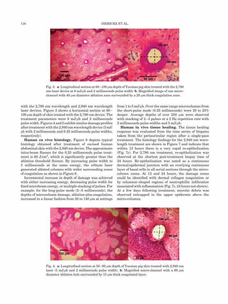
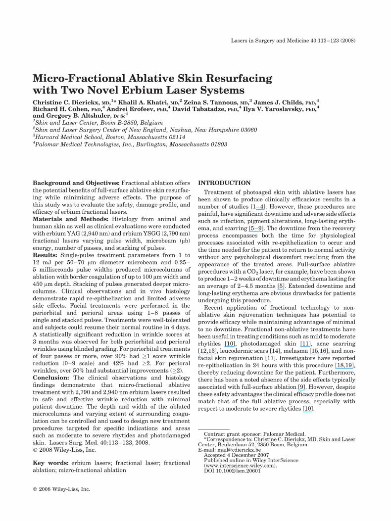
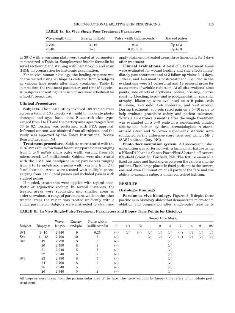
with the 2,790 nm wavelength and 2,940 nm wavelengthlaser devices. Figure 3 shows a horizontal section at 60–100 mm depth of skin treated with the 2,790 nm device. Thetreatment parameters were 8 mJ/mb and 2 millisecondspulse width. Figures 4 and 5 exhibit similar damage profilesafter treatment with the 2,940 nm wavelength device (5 mJ/mb with 2 milliseconds and 0.25 milliseconds pulse widths,respectively).Human ex vivo histology. Figure 6 depicts typical
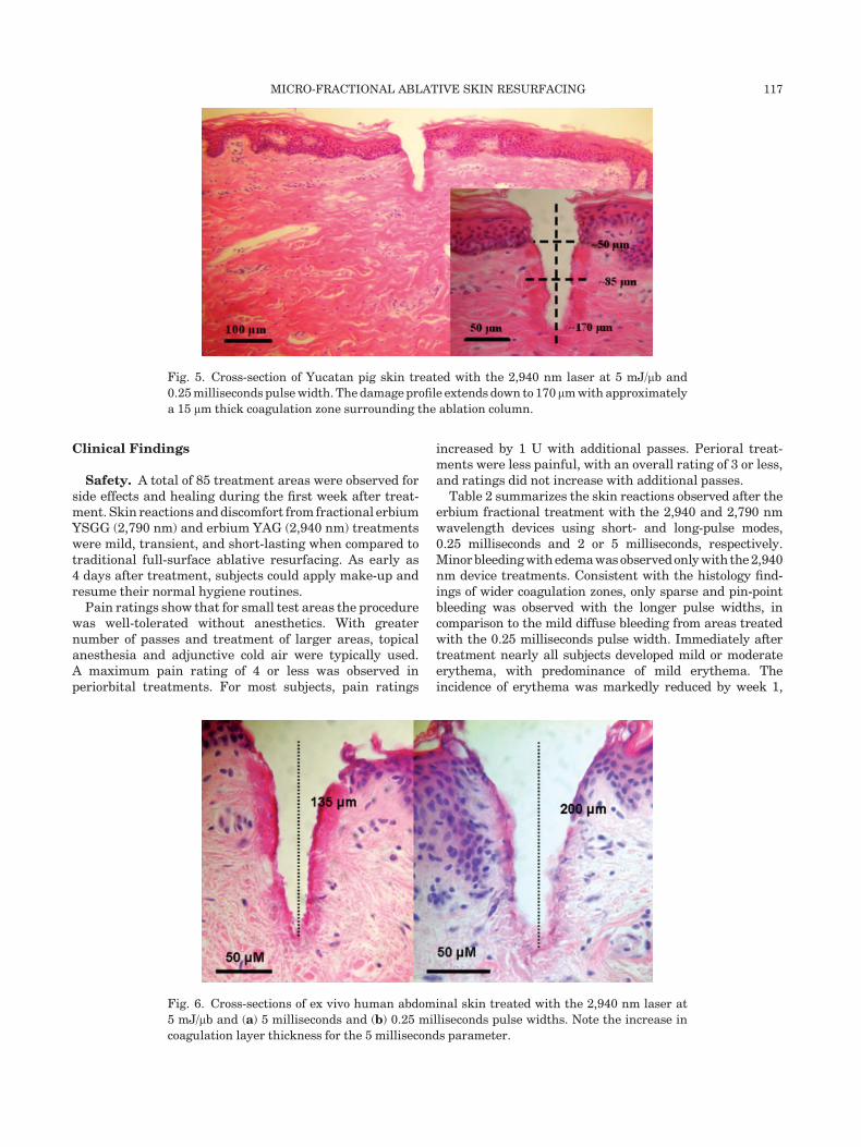
histology obtained after treatment of excised humanabdominal skin with the 2,940 nm device. The approximateintra-beam fluence for the 0.25 milliseconds pulse treat-ment is 60 J/cm2, which is significantly greater than theablation threshold fluence. By increasing pulse width to5 milliseconds at the same energy, the erbium lasergenerated ablated columns with wider surrounding zonesof coagulation as shown in Figure 6.
Incremental increase in depth of damage was achievedwith either increasing energy, decreasing pulse width forfixed microbeam energy, or multiple stacking of pulses. Forexample, for the long-pulse mode (2–5 milliseconds), thedepths of microcolumn damage, ablation plus coagulation,increased in a linear fashion from 50 to 140 mm at settings
from 1 to 5 mJ/mb. Over the same range microcolumns fromthe short-pulse mode (0.25 milliseconds) were 20 to 25%deeper. Average depths of over 250 mm were observedwith stacking of 2–3 pulses at a 2 Hz repetition rate with2 milliseconds pulse widths and 5 mJ/mb.Human in vivo tissue healing. The tissue healing
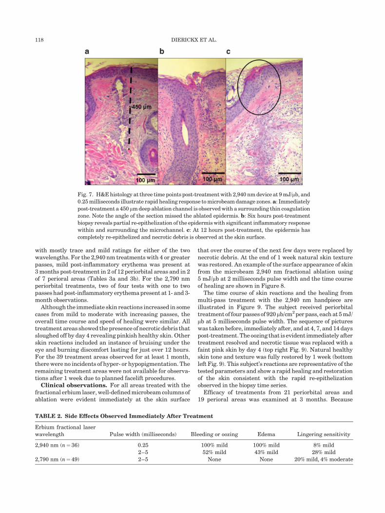
response was evaluated from the time series of biopsiestaken from the periauricular region after a single-passtreatment. The histology findings for the 2,940 nm wave-length treatment are shown in Figure 7 and indicate thatwithin 12 hours there is a very rapid re-epithelization(Fig. 7c). For 2,790 nm treatment, re-epithelization wasobserved at the shortest post-treatment biopsy time of24 hours. Re-epithelization was noted as a continuousdermal/epidermal junction with an overlying continuouslayer of basal cells in all serial sections through the micro-column zones. At 12 and 24 hours, the damage zonescould be identified with dermal collagen coagulation orby columnar-shaped regions of neutrophillic infiltrationassociated with inflammation (Fig. 7c, 24 hours not shown).At a few days following treatment, necrotic debris wasobserved entrapped in the upper epidermis above themicro-columns.
Fig. 3. a: Longitudinal section at 60–100 mm depth of Yucatan pig skin treated with the 2,790
nm laser device at 8 mJ/mb and 2 milliseconds pulse width. b: Magnified image of one micro-
channel with 40 mm diameter ablation zone surrounded by a 20 mm thick coagulation zone.
Fig. 4. a: Longitudinal section at 50–80 mm depth of Yucatan pig skin treated with 2,940 nm
laser (5 mJ/mb and 2 milliseconds pulse width). b: Magnified micro-channel with a 60 mm
diameter ablation hole surrounded by 15 mm thick coagulated layer.
116 DIERICKX ET AL.
Clinical Findings
Safety. A total of 85 treatment areas were observed forside effects and healing during the first week after treat-ment. Skin reactions and discomfort from fractional erbiumYSGG (2,790 nm) and erbium YAG (2,940 nm) treatmentswere mild, transient, and short-lasting when compared totraditional full-surface ablative resurfacing. As early as4 days after treatment, subjects could apply make-up andresume their normal hygiene routines.
Pain ratings show that for small test areas the procedurewas well-tolerated without anesthetics. With greaternumber of passes and treatment of larger areas, topicalanesthesia and adjunctive cold air were typically used.A maximum pain rating of 4 or less was observed inperiorbital treatments. For most subjects, pain ratings
increased by 1 U with additional passes. Perioral treat-ments were less painful, with an overall rating of 3 or less,and ratings did not increase with additional passes.
Table 2 summarizes the skin reactions observed after theerbium fractional treatment with the 2,940 and 2,790 nmwavelength devices using short- and long-pulse modes,0.25 milliseconds and 2 or 5 milliseconds, respectively.Minorbleedingwithedema was observed only with the 2,940nm device treatments. Consistent with the histology find-ings of wider coagulation zones, only sparse and pin-pointbleeding was observed with the longer pulse widths, incomparison to the mild diffuse bleeding from areas treatedwith the 0.25 milliseconds pulse width. Immediately aftertreatment nearly all subjects developed mild or moderateerythema, with predominance of mild erythema. Theincidence of erythema was markedly reduced by week 1,
Fig. 5. Cross-section of Yucatan pig skin treated with the 2,940 nm laser at 5 mJ/mb and
0.25 milliseconds pulse width. The damage profile extends down to 170 mm with approximately
a 15 mm thick coagulation zone surrounding the ablation column.
Fig. 6. Cross-sections of ex vivo human abdominal skin treated with the 2,940 nm laser at
5 mJ/mb and (a) 5 milliseconds and (b) 0.25 milliseconds pulse widths. Note the increase in
coagulation layer thickness for the 5 milliseconds parameter.
MICRO-FRACTIONAL ABLATIVE SKIN RESURFACING 117
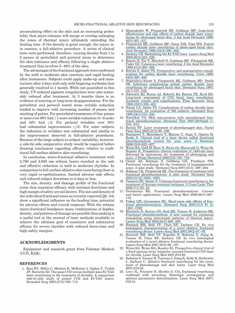
with mostly trace and mild ratings for either of the twowavelengths. For the 2,940 nm treatments with 4 or greaterpasses, mild post-inflammatory erythema was present at3 months post-treatment in 2 of 12 periorbital areas and in 2of 7 perioral areas (Tables 3a and 3b). For the 2,790 nmperiorbital treatments, two of four tests with one to twopasses had post-inflammatory erythema present at 1- and 3-month observations.
Although the immediate skin reactions increased in somecases from mild to moderate with increasing passes, theoverall time course and speed of healing were similar. Alltreatment areas showed the presence of necrotic debris thatsloughed off by day 4 revealing pinkish healthy skin. Otherskin reactions included an instance of bruising under theeye and burning discomfort lasting for just over 12 hours.For the 39 treatment areas observed for at least 1 month,there were no incidents of hyper- or hypopigmentation. Theremaining treatment areas were not available for observa-tions after 1 week due to planned facelift procedures.Clinical observations. For all areas treated with the
fractional erbium laser, well-defined microbeam columns ofablation were evident immediately at the skin surface
that over the course of the next few days were replaced bynecrotic debris. At the end of 1 week natural skin texturewas restored. An example of the surface appearance of skinfrom the microbeam 2,940 nm fractional ablation using5 mJ/mb at 2 milliseconds pulse width and the time courseof healing are shown in Figure 8.
The time course of skin reactions and the healing frommulti-pass treatment with the 2,940 nm handpiece areillustrated in Figure 9. The subject received periorbitaltreatment of four passes of 920mb/cm2 per pass, each at 5 mJ/mb at 5 milliseconds pulse width. The sequence of pictureswas taken before, immediately after, and at 4, 7, and 14 dayspost-treatment. The oozing that is evident immediately aftertreatment resolved and necrotic tissue was replaced with afaint pink skin by day 4 (top right Fig. 9). Natural healthyskin tone and texture was fully restored by 1 week (bottomleft Fig. 9). This subject’s reactions are representative of thetested parameters and show a rapid healing and restorationof the skin consistent with the rapid re-epithelizationobserved in the biopsy time series.
Efficacy of treatments from 21 periorbital areas and19 perioral areas was examined at 3 months. Because
TABLE 2. Side Effects Observed Immediately After Treatment
Erbium fractional laser
wavelength Pulse width (milliseconds) Bleeding or oozing Edema Lingering sensitivity
2,940 nm (n¼ 36) 0.25 100% mild 100% mild 8% mild
2–5 52% mild 43% mild 28% mild
2,790 nm (n¼ 49) 2–5 None None 20% mild, 4% moderate
Fig. 7. H&E histology at three time points post-treatment with 2,940 nm device at 9 mJ/mb, and
0.25 milliseconds illustrate rapid healing response to microbeam damage zones.a: Immediately
post-treatment a 450 mm deep ablation channel is observed with a surrounding thin coagulation
zone. Note the angle of the section missed the ablated epidermis. b: Six hours post-treatment
biopsy reveals partial re-epithelization of the epidermis with significant inflammatory response
within and surrounding the microchannel. c: At 12 hours post-treatment, the epidermis has
completely re-epithelized and necrotic debris is observed at the skin surface.
118 DIERICKX ET AL.

TABLE
3a.PeriorbitalWrin
kle
ReductionAfterMicro-F
ractionalErbiu
mTreatm
ent
Han
dp
iece
#P
erio
rbit
al
site
s
Para
met
ers,
ener
gy
(mJ/m
b)
Pass
esE
ryth
ema
Wri
nk
lere
du
ctio
n
(tw
oou
tof
thre
egra
der
su
sin
g0
–9
scale
)
Sta
cked
Tot
al
%of
test
site
s
at
1m
onth
%of
test
site
s
at
3m
onth
s
%of
test
site
sw
ith
scor
eim
pro
vem
ent�
1
%of
test
site
sw
ith
scor
eim
pro
vem
ent�
2
2,7
90
48
–9
01
–2
50
50
50
0
2,9
40
53
–6
01
00
60
20
2,9
40
83
–6
04
–6
38
12
88
38
2,9
40
45
26
–8
75
25
100
50
TABLE
3b.PerioralWrin
kle
ReductionAfterMicro-F
ractionalErbiu
mTreatm
ent
Han
dp
iece
#P
erio
ral
site
s
Para
met
ers,
ener
gy
(mJ/m
b)
Pass
esE
ryth
ema
Wri
nk
lere
du
ctio
n
(tw
oou
tof
thre
egra
der
su
sin
g0
–9
scale
)
Sta
cked
Tot
al
%of
test
site
s
at
1m
onth
%of
test
site
s
at
3m
onth
s
%of
test
site
sw
ith
scor
eim
pro
vem
ent�
1
%of
test
site
sw
ith
scor
eim
pro
vem
ent�
2
2,7
90
66
–9
01
00
16
0
2,9
40
65
0–
22
–3
16
033
0
2,9
40
75
1–
24
–6
29
29
57
57
MICRO-FRACTIONAL ABLATIVE SKIN RESURFACING 119

parameters were varied for each patient from side to side,left and right sides were evaluated as independent testsites. Results for the 2,790 and 2,940 nm handpiecessummarized in Tables 3a and 3b include the percentage oftested areas having erythema at 1 and 3 months and thepercentage of areas with wrinkle score reductions.The number of treated sites with at least two of the threegraders giving a score reduction of one or more (2nd to lastcolumn) or two or more (last column) was divided by thetotal number of treatment sites to determine percentage.
A total of 12 periorbital sites were treated with fourpasses or more and over 90% had �1 score wrinklereduction (0–9 scale) and 42% had �2. The overall averagereduction in the periorbital wrinkle score (post-pre) wasstatistically significant (t-test or Wilcoxon signed-rankstatistic: �1.26� 1.11, n¼ 21, P<0.0001). The use of fouror more passes resulted in an average reduction of 1.5, butwith the addition of double-stacked pulses the averagereduction increased to 2.4. Figure 10 shows before (top) andafter (bottom) photos following a single 2,940 nm periorbi-tal treatment of six passes at 5 mJ/mb, 5 milliseconds pulsewidth and density of 920 mb/cm2 per pass. The left sidereceived double-stacked pulses at two pulses per secondfor the first two passes and shows greater reductionin wrinkles, but mild post-inflammatory erythema at
3 months. Instances of erythema, a mild pink to slight redbelow the eye, were present at the 3-month observation in2 of 17 cases with 2,940 nm treatments. Because erythemafor the 2,790 nm treatments (see Table 3a) appeared tolast longer, testing was subsequently focused on definingparameters for the 2,940 nm handpiece.
Substantial improvements in perioral wrinkles wereobserved with aggressive treatments. For the seven treat-ment sites receiving four or more passes over 50% had �2score improvement. The overall average reduction inwrinkle score (post-pre) was statistically significant (t-testor Wilcoxon signed-rank statistic: �0.74� 1.43, n¼ 19,P<0.05). Those receiving four or more passes had anaverage reduction of 1.5 and for the four cases withimprovements the average reduction was 2.7. Figure 11shows before (top) and after (bottom) photos following a2,940 nm perioral treatment at 5 mJ/mb, 5 millisecondspulse width and density of 920 mb/cm2 per pass. Thefirst pass on both sides was double-stacked at two pulses persecond. The right side had an additional double-stackedpass and six passes total, while the left side had four totalpasses. The right side shows greater reduction in wrinkles,but mild post-inflammatory erythema at 3 months.
These clinical and histologic findings demonstrate theefficacy of micro-fractional ablative treatments in wrinkle
Fig. 8. Appearance of skin surface following fractional skin ablation from a single 5 mJ/mb,
2 milliseconds pulse of the 2,940 nm device at a density of 920 mb/cm2 shows rapid healing.
By day 7 the skin surface is restored.
120 DIERICKX ET AL.

reduction. In addition, this new approach is associatedwith mild side effects and reduced downtime compared totraditional ablative resurfacing methods.
DISCUSSION
The novel micro-fractional ablative device investigatedin this study provides a wide range of new capabilities forskin rejuvenation. The depth and width of the ablatedmicrocolumns and extent of surrounding coagulation canbe varied and used to design procedures targeted forspecific indications and areas. With a user selectable opticof 920 mb/cm2, both erbium lasers caused similar ablativedamage profiles with diameters for single-pulse moderanging from 60 to 100 mm and ablation depths from 20 to250 mm at energy settings up to 5 mJ/mb. Stacked pulsesprovided even deeper columns as compared to respectivesingle pulses. Coagulation zones around the ablated micro-columns varied in thickness from 5 to 25 mm and dependedupon pulse width and wavelength (Figs. 3–6). At equiv-alent settings, the 2,790 nm wavelength handpiecegenerated somewhat greater thermal damage than the2,940 nm handpiece. Traditional erbium ablation has
very minimal zones of thermal injury due to the combina-tion of high water absorption and short-pulse mode. Onenovel feature of the 2,940 nm system is its ability to providesmooth, long (millisecond range) pulses. This feature,combined with the micro-fractional delivery configuration,produced relatively large consistent zones of coagulationsurrounding the ablative microcolumns. Because the2,940 nm handpiece could provide both ablative (short-pulse) and ablative plus coagulative (long-pulse) modes ofmicro-fractional treatments, efforts were subsequentlyfocused on evaluating the 2,940 nm handpiece to definesafety limits and optimum treatment parameters forfurther clinical investigations.
The treatment tests with single-pass parameters clearlydemonstrate the safety and rapid healing of the micro-fractional ablative approach. Histologic findings confirmthat the epidermis undergoes a rapid re-epithelization(<24 hours) following micro-ablative ablation at settings of9–12 mJ/mb (Fig. 7, histology from 2,790 not shown). Basedupon the observed ablative column diameters of 75–100 mm, approximately 4–5% of the skin surface is ablatedin each pass when using an optical tip with a 920 mb/cm2
density. With multiple passes, however, there is an
Fig. 9. The rapid healing from multi-pass treatment with the 2,940 nm handpiece is shown in
the sequence of pictures. The top row from left to right shows the periorbital test area taken
before, immediately after, and 4 days post-treatment. The bottom row shows the test area 7
and 14 days post-treatment. The subject received periorbital treatment of four passes of 920mb/
cm2 per pass, each at 5 mJ/mb at 5 milliseconds pulse width. The oozing that is evident
immediately after treatment resolved by day 2 and necrotic tissue was replaced with a faint
pink skin by day 4. Natural healthy skin tone and texture was fully restored by 1 week.
MICRO-FRACTIONAL ABLATIVE SKIN RESURFACING 121

Fig. 10. Before (top) and 3-month post-treatment photos (bottom) following a single 2,940 nm
periorbital treatment of six passes at 5 mJ/mb, 5 milliseconds pulse width and density of 920 mb/
cm2 per pass. The left side received double stacked pulses at two pulses per second for the first
two passes and shows greater reduction in wrinkles, but mild post-inflammatory erythema.
Fig. 11. Before (top) and 3-month post-treatment photos (bottom) following a single 2,940 nm
perioral treatment at 5 mJ/mb, 5 milliseconds pulse width and density of 920 mb/cm2 per pass.
The first pass on both sides was double stacked at two pulses per second. The right side had an
additional double stacked pass and six passes total, while the left side had a total of four passes.
The right side shows greater reduction in wrinkles, but mild post-inflammatory erythema.
122 DIERICKX ET AL.
accumulating effect on the skin and an increasing proba-bility that micro-columns will merge or overlap enlargingthe zones of thermal injury ultimately extending thehealing time. If the density is great enough, the injury is,in essence, a full-ablative procedure. A series of clinicaltests were performed, therefore, varying density from 1 to8 passes at periorbital and perioral areas to determinethe skin tolerance and efficacy following a single ablativetreatment that involves 5–40% of the skin.
The advantages of the fractional approach were confirmedby the mild to moderate skin reactions and rapid healingafter treatments. Subjects could apply make-up and mois-turizers after 4 days with only mild lingering erythema thatgenerally resolved in 1 month. While not quantified in thisstudy, UV-induced pigment irregularities were also notice-ably reduced after treatment. At 3 months there is noevidence of scarring or long-term dyspigmentation. For theperiorbital and perioral tested areas wrinkle reductiontended to improve with increasing number of passes andstacking of pulses. For periorbital treatments of four passesor more over 90% had�1 score wrinkle reduction (0–9 scale)and 42% had �2. For perioral wrinkles over 50%had substantial improvements (�2). For some subjectsthe reduction in wrinkles was substantial and similar tothe improvement observed in full-ablative procedures.Because of the large subject-to-subject variability, however,a side-by-side comparative study would be required beforedrawing conclusions regarding efficacy relative to tradi-tional full-surface ablative skin resurfacing.
In conclusion, micro-fractional ablative treatment with2,790 and 2,940 nm erbium lasers resulted in the safeand effective reduction in mild to moderate rhytides. Incomparison to full-surface ablative skin resurfacing there isvery rapid re-epithelization, limited adverse side effectsand reduced subject downtime to 4 days or less.
The size, density, and damage profile of the fractionalzones that maximize efficacy with minimal downtime andhigh margin of safety are not known. The size and density ofthe individual fractional zones as recently reported [20–24]show a significant influence on the healing time, potentialfor adverse effects and overall response. With the erbiummicro-fractional handpiece many combinations of depths,density, and patterns of damage are possible thus making ita useful tool in the arsenal of laser methods available toachieve the ultimate goal of consistent and substantialefficacy for severe rhytides with reduced down-time andhigh safety margins.
ACKNOWLEDGMENTS
Equipment and research grant from Palomar Medical(CCD, KAK).
REFERENCES
1. Ross EV, Miller C, Meehan K, McKinlay J, Sajben P, TrafeliJP, Barnette DJ. One-pass CO2 versus multiple-pass Er:YAGlaser resurfacing in the treatment of rhytides: A comparisonside-by-side study of pulsed CO2 and Er:YAG lasers.Dermatol Surg 2001;27(8):709–715.
2. Manuskiatti W, Fitzpatrick RE, Goldman MP. Long-termeffectiveness and side effects of carbon dioxide laser resur-facing for photoaged facial skin. J Am Acad Dermatol 1999;40(3):401–411.
3. Fitzpatrick RE, Goldman MP, Satur NM, Tope WD. Pulsedcarbon dioxide laser resurfacing of photo-aged facial skin.Arch Dermatol 1996;132(4):395–402.
4. Zachary CB. Modulating the Er:YAG laser. Lasers Surg Med2000;26(2):223–226.
5. Ratner D, Tse Y, Marchell N, Goldman MP, Fitzpatrick RE,Fader DJ. Cutaneous laser resurfacing. J Am Acad Dermatol1999;41(3):365–389.
6. Horton S, Alster TS. Preoperative and postoperative consid-erations for carbon dioxide laser resurfacing. Cutis 1999;64(6):399–406.
7. Sriprachya-Anunt S, Fitzpatrick RE, Goldman MP, SmithSR. Infections complicating pulsed carbon dioxide laserresurfacing for photoaged facial skin. Dermatol Surg 1997;23(7):527–536.
8. Schwartz RJ, Burns AJ, Rohrich RJ, Barton FE, Byrd HS.Long-term assessment of CO2 facial laser resurfacing:Aesthetic results and complications. Plast Reconstr Surg1999;103(2):592–601.
9. Nanni CA, Alster TS. Complications of carbon dioxide laserresurfacing. An evaluation of 500 subjects. Dermatol Surg1998;24(3):315–320.
10. Narurkar VA. Skin rejuvenation with microthermal frac-tional photothermolysis. Dermatol Ther 2007;20(Suppl 1):S10–S13.
11. Rostan EF. Laser treatment of photodamaged skin. FacialPlast Surg 2005;21(2):99–109.
12. Hasegawa T, Matsukura T, Mizuno Y, Suga Y, Ogawa H,Ikeda S. Clinical trial of a laser device called fractionalphotothermolysis system for acne scars. J Dermatol2006;33(9):623–627.
13. Weiss RA, Gold M, Bene N, Biron JA, Munavalli G, Weiss M,Beasley K. Prospective clinical evaluation of 1440-nm laserdelivered by microarray for treatment of photoaging andscars. J Drugs Dermatol 2006;5(8):740–744.
14. Glaich AS, Rahman Z, Goldberg LH, Friedman PM.Fractional resurfacing for the treatment of hypopigmentedscars: A pilot study. Dermatol Surg 2007;33(3): 289–294.
15. Rokhsar CK, Fitzpatrick RE. The treatment of melasma withfractional photothermolysis: A pilot study. Dermatol Surg2005;31(12):1645–1650.
16. Tannous ZS, Astner S. Utilizing fractional resurfacing in thetreatment of therapy-resistant melasma. J Cosm Laser Ther2005;7(1):39–43.
17. Geromenus RG. Fractional photothermolysis: Currentand future applications. Lasers Surg Med 2006;38(3):169–176.
18. Fisher GH, Geronemus RG. Short-term side effects of frac-tional photothermolysis. Dermatol Surg 2005;31(9 Pt 2):1245–1249.
19. Manstein D, Herron GS, Sink RK, Tanner H, Anderson RR.Fractional photothermolysis: A new concept for cutaneousremodeling using microscopic patterns of thermal injury.Lasers Surg Med 2004;34(5):426–438.
20. Hantash BM, Bedi VP, Chan KF, Zachary CB. Ex vivohistological characterization of a novel ablative fractionalresurfacing device. Lasers Surg Med 2007;39(2):87–95.
21. Hantash BM, Bedi VP, Kapadia B, Rahman Z, Jiang K,Tanner H, Chan KF, Zachary CB. In vivo histologicalevaluation of a novel ablative fractional resurfacing device.Lasers Surg Med 2007;39(2):96–107.
22. Weiss RA, Weiss MA, Beasley KI. Prospective clinical trial ofa fixed spacing array computer scanned fractional CO2 laserfor rhytids. Laser Surg Med 2007;S19:49.
23. Rahman Z, Tanner H, Tournas J, Jiang K, Kelly K, BerkowitzL, Zachary C. Ablative fractional resurfacing for the treat-ment of photodamage and skin laxity. Laser Surg Med2007;S19:45.
24. Levy JL, Fournier N, Mordon S. CO2 fractional resurfacingcombined with aircooling: Histologic investigation andoptimal parameters determination. Laser Surg Med 2007;S19:51.
MICRO-FRACTIONAL ABLATIVE SKIN RESURFACING 123