Computer Interfaces: From Communication to Mind-Prosthesis Metaphor
Upload
khangminh22Category
view
1download
0
This work is licensed under a Creative Commons Attribution-NonCommercial 3.0 Unported License
Newcastle University ePrints - eprint.ncl.ac.uk
Bone MC, Naylor A. Failure of an ACCIS metal-on-metal hip resurfacing
prosthesis: A case report. Proceedings of the Institution of Mechanical
Engineers, Part H: Journal of Engineering in Medicine 2017, 231(12), 1188-
1194.
Copyright:
This is the authors’ accepted manuscript of an article that has been published in its final definitive
form by Sage, 2017
DOI link to article:
https://doi.org/10.1177/0954411917737457
Date deposited:
11/01/2018
Failure of an ACCIS metal-on-metal hip resurfacing prosthesis: A case report 1
2
Martin C Bone1, Andrew Naylor
13
4
Abstract 5
The introduction of coatings on joint replacements was intended to reduce wear volumes, 6
prevent corrosion and reduce metal ion release. However retrieval analysis to confirm their in 7
vivo performance has been limited. The aim of study was to examine the coating on a retrieved 8
hip prosthesis to determine if substantial damage or wear had occurred. A single ACCIS 9
titanium niobium nitride coated hip resurfacing prosthesis was retrieved for examination. Wear 10
volumes and surface roughness were measured to quantify the damage to the articulating 11
surfaces. The coating had been completely removed from substantial parts of both the femoral 12
head and the acetabular cup, corresponding to areas of wear as measured by a coordinate 13
measuring machine. The total wear rate (61 mm³/year) was high and corresponded with the 14
high metal ion levels recorded pre operatively (83 ppb Chromium and 110 ppb Cobalt). 15
Roughness data indicated that the coating has a lower roughness than the substrate and 16
substantially lower than the boundary between the coating and the substrate. The wear rate is 17
very high and it is likely that damage to the coating resulted in accelerated wear of the 18
prosthesis. No sudden change between the boundary of the substrate and the coating was 19
observed that would indicate delamination or failure of the coating substrate interface layer. 20
Whilst coatings may offer some theoretical benefits, they may also result in catastrophic failure 21
of the prosthesis. 22
23
Keywords 24
Page 1 of 22
http://mc.manuscriptcentral.com/(site)
Journal name
123456789101112131415161718192021222324252627282930313233343536373839404142434445464748495051525354555657585960
Preprin
t vers
ion
Wear Analysis/Testing (Biomechanics), Tribology of Coatings, Orthopaedic Tribology, 1
Orthopaedic Materials, Surfaces, Blood Metal Ion Levels 2
3
1School of Mechanical and Systems Engineering, Newcastle University, Newcastle upon Tyne, 4
UK 5
6
Corresponding author: Martin C Bone, School of Mechanical and Systems Engineering, 7
Newcastle University, Newcastle upon Tyne, UK. 8
9
Email: [email protected] 10
11
Introduction 12
Wear of metal-on-metal (MoM) hip replacements has been widely documented in the literature. 13
Whilst studies have shown that MoM hip prostheses have lower wear volumes than metal-on-14
polyethylene (MoP) prostheses, the number of wear particles released by a MoM hip prosthesis 15
is substantially higher (typically 7 to 30 times).1 In the quest for lower wear rates and higher 16
corrosion resistance, other designs of artificial hip joint have been developed and implanted. 17
One of these is the Advanced Ceramic Coated Implant Systems (ACCIS) hip prosthesis, a 18
ceramic surface engineered hip resurfacing, manufactured from cobalt-chromium-molybdenum 19
(CoCrMo) with titanium niobium nitride (TiNbN) coating.1, 2
According to the manufacturer, the 20
introduction of a coating was intended to reduce wear, prevent corrosion and reduce metal ion 21
release.2
22
23
Page 2 of 22
http://mc.manuscriptcentral.com/(site)
Journal name
123456789101112131415161718192021222324252627282930313233343536373839404142434445464748495051525354555657585960
Preprin
t vers
ion
ACCIS hip prostheses are cast and heat treated to reduce the size of block carbides on the 1
metal surfaces, as these may cause wear via abrasion.1, 2
The surfaces are then polished 2
before undergoing a surface microfinish to remove asperities down to a nanometre level.1, 2
A 3
TiNbN coating with a nominal thickness of 300±50 µm is then applied to the surfaces via the 4
process of physical vapour deposition (PVD).1, 2
This is said to alter the surface chemical 5
properties of a material without affecting the macro material and biochemical properties. In non-6
coated MoM CoCrMo alloy joints, block carbides have been identified as being useful for 7
reducing wear rates. One potential explanation as to why they were reduced in the ACCIS, is 8
that the smoother surface would provide more favourable conditions for the coating to bond to 9
the substrate. 10
11
To the authors’ best knowledge there are only a limited number of studies in the literature to 12
examine the performance of the ACCIS hip.2-5
de Villiers et al. examined three explanted 13
resurfacing prosthesis pairs.3 In all cases there were large areas where the coating had been 14
completely removed, indicative of three body wear, exposing the CoCrMo substrate.3 The three 15
pairs had a mean time in vivo of 29 months (range 26-33 months), and average blood metal ion 16
levels of 30 ppb and 24.6 ppb for cobalt and chromium respectively. Jemmett et al.4 reported 17
that seven prosthesis pairs were revised due to elevated metal ion levels, after a mean period 18
or 56 months in vivo (range 41-72 months). On average the measured levels for cobalt and 19
chromium were 7 ppb and 8 ppb respectively. It was not stated whether these were total hip 20
replacement or resurfacing. This is in contrast to a study by Hamelynck and Woering, which 21
measured blood metal ions from 60 patients with the ACCIS hip resurfacing prostheses up to 24 22
months post-surgery.2 The results indicated that the cobalt and chromium levels were lower 23
than the 7 ppb threshold6 advised by the Medicines and Healthcare products Regulatory 24
Page 3 of 22
http://mc.manuscriptcentral.com/(site)
Journal name
123456789101112131415161718192021222324252627282930313233343536373839404142434445464748495051525354555657585960
Preprin
t vers
ion
Agency (MHRA) in the UK. In 2015 Lapaj reported on a cohort of 11 titanium niobium TiNb 1
coated hip prostheses with time in vivo ranging from 1 month to 56 months.5 No gross failure of 2
the TiNb coating was reported, however all implants had evidence of defects in the coating 3
including pinholes and blisters with delamination.5 Due to the limited number of studies 4
available, the aim was to examine a retrieved ACCIS hip prosthesis to determine how the 5
coating had performed in vivo. The objectives of the study were to: 6
1. Quantify the volumetric wear from the articulating surfaces7
2. Measure the surface roughness of the coating, the exposed substrate and the boundary8
between the two regions and determine if there is any disparity 9
10
Methods 11
Patient and Prosthesis Details 12
A single uncemented ACCIS hip resurfacing prosthesis (Implantcast GmbH, Buxtehude, 13
Germany) consisting of a 48 mm diameter femoral head (cemented); and a 52 mm diameter 14
acetabular cup (uncemented), was obtained for volumetric wear analysis and measurement of 15
articulating surface roughness. The prosthesis was in vivo for 22 months and revised due to 16
high blood metal ion levels. The patient was female, was a healthy weight (BMI < 25), and was 17
41 years of age at the time of the revision. The patient had a history of Perthes disease and 18
acetabular dysplasia, for which she had previously undergone periacetabular osteotomy. The 19
ACCIS prosthesis was implanted by a consultant orthopaedic surgeon at Morriston Hospital, 20
Swansea, UK. The surgeon had implanted approximately 200 of these prostheses by the time 21
of her surgery. The patient had had no prosthesis implanted prior to the ACCIS, and had 22
undergone no other arthroplasty procedures to replace other joints of the body. 23
Page 4 of 22
http://mc.manuscriptcentral.com/(site)
Journal name
123456789101112131415161718192021222324252627282930313233343536373839404142434445464748495051525354555657585960
Preprin
t vers
ion
Prior to revision surgery the metal ion levels were 83 ppb Cr and 110 ppb Co. A macroscopic 1
examination of the explanted hip resurfacing was conducted prior to taking volumetric wear and 2
surface finish measurements. At the time of revision there were no signs of corrosion or wear 3
debris in the surrounding tissue. 4
5
Volumetric Wear Analysis 6
Volumetric wear was measured using a Mitutoyo Legex 322 (Mitutoyo Corporation, Kawasaki, 7
Kanagawa, Japan) Coordinate Measuring Machine (CMM), which has a manufacturer claimed 8
scanning accuracy of 0.8 µm. A 5 mm diameter scanning probe was used for the 9
measurements, whilst the results were processed using a custom MATLAB programme 10
described in detail elsewhere.7 This wear volume methodology has been validated and found 11
accurate to within 0.5 mm³.7, 8
12
13
Surface Roughness Analysis 14
Surface roughness measurements were performed using a ZYGO NewView 5000 (Zygo 15
Corporation, Middlefield, CT, USA) white light interferometer (resolution 1nm). Measurements 16
were taken at three key areas for both the head and cup: 1) where the coating was still intact; 2) 17
where the coating was completely removed exposing the CoCrMo substrate; 3) along the 18
edge/boundary between the coated surface and the exposed substrate. Three measurements 19
were taken from each of these key areas, from which both micrographs and 3D topographical 20
plots were obtained. 21
From the plots, three roughness parameters were selected: average roughness (Sa); skewness 22
(Ssk); and kurtosis (Sku). Average roughness (Sa) is described as the arithmetic mean of the z 23
axis heights (peaks and valleys) obtained from the plots, and hence the calculated values 24
Page 5 of 22
http://mc.manuscriptcentral.com/(site)
Journal name
123456789101112131415161718192021222324252627282930313233343536373839404142434445464748495051525354555657585960
Preprin
t vers
ion
provided an indication for the variation in heights. Guidelines for metal hip prostheses specify a 1
50 nm threshold for average roughness measurements only.9 Ssk and Sku are dimensionless 2
values. Ssk is a measure of surface amplitude with positive values indicative of a high number of 3
peaks above the surface; negative values indicative of a high number of valleys below the 4
surface. Sku is a measure of surface pitch with high positive values indicative of a high number 5
of sharp peaks and/or valleys. Root mean square roughness (Sq) was initially considered, but 6
discounted after reviewing the findings of a separates study, revealing similar Sa and Sq values 7
for orthopaedic applications.10
8
9
Results 10
Volumetric Wear 11
The TiNbN coating had been removed over a large region of both the femoral head and 12
acetabular cup revealing a wear scar of the CoCrMo substrate (Figure 1). The total wear volume 13
was 112 mm³ of which 50 mm³ was from the femoral head and 62 mm³ from the acetabular cup. 14
This equated to a wear rate of 61 mm³/year. The wear scar on the femoral head formed an 15
irregular elliptical shape, while the wear scar of the acetabular cup extended to a large but 16
localised section of the rim of the acetabular cup (Figure 2). The regions of coating removal 17
identified by the CMM were found to correlate with the worn sections of the prostheses (Figures 18
1 and 2). Although it is clear that the observed volumetric wear is primarily a result of removal of 19
the TiNbN coating, a smaller level of material loss of the CoCrMo substrate will have occurred. 20
21
Coated Surface 22
The TiNbN coated surfaces of the femoral head yielded a low mean Sa value of 33.61 nm 23
(StDev 4.21); along with a negative mean Ssk value of -2.38 (StDev 2.40); and a high mean Sku 24
Page 6 of 22
http://mc.manuscriptcentral.com/(site)
Journal name
123456789101112131415161718192021222324252627282930313233343536373839404142434445464748495051525354555657585960
Preprin
t vers
ion
value of 56.71 (StDev 53.00) (Table 1). The TiNbN surfaces of the acetabular cup returned 1
values somewhat lower in magnitude: for both Sa (18.19 nm, StDev 5.11); Ssk (-1.53, StDev 2
1.94); and Sku (36.57, StDev 19.90). An example micrograph and topographical plot obtained 3
from the femoral head, showing a uniform surface, is shown in Figure 3. 4
5
Substrate Surface 6
The exposed CoCrMo surfaces of the femoral head showed mean Sa values of 44.61 nm 7
(StDev 10.68). These were seen to be higher than for the cup (14.88 nm, StDev 0.26). The 8
mean Sku values were in the same approximate range for both the head (16.32, StDev 9.49) 9
and the cup (12.77, StDev 13.61). The femoral head yielded a negative mean Ssk value (-1.64, 10
StDev 0.45). A micrograph and topographical plot of the exposed CoCrMo surface is shown in 11
Figure 4. In contrast to this, the acetabular cup yielded a positive mean Ssk value (1.20, StDev 12
0.77). A micrograph and topographical plot obtained of the exposed CoCrMo surface of the 13
acetabular cup is shown in Figure 5. 14
15
Boundary surface 16
Measured values taken at the TiNbN-CoCrMo boundary were comparable for both the femoral 17
head and acetabular cup, with: very high mean Sa values reported for the head (245.55 nm, 18
StDev 12.7) and cup (211.29 nm, StDev 8.34); low mean Ssk values for the head (0.3, StDev 19
0.03) and cup (0.09 StDev 0.36); and low mean Sku values for the head (2.36, StDev 0.19) and 20
cup (2.5, StDev 0.45). The topography at the TiNbN-CoCrMo boundary is very similar for both 21
femoral head and acetabular cup. A distinct ridge is visible where the TiNbN coated surface and 22
exposed CoCrMo surface meet, as exemplified by a micrograph and a topographical plot 23
obtained from the femoral head (Figure 6). 24
Page 7 of 22
http://mc.manuscriptcentral.com/(site)
Journal name
123456789101112131415161718192021222324252627282930313233343536373839404142434445464748495051525354555657585960
Preprin
t vers
ion
1
Discussion 2
The wear rate reported here (61 mm³/year) is high. Lord et al previously noted that the mean 3
wear rate for a cohort of 22 DePuy Articular Surface Replacement (ASR) hip resurfacings was 4
22 mm³/year, a third of the value noted in this study.7
A later paper by Langton et al, examining 5
a cohort of 57 ASR hip resurfacings suggested that a combined wear rate of above 2.3 6
mm3/year was sufficient to cause a wear related failure.
11 Comparison of the current wear rate 7
(61 mm3/year) with those reported by previous studies for other types of hip resurfacings further 8
demonstrates the extremity of the wear reported here. For instance, a study by Witzleb et al. 9
examined 2 Birmingham Hip Resurfacing (BHR) prostheses reporting a mean wear rate of 15.6 10
mm3/year (range (8.4 – 22.8 mm
3/year).
1211
To the authors’ best knowledge there is only one other study offering wear rates for TiNbN 12
coated hip resurfacing prostheses.3 Total wear rates of 6.90, 8.28 and 16.22 mm
3/year were 13
reported for three prostheses in vivo for between 26 and 33 months. These wear rates were 14
substantially lower than that reported in the present study. 15
The shape of the wear scars corresponded with those seen previously on worn and explanted 16
hip resurfacings.13
The edge wear seen on the acetabular cup is a common feature of worn and 17
retrieved monoblock metal cups.13
18
At 83 ppb Cr and 110 ppb Co, the metal ion levels are high however this is perhaps 19
unsurprising given the high wear rate. It is important to note that these ion values do not include 20
Ti, Nb or N clearly demonstrating substantial wear of the substrate has occurred. For context, 21
the current MHRA guidelines for the UK state that “Blood metal ion level >7 ppb indicates 22
potential for soft tissue reaction”.6 Previously it has been shown that elevated blood metal ion 23
Page 8 of 22
http://mc.manuscriptcentral.com/(site)
Journal name
123456789101112131415161718192021222324252627282930313233343536373839404142434445464748495051525354555657585960
Preprin
t vers
ion
concentrations are associated with early failure of metal-on-metal hip implants secondary to 1
adverse reactions to metal debris, and that Co concentrations greater than 20 ppb are 2
frequently associated with metal staining of tissues and the development of osteolysis.14
3
Another paper showed that for ion concentrations above 4.5 ppb, abnormal wear was 4
indicated.15
The same paper also suggested that a whole blood Co of 100 ppb was associated 5
with a wear rate of between 21 – 59 mm3/year.
15 Those values fit well with the 110 ppb and 61 6
mm3/year reported in this case. 7
A possible explanation of the high wear rate is that the failure of the TiNbN coating resulted in 8
third body wear particles that accelerated the wear process. A similar process was seen 9
previously with the LPM proximal interphalangeal joint prosthesis, which utilised a similar type of 10
TiNb coating.16
Retrieval analysis confirmed that the surfaces had experienced extensive 11
damage to the TiNb coating with multi and multidirectional scratching on the articulating 12
surfaces indicating a third body wear16
. The coating was described as acting as a “hard grinding 13
paste” accelerating wear on the articulating surfaces. It is possible the same process may have 14
occurred in the ACCIS hip prosthesis. 15
This present study correlates with the findings of de Villiers et al3 and Jemmett et al
4 who both 16
reported that the use of coatings for metal hip resurfacings does not prevent metal ion release. 17
However in contrast to this, Hamelynck and Woering1 noted that the metal ion levels for a cohort 18
of 200 ACCIS resurfacing prostheses showed no raising of either cobalt or chromium at a 19
maximum follow up of 24 months. Cobalt and chromium metal ion levels from 60 patients were 20
reported in the Hamelynck and Woering study1, however these results were obtained from three 21
major hospitals, all in developed countries. It is also worth stating that the Hamelynck and 22
Page 9 of 22
http://mc.manuscriptcentral.com/(site)
Journal name
123456789101112131415161718192021222324252627282930313233343536373839404142434445464748495051525354555657585960
Preprin
t vers
ion
Woering study1 is a white paper published with the support of the manufacturer of ACCIS, 1
Implantcast. 2
The Sa roughness values of both the coated and uncoated surfaces were within the 50 nm 3
guidelines offered by the relevant ISO Standard.9 For comparison Joyce et al. reported on a 4
cohort of explanted DePuy ASR hip prostheses, noting mean Sa values ranging from 25 to 135 5
nm.17
Joyce et al also measured a single un-implanted prosthesis with reported head and cup 6
Sa values of 10 and 12 nm respectively. From this present study, the Sa values from both the 7
coated TiNbN; and exposed CoCrMo surfaces (Table 1) fell below the 50 nm ISO standard 8
threshold.9 However, the Sa values at the boundary were substantially higher than this standard 9
(246 nm for femoral head; 211 nm for acetabular cup). 10
Other roughness parameters (combination of negative skewness and high kurtosis) indicated 11
that the exposed surface of the head (Figure 4) had sharp valleys along the surface. Peaks 12
were found on the articulating surface of the acetabular cup, whilst unidirectional troughs were 13
found on the articulating surface of the femoral head. These phenomena have been noted 14
previously by de Villiers et al.3 after examining the surfaces of an explanted TiNbN acetabular 15
cup using energy dispersive x-ray spectroscopy (EDS) de Villiers et al. found that the majority of 16
the asperities were composed of Titanium, Niobium and Nitrogen leftover from the TiNbN 17
coating with a small minority of the asperities composed of CoCrMo from the substrate.3 This 18
sheds more light on the exposed CoCrMo surfaces of the ACCIS femoral head and acetabular 19
cup reported in this present study. It is prudent to assume that the hard asperities composed of 20
Titanium, Niobium and Nitrogen visible on the surface of the cup (Figure 5) were responsible for 21
creating the unidirectional scratches on the surface of the head (Figure 4). 22
Page 10 of 22
http://mc.manuscriptcentral.com/(site)
Journal name
123456789101112131415161718192021222324252627282930313233343536373839404142434445464748495051525354555657585960
Preprin
t vers
ion
Hauert et al. have previously shown that other types of coating are also susceptible to failure.18
1
The study reported on a first metatarsophalangeal (MTP) prosthesis manufactured from CoCr 2
with a diamond like carbon (DLC) coating.18
Upon revision the prostheses underwent retrieval 3
analysis which determined that the cause of failure was due to the interface layer between the 4
substrate and the coating.18
5
One limitation of the study is that it was based on a single explanted prosthesis. Ideally more 6
samples would have been analysed, however the ACCIS is a relatively uncommon prosthesis 7
which is not currently recorded in National Joint Registry.19
It is hence difficult to offer a 8
comparison on the clinical performance to other similar hip resurfacing prostheses. The authors 9
of this study are keen to see further details of the clinical record of the ACCIS prosthesis, and 10
hope that long term follow up reports covering more substantial patient cohorts are published in 11
due course. 12
13
Conclusion 14
To summarise, it is likely that wear of the coating produced TiNbN debris, which in turn led to 15
wear of the CoCrMo substrate and the high metal ions measured. This type of failure has been 16
reported previously for LPM finger prostheses16
which utilised a similar coating, as well as for 17
other explanted ACCIS hip prostheses. So whilst surface coatings of orthopaedic implants 18
theoretically offer benefits, failure of such coatings can result in catastrophic damage to the 19
implant. 20
21
Acknowledgements 22
Page 11 of 22
http://mc.manuscriptcentral.com/(site)
Journal name
123456789101112131415161718192021222324252627282930313233343536373839404142434445464748495051525354555657585960
Preprin
t vers
ion
The authors would like to thank Dr David Langton for carrying out the blood metal ion analysis. 1
We also want to acknowledge Professor Thomas Joyce of Newcastle University and Pryers 2
Solicitors LLP, York, for securing the prosthesis for analysis. 3
4
Funding statement 5
This research received no specific grant from any funding agency in the public, commercial, or 6
not-for-profit sectors. 7
8
Declaration of Conflicts of Interest 9
MCB has received consulting fees for medicolegal work concerning other failed metal-on-metal 10
hips. Newcastle University also received money through medicolegal litigation for the same 11
work. 12
13
Appendix 1 14
List of abbreviations 15
MoM: metal-on-metal 16
MoP: metal-on-polyethylene 17
ACCIS: Advanced Ceramic Coated Implant Systems 18
CoCrMo: cobalt-chromium-molybdenum 19
TiNbN: titanium niobium nitride 20
PVD: physical vapour deposit 21
MHRA: Medicines and healthcare products regulatory agency 22
CMM: Coordinate Measuring Machine 23
Sa: average roughness 24
Page 12 of 22
http://mc.manuscriptcentral.com/(site)
Journal name
123456789101112131415161718192021222324252627282930313233343536373839404142434445464748495051525354555657585960
Preprin
t vers
ion
Ssk: skewness 1
Sku: kurtosis 2
ASR: Articular Surface Replacement 3
BHR: Birmingham Hip Resurfacing 4
EDS: energy dispersive x-ray spectroscopy 5
MTP: metatarsophalangeal 6
7
References 8
1. Hamelynck KJ and Woering RG. The advanced ceramic coated implant systems9
(ACCIS) hip resurfacing prosthesis The Hip Resurfacing Handbook. Woodhead Publishing, 10
2013, p. 3-10. 11
2. Hamelynck KJ and Woering RG. Ceramic surface engineered metal-on-metal hips12
system for total hip arthroplasty and resurfacing hip arthroplasty (White paper). Implantcast, 13
2009. 14
3. de Villiers D, Hothi H, Khatkar H, et al. Lessons from retrievals: Retrievals help15
understand the reason for revision of coated hip arthroplasties. Proc IMechE, Part H: J 16
Engineering in Medicine. 2015; 229: 804-11. 17
4. Jemmett P, Parfitt D and Rice R. Early clinical failure of the ACCIS metal on metal hip18
arthroplasty system–A metal on metal hip with a difference. Acta Orthop Belg. 2016; 82: 491-6. 19
5. Łapaj Ł, Markuszewski J, Wendland J, Mróz A and Wierusz-Kozłowska M. Massive20
failure of TiNbN coating in surface engineered metal-on-metal hip arthroplasty: Retrieval 21
analysis. J Biomed Mater Res B Appl Biomater. 2015: 1043-9. 22
6. MHRA. MDA/2012/036 Appendix: Management recommendations for patients with23
metal-on-metal hip replacement implants: (2012, accessed 11 Sept 2015). 24
Page 13 of 22
http://mc.manuscriptcentral.com/(site)
Journal name
123456789101112131415161718192021222324252627282930313233343536373839404142434445464748495051525354555657585960
Preprin
t vers
ion
7. Lord JK, Langton DJ, Nargol AVF and Joyce TJ. Volumetric wear assessment of failed 1
metal-on-metal hip resurfacing prostheses. Wear. 2011; 272: 79-87. 2
8. Langton DJ, Sidaginamale RP, Holland JP, et al. Practical considerations for volumetric3
wear analysis of explanted hip arthroplasties. Bone Joint Res. 2014; 3: 60-8. 4
9. British Standards Institute. BS EN ISO 7206-2: Implants for surgery, partial and total hip5
joint prostheses. Articulating surfaces made of metallic, ceramic and plastics materials. 2011. 6
10. Naylor A, Talwalkar SC, Trail IA and Joyce TJ. Evaluating the surface topography of7
pyrolytic carbon finger prostheses through measurement of various roughness parameters. J 8
Funct Biomater. 2016; 7: 9. 9
11. Langton DJ, Jameson SS, Joyce TJ, et al. Accelerating failure rate of the ASR total hip10
replacement. J Bone Joint Surg Br. 2011; 93: 1011-6. 11
12. Witzleb WC, Hanisch U, Ziegler J and Guenther KP. In vivo wear rate of the12
Birmingham Hip Resurfacing arthroplasty. A review of 10 retrieved components. J Arthroplasty. 13
2009; 24: 951-6. 14
13. Morlock MM, Bishop N, Zustin J, Hahn M, Ruther W and Amling M. Modes of implant15
failure after hip resurfacing: morphological and wear analysis of 267 retrieval specimens. J 16
Bone Joint Surg Am. 2008; 90 Suppl 3: 89-95. 17
14. Langton DJ, Sidaginamale RP, Joyce TJ, et al. The clinical implications of elevated18
blood metal ion concentrations in asymptomatic patients with MoM hip resurfacings: a cohort 19
study. BMJ Open. 2013; 3. 20
15. Sidaginamale RP, Joyce TJ, Lord JK, et al. Blood metal ion testing is an effective21
screening tool to identify poorly performing metal-on-metal bearing surfaces. Bone Joint Res. 22
2013; 2: 84-95. 23
Page 14 of 22
http://mc.manuscriptcentral.com/(site)
Journal name
123456789101112131415161718192021222324252627282930313233343536373839404142434445464748495051525354555657585960
Preprin
t vers
ion
16. Bone MC, Cunningham JL, Lord J, Giddins G, Field J and Joyce TJ. Analysis of failed 1
Van Straten LPM proximal interphalangeal prostheses. J Hand Surg Eur Vol. 2013; 38: 313-20. 2
17. Joyce TJ, Langton DJ, Jameson SS and Nargol AVF. Tribological analysis of failed3
resurfacing hip prostheses and comparison with clinical data. Proc IMechE, Part J: J 4
Engineering Tribology. 2009; 223: 317-23. 5
18. Hauert R, Thorwarth G, Müller U, et al. Analysis of the in-vivo failure of the adhesive6
interlayer for a DLC coated articulating metatarsophalangeal joint. Diamond Relat Mater. 2012; 7
25: 34-9. 8
19. Registry NJ. 12th Annual Report of the National Joint Registry for Egland, Wales,9
Northern Ireland and Isle of Man. 2015. 10
11
Page 15 of 22
http://mc.manuscriptcentral.com/(site)
Journal name
123456789101112131415161718192021222324252627282930313233343536373839404142434445464748495051525354555657585960
Preprin
t vers
ion
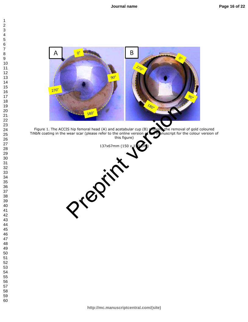
Figure 1. The ACCIS hip femoral head (A) and acetabular cup (B) showing the removal of gold coloured TiNbN coating in the wear scar (please refer to the online version of the manuscript for the colour version of
this figure)
137x67mm (150 x 150 DPI)
Page 16 of 22
http://mc.manuscriptcentral.com/(site)
Journal name
123456789101112131415161718192021222324252627282930313233343536373839404142434445464748495051525354555657585960
Preprin
t vers
ion
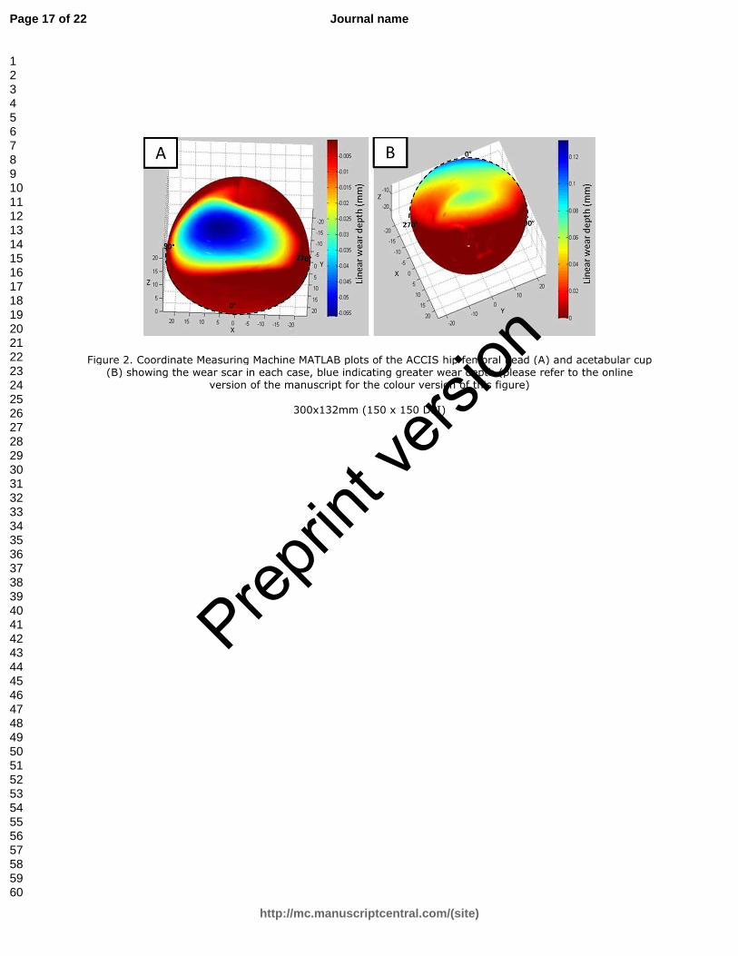
Figure 2. Coordinate Measuring Machine MATLAB plots of the ACCIS hip femoral head (A) and acetabular cup (B) showing the wear scar in each case, blue indicating greater wear depth (please refer to the online
version of the manuscript for the colour version of this figure)
300x132mm (150 x 150 DPI)
Page 17 of 22
http://mc.manuscriptcentral.com/(site)
Journal name
123456789101112131415161718192021222324252627282930313233343536373839404142434445464748495051525354555657585960
Preprin
t vers
ion
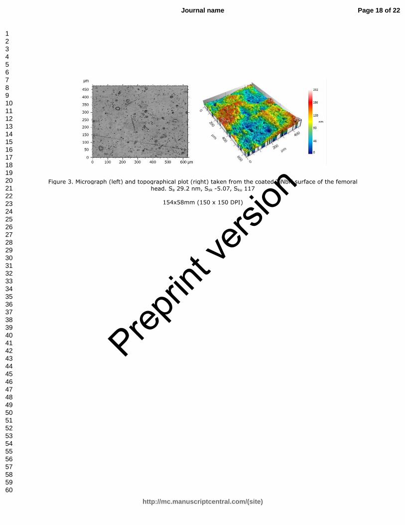
Figure 3. Micrograph (left) and topographical plot (right) taken from the coated TiNbN surface of the femoral head. Sa 29.2 nm, Ssk -5.07, Sku 117
154x58mm (150 x 150 DPI)
Page 18 of 22
http://mc.manuscriptcentral.com/(site)
Journal name
123456789101112131415161718192021222324252627282930313233343536373839404142434445464748495051525354555657585960
Preprin
t vers
ion
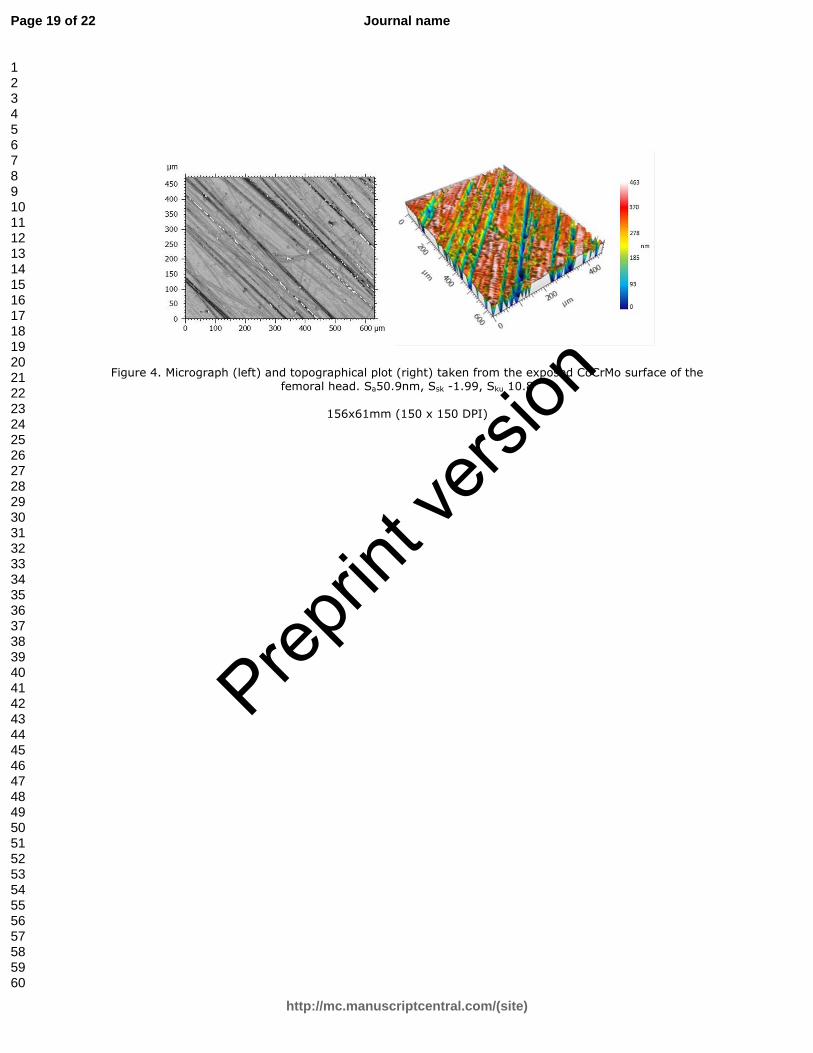
Figure 4. Micrograph (left) and topographical plot (right) taken from the exposed CoCrMo surface of the femoral head. Sa50.9nm, Ssk -1.99, Sku 10.8
156x61mm (150 x 150 DPI)
Page 19 of 22
http://mc.manuscriptcentral.com/(site)
Journal name
123456789101112131415161718192021222324252627282930313233343536373839404142434445464748495051525354555657585960
Preprin
t vers
ion
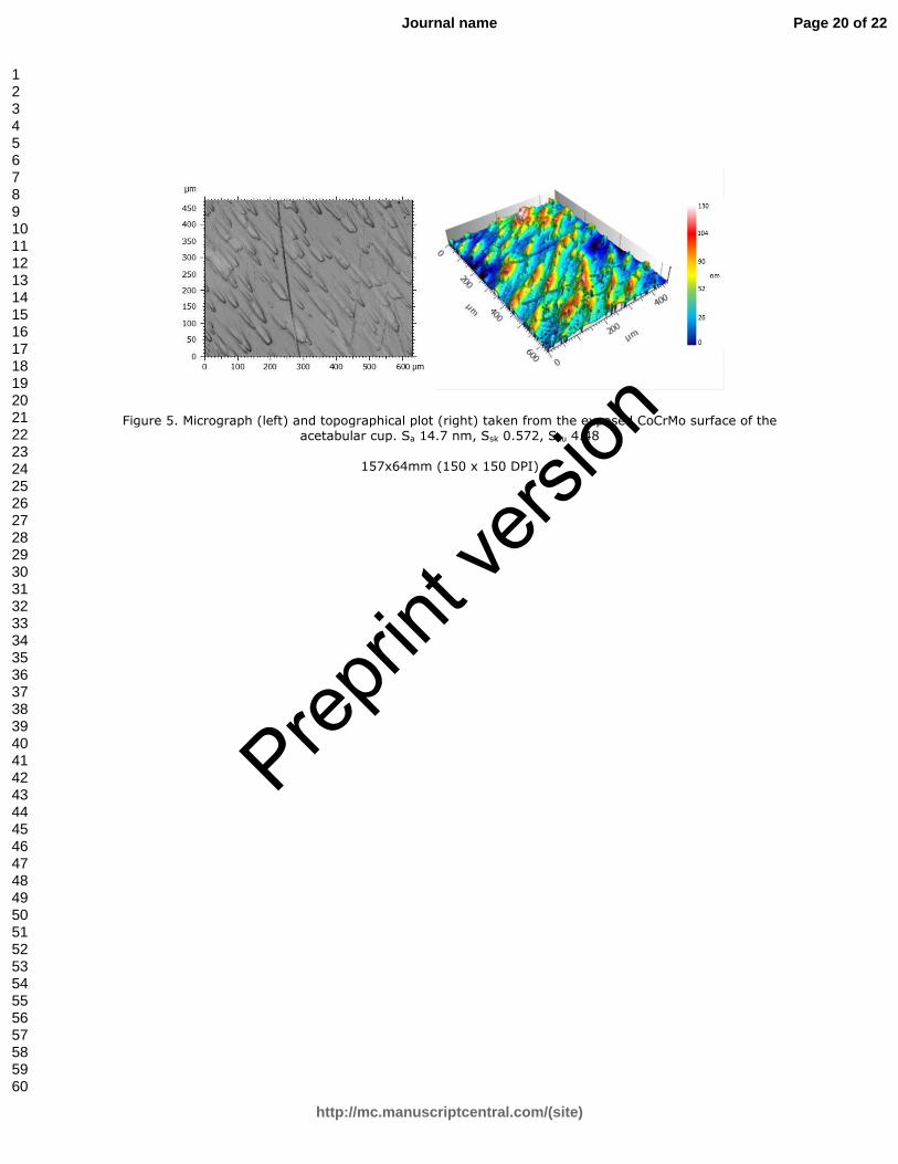
Figure 5. Micrograph (left) and topographical plot (right) taken from the exposed CoCrMo surface of the acetabular cup. Sa 14.7 nm, Ssk 0.572, Sku 4.48
157x64mm (150 x 150 DPI)
Page 20 of 22
http://mc.manuscriptcentral.com/(site)
Journal name
123456789101112131415161718192021222324252627282930313233343536373839404142434445464748495051525354555657585960
Preprin
t vers
ion
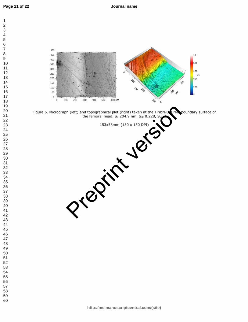
Figure 6. Micrograph (left) and topographical plot (right) taken at the TiNbN-CoCrMo boundary surface of the femoral head. Sa 204.9 nm, Ssk 0.228, Sku 2
153x58mm (150 x 150 DPI)
Page 21 of 22
http://mc.manuscriptcentral.com/(site)
Journal name
123456789101112131415161718192021222324252627282930313233343536373839404142434445464748495051525354555657585960
Preprin
t vers
ion
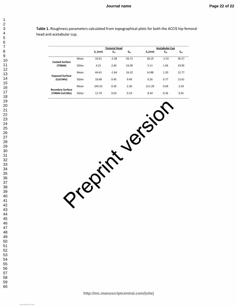
Table 1. Roughness parameters calculated from topographical plots for both the ACCIS hip femoral
head and acetabular cup.
Femoral Head Acetabular Cup
Sa (nm) Ssk Sku Sa (nm) Ssk Sku
Coated Surface
(TiNbN)
Mean 33.61 -2.38 56.71 18.19 -1.53 36.57
StDev 4.21 2.40 53.00 5.11 1.94 19.90
Exposed Surface
(CoCrMo)
Mean 44.61 -1.64 16.32 14.88 1.20 12.77
StDev 10.68 0.45 9.49 0.26 0.77 13.61
Boundary Surface
(TiNbN-CoCrMo)
Mean 245.55 0.30 2.36 211.29 0.09 2.50
StDev 12.70 0.03 0.19 8.34 0.36 0.45
Page 22 of 22
http://mc.manuscriptcentral.com/(site)
Journal name
123456789101112131415161718192021222324252627282930313233343536373839404142434445464748495051525354555657585960
Preprin
t vers
ion
View publication statsView publication stats
Copyright © 2022 FDOKUMEN


![The Resurfacing of Arabic Qaf [q] in the Speech of Young ...](https://static.fdokumen.com/doc/165x107/6327fe736d480576770d8d74/the-resurfacing-of-arabic-qaf-q-in-the-speech-of-young-.jpg)